Ups1p and Ups2p antagonistically regulate cardiolipin metabolism in mitochondria
Upload
independentCategory
view
4download
0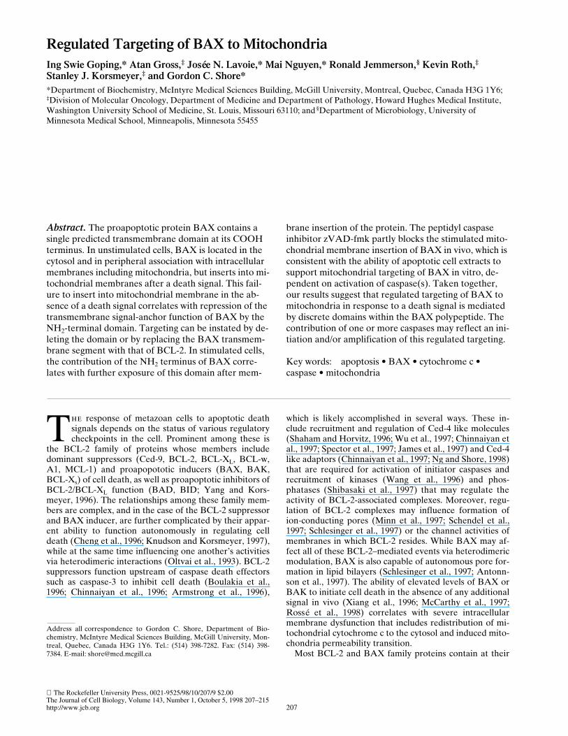
The Rockefeller University Press, 0021-9525/98/10/207/9 $2.00The Journal of Cell Biology, Volume 143, Number 1, October 5, 1998 207–215http://www.jcb.org 207
Regulated Targeting of BAX to Mitochondria
Ing Swie Goping,* Atan Gross,
‡
Josée N. Lavoie,* Mai Nguyen,* Ronald Jemmerson,
§
Kevin Roth,
‡
Stanley J. Korsmeyer,
‡
and Gordon C. Shore*
*Department of Biochemistry, McIntyre Medical Sciences Building, McGill University, Montreal, Quebec, Canada H3G 1Y6;
‡
Division of Molecular Oncology, Department of Medicine and Department of Pathology, Howard Hughes Medical Institute, Washington University School of Medicine, St. Louis, Missouri 63110; and
§
Department of Microbiology, University of Minnesota Medical School, Minneapolis, Minnesota 55455
Abstract.
The proapoptotic protein BAX contains a single predicted transmembrane domain at its COOH terminus. In unstimulated cells, BAX is located in the cytosol and in peripheral association with intracellular membranes including mitochondria, but inserts into mi-tochondrial membranes after a death signal. This fail-ure to insert into mitochondrial membrane in the ab-sence of a death signal correlates with repression of the transmembrane signal-anchor function of BAX by the NH
2
-terminal domain. Targeting can be instated by de-leting the domain or by replacing the BAX transmem-brane segment with that of BCL-2. In stimulated cells, the contribution of the NH
2
terminus of BAX corre-lates with further exposure of this domain after mem-
brane insertion of the protein. The peptidyl caspaseinhibitor zVAD-fmk partly blocks the stimulated mito-chondrial membrane insertion of BAX in vivo, which is consistent with the ability of apoptotic cell extracts to support mitochondrial targeting of BAX in vitro, de-pendent on activation of caspase(s). Taken together, our results suggest that regulated targeting of BAX to mitochondria in response to a death signal is mediated by discrete domains within the BAX polypeptide. The contribution of one or more caspases may reflect an ini-tiation and/or amplification of this regulated targeting.
Key words: apoptosis • BAX • cytochrome c • caspase • mitochondria
T
he
response of metazoan cells to apoptotic deathsignals depends on the status of various regulatorycheckpoints in the cell. Prominent among these is
the BCL-2 family of proteins whose members includedominant suppressors (Ced-9, BCL-2, BCL-X
L
, BCL-w,A1, MCL-1) and proapopototic inducers (BAX, BAK,BCL-X
s
) of cell death, as well as proapoptotic inhibitors ofBCL-2/BCL-X
L
function (BAD, BID; Yang and Kors-meyer, 1996). The relationships among these family mem-bers are complex, and in the case of the BCL-2 suppressorand BAX inducer, are further complicated by their appar-ent ability to function autonomously in regulating celldeath (Cheng et al., 1996; Knudson and Korsmeyer, 1997),while at the same time influencing one another’s activitiesvia heterodimeric interactions (Oltvai et al., 1993). BCL-2suppressors function upstream of caspase death effectorssuch as caspase-3 to inhibit cell death (Boulakia et al.,1996; Chinnaiyan et al., 1996; Armstrong et al., 1996),
which is likely accomplished in several ways. These in-clude recruitment and regulation of Ced-4 like molecules(Shaham and Horvitz, 1996; Wu et al., 1997; Chinnaiyan etal., 1997; Spector et al., 1997; James et al., 1997) and Ced-4like adaptors (Chinnaiyan et al., 1997; Ng and Shore, 1998)that are required for activation of initiator caspases andrecruitment of kinases (Wang et al., 1996) and phos-phatases (Shibasaki et al., 1997) that may regulate theactivity of BCL-2-associated complexes. Moreover, regu-lation of BCL-2 complexes may influence formation ofion-conducting pores (Minn et al., 1997; Schendel et al.,1997; Schlesinger et al., 1997) or the channel activities ofmembranes in which BCL-2 resides. While BAX may af-fect all of these BCL-2–mediated events via heterodimericmodulation, BAX is also capable of autonomous pore for-mation in lipid bilayers (Schlesinger et al., 1997; Antonn-son et al., 1997). The ability of elevated levels of BAX orBAK to initiate cell death in the absence of any additionalsignal in vivo (Xiang et al., 1996; McCarthy et al., 1997;Rossé et al., 1998) correlates with severe intracellularmembrane dysfunction that includes redistribution of mi-tochondrial cytochrome c to the cytosol and induced mito-chondria permeability transition.
Most BCL-2 and BAX family proteins contain at their
Address all
correspondence to Gordon C. Shore, Department of Bio-chemistry, McIntyre Medical Sciences Building, McGill University, Mon-treal, Quebec, Canada H3G 1Y6. Tel.: (514) 398-7282. Fax: (514) 398-7384. E-mail: [email protected]

The Journal of Cell Biology, Volume 143, 1998 208
extreme COOH terminus a single predicted transmem-brane segment (TM)
1
. In the case of BCL-2, the TM func-tions as a signal anchor that targets and inserts the proteinin a N
cyto
-C
in
orientation into the two main membrane lo-cations for this protein: the mitochondrial outer mem-brane and the ER/nuclear envelope (Hockenbery et al.,1990; Krajewski et al., 1993; Nguyen et al., 1993; Nguyen etal., 1994). Strikingly, however, the ability of BAX to trans-locate to membrane sites, including mitochondria, is regu-lated in certain contexts and depends upon the cell receiv-ing a death signal (Hsu et al., 1997; Wolter et al., 1997). Inthe absence of such a signal, BAX is found free in the cy-tosol or peripherally associated with endocellular mem-brane surfaces. Although the mechanism for regulated tar-geting of BAX or BAK remains to be elucidated, the factthat transient overexpression (Xiang et al., 1996; Rossé etal., 1998) or forced dimerization (Gross et al., 1998) of theprotein can lead to cell death in the absence of other sig-nals implies that the regulatory mechanism can be overrid-den in certain contexts.
Here we demonstrate that BAX targeting to mitochon-dria can be regulated by zVAD-sensitive caspase(s). Sig-nificantly, prevention of BAX targeting in the absence of adeath signal has been mapped to two regions of the mole-cule: the NH
2
-terminal ART domain and the COOH-ter-minal TM. Our results indicate a regulated mechanism bywhich a death signal can cause translocation of BAX tomitochondria where it mediates and amplifies membranedysfunction (reviewed in Reed, 1997; Hengartner, 1998).
Materials and Methods
Immunocytochemistry
FL5.12 cells were resuspended in fresh media containing 400 nM Mi-totracker Green FM (Molecular Probes, Inc., Eugene, OR). After a 15-min incubation, cells were washed and fixed in 3% paraformaldehyde andblocked with 2% normal goat serum (Vector Labs, Inc., Burlingame, CA).Anti-mBAX (P-19) Ab (Santa Cruz Biotechnology, Santa Cruz, CA) andCy3 goat anti–rabbit Ab (Jackson ImmunoResearch Laboratories, Inc.,West Grove, PA) were used to detect BAX immunoreactivity. Nucleiwere counterstained with Hoechst H33258 (1
m
g/ml). Slides were viewedand photographed with a Zeiss Axioskop fluorescence microscope and anMC 100 camera attachment. Confocal laser scanning was performed usinga Sarastro 2000 confocal laser scanning microscope (Molecular Dynamics,Inc., Sunnyvale, CA) and Image Space software (Molecular Dynamics).
Plasmids
Using standard recombinant DNA manipulations, cDNA encoding mu-rine
BAX
D
1-19 was constructed in pBluescript SK under control of the T7promoter; the resulting translation product had an initiating methioninelocated 20 amino acids downstream of the original methionine of the full-length BAX construct. cDNA encoding DHFR-BAX™ was created byPCR in Bluescript SK, and encoded murine dihydrofolate reductase fusedto the COOH-terminal 23 amino acids (aa 169–192) of BAX. Similarly,BAX-BCL-2™ was created by fusing amino acids 1–168 of BAX to aminoacid 218–239 of human BCL-2.
Mitochondria from Rat Heart
The heart of one male Sprague Dawley rat (
z
250 g) was placed in
z
100ml HIM buffer (0.2% wt/vol BSA, 200 mM mannitol, 70 mM sucrose, 10mM Hepes-KOH, 1 mM EGTA, pH 7.5) on ice, squeezed several times to
force out blood, and transferred to 7.5 ml HIM in a 15-ml corex tube. Allsubsequent steps were conducted at 4
8
C. The heart was homogenized witha Polytron homogenizer (Brinkmann Instruments Canada Ltd., Missis-sauga, ON, Canada) operating for 1 s at setting of 6.5. Nuclei and unbro-ken cells were pelleted at 1,800 rpm for 10 min in a Sorvall SS34 rotor. Thesupernatant was centrifuged for 10 min at 7,000 rpm. The resulting pelletwas resuspended in HIM (minus BSA), centrifuged at 1,800 rpm, and themitochondria were collected at 7,000 rpm. The mitochondrial pellet wasresuspended in cMRM (250 mM sucrose, 10 mM Hepes-KOH, 1 mMATP, 5 mM Na succinate, 0.08 mM ADP, 2 mM K
2
HPO
4
, pH 7.5) at aconcentration of 1 mg of mitochondrial protein per ml, and adjusted to 1mM dithiothreitol just before use.
Mitochondria from Cultured Cells
Cells were collected, washed twice with PBS, suspended in 2 ml HIM, andsubjected to Polytron homogenization for four bursts of 10 s each at a set-ting of 6.5. Mitochondria were then isolated according to the rat heart pro-tocol.
Mitoplasts
Protein import-competent mitoplasts (mitochondria with a severely dis-rupted outer membrane) in which the inner membrane and transbilayerelectrochemical potential remain intact were prepared from rat liver ex-actly as described in McBride et al. (1995).
Mitochondrial Protein Targeting In Vitro
cDNAs were transcribed and translated in the presence of
35
S-methioninein a cell-free rabbit reticulocyte lysate system (Promega Corp., Madison,WI) according to the manufacturer’s directions. For a standard mitochon-drial protein import reaction, 5
m
l of translation reaction was incubatedwith 20
m
l of Buffer A (20 mM Hepes-KOH, 10 mM KCl, 2.5 mM MgCl
2
,1 mM EDTA, 1 mM EGTA, 1 mM dithiothreitol, pH 7.5) and 25
m
l mito-chondria or mitoplasts in cMRM (1 mg protein/ml) at 30
8
C or 37
8
C for 30min to 2 h. The import reaction was then layered on a 500-
m
l cushion of1
3
MRM (250 mM sucrose, 10 mM Hepes-KOH, pH 7.5) and centrifugedfor 4 min at top speed in a microfuge at 4
8
C. For alkali extraction, the mi-tochondrial pellet was resuspended (0.25 mg protein/ml) in freshly pre-pared 0.1 M Na
2
CO
3
, pH 11.5, and incubated for 30 min on ice. The mem-branes were then pelleted in an airfuge (Beckman Instruments Canada,Inc., Montreal, PQ, Canada) operating for 10 min at 30 psi. The pellet wasanalyzed by SDS-PAGE and fluorography. For import assays that in-cluded apoptotic cell extract (see below), it replaced buffer A; the bufferused to make the extract, buffer A/ext, did not influence import.
Apoptotic Cell Extract
Extract was prepared at 4
8
C
according to the procedure of Liu et al.(1996) with some minor modifications. In brief, human KB epithelial cellswere harvested in PBS containing
1.3 mM Na citrate and 0.6
mM EDTA.The cell pellet was washed in buffer A/ext
(50 mM Pipes, 50 mM KCl, 5mM EGTA, 2 mM MgCl
2
, 1 mM dithiothreitol, 20
m
M cytochalasin B, pH7.4), and was then resuspended in an equal volume of buffer A/ext. Thecells were disrupted by five cycles of freeze-thaw interspersed by fivestrokes with a Wheaton glass homogenizer fitted with a B pestle, and cen-trifuged at 10
5
g
for 1 h in a Beckman Ti75 rotor. The resulting superna-tant contained 10 mg protein/ml. To deplete the supernatant of cyto-chrome c (Liu et al., 1996), 40
m
l of 2G8.B6 anti-cytochrome c antibody(7.7 mg protein/ml) was first incubated with 100
m
l of a 1:1 mixture of pro-tein A and protein G Sepharose resuspended in 200
m
l of PBS for 4 h at4
8
C. For the control reaction, 40
m
l of buffer A was substituted for the an-tibody. The beads were collected, washed with buffer A, and then incu-bated with 125
m
l of KB cell extract for 18 h at 4
8
C. The beads were recov-ered and the supernatant was collected.
PARP Cleavage
KB apoptotic cell extract (50–100
m
g protein) was incubated with 1 mMdATP in a final volume of 20
m
l of buffer A at 30
8
C for 1 h, and 1
m
l of
35
S-methionine–labeled PARP derived by transcription-translation in rabbitreticulocyte lysate was added. After 15 min at 30
8
C, 5
m
l of 5
3
SDS sam-ple buffer was added, and 12.5-
m
l aliquots of each reaction were analyzedby SDS-PAGE and fluorography.
1.
Abbreviations used in this paper
: HA, hemagglutinin epitope; PARP,poly(ADP ribosyl) polymerase; TM, transmembrane segment.
Goping et al.
BAX Targeting to Mitochondria
209
Results
BAX Integrates into Mitochondrial Membrane after a Death Stimulus In Vivo
Immunocytochemical analysis of FL5.12 hematopoieticcells revealed BAX in association with mitochondria, aswell as distributed throughout the cytoplasm and perinu-clear region (Fig. 1). When analyzed by immunoblotting,the mitochondrial fraction isolated from these cells re-vealed the presence of BAX as well as BCL-2, cytochromec oxidase subunit iv, and cytochrome c (Fig. 2
A
, lanes
1
and
2
). In contrast to BCL-2, which was resistant to extrac-tion at alkaline pH as a result of its integration into themembrane lipid bilayer (Nguyen et al., 1993; Nguyen et al.,1994), the majority of BAX was liberated under the sameconditions (Fig. 2
A
, lanes
3
and
4
; Fig. 2
C
, lanes
1
and
5
),indicative of a peripheral association with the organelle(Fujiki et al., 1982). After induction of cell death uponwithdrawal of IL-3 (Hockenbery et al., 1990), however,this situation was reversed and most of the BAX now ac-quired resistance to alkaline extraction (Fig. 2
A
, lanes
5
–
8
; Fig. 2
C
, lanes
3
and
7
). As expected, cytochrome cwas released in response to alkaline extraction, whereasBCL-2 and cytochrome c oxidase subunit iv
were retained,and these distributions were unaffected by the deathstimulus (Fig. 2
A
). We conclude, therefore, that BAX
demonstrates a specific response to the death signal invivo, moving from a membrane-peripheral to a mem-brane-integrated position.
Of note, integration of BAX into mitochondrial mem-brane after IL-3 withdrawal was also accompanied by anapparent change at the extreme NH
2
terminus of the pro-tein. In isolated mitochondria, this region of the BAXmolecule was now accessible to limited proteolysis by ex-ogenous trypsin or proteinase K, as revealed by the loss ofimmunoreactivity of the cleavage product to an antibody(N20) directed against amino acids 11–30 of BAX (Fig. 2
B
). The remaining protease-resistant BAX fragment, onthe other hand, retained immunoreactivity to an antibody(651) directed against amino acids 43–61.
Finally, BAX integration into mitochondrial membranein response to IL-3 withdrawal from FL5.12 cells for 12 hwas partly blocked by the wide spectrum caspase inhibitor,zVAD-fmk. Whereas the inhibitor did not significantly in-fluence recovery of total BAX associated with mito-chondria (Fig. 2
C
, lanes
3
and
4
), it reduced the amount ofalkaline-resistant membrane-integrated BAX that was re-covered with the organelle (Fig. 2
C
, lanes
7
and
8
). Thissuggests that caspases, which are activated in response toIL-3 withdrawal, stimulate mitochondrial membrane inte-gration of BAX in vivo.
An Apoptotic Cell Extract Stimulates BAX Targetingto Mitochondria In Vitro
To examine BAX targeting into mitochondria in vitro, weused the
35
S-labeled transcription-translation product of
BAX
cDNA in rabbit reticulocyte lysate combined withintact mitochondria isolated from rat heart. This is a well-documented system that faithfully reflects in vivo importof diverse mitochondrial proteins, including insertion ofintegral proteins into the mitochondrial outer membrane(Li and Shore, 1992; McBride et al., 1992). The variousBAX constructs that were used for these assays, and theirtargeting competence in vitro, are summarized in Fig. 3.
Transcription-translation of
BAX
cDNA yielded full-length protein, as well as a prominent product
z
2 kDsmaller and apparently arising from an internal translationinitiation, resulting in an NH
2
-terminal truncated BAX(designated BAX
D
ART). However, only the truncatedBAX
D
ART was membrane-integrated, as judged by ac-quired resistance to extraction at alkaline pH (Fig. 4
A
;compare lanes
2
and
5
). Insertion of this product was tem-perature-sensitive; it occurred at 30
8
C, but not at 4
8
C (Fig.5, lanes
2
–
5
), indicative of membrane integration ratherthan tight but nonspecific binding (McBride et al., 1992).Inspection of the BAX sequence revealed the presenceof an internal methionine at codon position 20 (Fig. 3).Enforced translation initiation from met codon 20 wasachieved by deleting codon 1, which yielded a product thatcomigrated in SDS PAGE with BAX
D
ART and demon-strated temperature-sensitive membrane integration (Fig.4
B
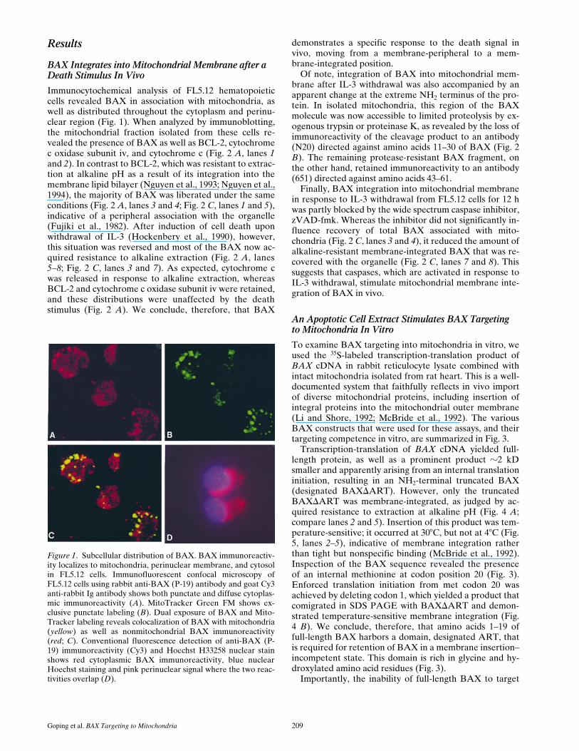
). We conclude, therefore, that amino acids 1–19 offull-length BAX harbors a domain, designated ART, thatis required for retention of BAX in a membrane insertion–incompetent state. This domain is rich in glycine and hy-droxylated amino acid residues (Fig. 3).
Importantly, the inability of full-length BAX to target
Figure 1. Subcellular distribution of BAX. BAX immunoreactiv-ity localizes to mitochondria, perinuclear membrane, and cytosolin FL5.12 cells. Immunofluorescent confocal microscopy ofFL5.12 cells using rabbit anti-BAX (P-19) antibody and goat Cy3anti-rabbit Ig antibody shows both punctate and diffuse cytoplas-mic immunoreactivity (A). MitoTracker Green FM shows ex-clusive punctate labeling (B). Dual exposure of BAX and Mito-Tracker labeling reveals colocalization of BAX with mitochondria(yellow) as well as nonmitochondrial BAX immunoreactivity(red; C). Conventional fluorescence detection of anti-BAX (P-19) immunoreactivity (Cy3) and Hoechst H33258 nuclear stainshows red cytoplasmic BAX immunoreactivity, blue nuclearHoechst staining and pink perinuclear signal where the two reac-tivities overlap (D).

The Journal of Cell Biology, Volume 143, 1998 210
mitochondria in vitro was overcome by supplementing theimport reaction mixture with an apoptotic cell extractderived from KB epithelial cells. This extract was pre-pared according to Liu et al. (1996), and involved cycles offreeze/thaw and homogenization of cells in hypotonic me-dium, causing swelling of mitochondria and consequentrelease of cytochrome c as a result of a ruptured outermembrane. The resulting high-speed cytosolic supernatantcontains endogenous procaspases whose activation can
be achieved by adding dATP and incubating the extractat 37
8
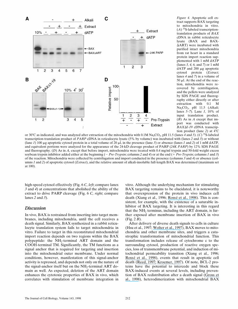
C, resulting in diagnostic cleavage of the caspase-3death substrate, poly(ADP ribosyl) polymerase (PARP;Liu et al., 1996). The presence of this extract in import re-actions stimulated binding (Fig. 4 A, lanes 3 and 4) and al-kaline-resistant membrane insertion of full-length BAX(lanes 6 and 7), but did not stimulate BAXDART inser-tion (lanes 6 and 7). Membrane-integrated BAX was ac-cessible to exogenous trypsin in these intact mitochondria
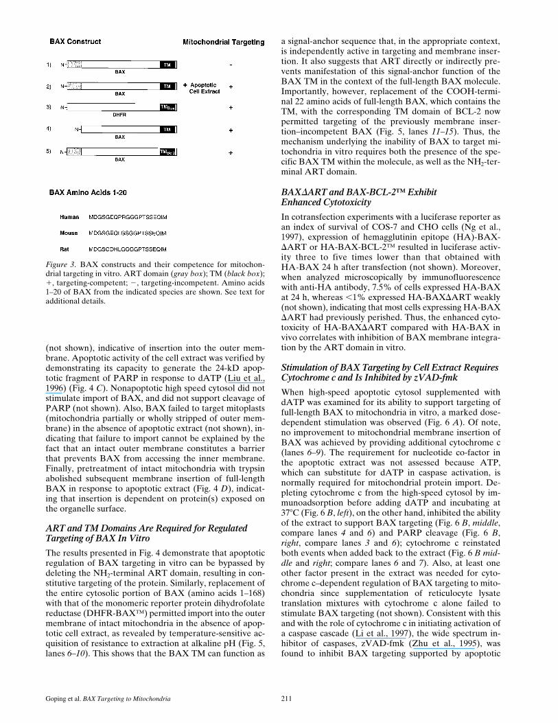
Figure 2. Mitochondrial BAX changes its alkali and protease sen-sitivity after a death stimulus. (A) BAX becomes alkali-resistantafter a death stimulus. Heavy membranes enriched in mitochon-dria were prepared from FL5.12 cells cultured in IL-3 (lanes 1–4)or deprived of IL-3 for 12 h (lanes 5–8), incubated in isotonicbuffer (lanes 1 and 2 and lanes 5 and 6), or in 0.1 M Na2CO3, pH11.5 (lanes 3 and 4 and lanes 7 and 8), and centrifuged at 200,000 gfor 45 min to yield supernatant (S) and pellet (P). The fractionswere analyzed by immunoblot for BAX, BCL-2, cytochrome c ox-idase subunit iv, and cytochrome c. (B) The NH2-terminus ofBAX is further exposed after a death stimulus. The mitochondrialfraction prepared from FL5.12 cells in IL-3 (lanes 1 and 2 andlanes 6 and 7) or deprived of IL-3 for 12 h (lanes 3–5, and lanes8–10) were incubated in isotonic buffer and treated with trypsin(30 mg/ml; lanes 2, 4, and 5) or proteinase K (100 mg/ml; prot. K,lanes 7, 9, and 10), for 20 min at 48C. Trypsin was inactivated byadding a 30-fold weight excess of soybean trypsin inhibitor, andproteinase K was inactivated with phenylmethylsulfonylfluoride(1 mM). The fractions were analyzed by immunoblot with anti-BAX 651 antibody (amino acids 43–61; lanes 1–4 and lanes 6–9)or with anti-BAX N20 antibody (amino acids 11–30; Santa CruzBiotechnology; lanes 5 and 10). (C) zVAD-fmk reduces mito-chondrial membrane integration of BAX after IL-3 withdrawal.The mitochondrial fraction prepared from FL5.12 cells main-tained in IL-3 (lanes 1, 2, 5, and 6) or deprived of IL-3 for 12 h(lanes 3, 4, 7, and 8) in the absence (lanes 1, 3, 5, and 7) or pres-ence (lanes 2, 4, 6, and 8) of 50 mM zVAD-fmk for 12 h, were an-alyzed either directly (lanes 1–4) or after extraction with 0.1 MNa2CO3, pH 11.5 (lanes 5–8), as described in A.
Goping et al. BAX Targeting to Mitochondria 211
(not shown), indicative of insertion into the outer mem-brane. Apoptotic activity of the cell extract was verified bydemonstrating its capacity to generate the 24-kD apop-totic fragment of PARP in response to dATP (Liu et al.,1996) (Fig. 4 C). Nonapoptotic high speed cytosol did notstimulate import of BAX, and did not support cleavage ofPARP (not shown). Also, BAX failed to target mitoplasts(mitochondria partially or wholly stripped of outer mem-brane) in the absence of apoptotic extract (not shown), in-dicating that failure to import cannot be explained by thefact that an intact outer membrane constitutes a barrierthat prevents BAX from accessing the inner membrane.Finally, pretreatment of intact mitochondria with trypsinabolished subsequent membrane insertion of full-lengthBAX in response to apoptotic extract (Fig. 4 D), indicat-ing that insertion is dependent on protein(s) exposed onthe organelle surface.
ART and TM Domains Are Required for Regulated Targeting of BAX In Vitro
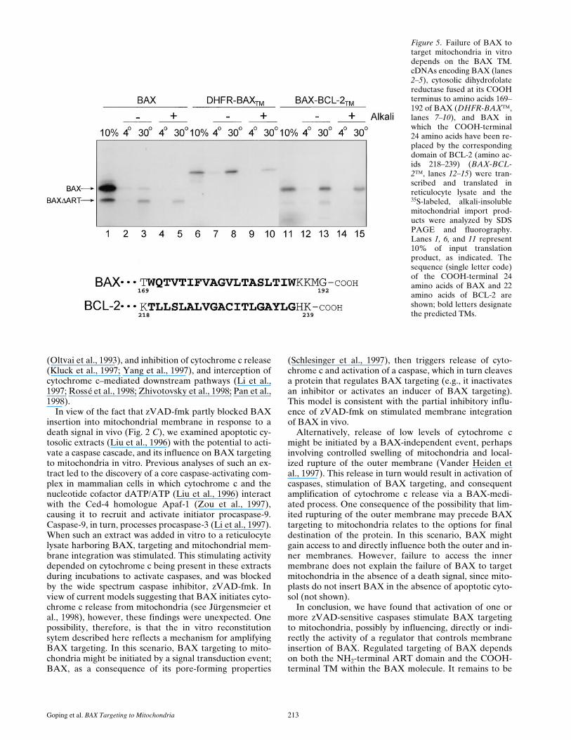
The results presented in Fig. 4 demonstrate that apoptoticregulation of BAX targeting in vitro can be bypassed bydeleting the NH2-terminal ART domain, resulting in con-stitutive targeting of the protein. Similarly, replacement ofthe entire cytosolic portion of BAX (amino acids 1–168)with that of the monomeric reporter protein dihydrofolatereductase (DHFR-BAX™) permitted import into the outermembrane of intact mitochondria in the absence of apop-totic cell extract, as revealed by temperature-sensitive ac-quisition of resistance to extraction at alkaline pH (Fig. 5,lanes 6–10). This shows that the BAX TM can function as
a signal-anchor sequence that, in the appropriate context,is independently active in targeting and membrane inser-tion. It also suggests that ART directly or indirectly pre-vents manifestation of this signal-anchor function of theBAX TM in the context of the full-length BAX molecule.Importantly, however, replacement of the COOH-termi-nal 22 amino acids of full-length BAX, which contains theTM, with the corresponding TM domain of BCL-2 nowpermitted targeting of the previously membrane inser-tion–incompetent BAX (Fig. 5, lanes 11–15). Thus, themechanism underlying the inability of BAX to target mi-tochondria in vitro requires both the presence of the spe-cific BAX TM within the molecule, as well as the NH2-ter-minal ART domain.
BAXDART and BAX-BCL-2™ ExhibitEnhanced Cytotoxicity
In cotransfection experiments with a luciferase reporter asan index of survival of COS-7 and CHO cells (Ng et al.,1997), expression of hemagglutinin epitope (HA)-BAX-DART or HA-BAX-BCL-2™ resulted in luciferase activ-ity three to five times lower than that obtained withHA-BAX 24 h after transfection (not shown). Moreover,when analyzed microscopically by immunofluorescencewith anti-HA antibody, 7.5% of cells expressed HA-BAXat 24 h, whereas ,1% expressed HA-BAXDART weakly(not shown), indicating that most cells expressing HA-BAXDART had previously perished. Thus, the enhanced cyto-toxicity of HA-BAXDART compared with HA-BAX invivo correlates with inhibition of BAX membrane integra-tion by the ART domain in vitro.
Stimulation of BAX Targeting by Cell Extract Requires Cytochrome c and Is Inhibited by zVAD-fmk
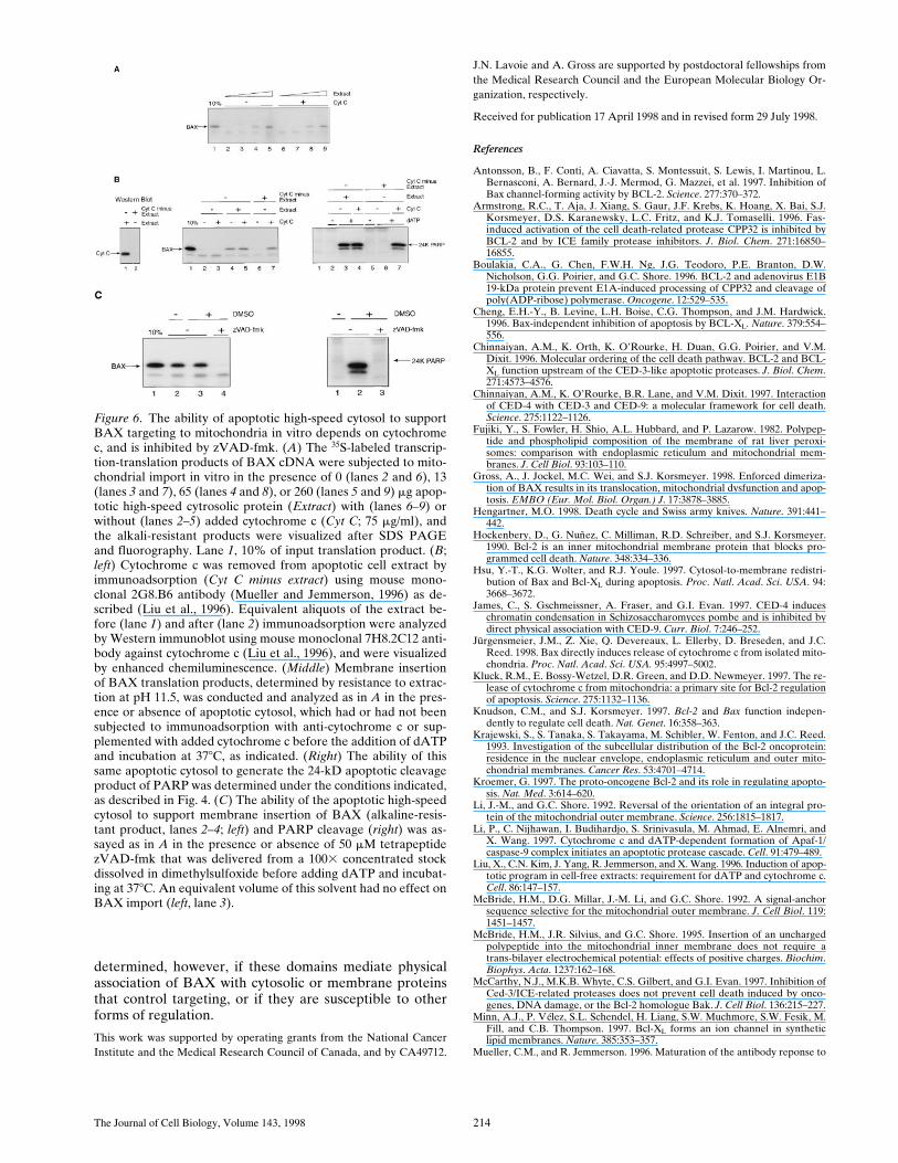
When high-speed apoptotic cytosol supplemented withdATP was examined for its ability to support targeting offull-length BAX to mitochondria in vitro, a marked dose-dependent stimulation was observed (Fig. 6 A). Of note,no improvement to mitochondrial membrane insertion ofBAX was achieved by providing additional cytochrome c(lanes 6–9). The requirement for nucleotide co-factor inthe apoptotic extract was not assessed because ATP,which can substitute for dATP in caspase activation, isnormally required for mitochondrial protein import. De-pleting cytochrome c from the high-speed cytosol by im-munoadsorption before adding dATP and incubating at378C (Fig. 6 B, left), on the other hand, inhibited the abilityof the extract to support BAX targeting (Fig. 6 B, middle,compare lanes 4 and 6) and PARP cleavage (Fig. 6 B,right, compare lanes 3 and 6); cytochrome c reinstatedboth events when added back to the extract (Fig. 6 B mid-dle and right; compare lanes 6 and 7). Also, at least oneother factor present in the extract was needed for cyto-chrome c–dependent regulation of BAX targeting to mito-chondria since supplementation of reticulocyte lysatetranslation mixtures with cytochrome c alone failed tostimulate BAX targeting (not shown). Consistent with thisand with the role of cytochrome c in initiating activation ofa caspase cascade (Li et al., 1997), the wide spectrum in-hibitor of caspases, zVAD-fmk (Zhu et al., 1995), wasfound to inhibit BAX targeting supported by apoptotic
Figure 3. BAX constructs and their competence for mitochon-drial targeting in vitro. ART domain (gray box); TM (black box);1, targeting-competent; 2, targeting-incompetent. Amino acids1–20 of BAX from the indicated species are shown. See text foradditional details.
The Journal of Cell Biology, Volume 143, 1998 212
high-speed cytosol effectively (Fig. 6 C, left; compare lanes3 and 4) at concentrations that abolished the ability of theextract to drive PARP cleavage (Fig. 6 C, right; comparelanes 2 and 3).
DiscussionIn vivo, BAX is restrained from inserting into target mem-branes, including mitochondria, until the cell receives adeath signal. Similarly, BAX synthesized in a rabbit reticu-locyte translation system fails to target mitochondria invitro. Failure to target in this reconstituted mitochondrialimport reaction depends on two regions within the BAXpolypeptide: the NH2-terminal ART domain and theCOOH-terminal TM. Significantly, the TM functions as asignal anchor that is required for targeting and insertioninto the mitochondrial outer membrane. Under normalconditions, however, manifestation of this signal-anchoractivity is repressed, and depends not only on the nature ofthe signal-anchor itself but on the NH2-terminal ART do-main as well. As expected, deletion of the ART domainenhances the cytotoxic properties of BAX in vivo, whichcorrelates with stimulation of membrane integration in
vitro. Although the underlying mechanism for stimulatingBAX targeting remains to be elucidated, it is noteworthythat overexpression of the protein in vivo induces celldeath (Xiang et al., 1996; Rossé et al., 1998). This is con-sistent, for example, with the existence of a saturable in-hibitor of BAX targeting. It is interesting in this regardthat the NH2 terminus, including the ART domain, is fur-ther exposed after membrane insertion of BAX in vivo(Fig. 2 B).
After delivery of diverse death signals to cells in culture(Hsu et al., 1997; Wolter et al., 1997), BAX moves to mito-chondria and other membrane sites, and triggers a cata-strophic transformation of mitochondrial function. Thistransformation includes release of cytochrome c to thesurrounding cytosol, production of reactive oxygen spe-cies, loss of transmembrane potential, and induction of mi-tochondrial permeability transition (Xiang et al., 1996;Rossé et al., 1998), events that result in apoptotic celldeath (Reed, 1997; Kroemer, 1997). Of note, BCL-2 pro-teins have the potential to intercede and block theseBAX-induced events at several levels, including preven-tion of BAX redistribution after a death signal (Gross etal., 1998), heterodimerization with mitochondrial BAX
Figure 4. Apoptotic cell ex-tract supports BAX targetingto mitochondria in vitro.(A) 35S-labeled transcription-translation products of BAXcDNA in rabbit reticulocytelysate (BAX and BAX-DART) were incubated withpurified intact mitochondriafrom rat heart in a standardprotein import reaction sup-plemented with 1 mM dATP(lanes 3, 4, 6, and 7) or 1 mMdATP and 200 mg apoptoticcytosol protein (Extract;lanes 4 and 7) in a volume of50 ml. At the end of the reac-tion, mitochondria were re-covered by centrifugation,and the pellets were analyzedby SDS PAGE and fluorog-raphy either directly or afterextraction with 0.1 MNa2CO3, pH 11.5 (Alkali;lanes 5–7). Lane 1, 10% ofinput translation product.(B) As in A except that im-port was conducted withBAXD1-19 cDNA transcrip-tion product (lane 2) at 48C
or 308C as indicated, and was analyzed after extraction of the mitochondria with 0.1M Na2CO3, pH 11.5 (lanes 4 and 5). (C) 35S-labeledtranscription-translation product of PARP cDNA in reticulocyte lysate (5% by volume) was incubated with (lanes 2 and 3) or without(lane 1) 100 mg apoptotic cytosol protein in a total volume of 20 ml, in the presence (lane 3) or absence (lanes 1 and 2) of 1 mM dATP,and equivalent portions were analyzed for the appearance of the 24-kD cleavage product of PARP (24K PARP) by 12% SDS PAGEand fluorography. (D) As in A, except that before import, mitochondria were treated with 0.4 mg/ml trypsin and 50-fold weight excesssoybean trypsin inhibitor added either at the beginning (2 Pre-Trypsin; columns 2 and 4) or at the end (1 Pre-Trypsin; columns 1 and 3)of the reaction. Mitochondria were collected by centrifugation and import conducted in the presence (columns 3 and 4) or absence (col-umns 1 and 2) of apoptotic cytosol (Extract), and the relative amount of alkali-insoluble full-length BAX was determined (maximum setat 100).
Goping et al. BAX Targeting to Mitochondria 213
(Oltvai et al., 1993), and inhibition of cytochrome c release(Kluck et al., 1997; Yang et al., 1997), and interception ofcytochrome c–mediated downstream pathways (Li et al.,1997; Rossé et al., 1998; Zhivotovsky et al., 1998; Pan et al.,1998).
In view of the fact that zVAD-fmk partly blocked BAXinsertion into mitochondrial membrane in response to adeath signal in vivo (Fig. 2 C), we examined apoptotic cy-tosolic extracts (Liu et al., 1996) with the potential to acti-vate a caspase cascade, and its influence on BAX targetingto mitochondria in vitro. Previous analyses of such an ex-tract led to the discovery of a core caspase-activating com-plex in mammalian cells in which cytochrome c and thenucleotide cofactor dATP/ATP (Liu et al., 1996) interactwith the Ced-4 homologue Apaf-1 (Zou et al., 1997),causing it to recruit and activate initiator procaspase-9.Caspase-9, in turn, processes procaspase-3 (Li et al., 1997).When such an extract was added in vitro to a reticulocytelysate harboring BAX, targeting and mitochondrial mem-brane integration was stimulated. This stimulating activitydepended on cytochrome c being present in these extractsduring incubations to activate caspases, and was blockedby the wide spectrum caspase inhibitor, zVAD-fmk. Inview of current models suggesting that BAX initiates cyto-chrome c release from mitochondria (see Jürgensmeier etal., 1998), however, these findings were unexpected. Onepossibility, therefore, is that the in vitro reconstitutionsytem described here reflects a mechanism for amplifyingBAX targeting. In this scenario, BAX targeting to mito-chondria might be initiated by a signal transduction event;BAX, as a consequence of its pore-forming properties
(Schlesinger et al., 1997), then triggers release of cyto-chrome c and activation of a caspase, which in turn cleavesa protein that regulates BAX targeting (e.g., it inactivatesan inhibitor or activates an inducer of BAX targeting).This model is consistent with the partial inhibitory influ-ence of zVAD-fmk on stimulated membrane integrationof BAX in vivo.
Alternatively, release of low levels of cytochrome cmight be initiated by a BAX-independent event, perhapsinvolving controlled swelling of mitochondria and local-ized rupture of the outer membrane (Vander Heiden etal., 1997). This release in turn would result in activation ofcaspases, stimulation of BAX targeting, and consequentamplification of cytochrome c release via a BAX-medi-ated process. One consequence of the possibility that lim-ited rupturing of the outer membrane may precede BAXtargeting to mitochondria relates to the options for finaldestination of the protein. In this scenario, BAX mightgain access to and directly influence both the outer and in-ner membranes. However, failure to access the innermembrane does not explain the failure of BAX to targetmitochondria in the absence of a death signal, since mito-plasts do not insert BAX in the absence of apoptotic cyto-sol (not shown).
In conclusion, we have found that activation of one ormore zVAD-sensitive caspases stimulate BAX targetingto mitochondria, possibly by influencing, directly or indi-rectly the activity of a regulator that controls membraneinsertion of BAX. Regulated targeting of BAX dependson both the NH2-terminal ART domain and the COOH-terminal TM within the BAX molecule. It remains to be
Figure 5. Failure of BAX totarget mitochondria in vitrodepends on the BAX TM.cDNAs encoding BAX (lanes2–5), cytosolic dihydrofolatereductase fused at its COOHterminus to amino acids 169–192 of BAX (DHFR-BAX™,lanes 7–10), and BAX inwhich the COOH-terminal24 amino acids have been re-placed by the correspondingdomain of BCL-2 (amino ac-ids 218–239) (BAX-BCL-2™, lanes 12–15) were tran-scribed and translated inreticulocyte lysate and the35S-labeled, alkali-insolublemitochondrial import prod-ucts were analyzed by SDSPAGE and fluorography.Lanes 1, 6, and 11 represent10% of input translationproduct, as indicated. Thesequence (single letter code)of the COOH-terminal 24amino acids of BAX and 22amino acids of BCL-2 areshown; bold letters designatethe predicted TMs.
The Journal of Cell Biology, Volume 143, 1998 214
determined, however, if these domains mediate physicalassociation of BAX with cytosolic or membrane proteinsthat control targeting, or if they are susceptible to otherforms of regulation.
This work was supported by operating grants from the National CancerInstitute and the Medical Research Council of Canada, and by CA49712.
J.N. Lavoie and A. Gross are supported by postdoctoral fellowships fromthe Medical Research Council and the European Molecular Biology Or-ganization, respectively.
Received for publication 17 April 1998 and in revised form 29 July 1998.
References
Antonsson, B., F. Conti, A. Ciavatta, S. Montessuit, S. Lewis, I. Martinou, L.Bernasconi, A. Bernard, J.-J. Mermod, G. Mazzei, et al. 1997. Inhibition ofBax channel-forming activity by BCL-2. Science. 277:370–372.
Armstrong, R.C., T. Aja, J. Xiang, S. Gaur, J.F. Krebs, K. Hoang, X. Bai, S.J.Korsmeyer, D.S. Karanewsky, L.C. Fritz, and K.J. Tomaselli. 1996. Fas-induced activation of the cell death-related protease CPP32 is inhibited byBCL-2 and by ICE family protease inhibitors. J. Biol. Chem. 271:16850–16855.
Boulakia, C.A., G. Chen, F.W.H. Ng, J.G. Teodoro, P.E. Branton, D.W.Nicholson, G.G. Poirier, and G.C. Shore. 1996. BCL-2 and adenovirus E1B19-kDa protein prevent E1A-induced processing of CPP32 and cleavage ofpoly(ADP-ribose) polymerase. Oncogene. 12:529–535.
Cheng, E.H.-Y., B. Levine, L.H. Boise, C.G. Thompson, and J.M. Hardwick.1996. Bax-independent inhibition of apoptosis by BCL-XL. Nature. 379:554–556.
Chinnaiyan, A.M., K. Orth, K. O’Rourke, H. Duan, G.G. Poirier, and V.M.Dixit. 1996. Molecular ordering of the cell death pathway. BCL-2 and BCL-XL function upstream of the CED-3-like apoptotic proteases. J. Biol. Chem.271:4573–4576.
Chinnaiyan, A.M., K. O’Rourke, B.R. Lane, and V.M. Dixit. 1997. Interactionof CED-4 with CED-3 and CED-9: a molecular framework for cell death.Science. 275:1122–1126.
Fujiki, Y., S. Fowler, H. Shio, A.L. Hubbard, and P. Lazarow. 1982. Polypep-tide and phospholipid composition of the membrane of rat liver peroxi-somes: comparison with endoplasmic reticulum and mitochondrial mem-branes. J. Cell Biol. 93:103–110.
Gross, A., J. Jockel, M.C. Wei, and S.J. Korsmeyer. 1998. Enforced dimeriza-tion of BAX results in its translocation, mitochondrial dysfunction and apop-tosis. EMBO (Eur. Mol. Biol. Organ.) J. 17:3878–3885.
Hengartner, M.O. 1998. Death cycle and Swiss army knives. Nature. 391:441–442.
Hockenbery, D., G. Nuñez, C. Milliman, R.D. Schreiber, and S.J. Korsmeyer.1990. Bcl-2 is an inner mitochondrial membrane protein that blocks pro-grammed cell death. Nature. 348:334–336.
Hsu, Y.-T., K.G. Wolter, and R.J. Youle. 1997. Cytosol-to-membrane redistri-bution of Bax and Bcl-XL during apoptosis. Proc. Natl. Acad. Sci. USA. 94:3668–3672.
James, C., S. Gschmeissner, A. Fraser, and G.I. Evan. 1997. CED-4 induceschromatin condensation in Schizosaccharomyces pombe and is inhibited bydirect physical association with CED-9. Curr. Biol. 7:246–252.
Jürgensmeier, J.M., Z. Xie, Q. Devereaux, L. Ellerby, D. Breseden, and J.C.Reed. 1998. Bax directly induces release of cytochrome c from isolated mito-chondria. Proc. Natl. Acad. Sci. USA. 95:4997–5002.
Kluck, R.M., E. Bossy-Wetzel, D.R. Green, and D.D. Newmeyer. 1997. The re-lease of cytochrome c from mitochondria: a primary site for Bcl-2 regulationof apoptosis. Science. 275:1132–1136.
Knudson, C.M., and S.J. Korsmeyer. 1997. Bcl-2 and Bax function indepen-dently to regulate cell death. Nat. Genet. 16:358–363.
Krajewski, S., S. Tanaka, S. Takayama, M. Schibler, W. Fenton, and J.C. Reed.1993. Investigation of the subcellular distribution of the Bcl-2 oncoprotein:residence in the nuclear envelope, endoplasmic reticulum and outer mito-chondrial membranes. Cancer Res. 53:4701–4714.
Kroemer, G. 1997. The proto-oncogene Bcl-2 and its role in regulating apopto-sis. Nat. Med. 3:614–620.
Li, J.-M., and G.C. Shore. 1992. Reversal of the orientation of an integral pro-tein of the mitochondrial outer membrane. Science. 256:1815–1817.
Li, P., C. Nijhawan, I. Budihardjo, S. Srinivasula, M. Ahmad, E. Alnemri, andX. Wang. 1997. Cytochrome c and dATP-dependent formation of Apaf-1/caspase-9 complex initiates an apoptotic protease cascade. Cell. 91:479–489.
Liu, X., C.N. Kim, J. Yang, R. Jemmerson, and X. Wang. 1996. Induction of apop-totic program in cell-free extracts: requirement for dATP and cytochrome c.Cell. 86:147–157.
McBride, H.M., D.G. Millar, J.-M. Li, and G.C. Shore. 1992. A signal-anchorsequence selective for the mitochondrial outer membrane. J. Cell Biol. 119:1451–1457.
McBride, H.M., J.R. Silvius, and G.C. Shore. 1995. Insertion of an unchargedpolypeptide into the mitochondrial inner membrane does not require atrans-bilayer electrochemical potential: effects of positive charges. Biochim.Biophys. Acta. 1237:162–168.
McCarthy, N.J., M.K.B. Whyte, C.S. Gilbert, and G.I. Evan. 1997. Inhibition ofCed-3/ICE-related proteases does not prevent cell death induced by onco-genes, DNA damage, or the Bcl-2 homologue Bak. J. Cell Biol. 136:215–227.
Minn, A.J., P. Vélez, S.L. Schendel, H. Liang, S.W. Muchmore, S.W. Fesik, M.Fill, and C.B. Thompson. 1997. Bcl-XL forms an ion channel in syntheticlipid membranes. Nature. 385:353–357.
Mueller, C.M., and R. Jemmerson. 1996. Maturation of the antibody reponse to
Figure 6. The ability of apoptotic high-speed cytosol to supportBAX targeting to mitochondria in vitro depends on cytochromec, and is inhibited by zVAD-fmk. (A) The 35S-labeled transcrip-tion-translation products of BAX cDNA were subjected to mito-chondrial import in vitro in the presence of 0 (lanes 2 and 6), 13(lanes 3 and 7), 65 (lanes 4 and 8), or 260 (lanes 5 and 9) mg apop-totic high-speed cytrosolic protein (Extract) with (lanes 6–9) orwithout (lanes 2–5) added cytochrome c (Cyt C; 75 mg/ml), andthe alkali-resistant products were visualized after SDS PAGEand fluorography. Lane 1, 10% of input translation product. (B;left) Cytochrome c was removed from apoptotic cell extract byimmunoadsorption (Cyt C minus extract) using mouse mono-clonal 2G8.B6 antibody (Mueller and Jemmerson, 1996) as de-scribed (Liu et al., 1996). Equivalent aliquots of the extract be-fore (lane 1) and after (lane 2) immunoadsorption were analyzedby Western immunoblot using mouse monoclonal 7H8.2C12 anti-body against cytochrome c (Liu et al., 1996), and were visualizedby enhanced chemiluminescence. (Middle) Membrane insertionof BAX translation products, determined by resistance to extrac-tion at pH 11.5, was conducted and analyzed as in A in the pres-ence or absence of apoptotic cytosol, which had or had not beensubjected to immunoadsorption with anti-cytochrome c or sup-plemented with added cytochrome c before the addition of dATPand incubation at 378C, as indicated. (Right) The ability of thissame apoptotic cytosol to generate the 24-kD apoptotic cleavageproduct of PARP was determined under the conditions indicated,as described in Fig. 4. (C) The ability of the apoptotic high-speedcytosol to support membrane insertion of BAX (alkaline-resis-tant product, lanes 2–4; left) and PARP cleavage (right) was as-sayed as in A in the presence or absence of 50 mM tetrapeptidezVAD-fmk that was delivered from a 1003 concentrated stockdissolved in dimethylsulfoxide before adding dATP and incubat-ing at 378C. An equivalent volume of this solvent had no effect onBAX import (left, lane 3).

Goping et al. BAX Targeting to Mitochondria 215
the major epitope on the self antigen mouse cytochrome c. Restricted Vgene usage, selected mutations, and increased affinity. J. Immunol. 157:5329–5338.
Ng, F.W.H., M. Nguyen, T. Kwan, P.E. Branton, D.W. Nicholson, J.A.Cromlish, and G.C. Shore. 1997. p28 Bap31, a Bcl-2/Bcl-X-L- and pro-caspase-8-associated protein in the endoplasmic reticulum. J. Cell Biol. 139:327–338.
Ng, F.W.H., and G.C. Shore. 1998. Bcl-XL cooperatively associates with theBap31 complex in the endoplasmic reticulum, dependent on procaspase-8and Ced-4 adaptor. J. Biol. Chem. 273:3140–3143.
Nguyen, M., D.G. Millar, V.W. Yong, S.J. Korsmeyer, and G.C. Shore. 1993.Targeting of Bcl-2 to the mitochonrial outer membrane by a COOH-termi-nal signal anchor sequence. J. Biol. Chem. 268:25265–25268.
Nguyen, M., P.E. Branton, P.A. Walton, Z.N. Oltavai, S.J. Korsmeyer, andG.C. Shore. 1994. Role of membrane anchor domain of Bcl-2 in suppressionof apoptosis caused by E1B-defective adenovirus. J. Biol. Chem. 269:16521–16524.
Oltvai, Z.N., C.L. Milliman, and S.J. Korsmeyer. 1993. Bcl-2 heterodimerizes invivo with a conserved homologue, Bax, that accelerates programmed celldeath. Cell. 74:609–619.
Pan, G., K. O’Rourke, and V.M. Dixit. 1998. Caspase-9, Bcl-XL, and Apaf-1form a ternary complex. J. Biol. Chem. 273:5841–5845.
Reed, J.C. 1997. Cytochrome c: can’t live with it-can’t live without it. Cell. 91:559–562.
Rossé, T., R. Olivier, L. Monney, M. Roger, S. Conus, I. Fellay, B. Jansen, andC. Borner. 1998. Bcl-2 prolongs cell survival after Bax-induced release of cy-tochrome c. Nature. 391:496–499.
Schendel, S.L., Z. Xie, M.O. Montal, S. Matsuyama, M. Montal, and J.C. Reed.1997. Channel formation by anti-apoptotic protein Bcl-2. Proc. Natl. Acad.Sci. USA. 94:5113–5118.
Schlesinger, P.H., A. Gross, X.-M. Yin, K. Yamamoto, M. Saito, G. Waksman,and S.J. Korsmeyer. 1997. Comparison of the ion channel characteristics ofproapoptotic BAX and antiapoptotic BCL-2. Proc. Natl. Acad. Sci. USA. 94:11357–11362.
Shaham, S., and J.R. Horvitz. 1996. Developing Caenorhabditis elegans neurons
may contain both cell-death protective and killer activities. Genes Dev. 10:578–591.
Shibasaki, F., E. Kondo, T. Akagi, and F. McKeon. 1997. Suppression of signal-ing through transcription factor NF-AT by interactions between calcineurinand Bcl-2. Nature. 386:728–731.
Spector, M.S., S. Desnoyers, D.J. Hoeppner, and M.O. Hengartner. 1997. Inter-action between the C. elegans cell-death regulators CED-9 and CED-4. Na-ture. 385:653–656.
Vander Heiden, M.G., N.S. Chandel, E.K. Williamson, P.T. Schumacker, andC.B. Thompson. 1997. Bcl-XL regulates the membrane potential and volumehomeostasis of mitochondria. Cell. 91:627–637.
Wang, H.-G., U.R. Rapp, and J.C. Reed. 1996. Bcl-2 targets the protein kinaseRaf-1 to mitochondria. Cell. 86:629–638.
Wolter, K.G., Y.-T. Hsu, C.L. Smith, A. Nechushtan, X.-G. Xi, and R.J. Youle.1997. Movement of Bax from the cytosol to mitochondria during apoptosis.J. Cell Biol. 139:1281–1292.
Wu, D., H.D. Wallen, and G. Nuñez. 1997. Interaction and regulation of subcel-lular localization of Ced-4 by Ced-9. Science. 275:1126–1129.
Xiang, J., D.T. Chao, and S.J. Korsmeyer. 1996. BAX-induced cell death maynot require interleukin 1b-converting enzyme-like proteases. Proc. Natl.Acad. Sci. USA. 93:14559–14563.
Yang, E., and S.J. Korsmeyer. 1996. Molecular Thanatopsis: a discourse on theBcl-2 family and cell death. Blood. 88:386–401.
Yang, J., X. Siu, K. Bhalla, C.N. Kim, A.M. Ibrado, J. Cai, T.-I. Peng, D.P.Jones, and X. Wang. 1997. Prevention of apoptosis by Bcl-2: release of cyto-chrome c from mitochondria blocked. Science. 275:1129–1132.
Zhivotovsky, B., S. Orrenius, O.T. Brustugun, and S.O. Døskeland. 1998. In-jected cytochrome c induces apoptosis. Nature. 39:449–450.
Zhu, H., H.O. Fearnhead, and G.M. Cohen. 1995. An ICE-like protease is acommon mediator of apoptosis induced by diverse stimuli in human mono-cytic THP.1 cells. FEBS Lett. 374:303–308.
Zou, H., W.J. Henzel, X. Liu, A. Lutschg, and X. Wang. 1997. Apaf 1, a humanprotein homologous to C. elegans CED-4, participates in cytochrome c-depen-dent activation of caspase-3. Cell. 90:405–413.
Copyright © 2022 FDOKUMEN