Mitochondrial protein import and the genesis of steroidogenic mitochondria
23
Mitochondrial Protein Import and the Genesis of Steroidogenic Mitochondria Andrew Midzak 1,2 , Malena Rone 1,2 , Yassaman Aghazadeh 1,2 , Martine Culty 1,2,3 , and Vassilios Papadopoulos 1,2,3,4,* 1 Research Institute of the McGill University Health Centre, McGill University, Montreal, Quebec, H3G 1A4, Canada 2 Department of Medicine, McGill University, Montreal, Quebec, H3G 1A4, Canada 3 Department of Pharmacology & Therapeutics, McGill University, Montreal, Quebec, H3G 1A4, Canada 4 Department of Biochemistry, McGill University, Montreal, Quebec, H3G 1A4, Canada Abstract The principal site of regulation of steroid hormone biosynthesis is the transfer of cholesterol from the outer to inner mitochondrial membrane. Hormonal stimulation of steroidogenic cells promotes this mitochondrial lipid import through a multi-protein complex, termed the transduceosome, spanning the two membranes. The transduceosome complex is assembled from multiple proteins, such as the steroidogenic acute regulatory (STAR) protein and translocator protein (TSPO), and requires their targeting to the mitochondria for transduceosome function. The vast majority of mitochondrial proteins, including those participating in cholesterol import, are encoded in the nucleus. Their subsequent mitochondrial incorporation is performed through a series of protein import machineries located in the outer and inner mitochondrial membranes. Here we review our current knowledge of the mitochondrial cholesterol import machinery of the transduceosome. This is complemented with descriptions of mitochondrial protein import machineries and mechanisms by which these machineries assemble the transduceosome in steroidogenic mitochondria. Keywords Mitochondria; Protein import; Steroidogenesis; Cholesterol Transport; Translocator protein; Steroidogenic acute regulatory protein 1. Introduction Steroid hormones produced by the adrenal glands and gonads of vertebrates are crucial determinants of a wide range of physiological functions, driving processes such as development, reproduction, and behavior. To generate steroid hormones, these tissues are equipped with a number of metabolic enzymes that metabolize precursor steroids into their * Correspondence at The Research Institute of the McGill University Health Center, Montreal General Hospital, 1650 Cedar Avenue, C10-148, Montreal, Quebec H3G 1A4, Canada. Tel: 514-934-1934 ext. 44580; Fax: 514-934-8261; [email protected]. Publisher's Disclaimer: This is a PDF file of an unedited manuscript that has been accepted for publication. As a service to our customers we are providing this early version of the manuscript. The manuscript will undergo copyediting, typesetting, and review of the resulting proof before it is published in its final citable form. Please note that during the production process errors may be discovered which could affect the content, and all legal disclaimers that apply to the journal pertain. NIH Public Access Author Manuscript Mol Cell Endocrinol. Author manuscript; available in PMC 2012 April 10. Published in final edited form as: Mol Cell Endocrinol. 2011 April 10; 336(1-2): 70–79. doi:10.1016/j.mce.2010.12.007. NIH-PA Author Manuscript NIH-PA Author Manuscript NIH-PA Author Manuscript
Transcript of Mitochondrial protein import and the genesis of steroidogenic mitochondria

Mitochondrial Protein Import and the Genesis of SteroidogenicMitochondria
Andrew Midzak1,2, Malena Rone1,2, Yassaman Aghazadeh1,2, Martine Culty1,2,3, andVassilios Papadopoulos1,2,3,4,*1 Research Institute of the McGill University Health Centre, McGill University, Montreal, Quebec,H3G 1A4, Canada2 Department of Medicine, McGill University, Montreal, Quebec, H3G 1A4, Canada3 Department of Pharmacology & Therapeutics, McGill University, Montreal, Quebec, H3G 1A4,Canada4 Department of Biochemistry, McGill University, Montreal, Quebec, H3G 1A4, Canada
AbstractThe principal site of regulation of steroid hormone biosynthesis is the transfer of cholesterol fromthe outer to inner mitochondrial membrane. Hormonal stimulation of steroidogenic cells promotesthis mitochondrial lipid import through a multi-protein complex, termed the transduceosome,spanning the two membranes. The transduceosome complex is assembled from multiple proteins,such as the steroidogenic acute regulatory (STAR) protein and translocator protein (TSPO), andrequires their targeting to the mitochondria for transduceosome function. The vast majority ofmitochondrial proteins, including those participating in cholesterol import, are encoded in thenucleus. Their subsequent mitochondrial incorporation is performed through a series of proteinimport machineries located in the outer and inner mitochondrial membranes. Here we review ourcurrent knowledge of the mitochondrial cholesterol import machinery of the transduceosome. Thisis complemented with descriptions of mitochondrial protein import machineries and mechanismsby which these machineries assemble the transduceosome in steroidogenic mitochondria.
KeywordsMitochondria; Protein import; Steroidogenesis; Cholesterol Transport; Translocator protein;Steroidogenic acute regulatory protein
1. IntroductionSteroid hormones produced by the adrenal glands and gonads of vertebrates are crucialdeterminants of a wide range of physiological functions, driving processes such asdevelopment, reproduction, and behavior. To generate steroid hormones, these tissues areequipped with a number of metabolic enzymes that metabolize precursor steroids into their
*Correspondence at The Research Institute of the McGill University Health Center, Montreal General Hospital, 1650 Cedar Avenue,C10-148, Montreal, Quebec H3G 1A4, Canada. Tel: 514-934-1934 ext. 44580; Fax: 514-934-8261;[email protected]'s Disclaimer: This is a PDF file of an unedited manuscript that has been accepted for publication. As a service to ourcustomers we are providing this early version of the manuscript. The manuscript will undergo copyediting, typesetting, and review ofthe resulting proof before it is published in its final citable form. Please note that during the production process errors may bediscovered which could affect the content, and all legal disclaimers that apply to the journal pertain.
NIH Public AccessAuthor ManuscriptMol Cell Endocrinol. Author manuscript; available in PMC 2012 April 10.
Published in final edited form as:Mol Cell Endocrinol. 2011 April 10; 336(1-2): 70–79. doi:10.1016/j.mce.2010.12.007.
NIH
-PA Author Manuscript
NIH
-PA Author Manuscript
NIH
-PA Author Manuscript

products, with each tissue expressing an individual battery of steroidogenic enzymes (Payneand Hales, 2004). Despite the diversity of the steroid hormone family, all steroids derivefrom a single precursor, cholesterol, which is transformed to the steroid pregnenolone in themitochondrial matrix by the cytochrome P450 cholesterol side chain cleavage enzyme,CYP11A1 (Jefcoate, 2002). By limiting access of the hydrophobic cholesterol molecule toCYP11A1, steroidogenic cells are able to control the amount of steroids they produce (Roneet al., 2009a). Much has been learned concerning the cellular expression and function ofproteins involved in mitochondrial cholesterol movement and metabolism. However, in lightof the vast body of knowledge accumulated over the past decade pertaining to mitochondrialprotein import, comparatively little is known concerning their incorporation into themitochondria. This review describes the mitochondrial steroidogenic and protein importmachineries and subsequently focuses on how the interplay of the two generatessteroidogenic mitochondria.
2. Steroid biosynthesisCholesterol serves as the metabolic precursor of all steroid hormones and is principallystored in the plasma membrane and lipid droplets of steroidogenic cells (Jefcoate, 2002;Mesmin and Maxfield, 2009). Steroid hormones are acutely synthesized in response tocirculating peptide hormones, such as adrenocorticotropic hormone, luteinizing hormone, orchorionic gonadotropin. These hormones bind their cognate receptors on the surface ofsteroidogenic cells (adrenocorticotropic hormone for the adrenal and luteinizing hormone/chorionic gonadotropin for the gonad) and stimulate intracellular signaling cascades, ofwhich the cAMP pathway, through protein kinase A (PKA), is the most prominent. Acutely,these events stimulate the flow of cholesterol to the inner mitochondrial membrane (IMM),where it is converted to pregnenolone by CYP11A1 (Jefcoate, 2002). In addition, hormonaland cAMP stimulation of steroidogenic cells is important for the chronic regulation ofsteroidogenesis, as continued exposure is necessary to ensure proper expression levels ofsteroidogenic proteins and steroidogenic metabolic flux (Simpson and Waterman, 1988).
Due to its hydrophobicity, cholesterol movement within the aqueous microenvironment ofthe cell requires the assistance of intracellular proteins (Mesmin and Maxfield, 2009). Thetranslocation of cholesterol to the IMM is such a case, as the mitochondrion is a double-membrane organelle and cholesterol must traverse the aqueous intermembrane space (IMS)between the outer mitochondrial membrane (OMM) and the IMM where CYP11A1 resides.To accomplish this, steroidogenic cells possess a multicomponent protein machine recentlynamed the transduceosome. This mitochondrial transduceosome is an ensemble ofcytoplasmic and resident mitochondrial proteins, which receives hormonal signals andassists in the translocation of cholesterol across the IMS at contact sites between the IMMand OMM (Rone et al., 2009a) (Fig. 1). The core steroidogenic protein of the IMM,CYP11A1, associates with, and receives electrons from, the flavoprotein ferrodoxinreductase (also known as adrenodoxin reductase; (FdxR) and the iron/sulphur proteinferrodoxin (also known as adrenodoxin; Fdx) (Miller, 2005). Such steroidogenic cytochromeP450 enzymes and their associated electron transport chains have also been demonstrated toorganize into higher order complexes (Praporski et al., 2009).
Also present in the IMM is the 35-kDa mitochondrial adenine nucleotide translocator(ANT). Though this protein has yet to have a definable role in mitochondrial cholesteroltransport, immunoprecipitation experiments have demonstrated that ANT coprecipitateswith two integral OMM proteins, presumably anchoring contact sites between the OMM andIMM. These OMM proteins were identified as the 31-kDa voltage-dependent anion channel(VDAC) and the 18-kDa translocator protein TSPO, previously named the peripheral-typebenzodiazepine receptor (McEnery et al., 1992; Papadopoulos et al., 2006). These particular
Midzak et al. Page 2
Mol Cell Endocrinol. Author manuscript; available in PMC 2012 April 10.
NIH
-PA Author Manuscript
NIH
-PA Author Manuscript
NIH
-PA Author Manuscript

OMM proteins have attracted considerable interest in steroidogenic research, as thebenzodiazepine class of drugs is able to stimulate steroid biosynthesis (Krueger andPapadopoulos, 1990; Papadopoulos et al., 1991a) and the VDAC-TSPO pair is the site ofbenzodiazepine binding to the OMM (Garnier et al., 1994). TSPO itself is able to binddifferent classes of drugs with the capacity to stimulate steroidogenesis (Papadopoulos et al.,1991b; Rupprecht et al., 2009) in addition to cholesterol (Lacapere et al., 2001), andknockdown of TSPO ablates steroid biosynthesis in steroidogenic cells (Hauet et al., 2005;Papadopoulos et al., 1997).
In addition to the mitochondrial proteins making up core structural and enzymaticcomponents of the transduceosome, cytoplasmic proteins are instrumental in regulating itsassembly and cholesterol transport activity. Hormonal stimulation was found to promote theclustering of TSPO, and this clustering was correlated with steroidogenesis and could besuppressed by the PKA inhibitor H-89 (Boujrad et al., 1996). This suggests an active role forPKA in the assembly of the transduceosome. Yeast two-hybrid screening for additionalcellular partners of TSPO yielded the acyl-coA binding domain family protein ACBD3/PAP7 (Li et al., 2001). ACBD3, which migrates from the Golgi to the mitochondria uponhormonal stimulation (Liu et al., 2006), is an A-kinase-associated protein and serves toscaffold the cytosolic PKA-RI subunits to the transduceosome (Li et al., 2001; Liu et al.,2006). Knockdown of ACBD3 suppresses hormone-induced steroidogenesis (Li et al., 2001)and the expression pattern of this protein is altered in the Carney complex neoplasticsyndrome (Liu et al., 2003). An additional ACBD protein family member, ACBD1,participates in transduceosome function. Originally identified through its ability to displacebenzodiazepine bound to GABA receptor sites in neurons (Costa and Guidotti, 1991), andhence originally named the diazepam binding inhibitor (DBI), ACBD1 acts on TSPO andstimulates steroidogenesis (Boujrad et al., 1993; Fan et al., 2010; Papadopoulos et al.,1991b)
Of the cytoplasmic transduceosome components, the protein that has garnered the greatestresearch attention is the steroidogenic acute regulatory protein, STAR. Mutations in STARwere found to underlie early childhood lethal congenital lipoid adrenal hyperplasia (lipoidCAH) in humans, a condition in which steroidogenesis is severely impaired and fattydeposits accumulate in steroidogenic cells (Lin et al., 1995; Miller, 2007). STAR wasoriginally identified as a 30-kDa phosphoprotein located in the mitochondrial matrix andinduced upon hormonal stimulation in adrenal and gonadal tissue (Pon et al., 1986; Pon andOrme-Johnson, 1986). Later, it was shown to be synthesized as a 37-kDa cytoplasmicprotein with a mitochondrial presequence tag that is imported into the mitochondrial matrixand processed to a 30-kDa form (Clark et al., 1994; King et al., 1995). Cellular andbiochemical work indicate that STAR functionally promotes steroidogenesis at the OMMand is inactive in the matrix (Bose et al., 2002; Miller, 2007), making the role of STARimport and processing unclear. Questions remain concerning the molecular factorsregulating STAR’s mitochondrial import, a topic that will be discussed below.
Though gene expression of the transduceosome components has been the focus of numerousstudies [see (Lavoie and King, 2009) and (Batarseh and Papadopoulos, 2010) for recentreviews], much less is known about the way cells target the transduceosome components totheir proper location in the mitochondria, a factor critical to their function in steroidogenesis,as demonstrated for CYP11A1 (Black et al., 1994). To ensure a proper background for ourdiscussion of mitochondrial targeting of the different transduceosome components, thefollowing section comprises a brief review of our current understanding of mitochondrialprotein import, limiting our discussion to those aspects of mitochondrial protein importimplicated in the assembly of the transduceosome. For more detailed information
Midzak et al. Page 3
Mol Cell Endocrinol. Author manuscript; available in PMC 2012 April 10.
NIH
-PA Author Manuscript
NIH
-PA Author Manuscript
NIH
-PA Author Manuscript
concerning mitochondrial protein import, readers are referred to some excellent recentreviews (Chacinska et al., 2009; van der Laan et al., 2010).
3. Mitochondrial Protein Import3.1. Mitochondrial protein import and chaperones
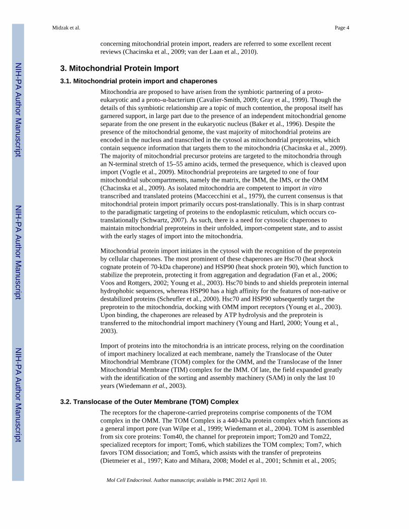
Mitochondria are proposed to have arisen from the symbiotic partnering of a proto-eukaryotic and a proto-α-bacterium (Cavalier-Smith, 2009; Gray et al., 1999). Though thedetails of this symbiotic relationship are a topic of much contention, the proposal itself hasgarnered support, in large part due to the presence of an independent mitochondrial genomeseparate from the one present in the eukaryotic nucleus (Baker et al., 1996). Despite thepresence of the mitochondrial genome, the vast majority of mitochondrial proteins areencoded in the nucleus and transcribed in the cytosol as mitochondrial preproteins, whichcontain sequence information that targets them to the mitochondria (Chacinska et al., 2009).The majority of mitochondrial precursor proteins are targeted to the mitochondria throughan N-terminal stretch of 15–55 amino acids, termed the presequence, which is cleaved uponimport (Vogtle et al., 2009). Mitochondrial preproteins are targeted to one of fourmitochondrial subcompartments, namely the matrix, the IMM, the IMS, or the OMM(Chacinska et al., 2009). As isolated mitochondria are competent to import in vitrotranscribed and translated proteins (Maccecchini et al., 1979), the current consensus is thatmitochondrial protein import primarily occurs post-translationally. This is in sharp contrastto the paradigmatic targeting of proteins to the endoplasmic reticulum, which occurs co-translationally (Schwartz, 2007). As such, there is a need for cytosolic chaperones tomaintain mitochondrial preproteins in their unfolded, import-competent state, and to assistwith the early stages of import into the mitochondria.
Mitochondrial protein import initiates in the cytosol with the recognition of the preproteinby cellular chaperones. The most prominent of these chaperones are Hsc70 (heat shockcognate protein of 70-kDa chaperone) and HSP90 (heat shock protein 90), which function tostabilize the preprotein, protecting it from aggregation and degradation (Fan et al., 2006;Voos and Rottgers, 2002; Young et al., 2003). Hsc70 binds to and shields preprotein internalhydrophobic sequences, whereas HSP90 has a high affinity for the features of non-native ordestabilized proteins (Scheufler et al., 2000). Hsc70 and HSP90 subsequently target thepreprotein to the mitochondria, docking with OMM import receptors (Young et al., 2003).Upon binding, the chaperones are released by ATP hydrolysis and the preprotein istransferred to the mitochondrial import machinery (Young and Hartl, 2000; Young et al.,2003).
Import of proteins into the mitochondria is an intricate process, relying on the coordinationof import machinery localized at each membrane, namely the Translocase of the OuterMitochondrial Membrane (TOM) complex for the OMM, and the Translocase of the InnerMitochondrial Membrane (TIM) complex for the IMM. Of late, the field expanded greatlywith the identification of the sorting and assembly machinery (SAM) in only the last 10years (Wiedemann et al., 2003).
3.2. Translocase of the Outer Membrane (TOM) ComplexThe receptors for the chaperone-carried preproteins comprise components of the TOMcomplex in the OMM. The TOM Complex is a 440-kDa protein complex which functions asa general import pore (van Wilpe et al., 1999; Wiedemann et al., 2004). TOM is assembledfrom six core proteins: Tom40, the channel for preprotein import; Tom20 and Tom22,specialized receptors for import; Tom6, which stabilizes the TOM complex; Tom7, whichfavors TOM dissociation; and Tom5, which assists with the transfer of preproteins(Dietmeier et al., 1997; Kato and Mihara, 2008; Model et al., 2001; Schmitt et al., 2005;
Midzak et al. Page 4
Mol Cell Endocrinol. Author manuscript; available in PMC 2012 April 10.
NIH
-PA Author Manuscript
NIH
-PA Author Manuscript
NIH
-PA Author Manuscript

Yano et al., 2004). Tom40, a β-barrel protein, serves as the principal channel for the importof polypeptides while Tom20 binds mitochondrial presequences and shuttles the proteins toTom40, with the assistance of Tom22 and Tom5 (Abe et al., 2000). Alternatively,preproteins without presequences and their chaperones associate with the accessory Tom70receptor, which facilitates transfer to the TOM complex (van Wilpe et al., 1999). Upontranslocation of the protein through the Tom40 pore, the presequence binds to Tom7, andthen binds to the IMS domain of Tom22. At this point, the protein is present in both thecytosol and the IMS of the mitochondria and targeting information nascent in thepolypeptide targets it to its next site of translocation (Chacinska et al., 2009).
3.3. Sorting and Assembly Machinery (SAM) ComplexOMM β-barrel proteins, such as Tom40, are imported into the OMM through the action ofthe TOM complex and subsequently inserted into the membrane through a distinct OMMimport assembly, the SAM complex. In yeast, the SAM complex consists of three proteins:Sam35, the initial receptor for the preprotein; Sam50, a β-barrel import pore; and Sam37,which functions to integrate the protein into the OMM (Pfanner et al., 2004). Upon thepresence of the preproteins in the IMS, the chaperone complex Tim8 – Tim13 [or thedistinct Tim9 – Tim12 pair (Chan and Lithgow, 2008)] binds to the preprotein and transfersit to the SAM receptor protein Sam35 (Chan and Lithgow, 2008). Sam35 transfers thepreprotein to Sam50; with its release into the lipid membrane regulated by Sam37 (Chan andLithgow, 2008; Kutik et al., 2008), though the exact mechanism of SAM-mediated proteininsertion into the OMM is still unknown (Becker et al., 2008; Kutik et al., 2008)
3.4. α-Helical Protein Incorporation into the OMMThough the most prevalent protein in the OMM is the β-barrel protein VDAC, the majorityof proteins present in the OMM are α-helical. Simple OMM proteins that span themembrane in one α-helix, such as Tom20 or Tom22, have been shown to havemitochondrial targeting sequences at either the N- or C-terminus (Rapaport, 2003). For theseproteins, several paths of import have been identified, with the transmembrane region andflanking positive amino acids functioning as a putative targeting sequence (Ahting et al.,2005; Rapaport, 2005). N-terminally-targeted proteins utilize the Tom40 pore but the TOMcomplex receptors Tom20 and Tom70 have been shown not to be utilized (Ahting et al.,2005; Van den Berg et al., 2004) (Fig. 2). Mim1 has been shown to promote the insertion ofproteins containing N-terminus targeting sequences, such as Tom20 (Dimmer and Rapaport2010). A mechanism for C-terminally-anchored OMM proteins has not been identified,though both lipid composition and the SAM complex have been implicated (Kemper et al.,2008; Setoguchi et al., 2006; Stojanovski et al., 2007). Proteins that span the OMM withmultiple transmembrane domains (MTD) have targeting sequences found throughout theprotein (Otera et al., 2007). The core TOM complex is not necessary for import of MTDproteins, but the TOM receptor Tom70 appears to be critical for import (Rone et al., 2009b).
3.5. Translocase of the Inner Membrane (TIM) ComplexFollowing TOM translocation to the IMS, there are two primary pathways for preproteinimport into the IMM that involve Tim22 and Tim23, respectively (Fig. 3). Tim22 plays arole in the import of the IMM metabolite carrier proteins, which do not contain an explicittargeting sequence, while Tim23 functions in the import of proteins containing apresequence targeted for the matrix (van der Laan et al., 2010). Tim23 is also instrumentalin the import of IMM proteins that span the membrane with a single α-helix (van der Laan etal., 2010). The pathway for import through Tim22 begins when Hsc70 and HSP90 target theprotein to the OMM where they pass through the TOM complex pore (Brix et al., 1999;Stanet al., 2003). Upon import into the IMS, the Tim9 – Tim12 pair binds to the protein andshuttles it to the Tim54 receptor (Endres et al., 1999;Vasiljev et al., 2004). Tim54 transfers
Midzak et al. Page 5
Mol Cell Endocrinol. Author manuscript; available in PMC 2012 April 10.
NIH
-PA Author Manuscript
NIH
-PA Author Manuscript
NIH
-PA Author Manuscript

the preprotein to the Tim22 pore, which then releases the protein into the IMM (Chacinskaet al., 2009).
The Tim23 protein complex is composed of three proteins regulating the import ofpreproteins into the IMM or the matrix: Tim50, Tim23, and Tim21 (Mokranjac and Neupert,2010). Upon import through the TOM pore, Tim50 binds and transfers the presequence-containing preprotein from Tom22 in the IMS to Tim23, the IMM protein import pore(Tamura et al., 2009). Tim21 competes with the presequence binding domain of Tom22,facilitating the transfer of the preprotein to Tim23 (Chacinska et al., 2005). In the case ofIMM proteins, a hydrophobic stretch in the preprotein (sorting signal) stalls import uponincorporation into the Tim23 pore, signaling insertion of the protein into the IMM throughlateral diffusion from the TIM pore (Popov-Celeketic et al., 2008). The non-hydrophobicportion of the preprotein is imported into the matrix through the preprotein assembly andmachinery (PAM) complex, where the targeting sequence is usually cleaved. The interactionbetween Tim23 and the PAM complex also supports the translocation of preproteinstargeted to the matrix.
3.6. Preprotein Assembly and Machinery (PAM) ComplexThe positive charge of the mitochondrial preprotein must overcome a steep thermodynamicbarrier generated by the proton gradient in the IMS to reach the mitochondrial matrix(Shariff et al., 2004). Such an energetic contribution is supplied by the PAM complex,which traps and pulls the preprotein into the mitochondrial matrix in an ATP-dependentmanner (Krayl et al., 2007). The PAM complex is composed of Tim44, mtHSP70, Pam16,and Pam18 (Fig. 3) (van der Laan et al., 2010). Tim44 is anchored to the matrix side of theIMM via its N-terminus. The membrane-facing side of Tim44 interacts with the Tim23 porewhile the matrix-facing side of Tim44 interacts with mtHSP70, which physically ratchets theprotein into the matrix (D’Silva et al., 2004; Rassow et al., 1994; Tomkiewicz et al., 2007).Pam16 and Pam18 subsequently interact with mtHSP70 to assist in the regulation of ATPhydrolysis, modulating the rate of protein import (Li et al., 2004).
3.7. Mitochondrial protein processingIn addition to importing protein across their double membranes, mitochondria process andremodel many of these proteins before utilization. It is also important for mitochondria todegrade misfolded or unwanted proteins. To this end, mitochondria contain multiplepeptidases and proteases, primarily localized to the IMM and the matrix, which targetproteins resident in the IMS and matrix. Upon import into the matrix, the presequence ofmany proteins targeted to the matrix is removed by the mitochondrial processing peptidase(MPP) (Gakh et al., 2002). Retention of the presequence appears to destabilize the proteinconformation, making it susceptible to increased proteolysis by mitochondrial peptidases(Mukhopadhyay et al., 2007). MPP is a heterodimer composed of α-MPP and β-MPP(Kalousek et al., 1993) located in the mitochondrial matrix. It is a metallopeptidase inhibitedby low pH and metal chelators (Miura et al., 1982). MPP recognizes the positively chargedα-helix of presequences in an extended conformation (Taylor et al., 2001) and cleaves thepresequence two residues after an arginine residue present in three sequence motifs(Hendrick et al., 1989; Schneider et al., 1998). For some mitochondrial preproteins, thepresequence is sequentially processed by MPP and an additional matrix metallopeptidase,Oct1 (Branda et al., 1999), which cleaves the remaining presequence eight amino acidsfollowing the MPP cleavage site. A third peptidase complex, the inner membrane peptidase(IMP), assists in the maturation of IMS proteins. IMP proteins are bound to the IMM andproject into the IMS (Jan et al., 2000) and require divalent cations and acidic phospholipidsfor activity (Schneider et al., 1991).
Midzak et al. Page 6
Mol Cell Endocrinol. Author manuscript; available in PMC 2012 April 10.
NIH
-PA Author Manuscript
NIH
-PA Author Manuscript
NIH
-PA Author Manuscript

A variety of mitochondrial members of the AAA+ (ATPases associated with various cellularactivities) superfamily have been identified (Gakh et al., 2002; Truscott et al., 2010).Though these enzymes appear to function principally in ATP-dependent protein qualitycontrol, several have been implicated in the import and processing of mitochondrialpreproteins (Rainey et al., 2006). The Lon and CplX proteases are located in themitochondrial matrix (Kang et al., 2002; Wang et al., 1993), and the Lon protease isconsidered to play a major role in mitochondrial protein clearance (Major et al., 2006;Ondrovicova et al., 2005). The IMM-bound proteases m-AAA (matrix-AAA) and i-AAA(intermembrane space-AAA) target proteins present in the matrix and IMS, respectively(Arlt et al., 1996; Leonhard et al., 1996). These proteases are considered the principalplayers in quality control of unassembled and misfolded proteins in the IMM (Koppen andLanger, 2007), though m-AAA has been noted be involved in the maturation of severalmitochondrial proteins (Nolden et al., 2005; Rainey et al., 2006).
4. Mitochondrial Import of Cholesterol Transduceosome ProteinsAs described above, the import of mitochondrial proteins occurs through a series ofelaborate and tightly regulated processes. In the subsequent sections, we describe theinterplay of these protein import pathways and our knowledge of the import of variousprotein components of the transduceosome, namely VDAC and TSPO on the OMM, andCYP11A1, Adx, and AdxR in the mitochondrial matrix.
4.1. Voltage-Dependent Anion Channel (VDAC)VDAC is an OMM β-barrel protein involved in the regulation of ions and small moleculesacross the OMM, influencing multiple cellular processes, including apoptosis and energytransduction (Vyssokikh and Brdiczka, 2003). VDAC has been shown to play an importantrole in steroidogenesis, interacting with TSPO at the OMM in both helping to anchor thetransduceosome to the OMM and assisting with the import of STAR (Bose et al., 2008; Liuet al., 2006).
The structure, but not homology, of VDAC is similar to Tom40, in that they both are β-barrel proteins that span the OMM 19 times (Zeth, 2010). Studies in the fungi Neurosporacrassa and Saccharomyces cerevisiae demonstrated that an internal targeting sequence ofVDAC interacts with both Tom20 and Tom22 and facilitates import by the TOM complex(Krimmer et al., 2001). Additional import partners were identified by OMMpermeabilization, promoting the loss of IMS space proteins and revealing a critical role forthe Tim8 – Tim13 heterodimer in VDAC import (Krimmer et al., 2001). Interaction ofVDAC with these IMS proteins promotes transfer to Sam35 and its subsequentincorporation into the OMM by Sam50 (Kozjak et al., 2003; Milenkovic et al., 2004).Removal of the C-terminus of VDAC inhibits its maturation, suggesting the presence of asignal in this region for the targeting or binding of VDAC to the SAM complex (Kutik et al.,2008).
Knockdown of Tom40 in HeLa cells by shRNA decreased VDAC import, furtheraccentuating the importance of the TOM complex for VDAC import in mammalian cells(Kozjak-Pavlovic et al., 2007). Reduction of the SAM complex component Sam50 in HeLacells also significantly reduced the import of VDAC, though this reduction correlated withreductions in the SAM complex proteins Metaxin1 (mammalian Sam37) and Metaxin2(mammalian Sam35) (Armstrong et al., 1997; Armstrong et al., 1999; Kozjak-Pavlovic etal., 2007). Knockdown of Metaxin2 revealed the importance of smaller SAM componentsfor VDAC import, as VDAC levels were decreased by 50% in the OMM of Metaxin2knockdown cells with only 10% assembling into a mature complex (Kozjak-Pavlovic et al.,2007). Though the targeting sequence for mammalian VDAC remains unknown, the TOM
Midzak et al. Page 7
Mol Cell Endocrinol. Author manuscript; available in PMC 2012 April 10.
NIH
-PA Author Manuscript
NIH
-PA Author Manuscript
NIH
-PA Author Manuscript

and SAM complexes have a demonstrably critical, and phylogenetically conserved, role forVDAC import and assembly in the OMM.
4.2. Translocator Protein (TSPO)TSPO is an 18-kDa OMM protein with a predicted structure of five transmembrane α-helices, (Culty et al., 1999; Joseph-Liauzun et al., 1998) a prediction recently supported bycryoelectron microscopy (Korkhov et al., 2010). Like all OMM proteins, TSPO does notpossess an explicit mitochondrial targeting presequence. Generation of a series of TSPOdeletion mutants revealed that the C-terminal half of the protein is necessary and sufficientto target TSPO to the OMM (Rone et al., 2009b). Analysis of these peptides revealed thepresence of a Schellman motif between amino acid 103–109, which functions to terminateone α-helix, form a short hairpin loop, and begin another α-helix (Viguera and Serrano,1995). Complete removal of this motif or mutation of a critical central glycine also resultedin inhibition of TSPO targeting and insertion into the OMM (Rone et al., 2009b). From thesestudies, it was proposed that TSPO functions as a C-terminally-anchored protein thatrequires a Schellman motif for correct protein folding and chaperone binding.
TSPO’s mitochondrial targeting and import are assisted by cytosolic chaperones,specifically Hsc70 and HSP90 (Rone et al., 2009b). Upon arrival of TSPO at the OMM, thechaperones interact with Tom70 in an ATP-dependant manner, releasing an import-competent TSPO (Otera et al., 2007; Rone et al., 2009b). Interestingly, upon its release,TSPO import does not require the core TOM complex proteins Tom20, Tom22, or Tom40,as determined by RNAi knockdown (Otera et al., 2007). Though the core TOM complex isnot necessary for TSPO import, these findings do not discount the possible contributions ofIMS import proteins. Removal of the IMS protein Tim8 by hypotonically stressing themitochondria decreased TSPO import without affecting OMM (Tom22) or IMM (Su9-DHFR) protein import (Otera et al., 2007). Additional machinery contributing to TSPOimport was determined using RNA silencing. Though knockdown of the SAM complexcomponent Sam50 did not affect TSPO levels (Otera et al., 2007), the Sam37 homolog,Metaxin1, was found to be necessary (Rone et al., 2009b). Sam37 has been demonstrated tomediate the insertion of proteins carrying C-terminal targeting information into the OMM(Stojanovski et al., 2007), and in light of the importance of TSPO’s C-terminus for targeting,these findings suggest a principal role of Metaxin1 for TSPO membrane insertion.
4.3. Cholesterol Side-Chain Cleavage Cytochrome P450 (CYP11A1)CYP11A1 comprises the core metabolic enzyme of the transduceosome, catalyzing theconversion of cholesterol to pregnenolone through cleavage of the aliphatic side chain ofcholesterol (Payne and Hales, 2004). Human CYP11A1 is encoded as a 521-amino acidpolypeptide with a 39-amino acid mitochondrial presequence, which is cleaved upon import(DuBois et al., 1981). Point mutation of the three positive amino acids in the presequencegreatly reduced the efficiency of import into the matrix, confirming the necessity of positiveamino acids for preprotein import (Nabi et al., 1983). However, the association of CYP11A1with the matrix was not affected by the presence of the presequence, as inhibition ofpresequence processing by the metal chelator o-phenanthroline did not affect CYP11A1mitochondrial integration and enzymatic activity (Ou et al., 1986). Studies of CYP11A1membrane topology using Na2CO3 treatment of isolated mitochondria revealed thatCYP11A1 tightly associates with mitochondrial membranes and has its N-terminusprotected from trypsin proteolysis, suggesting that the N-terminus anchored the protein tothe IMM (Ou et al., 1986). Despite these proteolytic protection studies, sequence analysescould not identify the hydrophobic sequence of the mature protein for IMM insertion(Nelson and Strobel, 1988), suggesting that CYP11A1 is fully imported into themitochondrial matrix and associated peripherally with the IMM. Modeling studies of
Midzak et al. Page 8
Mol Cell Endocrinol. Author manuscript; available in PMC 2012 April 10.
NIH
-PA Author Manuscript
NIH
-PA Author Manuscript
NIH
-PA Author Manuscript

CYP11A1 have proposed that membrane association occurs through an F-G loop andresidues surrounding the α-helix of the protein (Headlam et al., 2003; Storbeck et al., 2007),a hypothesis supported by mutational analyses (Pikuleva, 2004; Pikuleva et al., 2008).
Multiple studies examining the molecular mechanisms of CYP11A1 import have utilizedyeast as a model system. Upon import of the mammalian CYP11A1 into yeast mitochondria,the protein is quickly degraded, with only a small sample functionally integrating into theIMM (Minenko et al., 2008; Savel’ev et al., 1998). To determine the mechanism ofCYP11A1 import and further confirm where it is localized in the mitochondria, the 39-amino acid targeting sequence of CYP11A1 was removed and hybrid proteins were created,each with one of six differential mitochondrial targeting sequences, which would beimported into the mitochondria by one of the import machineries. Functional import wasmeasured by measuring the activity of CYP11A1 (Minenko et al., 2008). It was determinedthat the AAC-CYP11A1 complex, a fusion of CYP11A1 with the full size adeninenucleotide translocase component (AAC), was the most functional hybrid when targeted andimported into the mitochondria via the Tim22 complex. Decreased enzymatic activity wasseen when the complex was imported completely into the matrix and then reassembledthrough the PAM complex, as seen with the preadrenodoxin (Padx)-CYP11A1 complex(Minenko et al., 2008). Rather than indicating the requirement of the Tim22 pathway forimport, these results suggest that a tight association with the IMM needs to be maintainedfor the functionality of CYP11A1. As cholesterol is a hydrophobic molecule, this tightassociation would provide a mechanism for the transfer of cholesterol to CYP11A1. Theprobable mechanisms of import of CYP11A1 into the matrix would involve the Tim23-PAM pathway. However, how this tight association is maintained during import or re-established after import is not understood.
4.4. Ferredoxin (Fdx) and Ferredoxin Reductase (FdxR)CYP11A1 activity is regulated by the rate of electron transfer from the matrix proteins Fdxand FdxR (Miller, 2005). Fdx served as one of the first models of mitochondrial proteinimport, with initial work on Fdx import beginning in 1980. Padx, the precursor for Fdx, issynthesized in the cytosol at approximately 20-kDa and, upon import, its presequence of 58amino acid is cleaved, resulting in a 14-kDa protein (Nabi and Omura, 1980). Studies in E.Coli demonstrated that the presence of the presequence did not alter the correct foldingpattern of the protein or its ability to bind to FdxR, but did decrease its ability to bind toCYP11A1 (Goder et al., 1998). Targeting of Padx to the mitochondria occurs through eitherHSP70 or the mitochondrial import stimulation factor (MSF) (Hachiya et al., 1994; Hachiyaet al., 1995), though in early experiments it was shown that ribosomes synthesizing Padxwere also able to bind to the OMM (Nabi and Omura, 1983). This suggests that bothcytosolic synthesis and mitochondrial localized synthesis could lead to import and cleavageof Adx into the mitochondria (MacKenzie and Payne, 2004). FdR is also imported via amitochondrial targeting sequence, of which 32 amino acids are cleaved from the 462-aminoacid protein after import (Sagara et al., 1987), though no further examination of its importhas been completed. As these proteins are localized in the mitochondrial matrix where theyinteract with CYP11A1, it is possible that they are imported through the Tim23 importcomplex to the IMM membrane where the PAM complex would assist with import andproper folding in the matrix.
4.5. Steroidogenic Acute Regulatory Protein (STAR)STAR received a great deal of research attention after the discovery that its mutation resultsin lethal congenital adrenal lipoid hyperplasia (CAH), a disease characterized by theinability to synthesize steroids (Lin et al., 1995; Miller, 2005). Deletion of Star in miceresulted in an adrenal phenotype similar to that observed in the human disease, although
Midzak et al. Page 9
Mol Cell Endocrinol. Author manuscript; available in PMC 2012 April 10.
NIH
-PA Author Manuscript
NIH
-PA Author Manuscript
NIH
-PA Author Manuscript

gonadal function was affected to a lesser extend (Caron et al., 1997) STAR is expressed as a37-kDa preprotein composed of a cholesterol-binding START (STAR-related lipid transfer)domain preceded by a mitochondrial presequence tag (Clark et al., 1994; Strauss, III et al.,2003; Tsujishita and Hurley, 2000). In rodents, the STAR preprotein is targeted tomitochondria and proteolytically processed to yield a 32- and a final 30-kDa protein, whichis located in the mitochondrial matrix (Epstein and Orme-Johnson, 1991; Stocco andSodeman, 1991). Mutational analysis suggests the presence of proteolytic cleavage sites atpositions 39/40 and 55/56 of the bovine STAR protein, with similar predicted sites existingin other mammalian STAR homologs (Yamazaki et al., 2006). Upon reaching the matrix, the30-kDa STAR is degraded, with a half-life of four to five hours (Granot et al., 2003). Insidethe matrix, STAR protein levels are controlled by the ATP-dependent Lon protease, thoughthis regulation is undoubtedly complex as Lon proteases degrade STAR within five minutesin an E. coli model system (Granot et al., 2007). It is also unclear which mitochondrialproteases process STAR. Processing of STAR from a 37-kDa to 32-kDa to a 30-kDa proteinwas suggested by studies using mitochondrial toxins to impede protein import andproteolysis. The protonophore CCCP, which disrupts the mitochondrial membrane potential,stabilized 37-kDa STAR, while 1,10-orthophenanthroline, a transition metal chelatorproposed to inactivate metal-dependent proteases, stabilized the 32-kDa form (Artemenko etal., 2001). As membrane potential disruption blocks preproteins from inserting into, but notreaching, the IMM (van der Laan et al., 2010), these authors proposed that an IMS proteaseprocesses STAR from 37- to 32-kDa, which is further processed by a MPP upon crossingthe IMM (Artemenko et al., 2001). The IMP and i-AAA discussed above are bothmetallopeptidases facing the IMS, making them excellent candidates for STAR’s processingprotease, though further work will be required to confirm this hypothesis.
Though STAR contains a classical mitochondrial presequence and is thus presumed to beimported through the TOM-TIM complexes of the OMM and IMM, precise moleculardetails are lacking. This gap in our knowledge is especially pertinent in light of recent workwith the transduceosome components TSPO and VDAC. TSPO knockdown studiesexamining the functional relationship between STAR and TSPO in the stimulation ofsteroidogenesis demonstrated that STAR’s steroidogenic ability is dependent on thepresence of TSPO in MA-10 mouse Leydig tumor cells (Hauet et al., 2005). Intriguingly,decreasing TSPO expression reduced levels of the 30-kDa protein, but increased levels of37-kDa STAR. Similar results were obtained with a peptide antagonist of TSPO function(Gazouli et al., 2002; Hauet et al., 2005), suggesting a role for TSPO in STAR’s import andprocessing. This work has been more recently complemented by studies demonstrating thenecessity of VDAC in STAR’s steroidogenic function (Bose et al., 2008). In theseinvestigations, VDAC knockdown reduced STAR expression, and acute blockage of VDACwith polyanion inhibitors prevented the conversion of the 32-kDa form to the 30-kDa matrixform. Collectively, these studies demonstrate an integral correlation between STAR’ssteroidogenic function and its mitochondrial import. As shown schematically in Figure 4,this conclusion raises interesting questions for study of the interrelationship betweenmitochondrial protein and cholesterol import, questions currently under investigation in ourlaboratory.
5. Summary and ConclusionsThe large majority of mitochondrial proteins are encoded in the nucleus, translated in thecytoplasm, and imported into the mitochondria. The double mitochondrial membranes createfour mitochondrial subcompartments, in addition to the cytoplasm, which differ according totheir hydrophilic or hydrophobic nature: a hydrophobic outer mitochondrial membrane, ahydrophilic intermembrane space, a hydrophobic inner mitochondrial membrane, and ahydrophilic mitochondrial matrix. Proteins destined for these mitochondrial compartments
Midzak et al. Page 10
Mol Cell Endocrinol. Author manuscript; available in PMC 2012 April 10.
NIH
-PA Author Manuscript
NIH
-PA Author Manuscript
NIH
-PA Author Manuscript

also possess chemical natures similar to their destination (i.e., hydrophobic residues aretargeted to hydrophobic membrane compartments). As a consequence, proteins requirecellular machinery in the OMM and IMM to allow their transport to their particularmitochondrial localization through compartments that they could not freely cross. A similarsituation is observed with the lipid cholesterol, where a multi-protein machine termed thetransduceosome translocates cholesterol to the IMM and metabolizes it to the steroidpregnenolone. Core members of the transduceosome, such as TSPO, VDAC, and CYP11A1,are encoded in the nucleus and must be transported into the mitochondria for activity.Hormone stimulation induces the translocation of the ACBD3/PAP7 protein from the Golgiapparatus to the mitochondria, where it serves to scaffold the cytosolic PKA-RI subunits.Moreover, a critical regulatory element, STAR, is a mitochondria-targeted protein whoseactivity is increased upon PKA-mediated phosporylation, underscoring the tight interplaybetween mitochondrial protein and lipid import in steroidogenic mitochondria. Thisinterplay is a burgeoning field of research for protein import, as it has recently beenappreciated that mitochondrial phospholipid synthesis is dependent upon the protein importmachinery (Osman et al., 2009) and lipids critically regulate the function of translocasecomplexes (van der Laan et al., 2007). Finally, the observation that the activity of thetransduceosome components TSPO and VDAC regulate the import and processing of STAR(Bose et al., 2008; Hauet et al., 2005) further suggests a connection between mitochondriallipids and protein import. The mechanistic understanding of this process will increase ourunderstanding of mitochondrial biogenesis.
AcknowledgmentsThis work was supported by grants from the National Institutes of Health (R01 ES07747), the Canadian Institutesof Health Research (MOP102647), a Canada Research Chair in Biochemical Pharmacology to V.P., and apostdoctoral fellowship from CIHR (TGF36110) to A.M.
Abbreviations
Fdx ferredoxin
FdxR ferredoxin reductase
IMM inner mitochondrial membrane
IMS intermembrane space
MPP mitochondrial processing peptidase
OMM outer mitochondrial membrane
PAM preprotein assembly and machinery
PKA protein kinase A
SAM sorting and assembly machinery
STAR steroidogenic acute regulatory protein
TIM translocase of inner mitochondria
TOM translocase of outer mitochondria
TSPO translocator protein
VDAC voltage-dependent anion channel
Midzak et al. Page 11
Mol Cell Endocrinol. Author manuscript; available in PMC 2012 April 10.
NIH
-PA Author Manuscript
NIH
-PA Author Manuscript
NIH
-PA Author Manuscript

Reference ListAbe Y, Shodai T, Muto T, Mihara K, Torii H, Nishikawa S, Endo T, Kohda D. Structural basis of
presequence recognition by the mitochondrial protein import receptor Tom20. Cell 2000;100:551–560. [PubMed: 10721992]
Ahting U, Waizenegger T, Neupert W, Rapaport D. Signal-anchored proteins follow a unique insertionpathway into the outer membrane of mitochondria. J Biol Chem 2005;280:48–53. [PubMed:15501820]
Arlt H, Tauer R, Feldmann H, Neupert W, Langer T. The YTA10-12 complex, an AAA protease withchaperone-like activity in the inner membrane of mitochondria. Cell 1996;85:875–885. [PubMed:8681382]
Armstrong LC, Komiya T, Bergman BE, Mihara K, Bornstein P. Metaxin is a component of apreprotein import complex in the outer membrane of the mammalian mitochondrion. J Biol Chem1997;272:6510–6518. [PubMed: 9045676]
Armstrong LC, Saenz AJ, Bornstein P. Metaxin 1 interacts with metaxin 2, a novel related proteinassociated with the mammalian mitochondrial outer membrane. J Cell Biochem 1999;74:11–22.[PubMed: 10381257]
Artemenko IP, Zhao D, Hales DB, Hales KH, Jefcoate CR. Mitochondrial processing of newlysynthesized steroidogenic acute regulatory protein (StAR), but not total StAR, mediates cholesteroltransfer to cytochrome P450 side chain cleavage enzyme in adrenal cells. J Biol Chem2001;276:46583–46596. [PubMed: 11579102]
Baker A, Kaplan CP, Pool MR. Protein targeting and translocation; a comparative survey. Biol RevCamb Philos Soc 1996;71:637–702. [PubMed: 8923800]
Batarseh A, Papadopoulos V. Regulation of translocator protein 18 kDa (TSPO) expression in healthand disease states. Mol Cell Endocrinol 2010;327:1–12. [PubMed: 20600583]
Becker T, Vogtle FN, Stojanovski D, Meisinger C. Sorting and assembly of mitochondrial outermembrane proteins. Biochim Biophys Acta 2008;1777:557–563. [PubMed: 18423394]
Black SM, Harikrishna JA, Szklarz GD, Miller WL. The mitochondrial environment is required foractivity of the cholesterol side-chain cleavage enzyme, cytochrome P450scc. Proc Natl Acad Sci US A 1994;91:7247–7251. [PubMed: 8041774]
Bose HS, Lingappa VR, Miller WL. Rapid regulation of steroidogenesis by mitochondrial proteinimport. Nature 2002;417:87–91. [PubMed: 11986670]
Bose M, Whittal RM, Miller WL, Bose HS. Steroidogenic activity of StAR requires contact withmitochondrial VDAC1 and phosphate carrier protein. J Biol Chem 2008;283:8837–8845.[PubMed: 18250166]
Boujrad N, Hudson JR Jr, Papadopoulos V. Inhibition of hormone-stimulated steroidogenesis incultured Leydig tumor cells by a cholesterol-linked phosphorothioate oligodeoxynucleotideantisense to diazepam-binding inhibitor. Proc Natl Acad Sci U S A 1993;90:5728–5731. [PubMed:8390677]
Boujrad N, Vidic B, Papadopoulos V. Acute action of choriogonadotropin on Leydig tumor cells:changes in the topography of the mitochondrial peripheral-type benzodiazepine receptor.Endocrinology 1996;137:5727–5730. [PubMed: 8940407]
Branda SS, Yang ZY, Chew A, Isaya G. Mitochondrial intermediate peptidase and the yeast frataxinhomolog together maintain mitochondrial iron homeostasis in Saccharomyces cerevisiae. HumMol Genet 1999;8:1099–1110. [PubMed: 10332043]
Brix J, Rudiger S, Bukau B, Schneider-Mergener J, Pfanner N. Distribution of binding sequences forthe mitochondrial import receptors Tom20, Tom22, and Tom70 in a presequence-carryingpreprotein and a non-cleavable preprotein. J Biol Chem 1999;274:16522–16530. [PubMed:10347216]
Caron KM, Soo SC, Wetsel WC, Stocco DM, Clark BJ, Parker KL. Targeted disruption of the mousegene encoding steroidogenic acute regulatory protein provides insights into congenital lipoidadrenal hyperplasia. Proc Natl Acad Sci U S A 1997;94:11540–11545. [PubMed: 9326645]
Cavalier-Smith T. Predation and eukaryote cell origins: a coevolutionary perspective. Int J BiochemCell Biol 2009;41:307–322. [PubMed: 18935970]
Midzak et al. Page 12
Mol Cell Endocrinol. Author manuscript; available in PMC 2012 April 10.
NIH
-PA Author Manuscript
NIH
-PA Author Manuscript
NIH
-PA Author Manuscript
Chacinska A, Koehler CM, Milenkovic D, Lithgow T, Pfanner N. Importing mitochondrial proteins:machineries and mechanisms. Cell 2009;138:628–644. [PubMed: 19703392]
Chacinska A, Lind M, Frazier AE, Dudek J, Meisinger C, Geissler A, Sickmann A, Meyer HE,Truscott KN, Guiard B, Pfanner N, Rehling P. Mitochondrial presequence translocase: switchingbetween TOM tethering and motor recruitment involves Tim21 and Tim17. Cell 2005;120:817–829. [PubMed: 15797382]
Chan NC, Lithgow T. The peripheral membrane subunits of the SAM complex function codependentlyin mitochondrial outer membrane biogenesis. Mol Biol Cell 2008;19:126–136. [PubMed:17978093]
Clark BJ, Wells J, King SR, Stocco DM. The purification, cloning, and expression of a novelluteinizing hormone-induced mitochondrial protein in MA-10 mouse Leydig tumor cells.Characterization of the steroidogenic acute regulatory protein (StAR). J Biol Chem1994;269:28314–28322. [PubMed: 7961770]
Costa E, Guidotti A. Diazepam binding inhibitor (DBI): a peptide with multiple biological actions.Life Sci 1991;49:325–344. [PubMed: 1649940]
Culty M, Li H, Boujrad N, Amri H, Vidic B, Bernassau JM, Reversat JL, Papadopoulos V. In vitrostudies on the role of the peripheral-type benzodiazepine receptor in steroidogenesis. J SteroidBiochem Mol Biol 1999;69:123–130. [PubMed: 10418986]
D’Silva P, Liu Q, Walter W, Craig EA. Regulated interactions of mtHsp70 with Tim44 at thetranslocon in the mitochondrial inner membrane. Nat Struct Mol Biol 2004;11:1084–1091.[PubMed: 15489862]
Dietmeier K, Honlinger A, Bomer U, Dekker PJ, Eckerskorn C, Lottspeich F, Kubrich M, Pfanner N.Tom5 functionally links mitochondrial preprotein receptors to the general import pore. Nature1997;388:195–200. [PubMed: 9217162]
DuBois RN, Simpson ER, Tuckey J, Lambeth JD, Waterman MR. Evidence for a higher molecularweight precursor of cholesterol side-chain-cleavage cytochrome P-450 and induction ofmitochondrial and cytosolic proteins by corticotropin in adult bovine adrenal cells. Proc Natl AcadSci U S A 1981;78:1028–1032. [PubMed: 6262752]
Endres M, Neupert W, Brunner M. Transport of the ADP/ATP carrier of mitochondria from the TOMcomplex to the TIM22.54 complex. EMBO J 1999;18:3214–3221. [PubMed: 10369662]
Epstein LF, Orme-Johnson NR. Regulation of steroid hormone biosynthesis. Identification ofprecursors of a phosphoprotein targeted to the mitochondrion in stimulated rat adrenal cortex cells.J Biol Chem 1991;266:19739–19745. [PubMed: 1655794]
Fan AC, Bhangoo MK, Young JC. Hsp90 functions in the targeting and outer membrane translocationsteps of Tom70-mediated mitochondrial import. J Biol Chem 2006;281:33313–33324. [PubMed:16968702]
Fan J, Liu J, Culty M, Papadopoulos V. Acyl-coenzyme A binding domain containing 3 (ACBD3;PAP7; GCP60): an emerging signaling molecule. Prog Lipid Res 2010;49:218–234. [PubMed:20043945]
Gakh O, Cavadini P, Isaya G. Mitochondrial processing peptidases. Biochim Biophys Acta2002;1592:63–77. [PubMed: 12191769]
Garnier M, Dimchev AB, Boujrad N, Price JM, Musto NA, Papadopoulos V. In vitro reconstitution ofa functional peripheral-type benzodiazepine receptor from mouse Leydig tumor cells. MolPharmacol 1994;45:201–211. [PubMed: 8114671]
Gazouli M, Han Z, Papadopoulos V. Identification of a peptide antagonist to the peripheral-typebenzodiazepine receptor that inhibits hormone-stimulated leydig cell steroid formation. JPharmacol Exp Ther 2002;303:627–632. [PubMed: 12388644]
Goder V, Beckert V, Pfeil W, Bernhardt R. Impact of the presequence of a mitochondrium-targetedprecursor, preadrenodoxin, on folding, catalytic activity, and stability of the protein in vitro. ArchBiochem Biophys 1998;359:31–41. [PubMed: 9799557]
Granot Z, Geiss-Friedlander R, Melamed-Book N, Eimerl S, Timberg R, Weiss AM, Hales KH, HalesDB, Stocco DM, Orly J. Proteolysis of normal and mutated steroidogenic acute regulatory proteinsin the mitochondria: the fate of unwanted proteins. Mol Endocrinol 2003;17:2461–2476.[PubMed: 12958217]
Midzak et al. Page 13
Mol Cell Endocrinol. Author manuscript; available in PMC 2012 April 10.
NIH
-PA Author Manuscript
NIH
-PA Author Manuscript
NIH
-PA Author Manuscript

Granot Z, Kobiler O, Melamed-Book N, Eimerl S, Bahat A, Lu B, Braun S, Maurizi MR, Suzuki CK,Oppenheim AB, Orly J. Turnover of mitochondrial steroidogenic acute regulatory (StAR) proteinby Lon protease: the unexpected effect of proteasome inhibitors. Mol Endocrinol 2007;21:2164–2177. [PubMed: 17579211]
Gray MW, Burger G, Lang BF. Mitochondrial evolution. Science 1999;283:1476–1481. [PubMed:10066161]
Hachiya N, Komiya T, Alam R, Iwahashi J, Sakaguchi M, Omura T, Mihara K. MSF, a novelcytoplasmic chaperone which functions in precursor targeting to mitochondria. EMBO J1994;13:5146–5154. [PubMed: 7957079]
Hachiya N, Mihara K, Suda K, Horst M, Schatz G, Lithgow T. Reconstitution of the initial steps ofmitochondrial protein import. Nature 1995;376:705–709. [PubMed: 7651521]
Hauet T, Yao ZX, Bose HS, Wall CT, Han Z, Li W, Hales DB, Miller WL, Culty M, Papadopoulos V.Peripheral-type benzodiazepine receptor-mediated action of steroidogenic acute regulatory proteinon cholesterol entry into leydig cell mitochondria. Mol Endocrinol 2005;19:540–554. [PubMed:15498831]
Headlam MJ, Wilce MC, Tuckey RC. The F-G loop region of cytochrome P450scc (CYP11A1)interacts with the phospholipid membrane. Biochim Biophys Acta 2003;1617:96–108. [PubMed:14637024]
Hendrick JP, Hodges PE, Rosenberg LE. Survey of amino-terminal proteolytic cleavage sites inmitochondrial precursor proteins: leader peptides cleaved by two matrix proteases share a three-amino acid motif. Proc Natl Acad Sci U S A 1989;86:4056–4060. [PubMed: 2657736]
Jan PS, Esser K, Pratje E, Michaelis G. Som1, a third component of the yeast mitochondrial innermembrane peptidase complex that contains Imp1 and Imp2. Mol Gen Genet 2000;263:483–491.[PubMed: 10821182]
Jefcoate C. High-flux mitochondrial cholesterol trafficking, a specialized function of the adrenalcortex. J Clin Invest 2002;110:881–890. [PubMed: 12370263]
Joseph-Liauzun E, Delmas P, Shire D, Ferrara P. Topological analysis of the peripheralbenzodiazepine receptor in yeast mitochondrial membranes supports a five-transmembranestructure. J Biol Chem 1998;273:2146–2152. [PubMed: 9442055]
Kalousek F, Neupert W, Omura T, Schatz G, Schmitz UK. Uniform nomenclature for themitochondrial peptidases cleaving precursors of mitochondrial proteins. Trends Biochem Sci1993;18:249. [PubMed: 8212133]
Kang SG, Ortega J, Singh SK, Wang N, Huang NN, Steven AC, Maurizi MR. Functional proteolyticcomplexes of the human mitochondrial ATP-dependent protease, hClpXP. J Biol Chem2002;277:21095–21102. [PubMed: 11923310]
Kato H, Mihara K. Identification of Tom5 and Tom6 in the preprotein translocase complex of humanmitochondrial outer membrane. Biochem Biophys Res Commun 2008;369:958–963. [PubMed:18331822]
Kemper C, Habib SJ, Engl G, Heckmeyer P, Dimmer KS, Rapaport D. Integration of tail-anchoredproteins into the mitochondrial outer membrane does not require any known import components. JCell Sci 2008;121:1990–1998. [PubMed: 18495843]
King SR, Ronen-Fuhrmann T, Timberg R, Clark BJ, Orly J, Stocco DM. Steroid production after invitro transcription, translation, and mitochondrial processing of protein products of complementarydeoxyribonucleic acid for steroidogenic acute regulatory protein. Endocrinology 1995;136:5165–5176. [PubMed: 7588255]
Korkhov VM, Sachse C, Short JM, Tate CG. Three-dimensional structure of TspO by electroncryomicroscopy of helical crystals. Structure 2010;18:677–687. [PubMed: 20541505]
Kozjak V, Wiedemann N, Milenkovic D, Lohaus C, Meyer HE, Guiard B, Meisinger C, Pfanner N. Anessential role of Sam50 in the protein sorting and assembly machinery of the mitochondrial outermembrane. J Biol Chem 2003;278:48520–48523. [PubMed: 14570913]
Kozjak-Pavlovic V, Ross K, Benlasfer N, Kimmig S, Karlas A, Rudel T. Conserved roles of Sam50and metaxins in VDAC biogenesis. EMBO Rep 2007;8:576–582. [PubMed: 17510655]
Midzak et al. Page 14
Mol Cell Endocrinol. Author manuscript; available in PMC 2012 April 10.
NIH
-PA Author Manuscript
NIH
-PA Author Manuscript
NIH
-PA Author Manuscript

Krayl M, Lim JH, Martin F, Guiard B, Voos W. A cooperative action of the ATP-dependent importmotor complex and the inner membrane potential drives mitochondrial preprotein import. MolCell Biol 2007;27:411–425. [PubMed: 17074805]
Krimmer T, Rapaport D, Ryan MT, Meisinger C, Kassenbrock CK, Blachly-Dyson E, Forte M,Douglas MG, Neupert W, Nargang FE, Pfanner N. Biogenesis of porin of the outer mitochondrialmembrane involves an import pathway via receptors and the general import pore of the TOMcomplex. J Cell Biol 2001;152:289–300. [PubMed: 11266446]
Krueger KE, Papadopoulos V. Peripheral-type benzodiazepine receptors mediate translocation ofcholesterol from outer to inner mitochondrial membranes in adrenocortical cells. J Biol Chem1990;265:15015–15022. [PubMed: 2168398]
Kutik S, Stojanovski D, Becker L, Becker T, Meinecke M, Kruger V, Prinz C, Meisinger C, Guiard B,Wagner R, Pfanner N, Wiedemann N. Dissecting membrane insertion of mitochondrial beta-barrelproteins. Cell 2008;132:1011–1024. [PubMed: 18358813]
Lacapere JJ, Delavoie F, Li H, Peranzi G, Maccario J, Papadopoulos V, Vidic B. Structural andfunctional study of reconstituted peripheral benzodiazepine receptor. Biochem Biophys ResCommun 2001;284:536–541. [PubMed: 11394915]
Lavoie HA, King SR. Transcriptional regulation of steroidogenic genes: STARD1, CYP11A1 andHSD3B. Exp Biol Med (Maywood) 2009;234:880–907. [PubMed: 19491374]
Leonhard K, Herrmann JM, Stuart RA, Mannhaupt G, Neupert W, Langer T. AAA proteases withcatalytic sites on opposite membrane surfaces comprise a proteolytic system for the ATP-dependent degradation of inner membrane proteins in mitochondria. EMBO J 1996;15:4218–4229.[PubMed: 8861950]
Li H, Degenhardt B, Tobin D, Yao ZX, Tasken K, Papadopoulos V. Identification, localization, andfunction in steroidogenesis of PAP7: a peripheral-type benzodiazepine receptor- and PKA(RIalpha)-associated protein. Mol Endocrinol 2001;15:2211–2228. [PubMed: 11731621]
Li Y, Dudek J, Guiard B, Pfanner N, Rehling P, Voos W. The presequence translocase-associatedprotein import motor of mitochondria. Pam16 functions in an antagonistic manner to Pam18. JBiol Chem 2004;279:38047–38054. [PubMed: 15218029]
Lin D, Sugawara T, Strauss JF III, Clark BJ, Stocco DM, Saenger P, Rogol A, Miller WL. Role ofsteroidogenic acute regulatory protein in adrenal and gonadal steroidogenesis. Science1995;267:1828–1831. [PubMed: 7892608]
Liu J, Cavalli LR, Haddad BR, Papadopoulos V. Molecular cloning, genomic organization,chromosomal mapping and subcellular localization of mouse PAP7: a PBR and PKA-RIalphaassociated protein. Gene 2003;308:1–10. [PubMed: 12711385]
Liu J, Rone MB, Papadopoulos V. Protein-protein interactions mediate mitochondrial cholesteroltransport and steroid biosynthesis. J Biol Chem 2006;281:38879–38893. [PubMed: 17050526]
Maccecchini ML, Rudin Y, Blobel G, Schatz G. Import of proteins into mitochondria: precursor formsof the extramitochondrially made F1-ATPase subunits in yeast. Proc Natl Acad Sci U S A1979;76:343–347. [PubMed: 154672]
MacKenzie JA, Payne RM. Ribosomes specifically bind to mammalian mitochondria via protease-sensitive proteins on the outer membrane. J Biol Chem 2004;279:9803–9810. [PubMed:14668341]
Major T, von JB, Ruppert T, Mogk A, Voos W. Proteomic analysis of mitochondrial protein turnover:identification of novel substrate proteins of the matrix protease pim1. Mol Cell Biol 2006;26:762–776. [PubMed: 16428434]
McEnery MW, Snowman AM, Trifiletti RR, Snyder SH. Isolation of the mitochondrialbenzodiazepine receptor: association with the voltage-dependent anion channel and the adeninenucleotide carrier. Proc Natl Acad Sci U S A 1992;89:3170–3174. [PubMed: 1373486]
Mesmin B, Maxfield FR. Intracellular sterol dynamics. Biochim Biophys Acta 2009;1791:636–645.[PubMed: 19286471]
Milenkovic D, Kozjak V, Wiedemann N, Lohaus C, Meyer HE, Guiard B, Pfanner N, Meisinger C.Sam35 of the mitochondrial protein sorting and assembly machinery is a peripheral outermembrane protein essential for cell viability. J Biol Chem 2004;279:22781–22785. [PubMed:15067005]
Midzak et al. Page 15
Mol Cell Endocrinol. Author manuscript; available in PMC 2012 April 10.
NIH
-PA Author Manuscript
NIH
-PA Author Manuscript
NIH
-PA Author Manuscript

Miller WL. Steroidogenic acute regulatory protein (StAR), a novel mitochondrial cholesteroltransporter. Biochim Biophys Acta 2007;1771:663–676. [PubMed: 17433772]
Miller WL, Auchus RJ. The Molecular Biology, Biochemistry, and Physiology of HumanSteroidogenesis and Its Disorders. Endocr Rev. 2010 In Press. 10.1210/er.2010-0013
Minenko AN, Novikova LA, Luzikov VN, Kovaleva IE. Import of hybrid forms of CYP11A1 intoyeast mitochondria. Biochim Biophys Acta 2008;1780:1121–1130. [PubMed: 18621100]
Miura S, Mori M, Amaya Y, Tatibana M. A mitochondrial protease that cleaves the precursor ofornithine carbamoyltransferase. Purification and properties. Eur J Biochem 1982;122:641–647.[PubMed: 7037411]
Model K, Meisinger C, Prinz T, Wiedemann N, Truscott KN, Pfanner N, Ryan MT. Multistepassembly of the protein import channel of the mitochondrial outer membrane. Nat Struct Biol2001;8:361–370. [PubMed: 11276259]
Mokranjac D, Neupert W. The many faces of the mitochondrial TIM23 complex. Biochim BiophysActa 2010;1797:1045–1054. [PubMed: 20116361]
Mukhopadhyay A, Yang CS, Wei B, Weiner H. Precursor protein is readily degraded in mitochondrialmatrix space if the leader is not processed by mitochondrial processing peptidase. J Biol Chem2007;282:37266–37275. [PubMed: 17959599]
Nabi N, Kominami S, Takemori S, Omura T. Contributions of cytoplasmic free and membrane-boundribosomes to the synthesis of mitochondrial cytochrome P-450(SCC) and P-450(11 beta) andmicrosomal cytochrome P-450(C-21) in bovine adrenal cortex. J Biochem 1983;94:1517–1527.[PubMed: 6654870]
Nabi N, Omura T. In vitro synthesis of adrenodoxin and adrenodoxin reductase: existence of a putativelarge precursor form of adrenodoxin. Biochem Biophys Res Commun 1980;97:680–686.[PubMed: 7470120]
Nabi N, Omura T. In vitro synthesis of NADPH-adrenodoxin reductase and adrenodoxin of bovineadrenal cortex, and the existence of a large precursor of adrenodoxin. J Biochem 1983;94:1529–1538. [PubMed: 6197405]
Nelson DR, Strobel HW. On the membrane topology of vertebrate cytochrome P-450 proteins. J BiolChem 1988;263:6038–6050. [PubMed: 2834360]
Nolden M, Ehses S, Koppen M, Bernacchia A, Rugarli EI, Langer T. The m-AAA protease defectivein hereditary spastic paraplegia controls ribosome assembly in mitochondria. Cell 2005;123:277–289. [PubMed: 16239145]
Ondrovicova G, Liu T, Singh K, Tian B, Li H, Gakh O, Perecko D, Janata J, Granot Z, Orly J,Kutejova E, Suzuki CK. Cleavage site selection within a folded substrate by the ATP-dependentlon protease. J Biol Chem 2005;280:25103–25110. [PubMed: 15870080]
Osman C, Haag M, Potting C, Rodenfels J, Dip PV, Wieland FT, Brugger B, Westermann B, LangerT. The genetic interactome of prohibitins: coordinated control of cardiolipin andphosphatidylethanolamine by conserved regulators in mitochondria. J Cell Biol 2009;184:583–596. [PubMed: 19221197]
Otera H, Taira Y, Horie C, Suzuki Y, Suzuki H, Setoguchi K, Kato H, Oka T, Mihara K. A novelinsertion pathway of mitochondrial outer membrane proteins with multiple transmembranesegments. J Cell Biol 2007;179:1355–1363. [PubMed: 18158327]
Ou WJ, Ito A, Morohashi K, Fujii-Kuriyama Y, Omura T. Processing-independent in vitrotranslocation of cytochrome P-450(SCC) precursor across mitochondrial membranes. J Biochem1986;100:1287–1296. [PubMed: 3818579]
Papadopoulos V, Amri H, Li H, Boujrad N, Vidic B, Garnier M. Targeted disruption of the peripheral-type benzodiazepine receptor gene inhibits steroidogenesis in the R2C Leydig tumor cell line. JBiol Chem 1997;272:32129–32135. [PubMed: 9405411]
Papadopoulos V, Baraldi M, Guilarte TR, Knudsen TB, Lacapere JJ, Lindemann P, Norenberg MD,Nutt D, Weizman A, Zhang MR, Gavish M. Translocator protein (18kDa): new nomenclature forthe peripheral-type benzodiazepine receptor based on its structure and molecular function. TrendsPharmacol Sci 2006;27:402–409. [PubMed: 16822554]
Midzak et al. Page 16
Mol Cell Endocrinol. Author manuscript; available in PMC 2012 April 10.
NIH
-PA Author Manuscript
NIH
-PA Author Manuscript
NIH
-PA Author Manuscript

Papadopoulos V, Berkovich A, Krueger KE. The role of diazepam binding inhibitor and its processingproducts at mitochondrial benzodiazepine receptors: regulation of steroid biosynthesis.Neuropharmacology 1991a;30:1417–1423. [PubMed: 1664068]
Papadopoulos V, Berkovich A, Krueger KE, Costa E, Guidotti A. Diazepam binding inhibitor and itsprocessing products stimulate mitochondrial steroid biosynthesis via an interaction withmitochondrial benzodiazepine receptors. Endocrinology 1991b;129:1481–1488. [PubMed:1651852]
Payne AH, Hales DB. Overview of steroidogenic enzymes in the pathway from cholesterol to activesteroid hormones. Endocr Rev 2004;25:947–970. [PubMed: 15583024]
Pfanner N, Wiedemann N, Meisinger C, Lithgow T. Assembling the mitochondrial outer membrane.Nat Struct Mol Biol 2004;11:1044–1048. [PubMed: 15523480]
Pikuleva IA. Putative F-G loop is involved in association with the membrane in P450scc (P450 11A1).Mol Cell Endocrinol 2004;215:161–164. [PubMed: 15026189]
Pikuleva IA, Mast N, Liao WL, Turko IV. Studies of membrane topology of mitochondrial cholesterolhydroxylases CYPs 27A1 and 11A1. Lipids 2008;43:1127–1132. [PubMed: 18791760]
Pon LA, Hartigan JA, Orme-Johnson NR. Acute ACTH regulation of adrenal corticosteroidbiosynthesis. Rapid accumulation of a phosphoprotein. J Biol Chem 1986;261:13309–13316.[PubMed: 3020029]
Pon LA, Orme-Johnson NR. Acute stimulation of steroidogenesis in corpus luteum and adrenal cortexby peptide hormones. Rapid induction of a similar protein in both tissues. J Biol Chem1986;261:6594–6599. [PubMed: 3009462]
Popov-Celeketic D, Mapa K, Neupert W, Mokranjac D. Active remodelling of the TIM23 complexduring translocation of preproteins into mitochondria. EMBO J 2008;27:1469–1480. [PubMed:18418384]
Praporski S, Ng SM, Nguyen AD, Corbin CJ, Mechler A, Zheng J, Conley AJ, Martin LL.Organization of cytochrome P450 enzymes involved in sex steroid synthesis: PROTEIN-PROTEIN INTERACTIONS IN LIPID MEMBRANES. J Biol Chem 2009;284:33224–33232.[PubMed: 19805543]
Rainey RN, Glavin JD, Chen HW, French SW, Teitell MA, Koehler CM. A new function intranslocation for the mitochondrial i-AAA protease Yme1: import of polynucleotidephosphorylase into the intermembrane space. Mol Cell Biol 2006;26:8488–8497. [PubMed:16966379]
Rapaport D. Finding the right organelle. Targeting signals in mitochondrial outer-membrane proteins.EMBO Rep 2003;4:948–952. [PubMed: 14528265]
Rapaport D. How does the TOM complex mediate insertion of precursor proteins into themitochondrial outer membrane? J Cell Biol 2005;171:419–423. [PubMed: 16260501]
Rassow J, Maarse AC, Krainer E, Kubrich M, Muller H, Meijer M, Craig EA, Pfanner N.Mitochondrial protein import: biochemical and genetic evidence for interaction of matrix hsp70and the inner membrane protein MIM44. J Cell Biol 1994;127:1547–1556. [PubMed: 7798311]
Rone MB, Fan J, Papadopoulos V. Cholesterol transport in steroid biosynthesis: role of protein-proteininteractions and implications in disease states. Biochim Biophys Acta 2009a;1791:646–658.[PubMed: 19286473]
Rone MB, Liu J, Blonder J, Ye X, Veenstra TD, Young JC, Papadopoulos V. Targeting and insertionof the cholesterol-binding translocator protein into the outer mitochondrial membrane.Biochemistry 2009b;48:6909–6920. [PubMed: 19552401]
Rupprecht R, Rammes G, Eser D, Baghai TC, Schule C, Nothdurfter C, Troxler T, Gentsch C,Kalkman HO, Chaperon F, Uzunov V, McAllister KH, Bertaina-Anglade V, La Rochelle CD,Tuerck D, Floesser A, Kiese B, Schumacher M, Landgraf R, Holsboer F, Kucher K. Translocatorprotein (18 kD) as target for anxiolytics without benzodiazepine-like side effects. Science2009;325:490–493. [PubMed: 19541954]
Sagara Y, Takata Y, Miyata T, Hara T, Horiuchi T. Cloning and sequence analysis of adrenodoxinreductase cDNA from bovine adrenal cortex. J Biochem 1987;102:1333–1336. [PubMed:3448086]
Midzak et al. Page 17
Mol Cell Endocrinol. Author manuscript; available in PMC 2012 April 10.
NIH
-PA Author Manuscript
NIH
-PA Author Manuscript
NIH
-PA Author Manuscript

Savel’ev AS, Novikova LA, Kovaleva IE, Luzikov VN, Neupert W, Langer T. ATP-dependentproteolysis in mitochondria. m-AAA protease and PIM1 protease exert overlapping substratespecificities and cooperate with the mtHsp70 system. J Biol Chem 1998;273:20596–20602.[PubMed: 9685417]
Scheufler C, Brinker A, Bourenkov G, Pegoraro S, Moroder L, Bartunik H, Hartl FU, Moarefi I.Structure of TPR domain-peptide complexes: critical elements in the assembly of the Hsp70-Hsp90 multichaperone machine. Cell 2000;101:199–210. [PubMed: 10786835]
Schmitt S, Ahting U, Eichacker L, Granvogl B, Go NE, Nargang FE, Neupert W, Nussberger S. Roleof Tom5 in maintaining the structural stability of the TOM complex of mitochondria. J BiolChem 2005;280:14499–14506. [PubMed: 15701639]
Schneider A, Behrens M, Scherer P, Pratje E, Michaelis G, Schatz G. Inner membrane protease I, anenzyme mediating intramitochondrial protein sorting in yeast. EMBO J 1991;10:247–254.[PubMed: 1991446]
Schneider G, Sjoling S, Wallin E, Wrede P, Glaser E, von HG. Feature-extraction from endopeptidasecleavage sites in mitochondrial targeting peptides. Proteins 1998;30:49–60. [PubMed: 9443340]
Schwartz TU. Origins and evolution of cotranslational transport to the ER. Adv Exp Med Biol2007;607:52–60. [PubMed: 17977458]
Setoguchi K, Otera H, Mihara K. Cytosolic factor- and TOM-independent import of C-tail-anchoredmitochondrial outer membrane proteins. EMBO J 2006;25:5635–5647. [PubMed: 17110923]
Shariff K, Ghosal S, Matouschek A. The force exerted by the membrane potential during proteinimport into the mitochondrial matrix. Biophys J 2004;86:3647–3652. [PubMed: 15189861]
Simpson ER, Waterman MR. Regulation of the synthesis of steroidogenic enzymes in adrenal corticalcells by ACTH. Annu Rev Physiol 1988;50:427–440. [PubMed: 2837136]
Stan T, Brix J, Schneider-Mergener J, Pfanner N, Neupert W, Rapaport D. Mitochondrial proteinimport: recognition of internal import signals of BCS1 by the TOM complex. Mol Cell Biol2003;23:2239–2250. [PubMed: 12640110]
Stocco DM, Sodeman TC. The 30-kDa mitochondrial proteins induced by hormone stimulation inMA-10 mouse Leydig tumor cells are processed from larger precursors. J Biol Chem1991;266:19731–19738. [PubMed: 1918079]
Stojanovski D, Guiard B, Kozjak-Pavlovic V, Pfanner N, Meisinger C. Alternative function for themitochondrial SAM complex in biogenesis of alpha-helical TOM proteins. J Cell Biol2007;179:881–893. [PubMed: 18039934]
Storbeck KH, Swart P, Swart AC. Cytochrome P450 side-chain cleavage: insights gained fromhomology modeling. Mol Cell Endocrinol 2007;265–266:65–70.
Strauss JF III, Kishida T, Christenson LK, Fujimoto T, Hiroi H. START domain proteins and theintracellular trafficking of cholesterol in steroidogenic cells. Mol Cell Endocrinol 2003;202:59–65. [PubMed: 12770731]
Tamura Y, Harada Y, Shiota T, Yamano K, Watanabe K, Yokota M, Yamamoto H, Sesaki H, Endo T.Tim23-Tim50 pair coordinates functions of translocators and motor proteins in mitochondrialprotein import. J Cell Biol 2009;184:129–141. [PubMed: 19139266]
Taylor AB, Smith BS, Kitada S, Kojima K, Miyaura H, Otwinowski Z, Ito A, Deisenhofer J. Crystalstructures of mitochondrial processing peptidase reveal the mode for specific cleavage of importsignal sequences. Structure 2001;9:615–625. [PubMed: 11470436]
Tomkiewicz D, Nouwen N, Driessen AJ. Pushing, pulling and trapping--modes of motor proteinsupported protein translocation. FEBS Lett 2007;581:2820–2828. [PubMed: 17466297]
Truscott KN, Lowth BR, Strack PR, Dougan DA. Diverse functions of mitochondrial AAA+ proteins:protein activation, disaggregation, and degradation. Biochem Cell Biol 2010;88:97–108.[PubMed: 20130683]
Tsujishita Y, Hurley JH. Structure and lipid transport mechanism of a StAR-related domain. Nat StructBiol 2000;7:408–414. [PubMed: 10802740]
Van den Berg B, Clemons WM Jr, Collinson I, Modis Y, Hartmann E, Harrison SC, Rapoport TA. X-ray structure of a protein-conducting channel. Nature 2004;427:36–44. [PubMed: 14661030]
van der Laan M, Hutu DP, Rehling P. On the mechanism of preprotein import by the mitochondrialpresequence translocase. Biochim Biophys Acta 2010;1803:732–739. [PubMed: 20100523]
Midzak et al. Page 18
Mol Cell Endocrinol. Author manuscript; available in PMC 2012 April 10.
NIH
-PA Author Manuscript
NIH
-PA Author Manuscript
NIH
-PA Author Manuscript

van der Laan M, Meinecke M, Dudek J, Hutu DP, Lind M, Perschil I, Guiard B, Wagner R, Pfanner N,Rehling P. Motor-free mitochondrial presequence translocase drives membrane integration ofpreproteins. Nat Cell Biol 2007;9:1152–1159. [PubMed: 17828250]
van Wilpe S, Ryan MT, Hill K, Maarse AC, Meisinger C, Brix J, Dekker PJ, Moczko M, Wagner R,Meijer M, Guiard B, Honlinger A, Pfanner N. Tom22 is a multifunctional organizer of themitochondrial preprotein translocase. Nature 1999;401:485–489. [PubMed: 10519552]
Vasiljev A, Ahting U, Nargang FE, Go NE, Habib SJ, Kozany C, Panneels V, Sinning I, Prokisch H,Neupert W, Nussberger S, Rapaport D. Reconstituted TOM core complex and Tim9/Tim10complex of mitochondria are sufficient for translocation of the ADP/ATP carrier acrossmembranes. Mol Biol Cell 2004;15:1445–1458. [PubMed: 14668492]
Viguera AR, Serrano L. Experimental analysis of the Schellman motif. J Mol Biol 1995;251:150–160.[PubMed: 7643384]
Vogtle FN, Wortelkamp S, Zahedi RP, Becker D, Leidhold C, Gevaert K, Kellermann J, Voos W,Sickmann A, Pfanner N, Meisinger C. Global analysis of the mitochondrial N-proteomeidentifies a processing peptidase critical for protein stability. Cell 2009;139:428–439. [PubMed:19837041]
Voos W, Rottgers K. Molecular chaperones as essential mediators of mitochondrial biogenesis.Biochim Biophys Acta 2002;1592:51–62. [PubMed: 12191768]
Vyssokikh MY, Brdiczka D. The function of complexes between the outer mitochondrial membranepore (VDAC) and the adenine nucleotide translocase in regulation of energy metabolism andapoptosis. Acta Biochim Pol 2003;50:389–404. [PubMed: 12833165]
Wang N, Gottesman S, Willingham MC, Gottesman MM, Maurizi MR. A human mitochondrial ATP-dependent protease that is highly homologous to bacterial Lon protease. Proc Natl Acad Sci U SA 1993;90:11247–11251. [PubMed: 8248235]
Wiedemann N, Kozjak V, Chacinska A, Schonfisch B, Rospert S, Ryan MT, Pfanner N, Meisinger C.Machinery for protein sorting and assembly in the mitochondrial outer membrane. Nature2003;424:565–571. [PubMed: 12891361]
Wiedemann N, Truscott KN, Pfannschmidt S, Guiard B, Meisinger C, Pfanner N. Biogenesis of theprotein import channel Tom40 of the mitochondrial outer membrane: intermembrane spacecomponents are involved in an early stage of the assembly pathway. J Biol Chem2004;279:18188–18194. [PubMed: 14978039]
Yamazaki T, Matsuoka C, Gendou M, Izumi S, Zhao D, Artemenko I, Jefcoate CR, Kominami S.Mitochondrial processing of bovine adrenal steroidogenic acute regulatory protein. BiochimBiophys Acta 2006;1764:1561–1567. [PubMed: 17029976]
Yano M, Terada K, Mori M. Mitochondrial import receptors Tom20 and Tom22 have chaperone-likeactivity. J Biol Chem 2004;279:10808–10813. [PubMed: 14699115]
Young JC, Hartl FU. Polypeptide release by Hsp90 involves ATP hydrolysis and is enhanced by theco-chaperone p23. EMBO J 2000;19:5930–5940. [PubMed: 11060043]
Young JC, Hoogenraad NJ, Hartl FU. Molecular chaperones Hsp90 and Hsp70 deliver preproteins tothe mitochondrial import receptor Tom70. Cell 2003;112:41–50. [PubMed: 12526792]
Zeth K. Structure and evolution of mitochondrial outer membrane proteins of beta-barrel topology.Biochim Biophys Acta 2010;1797:1292–1299. [PubMed: 20450883]
Midzak et al. Page 19
Mol Cell Endocrinol. Author manuscript; available in PMC 2012 April 10.
NIH
-PA Author Manuscript
NIH
-PA Author Manuscript
NIH
-PA Author Manuscript

Figure 1. Transduceosome formation at the mitochondria upon hormonal stimulationCytosolic transduceosome proteins PKA, ACBD3, and ACBD1 are shown in blue whilemitochondrial matrix proteins STAR, CYP11A1, Adx, and AdxR are shown in purple.OMM transduceosome proteins VDAC and TSPO, and the IMM protein ANT, arerepresented in brown.
Midzak et al. Page 20
Mol Cell Endocrinol. Author manuscript; available in PMC 2012 April 10.
NIH
-PA Author Manuscript
NIH
-PA Author Manuscript
NIH
-PA Author Manuscript

Figure 2. Mechanisms of protein import into the OMMImport of α-helical proteins into the OMM requires the TOM complex, shown in orange,though exact mechanisms vary by targeting mechanisms located on the protein. Import of β-barrel proteins into the OMM requires both the TOM complex and the SAM complex,shown in blue. Transport of the preprotein from the TOM complex to the SAM complexuses the Tim8-Tim13 chaperone complex, shown in green.
Midzak et al. Page 21
Mol Cell Endocrinol. Author manuscript; available in PMC 2012 April 10.
NIH
-PA Author Manuscript
NIH
-PA Author Manuscript
NIH
-PA Author Manuscript

Figure 3. Mechanisms of protein import into the IMMAlpha-helical IMM proteins are imported via Tim23, shown in purple. Import of matrixproteins requires Tim23 and the additional PAM complex and mtHSP70 (blue). Import ofmulti-membrane spanning IMM proteins is accomplished through the small Tim proteinsTim9-Tim10, shown in green, which transport the preprotein to Tim22, shown in pink. TOMcomplex is shown in brown, Tim23 complex is shown in purple, and Tim22 complex isshown in pink.
Midzak et al. Page 22
Mol Cell Endocrinol. Author manuscript; available in PMC 2012 April 10.
NIH
-PA Author Manuscript
NIH
-PA Author Manuscript
NIH
-PA Author Manuscript
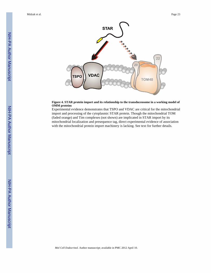
Figure 4. STAR protein import and its relationship to the transduceosome in a working model ofOMM proteinsExperimental evidence demonstrates that TSPO and VDAC are critical for the mitochondrialimport and processing of the cytoplasmic STAR protein. Though the mitochondrial TOM(faded orange) and Tim complexes (not shown) are implicated in STAR import by itsmitochondrial localization and presequence tag, direct experimental evidence of associationwith the mitochondrial protein import machinery is lacking. See text for further details.
Midzak et al. Page 23
Mol Cell Endocrinol. Author manuscript; available in PMC 2012 April 10.
NIH
-PA Author Manuscript
NIH
-PA Author Manuscript
NIH
-PA Author Manuscript