Isc1 regulates sphingolipid metabolism in yeast mitochondria
27
Isc1 regulates sphingolipid metabolism in yeast mitochondria Hiroshi Kitagaki a,d,e , L. Ashley Cowart c,a , Nabil Matmati a , Silvia Vaena de Avalos a , Sergei A. Novgorodov b , Youssef H. Zeidan a , Jacek Bielawski a , Lina M. Obeid b,c , and Yusuf A. Hannun a aDepartment of Biochemistry and Molecular Biology, Medical University of South Carolina, Charleston, South Carolina 29425 bDepartment of Medicine, Medical University of South Carolina, Charleston, South Carolina 29425 cRalph H. Johnson Veterans Affairs Medical Center, Charleston, South Carolina 29401 dMinistry of Education, Culture, Sports, Science and Technology, Chiyoda-ku, Toyko, Japan eNational Research Institute of Brewing, Higashihiroshima city, Hiroshima, Japan Abstract The Saccharomyces cerevisiae inositolsphingolipid phospholipase C (Isc1p), a homolog of mammalian neutral sphingomyelinases, hydrolyzes complex sphingolipids to produce ceramide in vitro. Epitope-tagged Isc1p associates with the mitochondria in the post-diauxic phase of yeast growth. In this report, the mitochondrial localization of Isc1p and its role in regulating sphingolipid metabolism were investigated. First, endogenous Isc1p activity was enriched in highly purified mitochondria, and western blots using highly purified mitochondrial membrane fractions demonstrated that epitope-tagged Isc1p localized to the outer mitochondrial membrane as an integral membrane protein. Next, LC/MS was employed to determine the sphingolipid composition of highly purified mitochondria which were found to be significantly enriched in α-hydroxylated phytoceramides (21.7 fold) relative to the whole cell. Mitochondria, on the other hand, were significantly depleted in sphingoid bases. Compared to the parental strain, mitochondria from isc1⊿ in the post-diauxic phase showed drastic reduction in the levels of α-hydroxylated phytoceramide (93.1 % loss compared to WT mitochondria with only 2.58 fold enrichment in mitochondria compared to whole cell). Functionally, isc1⊿ showed a higher rate of respiratory- deficient cells after incubation at high temperature and was more sensitive to hydrogen peroxide and ethidium bromide, indicating that isc1⊿ exhibits defects related to mitochondrial function. These results suggest that Isc1p generates ceramide in mitochondria, and the generated ceramide contributes to the normal function of mitochondria. This study provides a first insight into the specific composition of ceramides in mitochondria. Keywords Mitochondria; Sphingolipid; Sphingomyelinase; Lipidomics; ISC1; Yeast Address correspondence to; Yusuf A. Hannun, the Department of Biochemistry and Molecular Biology, Medical University of South Carolina, 173 Ashley Avenue, Charleston, South Carolina 29425 Tel.: 843−792−4321; Fax: 843−792−4322; E-mail: [email protected]. Publisher's Disclaimer: This is a PDF file of an unedited manuscript that has been accepted for publication. As a service to our customers we are providing this early version of the manuscript. The manuscript will undergo copyediting, typesetting, and review of the resulting proof before it is published in its final citable form. Please note that during the production process errors may be discovered which could affect the content, and all legal disclaimers that apply to the journal pertain. NIH Public Access Author Manuscript Biochim Biophys Acta. Author manuscript; available in PMC 2008 November 1. Published in final edited form as: Biochim Biophys Acta. 2007 November ; 1768(11): 2849–2861. NIH-PA Author Manuscript NIH-PA Author Manuscript NIH-PA Author Manuscript
Transcript of Isc1 regulates sphingolipid metabolism in yeast mitochondria

Isc1 regulates sphingolipid metabolism in yeast mitochondria
Hiroshi Kitagakia,d,e, L. Ashley Cowartc,a, Nabil Matmatia, Silvia Vaena de Avalosa, SergeiA. Novgorodovb, Youssef H. Zeidana, Jacek Bielawskia, Lina M. Obeidb,c, and Yusuf A.Hannuna
aDepartment of Biochemistry and Molecular Biology, Medical University of South Carolina, Charleston,South Carolina 29425
bDepartment of Medicine, Medical University of South Carolina, Charleston, South Carolina 29425
cRalph H. Johnson Veterans Affairs Medical Center, Charleston, South Carolina 29401
dMinistry of Education, Culture, Sports, Science and Technology, Chiyoda-ku, Toyko, Japan
eNational Research Institute of Brewing, Higashihiroshima city, Hiroshima, Japan
AbstractThe Saccharomyces cerevisiae inositolsphingolipid phospholipase C (Isc1p), a homolog ofmammalian neutral sphingomyelinases, hydrolyzes complex sphingolipids to produce ceramide invitro. Epitope-tagged Isc1p associates with the mitochondria in the post-diauxic phase of yeastgrowth. In this report, the mitochondrial localization of Isc1p and its role in regulating sphingolipidmetabolism were investigated. First, endogenous Isc1p activity was enriched in highly purifiedmitochondria, and western blots using highly purified mitochondrial membrane fractionsdemonstrated that epitope-tagged Isc1p localized to the outer mitochondrial membrane as an integralmembrane protein. Next, LC/MS was employed to determine the sphingolipid composition of highlypurified mitochondria which were found to be significantly enriched in α-hydroxylatedphytoceramides (21.7 fold) relative to the whole cell. Mitochondria, on the other hand, weresignificantly depleted in sphingoid bases. Compared to the parental strain, mitochondria fromisc1⊿ in the post-diauxic phase showed drastic reduction in the levels of α-hydroxylatedphytoceramide (93.1 % loss compared to WT mitochondria with only 2.58 fold enrichment inmitochondria compared to whole cell). Functionally, isc1⊿ showed a higher rate of respiratory-deficient cells after incubation at high temperature and was more sensitive to hydrogen peroxide andethidium bromide, indicating that isc1⊿ exhibits defects related to mitochondrial function. Theseresults suggest that Isc1p generates ceramide in mitochondria, and the generated ceramide contributesto the normal function of mitochondria. This study provides a first insight into the specificcomposition of ceramides in mitochondria.
KeywordsMitochondria; Sphingolipid; Sphingomyelinase; Lipidomics; ISC1; Yeast
Address correspondence to; Yusuf A. Hannun, the Department of Biochemistry and Molecular Biology, Medical University of SouthCarolina, 173 Ashley Avenue, Charleston, South Carolina 29425 Tel.: 843−792−4321; Fax: 843−792−4322; E-mail: [email protected]'s Disclaimer: This is a PDF file of an unedited manuscript that has been accepted for publication. As a service to our customerswe are providing this early version of the manuscript. The manuscript will undergo copyediting, typesetting, and review of the resultingproof before it is published in its final citable form. Please note that during the production process errors may be discovered which couldaffect the content, and all legal disclaimers that apply to the journal pertain.
NIH Public AccessAuthor ManuscriptBiochim Biophys Acta. Author manuscript; available in PMC 2008 November 1.
Published in final edited form as:Biochim Biophys Acta. 2007 November ; 1768(11): 2849–2861.
NIH
-PA Author Manuscript
NIH
-PA Author Manuscript
NIH
-PA Author Manuscript

1. IntroductionSphingolipids play crucial roles as signaling molecules in eukaryotic cells, regulating suchprocesses as stress responses, cell growth, differentiation, cell cycle arrest, endocytosis, andapoptosis [1-7]. Additionally, complex sphingolipids serve structural functions in membranes,and they may also undergo dynamic clustering with sterols to form lipid microdomains or‘rafts’ within the membrane bilayer. This domain structure has been proposed to play a role asplatforms for signal transduction and/or protein sorting [8].
Most, if not all, organelles contain sphingolipids as well as several sphingolipid-metabolizingenzymes [9-11]. Moreover, the hydrophobicity of bioactive sphingolipids restrains them totheir specific compartments (unless transported by specific mechanisms). These considerationsled to the proposal that sphingolipid function is compartment-specific [1]. In this context, manystudies in eukaryotes have recently started suggesting important roles for mitochondrialsphingolipids [11-13].
Ceramide in mitochondria has been shown to regulate apoptosis in mammalian cells. Forexample, when bacterial sphingomyelinase was targeted to mitochondria, it inducedmitochondrial ceramide generation and apoptosis [13], and mitochondrial ceramide has beenimplicated in the activation/translocation of the pro-apoptotic Bax [14]. Moreover, manystudies have reported that ceramide has biophysical effects on mitochondrial membranes, byincreasing the permeability of the outer and inner mitochondrial membranes [15], suppressingrespiratory chain activity [16], or forming channels [17]. Therefore, the role of ceramide inmitochondria is attracting wide interests in terms of basic biology and medical application.Other studies have suggested mitochondrial localization of some enzymes of sphingolipidmetabolism, including ceramide and dihydroceramide synthase [12,18], ceramidase [19],glycosyltransferase [20], human sialidase [21], and sphingosine kinase [22].
Saccharomyces cerevisiae has proven to be an invaluable model for studying sphingolipidbiosynthesis, in part because of its simplicity in sphingolipid metabolism relative tomammalian systems [3,23]. Interestingly, the S. cerevisiae Isc1p, the homolog of mammalianneutral sphingomyelinases, was recently shown to associate with mitochondria in the post-diauxic phase of yeast growth [24]. Isc1p produces ceramide from the complex sphingolipidsinositol phosphorylceramide (IPC), mannosylinositol phosphorylceramide (MIPC), andmannosyldiinositol phosphorylceramide (M(IP)2C) [25-28]. Isc1p was also shown to have arole in utilization of non-fermentable carbon sources, as an isc1⊿ strain grew very slowly inthe post-diauxic phase or when grown in media containing non-fermentable carbon sources[29]. Utilization of non-fermentable carbon sources requires intact mitochondrial function, thussuggesting a role for Isc1p in mitochondrial function.
In this study we sought to elucidate the submitochondrial localization of Isc1p, its role inmitochondrial sphingolipid metabolism, and mitochondria-related phenotypes of isc1⊿. Thesestudies have implications for compartment-specific sphingolipid metabolism and function invivo.
2. Materials and Methods2.1. Yeast strains and media
JK93α WT (MATα leu2−3, 112 ura3−52 rme1 trp1 his4) and JK93α isc1⊿ (MATα leu2−3,112 ura3−52 rme1 trp1 his4 ISC1::G418) [25] were used in this study. Standard yeast culturemedium was prepared using Difco YPD broth. Plates were prepared using Difco YPD agarand stored at 4°C. YPGE plates contained 1% yeast extract, 2% bactopeptone, 2% ethanol, 2%
Kitagaki et al. Page 2
Biochim Biophys Acta. Author manuscript; available in PMC 2008 November 1.
NIH
-PA Author Manuscript
NIH
-PA Author Manuscript
NIH
-PA Author Manuscript

glycerol and 2% agar. Yeast cells were cultured at 30°C with shaking unless otherwiseindicated.
2.2. Isolation of purified mitochondriaIsolation of purified mitochondria was basically performed essentially as described [30-32].Briefly, yeast cells were incubated to saturation at 30°C. Six liter flask containingapproximately 1.25 L YPD were inoculated with an overnight culture to OD600=0.1 andincubated at 30°C for 24 h with vigorous aeration. Yeast cells were centrifuged at 2,000 g for5 min, washed with water, resuspended in 100 mM Tris-SO4, pH 9.4, 10 mM DTT (2 ml/g wetcell), incubated at 30°C for 15 min, centrifuged at 2,000 g for 5 min, washed with SP buffer(1.2 M sorbitol, 20 mM K-P pH 7.4), dissolved in SP buffer containing zymolyase 100T (7ml/g cells, 1.5 mg/g cells), incubated at 30°C for 60 min, centrifuged at 2,500 g for 5 min,washed with SP buffer (7 ml/g wet cell) twice, resuspended in homogenization buffer (0.6 Msorbitol, 10 mM Tris-HCl, pH 7.4, 1 mM EDTA, 1 mM phenylmethylsulfonyl fluoride) (6.5ml/g cells), homogenized in a 40 ml dounce homogenizer coated with Teflon using 25 strokes,diluted two fold with homogenizing buffer, centrifuged for 5 min at 1,500 g to remove cellnuclei and debris, and centrifuged again at 2,500 g for 5 min. The supernatant was centrifugedat 12,000 g for 15 min. The supernatant was saved as the post-mitochondrial fraction and TCA-precipitated for further analysis. The pellet was resuspended in 30 ml SM buffer (250 mMsucrose, 1 mM EDTA, 10 mM MOPS-KOH, pH 7.2) and centrifuged at 2,500 g for 5 min. Thesupernatant was then centrifuged at 12,000 g for 15 min, and the pellet was resuspended in 10ml SM buffer as crude mitochondria. For purification of mitochondria, the amount ofmitochondrial protein was measured by OD280, adjusted to 5 mg/ml protein in SM buffer,homogenized with 10 strokes in a dounce homogenizer coated with Teflon, and subsequentlyloaded onto a four step sucrose gradient [1.5 ml of 60%, 4 ml of 32%, 1.5 ml of 23%, 1.5 mlof 15% of sucrose in 1 mM EDTA, 10 mM MOPS-KOH (pH 7.2)], and centrifuged at 134,000gfor 1 h with no braking. The 32/60% interface was then recovered with a Pasteur pipette andwashed with 2 vol of SM buffer at 12,000 g for 15 min. The mitochondrial fraction was againhomogenized with 10 strokes in a dounce homogenizer coated with Teflon and then appliedto a four step sucrose gradient as described above. The 32/60% interface was recovered,resuspended in 2 volumes of SM buffer, and centrifuged at 12,000 g for 15 min. The final pelletwas resuspended in SM buffer, divided into small aliquots, and frozen at −80°C.
2.3. LC/MS measurementsPurified mitochondria or yeast cells in the post-diauxic phase were snap-frozen in a methanol-dry ice bath and stored at −80°C. ESI/MS/MS analysis of endogenous phyto- anddihydrosphingosine bases, their phosphates, α-hydroxylated phytoceramide, and non-hydroxylated phyto- and dihydroceramide species were performed on a Thermo Finnigan TSQ7000 triple quadrupole mass spectrometer, operating in a Multiple Reaction Monitoringpositive ionization mode, using modified version [28,34]. Briefly, biological samples werefortified with internal standards (ISs: C17 base D-erythro-sphingosine, C17 sphingosine-1-phosphate, N-palmitoyl-D-erythro-C13 sphingosine, heptadecanoyl-D-erythro-sphingosineand C6-Phyto-ceramide), then extracted with 70% isopropanol:ethylacetate:pyridine:25%ammonia (60:20:2:0.5 by volume) solvent system. After evaporation and reconstitution in 150μl of methanol, samples were injected on the HP1100/TSQ 7000 LC/MS system and gradienteluted from the BDS Hypersil C8, 150 × 3.2 mm, 3 μm particle size column, with 1.0 mMmethanolic ammonium formate / 2 mM aqueous ammonium formate mobile phase system.Peaks corresponding to the target analytes and ISs were collected and processed using theXcalibur software system. Quantitative analysis was based on the calibration curves generatedby spiking an artificial matrix with the known amounts of the target analyte synthetic standardsand an equal amount of IS. The target analyte/IS peak areas ratios were plotted against analyzedconcentration. The target analyte/IS peak area ratios from the samples were similarly
Kitagaki et al. Page 3
Biochim Biophys Acta. Author manuscript; available in PMC 2008 November 1.
NIH
-PA Author Manuscript
NIH
-PA Author Manuscript
NIH
-PA Author Manuscript

normalized to their respective ISs and compared to the calibration curves, using a linearregression model.
2.4. Submitochondrial localization of Flag-Isc1pSubmitochondrial localization of Flag-Isc1p was determined as previously described [35].Briefly, 200 μg of highly purified mitochondria from isc1⊿+pYES/FLAG-ISC1 were pelletedat 10,000 g for 15 min and suspended in 40 μl SM buffer (250 mM sucrose, 1 mM EDTA, 10mM MOPS-KOH, pH 7.2). Samples were split into two equal portions. One hundred and eightymicroliters of SM buffer or EM buffer (1 mM EDTA, 10 mM MOPS-KOH, pH 7.2) was added,pipetted up and down 10 times to facilitate the rupture of the outer membrane from the swollenmitochondria, and incubated for 15 min on ice. Samples were split into two equal portions andtreated with or without 50 μg/ml proteinase K for 30 min on ice. Phenylmethylsulfonyl fluoridewas added to the solution (final concentration 2 mM). The sample was then centrifuged at12,000×g for 5 min, washed once and analyzed by western blotting using anti-Flag antibody.
2.5. Determination of association of proteins with membraneHighly purified mitochondria, purified from isc1⊿+pYES/FLAG-ISC1, were treated withoutosmotic shock (250 mM sucrose, 10 mM HEPES, pH 7.2, 1 mM EDTA), with osmotic shock(10 mM HEPES, pH 7.2, 1 mM EDTA), with osmotic shock and high salt (0.1 M NaCl, 10mM HEPES, pH 7.2, 1 mM EDTA), or with osmotic shock and alkali pH (0.1 M Na2CO3, pH11.2, 10 mM HEPES, pH 7.2, 1 mM EDTA). They were then centrifuged at 100,000×g for 10min, and the pellet and supernatant were analyzed by western blotting using antibody againstFlag.
2.6. Statistical analysisThe amounts of sphingolipid species of mitochondria prepared from WT and isc1⊿ arepresented as means ± standard error of the mean (SEM) of three lipid measurements from twoindependent cultures and mitochondrial purifications. The amounts of sphingolipid species ofthe whole cell are presented as means ± SEM of lipid measurements from three independentcultures. Statistical differences were evaluated by Student's unpaired t test.
2.7. MiscellaneousAntibodies against Cox3p, Porin, Dpm1p, V-ATPase (100kDa subunit), and Pgk1p wereobtained from Molecular Probes, and that against FLAG was from Sigma-Aldrich. Theantibody against Mge1p was a kind gift from Dr. C. M. Koehler.
3. Results3.1. Isc1p as an integral membrane protein localized to the outer membrane of mitochondria
Since epitope-tagged Isc1p was previously shown to associate with mitochondria in the post-diauxic phase, it became important to determine the mitochondrial localization of endogenousIsc1p. WT and isc1⊿ cells were incubated to the post-diauxic phase, collected bycentrifugation, and mitochondria were isolated to a high degree of purity using multi-stepcentrifugation in conjunction with a two-step sucrose gradient, which has been verified bymitochondrial proteome studies [30-33]. The purity of the mitochondria was confirmed bywestern blotting with an antibody against a cytosolic marker protein, Pgk1p, a vacuolar markerprotein, V-ATPase, an endoplasmic reticulum (ER) marker protein, Dpm1p, and amitochondrial marker protein, Cox3p. The results showed that the purified fraction was devoidof Pgk1p, V-ATPase and Dpm1p and highly enriched in Cox3p (Fig. 1A), indicating thatmitochondria were purified to a high degree, consistent with previous reports [30-32].Subsequent studies were performed using these highly purified mitochondria prepared from
Kitagaki et al. Page 4
Biochim Biophys Acta. Author manuscript; available in PMC 2008 November 1.
NIH
-PA Author Manuscript
NIH
-PA Author Manuscript
NIH
-PA Author Manuscript

post-diauxic phase cells. In order to determine the localization of endogenous Isc1p, Isc1penzymatic activity in the purified mitochondria was determined. Isc1p specific activity washighly concentrated in the mitochondrial fraction (2.8 pmol/μg/hr) when compared to thespecific activity of the whole cell homogenate (1.0 pmol/μg/hr) (Fig. 1B). Moreover, cells andmitochondria from the isc1⊿ strain contained only background activity, demonstrating theIsc1p is the primary enzyme responsible for the detected activity in mitochondria. Takentogether, these results demonstrate that Isc1p activity is indeed localized to mitochondria (Fig.1B).
Next it became important to determine if Isc1p is loosely associated with mitochondria or if itis an integral mitochondrial membrane protein. Mitochondria were treated with osmotic shock(to lyze the mitochondrial outer membrane) and then treated with either high salt or alkali pHextraction, centrifuged at 100,000 g for 10 min, and pellets were analyzed by western blotting.N-terminally tagged Flag-Isc1p [25] was not dissociated from the membrane with osmoticshock (Fig. 1C lanes 4 and 5), high salt (Fig. 1C lanes 6 and 7) or alkali pH (Fig. 1C lanes 8and 9). Cox3p, a known integral membrane protein behaved similarly whereas Mge1p, aperipheral mitochondrial protein became significantly dissociated from membranes in responseto treatment with alkaline pH. Taken together, these results indicate that Isc1p is a bona fidemembrane protein.
Next, the submitochondrial localization of Isc1p was determined using protease protectionassays. Proteinase K was added to purified mitochondria with or without osmotic shock, andthe degradation of several marker proteins was examined by western blotting. Using this assaywithout osmotic shock, exogenously added proteinase K can only access and degrade proteinsexposed from the outer mitochondrial membrane. On the other hand, with osmotic shock,proteinase K causes degradation of proteins contained in the outer membrane, in theintermembrane space, or exposed out of the inner membrane since osmotic shock causesrupture of the outer mitochondrial membrane while leaving the inner mitochondrial membraneintact. A previously characterized inner membrane protein, Cox3p, was not degraded even afterosmotic shock (Fig. 1D, lane 4), demonstrating that under these conditions, no proteasecleavage sites are exposed out of the inner membrane. On the other hand, porin, a markerprotein for outer mitochondrial membrane proteins, was degraded only after osmotic shock(Fig. 1D, lane 4), suggesting that it is inserted in the outer membrane so that it is degraded onlyafter rupture of the outer membrane. To the contrary, Flag-Isc1p was efficiently degraded evenwithout osmotic shock (Fig. 1D, lane 2), indicating that Flag-Isc1p localized to the outermembrane of mitochondria with exposed protease cleavage sites, or, alternatively, only N-terminally tagged FLAG is exposed from the outer membrane and Isc1p is localized inside theouter membrane. From all the above data, it could be concluded that Flag-Isc1p localizes tothe outer membrane of mitochondria and is integrated into the outer mitochondrial membrane.
3.2. Mitochondria are enriched in very long chain phytoceramidesIn previous reports, we showed that Isc1p degrades complex sphingolipids such as IPC, MIPCand M(IP)2C to form ceramides in vitro [25-27] and that Isc1p is activated byphosphatidylglycerol and cardiolipin [29], two phospholipids that reside in mitochondria[36,37]. These results, together with the mitochondrial localization of Isc1p, raised thepossibility that Isc1p may be responsible for generation of ceramides in mitochondria in vivo.To address this issue, we first determined the sphingolipid composition of mitochondria andcompared it to that of whole cells. WT cells were incubated to the post-diauxic phase, and themitochondria were isolated to a high degree of purity using a two-step sucrose gradient asdescribed above. The sphingolipid profile of the whole cell of WT and the mitochondriapurified from WT was determined using LC-MS [34]. The masses of sphingolipids werecalculated based on the phospholipid content.
Kitagaki et al. Page 5
Biochim Biophys Acta. Author manuscript; available in PMC 2008 November 1.
NIH
-PA Author Manuscript
NIH
-PA Author Manuscript
NIH
-PA Author Manuscript

In whole cells, α-hydroxylated C26-phytoceramide, phytosphingosine, dihydrosphingosine,and α-hydroxylated C14-phytoceramide constituted the major species (Fig. 2A), α-hydroxylated C24:1-, C24- and C18-phytoceramides and non-hydroxylated C26-phytoceramideswere next in abundance (Fig. 2B), and α-hydroxylated C20- and C22-phytoceramides and mostof the non-hydroxylated dihydroceramides and phytoceramides showed significantly lowerlevels (Fig. 2C).
In contrast, the results revealed, for the first time, that mitochondria contain predominantly α-hydroxylated C26-phytoceramide (Fig. 3A) with significantly lower levels of other ceramidesor sphingoid bases (Fig. 3B and 3C). These results were expressed relative to mitochondrialtotal phospholipids, and therefore, they represent the relative surface concentration of thesesphingolipids in membranes. It should be noted that even for the highest ceramide, the ratio tophospholipids was less than 3%, thus demonstrating that phospholipids are indeed in excessand that mitochondrial ceramides are minor species.
In order to determine the relative enrichment of mitochondrial ceramides to that of whole cells,we calculated the degree of ‘surface enrichment’ of these sphingolipids in mitochondria towhole cells by dividing the ceramide/phospholipid ratio in mitochondria over that in wholecells. This calculated ratio of mitochondrial/whole cell for α-hydroxylated C26-phytoceramideswas 21.7 fold, and this enrichment in mitochondria was also observed for other ceramidespecies (Fig. 3D), indicating that mitochondria were highly enriched with ceramide speciesmainly composed of α-hydroxylated phytoceramides. This result also suggests that the majorsource of α-hydroxylated phytoceramides of the whole cell is derived from the mitochondria.Non-hydroxylated ceramides also showed increases in the mitochondria relative to the wholecells (Fig. 3D). Notably, the levels of the non-hydroxylated phytoceramides anddihydroceramides were significantly lower than those of α-hydroxylated phytoceramides bothin whole cells and mitochondria (Fig. 2 and Fig. 3).
Reciprocally, the levels of phytosphingosine, the predominant ‘simple’ sphingolipid of yeast,were significantly decreased in mitochondria relative to whole cells (Fig. 3E, note inverted y-axis compared to 3D).
Taken together, the results demonstrate that in S. cerevisiae, mitochondria show selectiveenrichment in α-hydroxylated phytoceramides and selective depletion of sphingoid bases.These results were basically similar when sphingolipid levels were normalized by proteinamount (data not shown), indicating that these results are not due to selective difference in totalphospholipid levels between the whole cell and mitochondria.
3.3. Mitochondria of isc1⊿ show an altered sphingolipid profileNext, the sphingolipid profile of mitochondria prepared from isc1⊿ was determined andcompared to that of WT mitochondria in order to determine the contribution of Isc1p tomitochondrial sphingolipids. The major sphingolipids detected in whole cells of the isc1⊿strain were α-hydroxylated C26-phytoceramide, phytosphingosine, α-hydroxylated C14-phytoceramide and dihydrosphingosine (Fig. 4A) followed by non-hydroxylated C26-phytoceramide, α-hydroxylated C24-phytoceramide and non-hydroxylated C18:1-dihydroceramide (Fig. 4B), with other ceramides showing very low levels (Fig. 4C).
The predominant species of sphingolipids in mitochondria from isc1 ⊿ were α-hydroxylatedC26-phytoceramide, non-hydroxylated C26-phytoceramide, α-hydroxylated C14-phytoceramide and α-hydroxylated C24:1-phytoceramide (Fig. 4D), followed by α-hydroxylated C24-phytoceramide, non-hydroxylated C26:1-phytoceramide, phytosphingosineand dihydrosphingosine (Fig. 4E), with other sphingolipids being minor species (Fig. 4F).
Kitagaki et al. Page 6
Biochim Biophys Acta. Author manuscript; available in PMC 2008 November 1.
NIH
-PA Author Manuscript
NIH
-PA Author Manuscript
NIH
-PA Author Manuscript

Many of these species showed significant reduction in their enrichment in mitochondria. Mostnotably, the content of α-hydroxylated C26-phytoceramide in isc1⊿ mitochondria was only6.94% of that of WT mitochondria (Fig. 5A), and this decrease was significantly rescued byexpression of FLAG-ISC1 (Fig. 5C). As a consequence, the enrichment of α-hydroxylatedC26-phytoceramide in mitochondria was decreased to 2.58 fold in isc1⊿ (Fig. 4A and 4D)relative to the 21.7 fold in WT (Fig. 3D). This decrease in isc1⊿ mitochondria was alsoobserved for other ceramides (Fig. 5A).
These results suggest that Isc1p functions in cells to generate mitochondrial ceramides mainlycomposed of α-hydroxylated phyotceramides. Reciprocally, α-hydroxylated C14-phytoceramide and non-hydroxylated C26-phytoceramide were increased in isc1⊿mitochondria (Fig. 5B), suggesting a complementary pathway to overcome the decrease ofother ceramide species. These lipid changes in isc1⊿ mitochondria were partially suppressedby the expression of FLAG-ISC1 (Fig. 5C), although some of the lipids were not recovered to100%, probably because FLAG-ISC1 was expressed under the GAL1 promoter. Nontheless,this result indicates that the lipid changes observed in isc1⊿ mitochondria relative to WTmitochondria are not derived from artifact of mitochondrial purification. Phospholipid contentper protein in WT mitochondria was 34.9+/−3.14 (nmol/mg), while that in isc1⊿ mitochondriais 36.7+/−0.96 (nmol/mg), indicating that the mass profile changes induced by the deletion ofISC1 are not due to phospholipid changes in the mitochondria of isc1⊿. These resultsdemonstrate that the isc1⊿ has an altered mitochondrial sphingolipid profile, and suggest thatIsc1p produces selectively α-hydroxylated phytoceramides in mitochondria in vivo.
3.4. Disruption of ISC1 induces mitochondrial-associated defectsThe alteration of sphingolipid profile of mitochondria in vivo observed above suggested thatIsc1p may influence mitochondrial related functions. In order to address this hypothesis, weexamined the formation of petite colonies in isc1⊿. In S. cerevisiae, mitochondrial defectsoften manifest by growing as respiratory deficient, “petite” colonies on fermentable carbonsources. In addition, cells with compromised mitochondria form large numbers of petitecolonies after prolonged culture at elevated temperature when grown on either fermentable ornon-fermentable carbon sources [38,39]. To determine whether isc1⊿ would form petites underthese conditions, the isc1⊿ strain was grown at normal (30°C) and high temperature (39°C),and the fraction of petite colonies was determined by counting the ratio of colonies which havesmaller size than normal and are unable to grow on non-fermentable carbon source plates(YPGE). The results showed that the isc1⊿ strain exhibited a significantly increased ratio ofpetite colonies at normal temperature (p<0.01, Fig. 6A) and this phenotype was significantlyexacerbated by growth at high temperature (p<0.12, Fig. 6A).
Yeast cells with compromised mitochondria have also been shown to be sensitive to oxidativestress such as hydrogen peroxide [40]. To test whether the isc1⊿ strain was sensitive tohydrogen peroxide, yeast in log phase growth were treated with 1 mM hydrogen peroxide for45 min, and the number of colonies was measured. Figure 6B demonstrates that the number ofcolonies of WT cells treated with hydrogen peroxide was 58.0% of that of non-treated cellswhereas the number of colonies of isc1⊿ cells treated with hydrogen peroxide was 41.2% ofthat of non-treated cells, indicating that isc1⊿ is significantly sensitive to hydrogen peroxiderelative to WT (p<0.01).
Ethidium bromide has been reported to specifically damage mitochondrial DNA, and mutantsthat have dysfunctional mitochondria show altered sensitivity to this substance [41,42]. Toelucidate if isc1⊿ is sensitive to ethidium bromide, WT and isc1⊿ cells were treated with 50μM ethidium bromide and spotted onto a YPD plate. The results showed that isc1⊿ was moresensitive to ethidium bromide than WT (Fig. 6C). Together, these results indicate that isc1⊿
Kitagaki et al. Page 7
Biochim Biophys Acta. Author manuscript; available in PMC 2008 November 1.
NIH
-PA Author Manuscript
NIH
-PA Author Manuscript
NIH
-PA Author Manuscript

has defective mitochondrial phenotypes and reveal a role of sphingolipid in mitochondrialfunction in vivo.
4. DiscussionData in this report reveal that Isc1p of Saccharomyces cerevisiae localizes to the outermembrane of mitochondria as an integral membrane protein, and its activity is enriched inpurified mitochondria. Moreover, this study is the first to measure α-hydroxylated ceramidespecies and to show that mitochondria are highly enriched in very long chain α-hydroxylatedphytoceramides. Importantly isc1⊿ showed an altered mitochondrial sphingolipid profile,indicating that Isc1p regulates sphingolipid metabolism of mitochondria, especially theformation of α-hydroxylated very long chain phytoceramides. Finally, isc1⊿ displayed defectsin mitochondrial functions, suggesting, for the first time, a role for mitochondrial sphingolipidmetabolism in mitochondrial metabolic function in vivo.
Recent advances in methods for mitochondrial purification as well as newly developedlipidomic strategies have enabled a more comprehensive and precise assessment of simplesphingolipids in mitochondria in Saccharomyces cerevisiae. Previous data indicated thatmitochondria contain 7.4% of the total complex sphingolipid content, with an additional 30%in plasma membrane [10]. Another report demonstrated that mitochondria contain 4.6 nmol ofIPC/mg protein, 7.7 nmol of MIPC/mg protein and 5.6 nmol of M(IP)2C/mg protein, whileplasma membrane contains 34.9 nmol of IPC/mg protein, 69.9 nmol of MIPC/mg protein and120.5 nmol of M(IP)2C/mg protein [9]. It is important to note, however, that the mitochondrialfractions used for these studies were not derived from density gradient centrifugation, and thus,it is difficult to know the degree of contamination of these preparations with microsomalmembranes. Moreover, these previous reports studied only complex sphingolipids and did notaddress ceramides and sphingoid bases, the subjects of this study.
With this current analysis, it was found that mitochondria from WT cells are highly enriched,and specifically, with very long chain α-hydroxylated phytoceramides. This unexpected resultraises a number of questions as to the metabolism of very long chain sphingolipids in yeastand their function. Very little is known about the regulation of incorporation of very long chainfatty acids into sphingolipids or the subcellular sites of migration of these lipids. Both Lag1pand Lac1p ceramide synthases, thought to reside in the ER, are capable of introducing verylong chain fatty acids into ceramides [43], which in turn are incorporated in IPC, MIPC, andM(IP)2C [44-47]. While the bulk of these complex sphingolipids eventually arrives at theplasma membrane, published results show that a significant component of these lipids mayreside in the mitochondria [9]. Together with the current results that Isc1p is localized to theouter membrane of mitochondria (Fig. 1) and mitochondria purified from isc1 ⊿ exhibit a lowercontent of α-hydroxylated phytoceramides than that from WT (Fig. 5A), it is suggested thatthese complex sphingolipids become substrates for Isc1p either at the outer mitochondrialmembrane or in a compartment that comes in contact with the outer membrane (e.g.mitochondrial associated membranes or others). Further studies are required to determinewhere and how Isc1p interacts with yeast complex sphingolipids. Moreover, this study did notaddress the issue of topology of Isc1p, and therefore, it is not known whether its active sitefaces into the mitochondria or out of the mitochondria. This proposal that sphingolipids aregenerated at the outer membrane of mitochondria is also in agreement with the previous report[48] that detected threefold enrichment of ceramide in the outer membrane relative to the innermembrane of rat liver mitochondria. Considering that Isc1p is activated by cardiolipin andphosphatidylglycerol [26,29] and that these lipids are synthesized in the inner membrane ofmitochondria [49-51], Isc1p may be activated at the membrane contact site of outer and innermembrane [52,53]. Alternatively, up to 36.6% of cardiolipin has been detected in the outermembrane of mitochondria [54,55] and this could then activate Isc1p. Further investigation is
Kitagaki et al. Page 8
Biochim Biophys Acta. Author manuscript; available in PMC 2008 November 1.
NIH
-PA Author Manuscript
NIH
-PA Author Manuscript
NIH
-PA Author Manuscript

required for elucidation of pathways of subcellular transport and metabolism of complexsphingolipids.
The results also showed that mitochondria are relatively depleted in sphingoid bases, and thisis consistent with the known ER sites for de novo sphingolipid synthesis [56] or ceramidedegradation in the ER and Golgi [57,58].
The current results as well as previous results [29] implicating Isc1p in regulation ofmetabolism of non-fermentable carbon sources suggest a role for either the precursorsphingolipids and/or the generated α-hydroxylated phytoceramides in regulating metabolicfunctions of mitochondria. These could include signaling functions of the ceramides and/orstructural functions. The latter are consistent with the recent suggestion that manymitochondrial proteins reside in ‘rafts’ [59]. Moreover, genome-wide analysis revealed thatmost of ergosterol synthetic mutants showed defective mitochondrial morphology [60]. Since,ergosterol has been reported to form rafts together with sphingolipids [61], the results in thisstudy raise the possibility of sphingolipid/sterol structures/raft in the function of mitochondria.
The specific ceramide generated by the action of Isc1p could also play regulatory functions.In mammalian cells, ceramide, exogenously added or generated endogenously byoverexpression of mitochondrially-targeted sphingomyelinase, has been shown to induce/regulate Bax translocation and to be involved in mitochondrial dysfunction, such as increasesin the permeability of the outer membrane and release of cytochrome c [13,15,17,62]. However,whether endogenous ceramides are generated in eukaryotic mitochondria and exert effects onmitochondria in vivo has not been well evaluated and has been under significant discussion[63]. The results in this study show, for the first time, that native alteration in sphingolipidmetabolism in mitochondria causes dysfunctions of mitochondria in vivo, suggesting anintrinsic role of some specific ceramide in regulating mitochondrial functions. However,further studies are required for elucidation of sphingolipid-mediated functions in yeast.
In conclusion, we have determined the submitochondrial localization of Isc1p and its effect onmitochondrial sphingolipid metabolism, and elucidated that isc1⊿ exhibits mitochondria-related phenotypes. The findings contribute key knowledge toward complete understanding ofthe metabolism of mitochondrial sphingolipids and their roles in mitochondrial function ineukaryotic cells.
Acknowledgements
We thank Dr. Tatyana I. Gudz, Dr. Hiroko Hama, Dr. Kazuyuki Kitatani, Dr. Chiara Luberto, and Dr. FernandoAlvarez-Vasquez for helpful advice, Dr. Alicja Bielawska for lipid analyses and Dr. Carla M. Koehler for the antibodyagainst Mge1p. Liquid-chromatography-MS lipid analyses were performed at the Lipidomics Core at the MedicalUniversity of South Carolina. This material is supported in part by NIH R01 GM43825 (to YAH) and a VA MeritAward, Office of Research and Development, Department of Veterans Affairs, NIH R01 AG016583 (to LMO). L. A.C. would like to acknowledge support from the Department of Veteran's Affairs Merit Review Entry Program. A partof this study was financially supported by the Budget for Nuclear Research of the Ministry of Education, Culture,Sports, Science and Technology, based on the screening and counseling by the Atomic Energy Commission.
AbbreviationsIPC, inositol phosphorylceramide; MIPC, mannosylinositol phosphorylceramide; M(IP)2C,mannosyldiinositol phosphorylceramide; YPD, yeast/peptone/dextrose; YPGE, yeast/peptone/glycerol/ethanol; SEM, standard error of mean; ER, endoplasmic reticulum; IS, internalstandard; a-HO-, α-hydroxylated; Cer, ceramide; DHSph, dihydrosphingosine; Phyto-Sph,phytosphingosine; dhCn-Cer, dihydroceramide with Cn fatty acid; phytoCn-Cer,phytoceramide with Cn fatty acid.
Kitagaki et al. Page 9
Biochim Biophys Acta. Author manuscript; available in PMC 2008 November 1.
NIH
-PA Author Manuscript
NIH
-PA Author Manuscript
NIH
-PA Author Manuscript
References1. Hannun YA, Obeid LM. The Ceramide-centric universe of lipid-mediated cell regulation: stress
encounters of the lipid kind. J. Biol. Chem 2002;277:25847–25850. [PubMed: 12011103]2. Smith WL, Merrill AH Jr. Sphingolipid metabolism and signaling minireview series. J. Biol. Chem
2002;277:25841–25842. [PubMed: 12048220]3. Dickson RC, Lester RL. Metabolism and selected functions of sphingolipids in the yeast
Saccharomyces cerevisiae. Biochim. Biophys. Acta 1999;1438:305–321. [PubMed: 10366774]4. Spiegel S, Milstien S. Sphingosine-1-phosphate: an enigmatic signalling lipid. Nat. Rev. Mol. Cell.
Biol 2003;4:397–407. [PubMed: 12728273]5. Reynolds CP, Maurer BJ, Kolesnick RN. Ceramide synthesis and metabolism as a target for cancer
therapy. Cancer Lett 2004;206:169–180. [PubMed: 15013522]6. Jenkins GM, Hannun YA. Role for de novo sphingoid base biosynthesis in the heat-induced transient
cell cycle arrest of Saccharomyces cerevisiae. J. Biol. Chem 2001;276:8574–8581. [PubMed:11056159]
7. Zanolari B, Friant S, Funato K, Sutterlin C, Stevenson BJ, Riezman H. Sphingoid base synthesisrequirement for endocytosis in Saccharomyces cerevisiae. EMBO J 2000;19:2824–2833. [PubMed:10856228]
8. Futerman AH, Riezman H. The ins and outs of sphingolipid synthesis. Trends Cell Biol 2005;15:312–318. [PubMed: 15953549]2005
9. Hechtberger P, Zinser E, Saf R, Hummel K, Paltauf F, Daum G. Characterization, quantification andsubcellular localization of inositol-containing sphingolipids of the yeast, Saccharomyces cerevisiae.Eur. J. Biochem 1994;225:641–649. [PubMed: 7957179]
10. Patton JL, Lester RL. The phosphoinositol sphingolipids of Saccharomyces cerevisiae are highlylocalized in the plasma membrane. J. Bacteriol 1991;173:3101–3108. [PubMed: 1827112]
11. Futerman AH. Intracellular trafficking of sphingolipids: relationship to biosynthesis Biochim.Biophys. Acta 2006;1758:1885–18892.
12. Bionda C, Portoukalian J, Schmitt D, Rodriguez-Lafrasse C, Ardail D. Subcellularcompartmentalization of ceramide metabolism: MAM (mitochondria-associated membrane) and/ormitochondria? Biochem. J 2004;382:527–533. [PubMed: 15144238]
13. Birbes H, El Bawab S, Hannun YA, Obeid LM. Selective hydrolysis of a mitochondrial pool ofsphingomyelin induces apoptosis. FASEB J 2001;15:2669–2679. [PubMed: 11726543]
14. Kashkar H, Wiegmann K, Yazdanpanah B, Haubert D, Kronke M. M. Acid sphingomyelinase isindispensable for UV light-induced Bax conformational change at the mitochondrial membrane. J.Biol. Chem 2005;280:20804–20813. [PubMed: 15743760]
15. Novgorodov SA, Szulc ZM, Luberto C, Jones JA, Bielawski J, Bielawska A, Hannun YA, Obeid LM.Positively charged ceramide is a potent inducer of mitochondrial permeabilization. J. Biol. Chem2005;280:16096–16105. [PubMed: 15722351]
16. Gudz TI, Tserng KY, Hoppel CL. Direct inhibition of mitochondrial respiratory chain complex IIIby cell-permeable ceramide. J. Biol. Chem 1997;272:24154–24158. [PubMed: 9305864]
17. Siskind LJ, Kolesnick RN, Colombini M. Ceramide channels increase the permeability of themitochondrial outer membrane to small proteins. J. Biol. Chem 2002;277:26796–26803. [PubMed:12006562]
18. Shimeno H, Soeda S, Sakamoto M, Kouchi T, Kowakame T, Kihara T. Partial purification andcharacterization of sphingosine N-acyltransferase (ceramide synthase) from bovine livermitochondrion-rich fraction. Lipids 1998;33:601–605. [PubMed: 9655376]
19. El Bawab S, Roddy P, Qian T, Bielawska A, Lemasters JJ, Hannun YA. Molecular cloning andcharacterization of a human mitochondrial ceramidase. J. Biol. Chem 2000;275:21508–21513.[PubMed: 10781606]
20. Ardail D, Popa I, Bodennec J, Louisot P, Schmitt D, Portoukalian J. The mitochondria-associatedendoplasmic-reticulum subcompartment (MAM fraction) of rat liver contains highly activesphingolipid-specific glycosyltransferases. Biochem. J 2003;371:1013–1019. [PubMed: 12578562]
Kitagaki et al. Page 10
Biochim Biophys Acta. Author manuscript; available in PMC 2008 November 1.
NIH
-PA Author Manuscript
NIH
-PA Author Manuscript
NIH
-PA Author Manuscript

21. Yamaguchi K, Hata K, Koseki K, Shiozaki K, Akita H, Wada T, Moriya S, Miyagi T. Evidence formitochondrial localization of a novel human sialidase (NEU4). Biochem. J 2005;390:85–93.[PubMed: 15847605]
22. Liu H, Toman RE, Goparaju SK, Maceyka M, Nava VE, Sankala H, Payne SG, Bektas M, Ishii I,Chun J, Milstien S, Spiegel S. Sphingosine kinase type 2 is a putative BH3-only protein that inducesapoptosis. J. Biol. Chem 2003;278:40330–40336. [PubMed: 12835323]
23. Alvarez-Vasquez F, Sims KJ, Cowart LA, Okamoto Y, Voit EO, Hannun YA. Simulation andvalidation of modelled sphingolipid metabolism in Saccharomyces cerevisiae. Nature 2005;433:425–430. [PubMed: 15674294]
24. Vaena de Avalos S, Okamoto Y, Hannun YA. Activation and localization of inositolphosphosphingolipid phospholipase C, Isc1p, to the mitochondria during growth of Saccharomycescerevisiae. J. Biol. Chem 2004;279:11537–11545. [PubMed: 14699160]
25. Sawai H, Okamoto Y, Luberto C, Mao C, Bielawska A, Domae N, Hannun YA. Identification ofISC1 (YER019w) as inositol phosphosphingolipid phospholipase C in Saccharomyces cerevisiae. J.Biol. Chem 2000;275:39793–39798. [PubMed: 11006294]
26. Okamoto Y, Vaena De Avalos S, Hannun YA. Structural requirements for selective binding ofISC1 to anionic phospholipids. J. Biol. Chem 2002;277:46470–46677. [PubMed: 12244059]
27. Okamoto Y, Vaena de Avalos S, Hannun YA. Functional analysis of ISC1 by site-directedmutagenesis. Biochemistry 2003;42:7855–7862. [PubMed: 12820895]
28. Cowart LA, Okamoto Y, Lu X, Hannun YA. Distinct roles for de novo versus hydrolytic pathwaysof sphingolipid biosynthesis in Saccharomyces cerevisiae. Biochem. J 2005;393:733–740. [PubMed:16201964]
29. Vaena de Avalos S, Su X, Zhang M, Okamoto Y, Dowhan W, Hannun YA. The phosphatidylglycerol/cardiolipin biosynthetic pathway is required for the activation of inositol phosphosphingolipidphospholipase C, Isc1p, during growth of Saccharomyces cerevisiae. J. Biol. Chem 2005;280:7170–7177. [PubMed: 15611094]
30. Meisinger C, Pfanner N, Truscott KN. Isolation of yeast mitochondria. Methods Mol Biol2006;313:33–39. [PubMed: 16118422]
31. Sickmann A, Reinders J, Wagner Y, Joppich C, Zahedi R, Meyer HE, Schonfisch B, Perschil I,Chacinska A, Guiard B, Rehling P, Pfanner N, Meisinger C. The proteome of Saccharomycescerevisiae mitochondria. Proc. Natl. Acad. Sci. U S A 2003;100:13207–13212. [PubMed: 14576278]
32. Meisinger C, Sommer T, Pfanner N. Purification of Saccharomcyes cerevisiae mitochondria devoidof microsomal and cytosolic contaminations. Anal. Biochem 2000;287:339–342. [PubMed:11112284]
33. Ohlmeier S, Kastaniotis AJ, Hiltunen JK, Bergmann U. The yeast mitochondrial proteome, a studyof fermentative and respiratory growth. J. Biol. Chem 2004;279:3956–3979. [PubMed: 14597615]
34. Bielawski J, Szulc ZM, Hannun YA, Bielawska A. Simultaneous quantitative analysis of bioactivesphingolipids by high-performance liquid chromatography-tandem mass spectrometry. Methods2006;39:82–91. [PubMed: 16828308]
35. Ryan MT, Voos W, Pfanner N. Assaying protein import into mitochondria. Methods in Cell Biology2001;65:202–204.
36. Ostrander DB, Zhang M, Mileykovskaya E, Rho M, Dowhan W. W. Lack of mitochondrial anionicphospholipids causes an inhibition of translation of protein components of the electron transportchain. A yeast genetic model system for the study of anionic phospholipid function in mitochondria.J. Biol. Chem 2001;276:25262–25272. [PubMed: 11335731]
37. Daum G. Lipids of mitochondria. Biochim. Biophys. Acta 1985;822:1–42. [PubMed: 2408671]38. Jiang F, Ryan MT, Schlame M, Zhao M, Gu Z, Klingenberg M, Pfanner N, Greenberg ML. Absence
of cardiolipin in the crd1 null mutant results in decreased mitochondrial membrane potential andreduced mitochondrial function. J. Biol. Chem 2000;275:22387–22394. [PubMed: 10777514]
39. Zhong Q, Gohil VM, Ma L, Greenberg ML. Absence of cardiolipin results in temperature sensitivity,respiratory defects, and mitochondrial DNA instability independent of pet56. J. Biol. Chem2004;279:32294–32300. [PubMed: 15169766]
Kitagaki et al. Page 11
Biochim Biophys Acta. Author manuscript; available in PMC 2008 November 1.
NIH
-PA Author Manuscript
NIH
-PA Author Manuscript
NIH
-PA Author Manuscript

40. Demasi AP, Pereira GA, Netto LE. Cytosolic thioredoxin peroxidase I is essential for the antioxidantdefense of yeast with dysfunctional mitochondria. FEBS Lett 2001;509:430–434. [PubMed:11749968]
41. Slonimski PP, Perrodin G, Croft JH. Ethidium bromide induced mutation of yeast mitochondria:complete transformation of cells into respiratory deficient non-chromosomal “petites”. Biochem.Biophys. Res. Commun 1968;30:232–239. [PubMed: 5647224]
42. Dunn CD, Jensen RE. Suppression of a defect in mitochondrial protein import identifies cytosolicproteins required for viability of yeast cells lacking mitochondrial DNA. Genetics 2003;165:35–45.[PubMed: 14504216]
43. Guillas I, Kirchman PA, Chuard R, Pfefferli M, Jiang JC, Jazwinski SM, Conzelmann A. C26-CoA-dependent ceramide synthesis of Saccharomyces cerevisiae is operated by Lag1p and Lac1p. EMBOJ 2001;20:2655–2665. [PubMed: 11387200]
44. Dickson RC, Sumanasekera C, Lester CRL. Functions and metabolism of sphingolipids inSaccharomyces cerevisiae. Prog. Lipid. Res 2006;45:447–465. [PubMed: 16730802]
45. Uemura S, Kihara A, Iwaki S, Inokuchi J, Igarashi Y. Regulation of the transport and protein levelsof the inositol phosphorylceramide mannosyltransferases Csg1 and Csh1 by the Ca2+-binding proteinCsg2. J Biol Chem 2007;282:8613–8621. [PubMed: 17220303]
46. Uemura S, Kihara A, Inokuchi J, Igarashi Y. Csg1p and newly identified Csh1p function inmannosylinositol phosphorylceramide synthesis by interacting with Csg2p. J Biol Chem2003;278:45049–45055. [PubMed: 12954640]
47. Levine TP, Wiggins CA, Munro S. Inositol phosphorylceramide synthase is located in the Golgiapparatus of Saccharomyces cerevisiae. Mol. Biol. Cell 2000;11:2267–2281. [PubMed: 10888667]
48. Ardail D, Popa I, Alcantara K, Pons A, Zanetta JP, Louisot P, Thomas L, Portoukalian J. Occurrenceof ceramides and neutral glycolipids with unusual long-chain base composition in purified rat livermitochondria. FEBS Lett 2001;488:160–164. [PubMed: 11163764]
49. Sperka-Gottlieb CD, Hermetter A, Paltauf F, Daum G. Lipid topology and physical properties of theouter mitochondrial membrane of the yeast, Saccharomyces cerevisiae. Biochim. Biophys. Acta1988;946:227–234. [PubMed: 3061466]
50. Kuchler K, Daum G, Paltauf F. Subcellular and submitochondrial localization of phospholipid-synthesizing enzymes in Saccharomyces cerevisiae. J. Bacteriol 1986;165:901–910. [PubMed:3005242]
51. Simbeni R, Pon L, Zinser E, Paltauf F, Daum G. Mitochondrial membrane contact sites of yeast.Characterization of lipid components and possible involvement in intramitochondrial translocationof phospholipids. J. Biol. Chem 1991;266:10047–10049. [PubMed: 2037561]
52. Ardail D, Lerme F, Louisot P. Involvement of contact sites in phosphatidylserine import into livermitochondria. J. Biol. Chem 1991;266:7978–7981. [PubMed: 2022626]
53. Simbeni R, Paltauf F, Daum G. Intramitochondrial transfer of phospholipids in the yeast,Saccharomyces cerevisiae. J. Biol. Chem 1990;265:281–285. [PubMed: 2104619]
54. Zinser E, Sperka-Gottlieb CD, Fasch EV, Kohlwein SD, Paltauf F, Daum G. Phospholipid synthesisand lipid composition of subcellular membranes in the unicellular eukaryote Saccharomycescerevisiae. J. Bacteriol 1991;173:2026–2034. [PubMed: 2002005]
55. Hovius R, Lambrechts H, Nicolay K, de Kruijff B. Improved methods to isolate and subfractionaterat liver mitochondria. Lipid composition of the inner and outer membrane. Biochim. Biophys. Acta1990;1021:217–226. [PubMed: 2154259]
56. Hirschberg K, Rodger J, Futerman AH. The long-chain sphingoid base of sphingolipids is acylatedat the cytosolic surface of the endoplasmic reticulum in rat liver. Biochem. J 1993;290:751–757.[PubMed: 8457204]
57. Mao C, Xu R, Bielawska A, Obeid LM. Cloning of an alkaline ceramidase from Saccharomycescerevisiae. An enzyme with reverse (CoA-independent) ceramide synthase activity. J. Biol. Chem2000;275:6876–6884. [PubMed: 10702247]
58. Mao C, Xu R, Bielawska A, Szulc ZM, Obeid LM. Cloning and characterization of a Saccharomycescerevisiae alkaline ceramidase with specificity for dihydroceramide. J. Biol. Chem 2000;275:31369–31378. [PubMed: 10900202]
Kitagaki et al. Page 12
Biochim Biophys Acta. Author manuscript; available in PMC 2008 November 1.
NIH
-PA Author Manuscript
NIH
-PA Author Manuscript
NIH
-PA Author Manuscript

59. Bae TJ, Kim MS, Kim JW, Kim BW, Choo HJ, Lee JW, Kim KB, Lee CS, Kim JH, Chang SY, KangCY, Lee SW, Ko YG. Lipid raft proteome reveals ATP synthase complex in the cell surface.Proteomics 2004;4:3536–3548. [PubMed: 15378739]
60. Altmann K, Westermann B. Role of essential genes in mitochondrial morphogenesis inSaccharomyces cerevisiae. Mol. Biol. Cell 2005;16:5410–5417. [PubMed: 16135527]
61. Bagnat M, Keranen S, Shevchenko A, Shevchenko A, Simons K. Lipid rafts function in biosyntheticdelivery of proteins to the cell surface in yeast. Proc. Natl. Acad. Sci. U S A 2000;97:3254–3259.[PubMed: 10716729]
62. Ghafourifar P, Klein SD, Schucht O, Schenk U, Pruschy M, Rocha S, Richter C. Ceramide inducescytochrome c release from isolated mitochondria. Importance of mitochondrial redox state. J. Biol.Chem 1999;274:6080–6084. [PubMed: 10037689]
63. Kashkar H, Wiegmann K, Yazdanpanah B, Haubert D, Kronke M. Acid sphingomyelinase isindispensable for UV light-induced Bax conformational change at the mitochondrial membrane. J.Biol. Chem 2005;280:20804–20813. [PubMed: 15743760]
Kitagaki et al. Page 13
Biochim Biophys Acta. Author manuscript; available in PMC 2008 November 1.
NIH
-PA Author Manuscript
NIH
-PA Author Manuscript
NIH
-PA Author Manuscript

Kitagaki et al. Page 14
Biochim Biophys Acta. Author manuscript; available in PMC 2008 November 1.
NIH
-PA Author Manuscript
NIH
-PA Author Manuscript
NIH
-PA Author Manuscript

Fig. 1. Purification of mitochondria and submitochondrial fractionation of Flag-Isc1p(A) Validation of the purity of mitochondria. Forty micrograms of each sample (lanes 1,5; totalcell lysate, lanes 2,6; post-mitochondrial fraction, lanes 3,7; crude mitochondria and lanes 4,8;purified mitochondria) from WT (lanes 1−4) and isc1⊿ (lanes 5−8) were subjected to westernblotting against a cytosolic protein, Pgk1p, a vacuolar protein, V-ATPase, an ER membraneprotein, Dpm1p, and a mitochondrial membrane protein, Cox3p. (B) Isc1p activity of purifiedmitochondria. Isc1p activity of purified mitochondria and the whole cell from WT and isc1⊿was measured basically as previously described (24). (C) Membrane association of Flag-Isc1p.Mitochondria purified from isc1 ⊿+pYES/FLAG-ISC1 were treated without osmotic shock
Kitagaki et al. Page 15
Biochim Biophys Acta. Author manuscript; available in PMC 2008 November 1.
NIH
-PA Author Manuscript
NIH
-PA Author Manuscript
NIH
-PA Author Manuscript
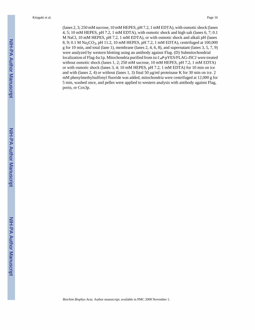
(lanes 2, 3; 250 mM sucrose, 10 mM HEPES, pH 7.2, 1 mM EDTA), with osmotic shock (lanes4, 5; 10 mM HEPES, pH 7.2, 1 mM EDTA), with osmotic shock and high salt (lanes 6, 7; 0.1M NaCl, 10 mM HEPES, pH 7.2, 1 mM EDTA), or with osmotic shock and alkali pH (lanes8, 9; 0.1 M Na2CO3, pH 11.2, 10 mM HEPES, pH 7.2, 1 mM EDTA), centrifuged at 100,000g for 10 min, and total (lane 1), membrane (lanes 2, 4, 6, 8), and supernatant (lanes 3, 5, 7, 9)were analyzed by western blotting using an antibody against Flag. (D) Submitochondriallocalization of Flag-Isc1p. Mitochondria purified from isc1⊿+pYES/FLAG-ISC1 were treatedwithout osmotic shock (lanes 1, 2; 250 mM sucrose, 10 mM HEPES, pH 7.2, 1 mM EDTA)or with osmotic shock (lanes 3, 4; 10 mM HEPES, pH 7.2, 1 mM EDTA) for 10 min on iceand with (lanes 2, 4) or without (lanes 1, 3) final 50 μg/ml proteinase K for 30 min on ice. 2mM phenylmethylsulfonyl fluoride was added, mitochondria were centrifuged at 12,000 g for5 min, washed once, and pelles were applied to western analysis with antibody against Flag,porin, or Cox3p.
Kitagaki et al. Page 16
Biochim Biophys Acta. Author manuscript; available in PMC 2008 November 1.
NIH
-PA Author Manuscript
NIH
-PA Author Manuscript
NIH
-PA Author Manuscript

Fig. 2. Sphingolipid profile of WT cellsWT cells were incubated to the post-diauxic phase. Sphingolipids were analyzed by LC-MSand normalized by organic phosphates as described in Materials and Methods. (A) Specieswhich are more than 0.040 pmol/nmol organic phosphate, (B) species which are more than0.0050 pmol/nmol organic phosphate but less than 0.040 pmol/nmol organic phophate, and(C) species which are less than 0.0050 pmol/nmol organic phosphate of the whole cell of WT.The results are mean values +/− SEM of lipid measurements from three independent cultures.
Kitagaki et al. Page 17
Biochim Biophys Acta. Author manuscript; available in PMC 2008 November 1.
NIH
-PA Author Manuscript
NIH
-PA Author Manuscript
NIH
-PA Author Manuscript

Kitagaki et al. Page 18
Biochim Biophys Acta. Author manuscript; available in PMC 2008 November 1.
NIH
-PA Author Manuscript
NIH
-PA Author Manuscript
NIH
-PA Author Manuscript

Fig. 3. Sphingolipid profile of WT mitochondriaWT cells were incubated to the post-diauxic phase, and mitochondria were purified as describedin Materials and Methods. Sphingolipids were analyzed by LC-MS and normalized by organicphosphates as described in Materials and Methods. (A) Species which are more than 0.040pmol/nmol organic phosphate, (B) species which are more than 0.0050 pmol/nmol organicphosphate but less than 0.040 pmol/nmol organic phophate, and (C) species which are less than0.0050 pmol/nmol organic phosphate of the WT mitochondria. The results are mean values +/− SEM of three lipid measurements from two independent mitochondrial purifications.Mitochondrial sphingolipids were divided by the amount of sphingolipids in the whole cellanalyzed in Fig. 2. (D) Ratio of mitochondrial sphingolipids to the sphingolipids of the whole
Kitagaki et al. Page 19
Biochim Biophys Acta. Author manuscript; available in PMC 2008 November 1.
NIH
-PA Author Manuscript
NIH
-PA Author Manuscript
NIH
-PA Author Manuscript

cell. Sphingolipids that were increased in mitochondria are shown. (E) Ratio of thesphingolipids in the whole cell to mitochondrial sphingolipids. Sphingolipids that weredecreased in mitochondria are shown.
Kitagaki et al. Page 20
Biochim Biophys Acta. Author manuscript; available in PMC 2008 November 1.
NIH
-PA Author Manuscript
NIH
-PA Author Manuscript
NIH
-PA Author Manuscript

Kitagaki et al. Page 21
Biochim Biophys Acta. Author manuscript; available in PMC 2008 November 1.
NIH
-PA Author Manuscript
NIH
-PA Author Manuscript
NIH
-PA Author Manuscript
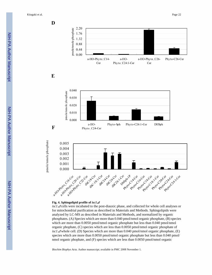
Fig. 4. Sphingolipid profile of isc1⊿isc1⊿ cells were incubated to the post-diauxic phase, and collected for whole cell analyses orfor mitochondrial purification as described in Materials and Methods. Sphingolipids wereanalyzed by LC-MS as described in Materials and Methods, and normalized by organicphosphates. (A) Species which are more than 0.040 pmol/nmol organic phosphate, (B) specieswhich are more than 0.0050 pmol/nmol organic phosphate but less than 0.040 pmol/nmolorganic phophate, (C) species which are less than 0.0050 pmol/nmol organic phosphate ofisc1⊿ whole cell. (D) Species which are more than 0.040 pmol/nmol organic phosphate, (E)species which are more than 0.0050 pmol/nmol organic phosphate but less than 0.040 pmol/nmol organic phophate, and (F) species which are less than 0.0050 pmol/nmol organic
Kitagaki et al. Page 22
Biochim Biophys Acta. Author manuscript; available in PMC 2008 November 1.
NIH
-PA Author Manuscript
NIH
-PA Author Manuscript
NIH
-PA Author Manuscript

phosphate of isc1⊿ mitochondria. The results are mean values +/− SEM of lipid measurementsfrom three independent cultures for the whole cell analysis, or, three lipid measurements fromtwo independent mitochondrial purifications for mitochondrial analysis.
Kitagaki et al. Page 23
Biochim Biophys Acta. Author manuscript; available in PMC 2008 November 1.
NIH
-PA Author Manuscript
NIH
-PA Author Manuscript
NIH
-PA Author Manuscript

Kitagaki et al. Page 24
Biochim Biophys Acta. Author manuscript; available in PMC 2008 November 1.
NIH
-PA Author Manuscript
NIH
-PA Author Manuscript
NIH
-PA Author Manuscript

Fig. 5. Comparison of mitochondrial sphingolipids of isc1⊿ with those of WTThe relative amounts of individual mitochondrial sphingolipids of isc1⊿ and isc1⊿+pYES/FLAG-ISC1 were compared with those of WT. (A) Ratio of the sphingolipids in WT relativeto those in isc1⊿ . Sphingolipids that were decreased in isc1⊿ are shown. (B) Ratio of thesphingolipids in isc1⊿ relative to those in WT. Sphingolipids that were increased in isc1⊿ areshown. (C) Ratio of the sphingolipids in isc1⊿ and isc1⊿+pYES/FLAG-ISC1 relative to thosein WT.
Kitagaki et al. Page 25
Biochim Biophys Acta. Author manuscript; available in PMC 2008 November 1.
NIH
-PA Author Manuscript
NIH
-PA Author Manuscript
NIH
-PA Author Manuscript

Fig. 6. isc1⊿ shows mitochondrial defects(A) Petite ratio of isc1⊿ . WT and isc1⊿ cells were inoculated at OD600 of 0.1 in YPD medium,incubated at 39°C for the indicated times, and plated onto YPD plates. Small colonies that grewon YPD plates were replica plated onto YPGE plates to confirm the inability to grow on non-fermentable carbon medium, and the numbers of the colonies which grew on YPD but not onYPGE plates were counted. Statistical differences were evaluated by Student's unpaired t testand are indicated by asterisks (**p<0.01, *p<0.12). (B) Hydrogen peroxide sensitivity ofisc1⊿. WT and isc1⊿ were treated with 1 mM hydrogen peroxide for 45 min, plated onto YPDand the formed colonies were counted. Statistical difference was evaluated by Student'sunpaired t test and is indicated by asterisks (**p<0.01). (C) Ethidium bromide sensitivity of
Kitagaki et al. Page 26
Biochim Biophys Acta. Author manuscript; available in PMC 2008 November 1.
NIH
-PA Author Manuscript
NIH
-PA Author Manuscript
NIH
-PA Author Manuscript

isc1⊿. WT and isc1⊿ cells were treated with 50 μM ethidium bromide, and 5 μl of thesuspension was spotted onto a YPD plate with serial dilutions at 10 fold.
Kitagaki et al. Page 27
Biochim Biophys Acta. Author manuscript; available in PMC 2008 November 1.
NIH
-PA Author Manuscript
NIH
-PA Author Manuscript
NIH
-PA Author Manuscript