Mitochondria as targets for detection and treatment of cancer
Upload
khangminh22Category
view
0download
0
Edited by
MitochondriaFrom Physiology to Pathology
Francesco Bruni
Printed Edition of the Special Issue Published in Life
www.mdpi.com/journal/life

Mitochondria: From Physiology to Pathology


Mitochondria: From Physiology to Pathology
Editor
Francesco Bruni
MDPI • Basel • Beijing • Wuhan • Barcelona • Belgrade • Manchester • Tokyo • Cluj • Tianjin

Editor
Francesco Bruni
Universita degli Studi di Bari Aldo Moro
Italy
Editorial Office
MDPI
St. Alban-Anlage 66
4052 Basel, Switzerland
This is a reprint of articles from the Special Issue published online in the open access journal Life (ISSN
2075-1729) (available at: https://www.mdpi.com/journal/life/special issues/mitochondria life).
For citation purposes, cite each article independently as indicated on the article page online and as
indicated below:
LastName, A.A.; LastName, B.B.; LastName, C.C. Article Title. Journal Name Year, Volume Number,
Page Range.
ISBN 978-3-0365-2151-0 (Hbk)
ISBN 978-3-0365-2152-7 (PDF)
© 2021 by the authors. Articles in this book are Open Access and distributed under the Creative
Commons Attribution (CC BY) license, which allows users to download, copy and build upon
published articles, as long as the author and publisher are properly credited, which ensures maximum
dissemination and a wider impact of our publications.
The book as a whole is distributed by MDPI under the terms and conditions of the Creative Commons
license CC BY-NC-ND.
Contents
About the Editor . . . . . . . . . . . . . . . . . . . . . . . . . . . . . . . . . . . . . . . . . . . . . . vii
Francesco Bruni
Mitochondria: From Physiology to PathologyReprinted from: Life 2021, 11, 991, doi:10.3390/life11090991 . . . . . . . . . . . . . . . . . . . . . . 1
Tatiana V. Kirichenko, Anastasia I. Ryzhkova, Vasily V. Sinyov, Marina D. Sazonova, Varvara A. Orekhova, Vasily P. Karagodin, Elena V. Gerasimova, Mikhail I. Voevoda, Alexander N. Orekhov, Yulia I. Ragino, Igor A. Sobenin and Margarita A. Sazonova
Impact of Mitochondrial DNA Mutations on Carotid Intima-Media Thickness in the Novosibirsk RegionReprinted from: Life 2020, 10, 160, doi:10.3390/life10090160 . . . . . . . . . . . . . . . . . . . . . 5
James Chapman, Yi Shiau Ng and Thomas J. Nicholls
The Maintenance of Mitochondrial DNA Integrity and Dynamics by Mitochondrial MembranesReprinted from: Life 2020, 10, 164, doi:10.3390/life10090164 . . . . . . . . . . . . . . . . . . . . . 15
Marija L. J. Medar, Dijana Z. Marinkovic, Zvezdana Kojic, Alisa P. Becin, Isidora M. Starovlah, Tamara Kravic-Stevovic, Silvana A. Andric and Tatjana S. Kostic
Dependence of Leydig Cell’s Mitochondrial Physiology on Luteinizing Hormone SignalingReprinted from: Life 2021, 11, 19, doi:10.3390/life11010019 . . . . . . . . . . . . . . . . . . . . . . 57
Elvira G. Zakirova, Vladimir V. Muzyka, Ilya O. Mazunin and Konstantin E. Orishchenko
Natural and Artificial Mechanisms of Mitochondrial Genome EliminationReprinted from: Life 2021, 11, 76, doi:10.3390/life11020076 . . . . . . . . . . . . . . . . . . . . . . 75
Veronika Kotrasova, Barbora Keresztesova, Gabriela Ondrovicova, Jacob A. Bauer, Henrieta Havalova, Vladimır Pevala, Eva Kutejova and Nina Kunova
Mitochondrial Kinases and the Role of Mitochondrial Protein Phosphorylation in Health and DiseaseReprinted from: Life 2021, 11, 82, doi:10.3390/life11020082 . . . . . . . . . . . . . . . . . . . . . . 97
Filipa Barroso Goncalves and Vanessa Alexandra Morais
PINK1: A Bridge between Mitochondria and Parkinson’s DiseaseReprinted from: Life 2021, 11, 371, doi:10.3390/life11050371 . . . . . . . . . . . . . . . . . . . . . . 135
Francesco Capriglia, Francesca Rizzo, Giuseppe Petrosillo, Veronica Morea, Giulia d’Amati,
Palmiro Cantatore, Marina Roberti, Paola Loguercio Polosa and Francesco Bruni
Exploring the Ability of LARS2 Carboxy-Terminal Domain in Rescuing the MELAS PhenotypeReprinted from: Life 2021, 11, 674, doi:10.3390/life11070674 . . . . . . . . . . . . . . . . . . . . . . 151
Francesco Bruni, Teresa Anna Giancaspero, Mislav Oreb, Maria Tolomeo, Piero Leone,
Eckhard Boles, Marina Roberti, Michele Caselle and Maria Barile
Subcellular Localization of Fad1p in Saccharomyces cerevisiae: A Choice at Post-TranscriptionalLevel?Reprinted from: Life 2021, 11, 967, doi:10.3390/life11090967 . . . . . . . . . . . . . . . . . . . . . . 169
v

About the Editor
Francesco Bruni My main research interests are in the field of biogenesis and mitochondrial
function. During my PhD studies, carried out at the University of Bari, I investigated mitochondrial
transcription termination mechanisms in Drosophila melanogaster, characterizing two different
transcription factors, DmTTF and D-MTERF3. Later, during my post doc scholarship, I studied the
function of D-MTERF5 as well as the role of human NRF-2 and Drosophila DREF in mitochondrial
biogenesis. In 2010, I began my research activity abroad at the Wellcome Centre for Mitochondrial
Research (Newcastle University), a European center of excellence for the study of mitochondrial
diseases. I identified and characterized a novel mitochondrial ribonuclease, named REXO2, and
participated in numerous clinical projects in collaboration with the diagnostic laboratory. At
the WCMR, which possessed two vital resources, the Mitochondrial Disease Patient Cohort and
the Mitochondrial BioResource, I also had the opportunity to study mitochondrial translation
mechanisms in both physiological and pathological conditions in patients with mitochondrial
diseases. In 2017, I came back to Italy and, since then, I have been working at the Department of
Biosciences, Biotechnologies and Biopharmaceutics (University of Bari), dividing my time between
research and teaching. I investigate the biochemical and clinical features of mitochondrial aa-tRNA
synthetases, exploring the therapeutic use of these enzymes. I am also interested in the mechanisms
by which mitochondria exercise a level of quality control between mitochondrial transcription and
translation processes, as well as the factors responsible for this activity. As a professor, I currently
teach molecular biology on the bachelor’s degree course in Biological Sciences.
vii


life
Editorial
Mitochondria: From Physiology to Pathology
Francesco Bruni
���������������
Citation: Bruni, F. Mitochondria:
From Physiology to Pathology. Life
2021, 11, 991. https://doi.org/
10.3390/life11090991
Received: 17 September 2021
Accepted: 19 September 2021
Published: 21 September 2021
Publisher’s Note: MDPI stays neutral
with regard to jurisdictional claims in
published maps and institutional affil-
iations.
Copyright: © 2021 by the author.
Licensee MDPI, Basel, Switzerland.
This article is an open access article
distributed under the terms and
conditions of the Creative Commons
Attribution (CC BY) license (https://
creativecommons.org/licenses/by/
4.0/).
Department of Biosciences, Biotechnologies and Biopharmaceutics, University of Bari Aldo Moro, Via Orabona 4,70125 Bari, Italy; [email protected]
Over the past decade, the role of mitochondria has extended beyond those tasks forwhich these organelles are historically known. Recent proteomics investigations havehighlighted the extraordinary complexity of mitochondrial protein organization, whichis a reflection of the numerous and disparate mitochondrial functions [1]. These includethe synthesis of most of the ATP present in the cell, apoptosis, ion homeostasis, cellularstress response, antioxidant control, redox regulation, mitophagy, involvement in variousbiosynthetic pathways and many more processes. Furthermore, mitochondria are dynamicorganelles, and their morphology can vary significantly within a cell. This is mainly dueto the mitochondrial fusion and fission processes, referred as mitochondrial dynamics,which are crucial for the organelles’ interactions with other cell compartments and for thecross-talk with the cell cycle and metabolism as well as differentiation and senescence [2].
An intriguing feature of mitochondria is that they possess their own DNA (mtDNA),which is functionally coordinated with the nuclear genome. Indeed, numerous nucleus-encoded proteins are required for complex molecular processes such as replication, tran-scription, RNA processing and degradation, translation, and assembly of respiratory chaincomplexes that take place inside the mitochondria [3,4]. On the other hand, mitochondrialgene expression plays an important role in the communication between mitochondria andthe nucleus, contributing to the regulation of cell physiology. Recent studies have pointedto novel RNA-driven mechanisms according to which mitochondrial-derived transcripts,in addition to their role in maintaining mitochondrial function, act as signalling moleculesmodulating the innate immune response [5].
Dysfunctions affecting the mitochondria can lead to various pathological conditions,including aging and neurodegenerative disorders, and are associated with a whole rangeof complex genetic disorders, known as mitochondrial diseases [6]. In recent years, greatadvances have been made in the field of mitochondrial diagnostics thanks to the rapid devel-opment of next-generation sequencing (NGS) technologies, particularly the whole-genomeapproach [7]. However, most of the aspects and mechanisms underlying mitochondrialdiseases remain unclear and, to date, effective therapies are still lacking. Therefore, athorough understanding of the molecular processes occurring in mitochondria is essentialfrom a pathological perspective.
The Special Issue “Mitochondria: from Physiology to Pathology” published in Life(ISSN 2075-1729) collects a series of research and review articles and aims to provide anupdated view of the main topics covering the physiological and the pathological aspects ofmitochondrial biology.
Several contributions focus on the mitochondrial genome and its link to the phys-iopathological aspects of the various mitochondrial processes. Chapman et al. [8] presenta comprehensive review on mtDNA metabolism, highlighting the interplay occurringbetween the organization of the mitochondrial genome, mitochondrial dynamics andcristae structure. After a brief excursus on the organization of mtDNA and on its mainprocesses, such as replication and transcription, the authors deal in depth with the pro-cesses of fission and fusion relative to the structure of mtDNA, discussing the knowngenetic defects that affect mitochondrial dynamics. To complete the picture, they describe
Life 2021, 11, 991. https://doi.org/10.3390/life11090991 https://www.mdpi.com/journal/life
1

Life 2021, 11, 991
the close relationship between the nucleoid, OPA1 and the other “cristae-shaping pro-teins” constituting the MICOS complex, whose interactions modulate the formation anddynamics of mitochondrial cristae. These are critical process that, if not well regulated,can prompt mitochondrial dysfunction with the onset of neuromuscular diseases. Anotherimportant process whose dysregulation can lead to severe pathologies is the elimination ofmitochondrial genome. Orishchenko and colleagues [9] review the mechanisms underlyingthe regulation of heteroplasmy level and mtDNA segregation, illustrating in detail thenatural process of mitochondrial genome elimination in both the somatic cells and thegermline. The development of methodologies for artificial manipulation of the mtDNAheteroplasmy level that aim either to prevent or potentially cure human diseases associatedwith mitochondrial dysfunction, is also examined.
Mitochondrial encephalopathy with lactic acidosis and stroke-like episodes (MELAS)is a metabolic disorder caused by point mutations in the mitochondrial tRNALeu(UUR) gene,with a prevalence of A>G substitution in position 3243. Several papers have reported thatoverexpression of human mitochondrial leucyl-tRNA synthetase (LARS2) or its C-terminaldomain (Cterm) has proven effective in rescuing the pathological phenotype in cellularmodels. In their research article, Capriglia et al. [10] investigated the molecular basisunderlying the ability of the Cterm domain in rescuing the MELAS phenotype. The cellularmodel employed, consisting of a trans-mitochondrial cybrid line with a >95% mutation load,confirmed the therapeutic potential of the Cterm peptide but also showed that its rescuingability was independent of the mitochondrial bioenergetics, unlike what has previouslybeen observed in other cybrid lines. The authors proposed that the beneficial effect of thispeptide could also be mediated by retrograde mitochondrial signals or, alternatively, by itspotential ability to bind regulatory RNA in the cytosol. This research indicates that the fullunderstanding of Cterm-rescuing mechanisms imposes the development of tissue-specificcellular models that accurately reproduce the pathological MELAS phenotype.
Another study, based on a cohort of 468 subjects from the Siberian region, was con-ducted by Kirichenko et al. [11] with the purpose of determining the impact of mitochon-drial heteroplasmy measurements in the prognosis of atherosclerosis development. Theyinvestigated the association of nine different mtDNA mutations with carotid intima-mediathickness (cIMT), a measurement of the artery wall thickness obtained by ultrasound imag-ing. Several mtDNA variants correlated with the mean cIMT, thus constituting potentialprognostic markers. Interestingly, the mutations m.13513G>A and m.14846G>A showeda significant inverse correlation being associated with a low value of cIMT, representinggood candidates for the development of anti-atherosclerotic gene therapies.
The dysregulation of mitochondrial functions certainly depends on faulty mitochon-drial gene expression at different levels, but also on other factors. One of these, which takeson particular importance in mitochondrial physiology, is the post-translational modifica-tion of mitochondrial proteins. Phosphorylation is commonly employed in mitochondriaeither to modify protein functions or to activate fundamental signalling pathways, insideand outside the mitochondria. Kotrasová et al. [12] center their review on mitochondrialkinases and their role in keeping organelles fully efficient. This function is exerted onvarious and distinct substrates: mitochondrial import machinery, subunits of respiratorychain complexes and proteins involved in the main steps of mtDNA maintenance andexpression. In addition, the activity of mitochondrial kinases is also important for organellequality control and apoptosis. One of the central key players in mitochondrial qualitycontrol pathways is PINK1 (PTEN-induced kinase 1). Barroso Gonçalves and Morais [13]emphasize how valuable the supply of PINK1 is for the mitochondrial clearance throughmitophagy and for the maintenance of mitochondrial homeostasis, thereby keeping cellshealthy and functional. The precise mechanisms that mediate PINK1 function in the dif-ferent mitochondrial pathways remain to be elucidated. Nevertheless, the genetic linkbetween PINK1 and Parkinson’s Disease (PD) has long been proven, particularly for thefamilial form of this pathology. Further research needs to be carried out to define whetherthe mitochondrial homeostasis imbalance, due to the aberrant function of PINK1, is the key
2

Life 2021, 11, 991
element that contributes to the pathological phenotype in both the familiar and sporadicforms of PD.
An important aspect of mitochondrial physiology is its hormone-mediated regulation.Medar et al. [14] analysed mitochondrial response to luteinizing hormone (LH) stimulationin Leydig cells, the major producers of steroid hormones that regulate sexual differentiationand development. In these cells, endocrine function requires the cAMP signalling pathway,whose involvement in modulating respiratory chain complexes activity, ATP production,biogenesis, import, dynamics, and mitochondrial-dependent apoptosis has been widelyinvestigated. The reported results, obtained by different experimental approaches in ratLeydig cells ex vivo and in vivo with a different LH environment and steroidogenic capacity,supported the involvement of LH-cAMP pathway in the regulation of mitochondrialbiogenesis and dynamics coupled with mitochondria-mediated steroidogenesis. Additionalstudies carried out in Leydig and other steroidogenic cells, e.g., derived from adrenal glandsand placenta, will shed light on the pathogenic mechanisms triggered by the presence ofunhealthy mitochondria in these tissues.
The last research article of this Special Issue focuses on the Saccharomyces cerevisiaeFAD1 gene, encoding the FAD synthase that adenylates the flavin mononucleotide (FMN)to flavin adenine dinucleotide (FAD), an essential coenzyme for various flavoenzymes.Barile and colleagues previously proved that FAD-forming activities, paralleled by FADprecursors uptake in mitochondria and mitochondrial FAD export to cytosol, could bespecifically revealed in mitochondria. However, a protein responsible for the synthesisof FAD had never been identified in yeast mitochondria. In this paper [15], the presenceof two Fad1p echoforms, dually localised to the cytosol and mitochondria, was reported.Intriguingly, the authors demonstrated the existence of two pools of FAD1 mRNAs with3’ untranslated regions (UTRs) of different length and containing a mitochondrial targetingsignal. Therefore, the 3′ UTRs would be responsible for the fate of Fad1p echoforms, withthe long FAD1 mRNA generating the mitochondrial Fad1p. In this context, the authorsdiscussed the role of specific RNA binding proteins (e.g., Puf3p) that modulate the importof the mitochondrial Fad1p echoform. Overall, the paper adds a useful piece of knowledgeto the post-transcriptional control of genes encoding mitochondrial proteins, proposing theexistence of novel regulatory mechanisms in yeast as well as in higher eukaryotes.
Author Contributions: F.B. wrote the manuscript. All authors have read and agreed to the publishedversion of the manuscript.
Funding: Fondi di Ateneo “Contributo Ordinario di Supporto alla Ricerca” 2015/16 and 2017/18(University of Bari Aldo Moro) to F.B.
Acknowledgments: The guest editor wishes to thank Pasqua Gramegna for the critical readingof the manuscript and Veronica Wang for her precious help with the editorial process. All thecontributors and those involved in the peer-review activity of the Special Issue of Life ‘Mitochondria:from Physiology to Pathology’ are gratefully acknowledged.
Conflicts of Interest: The author declares no conflict of interest.
References
1. Pfanner, N.; Warscheid, B.; Wiedemann, N. Mitochondrial Proteins: From Biogenesis to Functional Networks. Nat. Rev. Mol. CellBiol. 2019, 20, 267–284. [CrossRef] [PubMed]
2. Giacomello, M.; Pyakurel, A.; Glytsou, C.; Scorrano, L. The Cell Biology of Mitochondrial Membrane Dynamics. Nat. Rev. Mol.Cell Biol. 2020, 21, 204–224. [CrossRef] [PubMed]
3. Gustafsson, C.M.; Falkenberg, M.; Larsson, N.-G. Maintenance and Expression of Mammalian Mitochondrial DNA. Ann. Rev.Biochem. 2016, 85, 133–160. [CrossRef] [PubMed]
4. Pearce, S.F.; Rebelo-Guiomar, P.; D’Souza, A.R.; Powell, C.A.; Van Haute, L.; Minczuk, M. Regulation of Mammalian MitochondrialGene Expression: Recent Advances. Trends Biochem. Sci. 2017, 42, 625–639. [CrossRef] [PubMed]
5. Kotrys, A.V.; Szczesny, R.J. Mitochondrial Gene Expression and Beyond—Novel Aspects of Cellular Physiology. Cells 2020, 9, 17.[CrossRef] [PubMed]
6. Gorman, G.S.; Chinnery, P.F.; DiMauro, S.; Hirano, M.; Koga, Y.; McFarland, R.; Suomalainen, A.; Thorburn, D.R.; Zeviani, M.;Turnbull, D.M. Mitochondrial Diseases. Nat. Rev. Dis. Primers 2016, 2, 16080. [CrossRef] [PubMed]
3

Life 2021, 11, 991
7. Schon, K.R.; Ratnaike, T.; van den Ameele, J.; Horvath, R.; Chinnery, P.F. Mitochondrial Diseases: A Diagnostic Revolution. TrendsGenet. 2020, 36, 702–717. [CrossRef] [PubMed]
8. Chapman, J.; Ng, Y.S.; Nicholls, T.J. The Maintenance of Mitochondrial DNA Integrity and Dynamics by Mitochondrial Mem-branes. Life 2020, 10, 164. [CrossRef] [PubMed]
9. Zakirova, E.G.; Muzyka, V.V.; Mazunin, I.O.; Orishchenko, K.E. Natural and Artificial Mechanisms of Mitochondrial GenomeElimination. Life 2021, 11, 76. [CrossRef] [PubMed]
10. Capriglia, F.; Rizzo, F.; Petrosillo, G.; Morea, V.; d’Amati, G.; Cantatore, P.; Roberti, M.; Loguercio Polosa, P.; Bruni, F. Exploringthe Ability of LARS2 Carboxy-Terminal Domain in Rescuing the MELAS Phenotype. Life 2021, 11, 674. [CrossRef] [PubMed]
11. Kirichenko, T.V.; Ryzhkova, A.I.; Sinyov, V.V.; Sazonova, M.D.; Orekhova, V.A.; Karagodin, V.P.; Gerasimova, E.V.; Voevoda,M.I.; Orekhov, A.N.; Ragino, Y.I.; et al. Impact of Mitochondrial DNA Mutations on Carotid Intima-Media Thickness in theNovosibirsk Region. Life 2020, 10, 160. [CrossRef] [PubMed]
12. Kotrasová, V.; Keresztesová, B.; Ondrovicová, G.; Bauer, J.A.; Havalová, H.; Pevala, V.; Kutejová, E.; Kunová, N. MitochondrialKinases and the Role of Mitochondrial Protein Phosphorylation in Health and Disease. Life 2021, 11, 82. [CrossRef] [PubMed]
13. Gonçalves, F.B.; Morais, V.A. PINK1: A Bridge between Mitochondria and Parkinson’s Disease. Life 2021, 11, 371. [CrossRef][PubMed]
14. Medar, M.L.J.; Marinkovic, D.Z.; Kojic, Z.; Becin, A.P.; Starovlah, I.M.; Kravic-Stevovic, T.; Andric, S.A.; Kostic, T.S. Dependenceof Leydig Cell’s Mitochondrial Physiology on Luteinizing Hormone Signaling. Life 2020, 11, 19. [CrossRef] [PubMed]
15. Bruni, F.; Giancaspero, T.A.; Oreb, M.; Tolomeo, M.; Leone, P.; Boles, E.; Roberti, M.; Caselle, M.; Barile, M. Subcellular Localizationof Fad1p in Saccharomyces Cerevisiae: A Choice at Post-Transcriptional Level? Life 2021, 11, 967. [CrossRef]
4

life
Article
Impact of Mitochondrial DNA Mutations on CarotidIntima-Media Thickness in the Novosibirsk Region
Tatiana V. Kirichenko 1,*, Anastasia I. Ryzhkova 2, Vasily V. Sinyov 2,3, Marina D. Sazonova 2,
Varvara A. Orekhova 2,3,4, Vasily P. Karagodin 2,5, Elena V. Gerasimova 6, Mikhail I. Voevoda 7,
Alexander N. Orekhov 1,2,4, Yulia I. Ragino 7, Igor A. Sobenin 1,2,3 and Margarita A. Sazonova 2,3
1 Laboratory of Cellular and Molecular Pathology of Cardiovascular System, Research Institute of HumanMorphology, 3 Tsyurupy Str., 117418 Moscow, Russia; [email protected] (A.N.O.);[email protected] (I.A.S.)
2 Laboratory of Angiopathology, Institute of General Pathology and Pathophysiology, 8 Baltiyskaya Str.,125315 Moscow, Russia; [email protected] (A.I.R.); [email protected] (V.V.S.);[email protected] (M.D.S.); [email protected] (V.A.O.); [email protected] (V.P.K.);[email protected] (M.A.S.)
3 Laboratory of Medical Genetics, National Medical Research Center of Cardiology,15A 3 Cherepkovskaya Str., 121552 Moscow, Russia
4 Institute for Atherosclerosis Research, Skolkovo Innovative Center, 143025 Moscow, Russia5 Department of Commodity Science and Expertise, Plekhanov Russian University of Economics,
125993 Moscow, Russia6 V.A.Nasonova Research Institute of Rheumatology, 34A Kashirskoe sh., 115522 Moscow, Russia;
[email protected] Research Institute of Internal and Preventive Medicine–Branch of the Institute of Cytology and Genetics,
Siberian Branch of Russian Academy of Sciences, 630089 Novosibirsk, Russia; [email protected] (M.I.V.);[email protected] (Y.I.R.)
* Correspondence: [email protected]; Tel.: +7-910-461-5845
Received: 18 July 2020; Accepted: 20 August 2020; Published: 22 August 2020
Abstract: The search for markers of predisposition to atherosclerosis development is very importantfor early identification of individuals with a high risk of cardiovascular disease. The aim of the presentstudy was to investigate the association of mitochondrial DNA mutations with carotid intima-mediathickness and to determine the impact of mitochondrial heteroplasmy measurements in the prognosisof atherosclerosis development. This cross-sectional, population-based study was conducted in468 subjects from the Novosibirsk region. It was shown that the mean (carotid intima-mediathickness) cIMT correlated with the following mtDNA mutations: m.15059G>A (r = 0.159, p = 0.001),m.12315G>A (r = 0.119; p = 0.011), m.5178C>A (r = 0.114, p = 0.014), and m.3256C>T (r = 0.130,p = 0.011); a negative correlation with mtDNA mutations m.14846G>A (r = −0.111, p = 0.042) andm.13513G>A (r = −0.133, p = 0.004) was observed. In the linear regression analysis, the addition of theset of mtDNA mutations to the conventional cardiovascular risk factors increased the ability to predictthe cIMT variability from 17 to 27%. Multi-step linear regression analysis revealed the most importantpredictors of mean cIMT variability: age, systolic blood pressure, blood levels of total cholesterol,LDL and triglycerides, as well as the mtDNA mutations m.13513G>A, m.15059G>A, m.12315G>A,and m.3256C>T. Thus, a high predictive value of mtDNA mutations for cIMT variability wasdemonstrated. The association of mutation m.13513G>A and m.14846G>A with a low value of cIMT,demonstrated in several studies, represents a potential for the development of anti-atheroscleroticgene therapy.
Keywords: atherosclerosis; carotid intima-media thickness; mitochondrial mutations; cardiovascularrisk factors
Life 2020, 10, 160; doi:10.3390/life10090160 www.mdpi.com/journal/life5

Life 2020, 10, 160
1. Introduction
Current knowledge about the mechanisms of atherosclerosis development has significantlyincreased in recent decades, but atherosclerotic-based diseases still occupy a leading position in thestructure of mortality in developed countries [1]. In this regard, the early detection and treatmentof patients with a high risk of atherosclerosis development is a primary medical and social problem.Timely prevention of subclinical atherosclerosis has a potential to decrease cardiovascular morbidityand mortality. The search for markers of subclinical atherosclerosis is one of the fundamental factorsin the identification of individuals with a high risk of cardiovascular disease (CVD). The thicknessof the intimal-medial layer of the carotid arteries (cIMT) is considered to be a direct non-invasivemarker of subclinical atherosclerosis, used in clinical and epidemiological studies to assess the effectof conventional and new cardiovascular risk factors (CVRFs) on the progression of atheroscleroticlesions [2,3]. However, conventional CVRFs are poorly associated with cIMT, which suggests thepresence of other factors determining the risk atherosclerosis development [4,5].
Currently, the existence of a genetic predisposition to the development of atherosclerosis is not indoubt. In particular, a family history of CVD (coronary artery disease, acute myocardial infarction,and arterial hypertension in first-degree relatives under 60 years of age) is an independent predictorof the risk of these diseases and is taken into account in many modern algorithms for cardiovascularrisk prediction [6]. The association of numerous polymorphisms of the nuclear genome with risk ofCVD has been revealed [7]. At the same time, the effect of mutations of the mitochondrial genome ismore significant for the formation of atherosclerotic lesions since mitochondrial dysfunction leads toactivation of the key factors of atherogenesis, such as oxidative stress and inflammation [8]. Nowadays,the role of mitochondrial genome mutations in the predisposition to atherosclerosis development andcardiovascular disease is being investigated [9].
In our previous studies, variants of mitochondrial heteroplasmy were identified in samples of thehuman aorta with atherosclerotic lesions [10]. In several populations, the association of mitochondrialmutations in blood leukocytes with coronary artery disease and carotid atherosclerosis has beenstudied [11–13]. It was revealed in the IMPROVE study, a European multicenter study aimed toinvestigate the prognostic value of cIMT for future cardiovascular events, that latitude is an importantdeterminant of cIMT in Europe and traditional CVRFs do not fully explain cIMT variability in Europeanpopulation, which suggests the presence of other factors, including hereditary ones, in formationof a predisposition to atherosclerosis development [14]. In this regard, replicative studies on theassociation of mitochondrial heteroplasmy and carotid atherosclerosis need to be carried out in otherregions that differ in ethnic composition, as well as in socio-economic and geographical conditions.We have previously investigated the association between mitochondrial heteroplasmy and carotidatherosclerosis in a population of the European part of Russia [15]. The objective of the presentresearch is to study the possible relationship of mitochondrial heteroplasmy and cIMT variability inthe Siberian region.
2. Results
In total, 500 participants were included in the study; 32 of them were excluded from the analysisdue to unreadable ultrasound images (13) and the insufficient quality of their DNA samples (19).Therefore, 134 male and 334 female samples were analyzed. The clinical and laboratory characteristicsof the study participants are presented in Table 1. Female and male groups were different bythe following parameters: there were significantly more smokers in the male group (p = 0.038);men had higher systolic blood pressure (p = 0.001) and a lower level of high-density lipoproteins(p = 0.032). The additional estimated parameter in the female group was the period after menopause,which averaged 12.4 (7.4) years.
6

Life 2020, 10, 160
Table 1. Clinical and laboratory characteristics of the study participants.
Characteristic Total Group Men Women Difference, p-Value
Age, years 62.3 (5.7) 62.9 (5.4) 62.1 (5.8) 0.141BMI, kg/m2 28.6 (4.9) 28.4 (4.9) 28.7 (4.9) 0.515SBP, mm Hg 130 (16) 134 (18) 127 (15) 0.001DBP, mm Hg 81 (10) 82 (9) 80 (10) 0.121Smoking, % 9 14 7 0.038Diabetes, % 6 3 8 0.190
Family history of CVD, % 34 32 35 0.398Hypotensive therapy, % 55 51 57 0.442Total Cholesterol, mg/dL 231.7 (42.3) 227.4 (40.4) 233.5 (42.9) 0.131
TG, mg/dL 119.1 (73.3) 120.9 (74.6) 118.4 (72.8) 0.732HDL, mg/dL 49.8 (14.2) 47.8 (12.8) 50.6 (14.7) 0.032LDL, mg/dL 158.5 (38.3) 155.2 (37.0) 159.8 (38.8) 0.213cIMT, mm 0.778 (0.170) 0.772 (0.149) 0.777 (0.150) 0.731
Mitochondrial DNA (mtDNA) mutations m.13513G>A, m.3336T>C, m.15059G>A, m.12315G>A,m.1555A>G, m.5178C>A, m.14459G>A, m.14846G>A, and m.3256C>T were estimated in all the studyparticipants. Figure 1 represents an example of the results of the pyrosequencing for mutation m.5178.
Figure 1. Sample of the sequencing results for mutation m.5178C>A. For the detection of mutationm.5178C>A, a reverse primer for sequencing was used, so the replacement of G by T was analyzed.(A) The theoretical height of the nucleotide peaks in homoplasmy for the mutant allele 5178A in themitochondrial genome; (B) the theoretical height of the nucleotide peaks in homoplasmy for the normalallele 5178C in the mitochondrial genome; and (C) a pyrogram of a study participant’s DNA samplerevealed the heteroplasmy level for mutation m.5178C>A is 48%.
Table 2 demonstrates the levels of mtDNA mutations in total and in men and women separately.The difference between the male and female group was observed only for mutation m.15059G>A,which was significantly higher in women (p = 0.009).
7
Life 2020, 10, 160
Table 2. The MtDNA mutation levels of the study participants.
MtDNA Mutations, % Total Group Men Women Difference, p-Value
m.13513G>A 24.3 (13.8) 23.8 (13.5) 24.5 (13.9) 0.635m.3336T>C 3.9 (0.4) 3.1 (4.7) 4.2 (9.5) 0.086
m.15059G>A 11.7 (14.3) 9.5 (9.3) 12.6 (15.8) 0.009m.12315G>A 36.0 (24.1) 35.4 (23.3) 36.2 (24.4) 0.714m.1555A>G 22.8 (12.5) 24.2 (12.8) 22.2 (12.4) 0.070m.5178C>A 5.8 (12.3) 4.8 (11.3) 6.3 (12.7) 0.216
m.14459G>A 4.1 (5.5) 4.4 (5.4) 4.1 (5.6) 0.540m.14846G>A 17.1 (16.6) 17.7 (18.5) 16.9 (15.8) 0.654m.3256C>T 15.0 (16.3) 15.4 (15.5) 14.9 (16.7) 0.772
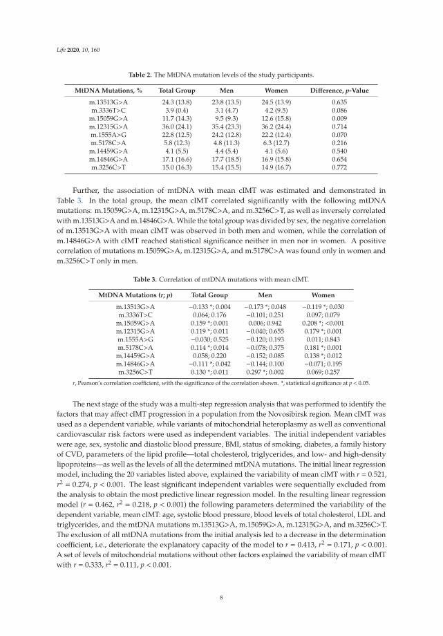
Further, the association of mtDNA with mean cIMT was estimated and demonstrated inTable 3. In the total group, the mean cIMT correlated significantly with the following mtDNAmutations: m.15059G>A, m.12315G>A, m.5178C>A, and m.3256C>T, as well as inversely correlatedwith m.13513G>A and m.14846G>A. While the total group was divided by sex, the negative correlationof m.13513G>A with mean cIMT was observed in both men and women, while the correlation ofm.14846G>A with cIMT reached statistical significance neither in men nor in women. A positivecorrelation of mutations m.15059G>A, m.12315G>A, and m.5178C>A was found only in women andm.3256C>T only in men.
Table 3. Correlation of mtDNA mutations with mean cIMT.
MtDNA Mutations (r; p) Total Group Men Women
m.13513G>A −0.133 *; 0.004 −0.173 *; 0.048 −0.119 *; 0.030m.3336T>C 0.064; 0.176 −0.101; 0.251 0.097; 0.079
m.15059G>A 0.159 *; 0.001 0.006; 0.942 0.208 *; <0.001m.12315G>A 0.119 *; 0.011 −0.040; 0.655 0.179 *; 0.001m.1555A>G −0.030; 0.525 −0.120; 0.193 0.011; 0.843m.5178C>A 0.114 *; 0.014 −0.078; 0.375 0.181 *; 0.001
m.14459G>A 0.058; 0.220 −0.152; 0.085 0.138 *; 0.012m.14846G>A −0.111 *; 0.042 −0.144; 0.100 −0.071; 0.195m.3256C>T 0.130 *; 0.011 0.297 *; 0.002 0.069; 0.257
r, Pearson’s correlation coefficient, with the significance of the correlation shown. *, statistical significance at p < 0.05.
The next stage of the study was a multi-step regression analysis that was performed to identify thefactors that may affect cIMT progression in a population from the Novosibirsk region. Mean cIMT wasused as a dependent variable, while variants of mitochondrial heteroplasmy as well as conventionalcardiovascular risk factors were used as independent variables. The initial independent variableswere age, sex, systolic and diastolic blood pressure, BMI, status of smoking, diabetes, a family historyof CVD, parameters of the lipid profile—total cholesterol, triglycerides, and low- and high-densitylipoproteins—as well as the levels of all the determined mtDNA mutations. The initial linear regressionmodel, including the 20 variables listed above, explained the variability of mean cIMT with r = 0.521,r2 = 0.274, p < 0.001. The least significant independent variables were sequentially excluded fromthe analysis to obtain the most predictive linear regression model. In the resulting linear regressionmodel (r = 0.462, r2 = 0.218, p < 0.001) the following parameters determined the variability of thedependent variable, mean cIMT: age, systolic blood pressure, blood levels of total cholesterol, LDL andtriglycerides, and the mtDNA mutations m.13513G>A, m.15059G>A, m.12315G>A, and m.3256C>T.The exclusion of all mtDNA mutations from the initial analysis led to a decrease in the determinationcoefficient, i.e., deteriorate the explanatory capacity of the model to r = 0.413, r2 = 0.171, p < 0.001.A set of levels of mitochondrial mutations without other factors explained the variability of mean cIMTwith r = 0.333, r2 = 0.111, p < 0.001.
8

Life 2020, 10, 160
In women, the initial set of predictors determined a cIMT value with r = 0.583, r2 = 0.340,p = 0.005; the most predictive variables were age, period after menopause, diabetes, blood levels of totalcholesterol, LDL and triglycerides, and mtDNA mutations m.13513G>A, m.14459G>A, and m.5178C>A(r= 0.540, r2 = 0.292, p< 0.001). For men, the initial linear regression model was not significant (r= 0.657,r2 = 0.431, p = 0.227); the most important predictors of mean cIMT variability in the male groupwere age, blood levels of total cholesterol, HDL and triglycerides, smoking, diabetes, and mitochondrialheteroplasmy m.3256C>T (r = 0.600, r2 = 0.360, p = 0.003).
3. Discussion
In present study, the relationship between mutations of the mitochondrial genome with carotidatherosclerosis in the Novosibirsk region was studied for the first time. It was demonstrated that variantsof mitochondrial heteroplasmy m.15059G>A, m.12315G>A, m.5178C>A, and m.3256C>T significantlycorrelated with the intima-media thickness of common carotid arteries; a negative correlation ofm.13513G>A and m.14846G>A with mean cIMT was revealed. When a correlation analysis was carriedout in men and women separately, it was shown that the mean cIMT correlated with m.13513G>A,m.15059G>A, m.12315G>A, m.5178C>A, and m.14459G>A in the female group and only withm.13513G>A and m.3256C>T in the male group. Probably, these results are associated with one of themain limitations in this study—the unequal volume of the male and female groups, so most correlationsdid not reach statistical significance in male group due to the small sample size. In previous studies,the association of variants of mitochondrial heteroplasmy was demonstrated in other populations.The association of mitochondrial heteroplasmy in the European part of Russia partially coincides withthe results of the present study in the Siberian region. A previous study in the Moscow region revealedthe positive correlation between carotid atherosclerosis and the following mitochondrial mutations:m.652delG, m.3256C>T, m.3336T>C, m.5178C>A, m.12315G>A, m.14459G>A, and m.15059G>A;and an inverse correlation for m.652insG, m.13513G>A, and m.14846G>A. [15]. In subjects derivedfrom a Kazakhstan population, m.12315G>A was associated with cIMT only in women, but a negativecorrelation of m.13513G>A with cIMT was confirmed in the total group [16].
The present study also included an estimation of the predictive value of mitochondrialheteroplasmy for cIMT variability along with conventional CVRFs using a linear regression analysis.The linear regression model, including a set of conventional cardiovascular risk factors together withthe investigated mitochondrial mutations, predicted the cIMT variability with r2 = 0.274. The coefficientof determination r2 shows that this set of factors explains only 27% of variability of the mean cIMTin the presented linear regression model; however, the exclusion of the levels of mitochondrialheteroplasmy from the analysis led to a decrease in the predictive ability of this model by 10%(r2 = 0.171), which indicates a high contribution of mitochondrial mutations in cIMT variability inparticipants of the present study. Exclusion of the non-significant variables during the multi-stepregression analysis resulted in a decrease in the determination coefficient to 0.218, i.e., reduced theexplanting ability of the model to 22%, but allowed identifying the most predictive variables—age,systolic blood pressure, parameters of the lipid profile, and the mtDNA mutations m.13513G>A,m.15059G>A, m.12315G>A, m.3256C>T. In our previous study, the factors affecting the cIMT value insubjects from Moscow were determined. It was shown that the same set of conventional cardiovascularrisk factors explained 21% of the cIMT variability. The inclusion of the m.652delG, m.3256C>T,m.13513G>A, m.14459G>A, and m.15059G>A heteroplasmy levels in the linear regression modelprovided a significantly better explanatory level of 36% [17].
The association of mitochondrial genome mutations, occurring during ontogenesis or inheritedthrough the maternal line, with the development of a number of pathological conditions in humansis widely known [18,19]. One more limitation of the present study was the fact that detected levelsof mitochondrial heteroplasmy did not reach 50%, while it is known that the level of mitochondrialheteroplasmy should exceed 50% for development of clinical manifestations [20]. However, a number ofother studies have demonstrated a significant relationship between cardiovascular diseases associated
9

Life 2020, 10, 160
with atherosclerosis and some mutations in the mitochondrial genome, determined at a level comparableto the results of the current study. For example, in a study of 482 patients with coronary heart disease,the variant of mitochondrial heteroplasmy m.16189T>C, at a level of 21.6%, was associated with anincreased risk of coronary heart disease compared to healthy participants who had a 4.5% prevalenceof m.16189T>C [21]. In another study it was shown that mtDNA with a 4977 bp deletion in blood cellswas at significantly higher level in patients with coronary artery disease in comparison with healthysubjects (26.2% vs. 4.5%) [22].
4. Materials and Methods
4.1. Study Design
The present study aimed to investigate the association of mitochondrial mutations with cIMTand to determine the most important factors affecting the development of carotid atherosclerosisin a Siberian population with a special focus on genetic predisposition, namely, mtDNA mutations.This cross-sectional, population-based trial was conducted in men and women aged 55–79 years fromthe Novosibirsk region. Conventional CVRFs, such as body mass index (BMI), systolic and diastolicblood pressure, smoking status, history diabetes mellitus, family history of CVD, treatment of arterialhypertension, blood lipids parameters (total cholesterol, triglycerides, and low- and high-densitylipoproteins) were assessed. All study participants were free of CVD. The recruitment of patientswas carried out at the Research Institute of Internal and Preventive Medicine, Novosibirsk, Russia.The additional inclusion criterion in the female group was time after menopause (>5 years). History ofangina pectoris, myocardial infarction, intermittent claudication, stroke or transient ischemic attacks,as well as arterial revascularization was exclusion criteria. The thickness of the intima-media layerof the common carotid arteries was determined as a direct quantitative characteristic of carotidatherosclerosis. Blood samples of the study participants were obtained to measure the lipid profile andthe levels of mtDNA mutations previously identified as related to atherosclerosis and cardiovasculardisease. The study was performed in accordance with the Declaration of Helsinki of 1975 and itsrevised version of 2013. The study protocol was approved by the Institute for Atherosclerosis ResearchCommittee on Human Research (Moscow, Russia). All study participants provided written informedconsent prior the inclusion in the study.
4.2. Measurements of cIMT
B-mode high-resolution ultrasonography with a linear array vascular probe 7.5 MHz on ultrasonicscanner SonoScape SSI-1000 (SonoScape, Shenzhen, China) was used for examination of the carotidarteries. The left and right common carotid arteries, the carotid bifurcation area, as well as the externaland internal carotid arteries were scanned [14]. The measurements of cIMT were performed at thefar wall of the common carotid artery 10 mm opposite the top of the carotid bifurcation in three fixedprojections—lateral, anterolateral, and posterolateral. The carotid ultrasound was carried out by oneresearcher throughout the study. Frozen images of the far wall of right and left carotid arteries in threeprojections (6 images for each person) were saved for subsequent analysis using the dedicated softwarepackage M’Ath (Metris, SRL, Argenteuil, France). The cIMT was measured as the distance from theleading edge of the first echogenic area to the leading edge of the second echogenic area. The averageof six measurements was considered as an integral indicator of the intima-media thickness.
10

Life 2020, 10, 160
4.3. DNA Isolation, PCR and Pyrosequencing
To perform pyrosequencing, total mtDNA was isolated from whole venous blood byphenol-chloroform extraction using the method previously developed by our group on the basis of themethod by Maniatis et al. [23]. Polymerase chain reaction (PCR) was performed to obtain fragments ofmtDNA containing the region of the investigated mutations [24]. MtDNA samples were kept in TEbuffer at a concentration of 0.03 μg/μL. The analysis of the heteroplasmy level in the mutations wascarried out using the quantitative method, developed by M. A. Sazonova et al. Pyrosequencing wasperformed by using the PSQ HS96MA (Biotage, Uppsala, Sweden) device, to determine the defectiveallele quantified by analyzing the peak heights in the pyrogram of the one-chained PCR-fragments of amitochondrial genome [25]. The heteroplasmy level was measured as a percent of the mtDNA mutantcopies. In our previous study we investigated 40 mitochondrial mutations in samples of the aorta’sintima and revealed a high level of heteroplasmy for some mutations in the atherosclerotic lesions [24].Further, these mutations were selected to assess the relationship between mitochondrial heteroplasmyin the blood leukocytes and ultrasound indicators of carotid atherosclerosis. The levels of thefollowing mtDNA mutations were measured: m.13513G>A, m.3336T>C, m.15059G>A, m.12315G>A,m.1555A>G, m.5178C>A, m.14459G>A, m.14846G>A, and m.3256C>T.
4.4. Statistical Analysis
Statistical analysis was performed using SPSS 27.0 (IBM SPSS Statistics, IBM Corp., Armonk,NY, USA,) [26]. A Kolmogorov–Smirnov test with Lilliefors’s correction was performed to estimatethe data distribution. The comparison of mean values for continuous variables was performed bya Mann–Whitney test, and for categorical variables by a chi-square Pearson’s test. Results wereexpressed in terms of the mean and standard deviation. Significance was defined at the 0.05 level ofconfidence. Pearson’s correlation coefficient was used for the correlation analysis between the cIMTand mtDNA mutations. Multi-step linear regression analysis was performed to identify the factorsaffecting the cIMT.
5. Conclusions
Association of the mtDNA mutations m.12315G>A, m.15059G>A, m.3256C>T, and m.5178C>A,and an inverse correlation of m.13513G>A and m.14846G>A, with thickening of the intimal-mediallayer of carotid arteries was found in the study participants from the Novosibirsk region. The set ofinvestigated mtDNA mutations had a high predictive value for cIMT variability and increased theexplanatory ability of the linear regression models by 10% when added to conventional cardiovascularrisk factors. The strong association of the mutation m.13513G>A and m.14846G>A with a lowvalue of cIMT, demonstrated in several studies, represents a potential for the development ofanti-atherosclerotic gene therapy, but further replication studies in other populations and in largersamples are required.
Author Contributions: Conceptualization, M.A.S. and A.N.O.; methodology, M.A.S. and I.A.S.; validation,T.V.K., V.V.S. and A.I.R.; formal analysis, T.V.K., M.A.S., V.V.S. and I.A.S.; investigation, A.I.R., M.D.S., V.A.O.,Y.I.R. and V.V.S.; resources, E.V.G., Y.I.R. and M.I.V.; data curation, V.P.K., V.A.O. and T.V.K.; writing—originaldraft preparation, T.V.K.; writing—review and editing, I.A.S., T.V.K. and V.P.K.; visualization, E.V.G., M.D.S. andA.I.R.; supervision, I.A.S. and M.I.V.; project administration, I.A.S., A.N.O. and M.I.V.; funding acquisition, M.A.S.,I.A.S., A.N.O. All authors have read and agreed to the published version of the manuscript.
Funding: This study was supported by Russian Science Foundation (Grant # 20-15-00364).
Conflicts of Interest: The authors declare no conflict of interest. The funders had no role in the design of thestudy; in the collection, analyses, or interpretation of data; in the writing of the manuscript, or in the decision topublish the results.
11

Life 2020, 10, 160
Abbreviations
BMI Body mass indexcIMT Carotid intima-media thicknessCVD Cardiovascular diseaseCVRFs Conventional cardiovascular risk factorsDBP Diastolic blood pressureHDL High-density lipoproteinsLDL Low-density lipoproteinsmtDNA Mitochondrial DNASBP Systolic blood pressure
References
1. Benjamin, E.J.; Muntner, P.; Alonso, A.; Bittencourt, M.S.; Callaway, C.W.; Carson, A.P.; Chamberlain, A.M.;Chang, A.R.; Cheng, S.; Das, S.R.; et al. Heart disease and stroke statistics-2019 update: A report from theAmerican Heart Association. Circulation 2019, 139, e56–e528. [CrossRef] [PubMed]
2. Bots, M.L.; Evans, G.W.; Tegeler, C.H.; Meijer, R. Carotid intima-media thickness measurements: Relationswith atherosclerosis, risk of cardiovascular disease and application in randomized controlled trials.Chin. Med. J. 2016, 129, 215–226. [CrossRef] [PubMed]
3. Kirichenko, T.V.; Myasoedova, V.A.; Ravani, A.L.; Sobenin, I.A.; Orekhova, V.A.; Romanenko, E.B.; Poggio, P.;Wu, W.-K.; Orekhov, A.N. Carotid atherosclerosis progression in postmenopausal women receiving a mixedphytoestrogen regimen: Plausible parallels with kronos early estrogen replacement study. Biology 2020, 9, 48.[CrossRef] [PubMed]
4. Adams, M.R.; Nakagomi, A.; Keech, A.; Robinson, J.; McCredie, R.; Bailey, B.P.; Freedman, S.B.; Celermajer, D.S.Carotid intima-media thickness is only weakly correlated with the extent and severity of coronary arterydisease. Circulation 1995, 92, 2127–2134. [CrossRef]
5. Lucaroni, F.; CicciarellaModica, D.; Macino, M.; Palombi, L.; Abbondanzieri, A.; Agosti, G.; Biondi, G.;Morciano, L.; Vinci, A. Can risk be predicted? An umbrella systematic review of current risk predictionmodels for cardiovascular diseases, diabetes and hypertension. BMJ Open 2019, 9, e030234. [CrossRef]
6. Hindieh, W.; Pilote, L.; Cheema, A.; Al-Lawati, H.; Labos, C.; Dufresne, L.; Engert, J.C.; Thanassoulis, G.Association between family history, a genetic risk score, and severity of coronary artery disease in patientswith premature acute coronary syndromes. Arterioscler. Thromb. Vasc. Biol. 2016, 36, 1286–1292. [CrossRef]
7. Rankinen, T.; Sarzynski, M.A.; Ghosh, S.; Bouchard, C. Are there genetic paths common to obesity,cardiovascular disease outcomes, and cardiovascular risk factors? Circ. Res. 2015, 116, 909–922. [CrossRef]
8. Geto, Z.; Molla, M.D.; Challa, F.; Belay, Y.; Getahun, T. Mitochondrial dynamic dysfunction as a maintriggering factor for inflammation associated chronic non-communicable diseases. J. Inflamm. Res. 2020, 13,97–107. [CrossRef]
9. Finsterer, J. Atherosclerosis can be mitochondrial: A review. Cureus 2020, 12, e6987. [CrossRef]10. Sobenin, I.A.; Sazonova, M.A.; Postnov, A.Y.; Bobryshev, Y.V.; Orekhov, A.N. Mitochondrial mutations are
associated with atherosclerotic lesions in the human aorta. Clin. Dev. Immunol. 2012, 2012, 832464. [CrossRef]11. Kirichenko, T.V.; Sobenin, I.A.; Khasanova, Z.B.; Orekhova, V.A.; Melnichenko, A.A.; Demakova, N.A.;
Grechko, A.V.; Orekhov, A.N.; Ble Castillo, J.L.; Shkurat, T.P. Data on association of mitochondrialheteroplasmy and cardiovascular risk factors: Comparison of samples from Russian and Mexican populations.Data Brief 2018, 12, 16–21. [CrossRef] [PubMed]
12. Mitrofanov, K.Y.; Zhelankin, A.V.; Shiganova, G.M.; Sazonova, M.A.; Bobryshev, Y.V.; Postnov, A.Y.;Sobenin, I.A.; Orekhov, A.N. Analysis of mitochondrial DNA Heteroplasmic mutations A1555G, C3256T,T3336C, C5178A, G12315A, G13513A, G14459A, G14846A and G15059A in CHD patients with the history ofmyocardial infarction. Exp. Mol. Pathol. 2016, 100, 87–91. [CrossRef] [PubMed]
13. Sazonova, M.A.; Zhelankin, A.V.; Barinova, V.A.; Sinyov, V.V.; Khasanova, Z.B.; Postnov, A.Y.; Sobenin, I.A.;Bobryshev, Y.V.; Orekhov, A.N. Dataset of mitochondrial genome variants associated with asymptomaticatherosclerosis. Data Brief 2016, 7, 1570–1575. [CrossRef] [PubMed]
12

Life 2020, 10, 160
14. Baldassarre, D.; Nyyssönen, K.; Rauramaa, R.; de Faire, U.; Hamsten, A.; Smit, A.J.; Mannarino, E.;Humphries, S.E.; Giral, P.; Grossi, E.; et al. Cross-sectional analysis of baseline data to identify the majordeterminants of carotid intima-media thickness in a European population: The IMPROVE study. Eur. Heart J.2010, 31, 614–622. [CrossRef]
15. Sazonova, M.A.; Sinyov, V.V.; Ryzhkova, A.I.; Galitsyna, E.V.; Khasanova, Z.B.; Postnov, A.Y.; Yarygina, E.I.;Orekhov, A.N.; Sobenin, I.A. Role of mitochondrial genome mutations in pathogenesis of carotidatherosclerosis. Oxid. Med. Cell. Longev. 2017, 2017, 6934394. [CrossRef] [PubMed]
16. Kirichenko, T.V.; Ragino, Y.I.; Voevoda, M.I.; Urazalina, S.J.; Khasanova, Z.B.; Orekhova, V.A.; Sinyov, V.V.;Sazonova, M.A.; Orekhov, A.N.; Sobenin, I.A. Data on association of mitochondrial heteroplasmy withcarotid intima-media thickness in subjects from Russian and Kazakh populations. Data Brief 2020, 29, 105136.[CrossRef] [PubMed]
17. Sobenin, I.A.; Myasoedova, V.A.; Kirichenko, T.V.; Orekhova, V.A.; Khasanova, Z.B.; Sinyov, V.V.;Melnichenko, A.A.; Grechko, A.V.; Orekhov, A.N. Profiling of risk of subclinical atherosclerosis: Possibleinterplay of genetic and environmental factors as the update of conventional approach. Vessel Plus 2019, 3,15. [CrossRef]
18. Wallace, D.C. A mitochondrial paradigm of metabolic and degenerative diseases, aging, and cancer: A dawnfor evolutionary medicine. Annu. Rev. Genet. 2005, 39, 359. [CrossRef]
19. Finsterer, J. Secondary manifestations of mitochondrial disorders. J. Zhejiang Univ. Sci. B 2020, 21, 590–592.[CrossRef]
20. Smith, P.M.; Lightowlers, R.N. Altering the balance between healthy and mutated mitochondrial DNA.J. Inherit. Metab. Dis. 2011, 34, 309–313. [CrossRef]
21. Mueller, E.E.; Eder, W.; Ebner, S.; Schwaiger, E.; Santic, D.; Kreindl, T.; Stanger, O.; Paulweber, B.; Iglseder, B.;Oberkofler, H.; et al. The mitochondrial T16189C polymorphism is associated with coronary artery diseasein Middle European populations. PLoS ONE 2011, 6, 16455. [CrossRef] [PubMed]
22. Botto, N.; Berti, S.; Manfredi, S.; Al-Jabri, A.; Federici, C.; Clerico, A.; Ciofini, E.; Biagini, A.; Andreassi, M.G.Detection of mtDNA with 4977 bp deletion in blood cells and atherosclerotic lesions of patients with coronaryartery disease. Mutat. Res. 2005, 570, 81–88. [CrossRef] [PubMed]
23. Little, P.F.; Treisman, R.; Bierut, L.; Seed, B.; Maniatis, T. Plasmid vectors for the rapid isolation andtranscriptional analysis of human beta-globin gene alleles. Mol. Biol. Med. 1983, 1, 473–488. [PubMed]
24. Sazonova, M.; Budnikov, E.; Khasanova, Z.; Sobenin, I.; Postnov, A.; Orekhov, A. Studies of the human aorticintima by a direct quantitative assay of mutant alleles in the mitochondrial genome. Atherosclerosis 2009, 204,184–190. [CrossRef]
25. Sazonova, M.A.; Postnov, A.I.; Orekhov, A.N.; Sobenin, I.A. A new method of quantitative estimation ofmutant allele in mitochondrial genome. Patol. Fiziol. Èksperimental’naia Ter. 2011, 4, 81–84.
26. IBM SPSS Statistics 27 Documentation. Available online: https://www.ibm.com/support/pages/downloading-ibm-spss-statistics-27/ (accessed on 15 July 2020).
© 2020 by the authors. Licensee MDPI, Basel, Switzerland. This article is an open accessarticle distributed under the terms and conditions of the Creative Commons Attribution(CC BY) license (http://creativecommons.org/licenses/by/4.0/).
13


life
Review
The Maintenance of Mitochondrial DNA Integrityand Dynamics by Mitochondrial Membranes
James Chapman 1,2,*, Yi Shiau Ng 1,3 and Thomas J. Nicholls 1,2,*
1 Wellcome Centre for Mitochondrial Research, Faculty of Medical Sciences, Newcastle University,Newcastle upon Tyne NE2 4HH, UK; [email protected]
2 Biosciences Institute, Faculty of Medical Sciences, Newcastle University, Newcastle upon Tyne NE2 4HH, UK3 Translational and Clinical Research Institute, Faculty of Medical Sciences, Newcastle University,
Newcastle upon Tyne NE2 4HH, UK* Correspondence: [email protected] (J.C.); [email protected] (T.J.N.)
Received: 27 July 2020; Accepted: 23 August 2020; Published: 26 August 2020
Abstract: Mitochondria are complex organelles that harbour their own genome. Mitochondrial DNA(mtDNA) exists in the form of a circular double-stranded DNA molecule that must be replicated,segregated and distributed around the mitochondrial network. Human cells typically possessbetween a few hundred and several thousand copies of the mitochondrial genome, located within themitochondrial matrix in close association with the cristae ultrastructure. The organisation of mtDNAaround the mitochondrial network requires mitochondria to be dynamic and undergo both fission andfusion events in coordination with the modulation of cristae architecture. The dysregulation of theseprocesses has profound effects upon mtDNA replication, manifesting as a loss of mtDNA integrity andcopy number, and upon the subsequent distribution of mtDNA around the mitochondrial network.Mutations within genes involved in mitochondrial dynamics or cristae modulation cause a wide rangeof neurological disorders frequently associated with defects in mtDNA maintenance. This reviewaims to provide an understanding of the biological mechanisms that link mitochondrial dynamicsand mtDNA integrity, as well as examine the interplay that occurs between mtDNA, mitochondrialdynamics and cristae structure.
Keywords: mitochondria; mtDNA; cristae; mitochondrial fission; mitochondrial fusion;mitochondrial diseas
1. Introduction
Mitochondria act as metabolic hubs within the cell to facilitate a myriad of essential cellularprocesses such as energy production, the regulation of apoptosis and cellular signalling pathways,amongst others. They are unique organelles in the fact that they harbour their own genome thatis distinct from the nuclear genome. In human cells, this consists of a circular double-strandedDNA molecule, referred to as mitochondrial DNA (mtDNA). A typical cell possesses between a fewhundred and several thousand copies of mtDNA that are replicated independently of the cell cyclewithin the mitochondrial matrix and segregated between mitochondria. These genomes are closelyinterlinked with the cristae ultrastructure of the mitochondrion. Once they have been replicated,mtDNA molecules are subsequently distributed around the mitochondrial network by processesthat rely on the plastic nature of mitochondria to undertake fission and fusion events, as well as themodulation of cristae structure. Defects in the fission and fusion machinery, or in proteins associatedwith modulating cristae structure, disrupt the even allocation of mtDNA throughout the network andsubsequently to daughter organelles and cells. Defects in mtDNA metabolism typically manifest as theaccumulation of mtDNA molecules harbouring deletions and/or as a depletion in the number of copiesof mtDNA per cell. The close association between mtDNA and cellular energy production means that
Life 2020, 10, 164; doi:10.3390/life10090164 www.mdpi.com/journal/life15

Life 2020, 10, 164
the loss of mtDNA number and integrity limits the capacity for the mitochondria to meet the energydemands of an organism. From a clinical perspective, patients carrying mutations within dynamics orcristae-associated genes display heterogeneous neurological phenotypes. There is a clear requirementto understand the basic biological processes linking mitochondrial structure with mtDNA maintenanceand its links to mitochondrial disease.
This review aims to disentangle the relationships that exist between mitochondrial dynamics,cristae structure and the organisation of mtDNA. In addition, the biological mechanisms that mayprompt the disruption of mtDNA integrity following the impairment of mitochondrial dynamics areassessed. Finally, these mechanisms are discussed in context with observations from the clinic.
2. Mitochondrial DNA
2.1. The Origins of mtDNA
mtDNA is present in the mitochondria of almost all eukaryotic organisms, and the adventof genome sequencing has allowed its evolutionary origins to be dissected. It is now understoodthat mtDNA derives from an event whereby a host cell engulfed an alphaproteobacterium [1].This endosymbiotic occurrence fostered a relationship in which the bacterium was utilised for itsenergy-producing capacity by the host cell. This gave rise to the first complex eukaryotic cells,and since then the course of evolution and the transfer of mitochondrial genes to the nucleus has led tomitochondrial genomes that vary significantly in both structure and size between modern eukaryoticorganisms. For example, higher plants harbour genomes that are typically 200–300 kb in size madeup of linear and small circular regions of DNA, whereas, algae and fungi have much smaller lineargenomes in the region of 30–90 kb [2,3].
Human mtDNA is 16,569 base pairs in length and is organised into a double-stranded circularstructure containing 37 genes which encodes for 13 mitochondrial proteins (Figure 1a) [4]. These proteinsare all essential components of the oxidative phosphorylation (OXPHOS) system. The OXPHOSmachinery is made up of four respiratory chain complexes and the ATP (adenosine triphosphate)synthase which are responsible for energy production in the currency of ATP [5]. In addition,mtDNA encodes 22 transfer RNA (tRNA) and 2 ribosomal RNA (rRNA) molecules which arecomponents of the mitochondrial translation system [4]. The rest of the mitochondrial proteome,currently estimated at 1158 proteins [6], is encoded by the nucleus as the result of the lateral transferof mitochondrial genes [1]. This evolutionary pressure towards mtDNA reduction means thathuman mtDNA possesses very few noncoding regions and contains areas of overlapping genes [4].The OXPHOS system is made up of approximately 90 proteins, and as such is comprised of the productsof both mitochondrial and nuclear genes [7]. This dual-genetic origin requires that nuclear-encodedsubunits be translated in the cytosol prior to being imported into the mitochondria. Conversely,subunits encoded by mtDNA are synthesised and assembled within the mitochondria by a dedicatedmitochondrial translation machinery. Once the nuclear subunits are imported, they are assembledalongside the mtDNA-encoded subunits to form the respiratory complexes that make up the electrontransport chain.
16

Life 2020, 10, 164
Figure 1. Schematic overview of the human mtDNA genome, its replication and the formation ofdeletions. (a) The structure of mtDNA highlighting the arrangement of protein coding genes, rRNAs(orange) and tRNAs (yellow). The replication origins of the heavy and light strand (OriH and OriL,respectively) are highlighted. (b) Enlargement of the mtDNA non-coding region (NCR) depictingthe arrangement of the heavy strand promoter (HSP) and light strand promoter (LSP), the threeconserved sequence boxes (CSB), OriH and the termination-associated sequence (TAS). The prematuretermination of the DNA synthesis of the H-strand at TAS results in the formation of a triple-strandeddisplacement-loop structure termed the D-loop. The short double stranded product formed within theD-loop is termed 7S DNA. (c) mtDNA replication is initiated at OriH and proceeds unidirectionallyuntil OriL is reached. At this point, DNA synthesis of the light strand is initiated, and both strands aresynthesised simultaneously until two completely replicated genomes are produced. The two replicatedgenomes are physically interlinked by a single-stranded overlap structure, termed a hemicatenane.This structure is resolved by topoisomerase 3α (TOP3α) to produce two separate mtDNA molecules.(d) Copy choice recombination model for the formation of mtDNA deletions. mtDNA deletionsgenerally occur in the major arc. The replication of a repeat sequence in the template heavy strand(yellow boxes) can lead to stalling of POLγ which results in its dissociation from the newly synthesisedDNA-end. When Polγ reanneals, it may associate at another repeat sequence further along the template.Following the completion of replication, this slippage event produces two mtDNA genomes; one fulllength molecule and a second heteroduplex molecule (which has a full-length heavy strand alongside adeletion-containing light strand). The subsequent replication of the heteroduplex molecule culminatesin the formation of mtDNA harbouring the deletion.
17

Life 2020, 10, 164
2.2. mtDNA Replication and Segregation
Unlike nuclear DNA, the replication of mtDNA occurs throughout the cell cycle. The processof mtDNA replication utilises a different set of proteins to those which carry out replication of thenuclear genome [8]. These proteins are all nuclear-encoded and are imported into the mitochondrialmatrix from the cytosol. The mitochondrial transcription machinery generates the primers for mtDNAsynthesis and can therefore be considered essential components of the mtDNA replication machinery.The mechanisms of replication will be outlined in brief here; for further details there are a number ofexcellent reviews on the topic [9,10].
2.2.1. mtDNA Transcription
Human mtDNA consists of a ‘heavy’ (guanine rich) and a ‘light’ (cytosine rich) strand, with eachstrand having a single promoter region located close together within the noncoding region (NCR)of the genome (Figure 1b) [11]. These are referred to as the light strand promoter (LSP) and heavystrand promoter (HSP) respectively and facilitate polycistronic transcription of both strands of mtDNAalmost in their entirety. Transcription initiation minimally requires the mitochondrial RNA polymerase(POLRMT) with mitochondrial transcription factor A (TFAM) and mitochondrial transcription factorB2 (TFB2M) [12,13]. Following initiation, the mitochondrial transcription elongation factor TEFMpromotes POLRMT processivity [14,15]. RNA primers responsible for the initiation of DNA synthesisare generated when transcription from the LSP is terminated prematurely around a series of conservedsequence blocks (CSBs) downstream of LSP, forming an R-loop [16]. This allows the DNA synthesismachinery to assemble and leads to the initiation of DNA synthesis from OriH [17,18].
2.2.2. mtDNA Replication
The protein complex responsible for DNA synthesis is termed the replisome. At the core of thereplisome is DNA polymerase-γ (POLγ) which is responsible for synthesising the DNA [19]. POLγ is aheterotrimer made up of a catalytic subunit (POLγA) which functions as a highly accurate proofreaderof the newly synthesised DNA [20]. The processivity of POLγA is increased by two copies of theaccessory subunit (POLγB) which interact with the DNA substrate [21,22]. In order for POLγ to accessand replicate the DNA, it is necessary for the dsDNA to first be unwound. This is performed bythe DNA helicase TWINKLE which travels in front of the replication fork and unwinds DNA in the5′ to 3′ direction [23]. The activity of both TWINKLE and POLγ is enhanced by the mitochondrialsingle stranded DNA-binding protein (mtSSB) [24]. mtSSB has also been demonstrated to bind to andstabilise the unwound single stranded DNA behind the replication fork [23,24].
During the initial phase of DNA synthesis only the H-strand is replicated. Once the replisome hastravelled approximately 12,000 bp it reaches the origin of L-strand replication (OriL), which becomessingle stranded and folds into a stem-loop structure [25]. This structure prevents mtSSB from bindingand provides a stretch of poly(T) ssDNA that is accessible to POLRMT [26,27]. POLRMT transcribesa short 25–75 bp primer on the single-stranded template, from which synthesis of the L-strand isinitiated [27]. See Figure 1c for a schematic overview of mtDNA replication. The strand displacementmodel proposes that the long tract of exposed H-strand ssDNA is coated and protected by mtSSB untilDNA synthesis is initiated from OriL. This model has been reviewed in depth elsewhere [9]. In additionto this model, the bootlace model proposes that the displaced single-stranded lagging-strand templateDNA is instead coated by RNA transcripts [28]. Fully double-stranded replication intermediatesreminiscent of coupled leading and lagging-strand DNA replication have also been observed andcharacterised using two-dimensional agarose gel electrophoresis [29–31].
2.2.3. Termination of mtDNA Replication
Once DNA synthesis is complete, the primers at OriL and OriH must be removed to allow forligation of the DNA ends. Studies have revealed that RNase H1 is involved in the process of primer
18

Life 2020, 10, 164
removal as the loss of RNase H1 in mouse embryonic fibroblasts (MEF) leads to the retention of theprimers at sites including both origins of replication; OriL and OriH [32]. The role of RNase H1 inremoving primers has been reconstituted in vitro at OriL [33]. Furthermore, the loss of RNase H1in vivo in mice is embryonically lethal as a consequence of significant mtDNA loss [34]. Followingprimer removal, the DNA ends need to be prepared to allow ligation by DNA ligase III to occur [35,36].In a reconstituted system, the mitochondrial genome maintenance exonuclease 1 (MGME1) proteinfacilitates efficient ligation by modifying single stranded DNA overhangs that occur on the 5′ endof the newly synthesised DNA following primer removal [36]. The loss of MGME1 in vivo has beenshown to result in diminished ligation at OriH [37].
Once replication is complete, the two genomes remain connected at the OriH region by a singlestranded linkage (termed a hemicatenane) and must be separated (see Figure 1c for a schematicoverview). Recent work has revealed that topoisomerase 3α, a type 1A topoisomerase, cleaves thesingle strand linkage to allow passage of one of the strands to occur, resulting in the separation of thetwo mtDNA molecules [38].
2.2.4. Formation of mtDNA Deletions
Common manifestations of defects in mtDNA maintenance are deletions and rearrangements ofmtDNA. Single, large-scale deletions can cause mitochondrial DNA diseases if they undergo clonalexpansion to accumulate beyond a biochemical threshold, typically 60–90% [39]. Such deletionsare sporadic and believed to be formed during embryonic development. The most well-studiedsingle deletion is the 4977 bp “common deletion”, underlying Pearson’s syndrome and Kearns–Sayresyndrome in early life, and chronic progressive external ophthalmoplegia (CPEO) in later life [40].Alternatively, multiple mtDNA deletions can occur secondarily to disease-causing mutations in nucleargenes that encode factors involved in mtDNA replication, nucleotide metabolism and mitochondrialdynamics [41]. Multiple deletions are also observed in post-mitotic tissues during normal ageing [42,43].Both the mechanism of deletion formation and the mechanism of clonal expansion of deletions havebeen the subject of debate. The clonal expansion of mtDNA deletions has been recently reviewedextensively elsewhere [44].
The formation of mtDNA deletions has been proposed to occur either during mtDNA replicationor as the result of double-strand breaks [45]. An early model proposed a slip-replication mechanismfor the formation of the common deletion [46]. This model involves the annealing of the displacedH-strand to a downstream repeat sequence in the leading-strand template, leading to the removalof the sequence between the two repeats [46]. A more recent model that is supported by in vitroreconstitution experiments suggests that deletions are the result of copy-choice recombination [45,47].Deletions in mtDNA predominantly form in the major arc between OriH and OriL in the direction ofreplication. During the synthesis of the L-strand, replication slippage can occur. Specifically, the 3′end of the nascent L-strand becomes dissociated from the H-strand template at one repeat sequence,and subsequently reanneals to another repeat sequence further along the template. This produces aheteroduplex molecule consisting of a complete H-strand and a shortened L-strand harbouring thedeletion. Subsequent rounds of replication will produce shortened mtDNA molecules containing thedeletion, see Figure 1d [47].
It has also been observed that inducing high levels of double-strand breaks in mtDNA, which arenormally rapidly degraded [48], can result in the formation of deletions [49,50]. Therefore, it has beensuggested that limited nucleolytic processing of double-strand breaks could lead to the annealing ofrepeat sequences and the generation of deletion-containing mtDNA molecules [45,51]. As mitochondriahave not been found to possess repair pathways for double-strand breaks comparable to those in thenucleus, this mechanism would presumably operate by a distinct mechanism.
19
Life 2020, 10, 164
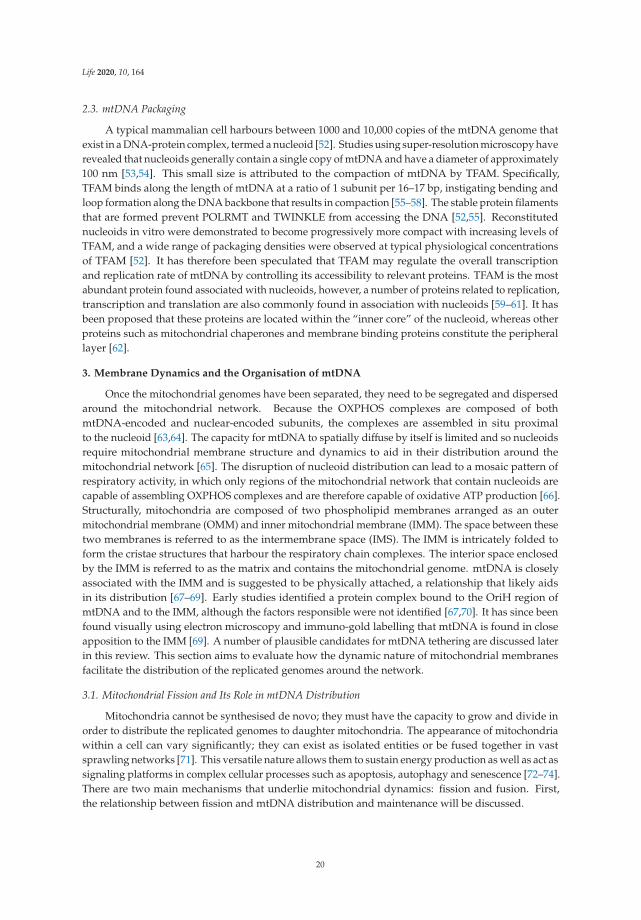
2.3. mtDNA Packaging
A typical mammalian cell harbours between 1000 and 10,000 copies of the mtDNA genome thatexist in a DNA-protein complex, termed a nucleoid [52]. Studies using super-resolution microscopy haverevealed that nucleoids generally contain a single copy of mtDNA and have a diameter of approximately100 nm [53,54]. This small size is attributed to the compaction of mtDNA by TFAM. Specifically,TFAM binds along the length of mtDNA at a ratio of 1 subunit per 16–17 bp, instigating bending andloop formation along the DNA backbone that results in compaction [55–58]. The stable protein filamentsthat are formed prevent POLRMT and TWINKLE from accessing the DNA [52,55]. Reconstitutednucleoids in vitro were demonstrated to become progressively more compact with increasing levels ofTFAM, and a wide range of packaging densities were observed at typical physiological concentrationsof TFAM [52]. It has therefore been speculated that TFAM may regulate the overall transcriptionand replication rate of mtDNA by controlling its accessibility to relevant proteins. TFAM is the mostabundant protein found associated with nucleoids, however, a number of proteins related to replication,transcription and translation are also commonly found in association with nucleoids [59–61]. It hasbeen proposed that these proteins are located within the “inner core” of the nucleoid, whereas otherproteins such as mitochondrial chaperones and membrane binding proteins constitute the peripherallayer [62].
3. Membrane Dynamics and the Organisation of mtDNA
Once the mitochondrial genomes have been separated, they need to be segregated and dispersedaround the mitochondrial network. Because the OXPHOS complexes are composed of bothmtDNA-encoded and nuclear-encoded subunits, the complexes are assembled in situ proximalto the nucleoid [63,64]. The capacity for mtDNA to spatially diffuse by itself is limited and so nucleoidsrequire mitochondrial membrane structure and dynamics to aid in their distribution around themitochondrial network [65]. The disruption of nucleoid distribution can lead to a mosaic pattern ofrespiratory activity, in which only regions of the mitochondrial network that contain nucleoids arecapable of assembling OXPHOS complexes and are therefore capable of oxidative ATP production [66].Structurally, mitochondria are composed of two phospholipid membranes arranged as an outermitochondrial membrane (OMM) and inner mitochondrial membrane (IMM). The space between thesetwo membranes is referred to as the intermembrane space (IMS). The IMM is intricately folded toform the cristae structures that harbour the respiratory chain complexes. The interior space enclosedby the IMM is referred to as the matrix and contains the mitochondrial genome. mtDNA is closelyassociated with the IMM and is suggested to be physically attached, a relationship that likely aidsin its distribution [67–69]. Early studies identified a protein complex bound to the OriH region ofmtDNA and to the IMM, although the factors responsible were not identified [67,70]. It has since beenfound visually using electron microscopy and immuno-gold labelling that mtDNA is found in closeapposition to the IMM [69]. A number of plausible candidates for mtDNA tethering are discussed laterin this review. This section aims to evaluate how the dynamic nature of mitochondrial membranesfacilitate the distribution of the replicated genomes around the network.
3.1. Mitochondrial Fission and Its Role in mtDNA Distribution
Mitochondria cannot be synthesised de novo; they must have the capacity to grow and divide inorder to distribute the replicated genomes to daughter mitochondria. The appearance of mitochondriawithin a cell can vary significantly; they can exist as isolated entities or be fused together in vastsprawling networks [71]. This versatile nature allows them to sustain energy production as well as act assignaling platforms in complex cellular processes such as apoptosis, autophagy and senescence [72–74].There are two main mechanisms that underlie mitochondrial dynamics: fission and fusion. First,the relationship between fission and mtDNA distribution and maintenance will be discussed.
20

Life 2020, 10, 164
3.1.1. Mitochondrial Fission
Mitochondrial fission is the division of a mitochondrion into multiple distinct mitochondria andhas been implicated in the distribution of nucleoids around the mitochondrial network [75–77].Fission requires the progressive constriction, and eventual scission, of the IMM and OMM.The initial constriction occurs at contact sites between the OMM and the endoplasmic reticulum (ER),where actin polymerisation provides the force required to contract the mitochondrial membrane [78–80].Further constriction is primarily fulfilled by dynamin-related protein 1 (DRP1), a cytosolic proteinthat translocates to the OMM and interacts with several adaptor proteins [81]. DRP1 binds withmitochondrial fission factor 1 (MFF), mitochondrial dynamics protein of 49kDa (MID49), MID51 andmitochondrial fission 1 protein (FIS1) [82–85]. Utilising GTP (guanine triphosphate), polymerisation ofDRP1 with MID49 and MID51 occurs leading to the formation of linear filaments. GTP hydrolysisinduces the oligomerisation of DRP1 and filament shortening to create rings which constrict aroundthe mitochondria [79,86]. It has now been established that DRP1-mediated constriction is sufficientfor the final scission step to separate mitochondria [87,88]. Recent studies have also highlighted theimportance of additional interorganelle contacts for mitochondrial fission, with roles for lysosomes infission regulation and with Golgi-derived vesicles during final scission [89,90].
It has been observed that ER-OMM contact sites that mark sites of mitochondrial division areoften also spatially located adjacent to replicated nucleoids [75–77], suggesting a role for fission inmtDNA segregation. Furthermore, the visualisation of DRP1 and MFF using confocal microscopyhas demonstrated colocalisation at sites adjacent to the nucleoid [69,77,91]. Where division occursbetween replicated nucleoids, the daughter mtDNA molecules are subsequently observed to be locatedat the tips of the separated mitochondria [75,76]. This mechanism ensures that following divisioneach mitochondrion receives a copy of the genome, and secondly functions to disperse nucleoidsthroughout the mitochondrial network. At this stage, it is unclear what signalling takes place to ensurethat division occurs between the two replicated nucleoids.
3.1.2. Defects in Mitochondrial Fission and Human Disease
At this point in time, the prevalence of human diseases secondary to defects in mitochondrialfission is not known. However, they would appear to be much less common than diseases that arerelated to mitochondrial fusion. The most common clinical manifestations that occur in relation todisruption of mitochondrial fission genes are subtypes of Charcot–Marie–Tooth (CMT) neuropathyand optic neuropathy, as identified following mutations that occur in DRP1, GDAP1, INF2, MFF andSLC25A46 [92–96]. Severe neurological presentations, such as neurodevelopmental delay and epilepsy,are observed following mutations of the DRP1 gene [97]. Leigh-like syndrome has been observed inpatients harbouring mutations in MFF and SLC25A46 [98,99]. Mutations in DNM2 have been linkedto CPEO and central core myopathy [100,101], a form of myotubular myopathy. Extra-neurologicalinvolvements are relatively uncommon, except cardiac arrhythmia and neutropenia, which have beenassociated with mutations in DNM2 [101,102], and glomerular disease, described in INF2-relateddisease (Appendix A Table A1).
Mitochondrial respiratory chain deficiencies and multiple mtDNA deletions have beendemonstrated in muscle biopsies taken from patients harbouring DNM2 mutations [103].Normal qualitative and quantitative assessments of mtDNA were reported in the skeletal muscle orfibroblasts that contain mutant forms of other mitochondrial fission-associated genes such as GDAP1,INF2 and MFF (Table A1) [93,94,98,104–109]. These observations would suggest that the biologicalconsequences of these genetic defects are tissue specific, given that only DNM2 mutations primarilymanifest with a myopathic phenotype [100,101]. Whilst other fission-associated genes predominantlycause peripheral neuropathy including optic neuropathy or CNS involvement, it is important to notethat these affected tissues are far less accessible for further characterisation compared to muscle biopsy.
21
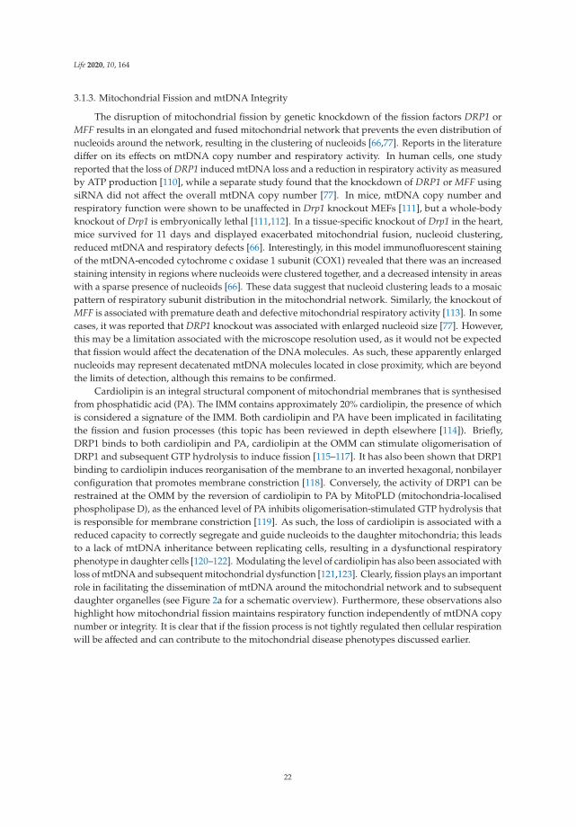
Life 2020, 10, 164
3.1.3. Mitochondrial Fission and mtDNA Integrity
The disruption of mitochondrial fission by genetic knockdown of the fission factors DRP1 orMFF results in an elongated and fused mitochondrial network that prevents the even distribution ofnucleoids around the network, resulting in the clustering of nucleoids [66,77]. Reports in the literaturediffer on its effects on mtDNA copy number and respiratory activity. In human cells, one studyreported that the loss of DRP1 induced mtDNA loss and a reduction in respiratory activity as measuredby ATP production [110], while a separate study found that the knockdown of DRP1 or MFF usingsiRNA did not affect the overall mtDNA copy number [77]. In mice, mtDNA copy number andrespiratory function were shown to be unaffected in Drp1 knockout MEFs [111], but a whole-bodyknockout of Drp1 is embryonically lethal [111,112]. In a tissue-specific knockout of Drp1 in the heart,mice survived for 11 days and displayed exacerbated mitochondrial fusion, nucleoid clustering,reduced mtDNA and respiratory defects [66]. Interestingly, in this model immunofluorescent stainingof the mtDNA-encoded cytochrome c oxidase 1 subunit (COX1) revealed that there was an increasedstaining intensity in regions where nucleoids were clustered together, and a decreased intensity in areaswith a sparse presence of nucleoids [66]. These data suggest that nucleoid clustering leads to a mosaicpattern of respiratory subunit distribution in the mitochondrial network. Similarly, the knockout ofMFF is associated with premature death and defective mitochondrial respiratory activity [113]. In somecases, it was reported that DRP1 knockout was associated with enlarged nucleoid size [77]. However,this may be a limitation associated with the microscope resolution used, as it would not be expectedthat fission would affect the decatenation of the DNA molecules. As such, these apparently enlargednucleoids may represent decatenated mtDNA molecules located in close proximity, which are beyondthe limits of detection, although this remains to be confirmed.
Cardiolipin is an integral structural component of mitochondrial membranes that is synthesisedfrom phosphatidic acid (PA). The IMM contains approximately 20% cardiolipin, the presence of whichis considered a signature of the IMM. Both cardiolipin and PA have been implicated in facilitatingthe fission and fusion processes (this topic has been reviewed in depth elsewhere [114]). Briefly,DRP1 binds to both cardiolipin and PA, cardiolipin at the OMM can stimulate oligomerisation ofDRP1 and subsequent GTP hydrolysis to induce fission [115–117]. It has also been shown that DRP1binding to cardiolipin induces reorganisation of the membrane to an inverted hexagonal, nonbilayerconfiguration that promotes membrane constriction [118]. Conversely, the activity of DRP1 can berestrained at the OMM by the reversion of cardiolipin to PA by MitoPLD (mitochondria-localisedphospholipase D), as the enhanced level of PA inhibits oligomerisation-stimulated GTP hydrolysis thatis responsible for membrane constriction [119]. As such, the loss of cardiolipin is associated with areduced capacity to correctly segregate and guide nucleoids to the daughter mitochondria; this leadsto a lack of mtDNA inheritance between replicating cells, resulting in a dysfunctional respiratoryphenotype in daughter cells [120–122]. Modulating the level of cardiolipin has also been associated withloss of mtDNA and subsequent mitochondrial dysfunction [121,123]. Clearly, fission plays an importantrole in facilitating the dissemination of mtDNA around the mitochondrial network and to subsequentdaughter organelles (see Figure 2a for a schematic overview). Furthermore, these observations alsohighlight how mitochondrial fission maintains respiratory function independently of mtDNA copynumber or integrity. It is clear that if the fission process is not tightly regulated then cellular respirationwill be affected and can contribute to the mitochondrial disease phenotypes discussed earlier.
22

Life 2020, 10, 164
Figure 2. Mitochondrial dynamics and its role in mtDNA distribution. (a) Schematic overview ofmitochondrial fission. Constriction of the mitochondrion occurs by the endoplasmic reticulum (ER) anddynamin related protein 1 (DRP1). The final scission step is performed by DRP1 to produce multipleindependent mitochondria. (b) Mitochondrial fusion outline. The mitofusins (MFNs) are responsiblefor tethering neighbouring mitochondria and fusing the outer membranes together. Fusion of the innermitochondrial membrane is mediated by optic atrophy 1 (OPA1). (c) Summary of mtDNA segregation
23

Life 2020, 10, 164
and distribution. First, newly replicated mtDNA molecules that are joined by a hemicatenane must bephysically separated by topoisomerase 3α. Failure of this process results in the formation of multiplephysically joined mtDNA genomes, termed mtDNA catenanes. Following separation, mitochondrialfission is required to distribute the replicated nucleoids into separate mitochondria and facilitatedistribution of the genomes around the mitochondrial network. An impairment of fission results in aclustered phenotype whereby many replicated nucleoids are observed in close proximity to each otherbut are not physically linked together. (d) A lack of mitochondrial fusion is associated with mtDNAinstability as observed by the progressive onset of mtDNA deletions and point mutations. In addition,the lack of fusion leads to a depletion in mtDNA copy number.
3.2. Mitochondrial Fusion and Its Role in mtDNA Maintenance and Distribution
Together with mitochondrial fission, mitochondrial fusion has been implicated in the disseminationof nucleoids around the network. Furthermore, fusion is recognized as an important mediator ofmtDNA maintenance. Defects in mtDNA maintenance may manifest as either a quantitative reductionin mtDNA copy number (depletion) or as an accumulation of rearranged mtDNA molecules (deletionsand/or duplications), all of which may be indicative of impaired mtDNA replication. Defects in theproteins that regulate mitochondrial fusion dynamics have been implicated in a range of geneticdiseases that will be discussed here and are summarised in Table A1. Their contribution to thedevelopment of disease will be described in relation to their role in mtDNA maintenance.
3.2.1. Mitochondrial Fusion
Mitochondrial fusion is the joining of two separate mitochondria and is important to enable thesharing of contents between neighbouring mitochondria [124]. The fusion of the OMM and IMM occursequentially, therefore the outer membranes are joined first. This is regulated by two outer membraneproteins, the dynamin related GTPases mitofusin 1 & 2 (MFN1 and MFN2). MFN1 is responsible fortethering the adjoining mitochondria together in a GTP-dependent dimerization process, promptinga conformational change which in turn mediates GTP hydrolysis by MFN1 to pull the membranestogether, resulting in mitochondrial fusion [125–127].The role of MFN2 is less clear, although it hasbeen implicated in interactions between mitochondria, as well as between mitochondria and theER [128–130]. Following OMM fusion the IMM can be fused. IMM fusion is controlled by opticatrophy 1 (OPA1). OPA1 exists in multiple forms of different sizes which are regulated by proteolyticprocessing; long OPA1 is IMM-anchored while the short OPA1 is soluble. The presence of OPA1 in itslong isoform has been associated with promoting IMM fusion [131]. Processing of OPA1 to its shortisoform either by YME1L1 or OMA1 is associated with mitochondrial fission [132,133].
There is evidence that OMM fusion proteins also play a role in the dissemination of nucleoids.It has been reported that the knockout of OMM fusion proteins Mfn1 & 2 in MEFs leads to the clusteringof nucleoids [134]. Using super-resolution microscopy these clusters were confirmed to be separateindividual nucleoids in close proximity rather than a cluster of interlinked DNA molecules. The roleof IMM fusion in nucleoid distribution is less clear. It has been reported by some groups that theloss of OPA1 is associated with a reduction in the number of nucleoids per cell, and in some cases aclustered phenotype [77,135,136]. In contrast, a recent study found that in MEFs the removal of Opa1was not associated with any alterations in mtDNA distribution, although there was a reduction in copynumber [134]. This normal nucleoid distribution following Opa1 knockout may be explained by the roleof MFN2 in forming a tether between the OMM and ER [137,138]. As discussed earlier, it is recognizedthat the ER localises at positions of nucleoid replication and is implicated in coordinating mitochondrialconstriction at these sites [75]. Taken together, this highlights the importance of the OMM acting as asignalling platform for coordinating the circulation of nucleoids around the mitochondrial network.
Interestingly, the knockdown of MFN1 & 2 in conjunction with DRP1 has been found to preventthe clustering of nucleoids in human cells [77]. Mechanistically, how the impairment of both fissionand fusion would restore nucleoid distribution is not clear as it may be expected that this would render
24
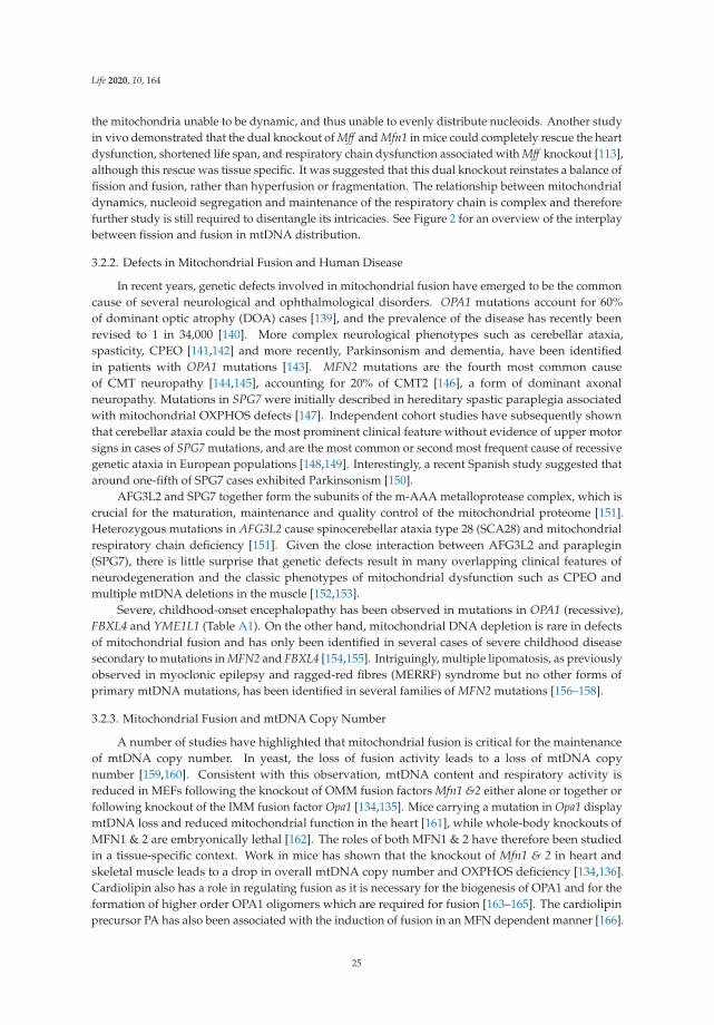
Life 2020, 10, 164
the mitochondria unable to be dynamic, and thus unable to evenly distribute nucleoids. Another studyin vivo demonstrated that the dual knockout of Mff and Mfn1 in mice could completely rescue the heartdysfunction, shortened life span, and respiratory chain dysfunction associated with Mff knockout [113],although this rescue was tissue specific. It was suggested that this dual knockout reinstates a balance offission and fusion, rather than hyperfusion or fragmentation. The relationship between mitochondrialdynamics, nucleoid segregation and maintenance of the respiratory chain is complex and thereforefurther study is still required to disentangle its intricacies. See Figure 2 for an overview of the interplaybetween fission and fusion in mtDNA distribution.
3.2.2. Defects in Mitochondrial Fusion and Human Disease
In recent years, genetic defects involved in mitochondrial fusion have emerged to be the commoncause of several neurological and ophthalmological disorders. OPA1 mutations account for 60%of dominant optic atrophy (DOA) cases [139], and the prevalence of the disease has recently beenrevised to 1 in 34,000 [140]. More complex neurological phenotypes such as cerebellar ataxia,spasticity, CPEO [141,142] and more recently, Parkinsonism and dementia, have been identifiedin patients with OPA1 mutations [143]. MFN2 mutations are the fourth most common causeof CMT neuropathy [144,145], accounting for 20% of CMT2 [146], a form of dominant axonalneuropathy. Mutations in SPG7 were initially described in hereditary spastic paraplegia associatedwith mitochondrial OXPHOS defects [147]. Independent cohort studies have subsequently shownthat cerebellar ataxia could be the most prominent clinical feature without evidence of upper motorsigns in cases of SPG7 mutations, and are the most common or second most frequent cause of recessivegenetic ataxia in European populations [148,149]. Interestingly, a recent Spanish study suggested thataround one-fifth of SPG7 cases exhibited Parkinsonism [150].
AFG3L2 and SPG7 together form the subunits of the m-AAA metalloprotease complex, which iscrucial for the maturation, maintenance and quality control of the mitochondrial proteome [151].Heterozygous mutations in AFG3L2 cause spinocerebellar ataxia type 28 (SCA28) and mitochondrialrespiratory chain deficiency [151]. Given the close interaction between AFG3L2 and paraplegin(SPG7), there is little surprise that genetic defects result in many overlapping clinical features ofneurodegeneration and the classic phenotypes of mitochondrial dysfunction such as CPEO andmultiple mtDNA deletions in the muscle [152,153].
Severe, childhood-onset encephalopathy has been observed in mutations in OPA1 (recessive),FBXL4 and YME1L1 (Table A1). On the other hand, mitochondrial DNA depletion is rare in defectsof mitochondrial fusion and has only been identified in several cases of severe childhood diseasesecondary to mutations in MFN2 and FBXL4 [154,155]. Intriguingly, multiple lipomatosis, as previouslyobserved in myoclonic epilepsy and ragged-red fibres (MERRF) syndrome but no other forms ofprimary mtDNA mutations, has been identified in several families of MFN2 mutations [156–158].
3.2.3. Mitochondrial Fusion and mtDNA Copy Number
A number of studies have highlighted that mitochondrial fusion is critical for the maintenanceof mtDNA copy number. In yeast, the loss of fusion activity leads to a loss of mtDNA copynumber [159,160]. Consistent with this observation, mtDNA content and respiratory activity isreduced in MEFs following the knockout of OMM fusion factors Mfn1 &2 either alone or together orfollowing knockout of the IMM fusion factor Opa1 [134,135]. Mice carrying a mutation in Opa1 displaymtDNA loss and reduced mitochondrial function in the heart [161], while whole-body knockouts ofMFN1 & 2 are embryonically lethal [162]. The roles of both MFN1 & 2 have therefore been studiedin a tissue-specific context. Work in mice has shown that the knockout of Mfn1 & 2 in heart andskeletal muscle leads to a drop in overall mtDNA copy number and OXPHOS deficiency [134,136].Cardiolipin also has a role in regulating fusion as it is necessary for the biogenesis of OPA1 and for theformation of higher order OPA1 oligomers which are required for fusion [163–165]. The cardiolipinprecursor PA has also been associated with the induction of fusion in an MFN dependent manner [166].
25

Life 2020, 10, 164
The disruption of cardiolipin levels is linked with a reduction in mtDNA copy number [121,123].ATAD3 (ATPase family AAA-domain containing protein 3 A) has also been implicated in maintainingmitochondrial fusion as its manipulation either by knockdown or overexpression leads to mitochondrialfragmentation [167–170]. This fragmented phenotype is mediated by the recruitment of DRP1 to theOMM via oligomerisation of the ATAD3 coiled-coil domain [170]. Furthermore, it was demonstratedthat in addition to activating DRP1, the dimerisation of ATAD3 provokes mtDNA instability bydisrupting the binding of TFAM and mtDNA. Indeed, the knockdown of ATAD3 has been associatedwith a reduction in mtDNA content [170]. In human fibroblasts that are either deficient for ATAD3 orharbour a duplication of the ATAD3 gene cluster, nucleoids were found to be enlarged and clusteredtogether suggesting a role of ATAD3 and fusion in mtDNA distribution [171,172]. The knockout ofATAD3 in skeletal muscle has been associated with the progressive formation of mtDNA deletions andcopy number depletion [173].
The mechanisms underlying the loss of mtDNA when fusion is impaired have long been unclear,although recent work has shed light on potential links between the two. In general, the loss of copynumber indicates that there is a defect with the process of mtDNA replication. It has been demonstratedthat mitochondrial fusion is necessary to facilitate high levels of replication through content mixing toensure a proper stoichiometry of replisome components. The loss of OPA1 alone or the collective lossof MFN1 & 2 together prompts an imbalance of replisome factors and thus leads to a reduced rate ofmtDNA replication, leading to mtDNA depletion [134].
3.2.4. Mitochondrial Fusion and mtDNA Integrity
It has been observed that aside from being required to maintain copy number, fusion appearsto play an important role in maintaining the integrity of the mitochondrial genome. For example,mutations in the OPA1 gene have been associated with the accumulation of mtDNA deletions andOXPHOS defects in the skeletal muscle of patients [141,174,175]. Similarly, patients with mutations inthe OMM fusion protein MFN2 display evidence of deletion-containing mtDNA genomes [154,176].In animal models, the knockout of Mfn1 & 2 in the skeletal muscle of mice is associated with an increasedoccurrence of point mutations and deletions of mtDNA [136]. On the other hand, another groupstudying the cardiac tissue of mice with MFN1 & 2 genetically removed found no differences in levelsof mtDNA mutations or deletions [134]. These studies highlight that effects may be tissue-specific,and that care must be taken when comparing effects in patients carrying missense mutations witheffects seen in knockout animal models. However, from a clinical perspective, mutations in the fusionmachinery have a clear association with the progressive onset of mtDNA mutations and deletions(see Table A1).
Multiple lines of evidence indicate that fusion plays a protective role against mutations. Work usingfibroblasts derived from MFN2 patients found that these cells display a reduced capacity to repairmtDNA damage [176]. This finding potentially suggests that fusion may preserve mtDNA integrityby enabling the repair of DNA damage or by facilitating the clearance of mitochondria harbouringdamaged DNA. In support of this notion, excessive mitochondrial fragmentation (which wouldoccur with impaired fusion) has been associated with an increased production of reactive oxygenspecies [177]. Indeed, in an Opa1 mutant mouse it was observed that there was an increased level ofROS coupled with a reduced antioxidant capacity [161]. A separate study used the mutator mouse,which has a mutation in the proofreading domain of polymerase PolgA, and consequently rapidlyaccumulates mtDNA mutations, crossed with a knockout of MFN1 [178]. The mutator mouse orthe MFN1 knockout mouse individually survive into adulthood, however crosses between the tworesulted in embryonic lethality [136]. In this case it is plausible that fusion exerts a protective effectthrough its ability to “dilute” mutation-containing mtDNA via content mixing and therefore preservemitochondrial function.
It remains unclear how defects in fusion contribute to the molecular mechanism of deletionformation. Deletions may conceivably accumulate at an increased rate either because of impaired
26

Life 2020, 10, 164
mtDNA replication or because of increased mtDNA damage. It has been speculated that the primarypurpose of mitochondrial fusion is to enable the sharing of contents between two mitochondria.Indeed, it has been published that matrix proteins and mtDNA are transferred between fusedmitochondria [179]. Conversely, mitochondria from dual MFN knockout cells were found to haveincreased protein heterogeneity compared to their wild-type counterparts [136]. It may therefore bethat the loss of content mixing associated with defective fusion impairs mtDNA replication through animbalance of replisome components [134]. This could then promote replication stalling and copy-choicerecombination, leading to increased deletion formation. Alternatively, it is possible that increasedmtDNA damage in the absence of fusion promotes the formation of deletion-containing mtDNAmolecules via double-strand break formation. However, a key limitation in the idea of mtDNA repairunderlying the formation of deletions is that at this stage the proteins that are responsible for DNArepair have not been identified as being present in mitochondria.
It is also conceivable that deletions accumulate in the absence of fusion because their removalis impaired. However, the observation that defects in fusion are associated with an accumulation ofmtDNA mutations and deletions appears counterintuitive, as it would be expected that a fragmentednetwork would be optimal for selective mitophagy to clear dysfunctional mitochondria away. Indeed,in a Drosophila melanogaster model of mtDNA heteroplasmy, the knockdown of MFN promoted themitophagy of fragmented mitochondria and a reduction in the mutant mtDNA load [180]. Similarly,a perpetually fused network due to the inhibition of fission by DRP1 or FIS1 in human cells wasassociated with a shift in heteroplasmy towards mutant mtDNA, possibly because of reduced mitophagy,although this was not addressed [181]. As such, it appears that the accumulation of mtDNA mutationsand deletions in the absence of fusion is not related to a reduction in mitophagy.
Collectively, these studies demonstrate that IMM and OMM fusion have an important role inmaintaining the integrity of mtDNA and coordinating its replication. However, the relationship iscomplex and further study is certainly required. It seems likely that the role of fusion in facilitatingcontent mixing between mitochondria is important to maintain the balance of proteins required formtDNA replication [134,136]. There is also evidence to suggest that fusion also has a protective roleagainst the effects of ROS and DNA damage [161,177]. This, coupled with an imbalance of replisomeproteins leading to replication stalling/slippage, could underlie the onset of mutations and deletionsassociated with fusion defects, as well as contribute to mtDNA copy number loss.
4. The Relationship between mtDNA and Mitochondrial Cristae Structure
The IMM is a complex structure that can be subdivided into two further regions: the innerboundary membrane (IBM) and the cristae (Figure 3b). The IBM runs parallel to the OMM andhouses proteins responsible for localising and importing proteins into the matrix, as well as insertingand assembling proteins into the IMM [182,183]. The IBM is divided at regions where the IMMis folded inwards to form cristae structures. Cristae are functionally active structures that containthe respiratory chain complexes and the ATP synthase [184]. At points where the cristae joins theIBM, the cristae narrows to form a cristae junction (approximately 20–40 nm in diameter) whichallows the separation of the intracristal space from the intermembrane space between the IMM andOMM [185–187]. The mitochondrial contact site and cristae organising system (MICOS) complex islocated at these junctions and plays a key role in maintaining their structure.
It has long been appreciated that mtDNA is located close to the IMM and to the cristae structure.It has been recently demonstrated using correlative 3D super-resolution fluorescence and electronmicroscopy that nucleoids are found within cristae regions of the mitochondria [68]. Sophisticatedsuper-resolution imaging has now revealed that nucleoids are typically located in the voids that formbetween groups of cristae rather than being embedded within the cristae structure itself [188]. This isin line with the observation that the size of the nucleoid, at approximately 100 nm, is greater than thegaps between cristae, which are found tightly packed together [53,54,188]. It is postulated that thesevoids where nucleoids are located may function to provide space for transcription, replication and
27

Life 2020, 10, 164
segregation to occur [68,188]. In this section we will summarise current data on cristae structure andmodulation, and its relationship with mtDNA maintenance and segregation.
Figure 3. The relationship between cristae modulators and mtDNA organisation. (a) Schematicoverview of a normal mitochondrion. (b) Enlarged cristae region depicting the spatial arrangement ofkey proteins that are necessary for dictating cristae structure and shape, as well as the organisation ofthe respiratory chain complexes. Proteins that are potentially responsible for the tethering of mtDNAto the IBM are also highlighted. (c) Loss of the fission factor DRP1 has been associated with theformation of mito-bulb structures. These are regions of dense cristae structure which harbour a numberof clustered mtDNA molecules. (d) The loss of IMM fusion and cristae shaping protein OPA1 results inmitochondria that display enlarged cristae junctions and a perturbed cristae structure. In addition,mtDNA copy number is reduced. (e) The loss of MICOS components (MIC60, MIC10 and MIC19) hasbeen associated with a complete loss of cristae junctions and the formation of cristae in concentriccircles. Nucleoid clustering is evident following the loss of MICOS components.
4.1. The Importance of Lipids in the IMM and Their Role in mtDNA Tethering
The capacity for mtDNA to freely diffuse around the mitochondrial network is limited, due inpart to the relatively large size of the nucleoid and to the high density of proteins located within
28

Life 2020, 10, 164
the matrix and cristae regions [53,69,189]. There is also evidence that has led to the suggestion thatmtDNA is tethered to the IMM [67,69,70,190,191]. However, at this stage the factors responsible remainpoorly understood.
Several studies have highlighted the importance of the lipid composition of the IMM to mtDNAattachment, replication and organisation. The composition of phospholipids in the IMM is tightlyregulated. Phospholipids have a hydrophobic lipid domain tail which is larger than their chargedhydrophilic head groups; this configuration forms a rough conical shape which pulls head groupstogether, inducing membrane tension and facilitating the bending of the IMM to form cristaeinvaginations [192,193]. Cardiolipin has an important role in stabilising IMM proteins such asthe respiratory complexes [194]. Alterations in the level of cardiolipin have been associated withmitochondrial dysfunction, mtDNA loss and the formation of abnormal cristae structures [121,123].Similarly, in mammalian cells the removal of the IMM phospholipid phosphatidylethanolamine hascomparable effects; mitochondrial function is reduced coupled with obvious swelling of mitochondriawhich lack distinct cristae structures [195].
It has been demonstrated in both yeast and mammalian cells that cardiolipin is physicallyassociated with the nucleoid [120]. Furthermore, as discussed earlier cardiolipin is a key constituent ofthe IMM and has a role in mitochondrial fission and fusion. As such, the role of cardiolipin in mtDNAmaintenance is multi-faceted. The loss of cardiolipin is associated with a reduced capacity to correctlysegregate and guide nucleoids to daughter mitochondria. In addition, this defective IMM-nucleoidassociation coupled with defects in mitochondrial dynamics leads to a lack of mtDNA inheritancebetween replicating cells resulting in a dysfunctional respiratory phenotype in daughter cells [120–122].
The composition of the mitochondrial IMM is subject to a relatively low ratio of cholesterol tophospholipids [196]. Cholesterol has been implicated in nucleoid organisation as its modulationeither through pharmacological inhibition or supplementation prompts the formation of aggregatednucleoids in human fibroblasts [171]. The majority of mitochondrial cholesterol is clustered inspecialised structures that span the IMM and OMM [197]. TWINKLE-containing nucleoids wereshown to be associated with these cholesterol structures in a complex that also contains ATAD3.ATAD3 is an essential protein that is anchored into the IMM, with its C-terminal AAA ATPase domainlocated in the mitochondrial matrix while its N-terminus interacts with the OMM where it is presentat ER-mitochondria contact sites [62,169,197–199]. It has been proposed that these cholesterol-richsites provide a platform for mtDNA replication, and act as an attachment site at ER-mitochondriajunctions to allow for the coordinated distribution of mtDNA. As discussed earlier, ATAD3 has beendemonstrated to have a regulatory role in mitochondrial morphology and dynamics and has beenimplicated in nucleoid organisation [167–170]. ATAD3 also has an important role in maintaining IMMarchitecture as the disruption of ATAD3 leads to impaired transfer of cholesterol from the ER intothe mitochondria and lipid metabolism [171,200]. The knockout of ATAD3 in skeletal muscle hasbeen associated with alterations in cholesterol metabolism and the progressive formation of mtDNAdeletions and copy number depletion [173]. As discussed earlier, the formation of these mtDNArearrangements has been associated with stalling of the replication machinery and a loss of cristaestructure. It has been suggested that ATAD3 may interact with OPA1 or the MICOS complex to stabilisecristae structure [173]. Collectively, it is clear that ATAD3 is an important regulator of mitochondrialdynamics, mtDNA attachment, maintenance and organisation. However, at this stage it is not entirelyclear whether these effects are directly associated with the loss of ATAD3 or due to cholesterol-mediatedalterations in the IMM structure as a consequence of the role of ATAD3 in the uptake of cholesterol tothe mitochondria [200].
In addition to ATAD3, OPA1 has also been implicated as having a potential role in the attachment ofnucleoids to the IMM [135]. OPA1 is embedded in the IMM, co-immunoprecipitates with nucleoids andas discussed earlier, its deletion has profound effects on nucleoid organisation [77,135,136]. Thus, it hasbeen suggested that OPA1 may have a role in membrane attachment of the nucleoid (Figure 3b). Loss ofeither ATAD3 or OPA1 has been shown to cause significant aberrations in cristae structure [161,197,201].
29

Life 2020, 10, 164
The structure of proteins and lipids within the IMM is also supported by a group of evolutionarilyconserved scaffolding proteins termed prohibitins. Prohibitin 1 and 2 are essential proteins that formhigh molecular weight ring-shaped heterooligomers in the IMM. They have also been found to associatewith nucleoids which prompts speculation that they may be involved in tethering of nucleoids tothe IMM [57]. It has been demonstrated that the prohibitins can influence mtDNA copy numberthrough their interactions with TFAM [202]. Cultured cells lacking the prohibitins display impairedcardiolipin maturation, loss of cristae structure and disorganisation of nucleoids [203–205]. It has alsobeen demonstrated that the effects of prohibitin 2 depletion upon cristae structure are dependent uponOPA1, a known modulator of cristae structure [204].
4.2. Modulators of Cristae Structure
Mitochondrial cristae are dynamic structures that can modulate their shape in response to variousphysiological conditions. As they are the primary site of the OXPHOS machinery, it is important thatthe folding of the IMM into cristae structures facilitates the most efficient manner of producing ATP.Recent work has found that individual cristae act as autonomous bioenergetic units, highlighting theimportance of their structural integrity [206]. Cristae structure is dictated by a variety of “cristae-shapingproteins” (Figure 3b). A key mediator of cristae architecture is the MICOS complex. The MICOScomplex consists of at least the subunits MIC10, MIC12, MIC13, MIC19, MIC25, MIC26, MIC27 andMIC60 [207]. Each of these proteins has a specific role in shaping the cristae structure. MIC60 is a corecomponent of the complex and is associated with the formation of cristae junctions and contact siteswith the OMM [208–210]. MIC10 can also bend membranes and is known to be responsible for forminghairpin structures in the IMM [211,212]. MIC13 has also been recently recognised as being essential formaintaining the stability of the MICOS complex and cristae junction formation [212]. MIC26 has a rolein cristae junction formation, and both MIC26 and MIC27 are necessary to maintain normal cristaearchitecture [213,214].
The F1F0 ATP synthase is embedded in the IMM and is localised to the cristae tips, where it existsin a dimer confirmation. These dimers are also assembled into oligomers. The presence of these dimersprompts a bending of the surrounding IMM lipid bilayer, highlighting their importance as mediatorsof cristae structure [215,216]. As such, the loss of the F1F0 ATP synthase is associated with a loss ofcristae invaginations [216]. However, the F1F0 ATP synthase may also act indirectly to maintain cristaemorphology, as it has also been recognized that the F1F0 ATP synthase interacts with the MICOScomplex [217]. It has also been reported that oligomerisation of F1F0 ATP synthase dimers is promotedby OPA1, which also has a role in cristae shaping [218].
The role of OPA1 in cristae shaping is reflected in its ability to define the diameter and width ofcristae junctions [209]. It has been demonstrated in yeast that the OPA1 homologue MGM1 is requiredto maintain cristae structure by tethering to other OPA1 molecules on the opposing IMM [219]. Indeed,it has been observed that high molecular weight multimers of OPA1 stabilise and induce the formationof tight cristae junctions, whereas lower-order OPA1 oligomers are associated with increased cristaejunction and lumen width [209]. The remodeling of cristae is mediated by the integral membraneprotease PARL. Processing by PARL produces a short, intramembrane-soluble form of OPA1 that bindswith the membrane-bound form to maintain tight cristae junctions [220]. There is some evidence thatOPA1 has a broader regulatory role and can adjust mitochondrial respiration by modulating cristaeshape. It has been reported that in response to hypoxia there is an increase in the abundance of OPA1oligomers that prevent cristae remodeling, thereby acting to enhance mitochondrial respiration [221].It has also been shown that OPA1 can interact with metabolic sensors in response to starvation, actingto adjust the cristae shape to maintain ATP production [222]. High molecular weight OPA1 multimershave also found to be associated with MIC60 and F1F0 ATP synthase [209,218,223]. Further work willbe required to understand the extent to which OPA1 dictates cristae shape alone, and how it functionsas part of an interactive network with other cristae shaping proteins.
30

Life 2020, 10, 164
A number of other proteins have been observed to have an impact on cristae structure, but forwhich the precise mechanisms are less well understood. MCL1 has been found to have a role inpreserving cristae ultrastructure and the maintenance of oligomeric ATP synthase [224]. The loss ofproteins that interact with cardiolipin have also been found to disrupt cristae architecture. Specifically,the prohibitins and UQCC3 have all been demonstrated to bind to cardiolipin, and their loss isassociated with alterations in cristae structure [204,225,226].
4.3. The Relationship between Cristae Structure and mtDNA
A number of studies indicate that there is an interesting but poorly-understood link betweenmtDNA and cristae ultrastructure. Dissecting this relationship is problematic because of the rolethat mtDNA-encoded proteins, such as components of the ATP synthase, play in maintaining cristaestructure. For example Rho-0 (ρ0) fibroblasts, which are devoid of mtDNA, display sparse cristaestructures and swollen mitochondria compared to mtDNA-containing cells [227–229]. Embryos lackingTFAM also lack mtDNA and display abnormal cristae structure [230]. Similarly, tissue-specific knockoutof TFAM in developed animals has also been shown to ablate cristae structure [231,232]. In culturedcells, the transient depletion of mtDNA using the replication inhibitor 2′-3′-dideoxycytidine also leadsto a loss of cristae structure [77]. Other data has suggested a direct role for mtDNA in maintainingcristae structure. The loss of DRP1 is associated with the formation of structures filled with verydensely packed cristae and clustered nucleoids termed “mito-bulbs” (Figure 3c) [77]. The formationof these cristae-rich mito-bulbs in DRP1 knockdown cells was not impacted when mitochondrialtranslation was inhibited using chloramphenicol. However, the depletion of mtDNA prior to DRP1knockdown prevented the formation of mito-bulbs. This suggests that mtDNA itself may be importantfor determining cristae architecture rather than its gene products. These data highlight the need forfurther study to understand how mtDNA contributes to the maintenance of normal cristae structure.
The loss of OPA1 has been associated with a reduced mtDNA copy number, both in modelsystems and in patients harbouring OPA1 mutations [135,161,233]. OPA1-deficient cultured cells alsoshow a distinct loss of cristae structure (Figure 3d) [201,234]. Similarly, in mice, both the mutation ofOpa1 in the heart and deletion of Opa1 in the skeletal muscle was found to cause the loss of cristaestructures [161,235,236]. Other in vivo models in which mtDNA content was reduced, such as thedeletion of Mfn2 in Purkinje cells or the dual knockout of Mfn1 & 2 in skeletal muscle, have also beenshown to be associated with sparse cristae structures. Whilst these observations support the idea thatthe loss of mtDNA is linked with the disruption of cristae structure, it is likely that the relationship isnot this simple. For example, one study noted that the loss of cristae structure in Opa1 mutant miceoccurred prior to the reduction in mtDNA content [235], although this is not surprising because of thekey role of OPA1 in maintaining cristae junctions [209].
Analysis of the relationship between mtDNA and cristae structure is also complicated by thefact that a number of interventions which alter mtDNA content also impact on the balance of fissionand fusion rates of mitochondria, and therefore the proper organisation of nucleoids around thenetwork. There is also evidence to suggest that normal cristae structure is required to facilitate fissionand fusion [210]. The MICOS complex can be destabilized by the loss of the core protein MIC60.This results in a striking phenotype of enlarged mitochondria that display abnormal circular cristae,commonly described as “concentric rings” or an “onion-like structure”, alongside an almost completeloss of cristae junctions (Figure 3e) [210,237]. It has also been reported that the loss of MIC60 causes areduction in the copy number of mtDNA (139). The downregulation of other MICOS subunits such asMIC19 and MIC10 also leads to a disruption of cristae structure [210]. The silencing of MIC19 andMIC60 led to the formation of enlarged and disorganised nucleoids in both mammalian cells andyeast, accompanied by a reduction in mtDNA transcription [210,238]. Similarly, it has been noted thatmutations in CHCHD10, a constituent of the MICOS complex, leads to a loss of cristae junctions andstructure. Furthermore, this was also shown to be linked with the formation of mtDNA deletions,as well as a reduction in nucleoid number without an overall effect on copy number [239].
31

Life 2020, 10, 164
It is worth noting that it is often difficult to determine whether apparently enlarged nucleoidsrepresent one single enlarged nucleoid consisting of multiple physically joined (catenated) mtDNAmolecules, or a group of nucleoids clustered in close proximity. An interesting aspect of the enlargementor clustering of nucleoids associated with the loss of MIC60 is that it was not reported to lead tothe formation of cristae-enriched mito-bulb structures as are seen following DRP1 knockdown [77].However, it is likely that these cells are unable to form dense cristae regions as a consequence of thefundamental role of MIC60 and the MICOS complex in shaping cristae [207]. Interestingly, MIC60knockdown cells showed a normal balance of fission and fusion compared to wild-type cells, but thisoccurred at a reduced rate with DRP1 being one of the mitochondrial dynamics proteins observed to bedownregulated. When DRP1 was overexpressed to promote mitochondrial fragmentation, the presenceof enlarged nucleoids associated with the loss of MIC60 was partially reverted [210]. Furthermore,work in yeast has found that when MIC60 (FCJ1) is knocked out in conjunction with DRP1 (DNM1) thenucleoids are further enlarged compared to the loss of MIC60 alone [238].
Cristae junctions have also been implicated in enabling nucleoid distribution. Unlike MIC60deletion, the knockout of F1F0 ATP synthase dimerization factors does not promote nucleoid aggregationor affect the number of cristae junctions. However, its knockout does reduce the number of cristaetips. The dual knockout of MIC60 (FCJ1) and dimerization partners of F1F0 ATP synthase results inan increased number of ring-like cristae structures that rescues the nucleoid aggregation phenotypeassociated with MIC60 deletion alone [238]. This finding indicates that cristae structure is likelyimportant for partitioning nucleoids to prevent their aggregation.
Addressed collectively, these studies support the idea that normal cristae structure is requiredto support the even distribution of mtDNA nucleoids around the mitochondrial network (Figure 3).Firstly, dynamic cristae structure is required to facilitate normal rates of fission and fusion, which asdiscussed earlier are key mediators of nucleoid dispersal. Secondly, cristae can also prevent theaggregation of nucleoids by forming partitioning structures. This is in logical agreement with theobservation that nucleoids are generally located in voids between groups of cristae structures [188].At this stage, it is unclear whether nucleoid partitioning is a precursor to the distribution of nucleoidsaround the network via fission. Therefore, further study will be necessary to understand the intricateinterplay between these two events.
4.4. Cristae Remodelling by Independent Fission and Fusion of Cristae Membranes
It has been speculated for some time that cristae are dynamic structures that can undergoremodelling of their membranes in response to various physiological conditions. However, studyingthese remodelling events in real-time has been particularly challenging until recently. A number ofgroups have developed methods that allow for the visualisation of cristae structures in live cells usingsuper-resolution microscopy [188,206,240]. Recent work has established that cristae within the samemitochondrion can have different membrane potentials, thus demonstrating that cristae can behaveas independent bioenergetic units [206]. This work suggests that within a mitochondrion cristae canfunctionally isolate themselves from one another, and it is postulated that this mechanism can preventindividual dysfunctional cristae from disrupting membrane potential in the broader mitochondrialnetwork. Further work has revealed that cristae membranes within a mitochondrion undergo fissionand fusion events in a MICOS-dependent manner [241]. Specifically, this study provides evidence for amodel in which MIC60 is evenly distributed along the IBM, acting as a docking or scaffolding platformto facilitate the formation of cristae junctions following the recruitment of MIC10. The recruitment ofother subunits to complete the MICOS complex allows cristae junctions to fully form. Both cristaejunctions and cristae membranes are dynamic, and it was observed that cristae junctions have thecapacity to split and merge. Cristae membranes can detach from one cristae junction, and fusewith either the same cristae junction or another cristae junction, such as one on the opposing IBM.It was demonstrated that the merging of cristae membranes is associated with changes in membrane
32

Life 2020, 10, 164
potential at that region, and that these events can facilitate content mixing between distinct cristaecompartments [241].
The fact that cristae continuously undergo remodelling and have the capacity to functionindependently raises the question of how these processes are involved in maintaining the integrityof mtDNA, as well as its segregation and normal distribution. This novel ability to study cristaedynamics in live cells allows an unprecedented opportunity for the field to address some of theseexperimentally difficult questions. For example, what is the importance of dynamic cristae membranesin maintaining mtDNA integrity by content mixing? Other directions may include understanding theinterplay between overall mitochondrial dynamics and cristae dynamics, as well as addressing howthe modelling of cristae membranes impacts the segregation and movement of mtDNA around themitochondrial network.
4.5. Genetic Defects Associated with Perturbed Mitochondrial Cristae Structure and Diseases
Mammalian MICOS comprises seven protein subunits of which defects in two encoding geneshave been linked to human disease so far. Mutations in MICOS13 (aka QIL1) cause severe infantilemitochondrial disease characterised by failure to thrive, microcephaly, truncal hypotonia, spasticityand cerebellar atrophy, lactic acidosis and 3-methylglutoconic aciduria (3-MGA) [242–244]. Subclinicalhepatic involvement manifesting with persistently abnormal liver function tests was also reported.Altered mitochondrial cristae morphology was evident in liver tissue and fibroblasts, but themitochondrial ultrastructure in muscle appeared normal. Multiple mitochondrial respiratory chaindeficiencies were identified in skeletal muscle and liver biopsies, and isolated complex IV deficiencywas present in fibroblasts. However, there was no evidence of mtDNA deletions or depletion in musclesand fibroblasts [242–244]. More recently, a mutation in APOO, encoding MIC26, has been reported tocause an X-linked disease associated with developmental delay, hypotonia, autistic spectrum disorder,gastrointestinal symptoms, lactic acidosis and abnormal carnitine profile [245]. The pathogenicity ofthe APOO variant was supported by work with fibroblasts and a fly model.
Homozygous mutations in the PINK1 gene, encoding for PTEN-induced kinase 1, were firstidentified in three consanguineous families with early-onset Parkinsonism through linkage analysis in2004 [246]. Overall, PINK1 mutations account for less than 10% of autosomal recessive Parkinson’sdisease (PD) [247]. Both Parkin and PINK1 have been shown to play crucial roles in mediatingthe mitophagy process [248]. More recently, PINK1 was found to maintain cristae junctions by thephosphorylation of the MICOS subunit MIC60 in both Drosophila melanogaster and human neurons [249].Furthermore, mutations within the mitochondrial targeting sequence of MIC60 were evident in asubset of PD patients. The introduction of these patient mutations into a Drosophila melanogaster modeldisrupted the ability for MIC60 to localise to mitochondria and prompted the formation of abnormalcristae junctions. Furthermore, the overexpression of MIC60 in a PINK1−/− model of PD compensatedfor the loss of PINK1-mediated phosphorylation and rescued both cristae defects and mitochondrialfunction [249]. In addition, mutations in the mitochondrial targeting sequence of CHCHD2 have beenidentified as a risk factor for PD and Lewy body disease [250].
CHCHD10 is a protein located in the mitochondrial inter-membrane space and it has beendemonstrated to play a role in the maintenance of cristae integrity [239]. Heterozygous mutations inCHCHD10 were first linked to motor neuron disease and frontotemporal dementia, myopathy andhyperkalemia, impaired respiratory chain function and multiple mtDNA deletions in the skeletal musclein a large family of French origin, and in a Spanish family (none of the family members underwentmuscle or skin biopsy) in 2014 [251]. Whilst mutations of CHCHD10 display a mitochondrial diseasethat resembles motor neuron disease, a large consortium of motor neuron disease (amyotrophic lateralsclerosis) patients (n = 4365) and healthy controls (n = 1832) from seven countries subsequentlydemonstrated that pathogenic CHCHD10 variants are exceptionally rare [252]. Therefore, in pureforms of amyotrophic lateral sclerosis it is not necessarily associated with CHCHD10 mutations.
33

Life 2020, 10, 164
Prominent extra-neurological disease has been observed following the disruption of severalproteins implicated in the maintenance of cristae structure, for example, hypertrophic cardiomyopathyoccurs in patients harbouring mutated forms of ATAD3A and TAZ (Barth syndrome) [253,254],and 3-MGA in ATAD3A, TAZ, MICOS13 and ATP5F1E [243,253,255,256].
5. Conclusions
The structure and dynamics of the mitochondrial membranes are required to maintain both theintegrity of mtDNA and well as its distribution within the mitochondrial network. Mitochondrialmembranes make a poorly understood contribution to mtDNA replication, the impairment of whichmanifests either as an inability to maintain a sufficient number of copies of mtDNA, or as rearrangementsof the genome. Following replication, the dynamics of mitochondria are also required for the segregationof mtDNA, and the disruption of mitochondrial dynamics can lead to the clustering of nucleoidswithin cells. The direct association between mtDNA and the respiratory chain means that an unevendistribution of nucleoids can lead to a mosaic pattern of respiratory activity within cells, which mayrepresent an under-appreciated molecular contributor to mitochondrial pathologies. Observationsfrom the clinic highlight the importance of these mechanisms, with mutations in genes associated withfusion, fission and cristae structure manifesting mainly in severe neurological disorders frequentlyassociated with mtDNA depletion or rearrangements.
Work from the laboratory puts forward the notion that mitochondrial fusion is necessary tomaintain adequate levels of mtDNA replication, likely highlighting the importance of content mixingfor maintaining a proper stoichiometry of replisome components between mitochondria. Indeed, it isplausible that this protein heterogeneity may prompt replication stalling resulting in the formation ofdeletions and rearrangements of mtDNA. The reduction in mitochondrial DNA copy number observedin conjunction with the disruption of fusion therefore likely occurs due to reduced rates of replication.
Defects in mitochondrial fission on the other hand have less of an association with the onsetof mtDNA rearrangements or a reduction in mtDNA copy number in the laboratory. However,mutations of fission genes still result in severe neurological disorders in the clinic. As mitochondrialfunction is often observed to be normal when fission is impaired, the mechanism underlying theseclinical phenotypes is not entirely clear at this stage. It is understood that mitochondrial fission isnecessary for mitochondrial quality control which can facilitate the accumulation of dysfunctionalmitochondria. Furthermore, fission is essential for the even allocation of nucleoids around themitochondrial network and to subsequent daughter cells. Disruption of fission leads to the clusteringof nucleoids. Indeed, the presence of mtDNA is intricately linked to the formation of normal cristaearchitecture. The clustering of nucleoids is associated with the formation of dense cristae regions in theform of mito-bulb structures, and the loss of mtDNA is often correlated with the loss of cristae structure.The data available suggests that dynamic cristae are necessary to facilitate normal rates of mitochondrialfission and fusion, as well as allow for the partitioning of mtDNA. As such the interactions betweendynamics, cristae and mtDNA organisation are tightly interwoven and interdependent. It is thereforelikely that if each of these processes is not tightly regulated then there are subsequent downstreameffects that disrupt the delicate balance and prompt the onset of mitochondrial dysfunction.
Author Contributions: Conceptualization, J.C. and T.J.N.; writing—original draft preparation, J.C. and Y.S.N.;writing—review and editing, T.J.N.; supervision, T.J.N.; funding acquisition, T.J.N. All authors have read andagreed to the published version of the manuscript.
Funding: T.J.N. is the recipient of a Sir Henry Dale Fellowship (213464/Z/18/Z), jointly funded by the WellcomeTrust and Royal Society, and a Rosetrees and Stoneygate Trust fellowship (M811). Y.S.N. holds an NIHR ClinicalLectureship in Neurology (CL-2016-01-003).
Conflicts of Interest: The authors declare no conflict of interest. The funders had no role in the design of thestudy; in the collection, analyses, or interpretation of data; in the writing of the manuscript, or in the decision topublish the results.
34
Life 2020, 10, 164
Abbreviations
3-MGA 3-methylglutoconic aciduriaAD autosomal dominantAR autosomal recessiveATAD3 ATPase family AAA domain-containing protein 3ATP adenosine triphosphateCK creatine kinaseCMT Charcot-Marie-ToothCOX1 cytochrome c oxidase 1 subunitCPEO chronic progressive external ophthalmoplegiaCSF cerebrospinal fluidDOA dominant optic atrophyDD developmental delayDRP1 dynamin-related protein 1ER endoplasmic reticulumESRF end-stage renal failureFIS1 mitochondrial fission 1 proteinFSGS focal segmental glomerulosclerosisGTP guanine triphosphateHSP heavy strand promoterIBM inner boundary membraneIMM inner mitochondrial membraneIMS intermembrane spaceLA lactic acidosisLS Leigh syndromeLSP light strand promoterMCL1 induced myeloid leukemia cell differentiation proteinMEF mouse embryonic fibroblastMERRF myoclonic epilepsy and ragged-red fibresMFN MitofusinMICOS mitochondrial contact site and cristae organising systemMID49 mitochondrial dynamics protein of 49kDaMitoPLD mitochondria-localised phospholipase DMRC mitochondrial respiratory chainmtDNA mitochondrial DNAMTERF1 mitochondrial transcription termination factor 1mtSSB mitochondrial single stranded DNA-binding proteinNCR noncoding regionOA optic atrophyOMA1 overlapping with the M-AAA protease 1 homologOMM outer mitochondrial membraneOPA1 optic atrophy 1OriH origin of H-strand replicationOriL origin of L-strand replicationOXPHOS oxidative phosphorylationPA phosphatidic acidPARL presenilin-associated rhomboid-likePD Parkinson’s diseasePOLγ DNA polymerase-γPOLRMT mitochondrial RNA polymeraserRNA ribosomal RNARRFs ragged-red fibres
35

Life 2020, 10, 164
RTA renal tubular acidosisSCA28 spinocerebellar ataxia type 28SNHL sensorineural hearing lossTEFM mitochondrial transcription elongation factorTFAM mitochondrial transcription factor ATFB2M mitochondrial transcription factor B2tRNA transfer RNAVLCFA very long chain fatty acidWM white matterYME1L1 YME1 like 1 ATPase
36

Life 2020, 10, 164
Ap
pe
nd
ixA
Ta
ble
A1
.Su
mm
ary
ofcl
inic
alp
heno
typ
e,ex
tra-
neu
rolo
gica
lin
volv
emen
tan
din
tegr
ity
ofm
itoc
hond
rial
DN
Aas
soci
ated
wit
hth
ege
neti
cd
efec
tsof
mit
ocho
ndri
aldy
nam
ics.
Ge
ne
Pro
tein
Pro
tein
Fu
nct
ion
Inh
eri
tan
ceC
lin
ica
lP
he
no
typ
eE
xtr
a-N
eu
rolo
gic
al
mtD
NA
Inte
gri
tyR
efe
ren
ces
RegulatorsofMitochondrialDynamics
DR
P1D
ynam
in-r
elat
edpr
otei
n1
Mit
ocho
ndri
alfis
sion
AD
Mic
roce
phal
y,ab
norm
albr
ain
deve
lopm
ent,
OA
,LA
,el
evat
edV
LCFA
;DD
,ref
ract
ory
epile
psy;
isol
ated
DO
A(O
PA5)
Non
e
EM&
conf
ocal
mic
rosc
opy
stud
ysh
owin
gco
ncen
tric
cris
tae
stru
ctur
ein
pati
ent-
deri
ved
fibro
blas
ts[9
7]
[92,
97,2
57,2
58]
CIV
defic
ienc
yin
mus
cle
[257
]
Elon
gate
dm
itoc
hond
ria
inop
tic
nerv
ean
dR
GC
laye
rw
itho
utax
onal
dege
nera
tion
inD
nm1l+/−
mic
e[2
58]
No
repo
rts
ofm
tDN
Ade
leti
ons
orde
plet
ion
DN
M2
Dyn
amin
2M
itoc
hond
rial
fissi
onA
D
Cen
tron
ucle
arm
yopa
thy;
dom
inan
tint
erm
edia
teC
MT
neur
opat
hyty
peB
asso
ciat
edw
ith
neut
rope
nia
and
cata
ract
;CPE
O,f
acia
lwea
knes
s,ne
ckfle
xor
wea
knes
s;se
vere
card
iom
yopa
thy
and
cent
ronu
clea
rm
yopa
thy
Car
diac
,neu
trop
enia
CO
Xne
gati
vefib
res
and
mul
tipl
em
tDN
Ade
leti
ons
insk
elet
alm
uscl
e[1
03]
[100
–103
,259
,260
]
GD
AP1
Gan
glio
side
Indu
ced
Diff
eren
tiat
ion
Ass
ocia
ted
Prot
ein
1
Mit
ocho
ndri
alfis
sion
AR
CM
T4A
(ear
lyon
setw
ith
rapi
dpr
ogre
ssio
nde
mye
linat
ing
neur
opat
hy)
Non
eN
ore
port
sof
mtD
NA
dele
tion
sor
depl
etio
n[9
3,10
6,10
8,10
9]A
DC
MT2
RV(a
xona
lneu
ropa
thy
wit
hho
arse
voic
ean
ddi
aphr
agm
atic
wea
knes
s)
INF2
Inve
rted
Form
in,
FH2
And
WH
2D
omai
nC
onta
inin
g
Mit
ocho
ndri
alfis
sion
AD
CM
T,do
min
ant,
inte
rmed
iate
type
,E(C
MTD
IE)w
ith
SNH
Lan
dre
nalp
heno
type
(FSG
S,pr
otei
nuri
aan
dES
RF)
Ren
alN
ore
port
sof
mtD
NA
dele
tion
sor
depl
etio
n[9
4,10
4,10
5,26
1]
MFF
Mit
ocho
ndri
alFi
ssio
nFa
ctor
Mit
ocho
ndri
alfis
sion
AR
DD
,pyr
amid
alsi
gns
and
neur
opat
hy;L
eigh
-lik
edi
seas
ew
ith
infa
ntile
spas
m,O
Aan
dne
urop
athy
Non
e
Nor
mal
mit
ocho
ndri
alre
spir
ator
ych
ain
acti
viti
esin
mus
cle;
sign
ifica
ntbr
anch
ing
ofm
itoc
hond
ria
inpa
tien
t-de
rive
dfib
robl
asts
[98]
[95,
98,1
07]
SLC
25A
46So
lute
Car
rier
Fam
ily25
Mem
ber
46
Mit
ocho
ndri
alfis
sion
AD
DO
Aan
dC
MT2
;her
edit
ary
mot
oran
dse
nsor
yne
urop
athy
,Typ
eV
IB(H
MSN
6B)
Non
e
Abn
orm
alm
itoc
hond
rial
cris
tae
follo
win
gkn
ockd
own
infib
robl
asts
[99]
[96,
99,2
62,2
63]
Mit
ocho
ndri
alel
onga
tion
follo
win
gkn
ockd
own
inZ
ebra
fish,
mtD
NA
inte
grit
yno
tass
esse
d[2
62]
AR
Prog
ress
ive
myo
clon
icat
axia
,OA
and
neur
opat
hy;L
S;po
ntoc
ereb
ella
rhy
popl
asia
and
apno
eaN
ore
port
sof
mtD
NA
dele
tion
sor
depl
etio
n
37

Life 2020, 10, 164
Ta
ble
A1
.C
ont.
Ge
ne
Pro
tein
Pro
tein
Fu
nct
ion
Inh
eri
tan
ceC
lin
ica
lP
he
no
typ
eE
xtr
a-N
eu
rolo
gic
al
mtD
NA
Inte
gri
tyR
efe
ren
ces
OPA
1O
PA1
Mit
ocho
ndri
alD
ynam
inLi
keG
TPas
e
IMfu
sion/c
rist
aesh
apin
g
AD
DO
A(m
ostc
omm
onge
neti
cca
use)
;DO
Apl
ussy
ndro
me
(CPE
O,a
taxi
a,ax
onal
neur
opat
hy,d
eafn
ess)
;sy
ndro
mic
Park
inso
nism
and
dem
enti
a
Car
diac
Abn
orm
alcr
ista
ein
OPA
1+/−
mou
sem
odel
[264
]
[141
,143
,174
,265
–27
0]A
RA
taxi
a,ea
rly-
onse
tOA
,gas
troi
ntes
tina
ldys
mot
ility
;Be
hrsy
ndro
me
(OA
and
atax
ia);
Infa
ntile
ence
phal
omyo
path
y,H
CM
and
OA
;Lei
gh-l
ike
imag
ing
chan
ges
CO
Xne
gati
vefib
res
and
mul
tipl
em
tDN
Ade
leti
ons
inca
ses
ofhe
tero
zygo
usm
utat
ion
[141
,174
]
Mul
tipl
ere
spir
ator
ych
ain
defic
ienc
ies
and
mtD
NA
depl
etio
nin
mus
cle
wit
hre
cess
ive
inhe
rita
nce
[265
]
MFN
2M
itof
usin
2O
Mfu
sion
AD
CM
T2A
;DO
Apl
us;D
D,p
rogr
essi
vew
eakn
ess,
failu
reto
thri
ve,a
xona
lsen
sori
mot
orne
urop
athy
,cho
rea
and
elev
ated
CSF
lact
ate
Lipo
ma
Mul
tipl
em
tDN
Ade
leti
ons
and
mtD
NA
depl
etio
nde
tect
edin
mus
cles
and
fibro
blas
ts[1
54]
[154
,157
,176
,271
–27
5]A
RSe
vere
,ear
ly-o
nset
axon
alne
urop
athy
,OA
;mul
tipl
esy
mm
etri
clip
omat
osis
,LA
and
axon
alne
urop
athy
RR
Fs,C
OX
defic
ient
fibre
s,al
tere
dm
itoc
hond
rial
mor
phol
ogy
and
mul
tipl
em
tDN
Ade
leti
onde
tect
edin
mus
cle
[176
]
CO
Xde
ficie
ntfib
res,
ultr
astr
uctu
ral
chan
ges
and
mtD
NA
depl
etio
n
FBLX
4F-
Box
And
Leuc
ine
Ric
hR
epea
tPro
tein
4O
Mfu
sion
AR
Ence
phal
opat
hy,m
icro
ceph
aly
and
pers
iste
ntLA
;en
ceph
alom
yopa
thy,
abno
rmal
WM
chan
ges,
LA,
dysm
orph
ism
,RTA
,sei
zure
sR
enal
(RTA
)
Mul
tipl
ere
spir
ator
ych
ain
defic
ienc
ies
and
mtD
NA
depl
etio
nin
pati
ent-
deri
ved
fibro
blas
ts[1
55]
[155
,276
–279
]N
orm
alM
RC
enzy
mes
butm
tDN
Ade
plet
ion
inm
uscl
e[2
76]
mtD
NA
depl
etio
n,re
duce
dre
spir
ator
yfu
ncti
onan
dul
tras
truc
ture
abno
rmal
itie
sin
mus
cle,
redu
ced
resp
irat
ory
func
tion
infib
robl
asts
[277
]
MST
O1
Mis
ato
Mit
ocho
ndri
alD
istr
ibut
ion
And
Mor
phol
ogy
Reg
ulat
or1
Inte
ract
sw
ith
mito
chon
dria
lfus
ion
mac
hine
ry
AR
Myo
path
y,el
evat
edC
K(1
200–
4500
IU/L
),pi
gmen
tary
reti
nopa
thy,
scol
iosi
san
dce
rebe
llar
atax
iaan
dat
roph
yN
one
Nor
mal
MR
Cen
zym
es,r
educ
edm
tDN
Aco
pynu
mbe
rin
mus
cle;
mit
ocho
ndri
alfr
agm
enta
tion
inpa
tien
t-de
rive
dfib
robl
asts
[280
]
[280
–282
]
AD
Myo
path
y,ce
rebe
llar
atax
iaan
dno
n-sp
ecifi
cne
urop
sych
iatr
icsy
mpt
oms
YM
E1L1
(YM
E1)
YM
E1lik
e1
ATP
ase
OPA
1pr
oces
sing
AR
Inte
llect
uald
isab
ility
,DD
,OA
,hea
ring
impa
irm
ent,
atax
ia,L
A,l
euko
ence
phal
opat
hic
chan
ges
onM
RI,
Non
eA
ltere
dcr
ista
eul
tras
truc
ture
inm
uscl
e,m
itoc
hond
rial
frag
men
tati
onin
pati
ent-
deri
ved
fibro
blas
ts[2
83]
[283
]
SPG
7SP
G7
Mat
rix
AA
APe
ptid
ase
Subu
nit,
Para
pleg
in
OPA
1pr
oces
sing
AR
Spas
tic
para
pleg
ia7;
spas
tic
atax
ia;c
ereb
ella
rat
axia
;C
PEO
and
myo
path
y;N
one
Alt
ered
mit
ocho
ndri
alul
tras
truc
ture
;m
ulti
ple
resp
irat
ory
chai
nde
ficie
ncie
sin
mus
cle
[147
,284
]
[147
,148
,153
,284
,285
]R
RFs
,CO
X-d
efici
entfi
bres
and
mul
tipl
em
tDN
Ade
leti
ons;
norm
alm
tDN
Aco
pynu
mbe
rin
mus
cle;
redu
ced
netw
ork
size
inpa
tien
t-de
rive
dfib
robl
asts
[153
]
AFG
3L2
AFG
3Li
keM
atri
xA
AA
Pept
idas
eSu
buni
t2O
PA1
proc
essi
ngA
DSp
inoc
ereb
ella
rat
axia
28(S
CA
28);
CPE
ON
one
RR
Fs,C
OX
defic
ient
fibre
san
dm
ulti
ple
mtD
NA
dele
tion
sin
mus
cle
[152
][1
52,2
86–2
88]
38

Life 2020, 10, 164
Ta
ble
A1
.C
ont.
Ge
ne
Pro
tein
Pro
tein
Fu
nct
ion
Inh
eri
tan
ceC
lin
ica
lP
he
no
typ
eE
xtr
a-N
eu
rolo
gic
al
mtD
NA
Inte
gri
tyR
efe
ren
ces
RegulatorsofCristaeStructure
MIC
OS1
3(M
IC13
;Q
IL1)
Mit
ocho
ndri
alC
onta
ctSi
teA
ndC
rist
aeO
rgan
izin
gSy
stem
Subu
nit1
3
Stab
ilise
sM
IC60
and
MIC
10in
the
MIC
OS
Com
plex
AR
Failu
reto
thri
ve,a
cqui
red
mic
roce
phal
y,tr
unca
lhy
poto
nia,
limb
spas
tici
tyce
rebe
llar
atro
phy,
LA,
3-M
GA
,ele
vate
dliv
ertr
ansa
min
ases
and
myo
clon
icse
izur
es
Live
r
Nor
mal
hist
olog
yan
dM
RC
activ
ities
inm
uscl
e;lo
wM
RC
enzy
mat
icac
tivi
ties
and
abno
rmal
cris
tae
inliv
er[2
43]
[243
,244
]
Mul
tipl
ere
spir
ator
ych
ain
defic
ienc
ies
inm
uscl
ean
dliv
er[2
44]
APO
O(M
IC26
)A
polip
opro
tein
OR
egul
ator
yro
lein
MIC
OS
com
plex
X-l
inke
dD
D,m
yopa
thy,
LA,c
ogni
tive
impa
irm
ent,
auti
stic
feat
ures
,abn
orm
alac
ylca
rnit
idin
epr
ofile
Non
eA
bnor
mal
ultr
astr
uctu
rein
fibro
blas
tsan
da
Dro
soph
ilaM
elan
ogas
ter
mod
el[2
45]
[245
]
CH
CH
D10
Coi
led-
Coi
l-Hel
ix-C
oile
d-C
oil-
Hel
ixD
omai
nC
onta
inin
g10
Stab
ilise
sM
ICO
Sco
mpl
exA
DFr
onto
tem
pora
ldem
enti
aan
dam
yotr
ophi
cla
tera
lsc
lero
sis;
late
onse
tspi
nalm
otor
neur
onop
athy
and
elev
ated
CK
;myo
path
y,el
evat
edC
Kan
dla
ctat
e;C
MT2
Non
e
RR
Fs,C
OX
defic
ient
fibre
s,m
ulti
ple
resp
irat
ory
chai
nde
ficie
ncie
s,ab
norm
alas
sem
bly
ofC
V,m
ulti
ple
mtD
NA
dele
tion
sin
mus
cle,
redu
ced
fusi
onan
dlo
ssof
cris
tae
stru
ctur
ein
pati
ent-
deri
ved
fibro
blas
t[25
1]
[239
,251
,252
,289
–29
2]
Mild
lyin
crea
sed
CO
Xde
ficie
ntfib
res
and
occa
sion
alR
RFs
[290
]and
mul
tiple
mtD
NA
dele
tion
s[2
90]
Mul
tipl
ere
spir
ator
ych
ain
defic
ienc
ies
(wor
stw
ith
CIV
),R
RFs
,lip
idac
cum
ulat
ion
and
abno
rmal
circ
ular
cris
tae
ultr
astr
uctu
re[2
91]
Loss
ofm
itoc
hond
rial
cris
tae
junc
tion
sw
ith
impa
ired
mit
ocho
ndri
alge
nom
em
aint
enan
cean
din
hibi
tion
ofap
opto
sis
inpa
tient
-der
ived
fibro
blas
ts[2
39]
PIN
K1
PTEN
Indu
ced
Kin
ase
1
Mit
opha
gy/I
nter
acts
wit
hM
IC60
tore
gula
teC
JsA
RPa
rkin
son
dise
ase
6(P
AR
K6)
Non
eC
ompl
exId
efici
ency
inD
roso
phila
Mel
anog
aste
rm
odel
[293
][2
46,2
93–2
97]
AT
P5F1
EF 1
F o-A
TPsy
ntha
se(s
ubun
itE)
ATP
Synt
hase
F1Su
buni
tEps
ilon
Cri
stae
shap
ing/
ATP
Prod
ucti
onA
RLA
,3-M
GA
,mild
men
talr
etar
dati
onan
dde
velo
pmen
tof
peri
pher
alne
urop
athy
Non
eC
ompl
exV
defic
ienc
yin
pati
ent-
deri
ved
fibro
blas
ts[2
56]
[256
]
AT
P5F1
AF 1
F o-A
TPsy
ntha
se(s
ubun
ita)
ATP
Synt
hase
F1Su
buni
tAlp
haC
rist
aesh
apin
g/A
TPpr
oduc
tion
AR
Intr
aute
rine
grow
thre
tard
atio
n,m
icro
ceph
aly,
hypo
toni
a,pu
lmon
ary
hype
rten
sion
,fai
lure
toth
rive
,en
ceph
alop
athy
,and
hear
tfai
lure
;neo
nata
l-on
set
ence
phal
opat
hyw
ith
intr
acta
ble
seiz
ures
,ext
ensi
vesi
gnal
chan
ges
invo
lvin
gW
M,b
asal
gang
liaan
dth
alam
us
Car
diac
Mul
tipl
ere
spir
ator
ych
ain
defic
ienc
yan
dm
tDN
Ade
plet
ion
inm
uscl
e[2
98]
[298
,299
]
Com
plex
Vde
ficie
ncy
inm
uscl
e[2
99]
IMMStructure
ATA
D3A
ATP
ase
Fam
ilyA
AA
Dom
ain
Con
tain
ing
3A
mtD
NA
and
cris
tae
orga
nisa
tion
De
novo
&A
R
Glo
balD
D,h
ypot
onia
,OA
,axo
naln
euro
path
y,m
ildly
elev
ated
lact
ate,
seiz
ures
,3-M
GA
and
hype
rtro
phic
card
iom
yopa
thy;
fata
lcon
geni
talp
onto
cere
bella
rhy
popl
asia
wit
ha
sim
plifi
edgy
ralp
atte
rn;c
ereb
ella
rat
roph
yw
ith
dyst
onia
and
atax
ia
Car
diac
Abn
orm
alcr
ista
ein
Dro
soph
ilaM
elan
ogas
ter
and
pati
ent-
deri
ved
fibro
blas
ts[2
53]
[171
,253
]m
tDN
Aab
norm
alit
ies
inpa
tien
t-de
rive
dfib
robl
asts
[171
]
TAZ
Tafa
zzin
Org
anis
atio
nof
card
iolip
inin
the
IMX
-lin
ked
Seve
rein
fant
ileca
rdio
myo
path
y(l
eftv
entr
icul
arno
ncom
pact
ion,
Bart
hsy
ndro
me
wit
hdi
late
dca
rdio
myo
path
y),s
udde
nca
rdia
cde
ath,
myo
path
y,ne
utro
peni
a,gr
owth
failu
re,3
-MG
A,f
acia
ldy
smor
phis
m
Car
diac
,neu
trop
enia
Alt
ered
mit
ocho
ndri
alul
tras
truc
ture
,st
acke
dan
dci
rcul
arcr
ista
e,in
pati
ent-
deri
ved
lym
phob
last
s[3
00]
[254
,255
,300
–304
]
39

Life 2020, 10, 164
References
1. Gray, M.W. Mitochondrial Evolution. Cold Spring Harb. Perspect. Biol. 2012, 4, a011403. [CrossRef]2. Gualberto, J.; Mileshina, D.; Wallet, C.; Niazi, A.K.; Weber-Lotfi, F.; Dietrich, A. The plant mitochondrial
genome: Dynamics and maintenance. Biochimie 2014, 100, 107–120. [CrossRef]3. Foury, F.; Roganti, T.; Lecrenier, N.; Purnelle, B. The complete sequence of the mitochondrial genome of
Saccharomyces cerevisiae. FEBS Lett. 1998, 440, 325–331. [CrossRef]4. Andrews, R.M.; Kubacka, I.; Chinnery, P.F.; Chrzanowska-Lightowlers, Z.M.; Turnbull, D.M.; Howell, N.
Reanalysis and revision of the Cambridge reference sequence for human mitochondrial DNA. Nat. Genet.1999, 23, 147. [CrossRef] [PubMed]
5. Chaban, Y.; Boekema, E.J.; Dudkina, N.V. Structures of mitochondrial oxidative phosphorylationsupercomplexes and mechanisms for their stabilisation. Biochim. Biophys. Acta 2014, 1837, 418–426.[CrossRef]
6. Calvo, S.E.; Clauser, K.R.; Mootha, V.K. MitoCarta2.0: An updated inventory of mammalian mitochondrialproteins. Nucleic Acids Res. 2015, 44, D1251–D1257. [CrossRef]
7. Chinnery, P.F.; Hudson, G. Mitochondrial genetics. Br. Med. Bull. 2013, 106, 135–159. [CrossRef]8. Falkenberg, M.; Larsson, N.-G.; Gustafsson, C.M. DNA Replication and Transcription in Mammalian
Mitochondria. Annu. Rev. Biochem. 2007, 76, 679–699. [CrossRef]9. Gustafsson, C.M.; Falkenberg, M.; Larsson, N.-G. Maintenance and Expression of Mammalian Mitochondrial
DNA. Annu. Rev. Biochem. 2016, 85, 133–160. [CrossRef]10. Falkenberg, M. Mitochondrial DNA replication in mammalian cells: Overview of the pathway. Essays Biochem.
2018, 62, 287–296.11. Berk, A.J.; Clayton, D.A. Mechanism of mitochondrial DNA replication in mouse L-cells: Asynchronous
replication of strands, segregation of circular daughter molecules, aspects of topology and turnover of aninitiation sequence. J. Mol. Biol. 1974, 86, 801–824. [CrossRef]
12. Falkenberg, M.; Gaspari, M.; Rantanen, A.; Trifunovic, A.; Larsson, N.-G.; Gustafsson, C.M. Mitochondrialtranscription factors B1 and B2 activate transcription of human mtDNA. Nat. Genet. 2002, 31, 289–294.[CrossRef]
13. Hillen, H.S.; Morozov, Y.I.; Sarfallah, A.; Temiakov, D.; Cramer, P. Structural Basis of MitochondrialTranscription Initiation. Cell 2017, 171, 1072–1081. [CrossRef] [PubMed]
14. Minczuk, M.; He, J.; Duch, A.M.; Ettema, T.J.; Chlebowski, A.; Dzionek, K.; Nijtmans, L.G.; Huynen, M.A.;Holt, I.J. TEFM (c17orf42) is necessary for transcription of human mtDNA. Nucleic Acids Res. 2011, 39,4284–4299. [CrossRef] [PubMed]
15. Posse, V.; Shahzad, S.; Falkenberg, M.; Hallberg, B.M.; Gustafsson, C.M. TEFM is a potent stimulator ofmitochondrial transcription elongation in vitro. Nucleic Acids Res. 2015, 43, 2615–2624. [CrossRef] [PubMed]
16. Posse, V.; Al-Behadili, A.; Uhler, J.P.; Clausen, A.R.; Reyes, A.; Zeviani, M.; Falkenberg, M.; Gustafsson, C.M.RNase H1 directs origin-specific initiation of DNA replication in human mitochondria. PLoS Genet. 2019,15, e1007781. [CrossRef]
17. Wanrooij, P.H.; Uhler, J.P.; Simonsson, T.; Falkenberg, M.; Gustafsson, C.M. G-quadruplex structures in RNAstimulate mitochondrial transcription termination and primer formation. Proc. Natl. Acad. Sci. USA 2010,107, 16072–16077. [CrossRef]
18. Wanrooij, P.H.; Uhler, J.P.; Shi, Y.; Westerlund, F.; Falkenberg, M.; Gustafsson, C.M. A hybrid G-quadruplexstructure formed between RNA and DNA explains the extraordinary stability of the mitochondrial R-loop.Nucleic Acids Res. 2012, 40, 10334–10344. [CrossRef]
19. Gray, H.; Wong, T.W. Purification and identification of subunit structure of the human mitochondrial DNApolymerase. J. Biol. Chem. 1992, 267, 5835–5841.
20. Yakubovskaya, E.; Chen, Z.; Carrodeguas, J.A.; Kisker, C.; Bogenhagen, D.F. Functional human mitochondrialdna polymerase forms a heterotrimer. J. Biol. Chem. 2005, 281, 374–382. [CrossRef]
21. Lee, Y.-S.; Kennedy, W.D.; Yin, Y.W. Structural insight into processive human mitochondrial DNA synthesisand disease-related polymerase mutations. Cell 2009, 139, 312–324. [CrossRef] [PubMed]
22. Carrodeguas, J.A.; Pinz, K.G.; Bogenhagen, D.F. DNA binding properties of human pol, γB. J. Biol. Chem.2002, 277, 50008–50014. [CrossRef] [PubMed]
40

Life 2020, 10, 164
23. Korhonen, J.A.; Pham, X.H.; Pellegrini, M.; Falkenberg, M. Reconstitution of a minimal mtDNA replisomein vitro. EMBO J. 2004, 23, 2423–2429. [CrossRef] [PubMed]
24. Korhonen, J.A.; Gaspari, M.; Falkenberg, M. TWINKLE Has 5′ -> 3′ DNA Helicase Activity and Is SpecificallyStimulated by Mitochondrial Single-stranded DNA-binding Protein. J. Biol. Chem. 2003, 278, 48627–48632.[CrossRef]
25. Martens, P.A.; Clayton, D.A. Mechanism of mitochondrial DNA replication in mouse L-cells: Localizationand sequence of the light-strand origin of replication. J. Mol. Biol. 1979, 135, 327–351. [CrossRef]
26. Fusté, J.M.; Wanrooij, S.; Jemt, E.; Granycome, C.E.; Cluett, T.J.; Shi, Y.; Atanassova, N.; Holt, I.J.;Gustafsson, C.M.; Falkenberg, M. Mitochondrial RNA Polymerase Is Needed for Activation of the Origin ofLight-Strand DNA Replication. Mol. Cell 2010, 37, 67–78. [CrossRef]
27. Wanrooij, S.; Fuste, J.M.; Farge, G.; Shi, Y.; Gustafsson, C.M.; Falkenberg, M. Human mitochondrial RNApolymerase primes lagging-strand DNA synthesis in vitro. Proc. Natl. Acad. Sci. USA 2008, 105, 11122–11127.[CrossRef]
28. Reyes, A.; Kazak, L.; Wood, S.R.; Yasukawa, T.; Jacobs, H.T.; Holt, I.J. Mitochondrial DNA replicationproceeds via a ‘bootlace’ mechanism involving the incorporation of processed transcripts. Nucleic Acids Res.2013, 41, 5837–5850. [CrossRef]
29. Yasukawa, T.; Kang, N. An overview of mammalian mitochondrial DNA replication mechanisms. J. Biochem.2018, 164, 183–193. [CrossRef]
30. Holt, I.J.; Jacobs, H.T. Unique features of DNA replication in mitochondria: A functional and evolutionaryperspective. BioEssays 2014, 36, 1024–1031. [CrossRef]
31. Holt, I.; Lorimer, H.E.; Jacobs, H.T. Coupled Leading- and Lagging-Strand Synthesis of MammalianMitochondrial DNA. Cell 2000, 100, 515–524. [CrossRef]
32. Holmes, J.B.; Akman, G.; Wood, S.R.; Sakhuja, K.; Cerritelli, S.M.; Moss, C.; Bowmaker, M.R.; Jacobs, H.T.;Crouch, R.J.; Holt, I.J.; et al. Primer retention owing to the absence of RNase H1 is catastrophic formitochondrial DNA replication. Proc. Natl. Acad. Sci. USA 2015, 112, 9334–9339. [CrossRef] [PubMed]
33. Al-Behadili, A.; Uhler, J.P.; Berglund, A.K.; Peter, B.; Doimo, M.; Reyes, A.; Wanrooij, S.; Zeviani, M.;Falkenberg, M. A two-nuclease pathway involving RNase H1 is required for primer removal at humanmitochondrial, O.r.i.L. Nucleic Acids Res. 2018, 46, 9471–9483. [CrossRef] [PubMed]
34. Cerritelli, S.M.; Frolova, E.G.; Feng, C.; Grinberg, A.; Love, P.E.; Crouch, R.J. Failure to Produce MitochondrialDNA Results in Embryonic Lethality in Rnaseh1 Null Mice. Mol. Cell 2003, 11, 807–815. [CrossRef]
35. Lakshmipathy, U.; Campbell, C. The Human DNA Ligase III Gene Encodes Nuclear and MitochondrialProteins. Mol. Cell. Biol. 1999, 19, 3869–3876. [CrossRef]
36. Uhler, J.P.; Thörn, C.; Nicholls, T.J.; Matic, S.; Milenkovic, D.; Gustafsson, C.M.; Falkenberg, M. MGME1processes flaps into ligatable nicks in concert with DNA polymerase gamma during mtDNA replication.Nucleic Acids Res. 2016, 44, 5861–5871. [CrossRef]
37. Nicholls, T.J.; Zsurka, G.; Peeva, V.; Schöler, S.; Szczesny, R.J.; Cysewski, D.; Reyes, A.; Kornblum, C.;Sciacco, M.; Moggio, M.; et al. Linear mtDNA fragments and unusual mtDNA rearrangements associatedwith pathological deficiency of MGME1 exonuclease. Hum. Mol. Genet. 2014, 23, 6147–6162. [CrossRef]
38. Nicholls, T.J.; Nadalutti, C.A.; Motori, E.; Sommerville, E.W.; Gorman, G.S.; Basu, S.; Hoberg, E.;Turnbull, D.M.; Chinnery, P.F.; Larsson, N.G.; et al. Topoisomerase 3alpha Is Required for DecatenationSegregation of Human mtDNA. Mol. Cell 2018, 69, 9–23.e6. [CrossRef]
39. Gorman, G.S.; Chinnery, P.F.; DiMauro, S.; Hirano, M.; Koga, Y.; McFarland, R.; Suomalainen, A.;Thorburn, D.R.; Zeviani, M.; Turnbull, D.M. Mitochondrial diseases. Nat. Rev. Dis. Primers 2016,2, 16080. [CrossRef]
40. Schon, E.A.; Rizzuto, R.; Moraes, C.T.; Nakase, H.; Zeviani, M.; DiMauro, S. A direct repeat is a hotspot forlarge-scale deletion of human mitochondrial DNA. Science 1989, 244, 346–349. [CrossRef]
41. Viscomi, C.; Zeviani, M. MtDNA-maintenance defects: Syndromes and genes. J. Inherit. Metab. Dis. 2017, 40,587–599. [CrossRef] [PubMed]
42. Bender, A.; Krishnan, K.J.; Morris, C.M.; Taylor, G.A.; Reeve, A.K.; Perry, R.H.; Jaros, E.; Hersheson, J.S.;Betts, J.; Klopstock, T.; et al. High levels of mitochondrial DNA deletions in substantia nigra neurons inaging and Parkinson disease. Nat. Genet. 2006, 38, 515–517. [CrossRef]
41

Life 2020, 10, 164
43. Kraytsberg, Y.; Kudryavtseva, E.; McKee, A.C.; Geula, C.; Kowall, N.W.; Khrapko, K. Mitochondrial DNAdeletions are abundant and cause functional impairment in aged human substantia nigra neurons. Nat. Genet.2006, 38, 518–520. [CrossRef] [PubMed]
44. Lawless, C.; Greaves, L.C.; Reeve, A.K.; Turnbull, D.M.; Vincent, A.E. The rise and rise of mitochondrialDNA mutations. Open Biol. 2020, 10, 200061. [CrossRef] [PubMed]
45. Nissanka, N.; Minczuk, M.; Moraes, C.T. Mechanisms of Mitochondrial DNA Deletion Formation. Trends Genet.2019, 35, 235–244. [CrossRef] [PubMed]
46. Shoffner, J.M.; Lott, M.T.; Voljavec, A.S.; Soueidan, S.A.; Costigan, D.A.; Wallace, D.C. SpontaneousKearns-Sayre/chronic external ophthalmoplegia plus syndrome associated with a mitochondrial DNAdeletion: A slip-replication model and metabolic therapy. Proc. Natl. Acad. Sci. USA 1989, 86, 7952–7956.[CrossRef]
47. Persson, Ö.; Muthukumar, Y.; Basu, S.; Jenninger, L.; Uhler, J.P.; Berglund, A.-K.; McFarland, R.; Taylor, R.W.;Gustafsson, C.M.; Larsson, E. Copy-choice recombination during mitochondrial L-strand synthesis causesDNA deletions. Nat. Commun. 2019, 10, 1–10.
48. Nissanka, N.; Bacman, S.R.; Plastini, M.; Moraes, C.T. The mitochondrial DNA polymerase gamma degradeslinear DNA fragments precluding the formation of deletions. Nat. Commun. 2018, 9, 2491. [CrossRef]
49. Srivastava, S.; Moraes, C.T. Double-strand breaks of mouse muscle mtDNA promote large deletions similarto multiple mtDNA deletions in humans. Hum. Mol. Genet. 2005, 14, 893–902. [CrossRef]
50. Bacman, S.R.; Williams, S.L.; Moraes, C.T. Intra-and inter-molecular recombination of mitochondrial DNAafter in vivo induction of multiple double-strand breaks. Nucleic Acids Res. 2009, 37, 4218–4226. [CrossRef]
51. Krishnan, K.J.; Reeve, A.K.; Samuels, D.C.; Chinnery, P.F.; Blackwood, J.K.; Taylor, R.W.; Wanrooij, S.;Spelbrink, J.N.; Chrzanowska-Lightowlers, Z.M.; Turnbull, D.M. What causes mitochondrial DNA deletionsin human cells? Nat. Genet. 2008, 40, 275–279. [CrossRef] [PubMed]
52. Farge, G.; Mehmedovic, M.; Baclayon, M.; Wildenberg, S.M.V.D.; Roos, W.H.; Gustafsson, C.M.; Wuite, G.J.;Falkenberg, M. In Vitro-Reconstituted Nucleoids Can Block Mitochondrial DNA Replication and Transcription.Cell Rep. 2014, 8, 66–74. [CrossRef] [PubMed]
53. Brown, T.A.; Tkachuk, A.N.; Shtengel, G.; Kopek, B.G.; Bogenhagen, D.F.; Hess, H.F.; Clayton, D.A.Superresolution fluorescence imaging of mitochondrial nucleoids reveals their spatial range, limits,and membrane interaction. Mol. Cell. Biol. 2011, 31, 4994–5010. [CrossRef] [PubMed]
54. Kukat, C.; Wurm, C.A.; Spahr, H.; Falkenberg, M.; Larsson, N.G.; Jakobs, S. Super-resolution microscopyreveals that mammalian mitochondrial nucleoids have a uniform size and frequently contain a single copyof mtDNA. Proc. Natl. Acad. Sci. USA 2011, 108, 13534–13539. [CrossRef]
55. Kaufman, B.A.; Durisic, N.; Mativetsky, J.M.; Costantino, S.; Hancock, M.A.; Grutter, P.; Shoubridge, E.A.The Mitochondrial Transcription Factor TFAM Coordinates the Assembly of Multiple DNA Molecules intoNucleoid-like Structures. Mol. Biol. Cell 2007, 18, 3225–3236. [CrossRef]
56. Ekstrand, M.I.; Falkenberg, M.; Rantanen, A.; Park, C.B.; Gaspari, M.; Hultenby, K.; Rustin, P.;Gustafsson, C.M.; Larsson, N.-G. Mitochondrial transcription factor A regulates mtDNA copy number inmammals. Hum. Mol. Genet. 2004, 13, 935–944. [CrossRef]
57. Bogenhagen, D.F. Mitochondrial DNA nucleoid structure. Biochim. Biophys. Acta 2012, 1819, 914–920.[CrossRef]
58. Takamatsu, C.; Umeda, S.; Ohsato, T.; Ohno, T.; Abe, Y.; Fukuoh, A.; Shinagawa, H.; Hamasaki, N.; Kang, D.Regulation of mitochondrial D-loops by transcription factor A and single-stranded DNA-binding protein.EMBO Rep. 2002, 3, 451–456. [CrossRef]
59. Wang, Y.; Bogenhagen, D.F. Human Mitochondrial DNA Nucleoids Are Linked to Protein Folding Machineryand Metabolic Enzymes at the Mitochondrial Inner Membrane. J. Biol. Chem. 2006, 281, 25791–25802.[CrossRef]
60. He, J.; Cooper, H.M.; Reyes, A.; Di Re, M.; Sembongi, H.; Litwin, T.R.; Gao, J.; Neuman, K.C.; Fearnley, I.M.;Spinazzola, A. Mitochondrial nucleoid interacting proteins support mitochondrial protein synthesis.Nucleic Acids Res. 2012, 40, 6109–6121. [CrossRef]
61. Han, S.; Udeshi, N.D.; Deerinck, T.J.; Svinkina, T.; Ellisman, M.H.; Carr, S.A.; Ting, A.Y. Proximity Biotinylationas a Method for Mapping Proteins Associated with mtDNA in Living Cells. Cell Chem. Biol. 2017, 24, 404–414.[CrossRef] [PubMed]
42

Life 2020, 10, 164
62. Bogenhagen, D.F.; Rousseau, D.; Burke, S. The Layered Structure of Human Mitochondrial DNA Nucleoids.J. Biol. Chem. 2007, 283, 3665–3675. [CrossRef] [PubMed]
63. Couvillion, M.T.; Soto, I.C.; Shipkovenska, G.; Churchman, L.S. Synchronized mitochondrial and cytosolictranslation programs. Nature 2016, 533, 499–503. [CrossRef] [PubMed]
64. Richter-Dennerlein, R.; Oeljeklaus, S.; Lorenzi, I.; Ronsör, C.; Bareth, B.; Schendzielorz, A.B.; Wang, C.;Warscheid, B.; Rehling, P.; Dennerlein, S. Mitochondrial Protein Synthesis Adapts to Influx of Nuclear-EncodedProtein. Cell 2016, 167, 471–483.e10. [CrossRef]
65. Gilkerson, R.W.; Schon, E.A.; Hernandez, E.; Davidson, M.M. Mitochondrial nucleoids maintain geneticautonomy but allow for functional complementation. J. Cell Biol. 2008, 181, 1117–1128. [CrossRef]
66. Ishihara, T.; Ban-Ishihara, R.; Maeda, M.; Matsunaga, Y.; Ichimura, A.; Kyogoku, S.; Aoki, H.; Katada, S.;Nakada, K.; Nomura, M. Dynamics of Mitochondrial DNA Nucleoids Regulated by Mitochondrial Fission IsEssential for Maintenance of Homogeneously Active Mitochondria during Neonatal Heart Development.Mol. Cell. Biol. 2014, 35, 211–223. [CrossRef]
67. Nass, M.M. Mitochondrial DNA. I. Intramitochondrial distribution and structural relations of single- \anddouble-length circular DNA. J. Mol. Biol. 1969, 42, 521–528. [CrossRef]
68. Kopek, B.G.; Shtengel, G.; Xu, C.S.; Clayton, D.A.; Hess, H.F. Correlative 3D superresolution fluorescenceand electron microscopy reveal the relationship of mitochondrial nucleoids to membranes. Proc. Natl. Acad.Sci. USA 2012, 109, 6136–6141. [CrossRef]
69. Iborra, F.J.; Kimura, H.; Cook, P.R. The functional organisation of mitochondrial genomes in human cells.BMC Biol. 2004, 2, 9. [CrossRef]
70. Albring, M.; Griffith, J.; Attardi, G. Association of a protein structure of probable membrane derivation withHeLa cell mitochondrial DNA near its origin of replication. Proc. Natl. Acad. Sci. USA 1977, 74, 1348–1352.[CrossRef]
71. Collins, T.J.; Berridge, M.J.; Lipp, P.; Bootman, M.D. Mitochondria are morphologically and functionallyheterogeneous within cells. EMBO J. 2002, 21, 1616–1627. [CrossRef] [PubMed]
72. Gomes, L.C.G.; Scorrano, L. Mitochondrial morphology in mitophagy and macroautophagy.Biochim. Biophys. Acta 2013, 1833, 205–212. [CrossRef] [PubMed]
73. Wang, C.; Youle, R.J. The role of mitochondria in apoptosis. Annu. Rev. Genet. 2009, 43, 95–118. [CrossRef][PubMed]
74. Chapman, J.; Fielder, E.; Passos, J.F. Mitochondrial dysfunction and cell senescence: Deciphering a complexrelationship. FEBS Lett. 2019, 593, 1566–1579. [CrossRef]
75. Lewis, S.C.; Uchiyama, L.; Nunnari, J. ER-mitochondria contacts couple mtDNA synthesis with mitochondrialdivision in human cells. Science 2016, 353, aaf5549. [CrossRef]
76. Murley, A.; Lackner, L.L.; Osman, C.; West, M.; Voeltz, G.K.; Walter, P.; Nunnari, J.; Youle, R.J. ER-associatedmitochondrial division links the distribution of mitochondria and mitochondrial DNA in yeast. ELife 2013, 2,e00422. [CrossRef] [PubMed]
77. Ban-Ishihara, R.; Ishihara, T.; Sasaki, N.; Mihara, K.; Ishihara, N. Dynamics of nucleoid structure regulatedby mitochondrial fission contributes to cristae reformation and release of cytochrome c. Proc. Natl. Acad.Sci. USA 2013, 110, 11863–11868. [CrossRef]
78. Friedman, J.R.; Lackner, L.L.; West, M.; DiBenedetto, J.R.; Nunnari, J.; Voeltz, G.K. ER Tubules Mark Sites ofMitochondrial Division. Science 2011, 334, 358–362. [CrossRef]
79. Korobova, F.; Ramabhadran, V.; Higgs, H.N. An Actin-Dependent Step in Mitochondrial Fission Mediatedby the ER-Associated Formin INF2. Science 2013, 339, 464–467. [CrossRef]
80. Manor, U.; Bartholomew, S.; Golani, G.; Christenson, E.; Kozlov, M.; Higgs, H.N.; Spudich, J.;Lippincott-Schwartz, J. A mitochondria-anchored isoform of the actin-nucleating spire protein regulatesmitochondrial division. ELife 2015, 4. [CrossRef]
81. Smirnova, E.; Griparic, L.; Shurland, D.-L.; Van Der Bliek, A.M. Dynamin-related Protein Drp1 Is Requiredfor Mitochondrial Division in Mammalian Cells. Mol. Biol. Cell 2001, 12, 2245–2256. [CrossRef] [PubMed]
82. Otera, H.; Wang, C.; Cleland, M.M.; Setoguchi, K.; Yokota, S.; Youle, R.J.; Mihara, K. Mff is an essential factorfor mitochondrial recruitment of Drp1 during mitochondrial fission in mammalian cells. J. Cell Biol. 2010,191, 1141–1158. [CrossRef] [PubMed]
83. James, D.I.; Parone, P.A.; Mattenberger, Y.; Martinou, J.-C. hFis1, a Novel Component of the MammalianMitochondrial Fission Machinery. J. Biol. Chem. 2003, 278, 36373–36379. [CrossRef] [PubMed]
43

Life 2020, 10, 164
84. Palmer, C.S.; Osellame, L.D.; Laine, D.; Koutsopoulos, O.S.; Frazier, A.E.; Ryan, M.T. MiD49 and MiD51, newcomponents of the mitochondrial fission machinery. EMBO Rep. 2011, 12, 565–573. [CrossRef]
85. Losón, O.C.; Song, Z.; Chen, H.; Chan, D.C. Fis1, Mff, MiD49, and MiD51 mediate Drp1 recruitment inmitochondrial fission. Mol. Biol. Cell 2013, 24, 659–667. [CrossRef]
86. Kalia, R.; Wang, R.Y.-R.; Yusuf, A.; Thomas, P.V.; Agard, D.A.; Shaw, J.M.; Frost, A. Structural basis ofmitochondrial receptor binding and constriction by DRP1. Nature 2018, 558, 401–405. [CrossRef]
87. Kamerkar, S.; Kraus, F.; Sharpe, A.; Pucadyil, T.J.; Ryan, M.T. Dynamin-related protein 1 has membraneconstricting and severing abilities sufficient for mitochondrial and peroxisomal fission. Nat. Commun. 2018,9, 5239. [CrossRef]
88. Fonseca, T.B.; Sánchez-Guerrero, Á.; Milosevic, I.; Raimundo, N. Mitochondrial fission requires DRP1 butnot dynamins. Nature 2019, 570, E34–E42. [CrossRef]
89. Wong, Y.C.; Ysselstein, D.; Krainc, D. Mitochondria–lysosome contacts regulate mitochondrial fission viaRAB7 GTP hydrolysis. Nature 2018, 554, 382–386. [CrossRef]
90. Nagashima, S.; Tábara, L.-C.; Tilokani, L.; Paupe, V.; Anand, H.; Pogson, J.H.; Zunino, R.; McBride, H.M.;Prudent, J. Golgi-derived PI(4)P-containing vesicles drive late steps of mitochondrial division. Science 2020,367, 1366–1371. [CrossRef]
91. Garrido, N.; Griparic, L.; Jokitalo, E.; Wartiovaara, J.; Van Der Bliek, A.M.; Spelbrink, J.N. Composition andDynamics of Human Mitochondrial Nucleoids. Mol. Biol. Cell 2003, 14, 1583–1596. [CrossRef]
92. Waterham, H.R.; Koster, J.; Van Roermund, C.W.; Mooyer, P.A.; Wanders, R.J.; Leonard, J.V. A Lethal Defectof Mitochondrial and Peroxisomal Fission. N. Engl. J. Med. 2007, 356, 1736–1741. [CrossRef] [PubMed]
93. Baxter, R.V.; Ben Othmane, K.; Rochelle, J.M.; Stajich, J.E.; Hulette, C.; Dew-Knight, S.; Hentati, F.; BenHamida, M.; Bel, S.; Stenger, J.E. Ganglioside-induced differentiation-associated protein-1 is mutant inCharcot-Marie-Tooth disease type 4A/8q21. Nat. Genet. 2001, 30, 21–22. [CrossRef] [PubMed]
94. Brown, E.J.; Schlöndorff, J.S.; Becker, D.J.; Tsukaguchi, H.; Tonna, S.J.; Uscinski, A.L.; Higgs, H.N.;Henderson, J.M.; Pollak, M.R. Mutations in the formin gene INF2 cause focal segmental glomerulosclerosis.Nat. Genet. 2010, 42, 72–76. [CrossRef] [PubMed]
95. Shamseldin, H.E.; Alshammari, M.; Al-Sheddi, T.; Salih, M.A.M.; Alkhalidi, H.; Kentab, A.; Repetto, G.M.;Hashem, M.O.; Alkuraya, F.S. Genomic analysis of mitochondrial diseases in a consanguineous populationreveals novel candidate disease genes. J. Med. Genet. 2012, 49, 234–241. [CrossRef] [PubMed]
96. Abrams, A.J.; Hufnagel, R.B.; Rebelo, A.; Zanna, C.; Patel, N.; Gonzalez, M.A.; Campeanu, I.J.; Griffin, L.B.;Groenewald, S.; Strickland, A.V.; et al. Mutations in SLC25A46, encoding a UGO1-like protein, cause anoptic atrophy spectrum disorder. Nat. Genet. 2015, 47, 926–932. [CrossRef]
97. Vanstone, J.R.; Smith, A.M.; McBride, S.; Naas, T.; Holcík, M.; Antoun, G.; Harper, M.-E.; Michaud, J.; Sell, E.;Chakraborty, P.; et al. DNM1L-related mitochondrial fission defect presenting as refractory epilepsy. Eur. J.Hum. Genet. 2015, 24, 1084–1088. [CrossRef]
98. Koch, J.; Feichtinger, R.G.; Freisinger, P.; Pies, M.; Schrödl, F.; Iuso, A.; Sperl, W.; Mayr, J.A.; Prokisch, H.;Haack, T.B. Disturbed mitochondrial and peroxisomal dynamics due to loss of MFF causes Leigh-likeencephalopathy, optic atrophy and peripheral neuropathy. J. Med. Genet. 2016, 53, 270–278. [CrossRef]
99. Janer, A.; Prudent, J.; Paupe, V.; Fahiminiya, S.; Majewski, J.; Sgarioto, N.; Rosiers, C.D.; Forest, A.; Lin, Z.;Gingras, A.-C.; et al. SLC 25A46 is required for mitochondrial lipid homeostasis and cristae maintenanceand is responsible for Leigh syndrome. EMBO Mol. Med. 2016, 8, 1019–1038. [CrossRef]
100. Bitoun, M.; Maugenre, S.; Jeannet, P.Y.; Lacène, E.; Ferrer, X.; Laforêt, P.; Martin, J.J.; Laporte, J.; Lochmüller, H.;Beggs, A.H.; et al. Mutations in dynamin 2 cause dominant centronuclear myopathy. Nat. Genet. 2005, 37,1207–1209. [CrossRef]
101. Claeys, K.G.; Züchner, S.; Kennerson, M.; Berciano, J.; Garcia, A.; Verhoeven, K.; Storey, E.; Merory, J.R.;Bienfait, H.M.E.; Lammens, M.; et al. Phenotypic spectrum of dynamin 2 mutations in Charcot-Marie-Toothneuropathy. Brain 2009, 132 Pt 7, 1741–1752. [CrossRef]
102. Böhm, J.; Biancalana, V.; Dechene, E.T.; Bitoun, M.; Pierson, C.R.; Schaefer, E.; Karasoy, H.; Dempsey, M.A.;Klein, F.; Dondaine, N.; et al. Mutation spectrum in the large GTPase dynamin 2, and genotype-phenotypecorrelation in autosomal dominant centronuclear myopathy. Hum. Mutat. 2012, 33, 949–959. [CrossRef][PubMed]
44

Life 2020, 10, 164
103. Gal, A.; Inczedy-Farkas, G.; Pál, E.; Remenyi, V.; Bereznai, B.; Gellér, L.; Szelid, Z.; Merkely, B.; Molnar, M.J.The coexistence of dynamin 2 mutation and multiple mitochondrial DNA (mtDNA) deletions in thebackground of severe cardiomyopathy and centronuclear myopathy. Clin. Neuropathol. 2015, 34, 89–95.[CrossRef] [PubMed]
104. Boyer, O.; Nevo, F.; Plaisier, E.; Funalot, B.; Gribouval, O.; Benoit, G.; Huynh Cong, E.; Arrondel, C.; Tête, M.J.;Montjean, R.; et al. INF2Mutations in Charcot–Marie–Tooth Disease with Glomerulopathy. N. Engl. J. Med.2011, 365, 2377–2388. [CrossRef] [PubMed]
105. Caridi, G.; Lugani, F.; Dagnino, M.; Gigante, M.; Iolascon, A.; Falco, M.; Graziano, C.; Benetti, E.; Dugo, M.; DelPrete, D.; et al. Novel INF2 mutations in an Italian cohort of patients with focal segmental glomerulosclerosis,renal failure and Charcot-Marie-Tooth neuropathy. Nephrol. Dial. Transpl. 2014, 29 (Suppl. S4), iv80–iv86.[CrossRef]
106. Crimella, C.; Tonelli, A.; Airoldi, G.; Baschirotto, C.; D’Angelo, M.G.; Bonato, S.; Losito, L.; Trabacca, A.;Bresolin, N.; Bassi, M.T.; et al. The GST domain of GDAP1 is a frequent target of mutations in the dominantform of axonal Charcot Marie Tooth type, 2.K. J. Med. Genet. 2010, 47, 712–716. [CrossRef]
107. Nasca, A.; Nardecchia, F.; Commone, A.; Semeraro, M.; Legati, A.; Garavaglia, B.; Ghezzi, D.; Leuzzi, V.Clinical and Biochemical Features in a Patient With Mitochondrial Fission Factor Gene Alteration. Front. Genet.2018, 9. [CrossRef]
108. Pedrola, L.; Espert, A.; Wu, X.; Claramunt, R.; Shy, M.E.; Palau, F. GDAP1, the protein causingCharcot-Marie-Tooth disease type 4A, is expressed in neurons and is associated with mitochondria.Hum. Mol. Genet. 2005, 14, 1087–1094. [CrossRef]
109. Pezzini, I.; Geroldi, A.; Capponi, S.; Gulli, R.; Schenone, A.; Grandis, M.; Doria-Lamba, L.; La Piana, C.;Cremonte, M.; Pisciotta, C.; et al. GDAP1 mutations in Italian axonal Charcot–Marie–Tooth patients:Phenotypic features and clinical course. Neuromuscul. Disord. 2016, 26, 26–32. [CrossRef]
110. Parone, P.A.; Da Cruz, S.; Tondera, D.; Mattenberger, Y.; James, D.I.; Maechler, P.; Barja, F.; Martinou, J.C.Preventing Mitochondrial Fission Impairs Mitochondrial Function and Leads to Loss of Mitochondrial DNA.PLoS ONE 2008, 3, e3257. [CrossRef]
111. Ishihara, N.; Nomura, M.; Jofuku, A.; Kato, H.; Suzuki, S.O.; Masuda, K.; Otera, H.; Nakanishi, Y.; Nonaka, I.;Goto, Y.; et al. Mitochondrial fission factor Drp1 is essential for embryonic development and synapseformation in mice. Nature 2009, 11, 958–966. [CrossRef] [PubMed]
112. Wakabayashi, J.; Zhang, Z.; Wakabayashi, N.; Tamura, Y.; Fukaya, M.; Kensler, T.W.; Iijima, M.; Sesaki, H.The dynamin-related GTPase Drp1 is required for embryonic and brain development in mice. J. Cell Biol.2009, 186, 805–816. [CrossRef] [PubMed]
113. Chen, H.; Ren, S.; Clish, C.; Jain, M.; Mootha, V.; McCaffery, J.M.; Chan, D.C. Titration of mitochondrialfusion rescues Mff-deficient cardiomyopathy. J. Cell Biol. 2015, 211, 795–805. [CrossRef] [PubMed]
114. Kameoka, S.; Adachi, Y.; Okamoto, K.; Iijima, M.; Sesaki, H. Phosphatidic Acid and Cardiolipin CoordinateMitochondrial Dynamics. Trends Cell Biol. 2017, 28, 67–76. [CrossRef]
115. Macdonald, P.J.; Stepanyants, N.; Mehrotra, N.; Mears, J.A.; Qi, X.; Sesaki, H.; Ramachandran, R. A dimericequilibrium intermediate nucleates Drp1 reassembly on mitochondrial membranes for fission. Mol. Biol. Cell2014, 25, 1905–1915. [CrossRef]
116. Francy, C.A.; Alvarez, F.J.D.; Zhou, L.; Ramachandran, R.; Mears, J.A. The Mechanoenzymatic Coreof Dynamin-related Protein 1 Comprises the Minimal Machinery Required for Membrane Constriction.J. Biol. Chem. 2015, 290, 11692–11703. [CrossRef]
117. Francy, C.A.; Clinton, R.W.; Fröhlich, C.; Murphy, C.; Mears, J.A. Cryo-EM Studies of Drp1 Reveal CardiolipinInteractions that Activate the Helical Oligomer. Sci. Rep. 2017, 7, 10744. [CrossRef]
118. Stepanyants, N.; Macdonald, P.J.; Francy, C.A.; Mears, J.A.; Qi, X.; Ramachandran, R. Cardiolipin’s propensityfor phase transition and its reorganisation by dynamin-related protein 1 form a basis for mitochondrialmembrane fission. Mol. Biol. Cell 2015, 26, 3104–3116. [CrossRef]
119. dachi, Y.; Itoh, K.; Yamada, T.; Cerveny, K.L.; Suzuki, T.L.; Macdonald, P.; Frohman, M.A.; Ramachandran, R.;Iijima, M.; Sesaki, H. Coincident Phosphatidic Acid Interaction Restrains Drp1 in Mitochondrial Division.Mol. Cell 2016, 63, 1034–1043.
120. Luévano-Martínez, L.A.; Forni, M.F.; Dos Santos, V.T.; Souza-Pinto, N.C.; Kowaltowski, A.J. Cardiolipin is akey determinant for mtDNA stability and segregation during mitochondrial stress. Biochim. Biophys. Acta2015, 1847, 587–598. [CrossRef]
45

Life 2020, 10, 164
121. Zhong, Q.; Gohil, V.M.; Ma, L.; Greenberg, M.L. Absence of Cardiolipin Results in Temperature Sensitivity,Respiratory Defects, and Mitochondrial DNA Instability Independent of pet56. J. Biol. Chem. 2004, 279,32294–32300. [CrossRef] [PubMed]
122. Chen, S.; Liu, D.; Finley, R.L.; Greenberg, M.L., Jr. Loss of mitochondrial DNA in the yeast cardiolipinsynthase crd1 mutant leads to up-regulation of the protein kinase Swe1p that regulates the G2/M transition.J. Biol. Chem. 2010, 285, 10397–10407. [CrossRef] [PubMed]
123. Acehan, D.; Malhotra, A.; Xu, Y.; Ren, M.; Stokes, D.L.; Schlame, M. Cardiolipin Affects the SupramolecularOrganisation of ATP Synthase in Mitochondria. Biophys. J. 2011, 100, 2184–2192. [CrossRef] [PubMed]
124. Ono, T.; Isobe, K.; Nakada, K.; Hayashi, J.-I. Human cells are protected from mitochondrial dysfunction bycomplementation of DNA products in fused mitochondria. Nat. Genet. 2001, 28, 272–275. [CrossRef]
125. Cao, Y.L.; Meng, S.; Chen, Y.; Feng, J.X.; Gu, D.D.; Yu, B.; Li, Y.J.; Yang, J.Y.; Liao, S.; Chan, D.C.; et al.MFN1 structures reveal nucleotide-triggered dimerization critical for mitochondrial fusion. Nature 2017, 542,372–376. [CrossRef]
126. Yan, L.; Qi, Y.; Huang, X.; Yu, C.; Lan, L.; Guo, X.; Rao, Z.; Hu, J.; Lou, Z. Structural basis for GTP hydrolysisand conformational change of MFN1 in mediating membrane fusion. Nat. Struct. Mol. Biol. 2018, 25,233–243. [CrossRef]
127. Qi, Y.; Yan, L.; Yu, C.; Guo, X.; Zhou, X.; Hu, X.; Huang, X.; Rao, Z.; Lou, Z.; Hu, J. Structures of humanmitofusin 1 provide insight into mitochondrial tethering. J. Cell Biol. 2016, 215, 621–629. [CrossRef]
128. Ishihara, N.; Eura, Y.; Mihara, K. Mitofusin 1 and 2 play distinct roles in mitochondrial fusion reactions viaGTPase activity. J. Cell Sci. 2004, 117, 6535–6546. [CrossRef]
129. De Brito, O.M.; Scorrano, L. Mitofusin 2 tethers endoplasmic reticulum to mitochondria. Nature 2008, 456,605–610. [CrossRef]
130. Filadi, R.; Greotti, E.; Turacchio, G.; Luini, A.; Pozzan, T.; Pizzo, P. Mitofusin 2 ablation increases endoplasmicreticulum-mitochondria coupling. Proc. Natl. Acad. Sci. USA 2015, 112, E2174–E2181. [CrossRef]
131. Wai, T.; Garcia-Prieto, J.; Baker, M.J.; Merkwirth, C.; Benit, P.; Rustin, P.; Ruperez, F.J.; Barbas, C.; Ibanez, B.;Langer, T. Imbalanced OPA1 processing and mitochondrial fragmentation cause heart failure in mice. Science2015, 350, aad0116. [CrossRef] [PubMed]
132. Griparic, L.; Kanazawa, T.; Van Der Bliek, A.M. Regulation of the mitochondrial dynamin-like protein Opa1by proteolytic cleavage. J. Cell Biol. 2007, 178, 757–764. [CrossRef] [PubMed]
133. Ehses, S.; Raschke, I.; Mancuso, G.; Bernacchia, A.; Geimer, S.; Tondera, D.; Martinou, J.C.; Westermann, B.;Rugarli, E.I.; Langer, T. Regulation of OPA1 processing and mitochondrial fusion by m-AAA proteaseisoenzymes and OMA1. J. Cell Biol. 2009, 187, 1023–1036. [CrossRef] [PubMed]
134. Ramos, E.S.; Motori, E.; Bruser, C.; Kuhl, I.; Yeroslaviz, A.; Ruzzenente, B.; Kauppila, J.H.K.; Busch, J.D.;Hultenby, K.; Habermann, B.H.; et al. Mitochondrial fusion is required for regulation of mitochondrial DNAreplication. PLoS Genet. 2019, 15, e1008085. [CrossRef]
135. Elachouri, G.; Vidoni, S.; Zanna, C.; Pattyn, A.; Boukhaddaoui, H.; Gaget, K.; Yu-Wai-Man, P.; Gasparre, G.;Sarzi, E.; Delettre, C.; et al. OPA1 links human mitochondrial genome maintenance to mtDNA replicationand distribution. Genome Res. 2010, 21, 12–20. [CrossRef]
136. Chen, H.; Vermulst, M.; Wang, Y.E.; Chomyn, A.; Prolla, T.A.; McCaffery, J.M.; Chan, D.C. MitochondrialFusion Is Required for mtDNA Stability in Skeletal Muscle and Tolerance of mtDNA Mutations. Cell 2010,141, 280–289. [CrossRef]
137. Sugiura, A.; Nagashima, S.; Tokuyama, T.; Amo, T.; Matsuki, Y.; Ishido, S.; Kudo, Y.; McBride, H.M.;Fukuda, T.; Matsushita, N.; et al. MITOL Regulates Endoplasmic Reticulum-Mitochondria Contacts viaMitofusin2. Mol. Cell 2013, 51, 20–34. [CrossRef]
138. Chen, Y.; Csordás, G.; Jowdy, C.; Schneider, T.G.; Csordás, N.; Wang, W.; Liu, Y.; Kohlhaas, M.; Meiser, M.;Bergem, S.; et al. Mitofusin 2-containing mitochondrial-reticular microdomains direct rapid cardiomyocytebioenergetic responses via interorganelle Ca(2+) crosstalk. Circ. Res. 2012, 111, 863–875. [CrossRef]
139. Yu-Wai-Man, P.; Griffiths, P.G.; Burke, A.; Sellar, P.W.; Clarke, M.P.; Gnanaraj, L.; Ah-Kine, D.; Hudson, G.;Czermin, B.; Taylor, R.W.; et al. The prevalence and natural history of dominant optic atrophy due to OPA1mutations. Ophthalmology 2010, 117, 1538–1546. [CrossRef]
140. Yu-Wai-Man, P.; Chinnery, P.F. Dominant optic atrophy: Novel OPA1 mutations and revised prevalenceestimates. Ophthalmology 2013, 120, 1712. [CrossRef]
46

Life 2020, 10, 164
141. Hudson, G.; Amati-Bonneau, P.; Blakely, E.L.; Stewart, J.D.; He, L.; Schaefer, A.M.; Griffiths, P.G.; Ahlqvist, K.;Suomalainen, A.; Reynier, P.; et al. Mutation of OPA1 causes dominant optic atrophy with externalophthalmoplegia, ataxia, deafness and multiple mitochondrial DNA deletions: A novel disorder of mtDNAmaintenance. Brain 2008, 131 Pt 2, 329–337. [CrossRef]
142. Yu-Wai-Man, P.; Griffiths, P.G.; Gorman, G.S.; Lourenco, C.M.; Wright, A.F.; Auer-Grumbach, M.; Toscano, A.;Musumeci, O.; Valentino, M.L.; Caporali, L.; et al. Multi-system neurological disease is common in patientswith OPA1 mutations. Brain 2010, 133 Pt 3, 771–786. [CrossRef]
143. Carelli, V.; Musumeci, O.; Caporali, L.; Zanna, C.; La Morgia, C.; Del Dotto, V.; Porcelli, A.M.; Rugolo, M.;Valentino, M.L.; Iommarini, L.; et al. Syndromic parkinsonism and dementia associated with OPA1 missensemutations. Ann. Neurol. 2015, 78, 21–38. [CrossRef] [PubMed]
144. Braathen, G.J.; Sand, J.C.; Lobato, A.; Høyer, H.; Russell, M.B. Genetic epidemiology of Charcot–Marie–Toothin the general population. Eur. J. Neurol. 2010, 18, 39–48. [CrossRef]
145. Braathen, G.J.; Sand, J.C.; Lobato, A.; Høyer, H.; Russell, M.B. MFN2 point mutations occur in 3.4% ofCharcot-Marie-Tooth families. An investigation of 232 Norwegian CMT families. BMC Med. Genet. 2010,11, 48. [CrossRef]
146. Feely, S.M.; Laura, M.; Siskind, C.E.; Sottile, S.; Davis, M.; Gibbons, V.S.; Reilly, M.M.; Shy, M.E. MFN2mutations cause severe phenotypes in most patients with, C.M.T.2.A. Neurology 2011, 76, 1690–1696.[CrossRef] [PubMed]
147. Casari, G.; De Fusco, M.; Ciarmatori, S.; Zeviani, M.; Mora, M.; Fernandez, P.; De Michele, G.; Filla, A.;Cocozza, S.; Marconi, R.; et al. Spastic paraplegia and OXPHOS impairment caused by mutations inparaplegin, a nuclear-encoded mitochondrial metalloprotease. Cell 1998, 93, 973–983. [CrossRef]
148. Pfeffer, G.; Pyle, A.; Griffin, H.; Miller, J.; Wilson, V.; Turnbull, L.; Fawcett, K.; Sims, D.; Eglon, G.;Hadjivassiliou, M.; et al. SPG7 mutations are a common cause of undiagnosed ataxia. Neurology 2015, 84,1174–1176. [CrossRef]
149. Hewamadduma, C.A.; Hoggard, N.; O’Malley, R.; Robinson, M.K.; Beauchamp, N.J.; Segamogaite, R.;Martindale, J.; Rodgers, T.; Rao, G.; Sarrigiannis, P.; et al. Novel genotype-phenotype and MRI correlationsin a large cohort of patients with SPG7 mutations. Neurol. Genet. 2018, 4, e279. [CrossRef]
150. De la Casa-Fages, B.; Fernández-Eulate, G.; Gamez, J.; Barahona-Hernando, R.; Morís, G.; García-Barcina, M.;Infante, J.; Zulaica, M.; Fernández-Pelayo, U.; Muñoz-Oreja, M.; et al. Parkinsonism and spastic paraplegiatype 7: Expanding the spectrum of mitochondrial Parkinsonism. Mov. Disord. 2019, 34, 1547–1561. [CrossRef]
151. Di Bella, D.; Lazzaro, F.; Brusco, A.; Plumari, M.; Battaglia, G.; Pastore, A.; Finardi, A.; Cagnoli, C.; Tempia, F.;Frontali, M.; et al. Mutations in the mitochondrial protease gene AFG3L2 cause dominant hereditary ataxiaSCA28. Nat. Genet. 2010, 42, 313–321. [CrossRef] [PubMed]
152. Gorman, G.S.; Pfeffer, G.; Griffin, H.; Blakely, E.L.; Kurzawa-Akanbi, M.; Gabriel, J.; Sitarz, K.; Roberts, M.;Schoser, B.; Pyle, A.; et al. Clonal expansion of secondary mitochondrial DNA deletions associated withspinocerebellar ataxia type 28. JAMA Neurol. 2015, 72, 106–111. [CrossRef] [PubMed]
153. Pfeffer, G.; Gorman, G.S.; Griffin, H.; Kurzawa-Akanbi, M.; Blakely, E.L.; Wilson, I.; Sitarz, K.; Moore, D.;Murphy, J.L.; Alston, C.L.; et al. Mutations in the SPG7 gene cause chronic progressive externalophthalmoplegia through disordered mitochondrial DNA maintenance. Brain 2014, 137 Pt 5, 1323–1336.[CrossRef]
154. Vielhaber, S.; Debska-Vielhaber, G.; Peeva, V.; Schoeler, S.; Kudin, A.P.; Minin, I.; Schreiber, S.; Dengler, R.;Kollewe, K.; Zuschratter, W.; et al. Mitofusin 2 mutations affect mitochondrial function by mitochondrialDNA depletion. Acta Neuropathol. 2013, 125, 245–256. [CrossRef] [PubMed]
155. Bonnen, P.E.; Yarham, J.W.; Besse, A.; Wu, P.; Faqeih, E.A.; Al-Asmari, A.M.; Saleh, M.A.; Eyaid, W.; Hadeel, A.;He, L.; et al. Mutations in FBXL4 cause mitochondrial encephalopathy and a disorder of mitochondrial DNAmaintenance. Am. J. Hum. Genet. 2013, 93, 471–481. [CrossRef]
156. Di Mauro, S.; Hirano, M.M.E.R.R.F. GeneReviews®; Adam, M.P., Ardinger, H.H., Pagon, R.A., Wallace, S.E.,Bean, L.J.H., Stephens, K., Amemiya, A., Eds.; University of Washington: Seattle, WA, USA, 1993. Availableonline: https://www.ncbi.nlm.nih.gov/books/NBK1520/ (accessed on 24 August 2020).
157. Sawyer, S.L.; Cheuk-Him Ng, A.; Innes, A.M.; Wagner, J.D.; Dyment, D.A.; Tetreault, M.; Majewski, J.;Boycott, K.M.; Screaton, R.A.; Nicholson, G. Homozygous mutations in MFN2 cause multiple symmetriclipomatosis associated with neuropathy. Hum. Mol. Genet. 2015, 24, 5109–5114. [CrossRef]
47

Life 2020, 10, 164
158. Capel, E.; Vatier, C.; Cervera, P.; Stojkovic, T.; Disse, E.; Cottereau, A.S.; Auclair, M.; Verpont, M.C.;Mosbah, H.; Gourdy, P.; et al. MFN2-associated lipomatosis: Clinical spectrum and impact on adipose tissue.J. Clin. Lipidol. 2018, 12, 1420–1435. [CrossRef]
159. Guan, K.; Farh, L.; Marshall, T.K.; Deschenes, R.J. Normal mitochondrial structure and genome maintenancein yeast requires the dynamin-like product of the MGM1 gene. Curr. Genet. 1993, 24, 141–148. [CrossRef]
160. Rapaport, D.; Brunner, M.; Neupert, W.; Westermann, B. Fzo1p is a mitochondrial outer membrane proteinessential for the biogenesis of functional mitochondria in Saccharomyces cerevisiae. J. Biol. Chem. 1998, 273,20150–20155. [CrossRef]
161. Chen, L.; Liu, T.; Tran, A.; Lu, X.; Tomilov, A.A.; Davies, V.; Cortopassi, G.; Chiamvimonvat, N.; Bers, D.M.;Votruba, M.; et al. OPA1 mutation and late-onset cardiomyopathy: Mitochondrial dysfunction and mtDNAinstability. J. Am. Heart Assoc. 2012, 1, e003012. [CrossRef]
162. Chen, H.; Detmer, S.A.; Ewald, A.J.; Griffin, E.E.; Fraser, S.E.; Chan, D.C. Mitofusins Mfn1 and Mfn2coordinately regulate mitochondrial fusion and are essential for embryonic development. J. Cell Biol. 2003,160, 189–200. [CrossRef] [PubMed]
163. Ban, T.; Ishihara, T.; Kohno, H.; Saita, S.; Ichimura, A.; Maenaka, K.; Ishihara, N. Molecular basis of selectivemitochondrial fusion by heterotypic action between OPA1 and cardiolipin. Nat. Cell Biol. 2017, 19, 856–863.[CrossRef] [PubMed]
164. DeVay, R.M.; Dominguez-Ramirez, L.; Lackner, L.L.; Hoppins, S.; Stahlberg, H.; Nunnari, J. Coassembly ofMgm1 isoforms requires cardiolipin and mediates mitochondrial inner membrane fusion. J. Cell Biol. 2009,186, 793–803. [CrossRef] [PubMed]
165. Ban, T.; Heymann, J.A.; Song, Z.; Hinshaw, J.E.; Chan, D.C. OPA1 disease alleles causing dominant opticatrophy have defects in cardiolipin-stimulated GTP hydrolysis and membrane tubulation. Hum. Mol. Genet.2010, 19, 2113–2122. [CrossRef]
166. Choi, S.Y.; Huang, P.; Jenkins, G.M.; Chan, D.C.; Schiller, J.; Frohman, M.A. A common lipid linksMfn-mediated mitochondrial fusion and SNARE-regulated exocytosis. Nat. Cell Biol. 2006, 8, 1255–1262.[CrossRef]
167. Ban-Ishihara, R.; Tomohiro-Takamiya, S.; Tani, M.; Baudier, J.; Ishihara, N.; Kuge, O. COX assembly factorccdc56 regulates mitochondrial morphology by affecting mitochondrial recruitment of Drp1. FEBS Lett.2015, 589 Pt B, 3126–3132. [CrossRef]
168. Cooper, H.M.; Yang, Y.; Ylikallio, E.; Khairullin, R.; Woldegebriel, R.; Lin, K.-L.; Euro, L.; Palin, E.; Wolf, A.;Trokovic, R.; et al. ATPase-deficient mitochondrial inner membrane protein ATAD3A disturbs mitochondrialdynamics in dominant hereditary spastic paraplegia. Hum. Mol. Genet. 2017, 26, 1432–1443. [CrossRef]
169. Gilquin, B.; Taillebourg, E.; Cherradi, N.; Hubstenberger, A.; Gay, O.; Merle, N.; Assard, N.; Fauvarque, M.O.;Tomohiro, S.; Kuge, O.; et al. The AAA+ ATPase ATAD3A controls mitochondrial dynamics at the interfaceof the inner and outer membranes. Mol. Cell Biol. 2010, 30, 1984–1996. [CrossRef]
170. Zhao, Y.; Sun, X.; Hu, D.; Prosdocimo, D.A.; Hoppel, C.; Jain, M.K.; Ramachandran, R.; Qi, X. ATAD3Aoligomerization causes neurodegeneration by coupling mitochondrial fragmentation and bioenergeticsdefects. Nat. Commun. 2019, 10, 1317. [CrossRef]
171. Desai, R.; Frazier, A.E.; Durigon, R.; Patel, H.; Jones, A.W.; Dalla Rosa, I.; Lake, N.J.; Compton, A.G.;Mountford, H.S.; Tucker, E.J.; et al. ATAD3 gene cluster deletions cause cerebellar dysfunction associatedwith altered mitochondrial DNA and cholesterol metabolism. Brain 2017, 140, 1595–1610. [CrossRef]
172. Gunning, A.C.; Strucinska, K.; Muñoz Oreja, M.; Parrish, A.; Caswell, R.; Stals, K.L.; Durigon, R.;Durlacher-Betzer, K.; Cunningham, M.H.; Grochowski, C.M.; et al. Recurrent De Novo NAHR ReciprocalDuplications in the ATAD3 Gene Cluster Cause a Neurogenetic Trait with Perturbed Cholesterol andMitochondrial Metabolism. Am. J. Hum. Genet. 2020, 106, 272–279. [CrossRef] [PubMed]
173. Peralta, S.; Goffart, S.; Williams, S.L.; Diaz, F.; Garcia, S.; Nissanka, N.; Area-Gomez, E.; Pohjoismaki, J.;Moraes, C.T. ATAD3 controls mitochondrial cristae structure in mouse muscle, influencing mtDNA replicationand cholesterol levels. J. Cell Sci. 2018, 131. [CrossRef] [PubMed]
174. Amati-Bonneau, P.; Valentino, M.L.; Reynier, P.; Gallardo, M.E.; Bornstein, B.; Boissiere, A.; Campos, Y.;Rivera, H.; de la Aleja, J.G.; Carroccia, R.; et al. OPA1 mutations induce mitochondrial DNA instability andoptic atrophy ‘plus’ phenotypes. Brain 2008, 131 Pt 2, 338–351. [CrossRef]
48

Life 2020, 10, 164
175. Lodi, R.; Tonon, C.; Valentino, M.L.; Manners, D.; Testa, C.; Malucelli, E.; La Morgia, C.; Barboni, P.;Carbonelli, M.; Schimpf, S.; et al. Defective mitochondrial adenosine triphosphate production in skeletalmuscle from patients with dominant optic atrophy due to OPA1 mutations. Arch. Neurol. 2011, 68, 67–73.[CrossRef] [PubMed]
176. Rouzier, C.; Bannwarth, S.; Chaussenot, A.; Chevrollier, A.; Verschueren, A.; Bonello-Palot, N.;Bonello-Palot, N.; Fragaki, K.; Cano, A.; Pouget, J.; et al. The MFN2 gene is responsible for mitochondrialDNA instability and optic atrophy ‘plus’ phenotype. Brain 2012, 135 Pt 1, 23–34. [CrossRef]
177. Yu, T.; Robotham, J.L.; Yoon, Y. Increased production of reactive oxygen species in hyperglycemic conditionsrequires dynamic change of mitochondrial morphology. Proc. Natl. Acad. Sci. USA 2006, 103, 2653–2658.[CrossRef]
178. Trifunovic, A.; Wredenberg, A.; Falkenberg, M.; Spelbrink, J.N.; Rovio, A.T.; Bruder, C.E.; Bohlooly, Y.M.;Gidlof, S.; Oldfors, A.; Wibom, R.; et al. Premature ageing in mice expressing defective mitochondrial DNApolymerase. Nature 2004, 429, 417–423. [CrossRef]
179. Legros, F.; Malka, F.; Frachon, P.; Lombes, A.; Rojo, M. Organisation and dynamics of human mitochondrialDNA. J. Cell Sci. 2004, 117 Pt 13, 2653–2662. [CrossRef]
180. Kandul, N.P.; Zhang, T.; Hay, B.A.; Guo, M. Selective removal of deletion-bearing mitochondrial DNA inheteroplasmic Drosophila. Nat. Commun. 2016, 7, 13100. [CrossRef]
181. Malena, A.; Loro, E.; Di Re, M.; Holt, I.J.; Vergani, L. Inhibition of mitochondrial fission favours mutant overwild-type mitochondrial DNA. Hum. Mol. Genet. 2009, 18, 3407–3416. [CrossRef]
182. Pfanner, N.; Meijer, M. The Tom and Tim machine. Curr. Biol. 1997, 7, R100–R103. [CrossRef]183. Herrmann, J.M.; Neupert, W. Protein insertion into the inner membrane of mitochondria. IUBMB Life 2003,
55, 219–225. [CrossRef]184. Wilkens, V.; Kohl, W.; Busch, K. Restricted diffusion of OXPHOS complexes in dynamic mitochondria delays
their exchange between cristae and engenders a transitory mosaic distribution. J. Cell Sci. 2013, 126 Pt 1,103–116. [CrossRef]
185. Perkins, G.; Renken, C.; Martone, M.E.; Young, S.J.; Ellisman, M.; Frey, T. Electron tomography of neuronalmitochondria: Three-dimensional structure and organisation of cristae and membrane contacts. J. Struct. Biol.1997, 119, 260–272. [CrossRef] [PubMed]
186. Perkins, G.; Song, J.Y.; Tarsa, L.; Deerinck, T.J.; Ellisman, M.H.; Frey, T.G. Electron tomography of mitochondriafrom brown adipocytes reveals crista junctions. J. Bioenergy Biomembr. 1998, 30, 431–442. [CrossRef]
187. Gilkerson, R.W.; Selker, J.M.; Capaldi, R.A. The cristal membrane of mitochondria is the principal site ofoxidative phosphorylation. FEBS Lett. 2003, 546, 355–358. [CrossRef]
188. Stephan, T.; Roesch, A.; Riedel, D.; Jakobs, S. Live-cell STED nanoscopy of mitochondrial cristae. Sci. Rep.2019, 9, 12419. [CrossRef] [PubMed]
189. Jajoo, R.; Jung, Y.; Huh, D.; Viana, M.P.; Rafelski, S.M.; Springer, M.; Paulsson, J. Accurate concentrationcontrol of mitochondria and nucleoids. Science 2016, 351, 169–172. [CrossRef]
190. Prachar, J. Ultrastructure of mitochondrial nucleoid and its surroundings. Gen. Physiol. Biophys. 2016, 35,273–286. [CrossRef]
191. Rajala, N.; Gerhold, J.; Martinsson, P.; Klymov, A.; Spelbrink, J.N. Replication factors transiently associatewith mtDNA at the mitochondrial inner membrane to facilitate replication. Nucleic Acids Res. 2013, 42,952–967. [CrossRef]
192. Khalifat, N.; Puff, N.; Bonneau, S.; Fournier, J.-B.; Angelova, M.I. Membrane Deformation under Local pHGradient: Mimicking Mitochondrial Cristae Dynamics. Biophys. J. 2008, 95, 4924–4933. [CrossRef] [PubMed]
193. Thomas, A.; Barriere, S.; Broseus, L.; Brooke, J.; Lorenzi, C.; Villemin, J.P.; Beurier, G.; Sabatier, R.; Reynes, C.;Mancheron, A.; et al. GECKO is a genetic algorithm to classify and explore high throughput sequencingdata. Commun. Biol. 2019, 2, 222. [CrossRef]
194. Pfeiffer, K.; Gohil, V.; Stuart, R.A.; Hunte, C.; Brandt, U.; Greenberg, M.L.; Schägger, H. Cardiolipin stabilizesrespiratory chain supercomplexes. J. Biol. Chem. 2003, 278, 52873–52880. [CrossRef]
195. Tasseva, G.; Bai, H.D.; Davidescu, M.; Haromy, A.; Michelakis, E.; Vance, J.E. PhosphatidylethanolamineDeficiency in Mammalian Mitochondria Impairs Oxidative Phosphorylation and Alters MitochondrialMorphology. J. Biol. Chem. 2012, 288, 4158–4173. [CrossRef] [PubMed]
196. Fleischer, S.; Rouser, G.; Fleischer, B.; Casu, A.; Kritchevsky, G. Lipid composition of mitochondria frombovine heart, liver, and kidney. J. Lipid Res. 1967, 8, 170–180. [PubMed]
49

Life 2020, 10, 164
197. Gerhold, J.M.; Cansiz-Arda, S.; Lohmus, M.; Engberg, O.; Reyes, A.; van Rennes, H.; Sanz, A.; Holt, I.J.;Cooper, H.M.; Spelbrink, J.N. Human Mitochondrial DNA-Protein Complexes Attach to a Cholesterol-RichMembrane Structure. Sci. Rep. 2015, 5, 15292. [CrossRef] [PubMed]
198. Hung, V.; Lam, S.S.; Udeshi, N.D.; Svinkina, T.; Guzman, G.; Mootha, V.K.; Carr, S.A.; Ting, A.Y. Proteomicmapping of cytosol-facing outer mitochondrial and ER membranes in living human cells by proximitybiotinylation. Elife 2017, 6, 1206. [CrossRef]
199. Hubstenberger, A.; Merle, N.; Charton, R.; Brandolin, G.; Rousseau, D. Topological analysis of ATAD3Ainsertion in purified human mitochondria. J. Bioenergy Biomembr. 2010, 42, 143–150. [CrossRef]
200. Issop, L.; Fan, J.; Lee, S.; Rone, M.B.; Basu, K.; Mui, J.; Papadopoulos, V.; Fan, J. Mitochondria-AssociatedMembrane Formation in Hormone-Stimulated Leydig Cell Steroidogenesis: Role of ATAD3. Endocrinology2015, 156, 334–345. [CrossRef]
201. Olichon, A.; Baricault, L.; Gas, N.; Guillou, E.; Valette, A.; Belenguer, P.; Lenaers, G. Loss of OPA1 perturbatesthe mitochondrial inner membrane structure and integrity, leading to cytochrome c release and apoptosis.J. Biol. Chem. 2003, 278, 7743–7746. [CrossRef]
202. Kasashima, K.; Sumitani, M.; Satoh, M.; Endo, H. Human prohibitin 1 maintains the organisation and stabilityof the mitochondrial nucleoids. Exp. Cell Res. 2008, 314, 988–996. [CrossRef] [PubMed]
203. Osman, C.; Haag, M.; Potting, C.; Rodenfels, J.; Dip, P.V.; Wieland, F.T.; Brügger, B.; Westermann, B.; Langer, T.The genetic interactome of prohibitins: Coordinated control of cardiolipin and phosphatidylethanolamine byconserved regulators in mitochondria. J. Cell Biol. 2009, 184, 583–596. [CrossRef]
204. Merkwirth, C.; Dargazanli, S.; Tatsuta, T.; Geimer, S.; Löwer, B.; Wunderlich, F.T.; von Kleist-Retzow, J.C.;Waisman, A.; Westermann, B.; Langer, T. Prohibitins control cell proliferation and apoptosis by regulatingOPA1-dependent cristae morphogenesis in mitochondria. Genes Dev. 2008, 22, 476–488. [CrossRef] [PubMed]
205. Richter-Dennerlein, R.; Korwitz, A.; Haag, M.; Tatsuta, T.; Dargazanli, S.; Baker, M.; Decker, T.; Lamkemeyer, T.;Rugarli, E.I.; Langer, T. DNAJC19, a mitochondrial cochaperone associated with cardiomyopathy, forms acomplex with prohibitins to regulate cardiolipin remodeling. Cell Metab. 2014, 20, 158–171. [CrossRef]
206. Wolf, D.M.; Segawa, M.; Kondadi, A.K.; Anand, R.; Bailey, S.T.; Reichert, A.S.; van der Bliek, A.M.;Shackelford, D.B.; Liesa, M.; Shirihai, O.S. Individual cristae within the same mitochondrion display differentmembrane potentials and are functionally independent. EMBO J. 2019, 38, e101056. [CrossRef]
207. Friedman, J.R.; Mourier, A.; Yamada, J.; McCaffery, J.M.; Nunnari, J. MICOS coordinates with respiratorycomplexes and lipids to establish mitochondrial inner membrane architecture. Elife 2015, 4, e07739. [CrossRef][PubMed]
208. Korner, C.; Barrera, M.; Dukanovic, J.; Eydt, K.; Harner, M.; Rabl, R.; Vogel, F.; Rapaport, D.; Neupert, W.;Reichert, A.S. The C-terminal domain of Fcj1 is required for formation of crista junctions and interacts withthe TOB/SAM complex in mitochondria. Mol. Biol. Cell 2012, 23, 2143–2155. [CrossRef] [PubMed]
209. Glytsou, C.; Calvo, E.; Cogliati, S.; Mehrotra, A.; Anastasia, I.; Rigoni, G.; Raimondi, A.; Shintani, N.;Loureiro, M.; Vazquez, J.; et al. Optic Atrophy 1 Is Epistatic to the Core MICOS Component MIC60 inMitochondrial Cristae Shape Control. Cell Rep. 2016, 17, 3024–3034. [CrossRef]
210. Li, H.; Ruan, Y.; Zhang, K.; Jian, F.; Hu, C.; Miao, L.; Gong, L.; Sun, L.; Zhang, X.; Chen, S.; et al. Mic60/Mitofilindetermines MICOS assembly essential for mitochondrial dynamics and mtDNA nucleoid organisation.Cell Death Differ. 2016, 23, 380–392. [CrossRef]
211. Barbot, M.; Jans, D.C.; Schulz, C.; Denkert, N.; Kroppen, B.; Hoppert, M.; Jakobs, S.; Meinecke, M. Mic10oligomerizes to bend mitochondrial inner membranes at cristae junctions. Cell Metab. 2015, 21, 756–763.[CrossRef]
212. Bohnert, M.; Zerbes, R.M.; Davies, K.M.; Muhleip, A.W.; Rampelt, H.; Horvath, S.E.; Boenke, T.; Kram, A.;Perschil, I.; Veenhuis, M.; et al. Central role of Mic10 in the mitochondrial contact site and cristae organizingsystem. Cell Metab. 2015, 21, 747–755. [CrossRef] [PubMed]
213. Koob, S.; Barrera, M.; Anand, R.; Reichert, A.S. The non-glycosylated isoform of MIC26 is a constituent of themammalian MICOS complex and promotes formation of crista junctions. Biochim. Biophys. Acta 2015, 1853,1551–1563. [CrossRef] [PubMed]
214. Weber, T.A.; Koob, S.; Heide, H.; Wittig, I.; Head, B.; van der Bliek, A.; Brandt, U.; Mittelbronn, M.; Reichert, A.S.APOOL is a cardiolipin-binding constituent of the Mitofilin/MINOS protein complex determining cristaemorphology in mammalian mitochondria. PLoS ONE 2013, 8, e63683. [CrossRef] [PubMed]
50

Life 2020, 10, 164
215. Davies, K.M.; Anselmi, C.; Wittig, I.; Faraldo-Gómez, J.D.; Kühlbrandt, W. Structure of the yeast F1Fo-ATPsynthase dimer and its role in shaping the mitochondrial cristae. Proc. Natl. Acad. Sci. USA 2012, 109,13602–13607. [CrossRef]
216. Paumard, P.; Vaillier, J.; Coulary, B.; Schaeffer, J.; Soubannier, V.; Mueller, D.M.; Brethes, D.; di Rago, J.P.;Velours, J. The ATP synthase is involved in generating mitochondrial cristae morphology. EMBO J. 2002, 21,221–230. [CrossRef]
217. Eydt, K.; Davies, K.J.; Behrendt, C.; Wittig, I.; Reichert, A.S. Cristae architecture is determined by an interplayof the MICOS complex and the F1Fo ATP synthase via Mic27 and Mic10. Microb. Cell 2017, 4, 259–272.[CrossRef]
218. Quintana-Cabrera, R.; Quirin, C.; Glytsou, C.; Corrado, M.; Urbani, A.; Pellattiero, A.; Calvo, E.; Vazquez, J.;Enriquez, J.A.; Gerle, C.; et al. The cristae modulator Optic atrophy 1 requires mitochondrial ATP synthaseoligomers to safeguard mitochondrial function. Nat. Commun. 2018, 9, 3399. [CrossRef]
219. Meeusen, S.; DeVay, R.; Block, J.; Cassidy-Stone, A.; Wayson, S.; McCaffery, J.M.; Nunnari, J. Mitochondrialinner-membrane fusion and crista maintenance requires the dynamin-related GTPase Mgm1. Cell 2006, 127,383–395. [CrossRef]
220. Cipolat, S.; Rudka, T.; Hartmann, D.; Costa, V.; Serneels, L.; Craessaerts, K.; Metzger, K.; Frezza, C.;Annaert, W.; D’Adamio, L.; et al. Mitochondrial rhomboid PARL regulates cytochrome c release duringapoptosis via OPA1-dependent cristae remodeling. Cell 2006, 126, 163–175. [CrossRef]
221. Khacho, M.; Tarabay, M.; Patten, D.; Khacho, P.; MacLaurin, J.G.; Guadagno, J.; Bergeron, R.; Cregan, S.P.;Harper, M.E.; Park, D.S.; et al. Acidosis overrides oxygen deprivation to maintain mitochondrial functionand cell survival. Nat. Commun. 2014, 5, 3550. [CrossRef]
222. Patten, D.A.; Wong, J.; Khacho, M.; Soubannier, V.; Mailloux, R.J.; Pilon-Larose, K.; MacLaurin, J.G.; Park, D.S.;McBride, H.M.; Trinkle-Mulcahy, L.; et al. OPA1-dependent cristae modulation is essential for cellularadaptation to metabolic demand. EMBO J. 2014, 33, 2676–2691. [CrossRef] [PubMed]
223. Darshi, M.; Mendiola, V.L.; Mackey, M.R.; Murphy, A.N.; Koller, A.; Perkins, G.A.; Ellisman, M.H.; Taylor, S.S.ChChd3, an inner mitochondrial membrane protein, is essential for maintaining crista integrity andmitochondrial function. J. Biol. Chem. 2011, 286, 2918–2932. [CrossRef] [PubMed]
224. Perciavalle, R.M.; Stewart, D.P.; Koss, B.; Lynch, J.; Milasta, S.; Bathina, M.; Temirov, J.; Cleland, M.M.;Pelletier, S.; Schuetz, J.D.; et al. Anti-apoptotic MCL-1 localizes to the mitochondrial matrix and couplesmitochondrial fusion to respiration. Nat. Cell Biol. 2012, 14, 575–583. [CrossRef]
225. Böttinger, L.; Horvath, S.E.; Kleinschroth, T.; Hunte, C.; Daum, G.; Pfanner, N.; Becker, T.Phosphatidylethanolamine and cardiolipin differentially affect the stability of mitochondrial respiratorychain supercomplexes. J. Mol. Biol. 2012, 423, 677–686. [CrossRef]
226. Desmurs, M.; Foti, M.; Raemy, E.; Vaz, F.M.; Martinou, J.C.; Bairoch, A.; Lane, L. C11orf83, a mitochondrialcardiolipin-binding protein involved in bc1 complex assembly and supercomplex stabilization. Mol. Cell Biol.2015, 35, 1139–1156. [CrossRef] [PubMed]
227. Marusich, M.F.; Robinson, B.H.; Taanman, J.W.; Kim, S.J.; Schillace, R.; Smith, J.L.; Capaldi, R.A. Expression ofmtDNA and nDNA encoded respiratory chain proteins in chemically and genetically-derived Rho0 humanfibroblasts: A comparison of subunit proteins in normal fibroblasts treated with ethidium bromide andfibroblasts from a patient with mtDNA depletion syndrome. Biochim. Biophys. Acta 1997, 1362, 145–159.
228. Fukuyama, R.; Nakayama, A.; Nakase, T.; Toba, H.; Mukainaka, T.; Sakaguchi, H.; Saiwaki, T.; Sakurai, H.;Wada, M.; Fushiki, S. A newly established neuronal rho-0 cell line highly susceptible to oxidativestress accumulates iron and other metals. Relevance to the origin of metal ion deposits in brains withneurodegenerative disorders. J. Biol. Chem. 2002, 277, 41455–41462. [CrossRef]
229. Srinivasan, S.; Guha, M.; Kashina, A.; Avadhani, N.G. Mitochondrial dysfunction and mitochondrialdynamics-The cancer connection. Biochim. Biophys. Acta Bioenergy 2017, 1858, 602–614. [CrossRef]
230. Larsson, N.G.; Wang, J.; Wilhelmsson, H.; Oldfors, A.; Rustin, P.; Lewandoski, M.; Barsh, G.S.; Clayton, D.A.Mitochondrial transcription factor A is necessary for mtDNA maintenance and embryogenesis in mice.Nat. Genet. 1998, 18, 231–236. [CrossRef]
231. Wang, J.; Wilhelmsson, H.; Graff, C.; Li, H.; Oldfors, A.; Rustin, P.; Bruning, J.C.; Kahn, C.R.; Clayton, D.A.;Barsh, G.S.; et al. Dilated cardiomyopathy and atrioventricular conduction blocks induced by heart-specificinactivation of mitochondrial DNA gene expression. Nat. Genet. 1999, 21, 133–137. [CrossRef]
51

Life 2020, 10, 164
232. Wredenberg, A.; Wibom, R.; Wilhelmsson, H.; Graff, C.; Wiener, H.H.; Burden, S.J.; Oldfors, A.; Westerblad, H.;Larsson, N.G. Increased mitochondrial mass in mitochondrial myopathy mice. Proc. Natl. Acad. Sci. USA2002, 99, 15066–15071. [CrossRef] [PubMed]
233. Kim, J.Y.; Hwang, J.M.; Ko, H.S.; Seong, M.W.; Park, B.J.; Park, S.S. Mitochondrial DNA content is decreasedin autosomal dominant optic atrophy. Neurology 2005, 64, 966–972. [CrossRef]
234. Frezza, C.; Cipolat, S.; Martins de Brito, O.; Micaroni, M.; Beznoussenko, G.V.; Rudka, T.; Bartoli, D.;Polishuck, R.S.; Danial, N.N.; De Strooper, B.; et al. OPA1 controls apoptotic cristae remodeling independentlyfrom mitochondrial fusion. Cell 2006, 126, 177–189. [CrossRef] [PubMed]
235. Tezze, C.; Romanello, V.; Desbats, M.A.; Fadini, G.P.; Albiero, M.; Favaro, G.; Ciciliot, S.; Soriano, M.E.;Morbidoni, V.; Cerqua, C.; et al. Age-Associated Loss of OPA1 in Muscle Impacts Muscle Mass, MetabolicHomeostasis, Systemic Inflammation, and Epithelial Senescence. Cell Metab. 2017, 25, 1374–1389.e6.[CrossRef] [PubMed]
236. Alavi, M.V.; Bette, S.; Schimpf, S.; Schuettauf, F.; Schraermeyer, U.; Wehrl, H.F.; Ruttiger, L.; Beck, S.C.;Tonagel, F.; Pichler, B.J.; et al. A splice site mutation in the murine Opa1 gene features pathology of autosomaldominant optic atrophy. Brain 2007, 130 Pt 4, 1029–1042. [CrossRef]
237. Rabl, R.; Soubannier, V.; Scholz, R.; Vogel, F.; Mendl, N.; Vasiljev-Neumeyer, A.; Korner, C.; Jagasia, R.; Keil, T.;Baumeister, W.; et al. Formation of cristae and crista junctions in mitochondria depends on antagonismbetween Fcj1 and Su e/g. J. Cell Biol. 2009, 185, 1047–1063. [CrossRef]
238. Itoh, K.; Tamura, Y.; Iijima, M.; Sesaki, H. Effects of Fcj1-Mos1 and mitochondrial division on aggregation ofmitochondrial DNA nucleoids and organelle morphology. Mol. Biol. Cell 2013, 24, 1842–1851. [CrossRef]
239. Genin, E.C.; Plutino, M.; Bannwarth, S.; Villa, E.; Cisneros-Barroso, E.; Roy, M.; Ortega-Vila, B.; Fragaki, K.;Lespinasse, F.; Pinero-Martos, E.; et al. CHCHD10 mutations promote loss of mitochondrial cristae junctionswith impaired mitochondrial genome maintenance and inhibition of apoptosis. EMBO Mol. Med. 2016, 8,58–72. [CrossRef]
240. Wang, C.; Taki, M.; Sato, Y.; Tamura, Y.; Yaginuma, H.; Okada, Y.; Yamaguchi, S. A photostable fluorescentmarker for the superresolution live imaging of the dynamic structure of the mitochondrial cristae. Proc. Natl.Acad. Sci. USA 2019, 116, 15817–15822. [CrossRef]
241. Kondadi, A.K.; Anand, R.; Hänsch, S.; Urbach, J.; Zobel, T.; Wolf, D.M.; Segawa, M.; Liesa, M.;Shirihai, O.S.; Weidtkamp-Peters, S.; et al. Cristae undergo continuous cycles of membrane remodelling in aMICOS-dependent manner. EMBO Rep. 2020, 21, e49776. [CrossRef]
242. Guarani, V.; Jardel, C.; Chrétien, D.; Lombès, A.; Bénit, P.; Labasse, C.; Lacène, E.; Bourillon, A.; Imbard, A.;Benoist, J.F.; et al. QIL1 mutation causes MICOS disassembly and early onset fatal mitochondrialencephalopathy with liver disease. Elife 2016, 5, e17163. [CrossRef] [PubMed]
243. Zeharia, A.; Friedman, J.R.; Tobar, A.; Saada, A.; Konen, O.; Fellig, Y.; Shaag, A.; Nunnari, J.; Elpeleg, O.Mitochondrial hepato-encephalopathy due to deficiency of QIL1/MIC13 (C19orf70), a MICOS complexsubunit. Eur. J. Hum. Genet. 2016, 24, 1778–1782. [CrossRef] [PubMed]
244. Gödiker, J.; Grüneberg, M.; DuChesne, I.; Reunert, J.; Rust, S.; Westermann, C.; Wada, Y.; Classen, G.;Langhans, C.D.; Schlingmann, K.P.; et al. QIL1-dependent assembly of MICOS complex-lethal mutation inC19ORF70 resulting in liver disease and severe neurological retardation. J. Hum. Genet. 2018, 63, 707–716.[CrossRef] [PubMed]
245. Benincá, C.; Zanette, V.; Brischigliaro, M.; Johnson, M.; Reyes, A.; Valle, D.A.D.; Robinson, A.R.; Degiorgi, A.;Yeates, A.; Telles, B.A.; et al. Mutation in the MICOS subunit gene APOO (MIC26) associated with anX-linked recessive mitochondrial myopathy, lactic acidosis, cognitive impairment and autistic features. J.Med. Genet. 2020. [CrossRef]
246. Valente, E.M.; Abou-Sleiman, P.M.; Caputo, V.; Muqit, M.M.; Harvey, K.; Gispert, S.; Ali, Z.; Del Turco, D.;Bentivoglio, A.R.; Healy, D.G.; et al. Hereditary early-onset Parkinson’s disease caused by mutations inPINK1. Science 2004, 304, 1158–1160. [CrossRef]
247. Pankratz, N.; Foroud, T. Genetics of Parkinson disease. Genet. Med. 2007, 9, 801–811. [CrossRef]248. Pickrell, A.M.; Youle, R.J. The roles of PINK1, parkin, and mitochondrial fidelity in Parkinson’s disease.
Neuron 2015, 85, 257–273. [CrossRef]249. Tsai, P.I.; Lin, C.H.; Hsieh, C.H.; Papakyrikos, A.M.; Kim, M.J.; Napolioni, V.; Schoor, C.; Couthouis, J.;
Wu, R.M.; Wszolek, Z.K.; et al. PINK1 Phosphorylates MIC60/Mitofilin to Control Structural Plasticity ofMitochondrial Crista Junctions. Mol. Cell 2018, 69, 744–756.e6. [CrossRef]
52

Life 2020, 10, 164
250. Ogaki, K.; Koga, S.; Heckman, M.G.; Fiesel, F.C.; Ando, M.; Labbé, C.; Lorenzo-Betancor, O.;Moussaud-Lamodière, E.L.; Soto-Ortolaza, A.I.; Walton, R.L.; et al. Mitochondrial targeting sequencevariants of the CHCHD2 gene are a risk for Lewy body disorders. Neurology 2015, 85, 2016–2025. [CrossRef]
251. Bannwarth, S.; Ait-El-Mkadem, S.; Chaussenot, A.; Genin, E.C.; Lacas-Gervais, S.; Fragaki, K.; Berg-Alonso, L.;Kageyama, Y.; Serre, V.; Moore, D.G.; et al. A mitochondrial origin for frontotemporal dementia andamyotrophic lateral sclerosis through CHCHD10 involvement. Brain 2014, 137 Pt 8, 2329–2345. [CrossRef]
252. Project MinE ALS Sequencing Consortium. CHCHD10 variants in amyotrophic lateral sclerosis: Where isthe evidence? Ann. Neurol. 2018, 84, 110–116. [CrossRef] [PubMed]
253. Harel, T.; Yoon, W.H.; Garone, C.; Gu, S.; Coban-Akdemir, Z.; Eldomery, M.K.; Posey, J.E.; Jhangiani, S.N.;Rosenfeld, J.A.; Cho, M.T.; et al. Recurrent De Novo and Biallelic Variation of ATAD3A, Encoding aMitochondrial Membrane Protein, Results in Distinct Neurological Syndromes. Am. J. Hum. Genet. 2016, 99,831–845. [CrossRef] [PubMed]
254. Bione, S.; D’Adamo, P.; Maestrini, E.; Gedeon, A.K.; Bolhuis, P.A.; Toniolo, D. A novel X-linked gene, G4.5. isresponsible for Barth syndrome. Nat. Genet. 1996, 12, 385–389. [CrossRef] [PubMed]
255. Clarke, S.L.; Bowron, A.; Gonzalez, I.L.; Groves, S.J.; Newbury-Ecob, R.; Clayton, N.; Martin, R.P.;Tsai-Goodman, B.; Garratt, V.; Ashworth, M.; et al. Barth syndrome. Orphanet J. Rare Dis. 2013, 8, 23.[CrossRef]
256. Mayr, J.A.; Havlícková, V.; Zimmermann, F.; Magler, I.; Kaplanová, V.; Jesina, P.; Pecinová, A.; Nusková, H.;Koch, J.; Sperl, W.; et al. Mitochondrial ATP synthase deficiency due to a mutation in the ATP5E gene for theF1 epsilon subunit. Hum. Mol. Genet. 2010, 19, 3430–3439. [CrossRef]
257. Sheffer, R.; Douiev, L.; Edvardson, S.; Shaag, A.; Tamimi, K.; Soiferman, D.; Meiner, V.; Saada, A. Postnatalmicrocephaly and pain insensitivity due to a de novo heterozygous DNM1L mutation causing impairedmitochondrial fission and function. Am. J. Med. Genet. A 2016, 170, 1603–1607. [CrossRef]
258. Gerber, S.; Charif, M.; Chevrollier, A.; Chaumette, T.; Angebault, C.; Kane, M.S.; Paris, A.; Alban, J.; Quiles, M.;Delettre, C.; et al. Mutations in DNM1L, as in OPA1, result in dominant optic atrophy despite oppositeeffects on mitochondrial fusion and fission. Brain 2017, 140, 2586–2596. [CrossRef]
259. Jungbluth, H.; Cullup, T.; Lillis, S.; Zhou, H.; Abbs, S.; Sewry, C.; Muntoni, F. Centronuclear myopathy withcataracts due to a novel dynamin 2 (DNM2) mutation. Neuromuscul. Disord. 2010, 20, 49–52. [CrossRef]
260. Susman, R.D.; Quijano-Roy, S.; Yang, N.; Webster, R.; Clarke, N.F.; Dowling, J.; Kennerson, M.; Nicholson, G.;Biancalana, V.; Ilkovski, B.; et al. Expanding the clinical, pathological and MRI phenotype of DNM2-relatedcentronuclear myopathy. Neuromuscul. Disord. 2010, 20, 229–237. [CrossRef]
261. Mademan, I.; Deconinck, T.; Dinopoulos, A.; Voit, T.; Schara, U.; Devriendt, K.; Meijers, B.; Lerut, E.;De Jonghe, P.; Baets, J. De novo INF2 mutations expand the genetic spectrum of hereditary neuropathy withglomerulopathy. Neurology 2013, 81, 1953–1958. [CrossRef]
262. Wan, J.; Steffen, J.; Yourshaw, M.; Mamsa, H.; Andersen, E.; Rudnik-Schöneborn, S.; Pope, K.; Howell, K.B.;McLean, C.A.; Kornberg, A.J.; et al. Loss of function of SLC25A46 causes lethal congenital pontocerebellarhypoplasia. Brain 2016, 139, 2877–2890. [CrossRef] [PubMed]
263. Hammer, M.B.; Ding, J.; Mochel, F.; Eleuch-Fayache, G.; Charles, P.; Coutelier, M.; Gibbs, J.R.; Arepalli, S.K.;Chong, S.B.; Hernandez, D.G.; et al. SLC25A46 Mutations Associated with Autosomal Recessive CerebellarAtaxia in North African Families. Neurodegener Dis. 2017, 17, 208–212. [CrossRef] [PubMed]
264. White, K.E.; Davies, V.J.; Hogan, V.E.; Piechota, M.J.; Nichols, P.P.; Turnbull, D.M.; Votruba, M. OPA1deficiency associated with increased autophagy in retinal ganglion cells in a murine model of dominantoptic atrophy. Investig. Ophthalmol. Vis. Sci. 2009, 50, 2567–2571. [CrossRef] [PubMed]
265. Spiegel, R.; Saada, A.; Flannery, P.J.; Burté, F.; Soiferman, D.; Khayat, M.; Eisner, V.; Vladovski, E.; Taylor, R.W.;Bindoff, L.A.; et al. Fatal infantile mitochondrial encephalomyopathy, hypertrophic cardiomyopathy andoptic atrophy associated with a homozygous OPA1 mutation. J. Med. Genet. 2016, 53, 127–131. [CrossRef][PubMed]
266. Delettre, C.; Lenaers, G.; Griffoin, J.M.; Gigarel, N.; Lorenzo, C.; Belenguer, P.; Pelloquin, L.; Grosgeorge, J.;Turc-Carel, C.; Perret, E.; et al. Nuclear gene OPA1, encoding a mitochondrial dynamin-related protein,is mutated in dominant optic atrophy. Nat. Genet. 2000, 26, 207–210. [CrossRef]
267. Alexander, C.; Votruba, M.; Pesch, U.E.; Thiselton, D.L.; Mayer, S.; Moore, A.; Rodriguez, M.; Kellner, U.;Leo-Kottler, B.; Auburger, G.; et al. OPA1, encoding a dynamin-related GTPase, is mutated in autosomaldominant optic atrophy linked to chromosome 3q28. Nat. Genet. 2000, 26, 211–215. [CrossRef]
53

Life 2020, 10, 164
268. Bonneau, D.; Colin, E.; Oca, F.; Ferré, M.; Chevrollier, A.; Guéguen, N.; Desquiret-Dumas, V.; N’Guyen, S.;Barth, M.; Zanlonghi, X.; et al. Early-onset Behr syndrome due to compound heterozygous mutations inOPA1. Brain 2014, 137, e301. [CrossRef]
269. Schaaf, C.P.; Blazo, M.; Lewis, R.A.; Tonini, R.E.; Takei, H.; Wang, J.; Wong, L.J.; Scaglia, F. Early-onset severeneuromuscular phenotype associated with compound heterozygosity for OPA1 mutations. Mol. Genet. Metab.2011, 103, 383–387. [CrossRef]
270. Rubegni, A.; Pisano, T.; Bacci, G.; Tessa, A.; Battini, R.; Procopio, E.; Giglio, S.; Pasquariello, R.; Santorelli, F.M.;Guerrini, R.; et al. Leigh-like neuroimaging features associated with new biallelic mutations in OPA1. Eur. J.Paediatr. Neurol. 2017, 21, 671–677. [CrossRef]
271. Züchner, S.; Mersiyanova, I.V.; Muglia, M.; Bissar-Tadmouri, N.; Rochelle, J.; Dadali, E.L.; Zappia, M.;Nelis, E.; Patitucci, A.; Senderek, J.; et al. Mutations in the mitochondrial GTPase mitofusin 2 causeCharcot-Marie-Tooth neuropathy type, 2.A. Nat. Genet. 2004, 36, 449–451. [CrossRef]
272. Kijima, K.; Numakura, C.; Izumino, H.; Umetsu, K.; Nezu, A.; Shiiki, T.; Ogawa, M.; Ishizaki, Y.; Kitamura, T.;Shozawa, Y.; et al. Mitochondrial GTPase mitofusin 2 mutation in Charcot-Marie-Tooth neuropathy type,2.A. Hum. Genet. 2005, 116, 23–27. [CrossRef]
273. Nicholson, G.A.; Magdelaine, C.; Zhu, D.; Grew, S.; Ryan, M.M.; Sturtz, F.; Vallat, J.M.; Ouvrier, R.A. Severeearly-onset axonal neuropathy with homozygous and compound heterozygous MFN2 mutations. Neurology2008, 70, 1678–1681. [CrossRef] [PubMed]
274. Polke, J.M.; Laurá, M.; Pareyson, D.; Taroni, F.; Milani, M.; Bergamin, G.; Gibbons, V.S.; Houlden, H.;Chamley, S.C.; Blake, J.; et al. Recessive axonal Charcot-Marie-Tooth disease due to compound heterozygousmitofusin 2 mutations. Neurology 2011, 77, 168–173. [CrossRef]
275. Renaldo, F.; Amati-Bonneau, P.; Slama, A.; Romana, C.; Forin, V.; Doummar, D.; Barnerias, C.; Bursztyn, J.;Mayer, M.; Khouri, N.; et al. MFN2, a new gene responsible for mitochondrial DNA depletion. Brain 2012,135 Pt 8, e223, 1–4; Author reply e4, 1–3. [CrossRef]
276. Barøy, T.; Pedurupillay, C.R.; Bliksrud, Y.T.; Rasmussen, M.; Holmgren, A.; Vigeland, M.D.; Hughes, T.;Brink, M.; Rodenburg, R.; Nedregaard, B.; et al. A novel mutation in FBXL4 in a Norwegian child withencephalomyopathic mitochondrial DNA depletion syndrome 13. Eur. J. Med. Genet. 2016, 59, 342–346.[CrossRef] [PubMed]
277. Antoun, G.; McBride, S.; Vanstone, J.R.; Naas, T.; Michaud, J.; Redpath, S.; McMillan, H.J.; Brophy, J.;Daoud, H.; Chakraborty, P.; et al. Detailed Biochemical and Bioenergetic Characterization of FBXL4-RelatedEncephalomyopathic Mitochondrial DNA Depletion. JIMD Rep. 2016, 27, 1–9. [PubMed]
278. Gai, X.; Ghezzi, D.; Johnson, M.A.; Biagosch, C.A.; Shamseldin, H.E.; Haack, T.B.; Reyes, A.; Tsukikawa, M.;Sheldon, C.A.; Srinivasan, S.; et al. Mutations in FBXL4, encoding a mitochondrial protein, cause early-onsetmitochondrial encephalomyopathy. Am. J. Hum. Genet. 2013, 93, 482–495. [CrossRef]
279. Huemer, M.; Karall, D.; Schossig, A.; Abdenur, J.E.; Al Jasmi, F.; Biagosch, C.; Distelmaier, F.; Freisinger, P.;Graham, B.H.; Haack, T.B.; et al. Clinical, morphological, biochemical, imaging and outcome parameters in21 individuals with mitochondrial maintenance defect related to FBXL4 mutations. J. Inherit. Metab. Dis.2015, 38, 905–914. [CrossRef]
280. Nasca, A.; Scotton, C.; Zaharieva, I.; Neri, M.; Selvatici, R.; Magnusson, O.T.; Gal, A.; Weaver, D.; Rossi, R.;Armaroli, A.; et al. Recessive mutations in MSTO1 cause mitochondrial dynamics impairment, leading tomyopathy and ataxia. Hum. Mutat. 2017, 38, 970–977. [CrossRef]
281. Gal, A.; Balicza, P.; Weaver, D.; Naghdi, S.; Joseph, S.K.; Várnai, P.; Gyuris, T.; Horváth, A.; Nagy, L.;Seifert, E.L.; et al. MSTO1 is a cytoplasmic pro-mitochondrial fusion protein, whose mutation inducesmyopathy and ataxia in humans. EMBO Mol. Med. 2017, 9, 967–984. [CrossRef]
282. Iwama, K.; Takaori, T.; Fukushima, A.; Tohyama, J.; Ishiyama, A.; Ohba, C.; Mitsuhashi, S.; Miyatake, S.;Takata, A.; Miyake, N.; et al. Novel recessive mutations in MSTO1 cause cerebellar atrophy with pigmentaryretinopathy. J. Hum. Genet. 2018, 63, 263–270. [CrossRef] [PubMed]
283. Hartmann, B.; Wai, T.; Hu, H.; MacVicar, T.; Musante, L.; Fischer-Zirnsak, B.; Stenzel, W.; Gräf, R.; vanden Heuvel, L.; Ropers, H.H.; et al. Homozygous YME1L1 mutation causes mitochondriopathy with opticatrophy and mitochondrial network fragmentation. Elife 2016, 5, 5. [CrossRef]
284. Wilkinson, P.A.; Crosby, A.H.; Turner, C.; Bradley, L.J.; Ginsberg, L.; Wood, N.W.; Schapira, A.H.; Warner, T.T.A clinical, genetic and biochemical study of SPG7 mutations in hereditary spastic paraplegia. Brain 2004, 127Pt 5, 973–980. [CrossRef]
54

Life 2020, 10, 164
285. Elleuch, N.; Depienne, C.; Benomar, A.; Hernandez, A.M.; Ferrer, X.; Fontaine, B.; Grid, D.; Tallaksen, C.M.;Zemmouri, R.; Stevanin, G.; et al. Mutation analysis of the paraplegin gene (SPG7) in patients with hereditaryspastic paraplegia. Neurology 2006, 66, 654–659. [CrossRef] [PubMed]
286. Banfi, S.; Bassi, M.T.; Andolfi, G.; Marchitiello, A.; Zanotta, S.; Ballabio, A.; Casari, G.; Franco, B. Identificationand characterization of AFG3L2, a novel paraplegin-related gene. Genomics 1999, 59, 51–58. [CrossRef]
287. Edener, U.; Wöllner, J.; Hehr, U.; Kohl, Z.; Schilling, S.; Kreuz, F.; Bauer, P.; Bernard, V.; Gillessen-Kaesbach, G.;Zühlke, C. Early onset and slow progression of SCA28, a rare dominant ataxia in a large four-generationfamily with a novel AFG3L2 mutation. Eur. J. Hum. Genet. 2010, 18, 965–968. [CrossRef] [PubMed]
288. Mariotti, C.; Brusco, A.; Di Bella, D.; Cagnoli, C.; Seri, M.; Gellera, C.; Di Donato, S.; Taroni, F. Spinocerebellarataxia type 28: A novel autosomal dominant cerebellar ataxia characterized by slow progression andophthalmoparesis. Cerebellum 2008, 7, 184–188. [CrossRef]
289. Penttilä, S.; Jokela, M.; Bouquin, H.; Saukkonen, A.M.; Toivanen, J.; Udd, B. Late onset spinal motorneuronopathy is caused by mutation in CHCHD10. Ann. Neurol. 2015, 77, 163–172. [CrossRef]
290. Auranen, M.; Ylikallio, E.; Shcherbii, M.; Paetau, A.; Kiuru-Enari, S.; Toppila, J.P.; Tyynismaa, H. CHCHD10variant p.(Gly66Val) causes axonal Charcot-Marie-Tooth disease. Neurol. Genet. 2015, 1, e1. [CrossRef]
291. Ajroud-Driss, S.; Fecto, F.; Ajroud, K.; Lalani, I.; Calvo, S.E.; Mootha, V.K.; Deng, H.X.; Siddique, N.;Tahmoush, A.J.; Heiman-Patterson, T.D.; et al. Mutation in the novel nuclear-encoded mitochondrial proteinCHCHD10 in a family with autosomal dominant mitochondrial myopathy. Neurogenetics 2015, 16, 1–9.[CrossRef]
292. Chaussenot, A.; Le Ber, I.; Ait-El-Mkadem, S.; Camuzat, A.; de Septenville, A.; Bannwarth, S.; Genin, E.C.;Serre, V.; Augé, G.; Brice, A.; et al. Screening of CHCHD10 in a French cohort confirms the involvement ofthis gene in frontotemporal dementia with amyotrophic lateral sclerosis patients. Neurobiol. Aging 2014, 35,2884.e1–e4. [CrossRef]
293. Morais, V.A.; Verstreken, P.; Roethig, A.; Smet, J.; Snellinx, A.; Vanbrabant, M.; Haddad, D.; Frezza, C.;Mandemakers, W.; Vogt-Weisenhorn, D. Parkinson’s disease mutations in PINK1 result in decreased ComplexI activity and deficient synaptic function. EMBO Mol. Med. 2009, 1, 99–111. [CrossRef] [PubMed]
294. Chishti, M.A.; Bohlega, S.; Ahmed, M.; Loualich, A.; Carroll, P.; Sato, C.; St George-Hyslop, P.; Westaway, D.;Rogaeva, E. T313M PINK1 mutation in an extended highly consanguineous Saudi family with early-onsetParkinson disease. Arch. Neurol. 2006, 63, 1483–1485. [CrossRef]
295. Ibáñez, P.; Lesage, S.; Lohmann, E.; Thobois, S.; De Michele, G.; Borg, M.; Agid, Y.; Dürr, A.; Brice, A.; TheFrench Parkinson’s Disease Genetics Study Group. Mutational analysis of the PINK1 gene in early-onsetparkinsonism in Europe and North Africa. Brain 2006, 129 Pt 3, 686–694.
296. Silvestri, L.; Caputo, V.; Bellacchio, E.; Atorino, L.; Dallapiccola, B.; Valente, E.M.; Casari, G. Mitochondrialimport and enzymatic activity of PINK1 mutants associated to recessive parkinsonism. Hum. Mol. Genet.2005, 14, 3477–3492. [CrossRef] [PubMed]
297. Rogaeva, E.; Johnson, J.; Lang, A.E.; Gulick, C.; Gwinn-Hardy, K.; Kawarai, T.; Sato, C.; Morgan, A.; Werner, J.;Nussbaum, R.; et al. Analysis of the PINK1 gene in a large cohort of cases with Parkinson disease. Arch. Neurol.2004, 61, 1898–1904. [CrossRef] [PubMed]
298. Lieber, D.S.; Calvo, S.E.; Shanahan, K.; Slate, N.G.; Liu, S.; Hershman, S.G.; Gold, N.B.; Chapman, B.A.;Thorburn, D.R.; Berry, G.T.; et al. Targeted exome sequencing of suspected mitochondrial disorders. Neurology2013, 80, 1762–1770. [CrossRef]
299. Jonckheere, A.I.; Renkema, G.H.; Bras, M.; van den Heuvel, L.P.; Hoischen, A.; Gilissen, C.; Nabuurs, S.B.;Huynen, M.A.; de Vries, M.C.; Smeitink, J.A.; et al. A complex V ATP5A1 defect causes fatal neonatalmitochondrial encephalopathy. Brain 2013, 136 Pt 5, 1544–1554. [CrossRef]
300. Saric, A.; Andreau, K.; Armand, A.S.; Møller, I.M.; Petit, P.X. Barth Syndrome: From MitochondrialDysfunctions Associated with Aberrant Production of Reactive Oxygen Species to Pluripotent Stem CellStudies. Front. Genet. 2015, 6, 359. [CrossRef]
301. Barth, P.G.; Scholte, H.R.; Berden, J.A.; Van der Klei-Van Moorsel, J.M.; Luyt-Houwen, I.E.;Van’t Veer-Korthof, E.T.; Van der Harten, J.J.; Sobotka-Plojhar, M.A. An X-linked mitochondrial diseaseaffecting cardiac muscle, skeletal muscle and neutrophil leucocytes. J. Neurol. Sci. 1983, 62, 327–355.[CrossRef]
55

Life 2020, 10, 164
302. D’Adamo, P.; Fassone, L.; Gedeon, A.; Janssen, E.A.; Bione, S.; Bolhuis, P.A.; Barth, P.G.; Wilson, M.; Haan, E.;Orstavik, K.H.; et al. The X-linked gene G4.5 is responsible for different infantile dilated cardiomyopathies.Am. J. Hum. Genet. 1997, 61, 862–867. [CrossRef] [PubMed]
303. Ichida, F.; Tsubata, S.; Bowles, K.R.; Haneda, N.; Uese, K.; Miyawaki, T.; Dreyer, W.J.; Messina, J.; Li, H.;Bowles, N.E.; et al. Novel gene mutations in patients with left ventricular noncompaction or Barth syndrome.Circulation 2001, 103, 1256–1263. [CrossRef] [PubMed]
304. Spencer, C.T.; Bryant, R.M.; Day, J.; Gonzalez, I.L.; Colan, S.D.; Thompson, W.R.; Berthy, J.; Redfearn, S.P.;Byrne, B.J. Cardiac and clinical phenotype in Barth syndrome. Pediatrics 2006, 118, e337–e346. [CrossRef][PubMed]
© 2020 by the authors. Licensee MDPI, Basel, Switzerland. This article is an open accessarticle distributed under the terms and conditions of the Creative Commons Attribution(CC BY) license (http://creativecommons.org/licenses/by/4.0/).
56

life
Article
Dependence of Leydig Cell’s Mitochondrial Physiology onLuteinizing Hormone Signaling
Marija L. J. Medar 1,†, Dijana Z. Marinkovic 1,†, Zvezdana Kojic 2, Alisa P. Becin 1, Isidora M. Starovlah 1,
Tamara Kravic-Stevovic 3, Silvana A. Andric 1 and Tatjana S. Kostic 1,*
���������������
Citation: Medar, M.L.J.; Marinkovic,
D.Z.; Kojic, Z.; Becin, A.P.; Starovlah,
I.M.; Kravic-Stevovic, T.; Andric, S.A.;
Kostic, T.S. Dependence of Leydig
Cell’s Mitochondrial Physiology on
Luteinizing Hormone Signaling. Life
2021, 11, 19. https://doi.org/
10.3390/life11010019
Received: 24 November 2020
Accepted: 29 December 2020
Published: 31 December 2020
Publisher’s Note: MDPI stays neu-
tral with regard to jurisdictional clai-
ms in published maps and institutio-
nal affiliations.
Copyright: © 2020 by the authors. Li-
censee MDPI, Basel, Switzerland.
This article is an open access article
distributed under the terms and con-
ditions of the Creative Commons At-
tribution (CC BY) license (https://
creativecommons.org/licenses/by/
4.0/).
1 Department of Biology and Ecology, Faculty of Sciences, University of Novi Sad, 21102 Novi Sad, Serbia;[email protected] (M.L.J.M.); [email protected] (D.Z.M.);[email protected] (A.P.B.); [email protected] (I.M.S.);[email protected] (S.A.A.)
2 Institute of Physiology, Faculty of Medicine, University of Belgrade, 11000 Belgrade, Serbia;[email protected]
3 Institute of Histology and Embriology, Faculty of Medicine, University of Belgrade, 11000 Belgrade, Serbia;[email protected]
* Correspondence: [email protected]; Tel.: +381-63-514-716† These authors equally contributed to study development.
Abstract: Knowledge about the relationship between steroidogenesis and the regulation of the mito-chondrial bioenergetics and dynamics, in steroidogenic cells, is not completely elucidated. Here weemployed in vivo and ex vivo experimental models to analyze mitochondrial physiology in Leydigcells depending on the different LH-cAMP environments. Activation of LH-receptor in rat Leydigcells ex and in vivo triggered cAMP, increased oxygen consumption, mitoenergetic and steroidogenicactivities. Increased mitoenergetic activity i.e., ATP production is achieved through augmentedglycolytic ATP production and a small part of oxidative phosphorylation (OXPHOS). Transcriptionof major genes responsible for mitochondrial dynamics was upregulated for Ppargc1a (regulatorof mitogenesis and function) and downregulated for Drp1 (main fission marker), Prkn, Pink1 andTfeb (mitophagy markers). Leydig cells from gonadotropin-treated rats show increased mitogenesisconfirmed by increased mitochondrial mass, increased mtDNA, more frequent mitochondria ob-served by a transmission electron microscope and increased expression of subunits of respiratoryproteins Cytc/CYTC and COX4. Opposite, Leydig cells from hypogonadotropic-hypogonadal ratscharacterized by low LH-cAMP, testosterone, and ATP production, reduced markers of mitogenesisand mitofusion (Mfn1/2, Opa1) associated with reduced mtDNA content. Altogether results underlineLH-cAMP signaling as an important regulator of mitochondrial physiology arranging mitochondrialdynamics, bioenergetic and steroidogenic function in Leydig cells.
Keywords: mitochondrial dynamics; mitoenergetics; mitosteroidogenesis; LH; cAMP; Leydig cell
1. Introduction
Mitochondria are multifunctional cellular organelles with essential roles in vitallyimportant processes including cellular energy production, reactive oxygen species (ROS)synthesis, apoptosis signaling, maintaining calcium homeostasis, and metabolic integration.In steroidogenic cells, the initial step of steroid hormones synthesis occurs in mitochondria.Despite the importance of the mitochondria in cellular and metabolic health, the detailsabout their organization and synchronization are not well characterized.
In Leydig cells, mitosteroidogenesis is enabled by cholesterol availability and mi-tochondrial targeting including steroidogenic enzyme localization in mitochondria. Itis controlled by the interaction of luteinizing hormone (LH) with its specific receptorwhich triggers the cyclic adenosine monophosphate–protein kinase A (cAMP-PRKA) sig-naling [1,2] that activates mitochondria-targeted StAR protein [3] and other proteins of
Life 2021, 11, 19. https://doi.org/10.3390/life11010019 https://www.mdpi.com/journal/life
57

Life 2021, 11, 19
transduceosome responsible for cholesterol transport into inner mitochondrial membrane.Once cholesterol comes to the inner mitochondrial membrane it is converted to preg-nenolone by CYP11A1 to start steroidogenesis [4]. The CYP11A1 cleaves the 20, 22 boundin the insoluble cholesterol to become soluble pregnenolone which activates a downstreamHSD3B and other steroidogenic enzymes to produce testosterone. Activation of mito-chondrial CYP11A1 is a hormonally regulated and rate-limiting step and is considered adeterminant of the steroidogenic capacity of the cells [5].
The mitoenergetic role arises from the generation of ATP by oxidizing hydrogensderived from carbohydrates and fats. Electrons from NADH are sent to multimeric mi-tochondrial protein Complex I (NADH dehydrogenase) which forwards to ubiquinone(coenzyme Q/CoQ) and then to Complex III (ubiquinol/cytochrome c oxidoreductase)which further shuttles the electrons to cytochrome c. Cytochrome c transfers electrons tocytochrome-containing Complex IV (cytochrome c oxidase, COX), the terminal proteincomplex of the mitochondrial respiratory chain, which uses the electrons to reduce O2 toyield H2O. The free energy of electron movement through the OXPHOS pathway is used topump protons (H+) out of the mitochondrial matrix into the mitochondrial intermembranespace, creating a capacitance across the inner membrane i.e., mitochondrial membranepotential (Δψm). This potential energy is utilized to drive ATP synthesis by the Complex V(ATP synthase) OXPHOS [6].
Subsequently, ATP is also synthesized by cytosolic glycolysis. Although glycolysisproduces much less ATP per cycle than OXPHOS, it nonetheless plays an important role instimulated and proliferated cells with high metabolic demand [7,8].
For efficient steroidogenesis in Leydig cells, functional mitochondria are required forboth Δψm and ATP synthesis [9,10]. Cellular bioenergetic demands are closely linked tomitochondrial dynamics whose maintenance is particularly important for efficient steroido-genesis [11]. Mitochondria are dynamic organelles able to rearrange themselves, throughcoordinated cycles of fusion and fission, to properly adapt to cellular metabolic needs. Mi-tochondrial fusion is responsible for the formation of the mitochondrial network, allowingmore efficient operation in both bioenergetic and metabolic senses. By contrast, fissionis responsible for fragmenting the mitochondrial network, which is normally associatedwith resting cells, or disposing of damaged organelles through mitophagy/autophagyprocesses [12,13]. Additionally, cells activate mitochondrial biogenesis to form new mito-chondria but also rearrange mitochondrial architecture to adapt to changes in metabolicdemands [14]. The balance between these states is crucial for maintaining a physiologicalmitochondrial network. The mitochondrial dynamics are enabled by the coordinated actionof many genes and following proteins that can be considered as a marker of mitochon-drial dynamics: the Ppargc1a/PGC1a plays a role in mitochondrial biogenesis throughtranscriptional regulation of its downstream genes such as Nrf1, Nrf2, and Tfam leading tosynthesis of mitochondrial DNA, proteins, and generation of new mitochondria [15,16];two key multifunctional gene products, Pink1/PINK1 and Prkn/PARKIN, jointly promotethe degradation of defective mitochondria providing quality control by mitophagy [17];mitochondrial fusion/fission is regulated by several essential genes such as Mfn1, Mfn2,Opa1, and Drp1 [14].
It is known that the cAMP/PRKA signaling at the outer membrane of mitochondriaregulates several processes, such as mitochondrial protein import [18,19], apoptosis [20],autophagy [21], mitophagy [22] and mitochondrial fission and fusion [11,23–25]. In addi-tion, mitochondrial cAMP signaling has been involved in modulation of OXPHOS [26,27]and regulation of ATP synthesis [27–30].
However, the details of the regulation of mitochondrial fission–fusion dynamics to-gether with biogenesis and mitophagy/autophagy remain to be completely elucidated,and is particularly poorly characterized in the hormonal regulation of steroidogenic func-tion. Despite the key role of mitochondria in steroid synthesis, the reports exploring therelationship between steroidogenesis, bioenergetics, and mitochondrial dynamics i.e., mito-chondrial fitness in respect to tropic stimulation of Leydig cells are insufficient. Therefore,
58

Life 2021, 11, 19
in this study, we analyzed the Leydig cell’s mitochondrial response/fitness on in vivo andex vivo LH stimulation. Results were analyzed from the point of view of the cAMP andtestosterone changes.
2. Materials and Methods
2.1. Animals
All experiments were conducted using male Wistar rats which were bred and raisedin the animal facility of the Faculty of Sciences, University of Novi Sad (Novi Sad, Serbia).The animals were sustained in carefully regulated environmental conditions (22 ± 2 ◦C;14 h light–10 h dark cycle) with unlimited access to water and food (ad libitum). Theexperiments were approved by local Ethical Committee on Animal Care and Use of theUniversity of Novi Sad (statement no. 01-201/3) operating under the rules of the NationalCouncil for animal welfare and the National Low for Animal Welfare (March 2009) and inaccordance with the National Research Council publication Guide for the Care and Use ofLaboratory Animals (copyright 1996, National Academy of Science, Washington, DC, USA)and European Convention for the Protection of Vertebrate Animals used for Experimentaland other Scientific Purposes (Council of Europe No 123, Strasbourg 1985).
2.2. In Vivo Experiments
The animals underwent two different in vivo treatments mimicking acute and chronicmanipulation of intracellular cAMP level: (1) treatment of intact male rats with agonist ofLH receptors in order to increase cAMP signaling in Leydig cells, (2) experimental modelof hypogonadotropic hypogonadism in order to downregulate reproductive axis and lowercAMP signaling in Leydig cells. In the first approach, a group of adult male rats weretreated with a subcutaneous injection of Pregnyl (Organon, Holland, active component ishuman chorionic gonadotropin (hCG)), 40 IU/50 μL per 100 g of animal weight (a dosethat effectively increases cAMP in rat Leydig cells [6]. The control group received the sameamount of 0.9% NaCl. Animals were decapitated 2 h and 6 h after treatment. In the secondapproach, adult male rats were injected intramuscularly with the long-lasting GnRH analogdiphereline (PharmaSwiss, Belgrade, Serbia; 0.29 mg/50 μL/100 g). Control rats wereinjected with the same amount of vehicle. After one month, animals were sacrificed in themorning. In both experimental approaches, trunk blood and testes were collected and usedfor further analysis.
2.3. Collection of Testicular Interstitial Fluid (TIF) and Preparation of Purified Leydig Cells
Leydig cells were isolated following the same protocol previously described by ourresearch group [9,31–33]. Briefly, after isolation, testes were decapsulated and main bloodvessel removed. With the intention to collect testicular interstitial fluid (TIF), testes wereplaced in the Falcon Mash 100 μm (Sigma, St. Louise, MO, USA) in 50 mL tubes and cen-trifuged at 100× g/7 min. TIF samples were stored at −80 ◦C until utilization. Testicular tis-sue was further used and placed in 50 mL plastic tubes containing 0.25 mg/mL collagenase;1.5%-bovine serum albumin (BSA); 20 mM 4-(2-hydroxyethyl)-1-piperazineethanesulfonicacid (HEPES)-M199, Sigma, St. Louise, Missouri (2 testes per tube). Cell isolation was con-tinued by placing plastic tubes into shaking-water bath (15 min/34 ◦C/120 cycles per min).In order to stop enzymatic reaction 40 mL of cold medium was added and seminiferoustubules were removed during filtration through Mesh cell strainer 100 μm (Sigma Inc.).Remaining interstitial cell suspension was centrifuged (160× g for 5 min) and resuspendedin 8 mL per tube Dulbecco’s Modified Eagle Medium/Nutrient Mixture F-12 (DMEM-F12) medium (Sigma, St. Louise, MO, USA). With the purpose of isolating Leydig cellsfrom interstitial cells different densities (1.080, 1.065 and 1.045 g/mL) of Percoll gradientwere used. Interstitial cells were centrifuged 1100× g for 28 min (brake free). Gradientfragments that contained Leydig cells (1.080/1.065 g/mL and 1.065/1.045 g/mL) werecollected, washed in M199-0.1% BSA and centrifuged at 200× g/5 min. Cell precipitatewas resuspended in 5 mL DMEM/F12 and the proportion of Leydig cells present in culture
59

Life 2021, 11, 19
was determined by staining for HSD3B activity using nitro blue tetrazolium (NBT) chloridetest [34] with some modifications). Briefly, Leydig cells were incubated in presence of 10 μMpregnenolone, 1 mg/mL NBT and 3 mg/mL β-NAD + for 1.5 h/34 ◦C/500 rpm/O2, infinal volume of 200 μL. At the end of the incubation period, the suspension was centrifugedfor 7 min/500× g. Supernatant was discarded while cells pellet resuspended in a drop ofmedium and put on the microscopic slide (two slides per tube). Positive (blue) cells werecounted using Image Tool Ink, Ver 3.00 software. According to HSD3B staining, presenceof Leydig cells in the culture was more than 90%. As for the Trypan blue exclusion test, cellviability was greater than 95%.
2.4. Ex Vivo Experiments
Leydig cell primary culture was obtained by plating 3 × 10⁶ cells in Petri dish (55 mm)and placed in a CO2 incubator at 34 ◦C for 3 h to recover and attach. Cells were respectivelytreated with hCG (100 ng/mL, Sigma, St. Louise, MO, USA) and oligomycin (20 μM, Sigma,St. Louise, MO, USA) or CMI (a PRKA inhibitor, 0.5 μM; 4-cyano-3-methylisoquinolinefrom Calbiochem, (San Diego, CA, USA)) [35]. After period of stimulation, cell mediumand cells were collected and stored until analysis.
2.5. Testosterone and cAMP Measurements
Testosterone level was measured in serum using radioimmunoassay (RIA). Androgensdetected in serum by RIA were referred to as testosterone + dihydrotestosterone (T + DHT)because the antitestosterone serum number 250 used in this study showed 100% cross-reactivity with DHT. All samples were measured in duplicate in one assay (sensitivity:6 pg/tube; intra-assay coefficient of variation: 5–8%; inter-assay coefficient of variation:7.5%). Cyclic nucleotide levels were measured by the cAMP enzyme-linked immunosorbentassay (ELISA) kit (Cayman Chemicals, Ann Arbor, MI, USA), with a quantification limit of0.1 pmol/mL for acetylated cAMP samples.
2.6. ATP Level, Mitochondrial Membrane Potential and O2 Consumption Measurements
Determination of ATP level was performed using the ATP Bioluminescence CLSII kit following manual instruction (Roche, lifescience.roche.com). Leydig cells (2 ×10⁶/0.5 mL) were incubated in shaking-water bath (1 h/34 ◦C/80 cycles per min) andcentrifuged (1200× g/5 min). Precipitated cells were resuspended in boiling water andTris-Ethylenediaminetetraacetic acid (EDTA) (1:9), boiled for 2 more minutes, centrifuged(900× g/1 min) and final supernatant was used for ATP measurement. Sample/standardand Luciferase reagent were mixed 1:1 and luminescence was measured by the Biosys-tems/luminometar (Fluoroscan, Ascent, FL, USA). Mitochondrial abundance and mito-chondrial membrane potential (ΔΨm) were determined by mitotrack green and tetram-ethylrhodamine (TMRE), respectively, staining for 20 min/34 ◦C/5% CO2 were appliedwith subsequent fluorescence reading (Fluoroscan, Ascent, FL, USA). The excitation wave-length used for the each test was 485 and 550 nm while emission wavelengths were 510and 590 nm respectively; after staining, cells were washed with 0.1% BSA-PBS [31,33,36].Oxygen consumption by Leydig cells suspension was measured by Clark electrode at 34 ◦Cand oxygen uptake and oxygraphic curves were obtained by digital multimeter VC820(Conard Electronic, Hirsau, Bavaria, Germany) and software Digiscope for Windows(version 2.06) [31].
2.7. Genomic DNA Purification, RNA Isolation, cDNA Synthesis and Real-Time PolymeraseChain Reaction (PCR) Relative Quantitative Analysis
Genomic DNA from Leydig cells was purified by Wizard® Genomic DNA PurificationKit (www.promega.com, #TM050) and total RNA isolation was performed using the RneasyMini Kit (www.qiagen.com) according to manuals instructions. Concentration and purity oftotal RNA and DNA were measured using BioSpec-nano (Shimadzu Biotech, Kyoto, Japan)followed by DNAse I treatment of the isolated RNA (www.invitrogen.com). First strand ofcDNA was synthesized using a High-Capacity cDNA Reverse Transcription Kit (Thermo
60

Life 2021, 11, 19
Fisher Scientific, Waltham, MA, USA) following manufacturer’s protocol. Relative geneexpression analysis was accomplished by quantitative real-time polymerase chain reaction(qRT-PCR) using SYBR-Green based technology (Applied Biosystems, Thermo FisherScientific, Waltham, MA, USA). The qRT-PCR reaction was performed in the presenceof 25 ng/5 μL cDNA from reverse transcription reaction and 500 nM specific primers.The expression of several reference genes (Actb, Rsp16, Rsp18, Gapdh) were tested. TheActb, Gapdh and Rsp16 were identified as the stable genes. In this study the relativequantification of all samples was analyzed in duplicate and Gapdh was used as endogenouscontrol. The MtNd1-B2m ratio estimated in genomic DNA was used for mtDNA copiesdetermination [37]. Sequences of all primers were arranged using www.ncbi.nih.gov/sites/entrez and showed in Table S1.
2.8. Protein Extraction and Western Blot Analysis
Platted cells (3 × 10⁶) were lysed by buffer (20 mM HEPES, 10 mM EDTA, 2.5 mMMgCl2, 1 mM dithiothreitol (DTT), 40 mM β-glicerophosphate, 1% NP-40, 2 μM leupeptin,1 μM aprotinin, 0.5 mM 4-bensenesulfonyl fluoride hydrochloride (AEBSF), phosphataseinhibitor cocktail [PhosSTOP, www.roche.com]) and the lysates were uniformed in proteinconcentration by Bradford method. Samples were mixed with SDS protein gel loading solu-tion (Quality Biological, Inc., Gaithersburg, MD, USA), boiled for 3 min, and separated byone-dimensional SDS–PAGE electrophoresis (www.bio-rad.com). Transfer of the proteins tothe PVDF membrane was performed by electroblotting overnight at 4 ◦C/40 A and efficacyof transfer was checked by staining and distaining of the gels. The membranes were blockedby 3% BSA-1× TBS for 2 h/RT, incubation with primary antibody was done overnight at4 ◦C, 0.1% Tween-1× TBS was used for washing membranes, and incubation with secondaryantibody was done for 1 h/RT [31]. Immunoreactive bands were detected using luminolreaction and MyECL imager (www.fischer.sci)/films and densitometric measurements wereperformed by Image J program version 1.32 (https://rsbweb.nich.gov//ij/download.html).Antiserums for COX4 (Cat. No. sc-58348), and actin (ACTN) (sc8432) were purchased fromSanta Cruz Biotechnology (Heidelberg, Germany), antiserum for cytochrome C (CYTC)(ABIN3024606) was obtained from Oncogene Research Products (San Diego, CA, USA).
2.9. Transmission Electron Microscope (TEM) Analysis of Leydig Cell Mitochondria
Testes were collected from control and hCG treated animals, and fragments of testicu-lar tissue were fixed in 3% glutaraldehyde, post-fixed in 1% osmium tetroxide, dehydratedin graded alcohols, and then embedded in Epon 812. The ultrathin sections were stainedin uranyl acetate and lead citrate and were examined using a Morgagni 268D electronmicroscope (FEI, Hillsboro, OR, USA).
2.10. Statistical Analysis
For in vivo studies the results represent group mean ± standard error of the mean(SEM) values of individual variations from 4 rats per group. For ex vivo measurementsdata represents mean ± SEM from three to five independent replicates. The results wereanalyzed by Mann–Whitney’s unpaired nonparametric two-tailed test (for two point dataexperiments), or for group comparison one-way analysis of variance (ANOVA), followedby the Student–Newman–Keuls multiple range test. Correlation analysis was performed inthe R studio using 95% confidence intervals.
61
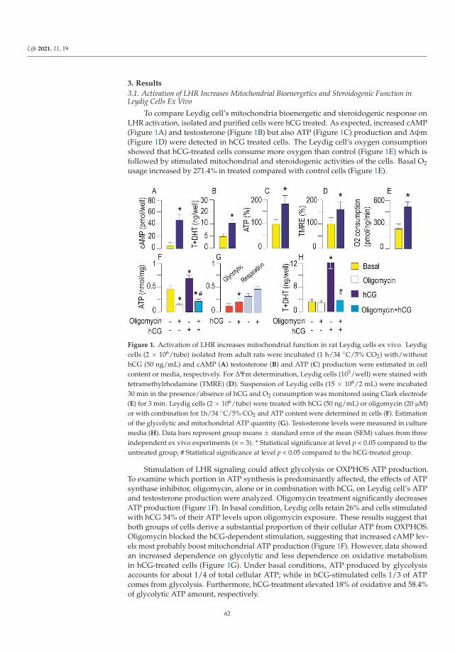
Life 2021, 11, 19
3. Results
3.1. Activation of LHR Increases Mitochondrial Bioenergetics and Steroidogenic Function inLeydig Cells Ex Vivo
To compare Leydig cell’s mitochondria bioenergetic and steroidogenic response onLHR activation, isolated and purified cells were hCG treated. As expected, increased cAMP(Figure 1A) and testosterone (Figure 1B) but also ATP (Figure 1C) production and Δψm(Figure 1D) were detected in hCG treated cells. The Leydig cell’s oxygen consumptionshowed that hCG-treated cells consume more oxygen than control (Figure 1E) which isfollowed by stimulated mitochondrial and steroidogenic activities of the cells. Basal O2usage increased by 271.4% in treated compared with control cells (Figure 1E).
Figure 1. Activation of LHR increases mitochondrial function in rat Leydig cells ex vivo. Leydigcells (2 × 10⁶/tube) isolated from adult rats were incubated (1 h/34 ◦C/5% CO2) with/withouthCG (50 ng/mL) and cAMP (A) testosterone (B) and ATP (C) production were estimated in cellcontent or media, respectively. For ΔΨm determination, Leydig cells (105/well) were stained withtetramethylrhodamine (TMRE) (D). Suspension of Leydig cells (15 × 106/2 mL) were incubated30 min in the presence/absence of hCG and O2 consumption was monitored using Clark electrode(E) for 3 min. Leydig cells (2 × 10⁶/tube) were treated with hCG (50 ng/mL) or oligomycin (20 μM)or with combination for 1h/34 ◦C/5% CO2 and ATP content were determined in cells (F). Estimationof the glycolytic and mitochondrial ATP quantity (G). Testosterone levels were measured in culturemedia (H). Data bars represent group means ± standard error of the mean (SEM) values from threeindependent ex vivo experiments (n = 3). * Statistical significance at level p < 0.05 compared to theuntreated group; # Statistical significance at level p < 0.05 compared to the hCG-treated group.
Stimulation of LHR signaling could affect glycolysis or OXPHOS ATP production.To examine which portion in ATP synthesis is predominantly affected, the effects of ATPsynthase inhibitor, oligomycin, alone or in combination with hCG, on Leydig cell’s ATPand testosterone production were analyzed. Oligomycin treatment significantly decreasesATP production (Figure 1F). In basal condition, Leydig cells retain 26% and cells stimulatedwith hCG 34% of their ATP levels upon oligomycin exposure. These results suggest thatboth groups of cells derive a substantial proportion of their cellular ATP from OXPHOS.Oligomycin blocked the hCG-dependent stimulation, suggesting that increased cAMP lev-els most probably boost mitochondrial ATP production (Figure 1F). However, data showedan increased dependence on glycolytic and less dependence on oxidative metabolismin hCG-treated cells (Figure 1G). Under basal conditions, ATP produced by glycolysisaccounts for about 1/4 of total cellular ATP; while in hCG-stimulated cells 1/3 of ATPcomes from glycolysis. Furthermore, hCG-treatment elevated 18% of oxidative and 58.4%of glycolytic ATP amount, respectively.
62

Life 2021, 11, 19
However, oligomycin did not affect basal but completely blocked hCG-dependenttestosterone stimulation, indicating that mitochondrial respiration is important for hCG-stimulated testosterone production (Figure 1H).
3.2. Activation of LHR Changed the Expression of Genes Important for Mitochondrial Function inRat Leydig Cells Ex Vivo
Considering the mitochondrial function and dynamics as interdependent processes theexpression of genes that control biogenesis, mitofission, mitofusion, and mitophagy weremonitored in conditions of excessive production of cAMP and stimulated steroidogenesis.Consequently, to determine if altered mitochondrial activity, due to LHR stimulation, isaccompanied by early or delayed changes in expression of genes crucial for mitochondrialdynamics, primary cell culture was established. Cells were treated with hCG for 30 min(hCG stimulation window; time 0 is when stimulation started), and gene expression weremonitored every 30 min for 6 h. The effectiveness of the hCG treatment was determinedby measuring intracellular cAMP concentration and expression of Star and Cyp11a1, asteroidogenesis-related gene known to be stimulated by hCG [30]. As expected, an in-creased level of cAMP (Figure 2A) followed by increased expression of Star and Cyp11a1(Figure 2D) was detected. Treatment also initiated the rise of Δψm (Figure 2B) and ATPproduction (Figure 2C). All the monitored values approached the basal values in the sixthhour. Furthermore, markers of mitogenesis (Ppargc1a and Cytc) increased early after hCGstimulation and were apparent for long times (Figure 2E). However, its downstream genesNrf1 and Tfam (Figure 2E) were reduced; Tfam expression decreased from the fourth hourindicating different regulation, probably indirect regulation. Mitofusion regulators (Mfn1/2,Opa1) were not disturbed by the treatment. Transcription of profisson Drp1 decreased veryearly, i.e., it was detected in the thirtieth minute of hCG-stimulation and approached thebasal values at the sixth hour (Figure 2E). Gene regulators of mitophagy/autophagy (Pink1,Prkn, Tfeb) decreased from the fourth hour onwards in hCG-stimulated cells (Figure 2E). Inthe sixth hour of stimulation, Ucp2, decreased (Figure 2E). The stimulatory effect of hCGon mitobiogenesis was supported by improved expression of COX4 and CYTC in Leydigcells (Figure 2F).
To confirm PRKA-dependent effect on mitochondrial biogenesis and dynamics, the pri-mary Leydig cell culture was incubated with gonadotrophin w/wo PRKA inhibitor (CMI)and mitochondrial mass together with expression of genes important for mitochondrialdynamics regulation were measured (Figure 3). Validation of inhibitor action was done bymeasuring testosterone production (Figure 3A), ΔΨm (Figure 3B) and Star transcription(Figure 3D). Results confirmed PRKA-dependent increase of mitochondrial mass followinghCG stimulation (Figure 3C). Additionally, the involvement of a PRKA-dependent regula-tion of mitochondrial biogenesis was confirmed by a reduction of hCG-stimulated Ppargc1aexpression in the presence of a PRKA-inhibitor (Figure 3D). PRKA inhibition was withouteffect on Mfn1, Mfn2 and Opa1 mRNA level (Figure 3D). However, the inhibitor preventedhCG-triggered increase in transcription of Fis1 but also hCG-evoked decrease in Drp1transcription (Figure 3D) suggesting cAMP-PRKA dependent regulation of mitofission.
63

Life 2021, 11, 19
Figure 2. Activation of LHR changed expression of genes important for mitochondrial function inrat Leydig cells ex vivo. Leydig cells from adult rats were isolated and primary culture was formed.The cells were stimulated with hCG (100 ng/mL) for 30 min, media changed and cell incubation wascontinued; cells were collected every 30 min for 6 h; moment when stimulation started was referredto as 0h. Cells were collected in indicated time points and used for measurement of intracellularconcentration of cAMP (A), ΔΨm (B) and ATP (C). RNA was isolated and quantitative real-timepolymerase chain reaction (qRT-PCR) was performed to detect expression level of steroidogenic (D)or genes important for mitochondrial function/dynamics (E). Abundances of CYTC and COX4 inLeydig cells stimulated w/wo hCG for 2 h were evaluated by Western blot (F). Representative blotsare shown. The bars near the respective bands show values obtained by scanning densitometryand normalized on the ACTN. Data points are group mean ± SEM values of three independentexperiments, n = 3. * Statistical significance between the groups for the same time point (p < 0.05).
64
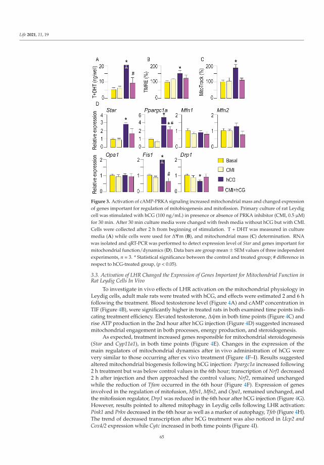
Life 2021, 11, 19
Figure 3. Activation of cAMP-PRKA signaling increased mitochondrial mass and changed expressionof genes important for regulation of mitobiogenesis and mitofission. Primary culture of rat Leydigcell was stimulated with hCG (100 ng/mL) in presence or absence of PRKA inhibitor (CMI, 0.5 μM)for 30 min. After 30 min culture media were changed with fresh media without hCG but with CMI.Cells were collected after 2 h from beginning of stimulation. T + DHT was measured in culturemedia (A) while cells were used for ΔΨm (B), and mitochondrial mass (C) determination. RNAwas isolated and qRT-PCR was performed to detect expression level of Star and genes important formitochondrial function/dynamics (D). Data bars are group mean ± SEM values of three independentexperiments, n = 3. * Statistical significance between the control and treated group; # difference inrespect to hCG-treated group, (p < 0.05).
3.3. Activation of LHR Changed the Expression of Genes Important for Mitochondrial Function inRat Leydig Cells In Vivo
To investigate in vivo effects of LHR activation on the mitochondrial physiology inLeydig cells, adult male rats were treated with hCG, and effects were estimated 2 and 6 hfollowing the treatment. Blood testosterone level (Figure 4A) and cAMP concentration inTIF (Figure 4B), were significantly higher in treated rats in both examined time points indi-cating treatment efficiency. Elevated testosterone, Δψm in both time points (Figure 4C) andrise ATP production in the 2nd hour after hCG injection (Figure 4D) suggested increasedmitochondrial engagement in both processes, energy production, and steroidogenesis.
As expected, treatment increased genes responsible for mitochondrial steroidogenesis(Star and Cyp11a1), in both time points (Figure 4E). Changes in the expression of themain regulators of mitochondrial dynamics after in vivo administration of hCG werevery similar to those occurring after ex vivo treatment (Figure 4F–I). Results suggestedaltered mitochondrial biogenesis following hCG injection: Ppargc1a increased following2 h treatment but was below control values in the 6th hour; transcription of Nrf1 decreased2 h after injection and then approached the control values; Nrf2, remained unchangedwhile the reduction of Tfam occurred in the 6th hour (Figure 4F). Expression of genesinvolved in the regulation of mitofusion, Mfn1, Mfn2, and Opa1, remained unchanged, andthe mitofission regulator, Drp1 was reduced in the 6th hour after hCG injection (Figure 4G).However, results pointed to altered mitophagy in Leydig cells following LHR activation:Pink1 and Prkn decreased in the 6th hour as well as a marker of autophagy, Tfeb (Figure 4H).The trend of decreased transcription after hCG treatment was also noticed in Ucp2 andCox4/2 expression while Cytc increased in both time points (Figure 4I).
65

Life 2021, 11, 19
Figure 4. Activation of LHR changed expression of genes important for mitochondrial function in ratLeydig cells in vivo. Rats were sacrificed 2 h and 6 h after hCG/vehicle application. Serum and TIFwere collected for measurement of testosterone (A) and cAMP (B), respectively. Leydig cells wereisolated and used for ΔΨm (C) and ATP (D) determination. RNA was isolated from Leydig cells andused in qRT-PCR analysis in order to examine expression level of genes that regulate mitochondrialfunction (E–I). Data points/bars are group mean ± SEM values of three independent experiments,n = 3. * Statistical significance between the groups for the same time point (p < 0.05).
Increased mitochondrial biogenesis in Leydig cells following gonadotropin injectionwas analyzed (Figure 5A). The Mitotracker-green dye has high affinity and specificity forlipid mitochondrial membranes and produces green fluorescence which was employedto examine mitochondrial abundance. The gonadotropin injection caused a 32% increasein mitochondrial abundance compared with control values (Figure 5A). Additionally, in-creased mitochondrial abundance due to LHR stimulation was confirmed by the estimationof mtDNA content (Figure 5B).
The ultrastructural appearances of mitochondria in Leydig cells from hCG treated andcontrol rats were assessed by transmission electron microscopy (TEM) analysis. Mitochon-dria were more frequently detected in hCG treated Leydig cells (Figure 5E,F), compared tocontrol Leydig cells (Figure 5C,D). Treated and control Leydig cells mitochondria had bothtubular and lamellar cristae, and occasionally mitochondria in gonadotropin stimulatedcells had more electron dense matrix (Figure 5E,F). Leydig cells treated with hCG had moreelectron lucent chromatin (euchromatin) (Figure 5E,F) while more regions of condensedchromatin (heterochromatin) were seen in nuclei of control cells (Figure 5C,D).
66

Life 2021, 11, 19
Figure 5. Activation of LHR increased mitochondrial mass and changed Leydig cell’s mitochondrialultrastructure. Two hours after hCG injection testes were isolated, fragments of testicular tissueimmediately fixed for transmission electron microscopy (TEM) analysis while the rest of the tissue wasused for Leydig cells isolation. Cells, individually isolated, were plated for mitochondrial abundancedetermination (105/well) using Mitotracker green (A) or for genomic DNA isolation (106/well) (B).The mtNd1/B2m ratio was used for mtDNA content determination. Ultramicrographs of Leydigcell from control rats at low (89,000×; C) and high magnification (28,000×; D) with well preservedmitochondria, and Leydig cell from hCG-treated rats at low (56,000×; E) and high magnification(28,000×; F) with numerous mitochondria with electron dense matrix. Data bar represents groupmeans ± SEM values from three rats (n = 3). * Statistical significance at level p < 0.05 compared to theuntreated group.
3.4. Mitochondrial Function Was Dampened in Leydig Cells from HypogonadotropicHypogonadal Rats
Given the high performance of mitochondria in Leydig cells after LHR stimulation,it was of interest to investigate the effects of a low level of LH and intracellular cAMPon mitochondrial physiology. To do this, the rats were treated with the long-lastingGnRH analog [32]. Such treatment in male rats causes hypogonadotropic hypogonadism,which is characterized by a low level of testosterone (Figure 6A) in circulation as well asa low level of cAMP in Leydig cells (Figure 6B). Additionally, treatment reduced Δψm(Figure 6C) and ATP production (Figure 6D). Expression of the main steroidogenic genesinvolved in steroid production in mitochondria Star and Cyp11a1 were decreased in Leydigcells from hypogonadal rats (Figure 6E). Furthermore, genes involved in the regulationof mitochondrial biogenesis Ppargc1a, Nrf1 and Nrf2 were reduced but Tfam remainedunchanged (Figure 6F). Hypogonadism has affected mitofission and mitofusion in Leydigcells by reducing transcription of Drp1 as well as Mfn1 and Mfn2 without changing Opa1(Figure 6G). Hypogonadism elevated Tfeb transcription in Leydig cells but not Pink1 andPrkn (Figure 6H). In Leydig cells from hypogonadal rats, transcription of Cox4/2 and Cytclessened but no changes were observed in Ucp2 (Figure 6I). Finally, the expression ofmitochondrial Nd1 decreased in Leydig cells from hypogonadal rats (Figure 6J).
67
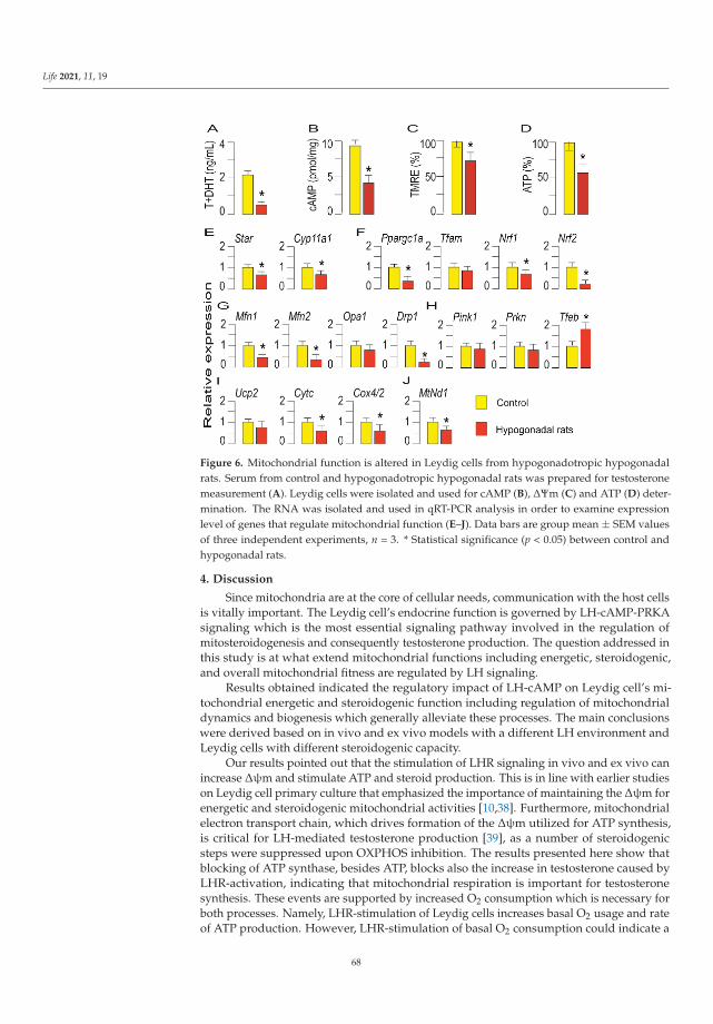
Life 2021, 11, 19
Figure 6. Mitochondrial function is altered in Leydig cells from hypogonadotropic hypogonadalrats. Serum from control and hypogonadotropic hypogonadal rats was prepared for testosteronemeasurement (A). Leydig cells were isolated and used for cAMP (B), ΔΨm (C) and ATP (D) deter-mination. The RNA was isolated and used in qRT-PCR analysis in order to examine expressionlevel of genes that regulate mitochondrial function (E–J). Data bars are group mean ± SEM valuesof three independent experiments, n = 3. * Statistical significance (p < 0.05) between control andhypogonadal rats.
4. Discussion
Since mitochondria are at the core of cellular needs, communication with the host cellsis vitally important. The Leydig cell’s endocrine function is governed by LH-cAMP-PRKAsignaling which is the most essential signaling pathway involved in the regulation ofmitosteroidogenesis and consequently testosterone production. The question addressed inthis study is at what extend mitochondrial functions including energetic, steroidogenic,and overall mitochondrial fitness are regulated by LH signaling.
Results obtained indicated the regulatory impact of LH-cAMP on Leydig cell’s mi-tochondrial energetic and steroidogenic function including regulation of mitochondrialdynamics and biogenesis which generally alleviate these processes. The main conclusionswere derived based on in vivo and ex vivo models with a different LH environment andLeydig cells with different steroidogenic capacity.
Our results pointed out that the stimulation of LHR signaling in vivo and ex vivo canincrease Δψm and stimulate ATP and steroid production. This is in line with earlier studieson Leydig cell primary culture that emphasized the importance of maintaining the Δψm forenergetic and steroidogenic mitochondrial activities [10,38]. Furthermore, mitochondrialelectron transport chain, which drives formation of the Δψm utilized for ATP synthesis,is critical for LH-mediated testosterone production [39], as a number of steroidogenicsteps were suppressed upon OXPHOS inhibition. The results presented here show thatblocking of ATP synthase, besides ATP, blocks also the increase in testosterone caused byLHR-activation, indicating that mitochondrial respiration is important for testosteronesynthesis. These events are supported by increased O2 consumption which is necessary forboth processes. Namely, LHR-stimulation of Leydig cells increases basal O2 usage and rateof ATP production. However, LHR-stimulation of basal O2 consumption could indicate a
68

Life 2021, 11, 19
leak of the respiration rate [40] which can be interpreted as increased O2 usage in expensefor steroid and ROS formation.
In LH stimulated cells, ATP is predominantly produced in OXPHOS but showed anincreased dependence on glycolysis and less dependence on oxidative metabolism. The ATPproduced in glycolysis increased by 58% while ATP produced in mitochondrial respirationincreased by 18% in gonadotropin-stimulated cells. The important metabolic coupling couldbe present. It is well documented that the cAMP-PRKA pathway plays a regulatory role inthe glycolytic metabolism, exerting a stimulatory effect on glucose transport and utilizationvia phosphorylation of different downstream target proteins [41,42]. Increasing glycolyticactivity in the cytosol could also lead to an adequate increase in NADH which are readilytransferred to the matrix of mitochondria where they can be involved in the OXPHOSprocess [43].
However, in LH stimulated cells a mitosteroidogenesis and energetics do not appearto be parallel processes. Our results showed the stimulatory effect of LH on mitochondrialsteroidogenesis lasts longer than the effect on ATP production. This may be the result of LH-cAMP-PRKA activation of genes important for mitosteroidogenesis (Star, Cyp11a1). Resultsfrom the hypogonadal model illustrate the described relationship among energetic andsteroidogenic mitochondrial parameters but on a reduced scale i.e., low cAMP, followed bydecreased Δψm, ATP, testosterone, and Star and Cyp11a1 transcription.
Effective energetic, steroidogenic, and other mitochondrial functions are essentiallydependent on a proper mitochondrial dynamic network regulated by mitochondrial fu-sion and fission [29,44]. Mitofusion represents a limiting step in the onset of processesrequired for the transport of intermediate products in/out mitochondria and is essentialfor cholesterol import into mitochondria and steroid formation [11,24]. Nonetheless, inLeydig cells with LH-activated steroidogenesis the expression of Mfn1, Mfn2, and Opa1,were unchanged suggesting regulatory action on the posttranslational level of these geneproducts. Under the same conditions, the decreased transcription of profission Drp1 wasobserved. Since mitofusion is a prerequisite for steroid hormone synthesis in Leydigcells [11], reduced Drp1 expression could facilitate mitochondrial fusion that might beimportant for the formation of the multiprotein complex that delivers cholesterol to theCYP11 system in addition to stimulation of oxidative phosphorylation. Indeed, blockingof DRP1 activity results in increased mitofusion in Leydig cells [25]. Moreover, PRKAphosphorylation of DRP1 inhibits mitofission enabling an optimal environment for steroidsbiosynthesis [25]. Results obtained by ex vivo PRKA inhibition prevented hCG-triggereddecrease of Drp1 transcription supporting PRKA-regulation of DRP1 expression.
It has been shown that inhibiting fission or promoting fusion decreases mitophagy,whereas enhanced fission precedes and facilitates it [45]. In LH-stimulated Leydig cellsthe expression of genes involved in quality control, mitophagy Pink1, Prkn and autophagyTfeb were decreased. Accordingly, the activation of the cAMP-PRKA pathway is associatedwith an inhibitory effect on mitophagy [29]. By contrast, the increased transcription ofmitobiogenesis marker, Ppargc1a in addition to Cytc as well as COX4 indicates LH-potentialfor new mitochondria formation. This is in agreement with observed increased PGC1ain Leydig cells cultivated with hCG for long [33]. Indeed, results obtained by Mitotrackclearly indicate LHR-regulated grow in mitochondrial mass. Moreover, TEM analysisrevealed more abundant mitochondria in Leydig cells obtained from hCG-treated rats.Gonadotropin treatment of Leydig cells stimulates expression of CYTC and COX4 sub-units of respiratory proteins which again points to increased biogenesis. However, thetranscriptional pattern of Cox4/2 over time, after in vivo hCG-treatment, differs from theprotein pattern, indicating possible paracrine regulation as well as the existence of a post-transcriptional/posttranslational modulatory mechanism. It is known that genes thatcontribute to the same respiratory protein complex are from both mtDNA and nuclearDNA [46]. Thus, mitochondria and the nucleus finely coordinate the transcription, trans-lation, and import of mitochondrial proteins to ensure the proper relationship betweenthe various OXPHOS components. This communication occurs bidirectionally in time
69

Life 2021, 11, 19
dependent manner indicating very sophisticated regulation of many signaling pathways.The nucleus can influence mitochondrial structure, replication, biogenesis, fission, andfusion, the number of mitochondria, and the mitophagy turnover rate [47]. Nonetheless,the regulation of respiratory proteins expression and different behavior of genes encod-ing the subunits together with proteins involved in mitochondrial dynamics includinganterograde and retrograde signaling in steroidogenic cells need to be further investigated.Results from the study on the hypogonadal model show that decreased steroidogenesisin Leydig cells is enabled by mitochondrial dysfunction. This is connected to decreasedmitochondrial engagement in steroidogenesis due to decreased expression of Star andCyp11a1, thereby decreased cholesterol import and convert into biologically active steroidprecursors; decreased aerobic energy production by mitochondria resulting in loweredATP levels; decreased mitochondrial biogenesis illustrated by decreased expression of themain marker of mitochondrial biogenesis (Ppargc1); amended mitochondrial dynamics i.e.,mitofusion due to decreased Mfn1 and Mfn2 as well as mitofission due to decreased Drp1expression with potential to increased autophagy as result of increased Tfeb.
Taken together, the results from all experimental models in this study revealed the de-pendence of mitochondrial physiology on LH-cAMP signaling in Leydig cells (Figure 7).Specifically, the expression level of genes that indicate the intensity of mitochondrial bio-genesis (Ppargc1a and Cytc; Figure 7C), as well as genes responsible for mitosteroidogenesis(Star, Cyp11a1; Figure 7D), are positively correlated with the level of cAMP in the cell.On the contrary expression of genes involved in the regulation of mitophagy (Prkn) andautophagy (Tfeb) are in negative correlation with cAMP level (Figure 7A).
Figure 7. Relationships between the concentration of cAMP and expression of genes responsiblefor mitochondrial physiology in Leydig cells. Data shown are pool from 3 different experimentalmodels (ex vivo and in vivo stimulation of LHR as well as experimental model for hypogonadotropichypogonadism). Data for several groups of genes involved in regulation of mitophagy (A), mitofusion(B), mitochondrial biogenesis (C) and mitochondrial steroidogenesis (D), covaried with cAMPconcentration. Ribbons represent 95% confidence intervals.
Results support the conclusion that the LH-cAMP pathway is involved in the controlof mitochondrial biogenesis and dynamics coupled with mitosteroidogenesis and energeticfunction; therefore, it significantly influences mitochondrial fitness in Leydig cells.
70

Life 2021, 11, 19
Supplementary Materials: The following are available online at https://www.mdpi.com/2075-1729/11/1/19/s1, Table S1.
Author Contributions: M.L.J.M.—acquisition of the data; analysis and interpretation of the data;final approval of the version to be submitted; D.Z.M.—acquisition of the data; analysis and draftingthe manuscript; revising manuscript critically for important intellectual content; final approval ofthe version to be submitted; A.P.B.—acquisition of the data; analysis and interpretation of the data;revising manuscript critically for important intellectual content; final approval of the version tobe submitted; Z.K.—analyses of O2 consumption; analysis and interpretation of the data; revisingmanuscript critically for important intellectual content; final approval of the version to be submitted;I.M.S.—acquisition of the data; final approval of the version to be submitted; T.K.-S.—TEM analysis;final approval of the version to be submitted; S.A.A.—acquisition of the data; analysis and interpreta-tion of the data; revising manuscript critically for important intellectual content; final approval of theversion to be submitted; T.S.K.—the conception and design of the research; acquisition of the data;analysis and interpretation of the data; drafting the manuscript; revising manuscript critically forimportant intellectual content; final approval of the version to be submitted. All authors—approvedthe final version of the manuscript; agree to be accountable for all aspects of the work in ensuring thatquestions related to the accuracy or integrity of any part of the work are appropriately investigatedand resolved; qualify for authorship, and all those who qualify for authorship are listed.
Funding: This research was supported by the Serbian Ministry of Education and TechnologicalDevelopment grant no. 173057 and CeRes grant, and the Autonomic Province of Vojvodina grants no.3822 and no. 2130.
Institutional Review Board Statement: The study was conducted according to the guidelines of theDeclaration of Helsinki, and approved by the Ethical Committee on Animal Care and Use of theUniversity of Novi Sad (statement no. 01-201/3).
Informed Consent Statement: Not applicable.
Data Availability Statement: All relevant data are available from the corresponding author onrequest. Further information and requests for data, resources and reagents should be directed to andwill be fulfilled by the corresponding author, Tatjana Kostic ([email protected]).
Acknowledgments: We are very grateful to Gordon Niswender (Colorado State University) forsupplying antibodies for radioimmunoassay analysis. Also we thank Aleksandar Baburski andSrdjan Sokanovic for technical assistance in conducting experiments.
Conflicts of Interest: The authors declare no conflict of interest.
References
1. Cooper, D.M.F. Regulation and organization of adenylyl cyclases and cAMP. Biochem. J. 2003, 375, 517–529. [CrossRef] [PubMed]2. Dufau, M.L. The luteinizing hormone receptor. Annu. Rev. Physiol. 1998, 60, 461–496. [CrossRef]3. Stocco, D.M.; Wang, X.; Jo, Y.; Manna, P.R. Multiple Signaling Pathways Regulating Steroidogenesis and Steroidogenic Acute
Regulatory Protein Expression: More Complicated than We Thought. Mol. Endocrinol. 2005, 19, 2647–2659. [CrossRef] [PubMed]4. Payne, A.H.; Hales, D.B. Overview of Steroidogenic Enzymes in the Pathway from Cholesterol to Active Steroid Hormones.
Endocr. Rev. 2004, 25, 947–970. [CrossRef] [PubMed]5. Miller, W.L. Steroid hormone synthesis in mitochondria. Mol. Cell. Endocrinol. 2013, 379, 62–73. [CrossRef]6. Midzak, A.; Papadopoulos, V. Adrenal Mitochondria and Steroidogenesis: From Individual Proteins to Functional Protein
Assemblies. Front. Endocrinol. 2016, 7, 106. [CrossRef]7. Heiden, M.G.V.; Cantley, L.C.; Thompson, C.B. Understanding the Warburg Effect: The Metabolic Requirements of Cell
Proliferation. Science 2009, 324, 1029–1033. [CrossRef]8. Zheng, J. Energy metabolism of cancer: Glycolysis versus oxidative phosphorylation (Review). Oncol. Lett. 2012, 4, 1151–1157.
[CrossRef]9. Andric, S.A.; Janjic, M.M.; Stojkov, N.J.; Kostic, T.S. Protein kinase G-mediated stimulation of basal Leydig cell steroidogenesis.
Am. J. Physiol. Metab. 2007, 293, E1399–E1408. [CrossRef]10. Midzak, A.S.; Chen, H.; Aon, M.A.; Papadopoulos, V.; Zirkin, B.R. ATP Synthesis, Mitochondrial Function, and Steroid
Biosynthesis in Rodent Primary and Tumor Leydig Cells. Biol. Reprod. 2011, 84, 976–985. [CrossRef]11. Duarte, A.; Poderoso, C.; Cooke, M.; Soria, G.; Maciel, F.C.; Gottifredi, V.; Podesta, E.J. Mitochondrial Fusion Is Essential for
Steroid Biosynthesis. PLoS ONE 2012, 7, e45829. [CrossRef] [PubMed]12. Meyer, J.N.; Leuthner, T.C.; Luz, A.L. Mitochondrial fusion, fission, and mitochondrial toxicity. Toxicology 2017, 391, 42–53.
[CrossRef] [PubMed]
71

Life 2021, 11, 19
13. Westermann, B. Bioenergetic role of mitochondrial fusion and fission. Biochim. Biophys. Acta Bioenerg. 2012, 1817, 1833–1838.[CrossRef] [PubMed]
14. Friedman, J.R.; Nunnari, J. Mitochondrial form and function. Nature 2014, 505, 335–343. [CrossRef]15. Dominy, J.E.; Puigserver, P. Mitochondrial Biogenesis through Activation of Nuclear Signaling Proteins. Cold Spring Harb. Perspect.
Biol. 2013, 5, a015008. [CrossRef]16. Li, P.A.; Hou, X.; Hao, S. Mitochondrial biogenesis in neurodegeneration. J. Neurosci. Res. 2017, 95, 2025–2029. [CrossRef]17. Palikaras, K.; Lionaki, E.; Tavernarakis, N. Mechanisms of mitophagy in cellular homeostasis, physiology and pathology. Nat.
Cell Biol. 2018, 20, 1013–1022. [CrossRef]18. Schmidt, O.; Harbauer, A.B.; Rao, S.; Eyrich, B.; Zahedi, R.P.; Stojanovski, D.; Schönfisch, B.; Guiard, B.; Sickmann, A.; Pfanner, N.;
et al. Regulation of Mitochondrial Protein Import by Cytosolic Kinases. Cell 2011, 144, 227–239. [CrossRef]19. Opalinska, M.; Meisinger, C. Metabolic control via the mitochondrial protein import machinery. Curr. Opin. Cell Biol. 2015, 33,
42–48. [CrossRef]20. Moujalled, D.; Weston, R.; Anderton, H.; Ninnis, R.; Goel, P.; Coley, A.; Huang, D.C.S.; Wu, L.; Strasser, A.; Puthalakath, H.
Cyclic-AMP-dependent protein kinase A regulates apoptosis by stabilizing the BH3-only protein Bim. EMBO Rep. 2011, 12, 77–83.[CrossRef]
21. Gomes, L.C.; Di Benedetto, G.; Scorrano, L. During autophagy mitochondria elongate, are spared from degradation and sustaincell viability. Nat. Cell Biol. 2011, 13, 589–598. [CrossRef] [PubMed]
22. Akabane, S.; Uno, M.; Tani, N.; Shimazaki, S.; Ebara, N.; Kato, H.; Kosako, H.; Oka, T. PKA Regulates PINK1 Stability and ParkinRecruitment to Damaged Mitochondria through Phosphorylation of MIC60. Mol. Cell 2016, 62, 371–384. [CrossRef] [PubMed]
23. Lefkimmiatis, K. cAMP signalling meets mitochondrial compartments. Biochem. Soc. Trans. 2014, 42, 265–269. [CrossRef][PubMed]
24. Duarte, A.; Castillo, F.; Podesta, E.J.; Poderoso, C. Mitochondrial Fusion and ERK Activity Regulate Steroidogenic AcuteRegulatory Protein Localization in Mitochondria. PLoS ONE 2014, 9, e100387. [CrossRef] [PubMed]
25. Park, J.-E.; Kim, Y.-J.; Lee, S.G.; Kim, J.Y.; Chung, J.-Y.; Jeong, S.-Y.; Koh, H.; Yun, J.; Park, H.T.; Yoo, Y.H.; et al. Drp1Phosphorylation Is Indispensable for Steroidogenesis in Leydig Cells. Endocrinology 2019, 160, 729–743. [CrossRef] [PubMed]
26. De Rasmo, D.; Signorile, A.; Santeramo, A.; Larizza, M.; Lattanzio, P.; Capitanio, G.; Papa, S. Intramitochondrial adenylyl cyclasecontrols the turnover of nuclear-encoded subunits and activity of mammalian complex I of the respiratory chain. Biochim. Biophys.Acta Bioenerg. 2015, 1853, 183–191. [CrossRef]
27. De Rasmo, D.; Micelli, L.; Santeramo, A.; Signorile, A.; Lattanzio, P.; Papa, S. cAMP regulates the functional activity, couplingefficiency and structural organization of mammalian F O F 1 ATP synthase. Biochim. Biophys. Acta Gen. Subj. 2016, 1857, 350–358.[CrossRef]
28. Acin-Perez, R.; Salazar, E.; Kamenetsky, M.; Buck, J.; Levin, L.R.; Manfredi, G. Cyclic AMP Produced inside MitochondriaRegulates Oxidative Phosphorylation. Cell Metab. 2009, 9, 265–276. [CrossRef]
29. Di Benedetto, G.; Gerbino, A.; Lefkimmiatis, K. Shaping mitochondrial dynamics: The role of cAMP signalling. Biochem. Biophys.Res. Commun. 2018, 500, 65–74. [CrossRef]
30. Di Benedetto, G.; Scalzotto, E.; Mongillo, M.; Pozzan, T. Mitochondrial Ca2+ Uptake Induces Cyclic AMP Generation in the Matrixand Modulates Organelle ATP Levels. Cell Metab. 2013, 17, 965–975. [CrossRef]
31. Andric, S.A.; Kojic, Z.; Bjelic, M.M.; Mihajlovic, A.I.; Baburski, A.Z.; Sokanovic, S.J.; Janjic, M.M.; Stojkov, N.J.; Stojilkovic, S.S.;Kostic, T.S. The opposite roles of glucocorticoid and α1-adrenergic receptors in stress triggered apoptosis of rat Leydig cells. Am.J. Physiol. Metab. 2013, 304, E51–E59. [CrossRef] [PubMed]
32. Baburski, A.Z.; Andric, S.A.; Kostic, T.S. Luteinizing hormone signaling is involved in synchronization of Leydig cell’s clock andis crucial for rhythm robustness of testosterone production. Biol. Reprod. 2019, 100, 1406–1415. [CrossRef] [PubMed]
33. Gak, I.A.; Radovic, S.M.; Dukic, A.R.; Janjic, M.M.; Stojkov-Mimic, N.J.; Kostic, T.S.; Andric, S.A. Stress triggers mitochondrialbiogenesis to preserve steroidogenesis in Leydig cells. Biochim. Biophys. Acta Bioenerg. 2015, 1853, 2217–2227. [CrossRef] [PubMed]
34. Payne, A.H.; Downing, J.R.; Wong, K.-L. Luteinizing Hormone Receptors and Testosterone Synthesis in Two Distinct Populationsof Ley dig Cells. Endocrinology 1980, 106, 1424–1429. [CrossRef]
35. Kostic, T.S.; Andric, S.A.; Stojilkovic, S.S. Receptor-Controlled Phosphorylation of α1 Soluble Guanylyl Cyclase EnhancesNitric Oxide-Dependent Cyclic Guanosine 5′-Monophosphate Production in Pituitary Cells. Mol. Endocrinol. 2004, 18, 458–470.[CrossRef]
36. Sokanovic, S.J.; Baburski, A.Z.; Kojic, Z.; Medar, M.L.J.; Andric, S.A.; Kostic, T.S. Aging-Related Increase of cGMP DisruptsMitochondrial Homeostasis in Leydig Cells. J. Gerontol. Ser. A Biol. Sci. Med. Sci. 2020. [CrossRef]
37. Rato, L.; Duarte, A.I.; Tomás, G.D.; Santos, M.S.; Moreira, P.I.; Socorro, S.; Cavaco, J.E.; Alves, M.G.; Oliveira, P.F. Pre-diabetesalters testicular PGC1-α/SIRT3 axis modulating mitochondrial bioenergetics and oxidative stress. Biochim. Biophys. Acta Bioenerg.2014, 1837, 335–344. [CrossRef]
38. Allen, J.A.; Shankara, T.; Janus, P.; Buck, S.; Diemer, T.; Hales, K.H.; Hales, D.B. Energized, Polarized, and Actively RespiringMitochondria Are Required for Acute Leydig Cell Steroidogenesis. Endocrinology 2006, 147, 3924–3935. [CrossRef]
39. Midzak, A.S.; Chen, H.; Papadopoulos, V.; Zirkin, B.R. Leydig cell aging and the mechanisms of reduced testosterone synthesis.Mol. Cell. Endocrinol. 2009, 299, 23–31. [CrossRef]
72
Life 2021, 11, 19
40. Salin, K.; Villasevil, E.M.; Anderson, G.J.; Selman, C.; Chinopoulos, C.; Metcalfe, N.B. The RCR and ATP/O Indices Can GiveContradictory Messages about Mitochondrial Efficiency. Integr. Comp. Biol. 2018, 58, 486–494. [CrossRef]
41. Palorini, R.; De Rasmo, D.; Gaviraghi, M.; Danna, L.S.; Signorile, A.; Cirulli, C.; Chiaradonna, F.; Alberghina, L.; Papa, S.Oncogenic K-ras expression is associated with derangement of the cAMP/PKA pathway and forskolin-reversible alterations ofmitochondrial dynamics and respiration. Oncogene 2012, 32, 352–362. [CrossRef] [PubMed]
42. Yu, T.; Yang, G.; Hou, Y.; Tang, X.; Wu, C.; Wu, X.-A.; Guo, L.; Zhu, Q.; Luo, H.; Du, Y.-E.; et al. Cytoplasmic GPER translocationin cancer-associated fibroblasts mediates cAMP/PKA/CREB/glycolytic axis to confer tumor cells with multidrug resistance.Oncogene 2017, 36, 2131–2145. [CrossRef] [PubMed]
43. Glancy, B.; Kane, D.A.; Kavazis, A.N.; Goodwin, M.L.; Willis, W.T.; Gladden, L.B. Mitochondrial lactate metabolism: History andimplications for exercise and disease. J. Physiol. 2020. [CrossRef] [PubMed]
44. Chen, H.; Chan, D.C. Emerging functions of mammalian mitochondrial fusion and fission. Hum. Mol. Genet. 2005, 14, R283–R289.[CrossRef]
45. Twig, G.; Elorza, A.; Molina, A.A.J.; Mohamed, H.; Wikstrom, J.D.; Walzer, G.; Stiles, L.; Haigh, S.E.; Katz, S.; Las, G.; et al. Fissionand selective fusion govern mitochondrial segregation and elimination by autophagy. EMBO J. 2008, 27, 433–446. [CrossRef][PubMed]
46. Ben-Shachar, D. The interplay between mitochondrial complex I, dopamine and Sp1 in schizophrenia. J. Neural Transm. 2009, 116,1383–1396. [CrossRef]
47. Wallace, D.C. The epigenome and the mitochondrion: Bioenergetics and the environment. Genes Dev. 2010, 24, 1571–1573.[CrossRef]
73


life
Review
Natural and Artificial Mechanisms of MitochondrialGenome Elimination
Elvira G. Zakirova 1, Vladimir V. Muzyka 1,2, Ilya O. Mazunin 3 and Konstantin E. Orishchenko 1,2,*
���������������
Citation: Zakirova, E.G.; Muzyka,
V.V.; Mazunin, I.O.; Orishchenko, K.E.
Natural and Artificial Mechanisms of
Mitochondrial Genome Elimination.
Life 2021, 11, 76. https://doi.org/
10.3390/life11020076
Academic Editor: Francesco Bruni
Received: 18 December 2020
Accepted: 19 January 2021
Published: 20 January 2021
Publisher’s Note: MDPI stays neutral
with regard to jurisdictional claims in
published maps and institutional affil-
iations.
Copyright: © 2021 by the authors.
Licensee MDPI, Basel, Switzerland.
This article is an open access article
distributed under the terms and
conditions of the Creative Commons
Attribution (CC BY) license (https://
creativecommons.org/licenses/by/
4.0/).
1 Federal Research Center Institute of Cytology and Genetics, Siberian Branch of the Russian Academyof Sciences, 630090 Novosibirsk, Russia; [email protected] (E.G.Z.); [email protected] (V.V.M.)
2 Department of Genetic Technologies, Novosibirsk State University, 630090 Novosibirsk, Russia3 Skolkovo Institute of Science and Technology, 143026 Skolkovo, Russia; [email protected]* Correspondence: [email protected]
Abstract: The generally accepted theory of the genetic drift of mitochondrial alleles during mam-malian ontogenesis is based on the presence of a selective bottleneck in the female germline. However,there is a variety of different theories on the pathways of genetic regulation of mitochondrial DNA(mtDNA) dynamics in oogenesis and adult somatic cells. The current review summarizes presentknowledge on the natural mechanisms of mitochondrial genome elimination during mammaliandevelopment. We also discuss the variety of existing and developing methodologies for artificialmanipulation of the mtDNA heteroplasmy level. Understanding of the basics of mtDNA dynamicswill shed the light on the pathogenesis and potential therapies of human diseases associated withmitochondrial dysfunction.
Keywords: mitochondrial DNA segregation; heteroplasmy; selective elimination; mitophagy;mitochondrial engineered nucleases
1. Introduction
Ten years after the discovery of the DNA double helix, Magrit and Sylvan Nass foundthat mitochondria harbor their own double-stranded DNA (mitochondrial DNA, mtDNA),whose structure differs from the nuclear DNA [1]. Currently, it is established that humancircular mtDNA of 16,659 bp in length encodes 37 genes, which are essential for oxidativephosphorylation (OXPHOS) and stable energy production by the cell [2,3].
Highly compact mtDNA possesses many features distinct from the nuclear genome [4],some of which lead to the unique genetic drift of mtDNA. The transmission of the mitochon-drial genome is strictly maternal. Uniparental inheritance was first shown in rats [5] andmice [6] and then confirmed in humans [7]. Although several studies debate the biparentalmtDNA inheritance [8–11], such cases seem to be very rare, and their mechanisms remainlargely unknown. Secondly, the mitochondrial genome exists in multiple copies. One so-matic cell contains around 103–104 molecules of mtDNA [12,13]. Most frequently, a cell hasonly one type of mitochondrial genome. This condition—homoplasmy—usually occurswhen all alleles of mtDNA are clonally expanded. However, considering the increasedmutation rate of mtDNA comparing to the nuclear DNA, defective copies of mtDNA couldcoexist with wild-type alleles in a cell [14–17]. This phenomenon referred as heteroplasmyis specific to mitochondrial genome.
Different cells in a multicellular organism might have diverse mtDNA species. More-over, the level of heteroplasmy may vary between the cells of the same tissue or organ,between the organs in one individual, and between individuals in a single family [18].Such heterogeneity may occur at different developmental stages and proceed in an unpre-dictable direction. Defining the causes, dynamics, and driving forces of heteroplasmy willprovide an insight on the inheritance and the progression of many mitochondrial diseases,the prevalence of which is approximately 1:4300 in the human population [19].
Life 2021, 11, 76. https://doi.org/10.3390/life11020076 https://www.mdpi.com/journal/life
75

Life 2021, 11, 76
Here, we review several mechanisms of heteroplasmy level regulation and mtDNAsegregation in somatic and germ cells, including the natural processes of mtDNA elimina-tion. Additionally, we discuss how cells control the diversity of mtDNA species. Addition-ally, we review the state-of-the-art approaches (including those under active development)for the artificial modulation of mtDNA heteroplasmy. Finally, we provide specific examplesof how these mechanisms can be used for uncovering the pathogenesis and rising noveltherapies of mitochondrial dysfunction-associated diseases.
2. Mitochondrial Genome Heteroplasmy
It is believed that due to the proximity of the respiratory chain complexes, the absenceof histones on mtDNA, and the lack of effective mechanisms of mtDNA repair, the mito-chondrial genome is extremely vulnerable to the effects of reactive oxygen species (ROS).However, it is known that mitochondria possess several DNA repair mechanisms presentin the nucleus. Most notably, there are base excision repair (BER) and microhomology-mediated end joining (MMEJ). The possibility of the presence of nucleotide excision re-pair (NER), mismatch repair (MMR), homologous recombination (HR,) and classical non-homologous end joining (NHEJ) is still under extensive debate [20]. In addition to that,even a rescue mechanism for the replication fork has been demonstrated in mitochon-dria [21]. However, the overall number of effectively proceeding DNA repair mechanismsin mitochondria is somewhat smaller than in the nucleus [20]. Moreover, although it isnow known that mtDNA is not naked, the nucleoid structures formed to protect mtDNAare not exactly analogous to histones in the nuclear genome [22], and therefore, mitochon-drial DNA is more available for mutagenic agents than the nuclear one. In turn, all thisleads to a 10–100-fold increase of mutation rate compared to the nuclear DNA [14–17].However, in a recent study, Kauppila et al. [23] demonstrated that the increase of ROSlevel in mitochondria does not lead to the growth in the number of de novo mutations inthe mitochondrial genome. Moreover, point mutations in mtDNA most frequently formas transitions, suggesting the high error rate of mitochondrial DNA polymerase (POLG)during the replication. Based on that, one could infer that the main mutagenesis-inducingfactor of mtDNA is an incorrect functioning of mitochondrial DNA replicative machinery.However, we cannot completely exclude the effect of ROS, intermolecular recombination,and specific mechanisms of mtDNA repair in mtDNA mutation formation.
MtDNA mutations could lead to the manifestation of a wide spectrum of neuromus-cular and neurodegenerative diseases [24]. Regardless of the cause, the mutated copiesof mtDNA can create the heteroplasmy within the cell. Depending on the mutation, cell,and tissue type, the critical heteroplasmy level for the manifestation of mitochondrialdisease varies between 60% and 90% of defective copies [25]. The differential sensitivityof organs to physiological changes in mitochondria is a crucial parameter to determinethe heteroplasmy threshold effect. The threshold in highly aerobic tissues such as muscle,heart, and central nervous system is usually lower than in less aerobically active tissues [26].In addition to the tissue specificity of the heteroplasmy threshold level, it also depends onwhether it is a point mutation (average critical level >80%) or a deletion (>60%) [27,28].
Until recently, it was considered that mtDNA heteroplasmy is an extremely rareevent and that its threshold level is achieved due to a clonal expansion of mutant mtDNAmolecules during the development [29]. However, ultra-deep resequencing revealedthat the majority of human population, regardless of age, has low-level heteroplasmyin many tissues [30,31]. The universality and the prevalence of heteroplasmy cannot beexplained only by spontaneous mutagenesis during early development. A portion ofheterogenous mtDNA is inherited, which is proved by the presence of some of the low-frequency mutant alleles in both offspring and mothers [32]. Once emerged, somatic andinherited mutations might either reach a threshold level during life or be transmitted tothe next generation preserving the low abundance, or even be completely eliminated aftera certain time [33]. Therefore, the heteroplasmy is dynamic, and the behavior of mtDNAhaplotypes segregation is rather unpredictable.
76

Life 2021, 11, 76
For the first time, a heteroplasmic shift within one generation was reported for theHolstein cows [34]. It is well established that one of the critical events affecting the geneticdrift of mtDNA is the female germline bottleneck during the oogenesis. Resulting from it,the number of mtDNA copies decreases from 100,000 in the fertilized oocyte to 200 ina primordial germline cell [35–38]. Therefore, only a selected subpopulation of mtDNAwould become prevalent in the formation of the mitochondrial bioenergetics function inthe next generation.
Currently, a bottleneck theory is confirmed in multiple model organisms and systems,including early embryogenesis in humans [16,37]. Mathematical and statistical modelsdemonstrated that the decrease in the amount of mtDNA in the germline cells is suffi-cient to shift the heteroplasmy level considerably between mother and offspring [39–42].Importantly, a bottleneck theory predicts only a stochastic mtDNA segregation duringembryogenesis, without considering a potentially significant change in heteroplasmy levelduring the entire span of individual development. The rate of the genetic drift of mtDNAmight be affected by the vegetative segregation of mitotic cells, the preferential replicationof certain mtDNA subpopulations, the subcompartmentalization of mtDNA into separatehomoplasmic clusters, and a number of other less studied factors [18,36,43]. Notably, ROShave been shown to direct mtDNA segregation toward homoplasmy in human primaryfibroblasts via a linear concatemer formation during mtDNA replication [44]. In addition,the intracellular mitochondrial quality control system might affect the consequent clonal ex-pansion of certain mtDNA variants. Although it is currently impossible to determine whichof the above mechanisms is the most relevant, it is obvious that together, they can leadto changes in the intracellular level of heteroplasmy during development, and, possibly,to the manifestation of mitochondrial disease.
3. Natural Mechanisms of mtDNA Elimination
3.1. A Selective Elimination of mtDNA in the Germline
Previously, it was considered that the animal mitochondrial genome undergoes nei-ther positive nor negative selection during the evolution. However, after analyzing theinheritance of mtDNA in different model organisms, several groups established thatmtDNA segregation proceeds under an intense negative selection during individual devel-opment [39,45,46]. After assessing the dependence of heteroplasmy level in the offspringon the mother’s heteroplasmy level for several mutations, Stewart et al. demonstratedthat pathological mutations are selectively eliminated during female oocyte maturation inmice [47]. Following this work, there have been multiple papers focused on the effects ofcertain mutations on mtDNA segregation in fruit fly [46,48], mouse [49], and human [16].In case of the presence of two different mitochondrial mutations within mouse mitochon-drial genome, a directional selection proceeded against a severe mutation causing an openreading frameshift in the ND6 gene. At the same time, a milder missense mutation incytochrome c oxidase I (CoI) gene was retained despite causing myopathy and cardiomyopa-thy in mice [49]. These results suggest the presence of effective complementation in themitochondrial genome, leading to the elimination of mutations with a strong pathogeniceffect while maintaining less pathogenic ones [46]. The mutation type and the possibilityof its inheritance also correlate with the mutation localization in the mitochondrial genome.For instance, mtDNA variants with mutations in the D-loop are transmitted more frequentlythan the variants with mutations in ribosomal RNA genes, while mtDNA mutations ofstructural and transport RNA genes are transmitted to the next generation with similarfrequencies [16]. As for protein-coding genes, mtDNA molecules with synonymous muta-tions are more frequently transferred to the next generation than nonsynonymous [47,50].Overall, the above studies suggest that the negative selection in female germline cellshelps to maintain and expand mtDNA variants that do not decrease the activity of themitochondrial respiratory chain.
77

Life 2021, 11, 76
Potential Mechanisms of mtDNA Negative Selection
There is scarce knowledge of the negative mtDNA selection during female germline de-velopment. One study suggests that it proceeds before the germline bottleneck [51]. By elim-inating oocytes with pathological mtDNA mutations, negative selection decreases theprobability of a severe mitochondrial disease manifestation and promotes non-pathologicalmtDNA haplotypes to the next generation [52]. The mitochondrial quality control (MQC)process is considered to predominantly regulate mtDNA variant selection [16]. PTEN-induced kinase 1 (PINK1)/Parkin-mediated mitophagy is the most well-established wayof MQC [53]. It is initiated in response to the increase of the proportion of defective compo-nents of the OXPHOS system, leading to selective elimination of the affected organelleswith pathogenic mtDNA molecules [54]. In response to the mitochondrial dysfunction,PINK1 kinase stabilizes at the outer mitochondrial membrane (OMM) and consequentlyrecruits ubiquitin E3 ligase Parkin [55,56] and several other autophagy receptors to theOMM [57]. Recruited enzymes ubiquitinate mitofusins (MFNs) and other proteins locatedat the OMM and are responsible for its fusion [58]. In turn, this inhibits the fusion ofdefective mitochondria to a single reticular network [59]. The suppression of mitochon-drial fusion in heteroplasmic cells could lead to the physical isolation of mitochondriacontaining mutant mtDNA for preventing the functional complementation with wild-typemolecules of mtDNA. In human cybrid cells derived from multiple different tissues andcontaining the CoI gene mutation, it has been shown that Parkin overexpression couldinduce mitophagy [53]. The overexpression of Pink1 and Parkin also leads to a selectiveelimination of mutant mtDNA in muscles of adult D. Melanogaster [60]. In contrast, Parkinknockout in C. elegans cells containing 60% of mtDNA with large-scale deletion [61] leadsto a heteroplasmy shift toward mutant mtDNA [62].
At first glance, negative selection via mitophagy matches well with a bottleneck hy-pothesis explaining the mtDNA amount reduction during the oogenesis. However, severalresearchers suggested that a bottleneck might form not only due to a reduction in thenumber of mtDNA molecules but also due to a focal replication of mtDNA subpopula-tion during oogenesis [51,63] and via the packaging of several mtDNA molecules intosegregation units [36,64]. Both pathways visibly limit the effective size of the mtDNApopulation, leading to intensified segregation of the selected mtDNA pool and consequentheteroplasmic shift in developing oocytes [65].
To test the hypothesis of the elimination of defective mtDNA copies via selective wild-type mtDNA replication, Hill et al. analyzed mtDNA selection in early embryonic oogenesisin multiple generations of D. melanogaster [48]. Fruit flies with homoplasmic temperature-sensitive mutation mt:CoIT300I in the cytochrome c oxidase (CoI) gene have an impairedeclosion phase and early lethality (within 5 days) during the incubation at increasedtemperature (29 ◦C). Heteroplasmic flies are not killed at high temperature. Nevertheless,among the offspring, flies with wild-type mtDNA were predominant. The identifieddependence of the fly development on the incubation temperature enabled determininghow the degradation of the high temperature-sensitive mtDNA variants proceed at themolecular level. It was shown that mtDNA selection starts as soon as the replication inthe late germarium. mt:CoIT300I mutation causes a reduction of cytochrome C oxidaseactivity at high temperature, consequently blocking mutant mtDNA replication in the germcells. However, how is oxidative chain impairment associated with mtDNA replicativeactivity? This phenomenon has been explained by Stewart et al. [18]. It is established thatall mitochondrial replicative proteins are encoded by nuclear genes and are imported fromthe cytoplasm. The transfer of these proteins depends on the mitochondrial membranepotential reflecting the functional state of mitochondria [66]. As a result, intermembraneprotein transport is suppressed in response to the decrease of the membrane potential andof the electron transport chain (ETC) activity. In turn, this leads to the blockage of syntheticprocesses in defective organelles. Therefore, the formation of the germline cells of the nextgeneration is under the nuclear genome control, and thus positive or negative mtDNA
78

Life 2021, 11, 76
selection happens depending on the synergy between the nuclear and the mitochondrialgenome. The driving forces of the negative selection are reviewed below.
According to the segregation units model, the bottleneck in the female germ cellsproceeds without a reduction in the mtDNA content. It potentially takes place due to thepackaging of mtDNA molecules in a small number of homoplasmic clusters, which seg-regate into daughter cells during mitosis [36,64]. Jenuth et al. have identified around 200segregation units in mice [37]. This number resembles an absolute quantity of mtDNAmolecules in forming mouse oocytes [40]. Considering the fact that on average, one nu-cleoid carries approximately 1.4 mtDNA molecules [67], one could assume that the nucleoidis the unit of a homoplasmic clustering and determines the bottleneck width. This pa-rameter defines which mtDNA molecules would shape the functional characteristics offuture oocytes.
The negative selection mechanisms presented above contribute to a decrease in thenumber of mtDNA molecules before or during the bottleneck. Moreover, these processestake place at the mitochondrial level. Meanwhile, mtDNA degeneration can occur at thecellular level. For example, primary oocytes with a high load of mtDNA mutations canalso be selectively degraded (follicular atresia) [68].
Currently, there is no direct empirical evidence as to which selection mechanism iskey in the germline bottleneck. Moreover, the functioning of these processes in somaticcells (for example, mitophagy) is not excluded. In any case, they all modulate the naturaldrift of mtDNA.
3.2. Dynamics of Heteroplasmy in Somatic Cells
Unlike in the germline, mtDNA mutations in somatic cells frequently demonstrateneutral patterns of segregation [69]. Recently, investigators have described several cases ofboth positive and negative somatic selection of mutant mtDNA subpopulations [70–72].
For instance, a gradual reduction in the number of mtDNA molecules with mt3243A>Gmutation in a gene encoding tRNALeu(UUR) in the patient’s hematopoietic stem cells hasbeen reported [73]. For example, the reason for such an exponential decrease of mutantmtDNA content could be spontaneous mtDNA vegetative segregation between daughtercells or a directed selection against the pathogenic mutation [70]. One of the pathways ofdirected selection might potentially be through the fragmentation of the mitochondrialnetwork and the elimination of impaired mitochondria by mitophagy. It becomes possi-ble if the mutation severely affects cell function and viability. A similar reduction of thecopy number of mtDNA with mt3243A>G mutation has been detected in epithelial [71]and buccal [74] cells during aging. The strong correlation between the age and the het-eroplasmy level matches well with the presence of a negative selection in mitotic cells.The mt3243A>G mutation mentioned above for the dividing cells segregates differently inneuronal model in vitro. Surprisingly, compared to hematopoietic stem and epithelial cells,iPSC populations differentiated into neurons in vitro demonstrated stable heteroplasmylevels upon both serial passaging and differentiation [75]. Moreover, iPSC-derived neuronsretain not only the heteroplasmy level but also the parental cell pathological phenotype [76].Overall, mtDNA segregation process largely depends on the examined cell type revealingthe oxidative chain performance as the major factor for such heterogeneity [77].
MtDNA segregation is a continuing process happening not only in developing go-nads and in dividing cells but also in many post-mitotic tissues throughout the lifespan.Inherited at low frequency or acquired during the life, pathogenic mtDNA variants couldreach a biochemical threshold level at any age due to the relaxed replication of mtDNA [78].Works on C. briggsae [79] or human cybrid cells [72], containing mitochondria with par-tially deleted mtDNA, demonstrated the preferential accumulation of defective mtDNAmolecules. The segregation toward mutant variants could be associated with reducedmtDNA size following a deletion because the repopulation of shorter molecules is muchfaster than of full-size wild-type mtDNA. Despite potential negative effects of mutations onmitochondrial function, a similar mechanism may be also inherent for the germline cells.
79

Life 2021, 11, 76
An unexpected positive effect of mtDNA negative selection failure was found inthe oncogenesis of several cancers. Heteroplasmic mtDNA mutations affecting ND1-6and ND4L genes stimulate tumor growth by activating anti-apoptotic pathways [80–83].At the same time, reaching a certain threshold heteroplasmy level or mutant mtDNAhomoplasmy promotes the inhibition of cancer cell mitochondrial function and the tumorsuppression [84–86]. In turn, this leads to a better recovery rate for patients with breastcancer or acute myeloid leukemia [87,88]. The negative correlation between mtDNAmutation burden and the tumor progression suggests the existence of a compensatorymechanism for cancer cell elimination and organism protection against metastasis anddisease relapse. However, the practical application of directed mtDNA mutagenesis islimited due to the lack of the information on the formation and development of differenttumor types.
All of the mechanisms that affect mtDNA segregation during development are sum-marized in Figure 1.
However, currently, we lack unambiguous answers to the following questions: how doesdirected mtDNA selection go; how many bottlenecks do cells undergo during the oogenesis;what is the dynamics of segregation for different mtDNA mutations? It is obvious thatidentified factors, which affect mtDNA dynamics, are not mutually exclusive and representa part of a unified process, which requires further extensive investigation.
3.3. Paternal mtDNA Degradation
As mentioned earlier, mtDNA is inherited maternally in the majority of animal species.The meaning of this inheritance remain poorly explored. Previously, it was hypothesizedthat the difference between male and female gamete sizes could block the compatibilityof paternal and maternal mitochondrial genomes in a zygote [89]. However, a unicellularalgae C. reinhardtii has similar in size gametes of two sexes, but still, only the maternalmitochondrial genome is inherited [90]. Considering the “unified model of organelleinheritance”, the growing evidence is accumulating for uniparental transmission of mtDNAto prevent the spread of selfish (fast replicating) alleles in the population [91,92]. Suchelements can be mutant sperm genomes that replicate faster than the wild-type genomebut do not adapt to the egg nucleus [93]. In addition to that, a possible reason for thematernal inheritance of mtDNA is a tendency of cells to maintain more metabolicallyeffective variants in the homoplasmic state [77,94]. However, uniparental inheritance isevolutionarily unstable, because mitochondria are subject to Muller’s ratchet [93].
To support a strictly maternal inheritance and to decrease the competition betweenparental mtDNA species, several mechanisms of paternal mtDNA degradation in bothspermatogenesis and fertilization evolved during the evolution.
80

Life 2021, 11, 76
Figure 1. The genetic dynamics of mitochondrial DNA (mtDNA) is regulated via diverse mechanisms during mammaliandevelopment. In the maturation process, female oocytes undergo negative selection supposedly happening just before thedevelopmental bottleneck. The oocytes with high mtDNA mutation load could be eliminated via follicular atresia. At thesame time, during spermatogenesis, endonuclease G (EndoG)-mediated mtDNA degradation occurs. During fertilization,the mitophagy of male mitochondria takes place to prevent paternal mtDNA transmission to the next generation (F1).Since the early stages of embryogenesis, mtDNA is randomly distributed between daughter cells during mitosis due tovegetative segregation. In non-dividing cells, one of mtDNA variants might have a replicative advantage due to relaxedreplication. These two mechanisms might proceed in parallel in different somatic tissues within one organism to regulatethe genetic dynamics of mtDNA. Additionally, mtDNA heteroplasmy can shift toward healthy or mutant variant indevelopment because of the mitophagy process (figure created in BioRender.com).
3.3.1. MtDNA Elimination during Spermatogenesis
DeLuca and O’Farrell analyzed all stages of male germ cells development ofD. Melanogaster to track mtDNA fate during spermatogenesis [95]. Spermatids of Drosophilaundergo a physical transformation during maturation. At the sperm individualizationstage, spermatids get rid of almost all cytoplasm with the majority of organelles, includingmitochondria. Mature spermatozoon still contains the remaining mitochondria but withoutmtDNA. A two-step mechanism is responsible for mitochondrial genome elimination.It includes primary nucleoid degradation with Endo G [95,96] combined with the elim-ination of remaining aggregates via their transport into caudal “waste bags”, which arethen extruded out of the spermatozoon [97]. In addition to Endo G, the molecular machin-ery to remove mtDNA in fruit fly spermatozoon contains a α-subunit of mitochondrialpolymerase γ (Pol γ). Supposedly, the physical proximity of proteins responsible for thereplication and the degradation of mtDNA underlies a balanced and stable nuclear controlof mitochondrial genome copy number [98]. However, this hypothesis still needs furtherexperimental confirmation.
Do the mammalian species have similar mechanisms of mtDNA degradation beforethe fertilization compared to Drosophila? Mouse sperm exhibits a three-fold decrease ofmtDNA quantity during its maturation, leading to the impaired fertility in mice [99]. A re-
81

Life 2021, 11, 76
verse effect associated with the increase of mtDNA level in sperm is found in human maleswith oligozoospermia and asthenozoospermia [100–102]. Several studies suggest that theprincipal mechanism of mammalian mtDNA degradation is the poly-ubiquitination ofdefective mitochondria and its consequent proteolysis in the epididymis duct [103,104].In mammals, including humans, mitochondria ubiquitination in sperm was confirmed bythe co-localization of the ubiquitin labeling with mitochondria at all stages of spermatogen-esis from spermatogenic cell to mature spermatozoon in fertilization [105,106].
3.3.2. Paternal mtDNA Elimination during Fertilization
A striking example of purifying selection is the elimination of mtDNA from thespermatozoon introduced to the oocyte during fertilization. It was initially consideredthat sperm mtDNA is not detected in the offspring due to a substantial dilution in thelarge amount of the oocyte mtDNA [8,97,107]. However, such model of a “passive di-lution” has not been confirmed extensively due to the lack of a unified opinion on thenumber of mtDNA molecules in mammalian spermatozoon [108–110]. Studies in this fieldallowed formulating a hypothesis on the mechanism of a directed degradation (“activeelimination”) of paternal mtDNA in a zygote [111,112]. A convincing evidence from atime-lapse fluorescent microscopy from different stages of embryogenesis of C. elegans hasdemonstrated that sperm mitochondria easily get into the oocyte; however, two hoursafter fertilization, they are all eliminated via autophagy [113,114]. Paternal mitochondriabecome depolarized in the oocyte. Its inner mitochondrial membrane (IMM) permeabilizes,which allows Endo G transfer from the intermembrane space of male mitochondria into themitochondrial matrix [97,115]. This leads to the cleavage of spermatozoon’s mitochondrialDNA [116]. MtDNA degradation promotes the formation of autophagy initiation andproteasome degradation signals on the surface of mitochondria [104,117,118]. When theassembly of the autophagolysosome is inhibited, paternal mtDNA molecules are retaineduntil the late embryonic stages and contribute to the formation of bioenergetics functionof mitochondria in the next generation. Additionally, the co-localization of autophagymarkers (LC3, GABARAP, and p62) with paternal mitochondria has been detected inzygotes of mice [113,119]. Most likely, mitophagy proceeds via the Parkin/Mitochondrialubiquitin ligase 1 (MulI)-dependent pathway [120]. Despite the differences in the pathwaysof mitophagy activation caused by fertilization, it is safe to assume that the elimination ofmtDNA via autophagy is a conserved mechanism among the absolute majority of animalspecies [121]. Sato et al. and Kaneda et al. review the information on the paternal mtDNAdegradation in different animals in detail [121,122].
Despite the growing evidence of active elimination of paternal mtDNA in mammals,any accurate information about its fate in fertilization and embryo formation is still lack-ing [112]. However, there are already several well-documented cases of paternal mtDNAinheritance in human and the scientific community aims to explore such events. [10,123]. Itis highly possible that a false impression of paternal mtDNA inheritance could be formeddue to the presence of NUMT (“Nuclear copies of mitochondrial genes”) pseudogenes,which are integrated into nuclear genome due to intergenomic recombination. However,there are no direct experiments proving this as for now; thus, one cannot exclude rarebiparental mtDNA transmission events.
3.4. The Driving Forces of mtDNA Segregation
More than 20 years ago, Jenuth et al. have identified the competition between mtDNAhaplogroups during the transfer from mother to offspring [37]. However, the molecularbasis of this phenomenon has stayed unexplored for a long time. To reveal the factorsinfluencing mtDNA segregation, special conplastic mice were produced [124,125]. Hetero-plasmy preferentially shifted toward the C57 variant in the germline of conplastic micepossessing the C57 mouse line nuclear genome and different (C57 and NZB) mtDNAhaplotypes [126]. Such a preference could be explained by the functional complementationof the nuclear and the mitochondrial genomes during OXPHOS system assembly. In mito-
82

Life 2021, 11, 76
chondria, around 70 proteins for the OXPHOS system encoded by the nuclear DNA and 13structural proteins encoded by the mtDNA have to physically complement each other toform functional multiprotein respiratory complexes. The incompatibility between the prod-ucts of the nuclear and the mitochondrial genomes promotes the reduction of mitochondrialactivity and cellular adaptive reaction at the early embryonic stages [125]. That makesthe heteroplasmic state unstable and promotes a selective segregation of cognate (to therespective nuclear DNA) mtDNA haplotype in the germline [94].
To understand the mito-nuclear heteroplasmy regulation, it is crucial to know thecomponents of the retrograde and the anterograde signaling maintaining certain mtDNAhaplotypes. After analyzing mtDNA segregation during the lifespan in 19 different tissuesof heteroplasmic C57/NZB mice with C57 nuclear background, it was determined thatthere is an association between cellular metabolic program, oxidative phosphorylation,and mitochondrial genome segregation [77]. It is known that the ETC of any cell typeis tuned to use of certain molecules as energetic substrates and, therefore, for a certainmetabolic pathway (for instance, glucose or lipid metabolism). In the case of mtDNAhomoplasmy, the function of the respiratory chain is not altered compared to the defaultmetabolic program. In the case of heteroplasmy, there is a dose-dependent imbalancebetween cellular metabolism and the ETC activity, which in turn induces the respiratorychain rearrangement and intensive ROS production [127]. In response to the increase ofROS level, the proteins encoded by the nuclear genome SCAF1 (supercomplex assemblyfactor 1), NNT (mitochondrial NAD(P) transhydrogenase), and OMA1 (metalloendopepti-dase, which is responsible for the control of the mitochondrial fusion) are imported intomitochondria to fine-tune the oxidative phosphorylation and to reduce the ROS level [128].At the same time, the ETC dysfunction activates factors that coordinate the replicationof mtDNA, mitochondria biogenesis, and mitophagy [129]. In the end, a suboptimalmitochondrial function due to heteroplasmy leads to the degradation of mitochondria,which have mtDNA variants generating excessive ROS.
Systematic studies of the driving forces of mtDNA heteroplasmy have considerablybroadened the understanding of the mechanisms of mtDNA segregation and the interac-tion between the nuclear and the mitochondrial genomes. During the parallel evolutionof these genomes, the pathways to coordinate a stable mitochondrial function were estab-lished. Along those lines, mtDNA segregation and elimination potentially maintain themitochondrial homoplasmic state. Consequently, homoplasmy is more preferable thanheteroplasmy, because it provides more stable energy input for the cell and reduces ROSproduction. The alteration of any component of this mechanism would potentially lead thecell, the tissue, or the organism to death.
4. Artificial Mechanisms of Mitochondrial Genome Elimination
Mitochondrial pathologies progressing with age do not currently have any commonterm and are classified into different subgroups of human diseases according to the Interna-tional classification of human diseases (ICD-11) of the World Health Organization. The lackof a shared etiology and pathogenesis of mitochondrial diseases causes complications inpatient’s diagnostics and therapy. Modern clinical diagnostics of mitochondrial pathologieswith traditional histochemical, immunohistochemical, and biochemical techniques in com-bination with high-throughput screening of mitochondrial and nuclear DNA considerablyimproves early diagnostics [112]. However, in many cases, after determining a type ofmitochondrial pathology, a clinician is not able to find an adequate curing approach dueto the lack of such disease-suppressing therapies in clinical practice. Sometimes, patientsundergo neurotrophic and metabolic therapies aimed at temporal symptomatic suppres-sion [112–115]. At the same time, modern reproductive and gene therapy techniques notonly enable reducing the heteroplasmy level in a patient’s somatic cells but also prevent atransfer of pathogenic mtDNA copies to the next generations.
83

Life 2021, 11, 76
4.1. Reproductive Technologies for the Prevention of Mutant mtDNA Transfer
Women with high heteroplasmy levels in oocytes are often diagnosed with infertility,because the reduced activity of the OXPHOS complexes leads to the suppression of embry-onic development at early stages [63,130]. In vitro fertilization (IVF) with preimplantationgenetic diagnostics (PGD) of embryos of mtDNA mutation load has become an effectiveinstrument of clinical practice for mitochondrial disease prevention. During this proce-dure, one or two cells from embryos are taken for the analysis of heteroplasmy level toassess the risk of mitochondrial disease manifestation. For a birth of a healthy child, it iscrucial to have a low (<5%) heteroplasmy level in the embryo [131]. It is assumed thatthis level is similar in all the cells and stable during the course of embryonic development.Considering the fact that mtDNA segregation mechanisms during embryogenesis arenot fully studied, one cannot exclude the return to the initial heteroplasmy level in thewhole embryo or in its specific tissues. In addition to that, some women have a significantnumber of mutant mtDNA molecules in the majority of oocytes; therefore, PGD does notassist well in such cases [132]. Instead, to prevent the transfer of mutant mtDNA to theoffspring, mitochondrial replacement therapy (MRT) or mitochondrial donation techniqueswere established [133–135]. This technology was first tested on animals [136–138]. There-after, it has been used in humans to prevent the transmission of mitochondrial diseases.The procedure might include the transfer of a maternal spindle (MST) [138,139], of a pronu-cleus (PNT) [140], or of a first polar body [141–144] into enucleated oocytes or zygotesof a healthy donor. As a result, a child born with MRT would have nuclear DNA fromboth parents but mitochondria from a donor woman. However, despite the reports aboutsuccessful births of children from “three parents” [140,145], the extensive application ofmitochondrial donation still remains impractical or even illegal in many countries due tounresolved technical and ethical issues [146–148].
The reversal of embryos to a pathogenic mitochondrial type represents one of theencountered technical problems. It is considered to occur with low possibility becauseduring the MRT, less than 3% of maternal mtDNA happen to be transferred [132,138].However, it was shown that around 15% of embryonic stem cells produced from theembryos after MRT show a complete return to the initial, mutant mtDNA variant [149–152].Such a phenomenon might be based on a reciprocal interaction between the nuclear andthe mitochondrial genome [153]. Another crucial disadvantage of mitochondrial donationis that it not only reduces the level of heteroplasmy in the next generation but also does notblock the transfer of mutant mtDNA completely. This issue could be resolved using thecombination of mitochondrial donation with gene therapy tools [154,155].
4.2. Gene Therapeutic Approaches for the Prevention of Pathogenic mtDNA Transmission
In addition to the previously discussed mitochondrial genome characteristics, anothercritical trait is the retention of mtDNA copy number in a cell. MtDNA haplotypes coulddegrade due to multiple reasons leading to the overall reduction of mtDNA pool inmitochondria. Surpassing a critical threshold of mtDNA content could cause the deathof not only mitochondria but the whole cell. To avoid cell death, mtDNA haplotypesunaffected by degradation factors actively replicate to restore the initial mtDNA copynumber [156]. If one could direct natural elimination mechanisms against pathogenicmtDNA molecules, it is possible to repopulate the mitochondrial genome with wild-type molecules to restore the impaired mitochondrial function. Many actively developedgene therapeutic approaches for shifting heteroplasmy level are based on the principlementioned above. These therapeutic instruments are being used in oocytes to preventpathogenic mtDNA transmission to the next generation, as well as in somatic cells to treatmitochondrial diseases [154].
4.2.1. Anti-Replicative Approaches
One of several possible ways to shift heteroplasmy toward the wild-type variant is tosuppress specifically the replication of mutant mtDNA. For the first time, this methodology
84

Life 2021, 11, 76
was demonstrated using peptide nucleic acids (PNAs) [157]. PNAs are the artificial analogsof oligonucleotides, in which a pseudo-peptide scaffold replaces the sugar phosphate one.The latter one contains N-(2-aminoethyl) glycine as a monomer. The PNA structure makesit more resistant to nuclease and protease cleavage [158] and enables introducing modifica-tions to directly import PNAs into the mitochondrial matrix [159,160]. Additionally, PNAsbind to the complementary single-stranded DNA with higher affinity than analogouscomplementary DNA [161]. PNA:DNA duplexes with single nucleotide mismatches aremore stable than similar DNA:DNA duplexes [162]. Using in vitro replication in physi-ological conditions, Taylor et al. demonstrated that PNAs might selectively inhibit thereplication of the respective single-stranded mtDNAs with deletions or single-nucleotidemutations [157]. Nevertheless, no striking results have been demonstrated on either cellculture or isolated mitochondria. Other than the rather effective import of PNAs into themitochondrial matrix, the researchers did not observe any inhibition of mutant mtDNAreplication [159,160]. That could be explained by the ineffectiveness or complete absence ofPNA binding to mtDNA or by the displacement of PNA molecules away from the mtDNAduring its replication or transcription. In any case, currently, PNAs could not be used forheteroplasmy shifting, and further experiments are necessary to search for potential PNAmodifications to improve the efficiency of target DNA binding in living cells.
Another group has been developing a similar approach to shift the heteroplasmy level.For the selective inhibition of mutant mtDNA replication, they used short RNA moleculeswith a region complementary to a target mtDNA sequence. Additionally, they insertedF- and D-stem loops from yeast transport RNA (tRNALys(CUU)) or the α and γ do-mains of 5S human ribosomal RNA into the structure of their recombinant anti-replicativeRNAs. As demonstrated earlier, these fragments could promote effective RNA importinside mitochondria [163–165]. The supposed mechanism of action of anti-replicative RNAmolecules is associated with the incapability of helicase Twinkle in mitochondrial replisometo displace RNA in short RNA–DNA duplexes [166]. Using cultured cybrid cells, it wasdemonstrated that the anti-replicative RNA approach causes heteroplasmy level reductionfor large deletion associated with Kearns–Sayer syndrome and for pathogenic point muta-tion A13514G in the ND5 gene. Notably, only some of the tested RNA variants induced thesuppression of mutant mtDNA replication [167–169]. Similar to PNA methodology, it isquestionable whether a single-stranded mtDNA is available for the anti-replicative RNAduring the replication and the effectiveness of their binding. Additionally, although manystudies demonstrate the transport of different RNAs inside mitochondria (detailed reviewby [170]), currently, there are no common opinion on the molecular mechanisms of nucleicacid trafficking inside mitochondria and its possible function there [171].
4.2.2. Anti-Genomic Approaches
An alternative method for the reduction of heteroplasmy level toward wild-typemtDNA is a specific elimination of pathogenic mtDNA. Considering the nature of mi-tochondrial genome multicopy, mechanisms of mtDNA repopulation, and the apparenthowever debated inefficiency of active mechanisms of double-stranded break (DSB) repairof mtDNA [20,172], one could speculate about the high effectiveness of a directed DSBintroduction into mutant mtDNA. Several studies illustrate the presence of some DSBrepair mechanisms taking place in the mitochondrial genome—namely, homologous re-combination (HR) and non-homologous end joining (NHEJ). One of the first indicatorsof HR in mitochondria was the finding of circular dimers and catenanes in mammalianmtDNA [173–176]. Moreover, human heart cells possess a unique configuration of complexmtDNA catenated networks [177]. Another evidence favoring the possibility of the HRactivity in mammalian mitochondrial genome is the identification of maternal–paternalmtDNA hybrids in muscle cells of a unique patient [178]. This is an unprecedented casefor humans, although the recombination of mtDNA variants from both parents has beendetected in other vertebrate and non-vertebrate animal species [179–181]. Thirdly, studieson the artificial introduction of DSBs into the mouse mitochondrial genome have shown the
85

Life 2021, 11, 76
occurrence of interspecific DNA exchange between different mtDNA haplotypes [182,183].All this, plus the identification of several analogs of well-identified in yeast mtDNA HRprotein participants in mammalian mitochondria suggest that HR is possible in mammalianmtDNA even in physiological conditions [184]. Another potential mechanism of DSB repairin the mitochondrial genome is one particular type of NHEJ—microhomology-mediatedend joining (MMEJ), as opposed to classical NHEJ [185]. It was indirectly confirmed whenMeiotic recombination 11 (Mre11) and Poly(ADP-Ribose) Polymerase 1 (PARP1)proteinswere found to localize in mitochondria and interact with mtDNA [186,187], and then, it wasdirectly demonstrated to be the principal DSB repair process in mitochondria [185].
Nevertheless, after a DSB is introduced, most of the linear mtDNA molecules rapidlydegrade due to the activity of mitochondrial replicative proteins (mtDNA polymeraseγ—POLG, DNA helicase Twinkle, exonuclease MGME1) [188], leading to heteroplasmyshifts. For DSB presentation into DNA structure, different nucleases could be used. The ma-jority of proteins essential for mitochondria functioning are encoded in the nuclear DNA.Therefore, TIM (trafficking through inner membrane)/TOM (trafficking through outermembrane) machinery has been formed for the protein transport through the inner and theouter mitochondrial membranes, respectively. The presence of a Mitochondrial TargetingSequence (MTS) at the protein N-terminus promotes its transport inside mitochondria [189].Therefore, adding an MTS to the enzyme, which specifically recognizes mutant mtDNAand introduces a DSB there, reduces the heteroplasmy level. Almost all instruments for thedirected elimination of pathogenic mtDNA are based on the above principle, and theseanti-genomic approaches are currently considered as the most promising future therapeuticmethodologies for diseases caused by mtDNA mutations.
For the first time, Srivastava and Moraes applied this methodology to manipulate theheteroplasmy level [190]. They combined restriction endonuclease PstI with the COX8Amitochondrial localization signal (MLS). By using cybrid cells with mouse and rat mtDNA(having different numbers of PstI cleavage sites), it was demonstrated that the applicationof a mitochondrial restriction endonuclease causes a considerable heteroplasmic shift.Mt8993T>G mutation in the ATP6 gene leads to the formation of an SmaI endonucleaserecognition site. This fact was employed for the specific degradation of a mutant mtDNAusing the SmaI enzyme containing the MLS [191]. The transient expression of modifiedSmaI in cybrid cells reduced the heteroplasmy level for mt8993T>G mutation and restoredATP production and mitochondrial membrane potential. Therefore, the above-mentionedworks confirmed the effectiveness of mitochondrial restriction endonucleases for hetero-plasmy level reduction for both scientific and potential therapeutic purposes.
However, the number of mutations forming unique recognition sites for restrictionendonucleases limits the application of this approach. Despite the identification of novelmtDNA polymorphisms, only a small fraction of them could become a target for mito-chondrial restriction endonucleases. In addition to that, the nuclear genome contains alarge number of restriction sites; therefore, one should always consider the off-target effectsthere, which potentially lead to unnecessary mutations.
Mitochondrially targeted designer nucleases enabled at least partly overcomingthis issue. Initially developed for the nuclear genome editing, the zinc finger nucleases(ZFNs) [192] and transcription activator-like effector nucleases (TALENs) [193] have beenadapted for the elimination of pathogenic mtDNA variants. Such nucleases consist ofthree parts (the MLS, the DNA-binding domain, the nuclease catalytic domain) fused inone protein [194,195]. As for mitochondrial restriction endonucleases, the MLS compriseda peptide of a protein naturally imported into mitochondria [196]. A nuclease domainwas taken from FokI nuclease, which produces DNA double-stranded breaks (DSBs) [197].The binding specificity of engineered nucleases with pathogenic mtDNA molecules waspromoted by a DNA-binding domain from a bacterial transcription activator-like effector(TALE) protein in case of mitochondrially targeted TALE nucleases (mitoTALENs) [194] orby zinc finger DNA-binding domain for mitochondrially targeted zinc finger-nucleases(mtZFNs) [195].
86

Life 2021, 11, 76
Transcription factors often contain zinc finger type DNA-binding domains [198],which, in turn, include the nuclear localization signal (NLS) [199]. For the effective mtZFNimport to mitochondria and the reduction of off-target effects in the nuclear DNA, NLSshould be eliminated [200]. Importantly, for mtZFNs and mtTALENs, one could constructa binding domain for virtually any DNA sequence [201], considerably expanding thespectrum of targetable mtDNA pathogenic mutations. MtZFNs and mtTALENs weresuccessfully utilized to shift the heteroplasmy level for both point mutations and deletionsof mtDNA to restore the affected mitochondrial functions [155,194,195,202–205]. Disadvan-tages of mtZFNs and mtTALENs include complex and laborious process of DNA-bindingdomain construction, which are associated with the repetitive nature of these domains formtTALENs and the complicated screening process for specific mtZFNs.
All of the above-described nucleases are artificially made proteins; therefore, the recog-nition and binding of a target DNA sequence is based on DNA–protein interactions. Clus-tered Regularly Interspaced Short Palindromic Repeats(CRISPR) and CRISPR-associatedprotein 9 (Cas9)—CRISPR/Cas9 is a novel and popular technology for nuclear genomeediting that employs a single-guide RNA (sgRNA)-mediated approach to target DNAsequences [206]. Target DNA recognition proceeds based on DNA–RNA binding, whichmakes it more specific compared to other methods. Additionally, CRISPR/Cas9 is moreeffective in genome editing and more flexible due to a simple procedure of the sgRNAcustomization [207]. Therefore, many groups including ours are working on the optimiza-tion of this system for mutant mtDNA elimination and mitochondrial genome editing.However, still it is far from an unambiguous confirmation for CRISPR/Cas9 effectivenessin mitochondria. Moreover, some researchers question even the theoretical possibility of itseffective use for pathogenic mtDNA elimination [171]. Analogous to anti-replicative RNAs,such skepticism arises due to the lack of information on the molecular mechanisms ofRNA transport inside mitochondria, speculating that the whole concept of mitochondrialCRISPR/Cas9 is controversial.
4.2.3. MtDNA Base-Editing
MtDNA editing is an exciting concept because it is essential to correct pathogenicmtDNA mutations to treat mitochondrial diseases and also to introduce mutations intomtDNA to acquire disease models and study fundamental processes of mtDNA function.The existing nuclear genome editing methodology is based on a modified CRISPR/Cas9system [208,209]; therefore, such direction for mtDNA editing seems controversial. How-ever, very recently, a protein-only system (DddA-derived cytosine base editor, DdCBE)has been introduced. It is based on bacterial toxin DddA with deaminase activity [210].This enzyme enables deaminating cytosine (C) preceded by a thymine (T) residue ina double-stranded DNA molecule. Deamination promotes C conversion to uracil (U),which pairs with adenine (A) residue. As a result, during mtDNA replication, A would beinserted complementary to U, and CG would be converted to a UA pair. Consequently,through DNA repair mechanisms, U is substituted by T residue, resulting in CG transitioninto TA. To reduce the level of unspecific deamination and cellular toxicity, the DddAdomain was split in two halves, which were both fused to the programmable DNA-bindingTALE domains, analogous to mtTALENs. Upon the binding of these proteins with twoadjacent mtDNA motifs, two halves of DddA could associate to form a functional cytidinedeaminase. Using human cell cultures, Mok et al. demonstrated that DdCBE effectivelyand specifically converts CG pairs into TA pairs. It does not show considerable off-targeteffects on both the mitochondrial and the nuclear DNA [210]. It is a promising technologyfor mitochondrial disease treatment because the majority of pathogenic mtDNA muta-tions are T > C transitions. Additionally, such an approach might be utilized to study themechanisms of mitochondrial diseases associated with mtDNA mutations and aging.
87

Life 2021, 11, 76
5. Conclusions
Mitochondrial diseases develop due to inherited or spontaneous mtDNA mutations.During the evolution, negative selection mechanisms were established to discard defectivecopies of mtDNA. These mechanisms reduce but not eliminate the risk of the manifestationof mitochondrial pathologies. Considering that, it is crucial to develop and modify mtDNAediting tools. At the same time, the understanding of natural mechanisms of mtDNAheteroplasmy level would facilitate the invention of new methodologies of mitochondrialdisease prevention and treatment.
Author Contributions: Conceptualization, E.G.Z. and K.E.O.; writing—original draft preparation,E.G.Z., K.E.O.; writing—review and editing, V.V.M., I.O.M.; visualization, E.G.Z.; supervision, K.E.O.;funding acquisition, K.E.O. All authors have read and agreed to the published version of the manuscript.
Funding: This work was supported by the Ministry of Education and Science of Russian Federation,grant number 2019-0546 (FSUS-2020-0040) and by the RFBR grant number 19-34-90087.
Conflicts of Interest: The authors declare no conflict of interest.
References
1. Nass, M.M.; Nass, S. Intramitochondrial fibers with DNA characteristics. I. Fixation and staining reactions. J. Cell Biol. 1963, 19,593–611. [CrossRef]
2. Anderson, S.; Bankier, A.T.; Barrell, B.G.; de Bruijn, M.H.L.L.; Coulson, A.R.; Drouin, J.; Eperon, I.C.; Nierlich, D.P.; Roe, B.A.;Sanger, F.; et al. Sequence and organization of the human mitochondrial genome. Nature 1981, 290, 457–465. [CrossRef]
3. Schatz, G.; Haslbrunner, E.; Tuppy, H. Deoxyribonucleic acid associated with yeast mitochondria. Biochem. Biophys. Res. Commun.1964, 15, 127–132. [CrossRef]
4. Nicholls, T.J.; Minczuk, M. In D-loop: 40 years of mitochondrial 7S DNA. Exp. Gerontol. 2014, 56, 175–181. [CrossRef]5. Francisco, J.F.; Brown, G.G.; Simpson, M.V. Further studies on types A and B rat mtDNAs: Cleavage maps and evidence for
cytoplasmic inheritance in mammals. Plasmid 1979, 2, 426–436. [CrossRef]6. Tatarenkov, A.; Avise, J.C. Rapid concerted evolution in animal mitochondrial DNA. Proc. Biol. Sci. 2007, 274, 1795–1798.
[CrossRef]7. Giles, R.E.; Blanc, H.; Cann, H.M.; Wallace, D.C. Maternal inheritance of human mitochondrial DNA (genetic polymor-
phism/restriction endonuclease cleavage map/blood platelets). Genetics 1980, 77, 6715–6719.8. Gyllensten, U.; Wharton, D.; Josefsson, A.; Wilson, A.C. Paternal inheritance of mitochondrial DNA in mice. Nature 1991, 352,
255–257. [CrossRef] [PubMed]9. Kidgotko, O.V.; Kustova, M.Y.; Sokolova, V.A.; Bass, M.G.; Vasilyev, V.B. Transmission of human mitochondrial DNA along the
paternal lineage in transmitochondrial mice. Mitochondrion 2013, 13, 330–336. [CrossRef] [PubMed]10. Luo, S.; Valencia, C.A.; Zhang, J.; Lee, N.C.; Slone, J.; Gui, B.; Wang, X.; Li, Z.; Dell, S.; Brown, J.; et al. Biparental inheritance of
mitochondrial DNA in humans. Proc. Natl. Acad. Sci. USA 2018, 115, 13039–13044. [CrossRef] [PubMed]11. St John, J.; Sakkas, D.; Dimitriadi, K.; Barnes, A.; Maclin, V.; Ramey, J.; Barratt, C.; De Jonge, C. Failure of elimination of paternal
mitochondrial DNA in abnormal embryos. Lancet 2000, 355, 200. [CrossRef]12. Legros, F.; Malka, F.; Frachon, P.; Lombès, A.; Rojo, M. Organization and dynamics of human mitochondrial DNA. J. Cell Sci.
2004, 117, 2653–2662. [CrossRef]13. Satoh, M.; Kuroiwa, T. Organization of multiple nucleoids and DNA molecules in mitochondria of a human cell. Exp. Cell Res.
1991, 196, 137–140. [CrossRef]14. Cassano, A.G.; Anderson, V.E.; Harris, M.E. Evidence for direct attack by hydroxide in phosphodiester hydrolysis.
J. Am. Chem. Soc. 2002, 124, 10964–10965. [CrossRef] [PubMed]15. Kaufman, B.A.; Picard, M.; Sondheimer, N. Mitochondrial DNA, nuclear context, and the risk for carcinogenesis.
Environ. Mol. Mutagen. 2019, 60, 455–462. [CrossRef] [PubMed]16. Floros, V.I.; Pyle, A.; DIetmann, S.; Wei, W.; Tang, W.W.C.; Irie, N.; Payne, B.; Capalbo, A.; Noli, L.; Coxhead, J.; et al. Segregation
of mitochondrial DNA heteroplasmy through a developmental genetic bottleneck in human embryos. Nat. Cell Biol. 2018, 20,144–151. [CrossRef]
17. Brown, W.M.; Prager, E.M.; Wang, A.; Wilson, A.C. Mitochondrial DNA sequences of primates: Tempo and mode of evolution.J. Mol. Evol. 1982, 18, 225–239. [CrossRef]
18. Stewart, J.B.; Chinnery, P.F. The dynamics of mitochondrial DNA heteroplasmy: Implications for human health and disease.Nat. Rev. Genet. 2015, 16, 530–542. [CrossRef]
19. Gorman, G.S.; Schaefer, A.M.; Ng, Y.; Gomez, N.; Blakely, E.L.; Alston, C.L.; Feeney, C.; Horvath, R.; Yu-Wai-Man, P.;Chinnery, P.F.; et al. Prevalence of nuclear and mitochondrial DNA mutations related to adult mitochondrial disease. Ann. Neurol.2015, 77, 753–759. [CrossRef]
20. Zinovkina, L.A. Mechanisms of Mitochondrial DNA Repair in Mammals. Biochem. 2018, 83, 233–249. [CrossRef]
88

Life 2021, 11, 76
21. Torregrosa-Muñumer, R.; Hangas, A.; Goffart, S.; Blei, D.; Zsurka, G.; Griffith, J.; Kunz, W.S.; Pohjoismäki, J.L.O. Replication forkrescue in mammalian mitochondria. Sci. Rep. 2019, 9. [CrossRef] [PubMed]
22. Spelbrink, J.N. Functional organization of mammalian mitochondrial DNA in nucleoids: History, recent developments, and futurechallenges. IUBMB Life 2010, 62, 19–32. [CrossRef] [PubMed]
23. Kauppila, J.H.K.; Bonekamp, N.A.; Mourier, A.; Isokallio, M.A.; Just, A.; Kauppila, T.E.S.; Stewart, J.B.; Larsson, N. Base-excision repair deficiency alone or combined with increased oxidative stress does not increase mtDNA point mutations in mice.Nucleic Acids Res. 2018, 46, 6642–6649. [CrossRef] [PubMed]
24. Wallace, D.C. Diseases of the mitochondrial DNA. Annu. Rev. Biochem. 1992, 61, 1175–1212. [CrossRef] [PubMed]25. Rossignol, R.; Faustin, B.; Rocher, C.; Malgat, M.; Mazat, J.P.; Letellier, T. Mitochondrial threshold effects. Biochem. J. 2003, 370,
751–762. [CrossRef] [PubMed]26. Bacman, S.R.; Gammage, P.A.; Minczuk, M.; Moraes, C.T. Manipulation of mitochondrial genes and mtDNA heteroplasmy.
Methods Cell Biol. 2020, 155, 441–487. [CrossRef]27. Tuppen, H.A.L.; Blakely, E.L.; Turnbull, D.M.; Taylor, R.W. Mitochondrial DNA mutations and human disease. Biochim. Biophys.
Acta Bioenerg. 2010, 1797, 113–128. [CrossRef] [PubMed]28. Rai, P.K.; Craven, L.; Hoogewijs, K.; Russell, O.M.; Lightowlers, R.N. Advances in methods for reducing mitochondrial DNA
disease by replacing or manipulating the mitochondrial genome. Essays Biochem. 2018, 62, 455–465. [CrossRef]29. Wei, W.; Chinnery, P.F. Inheritance of mitochondrial DNA in humans: Implications for rare and common diseases. J. Intern. Med.
2020, 287, 634–644. [CrossRef]30. Payne, B.A.I.; Wilson, I.J.; Yu-Wai-Man, P.; Coxhead, J.; Deehan, D.; Horvath, R.; Taylor, R.W.; Samuels, D.C.; Santibanez-Koref, M.;
Chinnery, P.F. Universal heteroplasmy of human mitochondrial DNA. Hum. Mol. Genet. 2013, 22, 384–390. [CrossRef]31. Li, M.; Schönberg, A.; Schaefer, M.; Schroeder, R.; Nasidze, I.; Stoneking, M. Detecting heteroplasmy from high-throughput
sequencing of complete human mitochondrial DNA genomes. Am. J. Hum. Genet. 2010, 87, 237–249. [CrossRef] [PubMed]32. Giuliani, C.; Barbieri, C.; Li, M.; Bucci, L.; Monti, D.; Passarino, G.; Luiselli, D.; Franceschi, C.; Stoneking, M.; Garagnani, P.
Transmission from centenarians to their offspring of mtDNA heteroplasmy revealed by ultra-deep sequencing. Aging (Albany. NY)2014, 6, 454–467. [CrossRef] [PubMed]
33. 3van den Ameele, J.; Li, A.Y.Z.; Ma, H.; Chinnery, P.F. Mitochondrial heteroplasmy beyond the oocyte bottleneck.Semin. Cell Dev. Biol. 2020, 97, 156–166. [CrossRef] [PubMed]
34. Hauswirth, W.W.; Laipis, P.J. Mitochondrial DNA polymorphism in a maternal lineage of Holstein cows. Proc. Natl. Acad.Sci. USA 1982, 79, 4686–4690. [CrossRef]
35. Cao, L.; Shitara, H.; Sugimoto, M.; Hayashi, J.I.; Abe, K.; Yonekawa, H. New evidence confirms that the mitochondrial bottleneckis generated without reduction of mitochondrial DNA content in early primordial germ cells of mice. PLoS Genet. 2009, 5,e1000756. [CrossRef]
36. Cao, L.; Shitara, H.; Horii, T.; Nagao, Y.; Imai, H.; Abe, K.; Hara, T.; Hayashi, J.I.; Yonekawa, H. The mitochondrial bottleneckoccurs without reduction of mtDNA content in female mouse germ cells. Nat. Genet. 2007, 39, 386–390. [CrossRef]
37. Jenuth, J.P.; Peterson, A.C.; Fu, K.; Shoubridge, E.A. Random genetic drift in the female germline explains the rapid segregationof mammalian mitochondrial DNA. Nat. Genet. 1996, 14, 146–151. [CrossRef]
38. Chen, X.; Prosser, R.; Simonetti, S.; Sadlock, J.; Jagiello, G.; Schon, E.A. Rearranged mitochondrial genomes are present in humanoocytes. Am. J. Hum. Genet. 1995, 57, 239–247. [CrossRef] [PubMed]
39. Freyer, C.; Cree, L.M.; Mourier, A.; Stewart, J.B.; Koolmeister, C.; Milenkovic, D.; Wai, T.; Floros, V.I.; Hagström, E.;Chatzidaki, E.E.; et al. Variation in germline mtDNA heteroplasmy is determined prenatally but modified during subsequenttransmission. Nat. Genet. 2012, 44, 1282–1285. [CrossRef] [PubMed]
40. Cree, L.M.; Samuels, D.C.; De Sousa Lopes, S.C.; Rajasimha, H.K.; Wonnapinij, P.; Mann, J.R.; Dahl, H.H.M.; Chinnery, P.F.A reduction of mitochondrial DNA molecules during embryogenesis explains the rapid segregation of genotypes. Nat. Genet.2008, 40, 249–254. [CrossRef] [PubMed]
41. Wonnapinij, P.; Chinnery, P.F.; Samuels, D.C. Previous Estimates of Mitochondrial DNA Mutation Level Variance Did Not Accountfor Sampling Error: Comparing the mtDNA Genetic Bottleneck in Mice and Humans. Am. J. Hum. Genet. 2010, 86, 540–550.[CrossRef] [PubMed]
42. Johnston, I.G.; Burgstaller, J.P.; Havlicek, V.; Kolbe, T.; Rülicke, T.; Brem, G.; Poulton, J.; Jones, N.S. Stochastic modelling, bayesianinference, and new in vivo measurements elucidate the debated mtDNA bottleneck mechanism. Elife 2015, 4, e07464. [CrossRef][PubMed]
43. Cree, L.M.; Samuels, D.C.; Chinnery, P.F. The inheritance of pathogenic mitochondrial DNA mutations. Biochim. Biophys. ActaMol. Basis Dis. 2009, 1792, 1097–1102. [CrossRef] [PubMed]
44. Ling, F.; Niu, R.; Hatakeyama, H.; Goto, Y.I.; Shibata, T.; Yoshida, M. Reactive oxygen species stimulate mitochondrial allelesegregation toward homoplasmy in human cells. Mol. Biol. Cell 2016, 27, 1684–1693. [CrossRef]
45. Chinnery, P.F.; Thorburn, D.R.; Samuels, D.C.; White, S.L.; Dahl, H.H.M.; Turnbull, D.M.; Lightowlers, R.N.; Howell, N.The inheritance of mitochondrial DNA heteroplasmy: Random drift, selection or both? Trends Genet. 2000, 16, 500–505. [CrossRef]
46. Ma, H.; Xu, H.; O’Farrell, P.H. Transmission of mitochondrial mutations and action of purifying selection in Drosophilamelanogaster. Nat. Genet. 2014, 46, 393–397. [CrossRef]
89

Life 2021, 11, 76
47. Stewart, J.B.; Freyer, C.; Elson, J.L.; Wredenberg, A.; Cansu, Z.; Trifunovic, A.; Larsson, N.-G. Strong purifying selection intransmission of mammalian mitochondrial DNA. PLoS Biol. 2008, 6, e10. [CrossRef]
48. Hill, J.H.; Chen, Z.; Xu, H. Selective propagation of functional mitochondrial DNA during oogenesis restricts the transmission ofa deleterious mitochondrial variant. Nat. Genet. 2014, 46, 389–392. [CrossRef]
49. Fan, W.; Waymire, K.G.; Narula, N.; Li, P.; Rocher, C.; Coskun, P.E.; Vannan, M.A.; Narula, J.; MacGregor, G.R.; Wallace, D.C.A mouse model of mitochondrial disease reveals germline selection against severe mtDNA mutations. Science 2008, 319, 958–962.[CrossRef]
50. Wei, W.; Tuna, S.; Keogh, M.J.; Smith, K.R.; Aitman, T.J.; Beales, P.L.; Bennett, D.L.; Gale, D.P.; Bitner-Glindzicz, M.A.K.;Black, G.C.; et al. Germline selection shapes human mitochondrial DNA diversity. Science 2019, 364. [CrossRef]
51. Wai, T.; Teoli, D.; Shoubridge, E.A. The mitochondrial DNA genetic bottleneck results from replication of a subpopulation ofgenomes. Nat. Genet. 2008, 40, 1484–1488. [CrossRef] [PubMed]
52. Mishra, P.; Chan, D.C. Mitochondrial dynamics and inheritance during cell division, development and disease. Nat. Rev. Mol.Cell Biol. 2014, 15, 634–646. [CrossRef] [PubMed]
53. Suen, D.F.; Narendra, D.P.; Tanaka, A.; Manfredi, G.; Youle, R.J. Parkin overexpression selects against a deleterious mtDNAmutation in heteroplasmic cybrid cells. Proc. Natl. Acad. Sci. USA 2010, 107, 11835–11840. [CrossRef] [PubMed]
54. Chen, Y.; Dorn, G.W. PINK1-phosphorylated mitofusin 2 is a parkin receptor for culling damaged mitochondria. Science 2013,340, 471–475. [CrossRef]
55. Ziviani, E.; Tao, R.N.; Whitworth, A.J. Drosophila Parkin requires PINK1 for mitochondrial translocation and ubiquitinatesMitofusin. Proc. Natl. Acad. Sci. USA 2010, 107, 5018–5023. [CrossRef]
56. Narendra, D.P.; Jin, S.M.; Tanaka, A.; Suen, D.-F.; Gautier, C.A.; Shen, J.; Cookson, M.R.; Youle, R.J. PINK1 is selectively stabilizedon impaired mitochondria to activate Parkin. PLoS Biol. 2010, 8, e1000298. [CrossRef]
57. Lazarou, M.; Sliter, D.A.; Kane, L.A.; Sarraf, S.A.; Wang, C.; Burman, J.L.; Sideris, D.P.; Fogel, A.I.; Youle, R.J. The ubiquitin kinasePINK1 recruits autophagy receptors to induce mitophagy. Nature 2015, 524, 309–314. [CrossRef]
58. Chan, D.C. Fusion and fission: Interlinked processes critical for mitochondrial health. Annu. Rev. Genet. 2012, 46, 265–287.[CrossRef]
59. Meeusen, S.; McCaffery, J.M.; Nunnari, J. Mitochondrial fusion intermediates revealed in vitro. Science 2004, 305, 1747–1752.[CrossRef]
60. Kandul, N.P.; Zhang, T.; Hay, B.A.; Guo, M. Selective removal of deletion-bearing mitochondrial DNA in heteroplasmic Drosophila.Nat. Commun. 2016, 7, 1–11. [CrossRef]
61. Liau, W.S.; Gonzalez-Serricchio, A.S.; Deshommes, C.; Chin, K.; LaMunyon, C.W. A persistent mitochondrial deletion reducesfitness and sperm performance in heteroplasmic populations of C. elegans. BMC Genet. 2007, 8, 1–11. [CrossRef]
62. Valenci, I.; Yonai, L.; Bar-Yaacov, D.; Mishmar, D.; Ben-Zvi, A. Parkin modulates heteroplasmy of truncated mtDNA in Caenorhab-ditis elegans. Mitochondrion 2015, 20, 64–70. [CrossRef]
63. Shoubridge, E.A.; Wai, T. Mitochondrial DNA and the mammalian oocyte. Curr. Top. Dev. Biol. 2007, 77, 87–111. [CrossRef][PubMed]
64. Raap, A.K.; Jahangir Tafrechi, R.S.; van de Rijke, F.M.; Pyle, A.; Wählby, C.; Szuhai, K.; Ravelli, R.B.G.; de Coo, R.F.M.;Rajasimha, H.K.; Nilsson, M.; et al. Non-random mtDNA segregation patterns indicate a metastable heteroplasmic segregationunit in m.3243A>G cybrid cells. PLoS ONE 2012, 7, e52080. [CrossRef] [PubMed]
65. Zhang, H.; Burr, S.P.; Chinnery, P.F. The mitochondrial DNA genetic bottleneck: Inheritance and beyond. Essays Biochem. 2018, 62,225–234. [CrossRef] [PubMed]
66. Chacinska, A.; Koehler, C.M.; Milenkovic, D.; Lithgow, T.; Pfanner, N. Importing mitochondrial proteins: Machineries andmechanisms. Cell 2009, 138, 628–644. [CrossRef] [PubMed]
67. Kukat, C.; Wurm, C.A.; Spahr, H.; Falkenberg, M.; Larsson, N.-G.; Jakobs, S. Super-resolution microscopy reveals that mammalianmitochondrial nucleoids have a uniform size and frequently contain a single copy of mtDNA. Proc. Natl. Acad. Sci. USA 2011,108, 13534–13539. [CrossRef]
68. Krakauer, D.C.; Mira, A. Mitochondria and germ-cell death. Nature 1999, 400, 125–126. [CrossRef]69. Palozzi, J.M.; Jeedigunta, S.P.; Hurd, T.R. Mitochondrial DNA Purifying Selection in Mammals and Invertebrates. J. Mol. Biol.
2018, 430, 4834–4848. [CrossRef]70. Rajasimha, H.K.; Chinnery, P.F.; Samuels, D.C. Selection against pathogenic mtDNA mutations in a stem cell population leads to
the loss of the 3243A→G mutation in blood. Am. J. Hum. Genet. 2008, 82, 333–343. [CrossRef]71. Olsson, C.; Johnsen, E.; Nilsson, M.; Wilander, E.; Syvänen, A.C.; Lagerström-Fermér, M. The level of the mitochondrial mutation
A3243G decreases upon ageing in epithelial cells from individuals with diabetes and deafness. Eur. J. Hum. Genet. 2001, 9,917–921. [CrossRef]
72. Diaz, F.; Bayona-Bafaluy, M.P.; Rana, M.; Mora, M.; Hao, H.; Moraes, C.T. Human mitochondrial DNA with large deletionsrepopulates organelles faster than full-length genomes under relaxed copy number control. Nucleic Acids Res. 2002, 30, 4626–4633.[CrossRef] [PubMed]
73. Hart, L.M.T.; Jansen, J.J.; Lemkes, H.H.; de Knijff, P.; Maassen, J.A. Heteroplasmy levels of a mitochondrial gene mutationassociated with diabetes mellitus decrease in leucocyte DNA upon aging. Hum. Mutat. 1996, 7, 193–197. [CrossRef]
90

Life 2021, 11, 76
74. Frederiksen, A.L.; Andersen, P.H.; Kyvik, K.O.; Jeppesen, T.D.; Vissing, J.; Schwartz, M. Tissue specific distribution of the3243A→G mtDNA mutation. J. Med. Genet. 2006, 43, 671–677. [CrossRef] [PubMed]
75. Hämäläinen, R.H.; Manninen, T.; Koivumäki, H.; Kislin, M.; Otonkoski, T.; Suomalainen, A. Tissue-and cell-type-specificmanifestations of heteroplasmic mtDNA 3243A>G mutation in human induced pluripotent stem cell-derived disease model.Proc. Natl. Acad. Sci. USA 2013, 110. [CrossRef]
76. Lorenz, C.; Lesimple, P.; Bukowiecki, R.; Zink, A.; Inak, G.; Mlody, B.; Singh, M.; Semtner, M.; Mah, N.; Auré, K.; et al. HumaniPSC-Derived Neural Progenitors Are an Effective Drug Discovery Model for Neurological mtDNA Disorders. Cell Stem Cell2017, 20, 659–674. [CrossRef]
77. Lechuga-Vieco, A.V.; Latorre-Pellicer, A.; Johnston, I.G.; Prota, G.; Gileadi, U.; Justo-Méndez, R.; Acín-Pérez, R.; Martínez-de-Mena, R.; Fernández-Toro, J.M.; Jimenez-Blasco, D.; et al. Cell identity and nucleo-mitochondrial genetic context modulateOXPHOS performance and determine somatic heteroplasmy dynamics. Sci. Adv. 2020, 6, eaba5345. [CrossRef] [PubMed]
78. Elson, J.L.; Samuels, D.C.; Turnbull, D.M.; Chinnery, P.F. Random intracellular drift explains the clonal expansion of mitochondrialDNA mutations with age. Am. J. Hum. Genet. 2001, 68, 802–806. [CrossRef]
79. Clark, K.A.; Howe, D.K.; Gafner, K.; Kusuma, D.; Ping, S.; Estes, S.; Denver, D.R. Selfish little circles: Transmission bias andevolution of large deletion-bearing mitochondrial DNA in Caenorhabditis briggsae nematodes. PLoS ONE 2012, 7, e41433.[CrossRef]
80. Dasgupta, S. Mitochondrion: I am more than a fuel server. Ann. Transl. Med. 2019, 7, 594. [CrossRef]81. Sharma, L.K.; Fang, H.; Liu, J.; Vartak, R.; Deng, J.; Bai, Y. Mitochondrial respiratory complex I dysfunction promotes tumorigene-
sis through ROS alteration and AKT activation. Hum. Mol. Genet. 2011, 20, 4605–4616. [CrossRef]82. Mayr, J.A.; Meierhofer, D.; Zimmermann, F.; Feichtinger, R.; Kögler, C.; Ratschek, M.; Schmeller, N.; Sperl, W.; Kofler, B. Loss of
complex I due to mitochondrial DNA mutations in renal oncocytoma. Clin. Cancer Res. 2008, 14, 2270–2275. [CrossRef] [PubMed]83. Park, J.S.; Sharma, L.K.; Li, H.; Xiang, R.; Holstein, D.; Wu, J.; Lechleiter, J.; Naylor, S.L.; Deng, J.J.; Lu, J.; et al. A heteroplasmic,
not homoplasmic, mitochondrial DNA mutation promotes tumorigenesis via alteration in reactive oxygen species generation andapoptosis. Hum. Mol. Genet. 2009, 18, 1578–1589. [CrossRef] [PubMed]
84. Calabrese, C.; Iommarini, L.; Kurelac, I.; Calvaruso, M.A.; Capristo, M.; Lollini, P.-L.; Nanni, P.; Bergamini, C.; Nicoletti, G.;De Giovanni, C.; et al. Respiratory complex I is essential to induce a Warburg profile in mitochondria-defective tumor cells.Cancer Metab. 2013, 1, 11. [CrossRef] [PubMed]
85. Leone, G.; Abla, H.; Gasparre, G.; Porcelli, A.M.; Iommarini, L. The oncojanus paradigm of respiratory complex I. Genes (Basel)2018, 9, 243. [CrossRef]
86. Gasparre, G.; Kurelac, I.; Capristo, M.; Iommarini, L.; Ghelli, A.; Ceccarelli, C.; Nicoletti, G.; Nanni, P.; De Giovanni, C.;Scotlandi, K.; et al. A mutation threshold distinguishes the antitumorigenic effects of the mitochondrial gene MTND1, anOncojanus function. Cancer Res. 2011, 71, 6220–6229. [CrossRef]
87. Damm, F.; Bunke, T.; Thol, F.; Markus, B.; Wagner, K.; Göhring, G.; Schlegelberger, B.; Heil, G.; Reuter, C.W.M.; Püllmann, K.; et al.Prognostic implications and molecular associations of NADH dehydrogenase subunit 4 (ND4) mutations in acute myeloidleukemia. Leukemia 2012, 26, 289–295. [CrossRef]
88. Mcmahon, S.; Laframboise, T. Mutational patterns in the breast cancer mitochondrial genome, with clinical correlates.Carcinogenesis 2014, 35, 1046–1054. [CrossRef]
89. Birky, C.W. Uniparental inheritance of mitochondrial and chloroplast genes: Mechanisms and evolution. Proc. Natl. Acad. Sci. USA1995, 92, 11331–11338. [CrossRef]
90. Boynton, J.E.; Harris, E.H.; Burkhart, B.D.; Lamerson, P.M.; Gillham, N.W. Transmission of mitochondrial and chloroplastgenomes in crosses of Chlamydomonas. Proc. Natl. Acad. Sci. USA 1987, 84, 2391–2395. [CrossRef]
91. Hurst, L.D. Selfish genetic elements and their role in evolution: The evolution of sex and some of what that entails. Philos. Trans.R. Soc. London. Ser. B Biol. Sci. 1995, 349, 321–332. [CrossRef]
92. Ladoukakis, E.D. Evolutionary genetics: Direct evidence of recombination in human mitochondrial DNA. Heredity (Edinb) 2004,93, 321. [CrossRef]
93. Greiner, S.; Sobanski, J.; Bock, R. Why are most organelle genomes transmitted maternally? BioEssays 2015, 37, 80–94. [CrossRef][PubMed]
94. Sharpley, M.S.; Marciniak, C.; Eckel-Mahan, K.; McManus, M.; Crimi, M.; Waymire, K.; Lin, C.S.; Masubuchi, S.; Friend, N.;Koike, M.; et al. Heteroplasmy of mouse mtDNA is genetically unstable and results in altered behavior and cognition. Cell 2012,151, 333–343. [CrossRef] [PubMed]
95. DeLuca, S.Z.; O’Farrell, P.H. Barriers to male transmission of mitochondrial DNA in sperm development. Dev. Cell 2012, 22,660–668. [CrossRef]
96. Chan, D.C.; Schon, E.A. Eliminating Mitochondrial DNA from Sperm. Dev. Cell 2012, 22, 469–470. [CrossRef] [PubMed]97. Yan, C.; Duanmu, X.; Zeng, L.; Liu, B.; Song, Z. Mitochondrial DNA: Distribution, mutations, and elimination. Cells 2019, 8, 379.
[CrossRef]98. Yu, Z.; O’Farrell, P.H.; Yakubovich, N.; DeLuca, S.Z. The mitochondrial DNA polymerase promotes elimination of paternal
mitochondrial genomes. Curr. Biol. 2017, 27, 1033–1039. [CrossRef]99. Wai, T.; Ao, A.; Zhang, X.; Cyr, D.; Dufort, D.; Shoubridge, E.A. The Role of Mitochondrial DNA Copy Number in Mammalian
Fertility. Biol. Reprod. 2010, 83, 52–62. [CrossRef]
91

Life 2021, 11, 76
100. Pereira, L.; Gonçalves, J.; Franco-Duarte, R.; Silva, J.; Rocha, T.; Arnold, C.; Richards, M.; Macaulay, V. No evidence for an mtDNArole in sperm motility: Data from complete sequencing of asthenozoospermic males. Mol. Biol. Evol. 2007, 24, 868–874. [CrossRef]
101. Ruiz-Pesini, E.; Díez-Sánchez, C.; López-Pérez, M.J.; Enríquez, J.A. The role of the mitochondrion in sperm function: Is there aplace for oxidative phosphorylation or is this a purely glycolytic process? Curr. Top. Dev. Biol. 2007, 77, 3–19. [CrossRef] [PubMed]
102. Tremellen, K. Oxidative stress and male infertility—A clinical perspective. Hum. Reprod. Update 2008, 14, 243–258. [CrossRef][PubMed]
103. Thompson, W.E.; Ramalho-Santos, J.; Sutovsky, P. Ubiquitination of Prohibitin in mammalian sperm mitochondria: Possible rolesin the regulation of mitochondrial inheritance and sperm quality control. Biol. Reprod. 2003, 69, 254–260. [CrossRef]
104. Sutovsky, P.; Moreno, R.D.; Ramalho-Santos, J.; Dominko, T.; Simerly, C.; Schatten, G. Ubiquitin tag for sperm mitochondria.Nature 1999, 402, 371–372. [CrossRef] [PubMed]
105. Sutovsky, P.; Moreno, R.D.; Ramalho-Santos, J.; Dominko, T.; Simerly, C.; Schatten, G. Ubiquitinated sperm mitochondria, selectiveproteolysis, and the regulation of mitochondrial inheritance in mammalian embryos. Biol. Reprod. 2000, 63, 582–590. [CrossRef][PubMed]
106. Tipler, C. Purification and characterization of 26S proteasomes from human and mouse spermatozoa. Mol. Hum. Reprod. 1997, 3,1053–1060. [CrossRef] [PubMed]
107. Pyle, A.; Hudson, G.; Wilson, I.J.; Coxhead, J.; Smertenko, T.; Herbert, M.; Santibanez-Koref, M.; Chinnery, P.F. Extreme-DepthRe-sequencing of mitochondrial DNA finds no evidence of paternal transmission in humans. PLoS Genet. 2015, 11, e1005040.[CrossRef] [PubMed]
108. May-Panloup, P.; Chrétien, M.F.; Savagner, F.; Vasseur, C.; Jean, M.; Malthièry, Y.; Reynier, P. Increased sperm mitochondrial DNAcontent in male infertility. Hum. Reprod. 2003, 18, 550–556. [CrossRef]
109. Manfredi, G.; Thyagarajan, D.; Papadopoulou, L.C.; Pallotti, F.; Schon, E.A.; Houston, H. The fate of human sperm-derivedmtDNA in somatic cells. Am. J. Hum. Genet 1997, 61, 953–960. [CrossRef]
110. Orsztynowicz, M.; Pawlak, P.; Podstawski, Z.; Nizanski, W.; Partyka, A.; Gotowiecka, M.; Kosiniak-Kamysz, K.; Lechniak, D.Mitochondrial DNA copy number in spermatozoa of fertile stallions. Reprod. Domest. Anim. 2016, 51, 378–385. [CrossRef]
111. Carelli, V. Keeping in shape the dogma of mitochondrial DNA maternal inheritance. PLoS Genet. 2015, 11, e1005179. [CrossRef][PubMed]
112. Burr, S.P.; Chinnery, P.F. Heredity and segregation of mtDNA. In The Human Mitochondrial Genome; Academic Press: New York,NY, USA, 2020; pp. 87–107. ISBN 9780128196564.
113. Al Rawi, S.; Louvet-Vallée, S.; Djeddi, A.; Sachse, M.; Culetto, E.; Hajjar, C.; Boyd, L.; Legouis, R.; Galy, V. Postfertilizationautophagy of sperm organelles prevents paternal mitochondrial DNA transmission. Science 2011, 334, 1144–1147. [CrossRef][PubMed]
114. Zhou, Q.; Li, H.; Xue, D. Elimination of paternal mitochondria through the lysosomal degradation pathway in C. elegans. Cell Res.2011, 21, 1662–1669. [CrossRef] [PubMed]
115. Parrish, J.; Li, L.; Klotz, K.; Ledwich, D.; Wang, X.; Xue, D. Mitochondrial endonuclease G is important for apoptosis in C. elegans.Nature 2001, 412, 90–94. [CrossRef] [PubMed]
116. Wiehe, R.S.; Gole, B.; Chatre, L.; Walther, P.; Calzia, E.; Ricchetti, M.; Wiesmüller, L. Endonuclease G promotes mitochondrialgenome cleavage and replication. Oncotarget 2018, 9, 18309–18326. [CrossRef]
117. Ashrafi, G.; Schwarz, T.L. The pathways of mitophagy for quality control and clearance of mitochondria. Cell Death Differ. 2013,20, 31–42. [CrossRef]
118. Wei, Y.; Chiang, W.C.; Sumpter, R.; Mishra, P.; Levine, B. Prohibitin 2 Is an Inner Mitochondrial Membrane Mitophagy Receptor.Cell 2017, 168, 224–238.e10. [CrossRef]
119. Tsukamoto, S.; Kuma, A.; Murakami, M.; Kishi, C.; Yamamoto, A.; Mizushima, N. Autophagy is essential for preimplantationdevelopment of mouse embryos. Science 2008, 321, 117–120. [CrossRef]
120. Rojansky, R.; Cha, M.Y.; Chan, D.C. Elimination of paternal mitochondria in mouse embryos occurs through autophagicdegradation dependent on PARKIN and MUL1. Elife 2016, 5, e17896. [CrossRef]
121. Sato, M.; Sato, K. Maternal inheritance of mitochondrial DNA by diverse mechanisms to eliminate paternal mitochondrial DNA.Biochim. Biophys. Acta Mol. Cell Res. 2013, 1833, 1979–1984. [CrossRef]
122. Kaneda, H.; Hayashi, J.I.; Takahama, S.; Taya, C.; Lindahl, K.F.; Yonekawa, H. Elimination of paternal mitochondrial DNA inintraspecific crosses during early mouse embryogenesis. Proc. Natl. Acad. Sci. USA 1995, 92, 4542–4546. [CrossRef] [PubMed]
123. Schwartz, M.; Vissing, J. Paternal Inheritance of Mitochondrial DNA. N. Engl. J. Med. 2002, 347, 576–580. [CrossRef] [PubMed]124. Yu, X.; Gimsa, U.; Wester-Rosenlöf, L.; Kanitz, E.; Otten, W.; Kunz, M.; Ibrahim, S.M. Dissecting the effects of mtDNA variations
on complex traits using mouse conplastic strains. Genome Res. 2009, 19, 159–165. [CrossRef] [PubMed]125. Latorre-Pellicer, A.; Moreno-Loshuertos, R.; Lechuga-Vieco, A.V.; Sánchez-Cabo, F.; Torroja, C.; Acín-Pérez, R.; Calvo, E.; Aix, E.;
González-Guerra, A.; Logan, A.; et al. Mitochondrial and nuclear DNA matching shapes metabolism and healthy ageing. Nature2016, 535, 561–565. [CrossRef]
126. Moraes, C.T. Sorting mtDNA Species—The Role of nDNA-mtDNA Co-evolution. Cell Metab. 2019, 30, 1002–1004. [CrossRef]127. Guarás, A.; Perales-Clemente, E.; Calvo, E.; Acín-Pérez, R.; Loureiro-Lopez, M.; Pujol, C.; Martínez-Carrascoso, I.; Nuñez, E.;
García-Marqués, F.; Rodríguez-Hernández, M.A.; et al. The CoQH2/CoQ Ratio Serves as a Sensor of Respiratory Chain Efficiency.Cell Rep. 2016, 15, 197–209. [CrossRef]
92

Life 2021, 11, 76
128. Latorre-Pellicer, A.; Lechuga-Vieco, A.V.; Johnston, I.G.; Hämäläinen, R.H.; Pellico, J.; Justo-Méndez, R.; Fernández-Toro, J.M.;Clavería, C.; Guaras, A.; Sierra, R.; et al. Regulation of Mother-to-Offspring Transmission of mtDNA Heteroplasmy. Cell Metab.2019, 30, 1120–1130.e5. [CrossRef]
129. Hock, M.B.; Kralli, A. Transcriptional control of mitochondrial biogenesis and function. Annu. Rev. Physiol. 2009, 71, 177–203.[CrossRef]
130. Russell, O.M.; Gorman, G.S.; Lightowlers, R.N.; Turnbull, D.M. Mitochondrial Diseases: Hope for the Future. Cell 2020, 181,168–188. [CrossRef]
131. Samuels, D.C.; Wonnapinij, P.; Chinnery, P.F. Preventing the transmission of pathogenic mitochondrial DNA mutations: Can weachieve long-term benefits from germ-line gene transfer? Hum. Reprod. 2013, 28, 554–559. [CrossRef]
132. Mitalipov, S.; Amato, P.; Parry, S.; Falk, M.J. Limitations of preimplantation genetic diagnosis for mitochondrial DNA diseases.Cell Rep. 2014, 7, 935–937. [CrossRef] [PubMed]
133. Wolf, D.P.; Mitalipov, N.; Mitalipov, S. Mitochondrial replacement therapy in reproductive medicine. Trends Mol. Med. 2015, 21,68–76. [CrossRef] [PubMed]
134. Gorman, G.S.; Grady, J.P.; Turnbull, D.M. Mitochondrial donation—How many women could benefit? N. Engl. J. Med. 2015, 372,885–887. [CrossRef]
135. Cree, L.; Loi, P. Mitochondrial replacement: From basic research to assisted reproductive technology portfolio tool-technicalitiesand possible risks. Mol. Hum. Reprod. 2015, 21, 3–10. [CrossRef] [PubMed]
136. Byrne, J.A.; Pedersen, D.A.; Clepper, L.L.; Nelson, M.; Sanger, W.G.; Gokhale, S.; Wolf, D.P.; Mitalipov, S. Producing primateembryonic stem cells by somatic cell nuclear transfer. Nature 2007, 450, 497–502. [CrossRef] [PubMed]
137. Sato, A.; Kono, T.; Nakada, K.; Ishikawa, K.; Inoue, S.I.; Yonekawa, H.; Hayashi, J.I. Gene therapy for progeny of mito-micecarrying pathogenic mtDNA by nuclear transplantation. Proc. Natl. Acad. Sci. USA 2005, 102, 16765–16770. [CrossRef]
138. Tachibana, M.; Sparman, M.; Sritanaudomchai, H.; Ma, H.; Clepper, L.; Woodward, J.; Li, Y.; Ramsey, C.; Kolotushkina, O.;Mitalipov, S. Mitochondrial gene replacement in primate offspring and embryonic stem cells. Nature 2009, 461, 367–372. [CrossRef]
139. Alikani, M.; Fauser, B.C.J.; García-Valesco, J.A.; Simpson, J.L.; Johnson, M.H. First birth following spindle transfer for mitochon-drial replacement therapy: Hope and trepidation. Reprod. Biomed. Online 2017, 34, 333–336. [CrossRef]
140. Zhang, J.; Liu, H.; Luo, S.; Lu, Z.; Chávez-Badiola, A.; Liu, Z.; Yang, M.; Merhi, Z.; Silber, S.J.; Munné, S.; et al. Live birth derivedfrom oocyte spindle transfer to prevent mitochondrial disease. Reprod. Biomed. Online 2017, 34, 361–368. [CrossRef]
141. Wakayama, T.; Yanagimachi, R. The First Polar Body Can Be Used for the Production of Normal Offspring in Mice1. Biol. Reprod.1998, 59, 100–104. [CrossRef]
142. Ma, H.; O’Neil, R.C.; Marti Gutierrez, N.; Hariharan, M.; Zhang, Z.Z.; He, Y.; Cinnioglu, C.; Kayali, R.; Kang, E.; Lee, Y.; et al.Functional Human Oocytes Generated by Transfer of Polar Body Genomes. Cell Stem Cell 2017, 20, 112–119. [CrossRef] [PubMed]
143. Wang, T.; Sha, H.; Ji, D.; Zhang, H.L.; Chen, D.; Cao, Y.; Zhu, J. Polar body genome transfer for preventing the transmission ofinherited mitochondrial diseases. Cell 2014, 157, 1591–1604. [CrossRef] [PubMed]
144. Greenfield, A.; Braude, P.; Flinter, F.; Lovell-Badge, R.; Ogilvie, C.; Perry, A.C.F. Assisted reproductive technologies to preventhuman mitochondrial disease transmission. Nat. Biotechnol. 2017, 35, 1059–1068. [CrossRef] [PubMed]
145. Zhang, J.; Zhuang, G.; Zeng, Y.; Grifo, J.; Acosta, C.; Shu, Y.; Liu, H. Pregnancy derived from human zygote pronuclear transfer ina patient who had arrested embryos after IVF. Reprod. Biomed. Online 2016, 33, 529–533. [CrossRef]
146. Palacios-González, C.; de Medina-Arellano, M.J. Mitochondrial replacement techniques and Mexico’s rule of law: On the legalityof the first maternal spindle transfer case. J. Law Biosci. 2017, 4, 50–69. [CrossRef]
147. Ishii, T. Potential impact of human mitochondrial replacement on global policy regarding germline gene modification.Reprod. Biomed. Online 2014, 29, 150–155. [CrossRef]
148. Hamilton, G. The mitochondria mystery. Nature 2015, 525, 444–446. [CrossRef]149. Hyslop, L.A.; Blakeley, P.; Craven, L.; Richardson, J.; Fogarty, N.M.E.; Fragouli, E.; Lamb, M.; Wamaitha, S.E.; Prathalingam, N.;
Zhang, Q.; et al. Towards clinical application of pronuclear transfer to prevent mitochondrial DNA disease. Nature 2016, 534,383–386. [CrossRef]
150. Hudson, G.; Takeda, Y.; Herbert, M. Reversion after replacement of mitochondrial DNA. Nature 2019, 574, E8–E11. [CrossRef]151. Kang, E.; Wu, J.; Gutierrez, N.M.; Koski, A.; Tippner-Hedges, R.; Agaronyan, K.; Platero-Luengo, A.; Martinez-Redondo, P.;
Ma, H.; Lee, Y.; et al. Mitochondrial replacement in human oocytes carrying pathogenic mitochondrial DNA mutations. Nature2016, 540, 270–275. [CrossRef]
152. Yamada, M.; Emmanuele, V.; Sanchez-Quintero, M.J.; Sun, B.; Lallos, G.; Paull, D.; Zimmer, M.; Pagett, S.; Prosser, R.W.;Sauer, M.V.; et al. Genetic Drift Can Compromise Mitochondrial Replacement by Nuclear Transfer in Human Oocytes. Cell StemCell 2016, 18, 749–754. [CrossRef]
153. Ramalho-Santos, J.; Varum, S.; Amaral, S.; Mota, P.C.; Sousa, A.P.; Amaral, A. Mitochondrial functionality in reproduction:From gonads and gametes to embryos and embryonic stem cells. Hum. Reprod. Update 2009, 15, 553–572. [CrossRef] [PubMed]
154. Nissanka, N.; Moraes, C.T. Mitochondrial DNA heteroplasmy in disease and targeted nuclease-based therapeutic approaches.EMBO Rep. 2020, 21, e49612. [CrossRef] [PubMed]
155. Reddy, P.; Ocampo, A.; Suzuki, K.; Luo, J.; Bacman, S.R.; Williams, S.L.; Sugawara, A.; Okamura, D.; Tsunekawa, Y.; Wu, J.; et al.Selective elimination of mitochondrial mutations in the germline by genome editing. Cell 2015, 161, 459–469. [CrossRef] [PubMed]
93
Life 2021, 11, 76
156. King, M.; Attardi, G. Human cells lacking mtDNA: Repopulation with exogenous mitochondria by complementation. Science1989, 246, 500–503. [CrossRef] [PubMed]
157. Taylor, R.W.; Chinnery, P.F.; Turnbull, D.M.; Lightowlers, R.N. Selective inhibition of mutant human mitochondrial DNAreplication in vitro by peptide nucleic acids. Nat. Genet. 1997, 15, 212–215. [CrossRef]
158. Demidov, V.V.; Potaman, V.N.; Frank-Kamenetskil, M.D.; Egholm, M.; Buchard, O.; Sönnichsen, S.H.; Nlelsen, P.E. Stability ofpeptide nucleic acids in human serum and cellular extracts. Biochem. Pharmacol. 1994, 48, 1310–1313. [CrossRef]
159. Chinnery, P.F.; Taylor, R.W.; Diekert, K.; Lill, R.; Turnbull, D.M.; Lightowlers, R.N. Peptide nucleic acid delivery to humanmitochondria. Gene Ther. 1999, 6, 1919–1928. [CrossRef]
160. Muratovska, A.; Lightowlers, R.N.; Taylor, R.W.; Turnbull, D.M.; Smith, R.A.J.; Wilce, J.A.; Martin, S.W.; Murphy, M.P. Targetingpeptide nucleic acid (PNA) oligomers to mitochondria within cells by conjugation to lipophilic cations: Implications formitochondrial DNA replication, expression and disease. Nucleic Acids Res. 2001, 29, 1852–1863. [CrossRef]
161. Egholm, M.; Buchardt, O.; Christensen, L.; Behrens, C.; Freier, S.M.; Driver, D.A.; Berg, R.H.; Kim, S.K.; Norden, B.; Nielsen, P.E.PNA hybridizes to complementary oligonucleotides obeying the Watson-Crick hydrogen-bonding rules. Nature 1993, 365,566–568. [CrossRef]
162. Orum, H.; Nielsen, P.E.; Egholm, M.; Berg, R.H.; Buchardt, O.; Stanley, C. Single base pair mutation analysis by PNA directedPCR clamping. Nucleic Acids Res. 1993, 21, 5332–5336. [CrossRef] [PubMed]
163. Kolesnikova, O.A.; Kazakova, H.; Comte, C.; Steinberg, S.; Kamenski, P.; Martin, R.P.; Tarassov, I.A.; Entelis, N. Selection of RNAaptamers imported into yeast and human mitochondria. Rna 2010, 16, 926–941. [CrossRef] [PubMed]
164. Magalhães, P.J.; Andreu, A.L.; Schon, E.A. Evidence for the presence of 5S rRNA in mammalian mitochondria. Mol. Biol. Cell1998, 9, 2375–2382. [CrossRef] [PubMed]
165. Entelis, N.S.; Kolesnikova, O.A.; Dogan, S.; Martin, R.P.; Tarassov, I.A. 5 S rRNA and tRNA import into human mitochondria:Comparison of in vitro requirements. J. Biol. Chem. 2001, 276, 45642–45653. [CrossRef] [PubMed]
166. Bowmaker, M.R.; Yang, M.Y.; Yasukawa, T.; Reyes, A.; Jacobs, H.T.; Huberman, J.A.; Holt, I.J. Mammalian Mitochondrial DNAReplicates Bidirectionally from an Initiation Zone. J. Biol. Chem. 2003, 278, 50961–50969. [CrossRef]
167. Comte, C.; Tonin, Y.; Heckel-Mager, A.-M.; Boucheham, A.; Smirnov, A.; Auré, K.; Lombès, A.; Martin, R.P.; Entelis, N.; Tarassov, I.Mitochondrial targeting of recombinant RNAs modulates the level of a heteroplasmic mutation in human mitochondrial DNAassociated with Kearns Sayre Syndrome. Nucleic Acids Res. 2013, 41, 418–433. [CrossRef]
168. Tonin, Y.; Heckel, A.-M.; Vysokikh, M.; Dovydenko, I.; Meschaninova, M.; Rötig, A.; Munnich, A.; Venyaminova, A.; Tarassov, I.A.;Entelis, N. Modeling of antigenomic therapy of mitochondrial diseases by mitochondrially addressed RNA targeting a pathogenicpoint mutation in mitochondrial DNA. J. Biol. Chem. 2014, 289, 13323–13334. [CrossRef]
169. Loutre, R.; Heckel, A.-M.; Jeandard, D.; Tarassov, I.; Entelis, N. Anti-replicative recombinant 5S rRNA molecules can modulatethe mtDNA heteroplasmy in a glucose-dependent manner. PLoS ONE 2018, 13, e0199258. [CrossRef]
170. Jeandard, D.; Smirnova, A.; Tarassov, I.; Barrey, E.; Smirnov, A.; Entelis, N. Import of Non-Coding RNAs into Human Mitochon-dria: A Critical Review and Emerging Approaches. Cells 2019, 8, 286. [CrossRef]
171. Gammage, P.A.; Moraes, C.T.; Minczuk, M. Mitochondrial Genome Engineering: The Revolution May Not Be CRISPR-Ized.Trends Genet. 2018, 34, 101–110. [CrossRef]
172. Moretton, A.; Morel, F.; Macao, B.; Lachaume, P.; Ishak, L.; Lefebvre, M.; Garreau-Balandier, I.; Vernet, P.; Falkenberg, M.; Farge, G.Selective mitochondrial DNA degradation following double-strand breaks. PLoS ONE 2017, 12, e0176795. [CrossRef] [PubMed]
173. Pohjoismäki, J.L.O.; Goffart, S. Of circles, forks and humanity: Topological organisation and replication of mammalian mitochon-drial DNA. BioEssays 2011, 33, 290–299. [CrossRef] [PubMed]
174. Holt, I.J.; Dunbar, D.R.; Jacobs, H.T. Behaviour of a population of partially duplicated mitochondrial DNA molecules in cellculture: Segregation, maintenance and recombination dependent upon nuclear background. Hum. Mol. Genet. 1997, 6, 1251–1260.[CrossRef] [PubMed]
175. Clayton, D.A.; Vinograd, J. Complex mitochondrial DNA in leukemic and normal human myeloid cells. Proc. Natl. Acad. Sci. USA1969, 62, 1077–1084. [CrossRef]
176. Clayton, D.A.; Vinograd, J. Circular dimer and catenate forms of mitochondrial DNA in human leukaemic leucocytes. Nature1967, 216, 652–657. [CrossRef]
177. Pohjoismäki, J.L.O.; Goffart, S.; Tyynismaa, H.; Willcox, S.; Ide, T.; Kang, D.; Suomalainen, A.; Karhunen, P.J.; Griffith, J.D.;Holt, I.J.; et al. Human heart mitochondrial DNA is organized in complex catenated networks containing abundant four-wayjunctions and replication forks. J. Biol. Chem. 2009, 284, 21446–21457. [CrossRef]
178. Kraytsberg, Y.; Schwartz, M.; Brown, T.A.; Ebralidse, K.; Kunz, W.S.; Clayton, D.A.; Vissing, J.; Khrapko, K. Recombination ofHuman Mitochondrial DNA. Science 2004, 304, 981. [CrossRef]
179. Hoolahan, A.H.; Blok, V.C.; Gibson, T.; Dowton, M. Evidence of animal mtDNA recombination between divergent populations ofthe potato cyst nematode Globodera pallida. Genetica 2012, 140, 19–29. [CrossRef]
180. Ujvari, B.; Dowton, M.; Madsen, T. Mitochondrial DNA recombination in a free-ranging Australian lizard. Biol. Lett. 2007, 3,189–192. [CrossRef]
181. Ciborowski, K.L.; Consuegra, S.; De Leániz, C.G.; Beaumont, M.A.; Wang, J.; Jordan, W.C. Rare and fleeting: An example ofinterspecific recombination in animal mitochondrial DNA. Biol. Lett. 2007, 3, 554–557. [CrossRef]
94
Life 2021, 11, 76
182. Srivastava, S.; Moraes, C.T. Double-strand breaks of mouse muscle mtDNA promote large deletions similar to multiple mtDNAdeletions in humans. Hum. Mol. Genet. 2005, 14, 893–902. [CrossRef] [PubMed]
183. Bacman, S.R.; Williams, S.L.; Moraes, C.T. Intra-and inter-molecular recombination of mitochondrial DNA after in vivo inductionof multiple double-strand breaks. Nucleic Acids Res. 2009, 37, 4218–4226. [CrossRef] [PubMed]
184. Chen, X.J. Mechanism of Homologous Recombination and Implications for Aging-Related Deletions in Mitochondrial DNA.Microbiol. Mol. Biol. Rev. 2013, 77, 476–496. [CrossRef] [PubMed]
185. Tadi, S.K.; Sebastian, R.; Dahal, S.; Babu, R.K.; Choudhary, B.; Raghavan, S.C. Microhomology-mediated end joining is theprincipal mediator of double-strand break repair during mitochondrial DNA lesions. Mol. Biol. Cell 2016, 27, 223–235. [CrossRef]
186. Dmitrieva, N.I.; Malide, D.; Burg, M.B. Mre11 is expressed in mammalian mitochondria where it binds to mitochondrial DNA.Am. J. Physiol. Regul. Integr. Comp. Physiol. 2011, 301, R632–R640. [CrossRef]
187. Brunyanszki, A.; Szczesny, B.; Virág, L.; Szabo, C. Mitochondrial poly(ADP-ribose) polymerase: The Wizard of Oz at work.Free Radic. Biol. Med. 2016, 100, 257–270. [CrossRef]
188. Peeva, V.; Blei, D.; Trombly, G.; Corsi, S.; Szukszto, M.J.; Rebelo-guiomar, P.; Gammage, P.A.; Kudin, A.P.; Becker, C.;Altmüller, J.; et al. Linear mitochondrial DNA is rapidly degraded by components of the replication machinery. Nat. Com-mun. 2018, 9, 1727. [CrossRef]
189. Schmidt, O.; Pfanner, N.; Meisinger, C. Mitochondrial protein import: From proteomics to functional mechanisms. Nat. Rev. Mol.Cell Biol. 2010, 11, 655–667. [CrossRef]
190. Srivastava, S.; Moraes, C.T. Manipulating mitochondrial DNA heteroplasmy by a mitochondrially targeted restriction endonucle-ase. Hum. Mol. Genet. 2001, 10, 3093–3099. [CrossRef]
191. Tanaka, M.; Borgeld, H.-J.; Zhang, J.; Muramatsu, S.; Gong, J.-S.; Yoneda, M.; Maruyama, W.; Naoi, M.; Ibi, T.; Sahashi, K.; et al.Gene therapy for mitochondrial disease by delivering restriction endonuclease SmaI into mitochondria. J. Biomed. Sci. 2002, 9,534–541. [CrossRef]
192. Carroll, D. Genome engineering with zinc-finger nucleases. Genetics 2011, 188, 773–782. [CrossRef]193. Joung, J.K.; Sander, J.D. TALENs: A widely applicable technology for targeted genome editing. Nat. Rev. Mol. Cell Biol. 2013, 14,
49–55. [CrossRef] [PubMed]194. Bacman, S.R.; Williams, S.L.; Pinto, M.; Peralta, S.; Moraes, C.T. Specific elimination of mutant mitochondrial genomes in
patient-derived cells by mitoTALENs. Nat. Med. 2013, 19, 1111–1113. [CrossRef] [PubMed]195. Gammage, P.A.; Rorbach, J.; Vincent, A.I.; Rebar, E.J.; Minczuk, M. Mitochondrially targeted ZFNs for selective degradation
of pathogenic mitochondrial genomes bearing large-scale deletions or point mutations. EMBO Mol. Med. 2014, 6, 458–466.[CrossRef] [PubMed]
196. Chin, R.M.; Panavas, T.; Brown, J.M.; Johnson, K.K. Optimized Mitochondrial Targeting of Proteins Encoded by Modified mRNAsRescues Cells Harboring Mutations in mtATP6. Cell Rep. 2018, 22, 2818–2826. [CrossRef]
197. Hiroyuki, S.; Susumu, K. New restriction endonucleases from Flavobacterium okeanokoites (FokI) and Micrococcus luteus (MluI).Gene 1981, 16, 73–78. [CrossRef]
198. Ravasi, T.; Huber, T.; Zavolan, M.; Forrest, A.; Gaasterland, T.; Grimmond, S.; Arakawa, T.; Carninci, P.; Kawai, J.; Hayashizaki, Y.Systematic characterization of the zinc-finger-containing proteins in the mouse transcriptome. Genome Res. 2003, 13, 1430–1442.[CrossRef]
199. Macara, I.G. Transport into and out of the Nucleus. Microbiol. Mol. Biol. Rev. 2001, 65, 570–594. [CrossRef]200. Minczuk, M.; Papworth, M.A.; Kolasinska, P.; Murphy, M.P.; Klug, A. Sequence-specific modification of mitochondrial DNA
using a chimeric zinc finger methylase. Proc. Natl. Acad. Sci. USA 2006, 103, 19689–19694. [CrossRef]201. Strauß, A.; Lahaye, T. Zinc fingers, TAL effectors, or Cas9-based DNA binding proteins: What’s best for targeting desired genome
loci? Mol. Plant 2013, 6, 1384–1387. [CrossRef]202. Gammage, P.A.; Gaude, E.; Van Haute, L.; Rebelo-Guiomar, P.; Jackson, C.B.; Rorbach, J.; Pekalski, M.L.; Robinson, A.J.;
Charpentier, M.; Concordet, J.P.; et al. Near-complete elimination of mutant mtDNA by iterative or dynamic dose-controlledtreatment with mtZFNs. Nucleic Acids Res. 2016, 44, 7804–7816. [CrossRef] [PubMed]
203. Minczuk, M.; Papworth, M.A.; Miller, J.C.; Murphy, M.P.; Klug, A. Development of a single-chain, quasi-dimeric zinc-fingernuclease for the selective degradation of mutated human mitochondrial DNA. Nucleic Acids Res. 2008, 36, 3926–3938. [CrossRef][PubMed]
204. Pereira, C.V.; Bacman, S.R.; Arguello, T.; Zekonyte, U.; Williams, S.L.; Edgell, D.R.; Moraes, C.T. mitoTev-TALE: A monomericDNA editing enzyme to reduce mutant mitochondrial DNA levels. EMBO Mol. Med. 2018, 10, e8084. [CrossRef] [PubMed]
205. Phillips, A.F.; Millet, A.R.; Tigano, M.; Dubois, S.M.; Crimmins, H.; Babin, L.; Charpentier, M.; Piganeau, M.; Brunet, E.; Sfeir, A.Single-Molecule Analysis of mtDNA Replication Uncovers the Basis of the Common Deletion. Mol. Cell 2017, 65, 527–538.e6.[CrossRef] [PubMed]
206. Jinek, M.; Chylinski, K.; Fonfara, I.; Hauer, M.; Doudna, J.A.; Charpentier, E. A programmable dual-RNA-guided DNAendonuclease in adaptive bacterial immunity. Science 2012, 337, 816–821. [CrossRef] [PubMed]
207. Hsu, P.D.; Lander, E.S.; Zhang, F. Development and applications of CRISPR-Cas9 for genome engineering. Cell 2014, 157,1262–1278. [CrossRef]
208. Gaudelli, N.M.; Komor, A.C.; Rees, H.A.; Packer, M.S.; Badran, A.H.; Bryson, D.I.; Liu, D.R. Programmable base editing of T to GC in genomic DNA without DNA cleavage. Nature 2017, 551, 464–471. [CrossRef]
95

Life 2021, 11, 76
209. Komor, A.C.; Kim, Y.B.; Packer, M.S.; Zuris, J.A.; Liu, D.R. Programmable editing of a target base in genomic DNA withoutdouble-stranded DNA cleavage. Nature 2016, 533, 420–424. [CrossRef]
210. Mok, B.Y.; de Moraes, M.H.; Zeng, J.; Bosch, D.E.; Kotrys, A.V.; Raguram, A.; Hsu, F.S.; Radey, M.C.; Peterson, S.B.;Mootha, V.K.; et al. A bacterial cytidine deaminase toxin enables CRISPR-free mitochondrial base editing. Nature 2020, 583,631–637. [CrossRef]
96

life
Review
Mitochondrial Kinases and the Role of Mitochondrial ProteinPhosphorylation in Health and Disease
Veronika Kotrasová 1,†, Barbora Keresztesová 1,2,†, Gabriela Ondrovicová 1, Jacob A. Bauer 1, Henrieta Havalová 1,
Vladimír Pevala 1, Eva Kutejová 1,* and Nina Kunová 1,2,*
���������������
Citation: Kotrasová, V.;
Keresztesová, B.; Ondrovicová, G.;
Bauer, J.A.; Havalová, H.; Pevala, V.;
Kutejová, E.; Kunová, N.
Mitochondrial Kinases and the Role
of Mitochondrial Protein
Phosphorylation in Health and
Disease. Life 2021, 11, 82. https://
doi.org/10.3390/life11020082
Received: 29 December 2020
Accepted: 20 January 2021
Published: 23 January 2021
Publisher’s Note: MDPI stays neutral
with regard to jurisdictional claims in
published maps and institutional affil-
iations.
Copyright: © 2021 by the authors.
Licensee MDPI, Basel, Switzerland.
This article is an open access article
distributed under the terms and
conditions of the Creative Commons
Attribution (CC BY) license (https://
creativecommons.org/licenses/by/
4.0/).
1 Institute of Molecular Biology, Slovak Academy of Sciences, Dúbravská Cesta 21, 845 51 Bratislava, Slovakia;[email protected] (V.K.); [email protected] (B.K.); [email protected] (G.O.);[email protected] (J.A.B.); [email protected] (H.H.); [email protected] (V.P.)
2 First Faculty of Medicine, Institute of Biology and Medical Genetics, Charles University, 128 00 Prague,Czech Republic
* Correspondence: [email protected] (E.K.); [email protected] (N.K.)† Authors contributed equally.
Abstract: The major role of mitochondria is to provide cells with energy, but no less importantare their roles in responding to various stress factors and the metabolic changes and pathologicalprocesses that might occur inside and outside the cells. The post-translational modification ofproteins is a fast and efficient way for cells to adapt to ever changing conditions. Phosphorylation isa post-translational modification that signals these changes and propagates these signals throughoutthe whole cell, but it also changes the structure, function and interaction of individual proteins. Inthis review, we summarize the influence of kinases, the proteins responsible for phosphorylation,on mitochondrial biogenesis under various cellular conditions. We focus on their role in keepingmitochondria fully functional in healthy cells and also on the changes in mitochondrial structureand function that occur in pathological processes arising from the phosphorylation of mitochondrialproteins.
Keywords: mitochondria; kinases; phosphorylation; disease
1. Introduction
Cells continuously undergo numerous changes as a result of metabolic conditions,stress and pathological situations. Mitochondria are organelles that contribute substantiallyto managing all of these challenges. Generally, cells respond to varying conditions intwo ways. One way is by synthesizing newly demanded proteins and degrading thosewhose level must be reduced. For mitochondria, a large amount of energy and carefulsignaling is needed to manage all the messages continuously flowing into and out of theorganelle. The second way is to modulate protein functionality through post-translationalmodifications (PTMs), which enables a rapid adaptation by precise modulation of stability,structure and function of particular proteins. Proteins can be phosphorylated, succinylated,ubiquitinated, sumoylated, acetylated, glycosylated, nitrated and malonylated. PTM is areversible process which allows modified proteins to be quickly returned to their originalstate when conditions are altered. Phosphorylation is a PTM that is involved both insignaling and in modifying protein functions and is frequently employed in mitochon-dria. Accordingly, proteomic studies have found that ~40% of the organellar proteome isphosphorylated [1–3]. Moreover, to date, at least 25 protein kinases have been reportedto either associate with mitochondria or have mitochondrial substrates (for rev. see [4–7]).Both Ser/Thr kinases, such as AKT [8,9], CK2 [10,11], GSK-3β [12], PKA [10,11], iso-forms of PKC [13,14], PINK1 [15–17], components of the MAPK signaling pathway (JKN,p38) [18,19], and LRRK2 [20], and tyrosine kinases, such as ABL [21,22] and members of theSRC family (FYN [23], SRC [24–26], LYN [27], FGR [28,29], CSK [30] and EGFR [31]) have
Life 2021, 11, 82. https://doi.org/10.3390/life11020082 https://www.mdpi.com/journal/life
97

Life 2021, 11, 82
been identified to function within mitochondria. The phosphorylation of mitochondrialproteins has been connected with the dysregulation of mitochondrial functions that isfound in neurodegenerative diseases, cancer, diabetes, heart dysfunction and aging. Here,we provide an overview of mitochondrial kinases, their substrates and functions and thediseases associated with their dysregulation.
2. Kinases on the Outer Mitochondrial Membrane
Protein kinases selectively modify other proteins by covalently attaching a phosphategroup to a specific amino-acid residue, thus allowing cells to rapidly and reversibly adaptto changing cellular conditions. The human kinome consists of more than 500 kinases [32],which potentially affect nearly every cellular process, including those taking place inmitochondria. A growing number of mitochondrial proteins have been found to be phos-phorylated, but only a fraction of the known protein kinases has been found either withinthe organelle itself or associated with its outer (OMM) or inner mitochondrial membranes(IMM) [4,5,33].
Almost no protein kinase has a known mitochondrial targeting signal, so their trans-port into mitochondria is still poorly understood. Several studies have shown that somekinases originally present in the cytosol use proteins embedded in the OMM as a scaf-fold [34,35]. For example, the cytosolic Jun N-terminal kinase (JNK) interacts with themembrane-bound protein Sab (SH3-binding protein 5), and disrupting this interaction has aneuroprotective effect in rat Parkinson’s disease (PD) models and cerebral ischemia [36,37].Interestingly, an anchoring protein for one kinase might be a substrate for another. In thiscase, Sab was shown to be phosphorylated by p38 (a mitogen-activated protein kinase,MAPK) [38]. A second example is the cAMP-dependent protein kinase A (PKA), whichcan be docked to the OMM by the A-kinase anchoring proteins (AKAPs), for exampleAKAP1 or the AKAP84 found in sperm [34,39]. AKAPs usually bind specifically to PKAregulatory subunit II, although several dual specificity AKAPs, which bind both regulatorysubunits I and II, have been reported [40]. AKAP1 was shown to be phosphorylated byAMPK (5′-AMP-activated protein kinase) [41]. AMPK, in addition to AKAP1, has onlytwo other currently known mitochondrial substrates, ACC2 (acetyl-CoA carboxylase 2),a lipid metabolism protein, and the mitochondrial fission factor (MFF), the receptor ofmitochondrial fission [42,43].
PKA is also associated with other mitochondrial compartments, though the pro-cess by which PKA moves across the OMM into the matrix is still not fully understood.PKA-mediated phosphorylation affects a wide variety of mitochondrial functions, includ-ing protein import, oxidative phosphorylation, steroid hormone metabolism, mtDNAmaintenance and apoptosis (see below for these). It also plays a role in mitophagy, aselective mitochondrial degradation process. PKA regulates mitophagy by modifyingDRP1 (dynamin-related protein 1), a protein belonging to the dynamin family of largeGTPases. Phosphorylation of DRP1 at Ser637 inhibits mitophagy and promotes mitochon-drial fusion, most likely by increasing its binding to mitochondrial elongation factor 1/2(MIEF1/2) [44,45]. A decrease in mitochondrial fission is also caused by the phosphory-lation of DRP1 by AMPK, which plays a protective role in nervous system mitochondria,as well as by DRP1 phosphorylation by the Ca2+/calmodulin-dependent protein kinase(CAMK) that acts in cardiomyocytes [46,47]. Mitochondrial fission is promoted, on theother hand, by DRP1 phosphorylation at Ser585 by CDK5 (cyclin-dependent kinase 5) [48],by modification of Tyr266 by the Abelson tyrosine kinase (ABL) [22], and by phosphoryla-tion at the highly conserved Ser616 by ERK1/2 (extracellular receptor kinase 1/2), whichincreased mitochondrial fragmentation in human pancreatic adenocarcinoma [49]. In anAlzheimer’s disease (AD) mouse model, GSK-3β (glycogen synthase kinase 3β) causesphosphorylation of DRP1 at Ser40 and Ser44, which promotes its GTPase activity, therebyincreasing mitochondrial division and neuronal apoptosis [50]. In dopaminergic mouseneuronal cells (SN4741), DRP1 Ser616 is phosphorylated by p38 MAPK, which increases itsmitochondrial translocation, resulting in mitochondrial dysfunction and neuronal loss [51].
98
Life 2021, 11, 82
The phosphorylation of DRP1 associated with mitophagy inhibition is abolished by theactivity of PTEN-induced putative kinase 1 (PINK1) [52]. PINK1, when targeted into themitochondria, disrupts the anchoring of PKA to the OMM by inhibiting its binding toAKAP1 and thus ensuring the necessary fission of damaged mitochondria for organellardegradation [53].
PINK1 is one of the few mitochondrial kinases to contain a true mitochondrial target-ing sequence (others are creatine kinase [54] and PAK5 (p21-activated kinase 5) [55]). Inhealthy mitochondria, PINK1 translocates into the matrix and, after phosphorylating itssubstrates, immediately undergoes rapid cleavage by the mitochondrial proteases MPP(mitochondrial processing peptidase) and PARL (presenilins-associated rhomboid-likeprotein) with further targeting to the proteasome [56–59]. When mitochondria lose theirmembrane potential, as a result of aging or chemical exposure, auto-phosphorylated PINK1accumulates in the OMM, forming a complex with proteins of the TOM (translocase of theouter membrane) machinery [60,61]. The kinase domain of PINK1 also phosphorylatesthe E3-ubiquitin ligase Parkin, which activates its enzymatic functions and triggers theprocesses leading to mitophagy [62].
Mutations in genes encoding either PINK1 or Parkin are implicated in the pathogenesisconnected with Parkinson’s disease [63–66] along with mutations to LRRK2 (leucine-richrepeat kinase 2), which is also found in the OMM [67,68]. Mutated LRRK2 confers aberrantkinase activity, which results in the increased phosphorylation of its substrates, leadingto malfunctions in various mitochondrial processes including increased reactive-oxygenspecies (ROS) production [69], mitochondrial dynamics [70], and mitophagy [71] and alsodysregulated calcium homeostasis [72].
Two other proteins involved in the mitochondrial fusion/fission cycle are also proteinkinase targets. Mitofusin 1 (Mfn1) when phosphorylated by ERK1/2 inhibits mitochondrialfusion [73] while mitofusin 2 (Mfn2) when phosphorylated by JNK becomes a substrate forthe proteasome and promotes the fragmentation of mitochondria in human osteosarcomacells [74]. Phosphorylation of Mfn2 by PKA at Ser442 inhibits the Ras signaling pathway inrat vascular smooth muscle (VSM) cells, suppressing VSM cell growth in vascular disorderssuch as post-angioplasty restenosis and atherosclerosis [75].
Additionally, two less well-known protein kinases have also been found associatedwith the OMM. Mammalian target of rapamycin (mTOR) is an important regulator of cellgrowth, protein and lipid synthesis, autophagy and other mitochondrial functions (forrev. see [76]); it was found in the outer membrane of human T-cell mitochondria [77,78].A direct substrate for this membrane-associated protein is still unknown, however it hasbeen proposed to have a role in apoptosis and leukemia [79]. At least in three cancer celllines, mTOR phosphorylates hexokinase II, which catalyzes one of the first steps of glucosemetabolism in mitochondria. This phosphorylation may have a decisive role in switchingbetween glycolytic pathway and oxidative phosphorylation, thus increasing tumor cellresistance [80,81]. In neural cells, including those in neuroblastoma, PAK5 was shown tobe localized to the OMM [55,82] where it phosphorylates the pro-apoptotic protein Bad,thereby inhibiting apoptosis [55,82].
To date, protein kinases confirmed to be localized to the OMM are Ser/Thr kinases. Inaddition to PKA [83,84], JNK [85], ERK1/2 [86], p38 MAPK [87], PINK1 [88], CDK5 [48],LRRK2 [89], mTOR [77], PAK5 [82], CAMK [47] and AMPK [90], they also include cyclin-dependent kinase 11 (CDK11) [91], casein kinases 1 and 2 (CK1, CK2) [92,93], and threeisoforms of protein kinase C (PKC α, PKC δ and PKC ε) [94–96]. All these kinases phospho-rylate diverse mitochondrial substrates, resulting in many different alterations to organellardynamics, protein import, metabolism, respiratory complex activity and apoptosis, all ofwhich will be more extensively discussed in the following chapters.
3. Phosphorylation in Mitochondrial Import Machinery
Mitochondria are semiautonomous organelles with their own transcription-translationsystem. Although they are able to synthesize some of their functionally important compo-
99

Life 2021, 11, 82
nents, most of the essential ions, small molecules, RNAs [97,98] and proteins [99] need tobe imported from the cytoplasm.
Indeed, most mitochondrial proteins are synthesized in the cytosol and only after-wards transported to their designated location within the organelle. Protein kinases alterthese processes in two different ways: they either phosphorylate the mitochondrial sig-naling pre-sequences of these proteins [100] or they modify the components of the importmachinery [99,101]. These components include the outer-membrane complexes translocaseof the outer membrane (TOM), mitochondrial import complex (MIM) and sorting andassembly machinery (SAM) and the inter- and inner-membrane systems translocase of theinner membrane (TIM) and mitochondrial intermembrane space assembly (MIA) (Figure 1).Moreover, the outer-membrane TOM complex was shown to directly cooperate with bothMIA and TIM in the inner mitochondrial membrane, as well as the SAM complex throughthe activity of the inter-membrane proteins TIM9 and TIM10 [102].
Figure 1. The major mitochondrial protein import pathways and the influence of protein kinases inSaccharomyces cerevisiae. MTS (mitochondrial targeting sequence)-carrying pre-proteins are importedthrough the TOM and TIM23 complexes. Proteins containing a hydrophobic sorting signal areembedded into the IMM while hydrophilic proteins are sent into the mitochondrial matrix throughthe PAM (protein import motor) complex. Cysteine-rich proteins are imported by the TOM and MIAprotein translocation systems. The precursors of β-barrel proteins are translocated through the TOMand TIM9/10 complexes and are sorted and assembled by the SAM complex. The precursors ofmetabolite carriers are imported through TOM, TIM9/10 and TIM22, and several α-helical OMMproteins are imported by the MIM complex. Protein kinase A (PKA) phosphorylates the TOM40and TOM22 precursors in the cytosol, thereby impairing their translocation and assembly into themature TOM complex. PKA also phosphorylates TOM70 which abolishes its interaction with themetabolite carrier/chaperone HSP70 complex. CK1 phosphorylates TOM22, promoting its assembly,whereas CK2 phosphorylates both, TOM20 and TOM22. Phosphorylation of TOM22 facilitates itsinteraction with TOM20 and stimulates its assembly into the mature TOM complex. CK2 furtherphosphorylates MIM1 enhancing its stability and promoting the MIM1-dependent import of theTOM20 and TOM70 precursors. OMM, mitochondrial outer membrane; IMM, mitochondrial innermembrane.; IMS, intermembrane space.
The phosphorylation of mitochondrial pre-sequences was studied in the plant Ara-bidopsis thaliana [100]. By searching the PhosPhAt 4.0 Database [103], 103 mitochondrialpre-proteins with one or more experimentally determined phosphorylation sites werefound. These included subunits of respiratory complexes I–V and proteins with functions
100

Life 2021, 11, 82
in the tricarboxylic acid (TCA) cycle, protein translocation and degradation, RNA editing,transport and several others. More detailed studies of the respiratory complex subunitsCAL2, SDH1-2, COX5b1, ATP17, F1β-1, F1β-2, and F1β-3 have shown that the N-terminalpre-sequences could be phosphorylated by several kinases with different but overlappingspecificities, which might influence the protein import into plant mitochondria [100].
The effect of phosphorylation on the Saccharomyces cerevisiae mitochondrial importcomplexes has been well described [10,11,104,105] (Figure 1). A mass spectrometry analysisof purified S. cerevisiae mitochondria allowed the different phosphorylation sites of proteinsin the TOM and MIM complexes to be mapped [105]. Several serine and threonine residueswere found to be phosphorylated, including Ser84, Ser87, Thr92, Ser168 and Ser172 inTOM20; Thr5, Ser20, Ser44, Ser46, Thr76, and Thr129 in TOM22; Ser2,Thr5, Ser11, Ser50,Ser54, Ser77, and Thr220 in TOM40; Ser10 in TOM5; Ser16 in TOM6; Ser6 in TOM7; Ser65,Thr66, Ser69, Ser78, Ser166, Ser174, Thr228, Thr234, Thr232, and Thr520 in TOM70; Ser55,Ser73, and Ser96 in TOM 71; and Ser12 and Ser16 in MIM1 (all from [106]).
TOM20, TOM22 and TOM70 function as receptors for the import of nuclear-encodedprecursor proteins into the mitochondria. TOM20 and TOM70 are responsible for the initialrecognition of mitochondrial precursors that are later transferred to the central receptor,TOM22, and, through the action of TOM40, transported across the outer mitochondrialmembrane. TOM20 is mainly phosphorylated by casein kinase 2 at Ser172, and inactivationof CK2 was shown to reduce the activity of both TOM20 and TOM22 [105]. Interestingly,however, the amino-acid substitution mutants TOM20S172A (no phosphorylation at Ser172)and TOM20S172E (mimicking phosphorylation at Ser172) showed no abnormalities in mi-tochondrial protein composition, TOM assembly or protein import [105,107]. In additionto being a CK2 substrate, TOM22 is also phosphorylated by casein kinase 1 [107]. CK2modifies TOM22 at two proximal serines, Ser44 and Ser46, both of which are present in theactive TOM complex and are already phosphorylated in the cytosol. On the other hand, theCK1-mediated phosphorylation of Thr57, which is required for the assembly of the TOMcomplex, takes place only after it has been imported into the mitochondria. Phosphoryla-tion and TOM22 assembly are significantly increased in mitochondria obtained from yeastgrown on glucose (a fermentable carbon source) compared to those from cells grown onglycerol (non-fermentable). Further observation showed that phosphorylations to TOM22mediated by CK1 and CK2 are important for the recruitment and assembly of TOM20 intothe TOM complex, thereby emphasizing the complex effects of phosphorylation on theformation and stability of the OMM import machinery and the biogenesis and dynamics ofmitochondria. This is even more apparent for the phosphorylation of TOM22, TOM70 andTOM40 by PKA. PKA phosphorylates TOM22 at Thr76 in the cytosol, which decreases itsbinding to mitochondria and thus considerably reduces its membrane integration. BothCK1 and PKA, therefore, have opposites effects on TOM22 under fermentable growthconditions. Whereas CK1-mediated phosphorylation stimulates TOM22 function, PKAstrongly inhibits it; thus, CK1 moderates the negative effect of PKA. In cells grown ona fermentable carbon source, TOM70 is phosphorylated at Ser174 in a PKA-dependentmanner [105] and the PKA treatment of mitochondria isolated from such cells showed thatonly a fraction of TOM70 is phosphorylated in vivo. Here, it seems that phosphorylationdoes not affect either the targeting or the oligomerization of TOM70 but does influenceits protein-transport function. TOM70 is the main receptor of non-cleavable hydrophobicprotein carriers (like AAC, the phosphate carrier and the dicarboxylate carrier) [108,109],which are delivered to TOM70 by the cytosolic HSP70 complex. TOM70 phosphorylation atSer174 substantially reduces the interaction between TOM70 and the precursor carrier pro-tein complex with Hsp70, thereby leading to a great impairment in the import of metaboliccarriers into the mitochondria [11,105].
TOM40, a hydrophobic channel that enables the majority of mitochondrial proteins tobe imported [110], is phosphorylated by PKA at Ser54 in the cytosol [104]. This phosphory-lation blocks the formation of the SAM–TOM40 complex intermediate important for theassembly of TOM40 into the mature TOM complex and inhibits its import into the OMM.
101

Life 2021, 11, 82
The whole process is negatively regulated by the presence of a fermentable carbon sourcelike glucose or sucrose when PKA is rapidly activated [111]. Under these conditions, theassembly of a functional TOM complex is impaired, leading to lower mitochondrial activity.When switched to nonfermentable growth conditions, PKA activity lowers, and TOM40 isimported into the OMM in response, forming the channel that allows the effective proteintransport and increased mitochondrial activity [104].
MIM1 is located in the OMM [112,113]. Although it is not a structural part of theTOM complex, it is required for the membrane insertion of the TOM complex components,including TOM20, TOM70 and TOM40. MIM1 is phosphorylated at Ser12 and Ser16 byCK2, partially in the cytosol and fully after its transport into the mitochondria [105], whichstrongly influences the assembly of TOM70 and TOM20 into the functional TOM complex.
Several MS analyses have shown that TOM complex components of human mitochon-dria are also phosphorylated [114–118], although more detailed functional studies are stilllacking. Phosphorylated serine, threonine and tyrosine residues were found in TOM20(Ser55, Ser135, Ser138), TOM22 (Ser15, Thr43, Thr45, Thr125), TOM40 (Tyr129, Ser142,Thr144, Ser157, Tyr194, Thr254, Thr255, Thr273, Ser317, Ser320, Thr338) and TOM70 (Ser19,Thr33, Thr85, Ser91, Ser96, Ser110, Tyr153, Tyr156, Ser253, Tyr260, Ser279, Tyr310, Tyr327,Tyr339, Ser434, Tyr601, Tyr607) [106].
Presently, it can only be assumed that some of these modifications may play rolesin human diseases, especially cancer as, to the best of our knowledge, no individualstudies have been done to date. In humans as in yeast, glucose repression substantiallyinhibits mitochondrial protein import and thereby reduces mitochondrial biogenesis. Insummary, phosphorylation of the import machinery components regulates the translocationof proteins at multiple levels and allows for very precise cellular responses to metabolicand energetic demands.
4. Kinases and Metabolism
Mitochondria are the centers of energy production for most eukaryotic organisms.They integrate the metabolism of carbohydrates and amino acids and fatty acids to generateenergy in the form of ATP. The key enzyme is the mitochondrial pyruvate dehydrogenasecomplex (PDC), which catalyzes the oxidative decarboxylation of pyruvate to acetyl-CoA [119]. This irreversible conversion links the glycolysis occurring in the cytosol to theKrebs cycle (citric or tricarboxylic acid (TCA) cycle) in the mitochondria and the synthesis offatty acids and steroids [120,121]. PDC is one of the largest and most complex multi-enzymesystems known [122]; its activity is controlled by multiple reversible phosphorylations. Infact, PDC was one of the first phosphorylation-regulated enzymes to be described [123].
The mammalian PDC is composed of four major components: a thiamine diphosphate-dependent pyruvate dehydrogenase (E1), a dihydrolipoamide transacetylase (E2) contain-ing covalently bound lipoyl groups, the flavoenzyme lipoamide dehydrogenase (E3), andan E3-binding protein (E3BP) [120]. It also contains a family of four pyruvate dehydroge-nase kinases (PDKs 1–4), which inactive it through phosphorylation, and two pyruvatedehydrogenase phosphatases (PDPs 1 and 2), which dephosphorylates the enzyme to itsactive form [121]. The PDK isoenzymes belong to the ATPase/kinase superfamily [124–126]and phosphorylate three serine residues in the E1 α subunit: Ser293 (phosphorylation site1), Ser300 (phosphorylation site 2) and Ser232 (phosphorylation site 3) [120,121,127,128].Phosphorylation of Ser264 completely inactivates PDC, while phosphorylation of sites 2and 3 maintains a low catalytic activity [129]. All four PDKs modify site 1 and 2; site 3 isphosphorylated only by PDK1 [129,130]. The activity of the PDKs has to be tightly regu-lated. Short-term regulation is carried out by metabolites; acetyl-CoA and NADH (bothare formed by the pyruvate dehydrogenase (PDH) reaction and fatty acid β-oxidation),activate the complex while the pyruvate generated by glycolysis or circulating lactateinhibit it [131].
The PDC can also be modulated by cytosolic JNK, one of the MAPKs [119].Phosphorylation-activated JNK was found in the OMM of primary cortical neurons where
102

Life 2021, 11, 82
it was translocated in a response to H2O2 or anisomycin treatment (an activator of JNK/p38)and the PDC was shown to be its specific phosphorylation target [119]. PhosphorylatedPDC reduces its pyruvate dehydrogenase activity, causing the cell to shift to anaerobic pyru-vate metabolism, which is characterized by increased levels of lactate in the cytosol [119].In aging rat heart, the amount of the phosphorylated, and therefore inactive, form of PDHwas significantly increased, however the PDK activity was only slightly lower than that inyounger rat cardiomyocytes [132]. The JNK-mediated inhibition of PDH and concomitantreduction of energy production seems to be especially important in tissues with high energyuptake like heart and brain or during brain aging [133]. Deficiencies in PDH regulationcaused by malfunctions of JNK were shown in the pathogenesis of neurodegenerativedisorders including Parkinson’s and Alzheimer’s diseases [134,135].
Hypoxia-induced glycolysis in the cytosol is coupled to suppressed mitochondrialpyruvate metabolism and oxidative phosphorylation [136]. Thus, pyruvate accumulated inthe cytosol is converted to lactate (the so-called Warburg effect) [137] and the subsequentmetabolic changes associated with acidosis promote cell proliferation and tumorigene-sis [136,138,139]. Hypoxic conditions induce a signaling pathway leading to the translo-cation of cytosolic PGK1 (phosphoglycerate kinase 1), the first ATP-generating enzymein the glycolytic pathway [140], into the mitochondria [136]. Mitochondrial PGK1, whoseexpression is upregulated in breast [141], pancreatic [142], ovarian [143], metastatic gas-tric [144] and colon cancer [145] and hepatocellular carcinomas [146], phosphorylates, andthereby activates, PDK1 at Thr338 [136]. The enhanced activity of PDK1 results in theinactivation of PDH by phosphorylation of Ser293, leading to the suppression of pyruvateutilization and ROS production in mitochondria, which is a poor prognosis for patientswith glioblastoma [136].
PDC was further shown to be phosphorylated by the Ser/Thr kinase TPKI/GSK-3β(Tau protein kinase I/glycogen synthase kinase 3β) [147], which was originally identifiedas a tau-kinase associated with brain microtubules, possibly involved in phosphorylation ofthe Tau protein that occurs in Alzheimer’s disease [148]; this kinase is presently known toplay a key role in the molecular pathophysiology of many neurodegenerative diseases [12].TPKI/GSK-3β translocates into the mitochondria upon stress-induction, where it phospho-rylates and inhibits PDH in rat hippocampal cell cultures, again resulting in dysfunctionalmitochondria and neuronal death [12,147].
A phosphoproteomic study of mitochondria isolated from potato tubers confirmedthat the PDC is massively phosphorylated. The phosphorylation of formate dehydrogenase(FDH, which catalyzes the oxidation of formate to CO2, reducing NAD+ to NADH in theprocess) at Thr76 and Thr333 was also observed [149]. Phosphorylation of FDH decreased inresponse to higher levels of pyruvate, formate and NAD+, whereas its activity was stronglyincreased by low mitochondrial oxygen concentrations, suggesting that phosphorylatedFDH has a role in hypoxia [149].
In addition to the pyruvate and formate dehydrogenases, several other mitochondrialmetabolic enzymes are phosphorylated by kinases. Aconitase, the enzyme catalyzing thesecond step of the TCA cycle (and also responsible for the metabolic regulation of iron andthe stabilization of mtDNA) [150,151], is modified by the FGR tyrosine kinase at Tyr71,Tyr544 and Tyr665 in rat brain mitochondria [29,152]. All three tyrosines reside in a regionthat is highly conserved in all eukaryotes, which indicates that these phosphorylations arelikely important for the stability and enzymatic activity of the protein [29]. FGR phospho-rylates succinate dehydrogenase complex subunit A (SDHA) at Tyr535 and Tyr596 [29,152].In S. cerevisiae, phosphorylation of citrate synthase, which mediates the first step of TCAcycle, at Ser462 completely abolishes its catalytic activity by inhibiting homodimer forma-tion [153]. A mass spectrometry analysis of Solanum tuberosum mitochondria found thatNAD-isocitrate dehydrogenase, succinyl-CoA ligase subunits α/β, NAD-malate dehydro-genase and 2 isoforms of NAD-malic enzyme were phosphorylated [149] (the physiologicalsignificance of these modifications remains largely unknown) [154].
103

Life 2021, 11, 82
Steroid hormone biosynthesis in mitochondria is also controlled through modifica-tions mediated by protein kinases. The first and rate-limiting step in steroidogenesis isthe transfer of cholesterol from the OMM to the IMM, which is facilitated by the Steroido-genic Acute Regulatory protein (StAR) [155]. Its inactive form resides within the IMM ormatrix. Upon hormone stimulation, StAR is translocated to the OMM in a mitochondria-fusion-dependent manner and activated [156]. Here, phosphorylation mediated by PKAfollowed by ERK1/2 occurs, both of which are necessary for maximal steroid produc-tion [155,157]. StAR contains two consensus PKA-phosphorylation sites, Ser55/56/57and Ser194/195, which are conserved across all eukaryotes [158,159]. PKA phosphoryla-tion allows the binding of a cholesterol molecule to the hydrophobic pocket of StAR [157].ERK1/2 phosphorylates StAR at Ser232, which elevates its activity and increases cholesteroltransport [155,157].
The StAR activity cycle enables many cholesterol molecules to be metabolized perunit of StAR protein. When unnecessary, StAR may be dephosphorylated, and therebydeactivated, by mitochondrial phosphatases, but left intact to become available later tobind another cholesterol molecule or it may be translocated into the mitochondrial matrixand degraded by the ATP-dependent protease Lon [160].
5. OXPHOS System Modification
NADH and FADH2, the electron carrier molecules produced by the TCA cycle, transfertheir electrons to oxygen in several steps through respiratory chain complexes embeddedin the inner mitochondrial membrane. This metabolic pathway, called oxidative phospho-rylation (OXPHOS), uses a series of enzymes to transport electrons and generate the protongradient used in ATP production. The OXPHOS system consists of five protein complexes(NADH dehydrogenase, succinate dehydrogenase, cytochrome c reductase, cytochromec oxidase and ATP synthase) each of which is made up of numerous subunits, some ofwhich are encoded by the mitochondrial DNA, and regulated by various PTMs includingphosphorylation [161] (Figure 2).
NADH dehydrogenase (complex I), the largest of the respiratory chain complexes,is conserved in all eukaryotes except several yeast species (e.g., S. cerevisiae, Schizosac-charomyces pombe) [162]. It catalyzes the oxidation of NADH by reducing coenzyme Q,and this process is associated with the translocation of protons across the IMM. NADHdehydrogenase is an L-shaped protein complex consisting of several cofactors and asmany as 45 subunits, 14 of which form the evolutionarily widely conserved core of theenzyme [163,164]. A bioinformatics analysis identified several potential phosphorylationsites, indicating that phosphorylation is prevalent in various subunits of complex I andmay play a role in its assembly and regulation [165].
In mouse fibroblasts, a nuclear-encoded 18 kDa subunit (referred to as AQDQ or theNDUFS4 subunit) was shown to become phosphorylated in elevated levels of intramito-chondrial cAMP, leading to a twofold increase of complex I activity. The mouse, bovine,human and Neurospora crassa sequences of this protein have consensus phosphorylationsites in both the leader sequence and the mature protein [166], and are phosphorylated bythe cAMP-dependent PKA [167,168]. The importance of the PKA recognition site withinthe NDUFS4 subunit was shown by studies on patients with a fatal neurological Leigh-like syndrome. An inherited autosomal recessive mutation of a 5 bp duplication in theNDUFS4 gene results in a 14 amino-acid extension of the mature NDUFS4 form whichcauses the loss of the phosphorylation site required to activate complex I without affectingthe assembly nor the activity of other complexes (i.e., cytochrome c oxidase) [169,170]. Thisdecreased activity of complex I resulted in the increased production of ROS [171,172]. Inaddition to regulating complex I activity, PKA also phosphorylates a serine residue in theC-terminal part of NDUFS4 that is necessary for its translocation from the cytosol into themitochondria [173].
104

Life 2021, 11, 82
Figure 2. The phosphorylation of mammalian OXPHOS system components. The OXPHOS systemconsists of five protein complexes (NADH dehydrogenase, succinate dehydrogenase, cytochromec reductase, cytochrome c oxidase and ATP synthase) embedded in the IMM. The protein kinase-regulated subunits are highlighted as follows: stimulation in green, repression in red and no effect inbrown. Phosphorylation by PKA, PINK1, CDK1 and SRC increases the activity of complex I (NADHdehydrogenase). Succinate dehydrogenase (complex II) is regulated by the action of the FGR and SRCkinases. The activity of cytochrome c (Cytc) oxidase (complex III) is upregulated by phosphorylationby the SRC kinase. The activity of cytochrome c is impaired upon phosphorylation by AKT andAMPK. Complex IV (cytochrome c reductase) is negatively regulated by PKA (independently ofcAMP levels) and positively by the cAMP-dependent action of PKA and PKC ε. Phosphorylationof ATP synthase (complex V) is upregulated by AKT and PKCα. Phosphorylation of the inhibitionfactor IF1 by PKA abolishes IF1 binding and its inhibition of ATP synthase activity. IMM, innermitochondrial membrane.
In humans, the complex I subunit NDUFA10 is phosphorylated at Ser250 by PINK1 [174].Studies in fly, mouse and human models demonstrated that PINK1 mutations affect theactivity of complex I and overall mitochondrial homeostasis by causing an impairedmitochondrial membrane potential, increased sensitivity to apoptotic stress and decreasedcomplex I activity [174,175]. Mutations in PINK1 that prevent it from phosphorylating itssubstrates is one of the most significant causes of a recessive form of Parkinson’s disease(PD) [174].
Other complex I subunits are phosphorylated by the cyclin B1/CDK1 kinase [176]. Inmouse and human cells, subunits NDUFV1, NDUFV3, NDUFS2, NDUFB6 and NDUFA12have been observed to be phosphorylated; the phosphorylated proteins increase mitochon-drial respiration and energy production, which is greatly needed in the later stages of thecell cycle [176]. Furthermore, complex I subunits, NDUFV2 and NDUFB10, were shown tobe phosphorylated by the SRC tyrosine kinase [25,26]. Phosphorylation of both subunitsincreased the levels of respiration, electron transfer and cellular ATP content [25,26]. Inhumans, inhibiting SRC leads to a decrease in mitochondrial respiration [26], while higherlevels of SRC were observed in various types of tumors, indicating that the SRC-mediatedmaintenance of mitochondrial functions may play a role in tumor cell proliferation [25].
Succinate dehydrogenase (SDH; complex II) is an enzyme with important dual roles inthe respiratory chain and TCA cycle. The SDHA flavoprotein is phosphorylated at Tyr215by c-SRC in vitro and in vivo [26]. Phosphorylation of SDHA has no effect on its respiratoryactivity but is important for efficient electron transfer through electron transfer chain (ETC)complex II and preventing ROS generation [26]. In male mice with defective SDHA Tyr215phosphorylation, a low humoral immune response was observed with the suppression
105

Life 2021, 11, 82
of both Ig production and formation of a germinal center against antigens [177]. SDHAis also regulated by the FGR kinase-mediated phosphorylation of Tyr604, which is highlyconserved among mammals [28,29]. The absence or inhibition of respiratory complex I(e.g., by rotenone) leads to increased SDHA phosphorylation and a concomitant increasein complex II activity, which may be an efficient compensatory mechanism for electrontransfer and FADH2 processing [28].
Not much is known about the phosphorylation of respiratory complex III. Severalsubunits were among the phosphoproteins identified by proteomic studies in humancells [178], although the functional significance of individual phosphorylations have notyet been elucidated. Here again, the SRC tyrosine kinase seems to be responsible forgenerally upregulating complex III activity. The same was shown for complexes I and IV,but not for the SRC-mediated decrease in activity of respiratory complex V [179].
Cytochrome c (Cytc) is a small protein containing a covalently bound heme group thatfunctions as an electron carrier between mitochondrial ETC complexes III and IV. Mam-malian Cytc contains several highly tissue-specific phosphorylation sites. PhosphorylatedTyr97, Tyr48, and Thr28 were found in heart, liver and kidney, respectively [180–182]. In ratrenal Cytc, AMPK is responsible for the phosphorylation of Thr28, which lies close to theheme group and reduces its ability to transfer electrons to complex IV [180]. A similar effectwas observed after introducing phosphorylation at Tyr48 [183]. Additionally, a phospho-rylated Thr58 conserved in mammalian somatic Cytc isoforms (replaced with isoleucinein testis) was found in kidney and a site-specific Ser47 phosphorylated by AKT kinase(protein kinase B) in brain [182,184]. All these Cytc phosphorylations reduce the levelof mitochondrial respiration, lower ROS generation, lower the mitochondrial membranepotential and decrease apoptotic activity (see Section 8 below) [180,182–184].
Human respiratory complex IV is composed of 13 subunits, three of which are en-coded by mitochondrial DNA [185]. Various studies suggest that the phosphorylationof different subunits may lead to both inhibition and activation of complex IV undervarious pathophysiological conditions. Tyr304 in cytochrome c oxidase catalytic subunitI (COX1) is the target of a cAMP-dependent phosphorylation by an as yet unidentifiedtyrosine kinase [186]. Tumor necrosis factor-α (TNFα), a pro-inflammatory cytokine par-ticipating in the immune response in mammals, seems to be responsible for triggeringthe signal transduction leading to the activation of an unknown mitochondrial kinasemodifying Tyr304 [187]. Like the Cytc phosphorylation mentioned above, phosphorylationof COX1 also leads to decreases in complex IV activity, ATP production and mitochondrialmembrane potential [186,187].
Respiratory complex IV is also regulated by PKA, which has a regulatory role inhypoxia and ischemia. Elevated levels of PKA under hypoxic conditions or myocardialischemia cause the hyperphosphorylation of several COX subunits, including COX1, COX4-1 and COX5b, which is associated with decreased complex IV activity [188]. Hypoxia-activated PKA, independently of mitochondrial cAMP levels, phosphorylates the COX5bsubunit. Accordingly, the substitution of phosphorylated COX5b with a phosphorylation-resistant protein retained the activity of complex IV. Thereby, hypoxia-activated PKAresponds to increased ROS production, induces PKA substrates phosphorylation andmediates signal for complex IV inhibition [189]. Under hypoxic and ischemic conditions,the levels of COX1, COX4-1 and COX5b are reduced [188]. Recent studies have shownthat the mitochondrial protease Lon, which regulates the levels of mitochondrial nucleoid-associated proteins [190], also degrades COX subunits [191]. Lon, induced by hypoxia andischemia, has a direct role in degrading phosphorylated COX4-1 and COX5b, whereas non-phosphorylated forms of these subunits were relatively resistant to Lon proteolysis [191]. Incontrast, Acin-Perez et al. [192] described a PKA-mediated phosphorylation of COX4-1 onSer58 that increased COX activity, probably by altering its ADP/ATP binding [192]. Otherstudies found that COX is regulated by protein kinase C, a Ser/Thr kinase PKC-ε [193,194].Activated PKC is translocated to various subcellular sites depending on cellular conditions.
106

Life 2021, 11, 82
In mitochondria, hypoxia-induced PKC-ε interacts with and phosphorylates COX4, whichmay have a protective role against hypoxia by activating complex IV [193,194].
ATP synthase (complex V) is the last enzyme in the OXPHOS system. It uses the energystored in the proton gradient across the IMM created by the ETC to drive the synthesisof ATP. In potato tuber mitochondria, two phosphorylated complex V proteins have beenidentified, the δ′-subunit of the F1-ATPase and the β-subunit of the ATPase [195]. Inhuman skeletal muscle, seven potential phosphorylation sites were found in the β-subunitof F1-ATP synthase [196]. Interestingly, patients with type 2 diabetes had downregulatedlevels of the phosphorylated F1-ATP synthase β-subunit [196]. These results suggestedthat this phosphorylation may be an important mechanism for regulating electron-coupledATP synthesis, and that disturbances in it may contribute to the pathogenesis of insulinresistance and type 2 diabetes [197]. Insulin resistance occurs when cells do not respondwell to insulin and, at the same time, cannot use glucose from blood for energy. Insulinalso stimulates the translocation of AKT to mitochondria, whose activation increasescomplex V activity [198]. In patients with insulin-resistant type 2 diabetes, a decrease in themitochondrial translocation of AKT and a related decrease in the activity of ATP synthasewere observed [198].
The activity of ATP synthase is also regulated by inhibitory factor 1 (IF1) [199,200].The binding of IF1 depends on its phosphorylation status. Dephosphorylated IF1, which isabundantly present in human carcinomas, inhibits both the synthase and hydrolase activi-ties of complex V and upregulates aerobic glycolysis. A PKA-mediated phosphorylation atSer39 abolishes IF1 binding to ATP synthase [199,201]. Further studies revealed that thePKCα-dependent phosphorylation of the α, β and δ subunits of ATP synthase leads to anincrease in the activity of ATP synthase, which suggests that PKCα might have a role inthe preservation of mitochondrial functions after cell injury [95,202].
ATP synthase subunits were also among those proteins identified by phosphopro-teomic studies of human cancers, however the exact function of these phosphorylations iscurrently not known [118].
Uncoupled respiration is a specific respiratory process that occurs in the mitochondriaof brown and beige adipose cells, also known as fat cells [203]. Brown adipose tissue(BAT) is involved in thermogenesis, when energy is dissipated into heat in response toconditions like hibernation, cold stress or food intake [204,205]. Mitochondrial dysfunctionin adipocytes can lead to metabolic diseases, including type II diabetes and obesity [204].Mitochondrial uncoupling protein 1 (UCP1) plays a major role in the uncoupled respirationin brown and activated beige adipocytes. UCP1 transfers protons from the intermembranespace back to the mitochondrial matrix before ATP synthase can use them, thereby produc-ing heat rather than ATP [206,207]. In BAT, UCP1 is inhibited by free purine nucleotides:ATP, ADP, GTP, and GDP [208]. Upon induction of thermogenesis, specific hormones (e.g.,norepinephrine) trigger a reaction cascade leading to cAMP production and activation oflipolysis through a PKA-mediated phosphorylation. Subsequently, PKA-phosphorylatedenzymes break down lipid droplets into smaller free fatty acids, which in turn activateUCP1 [206,209].
Phosphorylation was also shown to regulate the transcription of the UCP1 gene inseveral ways [210]. A phosphorylation reaction cascade involving the cAMP-dependentPKA and the p38 mitogen-activated protein kinase (p38 MAPK) leads to activation oftranscription factor 2 (ATF-2), which increases the expression of UCP1 [211]. PKA alsodirectly upregulates UCP1 transcription through the cAMP-response element bindingprotein (CREB) [206,210]. Shinoda et al. [212] also found that the formation of brown andbeige adipocytes is suppressed by casein kinase 2 (CK2) and CK2 inhibition stimulates theUCP1-mediated thermogenesis [212]. Yet another study showed that the phosphorylationof UCP1 at Ser51 increases upon cold stress. It is thought that phosphorylation increasesthe activity of UCP1, though the responsible kinase remains unknown [213].
107
Life 2021, 11, 82
6. Mitochondrial Quality Control
Mitochondria are highly dynamic cellular structures that have developed their ownsystems for detecting dysfunction and eliminating the damage. These include qualitycontrol mechanisms consisting of a sophisticated network of mitochondrial chaperones andproteases, which are together responsible for stabilizing, folding, refolding or completelyremoving proteins. When the mitochondrial proteome suffers extreme damage, mitophagyor cell death can occur [214–216]. The main components of the mitochondrial proteinquality control (MPQC) system include the heat-shock proteins Hsp60, Hsp70 (mortalin),Hsp90 (TRAP1), the Lon protease [217] and the caseinolytic peptidase ClpXP (absentin S. cerevisiae and S. pombe). Phosphorylation plays an important role in regulatingthe functions of all of them, and their inhibition or aberrant activation often leads topathological conditions.
Several chaperones in plant and mammalian mitochondria are phosphorylated. Forexample, the small heat-shock protein (sHSP) Hsp22 in Zea mays was found to be phos-phorylated in vivo [218]. In vegetative tissues, Hsp22 forms large oligomers of 9 or moresubunits and is constitutively expressed at very low levels but is strongly induced inresponse to heat stress and protects mitochondrial respiratory complex I [219–221]. Inmammalian systems, the phosphorylation of similar heat shock proteins (e.g., HSP27) ismediated by the activity of the mitogen-activated protein kinase MAPK [222,223], andusually shifts the distribution of sHSPs towards smaller species [224], however, the preciseeffect of phosphorylation on plant sHSPs remains elusive. Additional proteomic analysesrevealed phosphorylations of mitochondrial Hsp70 (Pisum sativum), Hsp90 and chaperoninHsp60 (Solanum tuberosum) [149]. Moreover, a tyrosine in Hsp75, a member of the HSP70family, was found to be phosphorylated in rat hepatoma cells treated with peroxovanadate(a non-specific inhibitor of tyrosine phosphatases) [225].
The human mitochondrial chaperone TRAP1 (TNF receptor-associated protein 1), amember of the highly conserved HSP90 family, is an important heat-shock protein which isinvolved in regulating mitochondrial energy metabolism in both healthy and damagedcells [226–228]. TRAP1 was found to be constitutively phosphorylated at low levels andphosphorylation significantly increased in response to oxidative stress [229]. Under theseconditions, the Ser/Thr kinase PINK1 acts as a major phosphorylation agent. The level ofTRAP1 phosphorylation is directly linked to the cytoprotective function of PINK1, whichcounteracts oxidative-stress induced apoptosis by suppressing the release of cytochromec from the mitochondria [229]. In Parkinson’s disease (PD), mutations in PINK1 abolishits ability to phosphorylate TRAP1, thereby increasing Cytc release into the cytosol andtriggering the cell death pathway; this might at least partially explain the mitochondrialdysfunction leading to neurodegeneration in PD patients [229].
TRAP1 expression is strongly induced in several types of tumors [230], where itis frequently correlated with progression, metastasis and disease recurrence [226,231].In carcinogenesis, TRAP1 may function as an effector of the deregulated extracellularsignal-regulated protein kinase 1/2 (ERK1/2) signaling pathway [232]. ERK1/2 directlyphosphorylates TRAP1 at two site-specific serines, Ser511 and Ser568, which promotesits activation and the concomitant downregulation of mitochondrial respiratory complexII (succinate dehydrogenase) [232]. The resulting accumulation of intracellular succinatecauses pseudohypoxia by stabilizing the hypoxia-inducible transcription factor (HIF-1),thereby activating the HIF-1 responsive genes independently of cellular oxygen levels,leading to further metabolic changes and progression to malignancy [227,233].
Interestingly, TRAP1 is also one of the proteins phosphorylated during sperm capaci-tation in humans, a process which is correlated with an increase in the tyrosine phospho-rylation of several proteins by PKA [234]. These include two PKA-membrane anchoringproteins, AKAP3 and AKAP4, and VCP/p97, a member of the AAA family (ATPasesassociated with diverse cellular activities) that presumably links sperm capacitation andacrosome reaction [234].
108

Life 2021, 11, 82
Mitochondrial Hsp60, together with its co-chaperone Hsp10, typically serves to main-tain mitochondrial homeostasis by assisting in the folding and trafficking of other pro-teins [235–237]. It also has other roles in mtDNA transactions and apoptosis, and even moreroles occur upon its translocation into the cytosol or extracellular space (for rev. see [238]).The phosphorylation of Hsp60 is involved in both physiological and pathological processes:phosphorylation of Tyr227 and Tyr243 were reported in non-small lung cancer cell lines aswell as in mouse hyperglycemic myoblasts [239,240]. The SRC-mediated phosphorylationof Tyr227 was also observed during rotavirus infection and resulted in Hsp60 degradation,thereby delaying the import of viral non-structural proteins (NSP4) into the mitochondria.This delay prevents premature host cell apoptosis, thus giving the rotavirus enough time toreplicate [241]. A tyrosine-phosphorylated Hsp60 was also found in the surface activationof sperm cell capacitation mentioned above [242].
The mitochondrial ATP-dependent protease Lon is one of the main components of theMPQC system and is involved in the degradation of the damaged, misfolded or unfoldedproteins that accumulate in the mitochondrial matrix [216,243]. Lon has been shown to besubjected to at least three different PTMs, including phosphorylation [106,244–246]. Lonexpression is induced by oxidative stress, endoplasmic reticulum (ER) stress, proteotoxicstress, starvation or hypoxia [247]. Lon upregulation has been observed in ischemic rabbithearts and macrophages under hypoxic conditions [248]. In hypoxia associated with cancer,Lon upregulation was linked to the stabilization of HIF-1 and its binding to the LONpromoter [249,250]. In prostate adenocarcinoma, the upregulation of Lon was connectedto the action of the AKT Ser/Thr kinase [245]. AKT phosphorylates Lon at two serines,Ser173 and Ser181, in the N-terminal domain, which increases its proteolytic activity. Thisenhanced protein degradation in turn restores the activity of ETC complexes II and V,and increases the oxygen consumption rate, ATP production and cell migration [245].Conversely, plant mitochondrial Lon was found to be phosphorylated at Ser654 in responseto a Xanthomonas citri infection. This phosphorylation rapidly impairs its proteolyticactivity [251], preventing it from degrading HrpG, a transcription regulator of the hrp/hrcgenes. Since these genes are not expressed, they cannot act against the X. citri virulenceresulting in necrosis in citrus fruits and green leaves [252].
Yet another mitochondrial protease involved in MPQC is the hetero-oligomeric proteincomplex, ClpXP, which mediates a proteolytic removal of misfolded proteins [253,254]that are often induced in response to stress [255–257]. A functional Clp protease is formedfrom two parts, a heptameric peptidase ClpP and a hexameric ATP-dependent chaperoneClpX, which combine to create a higher-order structure that resembles the barrel of thecytoplasmic 26S proteasome [258,259]. ClpP lacks ATPase activity and therefore retainsonly a low level of peptidase activity when ClpX is missing [260] while ClpX, as a regulatoryATPase, can still perform its chaperone function even in the absence of ClpP [261]. Themutual interaction of both ClpX and ClpP subunits is needed to activate the proteolyticfunction, in which ClpX unfolds a protein substrate using an ATP-driven translocationthrough its central pore into the ClpP proteolytic chamber [262].
ClpP has a well-characterized role in the mitochondrial unfolded protein response(mtUPR) of eukaryotes. This is a retrograde signaling pathway that maintains mitochon-drial protein homeostasis in response to mitochondrial-specific stress [263]. In mammals,the mtUPR might also be induced by changing the stoichiometry of the proteins encodedby mitochondrial and nuclear genomes. This is correlated with the upregulation of ClpPand Hsp60, which together minimize mitochondrial protein aggregation [255,264]. Onthe other hand, reduced levels of ClpP alters mitochondrial and cellular morphology,reduces cell proliferation, and results in lower mitochondrial respiration, lower oxygenconsumption rate and increased ROS production [263]. MtUPR was shown to be crucial fortumor cell survival during anti-cancer treatment [265], and is dependent on the activationof the cytosolic double-stranded-RNA-activated protein kinase PKR [266]. PKR or proteinkinase R is a Ser/Thr kinase constitutively and ubiquitously expressed in vertebrate cellsthat was first identified as a protector against viral infections [267,268]. In mice lacking a
109

Life 2021, 11, 82
functional PKR, no mtUPR-responsive proteins (e.g., chaperonin 60) were activated uponstress induction [266].
ClpXP was also shown to be dramatically upregulated in primary and disseminatedhuman tumors, which often correlates with reduced patient survival [269]. In these cases,an interaction between ClpXP and TRAP1 were observed with survivin, an inhibitor ofapoptosis, which acts as a scaffold bringing ClpP and TRAP1 together [269]. Seo et al. [269]found that succinate dehydrogenase subunit B (SDHB) of respiratory complex II was akey substrate whose loss profoundly affected mitochondrial bioenergetics and lead todecreased oxygen consumption, loss of ATP production and mtUPR activation (no otherETC complex was affected) [265]. Thus, ClpXP, TRAP1 and survivin provide a mechanismfor ensuring mitochondrial homeostasis in tumors, making any of these proteins a potentialtarget for anti-cancer therapy.
Proteomic analyses identified phosphorylated residues in both ClpP and ClpX. ClpXwas found to be phosphorylated at several serines, threonines and tyrosines, some of whichwere directly in the ATPase domain. The most frequently phosphorylated residue wasSer617, which was identified by more than 20 phosphoproteomic studies, predominantlyin different types of lung cancer tissues [106]. For the proteolytic part of the Clp protease,phosphorylations were found at Ser169, Ser173, Ser181, Ser231, Thr189 and Thr277, all ofwhich, except Thr277, were found in the ClpP protease domain [106]. To date, however,no further experimental data are available on how phosphorylation influences the ClpXPproteolytic or chaperone activities.
7. Nucleoid and Ribosomes (mtDNA Maintenance, Transcription and ProteinSynthesis)
Although a large number of mitochondrial proteins are known to be phosphorylated,only a few examples are currently known from those involved in mtDNA maintenanceand transactions. Even there several protein kinases could under various, often patholog-ical conditions, translocate into the mitochondria and influence the mtDNA-associatedprocesses.
Mitochondrial DNA is compacted into nucleoids, nucleoprotein complexes that areassociated with the IMM [270]. The most abundant group of nucleoid proteins are themtDNA-packaging proteins which contain one or two high-mobility group (HMG)-box do-mains that are critically involved in mtDNA binding and compacting (for review see [271]).The most studied and best characterized mitochondrial HMG-box proteins are the S. cere-visiae Abf2 (ARS-binding factor 2) and the human mitochondrial transcription factor A(TFAM), both of which are known to be phosphorylated [272–274]. Indeed, nuclear HMG-box proteins are frequently phosphorylated in response to changes in physiological state. Inthe nucleus, the Ca2+-phospholipid-dependent protein kinase C (PKC) seems to be respon-sible for many of the phosphorylation events, and has been identified as the modifier ofseveral chromatin-binding HMG-box proteins including mammalian HMG1/2 [275,276],insect HMG1 [277], human HMG14/17 [278] and both human and murine HMGY/I iso-forms (these are also phosphorylated by the protein kinases Cdc2 and MAPK [279,280]). Inall these proteins, phosphorylation has a negative effect on their DNA-binding properties,significantly weakening even abolishing their affinity for various DNA substrates [277–279].
Yeast mitochondrial Abf2 can be modified by PKA, which rapidly and specificallyphosphorylates Thr21 and Ser22 in the N-terminal segment of the Abf2 HMG-box 1in vitro [272]. Like its nuclear counterparts, phosphorylation in this region inhibits itsDNA-binding affinity for various types of DNA substrates and the DNA super-coilingactivity of Abf2 itself. As the ABF2 gene seems to be constitutively expressed, it is temptingto speculate that the inhibition of its activities might be the means by which mtDNAtransactions are regulated in yeast. Supporting this hypothesis is the fact that the amountof Abf2 protein in mitochondria changes more as a consequence of fluctuations in mtDNAcopy number rather than in changes in ABF2 gene expression [281]. Despite this, such achallenging hypothesis still requires much more thorough investigation.
110

Life 2021, 11, 82
Like Abf2, TFAM, the mammalian mtDNA-packaging protein, possesses several po-tential phosphorylation sites in its two HMG boxes. For example, in vitro PKA-mediatedphosphorylation affects several serines, with Ser55, Ser56 and Ser61 in HMG-box 1 beingthe most notable [273]. In the crystal structure of the TFAM–LSP (light-strand promoter)DNA complex [282], these residues were in direct contact with the DNA and could thusplay a decisive role in mtDNA-binding regulation. Indeed, phosphorylation in HMG1impaired the ability of TFAM to bind DNA and to activate the transcription of mitochon-drial DNA [273]. In the mitochondrial transcription complex, TFAM acts as an auxiliarytranscription factor in recruiting the mitochondrial RNA polymerase (POLRMT) and mito-chondrial transcription factor B (TFB2M) to the mtDNA promoter sites [283]. The massspectrometry analysis of Wang et al. [274] identified Ser177 in HMG2 as the ERK1/2phosphorylation site. Elevated ERK kinase activity has been shown in the degeneratingsubstantia nigra neurons of patients with PD and Lewy body disease (LBD), however it isstill unclear whether activated ERK localizes to the mitochondria normally or only gainsentry following mitochondrial injury [86]. Nonetheless, its presence within the mitochon-dria is sufficient to induce mitophagy [284]. In cancer, ERK-dependent signalling enhancesaerobic glycolysis, a metabolic pathway preferred by highly proliferating cells because itprovides intermediates compatible with rapid growth [285]. Mimicking the ERK-mediatedTFAM phosphorylation on Ser177 again negatively affects its DNA-binding ability, espe-cially the light-strand promoter sequence, with consequent effects on the levels of mtDNAtranscription, mitochondrial respiration and respiratory chain subunit expression. Thismay reveal an additional mechanism by which ERK1/2 downregulates mitochondrialfunctions [274].
In Arabidopsis thaliana chloroplasts, MFP1 (MAR-binding filament-like protein 1) servesas the major DNA-binding protein and has functions comparable to those of mitochondrialAbf2 or TFAM [286]. MFP1 was shown to associate with the thylakoid membranes andbind plastid DNA in a non-specific manner [286]. Its DNA-binding activity is largelyinhibited after the phosphorylation of either of two adjacent serine residues, Ser604 orSer605, by serine protein kinase CK2, whose α subunit homolog, cpCK2α, is a componentof the chloroplast transcription apparatus [287]. Three of the 10 currently known nucleoid-associated proteins in plastids [288] have a DNA-binding activity that can be influencedby phosphorylation. In addition to MFP1, sulfate reductase (SiR), a bifunctional enzymeacting in DNA compaction and sulfur assimilation [289,290] and SWIB-4, a domain ofSWI/SNF complex B functioning in cpDNA packaging [291] were both shown to lose theirDNA-binding abilities when phosphorylated [291,292].
Several proteins involved in mtDNA transcription and replication have either beenfound in a phosphorylated form or have been shown to be substrates of a particularprotein kinase. In S. cerevisiae, a PKA-mediated phosphorylation has been reported forMTF1 (mitochondrial transcription factor 1) [293], a protein that interacts with the mtRNApolymerase and facilitates promoter-specific transcription initiation [294]. MTF1, evolu-tionarily related to bacterial rRNA methyltransferases, is a functional homolog of humanTFB2M [295,296] and binds DNA in a non-sequence-specific manner. Two independent MSanalyses [297,298] found a single phosphorylation on Ser93, however direct informationon its consequences for mtDNA promoter binding or mitochondrial transcription initiationremain unknown.
Additionally, the activity of mTERF1 (mitochondrial termination factor 1), a factormediating mtDNA transcription termination in mammalian mitochondria, was shown tobe modulated by several phosphorylations and only the phosphorylated protein allowedaccurate transcription ending to occur [299]. Interestingly, dephosphorylation of mTERF1abolishes its termination activity, but not its DNA-binding ability [299], showing that thesetwo activities are separable and the binding of the protein to DNA is necessary but notsufficient to terminate transcription [300].
Mammalian CREB (cAMP-response element binding protein) is also activated by phos-phorylation. CREB normally resides in the nucleus, where it acts as a transcription factor
111

Life 2021, 11, 82
in the signaling pathways involved in synaptic transmission and neuronal survival [248].Despite having no mitochondrial localization signal, phospho-activated CREB was foundin the mitochondria of adult rat brain [301]. Following PKA-mediated activation on Ser133,CREB is able to enter mitochondria (through the TOM complex, with the help of mtHSP70)and bind CRE-like sequences in the non-coding region of the mtDNA [302,303]. Its deple-tion in mitochondria decreases the expression of mitochondrially encoded genes, resultingin the down-regulation of mitochondrial respiration which is one of the characteristics ofthe mitochondrial dysfunction associated with neurodegenerative disorders [302].
In the maturation of mitochondrial mRNA transcripts prior to translation in S. cere-visiae, an as yet unidentified protein kinase phosphorylates the 55-kDa RNA-bindingprotein DBP (dodecamer-binding protein) [304]. This phosphorylation activates DBP’shigh-affinity binding to conserved dodecamer sequences in the 3′ UTR region of maturemitochondrial mRNAs [305]. This region also serves as a target for the mitochondrial de-gradosome (mtEXO), the major regulator of yeast mitochondrial RNA turnover [306–308].Interestingly, the precise role of DBP in S. cerevisiae mtRNA processing has not been de-scribed, although its unique DNA-binding activity is known to be regulated by a reversiblephosphorylation [305].
In humans, the mitochondrial translational elongation factor (EF-Tu) is phosphory-lated in response to ischemia in rabbit cardiomyocytes [309]. EF-Tu is a GTPase that bindsaminoacyl-tRNAs and brings them to the ribosome. In bacteria, phosphorylation of Thr382of the EF-Tu homolog prevents such ternary complex formation and protein translation islargely inhibited [310]. Similarly, mitochondrial EF-Tu from isolated ischemic hearts hadmultiple phosphorylation sites, including the same highly conserved threonine, and at leastone additional serine residue. The cytosolic Ser/Thr kinase JNK, which translocates intomitochondria upon ischemic heart injury [311], was suggested to be a possible candidate.However, follow-up experiments excluded both JNK and the p38 MAP kinase because nochange occurred in the levels of EF-Tu phosphorylation upon their immuno-depletion [309];the kinase responsible for EF-Tu modification in mitochondria still remains to be identified.
The proper maintenance and expression of the mitochondrial genome in all organismsdepends on the faithful copying of mtDNA. The mitochondrial single-stranded DNA-binding protein (mtSSB) is a key component of the mitochondrial replisome [312], and isfundamentally involved in keeping the human mtDNA copy number levels constant. Itundergoes protein kinase-mediated changes, though these changes are often related topathological processes. mtSSB was shown to be phosphorylated by SRC, a mitochondriallylocalized non-receptor tyrosine kinase. SRC is an oncogene which has high expressionor activity in several types of solid tumors, including glioblastoma [313], prostate [314],breast [315], pancreas [316], colon [317], and lung cancers [318], and it is associated withincreased invasiveness, metastatic potential and lower patient survival [319]. The pres-ence of SRC in the mitochondria was confirmed in breast cancer, prostate carcinomaand bone osteosarcoma cell lines [25,320], and was correlated with altered mitochon-drial respiration [321]. In highly metastatic breast cancer cells, mtSSB was shown to bedownregulated [322] and similar downregulation was observed when mtSSB was phos-phorylated [24]. Phosphorylation of mtSSB on Tyr73 decreases mtDNA replication activity,leading to a reduction in mtDNA levels [24]. This reduced amount of mtDNA may furtherlower the expression of mitochondrially-encoded proteins (including the respiratory com-plex subunits), followed by OXPHOS deficiency and more aggressive phenotypes in breastcancer tumors [24].
8. Mitochondrial Kinases in Apoptosis
The role of mitochondria has been univocally established in apoptosis, a major formof regulated cell death that affects the processes of development, differentiation and tissuehomeostasis. The central regulatory role is played by the Bcl-2 protein family that aredivided into pro-apoptotic (Bax, Bak, Bok, Bad, Bid, Bik, Bim, Bmf, Noxa, Puma) andanti-apoptotic (Bcl-2, Bcl-xL, Bcl-W, Mcl-1) groups. Their dysregulation is often implicated
112

Life 2021, 11, 82
in a wide variety of diseases, including excessively apoptotic atrophy or the insufficientlycontrolled cell proliferation of cancer. The activities of the Bcl-2 family and other apoptosis-related proteins must therefore be strictly regulated either on the transcriptional or thepost-translational levels, which involves the phosphorylation and dephosphorylationactivities of many mitochondrial kinases and phosphatases (Figure 3 for mitochondrialkinases).
Figure 3. The apoptotic pathways influenced by mitochondrial protein phosphorylation. Pro-apoptotic (circled in blue) and anti-apoptotic proteins (circled in light red) are phosphorylated by theaction of various protein kinases (shown in red) as described in Section 8. The dashed black arrowsrepresent a translocation of the indicated protein out of the mitochondrion. Cytc, cytochrome c; P,phosphate group; UBQ, ubiquitin.
The mitochondrial pathway of cell death requires mitochondrial outer membranepermeabilization (MOMP) to release soluble proteins from the mitochondrial intermem-brane space, especially Cytc, which activates initiator caspase 9, which cleaves and ac-tivates the executioner downstream caspases [323]. The other proteins released includeSmac/DIABLO and OMI, which block the caspase inhibitor XIAP (X-linked inhibitor-of-apoptosis protein), thereby enabling apoptosis. In healthy cells, Bax localizes to the cytosoland Bak to the mitochondria, and both are inactive and able to shuttle between thosecompartments [324–326]. Bax and Bak can be directly activated by binding a subclass ofBH3-only proteins (Bid, PUMA and Bim) [327]. Upon activation, Bax accumulates in themitochondria. The phosphorylation of Bid at Ser64 and Ser66 in mouse mitochondria(Ser65 and Ser67 in humans) by CK1 prevented its activation and subsequent bindingto Bax and Bak, thereby inhibiting Fas-mediated apoptosis and the release of Cytc frommitochondria [328,329]. Phosphorylation of Bid at Tyr54 is frequently found in phospho-proteomic studies of leukemic, breast and gastric cancer tissues, though its precise role hasnot yet been described [106].
In early apoptosis, Cytc functions as a peroxidase of cardiolipin, a mitochondria-specific lipid that binds to Cytc, in the presence of H2O2 [330,331]. This pro-apoptotic eventpromotes the dissociation of Cytc from the IMM and its release into the cytosol [332]. Ofthe five Cytc phosphorylation sites noted above (Section 5), only Tyr48 seems to influenceapoptosis. Phosphorylation of Tyr97 showed only a small reduction in caspase-9 activityin vitro [333,334], and a phosphomimetic substitution of Tyr48 reduced caspase 3 activityby ~70% but left the cardiolipin peroxidase activity of Cytc unchanged [183].
As mentioned before, the MAPKs (JNK, ERK1/2, and p38) and PKA exhibit transientmitochondrial localization in response to various cellular stimuli and stresses which alter
113

Life 2021, 11, 82
the regulation of cell death pathways. Their main targets are the members of the Bcl-2protein family, which are largely phosphorylated [335]. In cerebral ischemia, JNK1 maybe an early mitochondrial effector responsible for JNK-mediated apoptosis [4,37]. Thenumerous kinases and phosphatases localized to the OMM coordinate organellar andcellular physiology by modulating the roles of the Bcl-2 protein family in metabolism,autophagy, and apoptosis. The interplay between ROS and the members of the Bcl-2family can determine whether a cell undergoes apoptosis [336]. Bcl-2 “senses” the extentof ROS production through the phosphorylation of its Ser70 by JNK, which releases it frommitochondria, and thereby alters ETC function, mitochondrial autophagy and antioxidantcapacity [337].
In humans, the pro-apoptotic protein Bad is phosphorylated at four serines, Ser74,Ser75, Ser99 and Ser118. Ser75 seems to be phosphorylated by ERK1/2, which has beencorrelated with several human cancers [106,115,338–340]. Likewise, Ser99 is a major AKTphosphorylation site [341]. In mice, the JNK kinase phosphorylates Bad at Ser128, whichsubsequently mediates its oligomerization and promotes its pro-apoptotic effect in primaryneurons by antagonizing the ability of growth factors to inhibit Bad-mediated apopto-sis [342,343]. On the other hand, JNK reduces apoptosis in interleukin-3-mediated cellsurvival by phosphorylating Bad at Thr201 and inactivating it [344]. Moreover, in mice,PAK5 can phosphorylate Bad on Ser112 and thereby prevent its migration to mitochondria,which, consequently, increases the levels of the anti-apoptotic protein Bcl-2 and inhibitsapoptosis [82]. Mitochondrial PKA and AKT also phosphorylate mouse Bad at Ser112and Ser155 (Ser75 and Ser118 in humans), as well as Bim, another pro-apoptotic protein,at Ser83 [345] and Ser65 during trophic factor deprivation (TFD) [346,347], both of whichreduce their inhibitory effects on the anti-apoptotic proteins Bcl-2 and Bcl-xL, therebypromoting cell survival [348,349]. Bim was also found to be phosphorylated by the actionof ERK1/2. Here, the interaction of ERK1/2 with heat shock protein B1 (HSPB1) facili-tates the degradation of phosphorylated Bim. This is disturbed in Charcot-Marie-Toothdisease, where HSPB1 mutations lead to high levels of Bim and the cells exhibit increasedsusceptibility to ER stress-induced cell death [350].
The human pro-apoptotic pore-forming protein Bax is phosphorylated at severalthreonine (Thr22, Thr86, Thr135, Thr140, Thr167 and Thr174) and serine residues (Ser60,Ser87, Ser163 and Ser184) [106], the physiological effects of most of which remain unclear.The PKA-mediated phosphorylation of Bax at Ser60 is known to promote its mitochon-drial translocation and MOMP [351], and the phosphorylation of Ser163 by GSK-3β wasobserved in the mitochondria of human embryonic kidney cells and cerebellar granuleneurons [352]. The phosphorylation of Thr167, located between the last two α-helices, byJNK promotes Bax oligomerization and induces apoptosis [353]. In both small cell andnon-small cell lung cancers, however, Bax is phosphorylated at Ser184, presumably by thePKC ζ isoform. Ser184 is inside the last α-helix, which is responsible for mitochondrialmembrane localization, and this phosphorylation led to a retention of the protein in thecytoplasm and inhibition of apoptosis [354].
In healthy cells, Bak, the second mitochondrial pore-forming pro-apoptotic protein,is also phosphorylated at several sites, including Tyr108, Tyr110, Ser117 and Thr148 [106].Dephosphorylation of Tyr108 and Ser117 seems to be an important regulatory step, wheredephosphorylated Tyr108 initiates the cell death process by conformational changes in theOMM [355]. Ser117 located in the hydrophobic groove of Bak is involved in binding toother pro-apoptotic partners (BH3-only proteins). Its dephosphorylation mediates furthermultimerization and, consequently, pore formation, which enables Bak pro-apoptoticfunction [356].
Bcl-xL is a critical anti-apoptotic protein that plays a role in normal embryogenesis, hu-man erythropoiesis and promotes the survival of differentiating pancreatic cells [357–359].Apoptosis is promoted when Bcl-xL residues Thr47 and Thr115 are phosphorylated in hu-man myeloid leukemia cells by stress-activated JNK [360]. Outside its function in apoptosis,Bcl-xL was shown to be phosphorylated at Ser49 and Ser62 during normal cell cycle pro-
114

Life 2021, 11, 82
gression and checkpoints [361,362]. The PLK3 (Polo-like kinase 3) phosphorylated Bcl-xL atSer49 accumulates in the centrosomes during G2 checkpoint induced by DNA damage andduring the final stages of mitosis and cytokinesis. Here, phosphorylated Bcl-xL translocatesto the microtubule-associated dynein motor protein and prevents chromosome instabil-ity [361]. Whereas Bcl-xL phosphorylated at Ser62 by PLK1 and MAPK14/SAPKp38αappears during the early stages of mitosis, and in telophase and cytokinesis is being rapidlydephosphorylated [362]. Phosphorylation of Ser62 was found to have increased aftertreatment with taxol, an anti-cancer drug used in treating prostate cancer [363].
Bcl-2, a major anti-apoptotic protein, possesses a transmembrane domain (TM), whichnormally associates the protein with the OMM or IMM. Bcl-2 is predominantly found to bephosphorylated (at Ser70, Ser87, Thr56 and Thr74) by the MAPK kinases (p38 and ERK1/2)leading to a reduction in its anti-apoptotic activities [364–366]. For example, phosphoryla-tion of Ser87 and Thr74 occur in normal human blood cells, but these two residues remainun-phosphorylated in hepatoma tumor cells, suggesting that dephosphorylation promotesBcl-2′s anti-apoptotic activity in tumor cells [366]. It was also shown that dephosphorylatedSer87 and Thr74 seemed to be a signal for ubiquitin-dependent degradation by the 26Sproteasome [367]. Bcl-2 phosphorylation at Ser70, Ser80, and Thr69 by JNK1 shown inhuman T lymphocytes arrested in G2/M phase in response to microtubule-damagingagents makes cells more susceptible to apoptosis [368].
Similarly, phosphorylation of the pro-survival protein Mcl-1 by JNK on residuesThr163 and Ser121 promotes apoptosis [369,370] while phosphorylation of Ser64 enhancesits anti-apoptotic activity [371]; phosphorylation by ERK1/2 exhibited a similar dualeffect [372]. The putative phosphorylation of the N-terminal domain of Smac/DIABLOby JNK mediates its release from the mitochondria to ubiquitinylate the inhibitors ofapoptosis (IAPs) meant for degradation, thereby maintaining the apoptotic process [373–375]. Smac/DIABLO is also phosphorylated at Ser67 by AKT, which promotes apoptosisin HeLa cells [376]. Overall, the precise mechanism by which JNK and other kinasespromote or inhibit cell survival proteins is not entirely clear and often exhibits a dualactivating/inhibiting character, where phosphorylation of the same protein by one kinasemay lead to opposite effects depending on the various cellular conditions (for rev. see [377].
Although Ser/Thr kinases seem to have the largest role in the Bcl-2 protein familyregulated apoptosis, there is a number of other protein kinases which are also involved.The cytosolic form of LYN (cLYN) inhibits the mitochondrial apoptotic pathway, andthis effect arises from its kinase activity [378–380]. The deactivation/inhibition of LYN bypharmacological inhibitors or siRNA in apoptosis-resistant cells causes their re-sensitizationto chemotherapy-induced apoptosis [381]. Overexpressed PKC δ promotes apoptosis inneoplastic and normal keratinocytes by targeting mitochondria, triggering the releaseof Cytc and disturbing the membrane potential [13]. Although ABL does not contain atypical mitochondrial localization signal, protein kinase C δ (PKC δ) could bind to ABLin the endoplasmic reticulum. This PKC δ–ABL complex translocates from the ER to themitochondria and then triggers apoptosis [382]. Cytosolic SRC, normally a proto-oncogene,possesses anti-apoptotic properties and shows increased protein levels and activity in avariety of human cancers [383]. And GSK-3β regulates mitochondria-mediated apoptosisby playing a role in determining the threshold for mitochondrial permeability transitionpore opening [384].
9. Conclusions
Kinases and phosphatases act together in mitochondria in a precisely conductedsymphony. Any disturbance of their functions by mutations or over- or under-regulationresults in severe diseases such as diabetes, cancer, neurodegeneration and, consequently,apoptosis. Here we have listed those kinases that have been found to be associated withthe outer mitochondrial membrane, described the effect of phosphorylation on the variouscomponents of the mitochondrial import machinery, reviewed the effect of kinases on theenzymes of the tricarboxylic acid cycle and steroid hormone biosynthesis, surveyed what
115

Life 2021, 11, 82
is presently known of the effect of phosphorylation on each component of the oxidativephosphorylation system as well as their influence on the uncoupled respiration that occursin brown and beige adipose tissue, examined their importance in the various systemsresponsible for mitochondrial protein quality control, considered their roles in mtDNAmaintenance, transcription, and translation, and briefly reviewed what is known of theirmany roles in apoptosis.
Despite the enormous number of studies published in the last two decades describingthe role of kinases in mitochondrial biogenesis, many unanswered or poorly answeredquestions still remain: What are the interaction partners that support or inhibit the kinases?Are mitochondrial proteins phosphorylated before or after entering the mitochondria orare both alternatives employed? How does phosphorylation or other post-translationalmodifications of one amino-acid residue influence the phosphorylation of another position?Can two or more kinases phosphorylate proteins simultaneously? Are certain amino acidsphosphorylated more rapidly? How does the composition of the mitochondrial membraneinfluence kinase interactions or membrane insertion? How does phosphorylation influencethe dynamics of the mitochondrial nucleoid and thus the replication, repair and transcrip-tion of mtDNA? Which kinases interact with the ribosomes and are involved in mtDNAtranslation? Answering these questions could substantially contribute to better under-standing how mitochondria contribute to stress responses and support healthy processesin the cell as well as the background of pathological processes.
Author Contributions: Conceptualization, E.K., N.K.; writing—original draft preparation, V.K., N.K.,B.K., G.O., E.K., H.H., V.P.; writing—review and editing, J.A.B., N.K., E.K., V.P.; visualization, E.K.,B.K., V.P., N.K.; supervision, E.K.; project administration, V.P.; funding acquisition, E.K. All authorshave read and agreed to the published version of the manuscript.
Funding: Research is funded by the Slovak Research and Development Agency (APVV) [APVV-15-0375 and APVV-19-0298], Scientific Grant Agency of the Ministry of Education, Science, Research,and Sport of the Slovak republic (VEGA) [2/0075/18]. NK and BK acknowledge the financial supportfrom the Czech Science Foundation Grant No. 1825144Y. This publication was created with thesupport of the Interreg V-A Slovakia-Austria program (www.sk-at.eu) for the project StruBioMol,ITMS: 305011X666 and is co-financed by the European Regional Development Fund.
Institutional Review Board Statement: Not applicable.
Informed Consent Statement: Not applicable.
Conflicts of Interest: The authors declare no conflict of interest. The funders had no role in the designof the study; in the collection, analyses, or interpretation of data; in the writing of the manuscript, orin the decision to publish the results.
Abbreviations
AAC ADP/ATP carrier proteinAbf2 ARS-binding factor 2ABL Abelson tyrosine kinaseACC2 acetyl-CoA carboxylase 2AD Alzheimer’s diseaseAKAP A-kinase anchoring proteinAKT/PKB protein kinase BAMPK 5′-AMP-activated protein kinaseBAT brown adipose tissueCAMK Ca2+/calmodulin-dependent protein kinaseCDK cyclin-dependent protein kinaseCK casein kinaseCOX cytochrome c oxidaseCREB cAMP-response element binding protein
116

Life 2021, 11, 82
Cytc cytochrome cDBP dodecamer-binding proteinDRP1 dynamin-related protein 1EF-Tu translational elongation factorER endoplasmic reticulumERK1/2 extracellular receptor kinase 1/2ETC electron transport chainFDH formate dehydrogenaseGSK-3β glycogen synthase kinase 3βHSP heat-shock proteinIF inhibitory factorIMM inner mitochondrial membraneHIF hypoxia-inducible transcription factorHMG high-mobility groupJNK Jun N-terminal kinaseLRRK2 leucine-rich repeat kinase 2MAPK mitogen-activated protein kinaseMFF mitochondrial fission factorMfn mitofusinMFP1 MAR-binding filament-like protein 1MIA mitochondrial intermembrane space assemblyMIEF1/2 mitochondrial elongation factor 1/2MIM mitochondrial import complexMOMP mitochondrial outer membrane permeabilizationMPP mitochondrial processing peptidaseMPQC mitochondrial protein quality controlmTERF1 mitochondrial termination factor 1MTF1 mitochondrial transcription factor 1mTOR mammalian target of rapamycinmtSSB mitochondrial single-stranded DNA-binding proteinmtUPR mitochondrial unfolded protein responseOMM outer mitochondrial membraneOXPHOS oxidative phosphorylationPAK5 p21-activated kinase 5PARL presenilins-associated rhomboid-like proteinPD Parkinson’s diseasePDC pyruvate dehydrogenase complexPDH pyruvate dehydrogenasePDK pyruvate dehydrogenase kinasePDP pyruvate dehydrogenase phosphatasePGK1 phosphoglycerate kinase 1PKA protein kinase APKC protein kinase CPKR protein kinase RPINK1 PTEN-induced putative kinase 1PLK3 Polo-like kinase 3POLRMT mitochondrial RNA polymerasePTM post-translational modificationROS reactive-oxygen speciesSAM sorting and assembly machinerySAPK stress-induced protein kinaseSDH succinate dehydrogenaseSDHA succinate dehydrogenase complex subunit AsHSP small heat-shock proteinStAR steroidogenic acute regulatory proteinTCA tricarboxylic acid cycleTFAM mitochondrial transcription factor ATFB2M mitochondrial transcription factor B
117

Life 2021, 11, 82
TFD trophic factor deprivationTIM translocase of the inner mitochondrial membraneTNFα tumor necrosis factor-αTOM translocase of the outer mitochondrial membraneTPKI Tau protein kinase ITRAP1 TNF receptor-associated protein 1UCP1 uncoupling protein 1VSM vascular smooth muscleXIAP X-linked inhibitor-of-apoptosis protein.
References
1. Kruse, R.; Hojlund, K. Mitochondrial phosphoproteomics of mammalian tissues. Mitochondrion 2017, 33, 45–57. [CrossRef][PubMed]
2. Giorgianni, F.; Koirala, D.; Weber, K.T.; Beranova-Giorgianni, S. Proteome analysis of subsarcolemmal cardiomyocyte mitochon-dria: A comparison of different analytical platforms. Int. J. Mol. Sci. 2014, 15, 9285–9301. [CrossRef] [PubMed]
3. Padrao, A.I.; Vitorino, R.; Duarte, J.A.; Ferreira, R.; Amado, F. Unraveling the phosphoproteome dynamics in mammal mitochon-dria from a network perspective. J. Proteome Res. 2013, 12, 4257–4267. [CrossRef] [PubMed]
4. Lucero, M.; Suarez, A.E.; Chambers, J.W. Phosphoregulation on mitochondria: Integration of cell and organelle responses. CNSNeurosci. Ther. 2019, 25, 837–858. [CrossRef] [PubMed]
5. Pagliarini, D.J.; Dixon, J.E. Mitochondrial modulation: Reversible phosphorylation takes center stage? Trends Biochem. Sci. 2006,31, 26–34. [CrossRef] [PubMed]
6. Salvi, M.; Brunati, A.M.; Toninello, A. Tyrosine phosphorylation in mitochondria: A new frontier in mitochondrial signaling. FreeRadic. Biol. Med. 2005, 38, 1267–1277. [CrossRef] [PubMed]
7. Sorriento, D.; Gambardella, J.; Fiordelisi, A.; Trimarco, B.; Ciccarelli, M.; Iaccarino, G.; Santulli, G. Mechanistic Role of Kinases inthe Regulation of Mitochondrial Fitness. Adv. Exp. Med. Biol. 2017, 982, 521–528. [CrossRef] [PubMed]
8. Corum, D.G.; Tsichlis, P.N.; Muise-Helmericks, R.C. AKT3 controls mitochondrial biogenesis and autophagy via regulation of themajor nuclear export protein CRM-1. FASEB J. 2014, 28, 395–407. [CrossRef] [PubMed]
9. Shaerzadeh, F.; Motamedi, F.; Khodagholi, F. Inhibition of akt phosphorylation diminishes mitochondrial biogenesis regulators,tricarboxylic acid cycle activity and exacerbates recognition memory deficit in rat model of Alzheimer’s disease. Cell Mol.Neurobiol. 2014, 34, 1223–1233. [CrossRef]
10. Gerbeth, C.; Mikropoulou, D.; Meisinger, C. From inventory to functional mechanisms: Regulation of the mitochondrial proteinimport machinery by phosphorylation. FEBS J. 2013, 280, 4933–4942. [CrossRef]
11. Opalinska, M.; Meisinger, C. Mitochondrial protein import under kinase surveillance. Microb. Cell 2014, 1, 51–57. [CrossRef][PubMed]
12. Yang, K.; Chen, Z.; Gao, J.; Shi, W.; Li, L.; Jiang, S.; Hu, H.; Liu, Z.; Xu, D.; Wu, L. The Key Roles of GSK-3beta in RegulatingMitochondrial Activity. Cell Physiol. Biochem. 2017, 44, 1445–1459. [CrossRef] [PubMed]
13. Li, L.; Lorenzo, P.S.; Bogi, K.; Blumberg, P.M.; Yuspa, S.H. Protein kinase Cdelta targets mitochondria, alters mitochondrialmembrane potential, and induces apoptosis in normal and neoplastic keratinocytes when overexpressed by an adenoviral vector.Mol. Cell Biol. 1999, 19, 8547–8558. [CrossRef] [PubMed]
14. Nowak, G.; Bakajsova, D.; Clifton, G.L. Protein kinase C-epsilon modulates mitochondrial function and active Na+ transportafter oxidant injury in renal cells. Am. J. Physiol. Renal Physiol. 2004, 286, F307–316. [CrossRef] [PubMed]
15. Matsuda, S.; Kitagishi, Y.; Kobayashi, M. Function and characteristics of PINK1 in mitochondria. Oxid Med. Cell Longev. 2013,2013, 601587. [CrossRef] [PubMed]
16. Plun-Favreau, H.; Hardy, J. PINK1 in mitochondrial function. Proc. Natl. Acad. Sci. USA 2008, 105, 11041–11042. [CrossRef]17. Kitagishi, Y.; Nakano, N.; Ogino, M.; Ichimura, M.; Minami, A.; Matsuda, S. PINK1 signaling in mitochondrial homeostasis and
in aging (Review). Int. J. Mol. Med. 2017, 39, 3–8. [CrossRef]18. Debattisti, V.; Gerencser, A.A.; Saotome, M.; Das, S.; Hajnoczky, G. ROS Control Mitochondrial Motility through p38 and the
Motor Adaptor Miro/Trak. Cell Rep. 2017, 21, 1667–1680. [CrossRef]19. Dhanasekaran, D.N.; Reddy, E.P. JNK signaling in apoptosis. Oncogene 2008, 27, 6245–6251. [CrossRef]20. Weindel, C.G.; Bell, S.L.; Vail, K.J.; West, K.O.; Patrick, K.L.; Watson, R.O. LRRK2 maintains mitochondrial homeostasis and
regulates innate immune responses to Mycobacterium tuberculosis. eLife 2020, 9. [CrossRef]21. Kumar, S.; Bharti, A.; Mishra, N.C.; Raina, D.; Kharbanda, S.; Saxena, S.; Kufe, D. Targeting of the c-Abl tyrosine kinase to
mitochondria in the necrotic cell death response to oxidative stress. J. Biol. Chem. 2001, 276, 17281–17285. [CrossRef] [PubMed]22. Zhou, L.; Zhang, Q.; Zhang, P.; Sun, L.; Peng, C.; Yuan, Z.; Cheng, J. c-Abl-mediated Drp1 phosphorylation promotes oxidative
stress-induced mitochondrial fragmentation and neuronal cell death. Cell Death Dis. 2017, 8, e3117. [CrossRef] [PubMed]23. Koc, E.C.; Miller-Lee, J.L.; Koc, H. Fyn kinase regulates translation in mammalian mitochondria. Biochim. Biophys. Acta Gen. Subj.
2017, 1861, 533–540. [CrossRef]
118
Life 2021, 11, 82
24. Djeungoue-Petga, M.A.; Lurette, O.; Jean, S.; Hamel-Cote, G.; Martin-Jimenez, R.; Bou, M.; Cannich, A.; Roy, P.; Hebert-Chatelain,E. Intramitochondrial Src kinase links mitochondrial dysfunctions and aggressiveness of breast cancer cells. Cell Death Dis. 2019,10, 940. [CrossRef] [PubMed]
25. Hebert-Chatelain, E.; Jose, C.; Gutierrez Cortes, N.; Dupuy, J.W.; Rocher, C.; Dachary-Prigent, J.; Letellier, T. Preservation ofNADH ubiquinone-oxidoreductase activity by Src kinase-mediated phosphorylation of NDUFB10. Biochim. Biophys. Acta 2012,1817, 718–725. [CrossRef]
26. Ogura, M.; Yamaki, J.; Homma, M.K.; Homma, Y. Mitochondrial c-Src regulates cell survival through phosphorylation ofrespiratory chain components. Biochem. J. 2012, 447, 281–289. [CrossRef]
27. Gringeri, E.; Carraro, A.; Tibaldi, E.; D’Amico, F.E.; Mancon, M.; Toninello, A.; Pagano, M.A.; Vio, C.; Cillo, U.; Brunati, A.M.Lyn-mediated mitochondrial tyrosine phosphorylation is required to preserve mitochondrial integrity in early liver regeneration.Biochem. J. 2009, 425, 401–412. [CrossRef]
28. Acin-Perez, R.; Carrascoso, I.; Baixauli, F.; Roche-Molina, M.; Latorre-Pellicer, A.; Fernandez-Silva, P.; Mittelbrunn, M.; Sanchez-Madrid, F.; Perez-Martos, A.; Lowell, C.A.; et al. ROS-triggered phosphorylation of complex II by Fgr kinase regulates cellularadaptation to fuel use. Cell Metab. 2014, 19, 1020–1033. [CrossRef]
29. Salvi, M.; Morrice, N.A.; Brunati, A.M.; Toninello, A. Identification of the flavoprotein of succinate dehydrogenase and aconitaseas in vitro mitochondrial substrates of Fgr tyrosine kinase. FEBS Lett. 2007, 581, 5579–5585. [CrossRef]
30. Tibaldi, E.; Brunati, A.M.; Massimino, M.L.; Stringaro, A.; Colone, M.; Agostinelli, E.; Arancia, G.; Toninello, A. Src-Tyrosinekinases are major agents in mitochondrial tyrosine phosphorylation. J. Cell Biochem. 2008, 104, 840–849. [CrossRef]
31. Che, T.F.; Lin, C.W.; Wu, Y.Y.; Chen, Y.J.; Han, C.L.; Chang, Y.L.; Wu, C.T.; Hsiao, T.H.; Hong, T.M.; Yang, P.C. Mitochondrialtranslocation of EGFR regulates mitochondria dynamics and promotes metastasis in NSCLC. Oncotarget 2015, 6, 37349–37366.[CrossRef] [PubMed]
32. Manning, G.; Whyte, D.B.; Martinez, R.; Hunter, T.; Sudarsanam, S. The protein kinase complement of the human genome. Science2002, 298, 1912–1934. [CrossRef] [PubMed]
33. Lim, S.; Smith, K.R.; Lim, S.T.; Tian, R.; Lu, J.; Tan, M. Regulation of mitochondrial functions by protein phosphorylation anddephosphorylation. Cell Biosci. 2016, 6, 25. [CrossRef] [PubMed]
34. Lin, R.Y.; Moss, S.B.; Rubin, C.S. Characterization of S-AKAP84, a novel developmentally regulated A kinase anchor protein ofmale germ cells. J. Biol. Chem. 1995, 270, 27804–27811. [CrossRef]
35. Wiltshire, C.; Matsushita, M.; Tsukada, S.; Gillespie, D.A.; May, G.H. A new c-Jun N-terminal kinase (JNK)-interacting protein,Sab (SH3BP5), associates with mitochondria. Biochem. J. 2002, 367, 577–585. [CrossRef]
36. Chambers, J.W.; Pachori, A.; Howard, S.; Iqbal, S.; LoGrasso, P.V. Inhibition of JNK mitochondrial localization and signaling isprotective against ischemia/reperfusion injury in rats. J. Biol. Chem. 2013, 288, 4000–4011. [CrossRef]
37. Nijboer, C.H.; Bonestroo, H.J.; Zijlstra, J.; Kavelaars, A.; Heijnen, C.J. Mitochondrial JNK phosphorylation as a novel therapeutictarget to inhibit neuroinflammation and apoptosis after neonatal ischemic brain damage. Neurobiol. Dis. 2013, 54, 432–444.[CrossRef]
38. Court, N.W.; Kuo, I.; Quigley, O.; Bogoyevitch, M.A. Phosphorylation of the mitochondrial protein Sab by stress-activated proteinkinase 3. Biochem. Biophys. Res. Commun. 2004, 319, 130–137. [CrossRef]
39. Affaitati, A.; Cardone, L.; de Cristofaro, T.; Carlucci, A.; Ginsberg, M.D.; Varrone, S.; Gottesman, M.E.; Avvedimento, E.V.;Feliciello, A. Essential role of A-kinase anchor protein 121 for cAMP signaling to mitochondria. J. Biol. Chem. 2003, 278, 4286–4294.[CrossRef]
40. Carnegie, G.K.; Means, C.K.; Scott, J.D. A-kinase anchoring proteins: From protein complexes to physiology and disease. IUBMBLife 2009, 61, 394–406. [CrossRef]
41. Hoffman, N.J.; Parker, B.L.; Chaudhuri, R.; Fisher-Wellman, K.H.; Kleinert, M.; Humphrey, S.J.; Yang, P.; Holliday, M.; Trefely, S.;Fazakerley, D.J.; et al. Global Phosphoproteomic Analysis of Human Skeletal Muscle Reveals a Network of Exercise-RegulatedKinases and AMPK Substrates. Cell Metab. 2015, 22, 922–935. [CrossRef] [PubMed]
42. Fullerton, M.D.; Galic, S.; Marcinko, K.; Sikkema, S.; Pulinilkunnil, T.; Chen, Z.P.; O’Neill, H.M.; Ford, R.J.; Palanivel, R.; O’Brien,M.; et al. Single phosphorylation sites in Acc1 and Acc2 regulate lipid homeostasis and the insulin-sensitizing effects of metformin.Nat. Med. 2013, 19, 1649–1654. [CrossRef] [PubMed]
43. Toyama, E.Q.; Herzig, S.; Courchet, J.; Lewis, T.L., Jr.; Loson, O.C.; Hellberg, K.; Young, N.P.; Chen, H.; Polleux, F.; Chan, D.C.;et al. Metabolism. AMP-activated protein kinase mediates mitochondrial fission in response to energy stress. Science 2016, 351,275–281. [CrossRef] [PubMed]
44. Cribbs, J.T.; Strack, S. Reversible phosphorylation of Drp1 by cyclic AMP-dependent protein kinase and calcineurin regulatesmitochondrial fission and cell death. EMBO Rep. 2007, 8, 939–944. [CrossRef] [PubMed]
45. Yu, R.; Liu, T.; Ning, C.; Tan, F.; Jin, S.B.; Lendahl, U.; Zhao, J.; Nister, M. The phosphorylation status of Ser-637 in dynamin-relatedprotein 1 (Drp1) does not determine Drp1 recruitment to mitochondria. J. Biol. Chem. 2019, 294, 17262–17277. [CrossRef]
46. Tsushima, K.; Bugger, H.; Wende, A.R.; Soto, J.; Jenson, G.A.; Tor, A.R.; McGlauflin, R.; Kenny, H.C.; Zhang, Y.; Souvenir, R.; et al.Mitochondrial Reactive Oxygen Species in Lipotoxic Hearts Induce Post-Translational Modifications of AKAP121, DRP1, andOPA1 That Promote Mitochondrial Fission. Circ. Res. 2018, 122, 58–73. [CrossRef]
119

Life 2021, 11, 82
47. Yang, Y.; Tian, Y.; Hu, S.; Bi, S.; Li, S.; Hu, Y.; Kou, J.; Qi, J.; Yu, B. Extract of Sheng-Mai-San Ameliorates Myocardial Ischemia-Induced Heart Failure by Modulating Ca(2+)-Calcineurin-Mediated Drp1 Signaling Pathways. Int. J. Mol. Sci. 2017, 18, 1825.[CrossRef]
48. Jahani-Asl, A.; Huang, E.; Irrcher, I.; Rashidian, J.; Ishihara, N.; Lagace, D.C.; Slack, R.S.; Park, D.S. CDK5 phosphorylates DRP1and drives mitochondrial defects in NMDA-induced neuronal death. Hum. Mol. Genet. 2015, 24, 4573–4583. [CrossRef]
49. Kashatus, J.A.; Nascimento, A.; Myers, L.J.; Sher, A.; Byrne, F.L.; Hoehn, K.L.; Counter, C.M.; Kashatus, D.F. Erk2 phosphorylationof Drp1 promotes mitochondrial fission and MAPK-driven tumor growth. Mol. Cell 2015, 57, 537–551. [CrossRef]
50. Yan, J.; Liu, X.H.; Han, M.Z.; Wang, Y.M.; Sun, X.L.; Yu, N.; Li, T.; Su, B.; Chen, Z.Y. Blockage of GSK3beta-mediated Drp1phosphorylation provides neuroprotection in neuronal and mouse models of Alzheimer’s disease. Neurobiol. Aging. 2015, 36,211–227. [CrossRef]
51. Gui, C.; Ren, Y.; Chen, J.; Wu, X.; Mao, K.; Li, H.; Yu, H.; Zou, F.; Li, W. p38 MAPK-DRP1 signaling is involved in mitochondrialdysfunction and cell death in mutant A53T alpha-synuclein model of Parkinson’s disease. Toxicol. Appl. Pharmacol. 2020, 388,114874. [CrossRef] [PubMed]
52. Han, H.; Tan, J.; Wang, R.; Wan, H.; He, Y.; Yan, X.; Guo, J.; Gao, Q.; Li, J.; Shang, S.; et al. PINK1 phosphorylates Drp1(S616) toregulate mitophagy-independent mitochondrial dynamics. EMBO Rep. 2020, 21, e48686. [CrossRef] [PubMed]
53. Pryde, K.R.; Smith, H.L.; Chau, K.Y.; Schapira, A.H. PINK1 disables the anti-fission machinery to segregate damaged mitochondriafor mitophagy. J. Cell Biol. 2016, 213, 163–171. [CrossRef] [PubMed]
54. Schlattner, U.; Tokarska-Schlattner, M.; Ramirez, S.; Bruckner, A.; Kay, L.; Polge, C.; Epand, R.F.; Lee, R.M.; Lacombe, M.L.;Epand, R.M. Mitochondrial kinases and their molecular interaction with cardiolipin. Biochim. Biophys. Acta 2009, 1788, 2032–2047.[CrossRef] [PubMed]
55. Cotteret, S.; Chernoff, J. Nucleocytoplasmic shuttling of Pak5 regulates its antiapoptotic properties. Mol. Cell Biol. 2006, 26,3215–3230. [CrossRef]
56. Greene, A.W.; Grenier, K.; Aguileta, M.A.; Muise, S.; Farazifard, R.; Haque, M.E.; McBride, H.M.; Park, D.S.; Fon, E.A. Mitochon-drial processing peptidase regulates PINK1 processing, import and Parkin recruitment. EMBO Rep. 2012, 13, 378–385. [CrossRef][PubMed]
57. Jin, Y.; Murata, H.; Sakaguchi, M.; Kataoka, K.; Watanabe, M.; Nasu, Y.; Kumon, H.; Huh, N.H. Partial sensitization of humanbladder cancer cells to a gene-therapeutic adenovirus carrying REIC/Dkk-3 by downregulation of BRPK/PINK1. Oncol. Rep.2012, 27, 695–699. [CrossRef]
58. Meissner, C.; Lorenz, H.; Weihofen, A.; Selkoe, D.J.; Lemberg, M.K. The mitochondrial intramembrane protease PARL cleaveshuman Pink1 to regulate Pink1 trafficking. J. Neurochem. 2011, 117, 856–867. [CrossRef]
59. Voigt, A.; Berlemann, L.A.; Winklhofer, K.F. The mitochondrial kinase PINK1: Functions beyond mitophagy. J. Neurochem. 2016,139 (Suppl. 1), 232–239. [CrossRef]
60. Lazarou, M.; Jin, S.M.; Kane, L.A.; Youle, R.J. Role of PINK1 binding to the TOM complex and alternate intracellular membranesin recruitment and activation of the E3 ligase Parkin. Dev. Cell 2012, 22, 320–333. [CrossRef]
61. Okatsu, K.; Oka, T.; Iguchi, M.; Imamura, K.; Kosako, H.; Tani, N.; Kimura, M.; Go, E.; Koyano, F.; Funayama, M.; et al. PINK1autophosphorylation upon membrane potential dissipation is essential for Parkin recruitment to damaged mitochondria. Nat.Commun. 2012, 3, 1016. [CrossRef] [PubMed]
62. Kondapalli, C.; Kazlauskaite, A.; Zhang, N.; Woodroof, H.I.; Campbell, D.G.; Gourlay, R.; Burchell, L.; Walden, H.; Macartney, T.J.;Deak, M.; et al. PINK1 is activated by mitochondrial membrane potential depolarization and stimulates Parkin E3 ligase activityby phosphorylating Serine 65. Open Biol. 2012, 2, 120080. [CrossRef] [PubMed]
63. Dawson, T.M.; Dawson, V.L. The role of parkin in familial and sporadic Parkinson’s disease. Mov. Disord. 2010, 25 (Suppl. 1),S32–S39. [CrossRef] [PubMed]
64. Ishihara-Paul, L.; Hulihan, M.M.; Kachergus, J.; Upmanyu, R.; Warren, L.; Amouri, R.; Elango, R.; Prinjha, R.K.; Soto, A.; Kefi, M.;et al. PINK1 mutations and parkinsonism. Neurology 2008, 71, 896–902. [CrossRef] [PubMed]
65. Kitada, T.; Asakawa, S.; Hattori, N.; Matsumine, H.; Yamamura, Y.; Minoshima, S.; Yokochi, M.; Mizuno, Y.; Shimizu, N. Mutationsin the parkin gene cause autosomal recessive juvenile parkinsonism. Nature 1998, 392, 605–608. [CrossRef] [PubMed]
66. Pilcher, H. Parkin implicated in sporadic Parkinson’s disease. Lancet Neurol. 2005, 4, 798. [CrossRef]67. Greggio, E.; Jain, S.; Kingsbury, A.; Bandopadhyay, R.; Lewis, P.; Kaganovich, A.; van der Brug, M.P.; Beilina, A.; Blackinton, J.;
Thomas, K.J.; et al. Kinase activity is required for the toxic effects of mutant LRRK2/dardarin. Neurobiol. Dis. 2006, 23, 329–341.[CrossRef]
68. Di Maio, R.; Hoffman, E.K.; Rocha, E.M.; Keeney, M.T.; Sanders, L.H.; De Miranda, B.R.; Zharikov, A.; Van Laar, A.; Stepan, A.F.;Lanz, T.A.; et al. LRRK2 activation in idiopathic Parkinson’s disease. Sci. Transl. Med. 2018, 10. [CrossRef]
69. Angeles, D.C.; Ho, P.; Chua, L.L.; Wang, C.; Yap, Y.W.; Ng, C.; Zhou, Z.; Lim, K.L.; Wszolek, Z.K.; Wang, H.Y.; et al. Thiolperoxidases ameliorate LRRK2 mutant-induced mitochondrial and dopaminergic neuronal degeneration in Drosophila. Hum.Mol. Genet. 2014, 23, 3157–3165. [CrossRef]
70. Wang, X.; Yan, M.H.; Fujioka, H.; Liu, J.; Wilson-Delfosse, A.; Chen, S.G.; Perry, G.; Casadesus, G.; Zhu, X. LRRK2 regulatesmitochondrial dynamics and function through direct interaction with DLP1. Hum. Mol. Genet. 2012, 21, 1931–1944. [CrossRef]
120

Life 2021, 11, 82
71. Hsieh, C.H.; Shaltouki, A.; Gonzalez, A.E.; Bettencourt da Cruz, A.; Burbulla, L.F.; St Lawrence, E.; Schule, B.; Krainc, D.;Palmer, T.D.; Wang, X. Functional Impairment in Miro Degradation and Mitophagy Is a Shared Feature in Familial and SporadicParkinson’s Disease. Cell Stem Cell 2016, 19, 709–724. [CrossRef] [PubMed]
72. Verma, M.; Callio, J.; Otero, P.A.; Sekler, I.; Wills, Z.P.; Chu, C.T. Mitochondrial Calcium Dysregulation Contributes to DendriteDegeneration Mediated by PD/LBD-Associated LRRK2 Mutants. J. Neurosci. 2017, 37, 11151–11165. [CrossRef] [PubMed]
73. Pyakurel, A.; Savoia, C.; Hess, D.; Scorrano, L. Extracellular regulated kinase phosphorylates mitofusin 1 to control mitochondrialmorphology and apoptosis. Mol. Cell 2015, 58, 244–254. [CrossRef] [PubMed]
74. Leboucher, G.P.; Tsai, Y.C.; Yang, M.; Shaw, K.C.; Zhou, M.; Veenstra, T.D.; Glickman, M.H.; Weissman, A.M. Stress-inducedphosphorylation and proteasomal degradation of mitofusin 2 facilitates mitochondrial fragmentation and apoptosis. Mol. Cell2012, 47, 547–557. [CrossRef]
75. Zhou, W.; Chen, K.H.; Cao, W.; Zeng, J.; Liao, H.; Zhao, L.; Guo, X. Mutation of the protein kinase A phosphorylation siteinfluences the anti-proliferative activity of mitofusin 2. Atherosclerosis 2010, 211, 216–223. [CrossRef]
76. de la Cruz Lopez, K.G.; Toledo Guzman, M.E.; Sanchez, E.O.; Garcia Carranca, A. mTORC1 as a Regulator of MitochondrialFunctions and a Therapeutic Target in Cancer. Front. Oncol. 2019, 9, 1373. [CrossRef]
77. Desai, B.N.; Myers, B.R.; Schreiber, S.L. FKBP12-rapamycin-associated protein associates with mitochondria and senses osmoticstress via mitochondrial dysfunction. Proc. Natl. Acad. Sci. USA 2002, 99, 4319–4324. [CrossRef]
78. Schieke, S.M.; Phillips, D.; McCoy, J.P., Jr.; Aponte, A.M.; Shen, R.F.; Balaban, R.S.; Finkel, T. The mammalian target of rapamycin(mTOR) pathway regulates mitochondrial oxygen consumption and oxidative capacity. J. Biol. Chem. 2006, 281, 27643–27652.[CrossRef]
79. Ramanathan, A.; Schreiber, S.L. Direct control of mitochondrial function by mTOR. Proc. Natl. Acad. Sci. USA 2009, 106,22229–22232. [CrossRef]
80. Lu, C.L.; Qin, L.; Liu, H.C.; Candas, D.; Fan, M.; Li, J.J. Tumor cells switch to mitochondrial oxidative phosphorylation underradiation via mTOR-mediated hexokinase II inhibition–a Warburg-reversing effect. PLoS ONE 2015, 10, e0121046. [CrossRef]
81. Roberts, D.J.; Tan-Sah, V.P.; Ding, E.Y.; Smith, J.M.; Miyamoto, S. Hexokinase-II positively regulates glucose starvation-inducedautophagy through TORC1 inhibition. Mol. Cell 2014, 53, 521–533. [CrossRef] [PubMed]
82. Cotteret, S.; Jaffer, Z.M.; Beeser, A.; Chernoff, J. p21-Activated kinase 5 (Pak5) localizes to mitochondria and inhibits apoptosis byphosphorylating BAD. Mol. Cell Biol. 2003, 23, 5526–5539. [CrossRef] [PubMed]
83. Ma, D.; Bai, X.; Zou, H.; Lai, Y.; Jiang, Y. Rheb GTPase controls apoptosis by regulating interaction of FKBP38 with Bcl-2 andBcl-XL. J. Biol. Chem. 2010, 285, 8621–8627. [CrossRef] [PubMed]
84. Tasken, K.; Aandahl, E.M. Localized effects of cAMP mediated by distinct routes of protein kinase A. Physiol. Rev. 2004, 84,137–167. [CrossRef] [PubMed]
85. Ito, Y.; Mishra, N.C.; Yoshida, K.; Kharbanda, S.; Saxena, S.; Kufe, D. Mitochondrial targeting of JNK/SAPK in the phorbol esterresponse of myeloid leukemia cells. Cell Death Differ. 2001, 8, 794–800. [CrossRef] [PubMed]
86. Zhu, J.H.; Guo, F.; Shelburne, J.; Watkins, S.; Chu, C.T. Localization of phosphorylated ERK/MAP kinases to mitochondria andautophagosomes in Lewy body diseases. Brain Pathol. 2003, 13, 473–481. [CrossRef]
87. Ballard-Croft, C.; Kristo, G.; Yoshimura, Y.; Reid, E.; Keith, B.J.; Mentzer, R.M., Jr.; Lasley, R.D. Acute adenosine preconditioningis mediated by p38 MAPK activation in discrete subcellular compartments. Am. J. Physiol. Heart Circ. Physiol. 2005, 288,H1359–H1366. [CrossRef]
88. Zhou, C.; Huang, Y.; Shao, Y.; May, J.; Prou, D.; Perier, C.; Dauer, W.; Schon, E.A.; Przedborski, S. The kinase domain ofmitochondrial PINK1 faces the cytoplasm. Proc. Natl. Acad. Sci. USA 2008, 105, 12022–12027. [CrossRef]
89. Biskup, S.; Moore, D.J.; Celsi, F.; Higashi, S.; West, A.B.; Andrabi, S.A.; Kurkinen, K.; Yu, S.W.; Savitt, J.M.; Waldvogel, H.J.; et al.Localization of LRRK2 to membranous and vesicular structures in mammalian brain. Ann. Neurol. 2006, 60, 557–569. [CrossRef]
90. Liang, J.; Xu, Z.X.; Ding, Z.; Lu, Y.; Yu, Q.; Werle, K.D.; Zhou, G.; Park, Y.Y.; Peng, G.; Gambello, M.J.; et al. Myristoylation confersnoncanonical AMPK functions in autophagy selectivity and mitochondrial surveillance. Nat. Commun. 2015, 6, 7926. [CrossRef]
91. Feng, Y.; Ariza, M.E.; Goulet, A.C.; Shi, J.; Nelson, M.A. Death-signal-induced relocalization of cyclin-dependent kinase 11 tomitochondria. Biochem. J. 2005, 392, 65–73. [CrossRef] [PubMed]
92. Bordin, L.; Cattapan, F.; Clari, G.; Toninello, A.; Siliprandi, N.; Moret, V. Spermine-mediated casein kinase II-uptake by rat livermitochondria. Biochim. Biophys. Acta 1994, 1199, 266–270. [CrossRef]
93. Clari, G.; Toninello, A.; Bordin, L.; Cattapan, F.; Piccinelli-Siliprandi, D.; Moret, V. Spermine effect on the binding of casein kinaseI to the rat liver mitochondrial structures. Biochem. Biophys. Res. Commun. 1994, 205, 389–395. [CrossRef] [PubMed]
94. Gordon, R.; Singh, N.; Lawana, V.; Ghosh, A.; Harischandra, D.S.; Jin, H.; Hogan, C.; Sarkar, S.; Rokad, D.; Panicker, N.; et al.Protein kinase Cdelta upregulation in microglia drives neuroinflammatory responses and dopaminergic neurodegeneration inexperimental models of Parkinson’s disease. Neurobiol. Dis. 2016, 93, 96–114. [CrossRef]
95. Nowak, G.; Bakajsova, D. Protein kinase C-alpha interaction with F0F1-ATPase promotes F0F1-ATPase activity and reducesenergy deficits in injured renal cells. J. Biol. Chem. 2015, 290, 7054–7066. [CrossRef]
96. Rathore, R.; Zheng, Y.M.; Li, X.Q.; Wang, Q.S.; Liu, Q.H.; Ginnan, R.; Singer, H.A.; Ho, Y.S.; Wang, Y.X. Mitochondrial ROS-PKCepsilon signaling axis is uniquely involved in hypoxic increase in [Ca2+]i in pulmonary artery smooth muscle cells. Biochem.Biophys. Res. Commun. 2006, 351, 784–790. [CrossRef]
121

Life 2021, 11, 82
97. Rubio, M.A.; Hopper, A.K. Transfer RNA travels from the cytoplasm to organelles. Wiley Interdiscip Rev. RNA 2011, 2, 802–817.[CrossRef]
98. Sieber, F.; Duchene, A.M.; Marechal-Drouard, L. Mitochondrial RNA import: From diversity of natural mechanisms to potentialapplications. Int. Rev. Cell Mol. Biol. 2011, 287, 145–190. [CrossRef]
99. Wiedemann, N.; Pfanner, N. Mitochondrial Machineries for Protein Import and Assembly. Annu. Rev. Biochem. 2017, 86, 685–714.[CrossRef]
100. Law, Y.S.; Ngan, L.; Yan, J.; Kwok, L.Y.; Sun, Y.; Cheng, S.; Schwenkert, S.; Lim, B.L. Multiple Kinases Can Phosphorylate theN-Terminal Sequences of Mitochondrial Proteins in Arabidopsis thaliana. Front. Plant. Sci. 2018, 9, 982. [CrossRef]
101. Moulin, C.; Caumont-Sarcos, A.; Ieva, R. Mitochondrial presequence import: Multiple regulatory knobs fine-tune mitochondrialbiogenesis and homeostasis. Biochim. Biophys. Acta Mol. Cell Res. 2019, 1866, 930–944. [CrossRef] [PubMed]
102. Becker, T.; Vogtle, F.N.; Stojanovski, D.; Meisinger, C. Sorting and assembly of mitochondrial outer membrane proteins. Biochim.Biophys. Acta 2008, 1777, 557–563. [CrossRef] [PubMed]
103. Heazlewood, J.L.; Durek, P.; Hummel, J.; Selbig, J.; Weckwerth, W.; Walther, D.; Schulze, W.X. PhosPhAt: A database ofphosphorylation sites in Arabidopsis thaliana and a plant-specific phosphorylation site predictor. Nucleic Acids Res. 2008, 36,D1015–1021. [CrossRef] [PubMed]
104. Rao, S.; Schmidt, O.; Harbauer, A.B.; Schonfisch, B.; Guiard, B.; Pfanner, N.; Meisinger, C. Biogenesis of the preprotein translocaseof the outer mitochondrial membrane: Protein kinase A phosphorylates the precursor of Tom40 and impairs its import. Mol. Biol.Cell 2012, 23, 1618–1627. [CrossRef]
105. Schmidt, O.; Harbauer, A.B.; Rao, S.; Eyrich, B.; Zahedi, R.P.; Stojanovski, D.; Schonfisch, B.; Guiard, B.; Sickmann, A.; Pfanner, N.;et al. Regulation of mitochondrial protein import by cytosolic kinases. Cell 2011, 144, 227–239. [CrossRef]
106. Hornbeck, P.V.; Zhang, B.; Murray, B.; Kornhauser, J.M.; Latham, V.; Skrzypek, E. PhosphoSitePlus, 2014: Mutations, PTMs andrecalibrations. Nucleic Acids Res. 2015, 43, D512–D520. [CrossRef]
107. Gerbeth, C.; Schmidt, O.; Rao, S.; Harbauer, A.B.; Mikropoulou, D.; Opalinska, M.; Guiard, B.; Pfanner, N.; Meisinger, C.Glucose-induced regulation of protein import receptor Tom22 by cytosolic and mitochondria-bound kinases. Cell Metab 2013, 18,578–587. [CrossRef]
108. Chacinska, A.; Koehler, C.M.; Milenkovic, D.; Lithgow, T.; Pfanner, N. Importing mitochondrial proteins: Machineries andmechanisms. Cell 2009, 138, 628–644. [CrossRef]
109. Neupert, W.; Herrmann, J.M. Translocation of proteins into mitochondria. Annu. Rev. Biochem. 2007, 76, 723–749. [CrossRef]110. Becker, T.; Guiard, B.; Thornton, N.; Zufall, N.; Stroud, D.A.; Wiedemann, N.; Pfanner, N. Assembly of the mitochondrial protein
import channel: Role of Tom5 in two-stage interaction of Tom40 with the SAM complex. Mol. Biol. Cell 2010, 21, 3106–3113.[CrossRef]
111. Zaman, S.; Lippman, S.I.; Zhao, X.; Broach, J.R. How Saccharomyces responds to nutrients. Annu. Rev. Genet. 2008, 42, 27–81.[CrossRef] [PubMed]
112. Becker, T.; Pfannschmidt, S.; Guiard, B.; Stojanovski, D.; Milenkovic, D.; Kutik, S.; Pfanner, N.; Meisinger, C.; Wiedemann, N.Biogenesis of the mitochondrial TOM complex: Mim1 promotes insertion and assembly of signal-anchored receptors. J. Biol.Chem. 2008, 283, 120–127. [CrossRef] [PubMed]
113. Becker, T.; Wenz, L.S.; Kruger, V.; Lehmann, W.; Muller, J.M.; Goroncy, L.; Zufall, N.; Lithgow, T.; Guiard, B.; Chacinska, A.; et al.The mitochondrial import protein Mim1 promotes biogenesis of multispanning outer membrane proteins. J. Cell Biol. 2011, 194,387–395. [CrossRef]
114. Kettenbach, A.N.; Gerber, S.A. Rapid and reproducible single-stage phosphopeptide enrichment of complex peptide mixtures:Application to general and phosphotyrosine-specific phosphoproteomics experiments. Anal. Chem. 2011, 83, 7635–7644.[CrossRef] [PubMed]
115. Mertins, P.; Mani, D.R.; Ruggles, K.V.; Gillette, M.A.; Clauser, K.R.; Wang, P.; Wang, X.; Qiao, J.W.; Cao, S.; Petralia, F.; et al.Proteogenomics connects somatic mutations to signalling in breast cancer. Nature 2016, 534, 55–62. [CrossRef]
116. Sharma, K.; D’Souza, R.C.; Tyanova, S.; Schaab, C.; Wisniewski, J.R.; Cox, J.; Mann, M. Ultradeep human phosphoproteomereveals a distinct regulatory nature of Tyr and Ser/Thr-based signaling. Cell Rep. 2014, 8, 1583–1594. [CrossRef]
117. Tsai, C.F.; Wang, Y.T.; Yen, H.Y.; Tsou, C.C.; Ku, W.C.; Lin, P.Y.; Chen, H.Y.; Nesvizhskii, A.I.; Ishihama, Y.; Chen, Y.J. Large-scaledetermination of absolute phosphorylation stoichiometries in human cells by motif-targeting quantitative proteomics. Nat.Commun. 2015, 6, 6622. [CrossRef]
118. Zhou, H.; Di Palma, S.; Preisinger, C.; Peng, M.; Polat, A.N.; Heck, A.J.; Mohammed, S. Toward a comprehensive characterizationof a human cancer cell phosphoproteome. J. Proteome Res. 2013, 12, 260–271. [CrossRef]
119. Zhou, Q.; Lam, P.Y.; Han, D.; Cadenas, E. c-Jun N-terminal kinase regulates mitochondrial bioenergetics by modulating pyruvatedehydrogenase activity in primary cortical neurons. J. Neurochem. 2008, 104, 325–335. [CrossRef]
120. Seifert, F.; Ciszak, E.; Korotchkina, L.; Golbik, R.; Spinka, M.; Dominiak, P.; Sidhu, S.; Brauer, J.; Patel, M.S.; Tittmann, K.Phosphorylation of serine 264 impedes active site accessibility in the E1 component of the human pyruvate dehydrogenasemultienzyme complex. Biochemistry 2007, 46, 6277–6287. [CrossRef]
121. Sugden, M.C.; Holness, M.J. Recent advances in mechanisms regulating glucose oxidation at the level of the pyruvate dehydroge-nase complex by PDKs. Am. J. Physiol. Endocrinol. Metab 2003, 284, E855–862. [CrossRef] [PubMed]
122

Life 2021, 11, 82
122. Zhou, Z.H.; McCarthy, D.B.; O’Connor, C.M.; Reed, L.J.; Stoops, J.K. The remarkable structural and functional organization of theeukaryotic pyruvate dehydrogenase complexes. Proc. Natl. Acad. Sci. USA 2001, 98, 14802–14807. [CrossRef] [PubMed]
123. Linn, T.C.; Pettit, F.H.; Reed, L.J. Alpha-keto acid dehydrogenase complexes. X. Regulation of the activity of the pyruvatedehydrogenase complex from beef kidney mitochondria by phosphorylation and dephosphorylation. Proc. Natl. Acad. Sci. USA1969, 62, 234–241. [CrossRef] [PubMed]
124. Bowker-Kinley, M.; Popov, K.M. Evidence that pyruvate dehydrogenase kinase belongs to the ATPase/kinase superfamily.Biochem. J. 1999, 344 Pt. 1, 47–53. [CrossRef]
125. Bowker-Kinley, M.M.; Davis, W.I.; Wu, P.; Harris, R.A.; Popov, K.M. Evidence for existence of tissue-specific regulation of themammalian pyruvate dehydrogenase complex. Biochem. J. 1998, 329 Pt. 1, 191–196. [CrossRef]
126. Steussy, C.N.; Popov, K.M.; Bowker-Kinley, M.M.; Sloan, R.B., Jr.; Harris, R.A.; Hamilton, J.A. Structure of pyruvate dehydrogenasekinase. Novel folding pattern for a serine protein kinase. J. Biol Chem 2001, 276, 37443–37450. [CrossRef]
127. Teague, W.M.; Pettit, F.H.; Yeaman, S.J.; Reed, L.J. Function of phosphorylation sites on pyruvate dehydrogenase. Biochem. Biophys.Res. Commun. 1979, 87, 244–252. [CrossRef]
128. Korotchkina, L.G.; Patel, M.S. Site specificity of four pyruvate dehydrogenase kinase isoenzymes toward the three phosphorylationsites of human pyruvate dehydrogenase. J. Biol. Chem. 2001, 276, 37223–37229. [CrossRef]
129. Korotchkina, L.G.; Patel, M.S. Probing the mechanism of inactivation of human pyruvate dehydrogenase by phosphorylation ofthree sites. J. Biol. Chem. 2001, 276, 5731–5738. [CrossRef]
130. Kolobova, E.; Tuganova, A.; Boulatnikov, I.; Popov, K.M. Regulation of pyruvate dehydrogenase activity through phosphorylationat multiple sites. Biochem. J. 2001, 358, 69–77. [CrossRef]
131. Kerbey, A.L.; Randle, P.J.; Cooper, R.H.; Whitehouse, S.; Pask, H.T.; Denton, R.M. Regulation of pyruvate dehydrogenase in ratheart. Mechanism of regulation of proportions of dephosphorylated and phosphorylated enzyme by oxidation of fatty acids andketone bodies and of effects of diabetes: Role of coenzyme A, acetyl-coenzyme A and reduced and oxidized nicotinamide-adeninedinucleotide. Biochem. J. 1976, 154, 327–348. [CrossRef] [PubMed]
132. Nakai, N.; Sato, Y.; Oshida, Y.; Yoshimura, A.; Fujitsuka, N.; Sugiyama, S.; Shimomura, Y. Effects of aging on the activities ofpyruvate dehydrogenase complex and its kinase in rat heart. Life Sci. 1997, 60, 2309–2314. [CrossRef]
133. Zhou, Q.; Lam, P.Y.; Han, D.; Cadenas, E. Activation of c-Jun-N-terminal kinase and decline of mitochondrial pyruvatedehydrogenase activity during brain aging. FEBS Lett. 2009, 583, 1132–1140. [CrossRef] [PubMed]
134. Peng, J.; Andersen, J.K. The role of c-Jun N-terminal kinase (JNK) in Parkinson’s disease. IUBMB Life 2003, 55, 267–271. [CrossRef][PubMed]
135. Zhu, X.; Raina, A.K.; Rottkamp, C.A.; Aliev, G.; Perry, G.; Boux, H.; Smith, M.A. Activation and redistribution of c-jun N-terminalkinase/stress activated protein kinase in degenerating neurons in Alzheimer’s disease. J. Neurochem. 2001, 76, 435–441. [CrossRef][PubMed]
136. Li, X.; Jiang, Y.; Meisenhelder, J.; Yang, W.; Hawke, D.H.; Zheng, Y.; Xia, Y.; Aldape, K.; He, J.; Hunter, T.; et al. Mitochondria-Translocated PGK1 Functions as a Protein Kinase to Coordinate Glycolysis and the TCA Cycle in Tumorigenesis. Mol. Cell 2016,61, 705–719. [CrossRef]
137. Warburg, O. On respiratory impairment in cancer cells. Science 1956, 124, 269–270.138. Yang, W.; Lu, Z. Regulation and function of pyruvate kinase M2 in cancer. Cancer Lett. 2013, 339, 153–158. [CrossRef]139. Yang, W.; Lu, Z. Pyruvate kinase M2 at a glance. J. Cell Sci. 2015, 128, 1655–1660. [CrossRef]140. Bernstein, B.E.; Hol, W.G. Crystal structures of substrates and products bound to the phosphoglycerate kinase active site reveal
the catalytic mechanism. Biochemistry 1998, 37, 4429–4436. [CrossRef]141. Zhang, D.; Tai, L.K.; Wong, L.L.; Chiu, L.L.; Sethi, S.K.; Koay, E.S. Proteomic study reveals that proteins involved in metabolic
and detoxification pathways are highly expressed in HER-2/neu-positive breast cancer. Mol. Cell Proteom. 2005, 4, 1686–1696.[CrossRef] [PubMed]
142. Hwang, T.L.; Liang, Y.; Chien, K.Y.; Yu, J.S. Overexpression and elevated serum levels of phosphoglycerate kinase 1 in pancreaticductal adenocarcinoma. Proteomics 2006, 6, 2259–2272. [CrossRef] [PubMed]
143. Duan, Z.; Lamendola, D.E.; Yusuf, R.Z.; Penson, R.T.; Preffer, F.I.; Seiden, M.V. Overexpression of human phosphoglycerate kinase1 (PGK1) induces a multidrug resistance phenotype. Anticancer Res. 2002, 22, 1933–1941. [PubMed]
144. Ahmad, S.S.; Glatzle, J.; Bajaeifer, K.; Buhler, S.; Lehmann, T.; Konigsrainer, I.; Vollmer, J.P.; Sipos, B.; Ahmad, S.S.; Northoff, H.;et al. Phosphoglycerate kinase 1 as a promoter of metastasis in colon cancer. Int. J. Oncol. 2013, 43, 586–590. [CrossRef] [PubMed]
145. Ai, J.; Huang, H.; Lv, X.; Tang, Z.; Chen, M.; Chen, T.; Duan, W.; Sun, H.; Li, Q.; Tan, R.; et al. FLNA and PGK1 are two potentialmarkers for progression in hepatocellular carcinoma. Cell Physiol. Biochem. 2011, 27, 207–216. [CrossRef]
146. Zieker, D.; Konigsrainer, I.; Tritschler, I.; Loffler, M.; Beckert, S.; Traub, F.; Nieselt, K.; Buhler, S.; Weller, M.; Gaedcke, J.; et al.Phosphoglycerate kinase 1 a promoting enzyme for peritoneal dissemination in gastric cancer. Int. J. Cancer 2010, 126, 1513–1520.[CrossRef]
147. Hoshi, M.; Takashima, A.; Noguchi, K.; Murayama, M.; Sato, M.; Kondo, S.; Saitoh, Y.; Ishiguro, K.; Hoshino, T.; Imahori, K.Regulation of mitochondrial pyruvate dehydrogenase activity by tau protein kinase I/glycogen synthase kinase 3beta in brain.Proc. Natl. Acad. Sci. USA 1996, 93, 2719–2723. [CrossRef]
148. Song, J.S.; Yang, S.D. Tau protein kinase I/GSK-3 beta/kinase FA in heparin phosphorylates tau on Ser199, Thr231, Ser235, Ser262,Ser369, and Ser400 sites phosphorylated in Alzheimer disease brain. J. Protein Chem. 1995, 14, 95–105. [CrossRef]
123

Life 2021, 11, 82
149. Bykova, N.V.; Stensballe, A.; Egsgaard, H.; Jensen, O.N.; Moller, I.M. Phosphorylation of formate dehydrogenase in potato tubermitochondria. J. Biol. Chem. 2003, 278, 26021–26030. [CrossRef]
150. Chen, X.J.; Butow, R.A. The organization and inheritance of the mitochondrial genome. Nat. Rev. Genet. 2005, 6, 815–825.[CrossRef]
151. Shadel, G.S. Mitochondrial DNA, aconitase ‘wraps’ it up. Trends Biochem. Sci. 2005, 30, 294–296. [CrossRef] [PubMed]152. Lewandrowski, U.; Sickmann, A.; Cesaro, L.; Brunati, A.M.; Toninello, A.; Salvi, M. Identification of new tyrosine phosphorylated
proteins in rat brain mitochondria. FEBS Lett. 2008, 582, 1104–1110. [CrossRef] [PubMed]153. Guo, X.; Niemi, N.M.; Hutchins, P.D.; Condon, S.G.F.; Jochem, A.; Ulbrich, A.; Higbee, A.J.; Russell, J.D.; Senes, A.; Coon, J.J.; et al.
Ptc7p Dephosphorylates Select Mitochondrial Proteins to Enhance Metabolic Function. Cell Rep. 2017, 18, 307–313. [CrossRef][PubMed]
154. Hofer, A.; Wenz, T. Post-translational modification of mitochondria as a novel mode of regulation. Exp. Gerontol. 2014, 56, 202–220.[CrossRef] [PubMed]
155. Duarte, A.; Castillo, A.F.; Podesta, E.J.; Poderoso, C. Mitochondrial fusion and ERK activity regulate steroidogenic acute regulatoryprotein localization in mitochondria. PLoS ONE 2014, 9, e100387. [CrossRef] [PubMed]
156. Bose, H.S.; Lingappa, V.R.; Miller, W.L. Rapid regulation of steroidogenesis by mitochondrial protein import. Nature 2002, 417,87–91. [CrossRef]
157. Castillo, A.F.; Orlando, U.; Helfenberger, K.E.; Poderoso, C.; Podesta, E.J. The role of mitochondrial fusion and StAR phosphoryla-tion in the regulation of StAR activity and steroidogenesis. Mol. Cell Endocrinol. 2015, 408, 73–79. [CrossRef]
158. Arakane, F.; King, S.R.; Du, Y.; Kallen, C.B.; Walsh, L.P.; Watari, H.; Stocco, D.M.; Strauss, J.F., 3rd. Phosphorylation of steroidogenicacute regulatory protein (StAR) modulates its steroidogenic activity. J. Biol. Chem. 1997, 272, 32656–32662. [CrossRef]
159. Fleury, A.; Mathieu, A.P.; Ducharme, L.; Hales, D.B.; LeHoux, J.G. Phosphorylation and function of the hamster adrenalsteroidogenic acute regulatory protein (StAR). J. Steroid Biochem. Mol. Biol. 2004, 91, 259–271. [CrossRef]
160. Granot, Z.; Kobiler, O.; Melamed-Book, N.; Eimerl, S.; Bahat, A.; Lu, B.; Braun, S.; Maurizi, M.R.; Suzuki, C.K.; Oppenheim,A.B.; et al. Turnover of mitochondrial steroidogenic acute regulatory (StAR) protein by Lon protease: The unexpected effect ofproteasome inhibitors. Mol. Endocrinol. 2007, 21, 2164–2177. [CrossRef]
161. van den Heuvel, L.; Smeitink, J. The oxidative phosphorylation (OXPHOS) system: Nuclear genes and human genetic diseases.Bioessays 2001, 23, 518–525. [CrossRef] [PubMed]
162. Nosek, J.; Fukuhara, H. NADH dehydrogenase subunit genes in the mitochondrial DNA of yeasts. J. Bacteriol. 1994, 176,5622–5630. [CrossRef] [PubMed]
163. Chen, R.; Fearnley, I.M.; Peak-Chew, S.Y.; Walker, J.E. The phosphorylation of subunits of complex I from bovine heart mitochon-dria. J. Biol. Chem. 2004, 279, 26036–26045. [CrossRef] [PubMed]
164. Papa, S.; De Rasmo, D.; Scacco, S.; Signorile, A.; Technikova-Dobrova, Z.; Palmisano, G.; Sardanelli, A.M.; Papa, F.; Panelli, D.;Scaringi, R.; et al. Mammalian complex I: A regulable and vulnerable pacemaker in mitochondrial respiratory function. Biochim.Biophys. Acta 2008, 1777, 719–728. [CrossRef] [PubMed]
165. Gowthami, N.; Sunitha, B.; Kumar, M.; Keshava Prasad, T.S.; Gayathri, N.; Padmanabhan, B.; Srinivas Bharath, M.M. Mappingthe protein phosphorylation sites in human mitochondrial complex I (NADH: Ubiquinone oxidoreductase): A bioinformaticsstudy with implications for brain aging and neurodegeneration. J. Chem. Neuroanat 2019, 95, 13–28. [CrossRef] [PubMed]
166. Scacco, S.; Vergari, R.; Scarpulla, R.C.; Technikova-Dobrova, Z.; Sardanelli, A.; Lambo, R.; Lorusso, V.; Papa, S. cAMP-dependentphosphorylation of the nuclear encoded 18-kDa (IP) subunit of respiratory complex I and activation of the complex in serum-starved mouse fibroblast cultures. J. Biol. Chem. 2000, 275, 17578–17582. [CrossRef] [PubMed]
167. De Rasmo, D.; Palmisano, G.; Scacco, S.; Technikova-Dobrova, Z.; Panelli, D.; Cocco, T.; Sardanelli, A.M.; Gnoni, A.; Micelli, L.;Trani, A.; et al. Phosphorylation pattern of the NDUFS4 subunit of complex I of the mammalian respiratory chain. Mitochondrion2010, 10, 464–471. [CrossRef]
168. Papa, S.; Sardanelli, A.M.; Cocco, T.; Speranza, F.; Scacco, S.C.; Technikova-Dobrova, Z. The nuclear-encoded 18 kDa (IP) AQDQsubunit of bovine heart complex I is phosphorylated by the mitochondrial cAMP-dependent protein kinase. FEBS Lett. 1996, 379,299–301. [CrossRef]
169. Papa, S.; Scacco, S.; Sardanelli, A.M.; Vergari, R.; Papa, F.; Budde, S.; van den Heuvel, L.; Smeitink, J. Mutation in the NDUFS4gene of complex I abolishes cAMP-dependent activation of the complex in a child with fatal neurological syndrome. FEBS Lett.2001, 489, 259–262. [CrossRef]
170. van den Heuvel, L.; Ruitenbeek, W.; Smeets, R.; Gelman-Kohan, Z.; Elpeleg, O.; Loeffen, J.; Trijbels, F.; Mariman, E.; de Bruijn, D.;Smeitink, J. Demonstration of a new pathogenic mutation in human complex I deficiency: A 5-bp duplication in the nuclear geneencoding the 18-kD (AQDQ) subunit. Am. J. Hum. Genet. 1998, 62, 262–268. [CrossRef]
171. De Rasmo, D.; Signorile, A.; Larizza, M.; Pacelli, C.; Cocco, T.; Papa, S. Activation of the cAMP cascade in human fibroblastcultures rescues the activity of oxidatively damaged complex I. Free Radic. Biol. Med. 2012, 52, 757–764. [CrossRef] [PubMed]
172. Piccoli, C.; Scacco, S.; Bellomo, F.; Signorile, A.; Iuso, A.; Boffoli, D.; Scrima, R.; Capitanio, N.; Papa, S. cAMP controls oxygenmetabolism in mammalian cells. FEBS Lett. 2006, 580, 4539–4543. [CrossRef]
173. De Rasmo, D.; Panelli, D.; Sardanelli, A.M.; Papa, S. cAMP-dependent protein kinase regulates the mitochondrial import of thenuclear encoded NDUFS4 subunit of complex I. Cell Signal. 2008, 20, 989–997. [CrossRef] [PubMed]
124

Life 2021, 11, 82
174. Morais, V.A.; Haddad, D.; Craessaerts, K.; De Bock, P.J.; Swerts, J.; Vilain, S.; Aerts, L.; Overbergh, L.; Grunewald, A.; Seibler, P.;et al. PINK1 loss-of-function mutations affect mitochondrial complex I activity via NdufA10 ubiquinone uncoupling. Science2014, 344, 203–207. [CrossRef] [PubMed]
175. Morais, V.A.; Verstreken, P.; Roethig, A.; Smet, J.; Snellinx, A.; Vanbrabant, M.; Haddad, D.; Frezza, C.; Mandemakers, W.;Vogt-Weisenhorn, D.; et al. Parkinson’s disease mutations in PINK1 result in decreased Complex I activity and deficient synapticfunction. EMBO Mol. Med. 2009, 1, 99–111. [CrossRef]
176. Wang, Z.; Fan, M.; Candas, D.; Zhang, T.Q.; Qin, L.; Eldridge, A.; Wachsmann-Hogiu, S.; Ahmed, K.M.; Chromy, B.A.; Nantajit,D.; et al. Cyclin B1/Cdk1 coordinates mitochondrial respiration for cell-cycle G2/M progression. Dev. Cell 2014, 29, 217–232.[CrossRef] [PubMed]
177. Ogura, M.; Inoue, T.; Yamaki, J.; Homma, M.K.; Kurosaki, T.; Homma, Y. Mitochondrial reactive oxygen species suppress humoralimmune response through reduction of CD19 expression in B cells in mice. Eur. J. Immunol. 2017, 47, 406–418. [CrossRef]
178. Zhao, X.; Leon, I.R.; Bak, S.; Mogensen, M.; Wrzesinski, K.; Hojlund, K.; Jensen, O.N. Phosphoproteome analysis of functionalmitochondria isolated from resting human muscle reveals extensive phosphorylation of inner membrane protein complexes andenzymes. Mol. Cell Proteom. 2011, 10, M110–000299. [CrossRef]
179. Arachiche, A.; Augereau, O.; Decossas, M.; Pertuiset, C.; Gontier, E.; Letellier, T.; Dachary-Prigent, J. Localization of PTP-1B,SHP-2, and Src exclusively in rat brain mitochondria and functional consequences. J. Biol. Chem. 2008, 283, 24406–24411.[CrossRef]
180. Mahapatra, G.; Varughese, A.; Ji, Q.; Lee, I.; Liu, J.; Vaishnav, A.; Sinkler, C.; Kapralov, A.A.; Moraes, C.T.; Sanderson, T.H.; et al.Phosphorylation of Cytochrome c Threonine 28 Regulates Electron Transport Chain Activity in Kidney: Implications for AmpKinase. J. Biol. Chem. 2017, 292, 64–79. [CrossRef]
181. Pecina, P.; Borisenko, G.G.; Belikova, N.A.; Tyurina, Y.Y.; Pecinova, A.; Lee, I.; Samhan-Arias, A.K.; Przyklenk, K.; Kagan, V.E.;Huttemann, M. Phosphomimetic substitution of cytochrome C tyrosine 48 decreases respiration and binding to cardiolipin andabolishes ability to trigger downstream caspase activation. Biochemistry 2010, 49, 6705–6714. [CrossRef] [PubMed]
182. Wan, J.; Kalpage, H.A.; Vaishnav, A.; Liu, J.; Lee, I.; Mahapatra, G.; Turner, A.A.; Zurek, M.P.; Ji, Q.; Moraes, C.T.; et al. Regulationof Respiration and Apoptosis by Cytochrome c Threonine 58 Phosphorylation. Sci. Rep. 2019, 9, 15815. [CrossRef] [PubMed]
183. Moreno-Beltran, B.; Guerra-Castellano, A.; Diaz-Quintana, A.; Del Conte, R.; Garcia-Maurino, S.M.; Diaz-Moreno, S.; Gonzalez-Arzola, K.; Santos-Ocana, C.; Velazquez-Campoy, A.; De la Rosa, M.A.; et al. Structural basis of mitochondrial dysfunction inresponse to cytochrome c phosphorylation at tyrosine 48. Proc. Natl. Acad. Sci. USA 2017, 114, E3041–E3050. [CrossRef] [PubMed]
184. Kalpage, H.A.; Wan, J.; Morse, P.T.; Lee, I.; Huttemann, M. Brain-Specific Serine-47 Modification of Cytochrome c RegulatesCytochrome c Oxidase Activity Attenuating ROS Production and Cell Death: Implications for Ischemia/Reperfusion Injury andAkt Signaling. Cells 2020, 9, 1843. [CrossRef] [PubMed]
185. Taanman, J.W. Human cytochrome c oxidase: Structure, function, and deficiency. J. Bioenerg. Biomembr. 1997, 29, 151–163.[CrossRef] [PubMed]
186. Lee, I.; Salomon, A.R.; Ficarro, S.; Mathes, I.; Lottspeich, F.; Grossman, L.I.; Huttemann, M. cAMP-dependent tyrosine phosphory-lation of subunit I inhibits cytochrome c oxidase activity. J. Biol. Chem. 2005, 280, 6094–6100. [CrossRef]
187. Samavati, L.; Lee, I.; Mathes, I.; Lottspeich, F.; Huttemann, M. Tumor necrosis factor alpha inhibits oxidative phosphorylationthrough tyrosine phosphorylation at subunit I of cytochrome c oxidase. J. Biol. Chem. 2008, 283, 21134–21144. [CrossRef]
188. Prabu, S.K.; Anandatheerthavarada, H.K.; Raza, H.; Srinivasan, S.; Spear, J.F.; Avadhani, N.G. Protein kinase A-mediatedphosphorylation modulates cytochrome c oxidase function and augments hypoxia and myocardial ischemia-related injury. J. Biol.Chem. 2006, 281, 2061–2070. [CrossRef]
189. Srinivasan, S.; Spear, J.; Chandran, K.; Joseph, J.; Kalyanaraman, B.; Avadhani, N.G. Oxidative stress induced mitochondrialprotein kinase A mediates cytochrome c oxidase dysfunction. PLoS ONE 2013, 8, e77129. [CrossRef]
190. Kunova, N.; Ondrovicova, G.; Bauer, J.A.; Bellova, J.; Ambro, L.; Martinakova, L.; Kotrasova, V.; Kutejova, E.; Pevala, V. The roleof Lon-mediated proteolysis in the dynamics of mitochondrial nucleic acid-protein complexes. Sci. Rep. 2017, 7, 631. [CrossRef]
191. Sepuri, N.B.V.; Angireddy, R.; Srinivasan, S.; Guha, M.; Spear, J.; Lu, B.; Anandatheerthavarada, H.K.; Suzuki, C.K.; Avadhani,N.G. Mitochondrial LON protease-dependent degradation of cytochrome c oxidase subunits under hypoxia and myocardialischemia. Biochim. Biophys. Acta Bioenerg. 2017, 1858, 519–528. [CrossRef] [PubMed]
192. Acin-Perez, R.; Gatti, D.L.; Bai, Y.; Manfredi, G. Protein phosphorylation and prevention of cytochrome oxidase inhibition byATP: Coupled mechanisms of energy metabolism regulation. Cell Metab. 2011, 13, 712–719. [CrossRef] [PubMed]
193. Barnett, M.; Lin, D.; Akoyev, V.; Willard, L.; Takemoto, D. Protein kinase C epsilon activates lens mitochondrial cytochrome coxidase subunit IV during hypoxia. Exp. Eye Res. 2008, 86, 226–234. [CrossRef] [PubMed]
194. Ogbi, M.; Johnson, J.A. Protein kinase Cepsilon interacts with cytochrome c oxidase subunit IV and enhances cytochrome coxidase activity in neonatal cardiac myocyte preconditioning. Biochem. J. 2006, 393, 191–199. [CrossRef] [PubMed]
195. Struglics, A.; Fredlund, K.M.; Moller, I.M.; Allen, J.F. Two subunits of the F0F1-ATPase are phosphorylated in the innermitochondrial membrane. Biochem. Biophys. Res. Commun. 1998, 243, 664–668. [CrossRef]
196. Hojlund, K.; Wrzesinski, K.; Larsen, P.M.; Fey, S.J.; Roepstorff, P.; Handberg, A.; Dela, F.; Vinten, J.; McCormack, J.G.; Reynet, C.;et al. Proteome analysis reveals phosphorylation of ATP synthase beta -subunit in human skeletal muscle and proteins withpotential roles in type 2 diabetes. J. Biol. Chem. 2003, 278, 10436–10442. [CrossRef]
125

Life 2021, 11, 82
197. Hojlund, K.; Yi, Z.; Lefort, N.; Langlais, P.; Bowen, B.; Levin, K.; Beck-Nielsen, H.; Mandarino, L.J. Human ATP synthase beta isphosphorylated at multiple sites and shows abnormal phosphorylation at specific sites in insulin-resistant muscle. Diabetologia2010, 53, 541–551. [CrossRef]
198. Yang, J.Y.; Deng, W.; Chen, Y.; Fan, W.; Baldwin, K.M.; Jope, R.S.; Wallace, D.C.; Wang, P.H. Impaired translocation and activationof mitochondrial Akt1 mitigated mitochondrial oxidative phosphorylation Complex V activity in diabetic myocardium. J. Mol.Cell Cardiol. 2013, 59, 167–175. [CrossRef]
199. Garcia-Bermudez, J.; Sanchez-Arago, M.; Soldevilla, B.; Del Arco, A.; Nuevo-Tapioles, C.; Cuezva, J.M. PKA Phosphorylates theATPase Inhibitory Factor 1 and Inactivates Its Capacity to Bind and Inhibit the Mitochondrial H(+)-ATP Synthase. Cell Rep. 2015,12, 2143–2155. [CrossRef]
200. Pullman, M.E.; Monroy, G.C. A Naturally Occurring Inhibitor of Mitochondrial Adenosine Triphosphatase. J. Biol. Chem. 1963,238, 3762–3769. [CrossRef]
201. Garcia-Aguilar, A.; Cuezva, J.M. A Review of the Inhibition of the Mitochondrial ATP Synthase by IF1 in vivo: ReprogrammingEnergy Metabolism and Inducing Mitohormesis. Front. Physiol. 2018, 9, 1322. [CrossRef] [PubMed]
202. Castellanos, E.; Lanning, N.J. Phosphorylation of OXPHOS Machinery Subunits: Functional Implications in Cell Biology andDisease. Yale J. Biol. Med. 2019, 92, 523–531. [PubMed]
203. Rousset, S.; Alves-Guerra, M.C.; Mozo, J.; Miroux, B.; Cassard-Doulcier, A.M.; Bouillaud, F.; Ricquier, D. The biology ofmitochondrial uncoupling proteins. Diabetes 2004, 53 (Suppl. 1), S130–S135. [CrossRef] [PubMed]
204. Lee, J.H.; Park, A.; Oh, K.J.; Lee, S.C.; Kim, W.K.; Bae, K.H. The Role of Adipose Tissue Mitochondria: Regulation of MitochondrialFunction for the Treatment of Metabolic Diseases. Int. J. Mol. Sci. 2019, 20, 4924. [CrossRef] [PubMed]
205. Ricquier, D. Uncoupling protein 1 of brown adipocytes, the only uncoupler: A historical perspective. Front. Endocrinol 2011, 2, 85.[CrossRef]
206. Jezek, P.; Jaburek, M.; Porter, R.K. Uncoupling mechanism and redox regulation of mitochondrial uncoupling protein 1 (UCP1).Biochim. Biophys. Acta Bioenerg. 2019, 1860, 259–269. [CrossRef]
207. Nicholls, D.G.; Locke, R.M. Thermogenic mechanisms in brown fat. Physiol. Rev. 1984, 64, 1–64. [CrossRef]208. Macher, G.; Koehler, M.; Rupprecht, A.; Kreiter, J.; Hinterdorfer, P.; Pohl, E.E. Inhibition of mitochondrial UCP1 and UCP3 by
purine nucleotides and phosphate. Biochim. Biophys. Acta Biomembr. 2018, 1860, 664–672. [CrossRef]209. Bast-Habersbrunner, A.; Fromme, T. Purine Nucleotides in the Regulation of Brown Adipose Tissue Activity. Front. Endocrinol.
2020, 11, 118. [CrossRef]210. Villarroya, F.; Peyrou, M.; Giralt, M. Transcriptional regulation of the uncoupling protein-1 gene. Biochimie 2017, 134, 86–92.
[CrossRef]211. Cao, W.; Daniel, K.W.; Robidoux, J.; Puigserver, P.; Medvedev, A.V.; Bai, X.; Floering, L.M.; Spiegelman, B.M.; Collins, S. p38
mitogen-activated protein kinase is the central regulator of cyclic AMP-dependent transcription of the brown fat uncouplingprotein 1 gene. Mol. Cell Biol. 2004, 24, 3057–3067. [CrossRef] [PubMed]
212. Shinoda, K.; Ohyama, K.; Hasegawa, Y.; Chang, H.Y.; Ogura, M.; Sato, A.; Hong, H.; Hosono, T.; Sharp, L.Z.; Scheel, D.W.; et al.Phosphoproteomics Identifies CK2 as a Negative Regulator of Beige Adipocyte Thermogenesis and Energy Expenditure. CellMetab. 2015, 22, 997–1008. [CrossRef] [PubMed]
213. Carroll, A.M.; Porter, R.K.; Morrice, N.A. Identification of serine phosphorylation in mitochondrial uncoupling protein 1. Biochim.Biophys. Acta 2008, 1777, 1060–1065. [CrossRef] [PubMed]
214. Anand, R.; Langer, T.; Baker, M.J. Proteolytic control of mitochondrial function and morphogenesis. Biochim. Biophys. Acta 2013,1833, 195–204. [CrossRef] [PubMed]
215. Baker, B.M.; Haynes, C.M. Mitochondrial protein quality control during biogenesis and aging. Trends Biochem. Sci. 2011, 36,254–261. [CrossRef]
216. Baker, M.J.; Tatsuta, T.; Langer, T. Quality control of mitochondrial proteostasis. Cold Spring Harb. Perspect Biol. 2011, 3. [CrossRef]217. Bender, T.; Lewrenz, I.; Franken, S.; Baitzel, C.; Voos, W. Mitochondrial enzymes are protected from stress-induced aggregation
by mitochondrial chaperones and the Pim1/LON protease. Mol. Biol. Cell 2011, 22, 541–554. [CrossRef]218. Lund, A.A.; Rhoads, D.M.; Lund, A.L.; Cerny, R.L.; Elthon, T.E. In vivo modifications of the maize mitochondrial small heat stress
protein, HSP22. J Biol. Chem. 2001, 276, 29924–29929. [CrossRef]219. Downs, C.A.; Heckathorn, S.A. The mitochondrial small heat-shock protein protects NADH:ubiquinone oxidoreductase of the
electron transport chain during heat stress in plants. FEBS Lett. 1998, 430, 246–250. [CrossRef]220. Lund, A.A.; Blum, P.H.; Bhattramakki, D.; Elthon, T.E. Heat-stress response of maize mitochondria. Plant. Physiol. 1998, 116,
1097–1110. [CrossRef]221. Lenne, C.; Douce, R. A Low Molecular Mass Heat-Shock Protein Is Localized to Higher Plant Mitochondria. Plant. Physiol. 1994,
105, 1255–1261. [CrossRef] [PubMed]222. Chen, H.F.; Chen, C.Y.; Lin, T.H.; Huang, Z.W.; Chi, T.H.; Ma, Y.S.; Wu, S.B.; Wei, Y.H.; Hsieh, M. The protective roles of
phosphorylated heat shock protein 27 in human cells harboring myoclonus epilepsy with ragged-red fibers A8344G mtDNAmutation. FEBS J. 2012, 279, 2987–3001. [CrossRef] [PubMed]
223. Zhou, M.; Lambert, H.; Landry, J. Transient activation of a distinct serine protein kinase is responsible for 27-kDa heat shockprotein phosphorylation in mitogen-stimulated and heat-shocked cells. J. Biol. Chem. 1993, 268, 35–43. [CrossRef]
126

Life 2021, 11, 82
224. Arrigo, A.P. Human small heat shock proteins: Protein interactomes of homo- and hetero-oligomeric complexes: An update.FEBS Lett. 2013, 587, 1959–1969. [CrossRef] [PubMed]
225. Hadari, Y.R.; Haring, H.U.; Zick, Y. p75, a member of the heat shock protein family, undergoes tyrosine phosphorylation inresponse to oxidative stress. J. Biol. Chem. 1997, 272, 657–662. [CrossRef] [PubMed]
226. Rasola, A.; Neckers, L.; Picard, D. Mitochondrial oxidative phosphorylation TRAP(1)ped in tumor cells. Trends Cell Biol. 2014, 24,455–463. [CrossRef]
227. Sciacovelli, M.; Guzzo, G.; Morello, V.; Frezza, C.; Zheng, L.; Nannini, N.; Calabrese, F.; Laudiero, G.; Esposito, F.; Landriscina, M.;et al. The mitochondrial chaperone TRAP1 promotes neoplastic growth by inhibiting succinate dehydrogenase. Cell Metab 2013,17, 988–999. [CrossRef] [PubMed]
228. Yoshida, S.; Tsutsumi, S.; Muhlebach, G.; Sourbier, C.; Lee, M.J.; Lee, S.; Vartholomaiou, E.; Tatokoro, M.; Beebe, K.; Miyajima, N.;et al. Molecular chaperone TRAP1 regulates a metabolic switch between mitochondrial respiration and aerobic glycolysis. Proc.Natl. Acad. Sci. USA 2013, 110, E1604–E1612. [CrossRef]
229. Pridgeon, J.W.; Olzmann, J.A.; Chin, L.S.; Li, L. PINK1 protects against oxidative stress by phosphorylating mitochondrialchaperone TRAP1. PLoS Biol. 2007, 5, e172. [CrossRef]
230. Kang, B.H.; Plescia, J.; Dohi, T.; Rosa, J.; Doxsey, S.J.; Altieri, D.C. Regulation of tumor cell mitochondrial homeostasis by anorganelle-specific Hsp90 chaperone network. Cell 2007, 131, 257–270. [CrossRef]
231. Kowalik, M.A.; Guzzo, G.; Morandi, A.; Perra, A.; Menegon, S.; Masgras, I.; Trevisan, E.; Angioni, M.M.; Fornari, F.; Quagliata, L.;et al. Metabolic reprogramming identifies the most aggressive lesions at early phases of hepatic carcinogenesis. Oncotarget 2016,7, 32375–32393. [CrossRef] [PubMed]
232. Masgras, I.; Sanchez-Martin, C.; Colombo, G.; Rasola, A. The Chaperone TRAP1 as a Modulator of the Mitochondrial Adaptationsin Cancer Cells. Front. Oncol. 2017, 7, 58. [CrossRef] [PubMed]
233. Selak, M.A.; Armour, S.M.; MacKenzie, E.D.; Boulahbel, H.; Watson, D.G.; Mansfield, K.D.; Pan, Y.; Simon, M.C.; Thompson, C.B.;Gottlieb, E. Succinate links TCA cycle dysfunction to oncogenesis by inhibiting HIF-alpha prolyl hydroxylase. Cancer Cell 2005, 7,77–85. [CrossRef] [PubMed]
234. Ficarro, S.; Chertihin, O.; Westbrook, V.A.; White, F.; Jayes, F.; Kalab, P.; Marto, J.A.; Shabanowitz, J.; Herr, J.C.; Hunt, D.F.; et al.Phosphoproteome analysis of capacitated human sperm. Evidence of tyrosine phosphorylation of a kinase-anchoring protein 3and valosin-containing protein/p97 during capacitation. J. Biol. Chem. 2003, 278, 11579–11589. [CrossRef]
235. Cappello, F.; Marino Gammazza, A.; Palumbo Piccionello, A.; Campanella, C.; Pace, A.; Conway de Macario, E.; Macario, A.J.Hsp60 chaperonopathies and chaperonotherapy: Targets and agents. Expert Opin. Ther. Targets 2014, 18, 185–208. [CrossRef][PubMed]
236. Richardson, A.; Landry, S.J.; Georgopoulos, C. The ins and outs of a molecular chaperone machine. Trends Biochem. Sci. 1998, 23,138–143. [CrossRef]
237. Vilasi, S.; Bulone, D.; Caruso Bavisotto, C.; Campanella, C.; Marino Gammazza, A.; San Biagio, P.L.; Cappello, F.; Conway deMacario, E.; Macario, A.J.L. Chaperonin of Group I: Oligomeric Spectrum and Biochemical and Biological Implications. Front.Mol. Biosci. 2017, 4, 99. [CrossRef] [PubMed]
238. Caruso Bavisotto, C.; Alberti, G.; Vitale, A.M.; Paladino, L.; Campanella, C.; Rappa, F.; Gorska, M.; Conway de Macario, E.;Cappello, F.; Macario, A.J.L.; et al. Hsp60 Post-translational Modifications: Functional and Pathological Consequences. Front.Mol. Biosci. 2020, 7, 95. [CrossRef]
239. Gu, Y.; Ande, S.R.; Mishra, S. Altered O-GlcNAc modification and phosphorylation of mitochondrial proteins in myoblast cellsexposed to high glucose. Arch. Biochem. Biophys. 2011, 505, 98–104. [CrossRef]
240. Rikova, K.; Guo, A.; Zeng, Q.; Possemato, A.; Yu, J.; Haack, H.; Nardone, J.; Lee, K.; Reeves, C.; Li, Y.; et al. Global survey ofphosphotyrosine signaling identifies oncogenic kinases in lung cancer. Cell 2007, 131, 1190–1203. [CrossRef]
241. Chattopadhyay, S.; Mukherjee, A.; Patra, U.; Bhowmick, R.; Basak, T.; Sengupta, S.; Chawla-Sarkar, M. Tyrosine phosphorylationmodulates mitochondrial chaperonin Hsp60 and delays rotavirus NSP4-mediated apoptotic signaling in host cells. Cell Microbiol.2017, 19. [CrossRef] [PubMed]
242. Asquith, K.L.; Baleato, R.M.; McLaughlin, E.A.; Nixon, B.; Aitken, R.J. Tyrosine phosphorylation activates surface chaperonesfacilitating sperm-zona recognition. J. Cell Sci. 2004, 117, 3645–3657. [CrossRef] [PubMed]
243. Desautels, M.; Goldberg, A.L. Liver mitochondria contain an ATP-dependent, vanadate-sensitive pathway for the degradation ofproteins. Proc. Natl. Acad. Sci. USA 1982, 79, 1869–1873. [CrossRef] [PubMed]
244. Akimov, V.; Barrio-Hernandez, I.; Hansen, S.V.F.; Hallenborg, P.; Pedersen, A.K.; Bekker-Jensen, D.B.; Puglia, M.; Christensen,S.D.K.; Vanselow, J.T.; Nielsen, M.M.; et al. UbiSite approach for comprehensive mapping of lysine and N-terminal ubiquitinationsites. Nat. Struct Mol. Biol. 2018, 25, 631–640. [CrossRef] [PubMed]
245. Ghosh, J.C.; Seo, J.H.; Agarwal, E.; Wang, Y.; Kossenkov, A.V.; Tang, H.Y.; Speicher, D.W.; Altieri, D.C. Akt phosphorylation ofmitochondrial Lonp1 protease enables oxidative metabolism and advanced tumor traits. Oncogene 2019, 38, 6926–6939. [CrossRef]
246. Gibellini, L.; Pinti, M.; Beretti, F.; Pierri, C.L.; Onofrio, A.; Riccio, M.; Carnevale, G.; De Biasi, S.; Nasi, M.; Torelli, F.; et al. Sirtuin 3interacts with Lon protease and regulates its acetylation status. Mitochondrion 2014, 18, 76–81. [CrossRef]
247. Gibellini, L.; De Gaetano, A.; Mandrioli, M.; Van Tongeren, E.; Bortolotti, C.A.; Cossarizza, A.; Pinti, M. The biology of Lonp1:More than a mitochondrial protease. Int. Rev. Cell Mol. Biol. 2020, 354, 1–61. [CrossRef]
127

Life 2021, 11, 82
248. Sepuri, N.B.V.; Tammineni, P.; Mohammed, F.; Paripati, A. Nuclear Transcription Factors in the Mitochondria: A New Paradigmin Fine-Tuning Mitochondrial Metabolism. Handb. Exp. Pharmacol. 2017, 240, 3–20. [CrossRef]
249. Fukuda, R.; Zhang, H.; Kim, J.W.; Shimoda, L.; Dang, C.V.; Semenza, G.L. HIF-1 regulates cytochrome oxidase subunits tooptimize efficiency of respiration in hypoxic cells. Cell 2007, 129, 111–122. [CrossRef]
250. Goto, M.; Miwa, H.; Suganuma, K.; Tsunekawa-Imai, N.; Shikami, M.; Mizutani, M.; Mizuno, S.; Hanamura, I.; Nitta, M.Adaptation of leukemia cells to hypoxic condition through switching the energy metabolism or avoiding the oxidative stress.BMC Cancer 2014, 14, 76. [CrossRef]
251. Zhou, X.; Teper, D.; Andrade, M.O.; Zhang, T.; Chen, S.; Song, W.Y.; Wang, N. A Phosphorylation Switch on Lon ProteaseRegulates Bacterial Type III Secretion System in Host. mBio 2018, 9. [CrossRef] [PubMed]
252. Brunings, A.M.; Gabriel, D.W. Xanthomonas citri: Breaking the surface. Mol. Plant. Pathol. 2003, 4, 141–157. [CrossRef] [PubMed]253. Baker, T.A.; Sauer, R.T. ClpXP, an ATP-powered unfolding and protein-degradation machine. Biochim. Biophys. Acta 2012, 1823,
15–28. [CrossRef] [PubMed]254. Kang, S.G.; Ortega, J.; Singh, S.K.; Wang, N.; Huang, N.N.; Steven, A.C.; Maurizi, M.R. Functional proteolytic complexes of the
human mitochondrial ATP-dependent protease, hClpXP. J. Biol. Chem. 2002, 277, 21095–21102. [CrossRef] [PubMed]255. Haynes, C.M.; Petrova, K.; Benedetti, C.; Yang, Y.; Ron, D. ClpP mediates activation of a mitochondrial unfolded protein response
in C. elegans. Dev. Cell 2007, 13, 467–480. [CrossRef] [PubMed]256. Haynes, C.M.; Ron, D. The mitochondrial UPR—Protecting organelle protein homeostasis. J. Cell Sci. 2010, 123, 3849–3855.
[CrossRef] [PubMed]257. Zhao, Q.; Wang, J.; Levichkin, I.V.; Stasinopoulos, S.; Ryan, M.T.; Hoogenraad, N.J. A mitochondrial specific stress response in
mammalian cells. EMBO J. 2002, 21, 4411–4419. [CrossRef] [PubMed]258. da Fonseca, P.C.; He, J.; Morris, E.P. Molecular model of the human 26S proteasome. Mol. Cell 2012, 46, 54–66. [CrossRef]259. Kang, S.G.; Maurizi, M.R.; Thompson, M.; Mueser, T.; Ahvazi, B. Crystallography and mutagenesis point to an essential role for
the N-terminus of human mitochondrial ClpP. J. Struct. Biol. 2004, 148, 338–352. [CrossRef]260. Kang, S.G.; Dimitrova, M.N.; Ortega, J.; Ginsburg, A.; Maurizi, M.R. Human mitochondrial ClpP is a stable heptamer that
assembles into a tetradecamer in the presence of ClpX. J. Biol. Chem. 2005, 280, 35424–35432. [CrossRef]261. Kasashima, K.; Sumitani, M.; Endo, H. Maintenance of mitochondrial genome distribution by mitochondrial AAA+ protein ClpX.
Exp. Cell Res. 2012, 318, 2335–2343. [CrossRef] [PubMed]262. Sauer, R.T.; Bolon, D.N.; Burton, B.M.; Burton, R.E.; Flynn, J.M.; Grant, R.A.; Hersch, G.L.; Joshi, S.A.; Kenniston, J.A.; Levchenko,
I.; et al. Sculpting the proteome with AAA(+) proteases and disassembly machines. Cell 2004, 119, 9–18. [CrossRef] [PubMed]263. Deepa, S.S.; Bhaskaran, S.; Ranjit, R.; Qaisar, R.; Nair, B.C.; Liu, Y.; Walsh, M.E.; Fok, W.C.; Van Remmen, H. Down-regulation of
the mitochondrial matrix peptidase ClpP in muscle cells causes mitochondrial dysfunction and decreases cell proliferation. FreeRadic Biol. Med. 2016, 91, 281–292. [CrossRef] [PubMed]
264. Houtkooper, R.H.; Mouchiroud, L.; Ryu, D.; Moullan, N.; Katsyuba, E.; Knott, G.; Williams, R.W.; Auwerx, J. Mitonuclear proteinimbalance as a conserved longevity mechanism. Nature 2013, 497, 451–457. [CrossRef] [PubMed]
265. Siegelin, M.D.; Dohi, T.; Raskett, C.M.; Orlowski, G.M.; Powers, C.M.; Gilbert, C.A.; Ross, A.H.; Plescia, J.; Altieri, D.C. Exploitingthe mitochondrial unfolded protein response for cancer therapy in mice and human cells. J. Clin. Invest. 2011, 121, 1349–1360.[CrossRef] [PubMed]
266. Rath, E.; Berger, E.; Messlik, A.; Nunes, T.; Liu, B.; Kim, S.C.; Hoogenraad, N.; Sans, M.; Sartor, R.B.; Haller, D. Induction ofdsRNA-activated protein kinase links mitochondrial unfolded protein response to the pathogenesis of intestinal inflammation.Gut 2012, 61, 1269–1278. [CrossRef]
267. Gal-Ben-Ari, S.; Barrera, I.; Ehrlich, M.; Rosenblum, K. PKR: A Kinase to Remember. Front. Mol. Neurosci. 2018, 11, 480. [CrossRef]268. Taniuchi, S.; Miyake, M.; Tsugawa, K.; Oyadomari, M.; Oyadomari, S. Integrated stress response of vertebrates is regulated by
four eIF2alpha kinases. Sci. Rep. 2016, 6, 32886. [CrossRef]269. Seo, J.H.; Rivadeneira, D.B.; Caino, M.C.; Chae, Y.C.; Speicher, D.W.; Tang, H.Y.; Vaira, V.; Bosari, S.; Palleschi, A.; Rampini, P.;
et al. The Mitochondrial Unfoldase-Peptidase Complex ClpXP Controls Bioenergetics Stress and Metastasis. PLoS Biol. 2016, 14,e1002507. [CrossRef]
270. Brown, T.A.; Tkachuk, A.N.; Shtengel, G.; Kopek, B.G.; Bogenhagen, D.F.; Hess, H.F.; Clayton, D.A. Superresolution fluorescenceimaging of mitochondrial nucleoids reveals their spatial range, limits, and membrane interaction. Mol. Cell Biol. 2011, 31,4994–5010. [CrossRef]
271. Vozarikova, V.; Kunova, N.; Bauer, J.A.; Frankovsky, J.; Kotrasova, V.; Prochazkova, K.; Dzugasova, V.; Kutejova, E.; Pevala, V.;Nosek, J.; et al. Mitochondrial HMG-Box Containing Proteins: From Biochemical Properties to the Roles in Human Diseases.Biomolecules 2020, 10, 1193. [CrossRef] [PubMed]
272. Cho, J.H.; Lee, Y.K.; Chae, C.B. The modulation of the biological activities of mitochondrial histone Abf2p by yeast PKA andits possible role in the regulation of mitochondrial DNA content during glucose repression. Biochim. Biophys. Acta 2001, 1522,175–186. [CrossRef]
273. Lu, B.; Lee, J.; Nie, X.; Li, M.; Morozov, Y.I.; Venkatesh, S.; Bogenhagen, D.F.; Temiakov, D.; Suzuki, C.K. Phosphorylation ofhuman TFAM in mitochondria impairs DNA binding and promotes degradation by the AAA+ Lon protease. Mol. Cell 2013, 49,121–132. [CrossRef] [PubMed]
128

Life 2021, 11, 82
274. Wang, K.Z.; Zhu, J.; Dagda, R.K.; Uechi, G.; Cherra, S.J., 3rd; Gusdon, A.M.; Balasubramani, M.; Chu, C.T. ERK-mediatedphosphorylation of TFAM downregulates mitochondrial transcription: Implications for Parkinson’s disease. Mitochondrion 2014,17, 132–140. [CrossRef] [PubMed]
275. Alami-Ouahabi, N.; Veilleux, S.; Meistrich, M.L.; Boissonneault, G. The testis-specific high-mobility-group protein, aphosphorylation-dependent DNA-packaging factor of elongating and condensing spermatids. Mol. Cell Biol. 1996, 16, 3720–3729.[CrossRef] [PubMed]
276. Ramachandran, C.; Yau, P.; Bradbury, E.M.; Shyamala, G.; Yasuda, H.; Walsh, D.A. Phosphorylation of high-mobility-groupproteins by the calcium-phospholipid-dependent protein kinase and the cyclic AMP-dependent protein kinase. J. Biol. Chem.1984, 259, 13495–13503. [CrossRef]
277. Wisniewski, J.R.; Schulze, E. High affinity interaction of dipteran high mobility group (HMG) proteins 1 with DNA is modulatedby COOH-terminal regions flanking the HMG box domain. J. Biol. Chem. 1994, 269, 10713–10719. [CrossRef]
278. Lund, T.; Berg, K. Metaphase-specific phosphorylations weaken the association between chromosomal proteins HMG 14 and 17,and DNA. FEBS Lett. 1991, 289, 113–116. [CrossRef]
279. Reeves, R.; Langan, T.A.; Nissen, M.S. Phosphorylation of the DNA-binding domain of nonhistone high-mobility group I proteinby cdc2 kinase: Reduction of binding affinity. Proc. Natl. Acad. Sci. USA 1991, 88, 1671–1675. [CrossRef]
280. Schwanbeck, R.; Wisniewski, J.R. Cdc2 and mitogen-activated protein kinases modulate DNA binding properties of the putativetranscriptional regulator Chironomus high mobility group protein I. J. Biol. Chem. 1997, 272, 27476–27483. [CrossRef]
281. Bogenhagen, D.F.; Rousseau, D.; Burke, S. The layered structure of human mitochondrial DNA nucleoids. J. Biol. Chem. 2008, 283,3665–3675. [CrossRef] [PubMed]
282. Rubio-Cosials, A.; Sidow, J.F.; Jimenez-Menendez, N.; Fernandez-Millan, P.; Montoya, J.; Jacobs, H.T.; Coll, M.; Bernado, P.; Sola,M. Human mitochondrial transcription factor A induces a U-turn structure in the light strand promoter. Nat. Struct. Mol. Biol.2011, 18, 1281–1289. [CrossRef] [PubMed]
283. Gaspari, M.; Falkenberg, M.; Larsson, N.G.; Gustafsson, C.M. The mitochondrial RNA polymerase contributes critically topromoter specificity in mammalian cells. EMBO J. 2004, 23, 4606–4614. [CrossRef] [PubMed]
284. Dagda, R.K.; Zhu, J.; Kulich, S.M.; Chu, C.T. Mitochondrially localized ERK2 regulates mitophagy and autophagic cell stress:Implications for Parkinson’s disease. Autophagy 2008, 4, 770–782. [CrossRef] [PubMed]
285. Marko, A.J.; Miller, R.A.; Kelman, A.; Frauwirth, K.A. Induction of glucose metabolism in stimulated T lymphocytes is regulatedby mitogen-activated protein kinase signaling. PLoS ONE 2010, 5, e15425. [CrossRef] [PubMed]
286. Jeong, S.Y.; Rose, A.; Meier, I. MFP1 is a thylakoid-associated, nucleoid-binding protein with a coiled-coil structure. Nucleic AcidsRes. 2003, 31, 5175–5185. [CrossRef] [PubMed]
287. Ogrzewalla, K.; Piotrowski, M.; Reinbothe, S.; Link, G. The plastid transcription kinase from mustard (Sinapis alba L.). Anuclear-encoded CK2-type chloroplast enzyme with redox-sensitive function. Eur. J. Biochem. 2002, 269, 3329–3337. [CrossRef]
288. Powikrowska, M.; Oetke, S.; Jensen, P.E.; Krupinska, K. Dynamic composition, shaping and organization of plastid nucleoids.Front. Plant. Sci. 2014, 5, 424. [CrossRef]
289. Sekine, K.; Fujiwara, M.; Nakayama, M.; Takao, T.; Hase, T.; Sato, N. DNA binding and partial nucleoid localization of thechloroplast stromal enzyme ferredoxin:sulfite reductase. FEBS J. 2007, 274, 2054–2069. [CrossRef]
290. Sekine, K.; Hase, T.; Sato, N. Reversible DNA compaction by sulfite reductase regulates transcriptional activity of chloroplastnucleoids. J. Biol. Chem. 2002, 277, 24399–24404. [CrossRef]
291. Melonek, J.; Matros, A.; Trosch, M.; Mock, H.P.; Krupinska, K. The core of chloroplast nucleoids contains architectural SWIBdomain proteins. Plant. Cell 2012, 24, 3060–3073. [CrossRef] [PubMed]
292. Chi-Ham, C.L.; Keaton, M.A.; Cannon, G.C.; Heinhorst, S. The DNA-compacting protein DCP68 from soybean chloroplasts isferredoxin:sulfite reductase and co-localizes with the organellar nucleoid. Plant. Mol. Biol. 2002, 49, 621–631. [CrossRef] [PubMed]
293. Tomaska, L. Phosphorylation of mitochondrial telomere binding protein of Candida parapsilosis by camp-dependent proteinkinase. Biochem. Biophys. Res. Commun. 1998, 242, 457–460. [CrossRef] [PubMed]
294. Matsunaga, M.; Jaehning, J.A. A mutation in the yeast mitochondrial core RNA polymerase, Rpo41, confers defects in bothspecificity factor interaction and promoter utilization. J. Biol. Chem. 2004, 279, 2012–2019. [CrossRef]
295. Cotney, J.; Shadel, G.S. Evidence for an early gene duplication event in the evolution of the mitochondrial transcription factorB family and maintenance of rRNA methyltransferase activity in human mtTFB1 and mtTFB2. J. Mol. Evol. 2006, 63, 707–717.[CrossRef]
296. Shutt, T.E.; Gray, M.W. Homologs of mitochondrial transcription factor B, sparsely distributed within the eukaryotic radiation,are likely derived from the dimethyladenosine methyltransferase of the mitochondrial endosymbiont. Mol. Biol. Evol. 2006, 23,1169–1179. [CrossRef]
297. Gnad, F.; de Godoy, L.M.; Cox, J.; Neuhauser, N.; Ren, S.; Olsen, J.V.; Mann, M. High-accuracy identification and bioinformaticanalysis of in vivo protein phosphorylation sites in yeast. Proteomics 2009, 9, 4642–4652. [CrossRef]
298. Soufi, B.; Kelstrup, C.D.; Stoehr, G.; Frohlich, F.; Walther, T.C.; Olsen, J.V. Global analysis of the yeast osmotic stress response byquantitative proteomics. Mol. Biosyst. 2009, 5, 1337–1346. [CrossRef]
299. Prieto-Martin, A.; Montoya, J.; Martinez-Azorin, F. Phosphorylation of rat mitochondrial transcription termination factor (mTERF)is required for transcription termination but not for binding to DNA. Nucleic Acids Res. 2004, 32, 2059–2068. [CrossRef]
129

Life 2021, 11, 82
300. Fernandez-Silva, P.; Martinez-Azorin, F.; Micol, V.; Attardi, G. The human mitochondrial transcription termination factor (mTERF)is a multizipper protein but binds to DNA as a monomer, with evidence pointing to intramolecular leucine zipper interactions.EMBO J. 1997, 16, 1066–1079. [CrossRef]
301. Cammarota, M.; Paratcha, G.; Bevilaqua, L.R.; Levi de Stein, M.; Lopez, M.; Pellegrino de Iraldi, A.; Izquierdo, I.; Medina, J.H.Cyclic AMP-responsive element binding protein in brain mitochondria. J. Neurochem. 1999, 72, 2272–2277. [CrossRef] [PubMed]
302. Lee, J.; Kim, C.H.; Simon, D.K.; Aminova, L.R.; Andreyev, A.Y.; Kushnareva, Y.E.; Murphy, A.N.; Lonze, B.E.; Kim, K.S.; Ginty,D.D.; et al. Mitochondrial cyclic AMP response element-binding protein (CREB) mediates mitochondrial gene expression andneuronal survival. J. Biol. Chem. 2005, 280, 40398–40401. [CrossRef] [PubMed]
303. Marinov, G.K.; Wang, Y.E.; Chan, D.; Wold, B.J. Evidence for site-specific occupancy of the mitochondrial genome by nucleartranscription factors. PLoS ONE 2014, 9, e84713. [CrossRef] [PubMed]
304. Li, H.; Zassenhaus, H.P. Purification and characterization of an RNA dodecamer sequence binding protein from mitochondria ofSaccharomyces cerevisiae. Biochem. Biophys. Res. Commun. 1999, 261, 740–745. [CrossRef]
305. Li, H.; Zassenhaus, H.P. Phosphorylation is required for high-affinity binding of DBP, a yeast mitochondrial site-specific RNAbinding protein. Curr. Genet. 2000, 37, 356–363. [CrossRef]
306. Dziembowski, A.; Piwowarski, J.; Hoser, R.; Minczuk, M.; Dmochowska, A.; Siep, M.; van der Spek, H.; Grivell, L.; Stepien, P.P.The yeast mitochondrial degradosome. Its composition, interplay between RNA helicase and RNase activities and the role inmitochondrial RNA metabolism. J. Biol. Chem. 2003, 278, 1603–1611. [CrossRef]
307. Hofmann, T.J.; Min, J.; Zassenhaus, H.P. Formation of the 3’ end of yeast mitochondrial mRNAs occurs by site-specific cleavagetwo bases downstream of a conserved dodecamer sequence. Yeast 1993, 9, 1319–1330. [CrossRef]
308. Osinga, K.A.; De Vries, E.; Van der Horst, G.; Tabak, H.F. Processing of yeast mitochondrial messenger RNAs at a conserveddodecamer sequence. EMBO J. 1984, 3, 829–834. [CrossRef]
309. He, H.; Chen, M.; Scheffler, N.K.; Gibson, B.W.; Spremulli, L.L.; Gottlieb, R.A. Phosphorylation of mitochondrial elongation factorTu in ischemic myocardium: Basis for chloramphenicol-mediated cardioprotection. Circ. Res. 2001, 89, 461–467. [CrossRef]
310. Lippmann, C.; Lindschau, C.; Vijgenboom, E.; Schroder, W.; Bosch, L.; Erdmann, V.A. Prokaryotic elongation factor Tu isphosphorylated in vivo. J. Biol. Chem. 1993, 268, 601–607. [CrossRef]
311. He, H.; Li, H.L.; Lin, A.; Gottlieb, R.A. Activation of the JNK pathway is important for cardiomyocyte death in response tosimulated ischemia. Cell Death Differ. 1999, 6, 987–991. [CrossRef] [PubMed]
312. Korhonen, J.A.; Gaspari, M.; Falkenberg, M. TWINKLE Has 5’ -> 3’ DNA helicase activity and is specifically stimulated bymitochondrial single-stranded DNA-binding protein. J. Biol. Chem. 2003, 278, 48627–48632. [CrossRef] [PubMed]
313. Kleber, S.; Sancho-Martinez, I.; Wiestler, B.; Beisel, A.; Gieffers, C.; Hill, O.; Thiemann, M.; Mueller, W.; Sykora, J.; Kuhn, A.; et al.Yes and PI3K bind CD95 to signal invasion of glioblastoma. Cancer Cell 2008, 13, 235–248. [CrossRef] [PubMed]
314. Tatarov, O.; Mitchell, T.J.; Seywright, M.; Leung, H.Y.; Brunton, V.G.; Edwards, J. SRC family kinase activity is up-regulated inhormone-refractory prostate cancer. Clin. Cancer Res. 2009, 15, 3540–3549. [CrossRef] [PubMed]
315. Verbeek, B.S.; Vroom, T.M.; Adriaansen-Slot, S.S.; Ottenhoff-Kalff, A.E.; Geertzema, J.G.; Hennipman, A.; Rijksen, G. c-Src proteinexpression is increased in human breast cancer. An immunohistochemical and biochemical analysis. J. Pathol. 1996, 180, 383–388.[CrossRef]
316. Fu, Y.; Zagozdzon, R.; Avraham, R.; Avraham, H.K. CHK negatively regulates Lyn kinase and suppresses pancreatic cancer cellinvasion. Int. J. Oncol. 2006, 29, 1453–1458. [CrossRef]
317. Bolen, J.B.; Veillette, A.; Schwartz, A.M.; Deseau, V.; Rosen, N. Analysis of pp60c-src in human colon carcinoma and normalhuman colon mucosal cells. Oncogene Res. 1987, 1, 149–168.
318. Masaki, T.; Igarashi, K.; Tokuda, M.; Yukimasa, S.; Han, F.; Jin, Y.J.; Li, J.Q.; Yoneyama, H.; Uchida, N.; Fujita, J.; et al. pp60c-srcactivation in lung adenocarcinoma. Eur. J. Cancer 2003, 39, 1447–1455. [CrossRef]
319. Elsberger, B.; Fullerton, R.; Zino, S.; Jordan, F.; Mitchell, T.J.; Brunton, V.G.; Mallon, E.A.; Shiels, P.G.; Edwards, J. Breast cancerpatients’ clinical outcome measures are associated with Src kinase family member expression. Br. J. Cancer 2010, 103, 899–909.[CrossRef]
320. Demory, M.L.; Boerner, J.L.; Davidson, R.; Faust, W.; Miyake, T.; Lee, I.; Huttemann, M.; Douglas, R.; Haddad, G.; Parsons, S.J.Epidermal growth factor receptor translocation to the mitochondria: Regulation and effect. J. Biol. Chem. 2009, 284, 36592–36604.[CrossRef]
321. Hebert-Chatelain, E. Src kinases are important regulators of mitochondrial functions. Int. J. Biochem. Cell Biol. 2013, 45, 90–98.[CrossRef] [PubMed]
322. Jiang, H.L.; Sun, H.F.; Gao, S.P.; Li, L.D.; Huang, S.; Hu, X.; Liu, S.; Wu, J.; Shao, Z.M.; Jin, W. SSBP1 Suppresses TGFbeta-DrivenEpithelial-to-Mesenchymal Transition and Metastasis in Triple-Negative Breast Cancer by Regulating Mitochondrial RetrogradeSignaling. Cancer Res. 2016, 76, 952–964. [CrossRef] [PubMed]
323. Dorstyn, L.; Akey, C.W.; Kumar, S. New insights into apoptosome structure and function. Cell Death Differ. 2018, 25, 1194–1208.[CrossRef] [PubMed]
324. Schellenberg, B.; Wang, P.; Keeble, J.A.; Rodriguez-Enriquez, R.; Walker, S.; Owens, T.W.; Foster, F.; Tanianis-Hughes, J.; Brennan,K.; Streuli, C.H.; et al. Bax exists in a dynamic equilibrium between the cytosol and mitochondria to control apoptotic priming.Mol. Cell 2013, 49, 959–971. [CrossRef] [PubMed]
130

Life 2021, 11, 82
325. Todt, F.; Cakir, Z.; Reichenbach, F.; Emschermann, F.; Lauterwasser, J.; Kaiser, A.; Ichim, G.; Tait, S.W.; Frank, S.; Langer, H.F.; et al.Differential retrotranslocation of mitochondrial Bax and Bak. EMBO J. 2015, 34, 67–80. [CrossRef]
326. Edlich, F.; Banerjee, S.; Suzuki, M.; Cleland, M.M.; Arnoult, D.; Wang, C.; Neutzner, A.; Tjandra, N.; Youle, R.J. Bcl-x(L)retrotranslocates Bax from the mitochondria into the cytosol. Cell 2011, 145, 104–116. [CrossRef]
327. Letai, A.; Bassik, M.C.; Walensky, L.D.; Sorcinelli, M.D.; Weiler, S.; Korsmeyer, S.J. Distinct BH3 domains either sensitize oractivate mitochondrial apoptosis, serving as prototype cancer therapeutics. Cancer Cell 2002, 2, 183–192. [CrossRef]
328. Desagher, S.; Osen-Sand, A.; Montessuit, S.; Magnenat, E.; Vilbois, F.; Hochmann, A.; Journot, L.; Antonsson, B.; Martinou, J.C.Phosphorylation of bid by casein kinases I and II regulates its cleavage by caspase 8. Mol. Cell 2001, 8, 601–611. [CrossRef]
329. Izeradjene, K.; Douglas, L.; Delaney, A.B.; Houghton, J.A. Casein kinase I attenuates tumor necrosis factor-related apoptosis-inducing ligand-induced apoptosis by regulating the recruitment of fas-associated death domain and procaspase-8 to thedeath-inducing signaling complex. Cancer Res. 2004, 64, 8036–8044. [CrossRef]
330. Belikova, N.A.; Vladimirov, Y.A.; Osipov, A.N.; Kapralov, A.A.; Tyurin, V.A.; Potapovich, M.V.; Basova, L.V.; Peterson, J.; Kurnikov,I.V.; Kagan, V.E. Peroxidase activity and structural transitions of cytochrome c bound to cardiolipin-containing membranes.Biochemistry 2006, 45, 4998–5009. [CrossRef]
331. Kagan, V.E.; Bayir, H.A.; Belikova, N.A.; Kapralov, O.; Tyurina, Y.Y.; Tyurin, V.A.; Jiang, J.; Stoyanovsky, D.A.; Wipf, P.; Kochanek,P.M.; et al. Cytochrome c/cardiolipin relations in mitochondria: A kiss of death. Free Radic. Biol. Med. 2009, 46, 1439–1453.[CrossRef] [PubMed]
332. Rajagopal, B.S.; Edzuma, A.N.; Hough, M.A.; Blundell, K.L.; Kagan, V.E.; Kapralov, A.A.; Fraser, L.A.; Butt, J.N.; Silkstone,G.G.; Wilson, M.T.; et al. The hydrogen-peroxide-induced radical behaviour in human cytochrome c-phospholipid complexes:Implications for the enhanced pro-apoptotic activity of the G41S mutant. Biochem. J. 2013, 456, 441–452. [CrossRef] [PubMed]
333. Garcia-Heredia, J.M.; Diaz-Quintana, A.; Salzano, M.; Orzaez, M.; Perez-Paya, E.; Teixeira, M.; De la Rosa, M.A.; Diaz-Moreno, I.Tyrosine phosphorylation turns alkaline transition into a biologically relevant process and makes human cytochrome c behave asan anti-apoptotic switch. J. Biol. Inorg. Chem. 2011, 16, 1155–1168. [CrossRef] [PubMed]
334. Guerra-Castellano, A.; Diaz-Quintana, A.; Perez-Mejias, G.; Elena-Real, C.A.; Gonzalez-Arzola, K.; Garcia-Maurino, S.M.; De laRosa, M.A.; Diaz-Moreno, I. Oxidative stress is tightly regulated by cytochrome c phosphorylation and respirasome factors inmitochondria. Proc. Natl. Acad. Sci. USA 2018, 115, 7955–7960. [CrossRef]
335. Hotamisligil, G.S.; Davis, R.J. Cell Signaling and Stress Responses. Cold Spring Harb Perspect Biol. 2016, 8. [CrossRef] [PubMed]336. Pohl, S.O.; Agostino, M.; Dharmarajan, A.; Pervaiz, S. Cross Talk Between Cellular Redox State and the Antiapoptotic Protein
Bcl-2. Antioxid Redox Signal. 2018, 29, 1215–1236. [CrossRef]337. Low, I.C.; Loh, T.; Huang, Y.; Virshup, D.M.; Pervaiz, S. Ser70 phosphorylation of Bcl-2 by selective tyrosine nitration of
PP2A-B56delta stabilizes its antiapoptotic activity. Blood 2014, 124, 2223–2234. [CrossRef]338. Rayavarapu, R.R.; Heiden, B.; Pagani, N.; Shaw, M.M.; Shuff, S.; Zhang, S.; Schafer, Z.T. The role of multicellular aggregation in
the survival of ErbB2-positive breast cancer cells during extracellular matrix detachment. J. Biol. Chem. 2015, 290, 8722–8733.[CrossRef]
339. Iqbal, A.; Eckerdt, F.; Bell, J.; Nakano, I.; Giles, F.J.; Cheng, S.Y.; Lulla, R.R.; Goldman, S.; Platanias, L.C. Targeting of glioblastomacell lines and glioma stem cells by combined PIM kinase and PI3K-p110alpha inhibition. Oncotarget 2016, 7, 33192–33201.[CrossRef]
340. Nalluri, S.; Ghoshal-Gupta, S.; Kutiyanawalla, A.; Gayatri, S.; Lee, B.R.; Jiwani, S.; Rojiani, A.M.; Rojiani, M.V. TIMP-1 InhibitsApoptosis in Lung Adenocarcinoma Cells via Interaction with Bcl-2. PLoS ONE 2015, 10, e0137673. [CrossRef]
341. Polzien, L.; Baljuls, A.; Rennefahrt, U.E.; Fischer, A.; Schmitz, W.; Zahedi, R.P.; Sickmann, A.; Metz, R.; Albert, S.; Benz, R.; et al.Identification of novel in vivo phosphorylation sites of the human proapoptotic protein BAD: Pore-forming activity of BAD isregulated by phosphorylation. J. Biol. Chem. 2009, 284, 28004–28020. [CrossRef] [PubMed]
342. Bhakar, A.L.; Howell, J.L.; Paul, C.E.; Salehi, A.H.; Becker, E.B.; Said, F.; Bonni, A.; Barker, P.A. Apoptosis induced by p75NTRoverexpression requires Jun kinase-dependent phosphorylation of Bad. J. Neurosci. 2003, 23, 11373–11381. [CrossRef] [PubMed]
343. Donovan, N.; Becker, E.B.; Konishi, Y.; Bonni, A. JNK phosphorylation and activation of BAD couples the stress-activatedsignaling pathway to the cell death machinery. J. Biol. Chem. 2002, 277, 40944–40949. [CrossRef] [PubMed]
344. Yu, C.; Minemoto, Y.; Zhang, J.; Liu, J.; Tang, F.; Bui, T.N.; Xiang, J.; Lin, A. JNK suppresses apoptosis via phosphorylation of theproapoptotic Bcl-2 family protein BAD. Mol. Cell 2004, 13, 329–340. [CrossRef]
345. Moujalled, D.; Weston, R.; Anderton, H.; Ninnis, R.; Goel, P.; Coley, A.; Huang, D.C.; Wu, L.; Strasser, A.; Puthalakath, H.Cyclic-AMP-dependent protein kinase A regulates apoptosis by stabilizing the BH3-only protein Bim. EMBO Rep. 2011, 12, 77–83.[CrossRef]
346. Putcha, G.V.; Le, S.; Frank, S.; Besirli, C.G.; Clark, K.; Chu, B.; Alix, S.; Youle, R.J.; LaMarche, A.; Maroney, A.C.; et al. JNK-mediatedBIM phosphorylation potentiates BAX-dependent apoptosis. Neuron 2003, 38, 899–914. [CrossRef]
347. Putcha, G.V.; Moulder, K.L.; Golden, J.P.; Bouillet, P.; Adams, J.A.; Strasser, A.; Johnson, E.M. Induction of BIM, a proapoptoticBH3-only BCL-2 family member, is critical for neuronal apoptosis. Neuron 2001, 29, 615–628. [CrossRef]
348. Datta, S.R.; Dudek, H.; Tao, X.; Masters, S.; Fu, H.; Gotoh, Y.; Greenberg, M.E. Akt phosphorylation of BAD couples survivalsignals to the cell-intrinsic death machinery. Cell 1997, 91, 231–241. [CrossRef]
349. Majewski, M.; Nieborowska-Skorska, M.; Salomoni, P.; Slupianek, A.; Reiss, K.; Trotta, R.; Calabretta, B.; Skorski, T. Activation ofmitochondrial Raf-1 is involved in the antiapoptotic effects of Akt. Cancer Res. 1999, 59, 2815–2819.
131

Life 2021, 11, 82
350. Kennedy, D.; Mnich, K.; Oommen, D.; Chakravarthy, R.; Almeida-Souza, L.; Krols, M.; Saveljeva, S.; Doyle, K.; Gupta, S.;Timmerman, V.; et al. HSPB1 facilitates ERK-mediated phosphorylation and degradation of BIM to attenuate endoplasmicreticulum stress-induced apoptosis. Cell Death Dis. 2017, 8, e3026. [CrossRef]
351. Arokium, H.; Ouerfelli, H.; Velours, G.; Camougrand, N.; Vallette, F.M.; Manon, S. Substitutions of potentially phosphorylatableserine residues of Bax reveal how they may regulate its interaction with mitochondria. J. Biol. Chem. 2007, 282, 35104–35112.[CrossRef] [PubMed]
352. Linseman, D.A.; Butts, B.D.; Precht, T.A.; Phelps, R.A.; Le, S.S.; Laessig, T.A.; Bouchard, R.J.; Florez-McClure, M.L.; Heidenreich,K.A. Glycogen synthase kinase-3beta phosphorylates Bax and promotes its mitochondrial localization during neuronal apoptosis.J. Neurosci. 2004, 24, 9993–10002. [CrossRef] [PubMed]
353. Kim, B.J.; Ryu, S.W.; Song, B.J. JNK- and p38 kinase-mediated phosphorylation of Bax leads to its activation and mitochondrialtranslocation and to apoptosis of human hepatoma HepG2 cells. J. Biol. Chem. 2006, 281, 21256–21265. [CrossRef] [PubMed]
354. Xin, M.; Gao, F.; May, W.S.; Flagg, T.; Deng, X. Protein kinase Czeta abrogates the proapoptotic function of Bax throughphosphorylation. J. Biol. Chem. 2007, 282, 21268–21277. [CrossRef] [PubMed]
355. Fox, J.L.; Ismail, F.; Azad, A.; Ternette, N.; Leverrier, S.; Edelmann, M.J.; Kessler, B.M.; Leigh, I.M.; Jackson, S.; Storey, A. Tyrosinedephosphorylation is required for Bak activation in apoptosis. EMBO J. 2010, 29, 3853–3868. [CrossRef] [PubMed]
356. Azad, A.; Fox, J.; Leverrier, S.; Storey, A. Blockade of the BAK hydrophobic groove by inhibitory phosphorylation regulatescommitment to apoptosis. PLoS ONE 2012, 7, e49601. [CrossRef] [PubMed]
357. Afreen, S.; Bohler, S.; Muller, A.; Demmerath, E.M.; Weiss, J.M.; Jutzi, J.S.; Schachtrup, K.; Kunze, M.; Erlacher, M. BCL-XLexpression is essential for human erythropoiesis and engraftment of hematopoietic stem cells. Cell Death Dis. 2020, 11, 8.[CrossRef]
358. Loo, L.S.W.; Soetedjo, A.A.P.; Lau, H.H.; Ng, N.H.J.; Ghosh, S.; Nguyen, L.; Krishnan, V.G.; Choi, H.; Roca, X.; Hoon, S.; et al.BCL-xL/BCL2L1 is a critical anti-apoptotic protein that promotes the survival of differentiating pancreatic cells from humanpluripotent stem cells. Cell Death Dis. 2020, 11, 378. [CrossRef]
359. Motoyama, N.; Wang, F.; Roth, K.A.; Sawa, H.; Nakayama, K.; Nakayama, K.; Negishi, I.; Senju, S.; Zhang, Q.; Fujii, S.; et al.Massive cell death of immature hematopoietic cells and neurons in Bcl-x-deficient mice. Science 1995, 267, 1506–1510. [CrossRef]
360. Kharbanda, S.; Saxena, S.; Yoshida, K.; Pandey, P.; Kaneki, M.; Wang, Q.; Cheng, K.; Chen, Y.N.; Campbell, A.; Sudha, T.; et al.Translocation of SAPK/JNK to mitochondria and interaction with Bcl-x(L) in response to DNA damage. J. Biol. Chem. 2000, 275,322–327. [CrossRef]
361. Wang, J.; Beauchemin, M.; Bertrand, R. Bcl-xL phosphorylation at Ser49 by polo kinase 3 during cell cycle progression andcheckpoints. Cell Signal. 2011, 23, 2030–2038. [CrossRef] [PubMed]
362. Wang, J.; Beauchemin, M.; Bertrand, R. Phospho-Bcl-x(L)(Ser62) plays a key role at DNA damage-induced G(2) checkpoint. CellCycle 2012, 11, 2159–2169. [CrossRef] [PubMed]
363. Basu, A.; Haldar, S. Identification of a novel Bcl-xL phosphorylation site regulating the sensitivity of taxol- or 2-methoxyestradiol-induced apoptosis. FEBS Lett. 2003, 538, 41–47. [CrossRef]
364. De Chiara, G.; Marcocci, M.E.; Torcia, M.; Lucibello, M.; Rosini, P.; Bonini, P.; Higashimoto, Y.; Damonte, G.; Armirotti, A.;Amodei, S.; et al. Bcl-2 Phosphorylation by p38 MAPK: Identification of target sites and biologic consequences. J. Biol. Chem.2006, 281, 21353–21361. [CrossRef] [PubMed]
365. Nencioni, L.; De Chiara, G.; Sgarbanti, R.; Amatore, D.; Aquilano, K.; Marcocci, M.E.; Serafino, A.; Torcia, M.; Cozzolino, F.;Ciriolo, M.R.; et al. Bcl-2 expression and p38MAPK activity in cells infected with influenza A virus: Impact on virally inducedapoptosis and viral replication. J. Biol. Chem. 2009, 284, 16004–16015. [CrossRef] [PubMed]
366. Tamura, Y.; Simizu, S.; Osada, H. The phosphorylation status and anti-apoptotic activity of Bcl-2 are regulated by ERK andprotein phosphatase 2A on the mitochondria. FEBS Lett. 2004, 569, 249–255. [CrossRef]
367. Breitschopf, K.; Haendeler, J.; Malchow, P.; Zeiher, A.M.; Dimmeler, S. Posttranslational modification of Bcl-2 facilitates itsproteasome-dependent degradation: Molecular characterization of the involved signaling pathway. Mol. Cell Biol. 2000, 20,1886–1896. [CrossRef]
368. Yamamoto, K.; Ichijo, H.; Korsmeyer, S.J. BCL-2 is phosphorylated and inactivated by an ASK1/Jun N-terminal protein kinasepathway normally activated at G(2)/M. Mol. Cell Biol. 1999, 19, 8469–8478. [CrossRef]
369. Inoshita, S.; Takeda, K.; Hatai, T.; Terada, Y.; Sano, M.; Hata, J.; Umezawa, A.; Ichijo, H. Phosphorylation and inactivation ofmyeloid cell leukemia 1 by JNK in response to oxidative stress. J. Biol. Chem. 2002, 277, 43730–43734. [CrossRef]
370. Xu, P.; Das, M.; Reilly, J.; Davis, R.J. JNK regulates FoxO-dependent autophagy in neurons. Genes Dev. 2011, 25, 310–322.[CrossRef]
371. Kobayashi, S.; Lee, S.H.; Meng, X.W.; Mott, J.L.; Bronk, S.F.; Werneburg, N.W.; Craig, R.W.; Kaufmann, S.H.; Gores, G.J. Serine 64phosphorylation enhances the antiapoptotic function of Mcl-1. J. Biol. Chem. 2007, 282, 18407–18417. [CrossRef] [PubMed]
372. Chang, S.H.; Hwang, C.S.; Yin, J.H.; Chen, S.D.; Yang, D.I. Oncostatin M-dependent Mcl-1 induction mediated by JAK1/2-STAT1/3 and CREB contributes to bioenergetic improvements and protective effects against mitochondrial dysfunction in corticalneurons. Biochim. Biophys. Acta 2015, 1853, 2306–2325. [CrossRef] [PubMed]
373. Deng, Y.; Ren, X.; Yang, L.; Lin, Y.; Wu, X. A JNK-dependent pathway is required for TNFalpha-induced apoptosis. Cell 2003, 115,61–70. [CrossRef]
132

Life 2021, 11, 82
374. Park, B. JNK1mediated phosphorylation of Smac/DIABLO at the serine 6 residue is functionally linked to its mitochondrialrelease during TNFalpha-induced apoptosis of HeLa cells. Mol. Med. Rep. 2014, 10, 3205–3210. [CrossRef] [PubMed]
375. Nijboer, C.H.; van der Kooij, M.A.; van Bel, F.; Ohl, F.; Heijnen, C.J.; Kavelaars, A. Inhibition of the JNK/AP-1 pathway reducesneuronal death and improves behavioral outcome after neonatal hypoxic-ischemic brain injury. Brain Behav. Immun. 2010, 24,812–821. [CrossRef] [PubMed]
376. Jeong, C.H.; Chun, K.S.; Kundu, J.; Park, B. Phosphorylation of Smac by Akt promotes the caspase-3 activation during etoposide-induced apoptosis in HeLa cells. Mol. Carcinog. 2015, 54, 83–92. [CrossRef]
377. Cook, S.J.; Stuart, K.; Gilley, R.; Sale, M.J. Control of cell death and mitochondrial fission by ERK1/2 MAP kinase signalling. FEBSJ. 2017, 284, 4177–4195. [CrossRef]
378. Gamas, P.; Marchetti, S.; Puissant, A.; Grosso, S.; Jacquel, A.; Colosetti, P.; Pasquet, J.M.; Mahon, F.X.; Cassuto, J.P.; Auberger,P. Inhibition of imatinib-mediated apoptosis by the caspase-cleaved form of the tyrosine kinase Lyn in chronic myelogenousleukemia cells. Leukemia 2009, 23, 1500–1506. [CrossRef]
379. Luciano, F.; Herrant, M.; Jacquel, A.; Ricci, J.E.; Auberger, P. The P54-cleaved form of the tyrosine kinase Lyn generated bycaspases during BCR-induced cell death in B lymphoma acts as a negative regulator of apoptosis. Faseb J. 2003, 17, 711. [CrossRef]
380. Contri, A.; Brunati, A.M.; Trentin, L.; Cabrelle, A.; Miorin, M.; Cesaro, L.; Pinna, L.A.; Zambello, R.; Semenzato, G.; Donella-Deana,A. Chronic lymphocytic leukemia B cells contain anomalous Lyn tyrosine kinase, a putative contribution to defective apoptosis. J.Clin. Invest. 2005, 115, 369–378. [CrossRef]
381. Mahon, F.X.; Hayette, S.; Lagarde, V.; Belloc, F.; Turcq, B.; Nicolini, F.; Belanger, C.; Manley, P.W.; Leroy, C.; Etienne, G.; et al.Evidence that resistance to nilotinib may be due to BCR-ABL, Pgp, or Src kinase overexpression. Cancer Res. 2008, 68, 9809–9816.[CrossRef] [PubMed]
382. Qi, X.; Mochly-Rosen, D. The PKCdelta -Abl complex communicates ER stress to the mitochondria—An essential step insubsequent apoptosis. J. Cell Sci. 2008, 121, 804–813. [CrossRef] [PubMed]
383. Ishizawar, R.; Parsons, S.J. c-Src and cooperating partners in human cancer. Cancer Cell 2004, 6, 209–214. [CrossRef] [PubMed]384. Juhaszova, M.; Zorov, D.B.; Kim, S.H.; Pepe, S.; Fu, Q.; Fishbein, K.W.; Ziman, B.D.; Wang, S.; Ytrehus, K.; Antos, C.L.; et al.
Glycogen synthase kinase-3beta mediates convergence of protection signaling to inhibit the mitochondrial permeability transitionpore. J. Clin. Invest. 2004, 113, 1535–1549. [CrossRef] [PubMed]
133

life
Review
PINK1: A Bridge between Mitochondria andParkinson’s Disease
Filipa Barroso Gonçalves and Vanessa Alexandra Morais *
���������������
Citation: Gonçalves, F.B.; Morais,
V.A. PINK1: A Bridge between
Mitochondria and Parkinson’s
Disease. Life 2021, 11, 371. https://
doi.org/10.3390/life11050371
Academic Editor: Francesco Bruni
Received: 2 April 2021
Accepted: 19 April 2021
Published: 21 April 2021
Publisher’s Note: MDPI stays neutral
with regard to jurisdictional claims in
published maps and institutional affil-
iations.
Copyright: © 2021 by the authors.
Licensee MDPI, Basel, Switzerland.
This article is an open access article
distributed under the terms and
conditions of the Creative Commons
Attribution (CC BY) license (https://
creativecommons.org/licenses/by/
4.0/).
Faculdade de Medicina, Instituto de Medicina Molecular—João Lobo Antunes, Universidade de Lisboa,1649-028 Lisbon, Portugal; [email protected]* Correspondence: [email protected]
Abstract: Mitochondria are known as highly dynamic organelles essential for energy production.Intriguingly, in the recent years, mitochondria have revealed the ability to maintain cell homeostasisand ultimately regulate cell fate. This regulation is achieved by evoking mitochondrial qualitycontrol pathways that are capable of sensing the overall status of the cellular environment. In a firstinstance, actions to maintain a robust pool of mitochondria take place; however, if unsuccessful,measures that lead to overall cell death occur. One of the central key players of these mitochondrialquality control pathways is PINK1 (PTEN-induce putative kinase), a mitochondrial targeted kinase.PINK1 is known to interact with several substrates to regulate mitochondrial functions, and notonly is responsible for triggering mitochondrial clearance via mitophagy, but also participates inmaintenance of mitochondrial functions and homeostasis, under healthy conditions. Moreover,PINK1 has been associated with the familial form of Parkinson’s disease (PD). Growing evidencehas strongly linked mitochondrial homeostasis to the central nervous system (CNS), a system that isreplenished with high energy demanding long-lasting neuronal cells. Moreover, sporadic cases ofPD have also revealed mitochondrial impairments. Thus, one could speculate that mitochondrialhomeostasis is the common denominator in these two forms of the disease, and PINK1 may playa central role in maintaining mitochondrial homeostasis. In this review, we will discuss the role ofPINK1 in the mitochondrial physiology and scrutinize its role in the cascade of PD pathology.
Keywords: PINK1; Parkinson’s disease; mitochondria homeostasis
1. Introduction
Mitochondria are essential for life; they play an intimate role in almost all aspects ofcellular function, since they are responsible for calcium intracellular homeostasis, metabo-lite biosynthesis, cell proliferation, differentiation and apoptosis, and, their most knownpurpose, energy production. This close interaction between mitochondria and the cell is themain reason why mitochondrial dysregulation is one of the underlying causes of severalpathologies, such as cancer, neurological diseases and metabolic disorders. However,mitochondria are highly dynamic organelles that evoke several mechanisms to respondto bioenergetic challenges, such as biogenesis, events of fusion/fission and ultimatelymitophagy, a pathway that controls their overall fate.
2. Mitochondria Homeostasis
Mitochondria are highly dynamic organelles that can change their morphologicalcharacteristics depending on their host cells environment and needs. Mitochondrial dy-namics is a balance dictated by fission (division of one mitochondria by Drp1) and fusion(merging of different mitochondria by Mitofusins 1/2 and Opa1) events [1]. Interestingly,these events may occur to remove damaged portions within mitochondria or even toallow mitochondria movement to a high demanding metabolic area [1]. Additionally,changes in ATP/ADP ratio trigger mitochondria biogenesis, where PGC-1alpha is the ma-jor regulator of mitochondrial mass increase [2]. Important to note that all these processes
Life 2021, 11, 371. https://doi.org/10.3390/life11050371 https://www.mdpi.com/journal/life
135

Life 2021, 11, 371
require a coordination of several mechanisms, including expression of mtDNA genes,nuclear–mitochondrial communication and protein expression and import. Disruptionof any of these processes can lead to a defective mitochondrial function. However, cellshave evolved mechanisms to sequester and remove potentially damaged mitochondria, aform of autophagy called mitophagy [3]. As all these pathways are intertwined, one couldargue that mitochondrial homeostasis and mitochondrial quality control are key regulatoryfeatures to maintain a healthy and robust pool of mitochondria in our cells. Consequently,when mitochondria go astray due to impairment of these pathways, the overall cell fateis dictated. A clear example of this is neurons, where mitochondrial dysfunction in thesehigh energy demanding cells leads to neuron loss and ultimately to the development ofneurodegenerative disorders.
At present, a complete picture of the molecular mechanisms involved in mitochondrialquality control is still to be unraveled. However, a key protein that has revealed to play avital role in the regulation of several mitochondrial pathways is PINK1 (Phosphatase andTensin homologue-induced kinase 1). This Serine/Threonine kinase is of special interestsince it has different roles depending on the overall mitochondrial steady state, controllingthis organelle’s fate [4]. Additionally, PINK1 is implicated in the etiology of Parkinson’sDisease (PD) as several mutations in this kinase are associated with early onset forms ofthis disease [5]. Thus, a detailed comprehension of PINK1’s biology will not only shed lighton etiology of the neurodegenerative disease PD, but will furthermore lead to an enhancedunderstanding of the molecular pathways that regulate overall mitochondrial homeostasis.
Herein, we will look into the implications of PINK1 in mitochondrial physiology andscrutinize the role this protein plays in PD pathology.
3. PINK1 Helps Maintain Mitochondrial Homeostasis
PINK1 gene was first identified in 2001, through a screen which aimed to analyzethe PTEN unregulated targets in endometrial cancer cells [6]. This gene is localized at thehuman chromosome 1p36.12 and encodes for a Serine/Threonine kinase with 581 aminoacid residues, and a molecular weight of approximately 63 kDa 4. PINK1 was found tobe regulated by Foxo3a, a downstream component of the PI3K/AKT pathway, whichpromotes PINK1 transcription by binding to its promoter [7]. Additionally, by bindingto its promoter, but under stress conditions, the nuclear factor κB up-regulates PINK1transcription [8] More recently, it was described a downregulation of the PINK1 gene bytwo different factors, p53 [9] and ATF3 [10].
PINK1 is composed by a mitochondrial targeting sequence (MTS) located at N-terminal side (Figure 1). Additionally, in the N-terminal, we find what has been named theouter mitochondrial membrane localization signal (OMS), responsible for holding PINK1to the outer mitochondrial membrane (OMM) upon depolarization [11,12]. The largestregion of PINK1 is constituted by its kinase domain, region where the majority of identifiedmutations associated with PD have been described, a topic that we will elaborate lateron this review. This kinase domain is constituted by N-lobe (residues 156–320 in humanPINK1; hPINK1) and C-lobe (residues in 321–511 hPINK1). Finally, the C-terminal portionof PINK1 has been associated with the regulation of its kinase activity, since mutationson this terminal downregulate autophosphorylation, but seems to positively affect PINK1kinase activity towards other substrates, such as histone H1 [13,14].
Despite being a widely studied protein, the resolution of human PINK1 crystal struc-ture is a dilemma due to the poor solubility and rapid degradation of this protein [13–15].To overcome this the field PINK1 insect orthologs (Tribolium castaneum, Tc; and Pedicu-lus humans corporis) were used to perform structural studies, as they have high in vitrokinase activity [16] and share a kinase domain sequence identity of approx. 40% to thehPINK1 [17]. However, others have reported different substrate selectively between PINK1orthologues [18]. Indeed, the experimental conditions used for TcPINK1 are not repli-cated when using a human form of PINK1, indicating probable regulatory differencesbetween the two orthologues. While accessing these differences, Aerts et al. found that
136

Life 2021, 11, 371
the full-length form of hPINK1 was not able to induce autophosphorylation, although it iscatalytic active since it is able to phosphorylate PINK1 substrates, namely Parkin [19]. Thissuggests that the N-terminal region of PINK1 may have an inhibitory effect. The majorityof studies demonstrating PINK1 kinase activity use truncated forms of PINK1, where partsof N-terminal are absent [13–15].
Figure 1. Human PINK1 domains and PD associated mutations. PINK1 is divided into differentregions. At the N-terminal are the regions responsible for the processing and delivery of PINK1to mitochondria: mitochondrial targeting sequence (MTS), the region recently called named themitochondria membrane localization signal (OMS) and the transmembrane sequence (TMS). Withinthe TMS resides the PARL cleavage sites. The kinase domain is divided into an N-lobe and C-lobe;it is also the PINK1 domain where the majority of PD-associated mutations are found, and wherethere are the well described phosphorylation sites [15]. At the N-lobe there are the different inserts(i1, i2 and i3) identified in bioinformatics studies performed using PINK1 insects structure. Theactivation loop, at the C-lobe, changes the proteins conformation from inactive to active state uponphosphorylation. PINK1-PD mutations can be divided into mutations that affect PINK1’s structure,kinase activity or substrate binding, depending on residues and protein regions affected: ATP bindingpocket (bordeaux), kinase core (dark blue), catalytic mutations (purple), insert 2 (yellow), insert 3(pink), activation loop (light blue) and C-terminal region (red).
As reviewed by Rassol and Trempe, important mechanistic information on PINK1’sautophosphorylation and substrate binding were achieved, by the structural, biochemicaland cellular studies done so far [20]. Bioinformatic and modelling approaches predictedthat PINK1 carries additional regions when compared to other kinases, namely three insertsin the N-lobe and two flanking regions on its kinase domain: NT linker and CTE (C-terminalextension). This last one lies on the C-lobe, away from active site and has an important rolein PINK1 stabilization [20]. This analysis also reveals that PINK1’s autophosphorylation isimportant to instigate a structural reorganization of insert 3 and consequently ubiquitin(Ub) binding, which needs two different articulations with PINK1, one with the N-lobeand other with the C-lobe, to form the active complex [20]. Although overall structureof PINK1 is likely conserved between species: there are relevant differences between thedifferent animal models, for instance differences in inserts. The N-lobe of PINK1 hasthree inserts [15], and the length of insert 1 varies between species while insert 3 is themost conserved [16]. Intriguingly, PINK1 is not only a promiscuous kinase that selects itssubstrate based on the cellular environment it encounters, but it also seems like PINK1 hasa different panoply of substrates depending on its host.
PINK1 is responsible for helping in maintaining homeostasis by interacting withseveral proteins. TRAP1 (TNF receptor associated protein 1/Heat shock protein 75) isan example, it plays a role in reducing reactive oxygen species (ROS) production andassisting protein folding [21,22]. Overexpression of PINK1 leads to an increase in phos-phorylation of endogenous TRAP1 [23]. Other reports show an interaction with HtrA2(high temperature requirement A2), also known as Omi, a protease released in the cytosol
137

Life 2021, 11, 371
during apoptosis [24]; Htr2A is phosphorylated in a PINK1-dependent manner, sincePINK1 siRNA leads to a decrease in Htr2A phosphorylation, although it is not clear if it isa direct phosphorylation [24].
Mitochondrial dynamics, a fundamental process to maintain a healthy mitochondrialpool, has shown to be regulated by PINK1. Studies performed using Drosophila showed thatPINK1 downregulation lead to an increase in mitochondria length, resulting from excessivefusion, while PINK1 overexpression promoted mitochondrial fission [25]. The cytosolicdynamin GTPase Drp1, protein involved in fission events, is regulated by PINK1. It hasbeen proposed that PINK1-mediated fission is dependent on the phosphorylation of Drp1 atresidue S616, since in PINK1 null cells and mouse tissue a reduction in phosphorylation atthis residue is observed [26]. Interestingly, when in presence of depolarized mitochondria,protein kinase A (PKA), which normally is recruited to OMM by AKAP1 to phosphorylateDrp1 at reside S637 and inhibit fission, is repressed by the presence and accumulationof PINK1, thereby ensuring the fission of damaged mitochondria [27]. Three putativePINK1-phosphorylation sites have been identified in Mitofusin 2 (Mfn2) at residues T111,S378 and S442. Interestingly, the phosphorylation of these residues leads to the suppressionof mitochondrial fusion, even though residues T111 and S442 conjugated phosphorylationpromotes Mfn2-Parkin binding [28]. Additionally, mitochondrial transport is regulated byPINK1, which can phosphorylate multiple sites of Miro, allowing normal mitochondrialmovement in axon terminals [29,30].
The uptake of Ca2+ in mitochondria is highly important for several processes, suchas oxidative phosphorylation or mitochondrial-induced apoptosis [31,32]. Several studiesreveal a role of PINK1 in the regulation of mitochondrial calcium levels, where PINK1 defi-ciency leads to impaired Ca2+ levels in different models: cells [33], cultured neurons [34,35]and zebrafish [36] but affects Ca2+ homeostasis evoking different pathways. Heemanet al. reported that PINK1 depletion impaired Ca2+ uptake [33], whereas Gandhi et al.reported a regulation of Ca2+ levels by PINK1 via Na+/Ca2+ exchanger [34], where PINK1deficiency causes an accumulation of Ca2+. More recently, dopaminergic neurons from aPINK1-null zebrafish model revealed an inhibition of mitochondrial Ca2+ uniporter (MCU)as being directly involved in the neurodegeneration process [36]. Still, new research hasbeen encouraged to unravel this pathway.
Interestingly, PINK1 is enriched in Mitochondria Associated Membranes (MAMs) [37],a widely recognized site of Ca2+ exchange with the ER (endoplasmatic reticulum) [38], tomaintain cellular bioenergetics and mitochondrial dynamics and transport [39]. AlthoughPINK1’s involvement in mitochondria–ER tethering still remains to be explored, it wasreported that upon CCCP treatment PINK1 accumulates on MAM’s and recruits Beclin1, apro-autophagic protein, to generate omegasomes [37], which could suggest a new pathwayfor PINK1 in mitophagy. Additionally, fibroblast derived from patients with PINK1 muta-tions exhibit an increased ER–mitochondria co-localization, resulting in an abnormal Ca2+
signaling [40].Without a doubt PINK1 is vital for overall mitochondria homeostasis as it regulates
several mitochondrial-related pathways within the cell. However, what dictates whichpathway will be ameliorated in PINK1 loss-of-function scenarios remains to be clarified.
4. PINK1 in Healthy Mitochondria
PINK1 has been described as a sensor of the overall health status of mitochondria,as the localization and substrate specificity of this kinase is defined when it encountershealthy versus unhealthy mitochondria (Figure 2).
In steady state, PINK1 is targeted to the mitochondria via its MTS. It has been es-tablished that MTS-carrying proteins interact with the translocase of outer and the innermembrane (TOM20 and TIM23, respectively) [41]. Along these lines, the conventionaltheory assumes that PINK1, being a MTS carrying protein, passes through these translo-cases and enters the matrix, where the MTS is cleaved by a mitochondrial processingprotease (MPP) [42]. Then, a second cleavage occur by the mitochondrial rhomboid pro-
138

Life 2021, 11, 371
tease PARL (Presenilins-associated rhomboid like protease), between Ala103 and Phe104 inthe transmembrane stretch, reducing the protein size to a 52kDa processed form [43–45],which is released in the cytosol and later degraded by the proteasome [46]. Consequently,PINK1 has a high turnover rate, and it is maintained at a low expression levels within thetissues [47,48].
Figure 2. PINK1 has different roles depending on mitochondria’s overall state. In the presenceof healthy mitochondria, PINK1 is internalized and phosphorylates, among other substrates, thecomplex I subunit NDUFA10 at the inner mitochondrial membrane (IMM). By regulating the enzy-matic activity of complex I, PINK1 modulates the overall electron transport chain (ETC) capacityand, ultimately, the overall output levels of ATP. Afterwards, PINK1 is sequential cleaved by theproteases MPP and PARL and released to the cytosol for degradation. PINK1 also has a protectiverole, as it phosphorylates Bcl-xl in order to inhibit apoptosis. When mitochondria are depolarized,PINK1 accumulates on the outer mitochondrial membrane (OMM), where it forms homodimers andundergoes autophosphorylation. After this, PINK1 recruits and phosphorylates Parkin, and conse-quently also phosphorylates ubiquitin. Due to Parkin’s E3 ubiquitin ligase activity, Parkin and PINK1create a positive feedback-loop, recruiting more ubiquitin and Parkin to be phosphorylated, creatingpoly-ubiquitin chains all around the surface of damaged mitochondria. This targets mitochondriafor degradation via mitophagy. Posteriorly, due to the recruitment of autophagic receptors, likeLC3, OPTN and NDP52, damaged mitochondria are engulfed and degraded via autophagy. Upondepolarization, ER-tethering to mitochondria is also hampered as PINK1 accumulates on MAMstructures recruiting Beclin1 to form omegasomes, which are autophagosome precursors. ΔΨm,mitochondrial membrane potential; CI, Complex I; CII, Complex II; CIII, Complex III; CIV, ComplexIV; CV, Complex V or ATP synthase; P, phosphorylation.
Despite the increasing number of reports, there are still some conflicting data aboutthe processing of PINK1 and localization of the kinase domain sub-mitochondrially. Infact, in the previously explained processing events, it is not clear how PARL, that residesin the intermembrane mitochondrial space (IMS), can cleave a protein that is associatedwith the TIM channel. Can PINK1 be partially imported? Some studies have shownthat PINK1 or processed forms of PINK1 are located inside mitochondria [13,42,43,49].While others have shown a partial PINK1 import, only for MPP and PARL cleavage tooccur, and the kinase domain remains at the outer membrane facing the cytosol [46,50].A few reports show that m-AAA proteases can be involved in this second cleavage ofPINK1, helping its translocation in the inner mitochondria membrane (IMM) producing aform accessible to PARL [42]. On the other hand, Liu et al. show that PINK1, after beingcleaved by PARL, suffers major conformational changes that allow the ubiquitination atLys137, though an unknown E3 ubiquitin ligase [51]. More studies must be done to clarifyPINK1 import and proteolytic processing inside mitochondria. By now we know thatPINK1 regulates itself by autophosphorylation, since two phosphorylation sites have beenreported, Ser228 and Ser402. Important to stress that Ser402 is located in PINK1’s activationloop, a crucial region to kinase activity even towards PINK1 substrates, contrarily to Ser228that seems to be dispensable. There are other two putative phosphorylation sites, T313 and
139

Life 2021, 11, 371
T257. Interestingly, the substitution of threonine in residue 313 to a methionine (T313M) isdescribed as a PD mutation, which created a higher curiosity around this residue but, likeT257, it does not affect PINK1 autophosphorylation.
Several studies have demonstrated altered mitochondria phenotypes in loss of func-tion PINK1 cells and animal models [52–55]. Drosophila PINK1 mutant displays motordisturbances, abnormal synaptic transmission, structural mitochondrial changes and de-creased membrane potential [52,53]. These different phenotypes were later explained asdownstream consequences of a reduced Complex I enzymatic activity in Drosophila andmouse models [55]. Moreover, Morais et al. demonstrated with this study the relevance ofPINK1 for overall electron transport chain (ETC) function, since wild-type (WT) hPINK1,but not a kinase inactive form of PINK1, was able to fully restore the enzymatic activity ofComplex I in PINK1-null models. At present, all evidence points out that when PINK1 en-counters a healthy mitochondria, it is internalized, processed and able to phosphorylationmitochondrial-resident substrates, such as the Complex I subunit NDUFA10.
5. PINK1 in Unhealthy Mitochondria
Across the IMS, essential for mitochondrial function, there is a membrane potential,which regulates the ETC and consequently the generation of ATP. In several cases of mi-tochondria damage, a loss of this potential occurs. When PINK1 encounters depolarizedmitochondria its import is halted, and an accumulation of the full-length form occurs onthe OMM [56,57]. In this case, PINK1 kinase activity suffers a shift, resulting in autophos-phorylation on Ser228 and Ser402 and PINK1 dimerization [19,58,59]. Afterwards, one ofthe most studied PINK1 interactions occurs, an event first identified in Drosophila as agenetic link to an E3 ubiquitin ligase Parkin [52,53]. Parkin, largely known to cooperatewith PINK1 in the removal of damaged mitochondria, interestingly is also known to bemutated in early onset PD cases [60,61]. The Drosophila studies reported that both PINK1and Parkin mutant flies exhibit indirect flight muscle and dopaminergic neuronal degener-ation accompanied by locomotive defects [52,53]. All these defects in PINK1 mutant flieswere restored when Parkin was re-introduced. However, the reintroduction of PINK1 didnot restore Parkin mutant fly defects, clearly indicating that Parkin works downstreamof PINK1.
5.1. PINK1/Parkin Mediated Mitophagy
The crystal structure of Parkin revealed that it naturally exists in an autoinhibitedconformation [62,63]. However, PINK1 phosphorylated ubiquitin (pUb) molecules in-teract with Parkin, altering its structure and making Ser65 of the ubiquitin-like domains(UBL) available for PINK1-mediated phosphorylation [64,65]. Only after these two stepsis Parkin fully activated. In PINK1-null mammalian cells, Parkin recruitment is com-pletely abolished, and is rescued when a WT but not a kinase-inactive form of PINK1 isre-introduced, confirming that Parkin needs an active PINK1 to be recruited to depolarizedmitochondria [56–66].
Parkin’s ligase activity will recruit more ubiquitin and Parkin molecules to be phospho-rylated by PINK1, creating poly-ubiquitin chains on the OMM [67]. This will further leadto an interaction with autophagy receptors containing an ubiquitin-binding domain andthe LC3-interacting region, such as OPTN, NDP52 and NBR1 [68–70], that once connectedto the autophagosome membrane will lead to mitochondrial clearance via mitophagy. Thep62 or sequestome-1 (SQSTM1) role in this pathway is still controversial, while a numberof studies demonstrate that it is recruited to depolarized mitochondria [71,72], others showthat it is not essential for mitophagy to occur [73].
Supplementary to mitophagy, some mitochondrial proteins that are ubiquitinated byParkin are degraded, such as Mitofusin and Miro1 leading to an impaired mitochondrialnetwork [74,75] and mitochondrial movement [29,76,77], respectively.
Important to note that how PINK1 is stabilized on the OMM still remains to be clarified.Previous studies using BN-PAGE analyses revealed that PINK1 accumulates on the OMM
140

Life 2021, 11, 371
in a phosphorylated form and is associated with the TOM40 complex [59,78]. More recently,Sekine et al. suggested that TOM7 may also play a role, as in TOM7 knockout (KO) cellsboth PINK1 and Parkin recruitment were defective after mitochondrial depolarizationtreatment [12].
Besides PINK1 stabilization, it is also not clear the mechanism by which Parkinis recruited to mitochondria. A recent study [79] proposes that MITOL (mitochondrialubiquitin ligase: also known as MARCH5) can ubiquitinate proteins at the OMM, whichwill facilitate the recruitment of Parkin. Other studies suggest that the phosphorylationof proteins on mitochondria surface by PINK1 will work as “Parkin receptors”, like forinstance ubiquitin [80], Mfn2 [81] or Miro [77].
Besides the putative phosphorylation of Htr2A in the “healthy mitochondria” path-way, PINK1 has also a protective role against apoptosis upon depolarization, when phos-phorylating BCL-xL [82]. This anti-apoptotic protein is localized at OMM, where in aphosphorylated form it prevents the release of cytochrome c and caspases, inhibitingapoptosis [83].
Additionally, in these “unhealthy” mitochondria pathways there was described a linkto PD. Zhu and collaborators observed mitochondria in autophagossomes in neurons ofPD patients [84]. Later on, several groups reported abnormal mitophagy in different PDmodels [85–87], facts that we will further explore in the next topic of this review, suggestinga link between a PINK1 regulated pathway and PD.
5.2. PINK1 Independent Mitophagy
Important to note that in the past few years a couple of papers show that PINK1may not be necessary for mitophagy. In 2017, Koentjoro discovered a mitochondrial au-tophagy receptor Nip1-like protein X (Nix) responsible for isolating mitochondria into au-tophagossomes in derived fibroblast from an asymptomatic patient carrying a homozygousParkin mutation, where all mitochondrial functions and dynamics, including mitophagy,were working [88]. Moreover, Koentjoro showed that Nix compensates for the loss ofPINK1/Parkin, as overexpression of Nix restores activation of mitophagy and mitochon-drial energy production. Yet, Nix mediated mitophagy, as well as the expression of thisprotein in PD patient brains or animal models, remains to be determined. McWilliams’team studied the function of PINK1 in vivo using a mito-QC reporter mice, which allowsthe visualization of mitophagy and mitochondrial architecture [89]. Analyzing PINK1 WTor KO mice crossed with mito-QC, they reported that PINK1 was present in all regions ofthe central nervous system, and that loss of PINK1 did not affect basal levels of mitophagy,when compared to WT mouse brains. Giving the hint that PINK1, mitophagy can occurin a PINK1 independent manner [89]. Nevertheless, it cannot be concluded that PINK1 isdispensable for all forms of mitophagy, indeed the precise role of this protein in vivo stillremains to be defined, and PINK1’s function may be cell type and content dependent.
Despite this, these recent findings only confirmed the complexity of this process.Therefore, we believe that the study of physiological mitophagy and its alteration whentriggered with pathological cascades needs further investigations.
6. PINK1 in PD Pathogenesis
Parkinson’s disease is the second most common neurodegenerative disorder, charac-terized by the loss of dopaminergic neurons (DA) in the substancia nigra and the formationof cytoplasmic inclusion bodies containing alpha-synuclein, called Lewy Bodies (LB). Whenabout 70% of neurons are lost, the communication between brain and muscle cells weakens,resulting in the classic motor symptoms as the resting tremor, stiffness, bradykinesia andpostural instability [90]. The etiology of this disease remains to be clarified but is consideredto be the result of a combination of genetics and environmental factors, and where agingalso has a crucial role. Indeed, PD cases are divided into sporadic and familiar; these lastones carry a heritable disease mutation in genes referred to as PARK genes, and accountfor 5–10% of all the PD cases [91].
141

Life 2021, 11, 371
6.1. PINK1 as a Genetic Cause of PD
Mutations in several of these PARK genes also cause mitochondrial dysfunctions,namely PINK1 (PARK6) 5 and Parkin (PARK2) [60,61]. PINK1 mutations cause autoso-mal recessive PD and certain clinical features are more common in patients with PINK1mutations. For example, the outstanding difference is the age of onset that in this case isaround 30 years of age. Additionally, patients harboring these mutations experience a morebenign course with slower progression, more gait and balance difficulties, a better responseto L-DOPA and increased dyskinesias [92,93]. Most patients with PINK1-associated PDexperience a good response to levodopa and they do not typically develop dementia,although some studies report psychiatric features [94]. It is important to point out the factthat PINK1 was detected in some LBs in sporadic cases as well as in samples carrying onlyone allele mutant for PINK1, which are clinically and pathologically indistinguishable fromthe sporadic form [49].
To date it has been reported in the Human Gene Mutation Database (HGMB) at least130 mutations in the PINK1 gene, and they can be homozygous or heterozygous; missensemutations, truncating mutations and exon rearrangements [5,16]. A large majority of PINK1mutations are in kinase domain, demonstrating the importance of PINK1 kinase activityin PD pathogenesis (Figure 1). The firstly identified PINK1 mutations were reported backin 2004 in Spanish (G309D) [5], Italian (W437X) [5], Filipino (L347P) [95], Japanese andIsraeli families (R246X/H271Q) [95]. When in 2017 Schubert et al. revealed the structure ofPhPINK1 in complex with ubiquitin [96], they were able to pinpoint the location of dozensof PINK1 PD causing mutations (Figure 1). The majority of them are in the kinase coreand affect the protein fold or in the activation-loop. Mutations known to affect catalysiswere mapped in the ATP binding pocket. These observations are in concordance withprevious observations where, for instance, G309D mutations described as in insert 3 arelargely described with defective kinase activity [16]. As we already described, PINK1 isinvolved in several pathways, and some have been proven to be altered in PD.
6.2. PINK1’s Link to Dysfunctions in the Respiratory Chain
The most known and described correlation between mitochondria and PD, is theinjection of a synthetic heroin, 1-methyl-4-phenyl-1,2,5,6-tetrahydropyridine (MPTP) thatrapidly generated Parkinsonism symptoms in drug abusers [97]. This compound has theability to pass the blood–brain barrier, where it is oxidized to MPDP+ by monoamineoxidase B in glia and serotonergic neurons and then is converted to MPP+. This metabolitehas a high affinity to dopamine transporters and causes an inhibition of Complex I, andpreferentially mediates the degeneration of SN dopaminergic neurons [98,99]. The correla-tion between mitochondria impairment and PD was later validated when reduced activityin complex I, II and IV was found in brain, skeletal muscle and platelets of patients withsporadic PD [100,101]. Posteriorly, also some pesticides and herbicides, namely rotenoneand paraquat that selectively inhibit Complex I, were also shown to cause Parkinsonism inanimal models [102].
Several reports that analyzed fibroblasts from patients harboring PINK1 mutations re-vealed alterations in the ETC and overall oxidative stress levels [103–105]. PINK1 deficientmice reveal impaired dopamine release [106], compromised mitochondrial respiration,increased sensitivity to oxidative stress, progressive weight loss and selective reduction inlocomotor activity in older animals [107]. Indeed, Morais et al. demonstrated that the keydefect in ETC at the level of Complex I in PINK1 KO mice was due to reduced phosphory-lation of the Complex I subunit NDUFA10 [106], confirming that PINK1 phosphorylationis required for Complex I function at the level of ubiquinone reduction. Phosphorylationat this site is a prerequisite for ubiquinone reduction, critical to mitochondrial bioener-getics and ROS production. ROS develops when electrons escape the ETC, especially atcomplexes I and III [107]. Interesting is the link between dopamine metabolism and ROS,where dopamine degradation causes an increase in ROS production [108], revealing apathogenic cascade from mitochondrial impairment to neuronal dysfunction, which could
142

Life 2021, 11, 371
explain neuronal death. Additionally, reduced enzymatic activity of Complex I has alsobeen observed in several PD patients. Indeed, an impaired Complex I was one of the firstobservations made in sporadic PD patients [109], providing a link between mitochondrialdysfunctions and PD. However, whether NDUFA10 phosphorylation is reduced in PDpatients harboring PINK1 mutations is still to be shown.
6.3. PINK1 in Mitophagy Context
Mitophagy is a key pathway for maintenance of mitochondrial health and, conse-quently, to maintain neuronal health. An impaired mitophagy results in an accumulationof damaged mitochondria, leading to neuronal death and consequent neurodegenera-tion, indeed an abnormal mitophagy has already been linked to neurodegeneration [110].This pathway was confirmed in PD patient-derived fibroblasts and iPSC-derived neu-rons [111–113]. Moreover, Lewy bodies contain aggregates of alpha-synuclein, ubiquitin,and other compounds [114], including mitochondria. One study has reported mitochon-dria within autophagossomes in neurons of PD patients [84]. Abnormal mitophagy wasposteriorly observed in several PD models [85–87], but the analysis of mitophagy hasto be done with extreme caution, since Rakovic and collaborators demonstrate that themitophagy process differs between human non-neuronal and neuronal cells, and that thecomparison between models with endogenous Parkin expression versus exogenous expres-sion may be misleading [112]. Moreover, the use of different experimental approaches toassess mitophagy have also contributed to contradictory findings in the field [115]. Studiesperformed in Drosophila using mt-Keima, a pH-dependent fluorescent protein fused toa mitochondrial targeting sequence, have shown that mitophagy occurs in muscle anddopaminergic neurons of aged flies [116]. However, in young adult flies no defects inmitophagy are observed, suggesting a role for PINK1 in age-dependent mitophagy. On theother hand, studies using mito-QC, a tandem mCherry-GFP tag fused to a mitochondrialtargeting sequence, showed that basal mitophagy is readily detectable and abundant inmany tissues of PINK1 deficient flies [117]. These findings provide evidence that, at leastin these experimental conditions, PINK1 and parkin are not essential for basal mitophagyin Drosophila, further underpinning that the molecular mechanisms of basal mitophagyremain largely obscure. Nevertheless, fibroblast from PINK1-PD patients confirmed dataobtained with other PINK1 models, such as the stabilization and recruitment of Parkin ondepolarized mitochondria [105]. Additionally, iPSC-derived neuron cells from an individ-ual with PINK1-PD mutations reported an impaired recruitment of Parkin to mitochondria,and increased mitochondrial copy number [113], indicating that clearance of defectivemitochondria is hampered. The relationship between mitochondria autophagy impairmentand its contribution to pathogenesis of PD seems more and more evident, but regardingthe PINK1/Parkin pathway, although it is critical to regulate mitophagy, the overall mech-anism is still unclear. We believe that modulation of this pathway may have a therapeuticpotential; nevertheless, investigation is still needed.
7. Conclusions
PINK1 has a pivotal role in several mitochondrial processes. One of these processesis the mitochondrial quality control pathway, known to be related to several diseases, asneurodegenerative diseases, cardiovascular diseases, diabetes or obesity. However, theexact mechanism behind most of PINK1 pathways still remains to be clarified, mostlybecause of PINK1 low expression levels and fast turn-over. This is something that the fieldhas been working and debating for a while, and at present several working tools are beingdeveloped to further strengthen these findings.
Parkinson’s disease is the second most common neurodegenerative disorder, thus clar-ification of its pathogenic mechanism and the development of new diagnostic approachesand effective therapeutics are eagerly awaited. Almost 20 years after PINK1 was linked tothis disease a lot of research has characterized this protein and its mutations associatedto PD. The discovery of PINK1’s link to Complex I activity was one of the major break-
143

Life 2021, 11, 371
throughs in the field. Important to note is that Complex I was linked to sporadic cases ofPD before the PINK1 connection to this disease, as a deficiency in Complex I has alreadybeen reported in the substantia nigra pars compacta (SNpc) of PD patients. More recently theknowledge that PINK1 is responsible for the phosphorylation of the subunit NDUFA10of the major ETC complex and that PINK1-PD mutations have Complex I deficiencies,created a new link between sporadic and familiar forms of PD. Understanding deregulatedmitochondrial functions through the role of PINK1 mutations in familial forms of PD,may elucidate whether these pathways are eventually also disrupted in sporadic cases.More recently, emerging data focus on PINK1 role in non-neuronal cells, giving rise tothe concept that loss of neurons in PD may be due to a brain environmental issue andnot neuronal per se. We consider that research, with for instance new culture models, isneeded to elucidate the mechanisms underlying PD and to develop potential biomarkersfor diagnostic purpose and neuroprotective strategies.
Author Contributions: Writing, F.B.G. and V.A.M. All authors have read and agreed to the publishedversion of the manuscript.
Funding: The authors are supported by Fundação para a Ciência e Tecnologia, Grant references forFBG SFRH/BD/134316/2017, VAM IF/01693/2014, Funding reference: PTDC/BIA-CEL/31230/2017by Fundação para a Ciência e a Tecnologia (FCT); ERC-StG-679168 by European Research Council;and EMBO-IG/3309 by European Molecular Biology Organization.
Institutional Review Board Statement: Not applicable.
Informed Consent Statement: Not applicable.
Data Availability Statement: Not applicable.
Acknowledgments: We would like to thank Helena Pinheiro (Instituto de Medicina Molecular) forthe elaboration of the illustrations.
Conflicts of Interest: The authors declare no conflict of interest.
References
1. Scorrano, L. Keeping mitochondria in shape: A matter of life and death. Eur. J. Clin. Investig. 2013, 43, 886–893. [CrossRef]2. Liang, H.; Ward, W.F. PGC-1α: A key regulator of energy metabolism. Am. J. Physiol.-Adv. Physiol. Educ. 2006, 30, 145–151.
[CrossRef] [PubMed]3. Kim, I.; Rodriguez-Enriquez, S.; Lemasters, J.J. Selective degradation of mitochondria by mitophagy. Arch. Biochem. Biophys. 2007,
462, 245–253. [CrossRef] [PubMed]4. Matsuda, S.; Kitagishi, Y.; Kobayashi, M. Function and Characteristics of PINK1 in Mitochondria. Oxidative Med. Cell. Longev.
2013, 2013, 1–6. [CrossRef] [PubMed]5. Valente, E.M.; Abou-Sleiman, P.M.; Caputo, V.; Muqit, M.M.K.; Harvey, K.; Gispert, S.; Ali, Z.; Del Turco, D.; Bentivoglio, A.R.;
Healy, D.G.; et al. Hereditary Early-Onset Parkinson’s Disease Caused by Mutations in PINK1. Science 2004, 304, 1158–1160.[CrossRef]
6. Unoki, M.; Nakamura, Y. Growth-suppressive effects of BPOZ and EGR2, two genes involved in the PTEN signaling pathway.Oncogene 2001, 20, 4457–4465. [CrossRef]
7. Mei, Y.; Zhang, Y.; Yamamoto, K.; Xie, W.; Mak, T.W.; You, H. FOXO3a-dependent regulation of Pink1 (Park6) mediates survivalsignaling in response to cytokine deprivation. Proc. Natl. Acad. Sci. USA 2009, 106, 5153–5158. [CrossRef] [PubMed]
8. Duan, X.; Tong, J.; Xu, Q.; Wu, Y.; Cai, F.; Li, T.; Song, W. Upregulation of human PINK1 gene expression by NFκB signalling. Mol.Brain 2014, 7, 57. [CrossRef]
9. Goiran, T.; Duplan, E.; Rouland, L.; El Manaa, W.; Lauritzen, I.; Dunys, J.; You, H.; Checler, F.; Da Costa, C.A. Nuclearp53-mediated repression of autophagy involves PINK1 transcriptional down-regulation. Cell Death Differ. 2018, 25, 873–884.[CrossRef]
10. Bueno, M.; Brands, J.; Voltz, L.; Fiedler, K.; Mays, B.; Croix, C.S.; Sembrat, J.; Mallampalli, R.K.; Rojas, M.; Mora, A.L. ATF3represses PINK1 gene transcription in lung epithelial cells to control mitochondrial homeostasis. Aging Cell 2018, 17, e12720.[CrossRef]
11. Okatsu, K.; Kimura, M.; Oka, T.; Tanaka, K.; Matsuda, N. Unconventional PINK1 localization to the outer membrane ofdepolarized mitochondria drives Parkin recruitment. J. Cell Sci. 2015, 128, 964–978. [CrossRef]
12. Sekine, S.; Wang, C.; Sideris, D.P.; Bunker, E.; Zhang, Z.; Youle, R.J. Reciprocal Roles of Tom7 and OMA1 during MitochondrialImport and Activation of PINK1. Mol. Cell 2019, 73, 1028–1043.e5. [CrossRef]
144

Life 2021, 11, 371
13. Silvestri, L.; Caputo, V.; Bellacchio, E.; Atorino, L.; Dallapiccola, B.; Valente, E.M.; Casari, G. Mitochondrial import and enzymaticactivity of PINK1 mutants associated to recessive parkinsonism. Hum. Mol. Genet. 2005, 14, 3477–3492. [CrossRef]
14. Sim, C.H.; Lio, D.S.S.; Mok, S.S.; Masters, C.L.; Hill, A.F.; Culvenor, J.G.; Cheng, H.-C. C-terminal truncation and Parkinson’sdisease-associated mutations down-regulate the protein serine/threonine kinase activity of PTEN-induced kinase-1. Hum. Mol.Genet. 2006, 15, 3251–3262. [CrossRef]
15. Beilina, A.; Van Der Brug, M.; Ahmad, R.; Kesavapany, S.; Miller, D.W.; Petsko, G.A.; Cookson, M.R. Mutations in PTEN-inducedputative kinase 1 associated with recessive parkinsonism have differential effects on protein stability. Proc. Natl. Acad. Sci. USA2005, 102, 5703–5708. [CrossRef] [PubMed]
16. Woodroof, H.I.; Pogson, J.H.; Begley, M.; Cantley, L.C.; Deak, M.; Campbell, D.G.; van Aalten, D.M.F.; Whitworth, A.J.; Alessi,D.R.; Muqit, M.M.K. Discovery of catalytically active orthologues of the Parkinson’s disease kinase PINK1: Analysis of substratespecificity and impact of mutations. Open Biol. 2011, 1, 110012. [CrossRef] [PubMed]
17. Rasool, S.; Soya, N.; Truong, L.; Croteau, N.; Lukacs, G.L.; Trempe, J. PINK 1 autophosphorylation is required for ubiquitinrecognition. EMBO Rep. 2018, 19, e44981. [CrossRef] [PubMed]
18. Aerts, L.; Craessaerts, K.; De Strooper, B.; Morais, V.A. In Vitro Comparison of the Activity Requirements and Substrate Specificityof Human and Triboleum castaneum PINK1 Orthologues. PLoS ONE 2016, 11, e0146083. [CrossRef]
19. Aerts, L.; Craessaerts, K.; De Strooper, B.; Morais, V.A. PINK1 Kinase Catalytic Activity Is Regulated by Phosphorylation onSerines 228 and 402. J. Biol. Chem. 2015, 290, 2798–2811. [CrossRef]
20. Rasool, S.; Trempe, J.-F. New insights into the structure of PINK1 and the mechanism of ubiquitin phosphorylation. Crit. Rev.Biochem. Mol. Biol. 2018, 53, 515–534. [CrossRef]
21. Hua, G.; Zhang, Q.; Fan, Z. Heat Shock Protein 75 (TRAP1) Antagonizes Reactive Oxygen Species Generation and Protects Cellsfrom Granzyme M-mediated Apoptosis. J. Biol. Chem. 2007, 282, 20553–20560. [CrossRef]
22. Kadye, R.; Kramer, A.H.; Joos-Vandewalle, J.; Parsons, M.; Njengele, Z.; Hoppe, H.; Prinsloo, E. Guardian of the Furnace:Mitochondria, TRAP1, ROS and stem cell maintenance. IUBMB Life 2014, 66, 42–45. [CrossRef] [PubMed]
23. Pridgeon, J.W.; Olzmann, J.A.; Chin, L.-S.; Li, L. PINK1 Protects against Oxidative Stress by Phosphorylating MitochondrialChaperone TRAP1. PLoS Biol. 2007, 5, e172. [CrossRef] [PubMed]
24. Plun-Favreau, H.; Klupsch, K.; Moisoi, N.; Gandhi, S.; Kjaer, S.; Frith, D.; Harvey, K.; Deas, E.; Harvey, R.J.; McDonald, N.; et al.The mitochondrial protease HtrA2 is regulated by Parkinson’s disease-associated kinase PINK1. Nat. Cell Biol. 2007, 9, 1243–1252.[CrossRef] [PubMed]
25. Yang, Y.; Ouyang, Y.; Yang, L.; Beal, M.F.; McQuibban, A.; Vogel, H.; Lu, B. Pink1 regulates mitochondrial dynamics throughinteraction with the fission/fusion machinery. Proc. Natl. Acad. Sci. USA 2008, 105, 7070–7075. [CrossRef] [PubMed]
26. Han, H.; Tan, J.; Wang, R.; Wan, H.; He, Y.; Yan, X.; Guo, J.; Gao, Q.; Li, J.; Shang, S.; et al. PINK 1 phosphorylates Drp1 S616 toregulate mitophagy-independent mitochondrial dynamics. EMBO Rep. 2020, 21, e48686. [CrossRef] [PubMed]
27. Pryde, K.R.; Smith, H.L.; Chau, K.-Y.; Schapira, A.H.V. PINK1 disables the anti-fission machinery to segregate damagedmitochondria for mitophagy. J. Cell Biol. 2016, 213, 163–171. [CrossRef] [PubMed]
28. Dorn, G.W. Mitofusins as mitochondrial anchors and tethers. J. Mol. Cell. Cardiol. 2020, 142, 146–153. [CrossRef] [PubMed]29. Wang, X.; Winter, D.; Ashrafi, G.; Schlehe, J.; Wong, Y.L.; Selkoe, D.; Rice, S.; Steen, J.; LaVoie, M.J.; Schwarz, T.L. PINK1 and
Parkin Target Miro for Phosphorylation and Degradation to Arrest Mitochondrial Motility. Cell 2011, 147, 893–906. [CrossRef]30. Tsai, P.-I.; Course, M.M.; Lovas, J.R.; Hsieh, C.-H.; Babic, M.; Zinsmaier, K.E.; Wang, X. PINK1-mediated Phosphorylation of Miro
Inhibits Synaptic Growth and Protects Dopaminergic Neurons in Drosophila. Sci. Rep. 2014, 4, 6962. [CrossRef]31. Williams, G.S.B.; Boyman, L.; Chikando, A.C.; Khairallah, R.J.; Lederer, W.J. Mitochondrial calcium uptake. Proc. Natl. Acad. Sci.
USA 2013, 110, 10479–10486. [CrossRef]32. Belosludtsev, K.N.; Dubinin, M.V.; Belosludtseva, N.V.; Mironova, G.D. Mitochondrial Ca2+ Transport: Mechanisms, Molecular
Structures, and Role in Cells. Biochemistry 2019, 84, 593–607. [CrossRef] [PubMed]33. Heeman, B.; Haute, C.V.D.; Aelvoet, S.-A.; Valsecchi, F.; Rodenburg, R.J.; Reumers, V.; Debyser, Z.; Callewaert, G.; Koopman,
W.J.H.; Willems, P.H.G.M.; et al. Depletion of PINK1 affects mitochondrial metabolism, calcium homeostasis and energymaintenance. J. Cell Sci. 2011, 124, 1115–1125. [CrossRef] [PubMed]
34. Gandhi, S.; Wood-Kaczmar, A.; Yao, Z.; Plun-Favreau, H.; Deas, E.; Klupsch, K.; Downward, J.; Latchman, D.S.; Tabrizi, S.J.;Wood, N.W.; et al. PINK1-Associated Parkinson’s Disease Is Caused by Neuronal Vulnerability to Calcium-Induced Cell Death.Mol. Cell 2009, 33, 627–638. [CrossRef] [PubMed]
35. Kostic, M.; Ludtmann, M.H.; Bading, H.; Hershfinkel, M.; Steer, E.; Chu, C.T.; Abramov, A.Y.; Sekler, I. PKA Phosphorylation ofNCLX Reverses Mitochondrial Calcium Overload and Depolarization, Promoting Survival of PINK1-Deficient DopaminergicNeurons. Cell Rep. 2015, 13, 376–386. [CrossRef]
36. Soman, S.; Keatinge, M.; Moein, M.; Da Costa, M.; Mortiboys, H.; Skupin, A.; Sugunan, S.; Bazala, M.; Kuznicki, J.; Bandmann, O.Inhibition of the mitochondrial calcium uniporter rescues dopaminergic neurons in pink1−/− zebrafish. Eur. J. Neurosci. 2017,45, 528–535. [CrossRef]
37. Gelmetti, V.; De Rosa, P.; Torosantucci, L.; Marini, E.S.; Romagnoli, A.; Di Rienzo, M.; Arena, G.; Vignone, D.; Fimia, G.M.; Valente,E.M. PINK1 and BECN1 relocalize at mitochondria-associated membranes during mitophagy and promote ER-mitochondriatethering and autophagosome formation. Autophagy 2017, 13, 654–669. [CrossRef]
145

Life 2021, 11, 371
38. Rizzuto, R.; Pinton, P.; Carrington, W.; Fay, F.S.; Fogarty, K.E.; Lifshitz, L.M.; Tuft, R.A.; Pozzan, T. Close Contacts with theEndoplasmic Reticulum as Determinants of Mitochondrial Ca2+ Responses. Science 1998, 280, 1763–1766. [CrossRef]
39. Yi, M.; Weaver, D.; Hajnóczky, G. Control of mitochondrial motility and distribution by the calcium signal: A homeostatic circuit.J. Cell Biol. 2004, 167, 661–672. [CrossRef]
40. Celardo, I.; Costa, A.C.; Lehmann, S.; Jones, C.; Wood, N.; Mencacci, N.E.; Mallucci, G.R.; Loh, S.H.Y.; Martins, L.M. Mitofusin-mediated ER stress triggers neurodegeneration in pink1/parkin models of Parkinson’s disease. Cell Death Dis. 2016, 7, e2271.[CrossRef]
41. Neupert, W.; Herrmann, J.M. Translocation of Proteins into Mitochondria. Annu. Rev. Biochem. 2007, 76, 723–749. [CrossRef]42. Greene, A.W.; Grenier, K.; Aguileta, M.A.; Muise, S.; Farazifard, R.; Haque, M.E.; McBride, H.M.; Park, D.S.; Fon, E.A. Mitochon-
drial processing peptidase regulates PINK1 processing, import and Parkin recruitment. EMBO Rep. 2012, 13, 378–385. [CrossRef][PubMed]
43. Jin, S.M.; Lazarou, M.; Wang, C.; Kane, L.A.; Narendra, D.P.; Youle, R.J. Mitochondrial membrane potential regulates PINK1import and proteolytic destabilization by PARL. J. Cell Biol. 2010, 191, 933–942. [CrossRef]
44. Deas, E.; Plun-Favreau, H.; Gandhi, S.; Desmond, H.; Kjaer, S.; Loh, S.H.; Renton, A.E.; Harvey, R.J.; Whitworth, A.J.; Martins,L.M.; et al. PINK1 cleavage at position A103 by the mitochondrial protease PARL. Hum. Mol. Genet. 2011, 20, 867–879. [CrossRef][PubMed]
45. Meissner, C.; Lorenz, H.; Weihofen, A.; Selkoe, D.J.; Lemberg, M.K. The mitochondrial intramembrane protease PARL cleaveshuman Pink1 to regulate Pink1 trafficking. J. Neurochem. 2011, 117, 856–867. [CrossRef]
46. Yamano, K.; Youle, R.J. PINK1 is degraded through the N-end rule pathway. Autophagy 2013, 9, 1758–1769. [CrossRef]47. Lin, W.; Kang, U.J. Characterization of PINK1 processing, stability, and subcellular localization. J. Neurochem. 2013, 106, 464–474.
[CrossRef]48. Takatori, S.; Ito, G.; Iwatsubo, T. Cytoplasmic localization and proteasomal degradation of N-terminally cleaved form of PINK1.
Neurosci. Lett. 2008, 430, 13–17. [CrossRef]49. Gandhi, S.; Muqit, M.M.K.; Stanyer, L.; Healy, D.G.; Abou-Sleiman, P.M.; Hargreaves, I.; Heales, S.; Ganguly, M.; Parsons, L.; Lees,
A.J.; et al. PINK1 protein in normal human brain and Parkinson’s disease. Brain 2006, 129, 1720–1731. [CrossRef] [PubMed]50. Becker, D.; Richter, J.; Tocilescu, M.A.; Przedborski, S.; Voos, W. Pink1 Kinase and Its Membrane Potential (Δψ)-dependent
Cleavage Product Both Localize to Outer Mitochondrial Membrane by Unique Targeting Mode. J. Biol. Chem. 2012, 287,22969–22987. [CrossRef]
51. Liu, Y.; Guardia-Laguarta, C.; Yin, J.; Erdjument-Bromage, H.; Martin, B.; James, M.; Jiang, X.; Przedborski, S. The Ubiquitinationof PINK1 Is Restricted to Its Mature 52-kDa Form. Cell Rep. 2017, 20, 30–39. [CrossRef]
52. Clark, I.E.; Dodson, M.W.; Jiang, C.; Cao, J.H.; Huh, J.R.; Seol, J.H.; Yoo, S.J.; Hay, B.A.; Guo, M. Drosophila pink1 is required formitochondrial function and interacts genetically with parkin. Nature 2006, 441, 1162–1166. [CrossRef] [PubMed]
53. Park, J.; Lee, S.B.; Lee, S.; Kim, Y.; Song, S.; Kim, S.; Bae, E.; Kim, J.; Shong, M.; Kim, J.-M.; et al. Mitochondrial dysfunction inDrosophila PINK1 mutants is complemented by parkin. Nature 2006, 441, 1157–1161. [CrossRef] [PubMed]
54. Exner, N.; Treske, B.; Paquet, D.; Holmström, K.; Schiesling, C.; Gispert, S.; Carballo-Carbajal, I.; Berg, D.; Hoepken, H.-H.; Gasser,T.; et al. Loss-of-Function of Human PINK1 Results in Mitochondrial Pathology and Can Be Rescued by Parkin. J. Neurosci. 2007,27, 12413–12418. [CrossRef] [PubMed]
55. Morais, V.A.; Verstreken, P.; Roethig, A.; Smet, J.; Snellinx, A.; Vanbrabant, M.; Haddad, D.; Frezza, C.; Mandemakers, W.;Vogt-Weisenhorn, D.; et al. Parkinson’s disease mutations in PINK1 result in decreased Complex I activity and deficient synapticfunction. EMBO Mol. Med. 2009, 1, 99–111. [CrossRef]
56. Matsuda, N.; Sato, S.; Shiba, K.; Okatsu, K.; Saisho, K.; Gautier, C.A.; Sou, Y.-S.; Saiki, S.; Kawajiri, S.; Sato, F.; et al. PINK1stabilized by mitochondrial depolarization recruits Parkin to damaged mitochondria and activates latent Parkin for mitophagy. J.Cell Biol. 2010, 189, 211–221. [CrossRef]
57. Narendra, D.P.; Jin, S.M.; Tanaka, A.; Suen, D.-F.; Gautier, C.A.; Shen, J.; Cookson, M.R.; Youle, R.J. PINK1 Is Selectively Stabilizedon Impaired Mitochondria to Activate Parkin. PLoS Biol. 2010, 8, e1000298. [CrossRef]
58. Okatsu, K.; Oka, T.; Iguchi, M.; Imamura, K.; Kosako, H.; Tani, N.; Kimura, M.; Go, E.; Koyano, F.; Funayama, M.; et al. PINK1autophosphorylation upon membrane potential dissipation is essential for Parkin recruitment to damaged mitochondria. Nat.Commun. 2012, 3, 1016. [CrossRef]
59. Okatsu, K.; Uno, M.; Koyano, F.; Go, E.; Kimura, M.; Oka, T.; Tanaka, K.; Matsuda, N. A Dimeric PINK1-containing Complex onDepolarized Mitochondria Stimulates Parkin Recruitment. J. Biol. Chem. 2013, 288, 36372–36384. [CrossRef]
60. Kitada, T.; Aakawa, S.; Hattori, N.; Matsumine, H.; Yokochi, M.; Mizuno, Y.; Shimizu, N. Mutations in the parkin gene causeautosolmal recessive juvenile parkinsonism. Nat. Lett. 1998, 169, 166–169. [CrossRef]
61. Shimura, H.; Hattori, N.; Kubo, S.-I.; Mizuno, Y.; Asakawa, S.; Minoshima, S.; Shimizu, N.; Iwai, K.; Chiba, T.; Tanaka, K.; et al.Familial Parkinson disease gene product, parkin, is a ubiquitin-protein ligase. Nat. Genet. 2000, 25, 302–305. [CrossRef]
62. Trempe, J.-F.; Fon, E.A. Structure and Function of Parkin, PINK1, and DJ-1, the Three Musketeers of Neuroprotection. Front.Neurol. 2013, 4, 38. [CrossRef] [PubMed]
63. Wauer, T.; Komander, D. Structure of the human Parkin ligase domain in an autoinhibited state. EMBO J. 2013, 32, 2099–2112.[CrossRef]
146

Life 2021, 11, 371
64. Kane, L.A.; Lazarou, M.; Fogel, A.I.; Li, Y.; Yamano, K.; Sarraf, S.A.; Banerjee, S.; Youle, R.J. PINK1 phosphorylates ubiquitin toactivate Parkin E3 ubiquitin ligase activity. J. Cell Biol. 2014, 205, 143–153. [CrossRef]
65. Koyano, F.; Okatsu, K.; Kosako, H.; Tamura, Y.; Go, E.; Kimura, M.; Kimura, Y.; Tsuchiya, H.; Yoshihara, H.; Hirokawa, T.; et al.Ubiquitin is phosphorylated by PINK1 to activate parkin. Nature 2014, 510, 162–166. [CrossRef] [PubMed]
66. Narendra, D.; Tanaka, A.; Suen, D.-F.; Youle, R.J. Parkin is recruited selectively to impaired mitochondria and promotes theirautophagy. J. Cell Biol. 2008, 183, 795–803. [CrossRef] [PubMed]
67. Ordureau, A.; Sarraf, S.A.; Duda, D.M.; Heo, J.-M.; Jedrychowski, M.P.; Sviderskiy, V.O.; Olszewski, J.L.; Koerber, J.T.; Xie, T.;Beausoleil, S.A.; et al. Quantitative Proteomics Reveal a Feedforward Mechanism for Mitochondrial PARKIN Translocation andUbiquitin Chain Synthesis. Mol. Cell 2014, 56, 360–375. [CrossRef] [PubMed]
68. Wong, Y.C.; Holzbaur, E.L.F. Optineurin is an autophagy receptor for damaged mitochondria in parkin-mediated mitophagy thatis disrupted by an ALS-linked mutation. Proc. Natl. Acad. Sci. USA 2014, 111, E4439–E4448. [CrossRef] [PubMed]
69. Lazarou, M.; Sliter, D.A.; Kane, L.A.; Sarraf, S.A.; Wang, C.; Burman, J.L.; Sideris, D.P.; Fogel, A.I.; Youle, R.J. The ubiquitin kinasePINK1 recruits autophagy receptors to induce mitophagy. Nature 2015, 524, 309–314. [CrossRef]
70. Heo, J.M.; Ordureau, A.; Paulo, J.A.; Rinehart, J.; Harper, J.W. The PINK1-PARKIN Mitochondrial Ubiquitylation Pathway Drivesa Program of OPTN/NDP52 Recruitment and TBK1 Activation to Promote Mitophagy. Mol. Cell 2015, 60, 7–20. [CrossRef]
71. Geisler, S.; Holmström, K.M.; Skujat, D.; Fiesel, F.C.; Rothfuss, O.C.; Kahle, P.J.; Springer, W. PINK1/Parkin-mediated mitophagyis dependent on VDAC1 and p62/SQSTM1. Nat. Cell Biol. 2010, 12, 119–131. [CrossRef]
72. Narendra, D.P.; Kane, L.A.; Hauser, D.N.; Fearnley, I.M.; Youle, R.J. p62/SQSTM1 is required for Parkin-induced mitochondrialclustering but not mitophagy; VDAC1 is dispensable for both. Autophagy 2010, 6, 1090–1106. [CrossRef]
73. Okatsu, K.; Saisho, K.; Shimanuki, M.; Nakada, K.; Shitara, H.; Sou, Y.-S.; Kimura, M.; Sato, S.; Hattori, N.; Komatsu, M.; et al.P62/SQSTM1 cooperates with Parkin for perinuclear clustering of depolarized mitochondria. Genes Cells 2010, 15, 887–900.[CrossRef] [PubMed]
74. Gegg, M.E.; Cooper, J.M.; Chau, K.-Y.; Rojo, M.; Schapira, A.H.; Taanman, J.-W. Mitofusin 1 and mitofusin 2 are ubiquitinated in aPINK1/parkin-dependent manner upon induction of mitophagy. Hum. Mol. Genet. 2010, 19, 4861–4870. [CrossRef]
75. Ziviani, E.; Tao, R.N.; Whitworth, A.J. Drosophila Parkin requires PINK1 for mitochondrial translocation and ubiquitinatesMitofusin. Proc. Natl. Acad. Sci. USA 2010, 107, 5018–5023. [CrossRef] [PubMed]
76. Birsa, N.; Norkett, R.; Wauer, T.; Mevissen, T.E.T.; Wu, H.-C.; Foltynie, T.; Bhatia, K.; Hirst, W.D.; Komander, D.; Plun-Favreau, H.;et al. Lysine 27 Ubiquitination of the Mitochondrial Transport Protein Miro Is Dependent on Serine 65 of the Parkin UbiquitinLigase. J. Biol. Chem. 2014, 289, 14569–14582. [CrossRef] [PubMed]
77. Safiulina, D.; Kuum, M.; Choubey, V.; Gogichaishvili, N.; Liiv, J.; Hickey, M.A.; Cagalinec, M.; Mandel, M.; Zeb, A.; Liiv, M.; et al.Miro proteins prime mitochondria for Parkin translocation and mitophagy. EMBO J. 2019, 38, 1–18. [CrossRef] [PubMed]
78. Lazarou, M.; Jin, S.M.; Kane, L.A.; Youle, R.J. Role of PINK1 Binding to the TOM Complex and Alternate Intracellular Membranesin Recruitment and Activation of the E3 Ligase Parkin. Dev. Cell 2012, 22, 320–333. [CrossRef]
79. Koyano, F.; Yamano, K.; Kosako, H.; Kimura, Y.; Kimura, M.; Fujiki, Y.; Tanaka, K.; Matsuda, N. Parkin-mediated ubiquitylationredistributes MITOL/March5 from mitochondria to peroxisomes. EMBO Rep. 2019, 20, e47728. [CrossRef] [PubMed]
80. Kazlauskaite, A.; Martínez-Torres, R.J.; Wilkie, S.; Kumar, A.; Peltier, J.; Gonzalez, A.; Johnson, C.; Zhang, J.; Hope, A.G.; Peggie,M.; et al. Binding to serine 65-phosphorylated ubiquitin primes Parkin for optimal PINK 1-dependent phosphorylation andactivation. EMBO Rep. 2015, 16, 939–954. [CrossRef]
81. Chen, Y.; Dorn, G.W. PINK1-phosphorylated mitofusin 2 is a Parkin receptor for culling damaged mitochondria. Science 2013,340, 471–475. [CrossRef]
82. Arena, G.; Gelmetti, V.; Torosantucci, L.; Vignone, D.; Lamorte, G.; De Rosa, P.; Cilia, E.; Jonas, E.A.; Valente, E.M. PINK1 protectsagainst cell death induced by mitochondrial depolarization, by phosphorylating Bcl-xL and impairing its pro-apoptotic cleavage.Cell Death Differ. 2013, 20, 920–930. [CrossRef]
83. Adams, J.M.; Cory, S. Bcl-2-regulated apoptosis: Mechanism and therapeutic potential. Curr. Opin. Immunol. 2007, 19, 488–496.[CrossRef]
84. Zhu, J.-H.; Guo, F.; Shelburne, J.; Watkins, S.; Chu, C.T. Localization of Phosphorylated ERK/MAP Kinases to Mitochondria andAutophagosomes in Lewy Body Diseases. Brain Pathol. 2003, 13, 473–481. [CrossRef]
85. Dagda, R.K.; Cherra, S.J.; Kulich, S.M.; Tandon, A.; Park, D.; Chu, C.T. Loss of PINK1 Function Promotes Mitophagy throughEffects on Oxidative Stress and Mitochondrial Fission. J. Biol. Chem. 2009, 284, 13843–13855. [CrossRef]
86. Chinta, S.J.; Mallajosyula, J.K.; Rane, A.; Andersen, J.K. Mitochondrial alpha-synuclein accumulation impairs complex I functionin dopaminergic neurons and results in increased mitophagy in vivo. Neurosci. Lett. 2010, 486, 235–239. [CrossRef]
87. Osellame, L.D.; Rahim, A.A.; Hargreaves, I.P.; Gegg, M.E.; Richard-Londt, A.; Brandner, S.; Waddington, S.N.; Schapira, A.H.;Duchen, M.R. Mitochondria and Quality Control Defects in a Mouse Model of Gaucher Disease—Links to Parkinson’s Disease.Cell Metab. 2013, 17, 941–953. [CrossRef] [PubMed]
88. Koentjoro, B.; Park, J.-S.; Sue, C.M. Nix restores mitophagy and mitochondrial function to protect against PINK1/Parkin-relatedParkinson’s disease. Sci. Rep. 2017, 7, srep44373. [CrossRef] [PubMed]
89. McWilliams, T.G.; Prescott, A.R.; Montava-Garriga, L.; Ball, G.; Singh, F.; Barini, E.; Muqit, M.M.; Brooks, S.P.; Ganley, I.G. BasalMitophagy Occurs Independently of PINK1 in Mouse Tissues of High Metabolic Demand. Cell Metab. 2018, 27, 439–449.e5.[CrossRef] [PubMed]
147

Life 2021, 11, 371
90. Fearnley, J.M.; Lees, A.J. Ageing and parkinson’s disease: Substantia nigra regional selectivity. Brain 1991, 114, 2283–2301.[CrossRef]
91. de Lau, L.M.L.; Breteler, M.M.B. The epidemiology of Parkinson’s disease. Lancet Neurol. 2006. [CrossRef]92. Albanese, A.; Valente, E.M.; Romito, L.M.; Bellacchio, E.; Elia, A.E.; Dallapiccola, B. The PINK1 phenotype can be indistinguishable
from idiopathic Parkinson disease. Neurology 2005, 64, 1958–1960. [CrossRef]93. Koros, C.; Simitsi, A.; Stefanis, L. Genetics of Parkinson’s Disease: Genotype–Phenotype Correlations. Int. Rev. Neurobiol. 2017,
132, 197–231. [CrossRef] [PubMed]94. Li, Y.; Tomiyama, H.; Sato, K.; Hatano, Y.; Yoshino, H.; Atsumi, M.; Kitaguchi, M.; Sasaki, S.; Kawaguchi, S.; Miyajima, H.;
et al. Clinicogenetic study of PINK1 mutations in autosomal recessive early-onset parkinsonism. Neurology 2005, 64, 1955–1957.[CrossRef] [PubMed]
95. Hatano, Y.; Li, Y.; Sato, K.; Asakawa, S.; Yamamura, Y.; Tomiyama, H.; Bs, H.Y.; Asahina, M.; Kobayashi, S.; Hassin-Baer, S.; et al.Novel PINK1 mutations in early-onset parkinsonism. Ann. Neurol. 2004, 56, 424–427. [CrossRef] [PubMed]
96. Schubert, A.F.; Gladkova, C.; Pardon, E.; Wagstaff, J.L.; Freund, S.M.V.; Steyaert, J.; Maslen, S.L.; Komander, D. Structure of PINK1in complex with its substrate ubiquitin. Nature 2017, 552, 51–56. [CrossRef]
97. Langston, J.W.; Ballard, P.; Tetrud, J.W.; Irwin, I. Chronic Parkinsonism in humans due to a product of meperidine-analogsynthesis. Science 1983, 219, 979–980. [CrossRef] [PubMed]
98. Javitch, J.A.; D’Amato, R.J.; Strittmatter, S.M.; Snyder, S.H. Parkinsonism-inducing neurotoxin, N-methyl-4-phenyl-1,2,3,6-tetrahydropyridine: Uptake of the metabolite N-methyl-4-phenylpyridine by dopamine neurons explains selective toxicity. Proc.Natl. Acad. Sci. USA 1985, 82, 2173–2177. [CrossRef]
99. Nicklas, W.J.; Vyas, I.; Heikkila, R.E. Inhibition of NADH-linked oxidation in brain mitochondria by 1-methyl-4-phenyl-pyridine,a metabolite of the neurotoxin, 1-methyl-4-phenyl-1,2,5,6-tetrahydropyridine. Life Sci. 1985, 36, 2503–2508. [CrossRef]
100. Bindoff, L.A.; Birch-Machin, M.A.; Cartlidge, N.E.F.; Parker, W.D.; Turnbull, D.M. Respiratory chain abnormalities in skeletalmuscle from patients with Parkinson’s disease. J. Neurol. Sci. 1991, 104, 203–208. [CrossRef]
101. Schapira, A.H.V.; Cooper, J.M.; Dexter, D.; Clark, J.B.; Jenner, P.; Marsden, C.D. Mitochondrial Complex I Deficiency in Parkinson’sDisease. J. Neurochem. 1990, 54, 823–827. [CrossRef] [PubMed]
102. Tanner, C.M.; Kamel, F.; Ross, G.W.; Hoppin, J.A.; Goldman, S.M.; Korell, M.; Marras, C.; Bhudhikanok, G.S.; Kasten, M.; Chade,A.R.; et al. Rotenone, paraquat, and Parkinson’s disease. Environ. Health Perspect. 2011, 119, 866–872. [CrossRef]
103. Hoepken, H.-H.; Gispert, S.; Morales, B.; Wingerter, O.; Del Turco, D.; Mülsch, A.; Nussbaum, R.L.; Müller, K.; Dröse, S.; Brandt,U.; et al. Mitochondrial dysfunction, peroxidation damage and changes in glutathione metabolism in PARK6. Neurobiol. Dis.2007, 25, 401–411. [CrossRef] [PubMed]
104. Piccoli, C.; Ripoli, M.; Quarato, G.; Scrima, R.; D’Aprile, A.; Boffoli, D.; Margaglione, M.; Criscuolo, C.; De Michele, G.; Sardanelli,A.; et al. Coexistence of mutations in PINK1 and mitochondrial DNA in early onset parkinsonism. J. Med. Genet. 2008, 45, 596–602.[CrossRef] [PubMed]
105. Piccoli, C.; Sardanelli, A.; Scrima, R.; Ripoli, M.; Quarato, G.; D’Aprile, A.; Bellomo, F.; Scacco, S.; De Michele, G.; Filla, A.; et al.Mitochondrial Respiratory Dysfunction in Familiar Parkinsonism Associated with PINK1 Mutation. Neurochem. Res. 2008, 33,2565–2574. [CrossRef]
106. Morais, V.A.; Haddad, D.; Craessaerts, K.; De Bock, P.-J.; Swerts, J.; Vilain, S.; Aerts, L.; Overbergh, L.; Grünewald, A.; Seibler, P.;et al. PINK1 Loss-of-Function Mutations Affect Mitochondrial Complex I Activity via NdufA10 Ubiquinone Uncoupling. Science2014, 344, 203–207. [CrossRef]
107. Kausar, S.; Wang, F.; Cui, H. The Role of Mitochondria in Reactive Oxygen Species Generation and Its Implications for Neurode-generative Diseases. Cells 2018, 7, 274. [CrossRef]
108. Quinlan, C.L.; Perevoshchikova, I.V.; Hey-Mogensen, M.; Orr, A.L.; Brand, M.D. Sites of reactive oxygen species generation bymitochondria oxidizing different substrates. Redox Biol. 2013, 1, 304–312. [CrossRef]
109. Mizuno, Y.; Ohta, S.; Tanaka, M.; Takamiya, S.; Suzuki, K.; Sato, T.; Oya, H.; Ozawa, T.; Kagawa, Y. Deficiencies in Complex Isubunits of the respiratory chain in Parkinson’s disease. Biochem. Biophys. Res. Commun. 1989, 163, 1450–1455. [CrossRef]
110. Palikaras, K.; Daskalaki, I.; Markaki, M.; Tavernarakis, N. Mitophagy and age-related pathologies: Development of newtherapeutics by targeting mitochondrial turnover. Pharmacol. Ther. 2017, 178, 157–174. [CrossRef]
111. Rakovic, A.; Grünewald, A.; Seibler, P.; Ramirez, A.; Kock, N.; Orolicki, S.; Lohmann, K.; Klein, C. Effect of endogenous mutantand wild-type PINK1 on Parkin in fibroblasts from Parkinson disease patients. Hum. Mol. Genet. 2010, 19, 3124–3137. [CrossRef][PubMed]
112. Rakovic, A.; Shurkewitsch, K.; Seibler, P.; Grünewald, A.; Zanon, A.; Hagenah, J.; Krainc, D.; Klein, C. Phosphatase and tensinhomolog (PTEN)-induced Putative Kinase 1 (PINK1)-dependent ubiquitination of endogenous parkin attenuates mitophagy:Study in human primary fibroblasts and induced pluripotent stem cell-derived neurons. J. Biol. Chem. 2013, 288, 2223–2237.[CrossRef]
113. Seibler, P.; Graziotto, J.; Jeong, H.; Simunovic, F.; Klein, C.; Krainc, D. Mitochondrial Parkin Recruitment Is Impaired in NeuronsDerived from Mutant PINK1 Induced Pluripotent Stem Cells. J. Neurosci. 2011, 31, 5970–5976. [CrossRef] [PubMed]
114. Spillantini, M.G.; Schmidt, M.L.; Lee VM, Y.; Trojanowski, J.Q.; Jakes, R.; Goedert, M. α-Synuclein in Lewy bodies. Nature 1997,388, 839–840. [CrossRef] [PubMed]
148

Life 2021, 11, 371
115. Liu, Y.-T.; Sliter, D.A.; Shammas, M.K.; Huang, X.; Wang, C.; Calvelli, H.; Maric, D.S.; Narendra, D.P. Mt-Keima detectsPINK1-PRKN mitophagy in vivo with greater sensitivity than mito-QC. Autophagy 2021, 1–10. [CrossRef]
116. Cornelissen, T.; Vilain, S.; Vints, K.; Gounko, N.; Verstreken, P.; Vandenberghe, W. Deficiency of parkin and PINK1 impairsage-dependent mitophagy in drosophila. eLife 2018, 7, e35878. [CrossRef]
117. Lee, J.J.; Sanchez-Martinez, A.; Zarate, A.M.; Benincá, C.; Mayor, U.; Clague, M.J.; Whitworth, A.J. Basal mitophagy is widespreadin Drosophila but minimally affected by loss of Pink1 or parkin. J. Cell Biol. 2018, 217, 1613–1622. [CrossRef] [PubMed]
149


life
Article
Exploring the Ability of LARS2 Carboxy-Terminal Domain inRescuing the MELAS Phenotype
Francesco Capriglia 1,†, Francesca Rizzo 1,†, Giuseppe Petrosillo 2, Veronica Morea 3, Giulia d’Amati 4,
Palmiro Cantatore 1, Marina Roberti 1,5, Paola Loguercio Polosa 1,5,*,‡ and Francesco Bruni 1,*,‡
���������������
Citation: Capriglia, F.; Rizzo, F.;
Petrosillo, G.; Morea, V.; d’Amati, G.;
Cantatore, P.; Roberti, M.; Loguercio
Polosa, P.; Bruni, F. Exploring the
Ability of LARS2 Carboxy-Terminal
Domain in Rescuing the MELAS
Phenotype. Life 2021, 11, 674.
https://doi.org/10.3390/life11070674
Academic Editor: Pascale Bélenguer
Received: 22 June 2021
Accepted: 8 July 2021
Published: 10 July 2021
Publisher’s Note: MDPI stays neutral
with regard to jurisdictional claims in
published maps and institutional affil-
iations.
Copyright: © 2021 by the authors.
Licensee MDPI, Basel, Switzerland.
This article is an open access article
distributed under the terms and
conditions of the Creative Commons
Attribution (CC BY) license (https://
creativecommons.org/licenses/by/
4.0/).
1 Department of Biosciences, Biotechnologies and Biopharmaceutics, University of Bari Aldo Moro,70125 Bari, Italy; [email protected] (F.C.); [email protected] (F.R.);[email protected] (P.C.); [email protected] (M.R.)
2 Institute of Biomembranes, Bioenergetics and Molecular Biotechnologies (IBIOM), National ResearchCouncil (CNR), 70125 Bari, Italy; [email protected]
3 Institute of Molecular Biology and Pathology (IMBP), National Research Council (CNR), 00185 Rome, Italy;[email protected]
4 Department of Radiological, Oncological and Pathological Sciences, Sapienza University of Rome,00185 Rome, Italy; [email protected]
5 Consorzio Interuniversitario Biotecnologie (CIB), 34149 Trieste, Italy* Correspondence: [email protected] (P.L.P.); [email protected] (F.B.)† These authors contributed equally to this work.‡ These authors contributed equally to this work.
Abstract: The m.3243A>G mutation within the mitochondrial mt-tRNALeu(UUR) gene is the mostprevalent variant linked to mitochondrial encephalopathy with lactic acidosis and stroke-like episodes(MELAS) syndrome. This pathogenic mutation causes severe impairment of mitochondrial proteinsynthesis due to alterations of the mutated tRNA, such as reduced aminoacylation and a lack of post-transcriptional modification. In transmitochondrial cybrids, overexpression of human mitochondrialleucyl-tRNA synthetase (LARS2) has proven effective in rescuing the phenotype associated withm.3243A>G substitution. The rescuing activity resides in the carboxy-terminal domain (Cterm) ofthe enzyme; however, the precise molecular mechanisms underlying this process have not been fullyelucidated. To deepen our knowledge on the rescuing mechanisms, we demonstrated the interactionsof the Cterm with mutated mt-tRNALeu(UUR) and its precursor in MELAS cybrids. Further, the effectof Cterm expression on mitochondrial functions was evaluated. We found that Cterm ameliorates denovo mitochondrial protein synthesis, whilst it has no effect on mt-tRNALeu(UUR) steady-state levelsand aminoacylation. Despite the complete recovery of cell viability and the increase in mitochondrialtranslation, Cterm-overexpressing cybrids were not able to recover bioenergetic competence. Thesedata suggest that, in our MELAS cell model, the beneficial effect of Cterm may be mediated by factorsthat are independent of the mitochondrial bioenergetics.
Keywords: Cterm; MELAS; transmitochondrial cybrids; aminoacyl-tRNA synthetases; LARS2;mitochondrial disease; therapeutic peptides
1. Introduction
Mitochondrial encephalomyopathies are a group of complex and clinically heteroge-neous metabolic disorders caused by a wide spectrum of mutations either in mitochon-drial DNA (mtDNA) or in nuclear genes encoding proteins that control mitochondrialfunctions [1]. Such genetic defects predominantly affect the mitochondrial oxidative phos-phorylation system (OXPHOS), which is responsible for most of the ATP supply in cells.OXPHOS is located in the mitochondrial inner membrane and consists of five complexes,denoted as complex I–V. Complex I, III, and IV generate a proton-motive force across theinner membrane, which is used by F1Fo-ATP synthase (complex V) to synthesize ATP.Only 13 subunits of the OXPHOS system are encoded by mtDNA, together with 2 rRNAs
Life 2021, 11, 674. https://doi.org/10.3390/life11070674 https://www.mdpi.com/journal/life
151

Life 2021, 11, 674
and 22 tRNAs necessary for mitochondrial translation machinery. The rest of the OXPHOSsubunits, as well as the other proteins that constitute the vast majority of the mitochondrialproteome, are nucleus-encoded and transported into mitochondria [2,3].
Mutations in the mt-tRNA genes are associated with several mitochondrial patholo-gies. An example is the well-documented mitochondrial encephalopathy with lactic aci-dosis and stroke-like episodes (MELAS) syndrome. MELAS patients typically developencephalopathy, stroke-like episodes before the age of 40, and lactic acidosis. Additionalclinical features involve other neurological symptoms, exercise intolerance, cardiomyopa-thy, deafness, and diabetes. About 80% of MELAS syndromes are caused by the m.3243A>Gmutation in the mt-tRNALeu(UUR) gene [4–6]. The base substitution causes destabilizationof the mutated mt-tRNALeu(UUR), thus adversely affecting stability, aminoacylation, andthe addition of a taurinomethyl group to the wobble uridine (τm5U34) [7,8]. These defectscontribute to the overall reduction in mitochondrial protein synthesis observed in MELASdisease [9–11].
Despite the increasing knowledge on MELAS pathogenesis, an effective therapy forthis and other mitochondrial diseases is still far from being available. As a possible strategyto overcome defects of mt-tRNAs, several molecular approaches have been proposed. Oneof these consists of the overexpression of human mitochondrial cognate or non-cognateaminoacyl-tRNA synthetases (aaRSs), which proved to rescue the defective viability and en-ergetic competence as well as mitochondrial protein synthesis in transmitochondrial cybridcell lines, a well-established cellular model of mt-tRNA mutations [12,13]. It was shown inMELAS cybrids that the rescuing activity of human mitochondrial leucyl-tRNA synthetase(LARS2) resided in the non-catalytic carboxy-terminal domain (Cterm, 67 residues long).A more detailed analysis ascribed the rescue capacity of the Cterm fragment to two shortβ-strand regions, denoted as peptide β30_31 (15 residues in length) and peptide β32_33(16 residues in length). The correction activity was observed following both overexpres-sion and exogenous administration to cells carrying the m.3243A>G MELAS mutation inthe mt-tRNALeu(UUR), as well as the m.8344A>G MERRF mutation in mt-tRNALys. Toexplain the rescuing effect of the Cterm and its peptides, a chaperonic activity towardsmutated tRNAs were proposed on the basis of in vitro evidences, which could result in thestabilization of a wild-type-like conformation of the tRNA [14–17].
In view of the development of a therapeutic strategy based on the use of Cterm-related small molecules, we decided to deepen our knowledge on the mechanisms thatunderlie the reported rescuing activity of the Cterm by using the cybrid system. Here,we provide evidence that the overexpressed Cterm domain is able to contact the cognatemutated tRNA in cultured cells, thus corroborating the “chaperonic” hypothesis previouslyformulated mainly on the basis of in vitro findings. We also evaluated the effect of Ctermoverexpression on a range of mitochondrial processes such as mitochondrial translation,steady-state level and aminoacylation efficiency of mt-tRNALeu(UUR), bioenergetics, andmitophagy. In agreement with previous findings [14–17], a complete recovery of cellviability was observed. Unexpectedly, despite an improvement of protein synthesis, theother analysed mitochondrial processes were not affected by Cterm expression.
2. Materials and Methods
2.1. Tissue Culture, Transfections and Viability Assay
For RNA immunoprecipitation (RIP) experiments, we used previously establishedosteosarcoma-derived (143B.TK−) cybrid cell lines from MELAS patients and controls [16].For all the other experiments, we used MELAS cells provided by Prof. A. Filipovska(University of Western Australia). Cybrids were cultured (37 ◦C, humidified 5% CO2)in Dulbecco’s Modified Eagle’s Medium (DMEM, Lonza, Basel, Switzerland) supple-mented with 10% (v/v) foetal bovine serum (FBS, ThermoFisher Scientific, Carlsbad, CA,USA), 1 mM sodium pyruvate, 1× non-essential amino acids, 50 μg/mL uridine, and 1×Antibiotic-Antimycotic (Euroclone, Milan, Italy).
152

Life 2021, 11, 674
The recombinant expression vector pcDNA6.2 bearing FLAG-tagged Cterm for tran-sient transfections was prepared as follows. The insert was obtained by PCR usingthe plasmid MTS-Cterm-FLAG [16] as a template and the following primers: For 5′-CACCATGTCCGTCCTGACGCCG-3′ and Rev 5′-CTACTTATCGTCGTCATCCTTGTAATC-3′. The amplicon was cloned into the pcDNA6.2/V5/GW/D-TOPO® vector (Invitrogen,Waltham, MA, USA) according to manufacturer’s instructions. Both Cterm and mock(empty vector) constructs were confirmed by sequencing. Transfections were carried outusing the DNA constructs and Lipofectamine™ 3000 Transfection Reagent (ThermoFisherScientific, Carlsbad, CA, USA) following manufacturer’s recommendations.
For the cell viability assay, cybrids were transfected in 6-well plates and maintainedin glucose standard medium. After 24 h of transfection, cells were harvested, seeded at alower confluency (1.5 × 10⁵ cells/well), and grown for an additional 24 h in either standardmedium or galactose medium [glucose-free DMEM (Sigma Aldrich, St. Louis, MO, USA)supplemented with 5 mM galactose, 1% sodium pyruvate, and 10% FBS]. Finally, cellswere harvested and counted in triplicate by a TC20TM automated cell counter (Bio-Rad,Hercules, CA, USA) in the presence of trypan blue solution (Sigma Aldrich, St. Louis,MO, USA).
2.2. Heteroplasmy Determination by PCR/RFLP Analysis
Total DNA was extracted from the cybrids using the Wizard® Genomic DNA Purifica-tion Kit (Promega, Madison, WI, USA) following manufacturer’s recommendations. ForRFLP analysis, the mtDNA 3243 locus was amplified using primers flanking the mutation,as follows: For 5′-CCTCGGAGCAGAACCCAACCT-3′ and Rev 5′-CGAAGGGTTGTAGTA-GCCCGT-3′. PCR products were digested by Apa I, separated by a 1.5% agarose gel, andstained with GelRed® (Biotium, Fremont, CA, USA). Heteroplasmy levels were determinedas a proportion of mutant (digested) to wild type (undigested) mtDNA and measured byImageQuant TL software (GE Healthcare Life Sciences, Marlborough, MA, USA).
2.3. RNA Extraction and RT-qPCR
Total and immunoprecipitated RNA was extracted with TRIsure™ reagent (MeridianBioscience, Cincinnati, OH, USA) and concentration was measured by NanoDrop™ 1000(ThermoFisher Scientific, Carlsbad, CA, USA). For RT-qPCR, RNA samples were reverse-transcribed using the High-Capacity cDNA Reverse Transcription Kit (ThermoFisherScientific, Carlsbad, CA, USA), according to the manufacturer’s instructions. Primers,as reported in Table 1, were designed using the NCBI Primer-BLAST tool; amplificationreactions were performed using the SsoAdvanced Universal SYBR® Green Supermix (Bio-Rad, Hercules, CA, USA) and analysed with the Applied Biosystem 7500 Fast Real-TimePCR System (ThermoFisher Scientific, Carlsbad, CA, USA). Relative quantification of theqPCR products was achieved by comparative Ct method; statistical analysis was performedusing the unpaired two-tailed Student’s t-test.
2.4. Whole Cell Extracts Preparation and Mitochondria Isolation
Whole cell extracts were obtained by solubilization in RIPA buffer (25 mM Tris-HCl,pH 7.6; 150 mM NaCl; 1% NP-40; 1% sodium deoxycholate; 1% SDS) with protease inhibitorcocktail (Sigma Aldrich, St. Louis, MO, USA). Extracts were cleared by 5 min centrifugationat 10,000× g, snap-frozen, and stored at −80 ◦C.
The mitochondria were isolated using differential centrifugation. Briefly, confluentcells (6–10 × 107) were washed twice in cold PBS, harvested by scraping, and pelleted at800× g for 7 min at 4 ◦C. Cells were broken by adding 10 mL of cold mitochondria isolationbuffer (MIB: 20 mM HEPES, pH 7.6; 220 mM mannitol; 70 mM sucrose; 1 mM EDTA;2 mg/mL BSA; 1 mM AEBSF), left on ice for 20 min, and homogenized by 15 strokes usinga Thomas homogenizer with a motor-driven Teflon pestle. Homogenate was centrifugedat 800× g for 5 min at 4 ◦C and the supernatant was subsequently spun at 10,000× gfor 10 min at 4 ◦C. Crude mitochondrial pellets were washed by suspending in 20 mL
153

Life 2021, 11, 674
of MIB without BSA and centrifuged at 10,000× g for 10 min at 4 ◦C. The mitochondriawere suspended in a small volume of MIB without BSA, and protein concentration wasdetermined by the Bradford protein assay. The mitochondria were snap-frozen in liquidnitrogen and stored at −80 ◦C.
Table 1. Primer sequences used for RT-qPCR analyses.
Amplicon Primer Sequence
mt-tRNALeu(UUR) For: GTTAAGATGGCAGAGCCCRev: GAAGAGGAATTGAACCTCTGAC
RNA19 For: TATACCCACACCCACCCAAGRev: GCGATTAGAATGGGTACAAT
mt-tRNALys For: ATAGGGCCCGTATTTACCCTARev: ATACGGTAGTATTTAGTTGG
mt-tRNATyr For: TGGTAAAAAGAGGCCTAACCCRev: ATGGCTGAGTGAAGCATTGG
ND4 mRNA For: CCATTCTCCTCCTATCCCTCAACRev: CACAATCTGATGTTTTGGTTAAAC
12S rRNA For: ACACTACGAGCCACAGCTRev: GCTACACCTTGACCTAACGTC
Cterm For: AAATTCCTGTGCCCCAACAARev: CTACTTATCGTCGTCATCCT
18S rRNA For: GTAACCCGTTGAACCCCATTRev: CCATCCAATCGGTAGTAGCG
2.5. RNA Immunoprecipitation
The MELAS cybrids were stably transfected with either MTS-Cterm-FLAG constructor an empty vector (mock) [16] (90% confluency), grown in 2 × 500 cm2 square dishes(Corning, Corning, NY, USA), washed with PBS, crosslinked in 1% formaldehyde-PBSsolution for 10 min at room temperature, and then treated with 0.125 M glycine, pH 7.0, for5 min. The mitochondria were isolated as described above, using a different mitochondriaisolation buffer (10 mM Tris-HCl, pH 7.4; 600 mM mannitol; 1 mM EGTA; 0.1% BSA;1 mM PMSF).
Mitochondrial proteins (600 μg) were immunoprecipitated by anti-FLAG M2 affinitygel (Sigma Aldrich, St. Louis, MO, USA), as reported elsewhere [18]. After crosslinkreversion, immunoprecipitated RNA was extracted as described above; isopropanol pre-cipitation was carried out overnight in the presence of 5 μg of glycogen (Roche, Basel,Switzerland) as a carrier. Mitochondrial RNAs in the immunoprecipitate were identifiedand quantified by RT-qPCR.
2.6. SDS-PAGE, BN-PAGE and Immunoblotting
For SDS-PAGE, whole cell proteins (40–100 μg, depending on the proteins beingprobed) were solubilized in 1× Laemmli buffer and separated by 12% Tris-Glycine-SDS minigels.
Blue-native PAGE (BN-PAGE) analysis was performed using NativePAGE NovexBis-Tris Gel System (Invitrogen, Waltham, MA, USA) according to the manufacturer’srecommendations. The mitochondrial pellets (40 μg) were solubilized in 50 μL of theNativePAGE Sample Buffer containing dodecylmaltoside (detergent/protein ratio 6:1) and1 mM AEBSF. After 20 min on ice, samples were centrifuged at 20,000× g for 30 min at 4 ◦C.Supernatants were supplemented with NativePAGE G250 Sample Additive (concentrationin the sample was 1/4th to 1/2nd of the detergent concentration) and 15 μg/lane werefractionated through 3–12% NativePAGE Novex Bis-Tris gel at 4 ◦C.
For western blotting, gels were electro-transferred at 4 ◦C for 2 h (OXPHOS subunits)or over-night (all the other proteins) onto polyvinylidene difluoride (PVDF) membranes
154

Life 2021, 11, 674
(Millipore, Burlington, MA, USA). Immunoblotting was performed according to standardtechniques. Primary antibodies (all from Abcam, Cambridge, UK; diluted 1:1000 exceptwhere differently indicated) were as follows: total OXPHOS antibody cocktail (1:250),anti-BNIP3, anti-BNIP3L/NIX, anti-citrate synthase, anti-SDHA, anti-COX I, anti-ATP5A,anti-UQCRC2, and anti-NDUFA9. Detection was performed with the HRP-conjugatedsecondary antibody (Bio-Rad, Hercules, CA, USA). Chemiluminescent detection wasachieved using Amersham ECL™ Prime Western blotting detection reagent (GE HealthcareLife Sciences, Marlborough, MA, USA) or Clarity Western ECL substrate (Bio-Rad, Hercules,CA, USA); signals were revealed by ChemiDoc MP Imaging System (Bio-Rad, Hercules,CA, USA).
2.7. [35S]-Labelling of Mitochondrial Translation Products
Exponentially growing cells in a 12-well plate were washed twice with methionine-free DMEM and incubated for 1 h at 37 ◦C with 300 μCi/mL [35S]-methionine (PerkinElmer, Waltham, MA, USA) in 300 μL of methionine-free DMEM supplemented with 10%dialysed FBS, emetine (100 μg/mL), and cycloheximide (100 μg/mL). After the radioactivepulse, cells were washed twice with phosphate-buffered saline (PBS) and finally dissolvedin denaturing buffer (3% SDS; 60 mM Tris-HCl, pH 8; 10 mM sucrose, 2 mM EDTA, 5%β-mercaptoethanol). Aliquots (20 μg) of total cell proteins were separated by 15% SDS-polyacrylamide gels and signals were detected by Typhoon FLA 9500 phosphorimager(GE Healthcare Life Sciences, Marlborough, MA, USA). Assessment of protein loading wasachieved by Coomassie blue staining.
2.8. High Resolution Northern Blot Analysis
High-resolution northerns were carried out as described in [19] with modifications.Total RNA (5 μg) was separated by 15% polyacrylamide-8 M urea gels and electroblottedto Hybond®-N+ nylon membranes (GE Healthcare Life Sciences, Marlborough, MA, USA).After transfer, RNA was crosslinked to the membranes at 37 ◦C for 1 h with freshly preparedEDAC [1-ethyl-3-(3-dimethylaminopropyl) carbodiimide, Sigma Aldrich, St. Louis, MO,USA] reagent. Probes (Table 2) were labelled with non-radioactive digoxigenin-dUTP usingthe DIG Oligonucleotide Tailing Kit, 2nd Generation (Roche, Basel, Switzerland), followingthe manufacturer’s recommendations. Pre-hybridization, hybridization, washing, andanti-DIG-AP (Roche, Basel, Switzerland; 1:10,000) incubation were carried out, as detailedelsewhere [20]. Chemiluminescence signals were detected by ChemiDoc MP ImagingSystem (Bio-Rad, Hercules, CA, USA).
Table 2. Oligonucleotides used for DIG-northern analyses.
Target Probe Sequence
mt-tRNALeu(UUR) TATGCGATTACCGGGCTCTGCCATCTTAAC
mt-tRNAGlu TATTCTCGCACGGACTACAACCACGAC
5S rRNA GGGTGGTATGGCCGTAGAC
For mt-tRNALeu(UUR) aminoacylation analysis, the same procedures as above werefollowed, except that total RNA was dissolved in acidic buffer (10 mM sodium acetate,1 mM EDTA, pH 5.2) and electrophoresed at 4 ◦C through acid (pH 5.2) 12% polyacrylamide-8 M urea gels to separate charged and uncharged tRNA pools.
2.9. Complex IV Activity Determination
Mitochondrial cytochrome c oxidase (complex IV) activity was measured spectropho-tometrically, essentially as described in [21] with some modifications. Cells were suspendedin hypotonic 20 mM potassium phosphate buffer (pH 7.4) and subjected to three cyclesof freezing and thawing. Enzymatic activity was measured at 37 ◦C in an assay mixturecomposed of 25 mM phosphate buffer (pH 7.0), 0.75 μM n-dodecyl-β-D-maltoside, and
155

Life 2021, 11, 674
50 μM reduced cytochrome c. The reaction started following the addition of 50 μg of cellprotein in a final volume of 1 mL. The specific activity of the enzyme was expressed asnmol of cytochrome c oxidized/min per mg of cell protein. The cyanide-insensitive rate ofcytochrome c oxidation was measured and subtracted.
2.10. Mitochondrial Oxygen Consumption Measurement
The mitochondrial oxygen consumption rate (OCR) was calculated by subtractingnon-mitochondrial OCR from cellular OCR. This was measured with a Clark-type oxygenelectrode (Hansatech Instruments Ltd., King’s Lynn, UK). Oxygen consumption in intactcybrids was measured at 37 ◦C in 0.5 mL of fresh culture medium lacking glucose, supple-mented with 10% foetal bovine serum and 1 mM sodium pyruvate, using a concentrationof 2 × 10⁶ cells/mL. Non-mitochondrial oxygen consumption was measured followingthe addition of 2 μM rotenone and 1.6 μM antimycin A. Results were expressed as nmolO2/min per 106 cells.
2.11. Lactate Assay
L-lactate production in cell culture medium was measured by spectrophotometricallymonitoring NADH formation following its oxidation to pyruvate in the presence of lactatedehydrogenase (LDH). Briefly, non-transfected and transfected cybrids were grown in6-well plates, and media were collected and centrifuged at 10,000× g for 5 min. Aliquotsof the supernatant (4–12 μL) were added to a reaction mixture containing 2.5 mM NAD+,320 mM glycine, pH 9.5, 320 mM hydrazine, and 5.8 U of bovine heart LDH (Sigma Aldrich,St. Louis, MO, USA). Reactions were carried out at 25 ◦C and followed for approximately15 min.
3. Results
3.1. Cterm Domain Interacts with the Mutated mt-tRNALeu(UUR) in MELAS Cybrids
The previously reported rescuing activity of the LARS2 carboxy-terminal domain(Cterm) overexpressed in MELAS mutant cybrids was explained by assuming that Cterm isable to interact with mutated mt-tRNALeu(UUR) [14]. However, such interaction had onlybeen characterised in vitro. Therefore, we decided to investigate the capacity of Cterm tobind mt-tRNALeu(UUR) in cultured cells. In addition, we explored whether this molecule isable to interact with other mitochondrial RNA species, since it is well documented thataaRSs also have the ability to bind non-tRNA substrates [22]. We employed a MELAScybrid line with a >95% mutation load that stably overexpressed the FLAG-tagged Ctermconstruct. Firstly, we assessed the Cterm expression by RT-qPCR (Figure S1). Next, weperformed RNA immunoprecipitation (RIP) analysis in mitochondrial lysates from MELAScybrids to identify the RNA species specifically contacted by the Cterm domain. RNAwas extracted from complexes immunoprecipitated via anti-FLAG M2 Affinity Gel, andthe relative levels of mature mt-tRNALeu(UUR) and its precursor RNA19 (16S rRNA +mt-tRNALeu(UUR) + ND1 mRNA) as well as two mitochondrial tRNAs (mt-tRNALys andmt-tRNATyr), ND4 mRNA and 12S rRNA, were measured by RT-qPCR (Figure 1). Inthe case of mature mt-RNAs, primers were internal to the mature RNA sequences; forRNA19, primers were designed outside the mt-tRNALeu(UUR), with forward and reverse(see Table 1) placed on the 16S rRNA and ND1, respectively.
We found that the mature and precursor forms of mt-tRNALeu(UUR) exhibited a 6- and8-fold enrichment, respectively, in immunoprecipitated RNA from Cterm-overexpressingcells with respect to cells transfected with an empty vector (mock) (Figure 1 and FigureS2). An enrichment was also measured for the other examined mitochondrial RNA species,although to a lesser extent. These results clearly demonstrate that: (i) a specific interactionoccurs between the Cterm domain and its cognate target tRNALeu(UUR) in MELAS cybrids,in agreement with the direct interaction previously demonstrated to occur in vitro [14]; and(ii) the Cterm also interacts with other mt-tRNAs as well as different species of mt-RNAs.
156

Life 2021, 11, 674
Figure 1. Cterm domain interacts with mutated mt-tRNALeu(UUR) in MELAS cybrids. Relative quan-tification of mt-RNA species by RT-qPCR was achieved on the Cterm complexes immunoprecipitatedwith anti-FLAG antibodies from MELAS cybrids stably expressing FLAG-tagged Cterm. Valuesare referred to immunoprecipitated RNAs obtained from MELAS cybrids stably transfected withempty vector (mock), fixed as 1-value (dashed line). The amount of analysed mt-RNA species in theinput was used as endogenous control. Results are presented as the mean ± S.D. Statistical analysiswas performed on three independent biological replicates using two-tailed Student’s t test; asterisks(**, p < 0.01; ***, p < 0.001) indicate the significance of the enrichment of each analysed mt-RNAin Cterm-overexpressing cells with respect to mock. Individual data points are depicted by whitediamond-shaped dots. Normal distribution of the six examined mt-RNAs was confirmed by theShapiro–Wilk test; means were simultaneously compared by one-way ANOVA (α: 0.05). Lower tablereports the post-hoc Tukey’s test p-values relative to compared pairs of samples (red: statisticallysignificant; green: not statistically significant).
3.2. Cterm Rescues Viability and Mitochondrial Translation in MELAS Cybrids
It is known that, in the vast majority of the cases, the MELAS 3243A>G mutationresults in a molecular phenotype characterized by a defective mitochondrial protein synthe-sis [9–11]. Therefore, we decided to assess the impact of Cterm peptide on mitochondrialtranslation efficiency. Since the cybrid lines used for RIP experiments and in previouswork [14–17] do not show defects in mitochondrial protein synthesis, from here on weemployed a MELAS cybrid cell line (kindly provided by Prof. A. Filipovska, Universityof Western Australia) that displays a depressed mitochondrial translation phenotype (seebelow). In these cells, mtDNA heteroplasmy analysis indicated a 3243A>G mutation loadof ~94% (Figure S3). As a first step, we tested how this system of MELAS cybrids respondedto Cterm overexpression in terms of cell viability. The experiment was designed as follows.Cells were routinely propagated on glucose; non-transfected (wild-type or control andMELAS) and transiently transfected MELAS cells (empty vector or mock, and Cterm-FLAGoverexpressing vector or Cterm) were transferred in either glucose or galactose medium.The latter forces cells to rely on the mitochondrial respiration for ATP synthesis throughpyruvate oxidation because conversion of galactose to pyruvate yields no net ATP. On thecontrary, the production of pyruvate via glycolytic metabolism of glucose yields two netATP molecules [23]. Viable cells were counted after 24 h. As expected, a significant growthdecline in galactose was observed for MELAS cells as compared to wild-type cybrids(Figure 2a). Most importantly, MELAS cybrids transfected with the Cterm-expressingvector showed a significant increase in viability with respect to mock and non-transfectedcells. The restored viability was comparable to that of wild-type cells. For each transfectionexperiment, Cterm expression was assessed by RT-qPCR (Figure S4). These results indicate
157

Life 2021, 11, 674
that Cterm is able to rescue the cell viability in line with what was previously reported ondifferent MELAS cybrid cell lines [14,16].
Figure 2. Cterm rescues viability and mitochondrial translation in MELAS cybrids. (a) Viability ofwild-type control cybrids (C) and MELAS cybrids either non-transfected (-) or transiently transfectedwith an empty pcDNA6.2 vector (mock) or a Cterm-overexpressing vector (Cterm) evaluated after24 h-incubation in galactose medium. The number of viable cells in galactose was normalized tothe number of viable cells in glucose. Results are presented as the mean ± S.D. Statistical analysiswas performed on three independent biological replicates using two-tailed Student’s t test; asterisks(***, p < 0.001) indicate the significance of the decreased viability in MELAS cells with respect tothe control. Normal distribution of MELAS samples was confirmed by the Shapiro–Wilk test andmeans were simultaneously compared by one-way ANOVA (α: 0.05) followed by post-hoc Tukey’stest; degree symbols (◦◦◦, p < 0.001) refer to the significance of the increase in cybrids overexpressingCterm with respect to the non-transfected and mock. Individual data points are depicted by whitediamond-shaped dots. (b) Metabolic [35S]-methionine labelling of mitochondrial translation productsperformed in wild-type cybrids (C) and in MELAS cybrids detailed in (a). Pulse-labelling (1 h) wascarried out 48 h after transfection; total cell protein (20 μg) were separated by 15% SDS-PAA gels.Mitochondrially encoded polypeptides were assigned, as in [24]. Coomassie blue staining (CBS) ofthe gel was used as loading control. (c) Metabolic [35S]-methionine labelling as in (b), except thatlabelling was performed 72 h after transfection (left panel). Labelling of MELAS cells treated withthe transfection reagent only (lipof.) was also carried out (right panel).
To assess the Cterm effect on de novo synthesis of mtDNA-encoded polypeptides,protein labelling was performed on 48- and 72-h transfected cells in the presence of [35S]-
158

Life 2021, 11, 674
methionine and inhibitors of cytosolic translation cycloheximide and emetine. For the 72-htransfection, cells were subjected to additional stress by not replacing the growth medium;this caused medium acidification and accumulation of cellular metabolism by-products.
In all of these experiments, the protein labelling pattern of wild-type cells showedthe typical profile of mitochondrial products (Figure 2b,c, Figures S5 and S6). A generalconsistent decrease in protein synthesis, which was much more pronounced after 72-htransfection, was observed in MELAS cybrids (Figure 2b,c and Figure S7). Interestingly,Cterm transient overexpression was able to significantly rescue most of the protein synthesisdefect observed in MELAS cells. Unexpectedly, mock cells exhibited a partial recovery ofthe newly synthesized polypeptide level. The mock stimulating effect does not seem todepend on the pcDNA6.2 plasmid since two different wild-type plasmids, namely pBADand pQE60, caused a similar increase (Figure S8). However, transfection with the vector-bearing Cterm construct clearly induced a better recovery of the deficit observed in MELAScybrids. Finally, to rule out any stimulating effect of the transfection reagent, we performeda protein labelling experiment in the presence of lipofectamine only, showing no effect(Figure 2c, right panel).
3.3. Cterm Has No Effect on mt-tRNALeu(UUR) Steady-State Level and Aminoacylation
Additional distinctive features of cells carrying the MELAS mutation are the decreasedsteady-state level and aminoacylation efficiency of mt-tRNALeu(UUR), due to 3243A>Gsubstitution that would cause structural destabilization of the mutated tRNA, making itprone to degradation. Thus, the interaction of Cterm with mt-tRNALeu(UUR) might serveto protect the tRNA against misfolding, preventing its degradation. In view of this, Ctermoverexpression in MELAS cybrids would result in a recovery of the mt-tRNALeu(UUR)
steady-state level. To test this hypothesis, we measured the mt-tRNALeu(UUR) contentby high-resolution northern blot analysis. As shown in Figure 3a, the steady-state levelof mt-tRNALeu(UUR) remarkably decreased in MELAS cells, with no recovery effect inthe cells transfected with Cterm and mock as well as with pBAD or pQE60 plasmids(Figure S9). As a control, we showed that the level of mt-tRNAGlu was unchanged in allanalysed conditions.
Since the observed increase in mitochondrial protein synthesis apparently did notseem to be related to the mt-tRNALeu(UUR) steady-state level, we investigated whetherCterm overexpression could somehow improve the aminoacylation efficiency of mutatedmt-tRNALeu(UUR) by rendering the bound tRNA more accessible to the aminoacyl-tRNAsynthetase. Our results, however, seem to rule out such a possibility because we did notfind any increase in the level of mt-tRNALeu(UUR) aminoacylation in Cterm-overexpressingcybrids (Figure 3b).
3.4. Cterm Does Not Affect Mitochondrial Bioenergetic Competence, Mitophagy andMitochondrial Mass
We then tested whether the observed rescuing effect of the Cterm domain on mito-chondrial protein synthesis reflects into any improvement of the oxidative phosphorylationcapacity. First, the steady-state level of the five subunits representative of the OXPHOSsystem was analysed by SDS-PAGE and immunoblotting (Figure 4a and Figure S10, up-per panel).
MELAS cells showed a severe decrease in NDUFB8, a nuclear-encoded subunit ofcomplex I and COX I, a mitochondrial-encoded subunit of complex IV, whilst the nuclear-encoded SDHB (complex II), UQCRC2 (complex III) and ATP5A (complex V) were un-changed. Cterm overexpression was unable to rescue the levels of both NDUFB8 and COXI subunits. Then, we measured the levels of the assembled respiratory chain complexesby immunoblotting of crude mitochondrial fractions solubilized with dodecylmaltosideand resolved by BN-PAGE (Figure 4b and Figure S10, lower panel). This analysis showedthat complex IV was markedly diminished in MELAS cells, as was complex I alone andin association with complex III (I+III). This is mostly in line with the data reported in
159

Life 2021, 11, 674
literature, showing that combined complexes I-IV deficiency is a general feature of MELASpatients carrying the m.3243A>G mutation [26]. Upon Cterm overexpression, no increasein the content of the affected respiratory complexes was detected, which is in accordancewith the immunoblot data relative to the OXPHOS subunits after SDS-PAGE (see Figure 4a).Immunodetection of complex V in wild-type control cells using antibodies against subunitalpha (ATP5A) revealed the presence of complex V holoenzyme along with a very smallamount of F1-containing sub-complexes, most likely V* (F1-ATPase with several c-subunits)and F1-ATPase domain alone (Figure 4b). We observed a similar complex V profile in theMELAS mitochondrial fraction, except that a marked increase in the sub-complex V* wasfound. Furthermore, Cterm overexpression did not cause any change in the complex Vprofile in MELAS cells.
Figure 3. Cterm has no effect on mt-tRNALeu(UUR) steady-state level and aminoacylation. (a) Steady-state levels of mt-tRNALeu(UUR) and mt-tRNAGlu in control (C) and MELAS cybrids, either non-transfected (-) or transfected with empty pcDNA6.2 vector (mock) or Cterm-overexpressing vector(Cterm), were determined by high-resolution northern blot analysis with the indicated specificoligonucleotide probes. Each of the three DIG-labelled oligonucleotides detected a band correspond-ing to the size expected for mt-tRNALeu(UUR) (78 nt), mt-tRNAGlu (72 nt), and 5S rRNA (120 nt,used as loading control), respectively. The asterisk (*) indicates a non-specific band detected bymt-tRNALeu(UUR) probe. (b) The aminoacylation levels of mt-tRNALeu(UUR) in wild-type cybrids(C) and MELAS cybrids [same as described in (a)] were estimated by acidic high resolution northernblot analysis. DIG-labelled oligos, as in (a), were probed to mt-tRNALeu(UUR) and 5S rRNA. Positionsof aminoacylated and deacylated tRNA (80 ◦C for 15 min at pH 8) are indicated on the left and are inagreement with Chomyn et al. [25].
We assessed COX activity in control and MELAS cells by spectrophotometric assay(Figure 4c), detecting a decrease in MELAS cybrids as expected, given the strong declinein assembled CIV (see Figure 4a). Following overexpression of Cterm, there was norecovery of COX activity in MELAS cells. In line with these data, the mitochondrial oxygenconsumption rate of Cterm-transfected cybrids did not increase (Figure 4d); furthermore,lactate production, which was higher in MELAS cells given their prevalent glycolyticmetabolism, was not affected by Cterm overexpression (Figure 4e).
160

Life 2021, 11, 674
Figure 4. Cterm does not improve mitochondrial bioenergetic competence. (a) SDS-PAGE westernblot analysis of representative subunits of complex I (NDUFB8), II (SDHB), III (UQCRC2), IV (COXI, mtDNA-encoded) and V (ATP5A or alpha subunit). Immunoblot was carried out using totalOXPHOS antibody cocktail (Abcam). Coomassie blue staining (CBS) of the membrane was usedas transfer control. (b) Blue-native PAGE western blot analysis of the respiratory chain assembledcomplexes in cybrid cells. Holoenzyme complexes and sub-complexes were visualized by singleprimary antibodies, as follows: NDUFA9 for complex I, UQCRC2 for complex III, COX I for complexIV, ATP5A for complex V, and SDHA for complex II (used as loading reference). F1-ATPase domainalone (F1) and with several c-subunits (V*) are also indicated. (c) Measurement of cytochrome coxidase (complex IV) activity in whole cybrid cells. Enzymatic activity is expressed as nmol ofcytochrome c oxidized/min per mg of proteins. (d) Mitochondrial oxygen consumption rate inintact cybrids. Each value is expressed as nmol of O2/min per 106 cells. (e) Extracellular lactatelevels were measured in cell growth media, normalized to total cellular proteins and expressedas fold change relative to control cells (fixed as 1-value). In panels (c–e), results are presented asthe mean ± S.D. and statistical analyses were performed on at least three independent biologicalreplicates using two-tailed Student’s t test; asterisks (**, p < 0.01; ***, p < 0.001) indicate the significanceof variations in MELAS samples with respect to the control. Individual data points are depicted bywhite diamond-shaped dots. For all the panels, cells were as previously specified.
To further asses the biochemical effect of Cterm on transfected cybrids, we evalu-ated mitochondrial degradation in control and MELAS cells by examining the levels ofmitophagy receptors BNIP3 and BNIP3-like (BNIP3L)/NIX (Figure 5). The results showeda marked NIX increase in MELAS non-transfected cells with respect to controls, in agree-
161

Life 2021, 11, 674
ment with previous results, showing that mitophagy activation occurs in MELAS cells toeliminate dysfunctional mitochondria [27,28]. The expression of BNIP3, instead, remainedalmost unvaried in MELAS cells as compared to controls, suggesting a little impact ofthis receptor on mitophagy in our MELAS cybrids. However, Cterm overexpression didnot substantially alter the expression level of either protein, which remained unchangedwith respect to non-transfected MELAS cells. We also examined mitochondrial mass byimmunoblot analysis of citrate synthase amount, a mitochondrial matrix enzyme whoselevel correlates with mitochondrial mass. We found that citrate synthase levels weresimilar in control and MELAS cells, and did not change in Cterm-overexpressing cells(Figure 5). Collectively, these findings indicate that Cterm overexpression had no impacton mitophagy and mitochondrial content.
Figure 5. Cterm does not affect mitophagy and mitochondrial mass. Western blot analysis of NIX,BNIP3 and citrate synthase was performed on whole cell lysate. Coomassie blue staining (CBS) ofthe membrane was used as a loading and transfer control.
4. Discussion
Mitochondrial encephalopathy with lactic acidosis and stroke-like episodes (MELAS)syndrome is among the most studied neurodegenerative mitochondriopathies. In 80% ofcases, this pathology is associated with the point mutation A3243G in the mt-tRNALeu(UUR)
gene [29,30]. The MELAS mutation disrupts the tertiary structure of the mutant tRNA,resulting in various defects spanning from improper RNA processing with accumula-tion of a large polycistronic precursor transcript to decreased tRNA stability and bothaminoacylation and τm5U34 wobble posttranscriptional modification deficiency [31–33].All of these defects are causative factors of impaired mitochondrial translation typical ofMELAS syndrome.
Numerous studies performed in yeast [34] and human cell lines [13–16] highlighted theability of the LARS2 carboxy-terminal domain (Cterm) to interact with several mt-tRNAs,including the cognate mt-tRNALeu(UUR), and to relieve cell growth and respiration defectsdue to mt-tRNA mutations. Furthermore, a similar rescuing effect by short Cterm-derivedpeptides was also observed in human MELAS cybrids [17].
The exact molecular mechanism of this rescue activity remains unclear, although achaperonic effect by Cterm on the mutated tRNA has been proposed, whereby the Ctermwould stabilize a functional tRNA conformation and attenuate the detrimental effects of mt-tRNA mutation. In light of this, we decided to undertake a study aimed at elucidating themolecular effect of Cterm on the mitochondrial function in MELAS cybrid-transfected cells.
Here, as a starting point of our investigation, we showed that overexpressed Ctermwas capable to interact with the mutated cognate mt-tRNALeu(UUR) in cultured cybrids;this finding is in line with the obtained in vitro results and proposed chaperonic effect ofCterm domain [14–17]. Then, we assessed whether Cterm was able to relieve mitochondrialmolecular defects in a MELAS cell line characterised by depressed mitochondrial transla-tion. In parallel with a significant increase in cell growth, we observed a mild but consistent
162

Life 2021, 11, 674
enhancement of de novo mitochondrial protein synthesis. The latter result is likely me-diated by the observed interaction between Cterm and the mutated mt-tRNALeu(UUR).However, our results indicate that the amelioration of both cell viability and de novo proteinsynthesis was not paralleled by a detectable increase in either steady-state level or aminoa-cylation efficiency of mt-tRNALeu(UUR). Failure to increase the level of mt-tRNALeu(UUR)
is actually not surprising because previous reports suggested that the chaperonic activityof Cterm would improve the function of mutated tRNALeu(UUR) by stabilizing its shaperather than preventing the degradation [16]. Mitochondrial tRNAs are known to havegreat structural instability compared to the bacterial counterparts [35], which explains whystabilizing proteins in general can improve mt-tRNA aminoacylation, especially in thepresence of destabilizing mutations (as discussed in [13]). In this framework, it is puzzlingthat we could not detect a clear enhancement of tRNALeu(UUR) aminoacylation by the sta-bilizing molecule Cterm. One possible explanation is that, since mutated mt-tRNALeu(UUR)
levels in MELAS cells are very low (as we show here) and only a fraction of the tRNAwould reasonably undergo aminoacylation by the intervention of Cterm, the signal ofaminoacylation enhancement would be below the detection limit of the technique we usedin this experiment. The improvement of mitochondrial translation driven by the Ctermdomain could also be ascribed to the capacity of the peptide to restore wobble taurinemodification on the mutated tRNA or to improve interactions with the ribosome or withother proteins required to exert its function.
We showed here that Cterm is able to bind not only the mature mt-tRNALeu(UUR)
but also its precursor, named RNA19. This is a polycistronic transcript including 16SrRNA, mt-tRNALeu(UUR), and ND1 mRNA, which accumulates because of processingdefects caused by the mutated tRNA. Increased levels of RNA19 have been describedin MELAS cells carrying the A3243G mutation [9,36] and also in cybrids bearing the mt-tRNALeu(UUR) A3302G mutation [37]. Attardi and colleagues [25] provided evidence fordefective polysome formation in MELAS cells, which could lead to mRNAs degradation re-sulting in a decreased translation rate. A possible cause of functionally deficient polysomescould be that RNA19 is incorporated into ribosomes rendering them rate-limiting fortranslation [31]. In light of this, the observed increase in mitochondrial protein synthesis inCterm-overexpressing cells might be due to the ability of the Cterm to sequester RNA19and/or promote its correct processing, minimizing the accumulation of such an abnor-mal RNA processing transcript. This hypothesis is also in agreement with our RIP data,which show a preferential enrichment of RNA19, notwithstanding the higher physiologicalamount of mature mt-tRNALeu(UUR) with respect to its precursor.
A puzzling finding of our study is that the observed rescuing did not correspond toan improvement in the analysed bioenergetic aspects. Steady-state levels of representativeOXPHOS subunits of complex I and IV remained low in the MELAS cybrids overexpressingCterm, as did the level of assembled complex I and IV. Accordingly, complex IV activityand mitochondrial oxygen consumption rate did not recover in the presence of the Ctermdomain. Moreover, Cterm expression did not influence the higher glycolytic flux peculiarof MELAS cells, as shown by lactate measurement. Interestingly, our MELAS cell linedisplayed a strong accumulation of ATP synthase F1-c subcomplexes, named V*. It wasshown in cultured cells [38] that intermediate subcomplexes V* increase when mitochon-drial translation is inhibited by doxycycline, indicating that, in order for the subcomplex toevolve to the fully assembled holoenzyme, the mitochondrially encoded subunits ATP6 andATP8 are needed. The strong increment of subcomplex V* in MELAS cells is in accordancewith the observed decreased level of subunits ATP6 and ATP8 (see Figure 2). Moreover,Cterm did not influence the complex V profile either on a quantitative or qualitative basis.The fact that overexpressed Cterm did not improve these bioenergetic defects could beexplained by assuming that, although increased, the observed translation efficiency is stillbelow a threshold for respiratory chain improvement. Even a longer timescale of Ctermexpression was not able to produce any increment in the OXPHOS subunit level (data notshown). In light of this, the slight increase in mitochondrial protein synthesis observed in
163

Life 2021, 11, 674
mock cells, which is anyhow lower than that caused by Cterm, has a negligible functionalsignificance, especially considering that no recovery of cell viability occurs upon mocktransfection. Finally, Cterm peptide had no impact on mitophagy and mitochondrial mass.
It is important to point out that, unlike what has been here described, some of usreported an increase in oxygen consumption using another MELAS cell line transfectedwith Cterm [14,16]. This discrepancy suggests that distinct MELAS cybrid lines may havepeculiar molecular pathways and compensatory mechanisms, in parallel with the variousphenotypes observed in MELAS patients with similar levels of heteroplasmy.
Overall, the reported data tend to exclude that Cterm capacity to restore cell vi-ability to the wild type level derives from a direct involvement of the mitochondrialfunctions analysed herein, suggesting that other mitochondrial pathways, as well as thoseextra-mitochondrial, that are possibly triggered by the mitochondrial status are involved.Mitochondrial dysfunction can influence nuclear gene expression in mammalian systemsby means of retrograde signalling mediated by ROS, Ca2+, ADP/ATP, NAD/NADH,and non-coding RNAs [39,40]. A study of transcriptional reprogramming conducted inthe MELAS cybrids highlighted various metabolic pathways that are differentially trig-gered by variations in mtDNA 3243 mutation heteroplasmy [41]. These pathways, whichrepresent both adaptive and maladaptive cellular responses, refer to a series of genesrelated to glycolysis for energy production, antioxidant and redox regulatory systems,autophagy/mitophagy, apoptosis, chromatin remodelling, and compartment-specific un-folded protein stress response. In this context, nuclear mRNAs or non-coding RNAs ofeither nuclear or mitochondrial origin, such as processed mt-tRNA fragments exportedto the cytoplasm or circular RNAs [40,42–44], may represent possible targets of cytosolicRNA-binding proteins for regulation of nuclear gene expression. In addition to mitochon-drial retrograde signalling to the cytoplasm, the full recovery of cell viability might be theresult of Cterm capacity to bind cytosolic RNA targets. This hypothesis is supported bythe observation that: (i) a certain amount of overexpressed Cterm is not imported intomitochondria and remains in the cytosol [13]; and (ii) Cterm interacts also with non-tRNAtargets (see Figure 1). This circumstance is in accordance with the hypothesis that theCterm mode of RNA recognition relies more on structural rather than on specific base–base interactions, as observed in the available 3D structures of whole LeuRS-tRNALeucomplexes from bacterial species [45]. This feature is reminiscent of the promiscuous RNAbinding mode typical of aaRSs [22,46].
In conclusion, our results on the rescuing ability of Cterm in MELAS cybrids openup to pathways that, in our cellular model, seem to be independent of the mitochondrialbioenergetics. Importantly, the demonstration that Cterm has rescuing activity in MELAScybrids, in spite of differences in the apparent mechanism of action, strongly supportsthe therapeutic potential of the Cterm and of molecules derived thereof [17], opening upnew perspectives for the treatment of mitochondrial diseases. Further work on additionalcellular models, preferably tissue-specific, will be required to fully unveil the molecularbasis of Cterm rescuing activity.
Supplementary Materials: The following are available online at https://www.mdpi.com/article/10.3390/life11070674/s1, Figure S1: Stable expression of Cterm-FLAG transcript, Figure S2: Rawdata from RIP experiments, Figure S3: RFLP analysis and quantification of m.3243A>G heteroplasmyin MELAS cybrids, Figure S4: Transient expression of Cterm-FLAG transcript, Figure S5: Gel scansand densitometry data of metabolic [35S]-methionine labelling replicates after 48-h transfection,Figure S6: Gel scans and densitometry data of metabolic [35S]-methionine labelling replicates after72-h transfection; Figure S7: Relative quantification of [35S]-labelling signals, Figure S8: Effect ofpBAD and pQE60 transfection on metabolic [35S]-methionine labelling of mitochondrial translationproducts, Figure S9: Effect of pBAD and pQE60 transfection on mt-tRNALeu(UUR) and mt-tRNAGlusteady-state levels, Figure S10: Relative quantification of SDS-PAGE and BN-PAGE signals.
164

Life 2021, 11, 674
Author Contributions: Conceptualization, G.d., P.C., M.R., P.L.P. and F.B.; methodology, M.R., P.L.P.and F.B.; formal analysis, F.C., F.R., G.P. and F.B.; investigation, F.C., F.R., G.P. and F.B.; resources, G.d.and P.L.P.; writing—original draft preparation, M.R., P.L.P. and F.B.; writing—review and editing,G.d., V.M., P.C., M.R., P.L.P. and F.B.; supervision, M.R., P.L.P. and F.B.; project administration, M.R.,P.L.P. and F.B.; funding acquisition, G.d., M.R., P.L.P. and F.B. All authors have read and agreed to thepublished version of the manuscript.
Funding: This research was funded by Progetto competitivo “Sviluppo Catalisi dell’Innovazionenelle Biotecnologie” (CIB); Fondi di Ateneo “Contributo Ordinario di Supporto alla Ricerca” 2015/16and 2017/18 (University of Bari Aldo Moro); AFM Télethon and Istituto Pasteur Italia-FondazioneCenci Bolognetti.
Institutional Review Board Statement: Not applicable.
Informed Consent Statement: Not applicable.
Data Availability Statement: Not applicable.
Acknowledgments: We are grateful to Aleksandra Filipovska (University of Western Australia) forsupplying MELAS transmitochondrial cybrids and to Claudio Lo Giudice (University of Bari AldoMoro) for his kind help with statistical analyses.
Conflicts of Interest: The authors declare no conflict of interest. The funders had no role in the designof the study; in the collection, analyses, or interpretation of data; in the writing of the manuscript, orin the decision to publish the results.
References
1. Gorman, G.S.; Chinnery, P.F.; DiMauro, S.; Hirano, M.; Koga, Y.; McFarland, R.; Suomalainen, A.; Thorburn, D.R.; Zeviani, M.;Turnbull, D.M. Mitochondrial Diseases. Nat. Rev. Dis. Primers 2016, 2, 16080. [CrossRef]
2. Shokolenko, I.N.; Alexeyev, M.F. Mitochondrial Transcription in Mammalian Cells. Front. Biosci. 2017, 22, 835–853. [CrossRef]3. Pfanner, N.; Warscheid, B.; Wiedemann, N. Mitochondrial Proteins: From Biogenesis to Functional Networks. Nat. Rev. Mol. Cell
Biol. 2019, 20, 267–284. [CrossRef] [PubMed]4. Majamaa-Voltti, K.; Peuhkurinen, K.; Kortelainen, M.-L.; Hassinen, I.E.; Majamaa, K. Cardiac Abnormalities in Patients with
Mitochondrial DNA Mutation 3243A>G. BMC Cardiovasc. Disord. 2002, 2, 12. [CrossRef]5. Bates, M.G.D.; Newman, J.H.; Jakovljevic, D.G.; Hollingsworth, K.G.; Alston, C.L.; Zalewski, P.; Klawe, J.J.; Blamire, A.M.;
MacGowan, G.A.; Keavney, B.D.; et al. Defining Cardiac Adaptations and Safety of Endurance Training in Patients withm.3243A>G-Related Mitochondrial Disease. Int. J. Cardiol. 2013, 168, 3599–3608. [CrossRef]
6. El-Hattab, A.W.; Adesina, A.M.; Jones, J.; Scaglia, F. MELAS Syndrome: Clinical Manifestations, Pathogenesis, and TreatmentOptions. Mol. Genet. Metab. 2015, 116, 4–12. [CrossRef] [PubMed]
7. Yasukawa, T.; Suzuki, T.; Suzuki, T.; Ueda, T.; Ohta, S.; Watanabe, K. Modification Defect at Anticodon Wobble Nucleotide ofMitochondrial TRNAs Leu (UUR) with Pathogenic Mutations of Mitochondrial Myopathy, Encephalopathy, Lactic Acidosis, andStroke-like Episodes. J. Biol. Chem. 2000, 275, 4251–4257. [CrossRef] [PubMed]
8. Kirino, Y.; Goto, Y.-I.; Campos, Y.; Arenas, J.; Suzuki, T. Specific Correlation between the Wobble Modification Deficiency inMutant TRNAs and the Clinical Features of a Human Mitochondrial Disease. Proc. Natl. Acad. Sci. USA 2005, 102, 7127–7132.[CrossRef]
9. King, M.P.; Koga, Y.; Davidson, M.; Schon, E.A. Defects in Mitochondrial Protein Synthesis and Respiratory Chain ActivitySegregate with the TRNA(Leu(UUR)) Mutation Associated with Mitochondrial Myopathy, Encephalopathy, Lactic Acidosis, andStrokelike Episodes. Mol. Cell. Biol. 1992, 12, 480–490. [CrossRef] [PubMed]
10. Sasarman, F.; Antonicka, H.; Shoubridge, E.A. The A3243G TRNALeu(UUR) MELAS Mutation Causes Amino Acid Misincorpo-ration and a Combined Respiratory Chain Assembly Defect Partially Suppressed by Overexpression of EFTu and EFG2. Hum.Mol. Genet. 2008, 17, 3697–3707. [CrossRef] [PubMed]
11. Suzuki, T.; Miyauchi, K.; Suzuki, T.; Yokobori, S.; Shigi, N.; Kondow, A.; Takeuchi, N.; Yamagishi, A.; Watanabe, K. Taurine-Containing Uridine Modifications in TRNA Anticodons Are Required to Decipher Non-Universal Genetic Codes in AscidianMitochondria. J. Biol. Chem. 2011, 286, 35494–35498. [CrossRef]
12. Rorbach, J.; Yusoff, A.A.; Tuppen, H.; Abg-Kamaludin, D.P.; Chrzanowska-Lightowlers, Z.M.A.; Taylor, R.W.; Turnbull, D.M.;McFarland, R.; Lightowlers, R.N. Overexpression of Human Mitochondrial Valyl TRNA Synthetase Can Partially Restore Levelsof Cognate Mt-TRNAVal Carrying the Pathogenic C25U Mutation. Nucleic Acids Res. 2008, 36, 3065–3074. [CrossRef]
13. Hornig-Do, H.T.; Montanari, A.; Rozanska, A.; Tuppen, H.A.; Almalki, A.A.; Abg-Kamaludin, D.P.; Frontali, L.; Francisci, S.;Lightowlers, R.N.; Chrzanowska-Lightowlers, Z.M. Human Mitochondrial Leucyl TRNA Synthetase Can Suppress Non CognatePathogenic Mt-TRNA Mutations. EMBO Mol. Med. 2014, 6, 183–193. [CrossRef]
165

Life 2021, 11, 674
14. Perli, E.; Giordano, C.; Pisano, A.; Montanari, A.; Campese, A.F.; Reyes, A.; Ghezzi, D.; Nasca, A.; Tuppen, H.A.; Orlandi, M.; et al.The Isolated Carboxy-Terminal Domain of Human Mitochondrial Leucyl-TRNA Synthetase Rescues the Pathological Phenotypeof Mitochondrial TRNA Mutations in Human Cells. EMBO Mol. Med. 2014, 6, 169–182. [CrossRef]
15. Giordano, C.; Morea, V.; Perli, E.; d’Amati, G. The Phenotypic Expression of Mitochondrial TRNA-Mutations Can Be Modulatedby Either Mitochondrial Leucyl-TRNA Synthetase or the C-Terminal Domain Thereof. Front. Genet. 2015, 6, 113. [CrossRef]
16. Perli, E.; Fiorillo, A.; Giordano, C.; Pisano, A.; Montanari, A.; Grazioli, P.; Campese, A.F.; Di Micco, P.; Tuppen, H.A.; Genovese, I.;et al. Short Peptides from Leucyl-TRNA Synthetase Rescue Disease-Causing Mitochondrial TRNA Point Mutations. Hum. Mol.Genet. 2016, 25, 903–915. [CrossRef]
17. Perli, E.; Pisano, A.; Pignataro, M.G.; Campese, A.F.; Pelullo, M.; Genovese, I.; de Turris, V.; Ghelli, A.M.; Cerbelli, B.; Giordano,C.; et al. Exogenous Peptides Are Able to Penetrate Human Cell and Mitochondrial Membranes, Stabilize Mitochondrial TRNAStructures, and Rescue Severe Mitochondrial Defects. FASEB J. 2020, 34, 7675–7686. [CrossRef]
18. Cámara, Y.; Asin-Cayuela, J.; Park, C.B.; Metodiev, M.D.; Shi, Y.; Ruzzenente, B.; Kukat, C.; Habermann, B.; Wibom, R.; Hultenby,K.; et al. MTERF4 Regulates Translation by Targeting the Methyltransferase NSUN4 to the Mammalian Mitochondrial Ribosome.Cell Metab. 2011, 13, 527–539. [CrossRef]
19. Bruni, F.; Proctor-Kent, Y.; Lightowlers, R.N.; Chrzanowska-Lightowlers, Z.M. Messenger RNA Delivery to Mitoribosomes—Hintsfrom a Bacterial Toxin. FEBS J. 2021, 288, 437–451. [CrossRef]
20. Kim, S.W.; Li, Z.; Moore, P.S.; Monaghan, A.P.; Chang, Y.; Nichols, M.; John, B. A Sensitive Non-Radioactive Northern BlotMethod to Detect Small RNAs. Nucleic Acids Res. 2010, 38, e98. [CrossRef]
21. Spinazzi, M.; Casarin, A.; Pertegato, V.; Salviati, L.; Angelini, C. Assessment of Mitochondrial Respiratory Chain EnzymaticActivities on Tissues and Cultured Cells. Nat. Protoc. 2012, 7, 1235–1246. [CrossRef] [PubMed]
22. Garin, S.; Levi, O.; Cohen, B.; Golani-Armon, A.; Arava, Y.S. Localization and RNA Binding of Mitochondrial Aminoacyl TRNASynthetases. Genes 2020, 11, 1185. [CrossRef] [PubMed]
23. Robinson, B.H.; Petrova-Benedict, R.; Buncic, J.R.; Wallace, D.C. Nonviability of Cells with Oxidative Defects in GalactoseMedium: A Screening Test for Affected Patient Fibroblasts. Biochem. Med. Metab. Biol. 1992, 48, 122–126. [CrossRef]
24. Chomyn, A.; Meola, G.; Bresolin, N.; Lai, S.T.; Scarlato, G.; Attardi, G. In Vitro Genetic Transfer of Protein Synthesis andRespiration Defects to Mitochondrial DNA-Less Cells with Myopathy-Patient Mitochondria. Mol. Cell Biol. 1991, 11, 2236–2244.[CrossRef]
25. Chomyn, A.; Enriquez, J.A.; Micol, V.; Fernandez-Silva, P.; Attardi, G. The Mitochondrial Myopathy, Encephalopathy, Lactic Aci-dosis, and Stroke-like Episode Syndrome-Associated Human Mitochondrial TRNA Leu(UUR) Mutation Causes AminoacylationDeficiency and Concomitant Reduced Association of MRNA with Ribosomes. J. Biol. Chem. 2000, 275, 19198–19209. [CrossRef][PubMed]
26. Jaksch, M.; Kleinle, S.; Scharfe, C.; Klopstock, T.; Pongratz, D.; Müller-Höcker, J.; Gerbitz, K.-D.; Liechti-Gallati, S.; Lochmuller, H.;Horvath, R. Frequency of Mitochondrial Transfer RNA Mutations and Deletions in 225 Patients Presenting with RespiratoryChain Deficiencies. J. Med. Genet. 2001, 38, 665–673. [CrossRef]
27. Cotán, D.; Cordero, M.D.; Garrido-Maraver, J.; Oropesa-Ávila, M.; Rodríguez-Hernández, A.; Gómez Izquierdo, L.; De la Mata,M.; De Miguel, M.; Lorite, J.B.; Infante, E.R.; et al. Secondary Coenzyme Q10 Deficiency Triggers Mitochondria Degradation byMitophagy in MELAS Fibroblasts. FASEB J. 2011, 25, 2669–2687. [CrossRef] [PubMed]
28. Garrido-Maraver, J.; Paz, M.V.; Cordero, M.D.; Bautista-Lorite, J.; Oropesa-Ávila, M.; de la Mata, M.; Pavón, A.D.; de Lavera,I.; Alcocer-Gómez, E.; Galán, F.; et al. Critical Role of AMP-Activated Protein Kinase in the Balance between Mitophagy andMitochondrial Biogenesis in MELAS Disease. Biochim. Biophys. Acta 2015, 1852, 2535–2553. [CrossRef]
29. Goto, Y.; Nonaka, I.; Horai, S. A Mutation in the TRNA Leu(UUR) Gene Associated with the MELAS Subgroup of MitochondrialEncephalomyopathies. Nature 1990, 348, 651–653. [CrossRef] [PubMed]
30. Kobayashi, Y.; Momoi, M.Y.; Tominaga, K.; Momoi, T.; Nihei, K.; Yanagisawa, M.; Kagawa, Y.; Ohta, S. A Point Mutation inthe Mitochondrial TRNALeu(UUR) Gene in Melas (Mitochondrial Myopathy, Encephalopathy, Lactic Acidosis and Stroke-likeEpisodes). Biochem. Biophys. Res. Commun. 1990, 173, 816–822. [CrossRef]
31. Schon, E.A.; Koga, Y.; Davidson, M.; Moraes, C.T.; King, M.P. The Mitochondrial TRNALeu(UUR) Mutation in MELAS: A Modelfor Pathogenesis. Biochim. Et Biophys. Acta BBA Bioenergy 1992, 1101, 206–209. [CrossRef]
32. Park, H.; Davidson, E.; King, M.P. The Pathogenic A3243G Mutation in Human Mitochondrial TRNALeu(UUR) Decreases theEfficiency of Aminoacylation. Biochemistry 2003, 42, 958–964. [CrossRef]
33. Kazuhito, T.; Wei, F.-Y. Posttranscriptional Modifications in Mitochondrial TRNA and Its Implication in Mitochondrial Translationand Disease. J. Biochem. 2020, 168, 435–444. [CrossRef]
34. Francisci, S.; Montanari, A.; De Luca, C.; Frontali, L. Peptides from Aminoacyl-TRNA Synthetases Can Cure the Defects Due toMutations in Mt TRNA Genes. Mitochondrion 2011, 11, 919–923. [CrossRef]
35. Fender, A.; Gaudry, A.; Jühling, F.; Sissler, M.; Florentz, C. Adaptation of Aminoacylation Identity Rules to MammalianMitochondria. Biochimie 2012, 94, 1090–1097. [CrossRef]
36. Kaufmann, P.; Shanske, S.; Hirano, M.; DiMauro, S.; King, M.P.; Koga, Y.; Schon, E.A. Mitochondrial DNA and RNA Processing inMELAS. Ann Neurol. 1996, 40, 172–180. [CrossRef]
166
Life 2021, 11, 674
37. Maniura-Weber, K.; Helm, M.; Engemann, K.; Eckertz, S.; Möllers, M.; Schauen, M.; Hayrapetyan, A.; von Kleist-Retzow, J.-C.;Lightowlers, R.N.; Bindoff, L.A.; et al. Molecular Dysfunction Associated with the Human Mitochondrial 3302A>G Mutation inthe MTTL1 (Mt-TRNALeu(UUR)) Gene. Nucleic Acids Res. 2006, 34, 6404–6415. [CrossRef]
38. Nijtmans, L.G.; Klement, P.; Houstek, J.; van den Bogert, C. Assembly of Mitochondrial ATP Synthase in Cultured Human Cells:Implications for Mitochondrial Diseases. Biochim. Biophys. Acta 1995, 1272, 190–198. [CrossRef]
39. Wallace, D.C.; Fan, W. Energetics, Epigenetics, Mitochondrial Genetics. Mitochondrion 2010, 10, 12–31. [CrossRef] [PubMed]40. Meseguer, S.; Navarro-González, C.; Panadero, J.; Villarroya, M.; Boutoual, R.; Sánchez-Alcázar, J.A.; Armengod, M.-E. The
MELAS Mutation m.3243A>G Alters the Expression of Mitochondrial TRNA Fragments. Biochim. Biophys. Acta BBA Mol. Cell Res.2019, 1866, 1433–1449. [CrossRef]
41. Picard, M.; Zhang, J.; Hancock, S.; Derbeneva, O.; Golhar, R.; Golik, P.; O’Hearn, S.; Levy, S.; Potluri, P.; Lvova, M.; et al.Progressive Increase in MtDNA 3243A>G Heteroplasmy Causes Abrupt Transcriptional Reprogramming. Proc. Natl. Acad. Sci.USA 2014, 111, E4033–E4042. [CrossRef]
42. Lu, M. Circular RNA: Functions, Applications and Prospects. ExRNA 2020, 2, 1. [CrossRef]43. Bravo, J.I.; Nozownik, S.; Danthi, P.S.; Benayoun, B.A. Transposable Elements, Circular RNAs and Mitochondrial Transcription in
Age-Related Genomic Regulation. Development 2020, 147, dev175786. [CrossRef] [PubMed]44. Meseguer, S. MicroRNAs and TRNA-Derived Small Fragments: Key Messengers in Nuclear–Mitochondrial Communication.
Front. Mol. Biosci. 2021, 8, 643575. [CrossRef]45. Tukalo, M.; Yaremchuk, A.; Fukunaga, R.; Yokoyama, S.; Cusack, S. The Crystal Structure of Leucyl-TRNA Synthetase Complexed
with TRNALeu in the Post-Transfer-Editing Conformation. Nat. Struct. Mol. Biol. 2005, 12, 923–930. [CrossRef] [PubMed]46. Sarkar, J.; Poruri, K.; Boniecki, M.T.; McTavish, K.K.; Martinis, S.A. Yeast Mitochondrial Leucyl-TRNA Synthetase CP1 Domain
Has Functionally Diverged to Accommodate RNA Splicing at Expense of Hydrolytic Editing. J. Biol. Chem. 2012, 287, 14772–14781.[CrossRef]
167


life
Article
Subcellular Localization of Fad1p in Saccharomyces cerevisiae:A Choice at Post-Transcriptional Level?
Francesco Bruni 1, Teresa Anna Giancaspero 1, Mislav Oreb 2, Maria Tolomeo 1, Piero Leone 1, Eckhard Boles 2,
Marina Roberti 1, Michele Caselle 3 and Maria Barile 1,*
���������������
Citation: Bruni, F.; Giancaspero, T.A.;
Oreb, M.; Tolomeo, M.; Leone, P.;
Boles, E.; Roberti, M.; Caselle, M.;
Barile, M. Subcellular Localization of
Fad1p in Saccharomyces cerevisiae: A
Choice at Post-Transcriptional
Level? Life 2021, 11, 967. https://
doi.org/10.3390/life11090967
Academic Editor: Theodoros Rampias
Received: 29 July 2021
Accepted: 13 September 2021
Published: 14 September 2021
Publisher’s Note: MDPI stays neutral
with regard to jurisdictional claims in
published maps and institutional affil-
iations.
Copyright: © 2021 by the authors.
Licensee MDPI, Basel, Switzerland.
This article is an open access article
distributed under the terms and
conditions of the Creative Commons
Attribution (CC BY) license (https://
creativecommons.org/licenses/by/
4.0/).
1 Department of Biosciences, Biotechnologies and Biopharmaceutics, University of Bari Aldo Moro,Via Orabona 4, 70125 Bari, Italy; [email protected] (F.B.); [email protected] (T.A.G.);[email protected] (M.T.); [email protected] (P.L.); [email protected] (M.R.)
2 Institute of Molecular Biosciences, Goethe-University Frankfurt, Max-von-Laue-Str. 9,60438 Frankfurt am Main, Germany; [email protected] (M.O.); [email protected] (E.B.)
3 Physics Department, University of Turin and INFN, Via P. Giuria 1, 10125 Turin, Italy; [email protected]* Correspondence: [email protected]; Tel.: +39-080-544-3604
Abstract: FAD synthase is the last enzyme in the pathway that converts riboflavin into FAD. InSaccharomyces cerevisiae, the gene encoding for FAD synthase is FAD1, from which a sole proteinproduct (Fad1p) is expected to be generated. In this work, we showed that a natural Fad1p existsin yeast mitochondria and that, in its recombinant form, the protein is able, per se, to both entermitochondria and to be destined to cytosol. Thus, we propose that FAD1 generates two echoforms—that is, two identical proteins addressed to different subcellular compartments. To shed light onthe mechanism underlying the subcellular destination of Fad1p, the 3′ region of FAD1 mRNA wasanalyzed by 3′RACE experiments, which revealed the existence of (at least) two FAD1 transcripts withdifferent 3′UTRs, the short one being 128 bp and the long one being 759 bp. Bioinformatic analysison these 3′UTRs allowed us to predict the existence of a cis-acting mitochondrial localization motif,present in both the transcripts and, presumably, involved in protein targeting based on the 3′UTRcontext. Here, we propose that the long FAD1 transcript might be responsible for the generation ofmitochondrial Fad1p echoform.
Keywords: FAD synthase; FAD1; mitochondria localization; Saccharomyces cerevisiae; mRNA; mitochondriallocalization motif
1. Introduction
Riboflavin (Rf or vitamin B2) deficiency in humans and experimental animals hasbeen linked to several diseases, such as cancer, cardiovascular diseases, anemia, abnormalfetal development, different neuromuscular and neurological disorders, some of which aretreatable with high doses of riboflavin. Among the latter, there are the multiple acyl-CoAdehydrogenase deficiency (MADD) and the Brown–Vialetto–Van Laere syndrome (BVVLS)(see [1] and refs therein). The role of this vitamin in cell metabolism depends on its conver-sion into flavin mononucleotide (FMN) and flavin adenine dinucleotide (FAD), which arethe redox cofactors of a large number of dehydrogenases, reductases, and oxidases. Most ofthese flavoenzymes are compartmented in the cellular organelles, where they are involvedin energy production and redox homeostasis as well as in different cellular regulatoryevents including protein folding, apoptosis, and chromatin remodeling [2–4]. The rele-vance of such processes merits further research aimed to better describe flavin homeostasisand flavoenzyme biogenesis, especially in those organisms that can be simple and suitablemodels for human pathologies. The conservation of the main biological processes within alleukaryotes, together with the possibility of simple and quick genetic manipulation, makethe budding yeast, Saccharomyces cerevisiae, a suitable model to understand the molecularmechanisms underlying human diseases [5–8].
Life 2021, 11, 967. https://doi.org/10.3390/life11090967 https://www.mdpi.com/journal/life
169

Life 2021, 11, 967
Yeasts, as well as other fungi, plants and bacteria, have the ability either to synthesizeRf de novo or to take it from outside, whereas mammals must obtain Rf from diet. The firsteukaryotic gene coding for a cellular Rf transporter was identified in S. cerevisiae as theproduct of the MCH5 gene [9]. More recently, three human Rf transporters have been clonedand characterized; they belong to a novel family of Rf transporters, namely, RFVT/SLC52,which exhibit no significant similarity to Mch5p [10,11]. Mutations in RFVTs have beenrecently linked to BVVLS, a neurodegenerative disorder characterized by cranial nervedeficits, bilateral sensorineural deafness, respiratory insufficiencies, and the degenerationof some spinal cord neurons [1,12].
Intracellular conversion of Rf to FAD is a ubiquitous pathway and occurs via thesequential actions of ATP:riboflavin 5’-phosphotransferase or riboflavin kinase (RFK, EC2.7.1.26), which phosphorylates the vitamin into FMN, and of ATP:FMN adenylyl trans-ferase or FAD synthase (FADS, EC 2.7.7.2) that adenylates FMN to FAD. Eukaryotesgenerally use two different enzymes for FAD production, whereas most prokaryotes de-pend on a single bifunctional enzyme [13,14]. The first eukaryotic genes encoding for RFKand FADS were identified in S. cerevisiae and named FMN1 [15] and FAD1 [16], respectively.
Fmn1p is a 24.5 kDa protein showing a sequence and structure similarity to theRKF-module of prokaryotic FADS and appears largely conserved through evolution. Im-munoblotting analysis of subcellular fractions revealed that Fmn1p is localized in micro-somes and in mitochondria [15]. Orthologs of S. cerevisiae FMN1 have been cloned andthe corresponding proteins purified from the yeast Schizosaccharomyces pombe and fromHomo sapiens. The crystal structures of both these proteins have been solved, revealing anovel ATP and Rf-binding fold [17–19].
Fad1p, the sole known protein isoform generated by S. cerevisiae FAD1 gene, is a35.5-kDa soluble enzyme that is essential for yeast life, whose crystal structure has beensolved in a complex with FAD in the active site [20]. Fad1p is a single-domain monofunc-tional enzyme belonging to the 3′-phosphoadenosine 5′-phosphosulfate (PAPS) reductasefamily and shows little or no sequence similarity to the prokaryotic FAD-forming en-zymes. The human gene for FADS, named FLAD1 [14,21], generates different alternativelyspliced transcripts encoding different isoforms, most of them containing the PAPS reduc-tase domain fused with an N-terminal molybdopterin-binding (MPTb) domain with FADhydrolyzing activity [22].
Our group previously demonstrated that S. cerevisiae mitochondria are able to catalyzeFAD hydrolysis via enzymatic activity, which is different from the already characterizedNUDIX hydrolases and regulated by the mitochondrial NAD redox status [23].
The relevance of FLAD1, postulated in our previous papers [24,25], emerged in 2016when FLAD1 was identified as a novel Riboflavin-responsive MADD disease gene [26].MADD defines a heterogeneous class of lipid storage myopathies associated with impairedfatty acid, amino acid and choline metabolisms [27]. In the frame of these studies, a novelisoform containing the sole PAPS domain, hFADS6, was identified and then characterizedin some detail [28]. The homology modeling of the PAPS reductase domain of humanFADS was performed using the orthologue from Candida glabrata as a template [28].
The simultaneous presence in the same cell of the different FLAD1 transcripts is inline with a possible different subcellular localization for the different isoforms in mam-mals. Consistently, the existence of distinct cytosolic, nuclear and mitochondrial FADSwas demonstrated in different rat models [29,30]. The human transcript variant 1 encodesa 65.3 kDa protein, hFADS1, which contains a predictable mitochondrial-targeting pep-tide [21]. Mitochondrial localization of hFADS1 was proven in vitro by mitochondrialimport assay and confocal microscopy [29].
Mitochondrial supply of FAD is crucial also in yeast, where it is expected to be deliv-ered to a number of nascent client apo-flavoenzymes [31]. This occurs in some cases, suchas for the flavoprotein subunit of succinate dehydrogenase (Sdh1p), via an autocatalyticcovalent flavinylation mechanism, which could be assisted by ancillary proteins [32–36].
170

Life 2021, 11, 967
The first eukaryotic gene encoding for a mitochondrial FAD translocator was identifiedin S. cerevisiae and named FLX1, whose human orthologue is SLC25A32. Interestingly,mutations in the SLC25A32 gene have been identified as the cause for RR-MADD [37,38],thus confirming the essential role of flavin cofactor supply for mitochondrial proteome.
The existence of a mitochondrial FADS isoform in yeast is still controversial as wellas the physiological role of the mitochondrial FAD translocator, Flx1p. Initially, it wasreported that FAD is synthesized by Fad1p exclusively in the cytosol [16] and, consequently,imported into mitochondria via Flx1p [39]. However, results from our laboratory showedthat, besides in the cytosol, FAD-forming activities can be specifically revealed in mitochon-dria, entailing FAD precursors uptake in mitochondria and mitochondrial FAD export tocytosol via Flx1p [40,41]. Moreover, FAD movement across the mitochondrial membranecatalyzed by Flx1p plays an additional regulatory role on apo-Sdh1p biogenesis at thepost-transcriptional level [42].
As a matter of fact, the protein responsible for FAD synthesis in S. cerevisiae mito-chondria remains to be identified and characterized. In this paper, we report the existenceof S. cerevisiae Fad1p as two distinct echoforms localized to both cytosol and mitochon-dria, and the presence of two populations of FAD1 mRNAs, which differ for their 3′UTRs.Whether and how the 3′UTRs play a role in the mechanism that destines Fad1p to eithercytosol or mitochondria will be dealt with.
2. Materials and Methods
2.1. Materials
All reagents and enzymes were from Sigma-Aldrich (St. Louis, MO, USA), Ther-moFisher Scientific (Waltham, MA, USA) and Merck KGaA (Darmstadt, Germany). Zy-molyase was from MP Biomedicals (Aurora, OH, USA). Bacto Yeast Extract and YeastNitrogen base were from BD Difco (Franklin Lakes, NJ, USA). Monoclonal antibody againstSTREP-tag (α-STREP) was obtained from IBA Lifesciences (Göttingen, Germany), mono-clonal antibody against actin (α-ACTIN) was from Abcam (Cambridge, MA, USA), sec-ondary anti-rabbit or anti-mouse IgG antibodies conjugated with peroxidase were fromThermoFisher Scientific (Waltham, MA, USA). The RNeasy Midi Kit was from Qiagen(Hilden, Germany). The enhanced AMV Reverse transcriptase kit was from Sigma-Aldrich(St. Louis, MO, USA). The Dynabeads mRNA Purification kit was from Invitrogen(Waltham, MA, USA). The Wizard SV Gel and PCR Clean-up system was from Promega(Madison, WI, USA).
2.2. Yeast Strains and Recombinant Multicopy Plasmids
The wild-type S. cerevisiae strain CEN.PK113-13D (also named EBY157, WT, or K26;genotypes MATα, MAL2-8c, SUC2 and ura3-52) was previously described in [40,42]. TheCENPK2-1C, derived from the CEN.PK yeast series, was used as a recipient strain to expressa recombinant Fad1p carrying a STREP tag at the N- or C-terminal end. To construct themulticopy plasmids p426HXT7-FAD1-STREP, p426-MET25-FAD1-STREP, and p426-MET25-STREP-FAD1, the FAD1 gene was cloned by PCR with a selected pair of primers designedto amplify the complete FAD1 open reading frame (ORF), skipping the contiguous 5′ and3′ UTR regions. The amplified fragments were cloned into the multicopy plasmid p426-HXT7-STREP or p426-MET25-STREP using the standard gap-repair procedure. In thesevectors, the recombinant FAD1 construct, encoding Fad1p fused to the STREP tag (Trp-Ser-His-Pro-Gln-Phe-Glu-Lys) at the N- or C-terminal end, was placed between the HXT7 orthe MET25 promoter and CYC1 terminator. The transformation of the CEN.PK113-13Dand CENPK2-1C strains with the recombinant or empty vectors was carried out accordingto the frozen competent cell procedure [43]; the transformed strains were selected for thepresence of URA3 genetic marker, which confers the ability to grow in a minimal syntheticliquid medium (SM, 6.7 g/L yeast nitrogen base supplemented with 25 mM Histidine,0.44 mM Leucin and 0.19 mM Tryptophan) without uracil. All the transformed strains usedin this study are summarized in Table 1.
171

Life 2021, 11, 967
Table 1. S. cerevisiae strains used in this study.
Strain Multicopy Plasmid
CEN.PK113-13D (EBY157, WT, K26) /K26 CTR p426-HXT7-STREPK26 FAD1-STREP p426-HXT7-FAD1-STREPCENPK2-1C FAD1-STREP p426-MET25-FAD1-STREPCENPK2-1C STREP-FAD1 p426-MET25-STREP-FAD1
2.3. Media and Growth Conditions
The wild-type K26 cells were grown aerobically at 30 ◦C with constant shakingin rich liquid medium (YEP, 10 g/L yeast extract, 20 g/L Bacto Peptone). The K26-transformed strains were grown aerobically at 30 ◦C with constant shaking in SM medium.The CENPK2-1C strain and the novel derived strains (see Table 1) were grown aero-bically at 30 ◦C with constant shaking in SM medium. Ethanol, glycerol, galactose orglucose (2% each) were used as carbon sources. The solid medium (YEP or SM) contained18 g/L agar.
2.4. Preparation of Cellular Extracts
Cells grown up to the early exponential phase (5 h) were harvested by centrifugation(8000× g for 5 min), washed with sterile water, resuspended in 250 μL of lysis buffer(10 mM Tris-HCl, pH 7.6, 1 mM EDTA, 1 mM dithiothreitol, 0.2 mM phenylmethanesul-fonylfluoride (PMSF), supplemented with one tablet of Roche protease inhibitor cocktailevery 10 mL of lysis buffer), and vortexed with glass beads for 10 min at 4 ◦C. The super-natant was removed and centrifuged at 3000× g for 5 min to remove cell debris.
2.5. Preparation of Spheroplasts, Cytoplasm, Mitochondria, and Submitochondrial Fractions
Spheroplasts were prepared using Zymolyase. Mitochondria and cytoplasm wereisolated from spheroplasts as described in [23,40]. Mitochondria, ruptured by osmoticshock, were centrifuged at 20,000× g for 30 min to separate the mitochondrial solublefraction and the mitochondrial membrane-enriched fraction.
2.6. Western Blotting
Protein from subcellular fractions or from cellular extracts were separated by SDS-PAGE and transferred onto a PVDF membrane as in [42]. The immobilized proteinswere incubated with either a monoclonal antibody against the STREP-tag raised in mouse(α-STREP) to detect the recombinant Fad1p or a polyclonal α-FADS antiserum (2000-folddilution) raised in rabbit to detect Fad1p [29]. Immuno-reactive materials were visualizedwith the aid of a secondary anti-rabbit or anti-mouse IgG antibody conjugated with peroxi-dase. PVDF membranes were also probed with monoclonal α-ACTIN antibody (10,000-folddilution, raised in mouse), to reveal yeast actin (Act1p).
2.7. RNA Isolation
The RNeasy Midi Kit was used to extract total RNA from WT cells grown up to earlyexponential phase. Total RNA concentration was quantified by monitoring absorbance to260 nm (OD260 nm), RNA integrity was verified by formaldehyde agarose gel electrophoresisand its purity was evaluated by measuring the OD260 nm/OD280 nm ratio.
2.8. 3′RACE Analysis
3′RACE analysis was performed on poly(A)+ RNA obtained from 75 μg of total RNAusing the Dynabeads mRNA Purification kit, according to the manufacturer’s instruc-tions. The purified poly(A)+ RNA was reverse-transcribed using the Enhanced AMVReverse Transcriptase Kit and the oligo(dT)-anchor primer [oligo(dT)-AP], according tothe manufacturer’s instructions. The cDNA pool was diluted 10-fold and these were used
172
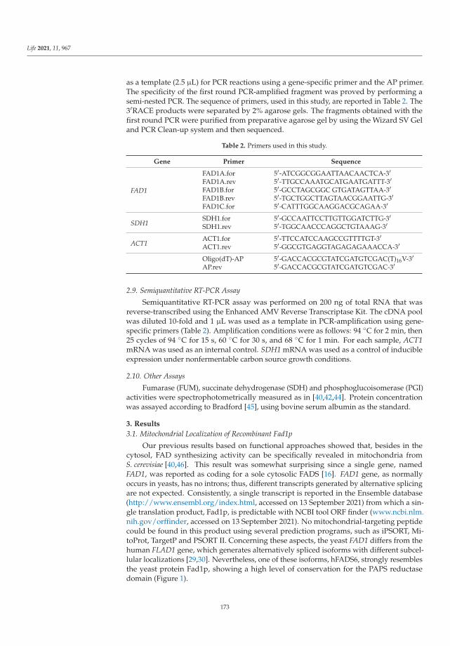
Life 2021, 11, 967
as a template (2.5 μL) for PCR reactions using a gene-specific primer and the AP primer.The specificity of the first round PCR-amplified fragment was proved by performing asemi-nested PCR. The sequence of primers, used in this study, are reported in Table 2. The3′RACE products were separated by 2% agarose gels. The fragments obtained with thefirst round PCR were purified from preparative agarose gel by using the Wizard SV Geland PCR Clean-up system and then sequenced.
Table 2. Primers used in this study.
Gene Primer Sequence
FAD1
FAD1A.for 5′-ATCGGCGGAATTAACAACTCA-3′FAD1A.rev 5′-TTGCCAAATGCATGAATGATTT-3′FAD1B.for 5′-GCCTAGCGGC GTGATAGTTAA-3′FAD1B.rev 5′-TGCTGGCTTAGTAACGGAATTG-3′FAD1C.for 5′-CATTTGGCAAGGACGCAGAA-3′
SDH1SDH1.for 5′-GCCAATTCCTTGTTGGATCTTG-3′SDH1.rev 5′-TGGCAACCCAGGCTGTAAAG-3′
ACT1ACT1.for 5′-TTCCATCCAAGCCGTTTTGT-3′ACT1.rev 5′-GGCGTGAGGTAGAGAGAAACCA-3′
Oligo(dT)-AP 5′-GACCACGCGTATCGATGTCGAC(T)16V-3′AP.rev 5′-GACCACGCGTATCGATGTCGAC-3′
2.9. Semiquantitative RT-PCR Assay
Semiquantitative RT-PCR assay was performed on 200 ng of total RNA that wasreverse-transcribed using the Enhanced AMV Reverse Transcriptase Kit. The cDNA poolwas diluted 10-fold and 1 μL was used as a template in PCR-amplification using gene-specific primers (Table 2). Amplification conditions were as follows: 94 ◦C for 2 min, then25 cycles of 94 ◦C for 15 s, 60 ◦C for 30 s, and 68 ◦C for 1 min. For each sample, ACT1mRNA was used as an internal control. SDH1 mRNA was used as a control of inducibleexpression under nonfermentable carbon source growth conditions.
2.10. Other Assays
Fumarase (FUM), succinate dehydrogenase (SDH) and phosphoglucoisomerase (PGI)activities were spectrophotometrically measured as in [40,42,44]. Protein concentrationwas assayed according to Bradford [45], using bovine serum albumin as the standard.
3. Results
3.1. Mitochondrial Localization of Recombinant Fad1p
Our previous results based on functional approaches showed that, besides in thecytosol, FAD synthesizing activity can be specifically revealed in mitochondria fromS. cerevisiae [40,46]. This result was somewhat surprising since a single gene, namedFAD1, was reported as coding for a sole cytosolic FADS [16]. FAD1 gene, as normallyoccurs in yeasts, has no introns; thus, different transcripts generated by alternative splicingare not expected. Consistently, a single transcript is reported in the Ensemble database(http://www.ensembl.org/index.html, accessed on 13 September 2021) from which a sin-gle translation product, Fad1p, is predictable with NCBI tool ORF finder (www.ncbi.nlm.nih.gov/orffinder, accessed on 13 September 2021). No mitochondrial-targeting peptidecould be found in this product using several prediction programs, such as iPSORT, Mi-toProt, TargetP and PSORT II. Concerning these aspects, the yeast FAD1 differs from thehuman FLAD1 gene, which generates alternatively spliced isoforms with different subcel-lular localizations [29,30]. Nevertheless, one of these isoforms, hFADS6, strongly resemblesthe yeast protein Fad1p, showing a high level of conservation for the PAPS reductasedomain (Figure 1).
173

Life 2021, 11, 967
Figure 1. Alignment of FAD synthase sequences from yeast and humans. The alignment of yeastFad1p with human FAD synthase isoform 6 (hFADS6) was performed by the Pairwise SequenceAlignment tool available at the EMBL-EBI website. The PAPS reductase domain (InterPro accession:IPR002500), present in both S. cerevisiae Fad1p (amino acids 55-231) and hFADS6 (amino acids132-286), is underlined and colored in red. Asterisks depict identical residues in the same positions,whereas colons and periods indicate strongly and weakly similar residues, respectively.
To gain some insight into the cellular destination of Fad1p, we investigated the sub-cellular localization of the recombinant form of Fad1p carrying the STREP-tag at theC-terminus and overexpressed in the CENPK2-1C strain (Table 1). Therefore, subcellu-lar fractions (spheroplasts, mitochondria and cytoplasm) were prepared from CENPK2-1CFAD1-STREP cells grown in ethanol up to early exponential growth phase and equalamounts of each fraction were analyzed by immunoblotting using the α-STREP antibody(Figure 2a). Even though the recombinant Fad1p–STREP was enriched in the cytoplasmicfraction, an evident immuno-reactive band was observed in the mitochondrial fraction,suggesting a mitochondrial localization for the recombinant Fad1p–STREP. Interestingly,the recombinant Fad1p–STREP was immunodecorated at the same size both in mitochon-dria and in cytoplasm. When the STREP-tag was moved from the C- to the N-terminal end,the ability of the recombinant STREP–Fad1p to enter mitochondria was completely lost,accompanied by a significant enrichment in the cytoplasmic fraction (Figure S1).
To further prove the ability of the recombinant Fad1p to enter mitochondria, weplaced Fad1p–STREP expression under the control of HXT7 promoter in the multicopyplasmid p426-HXT7-FAD1-STREP (Table 1) and used a different genetic strain, namely,K26. Subcellular fractions were prepared from K26FAD1-STREP cells grown in ethanolat the early exponential phase and analyzed by immunoblotting carried out with theα-STREP (Figure 2b). The recombinant protein Fad1p–STREP was immunodecorated atthe same size in both cytoplasm and mitochondria, where it was significantly enriched.When the K26FAD1-STREP cells were in the presence of the uncoupler carbonyl cyanide-p-trifluoromethoxy-phenylhydrazone (FCCP), the Fad1p–STREP import into mitochondriawas prevented (Figure 2b), as expected for a process depending on mitochondrial mem-brane potential [47]. This finding clearly indicates that the recombinant Fad1p is importedand localized to mitochondria.
174

Life 2021, 11, 967
(a)
(b)
Figure 2. Mitochondrial localization of recombinant Fad1p. Spheroplasts (Sphero), mitochondria(Mito) and cytoplasm (Cyt) were prepared from CENPK2-1C (a) and K26 (b) cells transformed withthe multicopy recombinant plasmid (Table 1). Both strains were grown up to the early exponentialphase on ethanol. K26FAD1-STREP cells were incubated in the absence or presence of FCCP (25 μM).Proteins (0.1 mg) were separated by SDS-PAGE, transferred onto PVDF membrane and detected witheither α-STREP or α-ACTIN antibodies.
3.2. Natural Fad1p Exists in Mitochondria
To ascertain whether the activity of FADS that we detected inside mitochondria isactually accountable to Fad1p, we analyzed the subcellular localization of natural Fad1pin cells grown either on nonfermentable (glycerol and ethanol) or fermentable (glucoseand galactose) carbon sources up to the early exponential phase (5 h) (Figure 3a). AFad1p band was detected in mitochondria prepared from glycerol-grown cells by usingthe α-FADS antiserum. It should be noted that even though actin (Act1p) was used as acytosolic marker [48], a minimal amount of Act1p was also found to be associated to themitochondrial fraction in agreement with [49]. In ethanol, an even more intense band wasdetected in the mitochondrial fraction, whereas in glucose-grown cells, the mitochondrialFad1p level was very low. Thus, the Fad1p content in mitochondria seems to dependon the carbon source. As a control, the dependence of the mitochondrial SDH specificactivity on different carbon sources was analyzed (Figure 3b), thus verifying the expectedglucose repression due to both transcriptional and post-transcriptional events [50,51]. It isnoteworthy that in the mitochondria prepared from galactose-grown cells, no band wasdetected (Figure 3a), even though we observed a high (induced) level of SDH activity(Figure 3b), in agreement with the presence of mitochondria in cells cultured in galactoseas the carbon source [52]. Differently from what was observed for the mitochondrial Fad1p,the cytosolic echoform did not appear to depend on the carbon source (Figure 3a).
175

Life 2021, 11, 967
(a)
(b)
Figure 3. Subcellular distribution of Fad1p in different carbon sources. (a) Spheroplasts (Sphero),mitochondria (Mito) and cytoplasm (Cyt) were prepared from WT cells grown up to the early ex-ponential phase at 30 ◦C in YEP liquid medium supplemented with the indicated carbon sources(2% each). Proteins (0.1 mg) were separated by SDS-PAGE, transferred onto PVDF membrane anddetected with either α-FADS or α-ACTIN antibodies. (b) The carbon source dependence of the mito-chondrial SDH-specific activity is reported as the means (±SD) of two independent measurements.
Cellular sublocalization of Fad1p was also evaluated in K26 cells grown in glycerol upto the stationary phase (24 h), as in [40]. Cytoplasmic and mitochondrial fractions wereprepared from spheroplasts and equal amounts of each fraction were used to reveal Fad1pby immunoblotting analysis (Figure S2). A faint α-FADS immuno-reactive band was foundin the cytoplasmic fraction. A much more evident band migrating at the same Mr was ob-served in total mitochondrial fraction, consistently enriched with respect to the spheroplastspecific amount. A similar enrichment was observed for the fumarase (FUM) and succinatedehydrogenase (SDH), two mitochondrial markers (Figure S2, histogram). The absence ofcytosolic contamination in mitochondria was demonstrated by the absence of phosphoglu-coisomerase (PGI) activity, a marker of cytosolic fraction (Figure S2, histogram). Isolatedmitochondria were further subfractionated in a membrane-enriched fraction (Mfr) and ina soluble fraction (Sfr). The Fad1p band was clearly detectable in both submitochondrialfractions, whose purity was confirmed by measuring the inner membrane marker SDHand the matrix marker FUM enzymatic activities (Figure S2, histogram).
Overall, on a molecular basis, these results support our previous functional data,proving that Fad1p exists in yeast mitochondria. Since no difference in size was observedbetween cytoplasmic and mitochondrial Fad1p, we postulated that the FAD1 gene mightgenerate two identical echoforms, destined to two distinct subcellular localizations.
176

Life 2021, 11, 967
3.3. Detection of Two FAD1 mRNAs with Different 3′UTR and Their Carbon SourceDependence Profile
In order to unravel the mechanism responsible for the subcellular destination of Fad1p,we considered a series of indications derived from the literature, which proposes a rolefor cis-acting 3′UTR elements in targeting transcripts to mitochondria [53,54], and from agenome-wide analysis, which suggests a high probability for FAD1 mRNA to be located onmitochondria-bound polysomes [54]; hence, the protein would be imported while beingtranslated in a co-translational process [55–57]. Therefore, we hypothesized that importantinformation for Fad1p localization could reside in the 3′UTR of FAD1 mRNA. Since noinformation was available, a combination of bioinformatic analyses and experimentalprocedures were carried out to gain knowledge about this untranslated region.
Firstly, we searched for putative polyadenylation sites (PASs), despite their complexity [58,59],and found two canonical signals (ATTAAA) in the 1 kbp region downstream of FAD1 ORFusing DNAFSMiner [60]. The proximal one was placed 50 nucleotides after the stop codonand the distal one was placed 339 nucleotides after the stop codon. The bioinformatic toolalso predicted other different PASs with similar scores, namely, AATAAA and TATCAA(Figure S3). These predictions suggested that FAD1 mRNA may be present in differentforms, whose length depends on the alternative use of distinct PASs in the transcript 3’UTR.
To test this hypothesis, 3′RACE experiments were performed on a template cDNAobtained from purified poly(A)+ RNA prepared from WT cells grown at the early exponen-tial phase on glucose as a carbon source and the gene-specific primers (Table 2; Figure 4a).When the primers FAD1A.for and AP were used, a single amplified product of about400 bp was obtained (Figure 4b, lane 1). The specificity of the 400 bp product was verifiedby a semi-nested PCR using the gene-specific primer FAD1C.for located 91 nucleotidesdownstream of FAD1A.for. The amplified product was obtained at the expected size, whichis about 300 bp (Figure 4b, lane 2). The 400 bp fragment was sequenced, revealing theexistence of FAD1 transcript with a 3′UTR of 128 nucleotides (Figure S3). To search forthe existence of a longer 3′UTR region of FAD1 mRNA, a PCR was performed using theprimers FAD1C.for and FAD1B.rev, the last being located downstream of the end of the3′UTR previously identified (Figure 4b); in this case, a product of about 450 nucleotideswas obtained (Figure 4b, lane 3), indicating the existence of a longer FAD1 mRNA. Todefine the entire length of the 3′UTR of this transcript, a PCR reaction was performed usingthe primers FAD1B.for and AP, obtaining a product of 550 bp (Figure 4b, lanes 4 and 5). Itsspecificity was proved by a semi-nested PCR with the primers FAD1B.for and FAD1B.rev,which gave rise to a fragment of about 100 bp, as expected (Figure 4b, lane 6). The 550 bpproduct was sequenced, showing the existence of an additional FAD1 mRNA with a longer3′UTR region of 759 bp (Figure S2). Similar results were obtained when the analysis wasperformed using cDNA as a template, obtained starting from purified poly(A)+ RNAprepared from WT cells grown on glycerol (Figure S4). Thus, FAD1 generates (at least) twoFAD1 mRNAs with different 3′UTR lengths. Based on the different length of the 3′UTR, wenamed them ‘short FAD1 mRNA’ and ‘long FAD1 mRNA’, respectively.
To understand whether or not the length of 3′UTR is responsible for the fate of Fad1pechoforms, the carbon source dependence of the FAD1 transcript levels was investigated.To this purpose, total RNA was extracted from WT cells grown at the early exponentialphase on different carbon sources, and the amount of FAD1 transcripts was evaluatedby semiquantitative RT-PCR, with ACT1 mRNA used as an internal control, essentiallyas in [42]. The long FAD1 mRNA was amplified using the primer pair FAD1A.for andFAD1B.rev, whereas the total FAD1 mRNA (short + long) was amplified using the primerpair FAD1A.for and FAD1A.rev (Figure S5). The RT-PCR product (540 bp) relative to thelong FAD1 mRNA complemented the 3′RACE data, confirming the presence of a longertranscript. The relative amount of total FAD1 mRNA did not depend on the carbon source,whereas a carbon source dependence was observed for the long FAD1 mRNA: its amountwas higher in ethanol grown cells, reduced in cells grown on glucose and almost absent ingalactose-grown cells. As a control, the expression pattern of SDH1 mRNA was analyzed; it
177
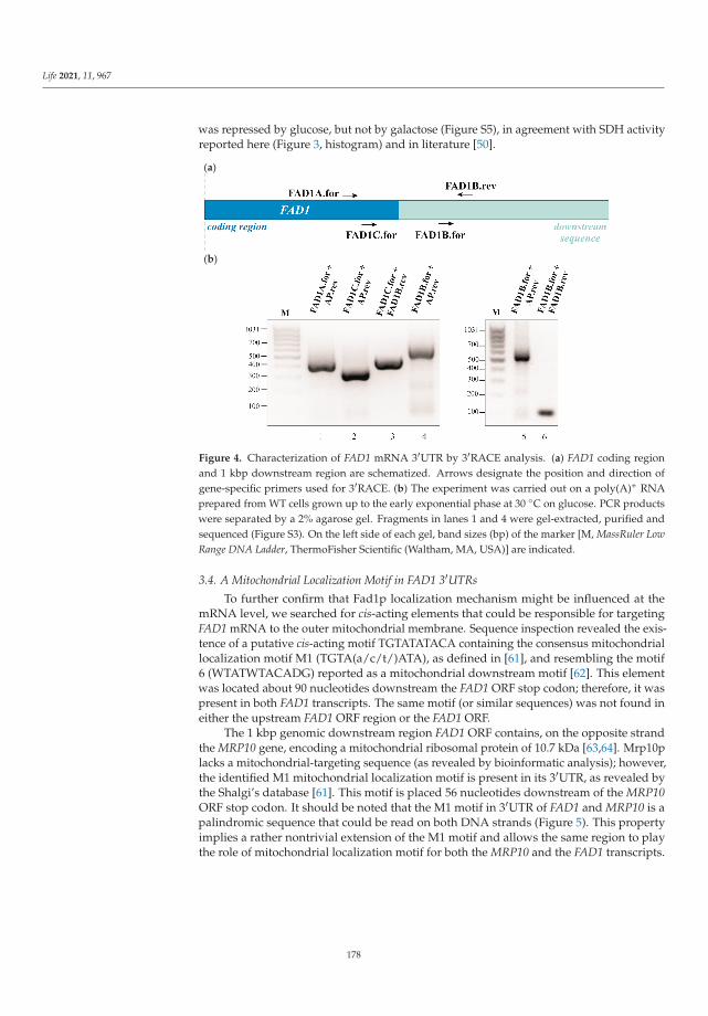
Life 2021, 11, 967
was repressed by glucose, but not by galactose (Figure S5), in agreement with SDH activityreported here (Figure 3, histogram) and in literature [50].
(a)
(b)
Figure 4. Characterization of FAD1 mRNA 3′UTR by 3′RACE analysis. (a) FAD1 coding regionand 1 kbp downstream region are schematized. Arrows designate the position and direction ofgene-specific primers used for 3′RACE. (b) The experiment was carried out on a poly(A)+ RNAprepared from WT cells grown up to the early exponential phase at 30 ◦C on glucose. PCR productswere separated by a 2% agarose gel. Fragments in lanes 1 and 4 were gel-extracted, purified andsequenced (Figure S3). On the left side of each gel, band sizes (bp) of the marker [M, MassRuler LowRange DNA Ladder, ThermoFisher Scientific (Waltham, MA, USA)] are indicated.
3.4. A Mitochondrial Localization Motif in FAD1 3′UTRs
To further confirm that Fad1p localization mechanism might be influenced at themRNA level, we searched for cis-acting elements that could be responsible for targetingFAD1 mRNA to the outer mitochondrial membrane. Sequence inspection revealed the exis-tence of a putative cis-acting motif TGTATATACA containing the consensus mitochondriallocalization motif M1 (TGTA(a/c/t/)ATA), as defined in [61], and resembling the motif6 (WTATWTACADG) reported as a mitochondrial downstream motif [62]. This elementwas located about 90 nucleotides downstream the FAD1 ORF stop codon; therefore, it waspresent in both FAD1 transcripts. The same motif (or similar sequences) was not found ineither the upstream FAD1 ORF region or the FAD1 ORF.
The 1 kbp genomic downstream region FAD1 ORF contains, on the opposite strandthe MRP10 gene, encoding a mitochondrial ribosomal protein of 10.7 kDa [63,64]. Mrp10placks a mitochondrial-targeting sequence (as revealed by bioinformatic analysis); however,the identified M1 mitochondrial localization motif is present in its 3′UTR, as revealed bythe Shalgi’s database [61]. This motif is placed 56 nucleotides downstream of the MRP10ORF stop codon. It should be noted that the M1 motif in 3′UTR of FAD1 and MRP10 is apalindromic sequence that could be read on both DNA strands (Figure 5). This propertyimplies a rather nontrivial extension of the M1 motif and allows the same region to playthe role of mitochondrial localization motif for both the MRP10 and the FAD1 transcripts.
178

Life 2021, 11, 967
Figure 5. The cis-acting motif M1 in the 3′UTR of FAD1 transcripts. Schematic representation of thepartial sequence of the S. cerevisiae genomic region comprised between nucleotides 372248 and 373617(chromosome IV). The white boxes indicate FAD1 ORF on the Crick strand and MRP10 ORF on theWatson strand; the orange box indicates the mitochondrial localization motif M1, placed about 87 and55 nucleotides downstream of the stop codon (TAA, highlighted in red) of FAD1 and MRP10 ORFs,respectively. The dotted arrows highlight the bidirectionality of the M1 palindromic sequence.
4. Discussion
Experiments reported in this paper point out the issue of the origin of mitochondrialFAD in S. cerevisiae mitochondria, strictly related with the problem of the identity and of thesubcellular localization of the FAD forming enzyme, FADS. Immunoblotting experimentsreported here reveal the presence of an α-FADS immuno-reactive band in mitochondria,showing the same Mr of that found in the spheroplasts and cytoplasm. This is in linewith the absence of a putative cleavable presequence in Fad1p, the sole peptide generatedby FAD1 gene. Thus, these results prove, on a molecular basis, Fad1p localization toyeast mitochondria and support the absence of a cleavable mitochondrial signal peptidein Fad1p.
It is well-documented that in eukaryotic cells, a protein can be located to two differentsubcellular compartments; this dual localization results in different echoforms—that is,proteins with identical or nearly identical amino acid sequences distinctly placed in thecell [65–67]. This is an important and frequent phenomenon; in fact, more than one third ofthe yeast mitochondrial proteome seems to be dual localized [68,69]. It has also becomeevident that dual localization can be regulated, induced and rebalanced in response toeither cellular signaling or changing extracellular conditions. Different dual-targetingmechanisms of mitochondrial proteins have been described or suggested, and, in somecase, they seem to be tightly regulated in time, location and function [65]. Our resultsdemonstrate that FAD1 generates two echoforms.
Analyzing the dependence of the Fad1p levels on fermentable and nonfermentablecarbon sources (Figure 3), we verified that the amount of Fad1p in mitochondria, butnot in the cytoplasm, depends on the carbon source being higher in ethanol and glycerolthan in the fermentable glucose. Interestingly, mitochondrial Fad1p almost disappeared ingalactose-grown cells. This finding may settle the discrepancy between results from our (seeIntroduction) and other laboratories [32,39] about the existence of mitochondrial FADS and,consequently, about the direction of FAD transport via the inner mitochondrial membranetranslocator Flx1p. The absence of mitochondrial Fad1p, observed in galactose-grown cells,implies that in this condition FAD must be transported from cytosol to mitochondria viaFlx1p (Figure 3 [32,39]). Vice versa, in a nonfermentable carbon source and in glucose,in the presence of mitochondrial Fad1p, Flx1p is expected to mediate FAD efflux frommitochondria to cytosol acting as a regulator of apo-flavoprotein biogenesis [40,42].
Using yeast strains overexpressing a recombinant form of Fad1p, the dual location ofnatural Fad1p was confirmed. Our data demonstrate that Fad1p is, per se, able to entermitochondria, despite the absence of a canonical mitochondrial-targeting sequence. A
179

Life 2021, 11, 967
“mitochondrial destination” message should be localized at the N-terminal end, since Fad1ploses its ability when the STREP-tag is moved from the C- to the N-terminal end (Figure S1).Presumably, the α1-helix in the first twenty amino acids at the N-terminal end, as well asthe contiguous α2-helix, indeed have amphipathic characteristics, with the basic residuesexposed to the solvent from the same side of the central axis, as revealed by the crystalstructure solved at 1.9 Å resolution (PDB entry: 2wsi) [20]. This would be in agreementwith the involvement of this protein moiety in the import process. Alternatively, anotherputative noncanonical/weak mitochondrial-targeting sequence [70] could be implicated inthe mitochondrial destination of Fad1p.
We also observed that in galactose-grown cells, the recombinant Fad1p-STREP enteredmitochondria per se [71], behaving differently from the natural Fad1p that was unable toenter mitochondria. All together, these data suggest the existence of a fine mechanismregulating the localization of the natural Fad1p, presumably at the transcript level and de-pendent on the carbon source. It is well-established that intracellular localization of mRNAhas a significant impact on the efficiency of its translation and, presumably, determines itsresponse to changing metabolic conditions or cellular stress [72].
Our attention was focused on the 3′UTR region of FAD1 mRNA. Since no informationwas available on this region, we carried out a 3′RACE analysis and found the existence of(at least) two FAD1 mRNAs with different 3′UTR lengths (Figure 4). This finding couldbe relevant for Fad1p cell biology, since it is well-known that 3′UTR regulates multipleaspects of mRNA metabolism, including subcellular localization, translation efficiency andstability, in cooperation with different RNA binding proteins [73]. A mechanism that couldhave a role in generating different transcripts with specific subcellular localizations and/orfunctions might be alternative polyadenylation [74,75]. We predicted different canonicaland noncanonical putative PASs localized in the first 1 kbp downstream of FAD1 ORF.Whether or not these PASs are actually active and responsible for the generation of the twoFAD1 transcripts are matter of ongoing investigation in our laboratory.
Searching for possible cis-acting motifs responsible for the peripheral mitochondriallocalization of FAD1 transcripts, we revealed the existence of the mitochondrial localizationmotif M1, placed 87 nucleotides downstream the FAD1 ORF stop codon (Figure 5). Thesame motif has been identified within a set of genes encoding mitochondrial proteins,whose mRNAs are translated by polyribosomes attached to the cytosolic side of the outermitochondrial membrane [76]. This motif is present in both FAD1 transcripts, whichmight localize close to the mitochondrial outer membrane. Intriguingly, this finding isin agreement with a value of mitochondrial localization of mRNA (MLR) for FAD1 equalto 87 [54]. This value is similar to the MLR values measured for other mRNAs, whoseproducts have been demonstrated to be internalized into mitochondria (i.e., 87.8 for SDH1transcript), thus indicating that Fad1p translation has a high probability to occur on themitochondrial outer membrane. Recently, it has been reported that mRNAs that initiatetranslation away from mitochondria experience a significant drop in mobility towardthe outer mitochondrial membrane and tend to remain there [77]. The M1 motif is acandidate binding site of the RNA binding protein Puf3p [78], localized on the cytosolicside of the outer mitochondrial membrane, where it interacts with some components ofthe mitochondrial protein import machinery [57,79]. Besides its role in mRNA targetingto mitochondria, Puf3p controls mRNA stability and translation efficiency according togrowth conditions and in response to oxidative stress [80–82]. The identified palindromicM1 motif is shared by the 3′UTR of FAD1 transcripts and the 3′ downstream region ofMRP10 gene, which is transcribed in the opposite direction with respect to FAD1 gene.Since an interaction between the M1 of MRP10 mRNA and Puf3p has been proven [83,84],an involvement of Puf3p in mitochondrial localization of Fad1p could be expected. Byanalyzing the carbon source dependence profile of FAD1 transcripts (Figure S3), we foundthat the long transcript is expressed in ethanol and in glycerol at a higher level thanin glucose; it is almost absent in galactose. The comparison between this profile andmitochondrial Fad1p levels (Figure 3) shows that the carbon source dependence of the long
180

Life 2021, 11, 967
FAD1 mRNA level strictly parallels the carbon source dependence of the mitochondrialFad1p level. This observation suggests that the long FAD1 mRNA might be responsiblefor the generation of Fad1p mitochondrial echoform. Therefore, although the M1 motif ispresent in both the short and long FAD1 transcripts, we propose that the different length ofthe 3′UTRs could be responsible for the distinct subcellular destinations of Fad1p. Thisdiverse destination could be explained by a differential Puf3p–M1 interaction caused by theexposure of M1 motif in different RNA contexts. In this scenario, the secondary structureof the two 3′UTRs may be causative of the selective interaction with Puf3p. Moreover, therecruitment of additional RNA binding proteins, presumably involved in mRNA decayor Puf3p interaction with mitochondrial import machinery [85], could also be affected by3′UTR length and structure. Therefore, Puf3p might regulate the dynamic balance betweentranslation efficiency, protein folding and import rate, thus establishing whether Fad1pmay be released in the cytosol or imported into mitochondria [81]. Additionally, it hasbeen shown that cytosolic ribosomes have the ability to ‘sense’ features relative to nascentpolypeptide chains; between those features, the sequence and structure of 3′UTR mRNAscan influence the polypeptide synthesis rate, modulating translation accordingly [76,86]. Itis noteworthy that the M1 motif was not included in the constructs used for the expressionof recombinant Fad1p. Nonetheless, the protein was capable, per se, of localizing tomitochondria in the presence of the mitochondrial membrane potential (Figure 2; [71]).Therefore, the M1 sequence would serve to fine-tune the cellular localization of Fad1pechoforms in various metabolic conditions, such as different carbon sources.
Our hypothesis is summarized in Figure 6. We speculate that, when Puf3p is boundto the M1 motif present in the long 3′UTR context (panel a), Fad1p synthesis efficiencyand/or folding rate are slowed down, allowing the co-translational import to proceed andgenerate the Fad1p mitochondrial echoform. Vice versa, when Puf3p interacts with theM1 in the short 3′UTR (panel b), a higher translation rate might cause a premature folding,thus favoring the cytosolic destination of Fad1p.
(a)
Figure 6. Cont.
181

Life 2021, 11, 967
(b)
Figure 6. Model depicting the dual location of Fad1p. The cartoon shows a possible role of Puf3p inthe different destination of Fad1p echoforms. (a) Puf3p binds to the long FAD1 mRNA and drives thenascent polypeptide in the mitochondrial matrix (MMX) via TOM–TIM complexes before the fullytranslated protein starts to fold. (b) Puf3p binds to the short FAD1 mRNA. As translation is presum-ably not sufficiently slowed down, premature and rapid folding of the nascent polypeptide in thecytosol (CYT) prevents Fad1p import across mitochondrial membranes (OMM—outer mitochondrialmembrane; IMM—inner mitochondrial membrane).
Supplementary Materials: The following are available online at https://www.mdpi.com/article/10.3390/life11090967/s1. Figure S1: Sublocalization of recombinant STREP-Fad1p. Figure S2: En-dogenous Fad1p mitochondrial localization in WT cells grown up to the stationary phase (24 h) onglycerol. Figure S3: FAD1 ORF and its 1 kbp downstream region. Figure S4: 3′RACE analysis carriedout on cells grown on glycerol (early exponential phase, 30 ◦C). Figure S5: Carbon source dependenceof FAD1 transcripts.
Author Contributions: Conceptualization, M.B., M.R., M.C., and E.B.; methodology, E.B. and M.R.;formal analysis, M.C., P.L., and F.B.; investigation, F.B., M.O., T.A.G., M.T., and P.L.; resources,E.B., M.R., and M.B.; writing—original draft preparation, M.B. and T.A.G.; writing—review andediting, M.R., F.B., and M.T.; supervision, M.C. and M.R.; project administration, M.B., M.O., M.R.,and M.C.; funding acquisition, M.B. All authors have read and agreed to the published version ofthe manuscript.
Funding: This work was supported by grants from MIUR PON-ricerca e competitività 2007–2013(project PON01_00937: “Modelli sperimentali biotecnologici integrati per lo sviluppo e la selezione dimolecole di interesse per la salute dell’uomo”) and from Università degli Studi di Bari “Aldo Moro”(Progetti Competitivi “Effetto di mutazioni di FLAD1 e di alterazioni dell’omeostasi delle flavinesullo stato redox e sulla biogenesi mitocondriale: uno studio integrato su fibroblasti umani”) to M.B.
Institutional Review Board Statement: Not applicable.
Informed Consent Statement: Not applicable.
Data Availability Statement: Not applicable.
Acknowledgments: The helpful collaboration of Emilia Dipalo, who participated as a student in theearly stages of this work, is gratefully acknowledged.
Conflicts of Interest: The authors declare no conflict of interest.
182

Life 2021, 11, 967
References
1. Tolomeo, M.; Nisco, A.; Leone, P.; Barile, M. Development of Novel Experimental Models to Study Flavoproteome Alterations inHuman Neuromuscular Diseases: The Effect of Rf Therapy. Int. J. Mol. Sci. 2020, 21, 5310. [CrossRef]
2. Joosten, V.; Van Berkel, W.J. Flavoenzymes. Curr. Opin. Chem. Biol. 2007, 11, 195–202. [CrossRef] [PubMed]3. Macheroux, P.; Kappes, B.; Ealick, S.E. Flavogenomics—A genomic and structural view of flavin-dependent proteins. FEBS J.
2011, 278, 2625–2634. [CrossRef] [PubMed]4. Barile, M.; Giancaspero, T.A.; Leone, P.; Galluccio, M.; Indiveri, C. Riboflavin transport and metabolism in humans. J. Inherit.
Metab. Dis. 2016, 39, 545–557. [CrossRef]5. Pimentel, C.; Batista-Nascimento, L.; Rodrigues-Pousada, C.; Menezes, R.A. Oxidative stress in Alzheimer’s and Parkinson’s
diseases: Insights from the yeast Saccharomyces cerevisiae. Oxidative Med. Cell. Longev. 2012, 2012, 132146. [CrossRef]6. Botstein, D.; Fink, G.R. Yeast: An experimental organism for 21st Century biology. Genetics 2011, 189, 695–704. [CrossRef]7. Tenreiro, S.; Outeiro, T.F. Simple is good: Yeast models of neurodegeneration. FEMS Yeast Res. 2010, 10, 970–979. [CrossRef]
[PubMed]8. Bastow, E.L.; Gourlay, C.W.; Tuite, M.F. Using yeast models to probe the molecular basis of amyotrophic lateral sclerosis. Biochem.
Soc. Trans. 2012, 39, 1482–1487. [CrossRef] [PubMed]9. Reihl, P.; Stolz, J. The monocarboxylate transporter homolog Mch5p catalyzes riboflavin (vitamin B2) uptake in Saccharomyces
cerevisiae. J. Biol. Chem. 2005, 280, 39809–39817. [CrossRef] [PubMed]10. Moriyama, Y. Riboflavin transporter is finally identified. J. Biochem. 2011, 150, 341–343. [CrossRef]11. Yonezawa, A.; Inui, K. Novel riboflavin transporter family RFVT/SLC52: Identification, nomenclature, functional characterization
and genetic diseases of RFVT/SLC52. Mol. Asp. Med. 2013, 34, 693–701. [CrossRef]12. O’Callaghan, B.; Bosch, A.M.; Houlden, H. An update on the genetics, clinical presentation, and pathomechanisms of human
riboflavin transporter deficiency. J. Inherit. Metab. Dis. 2019, 42, 598–607. [CrossRef]13. Yruela, I.; Arilla-Luna, S.; Medina, M.; Contreras-Moreira, B. Evolutionary divergence of chloroplast FAD synthetase proteins.
BMC Evol. Biol. 2010, 10, 311. [CrossRef]14. Giancaspero, T.A.; Colella, M.; Brizio, C.; Difonzo, G.; Fiorino, G.M.; Leone, P.; Brandsch, R.; Bonomi, F.; Iametti, S.; Barile, M.
Remaining challenges in cellular flavin cofactor homeostasis and flavoprotein biogenesis. Front. Chem. 2015, 3, 30. [CrossRef]15. Santos, M.A.; Jimenez, A.; Revuelta, J.L. Molecular characterization of FMN1, the structural gene for the monofunctional
flavokinase of Saccharomyces cerevisiae. J. Biol. Chem. 2000, 275, 28618–28624. [CrossRef]16. Wu, M.; Repetto, B.; Glerum, D.M.; Tzagoloff, A. Cloning and characterization of FAD1, the structural gene for flavin adenine
dinucleotide synthetase of Saccharomyces cerevisiae. Mol. Cell. Biol. 1995, 15, 264–271. [CrossRef]17. Bauer, S.; Kemter, K.; Bacher, A.; Huber, R.; Fischer, M.; Steinbacher, S. Crystal structure of Schizosaccharomyces pombe riboflavin
kinase reveals a novel ATP and riboflavin-binding fold. J. Mol. Biol. 2003, 326, 1463–1473. [CrossRef]18. Karthikeyan, S.; Zhou, Q.; Mseeh, F.; Grishin, N.V.; Osterman, A.L.; Zhang, H. Crystal structure of human riboflavin kinase
reveals a beta barrel fold and a novel active site arch. Structure 2003, 11, 265–273. [CrossRef]19. Karthikeyan, S.; Zhou, Q.; Osterman, A.L.; Zhang, H. Ligand binding-induced conformational changes in riboflavin kinase:
Structural basis for the ordered mechanism. Biochemistry 2003, 42, 12532–12538. [CrossRef] [PubMed]20. Leulliot, N.; Blondeau, K.; Keller, J.; Ulryck, N.; Quevillon-Cheruel, S.; Van Tilbeurgh, H. Crystal structure of yeast FAD synthetase
(Fad1) in complex with FAD. J. Mol. Biol. 2010, 398, 641–646. [CrossRef] [PubMed]21. Brizio, C.; Galluccio, M.; Wait, R.; Torchetti, E.M.; Bafunno, V.; Accardi, R.; Gianazza, E.; Indiveri, C.; Barile, M. Over-expression
in Escherichia coli and characterization of two recombinant isoforms of human FAD synthetase. Biochem. Biophys. Res. Commun.2006, 344, 1008–1016. [CrossRef] [PubMed]
22. Leone, P.; Galluccio, M.; Brizio, C.; Barbiroli, A.; Iametti, S.; Indiveri, C.; Barile, M. The hidden side of the human FAD synthase 2.Int. J. Biol. Macromol. 2019, 138, 986–995. [CrossRef]
23. Giancaspero, T.A.; Locato, V.; Barile, M. A regulatory role of NAD redox status on flavin cofactor homeostasis in S. cerevisiaemitochondria. Oxidative Med. Cell. Longev. 2013, 2013, 612784. [CrossRef]
24. Vergani, L.; Barile, M.; Angelini, C.; Burlina, A.B.; Nijtmans, L.; Freda, M.P.; Brizio, C.; Zerbetto, E.; Dabbeni-Sala, F. Riboflavintherapy. Biochemical heterogeneity in two adult lipid storage myopathies. Brain A J. Neurol. 1999, 122 Pt 12, 2401–2411. [CrossRef]
25. Gianazza, E.; Vergani, L.; Wait, R.; Brizio, C.; Brambilla, D.; Begum, S.; Giancaspero, T.A.; Conserva, F.; Eberini, I.; Bufano, D.; et al.Coordinated and reversible reduction of enzymes involved in terminal oxidative metabolism in skeletal muscle mitochondriafrom a riboflavin-responsive, multiple acyl-CoA dehydrogenase deficiency patient. Electrophoresis 2006, 27, 1182–1198. [CrossRef][PubMed]
26. Olsen, R.K.J.; Konarikova, E.; Giancaspero, T.A.; Mosegaard, S.; Boczonadi, V.; Matakovic, L.; Veauville-Merllie, A.; Terrile, C.;Schwarzmayr, T.; Haack, T.B.; et al. Riboflavin-Responsive and -Non-responsive Mutations in FAD Synthase Cause MultipleAcyl-CoA Dehydrogenase and Combined Respiratory-Chain Deficiency. Am. J. Hum. Genet. 2016, 98, 1130–1145. [CrossRef]
27. Gregersen, N.; Andresen, B.S.; Pedersen, C.B.; Olsen, R.K.; Corydon, T.J.; Bross, P. Mitochondrial fatty acid oxidation defects–remaining challenges. J. Inherit. Metab. Dis. 2008, 31, 643–657. [CrossRef]
28. Leone, P.; Galluccio, M.; Barbiroli, A.; Eberini, I.; Tolomeo, M.; Vrenna, F.; Gianazza, E.; Iametti, S.; Bonomi, F.; Indiveri, C.; et al.Bacterial Production, Characterization and Protein Modeling of a Novel Monofuctional Isoform of FAD Synthase in Humans: AnEmergency Protein? Molecules 2018, 23, 116. [CrossRef]
183

Life 2021, 11, 967
29. Torchetti, E.M.; Brizio, C.; Colella, M.; Galluccio, M.; Giancaspero, T.A.; Indiveri, C.; Roberti, M.; Barile, M. Mitochondriallocalization of human FAD synthetase isoform 1. Mitochondrion 2010, 10, 263–273. [CrossRef] [PubMed]
30. Giancaspero, T.A.; Busco, G.; Panebianco, C.; Carmone, C.; Miccolis, A.; Liuzzi, G.M.; Colella, M.; Barile, M. FAD synthesis anddegradation in the nucleus create a local flavin cofactor pool. J. Biol. Chem. 2013, 288, 29069–29080. [CrossRef]
31. Gudipati, V.; Koch, K.; Lienhart, W.D.; Macheroux, P. The flavoproteome of the yeast Saccharomyces cerevisiae. Biochim. EtBiophys. Acta 2014, 1844, 535–544. [CrossRef]
32. Hao, H.X.; Khalimonchuk, O.; Schraders, M.; Dephoure, N.; Bayley, J.P.; Kunst, H.; Devilee, P.; Cremers, C.W.; Schiffman, J.D.;Bentz, B.G.; et al. SDH5, a gene required for flavination of succinate dehydrogenase, is mutated in paraganglioma. Science 2009,325, 1139–1142. [CrossRef] [PubMed]
33. Ghezzi, D.; Goffrini, P.; Uziel, G.; Horvath, R.; Klopstock, T.; Lochmuller, H.; D’Adamo, P.; Gasparini, P.; Strom, T.M.; Prokisch, H.;et al. SDHAF1, encoding a LYR complex-II specific assembly factor, is mutated in SDH-defective infantile leukoencephalopathy.Nat. Genet. 2009, 41, 654–656. [CrossRef]
34. Robinson, K.M.; Lemire, B.D. Covalent attachment of FAD to the yeast succinate dehydrogenase flavoprotein requires importinto mitochondria, presequence removal, and folding. J. Biol. Chem. 1996, 271, 4055–4060. [CrossRef] [PubMed]
35. Kim, H.J.; Winge, D.R. Emerging concepts in the flavinylation of succinate dehydrogenase. Biochim. Biophys. Acta 2013, 1827,627–636. [CrossRef] [PubMed]
36. Rutter, J.; Winge, D.R.; Schiffman, J.D. Succinate dehydrogenase-Assembly, regulation and role in human disease. Mitochondrion2010, 10, 393–401. [CrossRef]
37. Schiff, M.; Veauville-Merllie, A.; Su, C.H.; Tzagoloff, A.; Rak, M.; Ogier de Baulny, H.; Boutron, A.; Smedts-Walters, H.; Romero,N.B.; Rigal, O.; et al. SLC25A32 Mutations and Riboflavin-Responsive Exercise Intolerance. N. Engl. J. Med. 2016, 374, 795–797.[CrossRef]
38. Hellebrekers, D.; Sallevelt, S.; Theunissen, T.E.J.; Hendrickx, A.T.M.; Gottschalk, R.W.; Hoeijmakers, J.G.J.; Habets, D.D.; Bierau, J.;Schoonderwoerd, K.G.; Smeets, H.J.M. Novel SLC25A32 mutation in a patient with a severe neuromuscular phenotype. Eur. J.Hum. Genet. 2017, 25, 886–888. [CrossRef]
39. Tzagoloff, A.; Jang, J.; Glerum, D.M.; Wu, M. FLX1 codes for a carrier protein involved in maintaining a proper balance of flavinnucleotides in yeast mitochondria. J. Biol. Chem. 1996, 271, 7392–7397. [CrossRef]
40. Bafunno, V.; Giancaspero, T.A.; Brizio, C.; Bufano, D.; Passarella, S.; Boles, E.; Barile, M. Riboflavin uptake and FAD synthesisin Saccharomyces cerevisiae mitochondria: Involvement of the Flx1p carrier in FAD export. J. Biol. Chem. 2004, 279, 95–102.[CrossRef]
41. Barile, M.; Passarella, S.; Bertoldi, A.; Quagliariello, E. Flavin adenine dinucleotide synthesis in isolated rat liver mitochondriacaused by imported flavin mononucleotide. Arch. Biochem. Biophys. 1993, 305, 442–447. [CrossRef]
42. Giancaspero, T.A.; Wait, R.; Boles, E.; Barile, M. Succinate dehydrogenase flavoprotein subunit expression in Saccharomycescerevisiae–involvement of the mitochondrial FAD transporter, Flx1p. FEBS J. 2008, 275, 1103–1117. [CrossRef]
43. Gietz, R.D.; Schiestl, R.H. Frozen competent yeast cells that can be transformed with high efficiency using the LiAc/SS carrierDNA/PEG method. Nat. Protoc. 2007, 2, 1–4. [CrossRef]
44. Giancaspero, T.A.; Locato, V.; De Pinto, M.C.; De Gara, L.; Barile, M. The occurrence of riboflavin kinase and FAD synthetaseensures FAD synthesis in tobacco mitochondria and maintenance of cellular redox status. FEBS J. 2009, 276, 219–231. [CrossRef]
45. Bradford, M.M. A rapid and sensitive method for the quantitation of microgram quantities of protein utilizing the principle ofprotein-dye binding. Anal. Biochem. 1976, 72, 248–254. [CrossRef]
46. Pallotta, M.L.; Brizio, C.; Fratianni, A.; De Virgilio, C.; Barile, M.; Passarella, S. Saccharomyces cerevisiae mitochondria cansynthesise FMN and FAD from externally added riboflavin and export them to the extramitochondrial phase. FEBS Lett. 1998,428, 245–249. [CrossRef]
47. Martin, J.; Mahlke, K.; Pfanner, N. Role of an energized inner membrane in mitochondrial protein import. Delta psi drives themovement of presequences. J. Biol. Chem. 1991, 266, 18051–18057. [CrossRef]
48. Traglia, H.M.; O’Connor, J.P.; Tung, K.S.; Dallabrida, S.; Shen, W.C.; Hopper, A.K. Nucleus-associated pools of Rna1p, theSaccharomyces cerevisiae Ran/TC4 GTPse activating protein involved in nucleus/cytosol transit. Proc. Natl. Acad. Sci. USA 1996,93, 7667–7672. [CrossRef] [PubMed]
49. Leadsham, J.E.; Gourlay, C.W. Cytoskeletal induced apoptosis in yeast. Biochim. Biophys. Acta 2008, 1783, 1406–1412. [CrossRef][PubMed]
50. Lombardo, A.; Cereghino, G.P.; Scheffler, I.E. Control of mRNA turnover as a mechanism of glucose repression in Saccharomycescerevisiae. Mol. Cell. Biol. 1992, 12, 2941–2948. [CrossRef]
51. Prieto, S.; De la Cruz, B.J.; Scheffler, I.E. Glucose-regulated turnover of mRNA and the influence of poly(A) tail length on half-life.J. Biol. Chem. 2000, 275, 14155–14166. [CrossRef]
52. Polakis, E.S.; Bartley, W. Changes in the enzyme activities of Saccharomyces cerevisiae during aerobic growth on different carbonsources. Biochem. J. 1965, 97, 284–297. [CrossRef]
53. Zahedi, R.P.; Sickmann, A.; Boehm, A.M.; Winkler, C.; Zufall, N.; Schonfisch, B.; Guiard, B.; Pfanner, N.; Meisinger, C. Proteomicanalysis of the yeast mitochondrial outer membrane reveals accumulation of a subclass of preproteins. Mol. Biol. Cell 2006, 17,1436–1450. [CrossRef] [PubMed]
184

Life 2021, 11, 967
54. Marc, P.; Margeot, A.; Devaux, F.; Blugeon, C.; Corral-Debrinski, M.; Jacq, C. Genome-wide analysis of mRNAs targeted to yeastmitochondria. EMBO Rep. 2002, 3, 159–164. [CrossRef] [PubMed]
55. Ahmed, A.U.; Fisher, P.R. Import of nuclear-encoded mitochondrial proteins: A cotranslational perspective. Int. Rev. Cell Mol.Biol. 2009, 273, 49–68.
56. Kellems, R.E.; Allison, V.F.; Butow, R.A. Cytoplasmic type 80S ribosomes associated with yeast mitochondria. IV. Attachment ofribosomes to the outer membrane of isolated mitochondria. J. Cell Biol. 1975, 65, 1–14. [CrossRef]
57. Eliyahu, E.; Lesnik, C.; Arava, Y. The protein chaperone Ssa1 affects mRNA localization to the mitochondria. FEBS Lett. 2012, 586,64–69. [CrossRef]
58. Dichtl, B.; Keller, W. Recognition of polyadenylation sites in yeast pre-mRNAs by cleavage and polyadenylation factor. TheEMBO J. 2001, 20, 3197–3209. [CrossRef]
59. Zhao, J.; Hyman, L.; Moore, C. Formation of mRNA 3’ ends in eukaryotes: Mechanism, regulation, and interrelationships withother steps in mRNA synthesis. Microbiol. Mol. Biol. Rev. 1999, 63, 405–445. [CrossRef]
60. Liu, H.; Han, H.; Li, J.; Wong, L. DNAFSMiner: A web-based software toolbox to recognize two types of functional sites in DNAsequences. Bioinformatics 2005, 21, 671–673. [CrossRef] [PubMed]
61. Shalgi, R.; Lapidot, M.; Shamir, R.; Pilpel, Y. A catalog of stability-associated sequence elements in 3’ UTRs of yeast mRNAs.Genome Biol. 2005, 6, R86. [CrossRef] [PubMed]
62. Kellis, M.; Patterson, N.; Endrizzi, M.; Birren, B.; Lander, E.S. Sequencing and comparison of yeast species to identify genes andregulatory elements. Nature 2003, 423, 241–254. [CrossRef]
63. Jin, C.; Myers, A.M.; Tzagoloff, A. Cloning and characterization of MRP10, a yeast gene coding for a mitochondrial ribosomalprotein. Curr. Genet. 1997, 31, 228–234. [CrossRef]
64. Kitakawa, M.; Graack, H.R.; Grohmann, L.; Goldschmidt-Reisin, S.; Herfurth, E.; Wittmann-Liebold, B.; Nishimura, T.; Isono, K.Identification and characterization of the genes for mitochondrial ribosomal proteins of Saccharomyces cerevisiae. Eur. J. Biochem.1997, 245, 449–456. [CrossRef]
65. Yogev, O.; Pines, O. Dual targeting of mitochondrial proteins: Mechanism, regulation and function. Biochim. Et Biophys. Acta 2011,1808, 1012–1020. [CrossRef]
66. Bader, G.; Enkler, L.; Araiso, Y.; Hemmerle, M.; Binko, K.; Baranowska, E.; De Craene, J.O.; Ruer-Laventie, J.; Pieters, J.;Tribouillard-Tanvier, D.; et al. Assigning mitochondrial localization of dual localized proteins using a yeast Bi-GenomicMitochondrial-Split-GFP. eLife 2020, 9, e56649. [CrossRef] [PubMed]
67. Ben-Menachem, R.; Pines, O. Detection of Dual Targeting and Dual Function of Mitochondrial Proteins in Yeast. Methods Mol.Biol. 2017, 1567, 179–195. [CrossRef] [PubMed]
68. Ben-Menachem, R.; Tal, M.; Shadur, T.; Pines, O. A third of the yeast mitochondrial proteome is dual localized: A question ofevolution. Proteomics 2011, 11, 4468–4476. [CrossRef]
69. Kisslov, I.; Naamati, A.; Shakarchy, N.; Pines, O. Dual-targeted proteins tend to be more evolutionarily conserved. Mol. Biol. Evol.2014, 31, 2770–2779. [CrossRef]
70. Dinur-Mills, M.; Tal, M.; Pines, O. Dual targeted mitochondrial proteins are characterized by lower MTS parameters and total netcharge. PLoS ONE 2008, 3, e2161. [CrossRef] [PubMed]
71. Barile, M.; University of Bari ‘Aldo Moro’, Bari, Italy. Personal communication, 2021.72. Lashkevich, K.A.; Dmitriev, S.E. mRNA Targeting, Transport and Local Translation in Eukaryotic Cells: From the Classical View
to a Diversity of New Concepts. Mol. Biol. 2021, 1–31. [CrossRef]73. Andreassi, C.; Riccio, A. To localize or not to localize: mRNA fate is in 3’UTR ends. Trends Cell Biol. 2009, 19, 465–474. [CrossRef]74. Sparks, K.A.; Dieckmann, C.L. Regulation of poly(A) site choice of several yeast mRNAs. Nucleic Acids Res. 1998, 26, 4676–4687.
[CrossRef]75. Turner, R.E.; Pattison, A.D.; Beilharz, T.H. Alternative polyadenylation in the regulation and dysregulation of gene expression.
Semin. Cell Dev. Biol. 2018, 75, 61–69. [CrossRef]76. Sylvestre, J.; Vialette, S.; Corral Debrinski, M.; Jacq, C. Long mRNAs coding for yeast mitochondrial proteins of prokaryotic origin
preferentially localize to the vicinity of mitochondria. Genome Biol. 2003, 4, R44. [CrossRef] [PubMed]77. Horton, P. A simulation study of mRNA diffusion and mitochondrial localization. bioRxiv 2019. [CrossRef]78. Gerber, A.P.; Herschlag, D.; Brown, P.O. Extensive association of functionally and cytotopically related mRNAs with Puf family
RNA-binding proteins in yeast. PLoS Biol. 2004, 2, e79. [CrossRef] [PubMed]79. Garcia-Rodriguez, L.J.; Gay, A.C.; Pon, L.A. Puf3p, a Pumilio family RNA binding protein, localizes to mitochondria and regulates
mitochondrial biogenesis and motility in budding yeast. J. Cell Biol. 2007, 176, 197–207. [CrossRef] [PubMed]80. Rowe, W.; Kershaw, C.J.; Castelli, L.M.; Costello, J.L.; Ashe, M.P.; Grant, C.M.; Sims, P.F.; Pavitt, G.D.; Hubbard, S.J. Puf3p induces
translational repression of genes linked to oxidative stress. Nucleic Acids Res. 2014, 42, 1026–1041. [CrossRef] [PubMed]81. Quenault, T.; Lithgow, T.; Traven, A. PUF proteins: Repression, activation and mRNA localization. Trends Cell Biol. 2011, 21,
104–112. [CrossRef]82. Saint-Georges, Y.; Garcia, M.; Delaveau, T.; Jourdren, L.; Le Crom, S.; Lemoine, S.; Tanty, V.; Devaux, F.; Jacq, C. Yeast mitochondrial
biogenesis: A role for the PUF RNA-binding protein Puf3p in mRNA localization. PLoS ONE 2008, 3, e2293. [CrossRef]83. Freeberg, M.A.; Han, T.; Moresco, J.J.; Kong, A.; Yang, Y.C.; Lu, Z.J.; Yates, J.R.; Kim, J.K. Pervasive and dynamic protein binding
sites of the mRNA transcriptome in Saccharomyces cerevisiae. Genome Biol. 2013, 14, R13. [CrossRef] [PubMed]
185

Life 2021, 11, 967
84. Lapointe, C.P.; Stefely, J.A.; Jochem, A.; Hutchins, P.D.; Wilson, G.M.; Kwiecien, N.W.; Coon, J.J.; Wickens, M.; Pagliarini, D.J.Multi-omics Reveal Specific Targets of the RNA-Binding Protein Puf3p and Its Orchestration of Mitochondrial Biogenesis. CellSyst. 2018, 6, 125–135.e6. [CrossRef] [PubMed]
85. Chacinska, A.; Koehler, C.M.; Milenkovic, D.; Lithgow, T.; Pfanner, N. Importing mitochondrial proteins: Machineries andmechanisms. Cell 2009, 138, 628–644. [CrossRef] [PubMed]
86. Gong, F.; Yanofsky, C. Instruction of translating ribosome by nascent peptide. Science 2002, 297, 1864–1867. [CrossRef] [PubMed]
186

MDPISt. Alban-Anlage 66
4052 BaselSwitzerland
Tel. +41 61 683 77 34Fax +41 61 302 89 18
www.mdpi.com
Life Editorial OfficeE-mail: [email protected]
www.mdpi.com/journal/life


MDPI
St. Alban-Anlage 66
4052 Basel
Switzerland
Tel: +41 61 683 77 34
Fax: +41 61 302 89 18
www.mdpi.com ISBN 978-3-0365-2152-7
Copyright © 2022 FDOKUMEN