A fluorescence assay for peptide translocation into mitochondria

doi:10.1016/j.jmb.2005.11.070 J. Mol. Biol. (2006) 356, 288–299
DNA Recombination Activity in Soybean Mitochondria
Medha Manchekar1, Karyn Scissum-Gunn1, Daqing Song2, Fayaz Khazi1
Stephanie L. McLean1 and Brent L. Nielsen1,2*
1Department of BiologicalSciences, 101 Life SciencesBuilding, Auburn UniversityAuburn, AL 36849, USA
2Department of Microbiology &Molecular Biology, 775 WIDBBrigham Young UniversityProvo, UT 84602, USA
0022-2836/$ - see front matter q 2005 E
Present addresses: M. ManchekarResearch Unit, University of AlabamAL, USA; K. Scissum-Gunn, Departand Science, Alabama State UniversUSA; F. Khazi, Howard Hughes MeChildren’s Hospital of PhiladelphiaMcClean, Division of Clinical Immutology, University of Alabama at Bi
Abbreviations used: mtDNA, mitPFGE, pulsed field gel electrophoreinversion gel electrophoresis; EM, ess, single-stranded.
E-mail address of the [email protected]
Mitochondrial genomes in higher plants are much larger and morecomplex as compared to animal mitochondrial genomes. There is growingevidence that plant mitochondrial genomes exist predominantly as acollection of linear and highly branched DNA molecules and replicate by arecombination-dependent mechanism. However, biochemical evidence ofmitochondrial DNA (mtDNA) recombination activity in plants haspreviously been lacking. We provide the first report of strand-invasionactivity in plant mitochondria. Similar to bacterial RecA, this activity fromsoybean is dependent on the presence of ATP and Mg2C. Western blotanalysis using an antibody against the Arabidopsis mitochondrial RecAprotein shows cross-reaction with a soybean protein of about 44 kDa,indicating conservation of this protein in at least these two plant species.mtDNA structure was analyzed by electron microscopy of total soybeanmtDNA and molecules recovered after field-inversion gel electrophoresis(FIGE). While most molecules were found to be linear, some moleculescontained highly branched DNA structures and a small but reproducibleproportion consisted of circular molecules (many with tails) similar torecombination intermediates. The presence of recombination intermediatesin plant mitochondria preparations is further supported by analysis ofmtDNA molecules by 2-D agarose gel electrophoresis, which indicated thepresence of complex recombination structures along with a considerableamount of single-stranded DNA. These data collectively provideconvincing evidence for the occurrence of homologous DNA recombi-nation in plant mitochondria.
q 2005 Elsevier Ltd. All rights reserved.
Keywords: plant mitochondrial DNA recombination; strand-invasionassays; soybean; two-dimensional agarose gel electrophoresis
*Corresponding authorIntroduction
A wide range of sizes of mitochondrial genomesexists across various classes of organisms. Higher
lsevier Ltd. All rights reserve
, Atherosclerosisa at Birmingham,
ment of Mathematicsity, Montgomery, AL,dical Institute, The, PA, USA; S. L.nology & Rheuma-
rmingham, AL, USA.ochondrial DNA;sis; FIGE, fieldlectron microscopy;
ing author:
plant mitochondrial genomes (208–2000 kbp) aremuch larger than their counterparts in vertebrates(16–17 kbp) or fungi (w25–80 kbp).1,2 The exten-sively characterized circular animal mitochondrialgenome is highly conserved among species, con-tains no introns, and has a very limited amount ofintergenic sequence.3 Plant mitochondrial DNA(mtDNA) contains introns in many genes andsome additional expressed genes as compared toanimal mitochondria, but most of the additionalsequences in plants are not expressed and do notappear to be essential.4 The complete mitochondrialgenome sequences are available for a number ofhigher plants, including Arabidopsis thaliana,5 andthe liverwort Marchantia polymorpha.6
Over the past several years there have been manyreports that the mitochondrial genomes in yeastand higher plants exist as primarily linear andbranched DNA molecules of widely varied sizes
d.

Figure 1. Detection of homologous strand exchangeactivity in soybean mitochondrial proteins eluted fromthe heparin-Sepharose column. Lane E, E. coli RecApositive control. KP, minus protein contol. L, loadingfraction (contains nuclease activity). Lanes 2–12, elutionfractions from the column. Peak activity was observed infractions 6–8. Nucleases that degrade the substrate DNAappear to be present in lanes 9–12.
Soybean MtDNA Recombination 289
that are considerably smaller than the predictedgenome size.7–12 Using pulsed field gel electro-phoresis (PFGE) of in-gel lysed mitochondria fromvarious species, we and others have reported thatonly about 6–12% of the molecules are circular.11,13–
16 The branched molecules observed are verysimilar to recombination intermediates observedfor yeast mtDNA17 or with T4 phage DNAreplication.18,19 Backert & Borner reported visual-ization of replication and recombination intermedi-ates in mtDNA from Chenopodium album, and manyof the linear DNA molecules observed were foundto have an extended single-stranded end.20 Suchends may be involved in strand invasion intohomologous regions of double-stranded DNA.Recombination of animal mtDNA appears tooccur very rarely.21
Restriction maps of nearly all plant mitochondrialgenomes predict a master circle with subgenomiccircular molecules resulting from recombinationacross large direct repeats.1,13,22–28 Molecules ofthese predicted sizes, however, are in very lowabundance or not detected. This can be explained ifplant mitochondrial genomes are circularly per-muted, as with phage T4.18,19 Oldenburg & Bendichreported that the predominantly linear mtDNAmolecules in Marchantia, a lower plant, are circularlypermuted with random ends.12 There is growingevidence that replication of plant mtDNA occurs by aT4-like recombination-dependent mechanism.19
Most higher plant mitochondrial genomes containat least one pair of large (O1 kbp) direct repeats,which may be involved in homologous recombi-nation to generate subgenomic molecules.27,29 How-ever, it is apparent that these large repeats areinsufficient to explain the range in sizes andstructures of molecules observed. Many smallerrepeats, ranging from as little as nine to a fewhundred base-pairs, have been identified in plantmitochondrial genomes of a number of species andmay be involved in homologous recombination.29 Ithas been proposed that the large repeats are involvedin reversible recombination, while the small repeatsmay result in stable rearrangements.2,29 There arethree mitochondrial plasmids in broad bean, andseveral variant plasmids have been characterized thatappear to have arisen from active double recombina-tion across repeated elements.30
There is evidence that active mtDNA recombi-nation occurs in at least some plant species. Someindividual plant mitochondria may contain less thana full genome equivalent of DNA, giving support forthe possibility of deletion by recombination betweendirect repeats of portions of the genome.31,32 Genomerearrangements thought to be due to mtDNArecombination have been reported in a variety ofplant species including A. thaliana.24,25,33–36 Both large(6.5 and 4.2 kb) direct repeats and numerous smaller(30–560 bp, totaling 144 in number) repeats arepresent in A. thaliana mtDNA.5 There is evidencethat recombination across the large repeats occursoften but with no loss of subsequent rearrange-ments,35 while the small repeats may not be active in
frequent recombination.5 Very short repeatedsequences have also been reported to be involved inmtDNA recombination in animal37 and yeast cells.38
In soybean mitochondria, sequence repeats of 9,23 and 299 bp have been characterized.33,34 Numer-ous rearrangements of genome sequences amongvarious soybean cultivars have been characterizedand appear to have arisen by homologous recombi-nation across these repeats,33,39,40 or across shortelements that are part of a 4.9 kb PstI fragment ofsoybean mtDNA.41 The 299 bp repeat has beenfound in several copies in mtDNA from soybeanand several other higher plants, suggesting that thissequence may represent a hot spot for mtDNArecombination in multiple plant species.34,41
These previous reports suggest that active homo-logous mtDNA recombination occurs in at least someplant species. We recently reported the identificationof a mitochondrial-targeted homologue of the Escher-ichia coli recA gene in A. thaliana.42 However, to datethere have been no reports on the characterization ofrecombination activity in plant mitochondria. Weprovide here the first report of such activity insoybean. These findings are supported by analysisof soybean mtDNA by electron microscopy and two-dimensional agarose gel electrophoresis.
Results
Strand transfer activity in partially purifiedsoybean mitochondrial protein fractions
Soybean mitochondria isolated on Percollgradients42 were used for preparation of a totallysate, which was enriched for DNA recombinationactivity by DEAE-cellulose and heparin-Sepharosechromatography as described in Materials andMethods. Fractions 6–8 from the heparin-Sepharosecolumn supported formation of joint molecules,indicating strand-exchange between the linearlabeled double-stranded DNA and homologouscircular single-stranded DNA molecules (Figure 1).

Figure 3. Western blot analysis of mitochondrial RecAproteins in soybean and Arabidopsis. Lane 1, molecularmass markers, with sizes shown at left; lane 2, totalprotein (10 mg) from the soybean mitochondrial heparin-Sepharose fraction used for activity assays in Figure 1;lane 3, total protein (120 mg) from soybean leaves; lane 4,total protein (100 mg) from Arabidopsis leaves.
290 Soybean MtDNA Recombination
Later fractions (9–12) appeared to contain signifi-cant nuclease activity that led to degradation of thesubstrate. The control assay without any mitochon-drial protein fraction (lane KP) showed that theformation of joint molecules is protein-dependent.
Optimum strand exchange activity was found tobe dependent upon the presence of ATP and Mg2C
(Figure 2), which are conserved properties ofbacterial RecA proteins. In the absence of bothMg2C and ATP, activity was only 15% of thatobtained in the complete reaction (Figure 2(b)).Some activity was consistently observed in theabsence of only ATP (Figure 2(a), lane 6). Theamount of activity in the absence of ATP was foundto average just over 33% (Figure 2(b)). This activitymay be due to the presence of some ATP still boundto the enzyme. It may also be possible that this plantenzyme may retain some activity in the absence ofATP.
An antibody against a unique region at theN-terminal end of the Arabidopsis mtRecA protein42
was used to determine if the soybean mitochondrialRecA activity is related to Arabidopsis RecA. Thisantibody cross-reacts with a protein of about 44 kDain the heparin-Sepharose fraction containing soy-bean mitochondrial RecA activity (Figure 3, lane 2),which represents the mature protein with themitochondrial targeting N-terminal presequence
Figure 2. Soybean mitochondrial DNA strand invasionassay. (a) Lane 1, RF dsDNA only; lane 2, ssDNA only;lane 3, E. coli RecA control; lane 4, non-concentratedsoybean mt protein. Lanes 5–8, concentrated soybean mtprotein; lane 5, complete reaction; lane 6, no ATP; lane 7,no Mg2C; lane 8, no ATP or Mg2C. Arrows indicate thelocations of the ssDNA and dsDNA substrates and themigration of joint molecules (labeled JM). (b) The chartshows the relative percentage of joint molecules producedunder each set of reaction conditions. The results shownare an average from three independent experiments.
removed. Lane 3 shows total protein from soybeanleaf tissue, including both the precursor and matureproteins. A very faint band of the same size as theprecursor protein is also observed in lane 2, whichmay represent some contamination of precursorprotein in the heparin-Sepharose preparation orprotein that is within some stage of the proteinimport process. The mature soybean proteinappears to be slightly larger than the ArabidopsisRecA protein (lane 4), while the precursor soybeanprotein in this species, prior to removal of theN-terminal sequence, is substantially larger thanthe Arabidopsis RecA precursor protein (comparelanes 3 and 4). This antibody does not react withchloroplast RecA, which is about 4 kDa smallerthan the mitochondrial RecA activity that weobserved.43 In addition, to confirm mitochondrialorigin of this activity and rule out contamination bythe chloroplast homologue, Western blot analysiswas performed using an antibody against chloro-phyll a/b binding protein, which indicates absenceof chloroplast protein contamination in our mito-chondrial preparations (data not shown here).42
Field-inversion gel electrophoresis separatesplant mtDNA molecules
We have utilized field-inversion gel electrophor-esis (FIGE) to separate populations of plantmtDNA.16 With this method the mitochondria arepurified, embedded in agarose, and lysed in theagarose just prior to electrophoresis to minimizehandling and shearing of the mtDNA.16 Themarkers used show the relative migration of linearversus circular DNA molecules under FIGE con-ditions. Depending on the specific FIGE conditionsused mtDNA populations were separated differ-ently within the gel (Figure 4). In addition, asignificant proportion of mtDNA moleculesremained well-bound under any conditions. Athigher voltages (180 V, 30–35 mA), separation oflong linear molecules was obtained, such as for thelambda concatamer (Figure 4, lane 4). However, themtDNA samples under these conditions yieldedonly the fast moving band that migrates roughly at

Figure 4. FIGE analysis of mtDNA molecules. FIGE wasperformed under different conditions (see the text). Lanes1, 2 and 9 contain soybean mtDNA; lane 14 containsturnip mtDNA, and the other lanes are either empty (lane3) or contain marker DNA molecules. Markers includelambda concatamer (lanes 4, 8 and 13, the monomer bandis quite faint in some lanes), lambda monomer DNA(lanes 5, 7 and 12; monomer and some dimer and trimerbands can be seen), and 1 kbp ladder (lane 10). Lanes 6and 11 contain supercoiled circular plasmid DNA(Epicentre BAC marker with sizes of 165, 120, 95, 55, 38,28 and 8 kbp, with bands of unequal intensity). Lanes 1–5,conditions (180 V, 35 mA) that show greater separation ofthe Lambda concatamer ladder (lane 4) and a fast diffuseband of linear mtDNA (lanes 1 and 2), while only a diffusesmear with no distinct bands of higher molecular massDNA are observed in these lanes. Lanes 11–14, conditions(120 V, 25 mA) that do not allow separation of the lambdaconcatamer (lanes 8 and 13), with this marker concen-trated in a compression zone (top arrow). Smaller linearmolecules resolve well, as seen in lane 10 (1 kbp ladder).The fast-migrating band in lanes 1, 2 and 9 (lower arrowin lane 9) was found to contain linear DNA moleculesranging in size from about 4–45 kbp (see Figure 5(e)). Theupper two arrows indicate regions of the gel from whichDNA was recovered for electron microscopy (Figure 5).The arrows in this lane correspond from top to bottom topopulations 1, 2 and 3, respectively (see Table 1). Lane 14,turnip mtDNA shows a similar pattern with the presenceof higher molecular mass DNA near the compressionzone and the distinct linear 11.3 kbp plasmid present inturnip mitochondria (bottom arrow in lane 14).44
Soybean MtDNA Recombination 291
the same rate as lambda monomer DNA (lanes 5, 7and 12) and a diffuse continuum above this bandthat gets fainter towards the lanes (Figure 4, lanes 1and 2). This diffuse band was found by EM tocontain linear molecules of various sizes(Figure 5(e)). At lower voltages (120–130 V, 20–25 mA), resolution of the lambda concatamer waslost and most of this marker DNA ran in acompression zone (Figure 4, lanes 8 and 13; some
monomer molecules can be seen), while separationof lambda monomer, dimer and trimer (lanes 7 and12 as compared to lane 5) molecules was enhanced.The supercoiled plasmid markers also showedgood separation over the size range from 8 to165 kbp under these conditions (lanes 6 and 11). The8 kbp plasmid marker migrates very near thelocation of the linear lambda monomer DNA inthe gel (compare lanes 6 and 7, and lanes 11 and 12).In addition, under these conditions the turnipmitochondrial linear 11.3 kbp plasmid (lane 14)was very distinct.44 Bands were also observed nearthe compression zone for both soybean (lane 9) andturnip (lane 14), with a diffuse smear between thisarea and the faster moving thick band. These resultsshow that similar electrophoresis patterns areobtained for mtDNA from both of these plantspecies.
Branched molecules observed by electronmicroscopy
DNA was recovered from different regions of theFIGE gel and analyzed by electron microscopy todetermine conformation of individual molecules.Representative results are shown in Figure 5(arrows in lane 9 indicate the areas of the gel fromwhich DNA was recovered for analysis). About75–80% of the total number of molecules observedwere linear, while the remainder were eitherbranched (Figure 5(a)), circular (Figure 5(b) and(c)) or contain complex networks (Figure 5(d)), withsizes of individual molecules ranging from about16 kbp to 35 kbp. Approximately 14–18% of themolecules from the well or from the slowermigrating populations 1 and 2 contained branchedor complex networks as shown in Figure 5(d)(summarized in Table 1), which are similar torecombination intermediates.13,17,20 Network mol-ecules were consistently observed over manysamplings, suggesting that they are not artifacts ofisolation. Some of the circular molecules appear tohave tails that may be a result of recombination orrolling circle replication (Figure 5(b) and (c)). Thecircular mtDNA molecules are much smaller (about18 kbp in (b) and 6 kbp in (c)) than the expectedtotal mitochondrial genome size and represent only4–7% of the total number of mtDNA moleculesobserved from within the gel (Table 1). The fastestmoving, diffuse population (Figure 4, lanes 1, 2 and9) was found to consist mostly of linear DNAmolecules ranging in size from approximately4–45 kbp (Figure 5(e)). The fast-migrating popu-lation 3 was also found to contain networks, butthese were of much smaller size and complexity ascompared to those from the other parts of the gel.The well-bound fraction (observed in Figure 4,lanes 9 and 14, but lost in lanes 1 and 2) was foundto consist of circular, rosette-type or linear DNAmolecules ranging in size from about 150 kbp orlarger (Table 1 and Figure 5(f) and (g)).
To ensure that the molecules observed by EMwere not an artifact of the FIGE or subsequent DNA

Figure 5. Electron microscopy of FIGE-separated mtDNA fractions. (a)–(d) From slower migrating regions of the gel(Figure 4, lane 9, upper arrow for molecules in (a) and (c), middle arrow for molecules in (b) and (d)); (e) from fastmigrating, diffuse band (bottom arrow in Figure 4, lane 9). (f) and (g) Well-bound mtDNA, showing typical long linearDNA molecules, with some rosette-like nature in the center of the molecule in (f). The bar in each panel represents 1 mmexcept for in (c), where the bar represents 0.5 mm.
Table 1. Summary of electron microscopy analysis ofsoybean mtDNA molecules recovered from FIGE gels
Percentage of molecules
Net-works Linear Circular Rosette
Totalno. of
samples
Well-bound 18 66 12.5 3.5 56Population 1 17.6 75.8 6.5 0 153Population 2 14 82 4 0 206Population 3 23.5 74.5 2 0 98
292 Soybean MtDNA Recombination
recovery, total mtDNA from gently lysed soybeanmitochondria were directly analyzed without agar-ose gel separation. Highly branched molecules wereobserved (Figure 6(a)), as well as circular moleculeswith tails (Figure 6(b)) and linear and circularmolecules (Figure 6(c) shows a circle of about51 kbp and a separate linear molecule of about20 kbp).
Analysis of mtDNA recombination intermediatesby two-dimensional gel electrophoresis
A schematic diagram of patterns expected fromneutral/neutral two-dimensional agarose gelelectrophoresis of recombination and replicationintermediates is shown in Figure 7. The varioustypes of DNA structures expected for each gelpattern are indicated. In general, the more complexthe molecule, the greater the effect on migration. Inaddition to the typical structures expected forreplicating DNA, the region where complexrecombination structures migrate is indicated(labeled R), and this is the region where multiplybranched molecules migrate.45,46
Soybean mtDNA was restricted with PstI andseparated by two-dimensional gel electrophoresis.Faint simple Y patterns (labeled Y in Figure 8) wereobserved in 2-D gels when probed with a 3.0 kbp(Figure 8(a) and (b)), 1.8 kbp (Figure 8(c) and (d)),5.35 kbp (Figure 8(e) and (f)) or other (not shown)soybean mtDNA PstI restriction fragments. In somecases the simple Y arc was very faint or smaller than
expected for the probe used, and difficult to resolvefrom the background due to a strong signalassociated with the monomer fragment (Figure 8).Complex recombination intermediates (labeled R)were detected with each probe, and these signalswere not sensitive to S1 digestion, in agreementwith the inferred complexity of the structures. Inaddition to the other patterns, an intersecting arcwas also observed with each probe (labeled S inFigure 8). This arc is sensitive to S1 nucleasedigestion (Figure 8(b), (d) and (f)) and likelyrepresents single-stranded (ss) DNA or fragmentscontaining ssDNA. Double Y-like patterns were alsoobserved and generally became much more appar-ent after digestion with S1 nuclease.
Fractionation of soybean mtDNA moleculesthrough BND-cellulose provided further supportfor the presence of single-stranded regions.47 Thehigh salt elution step from BND-cellulose shows a

Figure 6. Electron microscopy of soybean mtDNAmolecules from gently lysed isolated mitochondria tominimize shearing. (a) Complex branched moleculecontaining networks; (b) circular molecule with shorttail; (c) linear and circular DNA molecules. Thesemolecules are all smaller than the expected total genomesize. The bar in each panel represents 1 mm.
Figure 7. Schematic of potential 2-D gel electrophoresispatterns.45,55,69 The directions of electrophoresis in thefirst and second dimensions are shown, as well asmigration of linear monomer (M) and dimer (D)molecules. Other abbreviations: Y, simple Y forks; DY,double Y molecules; L, arc of linear molecules; X, simplerecombination structures; R, complex recombinationintermediates; S, arc of single-stranded DNA molecules.Sketches of molecules that contribute to each pattern arealso shown (not to scale; complex molecules may rangewidely in size).
Figure 8. Two-dimensional gel analysis of in vivoreplication intermediates in PstI-restricted soybeanmtDNA. In each pair of panels, the second wastreated with S1 nuclease to identify patterns specificto single-stranded DNA molecules. Orientation ofelectrophoresis is the same as shown in the schematic.(a) 3.0 kbp PstI fragment as probe, KS1 nucleasetreatment. (b) Same as (a) CS1 nuclease. (c) 1.8 kbpPstI fragment as probe, KS1 nuclease. (d) Same as (c)CS1 nuclease. (e) 5.35 kbp PstI fragment as probe,KS1 nuclease. (f) Same as (e) CS1 nuclease. S1nuclease-sensitive single-stranded arcs are labeledwith an S, simple Y patterns are labeled Y, potentialdouble Y arcs are labeled DY, and complex recombi-nation structures are labeled R. M, location of linearmonomer molecules hybridizing with the probe; D,dimers.
Soybean MtDNA Recombination 293

Figure 9. Two-dimensional gel electrophoresis of BNDcellulose-enriched soybean mtDNA. (a) Salt wash fractionafter binding of restricted soybean mtDNA to the BNDcellulose column. (b) High salt plus caffeine elutionfraction of mtDNA from BND cellulose. Blots wereprobed with the 3.0 kbp soybean mtDNA fragment usedin Figure 8. Abbreviations are the same as in Figure 8.
294 Soybean MtDNA Recombination
very strong double-stranded monomer spot butdoes not show the single-stranded arc (Figure 9(a)),while DNA from the high salt plus caffeine elutionshows a strong single-stranded arc in addition to afaint and smaller than expected (see above) simpleY pattern (Figure 9(b)). This fraction also shows avery strong enrichment of complex recombinationintermediates (labeled R in Figure 9(b)). Thispattern is very similar to observations of recombi-nation intermediates in malarial parasite mtDNA,45
while it is absent in the BND wash fraction(Figure 9(a)).
Discussion
Homologous DNA strand invasion activity insoybean mitochondria
We have optimized in vitro DNA recombinationassay conditions using E. coli RecA as a positivecontrol, and have applied these conditions to testfor strand exchange activity in plant mitochondrialextracts. These assays have confirmed the presenceof homologous strand exchange activity in partiallypurified soybean mitochondrial protein fractions(Figure 1) and we have shown that this activity isdependent on ATP and Mg2C (Figure 2). In theabsence of ATP about 1/3 of the activity wasretained as compared to the complete reaction(Figure 2, lane 6). This level of activity in theabsence of ATP was consistent with all ourpreparations, and may be due to the presence ofsome residual ATP remaining in the proteinpreparation. Alternatively, this enzyme may retainsome activity in the absence of ATP, although thiswould distinguish it from bacterial RecA. Nearly
undetectable activity is observed in the absence ofMg2C only or both ATP and Mg2C (lanes 7 and 8).The general properties of this enzyme are similar tothose of bacterial RecA proteins and the previouslyreported chloroplast RecA homologue.43,48 This isconsistent with our recent report of an Arabidopsisnuclear gene that encodes a mitochondrial targetedhomologue of E. coli RecA distinct from chloroplastRecA.42
Western blot analysis using an antibody specificfor a unique region at the N-terminal end of theArabidopsis mtRecA42 homologue confirms homo-logy with the soybean mitochondrial RecA-likeactivity. The antibody recognizes both the precursorprotein (higher Mr band) including the mitochon-drial targeting sequence and the mature proteinfrom mitochondria that has had the targetingsequence cleaved off (lower Mr band w44 kDa).The soybean mitochondrial RecA homologueappears to have a considerably longer targetingsequence of w10 kDa as compared to w3 kDa forthe Arabidopsis mtRecA.42 The DNA sequence forthe soybean gene is not yet available for analysis,but when it is it will be interesting to compare thetargeting and mature sequences between theArabidopsis and soybean homologues.
Analysis of mtDNA structure by FIGE and EManalysis
Pulsed field gel electrophoresis (PFGE) is used forseparation of very large DNA molecules that do notresolve in traditional agarose gels. Various types ofPFGE separations have been developed, includingCHEF, in which DNA molecules migrate inalternating diagonal angles down the gel, andFIGE, which utilizes a complete reversal of polarityat a specific forward/reverse time ratio duringelectrophoresis. FIGE has been shown to be usefulfor separating large open circle molecules, as itallows these molecules to disentangle from theagarose fibers during each reversal phase of therun.49 FIGE was previously used in our laboratoryto separate different populations of turnipmtDNA.17 This method has been shown to havesome advantages over CHEF pulsed field gelelectrophoresis for separation of non-linear DNAmolecules.50 It appears from our work that bothmethods have advantages and limitations.
Alteration of FIGE conditions can be used tomodify the resolution of non-linear DNA mole-cules. Under certain FIGE conditions (i.e. lowervoltage) the mtDNA from gently lysed purifiedmitochondria separates into some distinct bandsbut with diffuse areas above and below the bands(Figure 4). The bands that form between the welland the fastest migrating band may represent thecompression zone that is commonly observed forlarge molecules separated under some PFGEconditions, and is one potential advantage ofFIGE. With FIGE performed at higher voltages themtDNA does not form distinct lower mobilitybands, but rather separates as a continuum of
Soybean MtDNA Recombination 295
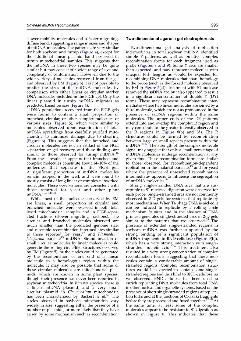
slower mobility molecules and a faster migrating,diffuse band, suggesting a range in sizes and shapesof mtDNA molecules. The patterns are very similarfor both soybean and turnip (Figure 4), except forthe additional linear plasmid band observed inturnip mitochondrial samples. This suggests thatthe mtDNA in these two species may be quitesimilar but may consist of a wide range of size andcomplexity of conformation. However, due to thewide variety of molecules recovered from the geland observed by EM (Figure 5) it is not possible topredict the sizes of the mtDNA molecules bycomparison with either linear or circular markerDNA molecules included in the FIGE gel. Only thelinear plasmid in turnip mtDNA migrates aspredicted based on size (Figure 4).
DNA populations recovered from the FIGE gelswere found to contain a small proportion ofbranched, circular, or other complex molecules ofvarious sizes (Figure 5), which were similar tomolecules observed upon examination of totalmtDNA spreadings from carefully purified mito-chondria to minimize damage due to shearing(Figure 6). This suggests that the branched andcircular molecules are not an artifact of the FIGEseparation or gel recovery, and these findings aresimilar to those observed for turnip mtDNA.16
From these results it appears that branched andcomplex molecules constitute about 14–18% of themolecules that separate in the FIGE gel.A significant proportion of mtDNA moleculesremain trapped in the well, and were found tomostly consist of long linear or complex networkedmolecules. These observations are consistent withthose reported for yeast and other plantmtDNA.10,11,15,51
While most of the molecules observed by EMare linear, a small proportion of circular andbranched molecules were observed in both totallysed mitochondrial samples and in FIGE-separ-ated fractions (slower migrating fractions). Thecircular and branched molecules observed aremuch smaller than the predicted genome size,and resemble recombination intermediates similarto those reported for yeast17 and Plasmodiumfalciparum parasite45 mtDNA. Strand invasion ofsmall circular molecules by linear molecules couldgenerate the rolling circle-like structures observedby EM (Figure 5), or the circles could be generatedby the recombination of one end of a linearmolecule to a homologous region within themolecule. It may also be possible that some ofthese circular molecules are mitochondrial plas-mids, which are known in some plant species,though their presence has never been reported insoybean mitochondria. In Brassica species, there isa linear mtDNA plasmid, and a very smallcircular plasmid in Chenopodium mitochondriahas been characterized by Backert et al.52 Thecircles observed in soybean mitochondria varywidely in size, suggesting either the presence of anumber of plasmids, or more likely that they havearisen by some mechanism such as recombination.
Two-dimensional agarose gel electrophoresis
Two-dimensional gel analysis of replicationintermediates in total soybean mtDNA identifiedsimple Y patterns, as well as putative complexrecombination forms for each fragment used asprobe (Figures 8 and 9). Some Y arcs are smallerthan expected, and may represent molecules withunequal fork lengths as would be expected forrecombining DNA molecules that share homologyto the probe (such as the forked molecule observedby EM in Figure 5(a)). Treatment with S1 nucleaseremoved the ssDNA arc, but also appeared to resultin a significant concentration of double Y (DY)forms. These may represent recombination inter-mediates where two linear molecules are joined by athird molecule, which are not as pronounced in thepresence of ssDNA regions within the samemolecules. The upper ends of the DY patternsextend into and overlap the complex R region andmay contribute to the greater intensity observed inthe R regions in Figure 8(b) and (d). The Rstructures could be formed by recombinationbetween large or small repeats present within themtDNA.24,53 The strength of the complex moleculesignal may suggest that only a small percentage ofmtDNA molecules undergo recombination at anygiven time. These recombination forms are similarto those observed for recombination-dependentreplication in the malarial parasite,45 and for yeastwhere the presence of unresolved recombinationintermediates appears to influence the segregationof mtDNA molecules.54
Strong single-stranded DNA arcs that are sus-ceptible to S1 nuclease digestion were observed foreach probe. Single-stranded arcs are not commonlyobserved in 2-D gels for systems that replicate bymost mechanisms. When T4 phage DNA is nicked itcan be induced to replicate by a rolling circlemechanism in vitro, and in the absence of DNAprimase generates single-stranded arcs in 2-D gelssimilar to the patterns that we observed.55 Thepresence of extended single-stranded regions insoybean mtDNA was further supported by thestrong binding of a significant population ofmtDNA fragments to BND-cellulose (Figure 9(b)),which has a very strong interaction with single-stranded nucleic acids.56 This treatment alsoresulted in a very strong concentration of complexrecombination forms, suggesting that these mol-ecules contain a considerable amount of single-stranded regions. Complex recombination struc-tures would be expected to contain some single-stranded regions and thus bind to BND-cellulose, aswe observed. BND-cellulose has been used toenrich replicating DNA molecules from total DNAin other nuclear and organelle systems, based on thepresence of short single-stranded regions at replica-tion forks and at the junctions of Okazaki fragmentsbefore they are processed and fused together.57–59Atthe same time, at least some of the complexmolecules appear to be resistant to S1 digestion asshown in Figure 8. This indicates that these

296 Soybean MtDNA Recombination
molecules likely contain a mixture of both double-stranded and single-stranded regions.
A significant amount of single-stranded DNA hasbeen observed in mtDNA preparations fromvarious plant species8,10 and from yeast, wheresingle-stranded regions affect PFGE patterns.51,60
Single-stranded regions of DNA molecules may bedirectly involved in strand-exchange steps ofrecombination and may be a result of DNAunwinding after introduction of nicks by either arandom or sequence-specific process. A double-stranded break, followed by preferential degra-dation of one strand by a 5 0-3 0 exonuclease, wouldlead to single-stranded regions with a free 3 0 end,which is the preferred element for strand inva-sion.20 Similar fragments could also be generated byexonuclease activity on linear DNA molecules,which appear to be the prevalent form ofmtDNA.9,11,16 Alternatively, single-stranded DNAmay result from rolling circle DNA replication if thetail does not undergo lagging strand syn-thesis.8,52,60,61
Taken together, the results presented here pro-vide strong support for the occurrence of activeDNA recombination in soybean mitochondria. Therelatively low amounts of recombination structuresobserved in 2-D gels (faint R patterns relative tostrong linear monomer signals) and by EM analysissuggest that only a small portion of mtDNAmolecules are undergoing recombination at anygiven time. The next question to be considered iswhether mtDNA recombination is essential formitochondrial function and plant growth. Analysisof mutants in mtDNA recombination proteins inArabidopsis is currently underway.
Materials and Methods
Isolation of soybean mitochondrial strand invasionactivity
Mitochondria from young soybean seedlings werepurified on Percoll gradients as described.62 The mito-chondrial band was recovered and the mitochondria werepelleted by centrifugation and washed. The pellets wereresuspended in a total of 10 ml of water containing 0.1%Triton X-100 and incubated on ice for 20 min to lyse themitochondria. The sample was centrifuged at 15,000g for20 min, and the supernatant was recovered and loaded ona 30 ml DEAE cellulose column equilibrated with bufferA (50 mM Tris–acetate (pH 7.5), 20% (v/v) glycerol,50 mM 2-mercaptoethanol, 0.2 mM PMSF, 0.1% TritonX-100) containing 50 mM sodium acetate. The columnwas then washed with 200 ml buffer A, followed byelution with buffer A containing 500 mM sodium acetate.Selected elution fractions were collected, pooled anddialyzed in buffer A overnight with two buffer changes.The dialyzed proteins were then loaded onto anequilibrated heparin Sepharose column (w5 cm3 columnvolume) at 0.3 ml/min. The column was washed with50 ml of buffer A at 0.33 ml/min. Proteins were elutedwith buffer A containing 0.6 M sodium acetate at 0.5 ml/min, and fractions assayed for strand invasion activity.
DNA substrates
M13 replicative form (RF) dsDNA and M13 single-stranded DNA were prepared as described.63 DNAconcentrations were determined by spectrophotometry(BioRad SmartSpec 3000). Replicative form DNA waslinearized by digestion with BamHI (BioLab), separatedin an agarose gel and recovered using a QIAquick kit(QIAGEN).
Soybean mtDNA strand invasion assays
Protein fractions were assayed for homologous strandinvasion activity as described.48,64 The reaction mixturescontained: 30 mM Tris–acetate (pH 7.5), 2 mM DTT,50 mg/ml BSA, 20 mM magnesium acetate, 2.5 mM ATP,15 ml protein fraction (for control, added 2 mg E. coli RecA(New England BioLab)), 15 mM ssDNA and 15 mM RFdsDNA, 1.8 mg E. coli single-stranded binding protein(SSB) (Promega) and dH2O to 25 ml final volume. In thefirst step of the reaction, Tris–acetate (pH 7.5), DTT, BSA,MgCl2, ATP, protein fraction (RecA for control), anddH2O were combined and incubated at 37 8C for 5 min. Inthe second step, ssDNA was added and incubationcontinued at 37 8C for 10 min. In the third step, SSB wasadded with incubation at 37 8C for another 10 min. For thefourth step, RF dsDNA was added and the samplesincubated at 37 8C for 60 min. Stop solution (0.5% (w/v)SDS, 50 mM EDTA) was then added followed byincubation for 5 min at 37 8C and 65 8C for 5 min. TheDNA was recovered and separated by agarose gelelectrophoresis, and visualized after staining by ethidiumbromide.
Isolation of soybean mitochondria for DNA analysis
Approximately 500 g of 1.5 to 2 week old etiolatedsoybean (Glycine max Merr. var Essex) seedlings werehomogenized in 150–200 g batches with two to threevolumes (ml/g) of cold STM buffer (0.5 M sucrose, 50 mMTris–HCl (pH 8.0), 5 mM MgCl2, 5 mM b-mercap-toethanol, 0.2 mM PMSF) in an ice-cold blender jar withthree 5 s bursts.65 Subsequent steps were carried out at4 8C. The resulting homogenate was filtered through fourlayers of cheesecloth and three layers of Miracloth(Calbiochem). The filtrate was centrifuged at 2500 rpmin a GSA (Sorvall) rotor for 20 min to remove chloroplastsand nuclei, and the resulting supernatant was centrifugedat 12,000 rpm in a GSA rotor for 30 min to pelletmitochondria. The pellets were resuspended in a total of200 ml cold STM buffer, both centrifugation steps wererepeated, and the final pellets were resuspended for DNApurification or for direct FIGE or EM analysis of DNAmolecules.
FIGE analysis of soybean mtDNA
The mitochondrial pellet from above was resuspendedin 1 ml of wash buffer, mixed with an equal volume of1.2% (w/v) low-melting point agarose prepared in 0.5XTBE and allowed to solidify in plug-molds. Mitochondrialmembranes were digested by incubating plugs in lysisbuffer (1% SDS, 0.1 M EDTA, 0.5 mg/ml proteinase K) at50 8C for 3 h overnight. The plugs were washed in severalvolumes of TE (10 mM Tris–HCl (pH 8.0), 1 mM EDTA).Field inversion gel electrophoresis was conducted essen-tially as described.16 The mtDNA from lysed mitochon-dria was resolved by FIGE for 40 h at 4 8C, with pulse

Soybean MtDNA Recombination 297
times ramped from 6 s to 24 s in a 3:1 TF:TR ratio.66 Twosets of conditions were used, the first at 35 mA (180 V),the second at 25 mA (125 V). Lambda DNA concatemerswere used as high molecular mass linear size markers.After electrophoresis, a portion of the gel was stainedwith 0.3 mg/ml ethidium bromide and photographed.The remainder of the gel was used for recovery of DNAfor EM.
Electron microscopy analysis of soybean mtDNA
FIGE-separated mtDNA samples were excised from theunstained portion of the gel and dialyzed in Gelase(Epicenter) buffer for 1 h at room temperature. Gel pieceswere then heated at 70 8C until completely melted, cooledto 45 8C and digested for 1 h with one unit of Gelase. ForEM analysis, 20 ml of the Gelase-digested sample or totalsoybean mtDNA was mixed in 200 ml of buffer containing0.25 M ammonium acetate, 0.01 M Tris–HCl (pH 8.0),0.001 M EDTA and 3 mg/ml cytochrome c, and 45 ml ofthis mixture was deposited on a Teflon block.67 The DNAwas allowed to diffuse for 20 min, and then transferred toparlodion-coated, 200 mesh copper grids by touching thegrid to the drop surface. The grids were washed in dH2Ofor 5 s, flooded with 140 ml of 0.4% Kodak Photoflo andstained for 30 s in 30 ml of 1% uranyl acetate.68 The gridswere washed in 90% (v/v) ethanol for 10 s, dried andviewed with a Zeiss transmission electron microscopeoperating at 60 kV. In addition, total mtDNA moleculeswere observed directly from lysed mitochondria withoutheating or Gelase treatment.
Purification of soybean mtDNA
Mitochondrial pellets from above were resuspendedand treated with DNase I,16 and mtDNA was purified bycesium chloride gradient centrifugation.16,27 The purity ofthe mtDNA was analyzed by probing a Southern blot63 ofrestriction-digested DNA with a tobacco nuclear 18 SrRNA gene and a chloroplast rpl16 gene labeled byrandom priming (Prime-A-Gene, Promega), with nodetectable hybridization (not shown).
2-D gel analysis of restricted soybean mtDNA
Total soybean mtDNA was restricted with PstI prior togel separation. For some experiments DNA samples wereenriched for molecules containing single-strandedregions by passage through BND-cellulose.47,56 The highsalt plus caffeine elution fraction was concentrated byprecipitation with ethanol and separated by 2-D gelelectrophoresis. Samples were loaded in a 0.5% agarosegel without EtBr, and electrophoresed in the firstdimension at 1 V/cm overnight in 1X TBE buffer.69 Thegel was stained with 0.3 mg/ml EtBr and photographed,and the sample lanes were cut apart and placedhorizontally at the top of a second gel. The seconddimension gel contained 1% agarose and 0.3 mg/ml EtBrin 1X TBE buffer.69 The gel was electrophoresed at 5 V/cmfor 6 h with buffer recirculation, after which the DNA wastransferred to nylon membrane by Southern blotting.63
Hybridization with specific soybean mtDNA PstI frag-ments was followed by exposure to X-ray film to visualizereplication intermediates specific for each probe. Toidentify patterns resulting from single-stranded DNAmolecules or regions, some samples were treated with S1nuclease (two units in a 30 ml reaction) for 20 min at 37 8C
after restriction digestion, prior to electrophoresis of thesample in the first dimension.59
Acknowledgements
This work was supported in part by grants fromthe NIH (GM066787), the Mentoring EnvironmentGrants Program (BYU), and by the Office of the VicePresident for Research, Auburn University. K.S.G.was supported in part by an NIH-MARC facultyfellowship (GM15475). We thank John Cupp, BennFronk and Joel Hall for assistance in data analysisand preparation of some of the Figures.
References
1. Mackenzie, S. & McIntosh, L. (1999). Higher plantmitochondria. Plant Cell, 11, 571–585.
2. Schuster, W. & Brennicke, A. (1994). The plantmitochondrial genome: physical structure, infor-mation content, RNA editing, and gene migration tothe nucleus. Annu. Rev. Plant Physiol. Plant Mol. Biol.45, 61–78.
3. Larsson, N.-G. & Clayton, D. A. (1995). Moleculargenetic aspects of human mitochondrial disease.Annu. Rev. Genet. 29, 151–178.
4. Binder, S., Marchfelder, A. & Brennicke, A. (1996).Regulation of gene expression in plant mitochondria.Plant Mol. Biol. 32, 303–314.
5. Unseld, M., Marienfeld, J., Brandt, P. & Brennicke, A.(1997). The mitochondrial genome of Arabidopsisthaliana contains 57 genes in 366,924 nucleotides.Nature Genet. 15, 57–61.
6. Oda, K., Yamato, K., Ohata, E., Nakamura, Y.,Takemura, M., Nozato, N. et al. (1992). Geneorganization from the complete sequence of liver-wort, Marchantia polymorpha, mitochondrial DNA: aprimitive form of plant mitochondrial genome. J. Mol.Biol. 223, 1–7.
7. Backert, S., Dorfel, P. & Borner, T. (1995). Investigationof plant organellar DNA by pulsed-field gel electro-phoresis. Curr. Genet. 28, 390–399.
8. Backert, S., Lurz, R., Oyarzabal, O. A. & Borner, T.(1997). High content, size and distribution of single-stranded DNA in the mitochondria of Chenopodiumalbum (L.). Plant Mol. Biol. 33, 1037–1050.
9. Bendich, A. J. & Smith, S. B. (1990). Moving picturesand pulsed-field gel electrophoresis show linear DNAmolecules from chloroplasts and mitochondria. Curr.Genet. 17, 421–425.
10. Bendich, A. J. (1996). Structural analysis of mitochon-drial DNA molecules from fungi and plants usingmoving pictures and pulsed-field gel electrophoresis.J. Mol. Biol. 255, 564–588.
11. Oldenburg, D. J. & Bendich, A. J. (1996). Size andstructure of replicating mitochondrial DNA incultured tobacco cells. Plant Cell, 8, 447–461.
12. Oldenburg, D. J. & Bendich, A. J. (2001). Mitochon-drial DNA from the liverwort Marchantia polymorpha:circularly permuted linear molecules, head-to-tailconcatemers, and a 5 0 protein. J. Mol. Biol. 310,549–562.

298 Soybean MtDNA Recombination
13. Backert, S., Nielsen, B. L. & Borner, T. (1997). Themystery of the rings: structure and replication ofmitochondrial genomes from higher plants. TrendsPlant Sci. 2, 477–483.
14. Jacobs, M. A., Payne, S. R. & Bendich, A. J. (1996).Moving pictures and pulsed-field gel electrophoresisshow only linear mitochondrial DNA molecules fromyeasts with linear-mapping and circular-mappingmitochondrial genomes. Curr. Genet. 30, 3–11.
15. Oldenburg, D. J. & Bendich, A. J. (1998). The structureof mitochondrial DNA from the liverwort, Marchantiapolymorpha. J. Mol. Biol. 276, 745–758.
16. Scissum-Gunn, K. D., Gandhi, M., Backert, S. &Nielsen, B. L. (1998). Separation of different confor-mations of plant mitochondrial DNA molecules byfield inversion gel eletrophoresis. Plant Mol. Biol. Rep.16, 219–229.
17. Sena, E. P., Revet, B. & Moustacchi, E. (1986). In vivohomologous recombination intermediates of yeastmitochondrial DNA analyzed by electron microscopy.Mol. Gen. Genet. 202, 421–428.
18. Mosig, G. (1987). The essential role of recombinationin phage T4 growth. Annu. Rev. Genet. 21, 347–371.
19. Mosig, G. (1998). Recombination and recombination-dependent DNA replication in bacteriophage T4.Annu. Rev. Genet. 32, 379–413.
20. Backert, S. & Borner, T. (2000). Phage T4-likeintermediates of DNA replication and recombinationin the mitochondria of the higher plant Chenopodiumalbum (L.). Curr. Genet. 37, 304–314.
21. Sato, A., Nakada, K., Akimoto, M., Ishikawa, K., Ono, T.,Shitara, H. et al. (2005). Rare creation of recombinantmtDNA haplotypes in mammalian tissues. Proc. NatlAcad. Sci. USA, 102, 6057–6062.
22. Andre, C. P. & Walbot, V. (1995). Pulsed-field gelmapping of maize mitochondrial chromosomes. Mol.Gen. Genet. 247, 255–263.
23. Bendich, A. J. (1993). Reaching for the ring: the studyof mitochondrial genome structure. Curr. Genet. 24,279–290.
24. Fauron, C., Casper, M., Gao, Y. & Moore, B. (1995). Themaize mitochondrial genome: dynamic, yetfunctional. Trends Genet. 11, 228–235.
25. Fauron, C. M. R., Casper, M., Gesteland, R. &Albertsen, M. (1992). A multi-recombination modelfor the mtDNA rearrangements seen in maize cmsTregenerated plants. Plant J. 2, 949–958.
26. Palmer, J. D. & Herbon, L. A. (1988). Plant mitochon-drial DNA evolves rapidly in structure, but slowly insequence. J. Mol. Evol. 28, 87–97.
27. Palmer, J. D. & Shields, C. R. (1984). Tripartitestructure of the Brassica campestris mitochondrialgenome. Nature, 307, 437–440.
28. Palmer, J. D., Makaroff, C. A., Apel, I. J. &Shirzadegan, M. (1990). Fluid structure of plantmitochondrial genomes: evolutionary and functionalimplications. In Molecular Evolution (Clegg, M. T. &O’Brien, S. J., eds), pp. 85–96, Alan R. Liss, New York.
29. Andre, C. P., Levy, A. & Walbot, V. (1992). Smallrepeated sequences and the structure of plantmitochondrial genomes. Trends Genet. 8, 128–132.
30. Flamand, M. C., Duc, G., Goblet, J. P., Hong, L.,Louis, O., Briquet, M. & Boutry, M. (1993). Variantmitochondrial plasmids of broad bean arose byrecombination and are controlled by the nucleargenome. Nucl. Acids Res. 21, 5468–5473.
31. Satoh, M., Nemoto, Y., Kawano, S., Nagata, T.,Hirokawa, H. & Kuroiwa, T. (1993). Organization of
heterogeneous mitochondrial DNA molecules inmitochondrial nuclei of cultured tobacco cells. Proto-plasma, 175, 112–120.
32. Small, I., Suffolk, R. & Leaver, C. J. (1989). Evolution ofplant mitochondrial genomes via substoichiometricintermediates. Cell, 58, 69–76.
33. Kanazawa, A., Tozuka, A., Kato, S., Mikami, T., Abe, J.& Shimamoto, Y. (1998). Small interspersed sequencesthat serve as recombination sites at the cox2 and atp6loci in the mitochondrial genome of soybean arewidely distributed in higher plants. Curr. Genet. 33,188–198.
34. Kato, S., Kanazawa, A., Mikami, T. & Shimamoto, Y.(1998). Evolutionary changes in the structures of thecox2 and atp6 loci in the mitochondrial genome ofsoybean involving recombination across small inter-spersed sequences. Curr. Genet. 34, 303–312.
35. Klein, M., Eckert-Ossenkopp, U., Schmiedeberg, I.,Brandt, P., Unseld, M., Brennicke, A. & Schuster, W.(1994). Physical mapping of the mitochondrialgenome of Arabidopsis thaliana by cosmid and YACclones. Plant J. 6, 447–455.
36. Marienfeld, J. R., Unseld, M., Brandt, P. & Brennicke, A.(1997). Mosaic open reading frames in the Arabidopsisthaliana mitochondrial genome. Biol. Chem. 378,859–862.
37. Schon, E. A., Rizzuto, R., Moraes, C. T., Nakase, H.,Zeviani, M. & DiMauro, S. (1989). A direct repeat is ahotspot for large-scale deletion of human mitochon-drial DNA. Science, 244, 346–349.
38. Sor, F. & Fukuhara, H. (1983). Unequal excision ofcomplementary strands is involved in the generationof palindromic repetitions of rhoK mitochondrialDNA in yeast. Cell, 32, 391–396.
39. Grabau, E. A., Davis, W. H., Phelps, N. D. &Gengenbach, B. G. (1992). Classification of soybeancultivars based on mitochondrial DNA restrictionfragment length polymorphisms. Crop Sci. 32,271–274.
40. Moeykens, C. A., Mackenzie, S. A. & Shoemaker, R. C.(1995). Mitochondrial genome diversity in soybean:repeats and rearrangements. Plant Mol. Biol. 29,245–254.
41. Hanlon, R. W. & Grabau, E. A. (1997). Comparison ofmitochondrial organization of four soybean cyto-plasmic types by restriction mapping. Soybean Genet.Newsletter, 24, 208–210.
42. Khazi, F. R., Edmondson, A. C. & Nielsen, B. L. (2003).An Arabidopsis homologue of bacterial RecA thatcomplements an E. coli recA deletion is targeted toplant mitochondria. Mol. Gen. Genet. 269, 454–463.
43. Cerutti, H., Osman, M., Grandoni, P. & Jagendorf, A. T.(1992). A homolog of Escherichia coli RecA protein inplastids of higher plants. Proc. Natl Acad. Sci. USA, 89,8068–8072.
44. Palmer, J. D., Shields, C. R., Cohen, D. B. & Orton, T. J.(1983). An unusual mitochondrial DNA plasmid inthe genus Brassica. Nature, 301, 725–728.
45. Preiser, P. R., Wilson, R. J. M., Moore, P. W.,McCready, S., Hajibagheri, M. A. N., Blight, K. J.et al. (1996). Recombination associated with replica-tion of malarial mitochondrial DNA. EMBO J. 15,684–693.
46. Han, Z. & Stachow, C. (1994). Analysis of Schizo-saccharomyces pombe mitochondrial DNA replicationby two dimensional gel electrophoresis. Chromosoma,103, 162–170.
Soybean MtDNA Recombination 299
47. Gamper, H., Lehman, N., Piette, J. & Hearst, J. (1985).Purification of circular DNA using benzoylatednaphthoylated DEAE-cellulose. Gene, 4, 157–164.
48. Cerutti, H. & Jagendorf, A. T. (1993). DNA strand-transfer activity in pea (Pisum sativum L.) chloroplasts.Plant Physiol. 102, 145–153.
49. Levene, S. & Zimm, B. (1987). Separation of open-circular DNA using pulsed-field electrophoresis. Proc.Natl Acad. Sci. USA, 84, 4054–4057.
50. Beverley, S. M. (1988). Characterization of the “un-usual” mobility of large circular DNAs in pulsed field-gradient electrophoresis. Nucl. Acids Res. 16, 925–939.
51. Maleszka, R. (1993). Single-stranded regions in yeastmitochondrial DNA revealed by pulsed-field gelelectrophoresis. Appl. Theor. Electrophoresis, 3, 259–263.
52. Backert, S., Meisner, K. & Borner, T. (1997). Uniquefeatures of the mitochondrial rolling circle-plasmidmp1 from the higher plant Chenopodium album (L.).Nucl. Acids Res. 25, 582–589.
53. Levy, A. A., Andre, C. P. & Walbot, V. (1991). Analysisof a 120-kilobase mitochondrial chromosome inmaize. Genetics, 128, 417–424.
54. Lockshon, D., Zweifel, S. G., Freeman-Cook, L. L.,Lorimer, H. E., Brewer, B. J. & Fangman, W. L. (1995).A role for recombination junctions in the segregationof mitochondrial DNA in yeast. Cell, 81, 947–955.
55. Belanger, K. G., Mirzayan, C., Kreuzer, H. E.,Alberts, B. M. & Kreuzer, K. N. (1996). Two-dimensional gel analysis of rolling circle replicationin the presence and absence of bacteriophage T4primase. Nucl. Acids Res. 24, 2166–2175.
56. Sedat, J. W., Kelly, R. B. & Sinsheimer, R. L. (1967).Fractionation of nucleic acid on benzoylated–naphthoylated DEAE cellulose. J. Mol. Biol. 26, 537–540.
57. Dijkwel, P. A., Vaughn, J. P. & Hamlin, J. L. (1991).Mapping of replication initiation sites in mammaliangenomes by two dimensional gel analysis: stabiliz-ation and enrichment of replication intermediates byisolation on the nuclear matrix. Mol. Cell. Biol. 11,3850–3859.
58. Hay, R. & dePamphilis, M. (1982). Initiation of simianvirus 40 DNA replication in vivo: location andstructure of 5 0-ends of DNA synthesized in the ori-region. Cell, 28, 767–779.
59. Lu, Z., Kunnimalaiyaan, M. & Nielsen, B. L. (1996).Characterization of replication origins flanking the23 S rRNA gene in tobacco chloroplast DNA. PlantMol. Biol. 32, 693–706.
60. Maleszka, R., Skelly, P. J. & Clark-Walker, G. D. (1991).Rolling circle replication of DNA in yeast mitochon-dria. EMBO J. 10, 3923–3929.
61. Backert, S., Dorfel, P., Lurz, R. & Borner, T. (1996).Rolling-circle replication of mitochondrial DNA inthe higher plant Chenopodium album (L.). Mol. Cell.Biol. 16, 6285–6294.
62. Hrubec, T., Robinson, J. & Donaldson, R. (1985).Isolation of mitochondria from soybean leaves ondiscontinuous Percoll gradients. Plant Physiol. 77,1010–1012.
63. Sambrook, J. & Russell, D. W. (2001). MolecularCloning: A Laboratory Manual, 3rd edit., ColdSpring Harbor Laboratory Press, Cold SpringHarbor, NY.
64. Steffen, S. E. & Bryant, F. R. (2001). Purification andcharacterization of the single-stranded DNA bindingprotein from Streptococcus pneumoniae. Arch. Biochem.Biophys. 388, 165–170.
65. Daniell, H., Zheng, D. & Nielsen, B. L. (1995). Isolationand characterization of an in vitro DNA replicationsystem from maize mitochondria. Bioch. Biophys. Res.Commun. 208, 287–294.
66. Heller, C. & Poll, F. (1990). Field inversion gelelectophoresis with different pulsed time ramps.Nucl. Acids Res. 18, 6299–6304.
67. Weiden, M., Osheim, Y., Beyer, A. & van der Ploeg, L.(1991). Chromosome structure: DNA nucleotidesequence elements of a subset of the minichromo-somes of the protozoan Trypanosoma brucei. Mol. Cell.Biol. 11, 3823–3834.
68. Serwer, P. (1978). A technique for observing extendedDNA in negatively stained specimens: observation ofbacteriophage T7 capsid–DNA complexes.J. Ultrastruct. Res. 65, 112–118.
69. Brewer, B. J. & Fangman, W. L. (1991). Mappingreplication origins in yeast chromosomes. BioEssays,13, 317–322.
Edited by J. Karn
(Received 8 August 2005; received in revised form 12 November 2005; accepted 22 November 2005)Available online 9 December 2005
Copyright © 2022 FDOKUMEN