![KARAKTERISTIKEKSTRAK AIR DAUN PUGUNTANO [Curanga fel-terrae (Lour.) Merr.]YANG BERPOTENSI SEBAGAI ANTELMINTIK](https://static.fdokumen.com/doc/165x107/633999c8a16e1672830dae6a/karakteristikekstrak-air-daun-puguntano-curanga-fel-terrae-lour-merryang-berpotensi.jpg)
The ribosomal protein P0 of soybean (Glycine max L. Merr.) has antigenic cross-reactivity to soybean...
13
See discussions, stats, and author profiles for this publication at: https://www.researchgate.net/publication/14011238 The ribosomal protein P0 of soybean (Glycine max L. Merr.) has antigenic cross-reactivity to soybean seed lectin Article in Plant Molecular Biology · June 1997 DOI: 10.1023/A:1005817114562 · Source: PubMed CITATIONS 5 READS 31 3 authors, including: Some of the authors of this publication are also working on these related projects: Antifungal Activity of Bacillus species against Fusarium and Analysis of the Mechanisms Used in Biocontrol. View project Keith L. Wycoff Planet Biotechnology, Inc. 30 PUBLICATIONS 1,633 CITATIONS SEE PROFILE Ann M. Hirsch University of California, Los Angeles 213 PUBLICATIONS 5,447 CITATIONS SEE PROFILE All content following this page was uploaded by Ann M. Hirsch on 03 December 2016. The user has requested enhancement of the downloaded file.
Transcript of The ribosomal protein P0 of soybean (Glycine max L. Merr.) has antigenic cross-reactivity to soybean...

Seediscussions,stats,andauthorprofilesforthispublicationat:https://www.researchgate.net/publication/14011238
TheribosomalproteinP0ofsoybean(GlycinemaxL.Merr.)hasantigeniccross-reactivitytosoybeanseedlectin
ArticleinPlantMolecularBiology·June1997
DOI:10.1023/A:1005817114562·Source:PubMed
CITATIONS
5
READS
31
3authors,including:
Someoftheauthorsofthispublicationarealsoworkingontheserelatedprojects:
AntifungalActivityofBacillusspeciesagainstFusariumandAnalysisoftheMechanismsUsedin
Biocontrol.Viewproject
KeithL.Wycoff
PlanetBiotechnology,Inc.
30PUBLICATIONS1,633CITATIONS
SEEPROFILE
AnnM.Hirsch
UniversityofCalifornia,LosAngeles
213PUBLICATIONS5,447CITATIONS
SEEPROFILE
AllcontentfollowingthispagewasuploadedbyAnnM.Hirschon03December2016.
Theuserhasrequestedenhancementofthedownloadedfile.

Plant Molecular Biology 34: 295–306, 1997. 295c 1997 Kluwer Academic Publishers. Printed in Belgium.
The ribosomal protein P0 of soybean (Glycine max L. Merr.) has antigeniccross-reactivity to soybean seed lectin
Keith L. Wycoff1;3, Pieternel van Rhijn1 and Ann M. Hirsch1;2;�
1Department of Molecular, Cell and Developmental Biology (�author for correspondence) and 2MolecularBiology Institute, University of California, Los Angeles, 405 Hilgard Ave., Los Angeles, CA 90095-1606, USA;3Present address: Planet Biotechnology, Inc., 2462 Wyandotte St., Mountain View, CA 94043, USA
Received 20 February 1996; accepted in revised form 3 March 1997
Key words: lectin, ribosomal protein P0, soybean
Abstract
Soybean (Glycine max L. Merr.) mutants lacking the ability to produce the lectin normally found in soybean seeds(SBL) are designated Le�. A protein of higher molecular weight that cross-reacts with antibodies raised to SBLwas found at nearly equivalent levels in roots, hypocotyls, and leaves, and at lower levels in cotyledons and dryseeds of both Le+ and Le� soybean cultivars. Earlier work suggested that this protein was a novel lectin. Clonesisolated from a Le� soybean root cDNA library produced a cross-reacting protein of the same size in Escherichiacoli. Sequence analysis of these clones revealed a high degree of similarity to the ribosomal protein P0. Thecross-reacting protein co-purified with ribosomes, and a monoclonal antibody raised to purified brine shrimp P0cross-reacted to the same protein. The protein showed no lectin activity in a hemagglutination assay, nor did it bindto an N-acetyl-D-galactosamine affinity column. On the basis of this evidence, we conclude that the SBL-cross-reacting protein is not a lectin but a homologue of the ribosomal protein P0. Consequently, Le� soybeans mustproduce a lectin that is dissimilar to SBL at both the DNA and amino acid levels and we suggest that it is this lectinwhich is involved in nodulation.
Introduction
The ability of legumes to associate in nitrogen-fixingsymbiosis with soil bacteria of the family Rhizobi-aceae, thus freeing them from dependence on soilnitrogen, has long been of interest. How the legume-Rhizobium symbiosis is initiated remains a key ques-tion. The interaction between the plant and bacteriaexhibits a high degree of specificity; for example,Rhizobium meliloti nodulates alfalfa and not soybean,whereas Bradyrhizobium japonicum infects soybeanand not alfalfa. Rhizobial Nod factors, substituted lipo-oligosaccharides produced by the bacteria in responseto plant signals, are one major determinant of spe-cificity (see reviews [9, 25]). Although Nod factor eli-cits both hair deformation and the beginnings of nodule
The nucleotide sequence data reported will appear in the EMBL,GenBank and DDBJ Nucleotide Sequence Databases under theaccession number L46848.
formation, rhizobia must physically contact the plantcell wall for normal infection threads to be formed (seereferences in [19]).
When rhizobia contact their host, they attach to sus-ceptible (emerging) root hairs [3]. Polysaccharides andperhaps oligosaccharides are thought to facilitate thisattachment. Interestingly, there is a strong correlationbetween the inoculation specificity of rhizobial specieson their legume hosts, and the ability of host-producedlectins to bind to Rhizobium cells. The lectin recogni-tion hypothesis, formulated more than 20 years ago,[4, 6, 18], proposed that lectin-mediated binding of thehomologous rhizobial strain to the root hair surface isan essential step in the determination of host-symbiontspecificity (see references in [21]).Heterologous rhizo-bia do not carry the appropriate carbohydrate ligands,and thus will not bind in a way that initiates noduleformation.
GR: 201001932, Pips nr. 136858 BIO2KAP
*136858* pla275us.tex; 7/05/1997; 13:50; v.7; p.1

296
Perhaps the most persuasive evidence in favor of thelectin-recognition hypothesis comes from the work ofDıaz et al. [10] who introduced a cloned pea seed lectingene into white clover (Trifolium repens). The trans-genic clover roots were nodulated by R. leguminosar-um bv. viciae, which normally nodulates pea orvetch and not clover. Site-directed mutagenesis ofthe carbohydrate-binding domain of the lectin demon-strated that the activity of this region was responsiblefor the change in host specificity [40]. The close rela-tionship between the two rhizobial biovars that nodu-late pea and clover makes it difficult, however, to drawbroad conclusions that might apply to other legume-Rhizobium interactions.
Lectin is present in young soybean roots and is mostabundant in the region of developing root hairs, whereinfection takes place [13]. The most compelling evi-dence that soybean seed lectin (SBL) or a related rootlectin has a stimulatory effect on the infection processcomes from work by Halverson and Stacey [15, 1617], who studied a B. japonicum mutant that showsa delayed nodulation phenotype on its soybean host.Pretreatment of the mutant or low inoculum densit-ies of wild-type B. japonicum with either SBL, rootexudates, or affinity-purified root lectin reversed thedelayed nodulation phenotype. This reversal is inhib-ited specifically by D-galactose, a known SBL ligand.
A soybean root lectin, isolated from soybean rootexudates [13] is similar, but not identical, to the well-characterized SBL. Whereas the SBL monomer has anapparent molecular weight of 28 to 30 kDa on SDS-PAGE, the root-specific lectin is 32 to 33 kDa. Hemag-glutinating activity of this lectin was inhibited best byN-acetyl-D-galactosamine and EPS from B. japonic-um, but not by EPS from R. phaseoli [13].
Soybean mutants lacking SBL (Le�) nodulatequite normally [14, 30], an observation that wouldseem to contradict the lectin recognition hypothesis.However, in addition to the D-galactose/N -acetyl-D-galactosamine-specific SBL, soybean seeds contain a45 kDa lectin specific for 4-O-methylglucuronic acid[31]. Southern hybridization and S1 protection exper-iments have failed to identify any other active lectingenes in soybean besides SBL and the 45 kDa lectin[29]. However, Vodkin and Raikhel [42] detected a33 kDa protein in the roots of both Le+ and Le� soy-beans that cross-reacted with polyclonal anti-SBL anti-bodies raised to denatured SBL. Spilatro et al. [36, 37]identified 33 and 28 kDa polypeptides from a Le+ soy-bean using the same antibody and showed that the N-terminus of each polypeptide had 62% identity to SBL.
They were unable, however, to show that these proteinshad lectin activity. Sengupta-Gopalan et al. [35] isol-ated and subjected mRNA from roots of Le� plants toin vitro translation, and produced a 32 kDa protein thatcross-reacted with anti-SBL antibodies. However, anSBL cDNA clone did not hybridize to the mRNA, sug-gesting that although the protein was immunologicallycross-reactive, the gene sequence was divergent. Thus,a number of studies pointed to the presence of a nov-el lectin which was important for nodulation of Le�
soybeans.Using the same antibody that recognized the cross-
reacting protein from soybean roots [42],we screened asoybean cv. Sooty (Le�) root cDNA expression libraryin an attempt to isolate a root lectin gene. Instead of aroot lectin, however, we found that the cross-reactingprotein was a homolog of the acidic ribosomal proteinP0. This result explains some of the earlier findings[35, 42] and also has important implications to thestudies of the involvement of lectins in nodulation.
Materials and Methods
Plant growth conditions
Seeds of soybean (Glycine max L. Merr.) cvs. Williams82 (Le+) and Sooty (Le�) were surface-sterilized bysoaking for 15 min in 30% (v/v) H2O2, and then washedextensively with sterile, deionized water. Sooty seedswere scarified by scratching with a scalpel before sur-face sterilization. To obtain tissue for extracts, seedswere germinated in a mixture of vermiculite and per-lite (1:1; v/v) that was moistened with water in plasticdishpans. Plants were grown at 24 �C, 80% relativehumidity, with 16 h days/8 h nights in a Convirongrowth cabinet. To obtain root exudates, seeds weregerminated on top of 2 layers of cheesecloth drapedover a wire mesh screen suspended above 1 l of steriletap water in a plastic dishpan. The dishpan was keptcovered with aluminum foil to maintain the plants indarkness.
Construction and screening of a cDNA library
One-cm long root tips were excised from 5-day-oldseedlings of cv. Sooty and rapidly frozen in liquidnitrogen. A method for isolating polysomal RNA yiel-ded 214 �g of polysomal RNA from 1 g of root tips[20]. Poly(A)+ RNA was prepared from total polyso-mal RNA using the polyATract mRNA isolation sys-
pla275us.tex; 7/05/1997; 13:50; v.7; p.2

297
tem (Promega) according to the manufacturer’s pro-tocol. From 168 �g of polysomal RNA, 1.0 �g ofpoly(A)+ mRNA was isolated. First and second strandcDNAs were made from 1.0 �g of poly(A)+ RNAusing the cDNA Synthesis System Plus (Amersham)according to the manufacturer’s protocol. The cDNAswere treated with EcoRI methylase, ligated to EcoRIlinkers, and cut with EcoRI (all from New England Bio-labs). The linker fragments were removed by passingthe mixture through a column of Sephadex G-50 (Phar-macia Biotech). One cDNA library was made fromeach of two fractions eluted from the column. Thefirst fraction, consisting of the leading half of the peakof cDNAs eluted (presumably containing the longestcDNAs) was used to make the library designated ‘50’.The second fraction, consisting of the trailing half ofthe peak of cDNAs eluted and presumed to containshorter cDNAs, was used to make the library des-ignated ‘150’. The cDNAs were ligated into EcoRI-cut and dephosphorylated �ZAP II arms (Stratagene).Ligated lambda DNA was packaged using GigapackII Gold Packaging Extract (Stratagene). Total titer ofthe two libraries constructed was estimated, by platingwith E. coli strain XL-1 Blue, to be 6:4� 105 fu.
The libraries were screened following a single amp-lification on E. coli strain SUREr (Stratagene). Foreach library, phage were plated with SURE cells ontofive plates at 50 000 pfu per 150 mm plate. Plates wereincubated at 42 �C for 4 h and expression of cDNAswas induced by overlaying plates with nitrocellulosefilters soaked in 10 mM isopropylthiogalactopyrano-side (IPTG). Plates were incubated an additional 4 h,then the filters were removed and replaced with dupli-cate filters, which were left on the plates overnight at42 �C. Filters were washed 20 min with TBS+ 0.02%TWEEN-20 and then incubated 4 h with BLOTTO(TBS+ 5% non-fat dry milk). They were subsequentlyincubated 16 to 24 h at 4 � with BLOTTO containing1�g ml�1 of an antibody raised against SDS-denaturedsoybean lectin (anti-SBL) which was affinity-purified.The antibody was a gift from L. Vodkin (University ofIllinois, Champagne-Urbana) and its reaction to soy-bean extracts has been previously reported [41]. Afterwashing twice with TBS and once with BLOTTO, thefilters were incubated 9 to 16 h with BLOTTO contain-ing a 1:1000 dilution of alkaline phosphatase-labeled,affinity-purifiedgoat anti-rabbit IgG antibodies (SigmaChemical Co.). The filters were then washed 3 timeswith TBS. Positive reactions were detected by incub-ating filters in a 1� color developing solution (5�is 500 mM Tris-HCl pH 9, 500 mM NaCl, 25 mM
MgCl2, 0.33 mg ml�1 NBT (nitro-blue tetrazoliump-toluidine salt), 0.165 mg ml�1 BCIP (5-bromo-4-chloro-3-indolyl phosphate)). Preliminary dot immun-oblot experiments showed that this assay could detectas little as 7.5 pg of soybean lectin, well within therange of fusion protein expected to be produced bysingle �ZAP II plaques.
A total of 35 positive plaques were identified bythe primary screening. Of these, 32 �ZAP II cloneswere purified and subjected to in vivo excision usingthe E. coli strain SOLR (Stratagene), resulting in theisolation of 30 cDNAs in the vector pBluescript SK.
SDS-PAGE and western immunoblotting
Plant and E. coli extracts were subjected to SDS-PAGE[23] on 12.5% polyacrylamide gels and then trans-ferred to nitrocellulose in an electro-blotting device(Hoeffer Scientific Products) using standard proced-ures [32]. Two-dimensional electrophoresis of rootextracts were performed as described previously [28].Molecular weight markers were obtained from BioRad.Blots were stained with Ponceau S (Sigma) andmolecular mass markers were outlined with ink. West-ern blots were probed with the anti-SBL antibodies byessentially the same protocol as used for screening ofplaques. On some blots, 1% BSA was used as blockingagent instead of 5% dry milk; there was no difference inthe results. Purified brine shrimp P0 and a monoclonalantibody against ribosomal P proteins were gifts fromDr T. Uchiumi (Niigata University School of Medicine,Niigata, Japan).
Nucleic acid isolation and analysis
DNA was isolated from leaves of 12-day-old seed-lings of soybean cv. Sooty by a mini-prep method[8]. Restriction digests and TBE-agarose gel elec-trophoresis were performed as described [32]. Forgenomic Southern blots, 10 �g DNA were loadedper lane. Total RNA were isolated from soybean tis-sues using RNA STAT-60 (Tel-Test ‘B’). The RNAwas size-fractionated on formaldehyde agarose gels[32]; 10 �g RNA was loaded per lane. Nucleic acidswere transferred onto Nytran membranes according tothe manufacturer’s protocol (Schleicher and Schuell).Blots were hybridized with DNA probes labeled with�[32P]-dCTP using an Oligo Labeling Kit (Pharma-cia). Predictions of mismatch allowed under differentstringency conditions were determined by the methodof Bonner et al. [5].
pla275us.tex; 7/05/1997; 13:50; v.7; p.3

298
Mini-preps of cDNA clones for restriction analysisand sequencing were prepared by the CTAB method[7]. Sequencing was by the dideoxy chain-terminationmethod [34]. Sequence analysis was performed ona VAX/VMS computer using the UWGCG softwarepackage. Databases were searched using the algorithmof Altschul et al. [1].
Plant tissue extracts, bacterial lysates and ribosomeisolation
Extracts of various tissues of cvs. Sooty and Will-iams 82 were performed as described [42], exceptthat fresh-frozen, rather than lyophilized, tissue wasused. Root exudates were prepared by a modificationof the method of Gade et al. [13]. Roots from 5-day-old seedlings (ca. 10 g) were excised and placed in abeaker with 150 ml of phosphate-bufferedsaline (PBS;137 mM NaCl, 1.5 mM KH2PO4, 8 mM Na2HPO4,3 mM KCl) + 50 mM galactose (enough to cover theroots). Roots were incubated at room temperature for7 h, removed from the solution, and the solution wascleared of debris by centrifugation. Half of the exudatewas lyophilized, resuspended in 7 ml of water and dia-lyzed extensively against PBS+ 0.02% sodium azide.Exudates were further concentrated by ultrafiltrationto 200 �l.
Small-scale lysates of E. coli containing cDNAclones were prepared essentially as described byArango et al. [2]. After addition of IPTG, cultureswere incubated for 4 h at 30 �C. Bacteria from 1 mlof culture were pelleted, resuspended in 80 �l waterand 20 �l 5� SDS sample buffer for SDS-PAGE [32].Large-scale E. coli lysates were performed essentiallyas described by Marston [26]. Protein bodies pelletedby centrifugation at 12 000 � g were resuspended inSDS sample buffer. The supernatant was subjected toprecipitation with 50% followed by 80% ammoniumsulfate. Ammonium sulfate precipitates were resuspen-ded in PBS + 0.02% NaN3, then dialyzed extensivelyagainst PBS + 0.02% NaN3 at 4 �C.
Whole ribosomes were isolated from leaf tissue ofsoybean cultivar Sooty and hypocotyls and roots ofcultivar Williams 82 by a modification of the methodof Montesano and Glitz [27]. Fresh tissue was found inliquid nitrogen to a fine powder and then immediatelysuspended in extraction buffer. After initial centrifu-gation to remove debris, the supernatant was filteredthrough MiraCloth (Calbiochem-Novabiochem).Mag-nesium acetate and Tris-HCl were added and the extractwas centrifuged again. A portion of this supernatant
(designated ‘crude extract’) was reserved and frozen,while the remainder was layered over a sucrose pad andcentrifuged at 150 000�g for 90 min at 4 �C. The pel-let (‘ribosomes’) was resuspended in buffer and storedat�80 �C. An aliquot of the solution above the sucrosepad (‘post-ribosomal supernatant’) was also saved andstored at �80 �C.
Red blood cell agglutination
Freshly harvested red blood cells from a New ZealandWhite rabbit (from R. Aguilera, UCLA) were washedand resuspended to 10% (v/v) in sterile PBS + 0.02%NaN3 (added as a preservative; sodium azide does notaffect agglutination). A 2% (v/v) suspension of eryth-rocytes in PBS+ 0.02% NaN3 was treated with trypsin(Sigma, 1 mg ml�1) for 1 h at 37 �C, then washed 5times with 5 volumes of PBS + 0.02% NaN3. Thistreatment has been shown to increase the sensitivityof rabbit red blood cells to agglutination by soybeanlectin by 200-fold [24]. Cells were suspended in PBS+ 0.02% NaN3 to give an OD620 = 1.5 (ca. 2% v/v).Agglutination was carried out in plastic spectrophoto-meter cuvettes. Purified SBL (Sigma) or tissue extractsfrom soybean cv. Sooty diluted in PBS+ 0.02% NaN3
were mixed with an equal volume of cells. The mix-ture was allowed to stand for 4 h at room temperatureand then gently inverted before measuring absorbance.Agglutination activity was determined by change inabsorbance at 610 nm.
Enzyme-linked immunosorbent assay (ELISA)
Soybean ribosomes and commercial SBL were dena-tured by boiling for 3 min in 1% SDS. Aliquots of50 �l (5 �g ml�1 ribosomes or 80 ng ml�1 SBL)were loaded into wells of microtiter plates (Immu-lon IV; Dynatech), which were stored overnight at4 �C. Unbound antigens were removed and plates wereincubated for 1 h at 25 �C with 100 �l/well block-ing solution (PBS+ 1% w/v BSA). Blocking solutionwas removed and anti-SBL antibody (1 �g ml�1) wasallowed to bind uncompeted, or mixed with ribosomes(at 5 and 20 �g ml�1) or commercial SBL (at 80 and320 ng ml�1) in PBS + 1% nonfat dry milk. Platescontaining competitor/antibody mixtures were incub-ated overnight at 4 �C. Unbound antibody was removedand the plate washed three times with PBS. Second-ary antibody solution, consisting of affinity-purifiedgoat anti-rabbit IgG alkaline phosphatase conjugate(Sigma) diluted 1:1000 in PBS + 1% BSA was added
pla275us.tex; 7/05/1997; 13:50; v.7; p.4

299
Figure 1. The cross-reacting protein (arrow) and soybean seed lectin (SBL) detected by antibodies to SBL in various soybean tissues. Westernblot of extracts from Sooty and Williams 82 tissues were probed with the anti-SBL antibody. A. Extracts were made from 1-cm root tips,hypocotyls, cotyledons, entire dry seeds, and leaves of 5-day-old plants. Exudates were from excised 5-day-old roots. A 20 �g portion of proteinwas loaded in each lane except for the exudate lanes (15 �g) and Williams 82 cotyledon and seed lanes (0.1 �g). B. Extracts from cotyledons of5-, 8- and 12-day-old Sooty seedlings. Each lane contains extract from 1.5 mg of cotyledon tissue. Protein per lane: 5 day, 60 �g; 8 day, 25 �g;12 day, 19.5 �g. Two proteins of 32 and 29 kDa (arrowheads) increase in amount as the cross-reacting protein (arrow) decreases over time.
(50 �l/well) and the plate incubated for 4 to 6 h at4 �C. The secondary antibody solution was removedand the plate washed 4 times with TBS. The substratesolution was then added and the plates were incubatedat 37 �C until a yellow color developed. The substratesolution consisted of Sigma 104 Phosphatase Substrate(p-nitrophenyl phosphate, disodium, hexahydrate) at1 mg ml�1 in 1 M diethanolamine pH 9.8, 0.5 mMMgCl2. The reaction was stopped by the addition of100 �l of 1 M NaOH. Absorbance at 405 nm wasassayed on a plate reader.
Results
Tissue specificity of cross-reacting protein
To determine the distribution throughout the plant ofproteins which cross-reacted with anti-SBL, blots ofextracts from tissues of 5-day-old seedlings of cvs.Williams 82 (Le+) and Sooty (Le�) were probedwith affinity-purified antiserum against SDS-denaturedSBL (Figure 1A). This antibody reacted more intenselyto the cross-reacting protein than antibody raised to nat-
ive SBL [42]. A cross-reacting protein with an appar-ent molecular mass of 37.5 kDa using our gel systemwas seen clearly in root and hypocotyl extracts of bothWilliams 82 and Sooty, and in leaf extracts of Sooty(Williams 82 leaves were not tested). The protein wasalso found at significantly lower levels in Sooty cotyle-don (Figure 1B) and dry seed extracts, along with twosmaller proteins of 32 and 29 kDa (arrowheads). Thesesmaller proteins were sometimes seen faintly in blotsof hypocotyl and leaf extracts, and they increased inabundance as the cross-reacting protein decreased inolder cotyledons (Figure 1B).
SBL was clearly identified in cotyledon and seedextracts from Williams 82, but not in similar extractsfrom Sooty (Figure 1A). Under our electrophores-is conditions, SBL ran as three bands with apparentmolecular weights between 31.5 and 33.5 kDa. Notethat 200-fold less protein was loaded in Williams 82cotyledon and seed extract lanes (Figure 1A). SBLwas detected in exudates of Williams 82 roots, but notin Sooty root exudates. The higher-molecular-weightcross-reacting protein was not detected in exudates ofeither cultivar.
pla275us.tex; 7/05/1997; 13:50; v.7; p.5

300
Figure 2. A western blot of proteins from E. coli lysates and soybeanleaf extract reacting with anti-SBL antibodies. Lane 1, 50–80%ammonium sulfate cut of soluble lysate of E. coli expressing p50-13. Lane 2, 50–80% ammonium sulfate cut of soluble lysate ofE. coli expressing pBluescript-II KS (vector control strain). Lane 3,Sooty leaf extract, 20 �g protein. E. coli lysate lanes contain lysatefrom 1 ml of culture.
Isolation and sequencing of cDNA clones
A cDNA library was made using mRNA from roottips of soybean cv. Sooty because first, any proteininvolved in early stages of infection by Bradyrhizobiumjaponicum should be present in this region of the root,and second, Sooty is lectin-minus and thus should notcontain SBL transcripts.
To ensure that all cDNAs would be from translatedmRNAs and that all translated mRNAs would be rep-resented, two cDNA libraries were constructed usingpoly(A)+ mRNA isolated from polysomes. Screen-ing of the libraries with anti-SBL yielded 35 positiveplaques: ten from library ‘150’ and 25 from library‘50’. Of these, 30 were purified and excised from the�ZAP II vector into plasmid clones: eight from library‘150’ and 22 from library ‘50’.
Clones were analyzed for insert size by EcoRIdigestion and separation on TBE agarose gels. All werepartially sequenced from the 50 end and some from the30 end. Eight of the cDNAs had inserts of ca. 1350 bpand nearly identical 50-terminal sequences. One clone,designated p50-13, was used for all subsequent charac-terizations. This and the other seven clones produced across-reactive protein of ca 37 500 Da in E. coli, whichwas soluble in E. coli lysates and not found in controlscontaining vector alone (Figure 2). Also, the proteinwas not found in inclusion bodies (data not shown).
Clone p50-13 contained a 1270 bp cDNA (1247excluding the poly(A) tail) and an ORF encoding aprotein of 320 amino acids (Figure 3).The deduced sizeof the encoded protein is 34 158 Da, with an isoelectricpoint of 4.97. Alignment of the complete sequence ofclone p50-13 with partial sequences of the other clonesrevealed that many of them were cDNAs of nearly thesame sequence, but truncated at the 50 end (Figure 3).None of the clones hybridized to a fragment containingthe SBL gene Le1 (data not shown).
A search of the databases revealed that p50-13 and21 of the other cDNAs probably represent soybeanhomologues of the acidic ribosomal protein P0. Clonep50-13 showed the highest similarity (77% identity atboth nucleotide and amino acid level) to a P0 clonefrom rice (accession number D21130). Highly signi-ficant matches were also found with P0 clones fromChenopodium rubrum (X15206), Arabidopsis thaliana(Z33760), and several animal and yeast species.
The cDNA clones were classified into one of fivegroups based on the degree of sequence similarity toeach other, and to sequences in the databases (datanot shown). Group I clones were all of ca. 1300 nt inlength and were 99–100% identical to p50-13 over theirsequenced length. The Group I clones had numerousstop codons between lacZ and the cDNA ORF. Group IIclones were all ca. 570 nt long and were 100% identicalto p50-13. The Group II clones were truncated at the 50
end, beginning at between nt 585 and nt 591 (Figure 3);they were in frame with lacZ. Group III clones wereonly 92–96% identical to p50-13 and were slightlytruncated at the 50 end, beginning between nt 51 andnt 298. Group IV clones showed no similarity to p50-13 (at the 50 end) but were very similar (63–66% nuc-leotide sequence identity) to sequences of ribosomalprotein P2 from Drosophila melanogaster (accessionnumber X05466) and from Parthenium argentatum(X78213). Three additional clones did not fit any ofthe other groups and were placed in Group V.
The deduced amino acid sequence of soybeanribosomal protein P0 was compared to the sequences ofother known plant P0 proteins (data not shown). Thesoybean sequence showed the greatest homology, atleast over the first 122 amino acids, to a sequence fromArabidopsis. The Chenopodium sequence differedfrom that of rice and soybean between amino acids243 and 293. All four sequences had leucine-zipperlike motifs, with leucines at positions 41, 48, and 55,and positions 84, 91, and 98. There were also leu-cines 7 amino acids apart, at positions 207 and 214and at positions 230 and 237 (in rice P0, position 237
pla275us.tex; 7/05/1997; 13:50; v.7; p.6

301
Figure 3. Nucleotide and deduced amino acid sequence of clone p50-13. Asterisks below the nucleotide sequence indicate the location of stopcodons closest to the 50 and 30 ends of the open reading frame. Marks (>) above the nucleotide sequence indicate the 50 ends of homologouscDNA clones.
is an isoleucine). Interestingly, all four of the plantsequences have the amino acid sequence RGD, whichis associated with cell adhesion activity (amino acids49–51) [11]. However, none of the animal P0s so faridentified have this sequence.
There is a potential N-glycosylation site at residueN197 in soybean P0, as there is in the homologoushuman and rat proteins. We did not test for glycosyla-tion of our ribosome preparations, nor has this ques-
tion been addressed in the animal systems. However,because anti-SBL reacts to the E. coli-produced pro-tein, it must not be reacting to a plant-produced carbo-hydrate attached to P0.
Organization and expression of P0 genes in soybean
Genomic Southern analysis suggested that the genecorresponding to p50-13 is part of a small gene family
pla275us.tex; 7/05/1997; 13:50; v.7; p.7

302
Figure 4. Genomic Southern analysis of soybean cv. Sooty P0 genehomologues. Genomic DNA (10 �g/lane) was digested to comple-tion, subjected to electrophoresis on a 1% agarose TBE gel, andblotted onto Nytran membrane. The probe was a fragment of thep50-13 cDNA clone that did not include the poly(A) tail. A. Auto-radiogram of blot washed at medium stringency. B. Autoradiogramof blot washed at high stringency. H, HindIII; E, EcoRI; B, BamHI;P, PstI.
(Figure 4). Six to seven hybridizing bands were seenwhen genomic DNA was digested with HindIII, BamHIor EcoRI, and the blot was washed at a stringency thatshould allow up to 13% mismatch. When washed at astringency that should allow 3% mismatch, then onlyone or two bands were seen. Because p50-13 containstwo PstI sites, two bands were expected in this lane (athird, internal PstI fragment of 200 bp ran off the gel).The presence of fewer bands,under medium stringencyconditions, in the lane containing PstI-digested DNAthan in the other lanes suggests that some of the genesare clustered. Some of the bands that were washedoff under high stringency conditions may representclosely related isoforms, including the genes encodingthe group III clones, which differed by 4 to 8% insequence from p50-13.
Transcripts of ca. 1.4 kb corresponding to clonep50-13 were found in roots, stems, hypocotyls, coty-ledons and leaves. The transcripts were most abundantin roots and least abundant in cotyledons based on itsproportion of total RNA and 18S ribosomal RNA (datanot shown).
Figure 5. Association of anti-SBL cross-reacting protein (arrow)with ribosomes. Ribosomes were isolated from Sooty leaf and Wil-liams 82 hypocotyl extracts. Twenty �g protein from three fractionswere subjected to SDS-PAGE. A portion of the gel (lanes 1–6) wasblotted onto nitrocellulose and probed with anti-SBL antibodies;lanes 1–3, Sooty leaf; lanes 4–6, Williams 82 hypocotyl; lanes 1 and4, crude extract; lanes 2 and 5, post-ribosomal supernatant; lanes 3and 6, purified ribosomes. Another portion of the gel (lanes 7 and 8)was stained with Coomassie brilliant blue; lane 7, low-molecular-weight size standards; lane 8, purified Sooty leaf ribosomes.
The cross-reacting protein is found in ribosomes
To confirm that the cross-reacting protein was aribosomal protein homologue, we isolated ribosomesand subjected them to western blot analysis. We foundthat the cross-reacting protein was present in totalSooty leaf extracts (arrow; Figure 5, lane 1) and in pur-ified ribosome preparations (lane 3), but not in extractsfrom which the ribosomes had been removed by centri-fugation (lane 2). Identical results were obtained withribosomes from Williams 82 roots (data not shown) andhypocotyls (Figure 5; lanes 4–6). When SDS-PAGEgels of purified ribosomes are stained with Coomassiebrilliant blue (Figure 5; lane 8), a 37.5 kDa band wasobserved.
To verify that the cross-reacting protein was P0,we used a monoclonal antibody that was raised againstbrine shrimp P0. This antibody is specific for the com-mon amino-terminal ends of the ribosomal proteinsP0, P1, and P2 [38]. When this monoclonal antibodywas utilized, there was cross-reaction to a band inSooty root extracts of the same molecular weight aswhen the SBL antibody was used (Figure 6; lanes 1,4). Similar results were found with purified ribosomes(not shown). Although the anti-SBL also cross-reactedwith the protein expressed in E. coli from clone p50-13 (Figure 6; lane 2), the anti-P0 monoclonal did not(lane 5). This suggests that the isoform of P0 cloned
pla275us.tex; 7/05/1997; 13:50; v.7; p.8
303
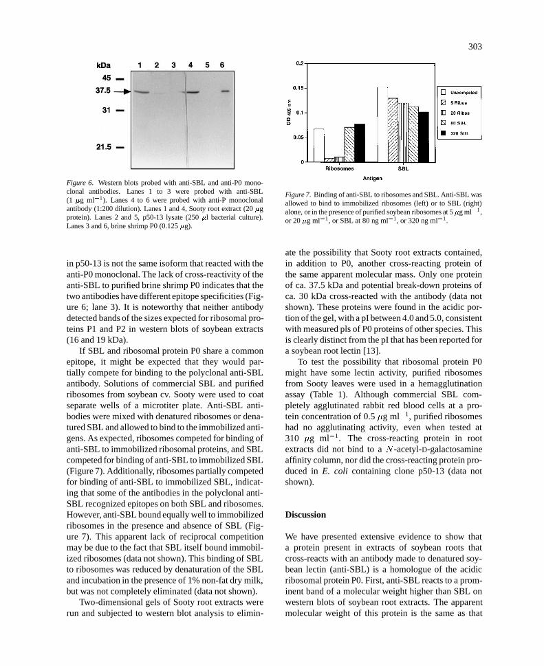
Figure 6. Western blots probed with anti-SBL and anti-P0 mono-clonal antibodies. Lanes 1 to 3 were probed with anti-SBL(1 �g ml�1). Lanes 4 to 6 were probed with anti-P monoclonalantibody (1:200 dilution). Lanes 1 and 4, Sooty root extract (20 �gprotein). Lanes 2 and 5, p50-13 lysate (250 �l bacterial culture).Lanes 3 and 6, brine shrimp P0 (0.125 �g).
in p50-13 is not the same isoform that reacted with theanti-P0 monoclonal. The lack of cross-reactivity of theanti-SBL to purified brine shrimp P0 indicates that thetwo antibodies have different epitope specificities (Fig-ure 6; lane 3). It is noteworthy that neither antibodydetected bands of the sizes expected for ribosomal pro-teins P1 and P2 in western blots of soybean extracts(16 and 19 kDa).
If SBL and ribosomal protein P0 share a commonepitope, it might be expected that they would par-tially compete for binding to the polyclonal anti-SBLantibody. Solutions of commercial SBL and purifiedribosomes from soybean cv. Sooty were used to coatseparate wells of a microtiter plate. Anti-SBL anti-bodies were mixed with denatured ribosomes or dena-tured SBL and allowed to bind to the immobilized anti-gens. As expected, ribosomes competed for binding ofanti-SBL to immobilized ribosomal proteins, and SBLcompeted for binding of anti-SBL to immobilized SBL(Figure 7). Additionally, ribosomes partially competedfor binding of anti-SBL to immobilized SBL, indicat-ing that some of the antibodies in the polyclonal anti-SBL recognized epitopes on both SBL and ribosomes.However, anti-SBL bound equally well to immobilizedribosomes in the presence and absence of SBL (Fig-ure 7). This apparent lack of reciprocal competitionmay be due to the fact that SBL itself bound immobil-ized ribosomes (data not shown). This binding of SBLto ribosomes was reduced by denaturation of the SBLand incubation in the presence of 1% non-fat dry milk,but was not completely eliminated (data not shown).
Two-dimensional gels of Sooty root extracts wererun and subjected to western blot analysis to elimin-
Figure 7. Binding of anti-SBL to ribosomes and SBL. Anti-SBL wasallowed to bind to immobilized ribosomes (left) or to SBL (right)alone, or in the presence of purified soybean ribosomes at 5 �g ml�1,or 20 �g ml�1, or SBL at 80 ng ml�1, or 320 ng ml�1.
ate the possibility that Sooty root extracts contained,in addition to P0, another cross-reacting protein ofthe same apparent molecular mass. Only one proteinof ca. 37.5 kDa and potential break-down proteins ofca. 30 kDa cross-reacted with the antibody (data notshown). These proteins were found in the acidic por-tion of the gel, with a pI between 4.0 and 5.0, consistentwith measured pls of P0 proteins of other species. Thisis clearly distinct from the pI that has been reported fora soybean root lectin [13].
To test the possibility that ribosomal protein P0might have some lectin activity, purified ribosomesfrom Sooty leaves were used in a hemagglutinationassay (Table 1). Although commercial SBL com-pletely agglutinated rabbit red blood cells at a pro-tein concentration of 0.5 �g ml�1, purified ribosomeshad no agglutinating activity, even when tested at310 �g ml�1. The cross-reacting protein in rootextracts did not bind to a N -acetyl-D-galactosamineaffinity column, nor did the cross-reacting protein pro-duced in E. coli containing clone p50-13 (data notshown).
Discussion
We have presented extensive evidence to show thata protein present in extracts of soybean roots thatcross-reacts with an antibody made to denatured soy-bean lectin (anti-SBL) is a homologue of the acidicribosomal protein P0. First, anti-SBL reacts to a prom-inent band of a molecular weight higher than SBL onwestern blots of soybean root extracts. The apparentmolecular weight of this protein is the same as that
pla275us.tex; 7/05/1997; 13:50; v.7; p.9

304
Table 1. Agglutination of rabbit erythrocytes by commercial SBL,ribosomes and post-ribosomal supernatant. Rabbit red blood cellswere mixed with purified soybean lectin, Sooty leaf ribosomes, Sootyleaf post-ribosomal supernatant, or buffer alone. Agglutination wasmeasured by the % reduction of absorbance at 620 nm after 4 h incub-ation.
Sample Protein conc. (�g ml�1) % Reduction of OD620
SBL 2.0 93
SBL 1.0 93
SBL 0.5 91
Ribosomes 310.0 15
Ribosomes 31.0 14
Ribosomes 3.1 16
Supernatant 31.0 31
None – 22
for P0 from other species. Second, cDNA clones isol-ated using this antibody produce a protein of the samesize in E. coli. Third, these clones are very similar insequence to previously cloned plant P0 genes. Fourth,the protein was present in purified ribosomes, and notin the supernatant from which the ribosomes had beenremoved. Fifth, the pI of the protein, as determined by2-D western blots of Sooty root extracts, was similar tothat of the acidic P0 of other species. Finally, antibod-ies raised to purified brine shrimp P0 react to a proteinof the same molecular weight in soybean root extractsas does the anti-SBL.
As might be expected for a ribosomal protein, P0was detected in all tissues tested by western blot analys-is. While its abundance was nearly equivalent in roots,hypocotyls, and leaves, it was much less abundantin cotyledons of 5-day-old seedlings. This may reflectthe beginning of senescence in cotyledons at this stage.The encoding RNA was also detected throughout theplant, although it was somewhat more abundant in rootsand hypocotyls than in cotyledons or leaves. The pres-ence of multiple copies in the genome, and divergentsequences among our cDNAs, raises the possibility thatdifferent isoforms of P0 may be expressed in differenttissues or at different times in development.
It might be expected that a ribosomal protein wouldnot be detected in root exudates, and this was indeedthe case for the cross-reacting protein. The presenceof SBL in root exudates of Williams 82, despite itsabsence in roots (cf. Figure 1), is consistent with thefindings of Gade et al. [13]. These results suggest thatthe SBL gene (Le1) is expressed at a low level in roots,but the product is rapidly secreted and the steady statelevel of SBL in roots is simply below the level of detec-
tion of the antibody. Kjemtrup et al. [22] determinedthat one of the polypeptides of phytohemagglutinin(PHA-E), normally a vacuolar protein, is released byroot cells into the culture medium. Thus, if an SBL-type lectin were present in Le� soybeans, it is likelythat it would also be found in root exudates.
The acidic ribosomal protein P0 is thought to bethe eukaryotic homologue of the E. coli protein L10.It forms a pentameric complex with two moleculeseach of P1 and P2 [39]. This pentamer forms the stalkof the 60 S ribosomal subunit, and plays an essentialrole in the elongation step of protein synthesis [33].We have explored the possibility that in soybean anisoform of P0 may also have lectin activity. It is notunprecedented for proteins normally associated withribosomes to have functions unrelated to protein syn-thesis. For example, a protein with vitronectin-likeactivity that cross-reacted with vitronectin antibodieswas recently identified as an isoform of EF1-� in high-er plants [44]. In Dictyostelium, an actin-binding pro-tein has also been identified as EF1-� [43]. We wereunable to demonstrate any lectin-like activity associ-ated with the soybean P0: ribosome preparations didnot agglutinate rabbit reticulocytes, nor did the proteinbind to a N-acetyl-D-galactosamine affinity column.Vodkin and Raikhel [42] were also unsuccessful inisolating the cross-reacting protein on a D-galactoseaffinity column. This does not, of course, exclude thepossibility that soybean P0 has lectin activity. Notall lectins agglutinate rabbit red blood cells, eitherbecause those cells do not carry the appropriate car-bohydrate ligand, or because the lectin is monovalent.Likewise, a lectin specific for any carbohydrate oth-er than galactose or N -acetyl-D-galactosamine wouldnot bind to either column. What can be concludedfrom these data is that the cross-reacting protein foundin Le� roots, which we have identified as a homo-logue of P0, is clearly distinct from the root lectinsidentified by Gade et al. [13] and by Halverson andStacey [15], which did have hemagglutination activity.The deduced amino acid sequence also clearly distin-guishes this protein from the vegetative soybean lectinidentified by Spilatro et al. [37].
We conclude that both the 32 kDa translationproduct which cross-reacted with anti-SBL but wasencoded by a gene sequence different than Le1 [35] andthe cross-reacting material (CRM) detected in soybeanpreviously using the same antibody [42] are likely tobe the protein we have identified as a P0 homologue.There are two possible explanations for the reactionof anti-SBL antibodies with ribosomal protein P0. The
pla275us.tex; 7/05/1997; 13:50; v.7; p.10

305
first is that a subset of the antibodies react with bothproteins due to the presence of a common epitope.The competition ELISA results are consistent withthis explanation. Alternatively, the antiserum from therabbit used in these experiments may contain two non-overlapping sets of antibody specificities.This could bedue to contamination by P0 of the SBL preparation thatwas used as both immunogen and affinity material forantibody purification. This seems unlikely, however,because the SBL was prepared from seeds, a tissuealready low in P0 (see Figure 1 and [42]), and waschromatographically pure (L. Vodkin, pers. comm.).
It would appear then that SBL and ribosomal pro-tein P0 have some common epitopes(s) recognizedby the anti-SBL antibodies. The common epitopewould need to reside in the carboxyl-terminal halfof the protein, because clones in Group II are miss-ing all the sequence before amino acid 155. A directsequence comparison between SBL and ribosomal pro-tein P0 revealed no extensive amino acid identities.Thelongest contiguous common amino acid sequences areLDLT (207–210 of P0; 274–277 of SBL) and VAVE(254–257 of P0; 153–156 of SBL). These short animoacid sequences may represent antigenic epitopes onboth proteins. An extensive deletion analysis wouldbe required to determine which, if either, of thesesequences represent the common epitope. Completesequencing of the P2 homologue clones may provide aclue. P2 sequences in plants have not previously beenreported, but in animals virtually the only commonsequence between P0 and P2 is the carboxyl-terminal20 to 30 amino acids.
The ability of root exudates from a Le� seed line(T102) to reverse the delayed nodulation phenotype ofB. japonicum strain HS111 in the same way as SBLstrongly suggests that soybean has another, geneticallydistinct lectin [15, 16, 17]. If so, its mRNA is neithercomplementary to the Le1 or Le2 genes [42], nor doesit react with antibodies raised to SBL (this report). It islikely that the root lectin identified by Gade et al. [13]is a product of the Le1 gene because this protein sharesnearly identical properties with SBL and moreover, itwas isolated from a Le+ cultivar. Similarly, the lectin-related protein described by Spilatro et al. [37] is alsolikely to be a product of a Le1-like gene as the aminoacid sequence of the N-terminus is very similar to thatof SBL.
Recently, Etzler and Murphy [12] have described alectin from Dolichos biflorus, DB46, which does notshare any significant homology with the other plantor animal lectin sequences that have been isolated so
far. Of the various ligands tested for their ability toinhibit the specific binding of carbohydrates by DB46,the strongest inhibition came from Nod factors puri-fied from B. japonicum USDA 110, which nodulatessoybean as well as Dolichos. Thus, it seems likelythat cv. Sooty, like Dolichos, will have an additionalvegetative lectin which is very different from the wellcharacterized seed lectin.
Acknowledgements
We are grateful to Bruce Brand for helping with thesequencing and to Ramin Yadagari and Paul Sandersof the Goldberg lab for their help in library construc-tion and isolation of polysomes. Larry Brill is thankedfor his help in the northern blot analysis and also forhis comments on the manuscript. We also extend ourgratitude to Bob Goldberg who supplied materials forcDNA construction and �Zap II cloning. Lastly, wethank Lila Vodkin for the anti-SBL and her helpfulcomments. This research is supported in part by NSF90-23888 and by NRICGP 93-37305-9144 to A.M.H.P.v.R. was supported by the D. Collen Foundation,K.U. Leuven, Belgium.
References
1. Altschul SF, Gish W, Miller W, Myers EW: Basic local align-ment search tool. J Mol Biol 215: 403–410 (1990).
2. Arango R, Adar R, Rozenblatt S, Sharon N: Expression ofErythrina corallodendron lectin in Escherichia coli. Eur J Bio-chem 205: 575–581 (1992).
3. Bhuvaneswari TV, Turgeon BG, Bauer WD: Early events inthe infection of soybean (Glycine max L. Merr) by Rhizobiumjaponicum. Plant Physiol 66: 1027–1031 (1980).
4. Bohlool BB, Schmidt EL: Lectins: a possible basis for spe-cificity in the Rhizobium-legume root nodule symbiosis. Sci-ence 185: 269–271 (1974).
5. Bonner TI, Brenner DJ, Neufeld BR, Britten RJ: Reduction inthe rate of DNA reassociation by sequence divergence. J MolBiol 81: 123 (1973).
6. Dazzo FB, Hubbell DH: Cross-reactive antigens and lectins asdeterminants of symbiotic specificity in the Rhizobium-cloversymbiosis. Appl Microb 30: 1017–1033 (1975).
7. Del Sal G, Manfioletti G, Schneider C: The CTAB-DNA pre-cipitation method: a common mini-scale preparation of tem-plate DNA from phagemids, phages or plasmids suitable forsequencing. BioTechniques 7: 514–520 (1989).
8. Dellaporta SL, Wood J, Hicks JB: A plant DNA miniprepara-tion: Version II. Plant Mol Biol Rep 1: 19–21 (1983).
9. Denarie J, Debelle F and Prome J-C: Rhizobium lipo-chitooligosaccharide nodulation factors: signaling moleculesmediating recognition and morphogenesis. Annu Rev Biochem65: 503–535 (1996).
pla275us.tex; 7/05/1997; 13:50; v.7; p.11

306
10. Dıaz CL, Melchers LS, Hooykaas PJJ, Lugtenberg BJJ, KijneJW: Root lectin as a determinant of host-plant specificity in theRhizobium-legume symbiosis. Nature 338: 579–581 (1989).
11. D’Sousa SE, Ginsberg MH, Plow EF: Arginyl-glycyl-asparticacid (RGD): a cell adhesion motif. Trends Biochem Sci 16:246–250 (1991).
12. Etzler ME and Murphy JB: Do legume vegetative tissue lec-tins play roles in plant-microbial interactions? In: Stacey G,Mullin B and Gresshoff PM (eds) Biology of Plant-MicrobeInteractions, pp. 105–110. International Society of MolecularPlant-Microbe Interactions, St Paul, MN (1996).
13. Grade W, Jack MA, Dahl JB, Schmidt EL, Wold F: The isola-tion and characterization of a root lectin from soybean (Glycinemax (L), cultivar Chippewa). J Biol Chem 256: 12905–12910(1981).
14. Goldberg RB, Hoschek G, Vodkin LO: An insertion sequenceblocks the expression of a soybean lectin gene. Cell 33: 465–475 (1983).
15. Halverson LJ, Stacey G: Host recognition in the Rhizobium-soybean symbiosis: detection of a protein factor in soybeanroot exudate which is involved in the nodulation process. PlantPhysiol 74: 84–89 (1984).
16. Halverson LJ, Stacey G: Host recognition in the Rhizobium-soybean symbiosis: evidence for the involvement of lectin innodulation. Plant Physiol 77: 621–625 (1985).
17. Halverson LJ, Stacey G: Effect of lectin on nodulation by wild-type Bradyrhizobium japonicum and a nodulation-defectivemutant. Appl Envir Microbiol 51: 753–760 (1986).
18. Hamblin J, Kent SP: Possible role of phytohemagglutinin inPhaseolus vulgaris L. Nature New Biol 245: 28–29 (1973).
19. Hirsch AM: Developmental biology of legume nodulation.New Phytol 122: 211–237 (1992).
20. Jackson AO, Larkins BA: Influence of ionic strength, pH, andchelation of divalent metals on isolation of polyribosomes fromtobacco leaves. Plant Physiol 57: 5–10 (1976).
21. Kijne J, Diaz C, de Pater S, Lugtenberg BJJ: Lectins in the sym-biosis between Rhizobia and leguminous plants. In: Franz H(ed) Advances in Lectin Research, pp. 15–50. Ullstein Mosby,Berlin (1992).
22. Kjemtrup S, Borkhsenious O, Raikhel NV, Chrispeels M: Tar-geting and release of phytohemaglutinin from the roots of beanseedlings. Plant Physiol 109: 603–610 (1995).
23. Laemmli UK: Cleavage of structural proteins during theassembly of the head of bacteriophage T4. Nature 227: 680–685 (1970).
24. Lis H, Sela B-A, Sachs L, Sharon N: Specific inhibition byN -acetyl-D-galactosamine of the interaction between soybeanagglutination and animal cell surfaces. Biochim Biophys Acta211: 582–585 (1970).
25. Long SR: Rhizobium symbiosis: Nod factors in perspective.Plant Cell 8: 1885–1898 (1996).
26. Marston FAO: The purification of eukaryotic polypeptidesexpressed in Escherichia coli. In: Glover DM (ed) DNA Clon-ing: A Practical Approach, pp. 59. IRL Press, Oxford (1987).
27. Montesano L, Glitz DG: Wheat germ cytoplasmic ribosomes.J Biol Chem 263: 4932–4938 (1988).
28. Norris JH, Macol LA, Hirsch AM: Nodulin gene expressionin effective nodules and in nodules arrested at three differentstages of development. Plant Physiol 88: 321–328 (1988).
29. Okamuro JK, Jofuku KD, Goldberg RB: Soybean seed lectingene and flanking nonseed protein genes are developmentallyregulated in transformed tobacco plants. Proc Natl Acad SciUSA 83: 8240–8244 (1986).
30. Pull SP, Pueppke SG, Hymowitz T, Orf JH: Soybean lineslacking the 120 000-Dalton seed lectin. Science 200: 1177–1279 (1978).
31. Rutherford WM, Dick WE, Cavins JF, Dombrink-KurtzmanMA, Slodki ME: Isolation and characterization of a soybeanlectin having 4-0-methylglucuronic acid specificity. Biochem-istry 25: 952–958 (1986).
32. Sambrook J, Fritsch EF, Maniatis T: Molecular Cloning: ALaboratory Manual. Cold Spring Harbor Press, Cold SpringHarbor, NY (1989).
33. Sanchez-Madrid F, Reyes R, Conde P, Ballesta JPG: Acidicribosomal proteins from eukaryotic cells: effect on ribosomalfunctions. Eur J Biochem 98: 409–416 (1979).
34. Sanger F, Nicklen S, Coulsen AR: DNA sequencing with chain-terminating inhibitors. Proc Natl Acad Sci USA 74: 5463–5467(1977).
35. Sengupta-Gopalan C, Pitas JW, Hall TC: Re-examination of therole of lectin in Rhizobium-Glycine symbiosis. In: Veeger C,Newton WE (eds) Advances in Nitrogen Fixation Research,p. 427. Nijhoff, The Hague (1984).
36. Spilatro SR, Anderson JM: Characterization of a soybean leafprotein that is related to the seed lectin and is increased withpod removal. Plant Physiol. 90: 1387–1393 (1989).
37. Spilatro SR, Cochran GR, Walker RE, Cablish KL, Bittner CC:Characterization of a new lectin of soybean vegetative tissues.Plant Physiol 110: 825–834 (1996).
38. Uchiumi T, Traut RR, Kominami R: Monoclonal antibodiesagainst acidic phosphoproteins P0, P1, and P2 of eukaryot-ic ribosomes as functional probes. J Biol Chem 265: 89–95(1990).
39. Uchiumi T, Wahba AJ, Trout RR: Topography and stoi-chiometry of acidic proteins in large ribosomal subunits fromArtemia salina as determined by crosslinking. Proc Natl AcadSci USA 84: 5580–5584 (1987).
40. van Eijsden RR, Dıaz CL, Hoedemaeker PJ, de Pater BS, KijneJW: Sugar binding activity of Pisum sativum lectin is essentialfor heterologous infection of transgenic white clover roots byRhizobium leguminosarum bv. viciae. Plant Mol Biol 29: 431–439 (1995).
41. Vodkin LO: Isolation and characterization of messenger RNAsfor seed lectin and Kunitz trypsin inhibitor in soybeans. PlantPhysiol 68: 766–771 (1981).
42. Vodkin LO, Raikhel NV: Soybean lectin and related proteinsin seeds and roots of Le+ and Le� soybean varieties. PlantPhysiol 81: 558–565 (1986).
43. Yang F, Demma M, Warren V, Dharmawardhane S, Condeel-is J: Identification of an actin-binding protein from Dictyosteli-um as elongation factor 1�. Nature 347: 494–496 (1990).
44. Zhu J-K, Damsz B, Kononowicz AK, Bressan RA, HasegawaPM: A higher plant extracellular vitronectin-like adhesion pro-tein is related to the translational elongation factor-1�. PlantCell 6: 393–404 (1994).
pla275us.tex; 7/05/1997; 13:50; v.7; p.12


![A Functional myoInositol Dehydrogenase Gene Is Required for Efficient Nitrogen Fixation and Competitiveness of Sinorhizobium fredii USDA191 To Nodulate Soybean (Glycine max [L.] Merr](https://static.fdokumen.com/doc/165x107/6312da2ab22baff5c40edaa4/a-functional-myoinositol-dehydrogenase-gene-is-required-for-efficient-nitrogen-fixation.jpg)


![Purification, molecular cloning and ethylene-inducible expression of a soluble-type epoxide hydrolase from soybean (Glycine max [L.] Merr](https://static.fdokumen.com/doc/165x107/631a26e0b41f9c8c6e0a1180/purification-molecular-cloning-and-ethylene-inducible-expression-of-a-soluble-type.jpg)


