Ups1p and Ups2p antagonistically regulate cardiolipin metabolism in mitochondria
A
asgCOthCtat©
K
Imp(
1t
rlnin
0d
Cell Calcium 40 (2006) 513–525
Calcium microdomains in mitochondria and nucleus
Marıa Teresa Alonso, Carlos Villalobos, Pablo Chamero,Javier Alvarez, Javier Garcıa-Sancho ∗
Instituto de Biologıa y Genetica Molecular (IBGM), Universidad de Valladolid and Consejo Superior de InvestigacionesCientıficas (CSIC), c/Sanz y Fores s/n, 47003 Valladolid, Spain
Received 1 August 2006; accepted 23 August 2006Available online 25 October 2006
bstract
Endomembranes modify the progression of the cytosolic Ca2+ wave and contribute to generate Ca2+ microdomains, both in the cytosolnd inside the own organella. The concentration of Ca2+ in the cytosol ([Ca2+]C), the mitochondria ([Ca2+]M) and the nucleus ([Ca2+]N) areimilar at rest, but may become very different during cell activation. Mitochondria avidly take up Ca2+ from the high [Ca2+]C microdomainsenerated during cell activation near Ca2+ channels of the plasma membrane and/or the endomembranes and prevent propagation of the higha2+ signal to the bulk cytosol. This shaping of [Ca2+]C signaling is essential for independent regulation of compartmentalized cell functions.n the other hand, a high [Ca2+]M signal is generated selectively in the mitochondria close to the active areas, which tunes up respiration to
he increased local needs. The progression of the [Ca2+]C signal to the nucleus may be dampened by mitochondria, the nuclear envelope origher buffering power inside the nucleoplasm. On the other hand, selective [Ca2+]N signals could be generated by direct release of storeda2+ into the nucleoplasm. Ca2+ release could even be restricted to subnuclear domains. Putative Ca2+ stores include the nuclear envelope,
heir invaginations inside the nucleoplasm (nucleoplasmic reticulum) and nuclear microvesicles. Inositol trisphosphate, cyclic ADP-ribosend nicotinic acid adenine dinucleotide phosphate have all been reported to produce release of Ca2+ into the nucleoplasm, but contribution ofhese mechanisms under physiological conditions is still uncertain.
2006 Elsevier Ltd. All rights reserved.
tsCeioftoudb
eywords: Calcium; Microdomains; Mitochondria; Nucleus; Aequorin
CALL our world Flatland, not because we call it so, but toake its nature clearer to you, my happy readers, who are
rivileged to live in Space.Edwin A. Abbott, Flatland A romance of many dimensions)
. What are microdomains and how do we evidencehem?
Vectorial metabolism was the frame for the chemiosmoticationale [1]. The term stresses that chemical reactions insideiving cells are not dimensionless but have spatial coordi-
ates. The three-dimensional nature of metabolism does alsomply that, even at the level of a single-cell, composition mayot be homogeneous, but domains with different concentra-∗ Corresponding author. Tel.: +34 983 423084; fax: +34 983 423588.E-mail address: [email protected] (J. Garcıa-Sancho).
si(
we
143-4160/$ – see front matter © 2006 Elsevier Ltd. All rights reserved.oi:10.1016/j.ceca.2006.08.013
ions of metabolites or ions may coexist. The idea of micro-copic hemispherical “domains” centered upon the activea2+ channels was developed in the early 1980s [2–4]. Theselectrophysiologists’ microdomains are very much restrictedn time (ms) and space (nm), and make possible local controlf phenomena such as regulated exocytosis with little inter-erence with other Ca2+-dependent physiological processeshat take place at a different cell location [5]. For the cell biol-gist, microdomains may be much wider and longer. We shallse the term here in its widest acception, to design any [Ca2+]omain of subcellular dimensions, from highly localized andrief hot spots to wide and durable changes, including con-iderations on how, why and when the Ca2+ concentrationn the mitochondrial matrix ([Ca2+]M) or the nucleoplasm
[Ca2+]N) may differ from the one in the cytosol ([Ca2+]C).Cytosolic Ca2+ buffers modify the propagation of the Ca2+
ave through the cytosol [6,7], but organella influences itven more deeply and contribute to the generation of sub-

514 M.T. Alonso et al. / Cell Calciu
Ffo
cispctCnmmoiiiifr
qopmiilfi[ad
omcvcatCmpcatbaoohs[rtiatatiomawar
iTtmqw
A[ttaotdc[
ig. 1. Ca2+ diffusion through the cytosol is slower than expected. In goingrom (A) to (B), Ca2+ must follow a very long pathway to circumventrganella. In addition, Ca2+ is taken up by organella as shown in (C).
ellular domains with different Ca2+ concentrations [8]. Thiss so because organella physically interrupt straight diffu-ional progression of the Ca2+ wave and forces a much longerathway (Fig. 1). In addition, some organella, such as mito-hondria or endoplasmic reticulum (ER) cisterna, also opposehe progression of the Ca2+ wave by taking up “en passant”a2+ (inset C). The ER may also amplify the [Ca2+]C sig-als by releasing stored Ca2+ by a Ca2+-induced Ca2+ releaseechanism [9,10]. On the other hand, the endomembranesodify the propagation of the [Ca2+]C wave to the interior
f the organella either dampening it (nucleus) or amplify-ng it (mitochondria). Our review will be concerned with thenterrelations among the different [Ca2+] domains reachedn cytosol, mitochondria and nucleus and how this allowsndependent regulation of different [compartmentalized] cellunctions. For details the reader is referred to other recenteviews on these matters [11–13].
Adequate spatial selectivity is required for detection anduantification of Ca2+ microdomains. Measurements insiderganella additionally require preferential location of therobe inside it. Some fluorescent dyes (rhod, mag-fura andag-fluo families) may, under certain conditions, concentrate
nside the mitochondria over the cytosol. The best selectivitys attained, however, by directing protein probes to specificocations by the use of targeting sequences. This strategy was
rst used with photoluminescent probes, such as aequorins14], and later with GFP-based fluorescent Ca2+ probes, suchs cameleons and pericams [15–17]. Aequorins have a largerynamic range and a steeper Ca2+ dependence than the flu-tiso
m 40 (2006) 513–525
rescent probes (Fig. 2A; compare fluo-3, R-pericam andag-fluo-4 to AEQ1). This is due to the fact that aequorins
an be read against virtually no background and hence aery small fraction of the total luminescence (down to 1/105)an be measured accurately. The steep Ca2+ dependence ofequorins is due to cooperation of three Ca2+ ions that bindo the protein. This steepness sharpens the definition of higha2+ microdomains as shown in Fig. 2B and C, where theodeled responses of mag-fluo-4 and aequorin are com-
ared (see figure legend for further explanation). Finally, byombining the wild-type and the low Ca2+ affinity mutatedequorin [18] with different coelenterazines (the cofactor forhe chemiluminescence reaction), very different affinities cane obtained [19–21]. This allows measurements of [Ca2+] invery wide range (10−8 to 10−3 M; Fig. 2A). The low lightutput, that hampers single-cell imaging (but see below), isne of the main disadvantages of aequorins. On the otherand, the fact that aequorin is consumed during the mea-urements makes unpractical long-term recordings of highCa2+] domains. Calculations of [Ca2+] are made from theatio L/LTOTAL, where L is the light output (cps) at a givenime point and LTOTAL is the number of counts remainingn the sample at that moment. Therefore, for calibration ofequorin, light output must be measured and added up duringhe whole experiment, which must be terminated by releasingll the residual luminescence, usually by lysing the cells athe end of the experiment. Even though precise calibrations difficult, large errors are unlikely because of the steepnessf the relation between L and [Ca2+]. On the other hand, theicroenvironment of the targeted aequorin could change the
ffinity for Ca2+ with regards to in vitro calibrations. Thus,hereas no changes were found for nuclear aequorin [22],
n increase of 3–30 times of the affinity for Ca2+ has beeneported for mitochondrial aequorin [23] (see also Fig. 2).
In vectorial metabolism the only [Ca2+] that really matterss the one at the active site of the Ca2+-dependent proteins.hese molecular domains could be sensed by probes tagged
o specific molecules or even to specific domains of macro-olecules. Some elegant approaches have been made to these
uestions using tailor-made protein probes [24], but it is muchhat remains to be investigated in this area.Mitochondria and nucleus handle Ca2+ very differently.
t rest, [Ca2+]C, [Ca2+]M and [Ca2+]N are all similar. WhenCa2+]C increases during cell activation, mitochondria tendso take up Ca2+ and hence [Ca2+]M tends, if anything,o become higher than [Ca2+]C. Because of their Ca2+-ccumulating tendency, mitochondria oppose the progressionf Ca2+ waves through the cytosol (Fig. 1) and help to shapehe cytosolic Ca2+ microdomains. The nuclear envelope (NE)oes, if anything, slow down the passage of Ca2+ from theytosol to the nucleoplasm, thus contributing to generate aCa2+]N domain that is diluted with regards to [Ca2+]C. On
he other side, it is controversial whether Ca2+ could, bypass-ng the cytosol, be released directly into the nucleoplasm frompecific calcium stores located either at the nuclear cisternaf the NE or even somewhere inside the nucleus. This mech-
M.T. Alonso et al. / Cell Calcium 40 (2006) 513–525 515
Fig. 2. Comparison of the properties of the fluorescent and the photoluminescent Ca2+ probes. (A) Calibration curves. For the fluorescent probes, lightemission (F) was estimated from a Michaelis function: (F = [Ca2+])/(KD + [Ca2+]), where the value of the dissociation constant, KD, was (in �M): 0.34for fluo-3, 1.7 for R-pericam and 22 for mag-fluo-4. The three aequorin (AEQ) systems shown, correspond to: wild-type aequorin with either the regularcoelenterazine (AEQ1) or coelenterazine n (AEQ2) or mutated low Ca2+ affinity aequorin [18] and coelentarazine n (AEQ3). The function used to relate Ca2+
and photoluminescence (expressed as fraction of the total counts emitted at every instant, L/LTOTAL, was: [Ca2+] (in M) = (R + (R × KTR) − 1)/(KR − (R × KR)),where R = (L/(Lmax × �)1/n), using for the constants the values reported before [21]. The red circles show the calibration values reported for in situ calibrationof mitochondrial aequorin using FCCP and ionomycin to collapse the H+ and the Ca2+ gradient, respectively [23]. (B) Spatial profile of a Ca2+ hot spot.Dissipation of the hot spot from point 0 was assumed to follow the following function: Sx = So × exp(−k × x), where So is the maximum Ca2+ concentration atthe hot spot, k the space constant and x is the distance. The fluorescence and photoluminescence profiles have been calculated assuming a Ca2+ concentrationo ven in (a as usedt two-dima end, the
aouc(fl
2
t[tstseicdotfa[wtta
Pbcdri(Ci1lcofivcccc
fmtc
C
f 10 �M at the hotspot and using the equations and the constant values giequorin and coelenterazine the following simplified polynomic equation whe fluorescence dye is about three times larger than with aequorin. (C) Thend aequorin. (For interpretation of the references to color in this figure leg
nism would generate [Ca2+]N domains higher than [Ca2+]C,r even subnuclear Ca2+ domains, if the Ca2+ release was notniform in all the nucleus. In conclusion, organella may thenontribute to generate: (i) cytosolic [Ca2+] microdomains;ii) [Ca2+] domains inside the organella that are very differentrom [Ca2+]C; (iii) suborganellar microdomains, in which theocal [Ca2+] may differ from the average organellar [Ca2+].
. Mitochondria
Ca2+ transport by mitochondria has received much atten-ion, both because of possible participation in shapingCa2+]C signals and because changes of [Ca2+]M are impor-ant by themselves for regulation of important cell functionsuch as respiration or programmed cell death [25]. Ca2+ isaken up by the Ca2+ uniporter, a low-affinity/high capacityystem driven by the mitochondrial membrane potential andxits through a Na+/Ca2+ exchanger and also through a Na+-ndependent system (Fig. 3A) [26,27]. Fig. 3B compares theoncentration-dependence and the capacity of the mitochon-rial Ca2+ uniporter (MCU) to the high affinity Ca2+ pumpsf the ER (SERCA) and plasma membrane (PMCA). Notehat both scales are logarithmic. Because of its low affinityor Ca2+ (K1/2, 20–50 �M) and high Hill number [2], thectivity of the MCU would be extremely low at rest, withCa2+]C near 10−7 M (A in Fig. 3B), and PMCA and SERCA
ould clearly dominate for compensation of the Ca2+ leakshrough the plasma membrane and the ER membrane, respec-ively. At the average [Ca2+]C concentrations reached duringctivation, near 10−6 M (B in Fig. 3B), Ca2+ transport by w
A), above. For mag-fluo-4: F = [Ca2+]/(22 × 10−6 + [Ca2+]). For wildtype: L/LTOTAL = 322.72 × [Ca2+] + 595238709 × ([Ca2+])2. The wideness with
ensional profile of the hotspot, in gray levels, is compared for mag-fluo-4reader is referred to the web version of the article.)
MCA and SERCA, now operating near its Vmax, would stille much larger than transport through MCU. It is only at con-entrations near 10−5 M (C in Fig. 3B) when MCU becomesominant. It can be argued such high concentrations are noteached even during strong physiological stimulation. Thiss true for the bulk [Ca2+]C, but higher Ca2+ concentrations10–100 �M) can be reached at local cells spots, at higha2+ microdomains [28,5]. The activity coefficient of Ca2+
nside the mitochondrial matrix seems to be very low, in the:1000 range [29–31], and this allows mitochondria to sinkarge Ca2+ loads with relatively little change of [Ca2+]M. Theapacity (Vmax) of the system may be as high as 5–6 mmol/lf cells per second in adrenal chromaffin cells [7,20,31], agure that approaches the rate of Ca2+ entry through theoltage-operated plasma membrane Ca2+ channels in theseells [6,7,32]. Global [Ca2+]C increases able to activate MCUan also be reached during Ca2+ overload under pathologicalircumstances and do usually drive to mitochondria-triggeredell death [25,33].
Regarding thermodynamics, at rest, there is a tremendousree energy change for entry of Ca2+ from the cytosol to theitochondria. If, by analogy with the chemiosmotic rationale
erminology [1], we express the driving force for Ca2+ as thealcium motive force (CMF, in mV) we can write:
MF = 30(pCaC − pCaM) +�ψM
= 30 log
([Ca2+]M
[Ca2+]C
)+�ψM
here �ΨM is the mitochondrial transmembrane potential.

516 M.T. Alonso et al. / Cell Calcium 40 (2006) 513–525
Fig. 3. Calcium transport in mitochondria. (A) The main transport systems are shown: the uniporter, in the center, driven by the mitochondrial membranepotential (ΨM) and the two exchangers (Na+/Ca2+ and H+/Ca2+) at each side. PTP, permeability transition pore. (B) Concentration-dependence of the differentC joint acA ined in aa to color
[o
2
frltN[osdmtabc1
bmes[idwlp[fivw
atLhfi2havtvpIdtdtsiat[efloamuh
2[
a2+ transport systems. MCU, mitochondrial calcium uniporter, in blue. TheTPases) is shown in red. Note double logarithmic scale. Based in data obtand maximum stimulation, respectively. (For interpretation of the references
That means that at equilibrium (CMF = 0) the ratioCa2+]M/[Ca2+]C would be as large as 106 for a �ΨM valuef −180 mV.
.1. The problem of the free [Ca2+] inside mitochondria
There is general consensus that despite the large CMFavouring Ca2+ entry into mitochondria, [Ca2+]M is, underesting conditions, close to [Ca2+]C. This is due both, to theow Ca2+ affinity of the mitochondrial Ca2+ uniporter ando the electrogenic Ca2+ extrusion through the mitochondriala+/Ca2+ exchanger. This system exchanges 3Na+ by 1Ca2+
26,27] and thus exit of each Ca2+ is accompanied by the entryf a positive charge into mitochondria. A remarkable conclu-ion from this is that the mitochondrial electrical potentialrives both influx and efflux of Ca2+ from the mitochondrialatrix. Consistently, mitochondrial membrane depolariza-
ion inhibits both Ca2+ entry and Ca2+ release [10]. Thectivity of the MCU is very small or absent at cytosolic [Ca2+]elow 0.5 �M [7,34], whereas its activity increases dramati-ally along the micromolar range, reaching saturation above00 �M [7,20] (Fig. 3B)
Activation of MCU occurs when the [Ca2+]C in the neigh-ourhood of the mitochondrial membrane increases to theicromolar range. Under these conditions, the rate of Ca2+
ntry through the uniporter exceeds that of Ca2+ extru-ion through the exchanger, leading to a fast increase inCa2+]M. However, there is presently no consensus regard-ng the free [Ca2+] that can be attained into mitochondriauring cell activation. Measurements in intact cells loadedith rhod-2 provide peak values of 2–3 �M during stimu-
ation of HeLa cells with histamine [35]. In mitochondriareparations loaded with fura-2-FF, large Ca2+ loads bring
Ca2+]M to concentrations not larger than 5 �M [36]. Therst measurements with mitochondria-targeted aequorin pro-ided similar values [14], but it seemed clear that the valuesere underestimated because of the massive consumption ofbo
tion of the plasma membrane and the ER Ca2+ pumps (PMCA and SERCAdrenal chromaffin cells [31]. A–C correspond to rest, moderate stimulationin this figure legend, the reader is referred to the web version of the article.)
equorin. In contrast, measurements with low-Ca2+-affinityargeted probes provide much higher [Ca2+]M estimates.ow-Ca2+-affinity aequorins indicate [Ca2+]M increases asigh as 200–700 �M during strong stimulation of chromaf-n cells [20,10,31] or GH3 pituitary cells [37]. Values of0–50 �M are found during stimulation of HeLa cells withistamine [38,39]. Consistently, low-affinity cameleons [40]nd ratiometric pericams [41] have provided peak [Ca2+]Malues of 50–100 and 10–50 �M, respectively, during his-amine stimulation of HeLa cells. It is remarkable that thealues reported by both, luminescent and fluorescent targetedrobes, converge to the same figures for the peak [Ca2+]M.n contrast, the much smaller values found with fluorescentyes could most probably be explained by Ca2+ saturation ofhe dye and non-specific distribution. Mislocalization of theye in compartments with low [Ca2+] may bias the estima-ion of the average value. We should recall that a similarituation occurred when the first measurements of [Ca2+]nside the endoplasmic reticulum ([Ca2+]ER) with targetedequorins revealed figures around 500 �M [42–44]. At thatime, fluorescent dyes had provided much lower estimates forCa2+]ER [45–48]. However, the current evidence, obtainedither with targeted fluorescent cameleons [49,40] or care-ul correction for mislocalization of dyes and other prob-ems [50,51], favours values for [Ca2+]ER that approach thenes reported by the aequorins. The same low-Ca2+-affinityequorin used for the above mentioned “consensus” [Ca2+]EReasurements is the one that reveals the high peak [Ca2+] val-
es in mitochondria, so that there is no reason to accept theigh [Ca2+]ER values but not the [Ca2+]M ones.
.2. Mitochondria as biosensors and controllers of localCa2+] microdomains in the immediate cytosol
The mitochondrial Ca2+ uniporter has a K1/2 for activationy cytosolic Ca2+ of 20–50 �M and a sigmoidal second-rder kinetics [26,20,21]. Activation starts at 2–5 �M and

l Calciu
sru[ampreiEf(eotsc(TlvtnTiAma
tcmc[t[toomcbmsvsCbtsie[tI
Codptmf
ncqdstpps1astaCarHorhl[
2
NH2ciCPCo[trmtmte
M.T. Alonso et al. / Cel
aturates above 100 �M [Ca2+]C [7,20,31]. Therefore, theange of [Ca2+] required for activation of mitochondrial Ca2+
ptake closely matches the one usually considered as “highCa2+] microdomains”. This has allowed to use mitochondrias biosensors of Ca2+ microdomains. The measured rate ofitochondrial Ca2+ uptake allows, using the known transport
arameters of the MCU, back-calculation of the local [Ca2+]Cequired to explain it [20,31]. This method has been used tostimate the Ca2+ microdomains induced by depolarisation-nduced plasma membrane Ca2+ entry or caffeine-inducedR Ca2+ release in chromaffin cells [20]. According to the
ast rate of Ca2+ accumulation into mitochondria observed�[Ca2+]M above 100 �M/s), these stimuli should have gen-rated [Ca2+]C microdomains of 30–40 �M in the vicinityf some mitochondria. These values are 15–20-fold higherhan the corresponding global cytosolic [Ca2+] peaks mea-ured with fluorescent dyes. In contrast, stimulation of HeLaells with histamine induced slower rates of [Ca2+]M increase2.6 �M/s) [38]. In GH3 pituitary cells, stimulation withRH, which releases Ca2+ from the ER, produced a simi-
ar rate of mitochondrial Ca2+ uptake [37]. Both agonists actia inositol 1,4,5-trisphosphate (IP3) suggesting that activa-ion of IP3 receptors generate a local [Ca2+]C microdomainot higher than 5 �M in the close proximity of mitochondria.his value is only five-fold higher than the mean [Ca2+]C
ncrease induced by histamine or TRH in their target cells.similar approach was used in permeabilized RBL mucosalast cells to show that IP3 produces local [Ca2+] transients
bove 16 �M [52].Because of the high capacity and rate of Ca2+ accumula-
ion by mitochondria, a large fraction of the Ca2+ entering theell during stimulation appears to be transiently buffered byitochondria [32,53,54,31]. This phenomenon allows mito-
hondria to modulate [Ca2+]C, but only in the local highCa2+]C microdomains. Because of the low-Ca2+-affinity ofhe uniporter, mitochondria take up little Ca2+ at the averageCa2+]C values found in the bulk cytosol during cell stimula-ion (around 1 �M; Fig. 3). Modulation by mitochondria cannly occur when [Ca2+]C rises above a few micromolar inrder to significantly activate the uniporter. This implies thatitochondria are particularly well designed to modulate local
ytosolic high [Ca2+] microdomains. Such a modulation haseen shown to occur under many circumstances. For example,itochondria have been shown to modulate catecholamine
ecretion in chromaffin cells [20], the Ca2+-dependence ofoltage-dependent Ca2+ channels [55], the rate of progres-ion of the [Ca2+]C waves [56] or the activity of capacitativea2+ channels (ICRAC) [57]. In the last case, sinking of Ca2+
y mitochondria seems to fulfill a double role: (i) to facili-ate a more complete emptying of the stores thus promoting atronger activation of ICRAC; (ii) to prevent Ca2+-dependentnactivation of the CRAC channels by opposing the gen-
ration of subplasmalemmal high-[Ca2+]C microdomains58–62]. In other systems, mitochondria tightly associated tohe endoplasmic reticulum may inhibit Ca2+ release throughP3 receptors (IP3Rs) [63], probably by taking up enought[bm
m 40 (2006) 513–525 517
a2+ to abolish the local positive feedback effects of Ca2+
n the IP3R. In fact, there is structural and functional evi-ence suggesting that mitochondria and ER are closely cou-led in terms of Ca2+ homeostasis and that mitochondriaake up Ca2+ much more effectively from the local [Ca2+]
icrodomains created after IP3-induced Ca2+ release thanrom a global [Ca2+]C increase [64–69,52,25,41,13].
Because of the importance of Ca2+ microdomains for aumber of physiological processes and the ability of mito-hondria to modulate them, an important and yet unresolveduestion is whether physiological modulation of mitochon-rial Ca2+ uptake exists. It has been shown recently that aeries of structurally related compounds reversibly activatehe mitochondrial Ca2+ uniporter [38,70,71]. These com-ounds include the p38 MAP kinase inhibitor SB202190,lant flavonoids as kaempferol, quercetin or genistein andynthetic estrogens as 1,3,5-tris(4-hydroxyphenyl)-4-propyl-H-pyrazole (PPT) or diethylstilbestrol. In spite of theirpparent functional diversity, all of these compounds shareome structural elements in their molecules which are essen-ial for stimulation of MCU [71]. The activation is due ton increase in the affinity of the uniporter for Ca2+ [38].a2+ entry can be increased up to 20-fold by this mech-nism, which could be extremely important to control theegulation by mitochondria of local [Ca2+]C microdomains.owever, further work will be necessary to find a physi-logical counterpart of these activators. Another mode ofegulation is the control of MCU expression. In this regard, itas been proposed recently that thyroid hormone could regu-ate the number of mitochondrial Ca2+ uniporters expressed72].
.3. Ca2+ exit from mitochondria
Ca2+ exit from mitochondria occurs mainly through thea+/Ca2+ exchanger and, to a lesser extent, through the+/Ca2+ exchange (Fig. 3A) [26,27], that accounts for0–40% of the total Ca2+ release from mitochondria inhromaffin and HeLa cells [20,73]. Other minor pathwaysnclude the permeability transition pore (PTP) and the owna2+ uniporter working in reversal. Long-lasting opening ofTP occurs only rarely under physiological conditions [25].a2+ release through the uniporter requires the simultaneousccurrence of mitochondrial depolarization and high [Ca2+]C10]. There is evidence that Ca2+ transfer from mitochondriao the ER may occur via mitochondrial Na+/Ca2+ exchangerseleasing Ca2+ close to the ER Ca2+ pumps [74,75]. Thisechanism allows both recycling the released Ca2+ back
o the ER as well as taking up Ca2+ close to the plasmaembrane and releasing it at inner regions of the cell close
o ER–Ca2+ pumps. Blockade of mitochondrial Na+/Ca2+
xchange with CGP37157 inhibited refilling of the ER and
his effect was mimicked by mitochondrial depolarization40]. In addition, the mitochondrial Na+/Ca2+ exchange haseen shown to modulate Ca2+ release and Ca2+ oscillationsediated by activation of IP3 receptors, probably by con-
5 l Calciu
tImrbhm
2m
wfecEetaacthmom5lmlspstlseCw
cdtmaw[bicttCcG
sirsic
haEcod
2
fimeceiceabc[t
abciCGMhSo[gahmewhs
i
18 M.T. Alonso et al. / Cel
rolling the local [Ca2+]C microdomain around them [73].nterestingly, a functional cross-talk between the plasmaembrane Na+/Ca2+ exchanger and IP3Rs has also been
eported recently [76]. Reverberation of [Ca2+]C oscillationsy repeated shuttling of Ca2+ between ER and mitochondriaas been described recently [77] and proposed to act as pace-aker.
.4. Subcellular domains inside mitochondria:itochondrial pools with different Ca2+ contents
If mitochondria are able to take up Ca2+ only at placeshere high [Ca2+]C microdomains are generated, then it
ollows that microdomains with high [Ca2+]M must be gen-rated at these places. These high [Ca2+]M mitochondriaoexist with other mitochondria where [Ca2+]M remains low.lectron microscopy X-ray microanalysis demonstrated thexistence of two mitochondrial pools with different Ca2+ con-ents after stimulation of adrenal chromaffin cells [78]. Asequorin is burned out during Ca2+ measurement, monitoringequorin consumption allows to trace the history of mito-hondrial calcium uptake. Thus, in mitochondria that haveaken up large amounts of Ca2+ all the aequorin is burned. Itas been shown that, after strong stimulation of adrenal chro-affin cells by depolarization with high K+, acetylcholine
r caffeine, aequorin is fully consumed in 30–50% of theitochondria, suggesting massive Ca2+ uptake. The other
0–70% of mitochondria are little affected and maintain aow [Ca2+]M, close to the one observed at rest [20]. Theitochondrial subpopulations able to accumulate Ca2+ over-
apped quite extensively for the different stimuli and repeatedtimulation did always stimulate the same mitochondrial sub-opulation. The simplest explanation is that the high [Ca2+]Mubpopulation corresponds to the mitochondria that are closero the plasma membrane, where the high [Ca2+]C domainsocate in the stimulated cells [20]. According to this view, theubplasmalemmal mitochondria would sink most of the Ca2+
ntering through the plasma membrane voltage-dependenta2+ channels, thus avoiding the progression of the [Ca2+]Cave towards the cell core [20,31].When stimulation terminates, Ca2+ is released from mito-
hondria through the Na+/Ca2+ exchanger and keeps [Ca2+]Ciscretely above resting levels for a long time. Accordingo our modeling, the location of the cytosolic high Ca2+
icrodomains is inverted at this stage: [Ca2+]C is kept highert the cell core than at the subplasmalemmal region, fromhich it is quickly pumped out by PMCA [31]. This high
Ca2+]C cell core microdomain is not very high in amplitudeut it is prolonged in time, and it has been hypothesized thatt could be of use to promote the migration of secretory vesi-les from the reserve pool towards the plasma membrane. Inhis way, vesicles become ready to be used in the next exocy-
otic episode [5,31]. Delimitation by mitochondria of the higha2+ microdomains generated by the voltage-dependent Ca2+hannels of the plasma membrane has also been proposed inH3 pituitary cells [37], pancreatic acinar cells [79–81] and
otmo
m 40 (2006) 513–525
ympathetic neurons (unpublished results by L. Nunez). Sim-larly, a close contact between ER and mitochondria allowingapid uptake of Ca2+ released by IP3 has been described ineveral cell types [64,65,67,68]. Propagation of focal [Ca2+]Cncreases produced by ER release is prevented by strategi-ally placed mitochondria [79,80].
Miniature Ca2+ signals restricted to single mitochondriaave been demonstrated in cardiac cells. These “Ca2+ marks”re due to mitochondrial uptake of the Ca2+ released from theR through the ryanodine receptors during spontaneous oraffeine-facilitated sparks [82]. On the other hand, inhibitionf mitochondrial Ca2+ uptake increases the frequency and theuration of the [Ca2+]C sparks.
.5. Mitochondrial Ca2+ oscillations
Since in many cells [Ca2+]C signals are oscillatory, ampli-cation of these signals by mitochondria at high [Ca2+]Cicrodomains should produce oscillations of [Ca2+]M, but
xperimental demonstration of this feature requires single-ell measurements. The increase of [Ca2+]M activates sev-ral mitochondrial NADH dehydrogenases [26]. The ensuingncrease of respiration rises the NADH/NAD ratio in mito-hondria, and this can be detected by specific fluorescencemission. The increase of [Ca2+]M, many times oscillatory,ssociated to cell activation was first demonstrated indirectlyy the increase of mitochondrial NAD(P)H in glomerulosaells [83–86], hepatocytes [87,88] and pancreatic beta cells89,90]. An increase of ATP synthesis associated to stimula-ion with glucose was also reported in beta cells [91,92].
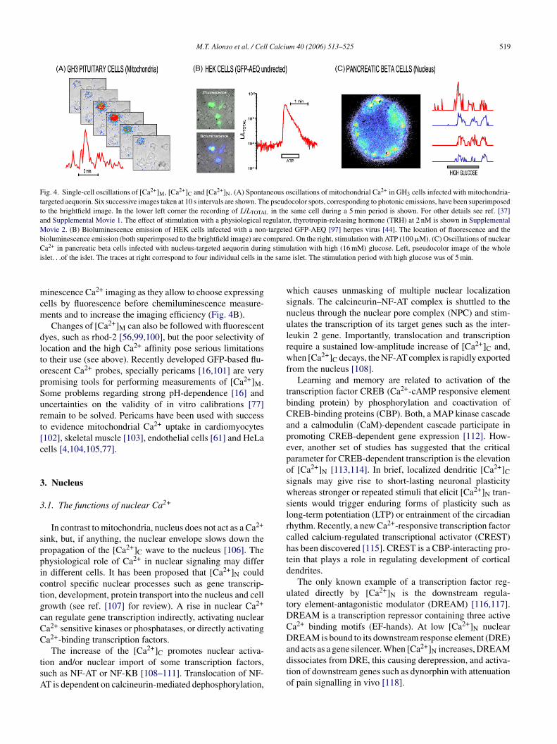
Single-cell measurements of aequorin bioluminescencere difficult because of the very low light output [93],ut combining the high sensitivity provided by photon-ounting cameras [94,95] and high expression of aequorinnduced by viral vectors [44,20] spontaneous mitochondriala2+ oscillations could be readily demonstrated in singleH3 pituitary cells [37] (Fig. 4A; see also Supplementalovie 1). These [Ca2+]M oscillations were modulated by the
ypophysiotrophic hypothalamic hormones ([37]; see alsoupplemental Movie 2). The [Ca2+]M oscillations take placenly in a mitochondrial pool close to the plasma membrane37]. Thus, subplasmalemmal mitochondria oppose the pro-ression of the Ca2+ wave towards the nucleus, where themplitude of the oscillations is smaller [96]. On the otherand, stimulation by [Ca2+]M of respiration in this pool ofitochondria would tune local metabolism to the increased
nergy needs [95,91,13,37]. Similarly, [Ca2+]M oscillationsere also evidenced in pancreatic beta cells stimulated withigh glucose (unpublished results with A. Nadal and I. Que-ada).
The fusion protein of aequorin with GFP seems to have anncreased chemiluminescence output, perhaps by an increase
f the half-life of the chimeric protein [97,98]. Expression ofhe GFP-AEQ can be checked very simply by fluorescenceonitoring in single cells. These and other chimera with flu-rescent proteins may improve very much practical photolu-
M.T. Alonso et al. / Cell Calcium 40 (2006) 513–525 519
Fig. 4. Single-cell oscillations of [Ca2+]M, [Ca2+]C and [Ca2+]N. (A) Spontaneous oscillations of mitochondrial Ca2+ in GH3 cells infected with mitochondria-targeted aequorin. Six successive images taken at 10 s intervals are shown. The pseudocolor spots, corresponding to photonic emissions, have been superimposedto the brightfield image. In the lower left corner the recording of L/LTOTAL in the same cell during a 5 min period is shown. For other details see ref. [37]and Supplemental Movie 1. The effect of stimulation with a physiological regulator, thyrotropin-releasing hormone (TRH) at 2 nM is shown in SupplementalMovie 2. (B) Bioluminescence emission of HEK cells infected with a non-targeted GFP-AEQ [97] herpes virus [44]. The location of fluorescence and thebioluminescence emission (both superimposed to the brightfield image) are compared. On the right, stimulation with ATP (100 �M). (C) Oscillations of nuclearC g stimui the sam
mcm
dltopSurt[c
3
3
sppictgcCC
tsA
wsnulrwf
tbCapeposwslrchtd
utDCD
a2+ in pancreatic beta cells infected with nucleus-targeted aequorin durinslet. . .of the islet. The traces at right correspond to four individual cells in
inescence Ca2+ imaging as they allow to choose expressingells by fluorescence before chemiluminescence measure-ents and to increase the imaging efficiency (Fig. 4B).Changes of [Ca2+]M can also be followed with fluorescent
yes, such as rhod-2 [56,99,100], but the poor selectivity ofocation and the high Ca2+ affinity pose serious limitationso their use (see above). Recently developed GFP-based flu-rescent Ca2+ probes, specially pericams [16,101] are veryromising tools for performing measurements of [Ca2+]M.ome problems regarding strong pH-dependence [16] andncertainties on the validity of in vitro calibrations [77]emain to be solved. Pericams have been used with successo evidence mitochondrial Ca2+ uptake in cardiomyocytes102], skeletal muscle [103], endothelial cells [61] and HeLaells [4,104,105,77].
. Nucleus
.1. The functions of nuclear Ca2+
In contrast to mitochondria, nucleus does not act as a Ca2+
ink, but, if anything, the nuclear envelope slows down theropagation of the [Ca2+]C wave to the nucleus [106]. Thehysiological role of Ca2+ in nuclear signaling may differn different cells. It has been proposed that [Ca2+]N couldontrol specific nuclear processes such as gene transcrip-ion, development, protein transport into the nucleus and cellrowth (see ref. [107] for review). A rise in nuclear Ca2+
an regulate gene transcription indirectly, activating nucleara2+ sensitive kinases or phosphatases, or directly activatinga2+-binding transcription factors.
The increase of the [Ca2+]C promotes nuclear activa-ion and/or nuclear import of some transcription factors,uch as NF-AT or NF-KB [108–111]. Translocation of NF-T is dependent on calcineurin-mediated dephosphorylation,
adto
lation with high (16 mM) glucose. Left, pseudocolor image of the wholee islet. The stimulation period with high glucose was of 5 min.
hich causes unmasking of multiple nuclear localizationignals. The calcineurin–NF-AT complex is shuttled to theucleus through the nuclear pore complex (NPC) and stim-lates the transcription of its target genes such as the inter-eukin 2 gene. Importantly, translocation and transcriptionequire a sustained low-amplitude increase of [Ca2+]C and,hen [Ca2+]C decays, the NF-AT complex is rapidly exported
rom the nucleus [108].Learning and memory are related to activation of the
ranscription factor CREB (Ca2+-cAMP responsive elementinding protein) by phosphorylation and coactivation ofREB-binding proteins (CBP). Both, a MAP kinase cascadend a calmodulin (CaM)-dependent cascade participate inromoting CREB-dependent gene expression [112]. How-ver, another set of studies has suggested that the criticalarameter for CREB-dependent transcription is the elevationf [Ca2+]N [113,114]. In brief, localized dendritic [Ca2+]Cignals may give rise to short-lasting neuronal plasticityhereas stronger or repeated stimuli that elicit [Ca2+]N tran-
ients would trigger enduring forms of plasticity such asong-term potentiation (LTP) or entrainment of the circadianhythm. Recently, a new Ca2+-responsive transcription factoralled calcium-regulated transcriptional activator (CREST)as been discovered [115]. CREST is a CBP-interacting pro-ein that plays a role in regulating development of corticalendrites.
The only known example of a transcription factor reg-lated directly by [Ca2+]N is the downstream regula-ory element-antagonistic modulator (DREAM) [116,117].REAM is a transcription repressor containing three activea2+ binding motifs (EF-hands). At low [Ca2+]N nuclearREAM is bound to its downstream response element (DRE)
nd acts as a gene silencer. When [Ca2+]N increases, DREAMissociates from DRE, this causing derepression, and activa-ion of downstream genes such as dynorphin with attenuationf pain signalling in vivo [118].
5 l Calciu
3
[tcobaam[dbmmFti
b[col[hdpiasC(nco[cav(wm
3
iorrtTwc
aogrlr[rddao[kdnsm
s[t[iccsdClpocar(attcF
teFn[tarrf
20 M.T. Alonso et al. / Cel
.2. Nuclear Ca2+ measurements
Many of the studies designed to compare [Ca2+]C andCa2+]N were performed using low molecular weight indica-ors, like fluo-3, measured by conventional epifluorescence,onfocal microscopy or two-photon microscopy. The resultsf these studies have often been suggestive of differencesetween the two compartments, but the probe accumulatesnd is brighter in the nucleoplasm than in the cytosol. Inddition, the dye properties, including the affinity for Ca2+,ay be modified by the cytoplasmic or nuclear environment
119]. Dyes bound to high molecular weight dextrans (fura-2extran, fluo-4 dextran and calcium green dextran) have alsoeen used successfully to report [Ca2+]N [120,121], but theyay well suffer the same shortcomings. It must be kept inind that other artefacts can bias the imaging techniques.or example, what seemed to be local Ca2+ hotspots inside
he nucleoplasm resulted to be [Ca2+]C changes taking placenside invaginations of the cytosol into the nucleus [122,123].
The protein-based indicators have the advantage ofeing specifically targeted to the organella. Cameleons15], pericams [16] and aequorins [124] have been suc-essfully targeted into the nucleus. The first generationf calmodulin-based Cameleons displayed disadvantagesike the sensitivity to the pH and the poor dynamic range15]. Improved Cameleons [125] targeted to the nucleus ofypothalamic neurons in vivo have successfully detectedifferences between [Ca2+]N and [Ca2+]C [126]. Ratiometricericam has been used successfully to measure [Ca2+]Nn beating cardiac cells [102]. The bioluminescent proteinequorin [124] and the chimeric GFP-aequorin [97] provideome distinct advantages over other genetically encodeda2+ indicators: low buffer capacity, wide dynamic range
Fig. 2) and little or no sensitivity to the cytosolic orucleoplasmic environment [98]. The main limitation in theontext of evidencing nuclear microdomains is the low lightutput and poor spatial resolution. However, imaging ofCa2+]N in GH3 individual pituitary and beta-pancreatic cellsould be performed using a herpes virus-based system tottain high probe expression (Fig. 4C) [96,127]. In any case,isualization and measurement of Ca2+ molecular domainssee above) would require a higher targeting resolution,hich could involve docking Ca2+ indicators to definedolecular locations through genetic manipulations.
.3. Nuclear calcium signalling
Upon stimulation, many cells generate Ca2+ signals bothn the cytosol and the nucleus. Despite the increasing amountf reports addressing this issue, the mechanisms of [Ca2+]Negulation are still controversial (see refs. [11,107] foreviews). The nucleus is surrounded by a double membrane,
he nuclear envelope, that separates it from the cytoplasm.he NE contains large (9 nm diameter) nuclear pores (NP)ith large ion conductance. It has been reported that NPonductance can be drastically reduced by ATP-dependent
Cnit
m 40 (2006) 513–525
ccumulation of Ca2+ inside the NE (gating) or by transportf macromolecules through nuclear pore complexes (plug-ing) [128], but results are contradictory (see ref. [11] foreview). The permeability of the nuclear envelope to cytoso-ic Ca2+ is a matter of controversy. While several authorseport that [Ca2+]N follows passively changes of [Ca2+]C124,129,130], others identify a significant diffusion bar-ier at the NE [131–134]. The point is not trivial, as it mayetermine whether or not the [Ca2+]C signals shall be trans-uced into changes of gene expression. In our hands, NEcts as a kinetic barrier that dampens high frequency [Ca2+]Cscillations without disturbing sustained [Ca2+]C increases96]. Other factors that may contribute to raise this nuclearinetic barrier are [Ca2+]C sinking by perinuclear mitochon-ria [88,79–81,25] or high Ca2+ buffering capacity inside theucleus [135]. Thus, through regulation of specific local Ca2+
ignals in each compartment, cytosolic and nuclear processesay manage some level of independent control.Fig. 5A summarizes five different modes in which [Ca2+]N
ignals could be generated. Apart from transmission of theCa2+]C signal through NE (1 in Fig. 5A), it has been reportedhat [Ca2+]N signals could be triggered independently ofCa2+]C signals by direct release of Ca2+ from the NE cisternanto the nucleoplasm. Inositol (1,4,5)-trisphosphate (IP3),yclic ADP-ribose (cADPR) and nicotinic acid adenine dinu-leotide phosphate (NAADP) have all been reported to induceuch a release [120,136–138,11]. In the case of IP3 the evi-ence is particularly strong as, in addition to data on nucleara2+ release [139,120–123,140,133], there is evidence for
ocal generation of IP3 by a nuclear inositide-specific phos-holipase C (see ref. [141] for review) and for the existencef IP3 receptors in the nucleus [142–150]. Recent patchlamp measurements have also shown functional release sitesctivated by IP3 in the inner NE membrane in Purkinje neu-ones [151]. Intranuclear production of cyclic ADP-ribosecADPR) and presence of ryanodine receptors in the NE haslso been reported [152,11,153]. Ca2+ released from NE cis-erna could reach the nucleoplasm either via the cytosol andhen through the NP (2 in Fig. 5A) or directly if the Ca2+
hannels of the NE could face towards the nucleoplasm (3 inig. 5A).
The efficiency of direct Ca2+ release from the nuclear cis-erna to the nucleoplasm could be very much increased by thexistence of invaginations of the NE inside the nucleus (4 inig. 5A). Invaginations of the NE protuding deep within theucleus have been described in several mammalian cell types154,155,122,123]. These invaginations have been proposedo form a complex reticular network that works as a Ca2+ stor-ge organelle inside the nucleus [146]. This nucleoplasmiceticulum expresses functional IP3 receptors (IP3Rs) able toelease Ca2+ in response to intranuclear IP3 [146] as well asunctional ryanodine receptors able to evoke Ca2+-induced
a2+ release within the nucleus [138]. This intricate sig-alling machinery could allow the release of calcium locallyn discrete regions inside the nucleus. Taking advantage ofhe specificity of targeted aequorins, we have recently found
M.T. Alonso et al. / Cell Calcium 40 (2006) 513–525 521
Fig. 5. Five mechanisms that may produce an increase of [Ca2+]N. (A) PM, plasma membrane; Cyt, cytosol; NE, nuclear envelope; NPL, nucleoplasm. Ca2+
may reach the nucleus coming from the cytosol [1]. Alternatively, Ca2+ may be released from the nuclear cisterna and reach the nucleus either passing through thecytosol [2] or by direct release to the nucleoplasm [3]. An invagination of the “nucleoplasmic reticulum” could also release Ca2+ to the core of the nucleoplasm[ ntaininm B, a mn
iptce
rinsotitaamobt[amhm
foorttya
dhTR[d
A
TE0Cg
A
b2
R
4]. Finally Ca2+ could be released directly to the nucleoplasm from Ca2+ coicroscopy [5,157]. (B) GH3 cells stained with an antibody against lamin
ucleoplasm are seen in many cells.
ndependent activation of nuclear Ca2+ signalling in GH3ituitary cells by IP3. The nucleus of these cells also con-ains deep invaginations of the NE into the nucleoplasm thatan even contain intracellular organella (Fig. 5B) (Chamerot al., unpublished results).
As mentioned above, the presence of IP3 receptors andyanodine receptors inside the nucleus has been reportedn several cell types, but the presence of such Ca2+ chan-els not associated to membranes is puzzling. In a recenteries of papers Huh and co-workers find that nuclear IP3Rsf chromaffin cells are associated to the Ca2+-binding pro-ein chromogranin B and to phospholipids [156] and, moremportantly, to very small (50 nm) nucleoplasmic vesicleshat release Ca2+ in response to IP3 [157]. This new mech-nism (5 in Fig. 5A) is extremely suggestive, as it wouldllow independent control of [Ca2+]N and the generation oficrodomains by localized stimuli. The total calcium content
f the nucleus is very high (about 11 mM) [157], much of itound to chromosomes [158]. Release of even a small frac-ion of this bound calcium would produce a large increase ofCa2+]N. In addition, such a release could be restricted to localreas inside the nucleus. The existence of subnuclear Ca2+
icrodomains is compatible with the view of the nucleus as aeterogeneous organelle in which various different processesust be regulated simultaneously.Although it seems clear that the mechanisms required
or independent control of [Ca2+]N and for the generationf [Ca2+]N microdomains exist, the physiological operationf these mechanisms has not been probed yet. IGF-1 waseported to activate nuclear PLC in 3T3 cells, with produc-
ion of diacylglicerol in the nuclear membrane [159], buthe associated [Ca2+]N changes have not been documentedet. IGF-1 has also been reported to increase [Ca2+]N and toctivate CREB in skeletal muscle cells [143,160] and in car-g nuclear microvesicles too small to be seen even by conventional electronarker of the internal side of the NE. Invaginations entering deep into the
iomyocytes [149]. On the other hand [Ca2+]N oscillationsave been documented in GH3 pituitary cells stimulated byRH [96], glucose-stimulated pancreatic beta cells ([84,127],utter, in this issue), ATP-stimulated airway epithelial cells
153] or testosterone-stimulated neurons [150], but [Ca2+]Coes also oscillate in all the above cases.
cknowledgements
We thank Mr. Jesus Fernandez for technical assistance.his work was funded by grants from the Ministerio deducacion y Ciencia (BFU2004-02765/BFI and BFU2005-5464) and from Junta de Castilla y Leon (VA016A05). Pablohamero holded a predoctoral fellowship from the Basqueovernment.
ppendix A. Supplementary data
Supplementary data associated with this article cane found in the online version, at: doi:10.1016/j.ceca.006.08.013.
eferences
[1] P. Mitchell, Chemiosmotic coupling in oxidative and photosyntheticphosphorylation, Biol. Rev. Camb. Philos. Soc. 41 (1966) 445–502.
[2] J.E. Chad, R. Eckert, Calcium domains associated with individ-
ual channels can account for anomalous voltage relations of CA-dependent responses, Biophys. J. 45 (1984) 993–999.[3] S.M. Simon, R.R. Llinas, Compartmentalization of the submembranecalcium activity during calcium influx and its significance in trans-mitter release, Biophys. J. 48 (1985) 485–498.
5 l Calciu
22 M.T. Alonso et al. / Cel[4] A.L. Fogelson, R.S. Zucker, Presynaptic calcium diffusion from vari-ous arrays of single channels Implications for transmitter release andsynaptic facilitation, Biophys. J. 48 (1985) 1003–1017.
[5] E. Neher, Vesicle pools and Ca2+ microdomains: new tools for under-standing their roles in neurotransmitter release, Neuron 20 (1998)389–399.
[6] Z. Zhou, E. Neher, Mobile and immobile calcium buffers in bovineadrenal chromaffin cells, J. Physiol. 469 (1993) 245–273.
[7] T. Xu, M. Naraghi, H. Kang, E. Neher, Kinetic studies of Ca2+ bindingand Ca2+ clearance in the cytosol of adrenal chromaffin cells, Biophys.J. 73 (1997) 532–545.
[8] J. Alvarez, M. Montero, J. Garcia-Sancho, Subcellular Ca2+ dynam-ics, News Physiol. Sci. 14 (1999) 161–168.
[9] M.T. Alonso, M.J. Barrero, P. Michelena, et al., Ca2+-induced Ca2+
release in chromaffin cells seen from inside the ER with targetedaequorin, J. Cell Biol. 144 (1999) 241–254.
[10] M. Montero, M.T. Alonso, A. Albillos, J. Garcia-Sancho, J. Alvarez,Mitochondrial Ca2+-induced Ca2+ release mediated by the Ca2+ uni-porter, Mol. Biol. Cell 12 (2001) 63–71.
[11] O. Gerasimenko, J. Gerasimenko, New aspects of nuclear calciumsignalling, J. Cell Sci. 117 (2004) 3087–3094.
[12] O.H. Petersen, M. Michalak, A. Verkhratsky, Calcium signalling: past,present and future, Cell Calcium 38 (2005) 161–169.
[13] R. Rizzuto, T. Pozzan, Microdomains of intracellular Ca2+: moleculardeterminants and functional consequences, Physiol. Rev. 86 (2006)369–408.
[14] R. Rizzuto, A.W. Simpson, M. Brini, T. Pozzan, Rapid changes ofmitochondrial Ca2+ revealed by specifically targeted recombinantaequorin, Nature 358 (1992) 325–327.
[15] A. Miyawaki, J. Llopis, R. Heim, et al., Fluorescent indicators forCa2+ based on green fluorescent proteins and calmodulin, Nature 388(1997) 882–887.
[16] T. Nagai, A. Sawano, E.S. Park, A. Miyawaki, Circularly permutedgreen fluorescent proteins engineered to sense Ca2+, Proc. Natl. Acad.Sci. U.S.A. 98 (2001) 3197–3202.
[17] B.N. Giepmans, S.R. Adams, M.H. Ellisman, R.Y. Tsien, The fluo-rescent toolbox for assessing protein location and function, Science312 (2006) 217–224.
[18] M. Montero, M. Brini, R. Marsault, et al., Monitoring dynamicchanges in free Ca2+ concentration in the endoplasmic reticulum ofintact cells, EMBO J. 14 (1995) 5467–5475.
[19] O. Shimomura, B. Musicki, Y. Kishi, S. Inouye, Light-emittingproperties of recombinant semi-synthetic aequorins and recombinantfluorescein-conjugated aequorin for measuring cellular calcium, CellCalcium 14 (1993) 373–378.
[20] M. Montero, M.T. Alonso, E. Carnicero, et al., Chromaffin-cell stim-ulation triggers fast millimolar mitochondrial Ca2+ transients thatmodulate secretion, Nat. Cell Biol. 2 (2000) 57–61.
[21] J. Alvarez, M. Montero, Ca2+ measurement with luminescent probesin the endoplasmic reticulum, in: O.H. Petersen (Ed.), Measuring Cal-cium and Calmodulin Inside and Outside Cells, Springer Lab Manual,Berlin, 2001, pp. 147–163.
[22] M.N. Badminton, J.M. Kendall, G. Sala-Newby, A.K. Campbell,Nucleoplasmin-targeted aequorin provides evidence for a nuclear cal-cium barrier, Exp. Cell Res. 216 (1995) 236–243.
[23] J.G. Pitter, P. Maechler, C.B. Wollheim, A. Spat, Mitochondriarespond to Ca2+ already in the submicromolar range: correlation withredox state, Cell Calcium 31 (2002) 97–104.
[24] J.E. Church, D. Fulton, Differences in eNOS activity because of sub-cellular localization are dictated by phosphorylation state rather thanthe local calcium environment, J. Biol. Chem. 281 (2006) 1477–1488.
[25] M.R. Duchen, Mitochondria and calcium: from cell signalling to cell
death, J. Physiol. 529 (2000) 57–68.[26] T.E. Gunter, D.R. Pfeiffer, Mechanisms by which mitochondria trans-port calcium, Am. J. Physiol. 258 (1990) C755–C786.
[27] P. Bernardi, Mitochondrial transport of cations: channels, exchangers,and permeability transition, Physiol. Rev. 79 (1999) 1127–1155.
m 40 (2006) 513–525
[28] G.J. Augustine, E. Neher, Calcium requirements for secretion inbovine chromaffin cells, J. Physiol. 450 (1992) 247–271.
[29] K.E. Coll, S.K. Joseph, B.E. Corkey, J.R. Williamson, Determinationof the matrix free Ca2+ concentration and kinetics of Ca2+ effluxin liver and heart mitochondria, J. Biol. Chem. 257 (1982) 8696–8704.
[30] D.F. Babcock, J. Herrington, P.C. Goodwin, Y.B. Park, B. Hille, Mito-chondrial participation in the intracellular Ca2+ network, J. Cell Biol.136 (1997) 833–844.
[31] C. Villalobos, L. Nunez, M. Montero, et al., Redistribution of Ca2+
among cytosol and organella during stimulation of bovine chromaffincells, FASEB J. 16 (2002) 343–353.
[32] J. Herrington, Y.B. Park, D.F. Babcock, B. Hille, Dominant role ofmitochondria in clearance of large Ca2+ loads from rat adrenal chro-maffin cells, Neuron 16 (1996) 219–228.
[33] E. Jambrina, R. Alonso, M. Alcalde, et al., Calcium influx throughreceptor-operated channel induces mitochondria-triggered paraptoticcell death, J. Biol. Chem. 278 (2003) 14134–14145.
[34] G.C. Sparagna, K.K. Gunter, S.S. Sheu, T.E. Gunter, Mitochon-drial calcium uptake from physiological-type pulses of calcium Adescription of the rapid uptake mode, J. Biol. Chem. 270 (1995)27510–27515.
[35] T.J. Collins, P. Lipp, M.J. Berridge, M.D. Bootman, MitochondrialCa2+ uptake depends on the spatial and temporal profile of cytosolicCa2+ signals, J. Biol. Chem. 276 (2001) 26411–26420.
[36] S. Chalmers, D.G. Nicholls, The relationship between free and totalcalcium concentrations in the matrix of liver and brain mitochondria,J. Biol. Chem. 278 (2003) 19062–19070.
[37] C. Villalobos, L. Nunez, P. Chamero, M.T. Alonso, J. Garcia-Sancho,Mitochondrial [Ca2+] oscillations driven by local high [Ca2+] domainsgenerated by spontaneous electric activity, J. Biol. Chem. 276 (2001)40293–40297.
[38] M. Montero, C.D. Lobaton, A. Moreno, J. Alvarez, A novel regulatorymechanism of the mitochondrial Ca2+ uniporter revealed by the p38mitogen-activated protein kinase inhibitor SB202190, FASEB J. 16(2002) 1955–1957.
[39] P. Pinton, S. Leo, M.R. Wieckowski, G. Di Benedetto, R. Rizzuto,Long-term modulation of mitochondrial Ca2+ signals by proteinkinase C isozymes, J. Cell Biol. 165 (2004) 223–232.
[40] S. Arnaudeau, W.L. Kelley, J.V. Walsh Jr., N. Demaurex, Mitochon-dria recycle Ca2+ to the endoplasmic reticulum and prevent the deple-tion of neighboring endoplasmic reticulum regions, J. Biol. Chem. 276(2001) 29430–29439.
[41] L. Filippin, P.J. Magalhaes, G. Di Benedetto, M. Colella, T. Pozzan,Stable interactions between mitochondria and endoplasmic reticulumallow rapid accumulation of calcium in a subpopulation of mitochon-dria, J. Biol. Chem. 278 (2003) 39224–39234.
[42] M.J. Barrero, M. Montero, J. Alvarez, Dynamics of [Ca2+] in the endo-plasmic reticulum and cytoplasm of intact HeLa cells. A comparativestudy, J. Biol. Chem. 272 (1997) 27694–27699.
[43] M. Montero, M.J. Barrero, J. Alvarez, [Ca2+] microdomains controlagonist-induced Ca2+ release in intact HeLa cells, FASEB J. 11 (1997)881–885.
[44] M.T. Alonso, M.J. Barrero, E. Carnicero, M. Montero, J. Garcia-Sancho, J. Alvarez, Functional measurements of [Ca2+] in the endo-plasmic reticulum using a herpes virus to deliver targeted aequorin,Cell Calcium 24 (1998) 87–96.
[45] A.M. Hofer, T.E. Machen, Technique for in situ measurement ofcalcium in intracellular inositol 1,4,5-trisphosphate-sensitive storesusing the fluorescent indicator mag-fura-2, Proc. Natl. Acad. Sci.U.S.A. 90 (1993) 2598–2602.
[46] F.W. Tse, A. Tse, B. Hille, Cyclic Ca2+ changes in intracellular stores
of gonadotropes during gonadotropin-releasing hormone-stimulatedCa2+ oscillations, Proc. Natl. Acad. Sci. U.S.A. 91 (1994) 9750–9754.[47] L. Combettes, T.R. Cheek, C.W. Taylor, Regulation of inositol trispho-sphate receptors by luminal Ca2+ contributes to quantal Ca2+ mobi-lization, EMBO J. 15 (1996) 2086–2093.
l Calciu
M.T. Alonso et al. / Cel[48] V.A. Golovina, M.P. Blaustein, Spatially and functionally distinctCa2+ stores in sarcoplasmic and endoplasmic reticulum, Science 275(1997) 1643–1648.
[49] R. Foyouzi-Youssefi, S. Arnaudeau, C. Borner, et al., Bcl-2 decreasesthe free Ca2+ concentration within the endoplasmic reticulum, Proc.Natl. Acad. Sci. U.S.A. 97 (2000) 5723–5728.
[50] A.M. Hofer, I. Schulz, Quantification of intraluminal free [Ca] inthe agonist-sensitive internal calcium store using compartmentalizedfluorescent indicators: some considerations, Cell Calcium 20 (1996)235–242.
[51] H. Mogami, A.V. Tepikin, O.H. Petersen, Termination of cytosolicCa2+ signals: Ca2+ reuptake into intracellular stores is regulated bythe free Ca2+ concentration in the store lumen, EMBO J. 17 (1998)435–442.
[52] G. Csordas, A.P. Thomas, G. Hajnoczky, Quasi-synaptic calcium sig-nal transmission between endoplasmic reticulum and mitochondria,EMBO J. 18 (1999) 96–108.
[53] R.J. White, I.J. Reynolds, Mitochondria accumulate Ca2+ followingintense glutamate stimulation of cultured rat forebrain neurones, J.Physiol. 498 (1997) 31–47.
[54] E.J. Kaftan, T. Xu, R.F. Abercrombie, B. Hille, Mitochondria shapehormonally induced cytoplasmic calcium oscillations and modulateexocytosis, J. Biol. Chem. 275 (2000) 25465–25470.
[55] J.M. Hernandez-Guijo, V.E. Maneu-Flores, A. Ruiz-Nuno, M. Villar-roya, A.G. Garcia, L. Gandia, Calcium-dependent inhibition of L, N,and P/Q Ca2+ channels in chromaffin cells: role of mitochondria, J.Neurosci. 21 (2001) 2553–2560.
[56] E. Boitier, R. Rea, M.R. Duchen, Mitochondria exert a negative feed-back on the propagation of intracellular Ca2+ waves in rat corticalastrocytes, J. Cell Biol. 145 (1999) 795–808.
[57] M. Hoth, C.M. Fanger, R.S. Lewis, Mitochondrial regulation of store-operated calcium signaling in T lymphocytes, J. Cell Biol. 137 (1997)633–648.
[58] M. Hoth, D.C. Button, R.S. Lewis, Mitochondrial control of calcium-channel gating: a mechanism for sustained signaling and transcrip-tional activation in T lymphocytes, Proc. Natl. Acad. Sci. U.S.A. 97(2000) 10607–10612.
[59] J.A. Gilabert, A.B. Parekh, Respiring mitochondria determine the pat-tern of activation and inactivation of the store-operated Ca2+ currentI (CRAC), EMBO J. 19 (2000) 6401–6407.
[60] A.B. Parekh, Store-operated Ca2+ entry: dynamic interplay betweenendoplasmic reticulum, mitochondria and plasma membrane, J. Phys-iol. 547 (2003) 333–348.
[61] R. Malli, M. Frieden, K. Osibow, W.F. Graier, Mitochondria efficientlybuffer subplasmalemmal Ca2+ elevation during agonist stimulation,J. Biol. Chem. 278 (2003) 10807–10815.
[62] L. Nunez, R.A. Valero, L. Senovilla, S. Sanz-Blasco, J. Garcia-Sancho, C. Villalobos, Cell proliferation depends on mitochondrialCa2+ uptake: inhibition by salicylate, J. Physiol. 571 (2006) 57–73.
[63] G. Hajnoczky, R. Hager, A.P. Thomas, Mitochondria suppress localfeedback activation of inositol 1,4,5-trisphosphate receptors by Ca2+,J. Biol. Chem. 274 (1999) 14157–14162.
[64] R. Rizzuto, M. Brini, M. Murgia, T. Pozzan, Microdomains with highCa2+ close to IP3-sensitive channels that are sensed by neighboringmitochondria, Science 262 (1993) 744–747.
[65] M.R. Duchen, A. Leyssens, M. Crompton, Transient mitochondrialdepolarizations reflect focal sarcoplasmic reticular calcium release insingle rat cardiomyocytes, J. Cell Biol. 142 (1998) 975–988.
[66] R. Rizzuto, P. Pinton, W. Carrington, et al., Close contacts withthe endoplasmic reticulum as determinants of mitochondrial Ca2+
responses, Science 280 (1998) 1763–1766.[67] G. Hajnoczky, G. Csordas, M. Madesh, P. Pacher, The machinery
of local Ca2+ signalling between sarco-endoplasmic reticulum andmitochondria, J. Physiol. 529 (2000) 69–81.
[68] G. Csordas, G. Hajnoczky, Sorting of calcium signals at the junctionsof endoplasmic reticulum and mitochondria, Cell Calcium 29 (2001)249–262.
m 40 (2006) 513–525 523
[69] G. Csordas, G. Hajnoczky, Plasticity of mitochondrial calcium sig-naling, J. Biol. Chem. 278 (2003) 42273–42282.
[70] M. Montero, C.D. Lobaton, E. Hernandez-Sanmiguel, et al., Directactivation of the mitochondrial calcium uniporter by natural plantflavonoids, Biochem. J. 384 (2004) 19–24.
[71] C.D. Lobaton, L. Vay, E. Hernandez-Sanmiguel, et al., Modulation ofmitochondrial Ca2+ uptake by estrogen receptor agonists and antag-onists, Br. J. Pharmacol. 145 (2005) 862–871.
[72] S.G. Robles, M. Franco, C. Zazueta, et al., Thyroid hormone mayinduce changes in the concentration of the mitochondrial calcium uni-porter, Comp. Biochem. Physiol. B Biochem. Mol. Biol. 135 (2003)177–182.
[73] E. Hernandez-SanMiguel, L. Vay, J. Santo-Domingo, C.D. Lobaton,A. Moreno, M. Montero, J. Alvarez, The mitochondrial Na+/Ca2+
exchanger plays a key role in the control of cytosolic Ca2+ oscillations,Cell Calcium 40 (2006) 53–61.
[74] R. Malli, M. Frieden, K. Osibow, et al., Sustained Ca2+ transferacross mitochondria is essential for mitochondrial Ca2+ buffering,sore-operated Ca2+ entry, and Ca2+ store refilling, J. Biol. Chem. 278(2003) 44769–44779.
[75] R. Malli, M. Frieden, M. Trenker, W.F. Graier, The role of mitochon-dria for Ca2+ refilling of the endoplasmic reticulum, J. Biol. Chem.280 (2005) 12114–12122.
[76] L.M. Solis-Garrido, A.J. Pintado, E. Andres-Mateos, M. Figueroa,C. Matute, C. Montiel, Cross-talk between native plasmalemmalNa+/Ca2+ exchanger and inositol 1,4,5-trisphosphate-sensitive Ca2+
internal store in Xenopus oocytes, J. Biol. Chem. 279 (2004)52414–52424.
[77] K. Ishii, K. Hirose, M. Iino, Ca2+ shuttling between endoplasmic retic-ulum and mitochondria underlying Ca2+ oscillations, EMBO Rep. 7(2006) 390–396.
[78] N.B. Pivovarova, J. Hongpaisan, S.B. Andrews, D.D. Friel,Depolarization-induced mitochondrial Ca accumulation in sympa-thetic neurons: spatial and temporal characteristics, J. Neurosci. 19(1999) 6372–6384.
[79] H. Tinel, J.M. Cancela, H. Mogami, et al., Active mitochondria sur-rounding the pancreatic acinar granule region prevent spreading ofinositol trisphosphate-evoked local cytosolic Ca2+ signals, EMBO J.18 (1999) 4999–5008.
[80] M.K. Park, M.C. Ashby, G. Erdemli, O.H. Petersen, A.V. Tepikin, Per-inuclear, perigranular and sub-plasmalemmal mitochondria have dis-tinct functions in the regulation of cellular calcium transport, EMBOJ. 20 (2001) 1863–1874.
[81] P.R. Johnson, N.J. Dolman, M. Pope, et al., Non-uniform distributionof mitochondria in pancreatic acinar cells, Cell Tissue Res. 313 (2003)37–45.
[82] P. Pacher, A.P. Thomas, G. Hajnoczky, Ca2+ marks: miniature calciumsignals in single mitochondria driven by ryanodine receptors, Proc.Natl. Acad. Sci. U.S.A. 99 (2002) 2380–2385.
[83] W.F. Pralong, L. Hunyady, P. Varnai, C.B. Wollheim, A. Spat, Pyri-dine nucleotide redox state parallels production of aldosterone inpotassium-stimulated adrenal glomerulosa cells, Proc. Natl. Acad.Sci. U.S.A. 89 (1992) 132–136.
[84] W.F. Pralong, A. Spat, C.B. Wollheim, Dynamic pacing of cellmetabolism by intracellular Ca2+ transients, J. Biol. Chem. 269 (1994)27310–27314.
[85] A. Spat, J.G. Pitter, T. Rohacs, G. Szabadkai, Stimulus-secretion cou-pling and mitochondrial metabolism in steroid-secreting cells, NewsPhysiol. Sci. 16 (2001) 197–200.
[86] A. Spat, L. Hunyady, Control of aldosterone secretion: a model forconvergence in cellular signaling pathways, Physiol. Rev. 84 (2004)489–539.
[87] G. Hajnoczky, L.D. Robb-Gaspers, M.B. Seitz, A.P. Thomas, Decod-ing of cytosolic calcium oscillations in the mitochondria, Cell 82(1995) 415–424.
[88] L.D. Robb-Gaspers, P. Burnett, G.A. Rutter, R.M. Denton, R.Rizzuto, A.P. Thomas, Integrating cytosolic calcium signals into
5 l Calciu
24 M.T. Alonso et al. / Celmitochondrial metabolic responses, EMBO J. 17 (1998) 4987–5000.
[89] E.D. Kennedy, R. Rizzuto, J.M. Theler, et al., Glucose-stimulatedinsulin secretion correlates with changes in mitochondrial and cytoso-lic Ca2+ in aequorin-expressing INS-1 cells, J. Clin. Invest. 98 (1996)2524–2538.
[90] P. Maechler, C.B. Wollheim, Mitochondrial signals in glucose-stimulated insulin secretion in the beta cell, J. Physiol. 529 (2000)49–56.
[91] L.S. Jouaville, P. Pinton, C. Bastianutto, G.A. Rutter, R. Rizzuto,Regulation of mitochondrial ATP synthesis by calcium: evidence for along-term metabolic priming, Proc. Natl. Acad. Sci. U.S.A. 96 (1999)13807–13812.
[92] H.J. Kennedy, A.E. Pouli, E.K. Ainscow, L.S. Jouaville, R. Riz-zuto, G.A. Rutter, Glucose generates sub-plasma membrane ATPmicrodomains in single islet beta-cells. Potential role for strate-gically located mitochondria, J. Biol. Chem. 274 (1999) 13281–13291.
[93] A. Chiesa, E. Rapizzi, V. Tosello, et al., Recombinant aequorin andgreen fluorescent protein as valuable tools in the study of cell sig-nalling, Biochem. J. 355 (2001) 1–12.
[94] L.S. Frawley, W.J. Faught, J. Nicholson, B. Moomaw, Real time mea-surement of gene expression in living endocrine cells, Endocrinology135 (1994) 468–471.
[95] G.A. Rutter, P. Burnett, R. Rizzuto, et al., Subcellular imaging ofintramitochondrial Ca2+ with recombinant targeted aequorin: signif-icance for the regulation of pyruvate dehydrogenase activity, Proc.Natl. Acad. Sci. U.S.A. 93 (1996) 5489–5494.
[96] P. Chamero, C. Villalobos, M.T. Alonso, J. Garcia-Sancho, Dampen-ing of cytosolic Ca2+ oscillations on propagation to nucleus, J. Biol.Chem. 277 (2002) 50226–50229.
[97] V. Baubet, H. Le Mouellic, A.K. Campbell, E. Lucas-Meunier, P.Fossier, P. Brulet, Chimeric green fluorescent protein-aequorin as bio-luminescent Ca2+ reporters at the single-cell level, Proc. Natl. Acad.Sci. U.S.A. 97 (2000) 7260–7265.
[98] K.L. Rogers, J. Stinnakre, C. Agulhon, et al., Visualization of localCa2+ dynamics with genetically encoded bioluminescent reporters,Eur. J. Neurosci. 21 (2005) 597–610.
[99] S. Voronina, T. Sukhomlin, P.R. Johnson, G. Erdemli, O.H. Petersen,A. Tepikin, Correlation of NADH and Ca2+ signals in mouse pancre-atic acinar cells, J. Physiol. 539 (2002) 41–52.
[100] E.C. Toescu, A. Verkhratsky, Neuronal ageing from an intraneuronalperspective: roles of endoplasmic reticulum and mitochondria, CellCalcium 34 (2003) 311–323.
[101] T. Nagai, S. Yamada, T. Tominaga, M. Ichikawa, A. Miyawaki,Expanded dynamic range of fluorescent indicators for Ca2+ by cir-cularly permuted yellow fluorescent proteins, Proc. Natl. Acad. Sci.U.S.A. 101 (2004) 10554–10559.
[102] V. Robert, P. Gurlini, V. Tosello, et al., Beat-to-beat oscillations ofmitochondrial [Ca2+] in cardiac cells, EMBO J. 20 (2001) 4998–5007.
[103] R. Rudolf, M. Mongillo, P.J. Magalhaes, T. Pozzan, In vivo monitoringof Ca2+ uptake into mitochondria of mouse skeletal muscle duringcontraction, J. Cell Biol. 166 (2004) 527–536.
[104] M. Frieden, D. James, C. Castelbou, A. Danckaert, J.C. Martinou,N. Demaurex, Ca2+ homeostasis during mitochondrial fragmentationand perinuclear clustering induced by hFis1, J. Biol. Chem. 279 (2004)22704–22714.
[105] G. Szabadkai, A.M. Simoni, M. Chami, M.R. Wieckowski, R.J. Youle,R. Rizzuto, Drp-1-dependent division of the mitochondrial networkblocks intraorganellar Ca2+ waves and protects against Ca2+-mediatedapoptosis, Mol. Cell 16 (2004) 59–68.
[106] M. Naraghi, T.H. Muller, E. Neher, Two-dimensional determination
of the cellular Ca2+ binding in bovine chromaffin cells, Biophys. J.75 (1998) 1635–1647.[107] M.D. Bootman, D. Thomas, S.C. Tovey, M.J. Berridge, P. Lipp,Nuclear calcium signalling, Cell. Mol. Life Sci. 57 (2000) 371–378.
m 40 (2006) 513–525
[108] R.E. Dolmetsch, K. Xu, R.S. Lewis, Calcium oscillations increasethe efficiency and specificity of gene expression, Nature 392 (1998)933–936.
[109] G.R. Crabtree, Generic signals and specific outcomes: signalingthrough Ca2+, calcineurin, and NF-AT, Cell 96 (1999) 611–614.
[110] G.R. Crabtree, E.N. Olson, NFAT signaling: choreographing thesocial lives of cells, Cell 109 (2002) S67–S79.
[111] E.M. Gallo, K. Cante-Barrett, G.R. Crabtree, Lymphocyte calciumsignaling from membrane to nucleus, Nat. Immunol. 7 (2006)25–32.
[112] R.E. Dolmetsch, U. Pajvani, K. Fife, J.M. Spotts, M.E. Greenberg,Signaling to the nucleus by an L-type calcium channel-calmodulincomplex through the MAP kinase pathway, Science 294 (2001)333–339.
[113] G.E. Hardingham, F.J. Arnold, H. Bading, Nuclear calcium signal-ing controls CREB-mediated gene expression triggered by synapticactivity, Nat. Neurosci. 4 (2001) 261–267.
[114] H. Bading, Transcription-dependent neuronal plasticity the nuclearcalcium hypothesis, Eur. J. Biochem. 267 (2000) 5280–5283.
[115] H. Aizawa, S.C. Hu, K. Bobb, et al., Dendrite development regulatedby CREST, a calcium-regulated transcriptional activator, Science 303(2004) 197–202.
[116] A.M. Carrion, W.A. Link, F. Ledo, B. Mellstrom, J.R. Naranjo,DREAM is a Ca2+-regulated transcriptional repressor, Nature 398(1999) 80–84.
[117] R. Gomez-Villafuertes, B. Torres, J. Barrio, et al., Downstream reg-ulatory element antagonist modulator regulates Ca2+ homeostasisand viability in cerebellar neurons, J. Neurosci. 25 (2005) 10822–10830.
[118] H.Y. Cheng, G.M. Pitcher, S.R. Laviolette, et al., DREAM is a criticaltranscriptional repressor for pain modulation, Cell 108 (2002) 31–43.
[119] C. Perez-Terzic, L. Stehno-Bittel, D.E. Clapham, Nucleoplasmic andcytoplasmic differences in the fluorescence properties of the calciumindicator Fluo-3, Cell Calcium 21 (1997) 275–282.
[120] O.V. Gerasimenko, J.V. Gerasimenko, A.V. Tepikin, O.H. Petersen,ATP-dependent accumulation and inositol trisphosphate- or cyclicADP-ribose-mediated release of Ca2+ from the nuclear envelope, Cell80 (1995) 439–444.
[121] L. Santella, K. Kyozuka, Effects of 1-methyladenine on nuclear Ca2+
transients and meiosis resumption in starfish oocytes are mimicked bythe nuclear injection of inositol 1,4,5-trisphosphate and cADP-ribose,Cell Calcium 22 (1997) 11–20.
[122] P.P. Lui, S.K. Kong, T.T. Kwok, C.Y. Lee, The nucleus of HeLa cellcontains tubular structures for Ca2+ signalling, Biochem. Biophys.Res. Commun. 247 (1998) 88–93.
[123] P.P. Lui, F.L. Chan, Y.K. Suen, T.T. Kwok, S.K. Kong, The nucleusof HeLa cells contains tubular structures for Ca2+ signaling with theinvolvement of mitochondria, Biochem. Biophys. Res. Commun. 308(2003) 826–833.
[124] M. Brini, M. Murgia, L. Pasti, D. Picard, T. Pozzan, R. Riz-zuto, Nuclear Ca2+ concentration measured with specifically targetedrecombinant aequorin, EMBO J. 12 (1993) 4813–4819.
[125] A. Miyawaki, O. Griesbeck, R. Heim, R.Y. Tsien, Dynamic and quan-titative Ca2+ measurements using improved cameleons, Proc. Natl.Acad. Sci. U.S.A. 96 (1999) 2135–2140.
[126] M. Ikeda, T. Sugiyama, C.S. Wallace, et al., Circadian dynamics ofcytosolic and nuclear Ca2+ in single suprachiasmatic nucleus neurons,Neuron 38 (2003) 253–263.
[127] C. Villalobos, A. Nadal, L. Nunez, et al., Bioluminescence imaging ofnuclear calcium oscillations in intact pancreatic islets of Langerhansfrom the mouse, Cell Calcium 38 (2005) 131–139.
[128] J.O. Bustamante, E.R. Michelette, J.P. Geibel, D.A. Dean, J.A.
Hanover, T.J. McDonnell, Calcium ATP and nuclear pore channelgating, Pflugers Arch. 439 (2000) 433–444.[129] H. Shirakawa, S. Miyazaki, Spatiotemporal analysis of calciumdynamics in the nucleus of hamster oocytes, J. Physiol. 494 (1996)29–40.
l Calciu
M.T. Alonso et al. / Cel[130] H. Nakazawa, T.H. Murphy, Activation of nuclear calcium dynamicsby synaptic stimulation in cultured cortical neurons, J. Neurochem.73 (1999) 1075–1083.
[131] M.N. Badminton, A.K. Campbell, C.M. Rembold, Differential regu-lation of nuclear and cytosolic Ca2+ in HeLa cells, J. Biol. Chem. 271(1996) 31210–31214.
[132] O.V. Gerasimenko, J.V. Gerasimenko, A.V. Tepikin, O.H. Petersen,Calcium transport pathways in the nucleus, Pflugers Arch. 432 (1996)1–6.
[133] P.P. Lui, S.K. Kong, K.P. Fung, C.Y. Lee, The rise of nuclear andcytosolic Ca2+ can be uncoupled in HeLa cells, Pflugers Arch. 436(1998) 371–376.
[134] M.N. Badminton, J.M. Kendall, C.M. Rembold, A.K. Campbell, Cur-rent evidence suggests independent regulation of nuclear calcium,Cell Calcium 23 (1998) 79–86.
[135] M.N. Teruel, W. Chen, A. Persechini, T. Meyer, Differential codes forfree Ca2+-calmodulin signals in nucleus and cytosol, Curr. Biol. 10(2000) 86–94.
[136] O.H. Petersen, O.V. Gerasimenko, J.V. Gerasimenko, H. Mogami,A.V. Tepikin, The calcium store in the nuclear envelope, Cell Calcium23 (1998) 87–90.
[137] J.V. Gerasimenko, Y. Maruyama, K. Yano, et al., NAADP mobilizesCa2+ from a thapsigargin-sensitive store in the nuclear envelope byactivating ryanodine receptors, J. Cell Biol. 163 (2003) 271–282.
[138] P. Marius, M.T. Guerra, M.H. Nathanson, B.E. Ehrlich, M.F. Leite,Calcium release from ryanodine receptors in the nucleoplasmic retic-ulum, Cell Calcium 39 (2005) 65–73.
[139] P. Nicotera, S. Orrenius, T. Nilsson, P.O. Berggren, An inositol 1,4,5-trisphosphate-sensitive Ca2+ pool in liver nuclei, Proc. Natl. Acad.Sci. U.S.A. 87 (1990) 6858–6862.
[140] D.J. Hennager, M.J. Welsh, S. DeLisle, Changes in either cytosolic ornucleoplasmic inositol 1,4,5-trisphosphate levels can control nuclearCa2+ concentration, J. Biol. Chem. 270 (1995) 4959–4962.
[141] L. Manzoli, A.M. Billi, A.M. Martelli, L. Cocco, Regulation ofnuclear phospholipase C activity, Acta Biochim. Pol. 51 (2004)391–395.
[142] J.P. Humbert, N. Matter, J.C. Artault, P. Koppler, A.N. Malviya,Inositol 1,4,5-trisphosphate receptor is located to the inner nuclearmembrane vindicating regulation of nuclear calcium signaling byinositol 1,4,5-trisphosphate. Discrete distribution of inositol phos-phate receptors to inner and outer nuclear membranes, J. Biol. Chem.271 (1996) 478–485.
[143] J.A. Powell, M.A. Carrasco, D.S. Adams, et al., IP3 receptor functionand localization in myotubes: an unexplored Ca2+ signaling pathwayin skeletal muscle, J. Cell Sci. 114 (2001) 3673–3683.
[144] K. Laflamme, O. Domingue, B.I. Guillemette, G. Guillemette,
Immunohistochemical localization of type 2 inositol 1,4,5-trisphosphate receptor to the nucleus of different mammalian cells, J.Cell. Biochem. 85 (2002) 219–228.[145] Y.H. Huh, S.H. Yoo, Presence of the inositol 1,4,5-triphosphate recep-tor isoforms in the nucleoplasm, FEBS Lett. 555 (2003) 411–418.
m 40 (2006) 513–525 525
[146] W. Echevarria, M.F. Leite, M.T. Guerra, W.R. Zipfel, M.H.Nathanson, Regulation of calcium signals in the nucleus by a nucle-oplasmic reticulum, Nat. Cell Biol. 5 (2003) 440–446.
[147] M.F. Leite, E.C. Thrower, W. Echevarria, et al., Nuclear and cytosoliccalcium are regulated independently, Proc. Natl. Acad. Sci. U.S.A.100 (2003) 2975–2980.
[148] K.D. Garcia, T. Shah, J. Garcia, Immunolocalization of type 2 inos-itol 1,4,5-trisphosphate receptors in cardiac myocytes from newbornmice, Am. J. Physiol. Cell Physiol. 287 (2004) C1048–C1057.
[149] C. Ibarra, M. Estrada, L. Carrasco, et al., Insulin-like growth factor-1induces an inositol 1,4,5-trisphosphate-dependent increase in nuclearand cytosolic calcium in cultured rat cardiac myocytes, J. Biol. Chem.279 (2004) 7554–7565.
[150] M. Estrada, P. Uhlen, B.E. Ehrlich, Ca2+ oscillations induced bytestosterone enhance neurite outgrowth, J. Cell Sci. 119 (2006)733–743.
[151] S.M. Marchenko, V.V. Yarotskyy, T.N. Kovalenko, P.G. Kostyuk, R.C.Thomas, Spontaneously active and InsP3-activated ion channels incell nuclei from rat cerebellar Purkinje and granule neurones, J. Phys-iol. 565 (2005) 897–910.
[152] O.A. Adebanjo, H.K. Anandatheerthavarada, A.P. Koval, et al., A newfunction for CD38/ADP-ribosyl cyclase in nuclear Ca2+ homeostasis,Nat. Cell Biol. 1 (1999) 409–414.
[153] I. Quesada, P. Verdugo, InsP3 signaling induces pulse-modulatedCa2+ signals in the nucleus of airway epithelial ciliated cells, Bio-phys. J. 88 (2005) 3946–3953.
[154] M. Fricker, M. Hollinshead, N. White, D. Vaux, Interphase nucleiof many mammalian cell types contain deep, dynamic, tubularmembrane-bound invaginations of the nuclear envelope, J. Cell Biol.136 (1997) 531–544.
[155] B.H. Clubb, M. Locke, 3T3 cells have nuclear invaginations contain-ing F-actin, Tissue Cell 30 (1998) 684–691.
[156] S.H. Yoo, S.W. Nam, S.K. Huh, S.Y. Park, Y.H. Huh, Presence of anucleoplasmic complex composed of the inositol 1,4,5-trisphosphatereceptor/Ca2+ channel, chromogranin B, and phospholipids, Bio-chemistry 44 (2005) 9246–9254.
[157] Y.H. Huh, S.K. Huh, S.Y. Chu, H.S. Kweon, S.H. Yoo, Presence ofa putative vesicular inositol 1,4,5-trisphosphate-sensitive nucleoplas-mic Ca2+ store, Biochemistry 45 (2006) 1362–1373.
[158] R. Strick, P.L. Strissel, K. Gavrilov, R. Levi-Setti, Cation-chromatinbinding as shown by ion microscopy is essential for the structuralintegrity of chromosomes, J. Cell Biol. 155 (2001) 899–910.
[159] N. Divecha, H. Banfic, R.F. Irvine, The polyphosphoinositide cycleexists in the nuclei of Swiss 3T3 cells under the control of a receptor(for IGF-I) in the plasma membrane, and stimulation of the cycleincreases nuclear diacylglycerol and apparently induces translocation
of protein kinase C to the nucleus, EMBO J. 10 (1991) 3207–3214.[160] C. Cardenas, J.L. Liberona, J. Molgo, C. Colasante, G.A. Mignery,E. Jaimovich, Nuclear inositol 1,4,5-trisphosphate receptors regulatelocal Ca2+ transients and modulate cAMP response element bindingprotein phosphorylation, J. Cell Sci. 118 (2005) 3131–3140.
Copyright © 2022 FDOKUMEN