Maintaining the balance of TDP-43, mitochondria, and ...
16
REVIEW Open Access Maintaining the balance of TDP-43, mitochondria, and autophagy: a promising therapeutic strategy for neurodegenerative diseases Chunhui Huang 1 , Sen Yan 2* and Zaijun Zhang 1* Abstract Mitochondria are the energy center of cell operations and are involved in physiological functions and maintenance of metabolic balance and homeostasis in the body. Alterations of mitochondrial function are associated with a variety of degenerative and acute diseases. As mitochondria age in cells, they gradually become inefficient and potentially toxic. Acute injury can trigger the permeability of mitochondrial membranes, which can lead to apoptosis or necrosis. Transactive response DNA-binding protein 43 kDa (TDP-43) is a protein widely present in cells. It can bind to RNA, regulate a variety of RNA processes, and play a role in the formation of multi-protein/RNA complexes. Thus, the normal physiological functions of TDP-43 are particularly important for cell survival. Normal TDP-43 is located in various subcellular structures including mitochondria, mitochondrial-associated membrane, RNA particles and stress granules to regulate the endoplasmic reticulum–mitochondrial binding, mitochondrial protein translation, and mRNA transport and translation. Importantly, TDP-43 is associated with a variety of neurodegenerative diseases, including amyotrophic lateral sclerosis, frontotemporal dementia and Alzheimer's disease, which are characterized by abnormal phosphorylation, ubiquitination, lysis or nuclear depletion of TDP-43 in neurons and glial cells. Although the pathogenesis of TDP-43 proteinopathy remains unknown, the presence of pathological TDP-43 inside or outside of mitochondria and the functional involvement of TDP-43 in the regulation of mitochondrial morphology, transport, and function suggest that mitochondria are associated with TDP-43-related diseases. Autophagy is a basic physiological process that maintains the homeostasis of cells, including targeted clearance of abnormally aggregated proteins and damaged organelles in the cytoplasm; therefore, it is considered protective against neurodegenerative diseases. However, the combination of abnormal TDP-43 aggregation, mitochondrial dysfunction, and insufficient autophagy can lead to a variety of aging-related pathologies. In this review, we describe the current knowledge on the associations of mitochondria with TDP-43 and the role of (Continued on next page) © The Author(s). 2020 Open Access This article is licensed under a Creative Commons Attribution 4.0 International License, which permits use, sharing, adaptation, distribution and reproduction in any medium or format, as long as you give appropriate credit to the original author(s) and the source, provide a link to the Creative Commons licence, and indicate if changes were made. The images or other third party material in this article are included in the article's Creative Commons licence, unless indicated otherwise in a credit line to the material. If material is not included in the article's Creative Commons licence and your intended use is not permitted by statutory regulation or exceeds the permitted use, you will need to obtain permission directly from the copyright holder. To view a copy of this licence, visit http://creativecommons.org/licenses/by/4.0/. The Creative Commons Public Domain Dedication waiver (http://creativecommons.org/publicdomain/zero/1.0/) applies to the data made available in this article, unless otherwise stated in a credit line to the data. * Correspondence: [email protected]; [email protected] 2 Ministry of Education CNS Regeneration Collaborative Joint Laboratory, Guangdong-Hongkong-Macau Institute of CNS Regeneration, Jinan University, Guangzhou 510632, China 1 Institute of New Drug Research, Guangdong Province Key Laboratory of Pharmacodynamic, Constituents of Traditional Chinese Medicine and New Drug Research, College of Pharmacy, Jinan University, Guangzhou 510632, China Huang et al. Translational Neurodegeneration (2020) 9:40 https://doi.org/10.1186/s40035-020-00219-w
-
Upload
khangminh22 -
Category
Documents
-
view
1 -
download
0
Transcript of Maintaining the balance of TDP-43, mitochondria, and ...
REVIEW Open Access
Maintaining the balance of TDP-43,mitochondria, and autophagy: a promisingtherapeutic strategy for neurodegenerativediseasesChunhui Huang1, Sen Yan2* and Zaijun Zhang1*
Abstract
Mitochondria are the energy center of cell operations and are involved in physiological functions and maintenanceof metabolic balance and homeostasis in the body. Alterations of mitochondrial function are associated with avariety of degenerative and acute diseases. As mitochondria age in cells, they gradually become inefficient andpotentially toxic. Acute injury can trigger the permeability of mitochondrial membranes, which can lead toapoptosis or necrosis. Transactive response DNA-binding protein 43 kDa (TDP-43) is a protein widely present incells. It can bind to RNA, regulate a variety of RNA processes, and play a role in the formation of multi-protein/RNAcomplexes. Thus, the normal physiological functions of TDP-43 are particularly important for cell survival. NormalTDP-43 is located in various subcellular structures including mitochondria, mitochondrial-associated membrane,RNA particles and stress granules to regulate the endoplasmic reticulum–mitochondrial binding, mitochondrialprotein translation, and mRNA transport and translation. Importantly, TDP-43 is associated with a variety ofneurodegenerative diseases, including amyotrophic lateral sclerosis, frontotemporal dementia and Alzheimer'sdisease, which are characterized by abnormal phosphorylation, ubiquitination, lysis or nuclear depletion of TDP-43in neurons and glial cells. Although the pathogenesis of TDP-43 proteinopathy remains unknown, the presence ofpathological TDP-43 inside or outside of mitochondria and the functional involvement of TDP-43 in the regulationof mitochondrial morphology, transport, and function suggest that mitochondria are associated with TDP-43-relateddiseases. Autophagy is a basic physiological process that maintains the homeostasis of cells, including targetedclearance of abnormally aggregated proteins and damaged organelles in the cytoplasm; therefore, it is consideredprotective against neurodegenerative diseases. However, the combination of abnormal TDP-43 aggregation,mitochondrial dysfunction, and insufficient autophagy can lead to a variety of aging-related pathologies. In thisreview, we describe the current knowledge on the associations of mitochondria with TDP-43 and the role of(Continued on next page)
© The Author(s). 2020 Open Access This article is licensed under a Creative Commons Attribution 4.0 International License,which permits use, sharing, adaptation, distribution and reproduction in any medium or format, as long as you giveappropriate credit to the original author(s) and the source, provide a link to the Creative Commons licence, and indicate ifchanges were made. The images or other third party material in this article are included in the article's Creative Commonslicence, unless indicated otherwise in a credit line to the material. If material is not included in the article's Creative Commonslicence and your intended use is not permitted by statutory regulation or exceeds the permitted use, you will need to obtainpermission directly from the copyright holder. To view a copy of this licence, visit http://creativecommons.org/licenses/by/4.0/.The Creative Commons Public Domain Dedication waiver (http://creativecommons.org/publicdomain/zero/1.0/) applies to thedata made available in this article, unless otherwise stated in a credit line to the data.
* Correspondence: [email protected]; [email protected] of Education CNS Regeneration Collaborative Joint Laboratory,Guangdong-Hongkong-Macau Institute of CNS Regeneration, JinanUniversity, Guangzhou 510632, China1Institute of New Drug Research, Guangdong Province Key Laboratory ofPharmacodynamic, Constituents of Traditional Chinese Medicine and NewDrug Research, College of Pharmacy, Jinan University, Guangzhou 510632,China
Huang et al. Translational Neurodegeneration (2020) 9:40 https://doi.org/10.1186/s40035-020-00219-w
(Continued from previous page)
autophagy in the clearance of abnormally aggregated TDP-43 and dysfunctional mitochondria. Finally, we discuss anovel approach for neurodegenerative treatment based on the knowledge.
Keywords: TDP-43, Mitochondria, Autophagy/mitophagy, Neurodegeneration
BackgroundNeurodegenerative diseases are a group of clinically heteroge-neous diseases characterized by the progressive loss or dys-function of neurons in the central nervous system (CNS) orthe peripheral nervous system, including Alzheimer’s disease(AD), Parkinson’s disease (PD), Huntington’s disease (HD),amyotrophic lateral sclerosis (ALS), and frontotemporal de-mentia (FTD) [1]. Although considerable progress has beenmade in understanding the mechanisms underlying the tran-sition from pathological changes in brain to neurodegenera-tive changes, treatments for these diseases have shownlimited effectiveness. A common feature of these neurodegen-erative diseases is the deposition of abnormally aggregatedproteins in extracellular or intracellular inclusions [2, 3].The discovery of transactive response DNA-binding protein
43 kDa (TDP-43) as the major component of ubiquitin-positive neuronal inclusion bodies is a milestone in under-standing the pathogenesis of ALS [4]. In the past decade, TDP-43, encoded by the TARDBP gene, was revealed as a key playerin the pathogenesis of various neurodegenerative diseases [5,6]. Since then, rapid progress has been made in understandingthe physiological functions of TDP-43 and its role in neurode-generation [7]. For example, mutations in the TDP-43 genecan cause ALS, and a direct link has been established betweenTDP-43 and neurodegenerative diseases [8–10]. The neurode-generative diseases associated with the abnormal aggregation ofTDP-43 are collectively referred to as “TDP-43 proteinopathy”[5]. Increasing evidence has suggested that the pathologicalTDP-43 interferes with multiple pathways including RNA me-tabolism, protein translation, stress-induced response, autoph-agy, endocytosis, ubiquitin-proteasome system, andmitochondrial function [11–17]. Although the neurotoxicity ofthe pathological TDP-43 protein has not been fully elucidated[18–20], the presence of pathologically related TDP-43 insideor outside the mitochondria and the functional participation ofTDP-43 in the regulations of mitochondrial morphology, trans-port and function suggest that mitochondria may be a thera-peutic target for TDP-43 proteinopathy [5, 17].Mitochondria play a vital role in cell bioenergy and
apoptosis [21]. Damage to mitochondrial function andcell energy production leads to irreversible cell death. Inrecent years, the function of mitochondria has becomean important topic in research and development oftherapeutic drugs for various neurological diseases in-cluding ALS, PD, and AD [22]. The structure, function,and localization of mitochondria are closely related toneurodegenerative diseases, and defects in mitochondrial
dynamics can contribute to neuronal diseases [23]. Mito-chondria have independent genomes and provide oxygenconsumption-driven synthesis of ATP (via oxidativephosphorylation, OXPHOS) [24]. However, as a by-product of normal breathing, mitochondria also producetoxic reactive oxygen species (ROS). In addition, themitochondrial genome accumulates mutations duringreplication, which ultimately affects the efficiency ofOXPHOS. The lack of mitochondria, excess ROS, orboth, are likely to be the driving forces of aging, as theyreduce the adaptability of cells, cause damage to otherorganelles, and cause mutations in the nuclear genome[25, 26]. In addition, mutant TDP-43 can damage mito-chondrial dynamics, and the overexpression of TDP-43can cause abnormal aggregation of mitochondria andloss of their normal functions, leading to progressive lossof neurons [27–29]. Moreover, inhibiting the mitochon-drial localization of TDP-43 can block the TDP-43-induced mitochondrial dysfunction [17, 30], suggestingthat the removal of abnormally aggregated TDP-43 anddysfunctional or damaged mitochondria and restoringthe interaction of TDP-43 with mitochondria may be aneffective way to treat neurodegenerative diseases.Autophagy is a basic physiological process that main-
tains the homeostasis of cells [31] by clearing abnormallyaggregated cytosolic proteins and damaged organelles. Au-tophagy involves a series of sequential events, includingdouble-membrane formation, elongation, vesicle matur-ation, and ultimately delivery of target substances to lyso-somes for degradation and recycling as an energy source[32]. When abnormal or misfolded proteins accumulate inthe cytoplasm, nucleus, and extracellular envelopes, theycan cause organelle damage and synaptic dysfunction inthe nervous system; and autophagy has a regulatory effecton these proteinopathies, including a variety of neurode-generative diseases [31]. The elimination of TDP-43 byautophagy has received increasing attention [33]. Autoph-agy can occur as a common phenomenon, such as whencells mobilize energy reserves or lack nutrition, or it canspecifically target intracellular components such as abnor-mally aggregated TDP-43 protein and dysfunctional ordamaged mitochondria to maintain cell survival [34, 35].Thus, research on the neuroprotective effects of autoph-agy may lead to the development of disease-modifyingtherapies for neurodegenerative diseases [36, 37].In summary, the combination of abnormal TDP-43 ag-
gregation, mitochondrial dysfunction, and insufficient
Huang et al. Translational Neurodegeneration (2020) 9:40 Page 2 of 16

autophagy may lead to a variety of aging-related patholo-gies. In this review, we first summarize current know-ledge on the associations between TDP-43 andmitochondria. Then, we describe the role of autophagyin clearing abnormally aggregated TDP-43 and dysfunc-tional or damaged mitochondria. Finally, we briefly dis-cuss how the studies of TDP-43 proteinopathy,mitochondrial dysfunction, and autophagy may providea novel approach for treatment of neurodegenerativediseases (Fig. 1).
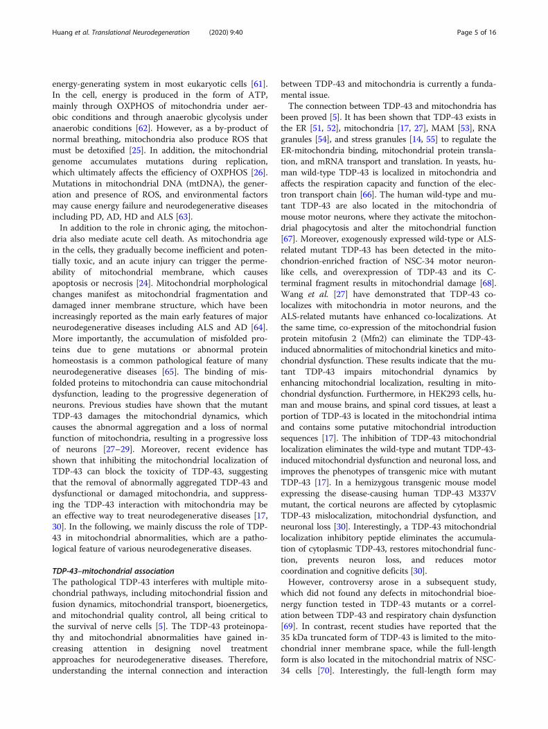
Main textTDP-43Structure and function of TDP-43TDP-43 is a DNA- and RNA-binding protein that be-longs to the heterogeneous nuclear ribonucleoproteinfamily [38]. It consists of 414 amino acids, is encoded bythe TARDBP gene (chromosome 1p36.2), and has a rela-tive molecular mass of approximately 43000 daltons[39]. TDP-43 contains four functional regions from N-to C-terminus: a nuclear localization sequence (NLS),two RNA recognition motifs (RRMs, RRM1 and RRM2),a nuclear export sequence (NES), and a glycine-rich do-main [40] (Fig. 2). Among them, the NLS and NES regu-late the shuttle of TDP-43 between nucleus andcytoplasm; RRM1 and RRM2 participate in DNA- orRNA-binding; the glycine-rich domain regulates the spli-cing activity of TDP-43 and its interactions with othercytokines or organelles, and contains occurrence of mostof the disease-related mutations [41, 42].TDP-43 was originally identified as a transcriptional
factor that suppresses the transcription of human im-munodeficiency virus 1 [43], and later was proved to bea pre-mRNA splicing regulator [44]. Under normal con-ditions, TDP-43 is widely present in human and rodenttissues and organs including the heart, lung, liver, spleen,kidney, muscle, and brain [44]. It is mainly located inthe nucleus and is capable of nucleus-cytoplasm shuttle[4, 45]. About 30% of TDP-43 protein can be found inthe cytoplasm, and its nuclear efflux is regulated by ac-tivity and pressure [46, 47]. TDP-43 can bind to RNAand regulate RNA synthesis, splicing, stability and trans-port, thereby affecting a variety of cellular processes[48]. It also plays a role in the formation of multi-protein/RNA complexes, participates in the regulation ofmicroRNAs and biogenesis, and binds to DNA to inhibitgene transcription [13, 49, 50]. TDP-43 in the cytoplasmcan interact with subcellular compartments such as theendoplasmic reticulum (ER) [51, 52], mitochondria [17,27], mitochondrial-associated membrane (MAM) [53],RNA granules [54], and stress granules [14, 55] to regu-late the ER-mitochondrial binding, mitochondrial pro-tein translation, and mRNA transport and translation.
Therefore, normal physiological functions of TDP-43 areparticularly important for cell survival.
Key roles of TDP-43 in neurodegenerative diseasesTDP-43 protein is the main component of tau-negativeand ubiquitin-positive inclusion bodies in cortical neu-rons of frontotemporal lobar degeneration and spinalmotor neurons of ALS [4]. TDP-43 aggregation andneuropathology have been observed in a series of uniqueneurodegenerative diseases, collectively referred to asTDP-43 proteinopathy [18, 56]. TDP-43 proteinopathyoccurs through the characteristic histopathologicaltransformation of TDP-43 in the disease [57]. In thepathological state, the TDP-43 proteinopathy occursthrough two pathological changes, gain of function andloss of function, which involve phosphorylation, ubiqui-tination, lysis, reduced solubility, and ectopic cytoplasmexpression [57, 58]. The gain of function refers to thecytotoxicity of TDP-43 under abnormal conditions [59].For example, TDP-43 phosphorylation and ubiquitina-tion are the main pathological changes in patients withTDP-43 proteinopathy, which increase the formation ofinsoluble inclusions, interfere with the normal functionof TDP-43 and lead to a cytotoxic form of TDP-43. Inaddition, as the C-terminal fragment of TDP-43 has asimilar sequence to prion protein, the spread of thistoxic peptide in adjacent neurons may also be patho-genic. On the other hand, loss of function refers to theweakening or disappearance of normal functions ofTDP-43 after structural change, resulting in abnormalneuronal function including impaired protein degrad-ation, changes in TDP-43-related splicing events, nucleartransport defects, loss of TDP-4 automatic adjustment,and the enhancement of TDP-43 self-interaction [59].At present, the pathogenesis of TDP-43-associated
pathology is still unclear, but its importance in neurode-generative diseases is self-evident and needs further in-vestigation [6, 60]. Therefore, it is necessary to furtherstudy the functions of TDP-43 in order to develop thera-peutic strategies that can increase the TDP-43 activity orprevent the TDP-43 aggregation to reduce the potentialtoxic effects, thereby preventing disease onset orprogression.
Mitochondria and TDP-43Mitochondria and neurodegenerative diseasesMitochondria have been extensively proven to partici-pate in the basic processes of the nervous system suchas energy and intermediate metabolism, calcium homeo-stasis, and apoptosis [61]. The CNS functions dependlargely on effective mitochondrial function because ofthe high energy demand of brain tissue. It is well knownthat mitochondria are the powerhouses of cells that pro-duce adenosine triphosphate (ATP), and are the main
Huang et al. Translational Neurodegeneration (2020) 9:40 Page 3 of 16
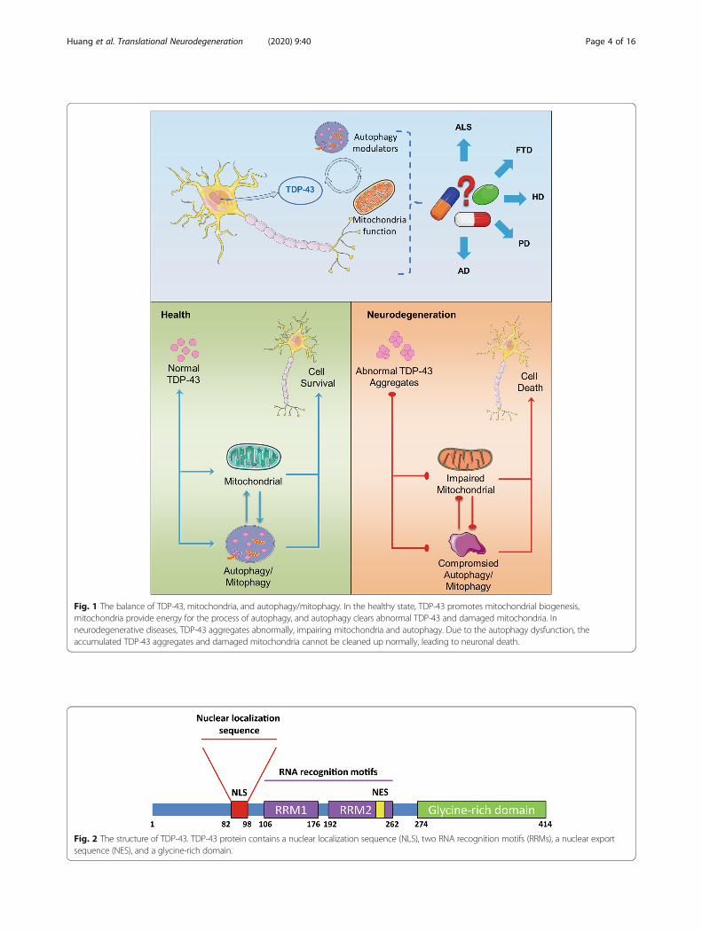
Fig. 1 The balance of TDP-43, mitochondria, and autophagy/mitophagy. In the healthy state, TDP-43 promotes mitochondrial biogenesis,mitochondria provide energy for the process of autophagy, and autophagy clears abnormal TDP-43 and damaged mitochondria. Inneurodegenerative diseases, TDP-43 aggregates abnormally, impairing mitochondria and autophagy. Due to the autophagy dysfunction, theaccumulated TDP-43 aggregates and damaged mitochondria cannot be cleaned up normally, leading to neuronal death.
Fig. 2 The structure of TDP-43. TDP-43 protein contains a nuclear localization sequence (NLS), two RNA recognition motifs (RRMs), a nuclear exportsequence (NES), and a glycine-rich domain.
Huang et al. Translational Neurodegeneration (2020) 9:40 Page 4 of 16
energy-generating system in most eukaryotic cells [61].In the cell, energy is produced in the form of ATP,mainly through OXPHOS of mitochondria under aer-obic conditions and through anaerobic glycolysis underanaerobic conditions [62]. However, as a by-product ofnormal breathing, mitochondria also produce ROS thatmust be detoxified [25]. In addition, the mitochondrialgenome accumulates mutations during replication,which ultimately affects the efficiency of OXPHOS [26].Mutations in mitochondrial DNA (mtDNA), the gener-ation and presence of ROS, and environmental factorsmay cause energy failure and neurodegenerative diseasesincluding PD, AD, HD and ALS [63].In addition to the role in chronic aging, the mitochon-
dria also mediate acute cell death. As mitochondria agein the cells, they gradually become inefficient and poten-tially toxic, and an acute injury can trigger the perme-ability of mitochondrial membrane, which causesapoptosis or necrosis [24]. Mitochondrial morphologicalchanges manifest as mitochondrial fragmentation anddamaged inner membrane structure, which have beenincreasingly reported as the main early features of majorneurodegenerative diseases including ALS and AD [64].More importantly, the accumulation of misfolded pro-teins due to gene mutations or abnormal proteinhomeostasis is a common pathological feature of manyneurodegenerative diseases [65]. The binding of mis-folded proteins to mitochondria can cause mitochondrialdysfunction, leading to the progressive degeneration ofneurons. Previous studies have shown that the mutantTDP-43 damages the mitochondrial dynamics, whichcauses the abnormal aggregation and a loss of normalfunction of mitochondria, resulting in a progressive lossof neurons [27–29]. Moreover, recent evidence hasshown that inhibiting the mitochondrial localization ofTDP-43 can block the toxicity of TDP-43, suggestingthat the removal of abnormally aggregated TDP-43 anddysfunctional or damaged mitochondria, and suppress-ing the TDP-43 interaction with mitochondria may bean effective way to treat neurodegenerative diseases [17,30]. In the following, we mainly discuss the role of TDP-43 in mitochondrial abnormalities, which are a patho-logical feature of various neurodegenerative diseases.
TDP-43–mitochondrial associationThe pathological TDP-43 interferes with multiple mito-chondrial pathways, including mitochondrial fission andfusion dynamics, mitochondrial transport, bioenergetics,and mitochondrial quality control, all being critical tothe survival of nerve cells [5]. The TDP-43 proteinopa-thy and mitochondrial abnormalities have gained in-creasing attention in designing novel treatmentapproaches for neurodegenerative diseases. Therefore,understanding the internal connection and interaction
between TDP-43 and mitochondria is currently a funda-mental issue.The connection between TDP-43 and mitochondria has
been proved [5]. It has been shown that TDP-43 exists inthe ER [51, 52], mitochondria [17, 27], MAM [53], RNAgranules [54], and stress granules [14, 55] to regulate theER-mitochondria binding, mitochondrial protein transla-tion, and mRNA transport and translation. In yeasts, hu-man wild-type TDP-43 is localized in mitochondria andaffects the respiration capacity and function of the elec-tron transport chain [66]. The human wild-type and mu-tant TDP-43 are also located in the mitochondria ofmouse motor neurons, where they activate the mitochon-drial phagocytosis and alter the mitochondrial function[67]. Moreover, exogenously expressed wild-type or ALS-related mutant TDP-43 has been detected in the mito-chondrion-enriched fraction of NSC-34 motor neuron-like cells, and overexpression of TDP-43 and its C-terminal fragment results in mitochondrial damage [68].Wang et al. [27] have demonstrated that TDP-43 co-localizes with mitochondria in motor neurons, and theALS-related mutants have enhanced co-localizations. Atthe same time, co-expression of the mitochondrial fusionprotein mitofusin 2 (Mfn2) can eliminate the TDP-43-induced abnormalities of mitochondrial kinetics and mito-chondrial dysfunction. These results indicate that the mu-tant TDP-43 impairs mitochondrial dynamics byenhancing mitochondrial localization, resulting in mito-chondrial dysfunction. Furthermore, in HEK293 cells, hu-man and mouse brains, and spinal cord tissues, at least aportion of TDP-43 is located in the mitochondrial intimaand contains some putative mitochondrial introductionsequences [17]. The inhibition of TDP-43 mitochondriallocalization eliminates the wild-type and mutant TDP-43-induced mitochondrial dysfunction and neuronal loss, andimproves the phenotypes of transgenic mice with mutantTDP-43 [17]. In a hemizygous transgenic mouse modelexpressing the disease-causing human TDP-43 M337Vmutant, the cortical neurons are affected by cytoplasmicTDP-43 mislocalization, mitochondrial dysfunction, andneuronal loss [30]. Interestingly, a TDP-43 mitochondriallocalization inhibitory peptide eliminates the accumula-tion of cytoplasmic TDP-43, restores mitochondrial func-tion, prevents neuron loss, and reduces motorcoordination and cognitive deficits [30].However, controversy arose in a subsequent study,
which did not found any defects in mitochondrial bioe-nergy function tested in TDP-43 mutants or a correl-ation between TDP-43 and respiratory chain dysfunction[69]. In contrast, recent studies have reported that the35 kDa truncated form of TDP-43 is limited to the mito-chondrial inner membrane space, while the full-lengthform is also located in the mitochondrial matrix of NSC-34 cells [70]. Interestingly, the full-length form may
Huang et al. Translational Neurodegeneration (2020) 9:40 Page 5 of 16
significantly affect mitochondrial metabolism andmorphology by inhibiting the expression of Complex Isubunits encoded by mitochondrial transcribed mRNA[70]. Additional evidence has shown that in the mousemodel of motor neuron disease, full-length TDP-43 hasincreased associations with mitochondria, while blockingthe TDP-43/mitochondrial interaction improves motordysfunction [71]. Moreover, recent studies have reportedthat cytoplasmic mislocalization and mitochondriallocalization of TDP-43 are common features in normalelderly mice [72].The co-localization of TDP-43 and mitochondria has
been demonstrated using different approaches, and sug-gests that TDP-43 plays a vital role in the damage of thestructure and function of mitochondria. Accordingly,understanding how TDP-43 co-localizes with mitochon-dria and results in abnormal mitochondrial structureand function has become a particularly critical issue.
TDP-43 mitochondrial localization pathwayTDP-43 has various isoforms generated by caspases,which can recognize the endogenous cleavage site in theprotein and generate two C-terminal fragments, namely,25-kDa and 35-kDa fragments [4, 73]. Unlike the 25-kDa fragment, the 35-kDa TDP-43 retains the RRM1and RRM2 sequences responsible for interacting withRNA, thereby maintaining the ability to regulate RNAmaturation [74]. However, this truncated form has a de-fective nuclear localization signal and accumulates in thecytoplasm, easily forming aggregates [75]. According tothe study by Wang et al. [17], the entry of TDP-43 intomitochondria is driven by three internal protein motifs(M1, M3, and M5) that are rich in hydrophobic aminoacids, while deletion of these motifs suppresses the mito-chondrial import of exogenously expressed TDP-43. The35-kDa fragment also lacks the M1 sequence that wasreported to drive TDP-43 mitochondrial localization,while retaining the M3 and M5 putative signals. More-over, mitochondrial chaperones that interact with TDP-43, including voltage-gated anion channel 1 and prohibi-tin 2, a key mitochondrial receptor, are another mechan-ism underlying the mitochondrial localization of TDP-43[71]. To date, although there are few data on how TDP-43 accumulates in the mitochondria, it is an indisputablefact that they have a close relationship. Therefore, anovel therapeutic strategy may be proposed to preventTDP-43 entry or attachment to mitochondria.
TDP-43 causes damage to mitochondriaAlthough the pathological mechanism of TDP-43 protei-nopathy is unclear, pathologically related TDP-43 hasbeen shown to exist inside and outside the mitochon-dria, and participate in the regulation of mitochondrialmorphology, transport, and function, suggesting that
mitochondria may be a target of TDP-43 proteinopathy.Numerous studies have demonstrated that pathologicalTDP-43 interferes with multiple mitochondrial pathwaysincluding mitochondrial fission and fusion kinetics,mitochondrial transport, bioenergetics, and mitochon-drial quality control [5].
TDP-43 and mitochondrial fission and fusion dynamicsStudies have shown that the mitochondrial fission andfusion dynamics are essential for almost all aspects ofmitochondrial function including respiratory complexassembly [76], ATP production [77], Ca2+ homeostasis[78], and ROS production [76]. Mitochondrial fissionand fusion are strictly controlled by several key regula-tory factors, including dynamin-related protein 1 (Drp1)and its recruitment factors for mitochondria such as Mffand Fis1 [79], Mfn1, Mfn2, and optic atrophy protein 1(OPA1) [23]. It is worth noting that the morphologicalchanges seen in the TDP-43 experimental model areconsistent with the reported changes in the expressionof mitochondrial fission and fusion regulators such asDrp1, Fis1, Mfn1 and OPA1 [71, 80]. Although themechanism by which TDP-43 regulates mitochondrialdynamics is still elusive, previous studies have reportedthat the mutant TDP-43-induced mitochondrial frag-mentation can be alleviated by overexpression of Mfn2,suggesting that the Mfn2-dependent fusion may be in-volved [71]. Consistently, a recent study showed thatthere is a physical interaction between TDP-43 andMfn2. However, the overexpression of wild-type TDP-43in the brain increased the expression of Mfn2, ratherthan reducing it, which indicates that the wild-type andmutant TDP-43 may interfere with mitochondrial dy-namics through different mechanisms.In addition to the neurofibrillary tangles and senile plaques,
cytoplasmic TDP-43 inclusions are also considered to be apossible proteinopathy in AD patients [81]. Although thereare limited studies on TDP-43 and mitochondrial dynamicsin AD-related experimental models, recent studies have re-ported that TDP-43 increases the expression of Mfn2, andoverexpression of wild-type TDP-43 causes mitochondrialenlargement and swollening in the hippocampal neurons ofAPP/Parkin–presenilin1 (PS1) transgenic mice [71]. How-ever, it is unclear how these findings are related to previouslyreported mitochondrial embrittlement and reduced Mfn2 ex-pression in AD patients and experimental models, but theco-presence of the two is a pathological feature of AD andmany other neurodegenerative diseases [82, 83]. The syner-gistic effect of TDP-43 and other protein diseases on mito-chondrial dynamics warrants detailed studies in the future.
TDP-43 and mitochondrial traffickingSynaptic loss is an important pathological feature pre-ceding neurodegeneration. The failure of correct
Huang et al. Translational Neurodegeneration (2020) 9:40 Page 6 of 16

positioning of mitochondria at the end of dendrites oraxons has long been considered to be associated withneurodegenerative diseases and to be a potential causeof synaptic loss [22]. In addition to the altered mito-chondrial morphology, impaired mitochondrial transportalso occurs in cell and animal models with TDP-43 aber-ration [5]. In primary motor neurons, overexpression ofwild-type TDP-43 leads to impaired anterograde andretrograde transport of mitochondria in axons and den-drites, which is further exacerbated in the context ofALS-related mutations [27]. Unexpectedly, similar toTDP-43 overexpression, the loss of TDP-43 also reducesmitochondrial transport in axons and dendrites, suggest-ing that TDP-43-mediated mitochondrial transport mayinvolve different pathways [27]. Importantly, the defectsin mitochondrial transport seem to be an early patho-logical feature of TDP-43 transgenic mice, which pre-cedes the onset of symptoms and even morphologicalabnormalities [84]. Interestingly, motor neurons withTDP-43 mutations in human-induced pluripotent stemcells show an age-dependent dramatic decrease in thespeed of mitochondrial movement at the proximal anddistal axons [85]. Conversely, no accumulation of inclu-sions or phosphorylated TDP-43 has been detected inthe cytoplasm [85], which further indicates that mutantTDP-43 may cause mitochondrial toxicity regardless ofthe proteinopathy.
TDP-43 and mitochondrial functionTDP-43 has unpredictable effects on mitochondrial func-tion. In experimental models related to TDP-43, mito-chondrial OXPHOS defects have been widely reported. InNSC-34 cells overexpressing wild-type or mutant TDP-43,decreases in mitochondrial complex I activity and mito-chondrial transmembrane potential have been observed,accompanied by the increased expression of mitochon-drial uncoupling protein 2, followed by a decrease in ATPsynthesis [67, 86]. Furthermore, full-length mitochondrialinternal TDP-43 can be combined with mitochondrialtranscribed mRNA encoding the OXPHOS complex Isubunit (ND3/6) to specifically impair its assembly andfunction [70], while truncated TDP-43 without the M1mitochondrial location sequence has no effect on ND3/6expression or mitochondrial function [70]. In addition,TDP-43-mutant ALS-derived lymphoblast cell lines ex-hibit perturbed mitochondrial function, including in-creased basal oxygen consumption rate and decreasedspare respiratory capacity, suggesting impaired energyproduction capacity of mitochondria [87]. Interestingly,the cytotoxicity of TDP-43 in yeasts can be changed bymanipulating mitochondrial function. Specifically, therespiration-related ROS can enhance the toxicity of TDP-43, so activating TDP-43 through respiration makes itmore toxic or makes TDP-43 targets more vulnerable
[88]. In addition, TDP-43 has been reported to interferewith the ER-mitochondrial association [53, 89], which isimportant for Ca2+ homeostasis, lipid metabolism, autoph-agy, and even protein transport. This evidence shows thatthere is an inextricable relationship between TDP-43 andmitochondria, which provides necessary information fortreating TDP-related neurodegenerative diseases. Consid-ering the ability of TDP-43 to regulate mitochondrialfunction, clearing abnormal TDP-43 or blocking the inter-action between TDP-43 and mitochondria may lead tounpredictable benefits to the disease. Simultaneously,mitochondria have a regulatory function on TDP-43 tox-icity, which suggests that dealing with mitochondrialdysfunction or abnormal mitochondria is one of the strat-egies for treating TDP-43-related diseases.
Autophagy clearance of misfolded TDP-43 and abnormalmitochondriaAutophagy and neurodegenerative diseasesAutophagy is a catabolic process that acts on all cells ofthe body and removes toxic and damaged substancesthrough the degradation process. The main regulatoryevent in the process of autophagy induction is the trig-gering of the interaction of the complex with nutrient-sensitive mTOR kinase and energy-sensitive AMP-activated protein kinase (AMPK) and involves more than35 autophagy-related genes (atg) [90, 91]. Specifically, adecrease in cellular energy activates AMPK, phosphoryl-ating Unc-51-like autophagy activating kinase 1 (ULK1)at serine 317 and serine 777 [90]. These phosphorylationevents in turn activate ULK1, which initiates autophagy[92]. Conversely, the presence of nutrients activatesmTORC1 (through amino acid binding), thereby phos-phorylating ULK1 on serine 757, leading to the inhib-ition of autophagy [90]. Therefore, both nutrition- andenergy-sensing mechanisms can prevent the occurrenceof autophagy, a process in which cells degrade and re-store cellular components through lysosomes to balanceenergy sources and structural units, thereby maintainingcell homeostasis and function [93]. Therefore, the reduc-tion of autophagy promotes the accumulation of sub-stances that otherwise are normally removed from cells,and adversely affects cell survival.Autophagy is necessary to maintain the normal func-
tion of the CNS to avoid accumulation of misfolded andaggregated proteins [33]. Extracellular or intracellular in-clusions contain abnormally aggregated proteins that areeasily aggregated and are a common feature of manyneurodegenerative diseases [2, 3]. Consistently, impairedautophagy is associated with the pathogenesis of variousneurodegenerative diseases [31]. Autophagy and prote-asome are considered to be the main ways to promoteprotein degradation. Once the autophagy function is im-paired, it will damage the homeostasis and physiological
Huang et al. Translational Neurodegeneration (2020) 9:40 Page 7 of 16
functions of cells [94]. For example, the elimination ofTDP-43 by autophagy can protect against a variety ofneurodegenerative diseases [33]. Furthermore, theneuron-specific deletion of essential autophagy genes(Atg5 and Atg7) inhibits autophagy and promotes theneurodegenerative phenotype, which is characterized byaxonal degeneration and accumulation of aggregation-prone proteins in the neuronal cytoplasm [95, 96].In addition to the removal of abnormally aggregated
proteins, the most relevant function of autophagy is toclean up damaged organelles, such as dysfunctionalmitochondria or damaged mitochondria [34, 35, 97]. Inparticular, mitophagy, which is the targeted phagocytosisand destruction of mitochondria by autophagy devices,is generally considered to be the mechanism primarilyresponsible for mitochondrial quality control [98]. Inter-estingly, oxidative stress is an effective regulator of au-tophagy, suggesting that functional interactions mayoccur between the lysosome and mitochondrial path-ways [99–101]. Moreover, recent observations indicatethat TDP-43 is not just a passive substrate for autoph-agy; instead, it seems to be actively involved in autoph-agy activation [102]. Inadvertently, it has been observedthat autophagy not only clears TDP-43 and damaged ordysfunctional mitochondria but can also be reverselyregulated by them. Therefore, it can be speculated thatthere is a balance among mitochondria, TDP-43, and au-tophagy, which is largely an entry point for the treat-ment of neurodegenerative diseases.
Autophagy and TDP-43
Regulation of TDP-43 by autophagy Numerous evi-dence has indicated that autophagy has a clearing effecton TDP-43. Studies have shown that the autophagy-related proteins such as LC3 and p62/SQSTM1 co-localize with TPD-43 aggregates, proving that autophagyis necessary for preventing accumulation of TDP-43 ag-gregates [103, 104]. In addition to the autophagy-relatedproteins p62/SQSTM1 and LC3, the autophagy regula-tors vasolin-containing protein and OPTN are co-localized with TDP-43 inclusions in the spinal motorneurons of patients with sporadic ALS [105, 106], whichfurther validates the effect of autophagy on TDP-43.Therefore, autophagy clearance is directly related to thepathological accumulation of TDP-43 aggregates.TDP-43 loss-of-function increases the activity of transcrip-
tion factor EB (TFEB), a major regulator of lysosomal bio-genesis and autophagy, and prevents the autophagosome-lysosomal fusion, while inhibition of mTORC1 signaling byrapamycin exacerbates the neurodegenerative phenotype inthe Drosophila model of TDP-43 deficiency [15]. Further-more, inhibition of autophagy with 3-methyladenine can pro-mote the accumulation of full-length TDP-43 and its
degradation products of 35 kDa and 25 kDa in N2a and SH-SY5Y cells [107]. Similar results were observed in HEK-293cells overexpressing GFP-TDP-43 WT or GFP-TDP-25 kDafragments. On the other hand, when autophagy is induced inN2a and SH-SY5Y cells by rapamycin treatment, the degrad-ation of different forms of TDP-43 is enhanced. These find-ings suggest that autophagy induction may be an effectivetherapeutic target for TDP-43 proteinopathy.The level of TDP-43 protein controlled by autophagy
can be jointly regulated by heat shock protein (HSP).For instance, induction of autophagy by downregulatingHSP-90 or cell division cycle 37 (CDC37) promotes thedegradation of HSP-90, CDC37 and TDP-43 proteincomplexes [108]. Same results have been observed in themotor neuron-like cell lines NSC-43 and SH-SY5Y[109–111]. Specifically, overexpression of small HspB8induces the degradation of TDP and its truncated formby increasing autophagy [110]. Subsequently, it wasfound that the upregulation of HspB8 induced by colchi-cine and doxorubicin enhances the expression of tfeb,p62/sqstm1 and lc3, indicating that HspB8 may activateautophagy [111]. Previous studies have shown that the Cterminus of Hsp70-interacting protein (CHIP) is a keyregulator of UPS and ALP [112]. The differential expres-sion of HspBP1 plays an important role in the elimin-ation of misfolded proteins in neurons and astrocytes.Overexpression of HspBP1 inhibits the activity of CHIPand causes abnormal aggregation of misfolded proteins,while the highly active CHIP contributes to the elimin-ation of mutant TDP-43 [113].Together, these studies show that autophagy affects
the aggregation of TDP-43 in vitro and in vivo, and thecontrol of TDP-43 protein level is critically essential.Autophagy is crucial for the occurrence and develop-ment of TDP-43-related neurodegenerative diseases.Regulation of autophagy toward the degeneration ofTDP-43 protein will become a potential strategy for thetreatment of these diseases.
Regulation of autophagy by TDP-43 Autophagy regu-lates the accumulation of TDP-43, and TDP-43 is in-volved in the regulation of autophagy. In the mRNAhybridization experiment, researchers found that TDP-43 bond to atg7 mRNA through its RRM1 domain [102].The downregulation of TDP-43 reduced the level of atg7mRNA, accompanied by decreased levels of the ATG7protein, LC3-II, p62/SQSTM1, autophagosomes, andubiquitinated inclusions, all of which are indicators ofimpaired autophagy [102].In addition, TDP-43 is required to maintain the
mTOR activity by stabilizing the mRNA level of RAP-TOR, and mTOR further maintains the cytoplasmic pos-ition of TFEB by phosphorylating TFEB [15]. Therefore,the downregulation of TDP-43 reduces the mTOR-
Huang et al. Translational Neurodegeneration (2020) 9:40 Page 8 of 16
dependent phosphorylation of TFEB and induces TFEBtranslocation to the nucleus, thereby increasing the ex-pression of lamp1, lamp2, atg5, beclin-1, cathepsinL andother autophagy-related genes [15]. Furthermore, the ac-cumulation of LC3-II, lysosomes and autophagosomes,and the downregulation of TDP-43 induce the accumu-lation of p62/SQSTM1, indicating that the TDP-43 is re-quired for autophagosome-lysosomal fusion becauseTDP-43 contributes to the stabilization of dynactin 1mRNA and dynactin 1 is involved in the autophagicbody-lysosomal fusion. Therefore, downregulation ofTDP-43 reduces the level of dynactin 1 protein, thusimpairing the autophagosome-lysosome fusion [88]. Inshort, the lack of TDP-43 eventually damages theprocess of autophagy flux.Latest research shows that the chaperone-mediated
autophagy (CMA) degrades TDP-43 protein and can beaffected by TDP-43 aggregation [114]. The aggregatedform of TDP-43 can interact with Hsc70, co-localizewith Lamp2A, and upregulate the levels of these mole-cules to enhance CMA, a lysosomal degradation pathway[114]. It has been speculated that TDP-43 is not only aCMA substrate, but the conversion of its physiologicaland pathological forms is controlled by CMA, and TDP-43 polymerization affects the performance of CMA.In summary, the binding of TDP-43 aggregates results
in the loss of TDP-43 function [115, 116], and the lack ofTDP-43 activity may increase cell stress by inhibiting au-tophagy. Considering this scenario, the lack of autophagywill further enhance the accumulation of TDP-43 aggre-gates, resulting in increased cell stress, cell death, and neu-rodegeneration. These findings have been confirmed in atransgenic mouse model overexpressing the 25-kDa TDP-43 fragment in the brain and spinal cord, which showed areduction in both autophagy and cognitive changes,accompanied by a decline in autophagy function with de-creased autophagy markers such as atg3, Atg7, LC3-II,p62/sqstm1 and Beclin 1 [117]. This provides a newinsight into the maintenance of the TDP-43–autophagybalance to fight neurodegenerative diseases.
Autophagy and mitochondria
Mitophagy Mitochondria are not only energy generatorsnecessary for tissue homeostasis, but also channels forprogrammed apoptosis and necrotic cell death. Strict con-trol of the quality and quantity of mitochondria is essentialfor normal functions of these organelles [98]. In neurons,mitophagy effectively removes damaged mitochondria,playing a fundamental role in mitochondrial and meta-bolic homeostasis, energy supply, neuron survival, andhealth [118]. Abnormal mitochondrial accumulation inAD is thought to be associated with mitophagy deficits.Research in the past few decades has shown that these
neurodegenerative diseases are related to mitochondrialdysfunction and impaired mitochondrial phagocytosis,which lead to the accumulation of protein aggregates andultimately to neurodegeneration [119].The most studied mitophagy pathway is mediated by
the PTEN-induced putative protein kinase 1 (PINK1)and Parkin. Mutations in PINK1 and PARK2 contributeto obvious mitochondrial dysfunctions, leading to degen-eration of muscles and neurons [120]. Abnormal mito-chondrial accumulation in AD is thought to be causedby multiple mechanisms of mitophagy defects, such asthe impaired PS1/γ-secretase–amyloid precursor proteinintracellular domain–PINK1 transcription axis [121]. InPD, PARK6 (coding PINK1) and PARK2 (coding Parkin)gene mutations will cause 5% of familial PD [122]. Inaddition, many genes related to ALS and FTD encodeproteins involved in mitophagy/selective autophagy,including OPTN, TBK1, p62, and receptor interactingprotein kinase 1, although their pathological contribu-tions remain to be clarified [123–126]. Moreover, theGAPDH-mediated mitophagy damage caused by mutantHtt is related to the pathogenesis of HD [127]. In sum-mary, mitochondrial damage is likely a commonphenomenon of neurodegenerative diseases, and dysreg-ulation of mitochondrial clearance may trigger variousforms of neurodegeneration.
Mitochondria regulate autophagy Damaged mitochon-dria are cleared by autophagy. In contrast, normal mito-chondria have an extremely important regulatory effect onthe induction of autophagy. Mitochondrial energydeprivation is the hub of autophagy induction [128]. Instarved cells, the outer membrane of mitochondria partici-pates in autophagosome biogenesis [35]. Specifically, theautophagy markers ATG5 and LC3 transiently localize tomitochondria during autophagy, suggesting that the mito-chondria contribute membrane to autophagosomes [129].Moreover, the autophagy markers ATG14 and ATG5 arelocalized at the ER-mitochondrial contact site during star-vation, and disruption of the mitochondrial/ER connec-tion greatly attenuates starvation-induced autophagy[130]. It is worth noting that the reduction of the ATP:AMP ratio in cells may act as a negative regulator ofmTOR through AMPK, or directly through the phosphor-ylation of ULK1 to activate autophagy [92].ROS produced in mitochondria were initially consid-
ered to be harmful byproducts of oxidative metabolism[99, 131], but recent research has suggested that ROSmay participate in the regulation of autophagy pathway[100, 132]. Nutritional starvation leads to the accumula-tion of H2O2 in mitochondria through mitochondrialPI3K, which is essential for the induction of autophagy[99]. Exogenous H2O2 treatment in malignant gliomacells can activate autophagy, and these cells exhibit
Huang et al. Translational Neurodegeneration (2020) 9:40 Page 9 of 16
reduced BCL2 expression and increased BAX levels,leading to the loss of mitochondrial membrane potentialand the release of cytochrome c [133]. Concomitantly,the H2O2 treatment results in increased levels of mam-malian Atg6 homologue beclin-1 and decreased mTORactivity, which contribute to the induction of autophagy[134]. The role of O2
- produced in mitochondria in theregulation of autophagy cannot be ignored [100]. Forexample, the anticancer agent sodium selenite inducesmitochondrial damage and activation of selective au-tophagy, during which the O2
- participates in the signaltransmission for autophagy activation [135]. Taken to-gether, mitochondria can be regarded as both “victims”of autophagy and regulators of the signaling pathwaysthat ultimately lead to autophagy. However, in TDP-43-related neurodegenerative diseases, research on mito-chondrial regulation of autophagy is insufficient, somore studies are needed. In other words, how to main-tain the balance between autophagy and mitochondrialregulation has become a new topic for neurodegenera-tive disease research.
Therapeutic strategies targeting TDP-43, mitochondria,and autophagyIt is clear that abnormally aggregated TDP-43, damagedmitochondria, and impaired autophagy play importantroles in neurodegenerative diseases. Abnormal accumu-lation of TDP-43 can damage mitochondria and autoph-agy. Furthermore, regulation of the autophagy pathwayby mitochondria is so important that the dysfunction ofmitochondria may block normal autophagy. Similarly,damaged mitochondria can interfere with normal TDP-43, while dysfunctional autophagy cannot clean up theaccumulated TDP-43 aggregates and the damaged mito-chondria. Therefore, it has been widely recognized thattargeting TDP-43 [136, 137], mitochondria [138, 139],and autophagy [36, 37] to protect neurons may be atherapeutic strategy. Therefore, maintaining the TDP-43-mitochondria-autophagy balance is a promising wayto treat neurodegenerative diseases.Several small-molecule drugs have been reported to
target TDP-43, mitochondria, and autophagy (Table 1),adding to the promising future of treatment of neurode-generative diseases. Their specific roles have been de-scribed in other reviews and will not be described here[138, 140, 141].In addition to a single target, there are drugs that act on
multiple targets at the same time, such as melatonin,which can resist oxidative stress and prevent the break-down of mitochondrial membrane potential [161], andcan also promote the basic level of autophagy, therebymaintaining the steady state and survival of neurons [162].As another example, nicotinamide mononucleotide canactivate mitochondrial unfolded protein response through
NAD+ replenishment [152], accompanied by enhancedautophagy by sirtuin-dependent deacetylation of Atg5,Atg7, and Atg8 [179]. In both in vitro and in vivo models,lithium prevents most of the pathological changes of ALSthrough mechanisms such as mitochondrial protection,autophagy induction, mitochondrial autophagy, and mito-chondrial biogenesis, all being evidence for targeting bothmitochondria and autophagy [178, 180–182].
DiscussionIn this paper, we review crucial roles of TDP-43, mito-chondria, and autophagy in neurodegenerative diseases.TDP-43 plays an important role in various DNA/RNAprocesses. Unfortunately, the accumulation of abnormalTDP-43 can cause severe damage to the mitochondrialmorphology, structure and function, as well as autoph-agy, leading to a variety of neurodegenerative diseases[7]. Subsequently, mitochondrial damage caused byTDP-43 further exacerbates the autophagy disorder. Inturn, the autophagy disorder causes a failure to clear theaccumulated TDP-43 and abnormal mitochondria,resulting in accumulation of TDP-43 and damaged mito-chondria in cells and thus cell death, eventually leadingto neurodegenerative diseases.These correlations have been confirmed in various stud-
ies from multiple angles. The most direct evidence is thatthe pathologically relevant TDP-43 has been repeatedlyproven to exist inside or outside the mitochondria, and isfunctionally involved in the regulation of mitochondrialmorphology, transport, and function [5]. In addition, mu-tant TDP-43 impairs the mitochondrial dynamics, and theoverexpression of TDP-43 will cause abnormal aggrega-tion and a loss of function of mitochondria, resulting inthe progressive loss of neurons [27–29]. It is worth men-tioning that inhibiting the mitochondrial localization ofTDP-43 can block its toxicity [17, 30], suggesting that theremoval of abnormally aggregated TDP-43 and dysfunc-tional or damaged mitochondria, and blocking the inter-action between TDP-43 and mitochondria may be aneffective way to treat neurodegenerative diseases. How-ever, discrepancies exist across studies. For example,TDP-43 is only present in the membrane of HEK293 orHeLa cells associated with mitochondria and in the mousebrain [69]. In contrast, recent studies using NSC-34 cellshave reported that the full-length and truncated form ofTDP-43 have different presence, with the truncated formrestricted to the intermembrane space of mitochondriawhile the full-length form also localizing in the mitochon-drial matrix [70]. Studies using mouse cortex and hippo-campal tissues have shown that there is truncated TDP-43, but no full-length form, in the mitochondria [71].Thus, TDP-43 may exist differently in different cells. Inaddition, the post-translational modifications may play acrucial role in the accumulation of mitochondrial TDP-43
Huang et al. Translational Neurodegeneration (2020) 9:40 Page 10 of 16
in disease. Therefore, the development of alternative ornovel methods to determine the mitochondrial sublocali-zation of different forms of TDP-43 warrants further in-vestigation. Furthermore, in patients with TDP-43-relatedneurodegenerative diseases, it is unclear how TDP-43binds to mitochondria. Although there is still controversy,all previously published studies unanimously support thedirect binding of TDP-43 to mitochondria.Interestingly, autophagy/mitophagy clears abnormally
aggregated proteins and impaired organs, includingTDP-43 and abnormal mitochondria [33–35], whichsuggests that regulating autophagy/mitophagy is an ex-tremely advantageous therapeutic approach. However,
the accumulated TDP-43 and damaged mitochondriahave an irreversible and disastrous effect on autophagy/mitophagy function [89, 114, 128]. That is, if any part ofthe balance of TDP-43, mitochondria, and autophagy/mitophagy is affected, a vicious circle will occur. There-fore, a strategy that improves any link or even multiplelinks may have beneficial effects against the disease.At present, the TDP-43-targetting drugs mainly work
by regulating TDP-43 levels and modifying TDP-43 sta-tus (e.g. phosphorylation) to reduce the accumulation ofabnormal TDP-43 aggregates [149]. Five broad treat-ment strategies have been proposed to directly or indir-ectly affect mitochondria in mitochondrial diseases:
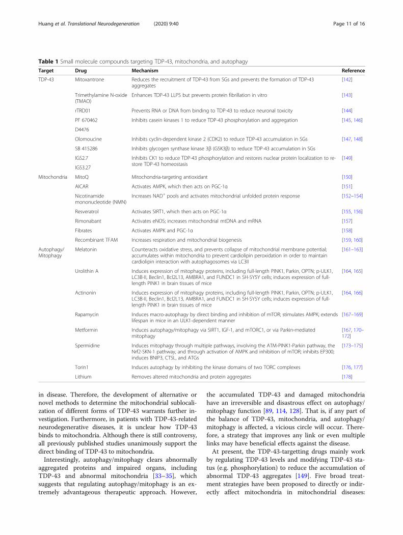
Table 1 Small molecule compounds targeting TDP-43, mitochondria, and autophagy
Target Drug Mechanism Reference
TDP-43 Mitoxantrone Reduces the recruitment of TDP-43 from SGs and prevents the formation of TDP-43aggregates
[142]
Trimethylamine N-oxide(TMAO)
Enhances TDP-43 LLPS but prevents protein fibrillation in vitro [143]
rTRD01 Prevents RNA or DNA from binding to TDP-43 to reduce neuronal toxicity [144]
PF 670462 Inhibits casein kinases 1 to reduce TDP-43 phosphorylation and aggregation [145, 146]
D4476
Olomoucine Inhibits cyclin-dependent kinase 2 (CDK2) to reduce TDP-43 accumulation in SGs [147, 148]
SB 415286 Inhibits glycogen synthase kinase 3β (GSK3β) to reduce TDP-43 accumulation in SGs
IGS2.7 Inhibits CK1 to reduce TDP-43 phosphorylation and restores nuclear protein localization to re-store TDP-43 homeostasis
[149]
IGS3.27
Mitochondria MitoQ Mitochondria-targeting antioxidant [150]
AICAR Activates AMPK, which then acts on PGC-1α [151]
Nicotinamidemononucleotide (NMN)
Increases NAD+ pools and activates mitochondrial unfolded protein response [152–154]
Resveratrol Activates SIRT1, which then acts on PGC-1α [155, 156]
Rimonabant Activates eNOS; increases mitochondrial mtDNA and mRNA [157]
Fibrates Activates AMPK and PGC-1α [158]
Recombinant TFAM Increases respiration and mitochondrial biogenesis [159, 160]
Autophagy/Mitophagy
Melatonin Counteracts oxidative stress, and prevents collapse of mitochondrial membrane potential;accumulates within mitochondria to prevent cardiolipin peroxidation in order to maintaincardiolipin interaction with autophagosomes via LC3II
[161–163]
Urolithin A Induces expression of mitophagy proteins, including full-length PINK1, Parkin, OPTN; p-ULK1,LC3B-II, Beclin1, Bcl2L13, AMBRA1, and FUNDC1 in SH-SY5Y cells; induces expression of full-length PINK1 in brain tissues of mice
[164, 165]
Actinonin Induces expression of mitophagy proteins, including full-length PINK1, Parkin, OPTN; p-ULK1,LC3B-II, Beclin1, Bcl2L13, AMBRA1, and FUNDC1 in SH-SY5Y cells; induces expression of full-length PINK1 in brain tissues of mice
[164, 166]
Rapamycin Induces macro-autophagy by direct binding and inhibition of mTOR; stimulates AMPK; extendslifespan in mice in an ULK1-dependent manner
[167–169]
Metformin Induces autophagy/mitophagy via SIRT1, IGF-1, and mTORC1, or via Parkin-mediatedmitophagy
[167, 170–172]
Spermidine Induces mitophagy through multiple pathways, involving the ATM-PINK1-Parkin pathway, theNrf2-SKN-1 pathway, and through activation of AMPK and inhibition of mTOR; inhibits EP300;induces BNIP3, CTSL, and ATGs
[173–175]
Torin1 Induces autophagy by inhibiting the kinase domains of two TORC complexes [176, 177]
Lithium Removes altered mitochondria and protein aggregates [178]
Huang et al. Translational Neurodegeneration (2020) 9:40 Page 11 of 16
repairing or preventing damage to organelles; inducingmitochondrial biogenesis; enhancing organelle qualitycontrol by stimulating the degradation of damaged mito-chondria or organelle components; manipulating mito-chondrial function to induce cell death; and changingmitochondrial signaling pathways or metabolic processes[138]. In addition, there are many potential therapeuticstrategies using gene therapy to correct for defectivegenes or for ectopic expression of mtDNA designed todegrade mutants or alter metabolic proteins [183, 184].In addition, the autophagy function can be induced tomaintain the steady state of proteins and organelles toexert neuroprotection [36].Although accumulating evidence has shown the protection
of regulation of TDP-43 or the mitochondrial or autophagyprocess against neurodegenerative diseases, it remains to beverified whether multi-step treatment is superior to thesingle-target treatment. In addition, the mechanism under-lying the balance among TDP-43, mitochondria and autoph-agy remains to be studied. On the one hand, we can clarifythe internal connections between TDP-43, mitochondria,and autophagy/mitophagy by establishing more advanta-geous models such as pig and non-human primate modelsof neurodegeneration [185, 186]. On the other hand, explor-ing and discovering new drugs targeting TDP-43, mitochon-dria and autophagy, either alone or in combination, is a keystep that must be taken. Inevitably, it is more helpful to ex-plore the effects of existing drugs that can separately regulateTDP-43, mitochondria, and autophagy in TDP-43-relatedneurodegenerative diseases on the other two targets.
ConclusionsIn conclusion, it is evident that TDP-43 protein pathology,mitochondrial disorders, and impaired autophagy arecommon prominent pathological features of major neuro-degenerative diseases including ALS, FTD, and AD. Inhib-ition of mitochondrial localization of TDP-43 is sufficientto alleviate mitochondrial dynamic abnormalities, neur-onal loss, and behavioral defects in transgenic mice withdifferent mutant forms of TDP-43. Due to the close rela-tionship between mitochondrial and autophagy functionsand TDP-43, and their contributions to the progression ofneurodegenerative diseases, we believe that maintainingthe balance among TDP-43, mitochondria, and autophagyis a promising strategy for the treatment of neurodegener-ative diseases.
Abbreviationsatg: Autophagy-related genes; AD: Alzheimer’s disease; ALS: Amyotrophiclateral sclerosis; AMPK: AMP-activated protein kinase; ATP: Adenosinetriphosphate; CDC37: Cell division cycle 37; CMA: Chaperone-mediatedautophagy; CNS: Central nervous system; Drp1: Dynamin-related protein 1;ER: Endoplasmic reticulum; Fis1: Fission protein 1; FTD: Frontotemporaldementia; HD: Huntington’s disease; HSP: Heat shock protein; HspB8: Heatshock protein B8; mtDNA: mitochondrial DNA; MAM: Mitochondrial-associated membrane; Mff: Mitochondrial fission factor; Mfn1: Mitofusin 1;
Mfn2: Mitofusin 2; NES: Nuclear export sequence; NFT: Neurofibrillary tangle;NLS: Nuclear localization sequence; OPA1: Optic atrophy protein 1;OXPHOS: Oxidative phosphorylation; PD: Parkinson’s disease; ROS: Reactiveoxygen species; RRM: RNA recognition motifs; TDP-43: Transactive response(TAR) DNA binding protein 43 kDa
AcknowledgmentsWe thank LetPub (www.letpub.com) for its linguistic assistance during thepreparation of this manuscript.
Authors’ contributionsCHH conducted the literature review, wrote initial draft, and conceived thefigures. All authors contributed to conceiving the outline, reviewing, andediting the manuscript and figures. ZJZ and SY approved the manuscript forsubmission. All authors read and approved the final manuscript.
FundingThis work was supported by the National Natural Science Foundation ofChina (81922026, 82073821, 81872842, 91649115); the National Key Researchand Development Program of China Stem Cell and Translational Research(2017YFA0105104); Guangdong Province Science and Technology PlanProject (2017A020211019, 2020A1515011061); the Fundamental ResearchFunds for the Central Universities (21619104); and Guangzhou Key ResearchProgram on Brain Science (202007030008).
Availability of data and materialsNot applicable.
Ethics approval and consent to participateNot applicable.
Consent for publicationNot applicable.
Competing interestsThe authors declare that they have no competing interests.
Received: 22 July 2020 Accepted: 14 October 2020
References1. Lin MT, Beal MF. Mitochondrial dysfunction and oxidative stress in
neurodegenerative diseases. Nature. 2006;443:787–95.2. Soto C, Pritzkow S. Protein misfolding, aggregation, and conformational
strains in neurodegenerative diseases. Nat Neurosci. 2018;21:1332–40.3. Spires-Jones TL, Attems J, Thal DR. Interactions of pathological proteins in
neurodegenerative diseases. Acta Neuropathol. 2017;134:187–205.4. Neumann M, Sampathu DM, Kwong LK, Truax AC, Micsenyi MC, Chou TT,
et al. Ubiquitinated TDP-43 in frontotemporal lobar degeneration andamyotrophic lateral sclerosis. Science. 2006;314:130–3.
5. Gao J, Wang L, Yan T, Perry G, Wang X. TDP-43 proteinopathy andmitochondrial abnormalities in neurodegeneration. Mol Cell Neurosci. 2019;100:103396.
6. Zuo-Shang X. Does a loss of TDP-43 function cause neurodegeneration?Mol Neurodegener. 2012;7:27.
7. Chen-Plotkin AS, Lee VMY, Trojanowski JQ. TAR DNA-binding protein 43 inneurodegenerative disease. Nat Rev Neurol. 2010;6:211–20.
8. Kabashi E, Valdmanis PN, Dion P, Spiegelman D, McConkey BJ, Vande VeldeC, et al. TARDBP mutations in individuals with sporadic and familialamyotrophic lateral sclerosis. Nat Genet. 2008;40:572–4.
9. Van Deerlin VM, Leverenz JB, Bekris LM, Bird TD, Yuan W, Elman LB, et al.TARDBP mutations in amyotrophic lateral sclerosis with TDP-43neuropathology: a genetic and histopathological analysis. Lancet Neurol.2008;7:409–16.
10. Buratti E, Baralle FE. The molecular links between TDP-43 dysfunction andNeurodegeneration. Adv Genet. 2009;66:1–34.
11. Polymenidou M, Lagier-Tourenne C, Hutt KR, Huelga SC, Moran J, Liang TY,et al. Long pre-mRNA depletion and RNA missplicing contribute toneuronal vulnerability from loss of TDP-43. Nat Neurosci. 2011;14:459–68.
Huang et al. Translational Neurodegeneration (2020) 9:40 Page 12 of 16
12. Tollervey JR, Curk T, Rogelj B, Briese M, Cereda M, Kayikci M, et al.Characterizing the RNA targets and position-dependent splicing regulationby TDP-43. Nat Neurosci. 2011;14:452–8.
13. Freibaum BD, Chitta RK, High AA, Taylor JP. Global analysis of TDP-43interacting proteins reveals strong association with RNA splicing andtranslation machinery. J Proteome Res. 2010;9:1104–20.
14. Colombrita C, Zennaro E, Fallini C, Weber M, Sommacal A, Buratti E, et al.TDP-43 is recruited to stress granules in conditions of oxidative insult. JNeurochem. 2009;111:1051–61.
15. Xia Q, Wang H, Hao Z, Fu C, Hu Q, Gao F, et al. TDP-43 loss of functionincreases TFEB activity and blocks autophagosome-lysosome fusion. EMBOJ. 2016;35:121–42.
16. Liu G, Coyne AN, Pei F, Vaughan S, Chaung M, Zarnescu DC, et al. Endocytosisregulates TDP-43 toxicity and turnover. Nat Commun. 2017;8:2092.
17. Wang W, Wang L, Lu J, Siedlak SL, Fujioka H, Liang J, et al. The inhibition ofTDP-43 mitochondrial localization blocks its neuronal toxicity. Nat Med.2016;22:869–78.
18. Janssens J, Van Broeckhoven C. Pathological mechanisms underlying TDP-43 driven neurodegeneration in FTLD-ALS spectrum disorders. Hum MolGenet. 2013;22:R77–87.
19. Bolognesi B, Faure AJ, Seuma M, Schmiedel JM, Tartaglia GG, Lehner B. Themutational landscape of a prion-like domain. Nat Commun. 2019;10:4162.
20. Kawakami I, Arai T, Hasegawa M. The basis of clinicopathologicalheterogeneity in TDP-43 proteinopathy. Acta Neuropathol. 2019;138:751–70.
21. Schapira AHV. Mitochondrial diseases. Lancet. 2012;379:1825–34.22. Burté F, Carelli V, Chinnery PF, Yu-Wai-Man P. Disturbed mitochondrial
dynamics and neurodegenerative disorders. Nat Rev Neurol. 2014;11:11–24.23. Detmer SA, Chan DC. Functions and dysfunctions of mitochondrial
dynamics. Nat Rev Mol Cell Biol. 2007;8:870–9.24. Green DR, Galluzzi L, Kroemer G. Mitochondria and the autophagy-
inflammation-cell death axis in organismal aging. Science. 2011;333:1109–12.25. Wallace DC, Fan W, Procaccio V. Mitochondrial energetics and therapeutics.
Annu Rev Pathol. 2010;5:297–348.26. Tait SW, Green DR. Mitochondria and cell death: outer membrane
permeabilization and beyond. Nat Rev Mol Cell Biol. 2010;11:621–32.27. Wang W, Li L, Lin WL, Dickson DW, Petrucelli L, Zhang T, et al. The ALS
disease-associated mutant TDP-43 impairs mitochondrial dynamics andfunction in motor neurons. Hum Mol Genet. 2013;22:4706–19.
28. Xu YF, Gendron TF, Zhang YJ, Lin WL, D'Alton S, Sheng H, et al. Wild-typehuman TDP-43 expression causes TDP-43 phosphorylation, mitochondrialaggregation, motor deficits, and early mortality in transgenic mice. JNeurosci. 2010;30:10851–9.
29. Huntley ML, Gao J, Termsarasab P, Wang L, Zeng S, Thammongkolchai T,et al. Association between TDP-43 and mitochondria in inclusion bodymyositis. Lab Investig. 2019;99:1041–8.
30. Wang W, Arakawa H, Wang L, Okolo O, Siedlak SL, Jiang Y, et al. Motor-coordinative and cognitive dysfunction caused by mutant TDP-43 could bereversed by inhibiting its mitochondrial localization. Mol Ther. 2017;25:127–39.
31. Ghavami S, Shojaei S, Yeganeh B, Ande SR, Jangamreddy JR, Mehrpour M,et al. Autophagy and apoptosis dysfunction in neurodegenerative disorders.Prog Neurobiol. 2014;112:24–49.
32. He C, Klionsky DJ. Regulation mechanisms and signaling pathways ofautophagy. Annu Rev Genet. 2009;43:67–93.
33. Budini M, Buratti E, Morselli E, Criollo A. Autophagy and its impact onneurodegenerative diseases: new roles for TDP-43 and C9orf72. Front MolNeurosci. 2017;10:170.
34. Kroemer G, Mariño G, Levine B. Autophagy and the integrated stressresponse. Mol Cell. 2010;40:280–93.
35. Bento CF, Renna M, Ghislat G, Puri C, Ashkenazi A, Vicinanza M, et al.Mammalian autophagy: how does it work? Annu Rev Biochem. 2016;85:685–713.
36. Scrivo A, Bourdenx M, Pampliega O, Cuervo AM. Selective autophagy as apotential therapeutic target for neurodegenerative disorders. Lancet Neurol.2018;17:802–15.
37. Harris H, Rubinsztein DC. Control of autophagy as a therapy forneurodegenerative disease. Nat Rev Neurol. 2011;8:108–17.
38. Baralle M, Buratti E, Baralle FE. The role of TDP-43 in the pathogenesis ofALS and FTLD. Biochem Soc Trans. 2013;41:1536–40.
39. Warraich ST, Yang S, Nicholson GA, Blair IP. TDP-43: a DNA and RNA bindingprotein with roles in neurodegenerative diseases. Int J Biochem Cell Biol.2010;42:1606–9.
40. Cohen TJ, Lee VM, Trojanowski JQ. TDP-43 functions and pathogenicmechanisms implicated in TDP-43 proteinopathies. Trends Mol Med. 2011;17:659–67.
41. Romano M, Buratti E, Romano G, Klima R, Del Bel BL, Stuani C, et al.Evolutionarily conserved heterogeneous nuclear Ribonucleoprotein (hnRNP)a/B proteins functionally interact with human and drosophila TAR DNA-binding protein 43 (TDP-43). J Biol Chem. 2014;289:7121–30.
42. Zhang YJ, Caulfield T, Xu YF, Gendron TF, Hubbard J, Stetler C, et al. The dualfunctions of the extreme N-terminus of TDP-43 in regulating its biologicalactivity and inclusion formation. Hum Mol Genet. 2013;22:3112–22.
43. Ou SH, Wu F, Harrich D, García-Martínez LF, Gaynor RB. Cloning andcharacterization of a novel cellular protein, TDP-43, that binds to humanimmunodeficiency virus type 1 TAR DNA sequence motifs. J Virol. 1995;69:3584–96.
44. Buratti E, Dörk T, Zuccato E, Pagani F, Romano M, Baralle FE. Nuclear factorTDP-43 and SR proteins promote in vitro and in vivo CFTR exon 9 skipping.EMBO J. 2001;20:1774–84.
45. Winton MJ, Igaz LM, Wong MM, Kwong LK, Trojanowski JQ, Lee VM.Disturbance of nuclear and cytoplasmic TAR DNA-binding protein (TDP-43)induces disease-like redistribution, sequestration, and aggregate formation. JBiol Chem. 2008;283:13302–9.
46. Barmada SJ, Skibinski G, Korb E, Rao EJ, Wu JY, Finkbeiner S. Cytoplasmicmislocalization of TDP-43 is toxic to neurons and enhanced by a mutationassociated with familial amyotrophic lateral sclerosis. J Neurosci. 2010;30:639–49.
47. Diaper DC, Adachi Y, Sutcliffe B, Humphrey DM, Elliott CJ, Stepto A, et al.Loss and gain of drosophila TDP-43 impair synaptic efficacy and motorcontrol leading to age-related neurodegeneration by loss-of-functionphenotypes. Hum Mol Genet. 2013;22:1539–57.
48. Ling SC, Polymenidou M, Cleveland DW. Converging mechanisms in ALSand FTD: disrupted RNA and protein homeostasis. Neuron. 2013;79:416–38.
49. Sephton CF, Cenik C, Kucukural A, Dammer EB, Cenik B, Han Y, et al.Identification of neuronal RNA targets of TDP-43-containingribonucleoprotein complexes. J Biol Chem. 2011;286:1204–15.
50. Ling S-C, Albuquerque CP, Zhou H, Cleveland DW. ALS-associatedmutations in TDP-43 increase its stability and promote TDP-43 complexeswith FUS/TLS. Proc Natl Acad Sci U S A. 2010;107:13318–23.
51. Li Q, Yokoshi M, Okada H, Kawahara Y. The cleavage pattern of TDP-43determines its rate of clearance and cytotoxicity. Nat Commun. 2015;6:6183.
52. Walker AK, Soo KY, Sundaramoorthy V, Parakh S, Ma Y, Farg MA, et al. ALS-associated TDP-43 induces endoplasmic reticulum stress, which drivescytoplasmic TDP-43 accumulation and stress granule formation. PLoS One.2013;8:e81170.
53. Stoica R, De Vos KJ, Paillusson S, Mueller S, Sancho RM, Lau KF, et al. ER-mitochondria associations are regulated by the VAPB-PTPIP51 interaction andare disrupted by ALS/FTD-associated TDP-43. Nat Commun. 2014;5:3996.
54. Alami NH, Smith RB, Carrasco MA, Williams LA, Winborn CS, Han SSW, et al.Axonal transport of TDP-43 mRNA granules is impaired by ALS-causingmutations. Neuron. 2014;81:536–43.
55. Liu-Yesucevitz L, Bilgutay A, Zhang YJ, Vanderweyde T, Citro A, Mehta T, et al.Tar DNA binding protein-43 (TDP-43) associates with stress granules: analysisof cultured cells and pathological brain tissue. PLoS One. 2010;5:e13250.
56. Lagier-Tourenne C, Polymenidou M, Cleveland DW. TDP-43 and FUS/TLS:emerging roles in RNA processing and neurodegeneration. Hum Mol Genet.2010;19:R46–64.
57. Kwong LK, Neumann M, Sampathu DM, Lee VM, Trojanowski JQ. TDP-43proteinopathy: the neuropathology underlying major forms of sporadic andfamilial frontotemporal lobar degeneration and motor neuron disease. ActaNeuropathol. 2007;114:63–70.
58. Cascella R, Capitini C, Fani G, Dobson CM, Cecchi C, Chiti F. Quantificationof the relative contributions of loss-of-function and gain-of-functionmechanisms in TAR DNA-binding protein 43 (TDP-43) Proteinopathies. J BiolChem. 2016;291:19437–48.
59. Halliday G, Bigio EH, Cairns NJ, Neumann M, Mackenzie IR, Mann DM.Mechanisms of disease in frontotemporal lobar degeneration: gain offunction versus loss of function effects. Acta Neuropathol. 2012;124:373–82.
60. Gao J, Wang L, Huntley ML, Perry G, Wang X. Pathomechanisms of TDP-43in neurodegeneration. J Neurochem. 2018. https://doi.org/10.1111/jnc.14327.
61. Chan DC. Mitochondria: dynamic organelles in disease, aging, anddevelopment. Cell. 2006;125:1241–52.
Huang et al. Translational Neurodegeneration (2020) 9:40 Page 13 of 16
62. Sabbatinelli J, Prattichizzo F, Olivieri F, Procopio AD, Rippo MR, Giuliani A.Where metabolism meets senescence: focus on endothelial cells. FrontPhysiol. 2019;10:1523.
63. Federico A, Cardaioli E, Da Pozzo P, Formichi P, Gallus GN, Radi E.Mitochondria, oxidative stress and neurodegeneration. J Neurol Sci. 2012;322:254–62.
64. Sasaki S, Horie Y, Iwata M. Mitochondrial alterations in dorsal root ganglioncells in sporadic amyotrophic lateral sclerosis. Acta Neuropathol. 2007;114:633–9.
65. Kawamata H, Manfredi G. Proteinopathies and OXPHOS dysfunction inneurodegenerative diseases. J Cell Biol. 2017;216:3917–29.
66. Braun RJ, Sommer C, Carmona-Gutierrez D, Khoury CM, Ring J, Büttner S,et al. Neurotoxic 43-kDa TAR DNA-binding protein (TDP-43) triggersmitochondrion-dependent programmed cell death in yeast. J Biol Chem.2011;286:19958–72.
67. Lu J, Duan W, Guo Y, Jiang H, Li Z, Huang J, et al. Mitochondrial dysfunctionin human TDP-43 transfected NSC34 cell lines and the protective effect ofdimethoxy curcumin. Brain Res Bull. 2012;89:185–90.
68. Hong K, Li Y, Duan W, Guo Y, Jiang H, Li W, et al. Full-length TDP-43 and itsC-terminal fragments activate mitophagy in NSC34 cell line. Neurosci Lett.2012;530:144–9.
69. Kawamata H, Peixoto P, Konrad C, Palomo G, Bredvik K, Gerges M, et al.Mutant TDP-43 does not impair mitochondrial bioenergetics in vitro andin vivo. Mol Neurodegener. 2017;12:37.
70. Salvatori I, Ferri A, Scaricamazza S, Giovannelli I, Serrano A, Rossi S, et al.Differential toxicity of TAR DNA-binding protein 43 isoforms depends ontheir submitochondrial localization in neuronal cells. J Neurochem. 2018;146:585–97.
71. Davis SA, Itaman S, Khalid-Janney CM, Sherard JA, Dowell JA, Cairns NJ,et al. TDP-43 interacts with mitochondrial proteins critical for mitophagyand mitochondrial dynamics. Neurosci Lett. 2018;678:8–15.
72. Termsarasab P, Thammongkolchai T, Gao J, Wang L, Liang J, Wang X.Cytoplasmic mislocalization and mitochondrial colocalization of TDP-43 arecommon features between normal aged and young mice. Exp Biol Med(Maywood). 2020;1535370220914253:1.
73. Zhang YJ, Xu YF, Dickey CA, Buratti E, Baralle F, Bailey R, et al. Progranulinmediates caspase-dependent cleavage of TAR DNA binding protein-43. JNeurosci. 2007;27:10530–4.
74. Kitamura A, Nakayama Y, Shibasaki A, Taki A, Yuno S, Takeda K, et al.Interaction of RNA with a C-terminal fragment of the amyotrophic lateralsclerosis-associated TDP43 reduces cytotoxicity. Sci Rep. 2016;6:19230.
75. Bozzo F, Salvatori I, Iacovelli F, Mirra A, Rossi S, Cozzolino M, et al. Structuralinsights into the multi-determinant aggregation of TDP-43 in motorneuron-like cells. Neurobiol Dis. 2016;94:63–72.
76. Cogliati S, Frezza C, Soriano ME, Varanita T, Quintana-Cabrera R, Corrado M,et al. Mitochondrial cristae shape determines respiratory chainsupercomplexes assembly and respiratory efficiency. Cell. 2013;155:160–71.
77. Benard G, Bellance N, James D, Parrone P, Fernandez H, Letellier T, et al.Mitochondrial bioenergetics and structural network organization. J Cell Sci.2007;120:838–48.
78. Szabadkai G, Simoni AM, Chami M, Wieckowski MR, Youle RJ, Rizzuto R. Drp-1-dependent division of the mitochondrial network blocks Intraorganellar Ca2+
waves and protects against Ca2+-mediated apoptosis. Mol Cell. 2004;16:59–68.79. Losóna OC, Songa Z, Chena H, Chan DC. Fis1, Mff, MiD49, and MiD51
mediate Drp1 recruitment in mitochondrial fissio. Mol Biol Cell. 2013;24:659–67.
80. Joshi AU, Saw NL, Vogel H, Cunnigham AD, Shamloo M, Mochly-Rosen D.Inhibition of Drp1/Fis1 interaction slows progression of amyotrophic lateralsclerosis. EMBO Mol Med. 2018;10:e8166.
81. James BD, Wilson RS, Boyle PA, Trojanowski JQ, Bennett DA, Schneider JA.TDP-43 stage, mixed pathologies, and clinical Alzheimer’s-type dementia.Brain. 2016;139:2983–93.
82. Wang X, Su B, Siedlak SL, Moreira PI, Fujioka H, Wang Y, et al. Amyloid-betaoverproduction causes abnormal mitochondrial dynamics via differentialmodulation of mitochondrial fission/fusion proteins. Proc Natl Acad Sci U SA. 2008;105:19318–23.
83. Wang X, Su B, Hg L, Li X, Perry G, Smith MA, et al. Impaired balance of mitochondrialfission and fusion in Alzheimer’s disease. J Neurosci. 2009;29:9090–103.
84. Magrané J, Cortez C, Gan W, Manfredi G. Abnormal mitochondrial transportand morphology are common pathological denominators in SOD1 andTDP43 ALS mouse models. Hum Mol Genet. 2014;23:1413–24.
85. Kreiter N, Pal A, Lojewski X, Corcia P, Naujock M, Reinhardt P, et al. Age-dependent neurodegeneration and organelle transport deficiencies inmutant TDP43 patient-derived neurons are independent of TDP43aggregation. Neurobiol Dis. 2018;115:167–81.
86. Hamilton BA, Wang P, Deng J, Dong J, Liu J, Bigio EH, et al. TDP-43 inducesmitochondrial damage and activates the mitochondrial unfolded proteinresponse. PLoS Genet. 2019;15:e1007947.
87. Pansarasa O, Bordoni M, Drufuca L, Diamanti L, Sproviero D, Trotti R, et al.Lymphoblastoid cell lines as a model to understand amyotrophic lateralsclerosis disease mechanisms. Dis Model Mech. 2018;11:dmm031625.
88. Park SK, Park S, Liebman SW. Respiration enhances TDP-43 toxicity, but TDP-43 retains some toxicity in the absence of respiration. J Mol Biol. 2019;431:2050–9.
89. Gautam M, Jara JH, Kocak N, Rylaarsdam LE, Kim KD, Bigio EH, et al.Mitochondria, ER, and nuclear membrane defects reveal early mechanismsfor upper motor neuron vulnerability with respect to TDP-43 pathology.Acta Neuropathol. 2019;137:47–69.
90. Kim J, Kundu M, Viollet B, Guan KL. AMPK and mTOR regulate autophagythrough direct phosphorylation of Ulk1. Nat Cell Biol. 2011;13:132–41.
91. Chantranupong L, Sabatini DM. The TORC1 pathway to protein destruction.Nature. 2016;536:155–6.
92. Egan DF, Shackelford DB, Mihaylova MM, Gelino S, Kohnz RA, Mair W, et al.Phosphorylation of ULK1 (hATG1) by AMP-activated protein kinase connectsenergy sensing to mitophagy. Science. 2011;331:456–61.
93. Yang Y, Coleman M, Zhang L, Zheng X, Yue Z. Autophagy in axonal anddendritic degeneration. Trends Neurosci. 2013;36:418–28.
94. Nijholt DAT, Kimpe LD, Elfrink HL, Hoozemans JJM, Scheper W. Removingprotein aggregates: the role of proteolysis in neurodegeneration. Curr MedChem. 2011;18:2459–76.
95. Hara T, Nakamura K, Matsui M, Yamamoto A, Nakahara Y, Suzuki-MigishimaR, et al. Suppression of basal autophagy in neural cells causesneurodegenerative disease in mice. Nature. 2006;441:885–9.
96. Komatsu M, Waguri S, Chiba T, Murata S, Iwata J, Tanida I, et al. Loss ofautophagy in the central nervous system causes neurodegeneration inmice. Nature. 2006;441:880–4.
97. Singh R, Cuervo AM. Autophagy in the cellular energetic balance. CellMetab. 2011;13:495–504.
98. Gustafsson AB, Dorn GW 2nd. Evolving and expanding the roles of Mitophagyas a homeostatic and pathogenic process. Physiol Rev. 2019;99:853–92.
99. Scherz-Shouval R, Shvets E, Fass E, Shorer H, Gil L, Elazar Z. Reactive oxygenspecies are essential for autophagy and specifically regulate the activity ofAtg4. EMBO J. 2007;26:1749–60.
100. Scherz-Shouval R, Elazar Z. Regulation of autophagy by ROS: physiology andpathology. Trends Biochem Sci. 2011;36:30–8.
101. Huang J, Lam GY, Brumell JH. Autophagy signaling through reactive oxygenspecies. Antioxid Redox Signal. 2011;14:2215–31.
102. Bose JK, Huang CC, Shen CK. Regulation of autophagy by neuropathologicalprotein TDP-43. J Biol Chem. 2011;286:44441–8.
103. Mizuno Y, Amari M, Takatama M, Aizawa H, Mihara B, Okamoto K.Immunoreactivities of p62, an ubiqutin-binding protein, in the spinalanterior horn cells of patients with amyotrophic lateral sclerosis. J NeurolSci. 2006;249:13–8.
104. Hiji M, Takahashi T, Fukuba H, Yamashita H, Kohriyama T, Matsumoto M.White matter lesions in the brain with frontotemporal lobar degenerationwith motor neuron disease: TDP-43-immunopositive inclusions co-localizewith p62, but not ubiquitin. Acta Neuropathol. 2008;116:183–91.
105. Maekawa S, Leigh PN, King A, Jones E, Steele JC, Bodi I, et al. TDP-43 isconsistently co-localized with ubiquitinated inclusions in sporadic and Guamamyotrophic lateral sclerosis but not in familial amyotrophic lateral sclerosiswith and without SOD1 mutations. Neuropathology. 2009;29:672–83.
106. King A, Maekawa S, Bodi I, Troakes C, Al-Sarraj S. Ubiquitinated, p62immunopositive cerebellar cortical neuronal inclusions are evident acrossthe spectrum of TDP-43 proteinopathies but are only rarely additionallyimmunopositive for phosphorylation-dependent TDP-43. Neuropathology.2011;31:239–49.
107. Caccamo A, Majumder S, Deng JJ, Bai Y, Thornton FB, Oddo S. Rapamycinrescues TDP-43 mislocalization and the associated low molecular massneurofilament instability. J Biol Chem. 2009;284:27416–24.
108. Jinwal UK, Abisambra JF, Zhang J, Dharia S, O'Leary JC, Patel T, et al. Cdc37/Hsp90 protein complex disruption triggers an autophagic clearance cascadefor TDP-43 protein. J Biol Chem. 2012;287:24814–20.
Huang et al. Translational Neurodegeneration (2020) 9:40 Page 14 of 16
109. Crippa V, Sau D, Rusmini P, Boncoraglio A, Onesto E, Bolzoni E, et al. Thesmall heat shock protein B8 (HspB8) promotes autophagic removal ofmisfolded proteins involved in amyotrophic lateral sclerosis (ALS). Hum MolGenet. 2010;19:3440–56.
110. Crippa V, Carra S, Rusmini P, Sau D, Bolzoni E, Bendotti C, et al. A role ofsmall heat shock protein B8 (HspB8) in the autophagic removal ofmisfolded proteins responsible for neurodegenerative diseases. Autophagy.2010;6:958–60.
111. Crippa V, D'Agostino VG, Cristofani R, Rusmini P, Cicardi ME, Messi E, et al.Transcriptional induction of the heat shock protein B8 mediates theclearance of misfolded proteins responsible for motor neuron diseases. SciRep. 2016;6:22827.
112. Kettern N, Rogon C, Limmer A, Schild H, Hohfeld J. The Hsc/Hsp70 co-chaperone network controls antigen aggregation and presentation duringmaturation of professional antigen presenting cells. PLoS One. 2011;6:e16398.
113. Zhao T, Hong Y, Yin P, Li S, Li XJ. Differential HspBP1 expression accountsfor the greater vulnerability of neurons than astrocytes to misfoldedproteins. Proc Natl Acad Sci U S A. 2017;114:E7803–E11.
114. Ormeño F, Hormazabal J, Moreno J, Riquelme F, Rios J, Criollo A, et al.Chaperone mediated autophagy degrades TDP-43 protein and is affectedby TDP-43 aggregation. Front Mol Neurosci. 2020;13:19.
115. De Conti L, Akinyi MV, Mendoza-Maldonado R, Romano M, Baralle M, BurattiE. TDP-43 affects splicing profiles and isoform production of genes involvedin the apoptotic and mitotic cellular pathways. Nucleic Acids Res. 2015;43:8990–9005.
116. Budini M, Romano V, Quadri Z, Buratti E, Baralle FE. TDP-43 loss of cellularfunction through aggregation requires additional structural determinantsbeyond its C-terminal Q/N prion-like domain. Hum Mol Genet. 2015;24:9–20.
117. Caccamo A, Shaw DM, Guarino F, Messina A, Walker AW, Oddo S. Reducedprotein turnover mediates functional deficits in transgenic mice expressing the25 kDa C-terminal fragment of TDP-43. Hum Mol Genet. 2015;24:4625–35.
118. Lou G, Palikaras K, Lautrup S, Scheibye-Knudsen M, Tavernarakis N, Fang EF.Mitophagy and neuroprotection. Trends Mol Med. 2020;26:8–20.
119. Kumar A, Dhawan A, Kadam A, Shinde A. Autophagy and mitochondria:targets in neurodegenerative disorders. CNS Neurol Disord Drug Targets.2018;17:696–705.
120. Whitworth AJ, Pallanck LJ. PINK1/Parkin mitophagy andneurodegeneration—what do we really know in vivo ? Curr Opin GenetDev. 2017;44:47–53.
121. Goiran T, Duplan E, Chami M, Bourgeois A, El Manaa W, Rouland L, et al.Beta-amyloid precursor protein intracellular domain controls mitochondrialfunction by modulating phosphatase and Tensin homolog-induced kinase 1transcription in cells and in Alzheimer mice models. Biol Psychiatry. 2018;83:416–27.
122. Valente EM, Abou-Sleiman PM, Caputo V, Muqit MM, Harvey K, Gispert S,et al. Hereditary early-onset Parkinson's disease caused by mutations inPINK1. Science. 2004;304:1158–60.
123. Ito Y, Ofengeim D, Najafov A, Das S, Saberi S, Li Y, et al. RIPK1 mediatesaxonal degeneration by promoting inflammation and necroptosis in ALS.Science. 2016;353:603–8.
124. Hawk MA, Gorsuch CL, Fagan P, Lee C, Kim SE, Hamann JC, et al. RIPK1-mediated induction of mitophagy compromises the viability of extracellular-matrix-detached cells. Nat Cell Biol. 2018;20:272–84.
125. Moore AS, Holzbaur EL. Dynamic recruitment and activation of ALS-associated TBK1 with its target optineurin are required for efficientmitophagy. Proc Natl Acad Sci U S A. 2016;113:E3349–58.
126. Weishaupt JH, Hyman T, Dikic I. Common molecular pathways inamyotrophic lateral sclerosis and Frontotemporal dementia. Trends MolMed. 2016;22:769–83.
127. Hwang S, Disatnik MH, Mochly-Rosen D. Impaired GAPDH-inducedmitophagy contributes to the pathology of Huntington's disease. EMBO MolMed. 2015;7:1307–26.
128. Yi C, Tong JJ, Yu L. Mitochondria: the hub of energy deprivation-inducedautophagy. Autophagy. 2018;14:1084–5.
129. Hailey DW, Rambold AS, Satpute-Krishnan P, Mitra K, Sougrat R, Kim PK,et al. Mitochondria supply membranes for autophagosome biogenesisduring starvation. Cell. 2010;141:656–67.
130. Hamasaki M, Furuta N, Matsuda A, Nezu A, Yamamoto A, Fujita N, et al.Autophagosomes form at ER-mitochondria contact sites. Nature. 2013;495:389–93.
131. Dan Dunn J, Alvarez LA, Zhang X, Soldati T. Reactive oxygen species andmitochondria: a nexus of cellular homeostasis. Redox Biol. 2015;6:472–85.
132. Moore MN. Autophagy as a second level protective process in conferringresistance to environmentally-induced oxidative stress. Autophagy. 2008;4:254–6.
133. Zhang H, Kong X, Kang J, Su J. Yang Li, Zhong J, Sun L. Oxidative stressinduces parallel autophagy and mitochondria dysfunction in human gliomaU251 cells. Toxicol Sci. 2009;110:376–88.
134. Finkel T. Oxidant signals and oxidative stress. Curr Opin Cell Biol. 2003;15:247–54.
135. Kim EH, Sohn S, Kwon HJ, Kim SU, Kim MJ, Lee SJ, et al. Sodium seleniteinduces superoxide-mediated mitochondrial damage and subsequentautophagic cell death in malignant glioma cells. Cancer Res. 2007;67:6314–24.
136. Budini M, Baralle FE, Buratti E. Targeting TDP-43 in neurodegenerativediseases. Expert Opin Ther Targets. 2014;18:617–32.
137. Guo L, Shorter J. Biology and pathobiology of TDP-43 and emergenttherapeutic strategies. Cold Spring Harb Perspect Med. 2017;7.
138. Murphy MP, Hartley RC. Mitochondria as a therapeutic target for commonpathologies. Nat Rev Drug Discov. 2018;17:865–86.
139. Dawson TM, Dawson VL. Mitochondrial mechanisms of neuronal cell death:potential therapeutics. Annu Rev Pharmacol Toxicol. 2017;57:437–54.
140. Brown DG, Shorter J, Wobst HJ. Emerging small-molecule therapeuticapproaches for amyotrophic lateral sclerosis and frontotemporal dementia.Bioorg Med Chem Lett. 2020;30:126942.
141. Whitaker RM, Corum D, Beeson CC, Schnellmann RG. Mitochondrialbiogenesis as a pharmacological target: a new approach to acute andchronic diseases. Annu Rev Pharmacol Toxicol. 2016;56:229–49.
142. Wheeler RJ, Lee HO, Poser I, Pal A, Doeleman T, Kishigami S, et al. Smallmolecules for modulating protein driven liquid-liquid phase separation intreating neurodegenerative disease. bioRxiv. 2019. https://doi.org/10.1101/721001.
143. Choi KJ, Tsoi PS, Moosa MM, Paulucci-Holthauzen A, Liao SJ, Ferreon JC,et al. A chemical chaperone decouples TDP-43 disordered domain phaseseparation from fibrillation. Biochemistry. 2018;57:6822–6.
144. Francois-Moutal L, Felemban R, Scott DD, Sayegh MR, Miranda VG, Perez-Miller S, et al. Small molecule targeting TDP-43’s RNA recognition motifsreduces Locomotor defects in a drosophila model of amyotrophic lateralsclerosis (ALS). ACS Chem Biol. 2019;14:2006–13.
145. Salado IG, Redondo M, Bello ML, Perez C, Liachko NF, Kraemer BC, et al.Protein kinase CK-1 inhibitors as new potential drugs for amyotrophiclateral sclerosis. J Med Chem. 2014;57:2755–72.
146. Hicks DA, Cross LL, Williamson R, Rattray M. Endoplasmic Reticulum StressSignalling Induces Casein Kinase 1-Dependent Formation of Cytosolic TDP-43 Inclusions in Motor Neuron-Like Cells. Neurochem Res. 2020;45(6):1354–64.https://doi.org/10.1007/s11064-019-02832-2.
147. Moujalled D, James JL, Parker SJ, Lidgerwood GE, Duncan C, Meyerowitz J,et al. Kinase inhibitor screening identifies Cyclin-dependent kinases andglycogen synthase kinase 3 as potential modulators of TDP-43 cytosolicaccumulation during cell stress. PLoS One. 2013;8:e67433.
148. Moujalled D, James JL, Yang S, Zhang K, Duncan C, Moujalled PSJ, et al.Phosphorylation of hnRNP K by cyclin-dependent kinase 2 controls cytosolicaccumulation of TDP-43. Hum Mol Genet. 2015;24:1655–69.
149. Palomo V, Tosat-Bitrian C, Nozal V, Nagaraj S, Martin-Requero A, Martinez A.TDP-43: a key therapeutic target beyond amyotrophic lateral sclerosis. ACSChem Neurosci. 2019;10:1183–96.
150. Dulovic M, Jovanovic M, Xilouri M, Stefanis L, Harhaji-Trajkovic L, Kravic-Stevovic T, et al. The protective role of AMP-activated protein kinase inalpha-synuclein neurotoxicity in vitro. Neurobiol Dis. 2014;63:1–11.
151. Sorrentino V, Romani M, Mouchiroud L, Beck JS, Zhang H, D'Amico D, et al.Enhancing mitochondrial proteostasis reduces amyloid-beta proteotoxicity.Nature. 2017;552:187–93.
152. Wagner GR, Bhatt DP, O'Connell TM, Thompson JW, Dubois LG, Backos DS,et al. A class of reactive acyl-CoA species reveals the non-enzymatic originsof protein acylation. Cell Metab. 2017;25:823–37.
153. Fang EF, Kassahun H, Croteau DL, Scheibye-Knudsen M, Marosi K, Lu H,et al. NAD + replenishment improves lifespan and healthspan in ataxiatelangiectasia models via mitophagy and DNA repair. Cell Metab. 2016;24:566–81.
154. Gong B, Pan Y, Vempati P, Zhao W, Knable L, Ho L, et al. Nicotinamideriboside restores cognition through an upregulation of proliferator-activatedreceptor-gamma coactivator 1alpha regulated beta-secretase 1 degradation
Huang et al. Translational Neurodegeneration (2020) 9:40 Page 15 of 16
and mitochondrial gene expression in Alzheimer’s mouse models.Neurobiol Aging. 2013;34:1581–8.
155. Menzies KJ, Singh K, Saleem A, Hood DA. Sirtuin 1-mediated effects ofexercise and resveratrol on mitochondrial biogenesis. J Biol Chem. 2013;288:6968–79.
156. Funk JA, Schnellmann RG. Accelerated recovery of renal mitochondrial andtubule homeostasis with SIRT1/PGC-1alpha activation following ischemia-reperfusion injury. Toxicol Appl Pharmacol. 2013;273:345–54.
157. Tedesco L, Valerio A, Cervino C, Cardile A, Pagano C, Vettor R, et al.Cannabinoid type 1 receptor blockade promotes mitochondrial biogenesisthrough endothelial nitric oxide synthase expression in white adipocytes.Diabetes. 2008;57:2028–36.
158. Cho YR, Lim JH, Kim MY, Kim TW, Hong BY, Kim YS, et al. Therapeutic effectsof fenofibrate on diabetic peripheral neuropathy by improving endothelialand neural survival in db/db mice. PLoS One. 2014;9:e83204.
159. Thomas RR, Khan SM, Portell FR, Smigrodzki RM, Bennett JP Jr. Recombinanthuman mitochondrial transcription factor a stimulates mitochondrialbiogenesis and ATP synthesis, improves motor function after MPTP, reducesoxidative stress and increases survival after endotoxin. Mitochondrion. 2011;11:108–18.
160. Thomas RR, Khan SM, Smigrodzki RM, Onyango IG, Dennis J, Khan OM, et al.RhTFAM treatment stimulates mitochondrial oxidative metabolism andimproves memory in aged mice. Aging. 2012;4:620–35.
161. Wongprayoon P, Govitrapong P. Melatonin as a mitochondrial protector inneurodegenerative diseases. Cell Mol Life Sci. 2017;74:3999–4014.
162. Jenwitheesuk A, Nopparat C, Mukda S, Wongchitrat P, Govitrapong P.Melatonin regulates aging and neurodegeneration through energymetabolism, epigenetics, autophagy and circadian rhythm pathways. Int JMol Sci. 2014;15:16848–84.
163. Boga JA, Caballero B, Potes Y, Perez-Martinez Z, Reiter RJ, Vega-Naredo I,et al. Therapeutic potential of melatonin related to its role as an autophagyregulator: a review. J Pineal Res. 2019;66:e12534.
164. Fang EF, Hou Y, Palikaras K, Adriaanse BA, Kerr JS, Yang B, et al. Mitophagyinhibits amyloid-beta and tau pathology and reverses cognitive deficits inmodels of Alzheimer's disease. Nat Neurosci. 2019;22:401–12.
165. Ryu D, Mouchiroud L, Andreux PA, Katsyuba E, Moullan N, Nicolet-Dit-FelixAA, et al. Urolithin a induces mitophagy and prolongs lifespan in C. elegansand increases muscle function in rodents. Nat Med. 2016;22:879–88.
166. Sun N, Yun J, Liu J, Malide D, Liu C, Rovira Ilsa I, et al. Measuring in vivomitophagy. Mol Cell. 2015;60:685–96.
167. Aliper A, Jellen L, Cortese F, Artemov A, Karpinsky-Semper D, Moskalev A,et al. Towards natural mimetics of metformin and rapamycin. Aging. 2017;9:2245–68.
168. Ravikumar B, Vacher C, Berger Z, Davies JE, Luo S, Oroz LG, et al. Inhibitionof mTOR induces autophagy and reduces toxicity of polyglutamineexpansions in fly and mouse models of Huntington disease. Nat Genet.2004;36:585–95.
169. Roth GS, Ingram DK. Manipulation of health span and function by dietarycaloric restriction mimetics. Ann N Y Acad Sci. 2016;1363:5–10.
170. Howell JJ, Hellberg K, Turner M, Talbott G, Kolar MJ, Ross DS, et al.Metformin inhibits hepatic mTORC1 signaling via dose-dependentmechanisms involving AMPK and the TSC complex. Cell Metab. 2017;25:463–71.
171. Song YM, Lee WK, Lee YH, Kang ES, Cha BS, Lee BW. Metformin restoresParkin-mediated Mitophagy, suppressed by cytosolic p53. Int J Mol Sci.2016;17:122.
172. Zheng Z, Chen H, Li J, Li T, Zheng B, Zheng Y, et al. Sirtuin 1-mediatedcellular metabolic memory of high glucose via the LKB1/AMPK/ROSpathway and therapeutic effects of metformin. Diabetes. 2012;61:217–28.
173. Gupta VK, Scheunemann L, Eisenberg T, Mertel S, Bhukel A, Koemans TS,et al. Restoring polyamines protects from age-induced memory impairmentin an autophagy-dependent manner. Nat Neurosci. 2013;16:1453–60.
174. Madeo F, Eisenberg T, Pietrocola F, Kroemer G. Spermidine in health anddisease. Science. 2018;359:eaan2788.
175. Qi Y, Qiu Q, Gu X, Tian Y, Zhang Y. ATM mediates spermidine-inducedmitophagy via PINK1 and Parkin regulation in human fibroblasts. Sci Rep.2016;6:24700.
176. Thellung S, Corsaro A, Nizzari M, Barbieri F, Florio T. Autophagy activatordrugs: a new opportunity in neuroprotection from misfolded proteintoxicity. Int J Mol Sci. 2019;20:901.
177. Thoreen CC, Kang SA, Chang JW, Liu Q, Zhang J, Gao Y, et al. An ATP-competitive mammalian target of rapamycin inhibitor reveals rapamycin-resistant functions of mTORC1. J Biol Chem. 2009;284:8023–32.
178. Fornai F, Longone P, Ferrucci M, Lenzi P, Isidoro C, Ruggieri S, et al.Autophagy and amyotrophic lateral sclerosis: the multiple roles of lithium.Autophagy. 2008;4:527–30.
179. Someya S, Yu W, Hallows WC, Xu J, Vann JM, Leeuwenburgh C, et al. Sirt3mediates reduction of oxidative damage and prevention of age-relatedhearing loss under caloric restriction. Cell. 2010;143:802–12.
180. Sarkar S, Floto RA, Berger Z, Imarisio S, Cordenier A, Pasco M, et al. Lithiuminduces autophagy by inhibiting inositol monophosphatase. J Cell Biol.2005;170:1101–11.
181. Tasdemir E, Maiuri MC, Tajeddine N, Vitale I, Criollo A, Vicencio JM, et al. Cellcycle-dependent induction of autophagy, mitophagy and reticulophagy.Cell Cycle. 2007;6:2263–7.
182. Natale G, Lenzi P, Lazzeri G, Falleni A, Biagioni F, Ryskalin L, et al.Compartment-dependent mitochondrial alterations in experimental ALS,the effects of mitophagy and mitochondriogenesis. Front Cell Neurosci.2015;9:434.
183. Minczuk M, Papworth MA, Kolasinska P, Murphy MP, Klug A. Sequence-specific modification of mitochondrial DNA using a chimeric zinc fingermethylase. Proc Natl Acad Sci U S A. 2006;103:19689–94.
184. Fernandez-Ayala DJM, Sanz A, Vartiainen S, Kemppainen KK, Babusiak M,Mustalahti E, et al. Expression of the Ciona intestinalis alternative oxidase(AOX) in Drosophila complements defects in mitochondrial oxidativephosphorylation. Cell Metab. 2009;9:449–60.
185. Wang G, Yang H, Yan S, Wang CE, Liu X, Zhao B, et al. Cytoplasmicmislocalization of RNA splicing factors and aberrant neuronal gene splicingin TDP-43 transgenic pig brain. Mol Neurodegener. 2015;10:42.
186. Yin P, Guo X, Yang W, Yan S, Yang S, Zhao T, et al. Caspase-4 mediatescytoplasmic accumulation of TDP-43 in the primate brains. ActaNeuropathol. 2019;137:919–37.
Huang et al. Translational Neurodegeneration (2020) 9:40 Page 16 of 16