Transport and metabolism of -lactate in Jerusalem artichoke mitochondria
10
Transport and metabolism of d-lactate in Jerusalem artichoke mitochondria Anna Atlante a , Lidia de Bari a,b , Daniela Valenti a , Roberto Pizzuto b , Gianluca Paventi b , Salvatore Passarella b, * a Istituto di Biomembrane e Bioenergetica, CNR, Via G. Amendola 165/A 70126, Bari, Italy b Dipartimento di Scienze Animali, Vegetali e dell’Ambiente, Universita ` del Molise, Via De Sanctis, 86100 Campobasso Italy Received 21 May 2004; received in revised form 8 March 2005; accepted 9 March 2005 Available online 31 March 2005 Abstract We report here initial studies on d-lactate metabolism in Jerusalem artichoke. It was found that: 1) d-lactate can be synthesized by Jerusalem artichoke by virtue of the presence of glyoxalase II, the activity of which was measured photometrically in both isolated Jerusalem artichoke mitochondria and cytosolic fraction after the addition of S-d-lactoyl-glutathione. 2) Externally added d-lactate caused oxygen consumption by mitochondria, mitochondrial membrane potential increase and proton release, in processes that were insensitive to rotenone, but inhibited by both antimycin A and cyanide. 3) d-lactate was metabolized inside mitochondria by a flavoprotein, a putative d-lactate dehydrogenase, the activity of which could be measured photometrically in mitochondria treated with Triton X-100. 4) Jerusalem artichoke mitochondria can take up externally added d-lactate by means of a d-lactate/H + symporter investigated by measuring the rate of reduction of endogenous flavins. The action of the d-lactate translocator and of the mitochondrial d-lactate dehydrogenase could be responsible for the subsequent metabolism of d-lactate formed from methylglyoxal in the cytosol of Jerusalem artichoke. D 2005 Elsevier B.V. All rights reserved. Keywords: d-lactate; Jerusalem artichoke mitochondria; d-lactate dehydrogenase; Glyoxalase II; Mitochondrial transport; Methylglyoxal pathway 1. Introduction There is accumulating evidence for the importance of a methylglyoxal pathway in the physiology of plants. For example, the development of methylglyoxal toxicity in cultured plant cells has been established by [1] and more recently a glyoxalase pathway was reported to play a role in conferring salinity tolerance to plants [2]. The methylglyoxal pathway is responsible for the detoxification of a-oxoaldehydes to the corresponding aldonic acids [3]. In brief, glyoxalase I converts the adduct between methylglyoxal and reduced glutathione (GSH) to S -lactoylglutathione (Lact-GSH), from which d-lactate and glutathione are released by glyoxalase II [4]. These processes have been extensively investigated using animal systems [5] and yeasts [6]. As far as plants are concerned, it has been shown that glyoxalase I and II are present in spinach and Aloe vera leaves [7] and the existence of a d-lactate producing- glyoxalase II has been reported in Zea mais [8]. In addition, a mitochondrial glyoxalase II has been purified from spinach leaves [9] and Arabidopsis thaliana [10]. In spite of these advances, there is an important gap in the knowledge of the processes in plants, in that there is currently no information 0005-2728/$ - see front matter D 2005 Elsevier B.V. All rights reserved. doi:10.1016/j.bbabio.2005.03.003 Abbreviations: AA, antimycin A; ASC, ascorbate; h-NH 2 -BUT, h- aminobutyrate; BUT, butyrate; CN , cyanide; a-CCN , a-cyano-4- hydroxycinnamate; DCIP, dichloroindophenol; d-LAC, d-lactate; d-LDH, d-lactate dehydrogenase; DTNB, 5,5V -dithiobis(2-nitrobenzoic acid); DC, electrical membrane potential; e.u., enzymatic unit; FCCP, carbonyl cyanide 4-trifluoromethoxyphenylhydrazone; G3P, glycerol-3-phosphate; GSH, reduced glutathione; JAM, Jerusalem artichoke mitochondria; Lact-GSH, S-d-lactoylglutathione; l-LAC, l-lactate; l-LDH, l-lactate dehydrogenase; MERS, mersalyl; TNB, 5-thio-2-nitrobenzoic acid; OLIGO, oligomycin; PheSUCC, phenylsuccinate; PHT, phthalonate; PMS, phenazine methosulphate; PYR, pyruvate; RAC, rotenone+antimycin A+cyanide; ROT, rotenone; SUCC, succinate; TMPD, N,N,NV ,NV -tetramethyl-p- phenylenediamine; TX-100, Triton X-100; VAL, valinomycin * Corresponding author. Tel.: +39 080 5443364; fax: +39 080 5443317. E-mail address: [email protected] (S. Passarella). Biochimica et Biophysica Acta 1708 (2005) 13 – 22 http://www.elsevier.com/locate/bba
Transcript of Transport and metabolism of -lactate in Jerusalem artichoke mitochondria

http://www.elsevier.com/locate/bba
Biochimica et Biophysica A
Transport and metabolism of d-lactate in Jerusalem
artichoke mitochondria
Anna Atlantea, Lidia de Baria,b, Daniela Valentia, Roberto Pizzutob,
Gianluca Paventib, Salvatore Passarellab,*
aIstituto di Biomembrane e Bioenergetica, CNR, Via G. Amendola 165/A 70126, Bari, ItalybDipartimento di Scienze Animali, Vegetali e dell’Ambiente, Universita del Molise, Via De Sanctis, 86100 Campobasso Italy
Received 21 May 2004; received in revised form 8 March 2005; accepted 9 March 2005
Available online 31 March 2005
Abstract
We report here initial studies on d-lactate metabolism in Jerusalem artichoke. It was found that: 1) d-lactate can be synthesized by
Jerusalem artichoke by virtue of the presence of glyoxalase II, the activity of which was measured photometrically in both isolated Jerusalem
artichoke mitochondria and cytosolic fraction after the addition of S-d-lactoyl-glutathione. 2) Externally added d-lactate caused oxygen
consumption by mitochondria, mitochondrial membrane potential increase and proton release, in processes that were insensitive to rotenone,
but inhibited by both antimycin A and cyanide. 3) d-lactate was metabolized inside mitochondria by a flavoprotein, a putative d-lactate
dehydrogenase, the activity of which could be measured photometrically in mitochondria treated with Triton X-100. 4) Jerusalem artichoke
mitochondria can take up externally added d-lactate by means of a d-lactate/H+ symporter investigated by measuring the rate of reduction of
endogenous flavins. The action of the d-lactate translocator and of the mitochondrial d-lactate dehydrogenase could be responsible for the
subsequent metabolism of d-lactate formed from methylglyoxal in the cytosol of Jerusalem artichoke.
D 2005 Elsevier B.V. All rights reserved.
Keywords: d-lactate; Jerusalem artichoke mitochondria; d-lactate dehydrogenase; Glyoxalase II; Mitochondrial transport; Methylglyoxal pathway
1. Introduction
There is accumulating evidence for the importance of a
methylglyoxal pathway in the physiology of plants. For
example, the development of methylglyoxal toxicity in
0005-2728/$ - see front matter D 2005 Elsevier B.V. All rights reserved.
doi:10.1016/j.bbabio.2005.03.003
Abbreviations: AA, antimycin A; ASC, ascorbate; h-NH2-BUT, h-aminobutyrate; BUT, butyrate; CN�, cyanide; a-CCN�, a-cyano-4-
hydroxycinnamate; DCIP, dichloroindophenol; d-LAC, d-lactate; d-LDH,
d-lactate dehydrogenase; DTNB, 5,5V-dithiobis(2-nitrobenzoic acid); DC,
electrical membrane potential; e.u., enzymatic unit; FCCP, carbonyl cyanide
4-trifluoromethoxyphenylhydrazone; G3P, glycerol-3-phosphate; GSH,
reduced glutathione; JAM, Jerusalem artichoke mitochondria; Lact-GSH,
S-d-lactoylglutathione; l-LAC, l-lactate; l-LDH, l-lactate dehydrogenase;
MERS, mersalyl; TNB, 5-thio-2-nitrobenzoic acid; OLIGO, oligomycin;
PheSUCC, phenylsuccinate; PHT, phthalonate; PMS, phenazine
methosulphate; PYR, pyruvate; RAC, rotenone+antimycin A+cyanide;
ROT, rotenone; SUCC, succinate; TMPD, N,N,NV,NV-tetramethyl-p-
phenylenediamine; TX-100, Triton X-100; VAL, valinomycin
* Corresponding author. Tel.: +39 080 5443364; fax: +39 080 5443317.
E-mail address: [email protected] (S. Passarella).
cultured plant cells has been established by [1] and more
recently a glyoxalase pathway was reported to play a role in
conferring salinity tolerance to plants [2].
The methylglyoxal pathway is responsible for the
detoxification of a-oxoaldehydes to the corresponding
aldonic acids [3]. In brief, glyoxalase I converts the adduct
between methylglyoxal and reduced glutathione (GSH) to
S-lactoylglutathione (Lact-GSH), from which d-lactate and
glutathione are released by glyoxalase II [4]. These
processes have been extensively investigated using animal
systems [5] and yeasts [6].
As far as plants are concerned, it has been shown that
glyoxalase I and II are present in spinach and Aloe vera
leaves [7] and the existence of a d-lactate producing-
glyoxalase II has been reported in Zea mais [8]. In addition,
a mitochondrial glyoxalase II has been purified from spinach
leaves [9] and Arabidopsis thaliana [10]. In spite of these
advances, there is an important gap in the knowledge of the
processes in plants, in that there is currently no information
cta 1708 (2005) 13 – 22
A. Atlante et al. / Biochimica et Biophysica Acta 1708 (2005) 13–2214
about the metabolic fate of the d-lactate that is produced in
the pathway, or about the possible role of mitochondria in its
metabolism. There is reason to suspect that mitochondria
may be involved in the light of our previous work in which it
was shown that d-Lactate is transported into the organelles
isolated both from rat liver [11] and from Saccharomyces
cerevisiae [12] and metabolized there.
Here we show the presence of glyoxalase II activity both
in the cytosol fraction and in isolated Jerusalem artichoke
mitochondria (JAM). Moreover, externally added d-lactate
was found to enter mitochondria via a novel carrier, namely
the d-lactate/H+ symporter. We also show that d-lactate can
be oxidized by JAM via a putative flavin-dependent d-
lactate dehydrogenase (d-LDH). The process involved
consumption of oxygen, proton release from mitochondria,
generation of membrane potential and synthesis of ATP.
2. Materials and methods
2.1. Materials
ADP, antimycin A, carbonyl cyanide 4-trifluoromethox-
yphenylhydrazone, dichloroindophenol, lactoyl-glutathione,
mannitol, phenazine methosulphate, NADH, NADP+, rote-
none, N ,N ,NV,NV-tetramethyl-p-phenylenediamine, h-amino-N-butyric, ascorbic, butyric, a-cyano-4-hydroxycin-
namic, glycerol-3-phosphoric, d-lactic, l-lactic, pyruvic and
succinic acids were obtained from SIGMA; phenylsuccinic
acid was obtained from Fluka. Sucrose, Triton X-100,
HEPES, Tris and sodium arsenite were from Baker.
Phthalonic acid was prepared as described in Ref. [13].
All chemicals were of purest grade available and were
used as Tris salts at pH 7.0–7.4 adjusted with Tris or HCl.
ROT, AA, FCCP and valinomycin were dissolved in ethanol.
Jerusalem artichoke tubers were kindly supplied by Prof.
A. De Santis (University of Ancona).
2.2. Isolation of JAM and peroxisomes and preparation of
the cytosolic fraction
Jerusalem artichoke mitochondria (JAM) were isolated as
in Ref. [14] and checked for their intactness [12,15].
Peroxisomes were isolated as in Ref. [16].
The cytosolic fraction was obtained by centrifuging
(105,000 �g for 60 min at 4 -C) the supernatant obtained
during isolation of JAM. Glucose-6-phosphate dehydrogen-
ase (G6PDH) (E.C. 1.1.1.49) was assayed as in Ref. [17].
2.3. Oxygen uptake studies
Oxygen uptake measurements were carried out at 25 -Cin 1.5 ml of the respiratory medium consisting of 210 mM
mannitol, 70 mM sucrose, 0.1 mM EDTA, 20 mM Tris–
HCl, pH 7.4, 3 mM MgCl2, 5 mM KH2PO4/K2HPO4 using
a Gilson 5/6 oxygraph with a Clark electrode.
2.4. Assay of glyoxalase II
Glyoxalase II assay was done photometrically at 412 nm
as in Ref. [9] using a Perkin-Elmer Lambda-5 spectropho-
tometer. Briefly, either the cytosolic fraction or JAM treated
with TX-100 (0.2%) was incubated at 25 -C in 2 ml of a
standard medium consisting of 0.29 mM sucrose, 10 mM
KCl, 20 mM HEPES–Tris pH 7.2, 1 mM MgCl2. GSH
formation was monitored following absorbance increase at
412 nm due to its reaction with 2 mM DTNB. The reaction
was started by the addition of Lact-GSH at the concen-
trations reported in the legends of the relevant Figures. The
specific activity was expressed as nmol NTB formed/
min�mg protein. ENTB, as determined under our exper-
imental conditions, was 13.9 mM� 1cm� 1 in a fairly good
agreement with the value given by Ref. [8].
2.5. Assay of d-lactate dehydrogenase
d-LDH assay was done photometrically at 600 nm
using a Perkin-Elmer Lambda-5 spectrophotometer, as in
Ref. [11]. Briefly, the mitochondrial sample was incubated
at 25 -C in 2 ml of the standard medium in the presence of
30 AM PMS and 50 AM DCIP. d-LDH activity was
assayed by measuring the decrease in absorbance at 600
nm due to the reduction of DCIP that occurred when 15
mM d-lactate was added to the sample. The activity was
expressed as nmol DCIP reduced/min�mg protein
(EDCIP=21 mM� 1 cm� 1).
2.6. Safranin O response assay
Safranin O response was monitored as in Ref. [11].
Measurements were carried out at 25 -C in 2 ml of standard
medium containing 12.5 AM safranin O and 1 mg
mitochondrial protein.
2.7. Measurements of proton movement
Proton movement across the mitochondrial membrane
was followed, as previously reported [11], using a Gilson 5/
6 Oxygraph, equipped with a Gilson pH 5 Servo Channel
electrode. Mitochondria were added, at 25 -C, to 1.5 ml of
the proton medium consisting of 150 mM NaCl, 10 mM
MgCl2, 1 mM EGTA–Tris, 2 mM Tris–HCl, pH 7.0.
Changes in pH of the mitochondrial suspension were
continuously monitored and directly recorded. The calibra-
tion was made by using HCl.
2.8. Fluorimetric and photometric assays
Changes in the redox state of either flavin or mitochon-
drial pyridine nucleotides were monitored fluorimetrically,
using a Perkin Elmer luminometer LS-5 with excitation–
emission wavelengths of 450–520 and 334–456 nm,
respectively [18].
A. Atlante et al. / Biochimica et Biophysica Acta 1708 (2005) 13–22 15
Uptake of d-lactate was monitored as in Ref. [11] by
following the reduction of intramitochondrial FAD/FMN
caused by externally adding the substrate to JAM (1 mg
protein). The mitochondria were pre-incubated in 2 ml of
standard medium and then ROT (2 Ag), AA (1.5 Ag) andcyanide (CN�, 1 mM) were added 2 min later to prevent the
oxidation of the newly synthesized FADH2/FMNH2 via the
respiratory chain.
Uptake of pyruvate was monitored by following the
reduction of intramitochondrial NAD(P)+, as in Ref. [18],
caused by externally adding the substrate to mitochondria
previously energized with an increase of DpH. The latter was
achieved by adding ascorbate (ASC, 5 mM) plus N,N,NV,NV-tetramethyl-p-phenylenediamine (TMPD, 0.5 mM) followed
by valinomycin (0.5 lg) plus KCl (25 mM), and treatment
3–5 min later with 2 lg rotenone to prevent the oxidation of
the newly synthesized NAD(P)H via the respiratory chain.
The rate of fluorescence change was obtained as tangent
to the initial part of the progress curve and expressed as
arbitrary units of either FAD/FMN or NAD(P)+ reduced/mg
mitochondrial protein.
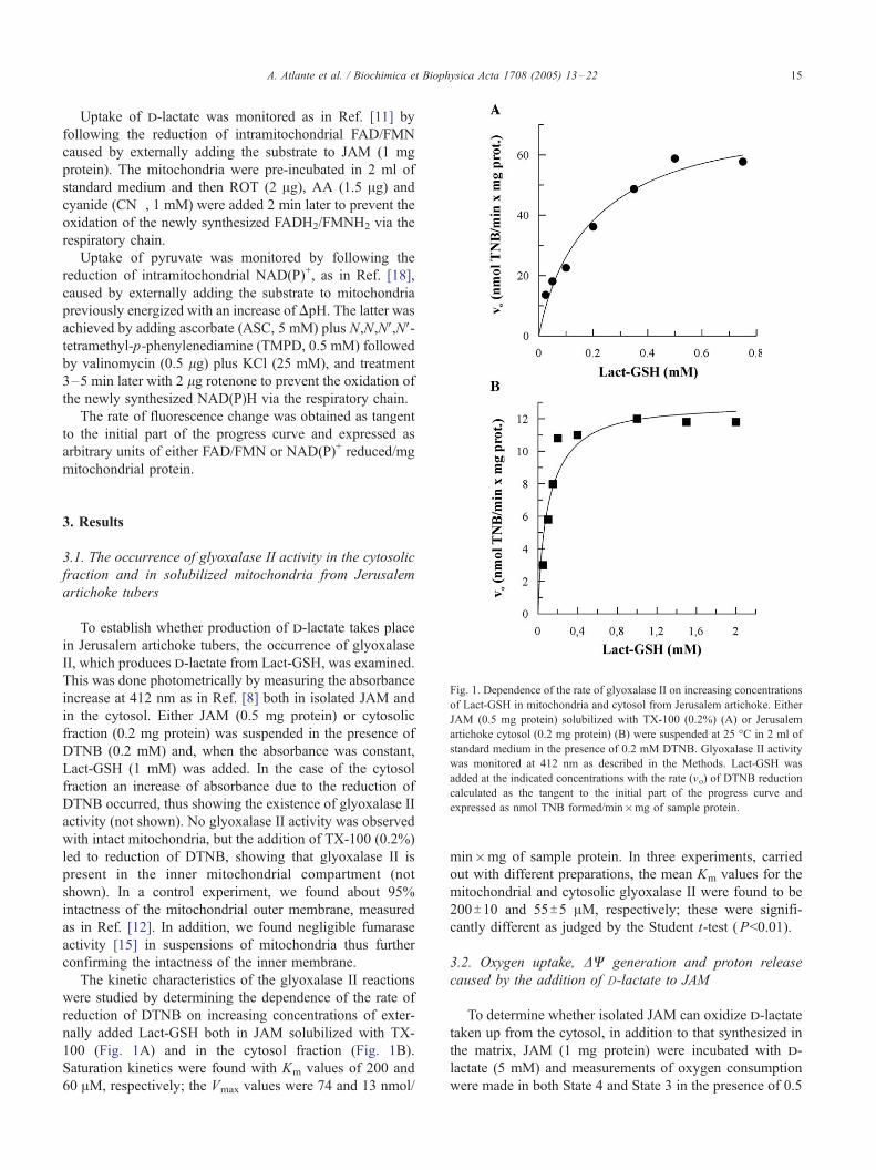
Fig. 1. Dependence of the rate of glyoxalase II on increasing concentrations
of Lact-GSH in mitochondria and cytosol from Jerusalem artichoke. Either
JAM (0.5 mg protein) solubilized with TX-100 (0.2%) (A) or Jerusalem
artichoke cytosol (0.2 mg protein) (B) were suspended at 25 -C in 2 ml of
standard medium in the presence of 0.2 mM DTNB. Glyoxalase II activity
was monitored at 412 nm as described in the Methods. Lact-GSH was
added at the indicated concentrations with the rate (vo) of DTNB reduction
calculated as the tangent to the initial part of the progress curve and
expressed as nmol TNB formed/min�mg of sample protein.
3. Results
3.1. The occurrence of glyoxalase II activity in the cytosolic
fraction and in solubilized mitochondria from Jerusalem
artichoke tubers
To establish whether production of d-lactate takes place
in Jerusalem artichoke tubers, the occurrence of glyoxalase
II, which produces d-lactate from Lact-GSH, was examined.
This was done photometrically by measuring the absorbance
increase at 412 nm as in Ref. [8] both in isolated JAM and
in the cytosol. Either JAM (0.5 mg protein) or cytosolic
fraction (0.2 mg protein) was suspended in the presence of
DTNB (0.2 mM) and, when the absorbance was constant,
Lact-GSH (1 mM) was added. In the case of the cytosol
fraction an increase of absorbance due to the reduction of
DTNB occurred, thus showing the existence of glyoxalase II
activity (not shown). No glyoxalase II activity was observed
with intact mitochondria, but the addition of TX-100 (0.2%)
led to reduction of DTNB, showing that glyoxalase II is
present in the inner mitochondrial compartment (not
shown). In a control experiment, we found about 95%
intactness of the mitochondrial outer membrane, measured
as in Ref. [12]. In addition, we found negligible fumarase
activity [15] in suspensions of mitochondria thus further
confirming the intactness of the inner membrane.
The kinetic characteristics of the glyoxalase II reactions
were studied by determining the dependence of the rate of
reduction of DTNB on increasing concentrations of exter-
nally added Lact-GSH both in JAM solubilized with TX-
100 (Fig. 1A) and in the cytosol fraction (Fig. 1B).
Saturation kinetics were found with Km values of 200 and
60 AM, respectively; the Vmax values were 74 and 13 nmol/
min�mg of sample protein. In three experiments, carried
out with different preparations, the mean Km values for the
mitochondrial and cytosolic glyoxalase II were found to be
200T10 and 55T5 AM, respectively; these were signifi-
cantly different as judged by the Student t-test (P<0.01).
3.2. Oxygen uptake, DW generation and proton release
caused by the addition of D-lactate to JAM
To determine whether isolated JAM can oxidize d-lactate
taken up from the cytosol, in addition to that synthesized in
the matrix, JAM (1 mg protein) were incubated with d-
lactate (5 mM) and measurements of oxygen consumption
were made in both State 4 and State 3 in the presence of 0.5

A. Atlante et al. / Biochimica et Biophysica Acta 1708 (2005) 13–2216
mM ADP (Fig. 2A). l-lactate and succinate, also at 5 mM,
were used as controls. No oxygen uptake was found as a
result of the addition of l-lactate in the absence or presence
of ADP. Oxygen uptake was found with a control respiratory
index equal to 2 and 2.5 for d-lactate and succinate,
respectively. In both cases, OLIGO (2.5 Ag) was found to
reduce the rate of oxygen consumption drastically, but the
rate was restored by the addition of the uncoupler FCCP
(1.25 AM). Both d-lactate and succinate-dependent oxygen
consumption were completely inhibited by AA (2 Ag), an
inhibitor of complex III of the respiratory chain, but was
totally insensitive to ROT (2 Ag), an inhibitor of complex I.
The ability of either the lactate isomers or succinate to
generate membrane potential (DC) was determined by using
safranin O as a DC probe. d-lactate and succinate (5 mM
each), but not l-lactate, were found to cause DC generation
as shown by the decrease in safranin O absorbance at 520
nm (Fig. 2B). The effect was completely insensitive to ROT
(2 Ag), but was prevented by AA (2 Ag) or cyanide (CN�, 1
mM) and was abolished by FCCP (1.25 AM).

A. Atlante et al. / Biochimica et Biophysica Acta 1708 (2005) 13–22 17
Proton movement across the mitochondrial membrane
due to the addition of either d-lactate or succinate (10 mM
each) was measured as a function of time (Fig. 2C) in the
absence or presence of ROT (2 Ag). In both cases, proton
efflux from JAM was found at a rate decreasing with time.
No proton ejection was found in the presence of either AA
or CN�, or when l-lactate (10 mM) was added.
3.3. Localization of a putative D-lactate dehydrogenase in
JAM
To determine the localization of the enzyme responsible
for the oxidation of d-lactate, intact JAM were assayed for
d-lactate dehydrogenase activity as reported in the
Methods. A negligible decrease in DCIP absorbance at
600 nm was found when d-lactate (15 mM) was added to
JAM (Fig. 3A), indicative of the absence of d-LDH
activity in outer membrane, in the intermembrane space or
on the outer side of the mitochondrial inner membrane, or
in any contamination, including peroxisomes, of the
mitochondrial suspension. Externally added phenylsucci-
nate (PheSUCC, 10 mM) failed to inhibit d-LDH activity.
In a control experiment, we confirmed that no oxidation of
succinate by succinate dehydrogenase (which is located on
the matrix side of the inner mitochondrial membrane)
occurred with intact JAM. Oxidation of succinate did
occur after the addition of TX-100 (0.2%) which solubi-
lized the mitochondrial membranes and allowed the
interaction between DCIP and the succinate dehydrogenase
complex (see inset a to Fig. 3A). Consistently, the
oxidation of d-lactate was similarly found after the
addition of TX-100 (0.2%) to JAM showing that the
localization of the putative d-lactate dehydrogenase is
either on the inner side of the inner mitochondrial
membrane or in the matrix (Fig. 3A).
Moreover, in order to confirm that PheSUCC itself
cannot affect the DCIP based assay, we incubated JAM with
glycerol-3-phosphate (G3P, 5 mM) in the absence or
presence of PheSUCC (10 mM). A decrease of absorbance
was found, possibly due to the reduction of external
glycerol-phosphate dehydrogenase, in a manner completely
insensitive to PheSUCC (see inset b to Fig. 3A).
The dependence of the rate of d-lactate oxidation on the
concentration of externally added d-lactate was inves-
Fig. 2. Oxygen consumption, DC generation and proton movement across the mito
mg protein) were suspended at 25 -C in 1.5 ml of the respiratory medium and the a
the following additions were made: d-lactate (D-LAC, 5 mM) (a), succinate (SUCC
of ROT (2 Ag), ADP (0.5 mM), OLIGO (2.5 Ag), FCCP (1.25 AM), AA (2 Ag). Nmin�mg mitochondrial protein. (B) JAM (1 mg protein) were suspended at 25 -Cthe following additions were made: d-lactate (d-LAC, 5 mM) (a), succinate (SUCC
of ROT (2 Ag), FCCP (1.25 AM). Where indicated, AA (2 Ag) or CN� (1 mM) wa
monitored at 520 nm as described in the Methods. (C) JAM (1 mg protein) were su
10 mM MgCl2, 1 mM EGTA–TRIS, 2 mM TRIS–HCl, pH 7.00. At the arrows th
(SUCC, 10 mM) (b), l-lactate (l-LAC, 10 mM) (c), either in the presence or absen
min before addition of the substrate. Number along the curve is the rate of proton
expressed as nmol H+ /min�mg protein.
tigated in JAM treated with TX-100. Saturation kinetics
were found with Km and Vmax values equal to 5 mM and
24 nmol/min�mg protein, respectively (Fig. 3B). In three
experiments, carried out with different preparations, the
mean Km and Vmax values for the mitochondrial d-LDH
were found to be 5T0.5 mM and 25T2 nmol/min�mg
protein, respectively.
3.4. D-lactate transport in JAM
The experiments reported above pose the question as to
how d-lactate produced in the cytosol can cross the
mitochondrial membrane. As an initial approach to this
problem, swelling experiments were carried out as in Ref.
[19]. JAM suspended in 0.18 M ammonium d-lactate
showed spontaneous swelling with a rate and an extent
significantly lower than those found with ammonium l-
lactate, as judged by statistical analysis carried out on five
swelling experiments, according to the Student t-test
(P <0.02). This shows that both d- and l-lactate can enter
mitochondria, but that the uptake is stereospecific (not
shown). d-lactate uptake was further investigated by
monitoring changes in the pH of the mitochondrial
suspension due to d-lactate addition to JAM incubated with
a cocktail of the respiratory chain inhibitors ROT (2 Ag),AA (2 Ag) and CN�(1 mM) (RAC). The addition of d-
lactate (5 mM) resulted in proton uptake by the mitochon-
dria (Fig. 4A). PheSUCC (10 mM) and mersalyl (MERS,
0.1 mM), both non-penetrant compounds, were found to
inhibit the rate of d-lactate-dependent proton uptake,
whereas a-cyano-4-hydroxycinnamate(a-CCN�, 25 AM)
and h-aminobutyrate(h-NH2-BUT, 10 mM) had no effect
on it. In a control experiment, when we energized JAM with
an increase of DpH by adding ASC (5 mM) plus TMPD (0.5
mM) and valinomycin (VAL, 0.5 Ag) in the presence of KCl
(25 mM), we found proton uptake due to the addition of 1
mM pyruvate (PYR, 1 mM) which was completely
insensitive to h-NH2-BUT (10 mM) but prevented by 5
AM a-CCN�(Fig. 4A).
Mitochondrial energization, strictly required for pyru-
vate-dependent proton uptake to occur, produced a signifi-
cant increase in the proton uptake caused by d-lactate.
The results in Fig. 4A suggest that d-lactate/H+ symport
is carrier-mediated and that it does not occur via the pyruvate
chondrial membrane accompanying d-lactate uptake into JAM. (A) JAM (1
mount of residual oxygen was measured as a function of time. At the arrows
, 5 mM) (b), l-lactate (l-LAC, 5 mM) (c), either in the presence or absence
umbers along the curves are rates of oxygen uptake expressed as nmol O2/
in 2 ml of standard medium containing safranin O (12.5 AM). At the arrows
, 5 mM) (b), l-lactate (l-LAC, 5 mM) (c), either in the presence or absence
s added 2 min before addition of the substrate. The safranin O response was
spended at 25 -C in 1.5 ml of proton medium consisting of 150 mM NaCl,
e following additions were made: d-lactate (d-LAC, 10 mM) (a), succinate
ce of ROT (2 Ag). Where indicated, AA (2 Ag) or CN� (1 mM) was added 2
efflux calculated as the tangent to the initial part of the progress curve and

Fig. 3. d-LDH activity assay in JAM and the dependence of the rate of d-lactate oxidation on increasing d-lactate concentrations. (A) JAM (0.2 mg protein)
were incubated in 2 ml of standard medium in the presence of PMS (30 AM) and either in the absence or presence of PheSUCC (10 mM). d-LDH activity was
monitored at 600 nm as described in the Methods. At the arrows DCIP (50 AM), d-LAC (15 mM) and TX-100 (0.2%) were added. The insets report control
experiments: in (a) at the arrows DCIP (50 AM), succinate (SUCC, 5 mM) and TX-100 (0.2%) were added to mitochondria added with PMS; in (b) at the
arrows glycerol phosphate (G3P, 5 mM), in the presence or absence of PheSUCC (10 mM), added 2 min before, was added to mitochondria added with PMS
and DCIP. Numbers along the curves are rates of d-lactate, succinate or glycerol-3-phosphate oxidation expressed as nmol DCIP reduced/min�mg protein. (B)
d-lactate was added at the indicated concentrations to JAM treated with TX-100 (0.2%) with the rate (vo) of DCIP reduction calculated as the tangent to the
initial part of the progress curve and expressed as nmol DCIP reduced/min�mg protein.
A. Atlante et al. / Biochimica et Biophysica Acta 1708 (2005) 13–2218
carrier. It was confirmed that PheSUCC (10 mM) and MERS
(0.1 mM) do not cause any non-specific effect on the
mitochondrial membrane, by using as a substrate butyrate
(BUT) (10 mM), which is known to enter mitochondria via
diffusion (see inset to Fig. 4A). In this case the addition of
BUT results in proton release in a manner insensitive to both
inhibitors but sensitive to the uncoupler FCCP; however,
when JAM were added with BUT in the presence of the
inhibitors of the respiratory chain, i.e. rotenone, antimycin A
and cyanide (RAC), proton uptake was found which proved
to be insensitive to both PheSUCC (10 mM) and MERS (0.1
mM) (inset to Fig. 4A).
In the same experiment, uptake and metabolism of d-
lactate was further investigated by determining the ability of
externally added d-lactate to reduce intra-mitochondrial
dehydrogenase cofactors. This was done by using fluorimet-
ric techniques that have previously been used to monitor
changes in redox state of pyridine nucleotides [18] and
flavins [11]. No significant reduction of mitochondrial
NAD(P)+ was found when d-lactate was added to JAM
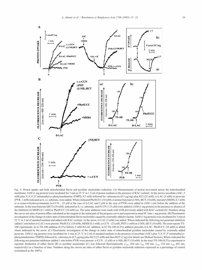
Fig. 4. Proton uptake and both mitochondrial flavin and pyridine nucleotides reduction. (A) Measurements of proton movement across the mitochondrial
membrane. JAM (1 mg protein) were incubated for 3 min at 25 -C in 1.5 ml of proton medium in the presence of RAC cocktail. At the arrows ascorbate (ASC, 5
mM) plus N,N,NV,NV-tetramethyl-p-phenylenediamine (TMPD, 0.5 mM) followed by valinomycin (0.5 Ag/mg) plus KCl (25 mM), d-LAC (5 mM) or pyruvate
(PYR, 1mM) indicated as S, i.e. substrate, were added.Where indicated PheSUCC (10mM),h-amino-butyrate (h-NH2-BUT, 10mM), mersalyl (MERS, 0.1mM)
or a-cyano-4-hydroxycinnamate (a-CCN�, 25 AM in the case of d-LAC and 5 AM in the case of PYR) were added to JAM 1 min before the addition of the
substrate. In the inset butyrate (BUT) (10 mM), indicated as S, i.e. substrate, and FCCP (1.25 AM)were added to JAM (1 mg protein) in the presence or absence of
the inhibitors (I) MERS (0.1 mM) or PheSUCC (10 mM) (a). The same additions were made with JAM previously added with RAC cocktail (b). Numbers along
the curves are rates of proton efflux calculated as the tangent to the initial part of the progress curve and expressed as nmol H+/min�mg protein. (B) Fluorimetric
investigation of the change in redox state of mitochondrial flavin nucleotides caused by externally addedd-lactate. JAM (1mg protein) were incubated for 3min at
25 -C in 2 ml of standard medium and added with RAC cocktail. At the arrow, d-LAC (5 mM) was added. Where indicated the following not penetrant inhibitors
(added 1min befored-LAC)were present: PheSUCC (10mM),MERS (0.1mM),a-CCN� (25 AM), PHT (1mM) orh-NH2-BUT (10mM). The inset reports TX-
100 experiments: in a) TX-100 addition (0.2%) follows 5 mM D-LAC addition; in b) TX-100 (0.2%) addition precedes d-LAC. PheSUCC (10 mM) is added
where indicated by the arrow. (C) Fluorimetric investigation of the change in redox state of mitochondrial pyridine nucleotides caused by externally added
pyruvate. JAM (1 mg protein) were incubated for 3 min at 25 -C in 2 ml of standard medium in the presence of ascorbate (ASC) plus N,N,NV,NV-tetramethyl-p-
phenylenediamine (TMPD) followed by valinomycin (0.5 Ag/mg) plus KCl (25 mM) and then ROT (2 Ag) (for details see Method Section). Where indicated the
following not penetrant inhibitors (added 1 min before PYR) were present: a-CCN� (5 AM) or h-NH2-BUT (10 mM). In the inset TX-100 (0.2%) experiment is
reported. Reduction of either flavin (B) or pyridine nucleotides (C) was followed fluorimetrically (kexc 450 nm/kem 520 nm; kexc 334 nm/kem 456 nm,
respectively) as a function of time. Numbers along the curves are rates of either flavin or pyridine nucleotide reduction expressed as a percentage of control
(considered as the 100%).
A. Atlante et al. / Biochimica et Biophysica Acta 1708 (2005) 13–22 19

A. Atlante et al. / Biochimica et Biophysica Acta 1708 (2005) 13–2220
previously incubated with ROT (not shown). On the other
hand, addition of d-lactate (5 mM) to JAM pre-incubated
with RAC cocktail (2 Ag ROT plus 2 Ag AA, plus 1 mM
CN�) resulted in a rapid decrease in fluorescence at wave-
lengths characteristic of oxidized flavins (450 nm/520 nm
excitation/emission) (Fig. 4B). This reduction of intramito-
chondrial flavin was strongly inhibited when either Phe-
SUCC (10 mM) or MERS (0.1 mM) were added before the
addition of d-lactate. Contrarily, no inhibition was found in
the presence of phthalonate (PHT, 1 mM), a-CCN�(25 AM)
or h-NH2-BUT (10 mM). To determine whether the rate of
oxidation of d-lactate via the putative d-LDH was limited by
the rate of its uptake, TX-100 (0.2%) was added to the JAM.
This increased the rate of flavin reduction showing that the
rate of oxidation of d-lactate reflects the rate of d-lactate/H+
A
181614121086420
14
12
10
8
6
4
2
0
1/v o
(a.u
./min
x m
g)-1 10 mM D-LAC
20 mM D-LAC
[PHENYLSUCCINATE] (mM)
B
v o (a
.u./m
in x
mg)
121086420
0.25
0.2
0.15
0.1
0.05
0
[D-LACTATE] (
Fig. 5. Dixon plot of the inhibition by PheSUCC of the rate of intramitochondrial fl
of d-lactate/H+ symport on increasing d-lactate concentrations. (A) JAM (1 mg p
rate of intramitochondrial flavin nucleotide was measured as in Fig. 4B, by using 10
the indicated concentrations. The rate (vo), measured as the tangent to the in
mitochondrial protein. The inset reports the plot of 1/i against 1/[PheSUCC], where
in the absence of PheSUCC, respectively. (B) d-lactate was added to JAM (1 m
reduction was measured as described in Fig. 4B. The rates (vo), measured as the
units/min�mg mitochondrial protein.
symport across the mitochondrial inner membrane. In the
same experiment, we found that PheSUCC (10 mM) was
ineffective in inhibiting the d-lactate oxidation occurring in
solubilized JAM (see inset to Fig. 4B).
When pyruvate was added to mitochondria energized
with an increase of DpH, we found a significant reduction of
mitochondrial NAD(P)+; this was completely prevented by
a-CCN� (5 AM), but insensitive to h-NH2-BUT (10 mM)
(Fig. 4C). TX-100 (0.2%) addition proved to increase the
rate of NAD(P)+ reduction (inset to Fig. 4C).
PheSUCC proved to be competitive inhibitor of the rate
of flavin reduction with Ki value equal to 9 mM (Fig. 5A).
Interestingly, the y intercepts of the lines fitting the
experimental points obtained in the presence of the inhibitor
coincide with the experimental values obtained in the
20
0.40.20
24
20
16
12
8
4
0
1/[PHENYLSUCCINATE] (mM)-1
1/i
20181614
mM)
avin nucleotides reduction due to externally added d-lactate and dependence
rotein) were incubated at 25 -C in 2 ml of standard medium. The reduction
mM and 20 mM d-lactate either in the absence or presence of PheSUCC at
itial part of the progress curve, is expressed in arbitrary units/min�mg
i= 1�v i /vo; v i and vo being the rate of d-lactate uptake in the presence and
g protein) at the indicated concentrations and the rate of flavin nucleotides
tangents to the initial part of the progress curves, are expressed as arbitrary

A. Atlante et al. / Biochimica et Biophysica Acta 1708 (2005) 13–22 21
absence of inhibitor. As expected in the light of the TX-100
experiments (see insets to Fig. 4B), this shows that
PHESUCC-sensitive d-lactate transport controls the rate
of the measured process, in accordance with the control
strength criterion [11], i.e. the rate of the flavin reduction
reflects the rate of the d-lactate uptake.
The data of Fig. 5A were also plotted as 1 / i against 1 /
[Inhibitor], where the fractional inhibition i is 1�vi /vo (see
insets). The y intercept was unity, showing that PheSUCC
can prevent d-lactate transport completely.
The dependence of the rate of d-lactate/H+ symport on
increasing d-lactate concentrations was measured. The
results were analyzed by means of a Michaelis–Menten
plot (Fig. 5B). Saturation kinetic was found with a Km value
equal to 16 mM.
4. Discussion
In this paper we show for the first time that d-lactate can
be transported into plant mitochondria, in particular those
from Jerusalem artichoke, and metabolized therein. The
sequence of events involved in metabolism of d-lactate
most likely includes d-lactate synthesis in cytosol due to
glyoxalase II, uptake via a d-lactate/H+ symporter, and
oxidation of the d-lactate to pyruvate by a putative d-LDH
located in the inner mitochondrial compartment.
The existence of glyoxalase II in Jerusalem artichoke
cytosol confirms previous findings [7,8,10,20], thus sug-
gesting that this enzyme occurs also in tubers.
In order to study the uptake of d-lactate by JAM, use was
made of spectroscopic techniques under conditions in which
mitochondrial metabolism is mostly active, thus allowing
for monitoring of mitochondrial reactions and of the traffic
of newly synthesized substrates across the mitochondrial
membrane. Mitochondria can take up d-lactate with net
carbon uptake in a proton-compensated manner. This was
substantiated by the observed swelling in isotonic solutions
of ammonium d-lactate which per se indicates that uptake of
d-lactate is proton-compensated. Moreover, d-lactate/H+
symport was investigated as in Ref. [11] by directly
measuring proton movement across the mitochondrial
membrane. d-lactate when added to JAM either in the
absence or presence of ROT caused proton ejection in a
manner inhibited by AA. In a set of parallel experiments, we
have investigated pyruvate uptake by JAM, showing that d-
lactate/H+ and pyruvate/H+ symports occur in a different
manner as revealed by the effects of a-CCN�. Interestingly,
pyruvate uptake strictly requires energized JAM as obtained
by an increase in DpH, whereas such a condition simply
increases the rate of d-lactate uptake. This provides a further
clear distinction between the two.
The results that we have reported are entirely consistent
with the existence of a mitochondrial d-LDH (see Fig. 3).
The enzyme is a flavoprotein capable of reducing complex
III, as shown by the reduction of the intramitochondrial
flavins in the presence of ROT. In this regard, the JAM D-
LDH is similar to the enzymes found in mitochondria from
S. cerevisiae [21] and rat liver [11] as well as that from
Escherichia coli [22].
Moreover, we show that the putative d-LDH activity can
be assayed after TX-100 addition to JAM thus suggesting
the localization of the enzyme on the inner side of the
mitochondrial inner membrane or in the matrix space. The
possibility that d-lactate is oxidized on the external face of
the inner membrane, with electrons transferred to the inner
surface for reaction with DCIP (which requires Triton to
gain access), can be ruled out on the basis of the lack of
inhibition on d-LDH activity, as assayed, both fluorimetri-
cally and using DCIP, in solubilized JAM, by PheSUCC
which proved to inhibit completely d-lactate dependent
flavin reduction. Thus we are forced to conclude that the
inhibition is dependent on a carrier mediated transport, this
strongly suggesting that the d-lactate must be taken up for
its mitochondrial metabolism to occur.
The localization of the enzyme is consistent with intra-
mitochondrial oxidation of d-lactate newly synthesized in
the matrix from lactoyl-glutathione by the mitochondrial
glyoxalase II [9,20,22–24], as well as for the cytosolic d-
lactate taken up by mitochondria. In initial experiments we
did not find any d-LDH activity in the cytosol fraction of
Jerusalem artichoke tubers (data not shown), and so the
possible occurrence of a cytosolic d-LDH remains for future
investigation.
The picture emerging from the results described in this
paper is as follow. The potentially toxic methylglyoxal is
metabolized to d-lactate in cytosol by glyoxalase II (Fig.
1B). Uptake of d-lactate by JAM occurs in a proton-
compensated manner via the d-lactate/H+ symporter (Fig.
4A,B). In the matrix, the imported d-lactate, as well as d-
lactate formed by the mitochondrial glyoxalase II (Fig 1A),
is oxidized to pyruvate via the d-LDH (Fig. 3). Electrons
flow to oxygen results in ATP synthesis as well as in
generation of a membrane potential (Fig. 2A,B). Finally, the
combined actions of cytosolic glyoxalase II, of the d-lactate
transporter and of the putative d-LDH provide a route for
removal of methylglyoxal which is toxic to plant cells [3].
Acknowledgements
The authors thank Prof Shawn Doonan for his critical
reading. This work was partially financed by Fondi di
Ricerca di Ateneo del Molise to S.P. and by PRIN 2004
‘‘Cross talk between organelles in response to oxidative
stress and programmed cell death in plants’’.
References
[1] R. Deswal, T.N. Chakaravarty, S.K. Sopory, The glyoxalase system in
higher plants: regulation in growth and differentiation, Biochem. Soc.
Trans. 21 (1993) 527–530.

A. Atlante et al. / Biochimica et Biophysica Acta 1708 (2005) 13–2222
[2] S.L. Singla-Pareek, M.K. Reddy, S.K. Sopory, Genetic engineering of
the glyoxalase pathway in tobacco leads to enhanced salinity
tolerance, Proc. Natl. Acad. Sci. U. S. A. 100 (2003) 14672–14677.
[3] M.P. Kalapos, Methylglyoxal in living organisms: chemistry, bio-
chemistry, toxicology and biological implications, Toxicol. Lett. 110
(1999) 145–175.
[4] P.J. Thornalley, The glyoxalase system: new developments towards
functional characterization of a metabolic pathway fundamental to
biological life, Biochem. J. 269 (1990) 1–11.
[5] D.J. Creighton, M. Migliorini, T. Pourmotabbed, M.K. Guha,
Optimization of efficiency in the glyoxalase pathway, Biochemistry
27 (1988) 7376–7384.
[6] A.M. Martins, C.A. Cordeiro, A.M. Ponces Freire, In situ analysis of
methylglyoxal metabolism in Saccharomyces cerevisiae, FEBS Lett.
499 (2001) 41–44.
[7] S.J. Norton, V. Talesa, W.J. Yuan, G.B. Principato, Glyoxalase I and
glyoxalase II from Aloe vera: purification, characterization and com-
parison with animal glyoxalases, Biochem. Int. 22 (1990) 411–418.
[8] S.J. Norton, G.B. Principato, V. Talesa, M. Lupattelli, G. Rosi,
Glyoxalase II from Zea mays: properties and inhibition study of the
enzyme purified by use of a new affinity ligand, Enzyme 42 (1989)
189–196.
[9] V. Talesa, G. Rosi, S. Contenti, C. Mangiabene, M. Lupattelli, S.J.
Norton, E. Giovannini, G.B. Principato, Presence of glyoxalase II in
mitochondria from spinach leaves: comparison with the enzyme from
cytosol, Biochem. Int. 22 (1990) 1115–1120.
[10] M.K. Maiti, S. Krishnasamy, H.A. Owen, C.A. Makaroff, Molecular
characterization of glyoxalase II from Arabidopsis thaliana, Plant
Mol. Biol. 35 (1997) 471–481.
[11] L. de Bari, A. Atlante, N. Guaragnella, G. Principato, S. Passarella, d-
lactate transport and metabolism in rat liver mitochondria, Biochem. J.
365 (2002) 391–403.
[12] M.L. Pallotta, D. Valenti, M. Iacovino, S. Passarella, Two separate
pathways for d-lactate oxidation by Saccharomyces cerevisiae
mitochondria which differ in energy production and carrier involve-
ment, Biochim. Biophys. Acta 1608 (2004) 104–113.
[13] I.B. Dry, E. Dimitriadis, A.D. Ward, J.T. Wiskich, The photo-
respiratory hydrogen shuttle. Synthesis of phthalonic acid and
its use in the characterization of malate/aspartate shuttle in
pea (Pisum sativum) leaf mitochondria, Biochem. J. 245 (1987)
669–675.
[14] A.C. Liden, I.M. Moller, Purification, characterization and storage of
mitochondria from Jerusalem artichoke tubers, Physiol. Plant. 72
(1988) 265–270.
[15] M. Neuburger, E.P. Journet, R. Bligny, J.P. Carde, R. Douce,
Purification of plant mitochondria by isopycnic centrifugation in
density gradients of percoll, Arch. Biochem. Biophys. 217 (1982)
312–323.
[16] E. Lopez-Huertas, L.M. Sandalio, L.A. del Rio, Integral membrane
polypeptides of pea leaf peroxisomes: characterization and response to
plant stress, Plant Physiol. Biochem. 33 (1995) 295–302.
[17] G.W. Lohr, H.D. Waller, Glucose-6-phosphate dehydrogenase, in:
H.U. Bergmeyer (Ed.), Methods Enzym. Anal., 1963, pp. 744–751.
[18] A. Atlante, S. Passarella, G.M. Minervini, E. Quagliariello, Glutamine
transport in normal and acidotic rat kidney mitochondria, Arch.
Biochem. Biophys. 315 (1994) 369–381.
[19] S. Passarella, A. Atlante, D. Valenti, L. de Bari, The role of
mitochondrial transport in energy metabolism, Mitochondrion 2
(2003) 319–343.
[20] V. Talesa, L. Uotila, M. Koivusalo, G. Principato, E. Giovannini,
G. Rosi, Isolation of glyoxalase II from two different compart-
ments of rat liver mitochondria. Kinetic and immunochemical
characterization of the enzymes, Biochim. Biophys. Acta 993
(1989) 7–11.
[21] A. Chelstowska, Z. Liu, Y. Jia, D. Amberg, R.A. Butow, Signalling
between mitochondria and the nucleus regulates the expression of a
new d-lactate dehydrogenase activity in yeast, Yeast 15 (1999)
1377–1391.
[22] O. Dym, E.A. Pratt, C. Ho, D. Eisenberg, The crystal structure of d-
lactate dehydrogenase, a peripheral membrane respiratory enzyme,
Proc. Natl. Acad. Sci. U. S. A. 97 (2000) 9413–9418.
[23] V. Talesa, L. Uotila, M. Koivusalo, G. Principato, E. Giovannini, G.
Rosi, Demonstration of glyoxalase II in rat liver mitochondria. Partial
purification and occurrence in multiple forms, Biochim. Biophys. Acta
955 (1988) 103–110.
[24] V. Talesa, G.B. Principato, S.J. Norton, S. Contenti, C. Mangiabene,
G. Rosi, Isolation of glyoxalase II from bovine liver mitochondria,
Biochem. Int. 20 (1990) 53–58.