Transcription Corepressor CtBP Is an NAD +-Regulated Dehydrogenase
Upload
khangminh22Category
view
0download
0
Polyclonal Activation of B Cells by Lactate Dehydrogenase-Elevating Virus is Mediated by NGlycans on the Short Ectodomain of the Primary Envelope Glycoprotein
PETER G.W. PLAGEMANN,l QUENTIN A. JONES,2 AND WILLIAM A. CAFRUNY2
1 Department of Microbiology, University of Minnesota, Minneapolis, MN, USA; 2Division of Basic Biomedical Sciences, Molecular Microbiology & Immunology Group, University ofSout} Dakota School of Medicine, Vermillion, SD, USA
1. INTRODUCTION
The common strains of lactate dehydrogenase-elevating virus (LDV-P and LD-vx) are primary examples for viruses that cause a permanent polyclonal activation of B cells that results in IgG2a hypergammaglobulinemia and the generation of autoantibodies and circulating immune complexes in their host, the mouse (Notkins et aI., 1966; Coutelier and van Snick, 1985; Li et aI, 1990). Plasma IgG2a levels increase from generally below 0.5 mg/ml to 2-6 mg/ml by two weeks post infection (p.i.) and remain elevated thereafter (see later). LDV -P/vx cause life-long persistent viremic infections (see Fig. lA) which are maintained by continuous rounds of replication in a renewable subpopulation of macrophages and resistance to host immune responses (Plagemann, 1996). Previous results have shown that the single neutralization epitope located on the short (about 30 amino acids long) ectodomain of the primary envelope glycoprotein, VP-3P, carries three large N-glycan chains in LDV-P and LDV-vx (see Fig. IA insert) that suppress the immunogenicity of the epitope and impair antibody neutralization of the virions of these quasispecies (Chen et aI., 2000; Plagemann et aI., 1999). The present results indicate that the
The Nidoviruscs (Coronaviruses and Arteriviruscs). Edited hy Ehud Lavi et al., Kluwcr Academic/Plenum Puhlishcrs, 200 I. 375

376 Polyclonal Activation of B Cells by LD V
three N-glycans on the VP-3P ectodomains of LDV -P/vx also playa critical role in the polyclonal activation of B cells by these LDV s.
o
A. ECTODOMAINS B. ECTODOMAINS
~'-'jJl ~. 'ill • C c...c C c...c
MNP·2 VP·3P
ENDODOMAINS
o C'
o lDV-vx
• lDV-P
o
MNp·2 VP·3P
ENDODOMAINS
I::. lDV-v
, lDV-C
~O------~----~--O~----~40------~8~O~
TIME Pol. (DAYS)
Figure 1. Time courses of viremia in FVB mice injected intraperitoneally with (A) biologically cloned LOV-P or LOV-vx and (B) with biologically cloned LOV-C or LOV-v (data are redrawn from Plagemann et al., 1999) and the proposed topography of VP-3P and MIVP-2 in the LOV envelope (inserts).
2. RESULTS
The conclusion that the N-glycans on the VP-3P ectodomain playa critical role in the polyclonal activation of B cells by LDV is provided by comparing the IgG2a hypergammaglobulinemia and immune complex formation in mice infected with biologically cloned LDV -P/vx with those in mice infected by two biologically cloned variants of LDV, LDV -C and LDV-v, which have gained neuropathogenicity for C58 and AKR mice, but at the same time have lost the ability for high viremic persistent infection (Fig. lB; Chen et aI., 2000). Previous studies have shown that the VP-3P

Peter G. W Plagemann et al. 377
ectodomains ofLDV-C/v lack the two N-terminal N-glycosylation sites (see Fig. lB, insert) present on the VP-3P ectodomains of LDV-P/vx (Fig. 1A insert) which greatly increases the immunogenicity of the neutralization epitope and the sensitivity of LDV -C/v to antibody neutralization.
This results in rapid suppression of their replication in mice (Fig. lB; Chen et aI., 2000; Plagemann et aI., 1999). We have now found that infection of C57BLl6 and FVB mice with LDV-C or LDV-v consistently induces a IgG2a hypergammaglobulinemia that was only one half or less as high as that induced by infection with LDV -P/vx (Fig. 2). Plasma IgG2a levels were quantified by capture ELISA as described previously (Li et aI.; 1990). In order to allow valid comparisons between the two classes ofLDV, we generally compared the time courses of IgG2a formation (4 time points each) in mice infected with at least one non-neuropathogenic and one neuropathogenic LDV in the same ELISA (2-fold plasma dilutions from 1 :4000 to 1: 128,000) and have used the same standard solution ofIgG2a for establishing standard curves (0.5 ng/ml to 1 mg/ml) in all assays. Similar results were obtained with duplicate mice assayed in the same experiment (Fig. 2C and D), and with additional mice infected with these LDVs (at least two other mice for each, data not shown) and no increase in plasma IgG2a was observed in an uninfected companion mouse (see Fig. 2C) consistent with previous results (Li et aI., 1990).
(A) C57BlJ6 (B)FVS (E) FVB (F) FVB
LDV-74
2 LDV-vx
LDV-P
o
Figure 2. Time courses of plasma IgG2a elevation in C57BLl6 and FYB mice after infection with the indicated LOY quasispecies. All time courses are for single mice, but in (C) and (0) results for two companion mice are presented. Results in each frame come from a single ELISA, and were estimated from the same IgG2a standard curve. Comparable results were obtained in repeat ELiSAs of the plasma samples (data not shown).

378 Polyclonal Activation of B Cells by LDV
Since LDV -v is a genetic recombinant of LDV -vx that has, by a double recombination, specifically acquired the 5' end ofORF5 ofLDV-C (~400 nt) that encodes the ectodomain of the primary envelope glycoprotein VP-3P (Fig. 3A; Li et al., 1999) the difference in polyclonal B cell activation between LDV-P/vx, on the one hand, and LDV-C/v, on the other hand, must reside in this segment of VP-3P. The most likely molecular structure responsible for this difference in the polyclonal B cell activation is the number of N-glycans associated with the VP-3P ectodomain (Fig. 1 and 3B) just as they are responsible for the other phenotypic differences between LDV -P/vx and LDV -C/v discussed already. There are no other amino acid differences in this VP-3P segment that correlate with the various differences in phenotypic properties of the two classes of LDV (Chen et a1., 1998; Li et a1., 1999).
A.
B.
UIY-c
11.0 11.5 ! !
12.0 !
12.5 I
13.0 13.5 14.0 kb ! ! ! An
ORFs ••• - OAFlb--i r---ORF 2--1 I-- OAF 4----1 I-- OAF 8-----i
1---OAF 3----1
VP·3M
LDV·vI
j---- OAF 5--1
Vp·3P
LDV-C
ectodomain
I-ORF 7.........j
M/VP·2 NNP·l
signAl peptide tran.membrane .eqment HfcLNRlliCFL I pSALLP VYFvLY ILST£NACVAGOSSTKNLIYTSTLCE LllVTGFOOHFCYIIV£Tf V I rpAVfH L I SLRI Lf ..... . • _ --- ---R------F------------------- -1------ -IIL-----I- --L .. ---- --------------------- ..... . ____________________ ------ -- ---- --- ------ --- - -- --- -1- - -- -- --- - -. - - •• --- ---- -- -- ---- ..... .
C-'tU!".'" - I ------ -------- --CC--- ------ ------1---··--- -IIL- ----1---- - -- ---- - -- --. -- ---- - ---- - -- ..... . C-YIJ/UOII/" -- - -- - - - - -- - - - -----c --------. --- --01--- - - -- -IIL- --- -1- - - - --- - - - - - - - - - - -- - - - --- - - - - -- ..•... c-'tue ,SS --------11-------- -------------------I-------ML---O-I--- --- .. - .... -. - ---. -- - - .. - - ........ .
-K·· KK -- 5,,1/-- -M-- -re ·1- - •. - --. - --A--I- ---- -- -IL--- - -1- - - - --- .. - -- - - - ---- -L-- - - --N--- ..... . UIY __ -K --K- - -SVII---RS---C- I -FF----S--G-SI----- ---IIL-----I----------- -- - - - ----L--- ---N- -- ..... .
Figure 3. Organization ofORFs 2-7 ofLOV and origins of the 3' and genome segments of the neuropathogenic genetic recombinant LOV-v (A) and amino acid sequences of the N-terminal ends of the VP-3Ps of various LOV quasi species and of neutralization escape mutants of LOV-C/v (B). For the origins of the mutants of LOV-C/v see Chen et al., 2000. The signal peptide and the first transmembrane segment are overlined and the potential N-glycosylation sites are in bold face letters (* indicates the N-glycosylation site that is conserved in all LOV isolates).
This hypothesis was further explored by examining plasma IgG2a levels m FVB mice after infection with three neutralization escape mutants of LDV-C and LDV-v (LDV-v-Tl05/38, LDV-C-Tl68/55, LDV-CT82/68N/76) whose VP-3P ectodomains had regained three N-glycosylation sites (see Fig. 3B). The plasma IgG2a elevation in duplicate mice infected with each of the three neutralization escape mutants was comparable to that in an LDV-P infected mouse and much higher than that in an LDV-C infected mouse (Fig. 4). Similarly, in a mouse infected with an LDV isolated from a wild house mouse in Montana (LDV-74), whose VP-3P ectodomain

Peter G. W Plagemann et al. 379
possesses all three N-glycosylation sites (Li et aI., 2000), plasma IgG2a levels increased similarly as in LDV -P and LDV -vx infected mice (Fig. 2F).
., CII CJ ~1
(A) LDY-y (8) LDY-C
T105l38 T168155 T82168N176
LDY-C
°O~~--~~~20~4~O~O--2~O~~40~O~~~~
TIME Pol. (DAYS)
Figure 4. Comparison of the time courses of plasma [gG2a elevation in duplicate FVB mice infected with three neutralization escape mutants of LDV-v and LDV-C (A-C) with those in LDV-P and LDV-C infected mice (D). All values were obtained in the same ELISA and estimated from the same [gG2a standard curve. Comparable results were obtained in a repeat ELISA of the plasma samples (data not shown).
An even more drastic difference between mice infected with the common non-neuropathogenic LDV s and the neuropathogenic mutants was observed in the formation of circulating 150-300 kDa immune complexes that have been found to become generated concomitant with an LDV induced polyc1onal activation of B cells (Cafruny et aI., 1986; Hu et aI., 1992). These complexes contain IgG2a which most likely represents autoantibodies that are generated as a result of the polyc1onal activation of B cells and bound to their auto antigens (Hu et aI., 1992). They are recognized by binding in PBSTween to ELISA plates that are not coated with antigen. They differ in size and IgG isotype specificity from infectious virion-antibody complexes that are also generated and persist in LDV -infected mice (Hu et aI., 1992). Fig. 5A-C illustrates the appearance of plate binding immune complexes in mice

380 Polyclonal Activation of B Cells by LD V
after infection with cloned LDV-P and LDV-vx. The time courses of immune complex formation were very similar for the two cloned quasispecies, just as was the case for the IgG2a hypergammaglobulinemia induced by them (Fig. 2) and similar to those previously reported for mice infected with original LDV isolates (Cafruny et aI., 1986; Hu et aI., 1992). In contrast, in our standard assay, which involves a 1 h incubation of the ELISA plates with alkaline phosphatase substrate, we detected little or no plate binding IgG in the plasmas of FVB or C57BLl6 mice infected with the neuropathogenic LDV-C or LDV-v (Fig. 5E-H, and data not shown).
(A) LDY-P (8) LDV-P (e) LDV-vx (D) LDV-74
• FVB FVB
o-T1 0.2
0
(F) LDV-C C57BL16
1/1 o-T1 __ T56
0.2 i c( e~~eee 1/1
0 0 'It
(G) LDV-y c(
C57BL16 o-T1
__ T56 0.2
eee !lee (H) LDV-y
0
FVB o-T1 __ T42
~ 0.2
0 50 200 800 50 200 800 50 200 800 50 200 800 50 200 800
0
PLASMA DILUTION-1
Figure 5. Time courses of appearance of ELISA plate binding activity in plasma of C57BLl6 and FYB mice after infection with the indicated LDYs. A405 is plotted against 2-fold dilutions of each plasma sample (T=time pj. in days). The results in (B), (E), and (G) come from the same plate-binding assay and so do the results in (A) and (E) and the results in (C) and (H). The results are for single mice, which generally have also been assayed for plasma IgG2a levels (Fig. 2). Comparable results were obtained in repeat assays of the plasma samples and in duplicate mice where available.
On the other hand, immune complex formation in mice infected with the three neutralization escape mutants of LDV-C and LDV-v, whose VP-3P ectodomains had regained three N-glycosylation sites (see Fig. 3B), was comparable to that in mice infected with LDV-P and LDV-vx rather than to that in mice infected with the LDV-v (or LDV-C) parent (Fig. 6). The same was the case for the immune complex formation in a mouse infected with the wild house mouse LDV -74 (Fig. 5D), whose VP-3P ectodomain possesses three N-glycosy1ation sites, just like those of LDV -P and LDV -vx.
Peter G. W Plagemann et al.
J
(D) UJV.P (E) LDV-YII IF) l.DV-¥
0- T1 0- T1 __ T14
tr 134 Ie- T42
o~~~~~~~~.~_~_~~.~_~_~~.~_~_~~.~_~_~
PLASMA DlLunON"
381
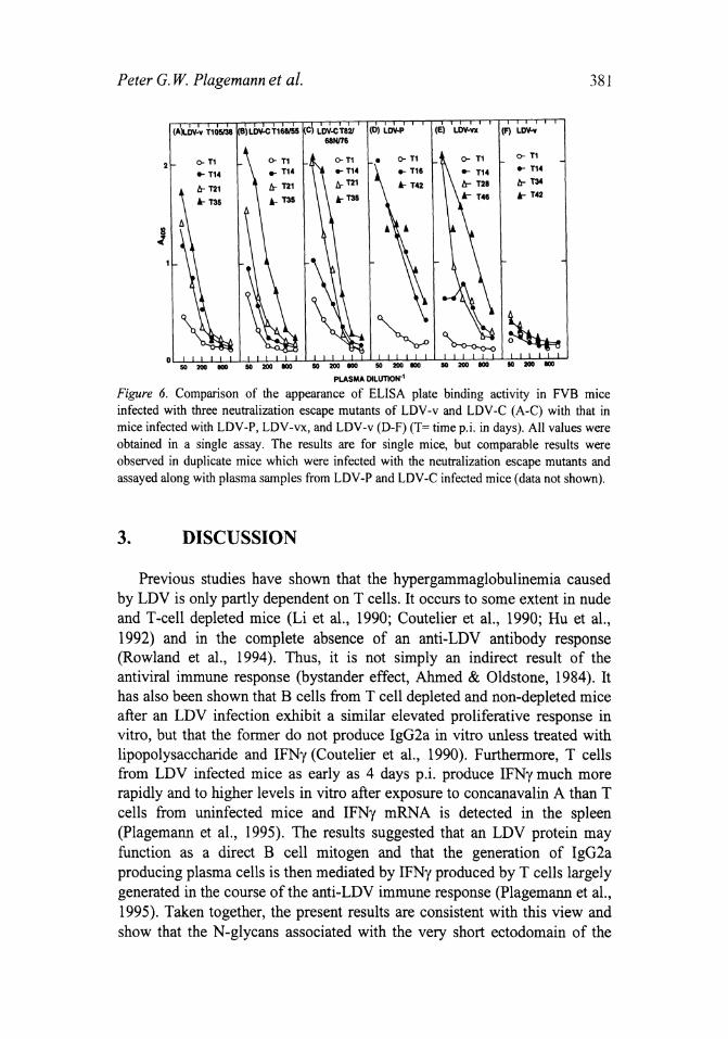
Figure 6. Comparison of the appearance of ELISA plate binding activity in FVB mice infected with three neutralization escape mutants of LDV-v and LDV-C (A-C) with that in mice infected with LDV-P, LDV-vx, and LDV-v (D-F) (T= time pj. in days). All values were obtained in a single assay. The results are for single mice, but comparable results were observed in duplicate mice which were infected with the neutralization escape mutants and assayed along with plasma samples from LDV-P and LDV-C infected mice (data not shown).
3. DISCUSSION
Previous studies have shown that the hypergammaglobulinemia caused by LDV is only partly dependent on T cells. It occurs to some extent in nude and T-cell depleted mice (Li et aI., 1990; Coutelier et aI., 1990; Hu et aI., 1992) and in the complete absence of an anti-LDV antibody response (Rowland et aI., 1994). Thus, it is not simply an indirect result of the antiviral immune response (bystander effect, Ahmed & Oldstone, 1984). It has also been shown that B cells from T cell depleted and non-depleted mice after an LDV infection exhibit a similar elevated proliferative response in vitro, but that the former do not produce IgG2a in vitro unless treated with lipopolysaccharide and IFNy (Coutelier et aI., 1990). Furthermore, T cells from LDV infected mice as early as 4 days p.i. produce IFNy much more rapidly and to higher levels in vitro after exposure to concanavalin A than T cells from uninfected mice and IFNy mRNA is detected in the spleen (Plagemann et aI., 1995). The results suggested that an LDV protein may function as a direct B cell mitogen and that the generation of IgG2a producing plasma cells is then mediated by IFNy produced by T cells largely generated in the course of the anti-LDV immune response (Plagemann et aI., 1995). Taken together, the present results are consistent with this view and show that the N-glycans associated with the very short ectodomain of the

382 Polyclonal Activation of B Cells by LD V
primary envelope glycoprotein VP-3P of LDV-P/vx (Fig. 1) seem to represent the direct B cell mitogen of these LDVs behaving like a TI-I antigen similar to bacterial polysaccharides, lipopolysaccharides and polymeric proteins (Janeway et aI., 1999).
It is unclear, however, why the N-glycans on the VP-3P ectodomain play the major role in B-cell activation, since the LDV genome encodes three other glycoproteins (encoded by ORFs 2, 3 and 4; Plagemann, 1996). Perhaps the VP-3P ectodomain with its N-glycans exposed on the surface of LDV virions forms a rather rigid structure that is required for its B cell mitogenic activity (Fig. IA). In addition, or alternatively, B cell activation may require cross linkage of receptor sites on the B cells, or at least multiple interactions between sites on the inducer and sites on a B cell as may be typical for II-I antigens (Janeway et aI., 1999). Such multiple interactions can probably only be accomplished by the high density of VP-3P ectodomains covering intact LDV virions, since the ORF 2 protein seems to be only a very minor envelope glycoprotein and the ORF 2 and ORF 3 proteins are non-structural glycoproteins (Faaberg and Plagemann, 1995; 1997).
If the N-glycans of the VP-3P ectodomain are solely responsible for the B cell mitogenic activity of LDV, one would predict that loss of all three Nglycosylation sites on VP-3P would abolish all B cell mitogenic activity of LDV. We have not been able to test this hypothesis since we have not been able to strip LDV virions of the N-glycans without loss of infectivity and have not found an LDV mutant that lacks all N-glycosylation sites in VP-3P. Thus, we cannot rule out that some other LDV glycoprotein may contribute to the B cell mitogenic activity ofLDV.
Another finding that requires an explanation is that the formation of plate binding immune complexes is reduced in LDV -C/v infected mice, as compared to LDV-P/vx infected mice, much more that the IgG2a hypergammaglobulinemia (Fig. 2 and 5). These results suggest that the increased production of polyc1onal IgG2a does not necessarily lead to the generation of immune complexes containing IgG2a autoantibodies. An explanation may be derived from the fact that only a very minor fraction of the total IgG2a that is produced in LDV-infected mice is sequestered in immune complexes (Hu et aI., 1992) as well as the likelihood that the autoantibodies in these complexes are produced by a subset of B cells, such as CD5 B cells (B 1 cells; Janeway et aI., 1999) that may respond differently to the mitogenic activity of LDV than the bulk of the B cells. On the other hand, it is possible that factors other than autoantibodies and their antigens are involved in the formation of the ELISA plate binding immune complexes since we have not been able to artificially generate plate binding immune

Peter G. W. Plagemann et al. 383
complexes in vitro or to regenerate the immune complexes once they have been dissociated (Hu et ai., 1992).
To our knowledge, this is the first report implicating N-glycans exposed on the surface of an enveloped virus in the polyclonal activation of B cells during an acute infection. It seems likely that other enveloped viruses that cause a polyclonal activation of B cells possess similar N-glycan containing structures associated with their envelope glycoproteins that function in a similar manner.
ACKNOWLEDGEMENTS
We thank Ron Jemmerson for editorial comments and Sara Veglahn and Patricia Nelson for excellent secretarial assistance.
REFERENCES
Ahmed, R., and Oldstone, M.B.A., 1984, Mechanisms and biological implications of virusinduced B-cell activation. In Concepts in Viral Pathogenesis (A.L. Notkins and M.B.A. Oldstone. eds.), Springer Verlag. New York, pp. 231-238.
Cafruny, W.A., Heruth, D.P., Jaqua, M.J., and Plagemann, P.G.W., 1986, Immunoglobulins that bind to uncoated ELISA plates: appearance in mice during infection with lactate dehydrogenase-elevating virus and in human anti-nuclear antibody positive sera. 1. Med. Virol. 19: 175-186.
Chen, Z., Li, K., and Plagemann, P.G.W., 2000, Neuropathogenicity and sensitivity to antibody neutralization oflactate dehydrogenase-elevating virus are determined by polylactosaminoglycan chains on the primary envelope glycoprotein. Virology. 266: 88-98.
Chen, Z., Li, K., Rowland, R.R.R., Anderson, G.W., and Plagemann, P.G.W., 1998, Lactate dehydrogenase-elevating virus variants: cosegregation of neuropathogenicity and impaired ability for high viremic persistent infection. J. Neurovirol. 4: 560-568.
Coutelier, 1.-P., Coulie, P.G., Wauters, P., Heremans, H., and van der Logt, 1.T.M., 1990, In vivo polyclonal B-lymphocyte activation elicited by murine viruses. 1. Virology. 64: 5383-5388.
Coutelier, 1.-P., and van Snick, 1., 1985, Isotypically restricted activation of B lymphocytes by lactic dehydrogenase virus. Eur. 1. lmmunol. 15: 250-255.
Faaberg, K.S., and Plagemann, P.G.W., 1995, The envelope proteins oflactate dehydrogenase-elevating virus and their membrane topography. Virology 212: 512-525.
Faaberg, K.S., and Plagemann, P.G.W., 1997, ORF3 of lactate dehydrogenase-elevating virus encodes a soluble, nonstructural, highly glycosylated, and antigenic protein. Virology 227: 245-251.
Hu, B., Even, c., and Plagemann, P.G.W., 1992, Immune complexes that bind to ELISA plates not coated with antigen in mice infected with lactate dehydrogenase-elevating virus: Relationship to IgG2a and IgG2b-specific polyclonal activation of B cells. Virallmmunol. 5: 27-38.

384 Polyclonal Activation of B Cells by LD V
Janeway, CA., Travers, P., Walport, M., and Capra, J.D., 1999, Immunobiology. 4th edn., pp. 3231-323. Oarland Publishing.
Li, K., Chen, Z., and Plagemann, P.O.W., 1999, High frequency genetic recombination of an arterivirus, lactate dehydrogenase-elevating virus, in mice and evolution of neuropathogenic variants. Virology 258: 73-83.
Li, K., Schuler, T., Chen, Z., Olass, O.E.O., Childs, lE., and Plagemann, P.O.W., 2000, Isolation of lactate dehydrogenase-elevating viruses from wild house mice and their biological and molecular characterization. Virus. Res., In press.
Li, X., Hu, B., Harty, IT., Even, C, and Plagemann, P.O.W., 1990, Polyc1onal B cell activation of Ig02a and Ig02b production by infection of mice with lactate dehydrogenase-elevating virus is partly dependent on CD4+ lymphocytes. Viral. lmmunol. 3: 273-288.
Notkins, A.L., Mergenhagen, S.E., Rizzo, A.A., Scheele, C, and Waldmann, T.A., 1966, Elevated y globulin and increased antibody production in mice infected with lactic dehydrogenase virus. 1. Exper. Med. 123: 347-356.
Plagemann, P.O.W., 1996, Lactate dehydrogenase-elevating virus and related viruses. In Virology, 3rd edn, (B.N. Fields, D.M. Knipe, & P.M. Howley, eds) Raven Press, New York, pp. 1105-1120.
Plagemann, P.O.W., Chen, Z., and Li., K., 1999, Polylactosaminoglycan chains on the ectodomain of the primary envelope glycoprotein of an arterivirus determine its neuropathogenicity, sensitivity to antibody neutralization and immunogenicity of the neutralization epitope. Curro Top. Virol. 1: 27-43.
Plagemann, P.O.W., Rowland, R.R.R., Even, C, and Faaberg, K.S., 1995, Lactate dehydrogenase-elevating virus--an ideal persistent virus? Seminars in lmmunopathobiol. 17: 167-186.
Rowland, R.R.R., Even, C, Anderson, O.W., Chen, Z., Hu, 8., and Plagemann, P.O.W., 1994, Neonatal infection of mice with lactate dehydrogenase-elevating virus results in suppression of humoral antiviral immune response but does not alter the course of viremia or the polyc1onal activation of B cells and immune complex formation. J. Gen. Viral. 75: 1071-1081.
Copyright © 2022 FDOKUMEN


![Polyclonal hematopoietic reconstitution in leukemia patients at remission after suppression of specific gene rearrangements [see comments]](https://static.fdokumen.com/doc/165x107/633576362532592417008ca6/polyclonal-hematopoietic-reconstitution-in-leukemia-patients-at-remission-after.jpg)