Warburg effect in chemosensitivity: Targeting lactate dehydrogenase-A re-sensitizes Taxol-resistant...
12
RESEARCH Open Access Warburg effect in chemosensitivity: Targeting lactate dehydrogenase-A re-sensitizes Taxol-resistant cancer cells to Taxol Ming Zhou 1,3† , Yuhua Zhao 1† , Yan Ding 1† , Hao Liu 1 , Zixing Liu 1 , Oystein Fodstad 1,4 , Adam I Riker 1,6 , Sushama Kamarajugadda 5 , Jianrong Lu 5 , Laurie B Owen 1 , Susan P Ledoux 2 , Ming Tan 1,2* Abstract Background: Taxol is one of the most effective chemotherapeutic agents for the treatment of patients with breast cancer. Despite impressive clinical responses initially, the majority of patients eventually develop resistance to Taxol. Lactate dehydrogenase-A (LDH-A) is one of the predominant isoforms of LDH expressed in breast tissue, which controls the conversion of pyruvate to lactate and plays an important role in glucose metabolism. In this study we investigated the role of LDH-A in mediating Taxol resistance in human breast cancer cells. Results: Taxol-resistant subclones, derived from the cancer cell line MDA-MB-435, sustained continuous growth in high concentrations of Taxol while the Taxol-sensitive cells could not. The increased expression and activity of LDH-A were detected in Taxol-resistant cells when compared with their parental cells. The downregulation of LDH- A by siRNA significantly increased the sensitivity of Taxol-resistant cells to Taxol. A higher sensitivity to the specific LDH inhibitor, oxamate, was found in the Taxol-resistant cells. Furthermore, treating cells with the combination of Taxol and oxamate showed a synergistical inhibitory effect on Taxol-resistant breast cancer cells by promoting apoptosis in these cells. Conclusion: LDH-A plays an important role in Taxol resistance and inhibition of LDH-A re-sensitizes Taxol-resistant cells to Taxol. This supports that Warburg effect is a property of Taxol resistant cancer cells and may play an important role in the development of Taxol resistance. To our knowledge, this is the first report showing that the increased expression of LDH-A plays an important role in Taxol resistance of human breast cancer cells. This study provides valuable information for the future development and use of targeted therapies, such as oxamate, for the treatment of patients with Taxol-resistant breast cancer. Background Taxol (paclitaxel) has recently emerged as an important agent in the treatment of human breast cancer as well as other tumor histologies, such as ovarian, prostate and non-small cell lung cancers [1,2]. The primary cellular targets of Taxol are the microtubules of cancer cells, which is vital for mitotic activity, cellular motility and proliferative capacity. Taxol stabilizes the microtubule structure by disrupting the dynamic equilibrium between soluble tubulin dimers and their polymerized form. It is also a potent inhibitor of chromosomal replication by blocking cells in the late G2 or mitotic phases of the cell cycle [3]. The resistance of cancer cells to Taxol and other chemotherapeutic agents is known to result in the subsequent recurrence and metastasis of cancer [4,5]. One known mechanism involved with cancer cell resistance to Taxol and other microtubule-stabilizing agents is the high-expression of the membrane P-glycoprotein that functions as a drug- efflux pump [6]. Other cellular mechanisms include the alterations of tubulin structure [7-9], changes in the drug-binding affinity of the microtubules [10] and cell cycle deregulation [11,12]. However, the detailed mole- cular mechanisms that may contribute to Taxol resis- tance of cancer cells are still not fully understood. * Correspondence: [email protected] † Contributed equally 1 Mitchell Cancer Institute, University of South Alabama, Mobile, Alabama, USA Zhou et al. Molecular Cancer 2010, 9:33 http://www.molecular-cancer.com/content/9/1/33 © 2010 Zhou et al; licensee BioMed Central Ltd. This is an Open Access article distributed under the terms of the Creative Commons Attribution License (http://creativecommons.org/licenses/by/2.0), which permits unrestricted use, distribution, and reproduction in any medium, provided the original work is properly cited.
-
Upload
sman1gresik -
Category
Documents
-
view
0 -
download
0
Transcript of Warburg effect in chemosensitivity: Targeting lactate dehydrogenase-A re-sensitizes Taxol-resistant...

RESEARCH Open Access
Warburg effect in chemosensitivity: Targetinglactate dehydrogenase-A re-sensitizesTaxol-resistant cancer cells to TaxolMing Zhou1,3†, Yuhua Zhao1†, Yan Ding1†, Hao Liu1, Zixing Liu1, Oystein Fodstad1,4, Adam I Riker1,6,Sushama Kamarajugadda5, Jianrong Lu5, Laurie B Owen1, Susan P Ledoux2, Ming Tan1,2*
Abstract
Background: Taxol is one of the most effective chemotherapeutic agents for the treatment of patients with breastcancer. Despite impressive clinical responses initially, the majority of patients eventually develop resistance to Taxol.Lactate dehydrogenase-A (LDH-A) is one of the predominant isoforms of LDH expressed in breast tissue, whichcontrols the conversion of pyruvate to lactate and plays an important role in glucose metabolism. In this study weinvestigated the role of LDH-A in mediating Taxol resistance in human breast cancer cells.
Results: Taxol-resistant subclones, derived from the cancer cell line MDA-MB-435, sustained continuous growth inhigh concentrations of Taxol while the Taxol-sensitive cells could not. The increased expression and activity ofLDH-A were detected in Taxol-resistant cells when compared with their parental cells. The downregulation of LDH-A by siRNA significantly increased the sensitivity of Taxol-resistant cells to Taxol. A higher sensitivity to the specificLDH inhibitor, oxamate, was found in the Taxol-resistant cells. Furthermore, treating cells with the combination ofTaxol and oxamate showed a synergistical inhibitory effect on Taxol-resistant breast cancer cells by promotingapoptosis in these cells.
Conclusion: LDH-A plays an important role in Taxol resistance and inhibition of LDH-A re-sensitizes Taxol-resistantcells to Taxol. This supports that Warburg effect is a property of Taxol resistant cancer cells and may play animportant role in the development of Taxol resistance. To our knowledge, this is the first report showing that theincreased expression of LDH-A plays an important role in Taxol resistance of human breast cancer cells. This studyprovides valuable information for the future development and use of targeted therapies, such as oxamate, for thetreatment of patients with Taxol-resistant breast cancer.
BackgroundTaxol (paclitaxel) has recently emerged as an importantagent in the treatment of human breast cancer as wellas other tumor histologies, such as ovarian, prostate andnon-small cell lung cancers [1,2]. The primary cellulartargets of Taxol are the microtubules of cancer cells,which is vital for mitotic activity, cellular motility andproliferative capacity. Taxol stabilizes the microtubulestructure by disrupting the dynamic equilibriumbetween soluble tubulin dimers and their polymerizedform. It is also a potent inhibitor of chromosomal
replication by blocking cells in the late G2 or mitoticphases of the cell cycle [3]. The resistance of cancercells to Taxol and other chemotherapeutic agents isknown to result in the subsequent recurrence andmetastasis of cancer [4,5]. One known mechanisminvolved with cancer cell resistance to Taxol and othermicrotubule-stabilizing agents is the high-expression ofthe membrane P-glycoprotein that functions as a drug-efflux pump [6]. Other cellular mechanisms include thealterations of tubulin structure [7-9], changes in thedrug-binding affinity of the microtubules [10] and cellcycle deregulation [11,12]. However, the detailed mole-cular mechanisms that may contribute to Taxol resis-tance of cancer cells are still not fully understood.
* Correspondence: [email protected]† Contributed equally1Mitchell Cancer Institute, University of South Alabama, Mobile, Alabama,USA
Zhou et al. Molecular Cancer 2010, 9:33http://www.molecular-cancer.com/content/9/1/33
© 2010 Zhou et al; licensee BioMed Central Ltd. This is an Open Access article distributed under the terms of the Creative CommonsAttribution License (http://creativecommons.org/licenses/by/2.0), which permits unrestricted use, distribution, and reproduction inany medium, provided the original work is properly cited.

Cancer cells, unlike their normal counterparts, useaerobic glycolysis with reduced mitochondrial oxidativephosphorylation for glucose metabolism. This persistenceof high lactate production by cancer cells in the presenceof oxygen, known as aerobic glycolysis, was first noted byOtto Warburg more than 75 years ago [13-15]. It wasrecognized that since cancer cells have increased cellgrowth and energy needs to sustain cell proliferation, ele-vated glycolytic activity insures that adequate ATP levelsare available to meet the demands of rapidly proliferatingtumor cells within a hypoxic microenvironment [16].Additionally, Taxol-resistant cancer cells may escape thetherapeutic effects of Taxol via the efflux transport sys-tems present within tumor cells. However, drug effluxand metabolism consumes large amounts of ATP that isgenerated via glycolysis, protecting cells from the lethaleffects of Taxol by sustaining the energy needed for cellu-lar drug efflux and metabolism. Thus, the energy distri-bution consumed in Taxol-resistant cells must bedramatically altered in order to accommodate for bothcell viability and long-term survival.Lactate dehydrogenase-A (LDH-A) is one of the main
isoforms of LDH expressed in breast tissue, controllingthe conversion of pyruvate to lactate of the cellular gly-colytic process [17]. It has been shown that LDH-Aplays a key role in glycolysis, growth properties andtumor maintenance of breast cancer cells [16,18]. Tounderstand the cellular mechanisms involved in theresistance of breast cancer cells to Taxol, we investi-gated on the association of LDH-A and Taxol resistancein breast cancer cells and the role of LDH-A in tumortherapeutics and drug sensitivity. Our results show thatcompared with their parental cells, the increased expres-sion and activity of LDH-A in Taxol-resistant cellsdirectly correlate with their sensitivity to glycolysis inhi-bitor oxamate. Furthermore, gene expression knock-down experiments with siRNA specific for LDH-A showan increased sensitivity of these cells to Taxol. In addi-tion, treatment of breast cancer cells with the combina-tion of Taxol with oxamate, reveals an synergisticallyinhibitory effect upon cell viability. Taken together,LDH-A plays an important role in Taxol resistance ofbreast cancer cells, serving as a promising therapeutictarget for overcoming Taxol resistance. Furthermore,the data are consistent with the role of LDH-A as anessential tumor maintenance gene, providing furtherinsight into the cellular and molecular mechanismsinvolved in Taxol-resistant breast cancer.
MethodsCells and cell cultureBreast cancer cells MDA-MB-435 (MDA-435), MDA-MB-231 (MDA-231), MCF7 and BT474 were purchasedfrom American Type Culture Collection (ATCC).
435TR1 and 435TRP cells are Taxol-resistant singleclone or pooled clones, which were developed from par-ental MDA-435 cells by treated with gradually increas-ing concentrations of Taxol in cell culture medium.MDA-231 cell line with stable knockdown of LDH-Awas constructed through transfection of MSCV-basedretroviral vector (MSCV/LTRmiR30-PIG). All of thesecells were cultured in DMEM/F-12 (Mediatech Inc.) andsupplemented with 10% FBS and Penicillin/Streptomycin.
Morphological observation of Taxol-resistant cellsThe cells were seeded in 6-well plates at 3 × 10⁵ cellsper well in duplicate. After 12 hr incubation, cells weretreated with or without 20 nM Taxol for 24 hrs, withuntreated cells serving as controls. The cells werewashed twice with PBS and then fixed with methanol/acetone (1:1), subsequently stained with 4’,6-diamidino-2-phenylindole (DAPI) in order to visualize the mor-phology of cell nucleus. The morphology of cells wasobserved with the fluorescence microscope.
Cell apoptosis assayThe cancer cells were treated with 20 nM Taxol for 48hrs. Two methods were used to detect apoptosis. 1) Theearly stage of apoptosis was detected by Annexin V/pro-pidium iodide staining with the Apoptosis Detection Kit(BD PharMingen). Briefly, aliquots of 105 Taxol-treatedcells were incubated with Annexin V/propidium iodidefor 15 min at room temperature. The cells were thenanalyzed by flow cytometry (BD LSR II). 2) The latestage of apoptosis was detected by Cell Death DetectionELISA PLUS kit (Roche) according to the manufac-turer’s instruction.
Western blottingCells were harvested and lysed in a buffer containing 50mM Tris-HCl, pH 7.5, 150 mM NaCl, 2 mM EDTA, 1%Triton, 1 mM PMSF and Protease Inhibitor Cocktail(Sigma) for 20 min on ice. Lysates were cleared by cen-trifugation at 14,000 rpm at 4°C for 10 min. Superna-tants were collected and protein concentrations weredetermined by the Bradford assay (Bio-rad). The pro-teins were then separated with a SDS/polyacrylamide geland transferred to a Nitrocellulose membrane (Bio-rad).After blocking in PBS with 5% non-fat dry milk for 1 hr,the membranes were incubated overnight at 4-8°C withthe primary antibodies in PBS with 5% non-fat dry milk.The following antibodies were utilized: anti-LDHA rab-bit antibody (1:1000, Cell Signaling); anti-PARP rabbitantibody (1:1000, Cell Signaling), anti-cleaved PARPRabbit antibody (1:1000, Cell Signaling), anti-Bcl2 rabbitantibody (1:1000, Cell Signaling), anti-Bcl-XL rabbitmonoclonal antibody (1:1000, Cell Signaling), anti-Cdc2
Zhou et al. Molecular Cancer 2010, 9:33http://www.molecular-cancer.com/content/9/1/33
Page 2 of 12

mouse monoclonal antibody (1:1000, Cell Signaling),,anti-p-Cdc2(Y15) rabbit monoclonal antibody (1:1000,Cell Signaling), and anti-b-actin monoclonal antibody(1:2000, Sigma). Membranes were extensively washedwith PBS and incubated with horseradish peroxidaseconjugated secondary anti-mouse antibody or anti-rabbitantibody (1:2,000, Bio-rad). After additional washes withPBS, antigen-antibody complexes were visualized withthe enhanced chemiluminescence kit (Pierce).
Detection of LDH ActivityThe total LDH activity in cell lysates was examinedaccording to the manufacturer’s instructions of theLDH-cytotoxicity assay kit (BioVision). Briefly, 2 × 10⁵
cells were seeded in a 24-well plate one day beforeassaying and all samples were analyzed in triplicate.Then cells were collected, washed and extracted for pro-tein to measure LDH activity. Results were normalizedbased upon total protein.
siRNA ExperimentssiRNA oligonucleotides for LDH-A was purchased fromSigma, with a scrambled siRNA (Sigma) used as a con-trol. Transfection was performed using the Oligofecta-mine Transfection reagent (Invitrogen) according to themanufacturer’s protocol. Forty-eight hours after trans-fection, whole-cell lysates were prepared for further ana-lysis by Western blot, LDH activity and Taxolcytotoxicity assay.
Cell Viability AssayA total of 5 × 10³ ~ 1 × 10⁴ cells/well were seeded in96-well plates. Twenty-four hours later, the medium wasreplaced with fresh medium with or without Taxol andincubated for 24 or 48 hrs, respectively. Taxol in combi-nation with various concentrations of oxamate were alsoused to treat the cells in order to investigate the effectof drug combinations. Cell viability was determined bytwo methods. 1) Using CellTiter 96 Aqueous One Solu-tion Cell Proliferation Assay (Promega) according to themanufacturer’s protocol; 2) by Typan Blue staining anddirect cell counting using hematocytometer.
Statistical analysisThe unpaired Student’s t-test was used for the data ana-lysis. All data were shown as mean ± standard error(SE). A statistical difference of P < 0.05 was consideredsignificant.
ResultsSelection and characterization of Taxol-resistant cancercellsMDA-435 cells were treated with gradually increasingconcentrations of Taxol in cell culture medium for
selection of Taxol-resistant cells. After successive Taxoltreatments for duration of 3 months, several resistantcell clones were developed from the MDA-435 cell line.Taxol-resistant clone 1 (435TR1) and Taxol-resistantpooled clones (435TRP) were used for all subsequentexperiments in this study.To compare the survival capacity of both Taxol-sensi-
tive and Taxol-resistant cells, MDA-435, 435TR1 and435TRP cells were treated with 20 nM Taxol for 24 hrs.Taxol-sensitive MDA-435 cells showed cell roundingand blebbing with empty spaces visualized within thecells. This suggested that a large portion of these cellswere arrested in G2/M phase, with some of these cellsundergoing apoptosis. However, no obvious morphologi-cal change was observed in Taxol-resistant 435TR1 and435TRP cells (Fig. 1A). Early stage apoptosis was exam-ined by flow cytometry analysis after staining withAnnexin V/propidium iodide, and late stage apoptosiswas detected by a Cell Death Detection ELISA PLUSkit, which examines the DNA fragmentation in theapoptotic cells. Both assays detected a smaller percen-tage of apoptotic cells in Taxol-resistant 435TR1 and435TRP, compared to their parental MDA-435 cellsafter treatment with 20 nM Taxol for 48 hrs (Fig. 1B).The protein expression of the cleaved Poly (ADP-ribose)polymerase (c-PARP), an important marker of caspase-mediated apoptosis [19,20], was also examined by Wes-tern blotting after the cells were treated with 20 nMTaxol for 48 hrs. We found much lower levels ofcleaved PARP and correspondingly much higher levelsof un-cleaved PARP in Taxol-resistant 435TR1 and435TRP cells, compared to parental MDA-435 cells (Fig.1C). Cell viability assay showed that 435TR1 and435TRP cells could tolerate much higher concentrationsof Taxol compared to MDA-435 cells, with their IC50concentrations found to be more than 30-fold higherthan those of MDA-435 cells (Fig. 1D).
Increased expression and activity of LDH-A in Taxol-resistant cellsTo examine the role of LDH-A in mediating Taxolresistance in human breast cancer cells, the expressionof LDH-A was examined in MDA-435, 435TR1 and435TRP cells. We found that LDH-A levels were mark-edly increased in 435TR1 and 435TRP cells, comparedto their parental MDA-435 cells (Fig. 2A). The activityof LDH was also increased about 2-fold in Taxol-resis-tant 435TR1 and 435TRP cells, compared to MDA-435cells (Fig. 2B). These results indicated that Taxol resis-tance is correlated with the increased LDH-A expressionand activity. Interestingly, treatment with Taxol resultedin the induction of LDH-A expression in a dose-depen-dent pattern in MDA-435 cells (Fig. 2C). We also identi-fied that LDH activity could also be induced by Taxol in
Zhou et al. Molecular Cancer 2010, 9:33http://www.molecular-cancer.com/content/9/1/33
Page 3 of 12

Figure 1 Characterization of Taxol-resistant cells. A, MDA-435, 435TR1 and 435TRP cells were treated with 20 nM Taxol for 24 hrs and theirmorphology was observed under fluorescence microscope. The phase image of these cells was shown at the top and the nucleus stained byDAPI was shown at the bottom (200 ×). B, MDA-435, 435TR1 and 435TRP cells were treated with 20 nM Taxol for 48 hrs and apoptosis wasexamined by flow cytometry using Annexin V/PI staining and by Cell Death Detection ELISA PLUS Kit. Fold induction value was calculatedfollowing the formula: mU of the sample (cells treated with Taxol)/mU of the corresponding negative control (cells without Taxol treatment). C,Taxol-resistant cells and their parental cells were treated without or with 20 nM Taxol for 48 hrs, then poly (ADP-ribose) polymerase (PARP) andits cleaved protein (c-PARP) were analyzed by Western blotting with specific antibodies, respectively. b-actin was used as a loading control. D,Cell viability analysis was performed to evaluate cytotoxicity of Taxol to MDA-435 and Taxol-resistant 435TR1 and 435TRP cells under treatmentwith indicated concentrations of Taxol for 48 hrs.
Zhou et al. Molecular Cancer 2010, 9:33http://www.molecular-cancer.com/content/9/1/33
Page 4 of 12

the Taxol-resistant cells (data not shown). To study themechanism that may contribute to the increased expres-sion and activity of LDH-A, MDA-435 cells were treatedwith CHX to block protein synthesis and the cells werefurther treated with or without Taxol for different times,the protein stability of LDH-A was measured by Wes-tern blot (Fig. 2D). The result showed that LDH-A pro-tein is more stable in Taxol treated cells than that ofuntreated cells. We further compared the mRNA levelof LDH-A in Taxol-treated and -untreated cells by qRT-PCR (Fig. 2E). The result showed that Taxol treatmentincreased the mRNA expression of LDH-A. Theseresults suggest that both protein stability and mRNAinduction by Taxol contribute to the up-regulation ofLDH-A in these cells.
The downregulation of LDH-A re-sensitizes Taxol-resistantcells to TaxolThe increase of LDH-A expression and LDH activitydetected in Taxol-resistant cells suggests that LDH-Amay play a critical role in Taxol resistance. Therefore,the effect of LDH-A downregulation on the sensitivityof Taxol was investigated. After LDH-A was downregu-lated efficiently by specific siRNA to LDH-A (Fig. 3A),LDH activity was decreased about 40% in MDA-435cells and about 55% in 435TR1 cells (Fig. 3B). Since theexpression and activity of LDH-A was upregulated inTaxol-resistant cancer cells (Fig. 2), we hypothesizedthat the downregulation of LDH-A by siRNA might re-sensitize Taxol-resistant cells to Taxol. To this end,LDH-A was knocked down with siRNA in 435TR1 andparental MDA-435 cells respectively, and then the cellswere treated with different concentrations of Taxol. Thedownregulation of LDH-A increased the sensitivity ofthese cells to Taxol, with Taxol-resistant 435TR1 cellsshowing about a 3-10 fold increase in cell growth inhibi-tion under 50-100 nM Taxol treatment measured byboth MTS assay (Fig. 3C) and direct cell counting(Additional file 1, Figure. S1). Interestingly, 435TR1 cellsshowed a much greater overall increased sensitivity toTaxol compared to their parental MDA-435 cells (Fig.3C and 3D). Similar assays were performed in anotherbreast cancer cell line BT474 (Fig. 4A-C), where theknockdown of LDH-A expression by siRNA increasedthe sensitivity to Taxol by at least 2-fold. To furtherconfirm these results, MDA-231 cells with stable knock-down of LDH-A by short-hairpin RNA (shRNA) wereused. Compared to those of control MDA-231 cells,LDH-A expression (Fig. 4D) and LDH activity (Fig. 4E)were dramatically decreased in LDH-A stably knock-down cells and these cells showed a much greater over-all increased sensitivity to Taxol (Fig. 4F). These resultsdemonstrated that LDH-A plays an important role inTaxol resistance. Since LDH is a critical enzyme in the
glycolytic pathway, our results suggest that inhibition ofglycolysis may re-sensitize Taxol-resistant cells to Taxol.
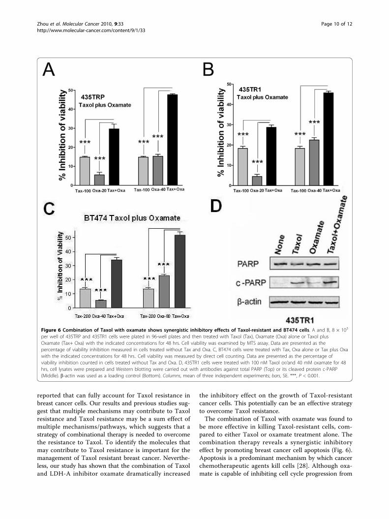
The combination of Taxol with oxamate shows synergisticinhibitory effect on breast cancer cellsOxamate is a pyruvate analog that directly inhibits theconverting process of pyruvate to lactate by LDH, there-fore, inhibits cell glycolysis [21]. We first examined theeffect of oxamate on LDH activity and cell viability ofMDA-435 and 435TR1 cells. Oxamate treatment led toa decrease of LDH activity (Fig. 5A) and an inhibition ofcell viability (Fig. 5B) in a dose-dependant manner, inboth MDA-435 and 435TR1 cells. Compared to MDA-435 cells, Taxol resistant 435TR1 cells showed a greatersensitivity to oxamate, consistent with the results ofLDH-A knockdown by siRNA (Fig. 3). Since glycolysisand mitochondrial oxidative phosphorylation are linkedprocesses [16], and we have previously shown thatLDH-A is critical in regulating glycolysis and growth ofbreast cancer cells [18], we reasoned that the increasedexpression and activity of LDH-A in Taxol-resistantcells may lead to an increase of glycolysis and a decreaseof mitochondrial oxidative phosphorylation. Thus, a spe-cific inhibitor of the mitochondrial oxidative phosphory-lation, oligomycin was utilized to treat these cells. Asexpected, Taxol-resistant 435TR1 cells were more resis-tant to oligomycin (Additional file 2, Figure. S2). Theseresults further support the notion that increased Taxolsensitivity by oxamate is a consequence of the inhibitionof cellular glycolysis.Since downregulation of LDH-A by siRNA or oxamatesignificantly inhibited the viability of the Taxol-resistantcells, we further investigated the effects of combiningTaxol with glycolysis inhibitor oxamate on Taxol-resis-tant breast cancer cells. In both Taxol-resistant 435TRPand 435TR1 cells (Fig. 6A and 6B; Additional file 3, Fig-ure. S3), and in BT474 cells (Fig. 6C), Taxol combinedwith oxamate were much more effective in inhibitingcell viability compared with either agent given alone.Similar treatment combinations were performed inanother breast cancer cell line, MCF7, with similarresults obtained (Additional file 4, Figure. S4). Takentogether, the combination of Taxol with oxamate has agreater capacity to inhibit Taxol-resistant cells comparedto either agent given alone.To further investigate the mechanism of oxamate-induced Taxol re-sensitization, we examined cellularapoptosis in these cells. PARP, a nuclear protein thatcan be easily cleaved by caspases, has been widely usedas an apoptosis marker [19,20]. The expression level oftotal PARP and cleaved PARP (c-PARP) were examinedin 435TR1 cells after treatment with Taxol, oxamate, ortheir combination for 48 hrs, respectively. We found asignificant increase of the levels of cleaved PARP after
Zhou et al. Molecular Cancer 2010, 9:33http://www.molecular-cancer.com/content/9/1/33
Page 5 of 12

treatment with the combination of Taxol and oxamatecompared to treatment with single agent (Fig. 6D). Thisindicates that cellular apoptosis is a mechanism involvedin the increased cell growth inhibitory effect of the com-bination treatment of Taxol with oxamate.
DiscussionIn this study, we investigated the role of LDH-A in theacquired Taxol resistance in multiple human breast can-cer cell lines. We identified that compared to Taxol-sen-sitive cells, Taxol-resistant cells possess an increasedexpression and activity of LDH-A, with its
downregulation resulting in an increased sensitivity ofTaxol resistant-cells to Taxol. In addition, compared toTaxol-sensitive cells, Taxol-resistant cells show a highersensitivity to the LDH inhibitor oxamate. Furthermore,when compared to single agent therapy, treating cellswith the combination of Taxol and oxamate show amuch stronger inhibitory effect on Taxol-resistant breastcancer cells by promoting cellular apoptosis. Theseresults demonstrate that LDH-A plays an important rolein Taxol resistance and potentially it can serve as a ther-apeutic target for overcoming Taxol resistance inpatients with breast cancer.
Figure 2 Increased LDH-A expression and activity in Taxol-resistant cells. A, Western blot was performed with an anti-LDH-A antibody oftotal cell extract from MDA-435, 435TR1 and 435TRP cells. The b-actin protein was used as a loading control. B, LDH activity in MDA-435, 435TR1and 435TRP were examined. C, MDA-435 cells were treated with increasing concentrations of Taxol for 24 hrs. The cell lysates were preparedand Western blotting was carried out with antibodies against to LDH-A and b-actin. D, LDHA protein stability assay was performed in MDA-435cells under the treatments of Taxol at 4 nM and CHX at 50 ug/ml followed by Western blotting assay to exam the protein expression level ofLDHA at 0 and 8 hrs (top). The relative intensity of LDHA band was normalized to its b-actin loading (bottom). E, LDHA mRNA level wasdetected by real-time PCR under 2 nM Taxol in MDA-435 cells. The LDHA primers used for PCR are: forward, 5’-tgg agt gga atg aat gtt gc-3’;reverse: 5’-ata gcc cag gat gtg tag cc-3’. Columns, mean of three independent experiments; bars, SE. ***, P < 0.001.
Zhou et al. Molecular Cancer 2010, 9:33http://www.molecular-cancer.com/content/9/1/33
Page 6 of 12

Taxol is a widely used chemotherapeutic agent for thetreatment of several types of cancers, including breastcancer. Taxol resistance may result in the subsequentrecurrence and metastasis of cancer, ultimately resultingin death. Although extensive investigations have beendone in regards to the resistance of cancer cells toTaxol, the specific mechanisms involved are still poorlyunderstood. Cancer cells are different from non-neoplas-tic cells in their metabolic properties, with normal cellsrelying primarily on the process of mitochondrial oxida-tive phosphorylation, consuming oxygen and glucose to
produce energy. In contrast, cancer cells depend mostlyupon glycolysis, the anaerobic breakdown of glucoseinto the energy-storing molecule ATP, even in the pre-sence of available oxygen [13-15,22,23]. Recently,research endeavors have been actively tried to make useof these unique bioenergetic properties to enhance thetherapeutic efficacy of killing cancer cells.LDH-A is one of the main isoforms of LDH expressed
in breast tissue, catalyzing the conversion of pyruvate tolactate [17]. We and others have previously shown thatLDH-A plays a critical role in glycolysis, growth
Figure 3 Knockdown of LDH-A increases the sensitivity of Taxol-resistant 435TR1 cells to Taxol. A, MDA-435 and 435TR1 cells weretransfected with scramble siRNA (Ctr) or LDH-A siRNA. 48 hrs after siRNA transfection, cell lysates were prepared and Western blotting wasperformed with antibodies against LDH-A. The b-actin protein was used as a loading control. B, LDH activity was examined from lysates of MDA-435 and 435TR1 48 hrs after siRNA transfection. C and D, 24 hrs after siRNA transfection, MDA-435 and 435TR1 cells were seeded into 96-wellplates at the density of 8 × 10³ cells per well, and treated with Taxol (5 nM and 10 nM for MDA-435, 50 nM and 100 nM for 435TR1) for 48 hrs.Then the cell viability was detected using a MTS reagent. Data are presented as the percentage of viability inhibition measured in cells treatedwithout Taxol. Columns, mean of three independent experiments; bars, SE. *, P < 0.05, **, P < 0.01, ***, P < 0.001.
Zhou et al. Molecular Cancer 2010, 9:33http://www.molecular-cancer.com/content/9/1/33
Page 7 of 12

properties and tumor maintenance of breast cancer cells[16,18]. Studies have shown that the LDH-A expressionin cancer cells is associated with radiosensitivity [24].LDH-A inhibition results in increased apoptosis via ROSproduction in cell with fumarate hydratase deficiencyand was viewed as a therapeutic strategy for treatmentof hereditary leiomyomatosis and renal cell cancer [25].However, the role of LDH in Taxol resistance of cancercell has not been explored. In this study, we selected apanel of Taxol-resistant cells by gradually increasing the
concentration of Taxol in the cell culture medium. Weused these, and other three breast cancer cell lines, tostudy the expression and activity of LDH-A in the devel-opment of Taxol resistance. To our knowledge, this isthe first report to provide direct evidence in support ofa role for LDH-A in acquired Taxol resistance inhuman breast cancer cells.We found that Taxol treatment resulted in the
increased LDH-A expression and activation in cancercells, which appears as a result of the induction of
Figure 4 Knockdown of LDH-A increases the sensitivity of breast cancer cell lines BT474 and MDA-231 to Taxol. A, BT474 cells weretransfected with scramble siRNA (Ctr) or si-LDHA. 48 hrs after siRNA transfection, cell lysates were prepared and immunoblot analyses werecarried out with antibodies against LDHA and b-actin. B, 48 hrs after siRNA transfection, cell lysates were prepared and LDHA activity wasexamined. Data are shown in percentage of LDH activity relative to Ctr-transfected cells. C, 48 hrs after siRNA transfection, cells were seeded into96-well plates at the density of 8 × 10³ cells per well. 12 hrs after incubation, the cells were then treated with various concentrations of Taxol for48 hrs. Then the cell viability was detected using a MTS reagent. Data are presented as the percentage of viability inhibition measured in cellstreated without Taxol. D, LDHA protein expression in MDA-MB-231 (Ctr) and stable LDHA knowdown MDA-MB-231 cells (sh-LDHA) weredetected to evaluate the efficiency of LDH-A knockdown by using an LDHA antibody. b-actin was used as a loading control. E, LDH activity wasexamined in MDA-MB-231 cells with and without stably knockdown of LDH-A. Data are shown in percentage of LDH activity relative to Ctr cells.F, MDA-MB-231 cells with or without stably knockdown of LDH-A were seeded into 96-well plates at the density of 8 × 10³ cells per well. 12 hrsafter incubation, the cells were treated with various concentrations of Taxol for 48 hrs. Then the cell viability was detected using a MTS reagent.Data are presented as the percentage of viability inhibition measured in cells treated without Taxol. Columns, mean of three independentexperiments; bars, SE.*, P < 0.05, **, P < 0.01, ***, P < 0.001.
Zhou et al. Molecular Cancer 2010, 9:33http://www.molecular-cancer.com/content/9/1/33
Page 8 of 12

LDH-A mRNA expression by Taxol. The downregula-tion of LDH-A by LDH-A siRNA and inhibition of LDHby oxamate led to increased sensitivity to Taxol in allthree breast cancer cell lines examined in this study.This indicated that Taxol treatment triggers a feedfor-ward cycle in which Taxol-induced activation of LDHresults in cancer cells better survival under Taxol treat-ment, likely through promoting cell glycolysis. A recentstudy has shown that cancer cells inhibit cytochrome c-mediated apoptosis by a mechanism through deregu-lated glucose metabolism [26]. Thus, the Taxol-inducedhigh expression and activity of LDH-A detected inTaxol-resistant cells could be a way of adaptation ofthese cells to Taxol treatment and to modulate glucosemetabolism and glycolysis to avoid apoptosis induced byTaxol. Targeting LDH by LDH siRNA or LDH inhibitoroxamate interrupts the feedforward cycle and rendersthe re-sensitization to Taxol. These results indicate thatLDH may potentially serve as an excellent target forovercoming Taxol resistance in human breast cancerpatients.Up-regulation of antiapoptotic Bcl-2 family members,
such as Bcl-2 and Bcl-XL, was reported to contribute toTaxol-induced apoptosis [27]. In addition, we previously
reported that the phosphorylation on tyrosine-15 ofCdc2 by ErbB2 in breast cancer cells resulting a delayedM phase entry and leading to an increased Taxol resis-tance [11]. We found that compared to the parentalMDA435 cells, Taxol-resistant MDA435TR1 andMDA435TRP cells express lower Bcl-2 and lower phos-phorylation level of Cdc2 at tyrosine-15 (Additional file5, Figure. S5). Based on the known functions of Bcl2and Y15-Cdc2 in Taxol resistance, these results can notexplain the increased resistance in MDA435TR1 andMDA435TRP cells. However, we found that Bcl-XL wasupregulated in Taxol-resistant cells (Additional file 5,Figure. S5). This might be another reason in addition toLDH-A for the increased Taxol resistance in these cells.It will be interesting to examine the relationshipbetween LDH-A and Bcl-XL in these cells in our futurestudies.The differences in cytotoxicity were some what mod-
est when the LDH-A were knocked down by siRNA.One of the reasons might be the relatively low sensitiv-ity of MTS assay to detect cell toxicity in our experi-ments. Another possible reason might be the relativelylow knocking down efficiency of LDH-A by the siRNA.In addition, as far, there is no any single molecule
Figure 5 Taxol-resistant cells are more sensitive to glycolysis inhibitor oxamate. A, MDA-435 and 435TR1 cells were treated with variousconcentrations of oxamate for 48 hrs, then LDH activity was detected. Data are shown as the percentage of LDH activity inhibition detected incells treated without oxamate. B, MDA-435 and 435TR1 were treated with various concentrations of oxamate for 48 hrs. Then the cell viabilitywas detected using a MTS reagent. Data are presented as the percentage of viability inhibition measured in cells treated without oxamate.Columns, mean of three independent experiments; bars, SE. *, P < 0.05. **, P < 0.01.
Zhou et al. Molecular Cancer 2010, 9:33http://www.molecular-cancer.com/content/9/1/33
Page 9 of 12
reported that can fully account for Taxol resistance inbreast cancer cells. Our results and previous studies sug-gest that multiple mechanisms may contribute to Taxolresistance and Taxol resistance may be a sum effect ofmultiple mechanisms/pathways, which suggests that astrategy of combinational therapy is needed to overcomethe resistance to Taxol. To identify the molecules thatmay contribute to Taxol resistance is important for themanagement of Taxol resistant breast cancer. Neverthe-less, our study has shown that the combination of Taxoland LDH-A inhibitor oxamate dramatically increased
the inhibitory effect on the growth of Taxol-resistantcancer cells. This potentially can be an effective strategyto overcome Taxol resistance.The combination of Taxol with oxamate was found to
be more effective in killing Taxol-resistant cells, com-pared to either Taxol or oxamate treatment alone. Thecombination therapy reveals a synergistic inhibitoryeffect by promoting breast cancer cell apoptosis (Fig. 6).Apoptosis is a predominant mechanism by which cancerchemotherapeutic agents kill cells [28]. Although oxa-mate is capable of inhibiting cell cycle progression from
Figure 6 Combination of Taxol with oxamate shows synergistic inhibitory effects of Taxol-resistant and BT474 cells. A and B, 8 × 10³
per well of 435TRP and 435TR1 cells were plated in 96-well plates and then treated with Taxol (Tax), Oxamate (Oxa) alone or Taxol plusOxamate (Tax+ Oxa) with the indicated concentrations for 48 hrs. Cell viability was examined by MTS assay. Data are presented as thepercentage of viability inhibition measured in cells treated without Tax and Oxa. C, BT474 cells were treated with Tax, Oxa alone or Tax plus Oxawith the indicated concentrations for 48 hrs.. Cell viability was measured by direct cell counting. Data are presented as the percentage ofviability inhibition counted in cells treated without Tax and Oxa. D, 435TR1 cells were treated with 100 nM Taxol or/and 40 mM oxamate for 48hrs, cell lysates were prepared and Western blotting were carried out with antibodies against total PARP (Top) or its cleaved protein c-PARP(Middle). b-actin was used as a loading control (Bottom). Columns, mean of three independent experiments; bars, SE. ***, P < 0.001.
Zhou et al. Molecular Cancer 2010, 9:33http://www.molecular-cancer.com/content/9/1/33
Page 10 of 12

G2 to M phase [29], we report here a novel function viainducing apoptotic cell death, with important implica-tions in the clinical treatment of Taxol-resistant cancers,such as breast cancer.The origin of MDA-MB-435 cells has recently been
called into question [30,31]. However, a latest literatureindicated that current stocks of both MDA-MB-435cells and M14 melanoma cells are in fact MDA-MB-435breast cancer cells instead of M14 melanoma cell line[32]. Nevertheless, we also examined three more breastcancer cell lines, ErbB2-overexpressing BT474 andErbB2-low-expressing MDA-231 and MCF-7, in orderto confirm our findings from MDA-MB-435 cells.In summary, the present study reveals that LDH-A
plays an important role in Taxol-resistance, with Taxol-induced expression and activity of LDH-A serving as animportant mechanism for the acquired resistance ofhuman breast cancer cells to Taxol. This study providesvaluable information for the development of targetedtherapies capable of inhibiting key targets, such as LDH-A. Further studies are needed to demonstrate whetherthe downregulation of LDH-A mediated re-sensitizationof breast cancer cells to Taxol is indeed a consequenceof inhibition of glycolysis. Another question arises as towhether the downregulation of other key molecules inthe glycolytic pathway may have the same effect as thedownregulation of LDH-A. In conclusion, the results ofour study highlight the importance of LDH-A in its rolein Taxol-resistance and open the door for possible ther-apeutic interventions in patients that have developed aresistance to Taxol.
ConclusionLDH-A plays an important role in Taxol resistance andinhibition of LDH-A re-sensitizes Taxol-resistant cellsto Taxol. This study provides valuable information forthe future development and use of targeted therapies,such as oxamate, for the treatment of patients withTaxol-resistant breast cancer.
List of abbreviationsLDH-A: Lactate dehydrogenase-A; DMEM: Dulbecco’smodified Eagle medium; c-PARP: the cleaved Poly(ADP-ribose) polymerase; CHX: Cycloheximide.
Additional file 1: Figure S1. Knockdown of LDH-A re-sensitizes 435TR1cell to Taxol by direct cell counting. 435TR1 cells were transfected withscramble siRNA (Ctr) or si-LDHA. 24 hrs after siRNA transfection, cellswere treated with 50 nM or 100 nM Taxol for 48 hrs. Cell numbers weredirectly counted by Typan Blue Staining. Data are presented as thepercentage of viability inhibition counted in cells treated without Taxol.Columns, mean of three independent experiments; bars, SE.*, P < 0.05, **,P < 0.01. si-LDHA transfection efficiency was showed on the right panel.Click here for file[ http://www.biomedcentral.com/content/supplementary/1476-4598-9-33-S1.PDF ]
Additional file 2: Figure S2. Taxol-resistant cells are more resistantto mitochondrial oxidative phosphorylation inhibitor oligomycin.MDA-435 and 435TR1 cells were seeded into 96-well plate at density of 5× 10³ cells per well. 12 hrs after incubation; cells were treated withvarious concentrations of oligomycin for 24 hrs. Then the cell viabilitywas detected using a MTS reagent, and data are presented as thepercentage of viability inhibition measured in cells treated withoutoligomycin. Columns, mean of three independent experiments; bars, SE.***, P < 0.001.Click here for file[ http://www.biomedcentral.com/content/supplementary/1476-4598-9-33-S2.PDF ]
Additional file 3: Figure S3. Combination of Taxol with oxamate showssynergistic inhibitory effects in Taxol-resistant cells by direct cellcounting. 435TR1 and 435TRP cells were seeded in 24-well plates andtreated with Tax, Oxa alone or Tax plus Oxa with the indicatedconcentrations for 48 hrs. Cell numbers were counted by Typan BlueStaining. Data are presented as the percentage of viability inhibitioncounted in cells treated without Tax and Oxa. Columns, mean of threeindependent experiments; bars, SE. *, P < 0.05. **, P < 0.01. ***, P < 0.001.Click here for file[ http://www.biomedcentral.com/content/supplementary/1476-4598-9-33-S3.PDF ]
Additional file 4: Figure S4. Combination of Taxol with oxamate showsbetter inhibition of MCF7 cells. A, 1 × 10⁴ per well of MCF7 cells wereplated into 96-well plate and treated with Taxol, Oxa, or Tax plus Oxawith indicated concentrations for 48 hrs. Cell viability was examined byMTS assay. Data are presented as the percentage of viability inhibitionmeasured in cells treated without Tax and Oxa. Columns, mean of threeindependent experiments; bars, SE.*, P < 0.05, **, P < 0.01. B, MCF7 cellswere treated with 10 nM Taxol or/and 16 mM oxamate for 48 hrs andcell lysates were prepared for Western blotting using antibodies againsttotal PARP (Top) or its cleaved protein c-PARP (Middle). b-actin was usedas a loading control (Bottom).Click here for file[ http://www.biomedcentral.com/content/supplementary/1476-4598-9-33-S4.PDF ]
Additional file 5: Figure S5. The expression of Bcl-2, Bcl-XL, Cdc2 andphosphorylation status of Cdc2 at Tyrosine 15. MDA-435, 435TR1 and TRPcells were collected, lysed and immunoblot analyses were carried outwith antibodies against Bcl-2, Bcl-XL, Cdc2 and p-Cdc2-Y15 and tubulin.Click here for file[ http://www.biomedcentral.com/content/supplementary/1476-4598-9-33-S5.PDF ]
AcknowledgementsMing Tan is a Vincent F. Kilborn, Jr. Cancer Research Scholar. We thank Dr.Eddie Reed from USA Mitchell Cancer Institute for critically reading themanuscript and Ms. Amy Brown for the editorial assistance. We are gratefulto the support from The Vincent F. Kilborn, Jr. Cancer Research Foundation(M. Tan) and The Norwegian Radiumhospitalet Legater (Project 334003, M.Tan. and O. Fodstad).
Author details1Mitchell Cancer Institute, University of South Alabama, Mobile, Alabama,USA. 2Department of Cell Biology and Neuroscience, University of SouthAlabama, Mobile, Alabama, USA. 3Cancer Research Institute, Central SouthUniversity, Changsha, China. 4Institute for Cancer Research, The NorwegianRadium Hospital, University of Oslo, Norway. 5Department of Biochemistryand Molecular Biology, University of Florida, Gainesville, Florida, USA.6Ochsner Cancer Institute, Ochsner Health System, New Orleans, Louisiana,USA.
Authors’ contributionsMZ designed and carried out the majority of the experiments and draftedthe manuscript. YZ, YD, HL, ZL involved in experimental design and carriedout some experiments, and helped to revise the manuscript. SK and JL
Zhou et al. Molecular Cancer 2010, 9:33http://www.molecular-cancer.com/content/9/1/33
Page 11 of 12

contributed the key reagents. OF, AR, LO, SL helped to revise themanuscript. MT conceived the study and supervised the overall experimentaldesign, execution and revised the manuscript. All authors read andapproved the final manuscript.
Competing interestsThe authors declare that they have no competing interests.
Received: 15 August 2009Accepted: 9 February 2010 Published: 9 February 2010
References1. Henley D, Isbill1 M, Fernando1 R, Foster1 JS, Wimalasena1 J: Paclitaxel
induced apoptosis in breast cancer cells requires cell cycle transit butnot Cdc2 activity. Cancer Chemother Pharmacol 2007, 59(2):235-249.
2. Tan M, Yu D: Molecular mechanisms of erbB2-mediated breast cancerchemoresistance. Adv Exp Med Biol 2007, 608:119-129.
3. Frankel Andrea, Buckman Robert, Kerbel SRobert: Abrogation of Taxol-induced G2-M Arrest and Apoptosis in Human Ovarian Cancer CellsGrown as Multicellular Tumor Spheroids. Cancer Res 1997, 57:2388-2293.
4. Chen LP, Cai SM, Fan JX, Li ZT: PEBA Regimen (Cisplatin, Etoposide,Bleomycin, and Adriamycin) in the Treatment of Drug-ResistantChoriocarcinoma. Gynecologic Oncology 2002, 56(2):231-234.
5. Donnenberg SVera, Donnenberg DAlbert: Multiple Drug Resistance inCancer Revisited: The Cancer Stem Cell Hypothesis. J Clin Pharmacol2005, 45:872-877.
6. Gottesman MM, Pastan I: Biochemistry of multidrug resistance mediatedby the multidrug transporter. Annu Rev Biochem 1993, 62:385-427.
7. Kavallaris M, Kuo DY, Burkhart CA, Regl DL, Norris MD, Haber M, Horwitz SB:Taxol-resistant epithelial ovarian tumors are associated with alteredexpression of specific beta-tubulin isotypes. J Clin Invest 1997,100(5):1282-1293.
8. Panda D, Miller HP, Banerjee A, Ludueña RF, Wilson L: Microtubuledynamics in vitro are regulated by the tubulin isotype composition. ProcNatl Acad Sci USA 1994, 91(24):11358-11362.
9. Martello LA, Verdier-Pinard P, Shen HJ, He L, Torres K, Orr GA, Horwitz SB:Elevated levels of microtubule destabilizing factors in a Taxol-resistant/dependent A549 cell line with an alpha-tubulin mutation. Cancer Res2003, 63(6):1207-1213.
10. Gonçalves A, Braguer D, Kamath K, Martello L, Briand C, Horwitz S, Wilson L,Jordan MA: Resistance to Taxol in lung cancer cells associated withincreased microtubule dynamics. Proc Natl Acad Sci 2001, 98:11737-11741.
11. Tan M, Jing T, Lan KH, Neal CL, Li P, Lee S, Fang D, Nagata Y, Liu J,Arlinghaus R, Hung MC, Yu D: Phosphorylation on tyrosine-15 of p34(Cdc2) by ErbB2 inhibits p34(Cdc2) activation and is involved inresistance to taxol-induced apoptosis. Mol Cell 2002, 9:993-1004.
12. Lu J, Tan M, Huang WC, Li P, Guo H, Tseng LM, Su XH, Yang WT,Treekitkarnmongkol W, Andreeff M, Symmans F, Yu D: Mitotic deregulationby survivin in ErbB2-overexpressing breast cancer cells contributes toTaxol resistance. Clin Cancer Res 2009, 15(4):1326-1334.
13. Warburg O: On respiratory impairment in cancer cells. Science 1956,124:269-270.
14. Kim JW, Dang CV: Cancer’s molecular sweet tooth and the Warburgeffect. Cancer Res 2006, 66:8927-8930.
15. Chen Z, Lu W, Garcia-Prieto C, Huang P: The Warburg effect and itscancer therapeutic implications. J Bioenerg Biomembr 2007, 39(3):267-274.
16. Fantin VR, St-Pierre J, Leder P: Attenuation of LDH-A expression uncoversa link between glycolysis, mitochondrial physiology, and tumormaintenance. Cancer Cell 2006, 9:425-434.
17. Dang CV, Semenza GL: Oncogenic alterations of metabolism. TrendsBiochem Sci 1999, 24:68-72.
18. Zhao YH, Zhou M, Liu H, Ding Y, Khong HT, Yu DH, Fodstad O, Tan M:Upregulation of lactate dehydrogenase A by ErbB2 through heat shockfactor 1 promotes breast cancer cell glycolysis and growth. Oncogene2009, 28:3689-3701.
19. Kaufmann SH, Desnoyers S, Ottaviano Y, Davidson NE, Poirier GG: Specificproteolytic cleavage of poly(ADP-ribose) polymerase: an early marker ofchemotherapy-induced apoptosis. Cancer Res 1993, 53(17):3976-3985.
20. Boulares AH, Yakovlev AG, Ivanova V, Stoica BA, Wang G, Iyer S, Smulson M:Role of poly(ADP-ribose) polymerase (PARP) cleavage in apoptosis.
Caspase 3-resistant PARP mutant increases rates of apoptosis intransfected cells. J Biol Chem 1999, 274(33):22932-22940.
21. Chang GG, Huang SM, Chiou SH: Kinetic mechanism of the endogenouslactate dehydrogenase activity of duck epsilon-crystallin. Arch BiochemBiophys 1991, 284(2):285-291.
22. DeBerardinis RJ, Lum JJ, Hatzivassiliou G, Thompson CB: The biology ofcancer: metabolic reprogramming fuels cell growth and proliferation.Cell Metab 2008, 7:11-20.
23. Kroemer G, Pouyssegur J: Tumor cell metabolism: cancer’s Achilles’ heel.Cancer Cell 2008, 13:472-482.
24. Gorlach A, Acker H: pO2- and pH-gradients in multicellular spheroids andtheir relationship to cellular metabolism and radiation sensitivity ofmalignant human tumor cells. Biochim Biophys Acta 1994, 1227:105-112.
25. Xie H, Valera VA, Merino MJ, Amato AM, Signoretti S, Linehan WM,Sukhatme VP, Seth P: LDH-A inhibition, a therapeutic strategy fortreatment of hereditary leiomyomatosis and renal cell cancer. MolecularCancer Therapeutics 2009, 8(3):626-635.
26. Vaughn1 EAllyson, Deshmukh Mohanish: Glucose metabolism inhibitsapoptosis in neurons and cancer cells by redox inactivation ofcytochrome c. Nat Cell Biol 2008, 10(12):1477-1483.
27. Basu A, DuBois G, Haldar S: Posttranslational modifications of Bcl2 familymembers–a potential therapeutic target for human malignancy. FrontBiosci 2006, 11:1508-1521.
28. Fisher DE: Apoptosis in cancer therapy: crossing the threshold. Cell 1994,78:539-42.
29. Thornburg JM, Nelson KK, Clem BF, Lane AN, Arumugam S, Simmons A,Eaton JW, Telang S, Chesney J: Targeting aspartate aminotransferase inbreast cancer. Breast Cancer Res 2008, 10(5):R84.
30. Sellappan S, Grijalva R, Zhou X, Yang W, Eli MB, Mills GB, Yu D: Lineageinfidelity of MDA-MB-435 cells: expression of melanocyte proteins in abreast cancer cell line. Cancer Res 2004, 64:3479-3485.
31. Welch DR: Technical considerations for studying cancer metastasis invivo. Clin Exp Metastasis 1997, 15:272-306.
32. Chambers AF: MDA-MB-435 and M14 cell lines: identical but not M14melanoma?. Cancer Res 2009, 69(13):5292-5293.
doi:10.1186/1476-4598-9-33Cite this article as: Zhou et al.: Warburg effect in chemosensitivity:Targeting lactate dehydrogenase-A re-sensitizesTaxol-resistant cancer cells to Taxol. Molecular Cancer 2010 9:33.
Submit your next manuscript to BioMed Centraland take full advantage of:
• Convenient online submission
• Thorough peer review
• No space constraints or color figure charges
• Immediate publication on acceptance
• Inclusion in PubMed, CAS, Scopus and Google Scholar
• Research which is freely available for redistribution
Submit your manuscript at www.biomedcentral.com/submit
Zhou et al. Molecular Cancer 2010, 9:33http://www.molecular-cancer.com/content/9/1/33
Page 12 of 12