In Silico inhibitors for Plasmodium falciparum lactate dehydrogenase

Theoretical site-directed mutagenesis. The
Asp168Ala mutant of L-Lactate Dehydrogenase
Silvia Ferrer‡, Estanislao Silla‡,§, Iñaki Tuñón‡*,Vicent Moliner§* and Ian H. Williams#
‡Departamento de Química Física; Universidad de Valencia, 46100 Burjassot,
Valencia,,Spain.
§Departament de Ciències Experimentals, Universitat Jaume I, 12071 Castelló, Spain.
#Department of Chemistry, University of Bath, BA2 7AY Bath, United Kingdom.
AUTHOR EMAIL ADDRESS: [email protected], [email protected]
ABSTRACT: The reduction of pyruvate to lactate catalyzed by the L-Lactate
dehydrogenase has been studied in this paper by means of hybrid Quantum Mechanical
/ Molecular Mechanical simulations. A very flexible molecular model consisting on the
full tetramer of the enzyme, together with the cofactor NADH, the substrate and solvent
water molecules has allowed to theoretically mimic site directed mutagenesis studies,
most of them previously experimentally performed. The potential energy surfaces
obtained for every single mutation, compared with the one corresponding to the native
enzyme, have been used to trace the possible reaction pathways and to locate and
characterize the structures corresponding to the stationary points. The analysis of the
results has been a very powerful tool to conclude about the role of key residues on the
vacuole formed in the active site of the enzyme. Our results are in very good agreement
with the previous conclusions derived from site directed mutagenesis. This strategy can
be extrapolated to other enzyme systems thus opening the door of a very promising
methodology that, in combination with the appropriate experimental technique, will
enable us to describe enzyme catalysis phenomenon and the particular role of the
residues that form the protein. This knowledge placed us in a privileged position to
modify the activity of enzymes and to propose efficient inhibitors.

INTRODUCTION
L-Lactate dehydrogenase (LDH) is a highly stereospecific metabolic enzyme which
catalyses the interconversion of pyruvate and L-lactate using the NADH/NAD+ pair as
redox cofactor. Enzymes like LDH, involved in metabolic pathways, have been the
subject of extensive research both experimentally and theoretically: while their
properties are now relatively well-known, details of their reaction mechanisms are still
not completely known.1
The chemical step of LDH, in the pyruvate to L-lactate direction (see Scheme I),
involves a hydride transfer from the dihydronicotinamide ring of NADH to the carbonyl
C atom of pyruvate and a proton transfer to the carbonyl O atom from a protonated
histidine residue (the His195 residue).2
Scheme I
The whole catalytic process goes through several stages: substrate binding,
unimolecular rearrangement consisting on a closure of a surface loop (99-110) down
over the active site, pyruvate reduction, reverse protein conformational change and
products release.1 As shown by Dunn et al.3 and by Waldman et al.,4 closing of the 99-
110 loop enforces isolation of the active site center from bulk solvent which is
necessary for the reaction to proceed at a noticeable rate. In the wild-type enzyme this
conformational rearrangement is in fact the rate limiting step.3
During the last decades, the advent of protein engineering has enabled us to define the
role of specific residues on the active site by substituting them by other aminoacids
presenting differences in charge or/and size.5-10 Site-directed mutagenesis has been
proved to be a good tool not only to change the rate limiting step of the full process, but
also to demonstrate the feasibility of applying rational protein engineering to optimize
and control the stereoselectivity of this particular enzyme when used in asymmetric
synthesis.10
NAD-H C
OH-His195+
NAD+ CH
OH
His195

In this sense, one of the most relevant experiments on the ionised residues that are
part of the vacuole formed by the 99-110 loop was that carried out by Holbrook and
Ingram,5 who prepared one of the first variants of LDH by site-directed mutagenesis, in
which the histidine at position 195 was removed, thus demonstrating its role as the
proton donor/acceptor in the reaction. The influence of Asp168 on the reaction was
evaluated by substituting the residue to Asn and Ala.6 This study supported the
hypothesis that the Asp168 contributes to catalytic efficiency by raising the pKa of
His195 thus favouring the double protonation state of this histidine and by anchoring it
to the most favourable orientation for enhancing its acid/base catalysis proton donating
and accepting properties. It has been shown that LDHs cannot form the enzyme-
cofactor-substrate ternary complex unless the imidazole group of His195 is protonated.
The effect of replacing the negative charged Asp168 residue by neutral ones is to
destabilize this ternary complex and, in contrast to the wild-type enzyme, the rate of
catalysis becomes limited by the chemical step.6 Another important mutation was the
Arg171Lys substitution that affected the catalytic properties of the enzyme by
weakening the substrate binding.7 The Arg109Gln mutation demonstrated the role of
this arginine in polarizing the pyruvate carbonyl group in the ground state and
stabilizing the transition state.8 This polarisation on the carbon-oxygen bond implies the
development of a positive charge on the C2 thus facilitating the hydride transfer.
Holbrook and co-workers studied the importance of the electrostatic charge balance in
the active site vacuole of NAD+-L-Lactate dehydrogenase by introducing an aspartic
acid at position 140 instead of the wild-type asparagine.9 They demonstrated that
Asn140Asp mutant enzyme has the same kcat as the native one at low pH, but at higher
pH the Km of the pyruvate increases. Experimental results and molecular modelling
showed that the presence of an anion on the position of the Asn140 raises the pKa of
His195 to greater than 10, thus facilitating the opening of the loop and the entry of
solvent molecules to the vacuole.9
Recent mutations on other residues as Ala245Lys resulted in a qualitative change in
the reaction mechanism by increasing the rigidity of the loop, thus provoking a relative
acceleration of the rearrangement of the enzyme-NADH-pyruvate complex making the
hydride transfer as the first slow step.1
The chemical reaction catalyzed by the LDH has been also extensively studied by
several theoretical methods, either in gas phase 11, 12 and more recently by means of
hybrid quantum mechanical / molecular mechanics methods (QM/MM).13-17
Nevertheless, although the sequential mechanism is more likely, different computational
groups’ proposals differ in the order in which the proton and hydride ions are
transferred. Thus, some found a mechanism in which the hydride transfer preceded
proton transfer in a stepwise manner,12, 13 while other studies 14, 15, 18 predicted a
concerted but asynchronous reaction in which the proton transfer was in a very
advanced stage of the reaction. A very flexible QM/MM treatment 17 found a
megadimensional energy hypersurface that allowed tracing across the two distinct
mechanism pathways. The difference in the timing of the hydride transfer and the
proton transfer is a matter of interest in mechanistic enzymology but, as it was pointed
out in this latter study, unfortunately the results at this point did not allow to make a
definitive statement as to which mechanism is preferred. Finally, we have recently
demonstrated the dependence of the chemical reaction mechanism of LDH on the
protonation state of the ionisable residues of the protein and on the level of theory
employed to describe the quantum region on a QM/MM.19 Corrections at MP2/6-
31G(d,p) level of the semiempirical AM1 description QM region rendered a PES that
describes an asynchronous concerted reaction pathway in which HT precedes PT with a
remarkable reduction on the effective enthalpy barrier, much more in accordance with
the expected value for this biological reaction.19
In this paper we carry out an exhaustive analysis of the LDH catalyzed reaction by
obtaining different potential energy surfaces of the native LDH as well as the
Asp168Ala mutant. Our results are compared with experimental studies already carried
out in this system in order to test our methodology and to explain the predicted role of
the ionized residues presented in the active site of the enzyme. Finally, we suggest new
mutations that could be experimentally carried out which would definitely corroborate
the proposed mechanism and the particular role of key residues on the enzyme active
site.
COMPUTATIONAL METHODS
The QM/MM energy hypersurfaces were obtained using the CHARMM29b1
program.20 The starting geometry comes from the 2.5 Å resolution ternary complex X-
ray crystal structure of the LDH tetramer from Bacillus stearothermophilus with NADH
cofactor and oxamate inhibitor.21 Once the oxamate was replaced by pyruvate,

CHARMM29b1 was also used to add hydrogens to all titratable residues at a state
complementary to pH 7. Due to the fact that standard pKa values of ionisable groups can
be shifted by local protein environments,19 an accurate assignment of the protonation
states of all these residues was carried out by recalculating the standard pKa values of
the aminoacids using the “cluster method” 22, 23 as implemented by Field and co-
workers.24 According to this method, each titratable residue in the protein is perturbed
by the electrostatic effect of the protein environment. As demonstrated in our previous
study on LDH,19 a non-accurate protonation state of only a few residues of the protein
can render erroneous PESs and thus erroneous reaction mechanisms or/and activation
barriers. After having properly added the hydrogen atoms, the system was placed in a
cavity deleted from a pre-formed 24 Å radius spheres of TIP3P 25 water molecules
centred on the acceptor carbon and acceptor oxygen atoms. Water molecules within 2.5
Ǻ of any non-hydrogen atoms of the protein, cofactor or substrate were removed. This
procedure was repeated 3 times by randomly rotating the water spheres to solvate
potential cavities of a single water configuration. Then after a short molecular dynamic
simulation of 5 ps to relax unfavourable contacts, the solvation process was repeated to
fill in additional cavities generated during the equilibration dynamics.18, 26 Once the
entire system was built, those atoms 24 Å away from the centre of carbonyl C and O
atoms of the pyruvate were kept frozen during the subsequent dynamics and
optimizations. The resulting model was a pseudo-sphere of 22141 atoms of enzyme, co-
factor, substrate and solvent, 15154 of them kept frozen. The entire chemical system
was divided into a QM region, treated by the AM1 semiempirical MO method,27 and a
MM region comprising the rest of the protein (CHARMM24 potentials) and the water
molecules. Generalized hybrid orbital (GHO)28, 29 method has been used to treat the
covalent bonds crossing the boundary between the QM and the MM regions, in order to
satisfy the valence of the QM fragments. The QM region was formed by the
dihydronicotinamide and the ribose ring of the NADH, the pyruvate, Arginines 109 and
171 and the His195. A picture of the active site showing the partition in the QM and
MM subsystems is presented in Figure 1, where the QM region is shown as the shaded
portion.

Figure 1. Schematic covalent structure for cofactor (NADH) substrate (pyruvate) and
some key residues of the active site. GHO are indicated as " • ", dividing the quantum
region (shaded area) and the classical region
The mutation was carried out by substituting the original residue of the native
structure by the new residue. Afterwards, the new tetramer was solvated by water
molecules following the same simulation protocol as in the native LDH described
above. Once the full system was relaxed, the same conditions were applied to all
mutated and native LDH systems, slightly varying the number of MM atoms from one
model to other, depending on the aminoacid substitution.
The PESs were obtained using two antisymmetric combinations of the distances
between the hydrogen atoms with their donor (rDH) and acceptor (rAH) atoms. The
reaction coordinate R1 for the hydride transfer is defined as the difference in the
distances of the bonds between the transferring hydride-ion and the donor (Cnic) and the
acceptor atoms (Cpyr, see Equation 1). For the proton transfer a reaction coordinate R2 is
defined as the difference in the distance of the bonds between the transferring hydrogen
atom and the donor (Nhis) and the acceptor (Opyr) atoms (Equation 2)
H3C
O
O
O
NHNH
Asp168C
O
O
171Arg
CH
H 2N
NH2 Arg109
CHH2N
NH2
Glu199C
OH
O
Asn140
C
NH2
OO
H
H
N
H
CONH2
O
OHOH
HHHH
H
His195
NADH
H3C
O
O
O
NHNH
Asp168C
O
O
171Arg
CH
H 2N
NH2 Arg109
CHH2N
NH2
Glu199C
OH
O
Asn140
C
NH2
OO
H
H
N
H
CONH2
O
OHOH
HHHH
H
His195
NADH

1pyr1nic HCHC1 rrR −= Equation 1
2pyr2his HOHN2 rrR −= Equation 2
In order to correct for the known deficiencies of the AM1 hamiltonian when
describing hydride and proton transfer we added MP2/6-31G(d,p) corrections as a 2-D
spline function of R1 and R2 coordinates.19
The CHARMM29b1 program was employed to carry out the grid scanning. Once the
PESs were obtained, localised approximate transition state (TS) structures were refined
using the recently modified version of the GRACE software.16 A partial-rational-
function-operator/adopted-basis-Newton-Raphson method was employed, utilising a
Hessian matrix of order 156 x 156, describing the curvature of the QM/MM energy
hypersurface for a subset of the system (the QM atoms), together with a diagonal
Hessian plus updates for the rest of the system. The r.m.s. residual gradient on the 52
atoms in the subset was less than 0.1 kcal/mol Å-1 in the optimised structure, while on
the remaining atoms (ca. 3000) it was less than 0.005 kcal/mol Å-1. Finally, the intrinsic
reaction coordinate (IRC)30 path was traced from the TSs in order to demonstrate
conclusively that the reported structure was indeed the TS for the correct chemical step.
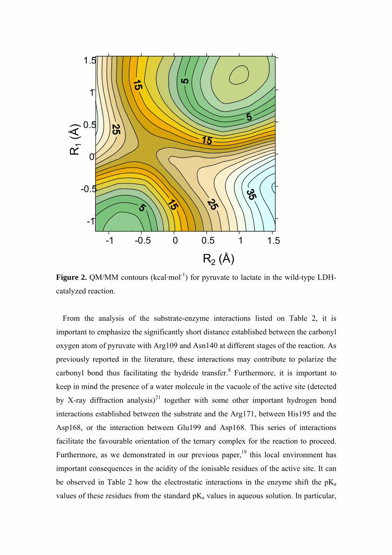
RESULTS AND DISCUSSION
Wild-type
The PES obtained for the interconversion of pyruvate into lactate in the active site of
the wild-type LDH enzyme is depicted on Figure 2, the energies for minima and saddle
points and selected QM/MM optimized interatomic distances of the different stationary
structures located on the PES are listed in Table 1.
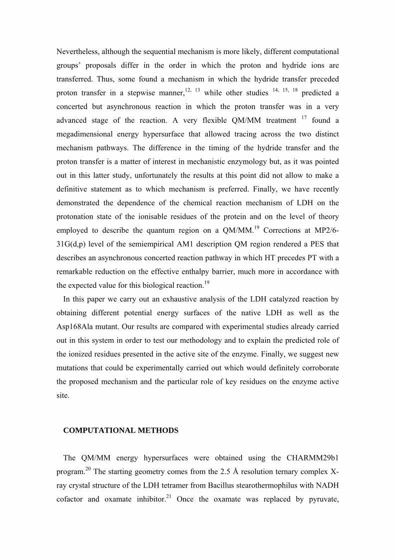
Table 1: Selected geometrical parameters of the different stationary point structures
on the QM/MM PES of the native type LDH and the mutant and potential energy
barriers (in kcal∙mol‐1).
Native LDH Asp168Ala
Pyruvate TS Lactate Pyruvate TS Lactate
Cnic∙∙∙∙H1 1.13 1.43 2.35 1.13 1.37 2.42
H1∙∙∙∙Cpyr 2.33 1.31 1.13 2.45 1.39 1.13
NHis195∙∙∙∙H2 1.05 1.09 1.93 1.05 1.13 2.20
H2∙∙∙∙Opyr 1.78 1.57 0.99 1.78 1.49 0.97
Opyr‐HHArg109 2.13 2.08 2.15 2.13 2.06 2.10
Opyr‐HDAsn140 2.11 2.01 3.00 2.85 2.56 3.13
Opyr‐NE2His195 2.72 2.61 2.77 2.75 2.58 3.00
ΔE� 21 24
Figure 2 clearly corresponds to a PES with a concerted but asynchronous reaction
pathway. In the TS the hydride transfer is considerably more advanced than the proton
transfer, this is to say that the hydride transfer precedes the proton transfer. Selected
structures of this PES were used as starting points to optimize and characterize the
stationary structures (reactants, transition structure and products). The relative total
energies are provided in Table 1, resulting in a potential energy barrier of 21 kcal mol-
1for the reaction.
R
TS
P
-1 -0.5 0 0.5 1 1.5
-1
-0.5
0
0.5
1
1.5
R2 (Å)
R1
(Å)
Figure 2. QM/MM contours (kcal·mol-1) for pyruvate to lactate in the wild-type LDH-
catalyzed reaction.
From the analysis of the substrate-enzyme interactions listed on Table 2, it is
important to emphasize the significantly short distance established between the carbonyl
oxygen atom of pyruvate with Arg109 and Asn140 at different stages of the reaction. As
previously reported in the literature, these interactions may contribute to polarize the
carbonyl bond thus facilitating the hydride transfer.8 Furthermore, it is important to
keep in mind the presence of a water molecule in the vacuole of the active site (detected
by X-ray diffraction analysis)21 together with some other important hydrogen bond
interactions established between the substrate and the Arg171, between His195 and the
Asp168, or the interaction between Glu199 and Asp168. This series of interactions
facilitate the favourable orientation of the ternary complex for the reaction to proceed.
Furthermore, as we demonstrated in our previous paper,19 this local environment has
important consequences in the acidity of the ionisable residues of the active site. It can
be observed in Table 2 how the electrostatic interactions in the enzyme shift the pKa
values of these residues from the standard pKa values in aqueous solution. In particular,

it is clear that the protein environment raises the pKa of some residues with respect to
aqueous solution (Glu199, His195 or Arg109), demonstrating they must be protonated
in the enzyme at pH 7, which favours the catalysis, and diminishes the value of Asp168,
proving the deprotonated state of this residue which, as previously showed, favours the
protonation state of His195.
As mentioned above, a deeper insight in the role of the different aminoacids of the
active site of the enzyme can be obtained by the analysis of the effect of different
mutations on the topology of the PES of the wild type enzyme. Changes in pKa values,
in the reaction mechanisms, structures and barrier heights are used to define the
catalytic role of the different residues. The results of the different simulations are
discussed in turn.
Table 3: pKa values of the different ionisable aminoacids in aqueous solution and calculated for the native type LDH and the mutant.
aqueous solution
native LDH
Asp168Ala
His195 6.0 7.6 7.3
Asp168 3.9 1.2 ‐
Glu199 4.1 7.4 4.2
Arg109 12.8 14.0 14.0
Asp168Ala Mutant
Analysis of the role of the negatively charged Asp168 has been carried out by
replacing this residue by the smaller, neutral alanine. It has been proposed that LDHs
cannot form an enzyme-cofactor-substrate complex unless the imidazole group of
His195 was protonated. A negatively charged Asp168 would contribute to catalytic
efficiency by raising the pKa of His195 to ensure that there is a proton available to be
transferred to the substrate from a suitably orientated positively charged imidazolium
group. Table 3 shows the pKa in the mutant enzyme. The expected severe drop in the
pKa value of His198 upon deletion of the negative charge of the neighbor residue

number 168 is not observed, in agreement with experimental observations.6 In the
mutant the pKa values of His195 and Glu199 are decreased to 7.3 and 4.2, respectively.
This means that a pH 7 the first one is still protonated while the glutamate becomes
unprotonated. When modeling the mutant we consequently removed a proton from the
carboxylate group of Glu199. It is the presence of this new negative charge in the
surroundings of His195 that prevents a drastic shift of hisitidine pKa towards lower
values in the mutant. The lower value of the His195 pKa means that a positively charged
imidazolium is slightly less favorable due to the loss of the strong electrostatic
interaction with Asp168, but the loss of this interaction is, to some extent, compensated
by the negative charge of the ionised Glu199. Consequently the PES of the chemical
reaction does not change dramatically as a result of this mutation. The resulting PES is
depicted in Figure 3 and corresponds to a concerted but asynchronous proton and
hydride transfer, as in the wild-type case. However, in this new PES the degree of
hydride and proton transfer reached at the TS is now quite different to the wild-type TS.
By comparison with the wild-type PES, the net energetic effect of this mutation is to
facilitate the hydride transfer process from NADH to the substrate while the proton
transfer from His195 to pyruvate is slightly destabilized. Then, the position of the TS on
the PES appears at more advanced values of the proton transfer coordinate (R1) and a
less advanced values of the hydride transfer coordinate (R1).

-1 -0.5 0 0.5 1
-0.5
0
0.5
1
1.5
2
R2 (Å)
R1
(Å)
Figure 2. QM/MM contours (kcal·mol-1) for pyruvate to lactate transformation in the
Asp168Ala mutated LDH-catalyzed reaction.
Table 1 provides some key distances of the stationary structures obtained using as
starting points for the search selected structures of the PES. According to Eqs 1 and 2
the hydride transfer coordinate in the wild-type TS is 0.12 Å and -0.02 Å in the mutant.
trend is just the opposite in the proton transfer coordinate -0.48 Å in the wild-type
enzyme and -0.36 Å in the Asp168Ala mutant. These changes can be observed in Figure
4, where we have overlapped the transition structures obtained in both enzymatic
environments. The energetic consequence is that the potential energy barrier associated
to the reaction in the Asp168Ala mutant is increased with respect to the wild-type
enzyme (24 and 21 kcal·mol-1, respectively). This result is in agreement with
experimental observations.6 The potential energy barrier increase (3 kcal·mol-1) is also
consistent with the 45 fold decrease in the kcat at 25ºC,6 which can be translated to an
increase of about 2.3 kcal·mol-1 in the activation free energy. Obviously both quantities
cannot be compared directly but the fact that they are in the same order of magnitude
gives as confidence in our results.

The changes observed both in the hydride and the proton transfer can be rationalized
on the basis of the displacement of the negative charge from Asp168 in the native
enzyme to Glu199 in the mutant. This second residue is more distant from NADH than
Asp168. The distance from the closer oxygen atom of the carboxylate group to the
carbon donor atom of NADH in the reactant state is 7.9 Å for Asp168 (wild-type
enzyme) and 9.4 for Glu199 (mutant enzyme). This displacement of a negative charge
in the active site clearly assists the hydride transfer from NADH to pyruvate. The net
effect on the proton transfer is somewhat trickier. As shown by the pKa calculations, the
displacement of the negative charge from Asp168 to Glu199, that is more distant from
His195, raises the acidity of this residue and thus one would expect an easier proton
transfer to the substrate. However, the proton transfer to the substrate is hindered upon
mutation. The reason is found in the relative position of Glu199 with respect to the
direction of the proton transfer. The vector traced from the proton donor atom to the
proton acceptor and to the carboxylate group of Glu199 form an angle close to 180º (see
Figure 4) and then the electric field created by the negative charge of this reside hinders
the displacement of a positive charge from the donor to the acceptor. While Asp168 is
closer to His195 than Glu199, the relative positioning with respect to the substrate is
quite different and in this case the angle formed by the respective position vector is far
from coliniarity (see Figure 4).
Figure 4. Superposition of the TS structures corresponding to the wild-type LDH (red)
and Asp168Ala mutant (blue).
Glu199
Asp168Ala
His195NADHPyr
Arg171

CONCLUSIONS
The reduction of pyruvate to lactate catalyzed by the LDH has been studied by
means of hybrid QM/MM simulations. A very flexible molecular model consisting
on the full tetramer of the enzyme, together with the cofactor NADH, the substrate
and solvent water molecules has allowed us to mimic computationally the effects of
site directed mutagenesis studies, most of which have previously been performed
experimentally. The PESs obtained for a single mutation, compared with the one
corresponding to the native enzyme, have been used to trace the possible reaction
pathways and to locate and characterize the structures corresponding to the
stationary points. Analysis of the reaction mechanisms and the properties of the
stationary structures is a very powerful tool for determining the roles of the key
residues on the vacuole formed in the active site of the enzyme. Furthermore, some
of the substitutions have dramatic effects on pKa values of ionisable residues, with
consequent implications for catalysis.
In the particular case of LDH we have shown that the chemical step can be described as
a concerted but asynchronous hydride and proton transfer. The electrostatic
characteriscts of the enzymatic environment affects the degree of proton and hydride
transfer attained in the transition state. When Asp168 is mutated to Alanine the pKa of
the protonated His165 is slightly decreased. This can modify the ratio of enzyme-
substrate complex found in the adequate protonation state for catalysis. In addition,
when a negative charge on residue 168 vanishes the pKa of Glu199 shifs, becoming
unprotonated at physiological pH. The net effect on the catalytic rate constant is an
increase of the potential energy barrier of the chemical process because while the
hydride transfer is assited the proton transfer to the substrate is more difficult. These
effects can be rationalized on the basis of the changes in the electrostatic characteristics
of the enzyme active site. We beleive that this example illustrates the possibilities of
chemical modelling in the prediction of catalytic properties of newly designed proteins.
REFERENCES
1. P. Kedzierski, K. Moreton, A. R. Clarke and J. J. Holbrook, Biochemistry, 2001, 40,
7247-7252.
2. J. J. Holbrook, A. Liljas, S. J. Steindel and M. G. Rossmann, ed. P. D. Boyer, Ed.,
Academic Press, New York, Editon edn., 1975, pp. 191-293.
3. C. R. Dunn, H. M. Wilks, D. J. Halsall, T. Atkinson, A. R. Clarke, H. Muirhead and
J. J. Holbrook, Philosophical Transactions of the Royal Society of London Series B-
Biological Sciences, 1991, 332, 177-&.
4. A. D. B. Waldman, K. W. Hart, A. R. Clarke, D. B. Wigley, D. A. Barstow, T.
Atkinson, W. N. Chia and J. J. Holbrook, Biochemical and Biophysical Research
Communications, 1988, 150, 752-759.
5. J. J. Holbrook and V. A. Ingram, Biochemical Journal, 1973, 131, 729-738.
6. A. R. Clarke, H. M. Wilks, D. A. Barstow, T. Atkinson, W. N. Chia and J. J.
Holbrook, Biochemistry, 1988, 27, 1617-1622.
7. K. W. Hart, A. R. Clarke, D. B. Wigley, W. N. Chia, D. A. Barstow, T. Atkinson and
J. J. Holbrook, Biochemical and Biophysical Research Communications, 1987, 146,
346-353.
8. A. R. Clarke, D. B. Wigley, W. N. Chia, D. Barstow, T. Atkinson and J. J. Holbrook,
Nature, 1986, 324, 699-702.
9. A. Cortes, D. C. Emery, D. J. Halsall, R. M. Jackson, A. R. Clarke and J. J.
Holbrook, Protein Science, 1992, 1, 892-901.
10. R. Sakowicz, M. Gold and J. B. Jones, Journal of the American Chemical
Society, 1995, 117, 2387-2394.
11. J. Wilkie and I. H. Williams, Journal of the American Chemical Society, 1992,
114, 5423-5425.
12. A. Yadav, R. M. Jackson, J. J. Holbrook and A. Warshel, Journal of the
American Chemical Society, 1991, 113, 4800-4805.

13. S. Ranganathan and J. E. Gready, Journal of Physical Chemistry B, 1997, 101,
5614-5618.
14. V. Moliner, A. J. Turner and I. H. Williams, Chemical Communications, 1997,
1271-1272.
15. A. J. Turner, V. Moliner and I. H. Williams, Physical Chemistry Chemical
Physics, 1999, 1, 1323-1331.
16. S. Marti and V. Moliner, Journal of Chemical Theory and Computation, 2005, 1,
1008-1016.
17. V. Moliner and I. H. Williams, Chemical Communications, 2000, 1843-1844.
18. S. Ferrer, J. J. Ruiz-Pernia, I. Tunon, V. Moliner, M. Garcia-Viloca, A.
Gonzalez-Lafont and J. M. Lluch, Journal of Chemical Theory and Computation,
2005, 1, 750-761.
19. S. Ferrer, E. Silla, I. Tunon, M. Oliva and V. Moliner, Chemical
Communications, 2005, 5873-5875.
20. B. R. Brooks, R. E. Bruccoleri, B. D. Olafson, D. J. States, S. Swaminathan and
M. Karplus, Journal of Computational Chemistry, 1983, 4, 187-217.
21. D. B. Wigley, S. J. Gamblin, J. P. Turkenburg, E. J. Dodson, K. Piontek, H.
Muirhead and J. J. Holbrook, Journal of Molecular Biology, 1992, 223, 317-335.
22. M. K. Gilson, Proteins-Structure Function and Genetics, 1993, 15, 266-282.
23. J. Antosiewicz, J. A. Mccammon and M. K. Gilson, Journal of Molecular
Biology, 1994, 238, 415-436.
24. M. J. Field, P. Amara, L. David and D. Rinaldo, personal communication.
25. W. L. Jorgensen, J. Chandrasekhar, J. D. Madura, R. W. Impey and M. L. Klein,
Journal of Chemical Physics, 1983, 79, 926-935.
26. M. Garcia-Viloca, K. Nam, C. Alhambra and J. L. Gao, Journal of Physical
Chemistry B, 2004, 108, 13501-13512.
27. M. J. S. Dewar, E. G. Zoebisch, E. F. Healy and J. J. P. Stewart, Journal of the
American Chemical Society, 1985, 107, 3902-3909.
28. J. L. Gao, P. Amara, C. Alhambra and M. J. Field, Journal of Physical
Chemistry A, 1998, 102, 4714-4721.
29. P. Amara, M. J. Field, C. Alhambra and J. L. Gao, Theoretical Chemistry
Accounts, 2000, 104, 336-343.
30. K. Fukui, Journal of Physical Chemistry, 1970, 74, 4161-4163.

31. J. Everse, R. E. Barnett, C. J. R. Thorne and N. O. Kaplan, Archives of
Biochemistry and Biophysics, 1971, 143, 444-460
32. J. W. Burgner and W. J. Ray, Biochemistry, 1974, 13, 4229-4237.
Copyright © 2022 FDOKUMEN