Mechanisms Driving the Lactate Switch in Chinese Hamster ...
42
Title: Mechanisms Driving the Lactate Switch in Chinese Hamster Ovary cells Authors: Fiona Hartley (University of Oxford), Tracy Walker (GlaxoSmithKline), Vicky Chung (GlaxoSmithKline), Karl Morten (University of Oxford) Corresponding authors: Fiona Hartley (Email – [email protected]; Telephone - +44 (0)1865 221015; Address - Nuffield Department of Obstetrics & Gynaecology, University of Oxford, Level 3, Women's Centre, John Radcliffe Hospital, Oxford, OX3 9DU). Dr. Karl Morten (Email - [email protected]; Telephone - +44 (0)1865 221032; Address - Nuffield Department of Obstetrics & Gynaecology, University of Oxford, Level 3, Women's Centre, John Radcliffe Hospital, Oxford, OX3 9DU). Running title: Mechanisms Driving the Lactate Switch 1 2 3 4 5 6 7 8 9 10 11 12 13 14 15 16 17 18 19 20 21 22 23 24 25 26 27 28 29 30 31 32 33 34 35 36 37 38 39 40 41 42 43 44 45 46 47 48 49 50 51 52 53 54 55 56 57 58 59 60
-
Upload
khangminh22 -
Category
Documents
-
view
0 -
download
0
Transcript of Mechanisms Driving the Lactate Switch in Chinese Hamster ...
Title: Mechanisms Driving the Lactate Switch in Chinese Hamster Ovary cells
Authors: Fiona Hartley (University of Oxford), Tracy Walker (GlaxoSmithKline), Vicky
Chung (GlaxoSmithKline), Karl Morten (University of Oxford)
Corresponding authors:
Fiona Hartley (Email – [email protected]; Telephone - +44 (0)1865 221015;
Address - Nuffield Department of Obstetrics & Gynaecology, University of Oxford, Level 3,
Women's Centre, John Radcliffe Hospital, Oxford, OX3 9DU).
Dr. Karl Morten (Email - [email protected]; Telephone - +44 (0)1865 221032;
Address - Nuffield Department of Obstetrics & Gynaecology, University of Oxford, Level
3, Women's Centre, John Radcliffe Hospital, Oxford, OX3 9DU).
Running title: Mechanisms Driving the Lactate Switch
123456789101112131415161718192021222324252627282930313233343536373839404142434445464748495051525354555657585960

Abstract
The metabolism of Chinese Hamster Ovary (CHO) cells in a production environment has
been extensively investigated. However, a key metabolic transition, the switch from lactate
production to lactate consumption, remains enigmatic. Though commonly observed in CHO
cultures, the mechanism(s) by which this metabolic shift is triggered is unknown. Despite
this, efforts to control the switch have emerged due to the association of lactate consumption
with improved cell growth and productivity. This review aims to consolidate current theories
surrounding the lactate switch. The influence of pH, NAD+/NADH, pyruvate availability and
mitochondrial function on lactate consumption are explored. A hypothesis based on the
cellular redox state is put forward to explain the onset of lactate consumption. Various
techniques implemented to control the lactate switch, including manipulation of the culture
environment, genetic engineering, and cell line selection are also discussed.
Keywords
Lactate, Metabolism, Lactate switch, Chinese Hamster Ovary, CHO, redox,
123456789101112131415161718192021222324252627282930313233343536373839404142434445464748495051525354555657585960

Introduction
The biopharmaceutical market is increasing 60% faster than the rest of the pharmaceutical
industry (Aggarwal, 2012) and is estimated to be worth $100 billion (Templeton, Dean,
Reddy, & Young, 2013). Since 1987, when the first mammalian-expressed therapeutic
protein was produced by Chinese Hamster Ovary (CHO) cells (Jayapal, Wlaschin, Hu, &
Yap, 2007), CHO cells have held a large share of that market.
Mammalian Cell Metabolism
The metabolism of CHO cells in a production environment has been an area of interest for
several years due to its association with productivity. The metabolism of CHO cells appears
to change throughout a production run and in response to nutrient availability. Typically,
mammalian cells are thought of as being either glycolytic or oxidative. Some cells survive by
primarily producing ATP through glycolysis. These cells consume glucose at a high rate and
produce lactate to replenish the NAD+ which is required for glycolysis to continue (Figure 1)
(Zheng, 2012). Glycolysis produces only 2 ATP per glucose molecule, compared to the ~36
ATP generated through oxidative phosphorylation (OXPHOS) (Valvona, Fillmore, Nunn, &
Pilkington, 2016). Some cells harness this and produce most of their ATP through OXPHOS
in the mitochondria. ATP synthase is driven by the proton gradient across the inner
mitochondrial membrane, which is itself generated by NADH and FADH2 feeding electrons
into the electron transport chain (ETC). The production of these reducing equivalents in
mitochondria occurs largely due to flux through the tricarboxylic acid (TCA) cycle. Pyruvate,
generated from glycolysis or amino acid catabolism, is commonly considered the default
substrate for powering the TCA cycle. However, glutamine is also a major driver of the TCA
cycle and can be catabolized as an energy source. In reality, most cells switch between
oxidative and glycolytic metabolism in response to the extracellular environment. For
123456789101112131415161718192021222324252627282930313233343536373839404142434445464748495051525354555657585960
example, cells grown in low glucose environments are able to upregulate mitochondrial
function to maximize ATP synthesis (Potter, Newport, & Morten, 2016).
The Lactate Switch
Most CHO cells in a production system are glycolytic and will initially produce lactate from
over 75% of the glucose supplied (Young, 2013). Concentrations of lactate below 20mM are
well tolerated, but if the concentration rises above 40mM deleterious effects on growth and
productivity occur (Fu et al., 2016). One of the key changes that can occur during a
production run is the transition from a lactate producing to a lactate consuming culture,
demonstrating a switch in metabolism. This metabolic switch is widely regarded as a
desirable characteristic. The accumulation of lactate not only limits cell growth, but increases
the osmolality of the media in a pH controlled environment due to base addition. Therefore,
switching to lactate consumption prevents acidification of the medium and, as less base is
added to control the pH, osmolality is maintained at a lower level and more nutrient feeds can
be added (Gagnon et al., 2011). Analysis of over 200 production runs revealed a strong
correlation between cultures which undergo the lactate switch and high productivity (Le et
al., 2012). Numerous other studies (Charaniya et al., 2010; Luo et al., 2012; Mulukutla,
Gramer, & Hu, 2012; Sun et al., 2013; Templeton et al., 2013) have also described lactate
consumption as beneficial, and associate it with improved metabolic efficiency (Liste-Calleja
et al., 2015; Luo et al., 2012).
Although the occurrence of the lactate switch is frequently reported in CHO cell literature,
the exact mechanism by which it arises remains unclear (Table 1). This review aims to
reconcile the literature surrounding this subject and proposes a reasonable hypothesis for the
driver of the lactate switch at a molecular level.
123456789101112131415161718192021222324252627282930313233343536373839404142434445464748495051525354555657585960

Part I: Molecular Mechanisms Behind the Lactate Switch
There are two key proteins involved in lactate consumption: the monocarboxylate transporter
(MCT) through which lactate enters and exits the cell in co-transport with H+ (Halestrap &
Price, 1999; Liste-Calleja et al., 2015); and lactate dehydrogenase (LDH) which interconverts
lactate into pyruvate allowing it to enter central metabolism. Control over either of these
proteins has the potential to control the lactate switch.
The Monocarboxylate Transporter
MCT regulation takes place predominantly at the expressional level. Studies have shown that
the expression of MCTs is altered in muscle in response to exercise or inactivity (Halestrap &
Price, 1999; Halestrap & Wilson, 2012), demonstrating that MCT levels can change in
response to stimuli. AMP-activated protein kinase (AMPK) and peroxisome proliferator-
activated receptor gamma coactivator 1α (PGC1α) are thought to be involved in the
transcriptional control of MCT1 (Halestrap & Wilson, 2012), the primary MCT found in
CHO cells (Jeong et al., 2001). However, once expressed, the activity of MCTs is controlled
entirely by the relative concentration of lactate and H+ across the cell membrane (Cheeti,
Warrier, & Lee, 2006; Halestrap & Price, 1999; Halestrap & Wilson, 2012; Wilkens,
Altamirano, & Gerdtzen, 2011). This property of MCTs can explain fluxes in extracellular
lactate concentration in a culture environment (Figure 2). Initially, the concentration of
lactate outside the cell is low, so any lactate produced by the cell is exported. Cytosolic
acidification, which occurs as a result of glycolysis, also drives lactate out of the cell by
creating a pH gradient across the plasma membrane. This results in accumulation of lactate in
the extracellular environment but benefits the cell as it works to de-acidify the cytosol. The
pH buffering applied to cells in bioreactors could explain why lactate reaches such high
concentrations in the culture media. Base addition to the medium increases the pH outside the
123456789101112131415161718192021222324252627282930313233343536373839404142434445464748495051525354555657585960

cell. This maintains the pH gradient across the plasma membrane, allowing the MCT to
continually export lactate. For lactate consumption to occur, one of two things must happen.
Either the concentration of H+ inside the cell must drop, and the influx of protons down their
concentration gradient will transport lactate alongside it (Figure 2D), or the concentration of
intracellular lactate must drop lower than that of extracellular lactate to the point where it can
overcome a pH gradient (Figure 2C). This could occur if protons are utilized in cellular
reactions or their production is decreased (e.g. a reduction in glycolytic flux), or if lactate is
oxidized by LDH. Oxidation of lactate would decrease its concentration within the cytosol
and drive the influx of lactate into the cell. Thus, a reversal of the H+ and lactate gradients
across the plasma membrane is required for lactate consumption (Wilkens et al., 2011). This
is supported by the work of Liste-Calleja et al. (2015). Their research on HEK293 cells
showed that if pH control was in place, the switch to lactate consumption did not occur until
glucose was depleted, but occurred when glucose was in excess (30mM) if the pH was not
controlled. A lack of pH control means H+ ions accumulate equally across the plasma
membrane, resulting in an influx of lactate into the cell at an earlier stage. Further, direct
addition of lactic acid at the beginning of the culture, which reduced the pH to <6.8, drove
lactate consumption whereas addition of sodium lactate did not. This emphasizes the
importance of balancing pH and lactate gradients between the intra and extracellular
environments. However, this mechanism cannot be used to fully explain lactate consumption
as there must be a change in LDH activity or in H+ levels prior to altered influx/efflux
patterns.
LDH Isozymes and Expression
For lactate to re-enter metabolism and be consumed, it must be oxidized to pyruvate via
LDH. This means that regulation of LDH can control the flux between pyruvate and lactate.
The activity of LDH has been shown to change throughout a fed-batch culture, with a change
123456789101112131415161718192021222324252627282930313233343536373839404142434445464748495051525354555657585960

in activity occurring around the time of the lactate switch (Ma et al., 2009).
There are three LDH genes; LDHA, LDHB and LDHC. LDHC is germ cell specific and forms
homotetramers (Odet et al., 2008; Valvona et al., 2016) while LDHA and LDHB, if expressed
in the same cell, can form heterotetramers. The LDHA gene encodes the LDH-M protein
while LDHB encodes LDH-H. Since LDH is tetrameric, it can exist in five different isoforms
depending on the ratio of LDH-M and LDH-H peptides which assemble. As the LDH-M and
LDH-H proteins differ in their affinity for pyruvate and lactate, each of these isozymes has
slightly different kinetic properties. LDH-M has a higher affinity for pyruvate, so
preferentially catalyzes the forward reaction, while LDH-H has a higher affinity for lactate so
is prone to catalyzing the reverse reaction (Figure 3).
The presence of LDH isozymes varies between tissues and is dependent on the metabolic
status of the cell (Ross et al., 2010; Valvona et al., 2016). No consensus is available for the
predominant form of LDH found in CHO cells. Some researchers claim LDHA is the sole
LDH gene expressed by CHO cells (Dorai et al., 2010), while others report mixed LDHA/B
(LDHA3B and LDHA2B2) isozymes (Jeong et al., 2001) or even claim the germ cell specific
LDHC to be present (Choi et al., 2007; Szperalski, Jung, Shao, Kantardjieff, & Hu, 2011).
LDHA transcripts have been reported by several research groups (Jeong et al., 2001; Qian et
al., 2011; Szperalski et al., 2011; Yuk et al., 2014) so it is likely that this gene, at the very
least, is expressed in CHO cells. Although seeming unusual, activation of the LDHC variant
has been described in human cancers, so the presence of this isozyme in CHO cells is not
implausible (Koslowski et al., 2002).
The different kinetic properties of each isozyme could allow the rate of lactate production and
consumption to be controlled by the relative abundance of each isoform. In this sense, a
switch to lactate consumption could be associated with increased LDHB expression.
123456789101112131415161718192021222324252627282930313233343536373839404142434445464748495051525354555657585960
Isozymes with more LDHB (LDH-H) subunits exhibit higher affinity for lactate so would
preferentially catalyze the reverse reaction, regenerating pyruvate. The expression profiles of
the LDH genes have been shown to change during aging (Ross et al., 2010), cell
transformation (Koslowski et al., 2002), hypoxia (Semenza et al., 1996), and in one instance
during CHO cell culture. LDH-C expression was reported to double after a temperature shift
during which lactate consumption was initiated (Szperalski et al., 2011). This is interesting,
as the LDH-C isoform is associated with an increase in the oxidative reaction, preferentially
using lactate as a substrate (Fu et al., 2016; Koslowski et al., 2002). Indeed, CHO cells
engineered to express LDH-C demonstrated increased uptake of supplemented lactate (Fu et
al., 2016).
However, LDH is a stable, highly expressed enzyme with high specific activity, thus it is
assumed a state close to dynamic equilibrium is reached within a cell (Halestrap & Wilson,
2012; S. H. Kim & Lee, 2007a; Quistorff & Grunnet, 2011a; Wahrheit, Niklas, & Heinzle,
2014). It has been argued that, as the kinetic properties of isozymes do not affect the
equilibrium constant (Keq), changes in LDH isoform ratios would not affect lactate
accumulation (Quistorff & Grunnet, 2011a). This is because, if the Keq is the same, the ratio
of products to reactants also remains the same. However, the isozyme pattern within a cell
would affect how quickly the reaction once again reaches equilibrium if a disturbance should
occur (Quistorff & Grunnet, 2011b; Wang et al., 2016). Additionally, even if the reaction is
considered close to dynamic equilibrium, the cellular environment is not a closed system.
Lactate and pyruvate can enter and exit the cell, while pyruvate, NAD+ and NADH can be
consumed or produced in other metabolic reactions. In a bioreactor, the dynamic culture
environment may mean the isozyme pattern adapts over time and exerts some control over
the transition to lactate consumption. Further research is needed to confirm which LDH genes
are expressed by CHO cells and to track their expression profiles during a production run to
123456789101112131415161718192021222324252627282930313233343536373839404142434445464748495051525354555657585960

determine the influence LDH gene expression has on the lactate switch.
LDH can be regulated at the level of expression, through transcription factors or epigenetic
modifications; by post-translational modifications; or by degradation control. There are many
reports of LDH transcription being up or downregulated. For example, BCL2 has been
reported to dramatically reduce LDHA transcripts (Dorai et al., 2010), silencing of AMPK
upregulated LDH expression (Faubert et al., 2013), while conflicting reports exist for the
effect of copper on LDHA transcript levels (Qian et al., 2011; Yuk et al., 2014). Many cancer-
related proteins such as c-Myc, KLF4, FOXM1, EGFR and HIF1 regulate LDHA expression
along with other factors such as cAMP, estrogen and even lactate itself (Miao, Sheng, Sun,
Liu, & Huang, 2013; Valvona et al., 2016). Stimulation of LDH expression by a high lactate
concentration was also reported by Korke et al. (2004). This list is far from exhaustive, and
with LDH playing a major role in redox homeostasis it is likely under complex but tight
control. Additionally, methylation of LDH genes has been shown to control transcript levels
in the cell, with LDHB expression being repressed in cancer cells due to hyper-methylation of
the promoter (Alcivar et al., 1991; Maekawa et al., 2003).
LDH Post-Translational Modifications
Furthermore, the presence of post-translational modifications influences LDH activity.
Phosphorylation increases tetrameric assembly of the protein and its affinity for NADH (J.
Fan et al., 2011), while acetylation inhibits LDH activity and drives its degradation (Zhao et
al., 2013). An increase in phosphorylation and a decrease in acetylation has been linked to
cancer, a disease which typically causes excess lactate production by cells due to the
Warburg effect (Potter et al., 2016; Warburg, Wind, & Negelein, 1927). Transient changes in
the post-translational modification status of LDH in CHO cells therefore have the potential to
alter the rate of lactate production, although whether this could drive complete reversal of
123456789101112131415161718192021222324252627282930313233343536373839404142434445464748495051525354555657585960

LDH activity is unknown.
Lactate:Pyruvate
Despite the ability of cells to exert control over LDH, its near equilibrium state means the
primary driver of LDH flux is the relative concentration of its substrates (Quistorff &
Grunnet, 2011a). Typically, the NAD+/NADH ratio in the cytosol is ~700:1 while the
lactate/pyruvate ratio is ~20:1 (Wilkens & Gerdtzen, 2015). If levels of pyruvate or NADH
rise, the forward reaction is favored, whereas if lactate or NAD+ levels rise, the reverse
reaction is favored. Changes to the usual product/reactant ratio move the position of
equilibrium can drive reversal of the LDH reaction and thus lactate consumption.
In this sense, lactate itself could be considered the driver of the switch. Indeed,
Kyriakopoulos and Kontoravdi (2014) reported that all of their cultures reach a similar lactate
concentration (~12.5mM) prior to the switch. However, this suggestion is difficult to
reconcile across the field. There are many reports of lactate concentrations reaching
significantly higher levels than this, in some cases >30mM (Y. Fan et al., 2015; Lu et al.,
2013; Luo et al., 2012; Nolan & Lee, 2011; Wahrheit et al., 2014) and of lactate consumption
continuing even when lactate is almost depleted (Le et al., 2012). Additionally, because
lactate is limited to this one metabolic reaction, its own concentration relies almost entirely
upon the relative concentrations of pyruvate, NAD+ and NADH. Consequently, even if
lactate concentration was the driver of the metabolic shift, it must first be preceded by a
change in pyruvate, NAD+ or NADH concentration.
Unlike lactate, pyruvate can partake in multiple metabolic pathways meaning its
concentration can be influenced by several factors. When pyruvate is metabolized by
enzymes other than LDH, the pyruvate pool is depleted. To replenish this, LDH catalyzes the
reverse reaction and lactate consumption ensues, meaning control over other pyruvate
123456789101112131415161718192021222324252627282930313233343536373839404142434445464748495051525354555657585960

consuming reactions could trigger the lactate switch. For example, phosphorylation of
pyruvate dehydrogenase (PDH) by pyruvate dehydrogenase kinase (PDK) results in its
inactivation (J. W. Kim, Tchernyshyov, Semenza, & Dang, 2006). During the early stages of
CHO cell cultures, PDH may be inactive due to phosphorylation, causing a buildup of
pyruvate which drives lactate production. If pyruvate dehydrogenase phosphatase then
dephosphorylated PDH, the pyruvate pool could be consumed by the mitochondria. The
subsequent reduction in cytosolic pyruvate would then trigger the reversal of LDH to
replenish pyruvate from the available lactate. Likewise, pyruvate carboxylase (PYC) and the
malic enzyme (decarboxylating malate dehydrogenase) feed pyruvate into the TCA cycle and
deplete the pyruvate pool. The theory that pyruvate is redirected into the mitochondria at the
time of the lactate switch is supported by experimental data showing increased OXPHOS at a
similar time point (Templeton et al., 2013; Zagari, Jordan, Stettler, Broly, & Wurm, 2013).
Glycolytic Flux Control
The intracellular pyruvate concentration is also affected by the rate of its production,
typically through glycolysis. High glycolytic flux can result in an accumulation of pyruvate
if the production rate exceeds its consumption. A high rate of pyruvate production causes the
accumulation of lactate observed during the early stages of fed-batch cultures (Wilkens et al.,
2011). This is supported by metabolic profiling data which show accumulation of alanine
(Sheikholeslami, Jolicoeur, & Henry, 2014; L. Zhang, Shen, & Zhang, 2004) which
sometimes undergoes its own metabolic shift similar to that of lactate (Ma et al., 2009).
Alanine transaminase produces alanine from pyruvate, so this is indicative of excess pyruvate
production. A drop in glycolytic flux leads to depletion of the pyruvate pool which lactate is
used to replenish. Glycolytic flux is regulated at several well-established control points. The
regulatory enzymes include hexokinase, phosphofructokinase (PFK), and pyruvate kinase
(Berg, Tymoczko, & Stryer, 2002). Feedback inhibition is common; for example, PFK can be
123456789101112131415161718192021222324252627282930313233343536373839404142434445464748495051525354555657585960

inhibited by ATP, low pH and lactate while hexokinase is inhibited by glucose 6-phosphate
(Berg et al., 2002; Halestrap & Price, 1999; Leite, Da Silva, Coelho, Zancan, & Sola-Penna,
2007). In this way, lactate consumption can be linked to glycolytic enzyme activity
(Mulukutla et al., 2012).
Glycolytic channeling has also been suggested as a method to control the pyruvate available
for conversion into lactate. Wahrheit et al. (2014) theorized that under conditions of high
glucose, channeling through glycolytic enzymes is high and a distinct pool of pyruvate forms
which is used to produce lactate. When glucose levels dropped, glycolytic enzymes were
more diffuse through the cytosol and pyruvate was freely available to enter the mitochondria.
In this way, glycolytic flux as well as the pyruvate pool available to LDH can be controlled,
and therefore the potential to drive the lactate switch exists.
High glycolytic rates are linked to rapidly dividing cells (Zheng, 2012). This could explain
the transition to lactate consumption being associated with entry into the stationary phase (Y.
Fan et al., 2015; Ma et al., 2009; Mulukutla et al., 2012), as glycolytic flux decreases
alongside cell growth. For example, Toussaint et al. (2016) reported that the switch to lactate
consumption occurred consistently 24 hours prior to the culture reaching maximum cell
density.
Glucose transporter 1 (GLUT1) transports glucose into cells. It is often overexpressed in
cancers (Berg et al., 2002), which generally have high glycolytic flux due to the Warburg
effect (Potter et al., 2016). Therefore, a reduction in GLUT1 expression could indirectly drive
the lactate switch by restricting glucose uptake and therefore glycolytic flux. This is
supported by experimental data in CHO systems which show lactate consumption occurs only
when glucose consumption and glycolytic flux are low (Le et al., 2012).
The availability of nutrients to feed into glycolysis also affects its total flux. This explains the
123456789101112131415161718192021222324252627282930313233343536373839404142434445464748495051525354555657585960
recurrent observation that cells switch to consuming lactate upon depletion of glucose
(Martinez et al., 2013; Sun et al., 2013; Tsao et al., 2005). The reduction in glycolytic flux
caused by lack of input depletes pyruvate and NADH leading to lactate consumption. In this
way, cells compensate for the carbon they can no longer obtain from the components of the
culture medium with lactate. However, other studies show the lactate switch occurring even
in the presence of excess glucose. In these instances, often another major carbon source,
glutamine, is instead depleted (Ghorbaniaghdam, Chen, Henry, & Jolicoeur, 2014; Nolan &
Lee, 2011; Wahrheit et al., 2014). Although unintuitive, depletion of glutamine could also
drive the lactate switch by reducing the pyruvate pool. Glutaminolysis, a pathway often
favored by cancer cells (Jin, Alesi, & Kang, 2016), can replenish pyruvate in the cell.
Glutamine enters the TCA cycle (Figure 1) after conversion to α-ketoglutarate and exits as
malate. Malate is then oxidized to pyruvate by the malic enzyme (decarboxylating malate
dehydrogenase). It follows then that exhaustion of extracellular glutamine could drive the
lactate switch if the contribution of glutaminolysis to the pyruvate pool had been significant.
Despite this, nutrient deprivation alone cannot be used to explain the lactate switch as the
phenomenon has been reported in fed batch cultures where nutrients are still present in excess
(Carinhas et al., 2013; Kyriakopoulos & Kontoravdi, 2014).
Redox Balancing
As NAD+ and NADH are also substrates of LDH (Figure 4), the redox state of the cytosol is
an important factor in determining the direction of the reaction. If NADH levels rise, such as
when glycolytic flux is high, LDH reduces pyruvate to lactate which regenerates NAD+. As
the reaction is reversible, LDH can be used to maintain redox balance. In this way, the redox
state of the cell can be linked to the relative lactate and pyruvate concentrations (Halestrap &
Wilson, 2012), meaning changes in redox potential could drive lactate consumption. This
theory can be integrated with reduced glycolytic flux driving a lactate shift. Initially, high
123456789101112131415161718192021222324252627282930313233343536373839404142434445464748495051525354555657585960
glycolytic flux causes an overflow in NADH production and lactate is produced to replenish
NAD+. A subsequent reduction in glycolytic flux reduces the NADH pool, shifting the
NAD+/NADH ratio and lactate is consumed to restore the balance. It has been suggested that
cytosolic NADH is consumed by mitochondria during the metabolic shift (Nolan & Lee,
2011). During lactate consumption, NADH is produced and the reducing power can be
indirectly transported into the mitochondria via the malate-aspartate shuttle (Mulukutla et al.,
2012). Again, this is supported by studies which demonstrate increased OXPHOS after the
lactate switch (Templeton et al., 2013; Zagari et al., 2013). The idea that lactate consumption
occurs with the aim of maintaining redox balance has been suggested by other researchers in
the field (Nolan & Lee, 2011; Wilkens et al., 2011; Zalai et al., 2015).
Further support for the redox argument comes from the accumulation of glycerol in CHO
cultures which has been reported by several groups (Blondeel et al., 2016; Carinhas et al.,
2013; Dickson, 2014; Sellick et al., 2015). The pathway for producing glycerol involves
another redox sensitive reaction, the conversion of dihydroxyacetone phosphate (DHAP) to
glycerol 3-phosphate alongside the oxidation of NADH. As glycerol accumulates in CHO
cultures, and the glycerol 3-phosphate dehydrogenase enzyme is reportedly upregulated upon
expression of a recombinant protein (Blondeel et al., 2016), the cells may be using this
reaction to restore the cytosolic NAD+ pool and allow continuation of glycolysis in a similar
manner to the LDH catalyzed reaction.
Redox potential is an important driver of biochemical reactions and a disturbance in its
balance is associated with poor cell health and damage to macromolecules such as proteins
and lipids (Gruning, Lehrach, & Ralser, 2010; Wilkens et al., 2011). If lactate consumers are
better able to regulate their redox balance through LDH, this could explain the improved
process performance. Conversely, cells unable to consume lactate have greater difficulty
maintaining their redox state leading to reduced titer and cell growth rates.
123456789101112131415161718192021222324252627282930313233343536373839404142434445464748495051525354555657585960

Mitochondrial Activity
The redox state of a cell is strongly affected by the balance between glycolysis and TCA
cycle activity (Zalai et al., 2015), implicating mitochondrial function in the lactate shift.
NADH is produced during glycolysis and consumed by the mitochondrial ETC. If this is
unbalanced, and glycolysis cannot meet the needs of the mitochondria, lactate can be
consumed as a method of elevating NADH. Thus, either a decrease in glycolysis or an
upregulation of mitochondrial activity could drive lactate consumption.
Copper deficiency has been shown to inhibit the lactate switch (Kang et al., 2014). Again, the
balance between glycolysis and mitochondrial function can explain this phenomenon. Copper
is a vital prosthetic site in complex IV of the ETC, meaning its deficiency compromises the
ETC and the requirement for NADH is reduced. NADH levels remain high and lactate is not
consumed. Further support for increased oxidative activity or decreased glycolysis driving the
lactate switch comes from Templeton et al. (2014). Upregulation of the anti-apoptotic gene
BCL2, which is known to interact with mitochondria, caused lactate consumption at a higher
rate. This was accompanied by increased activity of some mitochondrial enzymes. By this
mechanism, stimulation of mitochondrial function can be used to decrease the pyruvate and
NADH pools (Dorai et al., 2009) and tip the balance between glycolysis and OXPHOS.
Summary
As described, a myriad of potential control mechanisms exist for the switch to lactate
consumption. However, there are some which can be argued against based on the study
conducted by Gagnon et al. (2011). In this study, a glucose feed system was established based
on pH. Addition of glucose only occurred when the pH began to rise, as this was indicative of
lactate consumption and therefore glucose depletion. This resulted in glucose addition being
so tightly controlled that glucose was undetectable by their automated analyzer (<1.1mM)
123456789101112131415161718192021222324252627282930313233343536373839404142434445464748495051525354555657585960
and lactate fluctuations were so small they could not be accurately measured. To prevent any
discernible changes in lactate concentration, the switch between lactate production and
consumption must have occurred rapidly. For this reason, it is proposed that the switch to
lactate consumption is not initially driven by transcriptional changes as this would not allow a
rapid enough response. In this case, neither expression levels of the enzymes nor their relative
isoforms could drive the switch. However, this does not rule out expressional changes
occurring alongside the switch under normal conditions to achieve stable, long-term
metabolic adaptation.
In conclusion, it is likely the trigger of the lactate switch is the redox state of the cell. This
alters the flux through LDH due to the near equilibrium state of the reaction. It allows LDH
to behave as a powerful redox sensor, with both the ability to replenish NAD+ for the
continuation of glycolysis and to regenerate pyruvate when glycolysis slows through sensing
a reduction in NADH levels. A change in redox status would also happen quickly enough to
explain the findings of Gagnon et al. (2011). It is possible that a rapid response to the
metabolic/redox state exists, which is controlled by the relative ratio of pyruvate/lactate and
NAD+/NADH, then if the metabolic phenotype remains stable the cell will adapt its proteome
to the new conditions.
Part II: Approaches to Controlling the Lactate Switch
Because the lactate switch is considered a desirable characteristic in fed-batch culture, there
has been a significant amount of research into developing methods to ensure the switch
occurs (Zagari et al., 2013). Often, the strategies used target the aforementioned molecular
processes.
Metabolic engineering
A common method of improving metabolism in cultures is to genetically engineer metabolic
123456789101112131415161718192021222324252627282930313233343536373839404142434445464748495051525354555657585960
pathways in the cell. Advances such as the sequencing of the CHO cell genome (X. Xu et al.,
2011) and the development of a genome scale metabolic model (Hefzi et al., 2016) mean new
enzymes and pathways can be targeted rationally. With regards to lactate accumulation, the
genes selected for engineering tend to surround pyruvate metabolism (Chen, Liu, Xie, Sharp,
& Wang, 2001; Fogolin, Wagner, Etcheverrigaray, & Kratje, 2004; Jeong et al., 2006;
Toussaint et al., 2016).
Since lactate is produced and consumed by a single reaction, the most obvious choice when
engineering for reduced lactate formation is to knockout or downregulate LDH (Chen et al.,
2001; Jeong et al., 2006; S. H. Kim & Lee, 2007a). Generally, similar results were obtained
with this type of engineering, with knockdown of LDH being associated with decreased
glucose consumption and decreased production of lactate without impairment of cell viability
or productivity (Chen et al., 2001; S. H. Kim & Lee, 2007a). However, it should be
considered that reducing lactate production also hinders regeneration of NAD+ from NADH
in the cytosol, so it is possible that glycolytic flux could be restricted.
Other methods of metabolic engineering aim to reduce lactate formation by diverting
pyruvate into other pathways. For example, overexpression of yeast PYC, which localizes to
the cytoplasm, generates a favorable metabolic phenotype. Pyruvate is converted into
oxaloacetate which is then reduced into malate, forming NAD+ in the process. Malate is
transported into the mitochondria via the malate-aspartate shuttle (Wilkens & Gerdtzen,
2015) where it is oxidized back to oxaloacetate, forming NADH. This type of metabolism is
advantageous for several reasons. Firstly, the cytosolic pyruvate pool is decreased, reducing
the production of lactate through competition with LDH. Secondly, NAD+ is regenerated in
the cytoplasm without the need to produce lactate, allowing glycolysis to continue. Thirdly,
the malate pool within the mitochondria is increased. This encourages flux through the TCA
cycle, forming NADH in the mitochondria to fuel the ETC. Finally, the increased pool of
123456789101112131415161718192021222324252627282930313233343536373839404142434445464748495051525354555657585960
oxaloacetate formed from malate in the mitochondria will increase the rate of condensation of
oxaloacetate and acetyl CoA. This increases flux through the TCA cycle and reduces the
cytosolic pyruvate pool as pyruvate will be consumed to replenish acetyl CoA (Figure 1)
(Wilkens & Gerdtzen, 2015).
Attempts at expressing recombinant PYC in production systems have been met with varying
levels of success. The earliest research was carried out by Irani et al. (2002; 1999) in BHK-21
cells. Over two studies, they showed that clones expressing PYC had prolonged viability,
reduced uptake of glucose and glutamine, reduced lactate production and a higher protein
titer (Irani et al., 2002). Experiments in CHO cells produced largely comparable results with
an increase in viability paired with reduced lactate consumption again being observed
(Fogolin et al., 2004; S. H. Kim & Lee, 2007b; Toussaint et al., 2016). However, the effect on
growth profiles and maximum cell density varied (Fogolin et al., 2004; Toussaint et al.,
2016). Despite this, titer was increased in both studies (Fogolin et al., 2004; Toussaint et al.,
2016). The overarching conclusion is that metabolic efficiency was improved, with less
carbon lost to lactate. However, a more recent study (Wilkens & Gerdtzen, 2015) reported
lower growth rates, volumetric and cell specific productivities in PYC CHO cells, although
the glucose/lactate ratio was improved.
Diverting pyruvate away from LDH could also be achieved by upregulating PDH activity,
thereby feeding pyruvate into the TCA cycle via acetyl CoA (Figure 1). A study conducted
by Zhou et al. (2011) attempted to reduce lactate production through knockdown of both
LDH and PDK. They achieved a more desirable metabolic state with reduced lactate
accumulation. This change in metabolism was accompanied by increased volumetric and
specific productivity.
Over-expression of malate dehydrogenase 2 (MDH2) is another approach to reducing the
123456789101112131415161718192021222324252627282930313233343536373839404142434445464748495051525354555657585960

pyruvate pool. The rationale behind this approach is to relieve the bottleneck in the TCA
cycle caused by the MDH2-catalyzed reaction (Chong et al., 2010; Wilkens & Gerdtzen,
2015) and therefore reduce buildup of malate in the culture medium (Chong et al., 2010).
This should result in increased oxaloacetate in the mitochondria, leading to greater citrate
synthase activity and overall more pyruvate feeding into the TCA cycle. Additionally, this
reaction produces NADH, so should encourage a more favorable redox state in the
mitochondria. The two studies investigating over-expression of MDH2 in CHO cells had
mostly contradictory results. Chong et al. (2010) demonstrated an increase in cell number
whereas Wilkens and Gerdtzen (2015) showed a significant decrease. These results led to
opposing results on titer. However, both studies did succeed in reducing the amount of lactate
produced (Chong et al., 2010; Wilkens & Gerdtzen, 2015).
The utilization of alternative carbohydrates has been suggested as a method to reduce lactate
accumulation. The GLUT5 fructose transporter has a lower affinity for fructose than the
GLUT1 glucose transporter has for glucose. In this way, expression of GLUT5 and the
replacement of glucose in the media with fructose could reduce lactate accumulation by
reducing glycolytic flux (Wlaschin & Hu, 2007). CHO cells do not normally survive well in
media containing fructose but not glucose; this is due to low, or potentially absent, expression
of the GLUT5 transporter (Le et al., 2013; Wilkens & Gerdtzen, 2015; Wlaschin & Hu,
2007). Studies investigating the engineered expression of GLUT5 in the presence of fructose
show favorable lactate profiles indicating more efficient metabolism (Le et al., 2013; Wilkens
& Gerdtzen, 2015; Wlaschin & Hu, 2007). Le et al. (2013) used a time-sensitive promoter to
drive GLUT5 expression in later culture stages resulting in a biphasic approach. This meant
glucose could be used to stimulate cell growth in early culture, later followed by a switch to
fructose feeding which favored balanced metabolism.
Engineering CHO cells to be resistant to apoptosis has been investigated to prolong cell
123456789101112131415161718192021222324252627282930313233343536373839404142434445464748495051525354555657585960

viability in bioreactors (Dorai et al., 2010; Dorai et al., 2009; Templeton et al., 2014). This
type of engineering is largely successful with three studies reporting increased viable cell
densities in engineered clones (Dorai et al., 2010; Dorai et al., 2009; Templeton et al., 2014).
An interesting observation is the alteration in lactate metabolism seen in these apoptosis
resistant clones. Dorai et al. (2010; 2009) showed that engineered clones underwent the
lactate shift where control cell lines did not. This was supported by Templeton et al. (2014)
whose BCL-2∆ overexpressing cells had a slower rate of lactate production and a greater
uptake rate during the consumption phase compared to controls. Overall, they concluded that
more pyruvate was directed into the mitochondria rather than to lactate and that apoptosis
resistant cells did not rely as strongly on the production of lactate to maintain their redox
balance.
The heterogeneity of the CHO cell population must be considered in the interpretation of
studies surrounding metabolic engineering. Clonal variation can have a large impact on
growth, titer and metabolic efficiency. Simply testing one, single-cell derived clone is not
sufficient to draw conclusions. If attempts at identifying suitable proteins for engineering are
to be continued, mixed clonally-derived cell lines or a panel of single-cell derived
populations should be assessed.
Media and process control
Rather than engineering cells, their metabolism can be altered by changing their extracellular
environment. This includes both components of culture media and other process controls
such as pH and temperature.
Glucose is absorbed and metabolized by cells very quickly; this generally leads to inefficient
metabolism and a buildup of lactate in the culture medium (Altamirano, Paredes, Cairo, &
Godia, 2000). If another sugar that is imported or metabolized more slowly is used, this
123456789101112131415161718192021222324252627282930313233343536373839404142434445464748495051525354555657585960
would reduce glycolytic flux and therefore the accumulation of lactate. Galactose
(Altamirano, Cairo, & Godia, 2001; Altamirano, Illanes, Becerra, Cairo, & Godia, 2006;
Altamirano et al., 2000; Altamirano, Paredes, Illanes, Cairo, & Godia, 2004; Sun et al., 2013;
S. Xu, Hoshan, & Chen, 2016), fructose (Altamirano et al., 2000; Le et al., 2013; Wlaschin &
Hu, 2007; S. Xu et al., 2016), mannose (Altamirano et al., 2000; Berrios, Altamirano, Osses,
& Gonzalez, 2011; S. Xu et al., 2016) and maltose (S. Xu et al., 2016) have all been
investigated as alternative energy supplies.
Altamirano et al. extensively studied the use of galactose in CHO cell cultures. They initially
found that galactose was consumed more slowly than glucose, as expected, and that this
resulted in lower specific lactate production. However, cell density was drastically reduced
(Altamirano et al., 2000). To remedy this, they attempted growing cells in low glucose with
higher levels of galactose. This allowed a switch to lactate consumption upon glucose
depletion and increased cell growth (Altamirano et al., 2006). This was supported by later
studies (Sun et al., 2013) which demonstrated higher cell density, higher specific productivity
and the lactate switch when combining glucose and galactose in feeds.
A reduction in glycolytic flux and the resulting overflow into waste metabolites can also be
achieved by maintaining glucose at low levels in the culture environment without
supplementing other sugars. Controlled feeding aims to maintain low glucose without
allowing complete depletion. Historically, various methods have been used, including feeding
based on models of stoichiometry and cell growth, and feeding based on direct metabolic
measures such as oxygen consumption (Gambhir, Europa, & Hu, 1999; Xie & Wang, 1994;
W. C. Zhou, Rehm, & Hu, 1995). Tight control over glucose levels offered by technological
advances means glucose restriction is now a more plausible method of controlling lactate
accumulation. The use of a traditional fed-batch process raises concerns with regards to
glucose starvation as opposed to limitation. Although glucose starvation would still reduce
123456789101112131415161718192021222324252627282930313233343536373839404142434445464748495051525354555657585960
lactate buildup, this would have detrimental effects on product quality. Cells starved of
glucose for even short periods of time produce different glycosylation patterns on the protein
of interest (Toussaint et al., 2016).
Gagnon et al. (2011) developed a technique which used pH as a surrogate for remaining
glucose in the media. A rise in pH is indicative of lactate consumption by the cells as protons
are co-transported into cells by MCTs. Lactate consumption was presumed to be a result of
low glucose, so the system was programmed to feed glucose to the bioreactor when the pH
increased above a set-point. The addition of glucose triggers lactate production and a drop in
pH occurs, preventing further glucose feeding. Because this method relies primarily on pH
measurements, it is scalable in normal bioreactor systems and was shown to be successful up
to 2500L.
In recent years, more sophisticated techniques have been introduced involving online auto-
sampling giving real time monitoring of culture metabolites. Direct monitoring of glucose
and lactate can be used to prevent overflow metabolism (Matthews et al., 2016; A. Zhang et
al., 2015). Matthews et al. (2016) used Raman spectroscopy to measure glucose and lactate
levels, and only initiated glucose feeding when both glucose and lactate concentrations fell
below their set-points, preventing lactate accumulation.
Technological advances also allow larger scale omics studies to be conducted. For example,
Mulukutla et al. (2017) recently used metabolomic profiling to identify metabolites which
accumulated during a CHO fed batch culture. Of these, they identified several which resulted
from overflow metabolism and were inhibitory to cell growth. Thus, studies such as these can
allow rational design of culture media to prevent accumulation of inhibitory compounds.
Supplements can also be added to culture media in a bid to produce a favorable lactate
profile. Copper is a common example of this (Luo et al., 2012; S. Xu et al., 2016; Yuk et al.,
123456789101112131415161718192021222324252627282930313233343536373839404142434445464748495051525354555657585960
2015; Yuk et al., 2014). The metabolism of copper deficient cells is pushed towards a
glycolytic phenotype (Nargund, Qiu, & Goudar, 2015), potentially caused by perturbations in
ETC function (Kang et al., 2014; S. Xu et al., 2016). In this sense, the composition of the
culture medium can alter the balance between mitochondrial and glycolytic activity, with the
potential to drive the lactate switch.
However, altering the components of culture media does not come without risks. For
example, sugar composition can have a large effect on glycosylation of the final product
(Berrios et al., 2011; Surve & Gadgil, 2015; S. Xu et al., 2016) while high copper
concentrations were shown to increase basic variants (S. Xu et al., 2016; Yuk et al., 2015)
and cause product aggregation (Qian et al., 2011). The accumulation of waste products can
also affect the quality of the recombinant molecules (Toussaint et al., 2016). A balance must
be obtained between striving for quality and productivity.
The pH of culture media can be altered with relative ease through base or acid addition.
Decreasing the pH in culture systems has been shown to drive onset of the lactate switch in
CHO and HEK293 cells expressing recombinants proteins (Liste-Calleja et al., 2015; Zalai et
al., 2015). A pH shift may be paired with a temperature shift (Zalai et al., 2015), which is
again easy to control. Hypothermia reduces global metabolic activity, thereby reducing
glucose uptake and the production of lactate, although hypothermia alone is often not
sufficient to drive a complete switch in lactate metabolism (Fogolin et al., 2004; Nolan &
Lee, 2011; Tsai, Yoon, Chuppa, Konstantinov, & Naveh, 1996; Weidemann, Ludwig, &
Kretzmer, 1994).
Selection of cells lines
In a number of studies, cells grown under the same conditions display differing phenotypes
with regards to lactate production/consumption (Gagnon et al., 2011; Hinterkorner et al.,
123456789101112131415161718192021222324252627282930313233343536373839404142434445464748495051525354555657585960
2007; Luo et al., 2012; Zagari et al., 2013). This evidence suggests that the lactate
metabolism of a cell is somewhat predetermined and therefore the initial cell selection
process could be optimized to select clones with a favorable metabolic phenotype.
Hinterkorner et al. (2007) used Rhodamine 123 to stain and sort cells based on their
mitochondrial membrane potential. From a population of high lactate producers, sorting for
cells with low membrane potential gave rise to sub-clones which had lower rates of lactate
production alongside increased growth. Other studies have also linked mitochondrial function
to the lactate profile and suggested mitochondrial oxidative capacity as a possible selection
marker (Luo et al., 2012; Zagari et al., 2013). It is possible that cells with increased
mitochondrial capacity have a propensity to switch to lactate consumption as the balance
between glycolysis and the TCA cycle is more likely to favor the latter.
This leads to interesting possible developments in early clone selection. Enhancing the host
population for cells with desirable characteristics could prove a useful method for ensuring a
larger proportion of clones produced have favorable metabolic characteristics. In turn, this
would reduce the number of clones which must be expanded and assessed in early clone
selection, therefore reducing the cost and labor required. However, Hinterkorner et al. (2007)
found that even cells from a clonal population had a large amount of variation in their
specific glucose uptake and lactate production rates and went on to suggest that two rounds of
metabolic selection may be required, one for the host cell population and once after clonal
selection. This adds extra stages to the development process, but the long-term benefits have
the potential to outweigh the initial cost and time investment.
Summary
As described, a number of methods have been investigated in an attempt to control the lactate
switch, with varying levels of success. Some methods, such as supplementing the culture
123456789101112131415161718192021222324252627282930313233343536373839404142434445464748495051525354555657585960
medium, can be carried out with relative ease, whereas altering the cell line screening process
or introducing metabolically engineered cell lines would raise more difficulties from
technical and regulatory standpoints.
Variability in CHO cells is high due to the instability of their genome and changes in the
transcriptome of the cell (Li et al., 2016). This means there will be variation in host cells used
by individual research groups. Given that different results are sometimes obtained from CHO
cell clones derived from the same host population (Hinterkorner et al., 2007; Zagari et al.,
2013), results should be generalized with caution. Additionally, due to strong industry ties,
investigations into controlling the lactate switch are often conducted using proprietary media
and feeds. This means that the extent of differences in the initial culture environment is
unknown and cannot be properly accounted for. Some experiments are also conducted in
shake flasks rather than bioreactors, which lack the same control, again causing problems
when attempting to reconcile results and extrapolate findings to large scale production
systems (Gagnon et al., 2011).
It is worth considering that the desirable traits of a cell line vary during a production run.
High growth rates early in culture allow high cell densities to be reached, generally
improving volumetric titer. Later, balanced metabolism which generates high specific
productivity is preferred. As a result, the biphasic approach will likely become more popular
over the coming years. Feed design could be altered depending on the timing of its addition
and the use of inducible gene switches will allow genetic engineering to be controlled to
enhance growth or specific productivity depending on the culture phase.
Conclusion
Identifying the specific cause of the lactate switch would allow more focused strategies
aimed at its control to be developed. A deeper understanding of the molecular basis by which
123456789101112131415161718192021222324252627282930313233343536373839404142434445464748495051525354555657585960

the switch is triggered would also aid in generalizing results between research groups and cell
types. The CHO cell genome was published in 2011 (X. Xu et al.) and hamster microarrays
have since been developed (Qian et al., 2011); these tools will be invaluable in identifying
potential markers of the lactate switch moving forward. Moving away from studying this
phenomenon in a CHO cell system could also be of use. Lactate shuttling has been described
in brain tissues where astrocytes produce lactate that fuels OXPHOS in neurons (Potter et al.,
2016; Zheng, 2012). Similar shuttling patterns have also been described between cancer
associated fibroblasts and cancer cells in the tumor microenvironment; this is termed the
reverse Warburg effect (Potter et al., 2016; Zheng, 2012). Further investigation into these
systems, (e.g. comparative proteomic/transcriptomic analysis of the lactate producing and
lactate consuming cells) could shed light on the mechanism used by CHO cells to switch to
this phenotype.
Discovering why the lactate switch appears to be beneficial is also of importance. It stands to
reason that the increased ATP yield from oxidative metabolism during lactate consumption
allows cells to meet the energy requirements for protein synthesis. As stated in this review, it
could also be the case that lactate consumption better equips cells to maintain their redox
balnace, thus improving overall cell health and allowing increased rates of protein synthesis
From this review, a hypothesis detailing the mechanism behind the lactate switch has been
put forward. The nature of LDH catalyzed reaction means that the production and
consumption of lactate can be used to maintain the redox state. Thus, it stands to reason that
the purpose of lactate consumption is to restore the redox balance and therefore may be
triggered by a disturbance in the NAD+/NADH ratio. This ratio can be directly and indirectly
influenced by several other suggested triggers of the lactate switch, meaning the redox state
can be considered a universal effector in the switch to lactate consumption.
123456789101112131415161718192021222324252627282930313233343536373839404142434445464748495051525354555657585960

Acknowledgements
This work was supported by funding from the Biotechnology and Biological Sciences
Research Council (BBSRC) and GlaxoSmithKline (GSK).
The authors declare no conflicts of interest.
123456789101112131415161718192021222324252627282930313233343536373839404142434445464748495051525354555657585960

References
Aggarwal, S. (2012). What's fueling the biotech engine-2011 to 2012. Nature Biotechnology, 30(12), 1191-1197. doi:10.1038/nbt.2437
Alcivar, A. A., Trasler, J. M., Hake, L. E., Salehiashtiani, K., Goldberg, E., & Hecht, N. B. (1991). DNA Methylation and Expression of the Genes Coding for Lactate Dehydrogenase-A and Dehydrogenase-C During Rodent Spermatogenesis. Biology of Reproduction, 44(3), 527-535. doi:10.1095/biolreprod44.3.527
Altamirano, C., Cairo, J. J., & Godia, F. (2001). Decoupling cell growth and product formation in Chinese hamster ovary cells through metabolic control. Biotechnology and Bioengineering, 76(4), 351-360. doi:10.1002/bit.10096
Altamirano, C., Illanes, A., Becerra, S., Cairo, J. J., & Godia, F. (2006). Considerations on the lactate consumption by CHO cells in the presence of galactose. Journal of Biotechnology, 125(4), 547-556. doi:10.1016/j.jbiotec.2006.03.023
Altamirano, C., Paredes, C., Cairo, J. J., & Godia, F. (2000). Improvement of CHO cell culture medium formulation: Simultaneous substitution of glucose and glutamine. Biotechnology Progress, 16(1), 69-75. doi:10.1021/bp990124j
Altamirano, C., Paredes, C., Illanes, A., Cairo, J. J., & Godia, F. (2004). Strategies for fed-batch cultivation of t-PA producing CHO cells: substitution of glucose and glutamine and rational design of culture medium. Journal of Biotechnology, 110(2), 171-179. doi:10.1016/j.jbiotec.2004.02.004
Berg, J. M., Tymoczko, J. L., & Stryer, L. (2002). The Glycolytic Pathway Is Tightly Controlled. doi:https://www.ncbi.nlm.nih.gov/books/NBK22395/
Berrios, J., Altamirano, C., Osses, N., & Gonzalez, R. (2011). Continuous CHO cell cultures with improved recombinant protein productivity by using mannose as carbon source: Metabolic analysis and scale-up simulation. Chemical Engineering Science, 66(11), 2431-2439. doi:10.1016/j.ces.2011.03.011
Blondeel, E. J. M., Ho, R., Schulze, S., Sokolenko, S., Guillemette, S. R., Slivac, I., . . . Aucoin, M. G. (2016). An omics approach to rational feed Enhancing growth in CHO cultures with NMR metabolomics and 2D-DIGE proteomics. Journal of Biotechnology, 234, 127-138. doi:10.1016/j.jbiotec.2016.07.027
Carinhas, N., Duarte, T. M., Barreiro, L. C., Carrondo, M. J. T., Alves, P. M., & Teixeira, A. P. (2013). Metabolic Signatures of GS-CHO Cell Clones Associated With Butyrate Treatment and Culture Phase Transition. Biotechnology and Bioengineering, 110(12), 3244-3257. doi:10.1002/bit.24983
Charaniya, S., Le, H. O., Rangwala, H., Mills, K., Johnson, K., Karypis, G., & Hu, W. S. (2010). Mining manufacturing data for discovery of high productivity process characteristics. Journal of Biotechnology, 147(3-4), 186-197. doi:10.1016/j.jbiotec.2010.04.005
Cheeti, S., Warrier, B. K., & Lee, C. H. (2006). The role of monocarboxylate transporters in uptake of lactic acid in HeLa cells. International Journal of Pharmaceutics, 325(1-2), 48-54. doi:10.1016/j.ijpharm.2006.06.018
Chen, K. Q., Liu, Q., Xie, L. Z., Sharp, P. A., & Wang, D. I. C. (2001). Engineering of a mammalian cell line for reduction of lactate formation and high monoclonal antibody production. Biotechnology and Bioengineering, 72(1), 55-61. doi:10.1002/1097-0290(20010105)72:1<55::aid-bit8>3.0.co;2-4
Choi, Y. S., Lee, D. Y., Kim, I. Y., Kim, H. J., Park, H. W., Choe, T. B., & Kim, I. H. (2007). Enhancement of erythropoietin production in recombinant Chinese hamster ovary
123456789101112131415161718192021222324252627282930313233343536373839404142434445464748495051525354555657585960
cells by sodium lactate addition. Biotechnology and Bioprocess Engineering, 12(1), 60-72. doi:10.1007/bf02931805
Chong, W. P. K., Reddy, S. G., Yusufi, F. N. K., Lee, D. Y., Wong, N. S. C., Heng, C. K., . . . Ho, Y. S. (2010). Metabolomics-driven approach for the improvement of Chinese hamster ovary cell growth: Overexpression of malate dehydrogenase II. Journal of Biotechnology, 147(2), 116-121. doi:10.1016/j.jbiotec.2010.03.018
Dickson, A. J. (2014). Enhancement of production of protein biopharmaceuticals by mammalian cell cultures: the metabolomics perspective. Current Opinion in Biotechnology, 30, 73-79. doi:10.1016/j.copbio.2014.06.004
Dorai, H., Ellis, D., Keung, Y. S., Campbell, M., Zhuang, M. H., Lin, C. B., & Betenbaugh, M. J. (2010). Combining High-Throughput Screening of Caspase Activity with Anti-Apoptosis Genes for Development of Robust CHO Production Cell Lines. Biotechnology Progress, 26(5), 1367-1381. doi:10.1002/btpr.426
Dorai, H., Kyung, Y. S., Ellis, D., Kinney, C., Lin, C. B., Jan, D., . . . Betenbaugh, M. J. (2009). Expression of Anti-Apoptosis Genes Alters Lactate Metabolism of Chinese Hamster Ovary Cells in Culture. Biotechnology and Bioengineering, 103(3), 592-608. doi:10.1002/bit.22269
Fan, J., Hitosugi, T., Chung, T. W., Xie, J. X., Ge, Q. Y., Gu, T. L., . . . Chen, J. (2011). Tyrosine Phosphorylation of Lactate Dehydrogenase A Is Important for NADH/NAD(+) Redox Homeostasis in Cancer Cells. Molecular and Cellular
Biology, 31(24), 4938-4950. doi:10.1128/mcb.06120-11 Fan, Y., Del Val, I. J., Muller, C., Sen, J. W., Rasmussen, S. K., Kontoravdi, C., . . .
Andersen, M. R. (2015). Amino Acid and Glucose Metabolism in Fed-Batch CHO Cell Culture Affects Antibody Production and Glycosylation. Biotechnology and Bioengineering, 112(3), 521-535. doi:10.1002/bit.25450
Faubert, B., Boily, G., Izreig, S., Griss, T., Samborska, B., Dong, Z. F., . . . Jones, R. G. (2013). AMPK Is a Negative Regulator of the Warburg Effect and Suppresses Tumor Growth In Vivo. Cell Metabolism, 17(1), 113-124. doi:10.1016/j.cmet.2012.12.001
Fogolin, M. B., Wagner, R., Etcheverrigaray, M., & Kratje, R. (2004). Impact of temperature reduction and expression of yeast pyruvate carboxylase on hGM-CSF-producing CHO cells. Journal of Biotechnology, 109(1-2), 179-191. doi:10.1016/j.jbiotec.2003.10.035
Fu, T., Zhang, C. C., Jing, Y., Jiang, C., Li, Z. H., Wang, S. Y., . . . Wang, H. (2016). Regulation of cell growth and apoptosis through lactate dehydrogenase C over-expression in Chinese hamster ovary cells. Applied Microbiology and Biotechnology,
100(11), 5007-5016. doi:10.1007/s00253-016-7348-4 Gagnon, M., Hiller, G., Luan, Y. T., Kittredge, A., DeFelice, J., & Drapeau, D. (2011). High-
End pH-Controlled Delivery of Glucose Effectively Suppresses Lactate Accumulation in CHO Fed-Batch Cultures. Biotechnology and Bioengineering, 108(6), 1328-1337. doi:10.1002/bit.23072
Gambhir, A., Europa, A. F., & Hu, W. S. (1999). Alteration of cellular metabolism by consecutive fed-batch cultures of mammalian cells. Journal of Bioscience and Bioengineering, 87(6), 805-810. doi:10.1016/s1389-1723(99)80157-1
Ghorbaniaghdam, A., Chen, J. K., Henry, O., & Jolicoeur, M. (2014). Analyzing Clonal Variation of Monoclonal Antibody-Producing CHO Cell Lines Using an In Silico Metabolomic Platform. Plos One, 9(3), 18. doi:10.1371/journal.pone.0090832
Gruning, N. M., Lehrach, H., & Ralser, M. (2010). Regulatory crosstalk of the metabolic network. Trends in Biochemical Sciences, 35(4), 220-227. doi:10.1016/j.tibs.2009.12.001
123456789101112131415161718192021222324252627282930313233343536373839404142434445464748495051525354555657585960
Halestrap, A. P., & Price, N. T. (1999). The proton-linked monocarboxylate transporter (MCT) family: structure, function and regulation. Biochemical Journal, 343, 281-299. doi:10.1042/0264-6021:3430281
Halestrap, A. P., & Wilson, M. C. (2012). The monocarboxylate transporter family - Role and regulation. Iubmb Life, 64(2), 109-119. doi:10.1002/iub.572
Hefzi, H., Ang, K. S., Hanscho, M., Bordbar, A., Ruckerbauer, D., Lakshmanan, M., . . . Lewis, N. E. (2016). A Consensus Genome-scale Reconstruction of Chinese Hamster Ovary Cell Metabolism. Cell Systems, 3(5), 434-+. doi:10.1016/j.cels.2016.10.020
Hinterkorner, G., Brugger, G., Muller, D., Hesse, F., Kunert, R., Katinger, H., & Borth, N. (2007). Improvement of the energy metabolism of recombinant CHO cells by cell sorting for reduced mitochondrial membrane potential. Journal of Biotechnology, 129(4), 651-657. doi:10.1016/j.jbiotec.2007.02.002
Irani, N., Beccaria, A. J., & Wagner, R. (2002). Expression of recombinant cytoplasmic yeast pyruvate carboxylase for the improvement of the production of human erythropoietin by recombinant BHK-21 cells. Journal of Biotechnology, 93(3), 269-282. doi:10.1016/s0168-1656(01)00409-6
Irani, N., Wirth, M., van den Heuvel, J., & Wagner, R. (1999). Improvement of the primary metabolism of cell cultures by introducing a new cytoplasmic pyruvate carboxylase reaction. Biotechnology and Bioengineering, 66(4), 238-246. doi:10.1002/(sici)1097-0290(1999)66:4<238::aid-bit5>3.0.co;2-6
Jayapal, K. R., Wlaschin, K. F., Hu, W. S., & Yap, M. G. S. (2007). Recombinant protein therapeutics from CHO cells - 20 years and counting. Chemical Engineering Progress, 103(10), 40-47.
Jeong, D. W., Cho, I. T., Kim, T. S., Bae, G. W., Kim, I. H., & Kim, I. Y. (2006). Effects of lactate dehydrogenase suppression and glycerol-3-phosphate dehydrogenase overexpression on cellular metabolism. Molecular and Cellular Biochemistry, 284(1-2), 1-8. doi:10.1007/s11010-005-9004-7
Jeong, D. W., Kim, T. S., Lee, J. W., Kim, K. T., Kim, H. J., Kim, I. H., & Kim, I. Y. (2001). Blocking of acidosis-mediated apoptosis by a reduction of lactate dehydrogenase activity through antisense mRNA expression. Biochemical and Biophysical Research Communications, 289(5), 1141-1149. doi:10.1006/bbrc.2001.6091
Jin, L., Alesi, G. N., & Kang, S. (2016). Glutaminolysis as a target for cancer therapy. Oncogene, 35(28), 3619-3625. doi:10.1038/onc.2015.447
Kang, S., Xiao, G., Ren, D., Zhang, Z. Q., Le, N., Trentalange, M., . . . Bondarenko, P. V. (2014). Proteomics analysis of altered cellular metabolism induced by insufficient copper level. Journal of Biotechnology, 189, 15-26. doi:10.1016/j.jbiotec.2014.08.001
Kim, J. W., Tchernyshyov, I., Semenza, G. L., & Dang, C. V. (2006). HIF-1-mediated expression of pyruvate dehydrogenase kinase: A metabolic switch required for cellular adaptation to hypoxia. Cell Metabolism, 3(3), 177-185. doi:10.1016/j.cmet.2006.02.002
Kim, S. H., & Lee, G. M. (2007a). Down-regulation of lactate dehydrogenase-A by siRNAs for reduced lactic acid formation of Chinese hamster ovary cells producing thrombopoietin. Applied Microbiology and Biotechnology, 74(1), 152-159. doi:10.1007/s00253-006-0654-5
Kim, S. H., & Lee, G. M. (2007b). Functional expression of human pyruvate carboxylase for reduced lactic acid formation of Chinese hamster ovary cells (DG44). Applied Microbiology and Biotechnology, 76(3), 659-665. doi:10.1007/s00253-007-1041-6
Korke, R., Gatti, M. D., Lau, A. L. Y., Lim, J. W. E., Seow, T. K., Chung, M. C. M., & Hu, W. S. (2004). Large scale gene expression profiling of metabolic shift of mammalian
123456789101112131415161718192021222324252627282930313233343536373839404142434445464748495051525354555657585960
cells in culture. Journal of Biotechnology, 107(1), 1-17. doi:10.1016/j.jbiotec.2003.09.007
Koslowski, M., Tureci, Z., Bell, C., Krause, P., Lehr, H. A., Brunner, J., . . . Sahin, U. (2002). Multiple splice variants of lactate dehydrogenase C selectively expressed in human cancer. Cancer Research, 62(22), 6750-6755.
Kyriakopoulos, S., & Kontoravdi, C. (2014). A Framework for the Systematic Design of Fed-Batch Strategies in Mammalian Cell Culture. Biotechnology and Bioengineering, 111(12), 2466-2476. doi:10.1002/bit.25319
Le, H., Kabbur, S., Pollastrini, L., Sun, Z. R., Mills, K., Johnson, K., . . . Hu, W. S. (2012). Multivariate analysis of cell culture bioprocess data-Lactate consumption as process indicator. Journal of Biotechnology, 162(2-3), 210-223. doi:10.1016/j.jbiotec.2012.08.021
Le, H., Vishwanathan, N., Kantardjieff, A., Doo, I., Srienc, M., Zheng, X. L., . . . Hu, W. S. (2013). Dynamic gene expression for metabolic engineering of mammalian cells in culture. Metabolic Engineering, 20, 212-220. doi:10.1016/j.ymben.2013.09.004
Leite, T. C., Da Silva, D., Coelho, R. G., Zancan, P., & Sola-Penna, M. (2007). Lactate favours the dissociation of skeletal muscle 6-phosphofructo-1-kinase tetramers down-regulating the enzyme and muscle glycolysis. Biochemical Journal, 408, 123-130. doi:10.1042/bj20070687
Li, H. W., Chen, K. M., Wang, Z., Li, D., Lin, J. N., Yu, C., . . . Fang, J. M. (2016). Genetic analysis of the clonal stability of Chinese hamster ovary cells for recombinant protein production. Molecular Biosystems, 12(1), 102-109. doi:10.1039/c5mb00627a
Liste-Calleja, L., Lecina, M., Lopez-Repullo, J., Albiol, J., Sola, C., & Cairo, J. J. (2015). Lactate and glucose concomitant consumption as a self-regulated pH detoxification mechanism in HEK293 cell cultures. Applied Microbiology and Biotechnology,
99(23), 9951-9960. doi:10.1007/s00253-015-6855-z Lu, F., Toh, P. C., Burnett, I., Li, F., Hudson, T., Amanullah, A., & Li, J. C. (2013).
Automated dynamic fed-batch process and media optimization for high productivity cell culture process development. Biotechnology and Bioengineering, 110(1), 191-205. doi:10.1002/bit.24602
Luo, J., Vijayasankaran, N., Autsen, J., Santuray, R., Hudson, T., Amanullah, A., & Li, F. (2012). Comparative metabolite analysis to understand lactate metabolism shift in Chinese hamster ovary cell culture process. Biotechnology and Bioengineering, 109(1), 146-156. doi:10.1002/bit.23291
Ma, N. N., Ellet, J., Okediadi, C., Hermes, P., McCormick, E., & Casnocha, S. (2009). A Single Nutrient Feed Supports Both Chemically Defined NS0 and CHO Fed-Batch Processes: Improved Productivity and Lactate Metabolism. Biotechnology Progress, 25(5), 1353-1363. doi:10.1002/btpr.238
Maekawa, M., Taniguchi, T., Ishikawa, J., Sugimura, H., Sugano, K., & Kanno, T. (2003). Promoter hypermethylation in cancer silences LDHB, eliminating lactate dehydrogenase isoenzymes 1-4. Clinical Chemistry, 49(9), 1518-1520. doi:10.1373/49.9.1518
Martinez, V. S., Dietmair, S., Quek, L. E., Hodson, M. P., Gray, P., & Nielsen, L. K. (2013). Flux balance analysis of CHO cells before and after a metabolic switch from lactate production to consumption. Biotechnology and Bioengineering, 110(2), 660-666. doi:10.1002/bit.24728
Matthews, T. E., Berry, B. N., Smelko, J., Moretto, J., Moore, B., & Wiltberger, K. (2016). Closed Loop Control of Lactate Concentration in Mammalian Cell Culture by Raman Spectroscopy Leads to Improved Cell Density, Viability, and Biopharmaceutical
123456789101112131415161718192021222324252627282930313233343536373839404142434445464748495051525354555657585960
Protein Production. Biotechnology and Bioengineering, 113(11), 2416-2424. doi:10.1002/bit.26018
Miao, P., Sheng, S. L., Sun, X. G., Liu, J. J., & Huang, G. (2013). Lactate Dehydrogenase A in Cancer: A Promising Target for Diagnosis and Therapy. Iubmb Life, 65(11), 904-910. doi:10.1002/iub.1216
Mulukutla, B. C., Gramer, M., & Hu, W. S. (2012). On metabolic shift to lactate consumption in fed-batch culture of mammalian cells. Metabolic Engineering, 14(2), 138-149. doi:10.1016/j.ymben.2011.12.006
Mulukutla, B. C., Kale, J., Kalomeris, T., Jacobs, M., & Hiller, G. W. (2017). Identification and control of novel growth inhibitors in fed-batch cultures of Chinese hamster ovary cells. Biotechnology and Bioengineering, 114(8), 1779-1790. doi:10.1002/bit.26313
Nargund, S., Qiu, J. S., & Goudar, C. T. (2015). Elucidating the role of copper in CHO cell energy metabolism using C-13 metabolic flux analysis. Biotechnology Progress, 31(5), 1179-1186. doi:10.1002/btpr.2131
Nolan, R. P., & Lee, K. (2011). Dynamic model of CHO cell metabolism. Metabolic
Engineering, 13(1), 108-124. doi:10.1016/j.ymben.2010.09.003 Odet, F., Duan, C. W., Willis, W. D., Goulding, E. H., Kung, A., Eddy, E. M., & Goldberg,
E. (2008). Expression of the gene for mouse lactate dehydrogenase C (Ldhc) is required for male fertility. Biology of Reproduction, 79(1), 26-34. doi:10.1095/biolreprod.108.068353
Potter, M., Newport, E., & Morten, K. J. (2016). The Warburg effect: 80 years on. Biochemical Society Transactions, 44, 1499-1505. doi:10.1042/bst20160094
Qian, Y. M., Khattak, S. F., Xing, Z. Z., He, A. Q., Kayne, P. S., Qian, N. X., . . . Li, Z. J. (2011). Cell Culture and Gene Transcription Effects of Copper Sulfate on Chinese Hamster Ovary Cells. Biotechnology Progress, 27(4), 1190-1194. doi:10.1002/btpr.630
Quistorff, B., & Grunnet, N. (2011a). High brain lactate is not caused by a shift in the lactate dehydrogenase A/B ratio. Proc Natl Acad Sci U S A, 108(7), E21. doi:10.1073/pnas.1017750108
Quistorff, B., & Grunnet, N. (2011b). The isoenzyme pattern of LDH does not play a physiological role; except perhaps during fast transitions in energy metabolism. Aging-Us, 3(5), 457-460.
Ross, J. M., Oberg, J., Brene, S., Coppotelli, G., Terzioglu, M., Pernold, K., . . . Olson, L. (2010). High brain lactate is a hallmark of aging and caused by a shift in the lactate dehydrogenase A/B ratio. Proceedings of the National Academy of Sciences of the United States of America, 107(46), 20087-20092. doi:10.1073/pnas.1008189107
Sellick, C. A., Croxford, A. S., Maqsood, A. R., Stephens, G. M., Westerhoff, H. V., Goodacre, R., & Dickson, A. J. (2015). Metabolite profiling of CHO cells: Molecular reflections of bioprocessing effectiveness. Biotechnology Journal, 10(9), 1434-1445. doi:10.1002/biot.201400664
Semenza, G. L., Jiang, B. H., Leung, S. W., Passantino, R., Concordet, J. P., Maire, P., & Giallongo, A. (1996). Hypoxia response elements in the aldolase A, enolase 1, and lactate dehydrogenase A gene promoters contain essential binding sites for hypoxia-inducible factor 1. Journal of Biological Chemistry, 271(51), 32529-32537.
Sheikholeslami, Z., Jolicoeur, M., & Henry, O. (2014). Elucidating the effects of postinduction glutamine feeding on the growth and productivity of CHO cells. Biotechnology Progress, 30(3), 535-546. doi:10.1002/btpr.1907
Sun, Y. T., Zhao, L., Ye, Z. Y., Fan, L., Liu, X. P., & Tan, W. S. (2013). Development of a fed-batch cultivation for antibody-producing cells based on combined feeding strategy
123456789101112131415161718192021222324252627282930313233343536373839404142434445464748495051525354555657585960
of glucose and galactose. Biochemical Engineering Journal, 81, 126-135. doi:10.1016/j.bej.2013.10.012
Surve, T., & Gadgil, M. (2015). Manganese Increases High Mannose Glycoform on Monoclonal Antibody Expressed in CHO When Glucose is Absent or Limiting: Implications for Use of Alternate Sugars. Biotechnology Progress, 31(2), 460-467. doi:10.1002/btpr.2029
Szperalski, B., Jung, C., Shao, Z., Kantardjieff, A., & Hu, W.-S. (2011). LDH-C can be differentially expressed during fermentation of CHO cells. BMC Proceedings, 5(8). doi:10.1186/1753-6561-5-S8-P107
Templeton, N., Dean, J., Reddy, P., & Young, J. D. (2013). Peak antibody production is associated with increased oxidative metabolism in an industrially relevant fed-batch CHO cell culture. Biotechnology and Bioengineering, 110(7), 2013-+. doi:10.1002/bit.24858
Templeton, N., Lewis, A., Dorai, H., Qian, E. A., Campbell, M. P., Smith, K. D., . . . Young, J. D. (2014). The impact of anti-apoptotic gene Bcl-2 Delta expression on CHO central metabolism. Metabolic Engineering, 25, 92-102. doi:10.1016/j.ymben.2014.06.010
Toussaint, C., Henry, O., & Durocher, Y. (2016). Metabolic engineering of CHO cells to alter lactate metabolism during fed-batch cultures. Journal of Biotechnology, 217, 122-131. doi:10.1016/j.jbiotec.2015.11.010
Tsai, Y. S., Yoon, S. J., Chuppa, S., Konstantinov, K., & Naveh, D. (1996). Fermentor temperature as a tool for control of high-density perfusion cultures of mammalian cells. Abstracts of Papers of the American Chemical Society, 211, 128-BIOT.
Tsao, Y. S., Cardoso, A. G., Condon, R. G. G., Voloch, M., Lio, P., Lagos, J. C., . . . Liu, Z. (2005). Monitoring Chinese hamster ovary cell culture by the analysis of glucose and lactate metabolism. Journal of Biotechnology, 118(3), 316-327. doi:10.1016/j.jbiotec.2005.05.016
Valvona, C. J., Fillmore, H. L., Nunn, P. B., & Pilkington, G. J. (2016). The Regulation and Function of Lactate Dehydrogenase A: Therapeutic Potential in Brain Tumor. Brain Pathology, 26(1), 3-17. doi:10.1111/bpa.12299
Wahrheit, J., Niklas, J., & Heinzle, E. (2014). Metabolic control at the cytosol-mitochondria interface in different growth phases of CHO cells. Metabolic Engineering, 23, 9-21. doi:10.1016/j.ymben.2014.02.001
Wang, Y., Wei, L., Wei, D. B., Li, X., Xu, L. N., & Wei, L. N. (2016). Enzymatic Kinetic Properties of the Lactate Dehydrogenase Isoenzyme C-4 of the Plateau Pika (Ochotona curzoniae). International Journal of Molecular Sciences, 17(1), 14. doi:10.3390/ijms17010039
Warburg, O., Wind, F., & Negelein, E. (1927). THE METABOLISM OF TUMORS IN THE BODY. J Gen Physiol, 8(6), 519-530.
Weidemann, R., Ludwig, A., & Kretzmer, G. (1994). Low-Temperature Cultivation - A Step Towards Process Optimization. Cytotechnology, 15(1-3), 111-116. doi:10.1007/bf00762385
Wilkens, C. A., Altamirano, C., & Gerdtzen, Z. P. (2011). Comparative Metabolic Analysis of Lactate for CHO Cells in Glucose and Galactose. Biotechnology and Bioprocess Engineering, 16(4), 714-724. doi:10.1007/s12257-010-0409-0
Wilkens, C. A., & Gerdtzen, Z. P. (2015). Comparative Metabolic Analysis of CHO Cell Clones Obtained through Cell Engineering, for IgG Productivity, Growth and Cell Longevity. Plos One, 10(3), 15. doi:10.1371/journal.pone.0119053
123456789101112131415161718192021222324252627282930313233343536373839404142434445464748495051525354555657585960
Wlaschin, K. F., & Hu, W. S. (2007). Engineering cell metabolism for high-density cell culture via manipulation of sugar transport. Journal of Biotechnology, 131(2), 168-176. doi:10.1016/j.jbiotec.2007.06.006
Xie, L. Z., & Wang, D. I. C. (1994). Fed-Batch Cultivation of Animal-Cells Using Different Medium Design Concepts and Feeding Strategies. Biotechnology and Bioengineering, 43(11), 1175-1189. doi:10.1002/bit.260431123
Xu, S., Hoshan, L., & Chen, H. (2016). Improving lactate metabolism in an intensified CHO culture process: productivity and product quality considerations. Bioprocess and Biosystems Engineering, 39(11), 1689-1702. doi:10.1007/s00449-016-1644-3
Xu, X., Nagarajan, H., Lewis, N. E., Pan, S. K., Cai, Z. M., Liu, X., . . . Wang, J. (2011). The genomic sequence of the Chinese hamster ovary (CHO)-K1 cell line. Nature Biotechnology, 29(8), 735-U131. doi:10.1038/nbt.1932
Young, J. D. (2013). Metabolic flux rewiring in mammalian cell cultures. Current Opinion in Biotechnology, 24(6), 1108-1115. doi:10.1016/j.copbio.2013.04.016
Yuk, I. H., Russell, S., Tang, Y., Hsu, W. T., Mauger, J. B., Aulakh, R. P. S., . . . Joly, J. C. (2015). Effects of Copper on CHO Cells: Cellular Requirements and Product Quality Considerations. Biotechnology Progress, 31(1), 226-238. doi:10.1002/btpr.2004
Yuk, I. H., Zhang, J. D., Ebeling, M., Berrera, M., Gomez, N., Werz, S., . . . Szperalski, B. (2014). Effects of copper on CHO cells: Insights from gene expression analyses. Biotechnology Progress, 30(2), 429-442. doi:10.1002/btpr.1868
Zagari, F., Jordan, M., Stettler, M., Broly, H., & Wurm, F. M. (2013). Lactate metabolism shift in CHO cell culture: the role of mitochondrial oxidative activity. New Biotechnology, 30(2), 238-245. doi:10.1016/j.nbt.2012.05.021
Zalai, D., Koczka, K., Parta, L., Wechselberger, P., Klein, T., & Herwig, C. (2015). Combining Mechanistic and Data-Driven Approaches to Gain Process Knowledge on the Control of the Metabolic Shift to Lactate Uptake in a Fed-Batch CHO Process. Biotechnology Progress, 31(6), 1657-1668. doi:10.1002/btpr.2179
Zhang, A., Tsang, V. L., Moore, B., Shen, V., Huang, Y. M., Kshirsagar, R., & Ryll, T. (2015). Advanced process monitoring and feedback control to enhance cell culture process production and robustness. Biotechnology and Bioengineering, 112(12), 2495-2504. doi:10.1002/bit.25684
Zhang, L., Shen, H., & Zhang, Y. X. (2004). Fed-batch culture of hybridoma cells in serum-free medium using an optimized feeding strategy. Journal of Chemical Technology and Biotechnology, 79(2), 171-181. doi:10.1002/jctb.940
Zhao, D., Zou, S. W., Liu, Y., Zhou, X., Mo, Y., Wang, P., . . . Guan, K. L. (2013). Lysine-5 Acetylation Negatively Regulates Lactate Dehydrogenase A and Is Decreased in Pancreatic Cancer. Cancer Cell, 23(4), 464-476. doi:10.1016/j.ccr.2013.02.005
Zheng, J. (2012). Energy metabolism of cancer: Glycolysis versus oxidative phosphorylation (Review). Oncology Letters, 4(6), 1151-1157. doi:10.3892/ol.2012.928
Zhou, M. X., Crawford, Y., Ng, D., Tung, J., Pynn, A. F. J., Meier, A., . . . Shen, A. (2011). Decreasing lactate level and increasing antibody production in Chinese Hamster Ovary cells (CHO) by reducing the expression of lactate dehydrogenase and pyruvate dehydrogenase kinases. Journal of Biotechnology, 153(1-2), 27-34. doi:10.1016/j.jbiotec.2011.03.003
Zhou, W. C., Rehm, J., & Hu, W. S. (1995). High Viable Cell Concentration Fed-Batch Cultures of Hybridoma Cells through Online Nutrient Feeding. Biotechnology and Bioengineering, 46(6), 579-587. doi:10.1002/bit.260460611
123456789101112131415161718192021222324252627282930313233343536373839404142434445464748495051525354555657585960
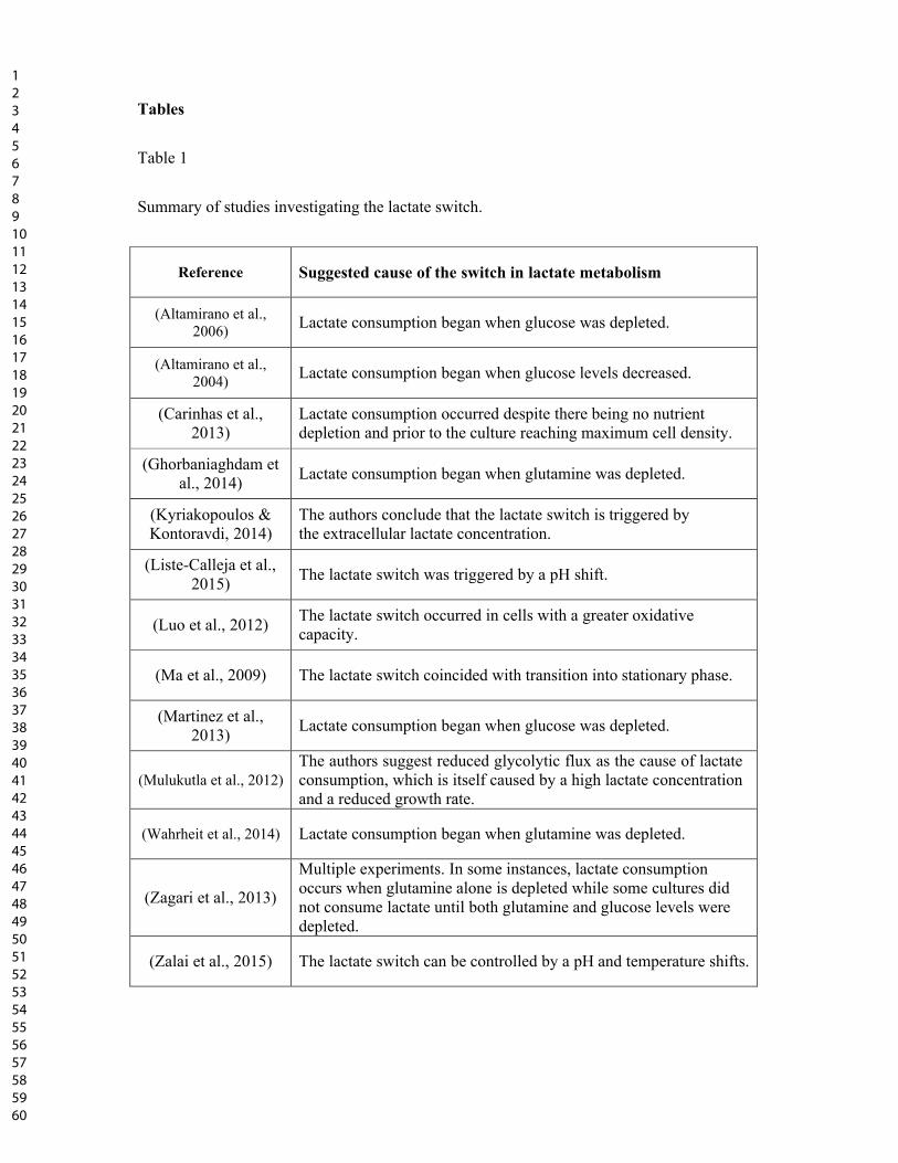
Tables
Table 1
Summary of studies investigating the lactate switch.
Reference Suggested cause of the switch in lactate metabolism
(Altamirano et al., 2006)
Lactate consumption began when glucose was depleted.
(Altamirano et al., 2004)
Lactate consumption began when glucose levels decreased.
(Carinhas et al., 2013)
Lactate consumption occurred despite there being no nutrient depletion and prior to the culture reaching maximum cell density.
(Ghorbaniaghdam et al., 2014)
Lactate consumption began when glutamine was depleted.
(Kyriakopoulos & Kontoravdi, 2014)
The authors conclude that the lactate switch is triggered by the extracellular lactate concentration.
(Liste-Calleja et al., 2015)
The lactate switch was triggered by a pH shift.
(Luo et al., 2012) The lactate switch occurred in cells with a greater oxidative capacity.
(Ma et al., 2009) The lactate switch coincided with transition into stationary phase.
(Martinez et al., 2013)
Lactate consumption began when glucose was depleted.
(Mulukutla et al., 2012) The authors suggest reduced glycolytic flux as the cause of lactate consumption, which is itself caused by a high lactate concentration and a reduced growth rate.
(Wahrheit et al., 2014) Lactate consumption began when glutamine was depleted.
(Zagari et al., 2013)
Multiple experiments. In some instances, lactate consumption occurs when glutamine alone is depleted while some cultures did not consume lactate until both glutamine and glucose levels were depleted.
(Zalai et al., 2015) The lactate switch can be controlled by a pH and temperature shifts.
123456789101112131415161718192021222324252627282930313233343536373839404142434445464748495051525354555657585960
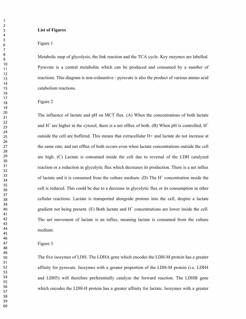
List of Figures
Figure 1
Metabolic map of glycolysis, the link reaction and the TCA cycle. Key enzymes are labelled.
Pyruvate is a central metabolite which can be produced and consumed by a number of
reactions. This diagram is non-exhaustive - pyruvate is also the product of various amino acid
catabolism reactions.
Figure 2
The influence of lactate and pH on MCT flux. (A) When the concentrations of both lactate
and H+ are higher in the cytosol, there is a net efflux of both. (B) When pH is controlled, H+
outside the cell are buffered. This means that extracellular H+ and lactate do not increase at
the same rate, and net efflux of both occurs even when lactate concentrations outside the cell
are high. (C) Lactate is consumed inside the cell due to reversal of the LDH catalyzed
reaction or a reduction in glycolytic flux which decreases its production. There is a net influx
of lactate and it is consumed from the culture medium. (D) The H+ concentration inside the
cell is reduced. This could be due to a decrease in glycolytic flux or its consumption in other
cellular reactions. Lactate is transported alongside protons into the cell, despite a lactate
gradient not being present. (E) Both lactate and H+ concentrations are lower inside the cell.
The net movement of lactate is an influx, meaning lactate is consumed from the culture
medium.
Figure 3
The five isozymes of LDH. The LDHA gene which encodes the LDH-M protein has a greater
affinity for pyruvate. Isozymes with a greater proportion of the LDH-M protein (i.e. LDH4
and LDH5) will therefore preferentially catalyze the forward reaction. The LDHB gene
which encodes the LDH-H protein has a greater affinity for lactate. Isozymes with a greater
123456789101112131415161718192021222324252627282930313233343536373839404142434445464748495051525354555657585960
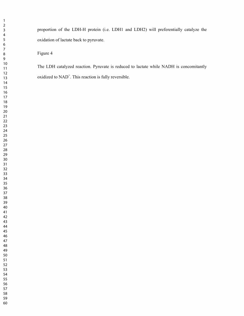
proportion of the LDH-H protein (i.e. LDH1 and LDH2) will preferentially catalyze the
oxidation of lactate back to pyruvate.
Figure 4
The LDH catalyzed reaction. Pyruvate is reduced to lactate while NADH is concomitantly
oxidized to NAD+. This reaction is fully reversible.
123456789101112131415161718192021222324252627282930313233343536373839404142434445464748495051525354555657585960
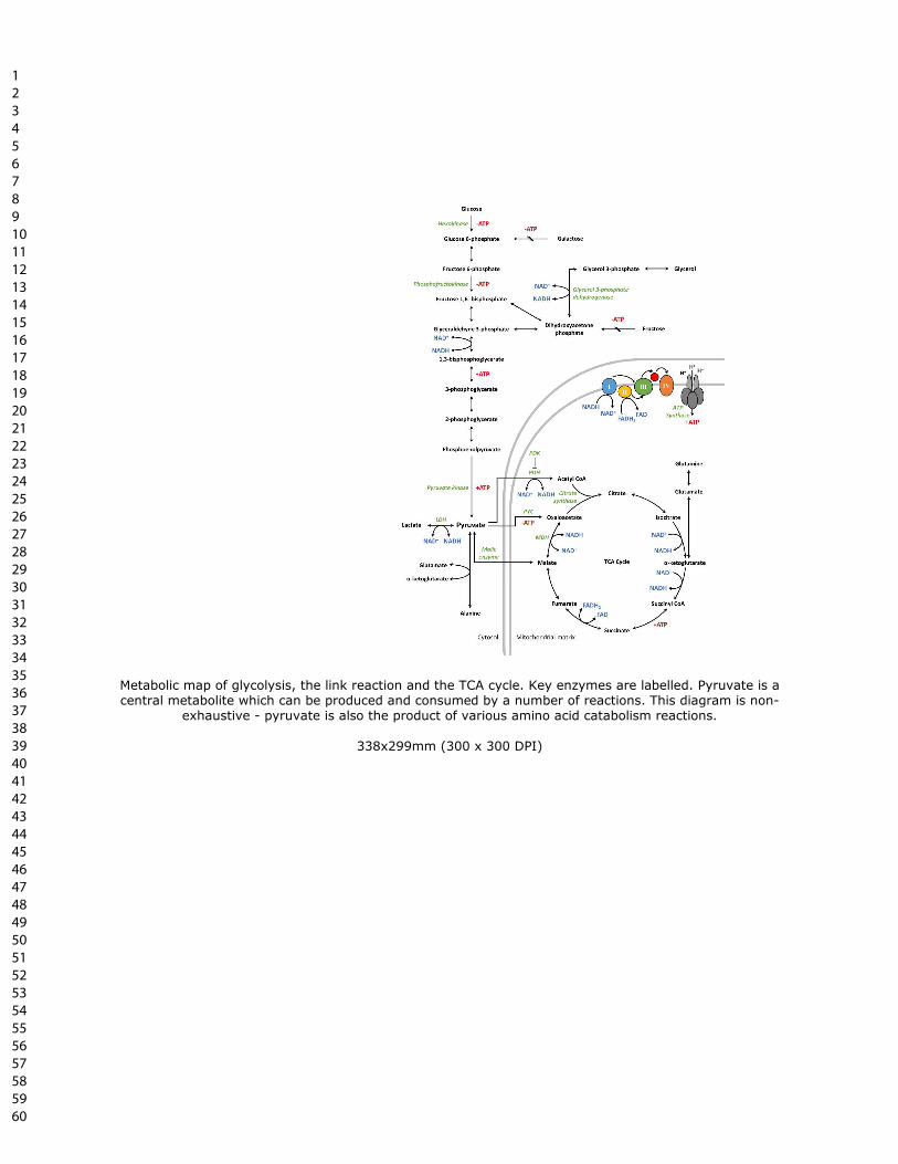
Metabolic map of glycolysis, the link reaction and the TCA cycle. Key enzymes are labelled. Pyruvate is a central metabolite which can be produced and consumed by a number of reactions. This diagram is non-
exhaustive - pyruvate is also the product of various amino acid catabolism reactions.
338x299mm (300 x 300 DPI)
123456789101112131415161718192021222324252627282930313233343536373839404142434445464748495051525354555657585960

The influence of lactate and pH on MCT flux. (A) When the concentrations of both lactate and H+ are higher in the cytosol, there is a net efflux of both. (B) When pH is controlled, H+ outside the cell are buffered. This means that extracellular H+ and lactate do not increase at the same rate, and net efflux of both occurs even
when lactate concentrations outside the cell are high. (C) Lactate is consumed inside the cell due to reversal of the LDH catalyzed reaction or a reduction in glycolytic flux which decreases its production. There is a net influx of lactate and it is consumed from the culture medium. (D) The H+ concentration inside the cell is reduced. This could be due to a decrease in glycolytic flux or its consumption in other cellular reactions.
Lactate is transported alongside protons into the cell, despite a lactate gradient not being present. (E) Both lactate and H+ concentrations are lower inside the cell. The net movement of lactate is an influx, meaning
lactate is consumed from the culture medium.
338x299mm (300 x 300 DPI)
123456789101112131415161718192021222324252627282930313233343536373839404142434445464748495051525354555657585960
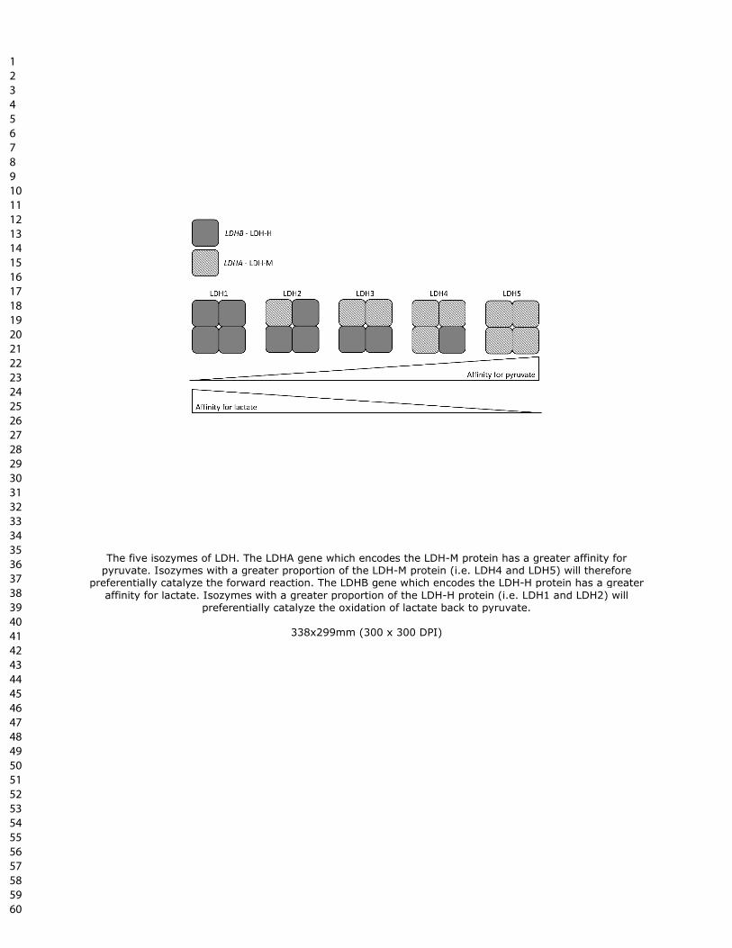
The five isozymes of LDH. The LDHA gene which encodes the LDH-M protein has a greater affinity for pyruvate. Isozymes with a greater proportion of the LDH-M protein (i.e. LDH4 and LDH5) will therefore
preferentially catalyze the forward reaction. The LDHB gene which encodes the LDH-H protein has a greater
affinity for lactate. Isozymes with a greater proportion of the LDH-H protein (i.e. LDH1 and LDH2) will preferentially catalyze the oxidation of lactate back to pyruvate.
338x299mm (300 x 300 DPI)
123456789101112131415161718192021222324252627282930313233343536373839404142434445464748495051525354555657585960
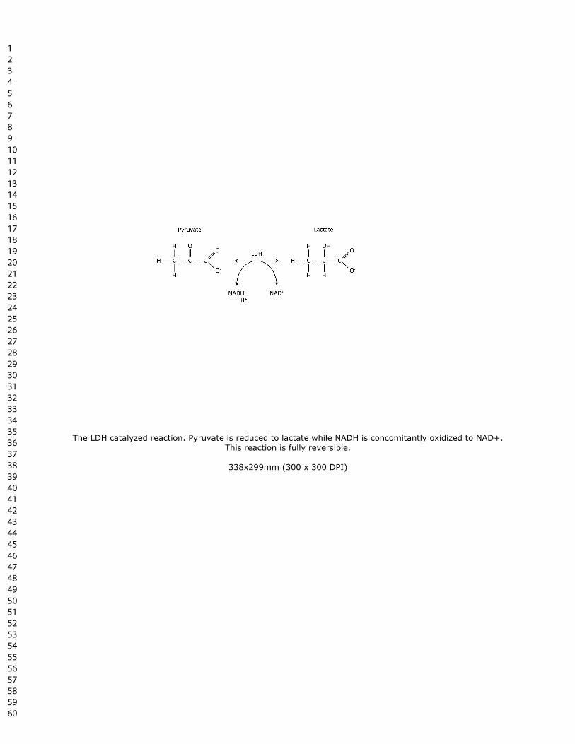
The LDH catalyzed reaction. Pyruvate is reduced to lactate while NADH is concomitantly oxidized to NAD+.
This reaction is fully reversible.
338x299mm (300 x 300 DPI)
123456789101112131415161718192021222324252627282930313233343536373839404142434445464748495051525354555657585960
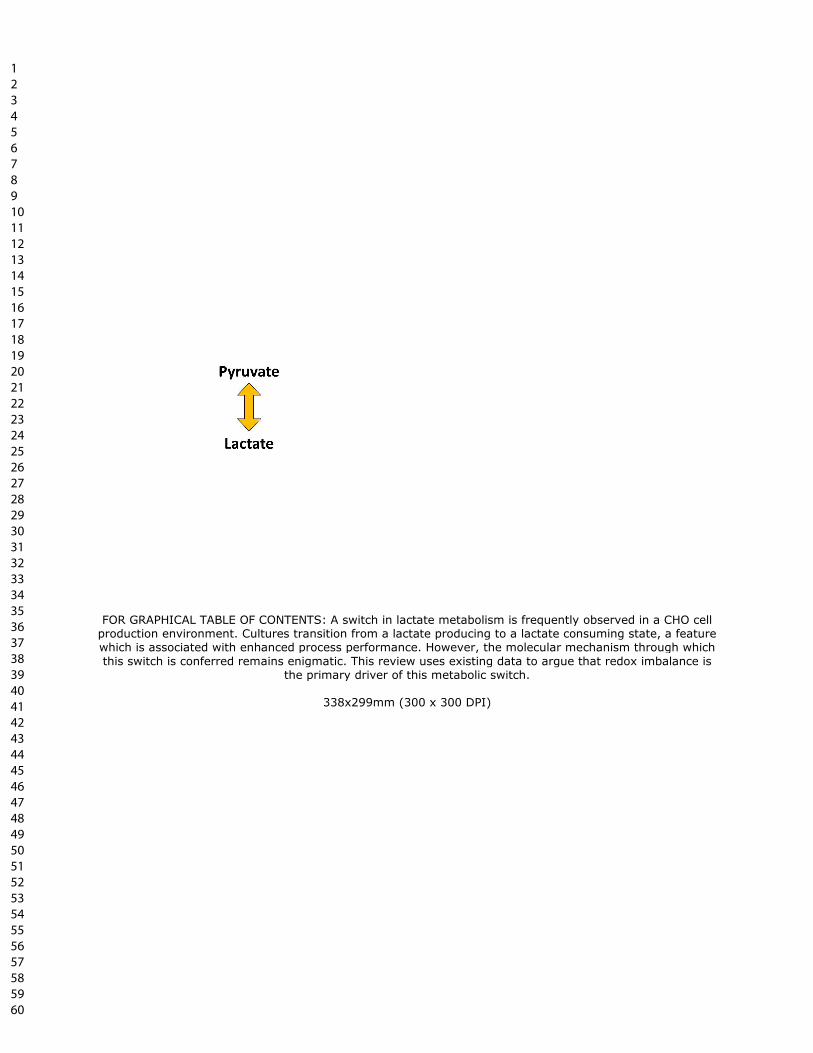
FOR GRAPHICAL TABLE OF CONTENTS: A switch in lactate metabolism is frequently observed in a CHO cell production environment. Cultures transition from a lactate producing to a lactate consuming state, a feature which is associated with enhanced process performance. However, the molecular mechanism through which
this switch is conferred remains enigmatic. This review uses existing data to argue that redox imbalance is the primary driver of this metabolic switch.
338x299mm (300 x 300 DPI)
123456789101112131415161718192021222324252627282930313233343536373839404142434445464748495051525354555657585960