The Respiratory Chain Components of Higher Plant Mitochondria
Upload
independentCategory
view
3download
0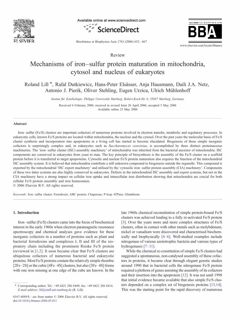
Biochimica et Biophysica Acta 1763 (2006) 652–667www.elsevier.com/locate/bbamcr
Review
Mechanisms of iron–sulfur protein maturation in mitochondria,cytosol and nucleus of eukaryotes
Roland Lill ⁎, Rafal Dutkiewicz, Hans-Peter Elsässer, Anja Hausmann, Daili J.A. Netz,Antonio J. Pierik, Oliver Stehling, Eugen Urzica, Ulrich Mühlenhoff
Institut für Zytobiologie, Philipps Universität Marburg, Robert-Koch-Str. 6, 35037 Marburg, Germany
Received 6 February 2006; received in revised form 26 April 2006; accepted 5 May 2006Available online 23 May 2006
Abstract
Iron–sulfur (Fe/S) clusters are important cofactors of numerous proteins involved in electron transfer, metabolic and regulatory processes. Ineukaryotic cells, known Fe/S proteins are located within mitochondria, the nucleus and the cytosol. Over the past years the molecular basis of Fe/Scluster synthesis and incorporation into apoproteins in a living cell has started to become elucidated. Biogenesis of these simple inorganiccofactors is surprisingly complex and, in eukaryotes such as Saccharomyces cerevisiae, is accomplished by three distinct proteinaceousmachineries. The ‘iron–sulfur cluster (ISC) assembly machinery’ of mitochondria was inherited from the bacterial ancestor of mitochondria. ISCcomponents are conserved in eukaryotes from yeast to man. The key principle of biosynthesis is the assembly of the Fe/S cluster on a scaffoldprotein before it is transferred to target apoproteins. Cytosolic and nuclear Fe/S protein maturation also requires the function of the mitochondrialISC assembly system. It is believed that mitochondria contribute a still unknown compound to biogenesis outside the organelle. This compound isexported by the mitochondrial ‘ISC export machinery’ and utilised by the ‘cytosolic iron–sulfur protein assembly (CIA) machinery’. Componentsof these two latter systems are also highly conserved in eukaryotes. Defects in the mitochondrial ISC assembly and export systems, but not in theCIA machinery have a strong impact on cellular iron uptake and intracellular iron distribution showing that mitochondria are crucial for bothcellular Fe/S protein assembly and iron homeostasis.© 2006 Elsevier B.V. All rights reserved.
Keywords: Iron–sulfur cluster; Ferredoxin; ABC protein; Chaperone; P-loop ATPase; Glutathione
1. Introduction
Iron–sulfur (Fe/S) clusters came into the focus of biochemicalinterest in the early 1960s when electron paramagnetic resonancespectroscopy and chemical analyses gave evidence for theseinorganic cofactors in a number of proteins such as plant andbacterial ferredoxins and complexes I, II and III of the res-piratory chain including the prominent Rieske Fe/S protein(reviewed in [1,2]. It soon became clear that Fe/S clusters areubiquitous cofactors of numerous bacterial and eukaryoticproteins.Most Fe/S proteins contain the relatively simple rhombic[2Fe–2S] or the cubic [4Fe–4S] clusters, but also [3Fe–4S] formswith one iron missing at one edge of the cube are known. In the
⁎ Corresponding author. Tel.: +49 6421 286 6449; fax: +49 6421 286 6414.E-mail address: [email protected] (R. Lill).
0167-4889/$ - see front matter © 2006 Elsevier B.V. All rights reserved.doi:10.1016/j.bbamcr.2006.05.011
late 1960s chemical reconstitution of simple protein-bound Fe/Sclusters was achieved leading to a fully re-activated Fe/S protein[3]. Over the years more and more complex structures of Fe/Sclusters, often in contact with other metals such as molybdenum,nickel or vanadium were discovered and characterised biochem-ically and biophysically [4–6]. Well-studied examples includenitrogenase of various azototrophic bacteria and various types ofhydrogenases [7–11].
While the chemical re-constitution of simple Fe/S clusters hadsuggested a spontaneous, non-catalysed assembly of these cofac-tors in proteins, it became clear through elegant genetic studiesaround 1990 that in bacterial cells the nitrogenase Fe/S proteinrequired a plethora of genes assisting the assembly of its cofactorsand their insertion into the apoprotein [12]. It was not until 1998that initial evidence became available that also simple Fe/S clus-ters depended on a complex set of biogenesis proteins [13,14].This was the starting point for the rapid discovery of numerous

653R. Lill et al. / Biochimica et Biophysica Acta 1763 (2006) 652–667
biogenesis components in both eubacteria and eukaryotes in-volved in the generation of Fe/S cofactors in vivo (see, e.g., [15–18]. To date, research on the biogenesis of Fe/S proteins is anintensive field concentrated on the functional characterisation ofparticipating proteins, the elucidation of the molecular mechan-isms underlying the pathway, and the identification of novel bio-genesis proteins. This review will summarise the currently knownprinciples and concepts of Fe/S protein biogenesis in (non-plant)eukaryotes. The survey is intended to introduce non-specialists tothis fascinating new field of biochemistry and cell biology ofmetals. More detailed or focused reviews have been publishedrecently elsewhere with particular emphasis on bacterial Fe/Sprotein maturation [19–21] or on biogenesis in yeast, mammals orplants [22–24].
1.1. A brief look at the structures and properties of Fe/S clustersand Fe/S proteins
Fe/S clusters are formally composed of ferrous (Fe2+) and/orferric (Fe3+) iron, inorganic sulfide (S2−) ions, and in rare casesadditional metals or cofactors [25,26]. Their chemical behaviour,however, cannot be described as that of a simple iron salt, butrather is best explained by the ligand field theory of co-ordinationchemistry. The most simple form of an Fe/S center is the [2Fe–2S]cluster. It is present in, e.g., ferredoxins, Rieske Fe/S protein, biotinsynthase and ferrochelatase (Table 1). Formally, the [4Fe–4S]clusters can be viewed as a duplication of [2Fe–2S] clusters. Theyconsist of a cubic structure with Fe and S occupying alternatingcorners of the cube which due to the shorter distance between theFe ions is usually distorted. This type of cluster is present in manyproteins such as aconitase and aconitase-like proteins, ferredoxins,sulfite reductase, and DNA glycosylase (Table 1). The Fe ionswithin the clusters have a tetrahedral co-ordination, as they addi-tionally bind to nucleophilic ligands of the protein. The mostcommon co-ordination partner of the Fe ions is the sulfur atom ofcysteine residues. However, also histidine, and in rare cases argi-nine, serine, peptidyl-N and non-protein ligands (homocitrate, CO,CN−) may be used. For example, in the [4Fe–4S] cluster ofaconitase-like proteins the fourth iron is not co-ordinated by aprotein-bound ligand, but binds to the substrate/product of theenzyme (e.g., citrate, homocitrate, isopropyl malate) or to a watermolecule. Hence, the co-ordination sphere of the fourth iron inaconitase-like proteins changes to octahedral.
Some proteins such as subunits of complexes I and II of therespiratory chain contain [3Fe–4S] clusters inwhich one corner ofthe cube is unoccupied (Table 1). There are numerous proteinswhich contain more than one Fe/S cluster. Themost extreme formis complex I (NADH-ubiquinone oxidoreductase; not present inSaccharomyces cerevisiae) of the respiratory chain which con-tains 8 (eukaryotes) to 9 (bacteria) Fe/S clusters [27]. Likewise,complex II (succinate-ubiquinone oxidoreductase) contains[2Fe–2S], [3Fe–4S] and [4Fe–4S] clusters [28]. Higher orderstructures of Fe/S clusters with additional metal centers such asthose present in bacterial nitrogenase, CO dehydrogenase orvarious hydrogenases have been studied extensively and many3D structures have been resolved [4]. Some Fe/S proteins such asxanthine dehydrogenase, plant aldehyde oxidase and plant nitrate
reductase contain an additional molybdenum cofactor whichrequires Fe/S proteins for its own synthesis [29]; see also articleby Mendel and Bittner [137] in this issue.
The electronic charge of the Fe/S center is concentrated at thesulfur moiety. Depending on the overall oxidation state of the Fe/Scluster the Fe ions may harbour unpaired electrons, and the re-sulting electron spin can be detected by electron paramagneticresonance (EPR) spectroscopy. This biophysical techniqueprovides valuable information mainly about the oxidation state,the type and the electronic environment of the clusters [30]. Otherbiophysical methods to study the structure and properties of theFe/S clusters are Mössbauer, EXAFS, ENDOR, NMR, and reso-nance Raman spectroscopies. They provide specific informationon the electronic and magnetic properties of the Fe/S clusters intheir particular environment. In UV/Vis spectrophotometric mea-surements Fe/S proteins exhibit typical absorptionmaxima around420 nm ([4Fe–4S] clusters) and 320, 410 and 560 nm ([2Fe–2S]).These spectral properties give rise to the dark brown colour ofpurified Fe/S holoproteins.
Fe/S clusters and Fe/S proteins are fairly sensitive to damage byoxidative conditions and may decompose readily in air or by theexposure to NO or H2O2. This propensity has been exploited innature for using Fe/S proteins as sensors and regulatory switches[31–33]; see also below).
Even though there is no single common consensus motif forbinding an Fe/S cluster, some primary structural features are reoc-curring. These include the conserved positioning of cysteine resi-dues in the protein. Examples include the CX4CX2CX≈30C motifin plant and mammalian [2Fe–2S] ferredoxins. [4Fe–4S] clustersare often co-ordinated by the consensus motif CX2CX2CX20–40Cwhich was originally defined in [4Fe–4S] ferredoxins, but seemspresent in many other members of the [4Fe–4S] cluster type. Itappears that the spacing of the cysteine residues can also bereversed. Usually, a conserved proline (or sometimes glycine)flanks one of the cysteine residues. Care has to be taken not toconfuse the presence of conserved cysteine residues with similarpatterns of Zn-finger proteins or other metal binding motifs.
Despite the existence of these fairly defined Fe/S consensussequences there exist numerous Fe/S proteins that do not followthese simple rules for binding the Fe/S cofactor. On the onehand, this lack of consensus makes it rather hard to predict andidentify new Fe/S proteins based on primary sequenceinformation contained in genomes. On the other hand, thisshows that binding of Fe/S clusters seems to rely on a substantialamount of flexibility of the protein partner during selection of theco-ordination sites within the protein. In fact, mutational analysesin some ferredoxins and other Fe/S proteins have shown that,upon exchange of particular cysteine to serine or alanine residues,the Fe/S proteinsmay be flexible enough to provide alternative co-ordination partners within the polypeptide sequence [30,34].Hence, frequently only mutation of two cysteine residues leads tothe complete loss of the Fe/S center.
1.2. Diverse functions of Fe/S proteins
The biological functions of Fe/S proteins can be grouped intothree categories, namely electron transfer, enzyme catalysis and

Table 1Fe/S proteins in (non-plant) eukaryotes: Function, bound cluster types, and sub-cellular localisation
Fe/S protein Yeast name Localisation Function Cluster type
Fe/S proteins involved in electron transportComplex I – Mitochondrial inner
membraneRespiration (NADH ubiquinone oxidoreductase) 8 clusters of 2F,
3F, 4FComplex II Sdh2 Mitochondrial inner
membraneRespiration (succinate dehydrogenase) 2F, 3F, 4F
Complex III Rip1 Mitochondrial innermembrane
Respiration (ubiquinone cytochrome c oxidoreductase,‘Rieske Fe/S protein’)
2F
Ferredoxin (adrenodoxin) Yah1 Mitochondrial matrix Maturation of Fe/S proteins, biosynthesis of heme A,steroid biosynthesis in mammals
2F
ETFQO Yor356w Mitochondria Catabolism of amino acids and choline, β-oxidation of fatty acids 4F
Fe/S proteins in metabolismAconitase Aco1 Mitochondrial matrix Citric acid cycle 4FHomoaconitase Lys4 Mitochondrial matrix Biosynthesis of lysine (aconitase-like) 4FDihydroxy-aciddehydratase
Ilv3 Mitochondrial matrix Biosynthesis of branched-chain amino acids 4F
Lipoate synthase Lip5 Mitochondrial matrix Biosynthesis of lipoic acid 2F ?, 4FBiotin synthase Bio2 Mitochondrial matrix Biosynthesis of biotin 2F, 4FFerrochelatase Hem15 Mitochondrial inner
membraneHeme biosynthesis (no cluster in Saccharomyces cerevisiae) 2F
MOCS1A – Mitochondrial matrix Biosynthesis of Moco (molybdenum co-factor) 3F ?, 4FIsopropylmalate isomerase Leu1 Cytosol Biosynthesis of leucine (aconitase-like) 4FSulfite reductase Ecm17 Cytosol Biosynthesis of methionine, contains siroheme 4FGlutamate dehydrogenase Glt1 Cytosol Biosynthesis of glutamate 4FCMP-N-acetyl-neuraminicacid hydroxylase
– Cytosol Biosynthesis of N-glycolyl neuraminic acid, Rieske-like protein 2F
Dihydro-pyrimidinedehydrogenase
– Cytosol Degradation of pyrimidine nucleotides 4× 4F
Xanthine dehydrogenase – Cytosol Degradation of xanthine to urate, contains FAD and molybdopterin 2× 2FDNA glycosylase Ntg2 Nucleus DNA repair (Endonuclease III-like glycosylase 2) 4F
Fe/S protein involved in regulation and assemblyIron regulatory protein 1(IRP1)
– Cytosol Post-transcriptional regulation of iron uptake, storage andutilisation in mammals (‘cytosolic aconitase’)
4F
ABC protein Rli1(ABCE1)
Rli1 Cytosol and nucleus Biogenesis of ribosomes, rRNA processing, translation initiation,assembly of RNA complexes
2× 4F ?
Glutaredoxin 2(mammals)
Grx2 Mitochondria Glutathione-dependent oxidoreductase (no cluster inSaccharomyces cerevisiae)
2F
Elp3 Elp3 Nucleus Histone acetyltransferase subunit of the Elongator complex,cluster binds S-adenosyl-methionine
4F
Sprouty (mammals) – Cytosol Negative regulator of receptor tyrosine kinase signalling 2F
Fe/S proteins involved in Fe/S protein biogenesisFerredoxin Yah1 Mitochondrial matrix Maturation of mitochondrial Fe/S proteins, possibly reduction of sulfur 2FP-loop ATPase Nbp35 Nbp35 Cytosol and nucleus Maturation of cytosolic/nuclear Fe/S proteins, complex with Cfd1 4F ?Iron-only hydrogenase-like Nar1
Nar1 Cytosol and nucleus Maturation of cytosolic/nuclear Fe/S proteins, binds to Nbp35 and Cia1 2 clusters ofcomplex type
Most Fe/S proteins contain clusters of the [2Fe–2S] (2F), [3Fe–4S] (3F), or [4Fe–4S] (4F) type. The chemical structure of some Fe/S clusters is currently unknown(indicated by ‘?’) due to the lack of spectroscopic and 3D structural information.
654 R. Lill et al. / Biochimica et Biophysica Acta 1763 (2006) 652–667
regulation (Table 1). The most well-known function of Fe/Sproteins is in various redox processes. This function is based on theproperty of the iron ions of the Fe/S clusters in switching between areduced (ferrous Fe2+) and oxidised (ferric Fe3+) state. Hence, theFe/S clusters can easily accept electrons from one partner anddonate them to othermolecules. The redox potential range dependsmainly on the protein environment of the clusters. For instance, therelatively high redox potential of Rieske-type Fe/S proteins iscaused by the two co-ordinating histidine ligands on one of the twoFe ions with the result that it can be reduced at a considerablyhigher potential than those Fe ions bound via cysteine residues.
Typical proteins involved in redox reactions are complexes I, IIand III of the respiratory chain, mitochondrial and plant fer-redoxins and the plant photosystem I. A fascinating example ofstep-wise electron transfer can be seen in respiratory complexes Iand II, where electrons are transferred from the substrates NADHand succinate, respectively, via several Fe/S clusters and other co-factors to the electron acceptor ubiquinone in the bacterial ormitochondrial inner membrane. The characteristic redox potentialsof Fe/S clusters range from −500 mV to +300 mV. The reducingenergy is typically a few hundred mV, but in some Fe/S proteinssuch as nitrogenase or 2-hydroxy-glutaryl-CoA dehydratase the

Fig. 1. Three distinct systems participate in the generation of Fe/S proteins ineukaryotes. Fe/S proteins have been identified in mitochondria, cytosol andnucleus of eukaryotes. The mitochondrial ISC assembly machinery is required formaturation of virtually all cellular Fe/S proteins. The ISC export apparatus and theCIA machinery are specifically involved in the formation of cytosolic and nuclearFe/S proteins. The Fe/S cluster in proteins is indicated by red/yellow circles.
655R. Lill et al. / Biochimica et Biophysica Acta 1763 (2006) 652–667
extra energy ofATP hydrolysis assures high reduction potentials ofgreater than 1 V [8,35]. In these fascinating proteins the Fe/Scluster is co-ordinated between two subunits of the protein, i.e. thecofactor serves as a bridging unit. The additional energy is sup-plied by conformational changes that are induced by ATP bindingand hydrolysis thus allowing the redox interaction with thesubstrates.
The classical example for a role of Fe/S clusters in enzymecatalysis is aconitase of the citric acid cycle (Table 1). Thisenzyme and related proteins such as homoaconitase (yeast Lys4involved in lysine biosynthesis) and isopropylmalate dehydra-tase (yeast Leu1, leucine biosynthesis) catalyse the removal andre-addition of water, resulting in an isomerisation of thesubstrates. In these enzymes, the fourth Fe of the [4Fe–4S]cluster serves as a Lewis acid to bind the substrate and stabilisethe transition state of the reaction. Other enzymatic reactionssupported by Fe/S clusters are the final steps of biotin and lipoicacid synthesis. In these cases, the enzymes bind two clusters, oneof which is possibly used as a source of the sulfur atom to beinserted into the precursors desthiobiotin and octanoic acid [36].An interesting case of an Fe/S protein is ferrochelatasecatalysing the final step of heme biosynthesis [37]; see alsoarticle by Ajioka et al. [138] in this issue. Since this enzyme is amajor consumer of iron in cells and relies on an Fe/S cluster forits function, the two major iron metabolising pathways areconnected in this protein. It is important to note that someferrochelatases such as that of S. cerevisiae (Hem15; Table 1) donot contain an Fe/S cluster. Further Fe/S proteins in eukaryotesare involved the biosynthesis of methionine (sulfite reductase)and the molybdenum cofactor (see article byMendel and Bittner[137] in this issue). For some of the mentioned enzymes it is stillunclear what exact role the cluster may play in catalysis. It is wellpossible that in some cases the Fe/S cluster may simply serve astructural role stabilising the rest of the polypeptide chain.However, such a stabilising functionwas long suspected for Fe/Sproteins involved in repair of DNA damage (DNA glycosylases;Table 1), yet recent investigations have provided ample evidenceto suggest a role of the Fe/S cofactor in DNA binding [38].
An intriguing function of Fe/S proteins lies in the regulationof biological processes. The chemical lability of the Fe/Scofactors and their pronounced sensitivity to oxidative damagehas been exploited by Nature to sense intracellular orenvironmental conditions by Fe/S clusters and hence regulategene expression in eukaryotes or prokaryotes. While regulationin bacteria by the transcription factors SoxR, IscR, and FNRoccurs on a transcriptional level, regulation in mammals by ironregulatory protein 1 (IRP1) is on a post-transcriptional level[31–33]. IRP1 appears to have a dual role in the cell. As an Fe/Sprotein it functions as a cytosolic aconitase, while underconditions leading to the loss of the Fe/S cluster the IRP1apoprotein binds to RNA stem–loop structures designated ironregulatory elements (IRE) of specific mRNAs encoding proteinsinvolved in iron uptake, utilisation, storage or export. As a resultof IRP1-IRE binding, some mRNAs are stabilised and thusprevented from degradation resulting in enhanced proteinsynthesis. In other mRNAs stem–loop structures at the 5′-untranslated end block efficient translation by the ribosome
leading to lower protein abundance (for details of IRP1 functionsee article by Wallander et al. [139] in this issue).
1.3. Overview on the biogenesis of Fe/S proteins in eukaryotes
In the late 1990s several research groupsworkingwith the yeastS. cerevisiae almost simultaneously realised that the biogenesis ofmitochondrial Fe/S proteins depended on a number of conservedproteins now known as members of the Fe/S cluster (ISC) as-sembly machinery (Figs. 1 and 2). Initially these groups wereinterested in understanding the rather diverse processes of oxi-dative stress, cellular iron homeostasis, and the function of mi-tochondrial chaperones or ABC transporters [14,39–41]. Theunifying and surprising theme was that all these groups studiedprocesses or proteins that played a decisive role in the assembly ofFe/S proteins. The mitochondrial ISC assembly system turned outto be strikingly similar to that of bacteria, and in all likelihood hasbeen inherited from the bacterial endosymbiont that gave rise tomodern mitochondria [19,42]. It was realised early on that mito-chondria perform a prime role in Fe/S protein biogenesis as theywere found to be responsible not only for maturation of Fe/Sproteins inside but also outside the organelle [41,43]. Since anABC transporter in the mitochondrial inner membrane wasrequired for efficient maturation of extra-mitochondrial Fe/Sproteins, this created the idea of amitochondrial “export system” tosupport the generation of Fe/S proteins in the cytosol and nucleus(Fig. 1).
It was not until 2003 that the first component of the so-calledcytosolic Fe/S protein assembly (CIA) machinery was discov-ered by a genetic approach [44]. Thus, the current view of Fe/Sprotein biogenesis in eukaryotes, as derived from studies in themodel organism S. cerevisiae, is that of an interplay of threecomplex proteinaceous systems termed ISC assembly, ISCexport, and CIA machineries (Fig. 1). The ISC assemblymachinery is required for all cellular Fe/S proteins, whereas thefunctions of the ISC export and the CIA machineries are

Fig. 2. A model for the mechanism of Fe/S protein biogenesis in mitochondria.After membrane potential-dependent (pmf) import of ferrous iron (Fe2+) facilitatedby the carrier proteinsMrs3 andMrs4 and other unknown proteins (X), iron is usedfor heme synthesis from protoporphyrin IX (PPIX) by ferrochelatase (Hem15) andfor the biogenesis of Fe/S proteins. The latter process starts with the release of sulfurfrom cysteine by the Nfs1/Isd11 cysteine desulfurase complex and the formation ofa transient Fe/S cluster on the Isu1/2 scaffold proteins. The electron (e−) transferchain NADH→ ferredoxin reductase Arh1→ ferredoxin Yah1 and the putativeiron donor Yfh1 (frataxin) are needed for transient Fe/S cluster synthesis on Isu1/2.ISC assembly proteins required later in biogenesis, i.e. after Fe/S cluster synthesison Isu1/2, are the dedicated chaperones Ssq1, Jac1 and Mge1 and the monothiolglutaredoxin Grx5. Isa1 and Isa2 are specifically and additionally required for thematuration of aconitase-type Fe/S proteins. The exact role of Nfu1 is unclear so far.Nfs1 performs a second function as a cysteine desulfurase in thio-uridine modifi-cation of mitochondrial tRNA (tRNAS).
656 R. Lill et al. / Biochimica et Biophysica Acta 1763 (2006) 652–667
restricted to the maturation of cytosolic and nuclear Fe/Sproteins. The high conservation of the ISC assembly, ISC exportand CIA proteins in virtually all eukaryotes [22] and pioneeringfunctional studies in human cell culture and multi-cellularorganisms (see below) support the view that biogenesis in lowerand higher eukaryotes occurs along similar pathways.
Before we concentrate on the pathways of Fe/S protein mat-uration, we will briefly address the issue of how the process canbe followed experimentally in vivo or in cell extracts. The mostclassical and widely used technique is to follow the activities ofparticular Fe/S proteins, functioning as enzymes (aconitase,isopropylmalate isomerase, sulfite reductase etc.) or in electrontransport (complexes II and III of the respiratory chain). Thisapproach usually requires verification that observed activitychanges are not caused by altered gene expression. Therefore,protein levels have to be analysed in parallel by immunostaining.The approach does not directly measure de novo assembly butrather the steady state levels of the Fe/S proteins. Thus, it may bedifficult to distinguish long-time damage of Fe/S proteins (forinstance by oxidative stress) from an impairment of the mat-uration process. A more direct assay of biogenesis is theincorporation of radiolabelled iron (or sulfur) into the nascentFe/S proteins [41,45–47]. Fe/S cluster synthesis and assemblyon the apoproteins is then measured by immunoprecipitating theFe/S proteins of interest with subsequent quantitative analysis byscintillation counting. Since the times used for radiolabelling areusually short, the assay gives a reasonable estimation of de novoassembly. Native gel electrophoresis can be employed to follow
the maturation of a [2Fe–2S] ferredoxin [48]. The techniqueallows the distinction of the apo- and holoforms of thisferredoxin, yet is restricted to isolated systems, e.g., isolatedmitochondria. Finally, the RNA binding of IRP1 (see above) hasbeen used in higher eukaryotes and in yeast to indirectly followits association with a [4Fe–4S] cluster [44,49–51]. Theincreased binding of IRP1 to RNA is reciprocally taken as ameasure of diminished amounts of Fe/S cluster associated withthe protein.
1.4. Biogenesis of mitochondrial Fe/S proteins—the first steps
The requirements for the making of an Fe/S cluster are de-ceptively trivial: inorganic iron and sulfide. While both compo-nents may occur in cells in free form, they both are toxic to the cell,and therefore the intracellular free concentrations have to be ratherlow. This may preclude the spontaneous assembly of an Fe/Scluster in cells, the more so as rather high concentrations of ironand sulfide and anaerobic conditions are needed for chemical invitro reconstitution of model Fe/S proteins. Therefore, sophisti-cated systems have been developed in living cells to assemble theclusters and to specifically insert them into proteins. In yeastmitochondria the ISC assembly system encompasses 14 knownproteins of diverse functions (Fig. 2). The similarity to the bacterialcounterparts suggests shared mechanisms of biosynthesis inbacteria and mitochondria. In fact, many functional observationsfor the ISC proteins have first been made with bacterial proteins.We summarise an emerging consensus mechanism of biogenesisfor the eukaryotic (yeast) ISC system making use of numerousobservations from the bacterial ISC system [19].
In mitochondrial Fe/S cluster assembly free cysteine is thesource of sulfurwhich is released by the cysteine desulfuraseNfs1in a pyridoxal phosphate-dependent fashion (Fig. 2). Nfs1 pos-sesses numerous well-studied relatives in bacteria such as NifS,IscS, and SufS of the NIF, ISC and SUF systems (reviewed in[52,53]. The purified Nfs1 enzyme produces sulfide in vitro [54].However, the sulfide production by Nfs1 (and its bacterial count-erparts) likely does not mimic the in vivo pathway of the sulfurfrom cysteine into an Fe/S cluster. As discussed above cells haveto prevent an unregulated release of toxic sulfide. To circumventliberation of sulfide, the sulfur is first transiently bound to Nfs1via a persulfide group at a conserved cysteine residue on theenzyme before it is further transferred to the Isu scaffold proteins(Fig. 2). These latter proteins may initially receive the sulfur alsoas a covalently bound persulfide (see below).
It has long been thought that Nfs1 is sufficient to support thistransfer reaction. However, sulfur transfer to the Isu proteinswas recently found to involve an additional essential proteindesignated Isd11 which forms a stable complex with Nfs1 but isnot conserved in the bacterial systems [55,56]. The precise roleof Isd11 is unclear so far, but it is interesting to compare thesituation with a similar complex in the bacterial or plastid SUFsystems [18,24], where the Nfs1 homologue SufS binds toSufE. This small protein transiently receives the sulfur releasedfrom cysteine by SufS, before sulfur is further transferred toscaffold proteins (e.g., SufA). Sulfur binding to SufE occurs via apersulfide group formed on a conserved cysteine residue. Since
657R. Lill et al. / Biochimica et Biophysica Acta 1763 (2006) 652–667
Isd11 does not contain any cysteine residue, the functionalmechanism of Isd11 must be different from that worked out forSufE.
A central concept of Fe/S protein biogenesis is the de novosynthesis of the Fe/S cluster on so-called scaffold proteins beforeits transfer to apoproteins [57]. In vivo studies in yeast have shownthat Isu1 (and presumably Isu2) fulfil this scaffold function inmitochondria [58]. Fe/S cluster associationwith Isu1was followedby radiolabelling cells with iron and immunoprecipitation of Isu1.In these experiments, over-produced Isu1 was necessary toovercome the detection limits. The specificity of Fe/S clusterassociation with Isu1 is evident from its dependence on other ISCproteins including Nfs1, Isd11, Yah1 and Yfh1 (see also below).
The source of iron for Fe/S cluster formation on the Isuscaffold proteins is less clear. Iron is taken up by mitochondriain reduced form (Fig. 2). Uptake requires an energised innermembrane and the members of the mitochondrial carrier familytermed Mrs3 and Mrs4 [59–63]. It presently is unclear whetherthese transporters are directly trafficking iron and in which form(free or chelated) iron may be transported. Since the two yeastcarriers are not essential for iron transfer into the mitochondrialmatrix, additional transporters must exist to support thisreaction (‘X’ in Fig. 2). The form of iron in which it is storedin the mitochondrial matrix before its use in Fe/S clusterassembly is unknown.
After import into mitochondria iron may be delivered to theIsu proteins byYfh1, the yeast homologue of frataxin (Fig. 2; seealso below). This model is now favoured by many groups in thefield. The purified protein was found to bind and oxidise iron,even though it is still debated whether this binding is specific orunspecific [64–66]. Further, Yfh1 binds to Isu1 in an iron-stimulated fashion in mitochondria [67] suggesting that complexformation may facilitate iron transfer from frataxin to Isu1 forFe/S cluster formation. In keeping with this view, Yfh1 wasfound to be necessary for transient Fe/S cluster assembly on Isu1[58] reassuring the role of frataxin/Yfh1 in Fe/S proteinassembly.
Frataxin was claimed to play various additional roles in hemesynthesis, iron storage, and prevention of oxidative stress prevail-ing in frataxin-deficient yeast cells.Most of these effects seem to bea secondary consequence of a defective Fe/S cluster assembly andthe accompanying iron accumulation in mitochondria (see below).In particular, the role of frataxin in iron storage is amatter of debate.This function requires the formation of a higher order structuresimilar to that of ferritin. However, mutations preventing the for-mation of aggregates ofYfh1 display no detectable phenotype [65].Frataxin/Yfh1 was proposed to deliver iron not only to Fe/S clusterbiosynthesis, but also to ferrochelatase for heme formation fromprotoporphyrin IX (Fig. 2; [68]). However, no primary defects inheme synthesis uponYfh1 depletionwere seen [45]. An interactionbetween ferrochelatase and Yfh1 was detected in vitro by plasmonresonance technique, but no complex formation was observed inmitochondria [69]. Further, experiments with triple mutants inYFH1, MRS3 and MRS4 genes suggested that the heme defect incells entirely lacking YFH1 may be indirect [62]. Finally, noimmediate ferrochelatase defect seems to be evident in human cellsdepleted of frataxin [50,70]. In summary, it seems reasonable to
believe that Yfh1/frataxin plays a role as an iron donor in Fe/Scluster assembly on the Isu scaffold proteins, yet the precise mo-lecular function remains to be established.
The chemistry of cluster formation of the Isu proteins is farfrom being understood (discussed in [18]. While experimentswith purified bacterial proteins have led to the conflictinginterpretation that either sulfur or iron initially bind to the Isu (orbacterial IscU or NifU) proteins, it is clear that three conservedcysteine residues are important for Fe/S cluster assembly andtransient binding. From a solution structure of the apoform ofIscU (determined by NMR) one might expect that bound Fe/Sclustersmay be surface-exposed, a situation that may facilitate thetransfer of the pre-assembled cluster to apoproteins. It was shownthat the bacterial IscU or NifU can assemble both [2Fe–2S] and[4Fe–4S] clusters, and recently it was demonstrated that theseclusters can be specifically transferred to appropriate apoproteins[71,72]. Thus, it appears that the Isu/IscU/NifU scaffolds arecapable of facilitating the assembly of both types of clusters [73].
In addition to Nfs1, Isd11 and Yfh1 two other ISC componentsare required early in biogenesis (Fig. 2; [43,58,74]). The ferredoxinYah1 and the ferredoxin reductase Arh1 form an electron transferchain that gains its electrons fromNADHand,most likely, transfersthem to the sulfur moiety (S0) released from cysteine by the Nfs1/Isd11 complex to generate the sulfide (S2−) present in the Fe/Scluster. The precise mode of how the sulfur is reduced and at whichstep of the reaction path this occurs has remained elusive. Interest-ingly, Yah1 carries a [2Fe–2S] cluster itself, and thus pre-existingholo-Yah1 is presumably involved in the conversion of newlyimported apo-Yah1 to the holoprotein.While the role ofYah1/Arh1in the reduction of sulfur is likely (yet not proven), it is not excludedthat the proteins are required at a second step of biogenesis, forinstance during Fe/S cluster transfer from the Isu scaffold proteinsto the acceptor apoproteins. Insight into this problem will re-quire the in vitro reconstitution of the reactions. To date, no in vitroevidence for the potential role of this electron chain has beenprovided.
1.5. Components acting late in Fe/S protein maturation
A dedicated chaperone system consisting of the Hsp70 chap-erone Ssq1, its cognate J-type co-chaperone Jac1, and thenucleotide exchange factor Mge1 has been long recognised to berequired for Fe/S protein assembly in mitochondria [16,40,48,75–77]. Recent in vivo and in vitro experiments havedemonstrated that the proteins are not required for de novo Fe/Scluster assembly on the Isu scaffold proteins [58,78]. Rather, upondepletion of these proteins up to fivefold more Fe/S clusteraccumulates on Isu1. This has been taken to propose a role for thechaperone system late in biogenesis, for instance in a partialreaction between the stage of transient Fe/S cluster binding to theIsu scaffold and the incorporation of the pre-assembled Fe/Scluster into target apoproteins (Fig. 2). One might assume thatthese chaperones specifically interact with the apoform of Fe/Sproteins to maintain them in a conformation capable of acceptingthe Fe/S cluster from the Isu scaffold proteins. However, nospecific interaction between these protein classes has beenreported hitherto.

658 R. Lill et al. / Biochimica et Biophysica Acta 1763 (2006) 652–667
Ssq1 is specifically targeted to its substrate Isu1 by Jac1 [77,79].Ssq1 binds to Isu1 via a highly conserved LPPVKmotif present inIsu/IscU proteins [80,81]. Binding is facilitated by the co-chap-erone Jac1 which also directly binds to Isu1 in a J-domain in-dependent fashion via its C-terminal domain [82]. Even thoughcomplex formation is not specific for either the apo- or holoform ofIsu/IscU, it is clear that the chaperone system functions in closecontact to the scaffold proteins. Hence, it seems reasonable topropose that the chaperones facilitate Fe/S cluster transfer, that theystabilise the conformation of the scaffold proteins or that theyregenerate the scaffold proteins for the next round of clusterassembly. A recently developed in vitro system to study Fe/Scluster formation on Isu1 with purified components may beextended to unravel the role of these (and other) components [78].
Another ISC component acting late in biogenesis is the mono-thiol glutaredoxin Grx5 (Fig. 2; [83]. Yeast contains two dithiol(Grx1, Grx2) and three monothiol (Grx3, Grx4, Grx5) glutar-edoxins, but only Grx5 seems to play a specific role in Fe/S proteinbiogenesis. As seenwith the chaperones, depletion ofGrx5 in yeastresults in a general defect in Fe/S protein assembly, yet transientlybound Fe/S clusters accumulate on Isu1 [58]. The exact function ofGrx5 remains unclear, but, in contrast to the role of the chaperones,Grx5 is dispensable for this process, as deletion of its gene resultsin a minor growth defect only. Recently, a zebrafish mutant withhypochromic anemia was found to be impaired in Grx5 function[84]. Zebrafish Grx5 could complement the respective yeastdeletion mutant. Loss of Fe/S cluster assembly in Grx5-deficientanimals activated IRP1 thus leading to decreased levels of δ-aminolaevulinate synthase 2 (ALAS2) and diminished hemesynthesis. These results not only show that Grx5 is important for(cytosolic) Fe/S protein assembly in higher eukaryotes, but alsodocumented that heme formation in the differentiating red cell isregulated by a member of the ISC assembly machinery (see also[63].
1.6. A specific role of Isa proteins in the maturation ofaconitase-like proteins
The mitochondrial proteins Isa1 and Isa2 contain a fairly con-served sequencemotif that is also present in IscA and SufA proteinsof bacteria. Most eukaryotes contain only close sequence relativesof Isa1. The Isa2 protein is unique in that closest relatives arepresent only in the genomes of a few yeasts. In S. cerevisiae the twoIsa proteins form a hetero-oligomer, and the deletion of their genesresults in similar phenotypic consequences, such as the failure togrow on non-fermentable carbon sources, loss of mitochondrialDNA and a general defect in respiration [85–87]. Initial experi-ments had indicated that yeast cells depleted in Isa1 and/or Isa2show a defect in Fe/S proteins. A more detailed study indicated aspecialised requirement of the Isa proteins for aconitase-type Fe/Sproteins in mitochondria (Fig. 2; U. Mühlenhoff, unpublished). Incontrast, the de novo assembly of other Fe/S proteins such as theferredoxin Yah1, Rieske Fe/S protein and biotin synthase wereunaffected. In the bacterial ISC and SUF systems the IscA proteinswere shown in vitro to serve as alternative scaffold proteins assem-bling an Fe/S cluster, which is competent for transfer to targetapoproteins (for reviews see [18,19,21]). This made it appear likely
that those proteins are the specific scaffolds for aconitase-type Fe/Sproteins. Several observations refute this idea for yeast as a model.First, maturation of aconitase-type Fe/S proteins required the func-tional involvement of the Isu proteins, in addition to the Isa pro-teins. Second, despite the possibility to assemble an Fe/S cluster onpurified Isa proteins in vitro, attempts to find an Isa-associated Fe/Scluster in vivo failed (U. Mühlenhoff, unpublished). Instead, theproteins isolated from cell extracts bind iron (but no sulfur). Finally,the phenotypes of Isa-depleted yeast cells are fully explained bythe defects in aconitase (e.g., glutamate auxotrophy, mtDNA loss,respiratory defect) and homoaconitase (lysine auxotrophy). Theyeast Isa proteins therefore perform a specific role in thematurationof aconitase-like Fe/S proteins. This is inmarked contrast to the roleof the Isu proteins which are responsible for maturation of virtuallyall cellular Fe/S proteins. Further clarification of the molecular roleof the Isa proteins in Fe/S cluster assembly will require faithful invitro approaches for reconstitution of aconitase.
1.7. The role of mitochondria in the maturation ofextra-mitochondrial Fe/S proteins
The importance of mitochondria in Fe/S protein assembly wasrecognised early on in themolecular dissection of this process [41].Virtually the entire ISC assembly system is required for maturationof extra-mitochondrial Fe/S proteins such as Leu1, Rli1, sulfitereductase, Nbp35, Nar1, and Ntg2 (Fig. 3). In two cases, for Nfs1and for the Isu proteins, experimental evidence has been providedthat these ISC proteins need to be located inside yeast mitochon-dria to be functional in extra-mitochondrial Fe/S protein assembly[41,54,73]. The exclusive presence of a cytosolic/nuclear form ofNfs1 or Isu1 did not lead to any Fe/S cluster incorporation intocytosolic/nuclear Fe/S proteins. This suggests that both Nfs1 andIsu1/Isu2 have to be located inside yeast mitochondria to be usefulin extra-mitochondrial Fe/S protein maturation.
It is still unknown what mitochondria provide for cytosolic/nuclear Fe/S protein assembly. The current consensus is thatthe mitochondrial ISC assembly machinery produces a sub-strate that is exported from mitochondria to serve in Fe/Scluster formation and incorporation in the cytosol. Compo-nents involved in this step are summarised as ISC exportmachinery [88]. A central component is the mitochondrialABC transporter Atm1 which is involved in mitochondria tocytosol trafficking (Fig. 3; [41]). The protein is located in theinner membrane with its ABC domains facing the matrix.Deletion of Atm1 is associated with severe effects including adrastic growth defect of yeast cells, loss of cytochromes andrespiration and an accumulation of iron in mitochondria.Gradual depletion of Atm1 in a regulated yeast mutantrevealed primary functions of Atm1 in Fe/S protein maturationin the cytosol and nucleus, and in iron accumulation inmitochondria (see below). In an attempt to biochemically char-acterise Atm1, the membrane protein was purified and reconsti-tuted into proteoliposomes [89]. Its ATPase activity is specificallystimulated by compounds containing free sulfhydryl (-SH) groups,in particular by peptides with multiple cysteine residues. It is thusconceivable that the substrate of Atm1 contains free thiol groups,possibly in a peptidic environment.
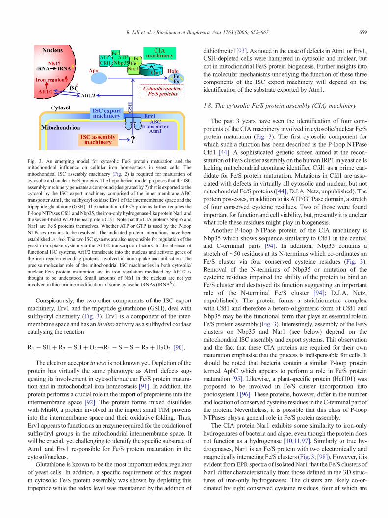
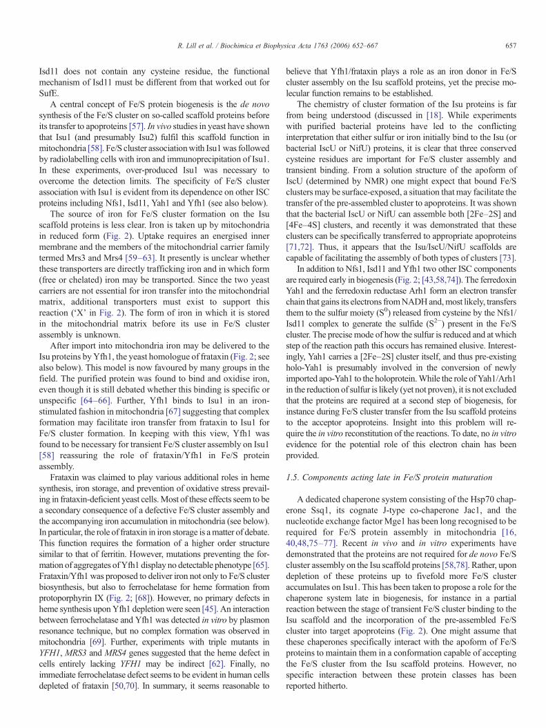
Fig. 3. An emerging model for cytosolic Fe/S protein maturation and themitochondrial influence on cellular iron homeostasis in yeast cells. Themitochondrial ISC assembly machinery (Fig. 2) is required for maturation ofcytosolic and nuclear Fe/S proteins. The hypothetical model proposes that the ISCassemblymachinery generates a compound (designated by ?) that is exported to thecytosol by the ISC export machinery comprised of the inner membrane ABCtransporter Atm1, the sulfhydryl oxidase Erv1 of the intermembrane space and thetripeptide glutathione (GSH). The maturation of Fe/S proteins further requires theP-loopNTPases Cfd1 andNbp35, the iron-only hydrogenase-like proteinNar1 andthe seven-bladedWD40 repeat protein Cia1. Note that the CIA proteins Nbp35 andNar1 are Fe/S proteins themselves. Whether ATP or GTP is used by the P-loopNTPases remains to be resolved. The indicated protein interactions have beenestablished in vivo. The two ISC systems are also responsible for regulation of theyeast iron uptake system via the Aft1/2 transcription factors. In the absence offunctional ISC systems, Aft1/2 translocate into the nucleus and activate genes ofthe iron regulon encoding proteins involved in iron uptake and utilisation. Theprecise molecular role of the mitochondrial ISC machineries in both cytosolic/nuclear Fe/S protein maturation and in iron regulation mediated by Aft1/2 isthought to be understood. Small amounts of Nfs1 in the nucleus are not yetinvolved in thio-uridine modification of some cytosolic tRNAs (tRNAS).
659R. Lill et al. / Biochimica et Biophysica Acta 1763 (2006) 652–667
Conspicuously, the two other components of the ISC exportmachinery, Erv1 and the tripeptide glutathione (GSH), deal withsulfhydryl chemistry (Fig. 3). Erv1 is a component of the inter-membrane space and has an in vitro activity as a sulfhydryl oxidasecatalysing the reaction
R1 � SHþ R2 � SHþ O2→R1 � S� S� R2 þ H2O2 [90].
The electron acceptor in vivo is not known yet. Depletion of the
protein has virtually the same phenotype as Atm1 defects sug-gesting its involvement in cytosolic/nuclear Fe/S protein matura-tion and in mitochondrial iron homeostasis [91]. In addition, theprotein performs a crucial role in the import of preproteins into theintermembrane space [92]. The protein forms mixed disulfideswith Mia40, a protein involved in the import small TIM proteinsinto the intermembrane space and their oxidative folding. Thus,Erv1 appears to function as an enzyme required for the oxidation ofsulfhydryl groups in the mitochondrial intermembrane space. Itwill be crucial, yet challenging to identify the specific substrate ofAtm1 and Erv1 responsible for Fe/S protein maturation in thecytosol/nucleus.Glutathione is known to be the most important redox regulatorof yeast cells. In addition, a specific requirement of this reagentin cytosolic Fe/S protein assembly was shown by depleting thistripeptide while the redox level was maintained by the addition of
dithiothreitol [93]. As noted in the case of defects in Atm1 or Erv1,GSH-depleted cells were hampered in cytosolic and nuclear, butnot in mitochondrial Fe/S protein biogenesis. Further insights intothe molecular mechanisms underlying the function of these threecomponents of the ISC export machinery will depend on theidentification of the substrate exported by Atm1.
1.8. The cytosolic Fe/S protein assembly (CIA) machinery
The past 3 years have seen the identification of four com-ponents of the CIA machinery involved in cytosolic/nuclear Fe/Sprotein maturation (Fig. 3). The first cytosolic component forwhich such a function has been described is the P-loop NTPaseCfd1 [44]. A sophisticated genetic screen aimed at the recon-stitution of Fe/S cluster assembly on the human IRP1 in yeast cellslacking mitochondrial aconitase identified Cfd1 as a prime can-didate for Fe/S protein maturation. Mutations in Cfd1 are asso-ciated with defects in virtually all cytosolic and nuclear, but notmitochondrial Fe/S proteins ([44]; D.J.A.Netz, unpublished). Theprotein possesses, in addition to its ATP/GTPase domain, a stretchof four conserved cysteine residues. Two of those were foundimportant for function and cell viability, but, presently it is unclearwhat role these residues might play in biogenesis.
Another P-loop NTPase protein of the CIA machinery isNbp35 which shows sequence similarity to Cfd1 in the centraland C-terminal parts [94]. In addition, Nbp35 contains astretch of ∼50 residues at its N-terminus which co-ordinates anFe/S cluster via four conserved cysteine residues (Fig. 3).Removal of the N-terminus of Nbp35 or mutation of thecysteine residues impaired the ability of the protein to bind anFe/S cluster and destroyed its function suggesting an importantrole of the N-terminal Fe/S cluster [94]; D.J.A. Netz,unpublished). The protein forms a stoichiometric complexwith Cfd1 and therefore a hetero-oligomeric form of Cfd1 andNbp35 may be the functional form that plays an essential role inFe/S protein assembly (Fig. 3). Interestingly, assembly of the Fe/Sclusters on Nbp35 and Nar1 (see below) depend on themitochondrial ISC assembly and export systems. This observationand the fact that these CIA proteins are required for their ownmaturation emphasise that the process is indispensable for cells. Itshould be noted that bacteria contain a similar P-loop proteintermed ApbC which appears to perform a role in Fe/S proteinmaturation [95]. Likewise, a plant-specific protein (Hcf101) wasproposed to be involved in Fe/S cluster incorporation intophotosystem I [96]. These proteins, however, differ in the numberand location of conserved cysteine residues in theC-terminal part ofthe protein. Nevertheless, it is possible that this class of P-loopNTPases plays a general role in Fe/S protein assembly.
The CIA protein Nar1 exhibits some similarity to iron-onlyhydrogenases of bacteria and algae, even though the protein doesnot function as a hydrogenase [10,11,97]. Similarly to true hy-drogenases, Nar1 is an Fe/S protein with two electronically andmagnetically interacting Fe/S clusters (Fig. 3; [98]). However, it isevident fromEPR spectra of isolatedNar1 that the Fe/S clusters ofNar1 differ characteristically from those defined in the 3D struc-tures of iron-only hydrogenases. The clusters are likely co-or-dinated by eight conserved cysteine residues, four of which are
660 R. Lill et al. / Biochimica et Biophysica Acta 1763 (2006) 652–667
concentrated at the N-terminus and the remainder of which arescattered in themiddle andC-terminal parts. Assembly of the Fe/Sclusters of Nar1 requires the mitochondrial ISC assembly andexport systems, and additionally Cfd1 and Nbp35 [94]. Appar-ently, a pre-existing functional CIAmachinery is necessary for theassembly of newly assembled components of this system. Thischicken and egg situation is reminiscent of the assembly of Yah1in the mitochondrial matrix and of the TOM complex in mito-chondrial outer membrane which depend on pre-existing com-ponents for their own de novo assembly.
The most recently discovered CIA member termed Cia1 wasoriginally identified on the basis of a fusion gene with CFD1 inSchizosaccharomyces pombe making it likely that the fusedproteins perform a function in the same process [99]. Depletion ofyeast Cia1 elicits a somewhat different phenotype as compared todefects in Cfd1, Nbp35 and Nar1. While Cia1 deficiency isaccompanied by an impairment in the assembly of true Fe/S targetproteins such as Leu1, Rli1 and Ntg2, Cia1 is not required for theFe/S cluster-containing CIA components Nar1 and Nbp35. On thefirst glimpse, this result is counterintuitive, as alsoNar1 andNbp35should initially behave as Fe/S targets before conversion intofunctional proteins. However, a closer inspection shows that thesituation may be reminiscent of the observations made for dep-letion of the matrix chaperone system Ssq1/Jac1/Mge1 or of Grx5with respect to Fe/S cluster accumulation on either the Isu1 scaf-fold protein or on target proteins (see above). Consequently, onemight expect a role of Cia1 late in biogenesis, i.e., after the functionof Nbp35 and Nar1. A further possibility arising from theseobservations is that Nbp35 and/or Nar1 might serve as scaffoldproteins binding an Fe/S cluster in a transient fashion. This hypo-thesis can now be tested following the lead of the studies with Isu1[58].
Cia1 is homologous to human Ciao1 which was proposed toplay a role in transcription regulation of the Wilms tumour sup-pressor protein, a protein not present in yeast. This suggests thatthe proteins may perform functions in both Fe/S protein bio-genesis and transcription regulation. This expectation is furthersupported by the sub-cellular localisation of Cia1 that differsfrom that of the other three CIA proteins [99]. Cia1 is mainlylocated in the nucleus with minor amounts in the cytosol, whilethe other three CIA proteins show the opposite sub-cellularpartitioning. In addition, the cellular concentration of Cia1 islarger than that of the other CIA proteins. Therefore, a role ofCia1 in transcription similar to human Ciao1 is not excluded.Cia1 is well conserved in eukaryotes and belongs to a largeprotein family characterised by theWD40 repeat domains [100].Even though more than 50 WD40 repeat proteins with closesimilarity to Cia1 are present in yeast, the protein is unique andcan be discriminated from other members with distinctfunctions.
At the moment, we can only speculate about the molecularmechanism underlying Fe/S protein assembly in the cytosol andnucleus of yeast. Future functional insights can make use ofseveral distinct protein interactions between the CIA compo-nents that have been defined by in vivo and in vitro experiments(Fig. 3; [99]). As mentioned above, Cfd1 and Nbp35 form a tightcomplex, while Nbp35 interacts with Nar1. The latter protein
serves as a specific docking partner of Cia1. The predicteddoughnut shape of Cia1 typical for WD40 proteins may beideally suited to serve as a platform for the binding of otherproteins during the assembly pathway of Fe/S proteins.Surprisingly, no direct contact has been reported yet for Cfd1and Cia1, despite the existence of the above mentioned fusiongene in S. pombe. Nevertheless, this genetic situation stronglysuggests a functional co-operation of the two proteins in the CIAmachinery-supported pathway. It is tempting to predict thatfuture research will see the identification of new components ofthis apparently complex machinery. This will facilitate theelucidation of the molecular mechanisms leading to thematuration of cytosolic/nuclear Fe/S proteins.
1.9. Fe/S protein assembly in higher eukaryotes
The ISC assembly, ISC export and CIA machineries are highlyconserved in eukaryotes from yeast to man [22,23]. Thus, onemight expect similar pathways and mechanisms of Fe/S proteinassembly in lower and higher eukaryotes. To date only a fewstudies exist to put this expectation to the test. The thoroughinvestigation of mammalian Fe/S protein assembly is an importanttopic for several reasons. First, pilot experiments have to examinethe degree of similarity or divergence in the yeast and mammalianFe/S protein maturation mechanisms. Second, components such asAtm1 are duplicated in mammals (ABCB6 and ABCB7; [101])posing the question of which particular function the individualproteins may perform. Third, diseases are associated with muta-tions in frataxin (Friedreich's ataxia) and ABCB7 (X-linkedsideroblastic anemia and ataxia, XLSA/A). Detailed informationon molecular issues of these disorders can be found in othercomprehensive reviews (see, e.g., [102–105]). Fourth and finally,the close connection of Fe/S protein assembly and (intra-)cellulariron homeostasis in yeast (see below) raises the question ofwhethersuch links exist in mammals. It has become clear over the pastdecade that the iron metabolism in yeast and mammals is governedby radically different mechanisms.
The first functional in vivo study on a specific component ofFe/S protein biogenesis in the human system has been performedon frataxin. Depletion of this protein by the RNAi technologyresulted in a growth defect of HeLa cells and a specific depletionof the activities of both mitochondrial and cytosolic Fe/S pro-teins [50]. Hence, this study confirmed findings made in yeastfor Yfh1, the yeast frataxin homologue, suggesting a function ofthe human protein in the biogenesis of Fe/S proteins. Since thedefects in Fe/S proteins accompany the depletion of frataxin, theFe/S protein deficiencies previously observed in Friedreich'sataxia patients appear to be specific rather than secondary ef-fects, e.g., by damage of Fe/S proteins [106]. Conspicuously, thefrataxin-depleted HeLa cells were not compromised in hemebiosynthesis catalysed by ferrochelatase suggesting that frataxindepletion in these cells was not severe enough to affect this Fe/Scluster-dependent enzyme. A similar effect was seen in culturedcells from Friedreich's ataxia patients [70]. Other alterations ofheme metabolites (conversion of heme b to heme a, down-regulation of the heme synthesis pathway)may be explained by adefect in the Fe/S cluster-containing adrenodoxin, a homologue
661R. Lill et al. / Biochimica et Biophysica Acta 1763 (2006) 652–667
of Yah1 required for heme a synthesis [107], and by IRP1function regulating ALAS2 expression (see above). In fact,cytosolic IRP1 was not efficiently converted into aconitase infrataxin-depleted cell culture suggesting that frataxin plays acrucial role in Fe/S protein assembly in the cytosol [50]. Anearlier study had provided initial, yet indirect evidence for theparticipation of energised mitochondria in the conversion ofIRP1 to cytosolic aconitase [49]. Recent RNAi-mediated depletionstudies in Drosophila have provided strong in vivo evidence for arole of frataxin in Fe/S protein biogenesis [108], while its knock-down in nematodes resulted in a pleiotropic phenotype with thepossibility of secondary effects [109]. Interestingly, an extra-mi-tochondrial localisation of frataxin in overexpressing cells has beenreported recently that is important for cell survival and preventionof mitochondrial damage [110,111]. The precise function of thecytosolic form of frataxin remains to be determined.
The cysteine desulfurase Nfs1 in human cells is locatedpredominantly in mitochondria, while minor amounts are presentin the cytosol and nucleus [112]. The protein shows activity as acysteine desulfurase, and, together with human Isu1, can assembleIRP1 holoprotein in vitro [113]. The differential targeting of humanNfs1 results from alternative translation initiation [23]. Thesubcellular distribution of human Nfs1 is reminiscent to that ofyeast Nfs1 where both the mitochondrial and nuclear versions areessential for cell viability [114]. Cell biological studies usingRNAitechnology to deplete human Nfs1 have now shown that themitochondrial version is indispensable for Fe/S protein matura-tion both inside and outside mitochondria [115]. The humanprotein could be replaced effectively by its mouse homologue,demonstrating the specificity of the RNAi approach. Removal ofthe mitochondrial targeting sequence from the murine Nfs1resulted in its cytosolic/nuclear localisation. This form does notcomplement the defects in cell growth and mitochondrial/cytosolic Fe/S protein activities of human Nfs1-depleted cells.Hence, the results demonstrate the importance of humanmitochondrial Nfs1 for biogenesis of both mitochondrial andcytosolic Fe/S proteins, and do not identify an independent role ofthe cytosolic/nuclear version of this protein. The situationtherefore is strikingly similar to yeast [54]. One should mention,however, that these data do not exclude a participation of extra-mitochondrial Nfs1 in Fe/S protein maturation, but this functionwould be in addition to mitochondrial Nfs1.
Genetic studies in yeast had suggested an essential task of Nfs1outsidemitochondria [114].What might be the function of Nfs1 inthe cytosol/nucleus? Pioneering studies on IscS, the bacterialhomolog ofNfs1, have shown its requirement as a sulfur donor forthiouridine modification of tRNAs [116]. The same function hasrecently been shown for yeastmitochondrial and cytosolic/nuclearNfs1 ([54,117]; Figs. 2 and 3). Thus, thiouridine modification oftRNAs in the nucleus seems to be a second indispensable functionof yeast Nfs1. It is likely, but has not been experimentallyconfirmed yet, that the nuclear fraction of Nfs1 is responsible forthiouridine modification. It seems possible that the human proteinmay play a similar role in the nucleus/cytosol as its yeastcounterpart.
Small amounts of human Isu1 and Nfu1 have been foundoutside mitochondria, while the majority is located inside these
organelles [118,119]. Isu1 in human cells is targeted to the cytosolby alternative splicing of a singlemRNA. Simultaneous depletionof both forms of human Isu1 by RNAi results in a strong defect inboth mitochondrial and cytosolic aconitase activities document-ing the importance of human Isu1 proteins for cellular Fe/S proteinbiogenesis [51]. In addition, both aconitases could not be reac-tivated following treatment with H2O2 or an iron chelator whichresults in damage or loss of the Fe/S cluster on aconitases. Thisshows that regeneration of cellular Fe/S proteins after cluster lossrequires functional Isu1. Selective depletion of eithermitochondrialor cytosolic Isu1 isoforms by RNAi was inefficient and did notsignificantly impact on the function of cellular Fe/S proteins pre-cluding conclusions on the role of the individual Isu1 isoformsunder normal growth conditions. In cells containing diminishedlevels of cytosolic Isu1 the activities of both aconitases fullyrecovered over time after treatment with H2O2 or iron chelator. Theslightly slower reactivation of cytosolic compared to mitochondrialaconitase was proposed to indicate a function of cytosolic Isu1 inthe regeneration of that Fe/S protein. Reduction of the mitochon-drial Isu1 level by RNAi led to impaired recovery of bothmitochondrial and cytosolic aconitases after iron chelator treatmentdemonstrating that this isoform is important for Fe/S proteinregeneration in both compartments. Taken together, these dataindicate that human Isu1 performs a crucial task in cellular Fe/Sprotein biogenesis. While mitochondrial Isu1 plays the major rolein Fe/S cluster biogenesis in human cells, cytosolic Isu1 mayenhance regeneration/repair of cytosolic Fe/S proteins duringrecovery from iron depletion or oxygen stress. It will be interestingto learn more about this repair process, as nothing is known so far,neither in yeast nor in human cells.
An extensive in vivo study has been performed on murineABCB7, the homologue of yeast Atm1 [120]. Ablation of the X-chromosome-linked ABCB7 gene is embryonic lethal indicatingthe important function of this protein in mammals. Liver is theonly tissue in which a viable gene knockout was obtained byconditional gene inactivation. Biochemical analyses of ABCB7-depleted liver tissue showed diminished activities of cytosolicFe/S proteins including IRP1, yet the function of mitochondrialFe/S proteins was unaltered. This phenotype is strikingly similarto the observations made for yeast Atm1 (see above), andconvincingly show that mitochondria play a crucial role incytosolic Fe/S protein biogenesis in mice. Together with theabovementioned studies on zebrafish Grx5 andMrs3 [63,84] theavailable body of data suggests that there is a high degree ofsimilarity in the mechanisms of Fe/S protein biogenesis in yeastand in higher eukaryotes. What appears to be strikingly differentfrom yeast in higher eukaryotes is the functional connection ofFe/S protein biogenesis to iron uptake regulation and hemesynthesis. This is certainly, but likely not exclusively due to thepresence of the cytosolic Fe/S protein IRP1 in higher eukaryotesbut not in yeast. Maturation of IRP1 strongly depends onmitochondrial ISC assembly and export components[51,63,115,120]. Since the presence or absence of an Fe/Scluster on IRP1 is crucial for the regulation of the synthesis ofkey components of iron homeostasis and also of heme meta-bolism in erythroid cells (see article by Wallander et al. [139] inthis issue), these two processes respond to the activity of
662 R. Lill et al. / Biochimica et Biophysica Acta 1763 (2006) 652–667
mitochondria in Fe/S protein biogenesis. While the critical roleof mitochondria in cellular iron homeostasis seems to beconserved, themode of regulation of iron homeostasis appears tobe entirely different in higher and lower eukaryotes.
1.10. The intimate link between Fe/S protein biogenesis andiron homeostasis
Depletion of virtually any component of the yeast ISC as-sembly and export systems not only results in Fe/S proteindefects, but also has severe consequences for (intra-)cellular ironhomeostasis [15]. For instance, depletion of Nfs1, Isu1/2 or Atm1results in amitochondrial iron overload [39–41]. At the same timeyeast cells respond with an increased iron uptake into the cellwhich on first sight seems somewhat counterintuitive becausealso the total cell iron is increased. Initially, it was believed that asatisfactory explanation was the depletion of cytosolic iron as aresult of the more efficient transport into mitochondria, therebyresulting in an activation of the iron-regulated transcriptionfactors Aft1/Aft2 (Fig. 3). However, clever experiments using theiron-dependent enzyme Erg25 of the ergosterol biosynthesispathway as an indicator for cytosolic iron levels argued againstmajor changes of the cytosolic iron concentration [121]. It wasshown in this and later studies that Aft1/Aft2 do not sensecytosolic iron but rather the iron availability inside mitochondriavia a product generated by the mitochondrial ISC assemblymachinery and exported by Atm1 (Fig. 3; [122]). Thus, the ISCassembly and export components are responsible for thesignalling of the iron status to Aft1/Aft2. Whether the compoundexported by Atm1 is identical, similar or entirely different fromthe substance exported from mitochondria to facilitate Fe/Sprotein assembly is unknown. At any rate, the export reactionleads to an inactivation of Aft1/Aft2 transcription factors in thecytosol, while conversely the CIA system is converted to afunctional biogenesis system (Fig. 3). It is clear from these andother studies that mitochondria, as the major “consumers” ofintracellular iron, are critical regulators of iron homeostasis inyeast. Surprisingly, changes in the activity of the CIA machineryhave no striking effects on iron uptake and storage in yeast cells.Depletion of these proteins neither leads to mitochondrial ironaccumulation nor to an increased transcription of iron uptakegenes by activation of Aft1/Aft2. Therefore, iron regulation inyeast is unlikely to be mediated by a canonical cytosolic Fe/Sprotein which is matured with the help of the CIA proteinsdistinguishing this regulatory step from the function of IRP1 inmammals. Rather, the molecule exported by Atm1 directly orindirectly leads to the regulation of the Aft1/Aft2 transcriptionalactivity. It is likely that, in addition to Aft1/Aft2, further proteinsare involved in this regulatory path.
1.11. Fe/S protein biogenesis is essential. Why?
The process of Fe/S proteins biogenesis in mitochondria hasbeen discovered after some 50 years of intense research on theseorganelles. Onemight ask how central thismitochondrial functionis for life. More than half of the components of the ISC assembly,ISC export andCIAmachineries are encoded by essential genes in
yeast [22]. The only other group of proteins fitting this criterionare the constituents of the mitochondrial biogenesis apparatus, i.e.the TOM and TIM proteins supporting preprotein import, mito-chondrial chaperones required for proper protein import andfolding, the presequence peptidase and some other import-asso-ciated factors. Thus, together with the synthesis of heme (seearticle by Ajioka et al. [138] in this issue), the pathway of Fe/Sprotein biogenesis seems to be a key task of mitochondria in yeastand higher eukaryotes, and organisms cannot survive without it.In contrast, respiration is dispensable when yeast cells are grownin the presence of fermentable carbon sources such as glucose.Even human cells in culture can survive the loss of respirationwhen fed with high amounts of glucose [123].
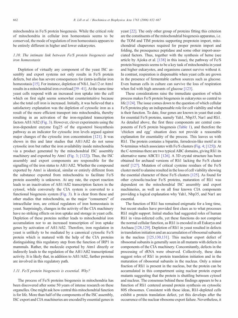
These considerations raise the immediate question of whichprocess makes Fe/S protein biogenesis in eukaryotes so central tolife [124]. The issue comes down to the question of which cellularFe/S proteins play an indispensable role for cell viability and whatis their function. To date, four genes are known in yeast that codefor essential Fe/S proteins, namely Yah1, Nbp35, Nar1 and Rli1.As detailed above, the first three components are central com-ponents of Fe/S protein biogenesis (Table 1), and therefore the‘chicken and egg’ situation does not provide a reasonableexplanation for essentiality of the process. This leaves us withRli1. The protein contains a bipartite, ferredoxin-like motif at itsN-terminus which associates with Fe/S clusters (Fig. 4; [125]). Atthe C-terminus there are two typical ABC domains, giving Rli1 itsalternative name ABCE1 [126]. A 3D crystal structure has beenobtained for archaeal versions of Rli1 lacking the Fe/S clustermotif [127]. Mutation of critical cysteine residues of the Fe/Sclustermotif to alanine resulted in the loss of cell viability showingthe essential character of these Fe/S clusters [125]. As found forother cytosolic/nuclear Fe/S proteins, maturation of Rli1 wasdependent on the mitochondrial ISC assembly and exportmachineries, as well as on all four known CIA componentsproviding a logical explanation why Yah1, Nbp35, and Nar1 areessential.
The function of Rli1 has remained enigmatic for a long time,but recent studies have provided first clues as to what processesRli1 might support. Initial studies had suggested roles of humanRli1 in virus-infected cells, yet these functions do not comprisethe normal cellular function, as Rli1 is encoded in all Eukarya andArchaea [128,129]. Depletion of Rli1 in yeast resulted in defectsin translation initiation and an accumulation of ribosomal subunitsin the nucleus [125,130,131]. This nuclear export defect ofribosomal subunits is generally seen in all mutants with defects incomponents of the CIA machinery. Concomitantly, defects in theprocessing of rRNA were observed. Collectively, these datasuggest roles of Rli1 in protein translation initiation and in thematuration of ribosomal subunits in the nucleus. Only a minorfraction of Rli1 is present in the nucleus, but the protein can beaccumulated in this compartment using nuclear protein exportmutants suggesting that the protein is shuttling between cytosoland nucleus. The consensus behind these findings appears to be afunction of Rli1 centered around protein synthesis on cytosolic80S ribosomes. Consistent with these ideas, Rli1-depleted cellsexhibit a protein translation defect, yet this develops after theoccurrence of the nuclear ribosome export failure. Nevertheless, it
Fig. 4. The essential Fe/S-cluster containing ABC protein Rli1. The bottom scheme shows the functional domains of Rli1. The highly conserved bipartite Fe/S clusterbinding motif at the N-terminus possibly co-ordinates two Fe/S clusters of unknown type (blue). The two ABC domains in the center and at the C-terminus are eachfollowed by a short hinge region (green). The numbers indicate the amino acid residues of the protein from S. cerevisiae. The top part shows a hypothetical sketch ofthe 3D domain structure of Rli1. The design was inspired by the 3D crystal structures of N-terminally truncated versions of Rli1 from Archaea lacking the Fe/Scluster domain [127]. As typical for ABC proteins, the ATP binding site and the ABC signature motif (Sig) of the two ABC domains mutually interact in the solvedstructures.
663R. Lill et al. / Biochimica et Biophysica Acta 1763 (2006) 652–667
seems unlikely that the translational defect upon Rli1 depletion isonly a secondary consequence of the nuclear ribosome exportdefect, as Rli1 was found to directly interact with majortranslation initiation factors in both yeast [130] and human cells[126]. These specific complex formations suggest a specificfunctional role of Rli1 in protein translation initiation. Collec-tively, Rli1might performmultiple roles in both normal and virus-infected cells. As a consensus model the protein may function inthe assembly of RNA–protein complexes such as ribosomalparticles, translation initiation complexes, and virus particles.Archaeal Rli1 proteins may prove useful to decipher the basicfunction(s) of this interesting protein.
In summary, the functional studies on Rli1 have pointed tocrucial link between two central and ancient processes in cells,Fe/S protein biogenesis and protein synthesis. The biogenesis ofFe/S proteins might have been a critical evolutionary step arisingfrom the catalytic phase of the “iron–sulfur” world proposed byWächtershäuser [132]. Protein synthesis facilitated by ribo-somes is central for the development of a living cell.
These contemplations provide logical explanation why mito-chondria (and Rli1) cannot be lost in Eukarya, not even in caseswhere respiration is dispensable. Such a situation is prevalent inparasitic pathogens which live (mostly) inside their host and feedon the host cytosol. Due to the ample availability ofmetabolites andenergy inside the host cell, many of these parasitic organisms havelost most of their mitochondrial functions such as hemebiosynthesis, citric acid cycle, and most prominently respiration.While these organisms are commonly known as “amitochondri-ates” because they lack classical mitochondria with cristae mem-branes, some of them recentlywere shown to containmitochondrialremnant organelles (for recent reviews on these organisms andorganelles see [42,133,134]. The most well-known forms of theserudimentary organelles are the hydrogenosomes and mitosomes. Itis noteworthy in the context of this review that both types oforganelles were shown to harbour basic components of the ISCassembly machinery (Isu1 and Nfs1) and an Hsp70, and thus maybe capable of producing Fe/S clusters [135,136]. In line with thediscussion on Rli1 (see above), the essentiality of this biosyntheticpathway may be the reason why the remnant organelles have notbeen lost in these organisms. A closer inspection of the genomes ofseveral amitochondriates indeed reveals that they contain sequence
relatives of the ISC assembly and CIA machineries, and of Rli1.Interestingly, counterparts of the ISC export machinery are lacking,raising the interesting question whether hydrogenosomes andmitosomes may also play a role in extra-organellar Fe/S proteinbiogenesis as expected from the presence of Rli1.
2. Conclusions and perspectives
The past 8 years have seen the identification of 21 componentswith a direct function in the assembly of Fe/S proteins in yeast as aparadigm for a eukaryotic cell. While initially the identification ofmitochondrial ISC components and their functional characterisa-tion has largely been influenced from findings made in the relatedISC system of bacteria, work on the Atm1-linked export reactionand the identification and characterisation of novel components ofthe CIA machinery is unique for eukaryotes. Despite the con-siderable progress that has been made in our understanding ofcellular Fe/S protein biogenesis, many challenges lie aheadincluding the elucidation of the molecular mechanisms underlyingFe/S cluster assembly and incorporation into apoproteins in mito-chondria, the definition of the exact function of the ISC com-ponents, the (bio)chemical mechanism of Fe/S cluster synthesis onthe Isu scaffold proteins, the identification of the nature of thecompound(s) exported by Atm1, the mechanism of the exportreaction, the analysis of the source of iron and sulfur for cytosolic/nuclear Fe/S protein assembly, the definition of potential scaffoldproteins in the cytosol/nucleus, the molecular mechanism of Fe/Sprotein assembly in the cytosol/nucleus, and finally the intriguingconnection of this process to iron homeostasis. While we hope thisreview has left the impression that basic components and conceptsof Fe/S protein maturation in eukaryotes have been identified, weforesee many exciting years of discovery and surprise for re-searchers involved in this field. Certainly, the elucidation of themolecular foundations of Fe/S protein biogenesis has moved into amore mature stage.
Acknowledgements
We thank Dr. W. Walden (Chicago) for discussion. Work inour laboratory is supported by grants from Sonderforschungs-bereiche 593 and TR1, Deutsche Forschungsgemeinschaft
664 R. Lill et al. / Biochimica et Biophysica Acta 1763 (2006) 652–667
(Gottfried-Wilhelm Leibniz program), the European Commis-sion (MitEURO), German-Israeli foundation GIF, and Fonds derchemischen Industrie. We thank B. Niggemeyer, N. Richhardt,and R. Rösser for excellent technical assistance.
References
[1] H. Beinert, R.H. Holm, E. Münck, Iron–sulfur clusters: nature's modular,multipurpose structures, Science 277 (1997) 653–659.
[2] H. Beinert, Iron–sulfur proteins: ancient structures, still full of surprises,J. Bioinorg. Chem. 5 (2000) 2–15.
[3] R. Malkin, J.C. Rabinowitz, The reconstitution of clostridial ferredoxin,Biochem. Biophys. Res. Commun. 23 (1966) 822–827.
[4] D.C. Rees, Great metalloclusters in enzymology, Ann. Rev. Biochem. 71(2002) 221–246.
[5] D.C. Rees, J.B. Howard, The interface between the biological and inorganicworlds: iron–sulfur metalloclusters, Science 300 (2003) 929–931.
[6] S. Merchant, B.W. Dreyfuss, Post-translational assembly of photosyn-thetic metalloproteins, Annu. Rev. Plant Physiol. Plant Mol. Biol. 49(1998) 25–51.
[7] P.C. Dos Santos, D.R. Dean, Y. Hu, M.W. Ribbe, Formation and insertionof the nitrogenase iron–molybdenum cofactor, Chem. Rev. 104 (2004)1159–1173.
[8] D.C. Rees, J.B. Howard, Nitrogenase: standing at the crossroads, Curr.Opin. Chem. Biol. 4 (2000) 559–566.
[9] F.A. Armstrong, Hydrogenases: active site puzzles and progress, Curr.Opin. Chem. Biol. 8 (2004) 133–140.
[10] Y. Nicolet, C. Cavazza, J.C. Fontecilla-Camps, Fe-only hydrogenases:structure, function and evolution, J. Inorg. Biochem. 91 (2002) 1–8.
[11] D.S. Horner, B. Heil, T. Happe, T.M. Embley, Iron hydrogenases-ancientenzymes in modern eukaryotes, Trends Biochem. Sci. 27 (2002) 148–153.
[12] D.R. Dean, J.T. Bolin, L. Zheng, Nitrogenase metalloclusters: structures,organization, and synthesis, J. Bacteriol. 175 (1993) 6737–6744.
[13] L. Zheng, V.L. Cash, D.H. Flint, D.R. Dean, Assembly of iron–sulfurclusters. Identification of an iscSUA-hscBA-fdx gene cluster fromAzotobacter vinelandii, J. Biol. Chem. 273 (1998) 13264–13272.
[14] J. Strain, C.R. Lorenz, J. Bode, S. Garland, G.A. Smolen, D.T. Ta, L.E.Vickery, V.C. Culotta, Suppressors of superoxide dismutase (SOD1)deficiency in Saccharomyces cerevisiae. Identification of proteins pre-dicted to mediate iron–sulfur cluster assembly, J. Biol. Chem. 273 (1998)31138–31144.
[15] R. Lill, G. Kispal, Maturation of cellular Fe/S proteins: the essentialfunction of mitochondria, Trends Biochem. Sci. 25 (2000) 352–356.
[16] E.A. Craig, J. Marszalek, A specialized mitochondrial molecularchaperone system: a role in formation of Fe/S centers, Cell. Mol. LifeSci. 59 (2002) 1658–1665.
[17] J. Frazzon, D.R. Dean, Formation of iron–sulfur clusters in bacteria—Anemerging field in bioinorganic chemistry, Curr. Opin. Chem. Biol. 7(2003) 166–173.
[18] M. Fontecave, S.O. Choudens, B. Py, F. Barras, Mechanisms of iron–sulfur cluster assembly: the SUF machinery, J. Biol. Inorg. Chem. 10(2005) 713–721.
[19] D.C. Johnson, D.R. Dean, A.D. Smith, M.K. Johnson, Structure, functionand formation of biological iron–sulfur clusters, Ann. Rev. Biochem. 74(2005) 247–281.
[20] F. Barras, L. Loiseau, B. Py, How Escherichia coli and Saccharomycescerevisiae build Fe/S proteins, Adv. Microb. Physiol. 50 (2005) 41–101.
[21] S.S. Mansy, J.A. Cowan, Iron–sulfur cluster biosynthesis: toward anunderstanding of cellular machinery and molecular mechanism, Acc.Chem. Res. 37 (2004) 719–725.
[22] R. Lill, U. Mühlenhoff, Iron–sulfur protein biogenesis in eukaryotes,Trends Biochem. Sci. 30 (2005) 133–141.
[23] T.A. Rouault, W.H. Tong, Iron–sulphur cluster biogenesis and mito-chondrial iron homeostasis, Nat. Rev., Mol. Cell. Biol. 6 (2005) 345–351.
[24] J. Balk, S. Lobreaux, Biogenesis of iron–sulfur proteins in plants, TrendsPlant Sci. 10 (2005) 324–331.
[25] P.V. Rao, R.H. Hom, Synthetic analogues of the active sites of iron–sulfurproteins, Chem. Rev. 104 (2004) 527–559.
[26] H. Beinert, J. Meyer, R. Lill, in: W.J. Lennarz, M.D. Lane (Eds.),Encyclopedia of Biological Chemistry, Iron–Sulfur Proteins, vol. 2,Academic Press, 2004, pp. 482–489.
[27] P. Hinchliffe, L.A. Sazanov, Organization of iron–sulfur clusters inrespiratory complex I, Science 309 (2005) 771–774.
[28] F. Sun, X. Huo, Y. Zhai, A. Wang, J. Xu, D. Su, M. Bartlam, Z. Rao,Crystal structure of mitochondrial respiratory membrane protein complexII, Cell 121 (2005) 1043–1057.
[29] R.R. Mendel, R. Hansch, Molybdoenzymes and molybdenum cofactor inplants, J. Exp. Bot. 53 (2002) 1689–1698.
[30] J.M. Moulis, V. Davasse, M.P. Golinelli, J. Meyer, I. Quinkal, Thecoordination sphere of iron–sulfur clusters: lessons from site-directedmutagenesis experiments, J. Bioinorg. Chem. 1 (1996) 2–14.
[31] P.J. Kiley, H. Beinert, The role of Fe–S proteins in sensing and regulationin bacteria, Curr. Opin. Microbiol. 6 (2003) 181–185.
[32] M.W. Hentze, M.U. Muckenthaler, N.C. Andrews, Balancing acts:molecular control of mammalian iron metabolism, Cell 117 (2004)285–297.
[33] K. Pantopoulos, Iron metabolism and the IRE/IRP regulatory system: anupdate, Ann. N. Y. Acad. Sci. 1012 (2004) 1–13.
[34] H. Matsubara, K. Saeki, in: R. Cammack (Ed.), Iron–Sulfur Proteins,Structural and Functional Diversity of Ferredoxins and Related Proteins,vol. 38, Academic Press, San Diego, 1992, pp. 223–280.
[35] W. Buckel, M. Hetzel, J. Kim, ATP-driven electron transfer in enzymaticradical reactions, Curr. Opin. Chem. Biol. 8 (2004) 462–467.
[36] J.T. Jarrett, The novel structure and chemistry of iron–sulfur clusters inthe adenosylmethionine-dependent radical enzyme biotin synthase, Arch.Biochem. Biophys. 433 (2005) 312–321.
[37] H.A. Dailey, Terminal steps of haem biosynthesis, Biochem. Soc. Trans.30 (2002) 590–595.
[38] O.A. Lukianova, S.S. David, A role for iron–sulfur clusters in DNArepair, Curr. Opin. Chem. Biol. 9 (2005) 145–151.
[39] J. Li, M. Kogan, S.A. Knight, D. Pain, A. Dancis, Yeast mitochondrialprotein Nfs1p coordinately regulates iron–sulfur cluster proteins, cellulariron uptake, and iron distribution, J. Biol. Chem. 274 (1999)33025–33034.
[40] B. Schilke, C. Voisine, H. Beinert, E. Craig, Evidence for a conservedsystem for iron metabolism in the mitochondria of Saccharomycescerevisiae, Proc. Natl. Acad. Sci. U.S.A. 96 (1999) 10206–10211.
[41] G. Kispal, P. Csere, C. Prohl, R. Lill, The mitochondrial proteins Atm1pand Nfs1p are required for biogenesis of cytosolic Fe/S proteins, EMBOJ. 18 (1999) 3981–3989.
[42] M. van der Giezen, J. Tovar, Degenerate mitochondria, EMBO Rep. 6(2005) 525–530.
[43] H. Lange, G. Kispal, A. Kaut, R. Lill, A mitochondrial ferredoxin isessential for biogenesis of intra- and extra-mitochondrial Fe/S proteins,Proc. Natl. Acad. Sci. U.S.A. 97 (2000) 1050–1055.
[44] A. Roy, N. Solodovnikova, T. Nicholson, W. Antholine, W.E. Walden, Anovel eukaryotic factor for cytosolic Fe–S cluster assembly, EMBO J. 22(2003) 4826–4835.
[45] U. Mühlenhoff, N. Richhardt, M. Ristow, G. Kispal, R. Lill, The yeastfrataxin homologue Yfh1p plays a specific role in the maturation ofcellular Fe/S proteins, Hum. Mol. Genet. 11 (2002) 2025–2036.
[46] U. Mühlenhoff, N. Richhardt, J. Gerber, R. Lill, Characterization of iron–sulfur protein assembly in isolated mitochondria: A requirement for ATP,NADH and reduced iron, J. Biol. Chem. 277 (2002) 29810–29816.
[47] O. Stehling, P.M. Smith, A. Biederbick, J. Balk, R. Lill, U. Mühlenhoff,Investigation of iron–sulfur protein maturation in eukaryotes, MethodsCell Biol. (in press).
[48] T. Lutz, B. Westermann, W. Neupert, J.M. Herrmann, The mitochondrialproteins Ssq1 and Jac1 are required for the assembly of iron sulfur clusters inmitochondria, J. Mol. Biol. 307 (2001) 815–825.
[49] C. Bouton, M.J. Chauveau, S. Lazereg, J.C. Drapier, Recycling of RNAbinding iron regulatory protein 1 into an aconitase after nitric oxideremoval depends on mitochondrial ATP, J. Biol. Chem. 277 (2002)31220–31227.

665R. Lill et al. / Biochimica et Biophysica Acta 1763 (2006) 652–667
[50] O. Stehling, H.P. Elsässer, B. Brückel, U. Mühlenhoff, R. Lill, Iron–sulfur protein maturation in human cells: evidence for a function offrataxin, Hum. Mol. Genet. 13 (2004) 3007–3015.
[51] W.H. Tong, T.A. Rouault, Functions of mitochondrial ISCU and cytosolicISCU in mammalian iron–sulfur cluster biogenesis and iron homeostasis,Cell. Metab. 3 (2006) 199–210.
[52] H. Mihara, N. Esaki, Bacterial cysteine desulfurases: their function andmechanisms, Appl. Microbiol. Biotechnol. 60 (2002) 12–23.
[53] D. Kessler, Enzymatic activation of sulfur for incorporation inbiomolecules, FEMS Microbiol Lett. (in press).
[54] U. Mühlenhoff, J. Balk, N. Richhardt, J.T. Kaiser, K. Sipos, G. Kispal, R.Lill, Functional characterization of the eukaryotic cysteine desulfuraseNfs1p from Saccharomyces cerevisiae, J. Biol. Chem. 279 (2004)36906–36915.
[55] N. Wiedemann, E. Urzica, B. Guiard, H. Müller, C. Lohaus, H.E. Meyer,M.T. Ryan, C. Meisinger, U. Mühlenhoff, R. Lill, N. Pfanner, Essentialrole of Isd11 in iron–sulfur cluster synthesis on Isu scaffold proteins,EMBO J. 25 (2006) 184–195.
[56] A.C. Adam, C. Bornhövd, H. Prokisch, W. Neupert, K. Hell, The Nfs1interacting protein Isd11 has an essential role in Fe/S cluster biogenesis inmitochondria, EMBO J. 25 (2006) 174–183.
[57] P. Yuvaniyama, J.N. Agar, V.L. Cash, M.K. Johnson, D.R. Dean, NifS-directed assembly of a transient [2Fe–2S] cluster within the NifU protein,Proc. Natl. Acad. Sci. U. S. A. 97 (2000) 599–604.
[58] U. Mühlenhoff, J. Gerber, N. Richhardt, R. Lill, Components involved inassembly and dislocation of iron–sulfur clusters on the scaffold proteinIsu1p, EMBO J. 22 (2003) 4815–4825.
[59] F. Foury, T. Roganti, Deletion of the mitochondrial carrier genes MRS3and MRS4 suppresses mitochondrial iron accumulation in a yeastfrataxin-deficient strain, J. Biol. Chem. 277 (2002) 24475–24483.
[60] U. Mühlenhoff, J. Stadler, N. Richhardt, A. Seubert, T. Eickhorst, R.J.Schweyen, R. Lill, G. Wiesenberger, A specific role of the yeastmitochondrial carriers Mrs3/4p in mitochondrial iron acquisition underiron-limiting conditions, J. Biol. Chem. 278 (2003) 40612–40620.
[61] L. Li, J. Kaplan, A mitochondrial-vacuolar signaling pathway in yeast thataffects iron and copper metabolism, J. Biol. Chem. 279 (2004) 33653–33661.
[62] Y. Zhang, E.R. Lyver, S.A. Knight, E. Lesuisse, A. Dancis, Frataxin andmitochondrial carrier proteins, Mrs3p and Mrs4p, cooperate in providingiron for heme synthesis, J. Biol. Chem. 280 (2005) 19794–19807.
[63] G.C. Shaw, J.J. Cope, L. Li, K. Corson, C. Hersey, G.E. Ackermann, B.Gwynn, A.J. Lambert, R.A. Wingert, D. Traver, N.S. Trede, B.A. Barut,Y. Zhou, E. Minet, A. Donovan, A. Brownlie, R. Balzan, M.J. Weiss, L.L.Peters, J. Kaplan, L.I. Zon, B.H. Paw, Mitoferrin is essential for erythroidiron assimilation, Nature 440 (2006) 96–100.
[64] T. Yoon, J.A. Cowan, Iron–sulfur cluster biosynthesis. Characterizationof frataxin as an iron donor for assembly of [2Fe–2S] clusters in ISU-typeproteins, J. Am. Chem. Soc. 125 (2003) 6078–6084.
[65] K. Aloria, B. Schilke, A. Andrew, E.A. Craig, Iron-induced oligomer-ization of yeast frataxin homologue Yfh1 is dispensable in vivo, EMBORep. 5 (2004) 1096–1101.
[66] M. Nair, S. Adinolfi, C. Pastore, G. Kelly, P. Temussi, A. Pastore,Solution structure of the bacterial frataxin ortholog, CyaY: mapping theiron binding sites, Structure 12 (2004) 2037–2048.
[67] J. Gerber, U. Mühlenhoff, R. Lill, An interaction between frataxin andIsu1/Nfs1 that is crucial for Fe/S cluster synthesis on Isu1, EMBO Rep. 4(2003) 906–911.
[68] E. Lesuisse, R. Santos, B.F. Matzanke, S.A. Knight, J.M. Camadro, A.Dancis, Iron use for haeme synthesis is under control of the yeast frataxinhomologue (Yfh1), Hum. Mol. Genet. 12 (2003) 879–889.
[69] H. Lange, U. Mühlenhoff, M. Denzel, G. Kispal, R. Lill, The hemesynthesis defect of mutants impaired in mitochondrial iron–sulfur proteinbiogenesis is caused by reversible inhibition of ferrochelatase, J. Biol.Chem. 279 (2004) 29101–29108.
[70] R.A. Schoenfeld, E. Napoli, A. Wong, S. Zhan, L. Reutenauer, D. Morin,A.R. Buckpitt, F. Taroni, B. Lonnerdal, M. Ristow, H. Puccio, G.A.Cortopassi, Frataxin deficiency alters heme pathway transcripts anddecreases mitochondrial heme metabolites in mammalian cells, Hum.Mol. Genet. 14 (2005) 3787–3799.
[71] J.N. Agar, C. Krebs, J. Frazzon, B.H. Huynh, D.R. Dean,M.K. Johnson, IscUas a scaffold for iron–sulfur cluster biosynthesis: sequential assembly of [2Fe–2S] and [4Fe–4S] clusters in IscU, Biochemistry 39 (2000) 7856–7862.
[72] A.D. Smith, G.N. Jameson, P.C. Dos Santos, J.N. Agar, S. Naik, C. Krebs, J.Frazzon, D.R. Dean, B.H. Huynh, M.K. Johnson, NifS-mediated assemblyof [4Fe–4S] clusters in the N- and C-terminal domains of the NifU scaffoldprotein, Biochemistry 44 (2005) 12955–12969.
[73] J. Gerber, K. Neumann, C. Prohl, U. Mühlenhoff, R. Lill, The yeast scaffoldproteins Isu1p and Isu2p are required inside mitochondria for maturation ofcytosolic Fe/S proteins, Mol. Cell. Biol. 24 (2004) 4848–4857.
[74] J. Li, S. Saxena, D. Pain, A. Dancis, Adrenodoxin reductase homolog(Arh1p) of yeast mitochondria required for iron homeostasis, J. Biol.Chem. 276 (2001) 1503–1509.
[75] R. Kim, S. Saxena, D.M. Gordon, D. Pain, A. Dancis, J-domain protein,Jac1p, of yeast mitochondria required for iron homeostasis and activity ofFe–S cluster proteins, J. Biol. Chem. 276 (2001) 17524–17532.
[76] C. Voisine, Y.C. Cheng, M. Ohlson, B. Schilke, K. Hoff, H. Beinert, J.Marszalek, E.A. Craig, Jac1, a mitochondrial J-type chaperone, isinvolved in the biogenesis of Fe/S clusters in Saccharomyces cerevisiae,Proc. Natl. Acad. Sci. U. S. A. 98 (2001) 1483–1488.
[77] R. Dutkiewicz, B. Schilke, H. Kneiszner, W. Walter, E.A. Craig, J.Marszalek, Ssq1, a mitochondrial Hsp70 involved in iron–sulfur (Fe/S)center biogenesis: Similarities to and differences from its bacterialcounterpart, J. Biol. Chem. 278 (2003) 29719–29727.
[78] R.Dutkiewicz, J.Marszalek, B. Schilke, E.A. Craig, R. Lill, U.Mühlenhoff,TheHsp70 chaperone Ssq1p is dispensable for iron–sulfur cluster formationon the scaffold protein Isu1p, J. Biol. Chem. 281 (2006) 7801–7808.
[79] K.G. Hoff, J.J. Silberg, L.E. Vickery, Interaction of the iron–sulfur clusterassembly protein IscU with the Hsc66/Hsc20 molecular chaperone system ofEscherichia coli, Proc. Natl. Acad. Sci. U. S. A. 97 (2000) 7790–7795.
[80] K.G. Hoff, J.R. Cupp-Vickery, L.E. Vickery, Contributions of the LPPVKmotif of the iron–sulfur template protein IscU to interactions with the Hsc66–Hsc20 chaperone system, J. Biol. Chem. 278 (2003) 37582–37589.
[81] R. Dutkiewicz, B. Schilke, S. Cheng, H. Knieszner, E.A. Craig, J.Marszalek, Sequence-specific interaction between mitochondrial Fe–Sscaffold protein Isu and Hsp70 Ssq1 is essential for their in vivo function,J. Biol. Chem. 279 (2004) 29167–29174.
[82] A.J. Andrew, R. Dutkiewicz, H. Knieszner, E.A. Craig, J. Marszalek,Characterization of the interaction between the J-protein Jac1 and Isu1,the scaffold for Fe–S cluster biogenesis, J. Biol. Chem. 281 (21) (2006)14580–14587.
[83] M.T. Rodriguez-Manzaneque, J. Tamarit, G. Belli, J. Ros, E. Herrero,Grx5 is a mitochondrial glutaredoxin required for the activity of iron/sulfur enzymes, Mol. Biol. Cell 13 (2002) 1109–1121.
[84] R.A. Wingert, J.L. Galloway, B. Barut, H. Foott, P. Fraenkel, J.L. Axe,G.J.Weber, K. Dooley, A.J. Davidson, B. Schmid, B.H. Paw, G.C. Shaw,P. Kingsley, J. Palis, H. Schubert, O. Chen, J. Kaplan, L.I. Zon, Defi-ciency of glutaredoxin 5 reveals Fe–S clusters are required for vertebratehaem synthesis, Nature 436 (2005) 1035–1039.
[85] L.T. Jensen, V.C. Culotta, Role of Saccharomyces cerevisiae ISA1 andISA2 in iron homeostasis, Mol. Cell. Biol. 20 (2000) 3918–3927.
[86] A. Kaut, H. Lange, K. Diekert, G. Kispal, R. Lill, Isa1p is a component ofthe mitochondrial machinery for maturation of cellular iron–sulfurproteins and requires conserved cysteine residues for function, J. Biol.Chem. 275 (2000) 15955–15961.
[87] W. Pelzer, U. Mühlenhoff, K. Diekert, K. Siegmund, G. Kispal, R. Lill,Mitochondrial Isa2p plays a crucial role in the maturation of cellulariron–sulfur proteins, FEBS Lett. 476 (2000) 134–139.
[88] J. Balk, R. Lill, The cell's cookbook for iron–sulfur clusters: recipes forfool's gold? ChemBioChem 5 (2004) 1044–1049.
[89] G. Kuhnke, K. Neumann, U. Mühlenhoff, R. Lill, Stimulation of theATPase activity of the yeast mitochondrial ABC transporter Atm1p bythiol compounds, Mol. Membr. Biol. 23 (2) (2006) 173–184.
[90] J.E. Lee, G. Hofhaus, T. Lisowsky, Erv1p from Saccharomyces cerevisiaeis a FAD-linked sulfhydryl oxidase, FEBS Lett. 477 (2000) 62–66.
[91] H. Lange, T. Lisowsky, J. Gerber, U. Mühlenhoff, G. Kispal, R. Lill, Anessential function of the mitochondrial sulfhydryl oxidase Erv1p/ALR inthe maturation of cytosolic Fe/S proteins, EMBO Rep. 2 (2001) 715–720.

666 R. Lill et al. / Biochimica et Biophysica Acta 1763 (2006) 652–667
[92] N. Mesecke, N. Terziyska, C. Kozany, F. Baumann, W. Neupert, K. Hell,J.M. Herrmann, A disulfide relay system in the intermembrane space ofmitochondria that mediates protein import, Cell 121 (2005) 1059–1070.
[93] K. Sipos, H. Lange, Z. Fekete, P. Ullmann, R. Lill, G. Kispal, Maturationof cytosolic iron–sulfur proteins requires glutathione, J. Biol. Chem. 277(2002) 26944–26949.
[94] A. Hausmann, D.J. Aguilar Netz, J. Balk, A.J. Pierik, U. Mühlenhoff, R.Lill, The eukaryotic P-loop NTPase Nbp35: an essential component ofthe cytosolic and nuclear iron–sulfur protein assembly machinery, Proc.Natl. Acad. Sci. U. S. A. 102 (2005) 3266–3271.
[95] E. Skovran, D.M. Downs, Lack of the ApbC or ApbE protein results in adefect in Fe–S cluster metabolism in Salmonella enterica serovarTyphimurium, J. Bacteriol. 185 (2003) 98–106.
[96] L. Lezhneva, K. Amann, J. Meurer, The universally conserved HCF101protein is involved in assembly of [4Fe–4S]-cluster-containing com-plexes in Arabidopsis thaliana chloroplasts, Plant J. 37 (2004) 174–185.
[97] J. Balk, A.J. Pierik, D. Aguilar Netz, U. Mühlenhoff, R. Lill, Thehydrogenase-like Nar1p is essential for maturation of cytosolic andnuclear iron–sulphur proteins, EMBO J. 23 (2004) 2105–2115.
[98] J. Balk, A.J. Pierik, D.J. Aguilar Netz, U. Muhlenhoff, R. Lill, Nar1p, aconserved eukaryotic protein with similarity to Fe-only hydrogenases,functions in cytosolic iron–sulphur protein biogenesis, Biochem. Soc.Trans. 33 (2005) 86–89.
[99] J. Balk, D.J. Aguilar Netz, K. Tepper, A.J. Pierik, R. Lill, The essentialWD40 protein cia1 is involved in a late step of cytosolic and nucleariron–sulfur protein assembly, Mol. Cell. Biol. 25 (2005) 10833–10841.
[100] T.F. Smith, C. Gaitatzes, K. Saxena, E.J. Neer, The WD repeat: a commonarchitecture for diverse functions, Trends Biochem. Sci. 24 (1999) 181–185.
[101] R. Lill, G. Kispal, in: B.I. Holland, S.P.C. Cole, K. Kuchler, C.F. Higgins(Eds.), ABC Proteins: From Bacteria to Man, ABC Transporters inMitochondria, Academic Press, Amsterdam, 2003, pp. 515–531.
[102] J. Gerber, R. Lill, Biogenesis of iron–sulfur proteins in eukaryotes:components, mechanism and pathology, Mitochondrion 2 (2002) 71–86.
[103] H. Puccio, M. Koenig, Friedreich ataxia: a paradigm for mitochondrialdiseases, Curr. Opin. Genet. Dev. 12 (2002) 272–277.
[104] R.B. Wilson, Frataxin and frataxin deficiency in Friedreich's ataxia,J. Neurol. Sci. 207 (2003) 103–105.
[105] I. Napier, P. Ponka, D.R. Richardson, Iron trafficking in the mitochondrion:novel pathways revealed by disease, Blood 105 (2005) 1867–1874.
[106] A. Rötig, P. de Lonlay, D. Chretien, F. Foury, M. Koenig, D. Sidi, A.Munnich, P. Rustin, Aconitase and mitochondrial iron–sulphur proteindeficiency in Friedreich ataxia, Nat. Genet. 17 (1997) 215–217.
[107] M.H. Barros, C.G. Carlson, D.M. Glerum, A. Tzagoloff, Involvement ofmitochondrial ferredoxin and Cox15p in hydroxylation of heme O, FEBSLett. 492 (2001) 133–138.
[108] P.R. Anderson, K. Kirby, A.J. Hilliker, J.P. Phillips, RNAi-mediatedsuppression of the mitochondrial iron chaperone, frataxin, in Drosophila,Hum. Mol. Genet. 14 (2005) 3397–3405.
[109] R.P. Vazquez-Manrique, P. Gonzalez-Cabo, S. Ros, H. Aziz, H.A. Baylis, F.Palau, Reduction of Caenorhabditis elegans frataxin increases sensitivity tooxidative stress, reduces lifespan, and causes lethality in a mitochondrialcomplex II mutant, FASEB J. 20 (2006) 172–174.
[110] F. Acquaviva, I. De Biase, L. Nezi, G. Ruggiero, F. Tatangelo, C. Pisano,A. Monticelli, C. Garbi, A.M. Acquaviva, S. Cocozza, Extra-mitochon-drial localisation of frataxin and its association with IscU1 duringenterocyte-like differentiation of the human colon adenocarcinoma cellline Caco-2, J. Cell Sci. 118 (2005) 3917–3924.
[111] I. Condo, N. Ventura, F. Malisan, B. Tomassini, R. Testi, A pool ofextramitochondrial frataxin that promotes cell survival, J. Biol. Chem.(in press).
[112] T. Land, T.A. Rouault, Targeting of a human iron–sulfur cluster assemblyenzyme, nifs, to different subcellular compartments is regulated throughalternative AUG utilization, Mol. Cell 2 (1998) 807–815.
[113] K. Li, W.H. Tong, R.M. Hughes, T.A. Rouault, Roles of the mammaliancytosolic cysteine desulfurase, ISCS, and scaffold protein, ISCU in iron–sulfur cluster assembly, J. Biol. Chem. 281 (18) (2006) 12344–12351.
[114] Y.Nakai,M.Nakai,H.Hayashi,H.Kagamiyama,Nuclear localizationof yeastNfs1p Is required for cell survival, J. Biol. Chem. 276 (2001) 8314–8320.
[115] A. Biederbick, O. Stehling, R. Rösser, B. Niggemeyer, Y. Nakai, H.P.Elsässer, R. Lill, Role of human mitochondrial Nfs1 in cytosolic iron–sulfur protein biogenesis and iron regulation, Mol. Cell. Biol. (in press).
[116] T. Kambampati, C.T. Lauhon, IscS is a sulfurtransferase for the in vitrobiosynthesis of 4-thiouridine in Escherichia coli tRNA, Biochemistry 38(2006) 16561–16568.
[117] Y. Nakai, N. Umeda, T. Suzuki, M. Nakai, H. Hayashi, K. Watanabe, H.Kagamiyama, Yeast Nfs1p is involved in thio-modification of bothmitochondrial and cytoplasmic tRNAs, J. Biol. Chem. 279 (2004)12363–12368.
[118] W.H. Tong, T. Rouault, Distinct iron–sulfur cluster assembly complexesexist in the cytosol and mitochondria of human cells, EMBO J. 19 (2000)5692–5700.
[119] W.H. Tong, G.N. Jameson, B.H. Huynh, T.A. Rouault, Subcellularcompartmentalization of human Nfu, an iron–sulfur cluster scaffoldprotein, and its ability to assemble a [4Fe–4S] cluster, Proc. Natl. Acad.Sci. U. S. A. 100 (2003) 9762–9767.
[120] C. Pondarre, B.B. Antiochos, D.R. Campagna, S.L. Clarke, E.L. Greer,K.M. Deck, A. McDonald, A.P. Han, A. Medlock, S.A. Anderson, R.S.Eisenstein, M.D. Fleming, The mitochondrial ATP-binding cassettetransporter Abcb7 is essential in mice and participates in cytosolic iron–sulphur cluster biogenesis, Hum. Mol. Genet. 15 (6) (2006) 953–964.
[121] O.S. Chen, R.J. Crisp, M. Valachovic, M. Bard, D.R. Winge, J. Kaplan,Transcription of the yeast iron regulon responds not directly to iron butrather to iron–sulfur cluster biosynthesis, J. Biol. Chem. 279 (2004)29513–29518.
[122] J.C. Rutherford, L. Ojeda, J. Balk, U. Mühlenhoff, R. Lill, D.R. Winge,Activation of the iron-regulon by the yeast Aft1/Aft2 transcription factorsdepends on mitochondrial, but not cytosolic iron–sulfur protein biogenesis,J. Biol. Chem. 280 (2005) 10135–10140.
[123] M.P. King, G. Attardi, Human cells lacking mtDNA: repopulation withexogenous mitochondria by complementation, Science 246 (1989) 500–503.
[124] R. Lill, Z. Fekete, K. Sipos, C. Rotte, Is there an answer? Why aremitochondria essential for life? IUBMB Life 57 (2005) 701–703.
[125] G. Kispal, K. Sipos, H. Lange, Z. Fekete, T. Bedekovics, T. Janaky, J.Bassler, D.J. Aguilar Netz, J. Balk, C. Rotte, R. Lill, Biogenesis ofcytosolic ribosomes requires the essential iron–sulphur protein Rli1p andmitochondria, EMBO J. 24 (2005) 589–598.
[126] Z.Q. Chen, J. Dong, A. Ishimura, I. Daar, A. Hinnebusch, M. Dean, Theessential vertebrate ABCE1 protein interacts with eukaryotic initiationfactors, J. Biol. Chem. 281 (11) (2006) 7452–7457.
[127] A. Karcher, K. Buttner, B. Martens, R.P. Jansen, K.P. Hopfner, X-raystructure of RLI, an essential twin cassette ABC ATPase involved inribosome biogenesis and HIV capsid assembly, Structure (Camb) 13(2005) 649–659.
[128] C. Bisbal, C. Martinand, M. Silhol, B. Lebleu, T. Salehzada, Cloning andcharacterization of a RNAse L inhibitor. A new component of theinterferon-regulated 2–5A pathway, J. Biol. Chem. 270 (1995)13308–13317.
[129] C. Zimmerman, K.C. Klein, P.K. Kiser, A.R. Singh, B.L. Firestein, S.C.Riba, J.R. Lingappa, Identification of a host protein essential forassembly of immature HIV-1 capsids, Nature 415 (2002) 88–92.
[130] J. Dong, R. Lai, K. Nielsen, C.A. Fekete, H. Qiu, A.G. Hinnebusch, Theessential ATP-binding cassette protein RLI1 functions in translation bypromoting preinitiation complex assembly, J. Biol. Chem. 279 (2004)42157–42168.
[131] A. Yarunin, V. Panse, E. Petfalski, D. Tollervey, E. Hurt, Functional linkbetween ribosome formation and biogenesis of iron–sulfur proteins,EMBO J. 24 (2005) 580–588.
[132] C. Huber, G. Wachtershauser, Peptides by activation of amino acids withCO on (Ni,Fe)S surfaces: implications for the origin of life, Science 281(1998) 670–672.
[133] S.D. Dyall, M.T. Brown, P.J. Johnson, Ancient invasions: fromendosymbionts to organelles, Science 304 (2004) 253–257.
[134] W. Martin, The missing link between hydrogenosomes and mitochondria,Trends Microbiol. 13 (2005) 457–459.
[135] J. Tovar, G. Leon-Avila, L.B. Sanchez, R. Sutak, J. Tachezy, M. Van DerGiezen, M. Hernandez, M. Muller, J.M. Lucocq, Mitochondrial remnant
667R. Lill et al. / Biochimica et Biophysica Acta 1763 (2006) 652–667
organelles ofGiardia function in iron–sulphur protein maturation, Nature426 (2003) 172–176.
[136] R. Sutak, P. Dolezal, H.L. Fiumera, I. Hrdy, A. Dancis, M. Delgadillo-Correa, P.J. Johnson, M. Muller, J. Tachezy, Mitochondrial-type assemblyof FeS centers in the hydrogenosomes of the amitochondriate eukary-ote Trichomonas vaginalis, Proc. Natl. Acad. Sci. U. S. A. 101 (2004)10368–10373.
[137] R.R. Mendel, F. Bittner, Cell biology of molybdenum, Biochim. Biophys.Acta 1763 (2006) 621–635.
[138] R.S. Ajioka, J.D. Phillips, J.P. Kushner, Biosynthesis of heme inmammals, Biochim. Biophys. Acta 1763 (2006) 722–735.
[139] M.L. Wallander, E.A. Leibold, R.S. Eisenstein, Molecular control ofvertebrate iron homeostasis by iron regulatory proteins, Biochim. Biophys.Acta 1763 (2006) 668–689.
Copyright © 2022 FDOKUMEN