Mammalian Reoviruses: Propagation, Quantification, and Storage
Upload
independentCategory
view
0download
0
Microinjection and growth of bacteria in the cytosolof mammalian host cellsMonika Goetz*†, Andreas Bubert*†‡, Gefu Wang§, Isabel Chico-Calero¶, Jose-Antonio Vazquez-Boland¶, Markus Beck*,Joerg Slaghuis*, Aladar A. Szalay§, and Werner Goebel*i
*Biocenter (Microbiology), University of Wurzburg, 97074 Wurzburg, Germany; §Department of Biochemistry, School of Medicine, Loma Linda University,Loma Linda, CA 92350; and ¶Facultad de Veterinaria, Universidad Complutense, 28040 Madrid, Spain
Edited by Emil C. Gotschlich, The Rockefeller University, New York, NY, and approved August 8, 2001 (received for review March 2, 2001)
Most facultative intracellular bacteria replicate in specialized phago-somes after being taken up by mammalian cells. Relatively fewintracellular bacteria escape the phagosomal compartment with thehelp of cytolytic (pore-forming) proteins and replicate in the host cellcytosol. Without such toxins, intracellular bacteria cannot reach thiscellular compartment. To circumvent the requirement of an ‘‘escape’’step, we developed a procedure allowing the efficient direct injectionof bacteria into the cytosol of mammalian cells. With this technique,we show that most bacteria, including extracellular bacteria andintracellular pathogens that normally reside in a vacuole, are unableto replicate in the cytosol of the mammalian cells. In contrast,microorganisms that replicate in the cytosol, such as Listeria mono-cytogenes, Shigella flexneri, and, to some extent, enteroinvasiveEscherichia coli, are able to multiply in this cellular compartment aftermicroinjection. Further L. monocytogenes with deletion in its PrfA-regulated hpt gene was found to be impaired in replication wheninjected into the cytosol. Complementation of the hpt mutation witha plasmid carrying the wild-type hpt gene restored the replicationability in the cytosol. These data indicate that cytosolic intracellularpathogens have evolved specific mechanisms to grow in this com-partment of mammalian cells.
Many pathogenic bacteria are able to trigger their uptake bymammalian cells, which is followed by efficient multiplica-
tion of the internalized bacteria inside of the host cells. Internal-ization of these bacteria involves normal phagocytosis when thehost cells are professional phagocytes, e.g., macrophages, or trig-gered phagocytosis in the case of nonprofessional phagocytic hostcells, such as epithelial cells, hepatocytes, fibroblasts, and endothe-lial cells (1, 2). After internalization, most intracellular bacteriareside and replicate inside membrane-bound vacuoles that arespecifically modified by the different bacteria (3, 4). Salmonellaenterica, Legionella pneumophila, members of the Mycobacteriumtuberculosis complex, Mycobacterium leprae, Brucella spp., Chla-mydia, Rhodococcus equi, and several others belong to this group ofintracellular bacteria. A smaller group of intracellular bacteria,including Shigella spp., the closely related enteroinvasive Esche-richia coli (EIEC), Listeria monocytogenes, Listeria ivanovii, andRicksettia spp., can escape from the primary phagosome into thehost cell cytosol where the bacteria proficiently replicate. Theselatter bacteria synthesize specific proteins that disrupt the phago-somal membrane, thus allowing bacterial entry into the cytosol. InL. monocytogenes, the required proteins are best characterized andcomprise the pore-forming lysteriolysin (LLO) and two phospho-lipases C, PlcA and PlcB (5, 6).
It has been reported that the introduction and expression ofthe listerial hly gene (encoding LLO) in Bacillus subtilis leads tothe release of these avirulent bacteria into the cytosol ofmammalian cells where they apparently replicate (7). Thisfinding raised the question whether any bacterium that gainsaccess to the cytosol of mammalian cells is able to replicate inthis seemingly nutrient-rich compartment. To address this im-portant issue, we developed a microinjection technique todirectly place bacteria tagged with the green fluorescent protein
(GFP) into the cytosol of epithelial cells and to study theircapability of cytosolic multiplication.
Materials and MethodsBacterial Strains and Plasmids. The L. monocytogenes wild-typestrain EGD and the isogenic mutants of this strain used in thisstudy were previously described (8–14).The Shigella flexneri iscAmutant was a gift of Dr. Sansonnetti (Paris), and the EIEC strainW7062 was provided by U. Karch (Wurzburg, Germany). Allother bacterial strains are from our own culture collection.
For amplification of the hpt gene, we used the following primerpair: 59-CGCAAGATAATGCTGCAGATAAGCGATTAT-ATG-39 and 59-GCCATGCCGCTGCAGCTATTATGGGT-GTCCTTT-39. These primers were derived from the known hptsequence of L. monocytogenes (I.C.-C., unpublished data). Forcloning purposes, we introduced PstI restriction sites into theprimers. The amplified fragment contains the hpt promoter and theentire structural gene for hpt. The amplification of the DNAsequence by PCR was performed in a 100-ml reaction volumefollowing standard protocols. This fragment was inserted into thePstI site of pLSV16-PactA-gfp (8), which resulted in the recombinantplasmid pJOE-PactA-gfp carrying hpt with its own promoter and gfpunder the control of the actA promoter, PactA.
Strain EGD and its mutant derivatives were transformed byelectroporation with the plasmid pLSV116-PactA-gfp (8, 15),pKSBC16-Psod-gfp (J. Daniels and A.B., unpublished data), orpJOE-PactA-gfp as described (8, 15).
Microinjection Procedure. A total of 5–8 pl of a suspension ofGFP-labeled bacteria in PBS buffer at a cell density of 106 bacteriaper ml was microinjected into a single Caco-2 cell. Bacteria weregrown in BHI medium to a logarithmic state (5 3 108 cells per ml),and the Caco-2 cells were cultured in Petri dishes (60 mm diameter)to semiconfluency in RPMI 1640 medium supplemented with 10%heat-inactivated FCS and 2 mM L-glutamine (GIBCOyBRL) at37°C in a humidified 5% CO2 atmosphere. Microinjection wasperformed with an automated transjector 5256 (Eppendorf) ap-plying a pressure of 110 hPa. Borosilicate capillaries (Sutter Instru-ments, Novato, CA) were made with a capillary pipette puller(Bachofer, Reutlingen, Germany). The average inner diameter ofthe used pipettes was 0.7 mm. The quality of each pipette used wascontrolled by microscopy. The microinjection was carried out in thepresence of 15 mgyml gentamicin in the culture medium. After themicroinjection of about 200 Caco-2 cells per a given assay, the
This paper was submitted directly (Track II) to the PNAS office.
Abbreviations: EIEC, enteroinvasive E. coli; LLO, lysteriolysin; GFP, green fluorescent pro-tein; G-1-P, glucose-1-phosphate.
†M.G. and A.B. contributed equally to this work.
‡Present address: Merck Company, Frankfurter Strasse 250, 64293 Darmstadt, Germany.
iTo whom reprint requests should be addressed. E-mail: [email protected].
The publication costs of this article were defrayed in part by page charge payment. Thisarticle must therefore be hereby marked “advertisement” in accordance with 18 U.S.C.§1734 solely to indicate this fact.
www.pnas.orgycgiydoiy10.1073ypnas.211106398 PNAS u October 9, 2001 u vol. 98 u no. 21 u 12221–12226
MIC
ROBI
OLO
GY
culture medium was removed, cells were washed three times withPBS buffer, and fresh culture medium with 50 mgyml gentamicinwas added. At defined time points, the microinjected Caco-2 cellswere analyzed by epifluorescence microscopy by using a Leica DMIRB microscope (Leica, Deerfield, IL). Double and triple imagesusing emission filter wavelengths of 320, 460, and 525 nm andphotographs were taken by using a MicroMAX CCD camera(Princeton Instruments, Trenton, NJ) and assembled using theMETAMORPH imaging software (Universal Imaging, Media, PA).
Test for Apoptosis or Necrosis of Microinjected Cells. Caco-2 cellsmicroinjected with the indicated bacteria or left uninfected werestained after 12 or 24 h with Hoechst 33342 (final concentration15 mgyml) for 20 min and with propidium iodide (final concen-tration 2 mgyml) for 2–3 min and immediately photographed.
ResultsThe Procedure of Microinjecting Bacteria into the Cytosol of Caco-2Cells. To establish and standardize microinjection of bacteria intothe cytosol of mammalian cells, we used the epithelial cell lineCaco-2 as host for the facultative intracellular bacterium L. mono-cytogenes. Caco-2 cells were grown to a semiconfluent state char-acterized by the formation of large separated cell islands. In thisgrowth state, the Caco-2 cells are tightly adherent to Petri dishes,which is essential for the successful microinjection of bacteria. Thetest bacterium, L. monocytogenes strain EGD, replicates afterinfection in the cytosol of Caco-2 cells (1, 2). To distinguish betweenbacteria microinjected into the host cell cytosol and bacteriareleased into the culture medium during the microinjection, wetook advantage of our recent observation (8) that the expression ofthe gfp cDNA (encoding the GFP) under the control of thepromoter for the listerial virulence gene actA is strongly activatedin the host cell cytosol. This PrfA-dependent promoter (PactA)shows, however, very low activity when the bacteria are grown inrich culture media or reside in the phagosome of infected host cells(8, 15). Hence, GFP-mediated fluorescence is expressed by listeriaethat are injected into the host cell cytosol but not by those listeriaethat are released during microinjection into the culture medium. Inaddition, we constructed a shuttle plasmid capable of replication inGram-positive and Gram-negative bacteria, which carries the gfpcDNA behind the listerial superoxide dismutase (sod) promoter.This PrfA-independent promoter leads to constitutive, high levelsof GFP expression in L. monocytogenes both within infected hostcells and in cell culture media. The stability of both plasmids in theanalyzed bacteria is high; when tested in vitro, the plasmid loss neverexceeds 1% per ten generations. Expression of GFP does not altercell viability or the doubling time of the used L. monocytogenesstrains in vitro.
Microinjection of the bacteria into Caco-2 cells was performed byusing an automated device that injects a constant volume ofbacterial suspension into the host cell cytosol with the aid ofspecially manufactured needles. The distance between two micro-injected Caco-2 cells was chosen to be at least ten cell diameters inthe monolayer to allow observation of single cell events. To avoidpossible infection of the Caco-2 cells by excess bacteria released intothe culture medium rather than injected into host cells, the exper-iments were carried out in the presence of gentamicin to killextracellular L. monocytogenes. As an additional control, the samenumber of bacteria microinjected into Caco-2 cells was added to anequal volume of gentamicin-containing culture medium with asimilar number of Caco-2 cells. Infected Caco-2 cells were neverobserved in these control assays, probably because of the lowmultiplicity of infection (,0.005 bacteria per Caco-2 cell) and thepresence of gentamicin in the culture medium. The efficiency ofmicroinjection was close to 10%, i.e., about 20 of 200 cells that wereroutinely microinjected in each experiment (four independentmicroinjections for each strain) contained intracellular (fluores-cent) bacteria. A single successfully microinjected Caco-2 cell
carried an average of one to two bacteria directly after injection(zero time). About 50% of these bacteria performed subsequentlyefficient replication (more than 32 bacteria in the primary infectedcell after 8 h postinjection) and cell spreading, whereas the othersshowed no or inefficient replication (less than 8 bacteria per cell)and no cell spreading. These latter inefficient replication eventsmight be due to a lack of full recovery of the host cells andyor thebacteria after microinjection and a possible flow of antibiotic intothe microinjected cells. For the calculation of replication efficien-cies (see Table 1) the 50% successfully microinjected cells werealways used, which showed the highest number of intracellularbacteria.
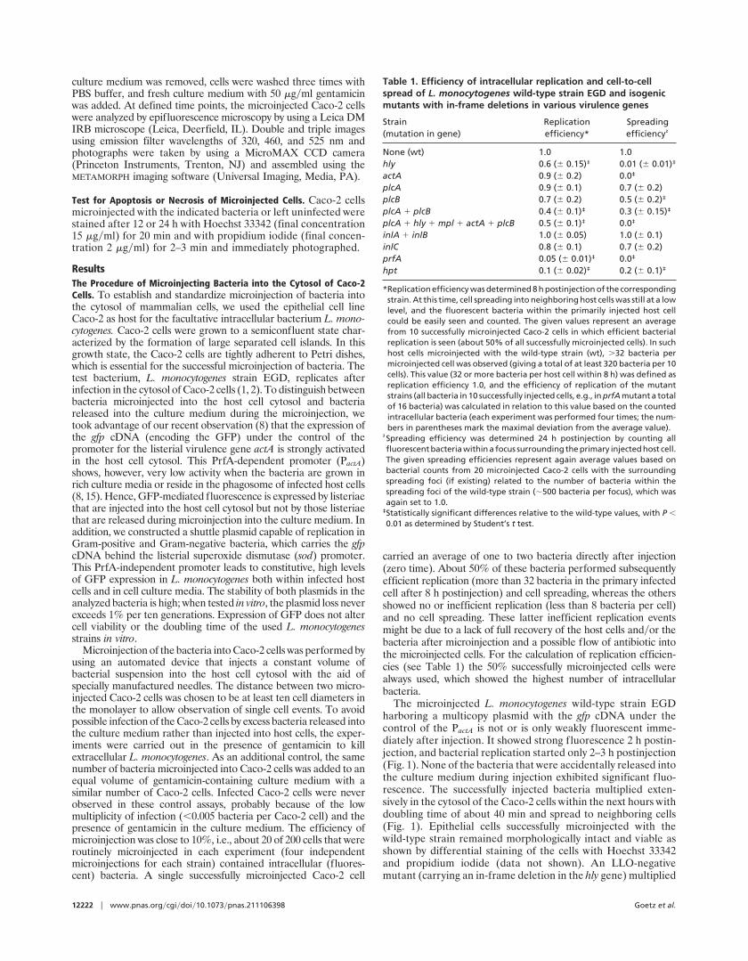
The microinjected L. monocytogenes wild-type strain EGDharboring a multicopy plasmid with the gfp cDNA under thecontrol of the PactA is not or is only weakly fluorescent imme-diately after injection. It showed strong fluorescence 2 h postin-jection, and bacterial replication started only 2–3 h postinjection(Fig. 1). None of the bacteria that were accidentally released intothe culture medium during injection exhibited significant fluo-rescence. The successfully injected bacteria multiplied exten-sively in the cytosol of the Caco-2 cells within the next hours withdoubling time of about 40 min and spread to neighboring cells(Fig. 1). Epithelial cells successfully microinjected with thewild-type strain remained morphologically intact and viable asshown by differential staining of the cells with Hoechst 33342and propidium iodide (data not shown). An LLO-negativemutant (carrying an in-frame deletion in the hly gene) multiplied
Table 1. Efficiency of intracellular replication and cell-to-cellspread of L. monocytogenes wild-type strain EGD and isogenicmutants with in-frame deletions in various virulence genes
Strain(mutation in gene)
Replicationefficiency*
Spreadingefficiency†
None (wt) 1.0 1.0hly 0.6 (6 0.15)‡ 0.01 (6 0.01)‡
actA 0.9 (6 0.2) 0.0‡
plcA 0.9 (6 0.1) 0.7 (6 0.2)plcB 0.7 (6 0.2) 0.5 (6 0.2)‡
plcA 1 plcB 0.4 (6 0.1)‡ 0.3 (6 0.15)‡
plcA 1 hly 1 mpl 1 actA 1 plcB 0.5 (6 0.1)‡ 0.0‡
inlA 1 inlB 1.0 (6 0.05) 1.0 (6 0.1)inlC 0.8 (6 0.1) 0.7 (6 0.2)prfA 0.05 (6 0.01)‡ 0.0‡
hpt 0.1 (6 0.02)‡ 0.2 (6 0.1)‡
*Replication efficiency was determined 8 h postinjection of the correspondingstrain. At this time, cell spreading into neighboring host cells was still at a lowlevel, and the fluorescent bacteria within the primarily injected host cellcould be easily seen and counted. The given values represent an averagefrom 10 successfully microinjected Caco-2 cells in which efficient bacterialreplication is seen (about 50% of all successfully microinjected cells). In suchhost cells microinjected with the wild-type strain (wt), .32 bacteria permicroinjected cell was observed (giving a total of at least 320 bacteria per 10cells). This value (32 or more bacteria per host cell within 8 h) was defined asreplication efficiency 1.0, and the efficiency of replication of the mutantstrains (all bacteria in 10 successfully injected cells, e.g., in prfA mutant a totalof 16 bacteria) was calculated in relation to this value based on the countedintracellular bacteria (each experiment was performed four times; the num-bers in parentheses mark the maximal deviation from the average value).
†Spreading efficiency was determined 24 h postinjection by counting allfluorescent bacteria within a focus surrounding the primary injected host cell.The given spreading efficiencies represent again average values based onbacterial counts from 20 microinjected Caco-2 cells with the surroundingspreading foci (if existing) related to the number of bacteria within thespreading foci of the wild-type strain (;500 bacteria per focus), which wasagain set to 1.0.
‡Statistically significant differences relative to the wild-type values, with P ,0.01 as determined by Student’s t test.
12222 u www.pnas.orgycgiydoiy10.1073ypnas.211106398 Goetz et al.
in the cytosol of the injected Caco-2 cells (Fig. 1), indicating thatthere is no membrane surrounding the injected bacteria (Fig. 1,Table 1). Only a few single bacteria were observed in neighbor-ing Caco-2 cells (see fluorescent bacteria indicated by the arrowsin Fig. 1), suggesting that the hly mutant may still form spreadingvacuoles but is unable to disrupt the vacuole formed duringcell-to-cell spread. Likewise, the actA mutant [which is alsoimpaired in cell-to-cell spread because of the inability of intra-cellular and intercellular movement (1, 2, 9, 10)] replicated in thecytosol of the injected cells at a similar rate as the wild-typestrain (Fig. 1, Table 1). As expected, it did not spread intoneighboring cells (Fig. 1). These data show that L. monocytogenesreproduces its normal behavior upon microinjection into thehost cell cytosol, even if the passage through the phagosomalcompartment is circumvented.
S. flexneri and Enteroinvasive E. coli, but Not Extracellular andFacultative Intracellular Bacteria That Normally Reside in a Vacuole,Replicate in the Cytosol of Caco-2 Cells. Similar to L. monocytogenes,Shigella spp. and the closely related EIEC strains are known toreplicate in the cytosol of infected host cells (16). An icsA mutantof S. flexneri, which is unable to spread to neighboring host cells(17), and the EIEC strain W7062 were transformed with thePsod-gfp-carrying plasmid. The Shigella mutant replicated evenmore efficiently than L. monocytogenes after microinjection intoCaco-2 cells, i.e., up to 70% of the successfully microinjectedhost cells (about 10% of the total microinjected cells) showedmore than 64 bacteria per cell, and the generation time ofShigellae within the Caco-2 cells was only 30 min compared with40 min observed for Listeriae. As expected by the icsA mutation,no cell-to-cell-spread was observed (Fig. 2). The EIEC wild-typestrain also replicated in the host cell cytosol (Fig. 2), butreplication was much less efficient than that of S. flexneri—i.e.,the number of replicating bacteria rarely exceeded 32, and onlyabout 30% of the successfully microinjected host cells showedreplicating bacteria. Unexpectedly, spreading of these bacteriainto neighboring cells was rarely observed. To determinewhether this was a strain-specific defect, we tested three addi-tional EIEC wild-type strains from different strain collectionsand obtained similar results. These data suggest that EIEC
strains are apparently less proficient in cytosolic replication andcell spreading (at least in Caco-2 cells) than S. flexneri.
S. enterica serovar Thyphimurium and L. pneumophila multiply inspecialized phagosomal compartments of phagocytic and nonph-agocytic mammalian host cells (3, 4). We have previously shown(11) that Salmonella typhimurium expressing LLO can disrupt thephagosomal membrane, it reaches the cytosol, but it does notproficiently replicate. All of the above bacterial species weretransformed with the Psod-gfp plasmid with the exception of L.pneumophila. For the latter microorganism, gfp cDNA was placedunder the control of the promoter of the mip gene (12). All strainsexhibited strong fluorescence already under extracellular condi-tions. Expression of GFP did not significantly affect cell viabilityand replication rate in vitro. When the S. typhimurium strain wasmicroinjected into Caco-2 cells, the injected bacteria showed strongfluorescence immediately upon injection, and fluorescence did notdiminish during the monitored 24 h (Fig. 2). Multiplication wasobserved in none of the Caco-2 cells, which carried a singlefluorescent bacterium during the entire 24-h observation period.Similar results were obtained after microinjection of GFP-tagged L.pneumophila (data not shown). It is unlikely that this failure ofintracellular replication is caused by gentamicin present during themicroinjection procedure because these latter Gram-negative bac-teria are even less sensitive to the antibiotic than L. monocytogenes,and microinjection performed in the absence of gentamicin also didnot show intracellularly growing bacteria. Although we cannotcompletely rule out the possibility that these latter bacteria aremore sensitive to the manipulation by the microinjection than L.monocytogenes, S. flexneri, and EIEC, our data rather suggest thatthese intracellular bacteria are unable to grow in the cytosol ofCaco-2 cells.
The inability of cytosolic replication of the latter bacteria aftermicroinjection into Caco-2 cells was surprising in the light ofprevious work (7), which showed that even truly extracellularbacteria, like B. subtilis, replicate in the cytosol of macrophageswhen provided with listeriolysin to open the phagosomal compart-ment. We therefore microinjected prototrophic extracellular patho-genic and nonpathogenic bacteria including Yersinia enterocolitica,the well characterized uropathogenic E. coli strain 536 (18), and B.subtilis DSM 401 into Caco-2 cells. These bacterial strains were also
Fig. 1. Microinjection of wild-type (WT) and mutant strains (Dhly and DactA) of L. monocytogenes into Caco-2 cells. Microinjection into Caco-2 cells wasperformed as described in Materials and Methods, using L. monocytogenes wild-type strain EGD and isogenic mutants with deletions in hly and actA. All threestrains harbor the same plasmid carrying PactA-gfp. Bacterial replication was determined after 3, 10, and 24 h. Arrows mark single fluorescent bacteria, whichappear 3 h after microinjection.
Goetz et al. PNAS u October 9, 2001 u vol. 98 u no. 21 u 12223
MIC
ROBI
OLO
GY
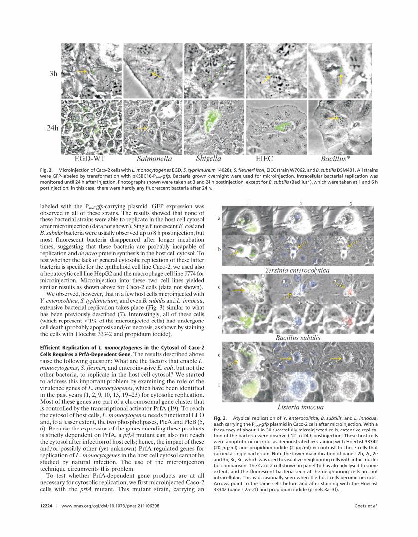
labeled with the Psod-gfp-carrying plasmid. GFP expression wasobserved in all of these strains. The results showed that none ofthese bacterial strains were able to replicate in the host cell cytosolafter microinjection (data not shown). Single fluorescent E. coli andB. subtilis bacteria were usually observed up to 8 h postinjection, butmost fluorescent bacteria disappeared after longer incubationtimes, suggesting that these bacteria are probably incapable ofreplication and de novo protein synthesis in the host cell cytosol. Totest whether the lack of general cytosolic replication of these latterbacteria is specific for the epithelioid cell line Caco-2, we used alsoa hepatocytic cell line HepG2 and the macrophage cell line J774 formicroinjection. Microinjection into these two cell lines yieldedsimilar results as shown above for Caco-2 cells (data not shown).
We observed, however, that in a few host cells microinjected withY. enterocolitica, S. typhimurium, and even B. subtilis and L. innocua,extensive bacterial replication takes place (Fig. 3) similar to whathas been previously described (7). Interestingly, all of these cells(which represent ,1% of the microinjected cells) had undergonecell death (probably apoptosis andyor necrosis, as shown by stainingthe cells with Hoechst 33342 and propidium iodide).
Efficient Replication of L. monocytogenes in the Cytosol of Caco-2Cells Requires a PrfA-Dependent Gene. The results described aboveraise the following question: What are the factors that enable L.monocytogenes, S. flexneri, and enteroinvasive E. coli, but not theother bacteria, to replicate in the host cell cytosol? We startedto address this important problem by examining the role of thevirulence genes of L. monocytogenes, which have been identifiedin the past years (1, 2, 9, 10, 13, 19–23) for cytosolic replication.Most of these genes are part of a chromosomal gene cluster thatis controlled by the transcriptional activator PrfA (19). To reachthe cytosol of host cells, L. monocytogenes needs functional LLOand, to a lesser extent, the two phospholipases, PlcA and PlcB (5,6). Because the expression of the genes encoding these productsis strictly dependent on PrfA, a prfA mutant can also not reachthe cytosol after infection of host cells; hence, the impact of theseandyor possibly other (yet unknown) PrfA-regulated genes forreplication of L. monocytogenes in the host cell cytosol cannot bestudied by natural infection. The use of the microinjectiontechnique circumvents this problem.
To test whether PrfA-dependent gene products are at allnecessary for cytosolic replication, we first microinjected Caco-2cells with the prfA mutant. This mutant strain, carrying an
Fig. 2. Microinjection of Caco-2 cells with L. monocytogenes EGD, S. typhimurium 14028s, S. flexneri iscA, EIEC strain W7062, and B. subtilis DSM401. All strainswere GFP-labeled by transformation with pKSBC16-Psod-gfp. Bacteria grown overnight were used for microinjection. Intracellular bacterial replication wasmonitored until 24 h after injection. Photographs shown were taken at 3 and 24 h postinjection, except for B. subtilis (Bacillus*), which were taken at 1 and 6 hpostinjection; in this case, there were hardly any fluorescent bacteria after 24 h.
Fig. 3. Atypical replication of Y. enterocolitica, B. subtilis, and L. innocua,each carrying the Psod-gfp plasmid in Caco-2 cells after microinjection. With afrequency of about 1 in 30 successfully microinjected cells, extensive replica-tion of the bacteria were observed 12 to 24 h postinjection. These host cellswere apoptotic or necrotic as demonstrated by staining with Hoechst 33342(20 mgyml) and propidium iodide (2 mgyml) in contrast to those cells thatcarried a single bacterium. Note the lower magnification of panels 2b, 2c, 2eand 3b, 3c, 3e, which was used to visualize neighboring cells with intact nucleifor comparison. The Caco-2 cell shown in panel 1d has already lysed to someextent, and the fluorescent bacteria seen at the neighboring cells are notintracellular. This is occasionally seen when the host cells become necrotic.Arrows point to the same cells before and after staining with the Hoechst33342 (panels 2a–2f) and propidium iodide (panels 3a–3f).
12224 u www.pnas.orgycgiydoiy10.1073ypnas.211106398 Goetz et al.
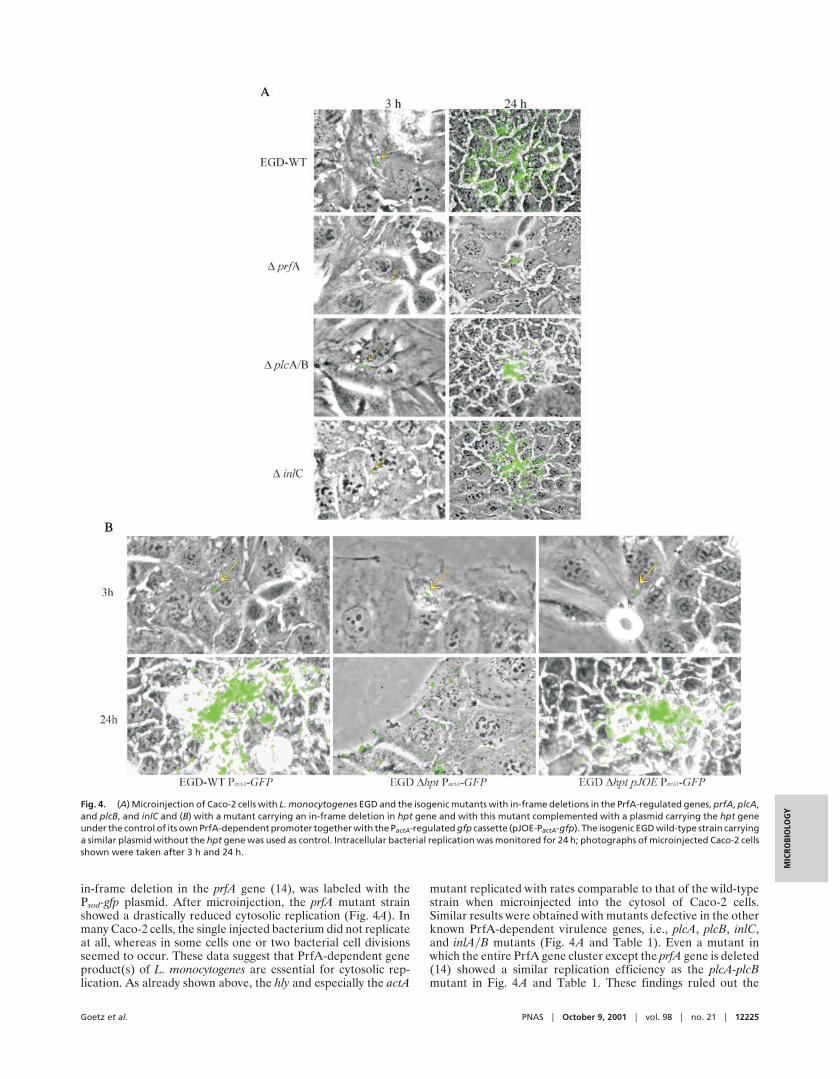
in-frame deletion in the prfA gene (14), was labeled with thePsod-gfp plasmid. After microinjection, the prfA mutant strainshowed a drastically reduced cytosolic replication (Fig. 4A). Inmany Caco-2 cells, the single injected bacterium did not replicateat all, whereas in some cells one or two bacterial cell divisionsseemed to occur. These data suggest that PrfA-dependent geneproduct(s) of L. monocytogenes are essential for cytosolic rep-lication. As already shown above, the hly and especially the actA
mutant replicated with rates comparable to that of the wild-typestrain when microinjected into the cytosol of Caco-2 cells.Similar results were obtained with mutants defective in the otherknown PrfA-dependent virulence genes, i.e., plcA, plcB, inlC,and inlAyB mutants (Fig. 4A and Table 1). Even a mutant inwhich the entire PrfA gene cluster except the prfA gene is deleted(14) showed a similar replication efficiency as the plcA-plcBmutant in Fig. 4A and Table 1. These findings ruled out the
Fig. 4. (A) Microinjection of Caco-2 cells with L. monocytogenes EGD and the isogenic mutants with in-frame deletions in the PrfA-regulated genes, prfA, plcA,and plcB, and inlC and (B) with a mutant carrying an in-frame deletion in hpt gene and with this mutant complemented with a plasmid carrying the hpt geneunder the control of its own PrfA-dependent promoter together with the PactA-regulated gfp cassette (pJOE-PactA-gfp). The isogenic EGD wild-type strain carryinga similar plasmid without the hpt gene was used as control. Intracellular bacterial replication was monitored for 24 h; photographs of microinjected Caco-2 cellsshown were taken after 3 h and 24 h.
Goetz et al. PNAS u October 9, 2001 u vol. 98 u no. 21 u 12225
MIC
ROBI
OLO
GY

known PrfA-dependent gene products for being responsible forthe observed strong impairment of the prfA mutant in cytosolicreplication and suggested that PrfA-dependent gene product(s)other than the known PrfA-dependent virulence factors wereindispensable for cytosolic replication of L. monocytogenes.
The PrfA-Regulated hpt Gene Encoding an Uptake System for Phos-phorylated Sugars Is Required for Efficient Replication of L. monocy-togenes in the Cytosol After Microinjection. It has been reported thatL. monocytogenes utilizes glucose-1-phosphate (G-1-P) in a strictlyPrfA-dependent manner (24). G-1-P is a possible carbon source forintracellular growth of L. monocytogenes in mammalian host cellsgenerated by degradation of glycogen (M.B., unpublished data).The gene hpt, responsible for G-1-P uptake in L. monocytogenesencoding a sugar phosphate permease, was recently identified. Thegene has been shown to be tightly regulated by PrfA. An L.monocytogenes mutant with an in-frame deletion in hpt is unable togrow on G-1-P in vitro (I.C.-C., unpublished results). When micro-injected in Caco-2 cells, this mutant strain showed strongly impairedreplication in the cytosol; cell spreading occurred also at a reducedrate (Fig. 4B and Table 1). Cytosolic replication and cell spreadingwere restored to almost wild-type levels upon complementationwith a plasmid carrying the hpt gene under the control of itsPrfA-dependent promotor (Fig. 4B). These results suggest thatG-1-P possibly may serve as a carbon source for growth of L.monocytogenes in the host cell cytosol, where uptake is facilitated bythe PrfA-regulated hpt gene.
DiscussionThe microinjection technique described here allows the directinjection of single bacteria into the cytosol of mammalian cells,such as epithelial cells, hepatocytes, or macrophages. Our dataobtained with this method show that the cytosol of a mammaliancell is not a rich nutrient broth that would support bacterialmultiplication in general (7, 25). Indeed, only few bacteria areable to efficiently replicate in this compartment after microin-jection. Interestingly, all bacteria found to be capable of cytosolicreplication after microinjection are facultative intracellularpathogens, like Shigella spp., the related enteroinvasive E. colistrains, and L. monocytogenes, which are already naturallyadapted to replication in the cytosol. These results lend supportto the idea that the proper adaptation of the bacterial metab-olism to the host cell environment is essential for successfulreplication in a specific host cell compartment (26). Obviously,
only a few of the tested bacterial species fulfill the metabolicrequirements for growth in the cytosol of mammalian cells. In L.monocytogenes, cytosolic replication seems to require the genefor a sugar phosphate transporter specifically controlled by thetranscription factor, PrfA, which differentially regulates most ofthe known listerial virulence genes (1, 2, 15, 19). PrfA is knownto be active particularly when the bacteria grow in the cytosol ofhost cells (8, 15), and the induced expression of this sugar uptakesystem by the host cell cytosol environment may enable L.monocytogenes to utilize glucose-1-phosphate (24), which ispossibly derived from glycogen of the host cell. Similar uptakesystems for phosphorylated sugars are also present in E. coli (27)and possibly other bacteria, but their expression in the cytosolicmilieu of host cells may be less efficient than the PrfA-regulateduptake system of L. monocytogenes. Further experiments willshow whether this PrfA-controlled uptake of glucose-1-phosphate is the only specific metabolic requirement for cyto-solic growth of L. monocytogenes.
At a low frequency, Caco-2 cells harboring a large number ofbacteria in the cytosol are observed with all tested bacterial speciesafter microinjection (about 1% of the cells containing microin-jected bacteria). All of these cells have undergone cell death. Theserare events are observed at a rate that corresponds roughly to therate of spontaneous apoptosis of Caco-2 cells under the cultureconditions. We therefore assume that this atypical cytosolic repli-cation occurs in apoptotic cells only and that such cells may providemore readily available resources for bacterial growth. The replicat-ing bacteria in these cells are always seen around the cell nucleus,suggesting that components released from the apoptotic nucleusmay serve as nutrients for bacterial growth. We suggest that theextensive cytosolic replication observed previously with LLO-expressing B. subtilis (7) and apathogenic L. innocua (28) might bealso the result of such events. An alternative explanation could bethat bacterial growth in the cytosol of intact host cells is restrictedby antibacterial components, and this restriction is abolished inapoptotic (necrotic) cells (29).
The described technique of microinjecting bacteria directlyinto the cytosol of mammalian cells may help to further unravelin detail the specific requirements for bacterial replication in thehost cell cytosol.
We thank P. Sansonetti and H. Karch for providing strains and R. Gross,M. Kuhn, and J. Daniels for helpful discussions and critical reading ofthe manuscript. This work was supported by the Deutsche Forschungs-gemeinschaft (SFB 479 B1) and the Fond der Chemischen Industrie.
1. Cossart, P. & Lecuit, M. (1998) EMBO J. 17, 3797–3806.2. Kuhn, M. & Goebel, W. (1997) Immunol. Rev. 158, 57–67.3. Finlay, B. & Falkow, S. (1997) Microbiol. Mol. Biol. Rev. 61, 136–169.4. Galan, J. E. (1996) Mol. Microbiol. 20, 263–271.5. Camilli, A., Goldfine, H. & Portnoy, D. A. (1991) J. Exp. Med. 173, 751–754.6. Smith, G. A. & Portnoy, D. A. (1993) Infect. Agents Dis. 2, 183–185.7. Bielecki, J., Youngman, P., Connelly, P. & Portnoy, D. A. (1990) Nature
(London) 345, 175–176.8. Dietrich, G., Bubert, A., Gentschev, I., Sokolovic, Z., Simm, A., Catic, A.,
Kaufmann, S. H., Hess, J., Szalay, A. A. & Goebel, W. (1998) Nat. Biotechnol.16, 181–185.
9. Kocks, C., Gouin, E., Tabouret, M., Berche, P., Ohayon, H. & Cossart, P. (1992)Cell 68, 521–531.
10. Domann, E., Wehland, J., Rohde, M., Pistor, S., Hartl, M., Goebel, W.,Leimeister-Wachter, M., Wuenscher, M. & Chakraborty, T. (1992) EMBO J.11, 1981–1990.
11. Gentschev, I., Sokolovic, Z., Mollenkopf, H. J., Hess, J., Kaufmann, S. H.,Kuhn, M., Krohne, G. F. & Goebel, W. (1995) Infect. Immun. 63, 4203–4205.
12. Engleberg, N. C., Carter, C., Weber, D. R., Cianciotto, N. P. & Eisenstein, B. I.(1989) Infect. Immun. 57, 1263–1270.
13. Engelbrecht, F., Chun, S.K., Ochs, C., Hess, J., Lottspeich, F. Goebel, W. &Sokolovic, Z. (1996) Mol. Microbiol. 21, 823–837.
14. Bockmann, R., Dickneite, C., Middendorf, B., Goebel, W. & Sokolovic, Z.(1996) Mol. Microbiol. 22, 643–653.
15. Bubert, A., Sokolovic, Z., Chun, S. K., Papatheodorou, L., Simm, A. & Goebel,W. (1999) Mol. Gen. Genet., 261, 323–336.
16. Zagaglia, C., Casalino, M., Colonna, B., Conti, C., Calconi, A. & Nicoletti, M.(1991) Infect. Immun. 59, 792–799.
17. Bernardini, M. L., Mounier, J., d’Hauteville, H., Coquis-Rondon, M. &Sansonetti, P. J. (1989) Proc. Natl. Acad. Sci. USA 86, 3867–3871.
18. Ritter, A., Blum, G., Emody, L., Kerenyi, M., Bock, A., Neuhierl, B., Rabsch,W., Scheutz, F. & Hacker, J. (1995) Mol. Microbiol. 17, 109–121.
19. Leimeister-Wachter, M., Haffner, C., Domann, E., Goebel, W. & Chakraborty,T. (1990) Proc. Natl. Acad. Sci. USA 87, 8336–8340.
20. Dramsi, S., Kocks, C., Forestier, C. & Cossart, P. (1993) Mol. Microbiol. 9, 931–941.21. Gaillard, J. L., Berche, P., Frehel, C., Gouin, E. & Cossart, P. (1991) Cell 65,
1127–1141.22. Lingnau, A., Domann, E., Hudel, M., Bock, M., Nichterlein, T., Wehland, J. &
Chakraborty, T. (1995) Infect. Immun. 63, 3896–3903.23. Raffelsbauer, D., Bubert, A., Engelbrecht, F., Scheinpflug, J., Simm, A., Hess,
J., Kaufmann, S. H. & Goebel, W. (1998) Mol. Gen. Genet. 260, 144–158.24. Ripio, M. T., Brehm, K., Lara, M., Suarez, M. & Vazquez-Boland, J. A. (1997)
J. Bacteriol. 179, 7174–7180.25. Marquis, H., Bouwer, H. G., Hinrichs, D. J. & Portnoy, D. A. (1993) Infect.
Immun. 61, 3756–3760.26. Goebel, W. & Kuhn, M. (2000) Curr. Opin. Microbiol. 3, 49–53.27. Island, M. D., Wei, B. Y. & Kadner, R. J. (1992) J. Bacteriol. 174, 2754–2762.28. Falzano, L., Fiorentini, C., Donelli, G., Michel, E., Kocks, C., Cossart, P.,
Cabanie, L., Oswald, E. & Boquet, P. (1993) Mol. Microbiol. 9, 1247–1254.29. Hiemstra, P. S., van der Barselaar, M. T., Roest, M., Nibbering, P. H. & van
Furth, R. (1999) J. Leukocyte Biol. 66, 423–428.
12226 u www.pnas.orgycgiydoiy10.1073ypnas.211106398 Goetz et al.
Copyright © 2022 FDOKUMEN