Nonselective cation channels as effectors of free radical–induced rat liver cell necrosis
Upload
independentCategory
view
1download
0
http://www.elsevier.com/locate/bba
Biochimica et Biophysica A
Review
The mammalian TRPC cation channels
Guillermo Vazquez, Barbara J. Wedel, Omar Aziz, Mohamed Trebak, James W. Putney Jr.*
The Calcium Regulation Section, National Institute of Environmental Health Sciences, National Institutes of Health,
Department of Health and Human Services, 111 TW Alexander Dr., Research Triangle Park, NC 27709, United States
Received 3 August 2004; received in revised form 27 August 2004; accepted 28 August 2004
Available online 11 September 2004
Abstract
Transient Receptor Potential-Canonical (TRPC) channels are mammalian homologs of Transient Receptor Potential (TRP), a Ca2+-
permeable channel involved in the phospholipase C-regulated photoreceptor activation mechanism in Drosophila. The seven mammalian
TRPCs constitute a family of channels which have been proposed to function as store-operated as well as second messenger-operated
channels in a variety of cell types. TRPC channels, together with other more distantly related channel families, make up the larger TRP
channel superfamily. This review summarizes recent findings on the structure, regulation and function of the apparently ubiquitous
TRPC cation channels.
Published by Elsevier B.V.
Keywords: Ion channel; Calcium channel; Non-selective cation channel; TRP channel; TRPC channel; Capacitative calcium entry; Store-operated channel;
Second messenger-operated channel; Inositol trisphosphate receptor; Diacylglycerol; Phospholipase C
1. Origin, classification and nomenclature of TRPCs
The recognition that the protein product derived from
the transient receptor potential (trp) gene from Drosophila
melanogaster encoded a PLC-activated Ca2+ permeable
channel eventually led to identification of seven TRP
homologs in mammals [1,2]. Today, the TRP superfamily
of channel forming proteins is comprised of these seven
homologs of Drosophila TRP together with more
distantly related channel genes identified through other
searches for channels with specific functions [3–8]. TRP
superfamily proteins fall into one of the three major
families on a phylogenetic basis: the TRPC or canonical
TRP family, with seven members (TRPC1 through
TRPC7), which are the most closely related to the
original Drosophila TRP channel; the TRPV family, with
six members (TRPV1–6) named after the first group
0167-4889/$ - see front matter. Published by Elsevier B.V.
doi:10.1016/j.bbamcr.2004.08.015
* Corresponding author. Tel.: +1 919 541 1420; fax: +1 919 541
7879.
E-mail address: [email protected] (J.W. Putney).
member, the vanilloid receptor; and the TRPM family,
with eight members (TRPM1–8), named after the original
member, melastatin (Fig. 1). Other more distantly related
members of the superfamily have been identified as genes
whose dysfunction forms the bases for certain inherited
diseases. The TRP superfamily and the corresponding
family and subfamily designations follow a recent
consensus nomenclature [9] that will be used throughout
this review.
TRP superfamily members are cation channels
involved in a continually growing number of cellular
functions [6], and overall share 20–60% homology. Based
on structural and functional similarities, the TRPC family
can be further subdivided into four different subfamilies:
TRPC1, TRPC2, TRPC3, 6 and 7 and TRPC4 and 5 (Fig.
1). TRPC2 is a pseudogene in humans, and in old world
monkeys and apes [10], but TRPC2 apparently forms
fully regulated channels in other mammalian species (see
below). TRPC3, 6 and 7 form a closely related subfamily,
sharing a high degree of amino acid identity (70–80%)
and functional, regulatory and pharmacological similar-
ities. A similarly close structural, and apparently func-
cta 1742 (2004) 21–36
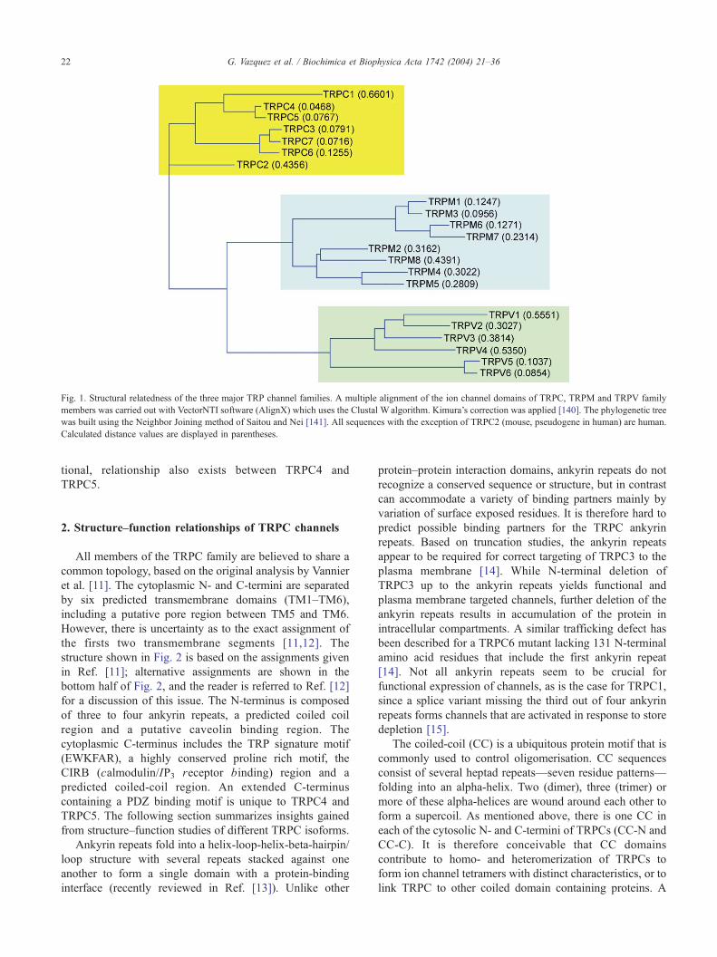
Fig. 1. Structural relatedness of the three major TRP channel families. A multiple alignment of the ion channel domains of TRPC, TRPM and TRPV family
members was carried out with VectorNTI software (AlignX) which uses the Clustal W algorithm. Kimura’s correction was applied [140]. The phylogenetic tree
was built using the Neighbor Joining method of Saitou and Nei [141]. All sequences with the exception of TRPC2 (mouse, pseudogene in human) are human.
Calculated distance values are displayed in parentheses.
G. Vazquez et al. / Biochimica et Biophysica Acta 1742 (2004) 21–3622
tional, relationship also exists between TRPC4 and
TRPC5.
2. Structure–function relationships of TRPC channels
All members of the TRPC family are believed to share a
common topology, based on the original analysis by Vannier
et al. [11]. The cytoplasmic N- and C-termini are separated
by six predicted transmembrane domains (TM1–TM6),
including a putative pore region between TM5 and TM6.
However, there is uncertainty as to the exact assignment of
the firsts two transmembrane segments [11,12]. The
structure shown in Fig. 2 is based on the assignments given
in Ref. [11]; alternative assignments are shown in the
bottom half of Fig. 2, and the reader is referred to Ref. [12]
for a discussion of this issue. The N-terminus is composed
of three to four ankyrin repeats, a predicted coiled coil
region and a putative caveolin binding region. The
cytoplasmic C-terminus includes the TRP signature motif
(EWKFAR), a highly conserved proline rich motif, the
CIRB (calmodulin/IP3 receptor binding) region and a
predicted coiled-coil region. An extended C-terminus
containing a PDZ binding motif is unique to TRPC4 and
TRPC5. The following section summarizes insights gained
from structure–function studies of different TRPC isoforms.
Ankyrin repeats fold into a helix-loop-helix-beta-hairpin/
loop structure with several repeats stacked against one
another to form a single domain with a protein-binding
interface (recently reviewed in Ref. [13]). Unlike other
protein–protein interaction domains, ankyrin repeats do not
recognize a conserved sequence or structure, but in contrast
can accommodate a variety of binding partners mainly by
variation of surface exposed residues. It is therefore hard to
predict possible binding partners for the TRPC ankyrin
repeats. Based on truncation studies, the ankyrin repeats
appear to be required for correct targeting of TRPC3 to the
plasma membrane [14]. While N-terminal deletion of
TRPC3 up to the ankyrin repeats yields functional and
plasma membrane targeted channels, further deletion of the
ankyrin repeats results in accumulation of the protein in
intracellular compartments. A similar trafficking defect has
been described for a TRPC6 mutant lacking 131 N-terminal
amino acid residues that include the first ankyrin repeat
[14]. Not all ankyrin repeats seem to be crucial for
functional expression of channels, as is the case for TRPC1,
since a splice variant missing the third out of four ankyrin
repeats forms channels that are activated in response to store
depletion [15].
The coiled-coil (CC) is a ubiquitous protein motif that is
commonly used to control oligomerisation. CC sequences
consist of several heptad repeats—seven residue patterns—
folding into an alpha-helix. Two (dimer), three (trimer) or
more of these alpha-helices are wound around each other to
form a supercoil. As mentioned above, there is one CC in
each of the cytosolic N- and C-termini of TRPCs (CC-N and
CC-C). It is therefore conceivable that CC domains
contribute to homo- and heteromerization of TRPCs to
form ion channel tetramers with distinct characteristics, or to
link TRPC to other coiled domain containing proteins. A

Fig. 2. Structure of TRPC channels. Top: Structural features of TRPC channels. See text for description of structural features. ANK, ankyrin-like repeats; CC-N
and CC-C are N-terminal and C-terminal coiled-coil domains, respectively. The region shaded in gray at the extreme C-terminus is specific for TRPC 4 and 5.
Bottom: The arrangement of the first three hydrophobic segments of TRPCs is uncertain; three alternatives are shown (A, B and C). See Refs. [11,12]. The
acronyms are: PDZ-B, PDZ binding domain; CC-N, N-terminal coiled coil region; CC-C, C-terminal coiled coil region; ANK1–4, ankyrin repeats 1–4; CIRB,
calmodulin/IP3 receptor binding region; PLC, phospholipase C; IP3R, IP3 receptor; CaM, calmodulin; LFW, amino acid motif conserved in the putative pore
region of all TRPCs.
G. Vazquez et al. / Biochimica et Biophysica Acta 1742 (2004) 21–36 23
recent study showed that CC-N of TRPC1 was able to
homodimerize based on a yeast two-hybrid screen while the
C-terminus including CC-C was unable to dimerize [16].
The microtubule destabilizing phosphoprotein stathmin has
recently been pulled out of a yeast two-hybrid screen using
the N-terminus of TRPC5 as a bait, and CC-N was found to
be responsible for the interaction, presumably via the
predicted CC region of stathmin [17]. This interaction
seemed to be specific for TRPC4 and TRPC5 since the N-
termini of TRPC4 and 5 but not TRPC1 or TRPC7 were
able to interact with the stathmin helical domain. TRPC5
appears to regulate axon length and growth cone morphol-
ogy in hippocampal neurons where its activation and
associated Ca2+ influx may prevent random axonal out-
growth. The C-terminal CC region can be deleted in TRPC3
without loss of plasma membrane targeting and channel
activation when compared with the wild type [14]. How-
ever, in TRPC1, calmodulin (CaM) binding to the predicted
CC-C domain has been reported and its deletion resulted in
diminished Ca2+-dependent inactivation of store-operated
Ca2+ entry [18].
Caveolins are enriched in plasma membrane micro-
domains called caveolae (for a recent review see Ref.
[19]). Co-immunoprecipitation or co-localization with cav-
eolins has been shown for TRPC1 and TRPC3 [20–22].
Caveolin-1 binding usually occurs at aromatic-rich (u)
regions that contain a specific spacing following the
consensus sequences uXuX4u, uX4uX2u, or uXuX4uX2u
[23]. A similar motif is conserved in all members of the
TRPC family located in the cytosolic N-terminus adjacent to
the first transmembrane domain [20]. Deletion of this region
prevents TRPC1 from targeting to the plasma membrane
and the respective mutant dominant-negatively affects store-
operated Ca2+ entry [20].
A systematic analysis of the predicted pore region of
TRPC family members has not yet been carried out.
However, several studies have examined the effect of point
mutations within the predicted pore region and extrac-
G. Vazquez et al. / Biochimica et Biophysica Acta 1742 (2004) 21–3624
ellular loops bordering the pore. Change of a conserved
LFW motif in the predicted pore helix of TRPC5 and
TRPC6 to AAA resulted in dominant negative mutants
[24,25]. A means to discriminate endogenous receptor
operated/store operated channels from overexpressed
TRPCs has been the fact that the latter are insensitive to
inhibition by lower micromolar concentrations of lantha-
nides. In the case of TRPC4 and TRPC5, higher micro-
molar concentrations of lanthanides potentiate currents,
while they inhibit overexpressed TRPC6 [26]. Jung et al.
[26] identified charged residues in the pore loops of
TRPC5 that when mutated from E to Q (E543Q and
E595Q/E598Q) caused a loss of lanthanide potentiation.
Likewise, E576K or D581K mutants of TRPC1 exhibited
decreased store-mediated Ca2+ but not Na+ current
suggesting that these residues play an important role in
calcium selectivity [27]. This is an interesting finding since
conventional thought of the function of a nonselective
cation channel would not predict that mutations would
affect Ca2+ permeability selectively; in other words, the
mutation was in reality a gain of function mutation
converting a nonselective channel to one selective for
monovalent cations. Interestingly, E576 is conserved
between TRPC1, 4 and 5 and part of a CX4CE motif
unique to these isoforms. Since these cysteines likely face
the extracellular space, they may form disulfide bridges
thus potentially explaining preferred heteromer formation
between TRPC1, 4 and 5 on one hand and the dcysteine-lessT TRPC3, 6 and 7 on the other [25].
A proline-rich motif (LPXPFXXXPSPK) downstream of
the EWKFAR motif is conserved in all members of the
TRPC family and is thought to be responsible for interaction
with Homer (for TRPC1) and/or immunophilins [28,29].
Homer proteins dimerize via a CC motif while the EVH
domain of each monomer is able to bind to a consensus
PPXXF motif in their target molecules thus linking, for
example, cell surface receptors and intracellular Ca2+ release
channels. In analogy to this scheme Homer binding to
TRPC1 and the IP3 receptor would make it a linker involved
in the proposed conformational coupling model of TRP
channel activation (discussed in a subsequent section). In
support of this hypothesis, mutations within the proline-rich
motif disrupted Homer binding and resulted in channels
with high constitutive activity and greatly reduced agonist
regulation [28]. A direct interaction of TRPC channels with
IP3 receptors will be discussed separately (Section 4.2). It is,
however, important to note that a TRPC1 mutant lacking the
entire cytoplasmic C-terminus was apparently able to
function as a store-operated channel [30].
Recently an interaction of TRPC with the immunophilins
FKBP12 and FKBP52 has been demonstrated [29]. Immu-
nophilins are peptidyl-prolyl cis–trans isomerases binding
to a XP motif in their binding partners. Pulldown experi-
ments showed that FKBP12 preferentially interacted with
TRPC3, 6 and 7, while FKBP52 preferred TRPC1, 4 and 5.
Mutation of the first proline in the proline rich motif of
TRPCs disrupted that interaction. Furthermore, FK506,
which displaces immunophilins from their targets, was able
to inhibit agonist-induced currents through TRPC6 channels
[29].
Evidence for functional roles of the CIRB (CaM/IP3receptor binding) region is discussed in Section 4.2.3. As
mentioned above, TRPC4 and 5 share an extended C-
terminus terminating in a PDZ binding motif. A part of this
region is missing in a TRPC4h splice variant, and this
variant has been proposed to have autoinhibitory function
[31]. This region has also been shown to interact with the C-
terminus of IP3 receptors and CaM [32,33]. Little is known
about the function of this region in TRPC5 and the degree of
homology between the C-termini of TRPC4 and TRPC5 is
very low. The PDZ binding motif TRL of TRPC4 and
TRPC5 seems to be responsible for the interaction with the
adaptor protein, NHERF, possibly linking the channels to
PLCh and the cytoskeleton [34]. Deletion of the PDZ
binding motif of TRPC4 not only reduced the surface
expression of TRPC4 but also changed it from a general
membrane distribution to a predominant expression in cell
outgrowths [35].
Recently, different patterns of N-glycosylation have been
summoned to explain different levels of constitutive activity
within members of the TRPC3, 6 and 7 family. TRPC3,
which is supposedly monoglycosylated in the first extrac-
ellular loop, shows high constitutive activity, while TRPC6,
dually glycosylated in the first and second extracellular loop
is tightly regulated. By converting TRPC3 into the TRPC6-
like dually glycosylated version and likewise converting
TRPC6 into the monoglycosylated TRPC3-like version,
Dietrich et al. [36] were able to alter constitutive activity as
hypothesized. It is worth noting, however, that TRPC7
exhibits considerable basal activity despite the fact that it
has a predicted N-glycosylation site in the first and second
extracellular loop like TRPC6 [37].
The major structural features of TRPC channel subunits
are summarized in Fig. 2.
3. Pharmacology and electrophysiological properties
3.1. TRPC1
TRPC1 was the first member of the TRPC family to be
cloned [1,2], and the first shown by electrophysiological
measurements to form a calcium-permeable cation channel
[15]. In this initial study, expression of TRPC1 in Chinese
hamster ovary cells (CHO) resulted in a linear nonselective
cation current, activated by intracellular infusion (via the
patch pipet) of either IP3 or thapsigargin to deplete
intracellular calcium stores. This current was shown to be
sensitive to Gd3+ (20 AM). Noise analysis provided
evidence that the current was passed through single
channels with a conductance of 16 pS. The current–voltage
relationship of these channels was shown to be predom-

G. Vazquez et al. / Biochimica et Biophysica Acta 1742 (2004) 21–36 25
inantly inward, with little outward current [38,39]. In this
respect, the TRPC1 current was described as Icrac-like [40]
(the properties of Icrac are discussed in a subsequent section
dealing with regulation of TRPC channels, and the behavior
of store-operated channels), although unlike Icrac, the current
was not Ca2+-selective.
3.2. TRPC2
There has been little direct work to date regarding the
electrophysiological characteristics of TRPC2. Work on
mice vomeronasal neural dendrites has shown that knocking
out the TRPC2 gene resulted in the near loss of a
diacylglycerol (DAG)-activated linear current, which was
not activated by store depletion [41], suggesting that TRPC2
may be a component of a DAG-operated channel in situ.
However, using fluorescence rather than electrophysiolog-
ical methodology, Vannier et al. [42] reported that expressed
TRPC2 appeared to behave as a store-operated channel.
3.3. TRPC3, 6 and 7
TRPC3, 6 and 7 appear to form nonselective cation
channels that show both inward and outward rectification, at
negative and positive voltages, respectively. The current–
voltage relationships for these TRPCs demonstrate a far
greater outward current (at positive voltages) than inward
current (at negative voltages) with a reversal potential at
around 0 mV.
Zhu et al. [43] first described TRPC3 as an agonist-
activated channel when expressed in HEK293 cells and
demonstrated that it was relatively insensitive to low
concentrations of Gd3+, but was blocked by concentrations
in the 100 AM range. Hofmann et al. [44] later demonstrated
that TRPC3 could also be activated directly by DAG
analogues.
TRPC3 channels have also been shown to have two
potential conductance states when expressed in HEK293
cells, one of 66pS and one of 17pS [45]. In this study it was
shown that these channels were activated by calcium store
depletion in intact cells, consistent with an earlier report by
Preuh et al. [46], or by addition of recombinant IP3receptors in excised patches. In a subsequent study,
however, activation of TRPC3 was shown to be dependent
solely upon DAG with no requirement for either IP3 or the
IP3 receptor [47].
Since their discovery, these channels have been shown
to be sensitive to a variety of agents with relative
sensitivities varying dramatically depending upon expres-
sion system. In HEK293 cells, TRPC3 was inhibited by
high concentrations of: SKF96365, verapamil, La3+, Gd3+
and Ni2+ [43] and was partially inhibited by moderate (30
AM) concentrations of 2-aminoethoxydiphenyl borane
(2APB) [48]; in DT40 cells by low concentrations of
Gd3+ [48]; and in CHO cells they are reported to be
sensitive to low concentrations of La3+, Gd3+ and
SKF96365 [49]. However, in the latter case, it appeared
that the lanthanides acted by first entering the cell and
blocking from the inside. This requirement for penetration,
which does not normally occur with lanthanides, may
explain the relative insensitivity of TRPC3 to lanthanides
in other cellular systems.
TRPC6 has also been shown to be a DAG-activated
channel [44] and is very similar to TRPC3 and 7 with regard
to its current–voltage relationship. The single channel
conductance was 35pS, with no second conductance state
observed [44]. TRPC6 does, however, possess a unique
characteristic to this TRPC subfamily. When expressed in
HEK293 cells, TRPC6 currents are enhanced by the cation
channel blocker flufenamate while TRPC3 and 7 currents
are not [50].
TRPC7, the final member of this subfamily, demonstrates
properties very similar to TRPC3 and 6 with regard to its
voltage–current relationship, and activation by DAG [37].
TRPC7 has demonstrable sensitivity to SKF96365 and, in a
similar fashion to TRPC3 and 6, is relatively insensitive to
lanthanides.
The differences between the three channel types may lie
in their ion selectivity, in that TRPC6 is reported to be
somewhat Ca2+-selective, while TRPC3 and TRPC7 do not
appear to be. Reported values for (PNa\PCa) are 1\1.5, 1\5
and 1\2 for TRPC3, 6 and 7, respectively (cf. [51]).
3.4. TRPC4 and 5
TRPC4 was originally cloned by Philipp et al. [52].
These investigators reported that, when expressed in
mammalian cells, TRPC4 resulted in a novel, inwardly-
rectifying Ca2+-selective current that was activated by
either IP3 or thapsigargin-induced store depletion. This role
of TRPC4 in store-operated entry has since been ques-
tioned with reports of TRPC4 being activated by G-protein
coupled receptors [53]. Interestingly, TRPC4 currents have
been shown to be potentiated by lanthanides [53], in
contrast to their inhibitory effects on endogenous store-
operated channels or other TRPCs. TRPC4 has a reported
single channel conductance of 41 pS [53], with a current–
voltage relationship similar to that of TRPC3, 6 and 7,
having a greater current at positive voltages than at
negative voltages.
TRPC5 was originally cloned by the same group who
first cloned TRPC4 [54], and was shown to have properties
expected of a store-operated channel. However, subsequent
studies from other laboratories demonstrated that TRPC5
behaved as a receptor-operated cation channel [53,55]. The
current–voltage relationship of this channel indicates that far
more current is passed at negative voltages (inward current)
than at positive. In a similar fashion to TRPC4, TRPC5
currents are also potentiated by lanthanides [53]. The single
channel conductance of TRPC5 has been estimated at
around 63 pS [53]. The reported ion selectivities of TRPC4
and 5 vary (see Ref. [6]).

G. Vazquez et al. / Biochimica et Biophysica Acta 1742 (2004) 21–3626
4. Signaling mechanisms and regulation of TRPC
channels
4.1. Store depletion
In most types of cells, membrane receptors coupled to
phosphoinositide-specific phospholipase C (PLC) promote
inositol 1,4,5-trisphosphate (IP3)-mediated release of Ca2+
from endoplasmic reticulum (ER) and subsequent Ca2+
entry across the plasma membrane which occurs, predom-
inantly, through a process known as capacitative calcium
entry (CCE) or store-operated calcium entry [56]. Under
physiological conditions, CCE is triggered when IP3discharges Ca2+ from ER stores; it is the subsequent
reduction in ER Ca2+ content that signals to the plasma
membrane for activation of store-operated channels (SOCs).
When measured by electrophysiological techniques, in
hematopoetic cells and perhaps some other cell types,
CCE corresponds to a highly Ca2+-selective current termed
the calcium-release activated calcium current, or Icrac [40].
In other cell types, most notably smooth muscle, the
currents underlying CCE appear to be less Ca2+-selective
[57,58], indicating that no one gene product is likely to be
responsible for store-operated currents in all cell types.
Neither the precise activation mechanism nor the molecular
identity of the SOCs is known with certainty [59,60]. The
fact that activation of Drosophila TRP channel was found to
require activation of the PLC cascade (see Ref. [5] and
references therein) originally suggested that the mammalian
counterparts might be molecular candidates for SOCs.
Consequently, a number of studies on the molecular nature
of SOCs have focused primarily on mammalian TRP
channels, particularly those from the TRPC family. As a
result, most of the members of the TRPC family, either
endogenous or ectopically expressed in cell lines, have been
implicated in store-operated cation entry.
There is a considerable body of evidence supporting the
idea that TRPC1 constitutes, or is a part of, SOCs (reviewed
in Ref. [61]). Ectopic expression of TRPC1 in many cell
lines has resulted in either nonselective [15] or Ca2+-
selective [62] cation channels activated by either agonist
stimulation or pharmacological depletion of stores with
thapsigargin. The most compelling evidence for TRPC1 as a
subunit of SOCs is derived from studies using TRPC1
antisense constructs. This strategy proved to be efficient in
reducing SOC entry in human submandibular gland cells
[62], HEK293 cells [63], Xenopus oocytes [64], human lung
epithelial A249 cells [65] and pulmonary artery smooth
muscle cells [66]. Additionally, genetic disruption of the
TRPC1 gene significantly reduced Icrac in chicken DT40 B
lymphocytes [67]. Other studies provided evidence for
TRPC1 as part of endogenous SOCs by using antibodies
directed against extracellular loops of TRPC1. Extracellular
application of these antibodies significantly reduced SOC
entry in vascular smooth muscle cells [68] and human
platelets [69].
TRPC2, which in humans appears to have degenerated to
a pseudogene [2], has been shown to be functional in
mouse, rat and other mammalian species. Ectopic expres-
sion of two splice variants of mouse TRPC2 (TRPC2a and
TRPC2b) in COS-M6 cells increased the magnitude of SOC
entry induced by muscarinic receptor stimulation [42],
although the constitutive activity of the overexpressed
proteins was not assayed. Importantly, an antibody targeting
an extracellular site of mouse TRPC2b, near the pore region,
blocked thapsigargin-evoked Ca2+ entry and the acrosome
reaction in sperm [70], suggesting that TRPC2 might be a
subunit of endogenous SOCs in sperm. Two additional
splice variants of mouse TRPC2 (TRPC2a and TRPC2h)cloned by a different lab [71] were devoid of any
measurable function upon heterologous expression, in
principle due to a plasma membrane trafficking defect.
Expression of a TRPC2 antisense construct in CHO cells,
which mainly express TRPC1 and TRPC2, significantly
reduced SOC entry induced by thapsigargin [72].
TRPC3 is the most extensively characterized of the
TRPC family members, although its role in CCE has been
controversial. While earlier observations suggested a role
for TRPC3 in CCE when transiently expressed in HEK293
cells [46,73], subsequent studies showed that this likely
reflected constitutive channel activity, rather than a
regulated mode of Ca2+ entry [43]. TRPC3 constitutive
activity has frequently caused misinterpretation of studies
aimed to address the role of store depletion in channel
regulation (see Ref. [48]). This is due to an apparent
increase in thapsigargin-induced Ca2+ entry, which results
from diminished ER Ca2+ buffering capacity, and the
concomitant enhancement of constitutive channel activity.
A considerable body of literature now supports the idea
that TRPC3 commonly behaves as a receptor-activated
channel that cannot be activated by store depletion
(discussed in Refs. [44,74–77]). An interesting exception
occurs with ectopic expression of TRPC3 in the avian B
lymphocyte DT40 cell line. In these cells, TRPC3 is
clearly activated by store depletion, but only when
expressed at very low levels [48,78,79], as conditions that
provide higher levels of channel protein expression give
rise to the receptor-regulated mode of TRPC3 activation
[79,80], indicating that the coupling mode of TRPC3
might be related to its expression level. Channel expres-
sion level was also shown to have a critical impact on the
coupling mechanism for TRPV6 activation [81]. A
thorough discussion of the potential relationship between
channel expression level and channel regulation has
recently been published [82].
Evidence for store-operated regulation of other members
of the TRPC3, 6 and 7 subfamily is also controversial.
Transient ectopic expression of mouse TRPC7 in HEK293
cells resulted in a constitutively active cation channel,
whose activity was further stimulated by activation of the
PLC-linked P2Y purinergic receptor, but not by store
depletion alone [37]. In contrast, Riccio et al. [83] reported
G. Vazquez et al. / Biochimica et Biophysica Acta 1742 (2004) 21–36 27
that stable expression of human TRPC7 in HEK293 cells
results in a cation channel activated by store depletion. The
authors suggested that the different coupling modes of
activation for mouse TRPC7 versus the human ortholog
could be due to a difference in the amino acid at position
111 (proline in their human TRPC7 clone vs. leucine in
mouse TRPC7). However, in the study by Riccio et al.,
TRPC7 constitutive activity was not assessed. Thus, based
on prior experiences with other TRPCs, it must be said that
for now there is no convincing evidence that TRPC7
behaves differently from its close relatives, TRPC3 and
TRPC6.
Although conflicting results exist regarding the role of
PLC-derived products, and more recently, phosphorylation
(see below) in regulation of TRPC6, the literature is
relatively consistent with regard to the role of store
depletion on channel activity. Both mouse and human
TRPC6, when ectopically expressed in cell lines, appear to
behave as nonselective cation channels whose activation is
independent of store depletion but linked to PLC-derived
products [44,50,84] (for a review, see Ref. [74]).
TRPC4 and TRPC5 are structurally similar, and thus it is
not surprising that, in a given study in a given laboratory,
they appear to be regulated similarly. Ectopic expression of
bovine TRPC4 in both HEK293 and CHO cells increased
SOC entry in response to either IP3 or thapsigargin [52,85].
In addition, bovine TRPC4 markedly augmented Icraccurrents when expressed in RBL cells [85]. Rat TRPC4
increased thapsigargin-induced inward calcium-dependent
chloride currents, an indirect reflection of SOC entry, when
expressed in Xenopus oocytes [64,86]. A TRPC4 antisense
construct markedly reduced an Icrac-like current in SBAC
cells, an adrenal cortical cell line, suggesting TRPC4 is part
of the endogenous CRAC channels in these cells [87].
Similarly, vascular endothelial cells derived from TRPC4
knock-out (TRPC4�/�) mice showed impaired store oper-
ated calcium entry [88]. In a recent study of the properties of
lung endothelial cells derived from the same TRPC4�/�
mice, Tiruppathi et al. [89] expanded the observations of
Freichel et al. [88], and found that absence of TRPC4 was
correlated with a loss of endothelial cell responses to
thrombin, suggesting a key involvement of TRPC4 in
microvascular permeability. More recently, TRPC4 anti-
sense oligonucleotides were shown to partially inhibit store-
operated calcium entry in mouse mesangial cells, suggesting
that TRPC4 might also form part of endogenous SOCs in
that cell type [90].
In contrast with these findings, mouse TRPC4 expressed
in HEK293 cells, adrenal chromaffin cells and PC12 cells
was found to form agonist-PLC regulated cation channels.
The channels could not be activated by IP3, DAG or store
depletion [53,91]. In another study, human TRPC4 was not
regulated by either store depletion or PLC activation, and
showed only constitutively active when expressed in
HEK293 cells [77]. Stable expression of a TRPC4 antisense
construct in the same cell line did not alter store-operated
entry [92]. Fewer reports have dealt with regulation of
TRPC5 compared to TRPC4. Almost simultaneously, two
different labs reported the cloning of mouse TRPC5, its
transient expression in HEK293 cells rendering a receptor-
activated channel, independent of store depletion for one
group [55] and store-operated for the other [54]. Sub-
sequently, in one instance in which one laboratory compared
both TRPC4 and TRPC5 in the same study, the same mode
of regulation was observed—PLC-dependent activation not
related to store depletion [53]. These authors expressed
mouse TRPC5 in CHO and HEK293 cells, and found that
the channel was activated by either G-protein-coupled
receptor or receptor tyrosine kinase stimulation but not by
IP3, DAG or store depletion. Upon ectopic expression in cell
lines, TRPC5 showed significant constitutive activity,
similar to that seen for other TRPC channels. Unlike
TRPC4, expression of mouse TRPC5 in Xenopus oocytes
significantly increased muscarinic receptor-induced cal-
cium-dependent chloride currents, but not those stimulated
by thapsigargin, indicating, again, that TRPC5 was receptor-
activated rather than store-operated [93].
4.2. Interaction between IP3 receptors and TRPC channels
The inositol 1,4,5 trisphosphate receptor (IP3R) is a
calcium release channel found predominantly in the
membrane of the endoplasmic reticulum (ER). IP3R
releases calcium from the ER in response to the second
messenger IP3 generated by the enzymatic activity of
phospholipase C (PLC). One of the hypotheses for the
mechanism of activation of capacitative calcium entry was
originally proposed by Irvine [94] and termed
bconformational couplingQ. According to this model,
further developed by Berridge [95], SOCs in the plasma
membrane interact with IP3 receptors in the endoplasmic
reticulum resulting in physical coupling of Ca2+ release
and Ca2+ entry. With the discovery of TRPC channels and
their subsequent proposal as candidates for capacitative
calcium entry channels, investigators assessed the possi-
bilities of a direct interaction between TRPC channels and
the IP3 receptor (for review, see Ref. [96]). Although its
physiological significance is still a matter of great debate,
physical interactions between the IP3R and virtually every
member of the TRPC family have been reported. Tang et
al. [97] described CaM and IP3R binding sites in all TRPC
proteins and suggested a reciprocal regulation of TRPC
channels by IP3R and CaM. The affinity of IP3R peptide in
displacing CaM was found to be lower in TRPC1,2,4,5
than in TRPC3,6,7 [97]. Specific findings relating to
interactions between IP3 receptors and the TRPC sub-
families are discussed below.
4.2.1. TRPC1
Rosado and Sage [98] showed that in human platelets
TRPC1 and type II IP3R co-immunoprecipitate when
internal calcium stores are depleted by thapsigargin and

G. Vazquez et al. / Biochimica et Biophysica Acta 1742 (2004) 21–3628
that xestospongin, an IP3R antagonist, blocks CCE. In a
subsequent study, the same authors showed that xestospon-
gin also blocks the thapsigargin-induced association of
TRPC1 with type II IP3R suggesting that TRPC1 is a
component of CCE channels, and that the mechanism of
activation involves interaction with IP3 receptors [99].
Rosado and Sage showed that the association with TRPC1
and IP3R is reversible; the TRPC1–IP3R complex disso-
ciates upon refilling of the stores. They treated platelets with
jasplakinolide, a drug that increases peripheral actin
accumulation, and found that this blocked CCE and
prevented the association of TRPC1 and IP3R (see also
ref. [100]). In a subsequent study, again in platelets, Rosado
et al. [69] showed that store depletion with an agonist (i.e.,
thrombin), as seen with thapsigargin, caused reversible
coupling of the type II IP3R with the Ca2+ entry channel,
TRPC1. A more recent study from the same laboratory
[101] reported a direct interaction between type II IP3receptor and TRPC1 within 1 s upon stimulation of human
platelets with thrombin, consistent with a gating mechanism
of the TRPC1 channels by IP3R. Mehta et al. [102] reported
a Rho-induced association of IP3R with TRPC1 that was
dependent on actin filament polymerization, and upon store
depletion either with agonist or thapsigargin. These authors
subsequently concluded that Rho activation signals inter-
action of IP3R with TRPC1 at the plasma membrane of
endothelial cells, and triggers Ca2+ entry following store
depletion. Similarly, Yuan et al. [28] proposed that the
adaptor protein Homer facilitates a physical association
between TRPC1 and the IP3R that is required for TRPC1
activation. Lockwich et al. [21] showed that endogenous
TRPC1 in HSG cells is Triton X-100-insoluble, suggesting
association with the cytoskeleton and lipid rafts; TRPC1
was found associated with caveolin, type III IP3R and Gaq,
suggesting the existence of a signalplex comprised of
TRPC1 and several players of the PLC pathway.
4.2.2. TRPC2
Tang et al. [97] showed that TRPC2, like all members of
the TRPC family, has the potential to interact with the IP3R.
The IP3R-binding domain was also found to interact with
CaM in a Ca2+-dependent manner with an affinity of 10 nM
and CaM inhibits the TRPC–IP3R interaction. The authors
then concluded that both CaM and IP3R play reciprocal
roles in controlling the activation of all TRPC channels.
Brann et al. [103] showed an overlapping of immunor-
eactivity for the type III IP3R with that of TRPC2 in the
epithelium of the vomeronasal organ of the rat. This
observation was further confirmed by co-immunoprecipita-
tion of type III IP3R and TRPC2 from vomeronasal organ
lysates.
4.2.3. TRPC3, 6 and 7
Although evidence for interaction with IP3R has been
reported for all members of this subfamily [97], most of the
evidence is derived from studies with TRPC3 channels. The
interaction between TRPC3 and IP3R was reviewed earlier
by Trebak et al. [74]. Presuming that TRPC proteins might
function as store-operated channels, Kiselyov et al. [45]
used a HEK293 cell line stably expressing TRPC3 (T3–9,
from Zhu et al. [43]) to demonstrate bconformational
couplingQ of TRPC3 and the IP3R. As pointed out by
Trebak et al. [74], a large body of evidence, including data
from Zhu et al. [43] on T3–9 cells, showed that TRPC3
overexpressed in HEK293 behaves as a non-store-operated,
receptor-activated channel. It is therefore unclear why, in the
electrophysiological studies by Kiselyov et al. [45], TRPC3
exhibited characteristics of store-operated channels. In the
studies of Kiselyov et al. [45], T3–9 cells stimulated by
carbachol or thapsigargin showed increased single channel
activity that was absent in wild-type HEK293 cells. Channel
activity declined after patch excision, but was reactivated by
IP3. After extensive washing of the patches, IP3 could no
longer activate TRPC3 channels. In this situation, channels
could be activated by a combination of IP3 and IP3R. The
ability of IP3 to activate TRPC3 channels in excised patches
was blocked by the IP3R antagonists, heparin and xesto-
spongin C. The authors concluded that TRPC3 channels are
store-operated and that the mechanism of activation
involves conformational coupling to the IP3R in its IP3bound state [45]. However, in a series of studies Trebak et
al. [47,48] evaluated the involvement of TRPC3 in store-
operated Ca2+ entry and the role of IP3 in TRPC3 activation.
When expressed in HEK293 cells, TRPC3 was shown to
form channels that are receptor-operated but not activated
by store depletion [48] in agreement with Zhu et al. [43]. In
a subsequent study Trebak et al. [47] demonstrated that
DAG is sufficient to activate TRPC3 and that neither IP3 nor
G proteins are required for TRPC3 activation in HEK293
cells, including the T3–9 cell line used by Kiselyov et al.
[45]. Furthermore, the IP3R antagonist, heparin, was unable
to block TRPC3 activation in response to agonist. In an
earlier study, Inoue et al. [50] showed that TRPC6 could not
be activated by IP3 and that DAG was sufficient for TRPC6
activation. A report by Albert and Large [104] describing a
native TRPC6-like calcium channel in rabbit portal vein
myocytes showed that those TRPC6-like channels could not
be activated by IP3; however, they report that IP3 appears to
synergize with DAG to produce greater TRPC6-like channel
activation.
Kiselyov et al. [105] further identified a TRPC3 binding
domain in the C-terminus of the IP3R. Transiently expressed
TRPC3 increased the basal rate of Ba2+ entry in HEK293
cells, and this was further increased by co-expression of the
IP3R binding domain or an IP3R construct lacking the
transmembrane domain, but not by the full-length IP3R
protein. This indicated that forms of IP3R that are uncoupled
from intracellular stores activate TRPC3, while the full-
length receptor may require store depletion or some addi-
tional signal.
Boulay et al. [106] also investigated the interaction
between human type 3 IP3R and human TRPC3. IP3R

G. Vazquez et al. / Biochimica et Biophysica Acta 1742 (2004) 21–36 29
proteins could be recovered in TRPC3 and TRPC6
immunoprecipitates. The authors identified the minimal
mutual binding domains in TRPC3 and IP3R: two sequences
in IP3R that interact with the C-terminus of TRPC3,
designated F2q and F2g, corresponding to amino acids
669–698 and 751–821, respectively. F2q is modestly
conserved among human IP3Rs, while F2g is well con-
served. For TRPC3, a small fragment designated C8,
corresponding to amino acids 777–797 in human TRPC3,
interacted with the N-terminus of IP3R. This sequence is
well conserved among TRPC3, TRPC6 and TRPC7.
Transient expression of the C7 fragment (54 amino acids
containing the C8 peptide) significantly inhibited entry due
to either agonist or thapsigargin in wild-type HEK293 cells.
Transient expression of an IP3R fragment, which contains
F2q, inhibited both agonist and thapsigargin-induced entry.
These experiments are suggestive of an involvement of
TRPC3 and IP3R in native store-operated Ca2+ entry in
wild-type HEK293.
Zhang et al. [107] identified a sequence in the C-
terminus of TRPC3 corresponding to amino acids 761–
795 (termed CIRB domain for CaM/IP3 receptor binding
domain, discussed above) that binds CaM, and overlaps
with the C8 region identified as the IP3R binding region
by Boulay et al. [106]. CaM was found to compete with
the IP3R peptide, F2q, for binding to CIRB and activation
of TRPC3 channels was also observed with a CaM
binding peptide, and with calmidazolium, a CaM antag-
onist [107].
A pharmacological study by Ma et al. [76] investigated
the involvement of IP3R in store-operated Ca2+ entry and
TRPC3-mediated cation entry by examining the actions of
2APB, a purported membrane permeant IP3R antagonist
[108]. These authors observed ATP-stimulated entry of
Sr2+ in a HEK293 cell line stably transfected with
TRPC3, but not in control cells; 2APB blocked this
entry. Furthermore, 2APB blocked capacitative Ca2+ entry
in response to agonist or thapsigargin in control HEK293
cells. Ma et al. [76] suggested that the action of 2APB is
consistent with the bconformational couplingQ hypothesis,
assuming that 2APB is acting by blocking IP3 receptors.
However, it is now clear that 2APB blocks the endoge-
nous store-operated Ca2+ pathway by acting directly on
SOCs [109–115]. It is currently unclear whether the effect
of 2APB on TRPC3 channels is also due to a direct
action on the channels, unrelated to IP3R. A study by Li
et al. [116] focused on the regulation of endogenous
TRPC3 channels. They found that the neurotrophin BDNF
activates a Ca2+-dependent nonselective cation current in
pontine neurons, reminiscent of that activated by TRPC3.
The BDNF-induced current could be blocked by the PLC
antagonist U73122 as well as by IP3R antagonists,
xestospongin C and heparin. While thapsigargin did not
activate this current, including IP3 in the patch pipette did.
The data from Li et al. [116] are consistent with an
endogenous TRPC3 that is activated by a PLC- and IP3-
dependent coupling mechanism, not related to store
depletion.
In summary, the exact role of IP3R in TRPC3 or SOC
activation is still unclear. In a DT40 cell line lacking all
forms of IP3R, TRPC3 is activated by receptor agonists to
the same extent as in wild-type cells [80] and this cell line
also exhibits normal Ca2+ entry following passive depletion
of the stores by thapsigargin [117,118]. Furthermore, the
physiological significance of the biochemical interactions is
uncertain. The effects of the TRPC3 and IP3R peptides are
small [106]. As shown by Trebak et al. [47], TRPC3
channels expressed in HEK293 cells are activated inde-
pendently of IP3 and G proteins; in addition, heparin could
not block TRPC3 activation via G protein-coupled agonists.
Nonetheless, Tang et al. [97] showed that all TRPC family
members (1 through 7) associate with IP3R (see also Ref.
[32]). Therefore, interaction with the IP3 receptor might play
a role in assembling signaling complexes that involve
phospholipase C, underlying IP3 receptors, and TRPC
channels.
4.2.4. TRPC4 and 5
The activation mechanism of the two members of this
subfamily was reviewed recently by Plant and Schaefer
[119]. Using a yeast two-hybrid assay and glutathione-S-
transferase pulldown experiments, Mery et al. [32]
reported that the C-terminus of an alternatively spliced
variant of TRPC4, the alpha isoform, but not the beta,
associates in vitro with the C-terminal domain of IP3R
receptors, types 1, 2 and 3. Subsequently, Tang et al. [97]
confirmed these findings and discovered an additional
CaM/IP3R binding region common to all members of the
TRPC family, as discussed above. In the presence of
Ca2+, the TRPC4–IP3R interaction was inhibited by CaM.
A synthetic peptide representing a TRPC-binding domain
of IP3R inhibited the binding of CaM to TRPC3, 6 and 7
more effectively than that to TRPC1, TRPC2 and TRPC4
and 5. In inside-out membrane patches, TRPC4 was
activated strongly by calmidazolium, an antagonist of
CaM. Kanki et al. [93] showed that mouse TRPC5
expressed in Xenopus oocytes was activated by agonist
but not by depletion of stores with thapsigargin. The
agonist activation of TRPC5 was inhibited by the PLC
inhibitor, U73122 and by the IP3R antagonist xestospon-
gin C. The authors also showed that the IP3R agonist,
adenophostin A, activated TRPC5 in a dose-dependent
manner. However, the effect of injection of IP3 itself on
TRPC5 activity was apparently not examined. TRPC5
activation by adenophostin A was blocked by a pre-
injection of either xestospongin C or a peptide mimicking
the IP3 binding domain of Xenopus IP3R [93]. These
authors concluded that the IP3R is essential for TRPC5
activation. This conclusion conflicts with the report that
TRPC5 transfected into the DT40 cell line lacking all
isoforms of the IP3R showed normal TRPC5 activation
[120].

G. Vazquez et al. / Biochimica et Biophysica Acta 1742 (2004) 21–3630
4.3. Role of membrane lipids in TRPC activation
When expressed in different cell lines, only members of
the TRPC3, 6 and 7 subfamily have been consistently
shown to be activated by membrane permeant DAG analogs
such as 1-oleoyl-2-acetyl-sn-glycerol (OAG). These chan-
nels can also be activated by DAG lipase and DAG kinase
inhibitors, but not by monoacylglycerols. This provides a
possible mechanism of activation of these channels by PLC-
coupled receptors, independently of IP3R or store depletion.
Hofmann et al. [44] reported that exogenously applied
DAG analogues can activate TRPC6 and TRPC3. These
authors demonstrated that both TRPC6 and TRPC3,
expressed transiently in COS cells, are activated by DAG,
most efficiently by the membrane permeant DAG analog,
OAG. They also demonstrated that the effects of DAG on
TRPC6 are not mediated by protein kinase C (PKC). Using
whole-cell patch clamp recordings and single cell fluorim-
etry, Trebak et al. [47] showed that receptor-mediated
activation of PLC activates TRPC3 via DAG production
independently of IP3. They also showed that phorbol esters
inhibit OAG-mediated activation of TRPC3 suggesting a
negative regulation of TRPC3 by PKC [47]. The activation
of TRPC3-mediated cation entry by DAG has been
confirmed by several groups using different expression
systems [44,49,76–78,80].
Ma et al. showed that in TRPC3-transfected HEK293
cells, TRPC3 was activated after exogenous application of
OAG or a DAG lipase inhibitor (RHC80267) to the cells,
while control cells showed no response to either of these
stimuli. In addition, OAG could activate TRPC3 after 2APB
treatment whereas ATP failed to activate TRPC3 in the
presence of 2APB. The authors concluded that DAG
activates TRPC3 by a mechanism distinct from activation
by IP3R [76]. However, as discussed in the IP3R section, it
is likely that the effect of 2APB on TRPC3 channels is due
to a direct action on the channels themselves, unrelated to
IP3R, as it is the case for native SOCs.
TRPC6 appeared to function as a receptor-regulated, but
not a store-regulated cation channel. Hofmann et al. [44]
showed that neither thapsigargin, ionomycin, PIP2 nor IP3activated TRPC6. The PLC inhibitor, U73122, blocked
agonist activation of TRPC6 leading the authors to conclude
that TRPC6 is activated through PLC production of DAG,
not by IP3 or IP3R, and not directly by G-proteins.
Following TRPC6 transfection into superior cervical gan-
glion neurons by Delmas et al. [121], OAG, but not
thapsigargin, activated a nonselective cation current, con-
firming that TRPC6 is not sensitive to store depletion. Inoue
et al. [50] confirmed that TRPC6 is activated by DAG in a
PKC-independent manner and is insensitive to activation by
thapsigargin or IP3.
Okada et al. [37] showed that OAG and the DAG lipase
inhibitor RHC80267 activated TRPC7-mediated cation
entry. The effect of OAG was PKC-independent but PMA
blocked activation of TRPC7 by OAG suggesting a negative
regulation of TRPC7 by PKC as described for TRPC3 and
TRPC6 [47,50]. Reciprocally, a PKC inhibitor, BIM I,
caused a slight potentiation of OAG effects in TRPC7-
mediated cation entry [37].
The mechanisms of activation of TRPC1, TRPC2 and
TRPC4 and 5 are much less certain. Ectopically expressed
TRPC4 and 5 were shown to be insensitive to DAG
although their activation clearly depended upon phospho-
lipase C [53]. Lintschinter et al. [39] reported that TRPC1
was only activated by DAG when associated with TRPC3;
however, subsequent work by Hofmann et al. [25] cast
doubt on the likelihood of co-assembly of TRPC3 and
TRPC1 subunits. The evidence that TRPC1 may be a
component of store-operated channels, or regulated by
IP3R, has been discussed above. The mechanism of
activation of TRPC2 is still largely unknown. The
sensitivity of TRPC2 to DAG when ectopically expressed
has apparently not been investigated. Recently, Lucas et al.
[41] identified a native calcium-permeable channel in
vomeronasal neuron dendrites that was activated by
DAG, independently of Ca2+, IP3, arachidonic acid and
PKC. When the TRPC2 gene was deleted in mice, a severe
deficit in the native DAG-activated channel was observed
suggesting the involvement of TRPC2 protein as a
component of this DAG-activated channel. Based on
antisense studies, TRPC4 has been implicated in arach-
idonic acid-mediated Ca2+ entry in mammalian cells [92].
This activation was PKC-independent.
4.4. Calcium and CaM
Calcium is known to influence TRPC activity, and there
is evidence that CaM is involved in this regulation. Initial
studies on TRPC3 channels concluded that TRPC3 could
function as a calcium-activated channel [75]. However,
stimulation by raising Ca2+ was only seen in whole cell
mode, and not with single channels in excised patches. It is
possible that this effect may reflect Ca2+ activation of PLC.
Nonetheless, there is considerable evidence for Ca2+ and
CaM regulation of TRPCs, more commonly in an inhibitory
mode.
CaM, a soluble Ca2+ binding and Ca2+ regulatory
protein, is known to be involved in the regulation of many
cellular functions, including channel activity [122]. CaM
has been shown to bind to Drosophila TRP [123,124] at two
distinct sites, both of which are capable of regulating
channel activity. Zhang et al. [107] demonstrated that
TRPC3 bound calcium-CaM at a site that overlapped with
the IP3 binding domain. In this study it was shown that
TRPC3 activity could be increased by protocols that
displaced CaM from TRPC3 and also by calmidazolium.
This CaM IP3 binding domain (CIRB) was later shown to be
present on other TRPC channels, with CaM binding in a
calcium-dependent manner [97]. In the presence of calcium,
this binding was inhibitory, although the apparent affinity of
TRPC channels for binding differed for each TRPC channel

G. Vazquez et al. / Biochimica et Biophysica Acta 1742 (2004) 21–36 31
type. Interestingly, when the CIRB region was deleted from
TRPC3 channels, a loss of function was observed that was
due to incorrect trafficking of the channel [14].
Although most studies have implicated Ca2+ and CaM as
inhibitory regulators of TRPC channels, in one study,
TRPC6 appeared to require CaM function for positive
regulation. Boulay [125] reported that CaM binding to
TRPC6 was inhibited by the CaM inhibitors calmidazolium
and trifluoperazine. These inhibitors also prevented agonist-
activated entry through TRPC6 channels, but not the
endogenous store-operated channels. However, in an earlier
study, the closely related channel, TRPC3, was shown to be
unaffected by the CaM inhibitor calmidazolium [126].
4.5. Phosphorylation of TRPC channels
Despite the existence of multiple potential phosphoryla-
tion sites in the primary sequence of all TRPCs, few studies
have specifically addressed the role of kinases in TRPC
channel activity. Phorbol 12-myristoyl 13-acetate (PMA)-
induced activation of PKC has been shown to block OAG-
induced activation of ectopically expressed mouse TRPC7
[37] and rat TRPC6 [127], but, curiously, reported to have
no effect on human TRPC6 [128]. Another PKC activator,
phorbol 12,13-dibutyrate, was reported to significantly
reduce spontaneous inward currents mediated by mouse
TRPC7 in HEK293 cells [37]. Recent work provided
pharmacological evidence that PKC negatively modulates
DAG-activation of the human isoform of TRPC3 [47]. The
authors showed that in TRPC3-expressing HEK293 cells,
activation of PKC with PMA completely abrogated the
ability of OAG to activate TRPC3, whereas down-regu-
lation of PMA-sensitive PKC isoforms prevented blockade
by the phorbol ester. Similar findings were reported by
Venkatachalam et al. [120], who showed that TRPC5 and
TRPC4 were also inhibited by phorbol esters. In their
studies, exogenous application of OAG, or pharmacological
inhibition of DAG metabolism, prevented activation of
human TRPC4 and TRPC5 when expressed in HEK293
cells or DT40 B lymphocytes.
Resolution of the temporal interrelation between DAG-
induced activation of TRPCs and DAG-dependent activa-
tion of PKC upon receptor stimulation, as well as
identification of the PKC isoform/s involved, would
significantly improve understanding of the physiological
relevance of such a mechanism in regulation of these
channels. Of note, recent work from Minke’s lab indicates
that Drosophila TRP and TRPL channel activities are
antagonized by PKC-dependent phosphorylation, but also
phosphatase inhibition by calyculin A (calyA) abolishes
channel activation, suggesting that a phosphorylation-
dephosphorylation cycle regulates channel activity [129].
As of this writing, there appear to be no studies
specifically addressing the role of protein phosphatases in
the signaling mechanisms for the mammalian TRPC
family.
In contrast to the findings reported for TRPC3, a positive
modulatory role of PKC was proposed in TRPC1 regulation
[130]. Pharmacological inhibition of PKC or expression of a
kinase-defective mutant of PKCa markedly inhibited IP3-
induced TRPC1-mediated currents in human endothelial
cells. Also, store depletion through either thrombin stim-
ulation or thapsigargin induced a rapid and sustained
phosphorylation of TRPC1. Channel phosphorylation
required functional PKCa, as it was not observed in cells
overexpressing the kinase-defective mutant of the enzyme.
Although the data indicated that PKCa activity is required
for signaling to TRPC1, the question remains whether the
channel itself is the direct substrate for PKCa.
Protein kinase G (PKG) was also reported to have a role
in negative regulation of human TRPC3 activity [131]. In
vitro analysis of TRPC3 phosphorylation showed that the
channel protein, either immunoprecipitated from HEK293
cells transiently expressing TRPC3 or synthesized in vitro,
could be phosphorylated by active PKGa1, and that a
specific PKG inhibitor abolished this phosphorylation. Point
mutations at two of the three potential PKG phosphorylation
sites within the TRPC3 sequence (Thr-11 and Ser-263)
reduced the phosphorylation signal in the in vitro PKG
assays. In the same study, fluorescence monitoring of
TRPC3-mediated calcium entry indicated that cGMP
inhibited TRPC3-mediated calcium entry in a PKG-depend-
ent fashion, an effect not observed in the TRPC3 mutants.
The nature of the assay used in this study makes it likely
that only constitutive activity of the channels was assessed.
In 32P-labeled platelets, TRPC6 was significantly phos-
phorylated by PKA and PKG, and associated with other
unidentified phosphoproteins; in this study, channel phos-
phorylation did not appear to affect either thrombin- or
OAG-induced TRPC6 activity [132].
The possibility that tyrosine kinases might play a role in
regulation of TRPCs has recently received some attention.
Hassock et al. [132] reported that in platelets, under
conditions that provide widespread tyrosine phosphoryla-
tion of cellular proteins, endogenous TRPC6 and TRPC1
did not undergo tyrosine phosphorylation. A more recent
report implicated the Src family kinase Fyn in epidermal
growth factor (EGF)-dependent regulation of human
TRPC6 [133]. In COS-7 cells overexpressing both Fyn
and TRPC6, the channel was tyrosine phosphorylated
following EGF treatment, and Fyn phosphorylated TRPC6
in an in vitro kinase assay. Fyn and TRPC6 were shown to
interact through the SH2 domain of Fyn and the channel N-
terminal domain of TRPC6 in a manner independent of
channel phosphorylation. In inside-out patches excised from
TRPC6-expressing HEK293 cells, Fyn-dependent phos-
phorylation of TRPC6 increased OAG-induced channel
activity. These findings contrast with those from Hassock et
al. [132] who observed no tyrosine phosphorylation of
endogenous TRPC6 in human platelets.
Thus, despite some contradictory findings, the currently
available evidence suggests that protein phosphorylation may

G. Vazquez et al. / Biochimica et Biophysica Acta 1742 (2004) 21–3632
play an important regulatory role in signaling to TRPC
channels. A number of kinases have been recently implicated
in regulation of TRP channels outside the TRPC family. PKC
and PKA modulate TRPV1 activity by direct channel
phosphorylation [134], whereas Src is involved in regulation
of TRPM7 [135], TRPV1 [136] and TRPV4 [137]. Thus,
protein phosphorylation is emerging as a potential critical
regulatory element within the signaling mechanisms to TRP
superfamily channels in general.
5. Understanding the physiological functions of TRPC
channels: future directions
In non-excitable cells, plasma membrane receptors
coupled to PLC drive mobilization of Ca2+ from ER stores
through IP3-mediated Ca2+ release, and Ca2+ entry across the
plasma membrane through both store-operated (SOC) and/or
non-store-operated (non-SOC) Ca2+ entry pathways. The
multitude of cellular functions and responses that are
dependent upon Ca2+ entry into the cell (reviewed in Refs.
[56,138,139] requires a finely tuned regulation of the Ca2+
channels mediating the entry process. Currently, the molec-
ular identity of SOCs and many other regulated channels,
especially those regulated by PLC, remains uncertain.
However, evidence continues to accrue indicating important
roles for members of the TRPC family of cation channels in
several different modes of Ca2+ entry. As discussed above,
and despite the existence of controversial results, each
member of the TRPC family has been implicated in both
store-operated and store-independent Ca2+ entry in different
cell lines. Importantly, TRPC channels share the common
property of being activated by PLC-linked signals, an early
and common event in G-protein-coupled receptor and
receptor tyrosine kinase calcium signaling in a variety of
physiological situations. Perhaps the most significant exam-
ple of the potential physiological importance of these
channels is as a component of the endogenous nonselective
cation channels in smooth muscle cells. In smooth muscle
cells, the biophysical properties of the endogenous channels
as well as their sensitivity to DAG closely resemble ectopi-
cally expressed TRPC6 [50], pointing to TRPC6 as an
important player in regulation of vascular tone.
The next era of research on TRPC channels should focus
on several issues. First, it is important to resolve the many
discrepancies in the literature that impede understanding of
the true functions and modes of regulation of these
channels. Understanding subtle effects of expression con-
ditions and environment, for example differences in
expression level, can help to resolve these inconsistencies.
Second, it is important to resolve the current conundrum
involving the role of TRPCs in store-operated channels.
Although there are clearly examples of store-operated
nonselective cation channels for which the TRPCs make
excellent candidates, there is also evidence for a role of
TRPCs in the more Ca2+-selective SOCs, such as Icrac. How
can channels that consistently behave as nonselective cation
channels when expressed ectopically form Ca2+-selective
channels in situ, or in fact do they? Finally we need to move
from the logical first step of understanding the regulation of
the channels in expression systems to investigations of their
roles in real cells in real physiological systems. To this end,
we will need to rely to a greater extent on knock-down
strategies such as knock-out animals and RNAi gene
silencing. Hopefully in the near future a clearer and more
consistent picture of the true functions and regulatory
mechanisms for TRPC channels will emerge.
References
[1] X. Zhu, P.B. Chu, M. Peyton, L. Birnbaumer, Molecular cloning of a
widely expressed human homologue for the Drosophila trp gene,
FEBS Lett. 373 (1995) 193–198.
[2] P.D. Wes, J. Chevesich, A. Jeromin, C. Rosenberg, G. Stetten, C.
Montell, TRPC1, a human homolog of a Drosophila store-operated
channel, Proc. Natl. Acad. Sci. U. S. A. 92 (1995) 9652–9656.
[3] R.C. Hardie, B. Minke, Novel Ca2+ channels underlying transduction
in Drosophila photoreceptors: implications for phosphoinositide-
mediated Ca2+ mobilization, Trends Neurosci. 16 (1993) 371–376.
[4] L. Birnbaumer, X. Zhu, M. Jiang, G. Boulay, M. Peyton, B. Vannier,
D. Brown, D. Platano, H. Sadeghi, E. Stefani, M. Birnbaumer, On
the molecular basis and regulation of cellular capacitative calcium
entry: Roles for Trp proteins, Proc. Natl. Acad. Sci. U. S. A. 93
(1996) 15195–15202.
[5] C. Montell, Visual transduction in Drosophila, Annu. Rev. Cell Dev.
Biol. 15 (1999) 231–268.
[6] D.E. Clapham, L.W. Runnels, C. Strqbing, The TRP ion channel
family, Nat. Rev., Neurosci. 2 (2002) 387–396.
[7] R. Vennekens, T. Voets, J.M. Bindels, G. Droogmans, B. Nilius,
Current understanding of mammalian TRP homologues, Cell
Calcium 31 (2002) 253–264.
[8] C. Zitt, C.R. Halaszovich, A. Lqckhoff, The TRP family of cation
channels: probing and advancing the concepts on receptor-activated
calcium entry, Prog. Neurobiol. 66 (2002) 243–264.
[9] C. Montell, L. Birnbaumer, V. Flockerzi, R.J. Bindels, E.A. Bruford,
M.J. Caterina, D.E. Clapham, C. Harteneck, S. Heller, D. Julius, I.
Kojima, Y. Mori, R. Penner, D. Prawitt, A.M. Scharenberg, G.
Schultz, N. Shimizu, M.X. Zhu, A unified nomenclature for the
superfamily of TRP cation channels, Mol. Cell 9 (2002) 229–231.
[10] E.R. Liman, H. Innan, Relaxed selective pressure on an essential
component of pheromone transduction in primate evolution, Proc.
Natl. Acad. Sci. 100 (2003) 3328–3332.
[11] B. Vannier, X. Zhu, D. Brown, L. Birnbaumer, The membrane
topology of human transient receptor potential 3 as inferred from
glycosylation-scanning mutagenesis and epitope immunocytochem-
istry, J. Biol. Chem. 273 (1998) 8675–8679.
[12] Y. Dohke, Y.S. Oh, I.S. Ambudkar, R.J. Turner, Biogenesis and
topology of the transient receptor potential Ca2+ channel TRPC1,
J. Biol. Chem. 279 (2004) 12242–12248.
[13] L.K. Mosavi, T.J. Cammett, D.C. Desrosiers, Z.y. Peng, The ankyrin
repeat as molecular architecture for protein recognition, Protein Sci.
13 (2004) 1435–1448.
[14] B.J. Wedel, G. Vazquez, R.R. McKay, G.St.J. Bird, J.W. Putney
Jr., A calmodulin/inositol 1,4,5-trisphosphate (IP3) receptor-bind-
ing region targets TRPC3 to the plasma membrane in a
calmodulin/IP3 receptor-independent process, J. Biol. Chem. 278
(2003) 25758–25765.
[15] C. Zitt, A. Zobel, A.G. Obukhov, C. Harteneck, F. Kalkbrenner, A.
Lqckhoff, G. Schultz, Cloning and functional expression of a human

G. Vazquez et al. / Biochimica et Biophysica Acta 1742 (2004) 21–36 33
Ca2+-permeable cation channel activated by calcium store depletion,
Neuron 16 (1996) 1189–1196.
[16] M. Engelke, O. Friedrich, P. Budde, C. Schafer, U. Niemann, C.
Zitt, E. Jqngling, O. Rocks, A. Lqckhoff, J. Frey, Structural
domains required for channel function of the mouse transient
receptor potential protein homologue TRP1b, FEBS Lett. 523
(2002) 193–199.
[17] A. Greka, B. Navarro, E. Oancea, A. Duggan, D.E. Clapham,
TRPC5 is a regulator of hippocampal neurite length and growth cone
morphology, Nat. Neurosci. 6 (2003) 837–845.
[18] B.B. Singh, X. Liu, J. Tang, M.X. Zhu, I.S. Ambudkar, Calmodulin
regulates Ca2+-dependent feedback inhibition of store-operated Ca2+
influx by interaction with a site in the C terminus of TrpC1, Mol.
Cell 9 (2002) 739–750.
[19] W.M. Krajewska, I. Maslowska, Caveolins: structure and function in
signal transduction, Cell. Mol. Biol. Lett. 9 (2004) 195–220.
[20] S.c. Brazer, B.B. Singh, X. Liu, W. Swaim, I.S. Ambudkar,
Caveolin-1 Contributes to Assembly of Store-operated Ca2+ Influx
Channels by Regulating Plasma Membrane Localization of TRPC1,
J. Biol. Chem. 278 (2003) 27208–27215.
[21] T.P. Lockwich, X. Liu, B.B. Singh, J. Jadlowiec, S. Wieland, I.S.
Ambudkar, Assembly of Trp1 in a signaling complex associated with
caveolin-scaffolding lipid raft domains, J. Biol. Chem. 275 (2000)
11934–11942.
[22] T. Lockwich, B.B. Singh, X. Liu, I.S. Ambudkar, Stapilization of
cortical actin induces internalization of transient receptor potential 3
(Trp3)-associated caveolar Ca2+ signaling complex and loss of Ca2+
influx without disruption of Trp3-inositol trisphosphate receptor
association, J. Biol. Chem. 276 (2001) 42401–42408.
[23] J. Couet, S.W. Li, T. Okamoto, T. Ikezu, M.P. Lisanti, Identification
of peptide and protein ligands for the caveolin-scaffolding domain:
Implications for the interaction of caveolin with caveolin-associated
proteins, J. Biol. Chem. 272 (1997) 6525–6533.
[24] C. Strubing, G. Krapivinsky, L. Krapivinsky, D.E. Clapham,
Formation of novel TRPC channels by complex subunit interactions
in embryonic brain, J. Biol. Chem. 278 (2003) 39014–39019.
[25] T. Hofmann, M. Schaefer, G. Schultz, T. Gudermann, Subunit
composition of mammalian transient receptor potential channels in
living cells, Proc. Natl. Acad. Sci. U. S. A. 99 (2002) 7461–7466.
[26] S. Jung, A. Mqhle, M. Schaefer, R. Strotmann, G. Schultz, T.D.
Plant, Lanthanides potentiate TRPC5 currents by an action at
extracellular sites close to the pore mouth, J. Biol. Chem. 278
(2003) 3562–3571.
[27] X. Liu, B.B. Singh, I.S. Ambudkar, TRPC1 is required for functional
store-operated Ca2+ channels. Role of acidic amino acid residues in
the S5–S6 region, J. Biol. Chem. 278 (2003) 11337–11343.
[28] J.P. Yuan, K. Kiselyov, D.M. Shin, J. Chen, N. Shcheynikov, S.H.
Kang, M.H. Dehoff, M.K. Schwarz, P.H. Seeburg, S. Muallem, P.F.
Worley, Homer binds TRPC family channels and is required for
gating of TRPC1 by IP(3) receptors, Cell 114 (2003) 777–789.
[29] W.G. Sinkins, M. Goel, M. Estacion, W.P. Schilling, Association of
immunophilins with mammalian TRPC channels, J. Biol. Chem. 279
(2004) 34521–34529.
[30] B.B. Singh, X. Liu, I.S. Ambudkar, Expression of truncated transient
receptor potential protein 1a (Trp1a). Evidence that the Trp1 C
terminus modulates store-operated Ca2+ entry, J. Biol. Chem. 275
(2000) 36483–36486.
[31] M. Schaefer, T.D. Plant, N. Stresow, N. Albrecht, G. Schultz,
Functional differences between TRPC4 splice variants, J. Biol.
Chem. 277 (2002) 3752–3759.
[32] L. Mery, F. Magnino, K. Schmidt, K.-H. Krause, Alternative splice
variants of hTrp4 differentially interact with the C-terminal portion
of the inositol 1,4,5-trisphosphate receptors, FEBS Lett. 487 (2001)
377–383.
[33] C. Trost, C. Bergs, N. Himmerkus, V. Flockerzi, The transient receptor
potential, TRP4, cation channel is a novel member of the family of
calmodulin binding proteins, Biochem. J. 355 (2001) 663–670.
[34] Y. Tang, J. Tang, Z. Chen, C. Trost, V. Flockerzi, M. Li, V. Ramesh,
M.X. Zhu, Association of mammalian Trp4 and phospholipase C
isozymes with a PDZ domain-containing protein, NHERF, J. Biol.
Chem. 275 (2000) 37559–37564.
[35] L. Mery, B. Straug, J.F. Dufour, K.H. Krause, M. Hoth, The
PDZ-interacting domain of TRPC4 controls its localization and
surface expression in HEK293 cells, J. Cell. Sci. 115 (2002)
3497–3508.
[36] A. Dietrich, M. Schnitzler, J. Emmel, H. Kalwa, T. Hofmann, T.
Gudermann, N-linked protein glycosylation is a major determinant
for basal TRPC3 and TRPC6 channel activity, J. Biol. Chem. 278
(2003) 47842–47852.
[37] T. Okada, R. Inoue, K. Yamazaki, A. Maeda, T. Kurosaki, T.
Yamakuni, I. Tanaka, S. Shimizu, K. Ikenaka, K. Imoto, Y. Mori,
Molecular and functional characterization of a novel mouse transient
receptor potential protein homologue TRP7. Ca2+-permeable cation
channel that is constitutively activated and enhanced by stimulation of
G protein-coupled receptor, J. Biol. Chem. 274 (1999) 27359–27370.
[38] C. Strqbing, G. Krapivinsky, L. Krapivinsky, D.E. Clapham, TRPC1
and TRPC5 form a novel cation channel in mammalian brain,
Neuron 29 (2001) 645–655.
[39] B. Lintschinger, M. Balzer-Geldsetzer, T. Baskaran, W.F. Graier, C.
Romanin, M.X. Zhu, K. Groschner, Coassembly of Trp1 and Trp3
proteins generates diacylglycerol- and Ca2+-sensitive cation chan-
nels, J. Biol. Chem. 275 (2000) 27799–27805.
[40] M. Hoth, R. Penner, Depletion of intracellular calcium stores
activates a calcium current in mast cells, Nature 355 (1992)
353–355.
[41] P. Lucas, K. Ukhanov, T. Leinders-Zufall, F. Zufall, A diacylgly-
cerol-gated cation channel in vomeronasal neuron dendrites is
impaired in TRPC2 mutant mice: mechanism of pheromone trans-
duction, Neuron 40 (2003) 551–561.
[42] B. Vannier, M. Peyton, G. Boulay, D. Brown, N. Qin, M. Jiang, X.
Zhu, L. Birnbaumer, Mouse trp2, the homologue of the human trpc2
pseudogene, encodes mTrp2, a store depletion-activated capacitative
Ca2+ channel, Proc. Natl. Acad. Sci. U. S. A. 96 (1999) 2060–2064.
[43] X. Zhu, M. Jiang, L. Birnbaumer, Receptor-activated Ca2+ influx via
human Trp3 stably expressed in human embryonic kidney (HEK)293
cells. Evidence for a non-capacitative calcium entry, J. Biol. Chem.
273 (1998) 133–142.
[44] T. Hofmann, A.G. Obukhov, M. Schaefer, C. Harteneck, T.
Gudermann, G. Schultz, Direct activation of human TRPC6 and
TRPC3 channels by diacylglycerol, Nature 397 (1999) 259–262.
[45] K. Kiselyov, X. Xu, G. Mozhayeva, T. Kuo, I. Pessah, G. Mignery,
X. Zhu, L. Birnbaumer, S. Muallem, Functional interaction between
InsP3 receptors and store-operated Htrp3 channels, Nature 396
(1998) 478–482.
[46] K.-D. Preug, J.K. Nfller, E. Krause, A. Gfbel, I. Schulz, Expressionand characterization of a trpl homolog from rat, Biochem. Biophys.
Res. Commun. 240 (1997) 167–172.
[47] M. Trebak, G.St.J. Bird, R.R. McKay, L. Birnbaumer, J.W. Putney
Jr., Signaling mechanism for receptor-activated TRPC3 channels, J.
Biol. Chem. 278 (2003) 16244–16252.
[48] M. Trebak, G.St.J. Bird, R.R. McKay, J.W. Putney Jr., Comparison
of human TRPC3 channels in receptor-activated and store-operated
modes. Differential sensitivity to channel blockers suggests funda-
mental differences in channel composition, J. Biol. Chem. 277
(2002) 21617–21623.
[49] C.R. Halaszovich, C. Zitt, E. Jqngling, A. Lqckhoff, Inhibition of
TRP3 by lanthanides. Block from the cytosolic side of the plasma
membrane, J. Biol. Chem. 275 (2000) 37423–37428.
[50] R. Inoue, T. Okada, H. Onoue, Y. Hara, S. Shimizu, S. Naitoh, Y. Ito,
Y. Mori, The transient receptor potential protein homologue TRP6 is
the essential component of vascular a1-adrenoceptor-activated Ca2+-
permeable cation channel, Circ. Res. 88 (2001) 325–332.
[51] D.E. Clapham, L.W. Runnels, C. Strubing, The TRP ion channel
family, Nat. Rev., Neurosci. 2 (2001) 387–396.

G. Vazquez et al. / Biochimica et Biophysica Acta 1742 (2004) 21–3634
[52] S. Philipp, A. Cavalie, M. Freichel, U. Wissenbach, S. Zimmer, C.
Trost, A. Marguart, M. Murakami, V. Flockerzi, A mammalian
capacitative calcium entry channel homologous to Drosophila TRP
and TRPL, EMBO J. 15 (1996) 6166–6171.
[53] M. Schaefer, T.D. Plant, A.G. Obukhov, T. Hofmann, T.
Gudermann, G. Schultz, Receptor-mediated regulation of the
nonselective cation channels TRPC4 and TRPC5, J. Biol. Chem.
275 (2000) 17517–17526.
[54] S. Philipp, J. Hambrecht, L. Braslavski, G. Schroth, M. Freichel,
M. Murakami, A. Cavalie, V. Flockerzi, A novel capacitative
calcium entry channel expressed in excitable cells, EMBO J. 17
(1998) 4274–4282.
[55] T. Okada, S. Shimizu, M. Wakamori, A. Maeda, T. Kurosaki, N.
Takada, K. Imoto, Y. Mori, Molecular cloning and functional
characterization of a novel receptor-activated TRP Ca2+ channel
from mouse brain, J. Biol. Chem. 273 (1998) 10279–10287.
[56] J.W. Putney Jr., Capacitative Calcium Entry, Landes Biomedical
Publishing, Austin, TX, 1997.
[57] E.S. Trepakova, P. Csutora, D.L. Hunton, R.B. Marchase, R.A.
Cohen, V.M. Bolotina, Calcium influx factor (CIF) directly
activates store-operated cation channels in vascular smooth muscle
cells, J. Biol. Chem. 275 (2000) 26158–26163.
[58] A.P. Albert, W.A. Large, A Ca2+-permeable non-selective cation
channel activated by depletion of internal Ca2+ stores in single rabbit
portal vein myocytes, J. Physiol. (Lond.) 538 (2002) 717–728.
[59] J.W. Putney Jr., L.M. Broad, F.-J. Braun, J.-P. Lievremont, G.St.J.
Bird, Mechanisms of capacitative calcium entry, J. Cell. Sci. 114
(2001) 2223–2229.
[60] K. Venkatachalam, D.B. van Rossum, R.L. Patterson, H.-T. Ma, D.L.
Gill, The cellular and molecular basis of store-operated calcium
entry, Nat. Cell Biol. 4 (2002) E263–E272.
[61] D.J. Beech, S.Z. Xu, D. McHugh, R. Flemming, TRPC1 store-
operated cationic channel subunit, Cell Calcium 33 (2003) 433–440.
[62] X. Liu, W. Wang, B.B. Singh, T. Lockwich, J. Jadlowiec, B.
O’Connell, R. Wellner, M.X. Zhu, I.S. Ambudkar, Trp1, a candidate
protein for the store-operated Ca2+ influx mechanism in salivary
gland cells, J. Biol. Chem. 275 (2000) 3403–3411.
[63] X. Wu, G. Babnigg, M.L. Villereal, Functional significance of
human trp1 and trp3 in store-operated Ca2+ entry in HEK-293 cells,
Am. J. Physiol. 278 (2000) C526–C536.
[64] Y. Tomita, S. Kaneko, M. Funayama, H. Kondo, M. Satoh, A.
Akaike, Intracellular Ca2+ store-operated influx of Ca2+ through
TRP-R, a rat homolog of TRP, expressed in Xenopus oocytes,
Neurosci. Lett. 248 (1998) 195–198.
[65] G.H. Brough, S. Wu, D. Cioffi, T.M. Moore, M. Li, N. Dean, T.
Stevens, Contribution of endogenously expressed Trp1 to a Ca2+-
selective, store-operated Ca2+ entry pathway, FASEB J. 15 (2001)
1727–1738.
[66] M. Sweeney, Y. Yu, O. Platoshyn, S. Zhang, S.S. McDaniel, J.X.J.
Yuan, Inhibition of endogenous TRP1 decreases capacitative Ca2+
entry and attenuates pulmonary artery smooth muscle cell prolifer-
ation, Am. J. Physiol. 283 (2002) L144–L155.
[67] Y. Mori, M. Wakamori, T. Miyakawa, M. Hermosura, Y. Hara, M.
Nishida, K. Hirose, A.Mizushima,M. Kurosaki, E.Mori, K. Gotoh, T.
Okada, A. Fleig, R. Penner, M. Iino, T. Kurosaki, Transient receptor
potential 1 regulates capacitative Ca2+ entry and Ca2+ release from
endoplasmic reticulum in B lymphocytes, J. Exp. Med. 195 (2002)
673–681.
[68] S.Z. Xu, D.J. Beech, TrpC1 is a membrane-spanning subunit of
store-operated Ca(2+) channels in native vascular smooth muscle
cells, Circ. Res. 88 (2001) 84–87.
[69] J.A. Rosado, S.L. Brownlow, S.O. Sage, Endogenously expressed
Trp1 is involved in store-mediated Ca2+ entry by conformational
coupling in human platelets, J. Biol. Chem. 277 (2002) 42157–42163.
[70] M.K. Jungnickel, H. Marreo, L. Birnbaumer, J.R. Lemos, H.M.
Florman, Trp2 regulates entry of Ca2+ into mouse sperm triggered by
egg ZP3, Nat. Cell Biol. 3 (2001) 499–502.
[71] T. Hofmann, M. Schaefer, G. Schultz, T. Gudermann, Cloning,
expression and subcellular localization of two splice variants of
mouse transient receptor potential channel 2, Biochem. J. 351 (2000)
115–122.
[72] P. Gailly, M. Colson-Van Schoor, Involvement of trp-2 protein in
store-operated influx of calcium in fibroblasts, Cell Calcium 30
(2001) 157–165.
[73] X. Zhu, M. Jiang, M. Peyton, G. Boulay, R. Hurst, E. Stefani,
L. Birnbaumer, trp, a novel mammalian gene family essential
for agonist-activated capacitative Ca2+ entry, Cell 85 (1996)
661–671.
[74] M. Trebak, G. Vazquez, G.St.J. Bird, J.W. Putney Jr., The TRPC3/6/
7 subfamily of cation channels, Cell Calcium 33 (2003) 451–461.
[75] C. Zitt, A.G. Obukhov, C. Strqbing, A. Zobel, F. Kalkbrenner, A.Lqckhoff, G. Schultz, Expression of TRPC3 in Chinese hamster
ovary cells results in calcium-activated cation currents not related to
store depletion, J. Cell Biol. 138 (1997) 1333–1341.
[76] H.-T. Ma, R.L. Patterson, D.B. van Rossum, L. Birnbaumer, K.
Mikoshiba, D.L. Gill, Requirement of the inositol trisphosphate
receptor for activation of store-operated Ca2+ channels, Science 287
(2000) 1647–1651.
[77] R.R. McKay, C.L. Szmeczek-Seay, J.-P. Lievremont, G.St.J. Bird, C.
Zitt, E. Jqngling, A. Lqckhoff, J.W. Putney Jr., Cloning and
expression of the human transient receptor potential 4 (TRP4) gene:
localization and functional expression of human TRP4 and TRP3,
Biochem. J. 351 (2000) 735–746.
[78] G. Vazquez, J.-P. Lievremont, G.St.J. Bird, J.W. Putney Jr., Trp3
forms both inositol trisphosphate receptor-dependent and independ-
ent store-operated cation channels in DT40 avian B-lymphocytes,
Proc. Natl. Acad. Sci. U. S. A. 98 (2001) 11777–11782.
[79] G. Vazquez, B.J. Wedel, M. Trebak, G.St.J. Bird, J.W. Putney Jr.,
Expression level of TRPC3 channel determines its mechanism of
activation, J. Biol. Chem. 278 (2003) 21649–21654.
[80] K. Venkatachalam, H.-T. Ma, D.L. Ford, D.L. Gill, Expression of
functional receptor-coupled TRPC3 channels in DT40 triple receptor
InsP3 knockout cells, J. Biol. Chem. 276 (2001) 33980–33985.
[81] L. Yue, J.-B. Peng, M.A. Hediger, D.E. Clapham, CaT1 manifests
the pore properties of the calcium release activated calcium channel,
Nature 410 (2001) 705–709.
[82] J.W. Putney Jr., The enigmatic TRPCs: multifunctional cation
channels, Trends Cell Biol. 14 (2004) 282–286.
[83] A. Riccio, C. Mattei, R.E. Kelsell, A.D. Medhurst, A.R. Calver,
A.D. Randall, J.B. Davis, C.D. Benham, M.N. Pangalos, Cloning
and functional expression of human short TRP7, a candidate
protein for store-operated Ca2+ influx, J. Biol. Chem. 277 (2002)
12302–12309.
[84] G. Boulay, X. Zhu, M. Peyton, M. Jiang, R. Hurst, E. Stefani, L.
Birnbaumer, Cloning and expression of a novel mammalian homolog
of Drosophila Transient Receptor Potential (Trp) involved in
calcium entry secondary to activation of receptors coupled by the
Gq class of G protein, J. Biol. Chem. 272 (1997) 29672–29680.
[85] J. Warnat, S. Philipp, S. Zimmer, V. Flockerzi, A. Cavalie, Phenotype
of a recombinant store-operated channel: highly selective permeation
of Ca2+, J. Physiol. (Lond.) 518 (1999) 631–638.
[86] M. Kinoshita, A. Akaike, M. Satoh, S. Kaneko, Positive regulation
of capacitative Ca2+ entry by intracellular Ca2+ in Xenopus oocytes
expressing rat TRP4, Cell Calcium 28 (2000) 151–159.
[87] S. Philipp, C. Trost, J. Warnat, J. Rautmann, N. Himmerkus, G.
Schroth, O. Kretz, W. Nastainczyk, A. Cavalie, M. Hoth, V.
Flockerzi, Trp4 (CCE1) protein is part of native calcium release-
activated Ca2+-like channels in adrenal cells, J. Biol. Chem. 275
(2000) 23965–23972.
[88] M. Freichel, S.H. Suh, A. Pfeifer, U. Schweig, C. Trost, P.
Weiggerber, M. Biel, S. Philipp, D. Freise, G. Droogmans, F.
Hofmann, V. Flockerzi, B. Nilius, Lack of an endotheilal store-
operated Ca2+ current impairs agonist-dependent vasorelaxation in
TRP4�/� mice, Nat. Cell Biol. 3 (2001) 121–127.

G. Vazquez et al. / Biochimica et Biophysica Acta 1742 (2004) 21–36 35
[89] C. Tiruppathi, M. Freichel, S.M. Vogel, B.C. Paria, D. Mehta, V.
Flockerzi, A.B. Malik, Impairment of store-operated Ca2+ entry in
TRPC4(�/�) mice interferes with increase in lung microvascular
permeability, Circ. Res. 91 (2002) 70–76.
[90] X. Wang, J.L. Pluznick, P. Wei, B.J. Padanilam, S.C. Sansom,
TRPC4 forms store-operated Ca2+ channels in mouse mesangial
cells, Am. J. Physiol., Cell Physiol. 287 (2004) C357–C364.
[91] A.G. Obukhov, M.C. Nowycky, TRPC4 can be activated by G-
protein-coupled receptors and provides sufficient Ca2+ to trigger
exocytosis in neuroendocrine cells, J. Biol. Chem. 277 (2002)
16172–16178.
[92] X. Wu, G. Babnigg, T. Zagranichnaya, M.L. Villereal, The role
of endogenous human Trp4 in regulating carbachol-induced
calcium oscillations in HEK-293 cells, J. Biol. Chem. 277
(2002) 13597–13608.
[93] H. Kanki, M. Kinoshita, A. Akaike, M. Satoh, Y. Mori, S. Kaneko,
Activation of inositol 1,4,5-trisphosphate receptor is essential for
the opening of mouse TRP5 channel, Mol. Pharmacol. 60 (2001)
989–998.
[94] R.F. Irvine, bQuantalQ Ca2+ release and the control of Ca2+ entry by
inositol phosphates—a possible mechanism, FEBS Lett. 263 (1990)
5–9.
[95] M.J. Berridge, Capacitative calcium entry, Biochem. J. 312 (1995)
1–11.
[96] L. Birnbaumer, G. Boulay, D. Brown, M. Jiang, A. Dietrich, K.
Mikoshiba, X. Zhu, N. Qin, Mechanism of capacitative Ca2+ entry
(CCE): interaction between IP3 receptor and TRP links the internal
calcium storage compartment to plasma membrane CCE channels,
Recent Prog. Horm. Res. 55 (2000) 127–161.
[97] J. Tang, Y. Lin, Z. Zhang, S. Tikunova, L. Birnbaumer, M.X. Zhu,
Identification of common binding sites for calmodulin and inositol
1,4,5-trisphosphate receptors on the carboxy termini of Trp channels,
J. Biol. Chem. 276 (2001) 21303–21310.
[98] J.A. Rosado, S.O. Sage, Coupling between inositol 1,4,5-trisphos-
phate receptors and human transient receptor potential channel 1
when intracellular Ca2+ stores are depleted, Biochem. J. 350 (2000)
631–635.
[99] J.A. Rosado, S.O. Sage, Activation of store-mediated calcium entry
by secretion-like coupling between the inositol 1,4,5-trisphosphate
receptor type II and human transient receptor potential (hTrp1)
channels in human platelets, Biochem. J. 356 (2001) 191–198.
[100] R.L. Patterson, D.B. van Rossum, D.L. Gill, Store-operated Ca2+
entry: evidence for a secretion-like coupling model, Cell 98 (1999)
487–499.
[101] S.L. Brownlow, S.O. Sage, Rapid agonist-evoked coupling of type II
Ins(1,4,5)P3 receptor with human transient receptor potential
(hTRPC1) channels in human platelets, Biochem. J. 375 (2003)
697–704.
[102] D. Mehta, G.U. Ahmmed, B.C. Paria, M. Holinstat, T. Voyno-
Yasenetskaya, C. Tiruppathi, R.D. Minshall, A.B. Malik, RhoA
interaction with inositol 1,4,5-trisphosphate receptor and transient
receptor potential channel-1 regulates Ca2+ entry. Role in signaling
increased endothelial permeability, J. Biol. Chem. 278 (2003)
33492–33500.
[103] J.H. Brann, J.C. Dennis, E.E. Morrison, D.A. Fadool, Type-specific
inositol 1,4,5-trisphosphate receptor localization in the vomeronasal
organ and its interaction with a transient receptor potential channel,
TRPC2, J. Neurochem. 83 (2002) 1452–1460.
[104] A.P. Albert, W.A. Large, Synergism between inositol phosphates and
diacylglycerol on native TRPC6-like channels in rabbit portal vein
myocytes, J. Physiol. 552 (2003) 789–795.
[105] K. Kiselyov, G.A. Mignery, M.X. Zhu, S. Muallem, The N-terminal
domain of the IP3 receptor gates store-operated hTrp3 channels,
Mol. Cell 4 (1999) 423–429.
[106] G. Boulay, D.M. Brown, N. Qin, M. Jiang, A. Dietrich, M.X. Zhu,
Z. Chen, M. Birnbaumer, K. Mikoshiba, L. Birnbaumer, Modulation
of Ca2+ entry by TRP-binding polypeptides of the inositol 1,4,5-
trisphosphate receptor (IP3R). Evidence for roles of TRP and the
IP3R in store depletion-activated Ca2+ entry, Proc. Natl. Acad. Sci.
U. S. A. 96 (1999) 14955–14960.
[107] Z. Zhang, J. Tang, S. Tikunova, J.D. Johnson, Z. Chen, N. Qin, A.
Dietrich, E. Stefani, L. Birnbaumer, M.X. Zhu, Activation of Trp3 by
inositol 1,4,5-trisphosphate receptors through displacement of
inhibitory calmodulin from a common binding domain, Proc. Natl.
Acad. Sci. U. S. A. 98 (2001) 3168–3173.
[108] T. Maruyama, T. Kanaji, S. Nakade, T. Kanno, K. Mikoshiba,
2APB, 2-aminoethoxydiphenyl borate, a membrane-penetrable
modulator of Ins(1,4,5)P3-induced Ca2+ release, J. Biochem. 122
(1997) 498–505.
[109] F.-J. Braun, L.M. Broad, D.L. Armstrong, J.W. Putney Jr., Stable
activation of single CRAC-channels in divalent cation-free solutions,
J. Biol. Chem. 276 (2001) 1063–1070.
[110] D. Bakowski, M.D. Glitsch, A.B. Parekh, An examination of the
secretion-like coupling model for the activation of the Ca2+ release-
activated Ca2+ current Icrac in RBL-1 cells, J. Physiol. (Lond.) 532
(2001) 55–71.
[111] R.B. Gregory, G. Rychkov, G.J. Barritt, Evidence that 2-amino-
ethyl diphenylborate is a novel inhibitor of store-operated Ca2+
channels in liver cells, and acts through a mechanism which does
not involve inositol trisphosphate receptors, Biochem. J. 354
(2001) 285–290.
[112] M. Prakriya, R.S. Lewis, Potentiation and inhibition of Ca2+ release-
activated Ca2+ channels by 2-aminoethyldiphenyl borate (2-APB)
occurs independently of IP3 receptors, J. Physiol. (Lond.) 536 (2001)
3–19.
[113] Y. Dobrydneva, P. Blackmore, 2-Aminoethoxydiphenyl borate
directly inhibits store-operated calcium entry channels in human
platelets, Mol. Pharmacol. 60 (2001) 541–552.
[114] H. Iwasaki, Y. Mori, Y. Hara, K. Uchida, H. Zhou, K. Mikoshiba, 2-
Aminoethoxydiphenyl borate (2-APB) inhibits capacitative calcium
entry independently of the function of inositol 1,4,5-trisphosphate
receptors, Recept. Channels 7 (2001) 429–439.
[115] J.M. Diver, S.O. Sage, J.A. Rosado, The inositol trisphosphate
receptor antagonist 2-aminoethoxydiphenylborate (2-APB) blocks
Ca2+ entry channels in human platelets: cautions for its use in
studying Ca2+ influx, Cell Calcium 30 (2001) 323–329.
[116] H.-S. Li, X.-Z.S. Xu, C. Montell, Activation of a TRPC3-dependent
cation channel through the neurotrophin BDNF, Neuron 24 (1999)
261–273.
[117] H. Sugawara, M. Kurosaki, M. Takata, T. Kurosaki, Genetic
evidence for involvement of type 1, type 2 and type 3 inositol
1,4,5-trisphosphate receptors in signal transduction through the B-
cell antigen receptor, EMBO J. 16 (1997) 3078–3088.
[118] L.M. Broad, F.-J. Braun, J.-P. Lievremont, G.St.J. Bird, T. Kurosaki,
J.W. Putney Jr., Role of the phospholipase C- inositol 1,4,5-
trisphosphate pathway in calcium release-activated calcium current
(Icrac) and capacitative calcium entry, J. Biol. Chem. 276 (2001)
15945–15952.
[119] T.D. Plant, M. Schaefer, TRPC4 and TRPC5: receptor-operated
Ca2+-permeable nonselective cation channels, Cell Calcium 33
(2003) 441–450.
[120] K. Venkatachalam, F. Zheng, D.L. Gill, Regulation of canonical
transient receptor potential (TRPC) channel function by diacylgly-
cerol and protein kinase C, J. Biol. Chem. 278 (2003) 29031–29040.
[121] P. Delmas, N. Wanaverbecq, F.C. Abogadie, M. Mistry, D.A. Brown,
Signaling microdomains define the specificity of receptor-mediated
InsP3 pathways in Neurons, Neuron 34 (2002) 209–220.
[122] Y. Saimi, C. Kung, Calmodulin as an ion channel subunit, Annu.
Rev. Physiol. 64 (2002) 289–311.
[123] L. Lan, M.J. Bawden, A.M. Auld, G.J. Barritt, Expression of
Drosophila trpl cRNA in Xenopus laevis oocytes leads to the
appearance of a Ca2+ channel activated by Ca2+ and calmodulin, and
by guanosine 5V[gamma-thio]triphosphate, Biochem. J. 316 (Pt 3)
(1996) 793–803.
G. Vazquez et al. / Biochimica et Biophysica Acta 1742 (2004) 21–3636
[124] C.G. Warr, L.E. Kelly, Identification and characterization of two
distinct calmodulin-binding sites in the Trpl ion-channel protein
of Drosophila melanogaster, Biochem. J. 314 (Pt 2) (1996)
497–503.
[125] G. Boulay, Ca2+-calmodulin regulates receptor-operated Ca2+ entry
activity of TRPC6 in HEK-293 cells, Cell Calcium 320 (2002)
201–207.
[126] M. Kamouchi, S. Philipp, V. Flockerzi, U. Wissenbach, A.
Mamin, L. Raeymaekers, J. Eggermont, G. Droogmans, B. Nilius,
Properties of heterologously expressed hTRP3 channels in bovine
pulmonary artery endothelial cells, J. Physiol. (Lond.) 518 (1999)
345–358.
[127] L. Zhang, D. Saffen, Muscarinic acetylcholine receptor regulation
of TRP6 Ca2+ channel isoforms, J. Biol. Chem. 276 (2001)
13331–13339.
[128] M. Estacion, S. Li, W.G. Sinkins, M. Gosling, P. Bahra, C. Poll, J.
Westwick, W.P. Schilling, Activation of human TRPC6 channels by
receptor stimulation, J. Biol. Chem. 279 (2004) 22047–22056.
[129] K. Agam, S. Frechter, B. Minke, Activation of the Drosophila TRP
and TRPL channels requires both Ca2+ and protein dephosphor-
ylation, Cell Calcium 35 (2004) 87–105.
[130] G.U. Ahmmed, D. Mehta, S. Vogel, M. Holinstat, B.C. Paria, C.
Tiruppathi, A.B. Malik, Protein kinase Calpha phosphorylates the
TRPC1 channel and regulates store-operated Ca2+ entry in
endothelial cells, J. Biol. Chem. 279 (2004) 20941–20949.
[131] H.Y. Kwan, Y. Huang, X. Yao, Regulation of canonical transient
receptor potential isoform 3 (TRPC3) channel by protein kinase G,
Proc. Natl. Acad. Sci. U. S. A. 101 (2004) 2625–2630.
[132] S.R. Hassock, M.X. Zhu, C. Trost, V. Flockerzi, K.S. Authi,
Expression and role of TRPC proteins in human platelets: evidence
that TRPC6 forms the store-independent calcium entry channel,
Blood 100 (2002) 2801–2811.
[133] C. Hisatsune, Y. Kuroda, K. Nakamura, T. Inoue, T. Nakamura, T.
Michikawa, A. Mizutani, K. Mikoshiba, Regulation of TRPC6
channel activity by tyrosine phosphorylation, J. Biol. Chem. 279
(2004) 18887–18894.
[134] L.S. Premkumar, G.P. Ahern, Induction of vanilloid receptor channel
by protein kinase C, Nature 408 (2000) 985–990.
[135] X. Jiang, E.W. Newell, L.C. Schlichter, Regulation of a TRPM7-
like current in rat brain microglia, J. Biol. Chem. 278 (2003)
42867–42876.
[136] X. Jin, N. Morsy, J. Winston, P.J. Pasricha, K. Garrett, H.I. Akbarali,
Modulation of TRPV1 by non-receptor tyrosine kinase, c-src kinase,
Am. J. Physiol., Cell Physiol. 287 (2004) C558–C563.
[137] H. Xu, H. Zhao, W. Tian, K. Yoshida, J.-B. Roullet, D.M. Cohen,
Regulation of a transient receptor potential (TRP) channel by
tyrosine phosphorylation, J. Biol. Chem. 278 (2003) 11520–11527.
[138] D.E. Clapham, Calcium signaling, Cell 80 (1995) 259–268.
[139] M.J. Berridge, P. Lipp, M.D. Bootman, The versatility and
universality of calcium signalling, Nat. Rev., Mol. Cell Biol. 1
(2000) 11–21.
[140] M. Kimura, The Neutral Theory of Molecular Evolution, Cambridge
University Press, Cambridge, England, 1983.
[141] N. Saitou, M. Nei, The neighbor-joining method: a new method
for reconstructing phylogenetic trees, Mol. Biol. Evol. 4 (1987)
406–425.
Copyright © 2022 FDOKUMEN