SURVEILLANCE MECHANISMS IN MAMMALIAN MEIOSIS Marina ...
198
ADVERTIMENT. Lʼaccés als continguts dʼaquesta tesi queda condicionat a lʼacceptació de les condicions dʼús establertes per la següent llicència Creative Commons: http://cat.creativecommons.org/?page_id=184 ADVERTENCIA. El acceso a los contenidos de esta tesis queda condicionado a la aceptación de las condiciones de uso establecidas por la siguiente licencia Creative Commons: http://es.creativecommons.org/blog/licencias/ WARNING. The access to the contents of this doctoral thesis it is limited to the acceptance of the use conditions set by the following Creative Commons license: https://creativecommons.org/licenses/?lang=en
-
Upload
khangminh22 -
Category
Documents
-
view
0 -
download
0
Transcript of SURVEILLANCE MECHANISMS IN MAMMALIAN MEIOSIS Marina ...

ADVERTIMENT. Lʼaccés als continguts dʼaquesta tesi queda condicionat a lʼacceptació de les condicions dʼúsestablertes per la següent llicència Creative Commons: http://cat.creativecommons.org/?page_id=184
ADVERTENCIA. El acceso a los contenidos de esta tesis queda condicionado a la aceptación de las condiciones de usoestablecidas por la siguiente licencia Creative Commons: http://es.creativecommons.org/blog/licencias/
WARNING. The access to the contents of this doctoral thesis it is limited to the acceptance of the use conditions setby the following Creative Commons license: https://creativecommons.org/licenses/?lang=en

SURVEILLANCE MECHANISMS
IN MAMMALIAN MEIOSIS
Thesis submitted by
Marina Marcet Ortega
To opt for the title of
Doctor in Cellular Biology
Doctoral thesis directed by
Dr. Ignasi Roig Navarro
Departament de Biologia Cel·lular, Fisiologia i Immunologia
Institut de Biomedicina i Biotecnologia (IBB)
Universitat Autònoma de Barcelona
Director PhD candidate
Dr. Ignasi Roig Navarro Marina Marcet Ortega
Bellaterra (Cerdanyola del Vallès), 2016

This work received financial support from:
Ministerio de Ciencia e innovación grant (BFU2010-18965)
FPI program fellowship (BES-2011-045381)
Estancias breves FPI (EEBB-I-13-06647, EEBB-I-14-08517, EEBB-I-15-09556)

A la meva família,

1
INDEX ABSTRACT
ACCRONYMS AND ABBREVIATIONS
1. INTRODUCTION ....................................................................................................................................... 13
1.1 Gametogenesis .................................................................................................................................. 13
1.1.1 Oogenesis .................................................................................................................................. 14
1.1.2 Spermatogenesis .................................................................................................................... 15
1.1.3 Meiosis ........................................................................................................................................ 16
1.1.4 Meiotic recombination ......................................................................................................... 18
1.2 Control of meiotic recombination............................................................................................. 20
1.2.1 Homologous chromosome synapsis .............................................................................. 21
1.2.2 Checkpoint mechanisms control meiotic progression ........................................... 25
1.3 Pachytene checkpoint 2 protein / TRIP13 ............................................................................ 29
1.3.1 Mammalian TRIP13 .............................................................................................................. 31
1.4 DNA damage response in somatic cells .................................................................................. 33
1.4.2 P53 family members ............................................................................................................. 36
2. OBJECTIVES ................................................................................................................................................ 47
3. MATERIALS AND METHODS .............................................................................................................. 53
3.1 BIOLOGICAL MATERIAL ............................................................................................................... 53
3.1.1 Experimental mutant mice ................................................................................................ 53
3.2 MOLECULAR BIOLOGY .................................................................................................................. 53
3.2.1 Genomic DNA extraction ..................................................................................................... 53
3.2.2 Genotyping................................................................................................................................ 54

2
3.2.3 RNA extraction ........................................................................................................................ 59
3.2.4 RNA sequencing ...................................................................................................................... 60
3.2.5 Real-time PCR .......................................................................................................................... 60
3.2.6 Protein extraction and quantification ........................................................................... 62
3.2.7 Western Blot ............................................................................................................................ 63
3.3 HISTOLOGY TECHNIQUES ........................................................................................................... 65
3.3.1 Fixation, embedding and sectioning .............................................................................. 65
3.3.2 PAS-Hematoxylin staining .................................................................................................. 67
3.3.3 TUNEL test: In situ cell death detection over tissue sections .............................. 67
3.4 CELL BIOLOGY AND CYTOLOGY TECHNIQUES .................................................................. 68
3.4.1 Spermatocyte nuclei spreads from fresh testis ......................................................... 68
3.4.2 Spermatocytes nuclei spreads from frozen testis .................................................... 69
3.4.3 Spermatocytes squashed preparation .......................................................................... 70
3.4.4 Immunofluorescence ............................................................................................................ 70
3.4.5 TUNEL staining over IF-stained slides .......................................................................... 72
3.4.6 RNA FISH and Immunofluorescence on spermatocytes ........................................ 73
3.4.7 EU Imaging on spermatocytes .......................................................................................... 78
3.4.8 Cell Sorting................................................................................................................................ 81
4. RESULTS ....................................................................................................................................................... 89
4.1 Study the pachytene arrest in male mammalian meiosis ............................................... 89
4.1.1 Determine other members of the signaling pathway that activates the
pachytene arrest in mouse spermatocytes ..................................................................................... 89
4.1.2 Evaluate the possibility of separating prophase stages through cell sorting
technique ...................................................................................................................................................... 91

3
4.1.3 Analysis of p53 role in an unperturbed mouse meiosis and possible
implications in the pachytene arrest ................................................................................................ 93
4.2 Study p53 family participation in the pachytene recombination-dependent arrest
mechanism ..................................................................................................................................................... 100
4.2.1 Evaluate p53 family members role in the activation of the Recombination-
dependent arrest in mouse spermatocytes ................................................................................. 100
4.2.2 Trip13 double mutants still present stage IV arrest ............................................. 104
4.2.3 TRIP13 involvement in the formation of the sex body ....................................... 107
4.2.4 p53 and TAp63 do not activate the sex body-deficient arrest ......................... 112
4.3 Analysis of TRIP13 role in meiotic transcription ............................................................ 114
4.3.1 Transcription levels in Trip13 mutants ..................................................................... 114
4.3.2 Specific RNA expression analysis in Trip13 mutants: RNA sequencing ....... 119
5. DISCUSSION ............................................................................................................................................. 131
5.1 Study the components that activate the pachytene arrest occurring in
mammalian spermatocytes ..................................................................................................................... 131
5.2 P53 role in meiosis ....................................................................................................................... 132
5.3 P53 family members p53 and TAp63 participate in the recombination-dependent
arrest …………………………………………………………………………………………………………………135
5.4 TRIP13’s new role in meiotic silencing ............................................................................... 141
5.4.1 TRIP13 is required to implement the meiotic sex chromosomes inactivation
……………………………………………………………………………………………………………141
5.4.2 TRIP13 mediates transcriptional silencing of the unsynapsed regions at
early meiotic stages ............................................................................................................................... 142
6. CONCLUSIONS ........................................................................................................................................ 153
7. BIBLIOGRAPHY ..................................................................................................................................... 157
9. ANNEX ........................................................................................................................................................ 179

4
Abstract
5
ABSTRACT In order to protect germinal cells from genomic instability, surveillance mechanisms
ensure that meiosis occurs properly. In mammals, spermatocytes that display recombination
or sex body defects experience an arrest at pachytene stage. Previous studies from our lab
described that the MRE11 complex-ATM-CHK2 pathway activates the recombination-
dependent arrest in the presence of unrepaired double strand breaks (DSBs). In this work we
aimed to identify if p53 family members, which are putative targets of ATM and CHK2,
participate in the activation of the recombination-dependent arrest. As a genetic approach, we
bred double mutant mice carrying a mutation of a member of the p53 family (p53, TAp63, p73)
in a Trip13 defective background. Trip13 mutation causes recombination defects, which
activate the recombination-dependent arrest in pachytene-stage spermatocytes. Thus, we
studied how the absence of p53 family members affected the arrest phenotype of Trip13mod/mod
spermatocytes. Our data showed that p53 and TAp63 deficiency, but not p73, allowed
spermatocytes to progress further into late pachynema, despite accumulating numerous
unrepaired DBSs. In addition, lack of p53 or TAp63 resulted in a decrease of apoptotic
spermatocytes at early pachytene stage. Therefore, our results indicate that p53 and TAp63
are responsible to activate the recombination-dependent arrest in mouse spermatocytes. Even
though, double mutant spermatocytes still arrested at pachytene stage. To study if double
mutant spermatocytes were arresting due to the activation of the sex body deficient arrest we
analyzed MSCI functionality in Trip13 mutants. Thus, by bypassing the recombination-
dependent arrest has allowed us to elucidate a role for TRIP13 protein in meiotic silencing,
which consequently triggers apoptosis in double mutants at late pachytene stage due to sex
body impairment. These results infer that the recombination-dependent and the sex-body
deficient arrest are activated by two genetically separated mechanisms. From the observation
that TRIP13 is required to implement MSCI silencing, we performed an exhaustive analysis of
transcription in Trip13 mutants. Our results suggested that RNA expression in Trip13 mutants
was increased in early meiotic stage spermatocytes, assessed by EU-labeling RNA and
phosphorylated(S2)-RNA polymerase II. Moreover, RNA sequencing data highlighted the
observation that sex chromosome genes and pre-meiotic genes are overexpressed in Trip13
mutants, suggesting that TRIP13 is required to maintain the expression of these genes at low
levels. Overall, the data presented in this work contributes to the understanding on how
surveillance mechanisms control several crucial steps of meiotic prophase progression in
mammalian spermatocytes.


Acronyms and Abbreviations
7
ACRONYMS AND ABBREVIATIONS
#: number
µl: microliter
µm: micrometer
AE: Axial Element
APC: Anaphase Promoting Complex
APS: ammonium persulfate
ATM: Ataxia Telangiectasia Mutated
ATR: Ataxia Telangiectasia and Rad-3
related
ATRIP: ATR Interacting Protein
BACs: Bacterial Artificial Chromosome
bp: base pair
BRCA1: Breast Cancer Protein 1
BSA: Bovine Serum Albumin
CDC25: Cell Division Cycle Protein
CDKs: Cycling-Dependent Kinases
cDNA: complementary DNA
CHK1: Checkpoint kinase 1
CHK2: Checkpoint kinase 2
CO: Crossover
DAPI: 4’,6-diamidino-2-phenylindole
DDR: DNA Damage Response
dHJ: double Holliday junction
DMC1: DNA meiotic recombinase
DNA: deoxyribonucleic acid
dNTP: desoxinucleotid phosphate
dpp: days post partum
DSBs: Double Strand Breaks
dsDNA: double stranded DNA
EDTA: Ethylenediaminetetraacetic acid
EtOH: Ethanol
EU: 5-Ethynyl Uridine
FBS: Fetal Bovine Serum
FDR: False discovery rate
Fig: Figure
FITC: Fluorescein Isothiocyanate
GBSS: Gey’s Balanced Solution
gDNA: genomic DNA
H1t: testicular H1 histone
HCl: Hydrochloride
Hop1: Homolog pairing protein 1
HORMAD: HORMA-domain containing
proteins
HRP: Horseradish Peroxidase
kDa: kilodalton
LB: Lysogeny broth
LH: Luteinizing Hormone
MDC1: Mediator of DNA-damage
checkpoint 1
Mec1: Mitosis entry checkpoint 1
mg: milligram
MgCl2: Magnesium chloride
ml: milliliter
MLH1: MutL Homolgue protein 1
mm: millimeter
mM: millimolar
MRN: Mre11-Rad50-Nbs1 complex
mRNA: messenger RNA
MSCI: Meiotic Sex Chromosome
Inactivation
MSUC: Meiotic Silencing of Unsynapsed
Chromatin
N/A: not applicable
NaAc: Sodium Acetate
Acronyms and Abbreviations
8
NaCl: Sodium chloride
NCO: Non-crossover
NHEJ: Non-Homologous End Joining
ºC: Celsius degrees
PAR: Pseudo-Autosomal Region
PAS: Periodic Acid Shiff
PBS: Phosphate Buffered Saline
PBST: PBS-Tween 20
Pch2: pachytene checkpoint gene 2
PCR: Polymerase Chain Reaction
PFA: Paraformaldehyde
PGCs: Primordial Germ Cells
Phospho(S2)-RNApol II: serine 2
phosphorylated RNA polymerase II
PI3K: Phosphoinositide 3-kinase
PTBG: PBS-Tween 20-BSA-Gelatin
qPCR: quantitative real time PCR
RAD51: Radiation Sensitive protein 51
rDNA: ribosomal DNA
RNA: ribonucleic acid
RNA-FISH: RNA Fluorescence in Situ
Hybridization
RNAseq: RNA sequencing
RPA: Replication Protein A
rpm: revolutions per minute
SAC: Spindle Assembly Checkpoint
SC: Synaptonemal Complex
Scml2: Scm-like 2 X-linked gene
SD: Standard Deviation
SDS: Sodium dodecyl sulfate
SDSA: Synthesis-Dependent Strand
Annealing
SPO11: Sporulation Protein 11
SSC: spermatogonial stem cells
ssDNA: single stranded DNA
SUMO-1: Small Ubiquitin-related
Modifier 1
SYCP1: Synaptonemal Complex Protein 1
SYCP2: Synaptonemal Complex Protein 2
SYCP3: Synaptonemal Complex Protein 3
Tel1: Telomere maintenance protein 1
TEMED: Tetramethylethylenediamine
TOPBP1: DNA Topoisomerase 2-binding
protein 1
TRIP13: Thyroid Hormone Receptor
Interacting Protein 13
Tris-HCl: Tris(hydroximethyl)
amonimethane hydrochloride
TUNEL: Terminaldeoxynucleotidyl
Transferase dUTP Nick End Labelling
Zfx: Zinc finger X-linked gene
Zfy1: Zinc finger Y-linked gene 1
Zfy2: Zinc finger Y-linked gene 2
γH2AX: serine 9 phosphorylated H2AX

9

10

11
INTRODUCTION


Introduction
13
1. INTRODUCTION
1.1 Gametogenesis
Sexual reproduction depends on the correct formation of haploid gametes. Germ
cells (2n) undergo several steps to ultimately form spermatozoa or eggs (1n). The half
chromosome complement will be reconstituted upon egg fertilization to become a diploid
zygote (2n) and develop into a new organism with genetic information coming from both
parents. Mammalian gametes present an extreme sexual dimorphism (Fig. 1.1).
Figure 1.1. Sexually dimorphic gametogenesis in mammals. Representative scheme of gamete production throughout lifespan highlighting the differences between sexes. Prenatally, primordial germ cells from both sexes undergo mitotic divisions to proliferate and migrate to the genital ridges. In fetal females (top section), proliferation of germ cells is followed by entry into meiosis. Oocytes present the peculiarity to arrest prophase I at diplotene stage until ovulation. Around birth time the oocyte pool suffers a heavily reduction by atresia and the surviving oocytes become surrounded by granulosa cells to form primordial follicles. Puberty promotes folliculogenesis progression, which comprises formation of several layers of granulosa cells and oocytes growth, in order to develop into secondary and antral follicles. After extrusion of the first polar body the oocyte is ovulated and remains arrested at metaphase II until fertilization. In contrast, in males (bottom section) mitotic proliferation of primordial germ cells is followed by an extended mitotic arrest. After birth, germ cells resume mitotic divisions to develop into spermatogonia that enter meiosis around 6 dpp to initiate the first meiotic wave. Primary spermatocytes develop into secondary spermatocytes and these to spermatids, which mature to form spermatozoa. Spermatogenesis is then maintained during adulthood. Image adapted from Hassold and Hunt (2001).

Introduction
14
For instance, while sperm are motile, eggs are immobile and contribute to most part
of the embryonic cytoplasm. Therefore, gamete selection has triggered differential
gametogenesis processes in the males and females (White-Cooper and Bausek 2010).
During embryonic development primordial germ cells (PGCs) migrate from the epiblast to
reach the genital ridge. PGCs associate with somatic cells to form the embryonic gonads
(seminiferous cords). At this embryonic point, male PGCs proliferate through several
rounds of mitosis and then undergo an arrest at embryonic day 12, which is maintained
until birth. Contrarily, female PGCs, proliferate and differentiate into oogonia which enter
meiosis and arrest at the end of prophase I while start to form primordial follicles. After
birth, around 3 days postpartum (dpp) in male mice, prospermatogonia start to migrate
from the center of the seminiferous cord to the basement membrane turning into
spermatogonial stem cells (SSCs) around 6 dpp (see Fig. 1.1) (Oatley and Brinster 2012;
Griswold and Oatley 2013).
1.1.1 Oogenesis
In mammals, female germ cells start the meiotic program during fetal
development. Early stages of prophase I occur before birth, and meiosis blocks after
diplonema, at dictyotene stage of meiosis I. Around this time, a large proportion of oocytes
is depleted by atresia, possibly regulated by a quality control mechanism (Tingen et al.
2009). The remaining oocytes surround by somatic granulosa cells forming primordial
follicles. Metabolic cooperation between oocytes and granulosa cells ensures substrate
provision for the growing oocytes and allows exchange of regulatory signals for cell cycle
progression and fertilization. Productive folliculogenesis starts after puberty, with
primordial follicles recruitment for further development into secondary follicles and
antral follicles (granulosa cells proliferate generating several cell layers and the oocyte
increases in size). Oocyte and granulosa cell communications influence the oocyte
maturation and meiosis progression, which is resumed in response to luteinizing hormone
(LH), involving germinal vesicle breakdown (GVBD) and completion of meiosis I. Finally,
once the first polar body is extruded the mature follicle is ready to be ovulated (Li and
Albertini 2013). At this point, oocytes remain arrested at metaphase II until they become
fertilized and completion of meiosis takes place (van den Hurk and Zhao 2005; Li and
Albertini 2013).

Introduction
15
1.1.2 Spermatogenesis
Spermatogenesis resumes at puberty and continues cyclically through adult’s
whole life (Cheng and Mruk 2010). Testes epithelium is composed by seminiferous
tubules, the structures that support germ cells undergoing spermatogenesis. The
generation of spermatozoa is maintained by SSC population that are located at the
basement membrane of the seminiferous tubule. Spermatogonia have the capacity to self-
renew and to form differentiated spermatogonia. From two populations of stem cells, type
A0 (reserve stem cells) and A1-4 (renewing stem cells), the second type divides to form B-
type spermatogonia, which continues to develop into transient populations: primary
spermatocyte, secondary spermatocyte and spermatid (Dym et al. 2009). Therefore, the
transition from spermatogonia into spermatocyte requires exit from the mitotic cell cycle
and commitment to meiosis. The differentiation of the first spermatogonias into
spermatozoa is known as the first wave of spermatogenesis, which occurs in a semi-
synchronic fashion starting around 6 days post-partum (dpp) in mice. DNA replication (S-
phase) occurs in B-type spermatogonia and then meiotic prophase initiates (White-Cooper
and Bausek 2010). As meiosis progresses, spermatocytes and post-meiotic cells advance
from the periphery of the tubules, close to the basal membrane, to the lumen of the tubule
(Fig. 1.2). After meiosis is completed, round spermatids (haploid cells) undergo
spermiogenesis which consists in drastic morphological changes to become mature motile
spermatozoa. Spermiogenesis comprises several processes, like nuclear polarization,
condensation and elongation; and assembly of the acrosome and the axoneme (central
component of the flagellum) (O’Donnell 2015). These processes are followed by a final
compaction of the chromatin, thanks to histone replacement by protamines (Braun 2001).
The last activation steps for sperm maturation and storage take place outside of the testis
in the epididymis.
In adult mice, different waves of spermatogenesis occur simultaneously, so the
seminiferous tubule epithelium contains germ cells at different development stages.
According to the different germ cell types each tubule contains, seminiferous tubule cross
sections can be classified into XII stages (Ahmed and de Rooij 2009). Somatic Sertoli cells
are in contact with the basement membrane of the tubule and intimately associate with
the developing germ cells (Fig. 1.2). They represent important contributors for the
maintenance of a spermatogonial stem cell niche environment by secreting soluble factors
essential for spermatogenesis. Outside the basement membrane, the interstitial tissue
mainly contains capillary cells, mesenchymal cells, macrophages, and Leydig cells
(testosterone producers) which may also contribute to the micro-environment (White-

Introduction
16
Cooper and Bausek 2010; Oatley and Brinster 2012). Male germline cell development also
depends on the expression of many testis-specific genes, so transcriptional regulation
becomes a key process (White-Cooper and Bausek 2010). For instance, 11% of genes
expressed in mouse spermatocytes are testis-specific, which are mainly implicated in
fertilization, transcriptional regulation, nuclear integrity and sperm motility/structure
(Choi et al. 2007).
Testis
Seminiferous tubules
Lumen
Erythrocyte
Leydig cell
Basement membrane
Spermatogonia
Pre-leptonema
Pachynema
Round
Elongated
Sertoli cell
Sertoli cell
Lumen
Spermatids
Spermatocytes
Epididymis
Figure 1.2. Organization of seminiferous tubules in the testis. Testis are organized into seminiferous tubules, the structures that contain spermatogenic cells supported by Sertoli cells. Spermatogonia, adjacent to the basement membrane, enter meiosis and as they differentiate into spermatocytes and spermatids they advance towards the lumen of the tubule. Once meiosis is completed, haploid cells, named spermatids, undergo spermiogenesis to become spermatozoa that are released in the lumen to finally maturate in the epididymis. Image modified from Cooke and Saunders (2002).
1.1.3 Meiosis
Meiosis is a specialized cell division that generates haploid gametes from a diploid
cell. Chromosome reduction is achieved through two consecutive chromosome
segregation events preceded by a single round of DNA replication (Kleckner 1996).
Paternal and maternal homologous chromosomes are separated in the first meiotic
division, followed by a second meiotic division that pulls apart sister chromatids of each
chromosome (Page and Hawley 2003). Following DNA replication, the first meiotic
division starts with an extraordinary prophase, which is subdivided into four consecutive
stages: leptonema, zygonema, pachynema and diplonema. These stages can be
cytologically distinguished by the configuration of the meiosis-specific proteinaceous
scaffold that forms along the chromosomes, the synaptonemal complex (Fig. 1.3) (Handel
and Schimenti 2010).

Introduction
17
At the onset of prophase, SPO11 protein generates deliberated double strand
breaks (DSBs) throughout the genome (Keeney et al. 1997). A characteristic feature of
meiosis is that these DSBs start to repair through homologous recombination, which uses
the homologous chromosome as a template. This process promotes paring initiation and
synapsis of the homologous chromosomes, reaching full synapsis at pachytene stage
followed by desynapsis at diplonema. All this chromosomal choreography is tightly
coordinated with the recombination process. In mouse, about 10% of the SPO11-
generated DSBs will be repaired as crossovers (CO) (Cole et al. 2012). The formation of CO
implies genetic exchange between the homologues, which increases genetic diversity, but
also provides a physical connection between the homologous chromosomes (called
chiasmata). These connections between the homologous chromosomes ensures correct
orientation on the spindle and chromosome segregation to eventually produce balanced
haploid gametes (Miller et al. 2013). Therefore, defects in CO formation can result in
aneuploid gametes, one of the major causes of miscarriages and chromosome abnormality
found in humans (Hassold et al. 2007).
Figure 1.3. Meiosis progression. Meiosis generates haploid gametes (n) from a diploid cell by two consecutive rounds of chromosome segregation preceded by an S-phase. The first meiotic division comprises a special prophase, which consists in four consecutive stages: leptonema, zygonema, pachynema and diplonema. At the onset of prophase, at leptonema, SPO11 protein induces formation of DSBs which initiates homologous recombination. As DSBs repair, homologous chromosomes start to pair and synapse at zygonema. At pachytene stage synapsis is completed and homologous chromosomes recombinate in order to provide physical connections between them (CO formation).Then, homologous chromosomes desynapse at diplonema stage. During Metaphase I and anaphase I, COs ensure segregation of homologous chromosomes into the opposite poles. Soon after, the second meiotic division segregates sister chromatids in order to form haploid cells. Image modified from Roig (2005).

Introduction
18
1.1.4 Meiotic recombination
Meiotic recombination initiates at leptotene stage when the evolutionary
conserved SPO11 enzyme (Keeney et al. 1997) cleaves DNA via a topoisomerase II-like
mechanism generating DSBs. SPO11 catalyzes DSBs with the help of other partner
proteins, like TOPO-VIBL, MEI1, MEI4 or REC114 in mice (Lam and Keeney 2015; Robert
et al. 2016). SPO11 remains covalently linked at the end of each DNA 5’ terminus, until it is
released by MRE11-dependent endonucleolytic incisions near the DSBs. Recognition of the
DSB sites by MRE11-complex, activates ATM which promotes the first wave of H2AX
phosphorylation on ser139 (γH2AX) at leptonema that will marks DSBs until they become
repaired (Mahadevaiah et al. 2001; Barchi et al. 2005; Bellani et al. 2005).
At the site of DSBs, Exo1 exonuclease resects DNA ends 5´to 3’, producing exposed
3’ single stranded DNA (ssDNA) ends (Zakharyevich et al. 2010; Garcia et al. 2011). Then,
the RecA homologs RAD51 and the meiosis-specific DMC1 proteins, bind to these ssDNA
ends (Plug et al. 1996); enhanced by RPA ssDNA binding protein (Krogh and Symington
2004). RAD51/DMC1 form the nucleoprotein filaments that catalyze the strand invasion
into the homologous DNA duplex to search for the homologous repair template and form a
joint molecule intermediate (Neale and Keeney 2006; Lam and Keeney 2015). At
zygonema, γH2AX and other recombination markers progressively decrease as DSBs
become repaired (Moens et al. 1997). During meiosis inter-homolog bias is implemented,
forcing the use of the homologous chromosome for DSB repair (instead of using the sister
chromatid as it occurs in somatic cells). At pachytene stage, most of the DSBs become
repaired preferentially using the homologue; however few unrepaired DSBs remain on the
X chromosome of the spermatocytes. In mammalian males, DSBs generated on the
asynapsed regions of the sex chromosomes (which lack an homologue) repair later,
presumably when the barrier for inter-sister chromatid repair becomes weaker (Lao and
Hunter 2010; Inagaki et al. 2010).
DSBs repair by homologous recombination can result as a CO, when genetic
exchange occurs between homologous chromosomes, or as a non-crossover (NCO)
(Börner et al. 2004). When the ssDNA tail invades the duplex DNA, repair can be processed
through double Holliday junction (dHJ) pathway (when strand invasion intermediates are
stabilized by the balanced action of the putative E3 SUMO ligase RNF212 and ubiquitin
ligase HEI10 (Reynolds et al. 2013; Qiao et al. 2014)). Alternatively, ssDNA tail
intermediates are displaced from the homolog and processed by synthesis-dependent
strand annealing pathway (SDSA). At the dHJ pathway, the invading strand captures the
second end of the DSB forming a dHJ intermediate junction that it resolves forming a CO.
Introduction
19
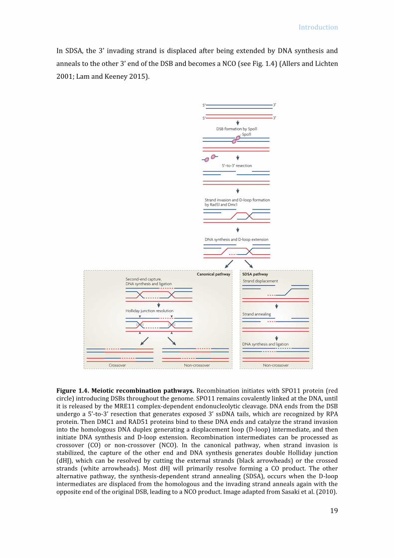
In SDSA, the 3’ invading strand is displaced after being extended by DNA synthesis and
anneals to the other 3’ end of the DSB and becomes a NCO (see Fig. 1.4) (Allers and Lichten
2001; Lam and Keeney 2015).
Figure 1.4. Meiotic recombination pathways. Recombination initiates with SPO11 protein (red circle) introducing DSBs throughout the genome. SPO11 remains covalently linked at the DNA, until it is released by the MRE11 complex-dependent endonucleolytic cleavage. DNA ends from the DSB undergo a 5’-to-3’ resection that generates exposed 3’ ssDNA tails, which are recognized by RPA protein. Then DMC1 and RAD51 proteins bind to these DNA ends and catalyze the strand invasion into the homologous DNA duplex generating a displacement loop (D-loop) intermediate, and then initiate DNA synthesis and D-loop extension. Recombination intermediates can be processed as crossover (CO) or non-crossover (NCO). In the canonical pathway, when strand invasion is stabilized, the capture of the other end and DNA synthesis generates double Holliday junction (dHJ), which can be resolved by cutting the external strands (black arrowheads) or the crossed strands (white arrowheads). Most dHJ will primarily resolve forming a CO product. The other alternative pathway, the synthesis-dependent strand annealing (SDSA), occurs when the D-loop intermediates are displaced from the homologous and the invading strand anneals again with the opposite end of the original DSB, leading to a NCO product. Image adapted from Sasaki et al. (2010).
Introduction
20
1.2 Control of meiotic recombination
Several meiotic mechanisms act in conjunction in order to ensure correct
formation and placement of the induced DSBs and to ensure the repair of these DSBs
resulting in proper COs or NCOs. In mouse, most DSBs occur at preferential recombination
sites (recombination hotspots) and seem to be determined by PRDM9, a DNA binding
methyltransferase. PRDM9 protein has a PR/SET domain with histone 3 lysine 4
(H3K4me3) methyltransferase activity at the N-terminus. At the C-terminus PRDM9
contains a highly polymorphic multiple-zinc-finger domains that recognize specific DNA
sequences. So when PRDM9 recognizes the DNA motif, the SET domain leaves an
epigenetic signature (H3K4me3) (Baudat et al. 2010; Parvanov et al. 2010). However,
PRDM9 is not required for DSB generation since in Prdm9-/- mouse DSBs are form but on a
different position than in wild type. In PRDM9 absence DSBs accumulate at promoter
regions or at other PRDM9-independent H3K4me-enriched sites, suggesting that besides
its methyltransferase activity, PRDM9 protein also recruits the recombination machinery
(sequestering the DSB machinery away from promoters) (Brick et al. 2012). Besides
PRDM9, it is thought that other high-order chromosome structures may influence DSB
locations (Lam and Keeney 2015).
Every chromosome needs to get a minimum number of DSBs in order to get at least
one CO, but due to its dangerous nature it is also important to tightly regulate them since
high levels of DSBs are deleterious for spermatocytes. For instance, Atm mutants present
increased levels of SPO11-generated DSBs. ATM-lacking spermatocytes have multiple
meiotic abnormalities (like meiotic arrest, unsynapsed chromosomes, failure to form a sex
body, fragmented SC, uncompleted meiotic recombination and no CO formation) (Barlow
et al. 1998) but interestingly some of these defects become alleviated in Spo11
heterozygosity (e. g. Spo11+/-Atm-/-) (Bellani et al. 2005; Barchi et al. 2008). Several
regulatory mechanisms have been found to control DSBs formation. Firstly, ATM protein
which is directly activated by DSBs early processing, inhibits DSB formation via a negative-
feedback control. In ATM deficiency, Drosophila and mice meiocytes present an increased
abundance of DSBs, assessed by DSBS markers or through augmented SPO11-
oligonucleotide complexes respectively (Joyce et al. 2011; Lange et al. 2011). The MRE11
complex, which sense DSBs and activates ATM, also participates in this feedback control of
DSBs formation in mice (Pacheco et al. 2015). In S. cerevisiae, both ATMTel1 and also
ATRMec1 control DSBs levels by down-regulating Spo11 cofactor Rec114 (Carballo et al.
2013). On another side, regulation of DSBs is also controlled by homologous chromosomes
engagement through ZMM proteins (factors that control recombination and SC

Introduction
21
completion). Yeast cells lacking ZMM proteins (zip1, zip3 or msh5 mutants) also present
increased formation of DSBs. It is suggested that when the two homologues engage, ZMM
proteins signal to stop making DSBs through a negative feedback loop (Thacker et al.
2014). Finally, there is an established window for DSBs formation during meiotic
prophase which its initiation seems to be controlled through meiosis-specific gene
transcription (coordinated after replication) and its termination is regulated by the
pachytene-exit Ndt80 transcription factor (Allers and Lichten 2001; Lam and Keeney
2015).
In mouse, about two hundred DSBs are generated at leptonema but only 10% of
these will be resolved as COs, but at least one CO per homologue pair is necessary to
ensure proper chromosome segregation at anaphase I. Connection between the
homologous chromosomes is required in order to provide tension in their centromeres
when they are aligned at the metaphase plate helping to pull each homologue to the
opposite poles. Not surprisingly, CO formation is highly regulated by three main CO
control processes (Wang et al. 2015). Firstly, assurance of the obligatory CO is required,
since getting at least one CO for each homologous pair ensures correct segregation.
Secondly, CO interference ensures that COs occur widely spaced along the chromosomes
(Bishop and Zickler 2004; Hillers 2004). ATM protein is required for the obligate CO
between the X and Y chromosome, and its deficiency results in increased number of COs
and with reduced CO interference (Barchi et al. 2008). In S. cerevisiae ATMTel1/ATRMec1
dependent phosphorylation of RPA and RNF212Zip3 control CO distribution (Bartrand et al.
2006; Serrentino et al. 2013). And finally, CO homeostasis guarantees a sustained number
of COs despite a variable initial dosage of DSBs. Recombination initiates with the
programmed DSB, but even if the number of the DSBs increases or decreases (due to cell-
to-cell variability or induced by mutations), the final number of COs remains similar to
avoid aneuploidy (Martini et al. 2006; Cole et al. 2012).
1.2.1 Homologous chromosome synapsis
DSBs formation is also facilitated by higher-order chromosome structures. Sister
chromatids organize into loops that anchor into the structural protein axis scaffold: the
axial element (AE). The loop-axis structure starts to organize coupled with DNA
replication and is completed at leptonema. The two principal proteins of the AEs are
SYCP3 and SYCP2, which are recruited to chromosome axes (Borde and de Massy 2013).
As homologous chromosomes start to pair at zygotene stage, the two axial elements of

Introduction
22
each chromosome assemble into the synaptonemal complex (SC). The SC is a zipper-like
structure that consists on the assembly of the two lateral elements (the former AEs), held
together by the transverse filaments of the central element, made of SYCP1 among other
proteins (de Vries et al. 2005). At pachytene stage homologous chromosomes are fully
synapsed giving place to bivalents. The SC complex is therefore important for driving
synapsis, recombination and holding the homologous chromosomes together during
meiotic prophase (see Fig. 1.5) (Zickler and Kleckner 1999; Page and Hawley 2004; Zickler
and Kleckner 2015).
Figure 1.5. Synaptonemal complex structure and organization. At the onset of prophase sister chromatids organize into loops that anchor into the chromosome axes that will form the synaptonemal complex (SC) scaffold. The SC is a zipper-like structure formed by the lateral elements (SYCP3 and SYCP2, in red) and the central element (SYCP1, in light green). At leptonema stage SYCP3 and SYCP2 proteins start to localize along the chromosome axes (axial elements). At the same time, repair proteins like DMC1 (grey) and RAD51 (orange) are recruited at the DSBs sites. Additionally, other proteins are located over the asynapsed axes, like HORMAD1 (turquoise line) and HORMAD2 (dark green line), which mediate the asynapsis signaling network. At zygonema, when homologous chromosomes start to synapse SYCP1 and other proteins are recruited between the lateral elements to forms the transverse filaments (central element). As DSB repair progresses mismatch repair MSH4 and MSH5 proteins (pink and purple respectively), localize over the recombination intermediates. At pachytene stage synapsis is completed and MLH1 and MLH3 proteins (light and dark blue respectively) mark crossover sites. Finally, homologous chromosomes start to desynapse at diplonema held together by the CO sites. Image modified from Cohen and Holloway (2010).
Introduction
23
Remarkably, meiotic recombination and homologous synapsis need to occur in a
coupled manner during meiotic prophase. As a matter of fact, there is a mutual
dependency between these processes, and failure of one will probably alter the correct
progression of the other (e.g. Dmc1-/- (Pittman et al. 1998), Sycp1-/- mutants (de Vries et al.
2005) or SC null mutants (Kouznetsova et al. 2011)). This fact, highlights the importance
of recombination and synapsis processes being monitored by surveillance mechanisms.
1.2.1.1 Asynapsis and transcriptional silencing
Asynapsis triggers meiotic silencing, which consists in a process where chromatin
remodeling inactivates the transcription from genes located at the unsynapsed regions
during late meiotic prophase. Some sensor proteins are retained over the axis only in
unsynapsed regions like HORMA (Hop1, Rev7 and Mad2) domain proteins HORMAD1
(Daniel et al. 2011) and HORMAD2 proteins (Wojtasz et al. 2012). Both HORMAD1 and
HORMAD2 are part of the meiotic chromosome axis. Once homologous chromosomes have
synapsed, HORMAD1/2 are depleted from the axes in a TRIP13-dependent manner
(Wojtasz et al. 2009; Roig et al. 2010). Thus, in pachytene-stage spermatocytes
HORMAD1/2 are only present on the unsynapsed portions of the X and Y chromosomes.
Surprisingly, at diplonema HORMADS1/2 are loaded again onto the chromosome axes.
Apart from sensing unsynapsed axis, HORMAD1/2 have other functions during meiosis.
HORMAD1 promotes efficient formation of DSBs, SC assembly and is necessary for
efficient expansion of ATR over the asynapsed chromatin in DSBs absence (Daniel et al.
2011), and HORMAD2 principally ensures recruitment of ATR and cofactors over the
unsynapsed axes (Wojtasz et al. 2012). Interestingly, the phosphorylated form of
HORMAD1 and HORMAD2 are exclusively located over the unsynapsed axes. The fact that
these proteins can experience post-translational modifications suggests that this serves as
a signal to promote recruitment of other factors that contribute to promote silencing and
the synapsis surveillance system (Fukuda et al. 2012). HORMAD1 and HORMAD2 mediate
zygotene asynapsis sensing signaling and promote BRCA1 localization on the asynapsed
axes. Then, ATR is recruited to asynapsed axes through a BRCA1-, HORMAD1-, HORMAD2-
dependent manner, and facilitates the enrichment of other cofactors to the unsynapsed
axes, like ATRIP, TOPBP1 (Refolio et al. 2011) or phosphorylated HORMAD1/2 (Turner et
al. 2005; Refolio et al. 2011; Royo et al. 2013; Turner 2015). If asynapsis persists, MDC1
effector cofactor helps ATR to translocate and accumulate over the chromatin loops
emanating from the asynapsed axes (Ichijima et al. 2011), catalyzing the second wave of
H2AX phosphorylation, the key epigenetic event to promote meiotic silencing of the
Introduction
24
unsynapsed chromatin (MSUC) (Perera et al. 2004; Turner 2015; Burgoyne et al. 2009;
Mahadevaiah et al. 2001; Royo et al. 2013). This second wave of H2AX phosphorylation is
independent of SPO11-induced DSBs, since Spo11 mutant mice experience silencing of
part of their unsynapsed chromosomes (Barchi et al. 2005; Bellani et al. 2005).
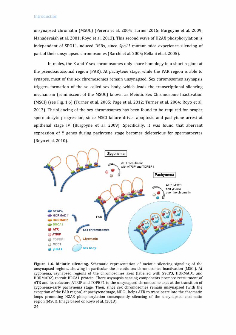
In males, the X and Y sex chromosomes only share homology in a short region: at
the pseudoautosomal region (PAR). At pachytene stage, while the PAR region is able to
synapse, most of the sex chromosomes remain unsynapsed. Sex chromosomes asynapsis
triggers formation of the so called sex body, which leads the transcriptional silencing
mechanism (reminiscent of the MSUC) known as Meiotic Sex Chromosome Inactivation
(MSCI) (see Fig. 1.6) (Turner et al. 2005; Page et al. 2012; Turner et al. 2004; Royo et al.
2013). The silencing of the sex chromosomes has been found to be required for proper
spermatocyte progression, since MSCI failure drives apoptosis and pachytene arrest at
epithelial stage IV (Burgoyne et al. 2009). Specifically, it was found that aberrant
expression of Y genes during pachytene stage becomes deleterious for spermatocytes
(Royo et al. 2010).
Figure 1.6. Meiotic silencing. Schematic representation of meiotic silencing signaling of the unsynapsed regions, showing in particular the meiotic sex chromosomes inactivation (MSCI). At zygonema, asynapsed regions of the chromosomes axes (labelled with SYCP3, HORMAD1 and HORMAD2) recruit BRCA1 protein. There asynapsis sensing components promote recruitment of ATR and its cofactors ATRIP and TOPBP1 to the unsynapsed chromosome axes at the transition of zygonema-early pachynema stage. Then, since sex chromosomes remain unsynapsed (with the exception of the PAR region) at pachytene stage, MDC1 helps ATR to translocate into the chromatin loops promoting H2AX phosphorylation consequently silencing of the unsynapsed chromatin region (MSCI). Image based on Royo et al. (2013).

Introduction
25
A characteristic of first meiotic division is that transcription is suppressed at the
onset of prophase, and it essentially restarts again at mid pachynema (Monesi, 1964),
when chromosomes are fully synapsed and most DSBs are repaired. In pachytene stage
spermatocytes, when autosomes reactivate transcription, sexual chromosomes are
subjected to transcriptional repression by the sex-body (Turner et al. 2004); which is
crucial to allow correct progression of meiosis (Royo et al. 2010). It is generally believed
that the meiotic sex chromosome inactivation (MSCI) is a manifestation of the global
meiotic silencing of unsynapsed chromosomes (MSUC) that is detectable at the end of
zygotene stage (Burgoyne et al. 2009; Turner et al. 2005). Moreover, it has been
hypothesized that MSUC could also be active during all meiotic prophase. Thus, the
repression of transcription that occurs at the onset of meiotic prophase could also be a
global manifestation of the MSUC mechanism(Page et al. 2012).
1.2.2 Checkpoint mechanisms control meiotic progression
Meiotic prophase comprises the execution of many processes that need to be
coordinated successfully in order to obtain viable gametes. For this, meiotic progression,
recombination and homologous chromosome synapsis occur concomitantly in a highly
regulated manner. For instance, correct repair of the SPO11-induced DSBs becomes a
determining process, since errors at this point could contribute to generate genomic
instability and introduce germ line mutations. Therefore, it is essential that meiotic
prophase is subjected to stringent surveillance mechanisms that can detect deleterious
events during prophase, delay cell cycle and, if necessary, promote programmed cell
death.
Depending on the organisms, different strategies are adopted. In S. cerevisiae for
example, persistent DSBs or synapsis defects result in a prolonged prophase that can be
followed by DNA damage adaptation and forced meiosis or mitotic growth (Roeder and
Bailis 2000; Subramanian and Hochwagen 2014). Remarkably, in many organisms, most
known mechanisms that control prophase progression involve the DNA damage response
(DDR) proteins ATM and ATR (Roeder and Bailis 2000; MacQueen and Hochwagen 2011;
Subramanian and Hochwagen 2014). ATM and ATR are conserved serine/threonine
kinases that in meiosis respond to different forms of DNA damage and synapsis errors, and
achieve their functions in coordination with other cofactors and through phosphorylation
of a large net of substrates. Detection of the DSBs is mainly driven by ATM/ATR, however
following downstream signaling differs among organisms.

Introduction
26
Cell cycle arrest in S. cerevisiae occurs in the presence of recombination and
synapsis defects, such as Dmc1, Zip1 or Hop2 mutants and checkpoint activation requires
Mek1 (a meiosis specific CHK2 homolog). Drosophila similarly requires Mnk (CHK2)
protein to activate the checkpoint that occurs in mutants defective for DSB repair proteins
(e.g. okr, spn-B and spn-C) (Roeder and Bailis 2000; MacQueen and Hochwagen 2011). This
meiotic checkpoint is also active in C. elegans syp-1 and rad-51 mutants; but contrary,
checkpoint apoptosis depends on CHK1 presence to promote p53 activation (Jaramillo-
Lambert et al. 2010). The downstream targets of the pachytene checkpoint identified in S.
cerevisiae are Swee1, which phosphorylates and inactivates Cdc28 (Leu and Roeder 1999);
and Ndt80, a meiotic transcription factor of genes required to exit pachytene (like CLB1)
(Chu and Herskowitz 1998).
1.2.2.1 The pachytene arrest in mammals
The pachytene checkpoint ensures that recombination and synapsis are fully
completed before progressing to diplonema stage, but in the presence of deleterious
events it can promote cell cycle arrest and apoptosis. Mouse surveillance mechanisms act
at two different stages: either monitoring recombination or synapsis at pachytene stage,
or later at metaphase I controlling bipolar attachment to the spindle (known as the spindle
assembly checkpoint, SAC) (Di Giacomo et al. 2005; Barchi et al. 2005; Touati and
Wassmann 2016).
In mammalian males, several studies suggest the presence of at least two
independent control mechanisms that would monitor meiotic prophase progression and
activate the arrest that occurs at pachytene stage in spermatocytes. One is dependent on
the correct repair of the induced DSBs (referred as recombination-dependent arrest)
(Barchi et al. 2005; Pacheco et al. 2015) and the other depends on the proper meiotic
silencing of the sex chromosomes by the sex body (named here as sex body-deficient
arrest) (Barchi et al. 2005; Royo et al. 2010). Cytologically, the activation of these two
arrest mechanisms can be distinguished using the incorporation of the testis specific
histone 1t (H1t) as a marker, which in wild type mouse accumulates on chromatin from
mid-pachynema onwards (Drabent et al. 1996; Inselman et al. 2003). It has been described
that spermatocytes with persistent unrepaired DSBs (e.g., Dmc1-/- (Pittman et al. 1998;
Yoshida et al. 1998)), arrest at early pachytene stage before expressing H1t (Barchi et al.
2005; Pacheco et al. 2015). In contrast, mutant spermatocytes that arrest due to a
defective sex body (e.g., Spo11-/- (Baudat et al. 2000; Romanienko and Camerini-Otero
2000)) reach mid/late pachynema and accumulate H1t (Barchi et al. 2005; Pacheco et al.
Introduction
27
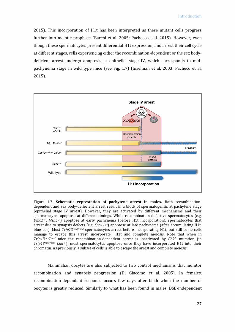
2015). This incorporation of H1t has been interpreted as these mutant cells progress
further into meiotic prophase (Barchi et al. 2005; Pacheco et al. 2015). However, even
though these spermatocytes present differential H1t expression, and arrest their cell cycle
at different stages, cells experiencing either the recombination-dependent or the sex body-
deficient arrest undergo apoptosis at epithelial stage IV, which corresponds to mid-
pachynema stage in wild type mice (see Fig. 1.7) (Inselman et al. 2003; Pacheco et al.
2015).
Figure 1.7. Schematic represtation of pachytene arrest in males. Both recombination-dependent and sex body-defiecient arrest result in a block of spermatogensis at pachytene stage (epithelial stage IV arrest). However, they are activated by different mechanisms and their spermatocytes apoptose at different timings. While recombination-defective spermatocytes (e.g. Dmc1-/-, Msh5-/-) apoptose at early pachynema (before H1t incorporation), spermatocytes that arrest due to synapsis defects (e.g. Spo11-/-) apoptose at late pachynema (after accumulating H1t, blue bar). Most Trip13mod/mod spermatocytes arrest before incorporating H1t, but still some cells manage to escape this arrest, incorporate H1t and complete meiosis. Note that when in Trip13mod/mod mice the recombination-dependent arrest is inactivated by Chk2 mutation (in Trip13mod/mod Chk-/-), most spermatocytes apoptose once they have incorporated H1t into their chromatin. As previously, a subset of cells is able to escape the arrest and complete meiosis.
Mammalian oocytes are also subjected to two control mechanisms that monitor
recombination and synapsis progression (Di Giacomo et al. 2005). In females,
recombination-dependent response occurs few days after birth when the number of
oocytes is greatly reduced. Similarly to what has been found in males, DSB-independent

Introduction
28
arrest due to synapsis defects occurs later, and as a result the pool of oocytes is reduced
about two weeks after birth (e.g., Spo11-/- mutants) (Di Giacomo et al. 2005).
1.2.2.1.1 The recombination dependent arrest
Unrepaired DSBs promote the activation of ATM/ATR kinases triggering a delay in
meiotic progression in many organisms, which is called the recombination-dependent
checkpoint/arrest. This response gives more time to repair the DSBs before progressing to
the next stages, or if cells are unable to repair the DSBs they are eliminated through
apoptosis (MacQueen and Hochwagen 2011). In mice, recombination failure triggers
pachytene block (e.g. Dmc1-/-, Msh4-/-, Msh5-/- or Trip13mod/mod) (Pittman et al. 1998; de
Vries et al. 1999; Edelmann et al. 1996, 1999; Yoshida et al. 1998; Kneitz et al. 2000).
Recently, some of the proteins involved in the activation of the recombination-
dependent arrest in mouse meiocytes have been reported thanks to the use of Trip13
mutants (Pacheco et al. 2015; Bolcun-Filas et al. 2014). Trip13 mutants present
recombination defects, triggering the activation of the recombination-dependent arrest,
which blocks spermatocyte progression at an early pachytene stage (before H1t
incorporation) (Li et al. 2007; Roig et al. 2010; Pacheco et al. 2015). Mutation of MRE11,
NBS1, ATM or CHK2 bypassed Trip13 arrest by increasing the number of H1-positive cells
and reducing the number of spermatocytes that apoptose at early pachynema (see
Trip13mod/mod versus Trip13mod/mod Chk2-/- in Fig. 1.7). Therefore, the MRE11 complex, ATM
and CHK2 signaling pathway implements the recombination-dependent arrest occurring
in mouse spermatocytes (Pacheco et al. 2015). Similarly, CHK2 protein through p53 and
TAp63 downstream activation, are responsible for eliminating oocytes with persistent
unrepaired DSBs; as CHK2 or p53 (and TAp63 in some extent) rescue Trip13 mutants
oocyte loss (Suh et al. 2006; Bolcun-Filas et al. 2014; Kim and Suh 2014).
1.2.2.1.2 The synapsis dependent arrest
In mouse, the asynapsed regions of the chromosomes are detected and silenced by
the MSUC mechanism. This meiotic silencing serves as a prophase surveillance
mechanism, since impaired synapsis activates this mechanism promoting a meiotic arrest.
Mouse meiocytes lacking Spo11 are not able to properly complete homologous
chromosome synapsis driving a DSB-independent apoptosis, which is distinctively
activated in males or females due to sexual dimorphism (Baudat et al. 2000; Romanienko
and Camerini-Otero 2000; Di Giacomo et al. 2005; Barchi et al. 2005).

Introduction
29
In males, the MSUC mechanism detects the asynapsed X and Y chromosomes and
promotes the MSCI. This silencing of the X and Y genes is crucial for meiotic progression,
since misexpression of sex chromosome genes at pachynema becomes “toxic” for
spermatocytes (Royo et al. 2010). Extensive asynapsis of the autosomes in synapsis-
defective mutants (e.g. in Spo11-/-) or associated with persistent DNA damage (e.g. Dmc1-/-
mutants), is thought to dilute the MSUC silencing cofactors along the asynapsed axes (e.g.
HORMAD1/HORMAD2 remain spread over all the autosomes; BRCA1 and ATR remain
sequestered at the DSBs sites). Consequently, this alteration of the silencing cofactors
mitigates MSUC mechanism, specially attenuating silencing of the XY chromosomes, which
results in apoptosis due to sex chromosome genes expression (Mahadevaiah et al. 2008;
Royo et al. 2010).
On the other hand, in mouse synapsis-defective females, MSUC detects asynapsed
chromosome axes and promotes silencing of these regions. It has been found that oocytes
display a less efficient meiotic silencing on asynapsed chromosomes than males, with
absence of the epigenetic marker H3K9me (Taketo and Naumova 2013; Cloutier et al.
2015b). Therefore, gene silencing across the γH2AX-coatted unsynapsed chromosome
occurs in a stochastic manner, which would possibly affect genes required for post-
pachytene oocyte progression and consequently drive a meiotic arrest (Cloutier et al.
2015b). In fact, oocyte loss due to extended asynapsis of Spo11 mutants can be rescued by
deletion of silencing components like Hormad1, Hormad2 or H2ax (Daniel et al. 2011;
Wojtasz et al. 2012; Cloutier et al. 2015a).
1.3 Pachytene checkpoint 2 protein / TRIP13
Pch2 (Pachytene checkpoint 2 gene) is a widely conserved protein that can be
found in S. cerevisiae, A. thaliana, D. melanogaster, C. elegans (pch-2) and mammals (called
TRIP13). Pch2 belongs to AAA+ ATPases family, a diverse group of enzymes that couple
ATP binding and hydrolysis inducing conformational changes on substrates. They
participate in multiple cellular processes including protein degradation, transcriptional
regulation, DNA replication, protein folding and unfolding and assembly and disassembly
of protein complexes (Hanson and Whiteheart 2005; Tucker and Sallai 2007). Like many
AAA+ ATPases, Pch2 oligomerizes into hexameric rings with a central pore in the presence
of nucleotides (Chen et al. 2013). In yeast, Pch2 is a meiosis specific gene that is
transcribed before entering meiosis I and peaks at pachytene stage having multiple
functions (San-Segundo and Roeder 1999). Pch2 controls CO outcome, as pch2 deficiency

Introduction
30
results in defective interhomolog bias, reduced CO interference, increased CO levels and
defective homeostasis (Zanders and Alani 2009; Joshi et al. 2009; Wu and Burgess 2006;
Börner et al. 2008).
Pch2 was first identified in S. cerevisiae as a checkpoint protein that bypassed zip1
arrest (San-Segundo and Roeder 1999). Zip1 protein is a component of the central element
of the SC, and its absence drives synapsis defects and accumulation of recombination
intermediates, triggering a checkpoint arrest at pachytene stage. The zip1 pch2 double
mutants recover wild type levels of sporulation frequency. Thus, it was rapidly assumed
that Pch2 was required to implement the synapsis-dependent checkpoint in yeast. Studies
in other model organisms confirmed this was a conserved function, C. elegans Pch2 is also
required to induce the apoptosis originated by defective homologous synapsis (Bhalla and
Dernburg 2005). Nonetheless, Pch2 checkpoint functions are more complex since yeast
Pch2 absence also abolishes zip2 and dmc1 mutants arrest (San-Segundo and Roeder
1999). Furthermore, Pch2 homologue in Drosophila was found to be required for the delay
in oocyte selection produced by recombination defects (Joyce and McKim 2009).
In yeast, Pch2 has been reported to interact with DDR proteins in response to
unprocessed DSBs. Since Pch2 has been found to interact with Xrs2 (NBS1 homologue
from the MRE11 complex) and via Tel1ATM pathway Pch2 facilitates Hop1HORMAD1/2
phosphorylation; it has been proposed that Pch2 could help to recruit Hop1 to DSBs sites
to facilitate Hop1 roles in DSB formation, interhomolog bias and checkpoint function
(Carballo et al. 2008; Ho and Burgess 2011; Panizza et al. 2011). Hop1 is a DNA binding
protein that contains a zinc finger domain and a HORMA domain that localizes over the
chromosome axes and can be phosphorylated by Mec1ATR and Tel1ATM kinases in response
to DNA damage (Hollingsworth et al. 1990; Carballo et al. 2008). In vitro Pch2 binds to
Hop1HORMAD1/2 and can displace it from DNA substrates (Chen et al. 2013), which can be
related with Pch2 control over chromosome axis conformation. Pch2 regulates Hop1
localization on the chromosome axes by promoting the Zip1/Hop1 alternating pattern,
where Hop1 tends to localize at designated CO sites. Absence of Pch2 results in uniform
distribution of Hop1 over the entire Zip1 length (Börner et al. 2008). Therefore, Pch2 is
proposed to bind Hop1 in vivo and induce a conformational change upon ATP hydrolysis,
modulating Hop1 localization at specific sites to regulate interhomolog repair (Joshi et al.
2009; Chen et al. 2013).
Interestingly, in yeast, most Pch2 protein localizes in the nucleolus, the region
containing ribosomal DNA (rDNA). Nucleolar Pch2 it is thought to represses DSBs
formation and interhomolog recombination in rDNA by excluding Hop1 from the

Introduction
31
nucleolus (San-Segundo and Roeder 1999). The rest of Pch2 protein is present forming
foci on the synapsed chromosomes co-localizing with Zip1 (San-Segundo and Roeder
1999). Nucleolar Pch2 localization is Sir2-dependent. Sir2 is a chromatin silencing factor
(modulates closed chromatin conformation) that also localizes in the nucleolus and
controls repression of recombination in the rDNA (Gotta et al. 1997; Gottlieb and Esposito
1989). Indeed, sir2 mutation also bypassed zip1 arrest, showing that the nucleolus Pch2
fraction is the one responsible to control the meiotic checkpoint (San-Segundo and Roeder
1999).
1.3.1 Mammalian TRIP13
In mouse, TRIP13 (thyroid hormone receptor interacting protein 13) is the
mammalian Pch2 orthologue, which is expressed during development and in many tissues.
It is also expressed in testis, where it presents a specific isoform (lacking the Walker B
ATPase motif) in spermatocytes and spermatids (Roig et al. 2010). TRIP13 is required for
gametogenesis completion of both male and female (Li et al. 2007; Roig et al. 2010). Trip13
null mutation is incompatible with embryo development (J. Schimenti, personal
communication), thus most studies performed in mouse have used hypomorphic
mutations that significantly decrease Trip13 expression. Two Trip13 mutants where
generated from ES cell lines containing a gene-trap disrupted allele either in the second
intron (CH0621 cell line) or the third intron (RRB047 cell line), leading to two
hypomorphic mutations with distinct penetrance phenotype (Li et al. 2007; Roig et al.
2010). Trip13RRB047 (or Trip13Gt or Trip13mod, for moderate) testes present lower Trip13
expression levels than wild type, but these are even more reduced in Trip13CH0621 (or
Trip13sev, for severe), which is related to a more penetrant effect (Roig et al. 2010). Male
Trip13mod/mod mutant mice present reduced testes size associated to pachytene arrest. Most
spermatocytes from Trip13mod/mod present recombination defects: reduced Rad51 foci at
leptonema, inefficient recombination completion, reduced CO formation and altered CO
interference (Li et al. 2007; Roig et al. 2010). However, most spermatocytes carrying the
moderate Trip13 mutant allele are able to achieve full homologous synapsis despite the
presence of abundant recombination intermediates. Importantly, few Trip13mod/mod
escapers (that probably contain high TRIP13 levels) can complete meiosis reaching round
spermatid stage (Li et al. 2007; Roig et al. 2010). Still, since most Trip13mod/mod
spermatocytes accumulate unrepaired DSBs they activate the recombination-dependent
arrest and apoptose at early pachynema (before incorporating H1t), triggering epithelial
Introduction
32
stage IV arrest at pachytene stage (Li et al. 2007; Roig et al. 2010; Pacheco et al. 2015).
Characterization of the Trip13 severe mutant allele revealed that Trip13sev/sev
spermatocytes are not able to complete synapsis and no spermatocytes progress any
further than zygotene stage (no escapers found) (Roig et al. 2010). Regarding females,
oogenesis is also defective for both allele mutants. At 21 dpp Trip13 moderate mutant
females have fewer developing follicles than the wild type, but no primordial follicles are
observed. Trip13 severe female phenotype is more penetrant, since no oocytes are found at
already 21 dpp; such early block of folliculogenesis is associated with recombination
failure (Di Giacomo et al. 2005), suggesting that TRIP13 is also required for completing
meiotic recombination in oocytes. (Li et al. 2007; Roig et al. 2010).
Similarly to Pch2, mouse TRIP13 also regulates the chromosome axis composition
by controlling localization of HORMAD1 and HORMAD2 proteins (yeast Hop1) (Wojtasz et
al. 2009; Fukuda et al. 2010). In wild type spermatocytes, HORMAD1 and HORMAD2
proteins become depleted from chromosomal axis once synapsis has occurred (Wojtasz et
al. 2009). Instead, in pachytene-stage Trip13mod/mod spermatocytes HORMAD proteins are
still present on the synapsed axis. Thus, TRIP13 is required to deplete HORMAD1 and
HORMAD2 from the synapsed chromosome axis (Wojtasz et al. 2009; Roig et al. 2010).
This role on HORMAD1 and HORMAD2 might explain the fact that TRIP13 protein has
been recently found to be required to form a functional sex body. In Trip13mod/mod
spermatocytes HORMAD proteins cover all the axes of the autosomes, which limit the
asynapsis signaling over the sex body. Indeed, Trip13mod/mod mutant spermatocytes fail to
properly load ATR over the sex chromosome axes, which leads to reduced H2AX
phosphorylation and SUMO-1 incorporation over the sex body chromatin, resulting in
inefficient MSCI (see results in section 4.3.2 and (Pacheco et al. 2015)).
TRIP13 is emerging as a key regulator of chromosomes structure, through
controlling several HORMA-domain containing proteins. In somatic cells, TRIP13 has been
found to be a kinetochore protein that interacts with several mitotic regulators, among
them p31comet and MAD2 (HORMA domain proteins). The Spindle Assembly Checkpoint
(SAC, composed by MAD2, BUBR1 and BUB3 proteins) prevents chromosome segregation
until all chromosomes are correctly bioriented at the spindle, by inhibiting the Anaphase
Promoting Complex (APC). Precisely, TRIP13 biding to p31comet has been found to promote
MAD2 release from the SAC and consequently turns off the checkpoint (Tipton et al. 2012;
Wang et al. 2014; Eytan et al. 2014). Remarkably, TRIP13 has been found to be
overexpressed in multiple cancers, which is suggested to lead malignant transformation,
aggressive tumor growth and treatment resistance due to premature mitotic checkpoint

Introduction
33
signaling and enhanced DNA repair. Moreover, TRIP13 has been shown to promote DNA
repair via NHEJ in human cells; by its interaction with KU70, KU80 and DNA-PKcs
(members of the NHEJ repair pathway). (Banerjee et al. 2014).
1.4 DNA damage response in somatic cells
As DNA damage represents a hazardous event for organisms, cells have developed
a highly organized network of signaling pathways, named DNA damage response (DDR).
The DDR detects the presence of DNA lesions through “sensor” proteins and via
“transducers” amplifies the signal (protein kinase cascade) to activate a series of effectors
that will trigger DNA repair, cell cycle or apoptosis. Slowing down cell cycle progression
allows extra time for DNA repair, and if the DNA damage cannot be repaired programmed
cell death is activated in order to avoid genome integrity defects. Two PIKKs
(phosphoinositide-3-kinase related proteins) family members are the principal
surveillance regulators of this response: the ATM (ataxia-telangiectasia mutated) and ATR
(ataxia-telangiectasia and Rad-3 related protein).Both ATM and ATR promote the DDR by
phosphorylating a large subset of substrates (Fig. 1.8) (Matsuoka et al. 2007).
Of the various forms of DNA damage, DSB represent the most dangerous form. The
two helix strands are broken simultaneously, which makes it more difficult to repair and
more prone to inappropriate recombination. When DSBs occur in somatic cells, they are
detected by the MRE11 complex (MRE11, RAD50, NBS1) which activates the downstream
serine/threonine kinase ATM (Lavin 2007; Paull 2015). By binding to the MRE11 complex,
at the DSBs sites, ATM is retained over the chromatin and transduces the downstream
signaling pathway (Derheimer and Kastan 2010). ATM spreads the signaling cascade by
phosphorylating a large set of target effectors involved in DNA repair, cell cycle
progression and apoptosis (Stracker et al. 2013; Paull 2015). In somatic cells, ATM
impairment reduces DSBs repair capacity, defective activation of checkpoints and
apoptosis. ATM substrates include CHK2 (Chaturvedi et al. 1999; Matsuoka et al. 2000)
and p53 proteins among others (Fig. 1.8) (Canman et al. 1998; Khanna et al. 1998; Banin et
al. 1998).
Other mechanisms, like the repair of DSBs by homologous recombination or
replication stress, may lead to the formation of single stranded DNA (ssDNA), which
primarily activates ATR. RPA protein binds to the ssDNA, which promotes ATR
recruitment to the ssDNA lesion through the help of ATRIP (ATR-interacting protein)

Introduction
34
cofactor (Cortez et al. 2001) and binding of TOPBP1 activator (Acevedo et al. 2016). ATR
activation also requires binding of the 9-1-1 complex (RAD9-RAD1-HUS1) to the adjacent
ssDNA/dsDNA (Yan and Michael 2009). Once activated, ATR phosphorylates several
substrates, among them CHK1 kinase that controls intra-S and G2/M checkpoints
(Cimprich and Cortez 2008).
Figure 1.8. DNA damage response by ATM kinase in somatic cells. DSBs are detected by the MRE11 complex (MRE11-RAD50-NBS1) which recruits ATM to the DSBs sites and promotes its auto-phosphorylation. ATM phosphorylates H2AX and a large set of downstream targets to expand the DDR cascade, which promote DNA repair, cell cycle arrest and apoptosis. ATM principal substrates are CHK2 kinase and P53 protein. Both ATM and CHK2 can phosphorylate p53 transcription factor. Upon phosphorylation, p53 unbinds MDM2 (which targets p53 for degradation through the ubiquitin proteolytic pathway) and becomes stabilized being able to perform its function. As a transcription factor, p53 principal functions consist in checkpoint activation, apoptosis and DNA repair. Through p21Waf1 activation, p53 arrest cell cycle at G1-S; and G2-M arrest by activating GADD45 and 14-3-3σ. Besides, p53 promotes pro-apoptotic such as PUMA, NOXA or BAX. Moreover, p53 also promotes DNA repair and controls its own levels by a negative feedback loop activating MDM2 expression.

Introduction
35
1.4.1.1 Cell cycle control and apoptosis by the DDR
Cell cycle is a complex biological process by which identical diploid chromosome
sets are transmitted to two daughter cells. This happens through four sequential phases:
G1, S, G2 and M-phase (Lavin and Kozlov 2007). Thus, when DNA damage occurs it is
important that the cycle progression of these phases is regulated by checkpoint
mechanisms; which allow extra time for repair in order to maintain genomic integrity. If
the damage is not repaired, the checkpoint mechanisms activate apoptosis in order to
eliminate the damaged cell and thus guarantee genome integrity.
In DNA damaged cells, DSBs activate ATM and ATR which phosphorylates several
substrates that control cell cycle progression at G1/S, S-phase and at G2/M transition. ATM
notably phosphorylates CHK2 a serine/threonine kinase, which has an important role as a
signal transducer, and p53 tumor suppressor gene. For G1/S checkpoint activation, ATM
and CHK2 induce p53 tumor suppressor phosphorylation at serine15 (S15) (Banin et al.
1998; Barlow et al. 1997a; Derheimer and Kastan 2010). Once phosphorylated, p53
becomes stabilized and promotes transcription of target genes, delaying G1 to S-phase
(Kastan and Lim 2000) and G2/M transition (Taylor and Stark 2001). One of the principal
targets of p53 is p21WAF-1, which binds and inhibits the S-phase promoting Cdk2-CyclinE
complex, and also Cdk4-CyclinD (preventing Rb phosphorylation which promotes S-phase
progression) (see other p53 targets in the next section 1.3.2.1 and Fig. 1.8) (Harper et al.
1993; Sancar et al. 2004). Other mediators of cell cycle progression that are included as
ATM substrates are MDM2 and MDMX, which influence p53 stabilization (Lavin and
Kozlov 2007). Besides, CHK2 also activates the G1/S arrest by phosphorylating CDC25A
phosphatase and promoting its degradation (CDC25 phosphatases control cell cycle
progression by removing inhibitory residue phosphorylation from cycling-dependent-
kinases (CDKs)) (Falck et al. 2001).
Independent of p53, ATM regulates intra S-phase arrest via two pathways. One is
the ATM-CHK2-CDC25A-Cdk2. The second requires phosphorylation of SMC1, BRCA1,
NBS1 and FANCD2 phosphorylation on multiple sites, which promote the recovery of
collapsed replication forks (Kim et al. 2002; Yazdi et al. 2002). Moreover, CHK2 activation
regulates G2/M checkpoint through phosphorylation of CDC25 phosphatase, which
promotes CDC25 binding to 14-3-3σ protein (becomes degraded) and blocks activation of
cyclin B-dependent CDC2 kinase, which is required for initiation of mitosis (Peng et al.
1997).
Introduction
36
ATR activates CHK1 kinase, which phosphorylates CDC25 family regulators.
Downregulation of CDC25A through phosphorylation promotes p53 stabilization
triggering G1/S arrest and inhibits replication origins (Sancar et al. 2004).
Phosphorylation of CDC25C on serine-216 creates a binding site for 14-3-3σ protein which
inhibits the phosphatase activity (Peng et al. 1997); preventing cell cycle progression by
activating the intra-S checkpoint (Sanchez 1997; Feijoo et al. 2001). During S-phase, ATR
also promotes phosphorylation of BRCA1 and NBS1 to inhibit replication and promote
repair. DNA damage during G2 phase also promotes the ATR-CHK1-CDC25 pathway to
activate the G2/M checkpoint (Sancar et al. 2004).
1.4.2 P53 family members
The mammalian p53 family includes: p53 (Lane and Crawford 1979; Linzer and
Levine 1979), p63 (Yang et al. 1998) and p73 (Jost et al. 1997); which are transcription
factors encoded by three highly conserved genes (Dötsch et al. 2010; Belyi et al. 2010).
Each p53 family member has a similar structure with three major domains consisting of an
amino-terminal transactivation domain (TA), a DNA binding domain in the center and a
carboxy-terminal oligomerization domain. Additionally, only p63 and p73 present a
carboxy-terminal sterile α-motif (SAM) involved in protein-protein interaction and a
transcription inhibition motif (TI) (see Fig. 1.9) (Dötsch et al. 2010; Allocati et al. 2012).
Because of the presence of two alternative promoters at the amino-terminus different
isoforms are transcribed in p53, p63 and p73. For instance, p53 has at least six different
isoforms which are transcribed in a tissue-dependent manner (Bourdon et al. 2005). For
p63 and p73 these promoters also generate several transcript variants, and among these,
two major isoforms are: the transactivating isoform (containing the TA domain) and the
ΔN isoforms produced by the second promoter (lacking the TA) (Yang et al. 2002; Allocati
et al. 2012). Generally, the TA isoforms tend to have tumor suppressor activities, while the
ΔN isoforms exert dominant-negative effects, since these variants can bind DNA but do not
promote transcription. However the ΔN isoforms have also been seen to activate some
targets (Dötsch et al. 2010; Allocati et al. 2012; Yang et al. 2002; Bourdon et al. 2005).
Additional splicing of the carboxy terminus and posttranslational cleavage generates
several other isoforms resulting in more complexity (Dötsch et al. 2010; Bourdon et al.
2005; Yang et al. 2002; Levine et al. 2011). Because of structural differences and biological
functions, each member activates a specific transcriptional network, however there is a
common set of shared gens, specially DDR responsive genes (Lin et al. 2009). For instance,

Introduction
37
both p53 and TAp63 can promote transcription of the same genes (Yang et al. 1998), and
can associate with each other through direct interaction (Gaiddon et al. 2001)). Although
tetramerization of p53 with the other family member is less clear, p63 and p73 have been
found to form heterotetramers (Joerger et al. 2009). The interplay between the family
members, multiple isoforms, their differential tissue expression and ability to oligomerize
yields in high complexity and diverse biological functions including prominent roles in the
DDR (Candi et al. 2014; Joerger et al. 2009; Murray-Zmijewski et al. 2006).
Figure 1.9. P53 family members gene structure. The genes encoding p53 family members: Trp53, Trp63 and Trp73 are highly conserved and maintain a similar structure. The three members present a trans-activating domain (TA) at the amino-terminus (N), a very similar DNA-binding domain (DBD) and an oligomerization domain (OD) at the carboxyl-terminus. The (PR) correspond to Pro-domains, and p53 contains a nuclear localization signal (NLS). Only p63 and p73 have a sterile α-motif domain (SAM) and a transcription inhibitory domain (TI) at the C-terminal end (C). The presence of two alternate promoters (P1 and P2), 3’-splicing events and posttranslational cleavage generates several distinct isoforms for all the three members. Both p63 and p73 present two principal isoforms: one that contains the TA domain (TAp63 and TAp73), the other is transcribed by P2 amino-truncated (ΔNp63 or ΔNp73) lacking the TA (and principally act as dominant-negative effectors). All this complexity, added to the fact that p53 family member can heteroligomerize, emphasizes the importance of balancing the various members and their isoforms. Image based on Levine et al. (2011).
Although p63 and p73 are able to regulate p53 target genes (Lin et al. 2009), and,
like p53, induce cell cycle arrest and apoptosis, studies from knockout mice have
demonstrated that each member has distinct functions. The p53 knockout mice are viable
and present a normal development, with higher predisposition to tumor malignancy
(Donehower et al. 1992; Jacks et al. 1994). Contrarily, p63 knockout mice show several

Introduction
38
developmental defects, including limbs development failure and epithelial morphogenesis
defects (attributed to ΔN isoforms); and do not survive beyond a few days after birth
(Mills et al. 1999). Differently, p73 knockout mice suffer neurodevelopmental
hippocampal dysgenesis, pheromone sensory pathway alteration and immunological
defects such as chronic infections and inflammation (Yang et al. 2000). However, both p63
and p73 mutants also present development of spontaneous tumors (Flores et al. 2005).
1.4.2.1 p53
The principal role of p53 is to stop proliferation of stressed cells by inhibiting cell
cycle progression or promoting apoptosis, in response to diverse stresses such as DNA
damage, in order to guarantee genome integrity. Importantly, p53 protein stands out for
being a tumor suppressor gene, which explains why is mutated in 50% of human cancers
(Petitjean et al. 2007). One of the principal stresses that cells may suffer is DNA damage.
Indeed, generation of DSBs promotes an increase of p53 levels. In response to stress, p53
is displaced from its negative regulator MDM2, by independent pathways that activate the
p53 network. The main pathway responds to DNA damage, where activation of ATM at the
DSBs sites phosphorylates CHK2 kinase, which subsequently phosphorylates p53 at the
amino terminus. This phosphorylation on p53 affects its ability to bind MDM2 and
consequently avoids its degradation (Fig. 1.8) (Vogelstein et al. 2000). Degradation of p53
requires ubiquitin labelling by the MDM2 protein, which works as a negative feedback
loop since MDM2 expression is also regulated by p53 (Momand et al. 2000). A second
pathway is activated by aberrant hyperproliferation signals, resulting from oncogenes Ras
or MYC signaling, where p53 activation depends on p14ARF. Stabilization of ARF promotes
its binding to MDM2, which inhibits MDM2 activity and sequesters it into the nucleolus
(Sherr and Weber 2000; Lin and Lowe 2001). Besides these two main classical pathways,
p53 is also activated in response to replicative stress, hypoxia, oxidative stress,
ribonucleotide depletion, nutrient starvation and telomere attrition (Fig. 1.10) (Bieging et
al. 2014).
These pathways inhibit degradation of p53, leading to its stabilization and
increased levels, promoting p53 to bind DNA and consequent transcription. Through the
tetramerization domain, p53 monomers form active tetramers that recognize p53 target
binding sites (Friedman et al. 1993). Historically two main subsets of p53 target genes
were identified: negative regulators of cell cycle progression and apoptosis inducers.
However, the number of p53 target genes is very extensive, including genes involved in

Introduction
39
DNA repair, senescence, metabolism, autophagy, stem cells biology, tumor invasion and
tumor microenvironment (Fig. 1.10) (Bieging et al. 2014).
Regarding cell cycle regulation, p53 promotes transcription of the cyclin
dependent kinase (cdk) p21 which inhibits Cyclin-E/CDK2 complex, causing an arrest of
cell cycle progression from G1 to S-phase (el-Deiry et al. 1994; Kastan and Lim 2000).
Moreover, p53 seems to maintain the G2/M checkpoint in part by up-regulating other
DNA-damage genes GADD45, BTG, 14-3-3σ or by signaling retinoblastoma protein (Lavin
and Kozlov 2007; Derheimer and Kastan 2010; Jin et al. 2000). If DNA damage persists,
p53 levels increase and induce transcription of pro-apoptotic genes like Puma, Noxa or the
Bcl-2 associated Bax, which activate caspases and programmed cell death (Fig. 1.8 and
1.10) (Roos and Kaina 2006; Oren 1994; Reinhardt and Schumacher 2012). Moreover,
p53 has also been found to eliminate damaged cells through the innate immune system
and to participate in tissue degenerating and aging (Reinhardt and Schumacher 2012).
Figure 1 10
The p53 network: activating signals and responses in somatic cells. p53 protein is activated upon stresses coming from different sources: hypoxia, oxidative stress, replicative stress, ribosomal nucleotide depletion, nutrient deprivation, hyperproliferative signals, DNA damage and telomere attrition. Once p53 is activated and stabilized it promotes transcription of several genes which principally lead towards tumor suppression. Among several responses, the classical ones include cell cycle arrest, apoptosis, promote DNA repair and senescence. Other responses are related with activation of autophagy, regulation of the microenvironment and tumor invasion; as well as inhibiting tumor metabolism and stem-cell renewal. Image adapted from Bieging et al. (2014).

Introduction
40
1.4.2.2 p63 and p73
Both p63 and p73 can control cell cycle progression and apoptosis through similar
mechanisms attributed to p53. However, the mechanisms remain less clear than in p53
due to redundant properties and balance complexity. For instance, p53-dependent
apoptosis upon DNA damage in mouse embryonic fibroblasts requires the presence of at
least either p63 or p73 (Flores et al. 2002). Expression of p73 during cell cycle peaks at the
S-phase and during mitosis p73 is phosphorylated by p34cdc2-CyclinB complex (Fulco et al.
2003). G1/S checkpoint arrest can be induced by TAp73, but also by TAp63, through
transcriptional up regulation of p21 (Jost et al. 1997; Zhu et al. 1998; Osada et al. 1998;
Dohn et al. 2001). Another target of p73 is p57/Kip2 (a cyclin-dependent kinase inhibitor),
which coordinates mitotic exit and transition to G1 (Merlo et al. 2005); but is the ΔNp63α
isoform that induces p57/Kip2 activation (Beretta et al. 2005; Pateras et al. 2009).
Surprisingly, ΔN isoforms (although lacking the transactivation domain) can induce
transcription of some gens at low levels (Dohn et al. 2001; Allocati et al. 2012). However,
in general ΔN isoforms usually repress promoters by acting as dominant negative forms
and thus allowing cell cycle progression. Moreover, in the absence of p53, p73 has been
shown to regulate cyclin-Cdks: cyclin E-Cdk2 and cyclin A-Cdk2 and cyclin B-Cdk1 in G1
phase (Talos et al. 2007). Like p53, TAp73 also contributes to G2/M checkpoint by
upregulating 14-3-3σ (Zhu et al. 1998) and by repressing the expression of other
regulators like CDC25B, CDC25C, cdk1, Cyclin B1, Cyclin B2, cdc2 (Jung et al. 2001; Allocati
et al. 2012). Interestingly, p63 and ΔNp63 have similar repressive activity over Cyclin B2,
CDC25C and CDC2 (Allocati et al. 2012; Lau et al. 2013) but also activate induction of
GADD45 to regulate G2/M checkpoint (Dohn et al. 2001). Additionally, TAp73 has been
found to participate in the SAC checkpoint, which prevents anaphase progression by
negatively regulating CDC20, restricting anaphase promoting complex (APC)
polyubiquitination activity. TAp73 interacts and cooperates with spindle assembly
checkpoints BUB1, BUB3 and BUBR1 proteins. Thus, p73 deficient expression results in an
increased aneuploidy (Vernole et al. 2009; Tomasini et al. 2009).
Finally, TAp63 and TAp73 can also induce apoptosis through several mechanisms
in response to DNA damage (Talos et al. 2007; Toh et al. 2010). Both p53 family members
can control upregulation of the pro-apoptotic Bcl-2 family member BAX, furthermore, p73
promotes expression of other pro-apoptotic genes, like PUMA and NOXA among others
(Jost et al. 1997; Melino et al. 2004; Levine et al. 2011; Yoon et al. 2015).

Introduction
41
1.4.2.3 P53 family members in mammalian reproduction
The p53 family genes are conserved from invertebrates to mammals. The fact that
p53 family members are present in organisms with short life spans, like Drosophila or C.
elegans, suggests that p53 tumor suppression role may not be the original function of the
family. Accordingly, recent findings shed light to the p53 family participation in
development and fecundity (Hu 2009).
Importantly, p53 family members have been found to participate into surveillance
mechanisms during meiosis in non-mammalian species. p53-like proteins in Drosophila
(Dmp53) and C. elegans (CEP-1) are highly expressed in the germ cells. Dmp53 plays an
important function in regulating DNA damage-induced apoptosis of primordial germ cells
and participates in the ovary development (Brodsky et al. 2000; Sogame et al. 2003).
Furthermore, Dmp53 is activated in oocytes in response to SPO11-generated DSBs, and
such activation is conserved in mouse spermatocytes (Lu et al. 2010). Similarly, CEP-1
eliminates defective germ cells after irradiation through DNA damaged-induced apoptosis
(Derry et al. 2001).
Regarding mammals, p53 is highly expressed during spermatogenesis, and its
activation is enhanced after irradiation in spermatocytes and spermatogonia, suggesting a
role in DNA damage response (Sjöblom and Lähdetie 1996; Almon et al. 1993; Schwartz et
al. 1993; Beumer et al. 1998). In addition, p53 defective mice present degenerative giant-
cells attributed to spermatocytes being unable to repair DSBs (Rotter et al. 1993), and
p53-dependent apoptosis is observed on Arf mutant spermatocytes, which display
increased levels of γH2AX at pachynema (Churchman et al. 2011). In females, as
mentioned before, p53 and TAp63 participate in the activation of the recombination-
dependent arrest in mouse oocytes (Bolcun-Filas et al. 2014). Also, p53 is important for
implantation of the fertilized eggs into the uterus. Indeed, p53-/- females give birth to
smaller litters, which seems to be a consequence of p53 transcriptional regulation of its
target gene leukemia inhibitory factor (LIF), which is required for implantation of the
blastocyst (Hu et al. 2007).
The role of p63 in protecting female germ line integrity has been extensively
studied. Especially the TAp63 isoforms, but also the ΔNp63 isoforms, are highly expressed
in mouse ovaries and testis (Hu 2009). However, in the absence of p63, meiocytes appear
to progress normally (Kurita et al. 2005). Nevertheless, p63 was found to be necessary to
eliminate irradiated dictyotene-arrested oocytes, protecting them from genomic
instability (Suh et al. 2006; Livera et al. 2008). The phosphorylation of TAp63α isoform by

Introduction
42
ABL1 is important for stabilizing TAp63 and executing DNA-damage control mechanism.
These findings opened potential medical implications, since inhibition of p63 or its
activators could help to preserve oocyte reserve during chemotherapy (Gonfloni et al.
2009).
p73-/- mice do not show reproduction defects, however TAp73 null mutants
present female infertility and impaired spermatogenesis. Male mouse presented impaired
spermatogonial proliferation with increased DNA damage and apoptosis on the
seminiferous tubules, resulting in a reduction of spermatocytes and spermatids (Tomasini
et al. 2008; Inoue et al. 2014). TAp73 is highly expressed in oocytes, and has been found to
regulate several oocyte processes. Indeed, TAp73 deficient females presented more
oocytes with spindle abnormalities and exhibited altered folliculogenesis. Moreover, their
embryos presented multiple defects like multinucleated blastomeres, leading to
embryonic preimplantation failure (Tomasini et al. 2008). Therefore, it has been proposed
that the TAp73 isoform participates in the oocyte SAC checkpoint, preventing anaphase
progression until chromosomes are properly oriented and attached to the spindle.
Altogether, all these evidences highlight the important role of p53 family members
in protecting the germ line from DNA damage and maintaining genomic stability, ensuring
successful sexual reproduction.

43

44

45
OBJECTIVES


Objectives
47
2. OBJECTIVES
The aim of this work was to better characterize the surveillance mechanisms that exist
in mouse meiotic prophase, which contribute to the critical role of maintaining genome
integrity of the species. The DNA damage signaling pathway has been associated with the
activation of meiotic checkpoint mechanisms in several organisms. Previous work from
our group highlighted the role of the MRE11complex-ATM-CHK2 pathway in activating the
recombination-dependent arrest in mouse spermatocytes. Therefore, we aimed to identify
other proteins involved in the recombination-dependent arrest. Since in somatic cells it is
well established that ATM and CHK2 induce p53 phosphorylation to promote cell cycle
checkpoint activation, we decided to focus our work in studying the role of the p53 family
members in this arrest mechanism. To do so, we used Trip13 mutants as a model of
spermatocytes that arrest at pachytene stage due to the presence of unrepaired DSBs (Li
et al. 2007; Roig et al. 2010). Our data from analyzing recombination-dependent arrest in
Trip13 mutants prompt us to investigate TRIP13 role in meiotic silencing of the
unsynapsed chromatin (MSUC) and in particular in the sex chromosomes (MSCI).
We established the following objectives in order to accomplish our purposes:
I. Identify the components that mediate the recombination-dependent arrest.
For this, we analyzed activation of several candidate proteins in recombination
and/or synapsis defective mutants.
II. Study p53 role in male mouse meiosis. To achieve this objective we
characterized in detail synapsis, recombination and prophase progression in
p53 mutant spermatocytes.
III. Evaluate the participation of p53 family members in the recombination-
dependent arrest. To address this objective we studied the phenotype of
double mutants that carried p53, TAp63 or p73 mutation in combination with a
hypomorphic mutation of the Trip13 gene.
IV. Analyze TRIP13 role in meiotic silencing of unsynapsed chromosomes. We
studied the transcriptional silencing occurring on unsynapsed chromosomes of
Trip13 mutant spermatocytes.

Objectives
48
V. Analyze the role of TRIP13 in the transcriptional regulation during meiotic
prophase. We studied the global RNA expression levels and transcription
profile in Trip13 mutants to investigate TRIP13 involvement in the regulation
of gene expression during meiosis.

49

50

51
MATERIALS AND METHODS


Materials and Methods
53
3. MATERIALS AND METHODS
3.1 BIOLOGICAL MATERIAL
3.1.1 Experimental mutant mice
Mice carrying Trip13, p53, TAp63, p73, Spo11 and Dmc1 mutations were previously
generated and described elsewhere (Roig et al. 2010; Jacks et al. 1994; Su et al. 2009; Yang
et al. 2000; Baudat et al. 2000; Pittman et al. 1998). These lines were maintained in a
C57Bl/6-129/Sv mixed background. All experiments were performed using at least two
animals (unless mentioned in the text) and compared with control littermates when
possible, or from animals of closely related parents.
Experiments performed in this study comply with US and EU regulations and were
approved by the Ethics Committee of the UAB and Catalan Government and by the MSKCC
Institutional Animal Care and Use Committee.
3.2 MOLECULAR BIOLOGY
3.2.1 Genomic DNA extraction
Tail-extracted genomic DNA was used for genotyping, in order to detect the
newborn mice carrying the described mutations.
Protocol:
- Place 0.5 mm of mouse tail into a 1.5 ml tube.
- Add 490 µl of Lysis buffer (0.1 M Tris-HCl pH= 8.5-9, 0.2 M NaCl, 0.2% SDS and 5 mM
EDTA in Milli-Q water) + 0.4 µg/µl of Proteinase K (10 µl of 20 mg/ml stock, Roche
Diagnostics).
- Incubate tubes overnight at 56ºC and 350 rpm in a thermomixer.
- Centrifuge tubes at 13200 rpm for 15 minutes. Transfer the supernatant into a new
tube containing 0.5 ml of isopropanol. Shake vigorously until DNA precipitates.
- Centrifuge tubes at 13200 rpm for 3 minutes. Discard supernatant (without disturbing
the pellet formed at the bottom).
- Add 0.5 of ice-cold 70% ethanol to wash the pellet. Shake the tube.

Materials and Methods
54
- Centrifuge tubes at 13200 rpm for 3 minutes. Discard supernatant and air dry the
pellet at room temperature.
- Add 100 µl of Milli-Q water and dissolve the pellet placing the tubes at 60ºC for 10
minutes. Keep the extracted DNA at -20ºC.
3.2.2 Genotyping
Genomic DNA was amplified by the Polymerase Chain Reaction (PCR) in order to
genotype the mice. Previously designed primers for every gene were used to amplify the
wild type or the mutated allele, which were distinguished based on the corresponding
length of the amplified products.
Table 3.1 p53 primers
Primer name Allele
specificity Sequence
Amplified
product
p53x6-5 R +/- 5’ – ACAGCGTGGTGGTACCTTAT – 3’
neop53 - 5’ – TCCTCGTGCTTTACGGTATC – 3’ 525 bp
p53x7 F + 5’ – TATACTCAGAGCCGGCCT – 3’ 375 bp
Table 3.2 TAp63 primers
Primer name Allele
specificity Sequence
Amplified
product
WT-R +/- 5’ – ACTGAGCATATAGGCCTAGCAC – 3’
TAp63-KO-F - 5’ – CCACATAGCCATATCTGCC – 3’ 700 bp
WT-F + 5’ – ACTGGTGAGCTTGTAGAATCGG – 3’ 300 bp
Table 3.3 p73 primers
Primer name Allele
specificity Sequence
Amplified
product
ex1-188-13 +/- 5’ – GGGCCATGCCTGTCTACAAGAA – 3’
p73neo - 5’ – GAAAGCGAAGGAGCAAAGCTG – 3’ 400 bp
ex5-188-19 + 5’ – CCTTCTACACGGATGAGGTG – 3’ 600 bp

Materials and Methods
55
Table 3.4 Spo11 primers
Primer name Allele
specificity Sequence
Amplified
product
SP16R +/- 5’ – ATGTTAAAAAACATAGGTTTGTAAGTT – 3’
Forward PRSF2 - 5’ – CTGAGCCCAGAAAGCGAAGGAA – 3’ 600 bp
Forward SP1F + 5’ – CTACCTAGATTCTGGTCTAAGC – 3’ 1024 bp
Table 3.5 Trip13 moderate primers
Primer name Allele
specificity Sequence
Amplified
product
RRB047 R +/- 5’ – ACCCACCTTTGACCATTTAGTTT – 3’
RRB047 MUT F - 5’ – CCACGCTCACCGGCTCCAGA – 3’ 855 bp
RRB047 WT F + 5’ – GGGAGGGAGGCGTGGCAGAG – 3’ 387 bp
Table 3.6 Trip13 moderate primers
Primer name Allele
specificity Sequence
Amplified
product
TRIP13GENO R5 +/- 5’ – CTATAGTGCCCTTAGGCTCAGG – 3’
TRIP13GENO F4 - 5’ – CGGGCCCTATCTTTTCAGTTCC – 3’ 1150
RRB047 End F2 + 5’ – TAACCCACTCGTGCACCCAACT – 3’ 1050
Table 3.7 Trip13 severe primers
Primer name Allele
specificity Sequence
Amplified
product
SIGTR 621 R2 +/- 5’ – CCAGGGCACATCAGTAGGAAGC – 3’
RRB047END-F4 - 5’ – GCGGTTAGCTCCTTCGGTCCTC – 3’ 642 bp
SIGTR 621 F2 + 5’ – CTCAGAGAAGACCCAAAGCACAGT – 3’ 319 bp

Materials and Methods
56
Table 3.8 Dmc1 primers
Primer name Allele
specificity Sequence
Amplified
product
DmcFor2 - 5’ – CCACTGCCCGGCCAGATTACAT – 3’
pGKR - 5’ – GACTAGGGGAGGAGTAGAAGGT – 3’ 280 bp
DP2 + 5’ – GAAGTCAGAGGACAACTTGCC – 3’
DP18 + 5’ – CTGTCTGGGGTTGAATTCTGTC – 3’ 165 bp
Protocol:
- Add 1 µl of genomic DNA or 1 µl of MilliQ-water for the negative control into a 0.2 µl
PCR tube.
- Prepare the PCR Mix following manufacturer Canvax mix Horse-Power Taq DNA
Polymerase recombinant Kit, and add the corresponding volume to the 0.2 µl PCR
tube.
Table 3.9 PCR Mix reaction
PCR Mix Volume (for a 20 µl reaction)
10x PCR buffer 2 µl
MgCl2 25 mM 1.36 µl
dNTPs 8 mM 2 µl
Primer Forward 10 µM 1 µl
WT Primer Reverse 10 µM 1 µl
Mutant Primer Reverse 10 µM 1 µl*
Template Genomic DNA 1 µl
Horse-Power-Taq DNA polymerase (5 U/μL) 0.5 µl
MilliQ-water to 20 µl
* 2 µl for neop53 primer, separate reactions for the wild type and mutant bands of Trip13
moderate (RRB047 R, RRB047 MUT F, RRB047 WT F) and add a fourth primer for Dmc1.

Materials and Methods
57
- Place PCR tubes in the thermal cycler and perform the corresponding cycling program
for each genotyping reaction.
Table 3.10 p53 cycling program
x 25 cycles
Table 3.11 TAp63 cycling program
x 30 cycles
Table 3.12 p73 cycling program
x 40 cycles
p53
Predenaturation 94ºC for 2 minutes
Denaturation 94ºC for 45 seconds
Annealing 60ºC for 1 minute
Extension 72ºC for 21 minute1
Hold 4ºC
TAp63
Predenaturation 94ºC for 3 minutes
Denaturation 94ºC for 1 minute
Annealing 60ºC for 1.5 minutes
Extension 72ºC for 2 minutes
Final extension 72ºC for 5 minutes
Hold 4ºC
p73
Predenaturation 95ºC for 5 minutes
Denaturation 95ºC for 30 seconds
Annealing 60ºC for 30 seconds
Extension 72ºC for 1.5 minutes
Final extension 72ºC for 7 minutes
Hold 4ºC
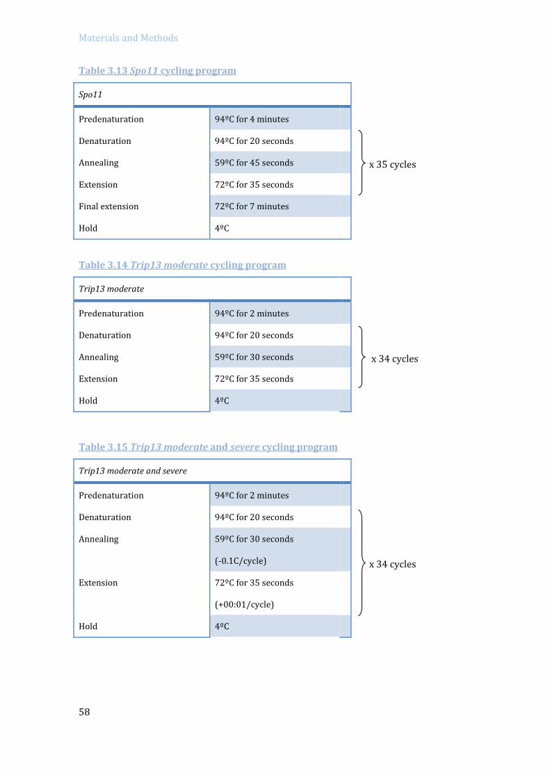
Materials and Methods
58
Table 3.13 Spo11 cycling program
x 35 cycles
Table 3.14 Trip13 moderate cycling program
x 34 cycles
Table 3.15 Trip13 moderate and severe cycling program
x 34 cycles
Spo11
Predenaturation 94ºC for 4 minutes
Denaturation 94ºC for 20 seconds
Annealing 59ºC for 45 seconds
Extension 72ºC for 35 seconds
Final extension 72ºC for 7 minutes
Hold 4ºC
Trip13 moderate
Predenaturation 94ºC for 2 minutes
Denaturation 94ºC for 20 seconds
Annealing 59ºC for 30 seconds
Extension 72ºC for 35 seconds
Hold 4ºC
Trip13 moderate and severe
Predenaturation 94ºC for 2 minutes
Denaturation 94ºC for 20 seconds
Annealing 59ºC for 30 seconds
(-0.1C/cycle)
Extension 72ºC for 35 seconds
(+00:01/cycle)
Hold 4ºC

Materials and Methods
59
Table 3.16 Dmc1 cycling program
x 39 cycles
- Run the amplification products in a 1% agarose gel with 1% of Ethidium bromide,
together with a weight molecular marker.
- Read the gel with GelDoc XR+ system (Bio-Rad) and determine the size of the
amplified bands.
3.2.3 RNA extraction
RNA was extracted from whole testis using the RNeasy Plus mini kit from Qiagen, in
order to perform Real time PCR or RNA sequencing.
Protocol:
- Cut a piece of frozen testis, a quarter of an adult wild type testis or use a whole testis
for mutant or juvenile mice (about 30 mg or less).
- Disaggregate the tissue in a 1.5 µl tube with a pestle.
- Add 600 µl of RLT buffer + 10 µl β-mercaptoethanol and homogenize the lysate with a
20-gauge needle and a syringe (pass the lysate five times).
- Centrifuge three minutes at maximum speed.
- Transfer the supernatant into a gDNA Eliminator spin column paced in a 2ml collection
tube.
- Centrifuge for 30 seconds at 10000 rpm. Discard column.
- Add 600 µl of 70% ethanol to the eluted liquid and mix with the pipette.
Dmc1
Predenaturation 94ºC for 2 minutes
Denaturation 94ºC for 20 seconds
Annealing 62ºC for 30 seconds
(-0.1ºC/cycle)
Extension 72ºC for 25 seconds
(+ 1 second/cycle)
Hold 4ºC
Materials and Methods
60
- Transfer 700 µl to the RNeasy spin column.
- Centrifuge for 15 seconds at 10000 rpm. Discard eluted liquid, repeat previous step
and discard liquid.
- Add 700 µl of RWI buffer to the column to wash the column membrane.
- Centrifuge for 15 seconds at 10000 rpm and eliminate eluted liquid.
- Add 500 µl of RPE buffer to wash the column membrane.
- Centrifuge for 2 minutes at 10000 rpm and eliminate eluted liquid.
- Place column to a new 1.5 ml tube and add 30 µl of RNase-free water to the column.
- Centrifuge for 1 minute at 10000 rpm to obtain the extracted RNA diluted in the eluted
water.
- Quantify the extracted RNA with Nanodrop (Thermo scientific) and its quality with the
Bioanalyzer (Agilent 2100).
3.2.4 RNA sequencing
For RNA sequencing 2 µg of RNA, with a RNA Integrity Number (RIN) > 8, were
sequenced at Centre Nacional d’Anàlisi Genòmica (CNAG). RNA samples were quantified
again with qubit-RNA Fluorometer. In order to generate the cDNA sequencing library poly-
A tails primers were used. From this “Stranded mRNA” library it was performed the
mRNA-Sequencing for a paired-end 75 pb read length with sequencing depth of >55 M of
reads for each sample (Illumina).
The 75x2 pair-end reads were mapped with the software GEM-Spit-RNA-Mapper
(to map the reads to the mouse genome reference annotation GRCm38) (Marco-Sola et al.
2012). To quantify the transcripts that maps to each gene Flux Capacitator software was
used (Montgomery et al. 2010). Then, to study the differential expression, and to
normalize: number of reads per gene/total of mapped reads, between the wild type and
mutant mice the R EdgeR software was used (Robinson et al. 2010). This algorithm
normalizes the data with the TMM method (Robinson and Oshlack 2010) considering
different library sizes and different RNA composition between the samples.
3.2.5 Real-time PCR
RNA sequencing was validated with quantitative PCR gene expression analysis.

Materials and Methods
61
Protocol:
- 2µg of total RNA were used for cDNA synthesis with iScript Advanced cDNA Synthesis
(Bio-rad), which uses oligo(dT) and random primers, according to manufacturer’s
instructions:
Table 3.17 Reverse transcription mix
Mix 20 µl reaction
Reaction mix 4 µl
Reverse Transcriptase 1 µl
RNA 2 µg
MilliQ-water Up to 20 µl
- Tubes were first incubated for 30 minutes at 42ºC and then 5 minutes at 85ºC.
- All samples used in the experiment were reverse transcribed at the same time and
diluted into a concentration of 15 ng/µl with nuclease-free water.
- For Real Time-PCR (qPCR) amplification intro-spanning PrimePCR primers (Bio-Rad)
and iTaq Universal SYBR Green Supermix (Bio-Rad) were used.
Table 3.18 PrimePCR primers used
Gene UniqueAssayID
Atp8b3 qMmuCID0022076
Hmbs qMmuCID0022816
Qrich2 qMmuCID0017034
Tktl1 qMmuCID0021812
Trip13 qMmuCID0019092
Xaf1 qMmuCID0009205
Zfy2 qMmuCID0021345

Materials and Methods
62
- 96-well plate were designed and each well contained (Table 3.19):
Table 3.19 Real time PCR reaction mix
Mix 10 µl reaction
PrimePCR primers 0.5 µl
SYBR green 5 µl
cDNA 1 µl (15ng)
MilliQ-water 3.5 µl
- The protocol program consisted in an initial denaturation step at 95ºC for 3 minutes,
followed by 40 cycles of denaturation at 95ºC for 10 seconds, primer annealing and
extension at 60ºC for 30 seconds. After the extension cycles, a step at 95ºC for 5
seconds and a melting curve 65ºC to 95º (at an increment of 0.5ºC every 5 seconds)
was applied.
- RT-PCR was performed in a CFX96™ Real-Time PCR Detection System thermocycler
(Bio-Rad) and analyzed with Bio-Rad CFX Manager 3.1 software.
- Housekeeping Hmbs gene was used as a control to normalize expression.
- All samples were run in triplicate and non-template controls were included.
3.2.6 Protein extraction and quantification
Protocol:
- Dissect testis and decapsulate or use frozen testis.
- Resuspend the tissue in 500 µl of RIPA buffer (50 mM Tris pH=8, 1% Triton X-100,
0.1% SD, 150 mM NaCl, 1 mM EDTA, 0.5 % Sodyum Deoxycholate and supplement
with 1x Proteases inhibitor cocktail (PI, Roche Dianostics) and 10 mM NaF) for wild
type testis in a 1.5 ml tube (or 200 µl of RIPA buffer for juveniles or mutant mice
testis).
- Disaggregate the tissue using a plastic pestle.
- Place the tubes in the thermomixer for 10 minutes at 95ºC.
- Disaggregate again the tissue with the pestle.

Materials and Methods
63
- Centrifuge at 13200 rpm for 10 minutes at 4ºC.
- Keep the supernatant and transfer it into a new 1.5 ml tube. Take an aliquot for
protein quantification and freeze the remaining protein extract at -80ºC.
- Quantify total protein levels in the extract using Pierce BCA protein Assay Kit (Thermo
scientific), based on colorimetric detection. Following manufacturer instructions,
prepare BSA solutions ranging from 0 to 2000 µg/ml in order to obtain a standard
curve. Add working reagent to the BSA dilutions and unknown samples. Incubate the
tubes for 30 minutes at 37ºC in the thermomixer and then cooled on ice. Read protein
concentration with the spectrophotometer (Cary 400 Bio) at 562 nm absorbance.
3.2.7 Western Blot
Western Blot technique was used in order to detect the levels of specific proteins
in mutant and wild type mice. A total protein extract was prepared from whole testis and
quantified with colorimetric detection. Running the samples into an electrophoresis gel
separates the specific proteins according to their specific molecular weight (kDa) and
when transferred into a membrane the protein bands of interest are detected with specific
primary antibodies.
Protocol:
Gel Electrophoresis
- Take 30-40 µg of proteins for each sample, and equilibrate them with Laemmli buffer.
If final volumes are very different, adjust with RIPA buffer.
- Boil for 5 minutes at the thermoblock to denature the proteins and spin down. Cool
samples at 4ºC.
- Make a 7-15% SDS Acrylamide gel (1.5 M Tris-HCl pH= 8.8, 20% SDS, 10% APS,
TEMED for the running gel solution, and 1 M Tris-HCl pH= 6.8, 20% SDS, 10% APS,
TEMED) using the Mini-Protean cell casting module 1mm (Bio-Rad).
- Introduce the gel into the electrophoretic chamber and add running buffer (250 mM
Tris base, 14.4% Glycine, 1% SDS in Milli-Q water).
- Load the 30-40 µg of proteins with Laemmli buffer (optionally adjusted with RIPA
buffer) into the wells of the gel and add Dual Color marker (Bio-Rad).
- Run at 80-100 volts for 1.5 hours approximately.

Materials and Methods
64
Semi-dry transfer
- Transfer the gel into a PVDF membrane using Trans-Blot Turbo system (Bio-Rad) with
the 7 minutes transfer program.
Antibody Incubation
- Block the membrane with blocking solution: 5% fat-free milk, 0.1% Tween-20 in PBS.
- Block for 30 minutes at room temperature in agitation.
- Incubate with primary antibody of interest diluted with blocking solution, overnight at
4ºC in agitation.
- Wash the membrane three times with PBST (0.1% Tween-20 in PBS) for 5 minutes in
agitation.
- Incubate with secondary antibody conjugated with horseradish peroxidase (HRP)
diluted in PBST for 1 hour at room temperature in agitation.
- Wash three times with PBST for 5 minutes in agitation.
- Develop with Luminata Forte Western HRP Substrate (Millipore) for
Chemiluminescent detection using the VersaDoc (Bio-Rad).
Table 3.20 List of primary antibodies used for Western blot
Antigen Host Company Dilution
β-ACTIN Mouse Abcam 1:10000
CHK2 Mouse Abcam 1:1000
PUMA Rabbit Sigma 1:1000
BAX Rabbit Abcam 1:1500
E2F1 Rabbit Santa Cruz 1:3000
Table 3.21 List of secondary antibodies used for Western blot
Target Host Company Dilution
Anti-mouse-HRP Goat Bio-Rad 1:3000
Anti-rabbit-HRP Goat Bio-Rad 1:3000

Materials and Methods
65
Stripping
If it was necessary to reincubate the membrane with another primary antibody,
stripping was performed to eliminate the previous antibody signal.
- Wash the membrane for 15 minutes with stripping buffer (15% Glycine, 1% SDS and
0.1% Tween-20).
3.3 HISTOLOGY TECHNIQUES
3.3.1 Fixation, embedding and sectioning
Testis from 2 to 6 months old adult mice were collected and processed for
histology. Testes were fixed either with Bouin’s solution or 4% paraformaldehyde,
embedded in paraffin and sectioned. For histological staging analysis (Russell et al. 1993),
testes sections fixed with Bouin’s solution were stained with Periodic Acid-Shiff (PAS) and
Hematoxilyn. Testes sections fixed with paraformaldehyde were used for TUNEL or
Immunofluorescence analysis.
Protocol:
Bouin’s fixation
- Prepare fresh fixative Bouin’s solution (70% of Saturated Picric acid, 25 %
Formaldehyde, 35% and 5 % Acetic acid) and keep it at 4ºC.
- Remove testis from mice.
- Punctuate the tunica albuginea of the testis with forceps to perfuse.
- Place freshly dissected tissue into a 50 ml tube with 5-8 ml of Bouin’s solution.
- Keep it overnight at 4ºC.
Paraformaldehyde fixation
- Prepare fresh fixative 4% paraformaldehyde solution made in PBS (pH=7.4) and keep
it at 4ºC.
- Remove testis from mice.
- Punctuate the tunica albuginea of the testis with forceps to perfuse.
- Place freshly dissected tissue into a 50 ml tube with 5-8 ml of the 4%
paraformaldehyde Solution.
- Keep it overnight at 4ºC.
Materials and Methods
66
Dehydration washes and deparaffinization
- Submerge the sample two times in cold PBS for 30 minutes.
- Submerge the sample in cold 50% ethanol for 30 minutes.
- Submerge the sample in cold 70% ethanol for 30 minutes (or keep it at this step for 2-
4 months).
- Submerge the sample in 85% ethanol for 30 minutes.
- Submerge the sample in with 96% ethanol for 30 minutes.
- Submerge the sample two times in 100% ethanol for 30 minutes.
- Submerge the sample three times in Histo-Clear (National Diagnostics) ethanol for 30
minutes.
- Place the tissue into a cassette.
- Submerge the cassette in 1:1 Histo-Clear/Paraffin for 45 minutes at 56ºC.
- Submerge the cassette in 100% paraffin at 56°C overnight to allow proper paraffin
infiltration.
Embedding testis in Paraffin blocks
- Turn on the heat block of the paraffin station and introduce the metal mold in the 60ºC
melted paraffin bath.
- Open the cassette, transfer the tissue over the mold and place it on the cold plate.
Using the forceps orient the testis to a vertical position.
- Add hot paraffin to cover the tissue and let the block form over the cold platform for
two hours.
- Keep the blocks overnight at room temperature. Store at 4ºC.
Sectioning
- Section the paraffin blocks at a thickness of 6 µm using the microtome (which is the
thickness of a cell), and let the sections float onto a hot water bath.
- Transfer the sections onto poli-lysine treated slides and dry them into an incubator
overnight at 37ºC.
Deparaffinization and re-hydration washes
- Deparaffinize the slides with three washes for 5 minutes in xylene.
- Rehydrate the tissue with a gradient of ethanol washes:
- Submerge two times with 100% ethanol for 3 minutes.
Materials and Methods
67
- Submerge two times with 96% ethanol for 2 minutes.
- Submerge two times with 70% ethanol for 2 minutes.
- Submerge with distilled water for 2 minutes.
3.3.2 PAS-Hematoxylin staining
The Periodic Acid Schiff method is used to stain the mucopolysaccharid and
glycogen structures in the tissue. Hematoxylin stains acidic compounds, such as DNA or
RNA into dark blue.
Protocol:
- Immerse the slides in a 1% Periodic Acid solution for 15 minutes at room temperature.
- Wash two times in distilled water for 3 minutes.
- Incubate the slides in Schiff’s reagent for 30 minutes, at room temperature and
darkness.
- Wash two times in sulphurous water (10% Potassium metabisulfite, 0.1 M HCl in Milli-
Q water) for 3 minutes.
- Wash two times in distilled water for 3 minutes.
- Stain with Mayer’s Hematoxylin for 1 minute at room temperature.
- Rinse with running tap water to eliminate Hematoxylin excess.
Dehydration and mounting
Use a gradient of ethanol series to dehydrate the tissue:
- Submerge two times with 70% ethanol for 2 minutes.
- Submerge two times with 96% ethanol for 2 minutes.
- Submerge two times with 100% ethanol for 3 minutes.
- Submerge two times in xylene for 5 minutes.
- Place several drops of DPX mounting medium over each section and add a coverslip.
Let air dry the slides.
- Analyze with an Optical microscope.
- Images were captured in a Zeiss Axioskop microscope with a ProgRes C10 camera
using the ProgRes Pro 2.7.7 software. Images were then processed with Photoshop.
3.3.3 TUNEL test: In situ cell death detection over tissue sections
In order to detect apoptotic cells in the sectioned tissue the In situ Cell Death
Detection kit (Roche Diagnostics) was used. With this TdT-mediated dUTP-X nick end

Materials and Methods
68
labeling (TUNEL method), the DNA damage (single and double strand breaks) are labelled.
The terminal deoxynucleotidyl transferase (TdT) catalyzes the incorporation of modified
nucleotides –which contain a fluorescein- at the free 3’-OH DNA ends so that the apoptotic
cells can be visualized under a fluorescence microscope.
Protocol:
- Deparaffinize and rehydrate the sectioned tissue.
- Wash the slides with PBS for 2 minutes at room temperature.
- Permeabilize with 0.5% Triton X-100 in PBS for 15 minutes.
- Wash two times with PBS for 2 minutes.
- Add 100 µl of Dako solution (to reduce background) on each slide, cover with parafilm
and incubate in a humid chamber for 15 minutes at room temperature.
- Rinse the slides in PBS for 2 minutes.
- Add 6-10 µl of TUNEL reaction mix (10% TdT enzyme solution in buffer solution) over
each section. Incubate in a humid chamber for 1 hour at 37ºC.
- Wash the slides three times in PBS for 5 minutes.
- Add 20 µl of Vectashield mounting medium (Vector) containing DAPI at a
concentration of 0.1 μg/ml and cover with a coverslip.
- Analyze with the fluorescence microscope or store at -20ºC.
- Images were captured in a Zeiss Axioskop microscope with a ProgRes C10 camera
using ProgRes Pro 2.7.7. Images were then processed with Photoshop.
3.4 CELL BIOLOGY AND CYTOLOGY TECHNIQUES
3.4.1 Spermatocyte nuclei spreads from fresh testis
Spermatocyte preparations can be made from fresh mouse testis. Since the nuclear
membrane is still functional in these cells, a hypotonic sucrose buffer can be used in order
to expand the cell. With this technique, we can study the nuclear proteins of a specific cell
by immunofluorescence.
Protocol:
- Remove the testis from the mouse and rinse them on a petri dish with cold PBS.
- Decapsulate the testis with forceps to release the seminiferous tubules into 200 µl PBS
and mince the tubules with the scalpel.
- Transfer the cell suspension into a 15 ml tube containing 10 ml of PBS and pipette
back and forth to homogenize. Let large pieces settle to the bottom of the 15 ml tube.

Materials and Methods
69
- Transfer 1 ml of cell suspension into ten 1.5 ml tubes.
- Centrifuge the 1.5 ml tubes at 1500 rpm for 5 minutes.
- Discard supernatant and remove the remaining PBS with a pipette.
- Add 40 µl of 0.1 M sucrose solution at 37ºC and resuspend the pellet.
- Prepare Superfrost slides on a humid chamber and distribute 65 µl of cold fixative
solution (1% paraformaldehyde, 0.1% Triton X-100 and 1x protease inhibitor cocktail
(Roche Diagnostics) in MilliQ-water, pH= 9.2).
- Add 20 µl of cell suspension onto each slide.
- Close the humid chamber and let cells fix for 2 hours at room temperature.
- Open the chamber and let slides air dry for 30-60 minutes.
- Rinse slides with 0.4% Photo-Flo solution (Kodak) for 2 minutes.
- Air dry the slides completely and store them at -80ºC.
3.4.2 Spermatocytes nuclei spreads from frozen testis
Nuclear surface spreads were also prepared from frozen testis samples, which can
be preserved and stored at -80ºC for many months. This method has also the advantage
that only a very little piece of tissue is needed. The principal difference between frozen
and fresh testis spreading techniques is that once the testis are frozen, the cell membrane
breaks, so in this case a detergent (Lipsol) is used in order to help to expand the volume of
the spermatocytes.
Protocol:
- Keep all solutions cold and add fresh Protease inhibitor cocktail (Complete EDTA-
free Protease Inhibitor Cocktail Tablets, Roche Diagnostics).
- Cut a small portion of mouse frozen testis with a scalpel over a glass slide (placed on
ice platform).
- Add up to 60 µl of cold PBS and mince the sample with the scalpel.
- Transfer the cellular suspension into 1.5 µl tube and pipette few times to homogenize.
Let large pieces settle down to the bottom.
- Place 20 µl of the cellular suspension on each slide.

Materials and Methods
70
- Add 80 µl of 1% Lipsol (detergent solution) and leave for 10-15 minutes (12 minutes
usually enough).
- Fix the cells by adding 150 µl of fixative solution (1% paraformaldehyde, 0.15% Triton
X-100 and MilliQ water; pH= 9.2-9.4) and let the slides fix for 2 hours in a humid
chamber at room temperature.
- Open the humid chamber and let slides air-dry at room temperature.
- Wash slides four times for 2 minutes with 0.4% Photo-Flo (Kodak) solution.
- Slides can be used for immunofluorescence, or air dried and stored at -80ºC.
3.4.3 Spermatocytes squashed preparation
Squashed spermatocyte preparations –which preserve nuclear chromosome
structure- were performed as previously described (Page et al. 1998).
Protocol:
- Prepare the slides to use by rinsing them in chloroform/ethanol (1:1) for 2 minutes
and air dry them.
- Cut a small portion of frozen testis tissue.
- Disaggregate the tissue in 30 µl of fixing solution (2% paraformaldehyde, 0.05% Triton
X-100 in PBS; pH= 7.4) for 10 minutes at room temperature.
- Add 6 µl of the cellular suspension to each slide. Place a coverslip over the slide and
apply pressure to squash the cells.
- Submerge the slide in liquid nitrogen and keep it in dry ice.
- Proceed to perform immunofluorescence or store slides at -80ºC.
3.4.4 Immunofluorescence
Spermatocyte preparations were stained for immunofluorescence in order to
visualize the proteins of interest. Immunofluorescence is a technique based on
fluorescent-labeled antibodies, were a primary antibody targets a specific antigen, and
then a secondary antibody –which is labeled with a fluorophore-, binds specifically to the
primary antibody.

Materials and Methods
71
Protocol:
- Block slides with PTBG (0.2% BSA, 0.2% gelatin, 0.05% Tween-20 in PBS) for 10 minutes
in agitation.
- Prepare the primary antibody dilution in PTBG and add 100 µl to each slide (cover with
parafilm). Incubate at 4ºC in a humidified chamber overnight.
- Wash four times for 3 minutes in agitation with PTBG.
- Prepare the secondary antibody dilution in PTBG and add 100 µl to each slide (cover with
parafilm). Incubate at 37ºC in a humidified chamber for 1 hour.
- Wash four times for 3 minutes with PTBG in agitation. Drain the slide.
- Add 20 µl of Vectashield mounting medium (Vector) containing DAPI at a concentration of
0.1 μg/ml and cover with a coverslip.
- Observe under the fluorescence microscope or store at-20º C (or 4º C for short periods).
- Images were captured in a Zeiss Axioskop microscope with a ProgRes C10 camera using
ProgRes Pro 2.7.7. Images were then processed with Photoshop.
Table 3.22 List of the primary antibodies used for immunofluorescence
Antigen Host Source Dilution
SYCP3 Mouse Abcam 1:200-1:400
SYCP3 Rabbit Abcam 1:200-1:400
SYCP3 Rabbit Santa Cruz 1:200-1:400
SYCP3 Goat Santa Cruz 1:50-1:100
γH2AX (ser 139) Mouse Millipore 1:100-1:400
γH2AX (ser 139) Rabbit Santa Cruz 1:100
H1T Guinea pig Dr. M.A. Handel 1:500
ATR Rabbit Calbiochem 1:100
SUMO-1 Mouse Invitrogen 1:100
MLH1 (G168-15) Mouse BD Biosciences 1:50
TOPBP1 Rabbit Abcam 1:10-1:50
Phospho(S2)-RNA pol II Mouse Abcam 1:100

Materials and Methods
72
Table 3.23 List of the secondary antibodies used for immunofluorescence
Dye Antigen Host Company Dilution
FITC Mouse Goat Jackson Immunoresearch 1:200
FITC Rabbit Goat Jackson Immunoresearch 1:200
Cy3 Mouse Goat Jackson Immunoresearch 1:200
Cy3 Rabbit Goat Jackson Immunoresearch 1:200
Cy3 Guinea
Pig
Goat Jackson Immunoresearch 1:200
Cy5 Mouse Goat Jackson Immunoresearch 1:100
Alexa 488 Mouse Donkey Life Technologies 1:200-1:400
Alexa 594 Goat Donkey Molecular Probes 1:200
Alexa 594 Mouse Goat Molecular Probes 1:200
Alexa 594 Rabbit Chicken Molecular Probes 1:50-1:400
Alexa 647 Rabbit Donkey Molecular Probes 1:100-1:200
3.4.5 TUNEL staining over IF-stained slides
The In Situ Cell Death Detection kit (Roche Diagnostics) was used to detect
apoptotic spermatocytes in immunostained slides.
Protocol:
- Wash three times immunostained-slides for 5 minutes with 0.5% Triton X-100 in PBS.
- Wash slides three times for 1 minute with PBS.
- Add 50 µl of TdT reaction mixture (45 µl of solution 2 + 5 µl solution 1 enzyme,
according to manufacturer’s protocol) and cover with parafilm.
- Incubate at 37ºC in a humid chamber for 1 hour.
- Wash three times for 5 minutes with PBS.

Materials and Methods
73
3.4.6 RNA FISH and Immunofluorescence on spermatocytes
RNA fluorescence in situ hybridization (RNA-FISH) technique permits to detect gene
transcription at the cellular level, which allows to directly observing gene expression at a
specific locus in every meiotic phase. The technique consists in applying a digoxigenin-
labeled probe to a permeabilized and fixed nuclei, followed by stringency washing and to
observe the probe-transcript by fluorescence microscopy (Mahadevaiah et al. 2008;
Mahadevaiah et al. 2009). BAC DNA probes used for this study were: Scml2, RP24-204O18
(CHORI BACPAC library) and Zfx, bMQ-372M23 (Mouse bMQ BAC library). BAC-containing
bacteria (BACs) were grown in LB-chloramphenicol culture overnight at 37ºC. A standard
miniprep method was used to isolate BAC DNA.
Protocol:
BAC DNA extraction and probe preparation
BACs culture: (keep sterility)
- Set up 25 ml of LB medium (with Cloramphenicol 10µg/ml) into a 50 ml falcon.
- Add 5 µl of BAC glycerol stock.
- -Incubate at 37ºC overnight in agitation.
DNA extraction:
- Keep the BACs culture at 4ºC until proceeding.
- Prepare miniprep solutions (in Milli-Q water):
P1: 15 mM Tris pH= 8, 10Mm EDTA, 100 µg/ml RNAse A.
P2: 0.2 M NaOH, 1% SDS, made fresh.
P3: 3 mM KOAc; pH= 5.5 (set pH with Acetic acid).
- Centrifuge at 4500 rpm the 25 ml BACs culture at 4ºC for 15 minutes. Remove the
supernatant.
- Re-suspend the pellet in 1.7 ml of P1 by vortexing (no more vortex after this point).
- Add 1.7 ml of P2 and invert twenty times to mix. Incubate for 5 minutes at room
temperature. BACs will lyse and become viscous.
- Add 1.7 ml of P3 and invert twenty times to mix. Incubate 5 minutes on ice. Genomic
DNA and proteins denature (observe a white precipitate).

Materials and Methods
74
- Aliquot 1 ml into five 1.5 ml tubes. Centrifuge at 13000 rpm at room temperature for
10 minutes.
- Take 900 µl of the supernatant into five new 1.5 ml tubes.
- Add 540 µl of isopropanol and invert to mix. Immediately centrifuge at 14000 rpm at
room temperature for 15 minutes. BAC DNA will precipitate.
- Remove isopropanol by decanting and finish the process with the pipette.
- Wash with 500 µl of cold 75% ethanol and centrifuge at 12000 rpm for 5 minutes.
Discard liquid and spin again. Remove all liquid with the pipette.
- Air dry the pellets (for approximately 12 minutes).
- Add 120 µl of water to each 1.5 ml tube to dissolve the DNA. Mix and combine the DNA
solution from all the tubes.
- Add 2 µl of RNase A and 33 µl of buffer H (500 mM Tris-HCl, pH 7.5 100 mM MgCl2 10
mM Dithiothreitol (DTT) 1000 mM NaCl).
- Mix and incubate at 37ºC on a water bath overnight.
Phenol:Chloroform DNA extraction:
- Add 440 µl of saturated Phenol (kept at 4ºC). Mix by inverting twenty times (Perform
in fume hood).
- Centrifuge at 12000 rpm for 5 minutes. Slowly remove 430 µl from the top layer
(which contains the DNA) to a new 1.5 ml tube.
- Add 430 µl of Phenol:Chloroform:Isoamyl Alcohol (PCI). Mix up and down vigorously,
until it becomes white.
- Centrifuge at 12000 rpm for 5 minutes. Slowly remove 420 µl from the top layer to a
new 1.5 ml tube.
- Add 420 µl of Chloroform:Isoamyl Alcohol (CI). Mix up and down vigorously.
- Centrifuge at 12000 rpm for 5 minutes. Slowly remove 410 µl from the top layer to a
new 1.5 ml tube.
- To precipitate DNA: Add 40 µl of 3M Sodium acetate (NaAc) (pH 5.2) and 1000 µl of
100% ethanol. Shake vigorously.
- Centrifuge at 13000 rpm for 15 minutes. Decant and remove liquid with the pipet.

Materials and Methods
75
- Wash with 400 µl of 75% ethanol. Centrifuge at 13000 rpm for 5 minutes. Decant and
spin again. Remove remaining liquid with the pipet.
- Let the pellet air dry (for approximately 12 min).
- Dissolve in 20 µl of MilliQ-water for each 1.5 ml tube and leave it at room temperature
overnight or for 10 min at 37ºC to dissolve DNA. Combine the four 1.5 ml tubes with
BAC DNA in one tube.
Probe labelling:
- Into a 1.5 ml round tap tube add: 16 µl of BAC DNA (approximately 2µg) and 4 µl of the
DIGMIX (DIG-Nick Translation Mix, Roche). Incubate at 15ºC for 50 minutes in a water
bath (made with ice and water).
- Run in a 2% agarose gel with 6.25% Ethidium Bromide.
- Run 2 µl of Nick translation reaction DNA + 8 µl of loading buffer at 90 volts for 30
minutes.
- A smear should be around 500 bp (until 100 bp would stil be fine). In case the DNA
needs to be cut more, put the probes for an extra 2-3 minutes at 37ºC.
- Precipitate the entire probe which will be used for nine reactions, mix in a 1.5 µl tube:
- 18 µl of probe.
- 27 µl of Cot-1 DNA (Invitrogene).
- 9 µl salmon sperm DNA (Stratagene).
- 160 µl 100% ethanol.
- Centrifuge at 13000 rpm at 4ºC for 10-15 minutes. Discard liquid.
- Add 200 µl of 75% ethanol.
- Centrifuge at 13000 rpm at 4ºC for 5 minutes. Remove liquid and spin. Remove the
rest of the remaining liquid.
- Let the pellet air dry for approximately 12-15 minutes.
- Add 135 µl of formamide to the probe stock tube. For each slide, 15 µl of this stock will
be used.
- Keep at 37ºC for 10 minutes in order to dissolve. Store at -20ºC.

Materials and Methods
76
RNA FISH slides preparation
Probe hybridization:
- Prepare 4% paraformaldehyde in PBS pH= 7.4 (dissolve at 65ºC in a water bath and
filter).
- Prepare CSK buffer: 100 mM NaCl, 300 mM sucrose, 3mM MgCl2, 10 mM PIPES, 0.5%
Triton X-100 and 2 mM vanadyl ribonucleoside (New England Biolabs).
- Boil Superfrost slides for 10 minutes and let them air dry.
- Mince frozen testis samples with the scalpel and add three drops of RPMI medium. Tilt
the disaggregated material and add more drops according to the number of slides.
- Mix with Pasteur pipette.
- Add one drop of cells suspension in the middle of the slide (let it settle for 2 minutes).
- Add 4 drops of CSK buffer for 10 minutes to permeabilize.
- Add 6 drops of 4% paraformaldehyde for 10 minutes to fix.
- Meanwhile, denature the probe: Place 15 µl of probe at 80oC for 10 minutes. Spin and
add 15 µl of 2x hybridization buffer (4x SSC, 50% dextran sulphate, 2 mg/ml BSA and
2 mM Vanadyl ribonucleoside) and place at 37ºC for 20 minutes (to pre-hybridize).
- Decant liquid from the slides (with absorbing paper).
- Rinse slides in cold PBS (each genotype with different PBS).
- Dehydrate slides with cold ethanol series washes:
- Wash two times with 70% ethanol for 3 minutes.
- Wash with 80% ethanol for 3 minutes.
- Wash with 95% ethanol for 3 minutes.
- Wash with 100% ethanol for 3 minutes.
- Air dry the slides vertically.
- Spin and add 30 µl of the probe to the slides and cover with a 50 mm coverslip.
- Incubate in a humid chamber (filled with Formamide) at 37ºC overnight.
Stringency washes and probe detection:
- Prepare solution A: 50% Formamide in SSC, pH= 7.2-7.4.
Materials and Methods
77
- Prepare solution B: 2x SCC pH= 7-7.2.
- Prepare solution C: 4x SCC, 0.1% Tween-20, pH= 7-7.2.
- Prepare blocking buffer: 4x SCC, 4 mg/ml BSA and 0.1% Tween-20.
- Prepare detection buffer: 4x SCC, 1 mg/ml BSA and 0.1% Tween-20.
- Place a mixer and a water bath at 45ºC in the fume-hood. Keep solution A and solution
B at 42ºC.
- Wash three times with solution A for 5 minutes (remove coverslip after the first wash).
- Wash three times with solution B for 5 minutes.
- Rinse with solution C.
- Add 100 µl of blocking buffer and place a 60 mm coverslip. Incubate at 37ºC in a humid
chamber for 30 minutes.
- Prepare the antibodies with detection buffer or solution C and centrifuge them at
13000 rpm at room temperature for 5 minutes:
- Anti-digoxigenin-FITC (Millipore) (1:10).
- Primary antibodies (see Table 3.11).
- Secondary antibodies (see Table 3.12).
- Add the anti-digoxigenin-FITC 50 µl for slide and incubate at 37ºC for 1 hour.
- Wash three times with solution C for 2 minutes.
- Add the primary antibodies: 50 µl for slide and incubate at room temperature for 30
minutes.
- Wash three times with solution C for 2 minutes.
- Add secondary antibodies: 50 µl for slide and incubate at room temperature for 30
minutes.
- Wash three times with solution C for 2 minutes.
- Mount in Vectashield mounting medium with DAPI (at a concentration of 1.5 µg/ml).
- Keep slides for 15-30 minutes at 4ºC, and then store them at -20ºC.
- Analyze slides on a fluorescence microscope.
- Images were captured with Olympus IX70 inverted microscope and computer-assisted
(DeltaVision) CCD camera (Photometrics) and processed with ImageJ and Photoshop.

Materials and Methods
78
3.4.7 EU Imaging on spermatocytes
To analyze global RNA expression in meiotic cells, we have used the EU Click-it
Imaging technique (Click-iT® RNA Alexa Fluor® 488 Imaging Kit, Life Technologies) to
cytologically detect newly synthesized RNA. We obtained a cell suspension from fresh
testis and then incubated these cells in the presence of a Uridine analogue (5-Ethynyl
Uridine (EU), Life Technologies), which is specifically incorporated to nascent RNA. From
this cell suspension slides are prepared using a protocol similar to the RNA-FISH
(Mahadevaiah et al. 2009), in order to preserve the nuclei structure and keep cellular
volume. Then, modified RNA was labeled with a dye (Alexa Fluor 488) through a
chemoselective ligation or “click” reaction between an azide-containing dye and an alkyne,
which allows the visualization of the RNA by fluorescence microscopy. With this technique
we can cytologically evaluate the RNA expression of cells at different stages of prophase.
Protocol:
Enzymatic testis dissociation
- Isolate the two testes from the mouse and decapsulate.
- Transfer the testis (one for the wild type, and two for the mutant mice) to a 50 ml tube
containing 10 ml Gey’s Balanced Solution (GBSS, Sigma) + 0.5 mg/ml Collagenase
(Sigma, freshly made).
- Incubate at 33ºC and 350 rpm for 15 minutes in the thermomixer, shaking vigorously
every 5 minutes to disassociate the tubules.
- Let the seminiferous tubules settle down to the bottom for 4 minutes at room
temperature. Discard supernatant.
- Wash two times with 10 ml GBSS, centrifuge at 2400 rpm at room temperature for 3
minutes.
- Resuspend tubules in 10 ml GBSS containing 0.5 mg/ml Trypsin (Sigma) and 2 µg/ml
DNase I.
- Incubate at 33ºC and 350 rpm for 15 minutes (for the last 3 minutes mix up and down
gently with a Pasteur pipette, so that spermatocytes become suspended and
homogeneous).

Materials and Methods
79
- Stop Trypsin reaction by adding 0.5 ml of Fetal Bovine Serum (5% FBS, Gibco Life
Technologies) and mix.
- Filter the cell suspension through a 70 µm strain to a new 50 ml tube (to remove
clumps). Centrifuge at 2400 rpm at room temperature for 3 minutes. Discard
supernatant.
- Wash two times with 4 ml GBSS + 5% FBS + 2 µg/ml DNase I.
- Centrifuge at 2400 rpm at room temperature for 3 minutes. Discard supernatant.
EU incubation and spreads preparation
- Resuspend pellet in 500 µl of 5 mM EU in of RPMI medium (with 10% FCS and 1x
penicillin/spretomycin/fungizone) in a 50 ml tube (and incubate control with 500 µl
RPMI without EU).
- Incubate at 33ºC and 350 rpm for 2.5 hours at the thermomixer.
- Prepare 4% paraformaldehyde in PBS, pH= 7.4.
- Prepare CSK buffer: 100 mM NaCl, 300mM sucrose, 3 mM MgCl2, 10 mM PIPES, and
add freshly 0.5% Triton X-100 and 2 mM Vanadyl ribonucleoside (New England
Biolabs).
- Place 60 µl of cell suspension per slide (Superfrost) over a cold platform, al let it settle
down for 10 minutes.
- Add four drops of CSK buffer for 13 minutes.
- Add six drops of 4% paraformaldehyde in PBS for 30 minutes.
- Tilt cold platform to eliminate liquid excess from the slides.
- Rinse slides in cold PBS.
- Air dry slides.
- Store at -80ºC or proceed to Click-iT reaction.
Click-iT EU staining reaction and IF
- Wash two times with cold PBS at 4ºC for 3 minutes.
- Wash with cold 0.5% Triton-PBS at 4ºC for 30 minutes (to permeabilize cells).

Materials and Methods
80
- Wash with cold PBS at 4ºC for 3 minutes.
- Prepare Click-iT Reaction master mix according manufacturer (Click-iT® RNA Alexa
Fluor® 488 Imaging Kit, Life Technologies): 428 µl compound C + 20 µl compound D +
1.8 µl Biotin Azide (Life Technolgies) + 50 µl working solution + 5 µl Vanadyl
ribonucleoside.
- Add 100 µl of the Mix to each slide and cover with parafilm.
- Incubate in a humid chamber at room temperature for 30 minutes.
- Add 100 µl of Rinse buffer to each slide, cover with parafilm and let it rest for 5
minutes.
- Rinse slides with cold PBS.
- Block slides with cold PTBG (0.2% BSA, 0.2% gelatin, 0.05% Tween-20 in PBS) for 10
minutes.
- Add primary antibodies in PTBG with 2 mM Vanadyl ribonucleoside: Streptavidin
Alexa Fluor 488 (1:100, Life Technologies), mouse anti-SYCP3 at 1:100 (Santa Cruz)
and rabbit anti-γH2AX (1:100, Millipore).
- Incubate primary antibodies in a humid chamber at 37ºC for 1 hour.
- Wash three times with PBS for 5 minutes.
- Add secondary antibodies in PTBG with 2 mM Vanadyl ribonucleoside: anti-mouse
Alexa Fluor 594 (1:200) and anti-rabbit Alexa Fluor 647 (1:200).
- Incubate secondary antibodies in a humid chamber at room temperature for 1 hour.
- Wash three times with PBS for 5 minutes.
- Mount slides with Vectashield mounting medium with DAPI at a concentration of 1.5
μg/ml (Vector) and a coverslip.
- Visualize slides under a fluorescence microscope (Axio2 microscope, Zeiss) and
capture images with a CCD camera and process them with Slidebook software package
(Intelligent Imaging Innovations). Capture 0.5 μm stacks from the top to the bottom of
the cells. From these stacks create a sum projection image.
- Measure fluorescence intensity with ImageJ 1.49g software (National Institutes of
Health, USA; http://imagej.nih.gov/ij/). Quantify EU intensity signal taking into
account the nuclei area identified with DAPI, and obtain background signal from the
surrounding areas. Use the ImageJ parameter Integrated density to plot the graphics

Materials and Methods
81
(which is the sum of the values of the pixels in that selection, equivalent to the product
of the Area and the Mean Gray Value –fluorescence intensity values-).
- Quantification of phosphorylated RNA polymerase II was performed over slides
obtained following this protocol but cultivated without EU. IF was performed as in
3.4.4 . Images were obtained and processed as in EU quantification.
3.4.8 Cell Sorting
The Fluorescence activated cell sorting (FACS) of live cells technique is used to
separate the cellular heterogeneity present in the testis into meiotic sub-populations. This
technique uses flow cytometry to separate the cells according to their fluorescent labeling
with Hoechst 33342 and Propidium Iodide. Hoechst 33342 dye diffuses through plasma
membrane and bind to DNA poly(dAT) sequences, helping to detect DNA content and
chromatin structure. Meiotic cells are classified according to the blue fluorescence (which
depends on the DNA content); and the red fluorescence they emit (which corresponds to
chromatin complexity). Propidium iodide fluorochrome staining penetrates through
impaired plasma membranes, allowing to discriminate dead cells (for an example see Fig.
3.1) (Bastos et al. 2005; Cole et al. 2014).
Protocol:
Enzymatic testis dissociation
- Isolate the two testes from the mouse and decapsulate them.
- Transfer the testis (one for the wild type, and two for the mutant mice) to a 50 ml tube
containing 10 ml GBSS containing 0.5 mg/ml Collagenase (Sigma).
- Incubate at 33ºC and 350 rpm for 15 minutes in the thermomixer, shaking vigorously
every 5 minutes to disassociate the tubules.
- Let the seminiferous tubules settle down to the bottom for 4 minutes at room
temperature. Discard supernatant.
- Wash two times with 10 ml GBSS, centrifuge at 2400 rpm at room temperature for 3
minutes.
- Resuspend tubules in 10 ml GBSS containing 0.5 mg/ml Trypsin (Sigma) and 2 µg/ml
DNase I.

Materials and Methods
82
- Incubate at 33ºC and 350 rpm for 15 minutes (for the last 3 minutes mix up and down
gently with a Pasteur pipette, so that spermatocytes become suspended and
homogeneous).
- Stop Trypsin reaction by adding 5% of Fetal Bovine Serum (FBS, Gibco Life
Technologies) and mix.
- In order to individualize cells filter the cell suspension through a 70 µm strain (BD
Falcon) to a new 50 ml tube. Centrifuge at 2400 rpm at room temperature for 3
minutes. Discard supernatant.
- Wash two times with 4 ml GBSS containing 5% FBS and 2 µg/ml DNase I. If processing
several samples keep them on ice.
- Centrifuge at 2400 rpm at room temperature for 3 minutes to spin the pellet and
discard supernatant.
- Stain the cells with Hoechst 33342 by adding 4 ml of GBSS containing 10 µg/ml
Hoechst 33342, 5% FBS (Life Technologies) and 2 µg/ml DNase I and incubate in the
thermomixer at 33ºC for 45 minutes at 350 rpm.
- Centrifuge at 2400 rpm at room temperature for 3 minutes to spin the pellet and
remove supernatant.
- Just before fluorescent-activated cell sorting stain with Propidium Iodide by adding 0.2
µg/ml of Propidium Iodide in 1 ml GBSS containing 5% FBS and 2 µg/ml DNase I to
resuspend the cells. Mix gently.
- Filter the cell suspension through a 40 µm strainer of the FACS tube. Leave cell
suspension on ice until proceeding to sorter.
- Take FACS tubes for collecting the sorted cells (according to number of populations
collected) with 300 µl of GBSS containing 5% FBS in each tube.
- Sort cells in the FACS core facility using FCCF-MoFlo1 (Dako) flow cytometer with a
350-nm argon laser.
- Proceed to prepare spermatocytes spreads with the sorted cells. Place the cell
suspension (all the volume, or divide into fractions if cells are needed for other
purposes) into a new eppendorf. Centrifuge for 3 minutes at room temperature at
2400 rpm. Remove supernatant.
- Add 100 µl of 0.1 M sucrose to each eppendorf and keep it for 8-10 minutes.
- Add 65 µl of 1% paraformaldehyde with 0.1% Triton X-100 to each slide.

Materials and Methods
83
- Add all the cell suspension to each slide, and keep it for 2 hours into a humid chamber
at room temperature. Then, open the chamber and let the slides completely air dry.
- Wash two times with 0.4 % Photo-Flo solution for 1 minute.
- Wash slides one time with Milli-Q water.
- Block slide with PTBG for 10 minutes in agitation.
- Proceed to stain SYCP3 and γH2AX for immunofluorescence in order to classify
meiotic sub stages as in 3.4.4.
Figure 3.1. Example of published cell sorting to obtain spermatocytes population. In this work, single cell suspension stained with Hoechst 33342 and Propidium iodide was sorted in a MoFlo cytometer. The plot shows different cell type populations separated according blue and red fluorescence. Examples of gated cells (98% MLH1-positive pachynema and diplonema spermatocytes (stained with MLH1 (green), SYCP3 (red) and DAPI (blue)) are shown as representative images. Image taken from Cole et al. (2014)


85

86

87
RESULTS


Results
89
4. RESULTS
4.1 Study the pachytene arrest in male mammalian
meiosis
4.1.1 Determine other members of the signaling pathway that activates the
pachytene arrest in mouse spermatocytes
Several studies have involved DDR proteins in meiotic checkpoints in several
organisms (MacQueen and Hochwagen 2011). For our work, we wanted to study if we
could use biochemical tools to detect the activation of the DDR pathway in recombination-
deficient mutants in mouse meiosis. As a first approach, we performed Western blot
against several proteins of the DDR pathway from adult whole testis protein extracts. We
used Trip13mod/mod mutants, as mice that present recombination impairment (Roig et al.
2010; Li et al. 2007) and Arf-/- mutants, which present a p53-dependent apoptosis due to
accumulation of H2AX (Churchman et al. 2011). We also examined whole testis extracts
from wild type mice as controls, and Spo11-/- mutants as negative control (since their
spermatocytes do not present SPO11-generated DSBs (Baudat et al. 2000)).
Specifically, since our group reported that the MRE11 complex-ATM-CHK2
pathway participated in the activation of the recombination-dependent arrest (Pacheco et
al. 2015), first we wanted to see if we could observe this activation by Western blot and
screen for other known factors of this signaling pathway. We focused our investigations on
the following proteins: CHK2, PUMA, BAX and E2F1. CHK2 is the checkpoint effector
kinase activated by ATM in response to DSBs (Chaturvedi et al. 1999). In somatic cells,
both ATM and CHK2 signal downstream to p53, which induces transcriptional activation
of multiple genes, including the pro-apoptotic factors PUMA and BAX (Roos and Kaina
2006). The transcription factor E2F1 is phosphorylated and activated by CHK2 in response
to DNA damage, resulting in apoptosis entry (Stevens et al. 2003).
AS regards CHK2, we observed an increased signal in Trip13mod/mod mutant samples
compared to the wild type, and also the appearance of a lower mobility band, presumably
the phosphorylated form of CHK2 (Fig. 4.1A). However, we also observed a similar
intensity band and a phospho-CHK2 band in Spo11-/- mutant. The CHK2 band on Arf-/-
mutant presented an intermediate intensity, higher than the wild type but less intense
than the ones observed in either Trip13mod/mod or Spo11-/- mutants, and no lower mobility

Results
90
band could be observed in this case (Fig. 4.1A). These results show that CHK2 protein is
more activated in Trip13mod/mod mutant than in the wild type. However since we observe a
similar band and phosphorylation on Spo11-/- mutant, we cannot conclude that this is
specific of the activation of the recombination-dependent arrest. Also, we expected to find
more CHK2 protein in the Arf-/- mutant, which we know they do experience a p53-
dependent recombination-dependent arrest.
Figure 4.1. Activation of DDR proteins in pachytene-stage arrested mutant testes. Western blot analysis of (A) CHK2, (B) E2F1, (C) BAX and (D) PUMA of the indicated genotypes from adult mice testes. Below each panel, β-ACTIN serves as a loading control.
For E2F1 protein, we observed that the bands present in Trip13mod/mod and Spo11-/-
lanes were more intense than those found in the wild type. However, for Arf-/- mutant,
E2F1 band was similar to the wild type (Fig. 4.1B). These results are similar to what we
observed for CHK2 protein, where there is a stronger band on Trip13mod/mod that could
suggest that E2F1 is activated when a recombination-dependent arrest occurs, but we
cannot conclude that since we find a similar band on Spo11-/- mutant which has no DSBs.
And again, for the other recombination-defective mutant we analyzed, Arf-/-, we don’t see
an increased presence of either E2F1 or any of its post-translationally modified versions
as compared to wild type samples, suggesting the contradictory result that E2F1 does not
participate in recombination-defective arrest occurring in Arf mutants.

Results
91
We also examined the pro-apoptotic BAX and PUMA proteins. For BAX protein, we
observed an increased presence in Trip13mod/mod and Spo11-/- mutants, and a slighter
increase in Arf-/- mutants, in comparison with the wild type (Fig. 4.1C). These results
would suggest that BAX protein is in some extent activated when a pachytene block
occurs, independently from being recombination-dependent or sex-body deficient related.
Surprisingly, PUMA protein levels where decreased in Trip13mod/mod and Arf-/- mutants
compared to the wild type, and almost absent in Spo11-/- mutants (Fig. 4.1D). These results
would suggest that the apoptosis occurring at pachytene-stage is independent of PUMA.
Globally, results from CHK2, E2F1 and BAX suggest that proteins from the DDR
pathway might be activated under recombination-dependent arrest conditions in
Trip13mod/mod mutants. But inconclusively, this activation seemed to not occur in Arf-/-
mutants, suggesting that their apoptosis is independent of the DDR proteins analyzed
here. However, since we also see increased levels of CHK2, E2F1 and BAX proteins in
Spo11-/- mutants, we cannot conclude if these results are due to the recombination-
dependent arrest activation or as a consequence of a pachytene block itself. Nevertheless,
we need to take into account that when comparing adult mutant testis arrested at
pachytene stage with wild type mice, the percentage of the different kinds of
spermatogenic cells between them is considerably different. Furthermore, the different
proteins analyzed are present in all testis cell types, therefore we cannot conclude that the
amount of protein we observe comes specifically from the spermatocytes. Thus, since
results obtained from the Western blot analysis are inconclusive and contradictory, we
had to address our question through other different approaches that allowed us to obtain
highly enriched spermatocyte samples.
4.1.2 Evaluate the possibility of separating prophase stages through cell
sorting technique
As a first attempt, we sought to classify spermatocytes into every prophase stage
with the purpose to perform specific cell type Western blot analysis, or other techniques
like RNA sequencing. For that purpose, we set up a technique to purify different testis cell
type populations by FACS-sorting and gated them accordingly to previously published
reports, where cells separate according to its DNA content (blue fluorescence) and
chromatin complexity (red fluorescence) (see section 3.4.8) (Bastos et al. 2005; Getun et
al. 2011; Cole et al. 2014). We performed cell sorter technique in adult mice testis and
prepared spermatocyte spreads from the gated cells in order to classify the collected cells.

Results
92
To cytologically classify prophase stages, we applied immunofluorescence against SYCP3
(a component of the axial element of the synaptonemal complex (Lammers et al. 1994))
and H2AX. We were able to get plots with separated populations that looked similar to
the previously reported ones (Bastos et al. 2005; Getun et al. 2011; Cole et al. 2014).
However, our plots often varied from each other, which made it difficult to keep the same
exact gates between different experiments, and we were not able to gate cells into highly
enriched cell type population in any experiment. Usually, leptonema and zygonema
populations were mixed in our R8 gated cells, which should mostly correspond to
zygotene cells (according to previously published plots). The corresponding pachynema
gate, R5, also collected some zygonema and diplonema cells, and pachytene-stage cells did
not even represent half of the total number of cells. For R4 gate, which should correspond
to diplonema spermatocytes, we collected a majority of diplonema spermatocytes and few
pachytene cells (see two plots examples and corresponding tables with quantifications in
Fig. 4.2A,B).
Figure 4.2. Sorting of adult and juvenile mouse spermatocytes. (A-B) Representative FACS plots of two independent experiments from adult mice and (C) from 16 dpp juvenile mice testis. Single-cell suspensions from whole testis were stained with Hoechst 33342 and propidium iodide are plotted. Cells are classified on the Y axis according to the blue fluorescence (which depends on the DNA content); and the red fluorescence on the X axis (which corresponds to chromatin complexity). Sorted cells were obtained from the indicated gates: R8, R5 and R4. Below the plots, tables show the percentage of spermatocytes collected from each gate.
Besides, we also sought to use juvenile mice since they present a first synchronic
meiotic wave. We performed the same technique in juvenile mice between 10 and 16 dpp,

Results
93
and found the same inconvenient that we still collected cells at different stages in the gates
analyzed. Although the first meiotic wave is very synchronous, when some spermatocytes
reached zygonema, there were other spermatocytes still at leptonema stage that sorted in
the same gate, and pachynema spermatocytes were also mixed with zygotene cells (see an
example of a 16 dpp plot in Fig. 4.2C). Therefore, as we were not able to perform
reproducible and reliable plots to obtain highly enriched cell-type populations, we
discarded the use of this technique for Western blot analysis or either RNA sequencing
analysis (see below).
4.1.3 Analysis of p53 role in an unperturbed mouse meiosis and possible
implications in the pachytene arrest
Recent studies have suggested that p53 may have a role in mouse meiosis. For
instance, p53 is activated in response to SPO11-generated DSBs (Lu et al. 2010). Also, p53
mediates apoptosis of Arf mutant spermatocytes, that accumulated H2AX foci at
pachynema (Churchman et al. 2011). First studies that analyzed p53 null mice phenotype
reported normal development and proper fertility (Donehower et al. 1992; Jacks et al.
1994). Nonetheless, others described that partial impairment of p53 expression was
associated with the presence of testicular multinucleated “giant-cells”, suggesting that p53
was expressed at pachytene stage to allow proper DNA repair (Rotter et al. 1993). Indeed,
it was only recently found that p53 participates in the elimination of oocytes with
persistent unrepaired DSBs (Bolcun-Filas et al. 2014). However, since genetic evidences of
p53 role in male pachytene quality control remained incomplete, we decided to perform
an exhaustive analysis of p53 mutants meiotic phenotype.
4.1.3.1 Analysis of p53 mutant mice spermatogenesis
Testicular size is a good indicator of spermatogenesis progression, as most of the
mutations that compromise meiosis result in a reduction of testis weight. Therefore, first
of all we analyzed testis size relative to the body weight. p53-/- mouse testis weight
(Mean±SV= 0.96±0.15) was comparable to their wild type littermates (0.92±0.18, P=0.51
T-test; Fig. 4.3A). This was an expected result, as it was previously described that mice
carrying p53 mutation are fully fertile (Jacks et al. 1994).
To have a better understanding of how meiosis proceeds in the absence of p53, we
examined histological sections of p53 mutant mouse testis. Germ cells going through

Results
94
spermatogenesis develop within the seminiferous tubules which are arranged in circular
layers. Pre-meiotic cells (spermatogonia) are located at the periphery of the tubule and as
they progress through meiosis (spermatocytes) they advance towards the lumen until
becoming post-meiotic cells (spermatids) (Russell et al. 1993). Histological cross sections
from p53-/- testis showed that as in the wild type mice (Fig. 4.3B), all spermatogenic cell
types were present in seminiferous tubules of p53 mutant mice (Fig. 4.3C). We did not
found any “giant cells” in our mutants. This is consistent with the fact that this
degenerative phenotype depends on the genetic background of the mice and is not present
in C57BL/6 and 129/Sv mixed background mice (Rotter et al. 1993), like the one we have
in our colony.
Figure 4.3. Absence of p53 does not alter spermatogenesis. (A) Graph shows normalized testis weight (testis weight divided by body weight) of wild type and p53-/- testis. Horizontal lines represent means. Means (± SD) are indicated above the graph. (B-C) Representative tubule sections from adult testes of the indicated genotypes stained with PAS-Hematoxylin. Wild type and p53-/-
seminiferous tubules contain spermatogonia, spermatocytes and spermatids. Scale bar in C represents 20 µm and applies to all panels.
Increased apoptosis in seminiferous tubules can also be an indicator of impaired
meiosis. Therefore, we performed TUNEL test assay on histological sections to determine
if apoptosis levels are altered in p53 mutants (Fig. 4.4). We observed a minor but
statistically significant increase in the number of apoptotic cells per tubule in p53-/- testis
sections (Mean±SV=0.38±1.01) compared to the wild type (0.29±0.92, P=0.009, T-test, Fig.
4.4A). Even though it was statistically significant, as we only found one more TUNEL-
positive cell in every ten tubules, we reasoned that this difference may not be biologically
relevant. Altogether, results from testis size and histological analysis showed that
spermatogenesis is not compromised in the absence of p53.

Results
95
Figure 4.4 Absence of p53 slightly increases apoptosis at the seminiferous tubules. (A) Quantification of the number of TUNEL-positive cells per tubule of the indicated genotypes. Apoptotic levels of p53-/- are slightly significantly higher than wild type. Horizontal lines represent the mean, which is indicated over the graph (mean ± SD). N shows the number of tubules counted per genotype. (B-C) Representative tubules are shown from testis sections of a wild type and p53-/- testis stained with TUNEL to detect apoptotic spermatocytes (green) and DAPI (blue). Scale bar in C represents 20 µm and applies to all panels.
4.1.3.2 Study of p53 role in synapsis and prophase progression in mouse
spermatocytes
Because p53 is implicated in cell cycle control as a checkpoint protein in somatic
cells (Vogelstein et al. 2000) and in females it was found that its absence accelerated the
initiation of prophase stages (Ghafari et al. 2009), we exhaustively studied meiotic
progression in p53-/- spermatocytes.
As mentioned above, the first wave of spermatogenesis is semisynchronous and
begins around 6 days post-partum (dpp). Pachytene stage appears approximately at 12-14
dpp and afterwards, the first spermatocytes that initiate diplotene stage appear around
17-18 dpp. After this first meiotic wave, in adult males meiosis becomes asynchronous and
thus, all kind of spermatocytes can be found in any given testis sample.
In order to evaluate if p53 monitors meiotic progression, and for example is able to
delay cell cycle -which would increase the available time to repair DSBs-, we studied the
chronology of the first wave of meiosis in juvenile mice. For this, we performed
immunofluorescence over 17 dpp spermatocyte spreads and stained the axial element of
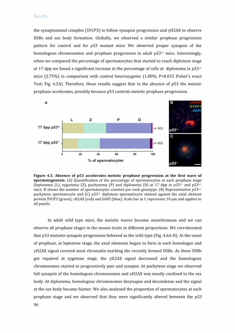
Results
96
the synaptonemal complex (SYCP3) to follow synapsis progression and γH2AX to observe
DSBs and sex body formation. Globally, we observed a similar prophase progression
pattern for control and for p53 mutant mice. We observed proper synapsis of the
homologous chromosomes and prophase progression in adult p53-/- mice. Interestingly,
when we compared the percentage of spermatocytes that started to reach diplotene stage
at 17 dpp we found a significant increase in the percentage of cells at diplonema in p53-/-
mice (2.75%) in comparison with control heterozygotes (1.00%, P=0.015 Fisher’s exact
Test; Fig. 4.5A). Therefore, these results suggest that in the absence of p53 the meiotic
prophase accelerates, possibly because p53 controls meiotic prophase progression.
Figure 4.5. Absence of p53 accelerates meiotic prophase progression at the first wave of spermatogenesis. (A) Quantification of the percentage of spermatocytes at each prophase stage (leptonema (L), zygonema (Z), pachynema (P) and diplonema (D) at 17 dpp in p53-/- and p53+/- mice. N shows the number of spermatocytes counted per each genotype. (B) Representative p53+/- pachytene spermatocyte and (C) p53-/- diplotene spermatocyte stained against the axial element protein SYCP3 (green), H2AX (red) and DAPI (blue). Scale bar in C represents 10 µm and applies to all panels.
In adult wild type mice, the meiotic waves become asynchronous and we can
observe all prophase stages in the mouse testis in different proportions. We corroborated
that p53 mutants synapsis progression behaved as the wild type (Fig. 4.6A-H). At the onset
of prophase, at leptotene stage, the axial elements begun to form in each homologue and
γH2AX signal covered most chromatin marking the recently formed DSBs. As these DSBs
got repaired at zygotene stage, the γH2AX signal decreased and the homologous
chromosomes started to progressively pair and synapse. At pachytene stage we observed
full synapsis of the homologous chromosomes and γH2AX was mostly confined to the sex
body. At diplonema, homologous chromosomes desynapse and decondense and the signal
at the sex body became fainter. We also analyzed the proportion of spermatocytes at each
prophase stage and we observed that they were significantly altered between the p53

Results
97
mutant and control mice (P=0.00047, G Test). Distinctively, there was a statistically
significant increase in the number of zygotene stage spermatocytes in p53-/- mice
(24.42%) compared with the wild type (15.23) (P=0.0027, Fisher’s exact test; Fig. 4.6J).
Additionally, the percentage of diplonema spermatocytes presented a significant reduction
in p53 mutant mice (24.96%) in comparison with the wild type (33.13%) (P=0.0393,
Fisher’s exact test; Fig. 4.6I). The percentage of leptonema cells in p53-/- (6.60%) and wild
type (5.18%) was not significantly different (P=0.4404, Fisher’s exact test) and neither the
percentage of pachynema spermatocytes in p53-/- (44.03%) and wild type (46.46%)
(P=0.6237, Fisher’s exact test). This alteration of percentage stage pattern suggests that
p53 absence might have some effect on spermatocyte meiotic progression.
Figure 4.6. Meiotic progression in adult p53 mutants. (A-J) Spread chromosomes from representative spermatocytes of p53-/- and wild type mice from leptonema to diplonema stage, stained against the axial element protein SYCP3 (green), H2AX (red) and DAPI (blue). Scale bar in H represents 10 µm and applies to all panels. (K) Quantification of the percentage of spermatocytes at each prophase stage (leptonema (L), zygonema (Z), pachynema (P) and diplonema (D)) in p53-/- and wild type adult mice. (n) shows the number of spermatocytes counted per each genotype.

Results
98
Altogether, these results indicate that although p53 could have some control over
cell cycle progression, it is dispensable to correctly complete meiotic prophase. This is an
expected observation for a presumably checkpoint protein, where in normal conditions
we might not observe any drastic phenotype in its absence.
4.1.3.3 Analysis of p53 participation in meiotic recombination
One of the first signs of the activation of the DNA damage response is the
phosphorylation of the histone H2AX (γH2AX) around the DSBs sites, where it persists
until these are repaired. In order to study meiotic recombination in the absence of p53, we
used γH2AX as a DSBs marker. We examined the number of γH2AX foci (determined as
every faint signal over the synaptonemal complex) at pachynema and diplonema stages. At
early pachynema, we found similar numbers of γH2AX foci between p53 mutant
(Mean±SD= 22.54±12.78) and wild type cells (Fig. 4.7A) (24.21±12.54, P=0.49, T test).
Figure 4.7. Absence of p53 does not affect DSBs repair. (A) Quantification of the number of H2AX foci per spermatocyte at each indicated stage in wild type and p53-/- mice. The large, bright blobs of H2AX staining are the sex bodies and the smaller H2AX foci reflect unrepaired DSBs (orange arrowheads) Horizontal lines represent means. Means (± SD) are indicated above the graph, (n.s.) indicates not significant compared to the wild type. The number of cells counted (n) is indicated below the graph. (B-E) Representative pachytene spermatocytes are shown from wild type and p53-/- mice, stained against γH2AX (red), SYCP3 (green) and H1t (blue). Scale bar in E represents 10 µm and applies to all panels.
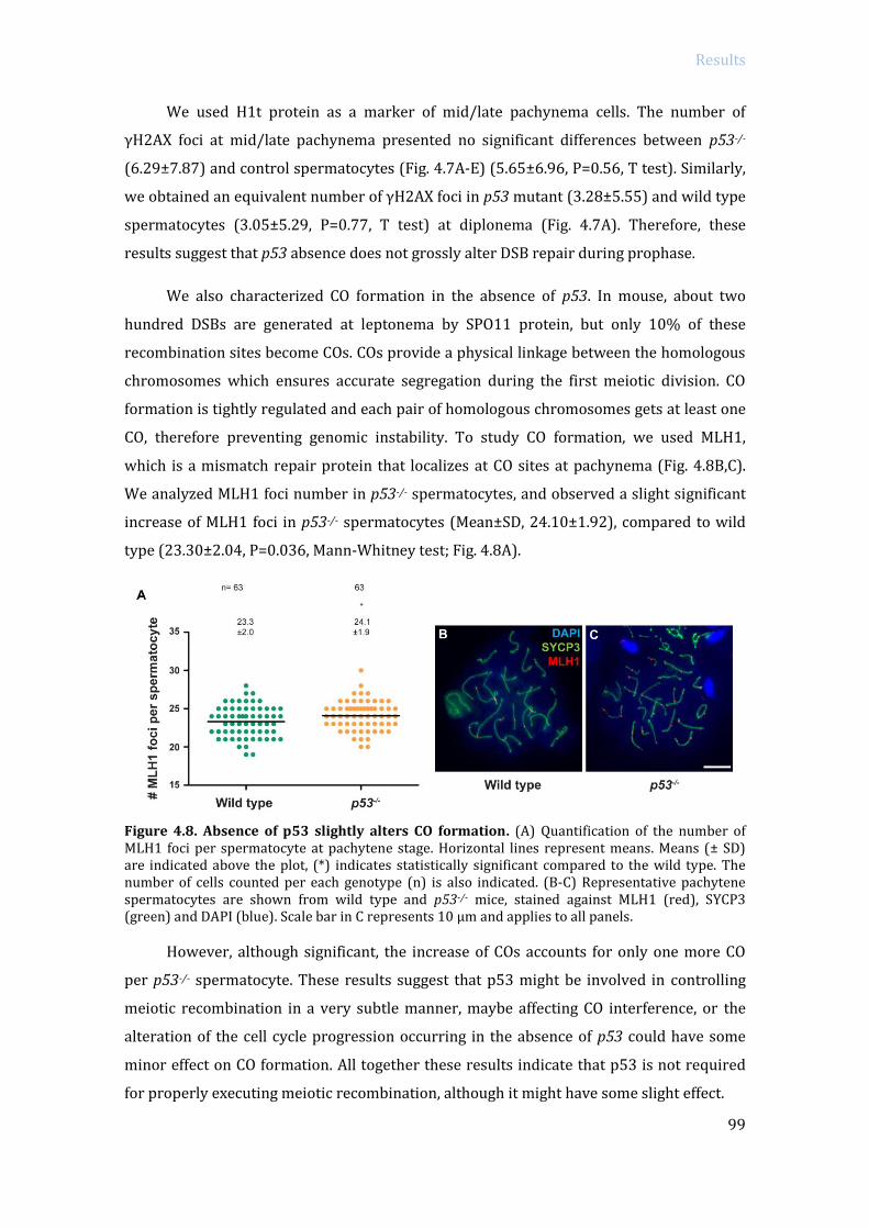
Results
99
We used H1t protein as a marker of mid/late pachynema cells. The number of
γH2AX foci at mid/late pachynema presented no significant differences between p53-/-
(6.29±7.87) and control spermatocytes (Fig. 4.7A-E) (5.65±6.96, P=0.56, T test). Similarly,
we obtained an equivalent number of γH2AX foci in p53 mutant (3.28±5.55) and wild type
spermatocytes (3.05±5.29, P=0.77, T test) at diplonema (Fig. 4.7A). Therefore, these
results suggest that p53 absence does not grossly alter DSB repair during prophase.
We also characterized CO formation in the absence of p53. In mouse, about two
hundred DSBs are generated at leptonema by SPO11 protein, but only 10% of these
recombination sites become COs. COs provide a physical linkage between the homologous
chromosomes which ensures accurate segregation during the first meiotic division. CO
formation is tightly regulated and each pair of homologous chromosomes gets at least one
CO, therefore preventing genomic instability. To study CO formation, we used MLH1,
which is a mismatch repair protein that localizes at CO sites at pachynema (Fig. 4.8B,C).
We analyzed MLH1 foci number in p53-/- spermatocytes, and observed a slight significant
increase of MLH1 foci in p53-/- spermatocytes (Mean±SD, 24.10±1.92), compared to wild
type (23.30±2.04, P=0.036, Mann-Whitney test; Fig. 4.8A).
Figure 4.8. Absence of p53 slightly alters CO formation. (A) Quantification of the number of MLH1 foci per spermatocyte at pachytene stage. Horizontal lines represent means. Means (± SD) are indicated above the plot, (*) indicates statistically significant compared to the wild type. The number of cells counted per each genotype (n) is also indicated. (B-C) Representative pachytene spermatocytes are shown from wild type and p53-/- mice, stained against MLH1 (red), SYCP3 (green) and DAPI (blue). Scale bar in C represents 10 µm and applies to all panels.
However, although significant, the increase of COs accounts for only one more CO
per p53-/- spermatocyte. These results suggest that p53 might be involved in controlling
meiotic recombination in a very subtle manner, maybe affecting CO interference, or the
alteration of the cell cycle progression occurring in the absence of p53 could have some
minor effect on CO formation. All together these results indicate that p53 is not required
for properly executing meiotic recombination, although it might have some slight effect.

Results
100
4.2 Study p53 family participation in the pachytene
recombination-dependent arrest mechanism
Our group has described that the DDR proteins MRE11, NBS1, ATM and CHK2,
promote the activation of the recombination-dependent arrest of Trip13mod/mod mutant
spermatocytes at pachynema (Pacheco et al. 2015). Therefore, based on these results, we
asked if p53 family might be involved in the activation of this arrest, since p53 family
members are activated downstream of ATM and CHK2 in somatic DDR.
To determine if p53 family members participate in the activation of the
recombination-dependent arrest, and analyzed if their absence could rescue Trip13
mutant arrest, we bred double mutant mice combining p53 family members with Trip13
mutation as follow: Trip13mod/mod p53-/-, Trip13mod/mod TAp63-/- and Trip13mod/mod p73-/- (also
referred from now on as double mutants), and subsequently analyzed their phenotype.
4.2.1 Evaluate p53 family members role in the activation of the
Recombination-dependent arrest in mouse spermatocytes
4.2.1.1 Study the ability of bypassing the recombination-dependent arrest
As mentioned earlier, the pachytene checkpoint in male mouse meiosis is a control
mechanism that can be activated by two main arrest pathways. One responds to defective
repair of DSBs (referred to as recombination-dependent arrest) (Barchi et al. 2005; Di
Giacomo et al. 2005), and the other responds to failure in transcriptional silencing of the
non-homologous portions of the sex chromosomes (sex body-deficient arrest) (Royo et al.
2010; Turner et al. 2005). These arrest mechanisms can be distinguished cytologically
using the incorporation of the testis-specific histone variant H1t (Barchi et al. 2005),
which in wild type mice accumulates on chromatin from mid-pachynema onwards (Fig.
4.9A) (Inselman et al. 2003). Mutant spermatocytes that fail to complete meiotic
recombination (e.g., Dmc1-/- or Trip13mod/mod), arrest meiosis before incorporating H1t;
whereas mutants that fail to complete MSCI (e.g., Spo11-/-), present H1t-positive
spermatocytes (Barchi et al. 2005; Pacheco et al. 2015). According to this, when the
recombination-dependent arrest is inactivated (e.g., Trip13mod/mod Chk2-/-), spermatocytes

Results
101
progress further and accumulate H1t –despite having multiple unrepaired DSBs (Pacheco
et al. 2015).
First of all, as we previously demonstrated for Trip13mod/mod Chk2-/- mutants
(Pacheco et al. 2015), if the recombination-dependent arrest is deactivated in the double
mutants, we would expect an increased number of cells expressing H1t compared to the
single Trip13mod/mod mutant. Thus, we stained squashed spermatocytes against SYCP3 and
H1t. In wild-type mice, 53.0% of spermatocytes were H1t-positive, whereas in
Trip13mod/mod mutants we only found a 21.3% of H1t-positive spermatocytes. All these H1t-
positive cells have been attributed to be previously reported escapers that complete
meiosis (Pacheco et al. 2015; Li et al. 2007; Roig et al. 2010). However, interestingly, we
observed that a 47.5% of Trip13mod/mod p53-/- and a 51.3% of Trip13mod/mod TAp63-/-
spermatocytes were H1t-positive (Fig. 4.9B). Therefore, we found a significant increase of
H1t-positive cells in the p53 and TAp63 double mutants compared to the single
Trip13mod/mod mutant (respectively P=0.0006 and P=0.0034, T test).
Figure 4.9. Absence of p53 or TAp63, but not p73, allows TRIP13-deficient spermatocytes to accumulate H1t. (A) Schematic representation of arrest events and H1t incorporation from early pachynema to diplonema. (B) Bars graph showing the average percentage of spermatocytes that have incorporated H1t for each genotype. The total number of cells analyzed is shown above each bar (n) and error bars represent standard deviation (SD).
Results
102
In contrast, Trip13mod/mod p73-/- showed 16.5% of H1t-positive spermatocytes,
which resembles what we found in the single Trip13mod/mod (P=0.0898, T test; Fig. 4.9B).
These results suggest that in the absence of p53 or TAp63, but not p73, Trip13 mutant
spermatocytes are able to progress further into meiotic prophase and thus to bypass the
recombination-dependent arrest.
4.2.1.2 Determine the presence of unrepaired DSBs after bypassing the
recombination-dependent arrest
As a consequence of escaping the recombination-dependent arrest, we would
expect that double mutant H1t-positive spermatocytes accumulate more unrepaired DSBs
than H1t-positive Trip13 single mutant cells. Thus, we quantified γH2AX patches in single
and double mutant spermatocytes.
Before the activation of the recombination-dependent arrest, thus, in H1t-negative
cells, numbers of unrepaired DSBs in the single Trip13mod/mod mutant and p53 and p73
double mutants were similar (P=0.36 and P=0.086, T-test, respectively) or slightly
decreased for Trip13mod/mod TAp63-/- (P=0.0005, T-test, Fig. 4.10Q). Interestingly,
Trip13mod/mod p53-/- and Trip13mod/mod TAp63-/- H1t-positive cells presented a significant
increase in the number of unrepaired DSBs compared to the single Trip13 mutant
(Trip13mod/mod vs. Trip13mod/mod p53-/- P≤0.0001, and vs. Trip13mod/mod TAp63-/- P≤0.0001, T-
test; Fig. 4.10E-F,I-J,Q). In contrast, Trip13 p73 double mutant H1t-positive cells displayed
similar number of γH2AX patches as Trip13mod/mod (P=0.488, T-test; Fig. 4.10M-N,Q), which
agrees with previous results suggesting that p73 does not participate in the
recombination-dependent arrest.
To further corroborate that cells progressing to late meiotic prophase in Trip13
p53 and Trip13 TAp63 double mutants accumulated more unrepaired DSBs, we analyzed
presence of unrepaired DSBs at diplonema cells from single and double mutants. Both p53
and TAp63 double mutants presented an increased number of unrepaired DSBs when
compared to Trip13 single mutant (Trip13mod/mod vs. Trip13mod/mod p53-/- P=0.00003, and vs.
Trip13mod/mod TAp63-/- P=0.00025, negative binomial regression, Fig. 4.10G-H,K-L,Q),
whereas p73 double mutant showed no significant difference with Trip13mod/mod mutant
(P=0.648, negative binomial regression test; Fig. 4.10O-P,Q).

Results
103
Figure 4.10. H1t-positive spermatocytes of Trip13mod/mod p53-/- and Trip13mod/mod TAp63-/- accumulate more unrepaired DSBs than H1t-positive Trip13mod/mod spermatocytes. (A-P) Spread chromosomes from representative spermatocytes of the indicated genotypes at mid/late pachynema (first column) or diplonema (second column) stained against the axial element protein SYCP3 (green), H1t (blue), and H2AX (red). Large bright blobs of H2AX staining are the sex bodies (arrows); the smaller H2AX patches reflect unrepaired DSBs (orange arrowheads). Notice the presence of multiple unrepaired DSBs in Trip13mod/mod p53-/- and Trip13mod/mod TAp63-/-, but not in Trip13mod/mod p73-/-. Scale bar in P represents 10 µm and applies to all panels. (Q) Quantification of the number of H2AX patches per spermatocyte. Horizontal lines represent means. Means (± SD) are indicated above the graph, and the number of cells counted (n) is indicated below. Above, (n.s.) indicates not significant and (*) indicates significantly different relative to Trip13mod/mod (T-test for pachynema stages; negative binomial regression for diplonema stage).

Results
104
4.2.1.3 Study of the influence of the p53 family members in the apoptotic profile of
Trip13 mutants
Finally to corroborate the involvement of p53 and TAp63 in the activation of the
recombination-dependent arrest, we tested another prediction from our model. If
Trip13mod/mod p53-/- and Trip13mod/mod TAp63-/- are able to escape the recombination-
dependent arrest; they would present less H1t-negative apoptotic spermatocytes than the
single Trip13mod/mod. Therefore, we investigated the apoptotic profile of double mutants
performing TUNEL staining on IF-stained slides. As expected, most Trip13mod/mod TUNEL-
positive spermatocytes were H1t-negative (80.1%), similarly to what we observed in
Trip13mod/mod p73-/- (73.3%, 1 mouse analyzed, P=0.2127, Fisher’s exact test; Fig. 4.11). In
contrast, the percentage of apoptotic H1t-negative spermatocytes in Trip13mod/mod p53-/-
and Trip13mod/mod TAp63-/- was significantly lower (26.8%, P=0.002; and 7%, P=0.0007, T
test, respectively; Fig. 4.11).
All together, these results suggest that p53 and TAp63, but not p73, are required to
hold spermatocytes with numerous unrepaired DSBs at early pachynema stage (H1t-
negative). Therefore, p53 and TAp63 control progression of male meiotic prophase, so
that when recombination is impaired, they mediate the arrest of meiotic progression.
4.2.2 Trip13 double mutants still present stage IV arrest
We previously reported that Trip13mod/mod Chk2-/- spermatocytes, although escaping
the recombination-dependent arrest, still present a block in spermatogenesis progression
at epithelial stage IV due to sex body-deficient arrest activation. Thus, we analyzed the
histological phenotype of the double mutants Trip13mod/mod p53-/- and Trip13mod/mod TAp63-/-
. Both p53 and TAp63 double mutants, and also p73 double mutant, showed an arrest at
epithelial stage IV (Fig. 4.12C-E). Since mutants that arrest at epithelial stage IV present an
increase in the number of spermatocytes undergoing apoptosis (Yoshida et al. 1998;
Baudat et al. 2000), we performed TUNEL staining on histological sections and analyzed
the number of apoptotic cells present in Trip13 p53 and Trip13 TAp63 double mutants
(Fig. 4.13A-C). TUNEL staining on histological sections demonstrated that the occurrence
of apoptosis in the p53 and TAp63 double mutants was similar to the Trip13mod/mod single
mutant (Fig. 4.13D). Taken together, these data show that Trip13 p53 and Trip13 TAp63
double mutants still presented an arrest epithelial stage IV, which suggests that these
mutants might present sex body defects that activate the sex body-deficient arrest.

Results
105
Figure 4.11. Apoptosis tends to occur at a later (H1t-positive) stage in TRIP13-deficient spermatocytes lacking p53 or TAp63, but not p73. (A-P) Representative apoptotic (TUNEL-positive) pachytene-stage spermatocytes stained for SYCP3 and TUNEL (green), H1t (blue) and H2AX (red). Note the presence of multiple H2AX patches and abnormal sex body in the apoptotic, H1t-positive spermatocytes from the Trip13mod/mod p53-/- and Trip13mod/mod TAp63-/- mutants. Scale bar in P represents 10 µm and applies to all panels. (Q) Percentage of apoptotic spermatocytes that are H1t-negative or H1t-positive. The total number of cells analyzed is shown (n) and error bars represent SD (one Trip13mod/mod p73-/- mouse analyzed).

Results
106
Figure 4.12. Absence of p53 family members does not alleviate the epithelial stage IV arrest in TRIP13-deficient mice. (A-E) Representative tubule sections from adult testes stained with PAS-H. Scale bar in E represents 20 µm and applies to all panels. (A) Wild type seminiferous tubule containing spermatogonia, spermatocytes and spermatids. (B-E) Trip13mod/mod single mutant and p53 family member double mutant sections showing condensed (apoptotic) spermatocytes in stage IV tubules, with only few escapers observed.
Figure 4.13. Absence of p53 family members does not alleviate spermatocyte apoptosis at epithelial stage IV in TRIP13-deficient mice. (A-C) Representative tubules are shown from testis sections of the indicated genotypes stained by TUNEL to detect apoptotic spermatocytes (green). Scale bar in A represents 40 µm and applies to panels A-C. (D) Quantification of the percentage of tubules with TUNEL-positive cells grouped in 0, 1 to 10 or more than 10 TUNEL-positive cells per tubule. The number of cells analyzed (n) is shown above each bar (one Trip13mod/mod p53-/- and one Trip13mod/mod TAp63-/- mouse analyzed).

Results
107
4.2.3 TRIP13 involvement in the formation of the sex body
4.2.3.1 Qualitative analysis of sex body formation in Trip13 mutants
To determine if Trip13mod/mod p53-/- and Trip13mod/mod TAp63-/- epithelial stage IV
arrest was due to MSCI failure, like in Trip13mod/mod Chk2-/- mutants (Pacheco et al. 2015),
we analyzed the morphology and functionality of the sex body in Trip13 p53 and Trip13
TAp63 double mutants. We previously showed that Trip13mod/mod mutants present MSCI
defects. If MSCI fails, expression of sex chromosome genes at pachytene stage becomes
deleterious and sufficient to cause an arrest and ultimately apoptosis of spermatocytes
(Royo et al. 2010). In order to qualitatively study the morphology of the sex body at
pachytene stage, we stained several sex body markers in spermatocyte spreads: γH2AX,
ATR (the kinase responsible for H2AX phosphorylation in the sex body (Royo et al. 2013)),
and SUMO-1 (which accumulates on the X and Y chromatin at pachynema in an ATR
dependent manner (Royo et al. 2013)) (see Fig. 4.14A-L for representative images and
Table 4.1 for quantification).
Figure 4.14. Sex body defects in TRIP13-deficient cells lacking p53, TAp63 or p73. (A-D) Representative H1t-positive pachytene spermatocytes of the indicated genotypes stained for SYCP3 (green), H2AX (red) and H1t (blue). (E-L) Representative pachytene spermatocytes of the indicated genotypes stained against SYCP3 (green), either ATR (D-F) or SUMO-1 (G-I) (red) and DAPI (blue). Scale bar in D represents 10 µm and applies to all panels. White arrows indicate sex bodies.

Results
108
For each marker, the p53 and TAp63 double mutants displayed sex body
abnormalities that were qualitatively and quantitatively indistinguishable from the
Trip13mod/mod single mutant. Specifically, by observing γH2AX staining we found
abnormally elongated sex bodies in approximately two thirds of pachytene-stage
spermatocytes corresponding to double mutants Trip13mod/mod p53-/- (70%) and
Trip13mod/mod TAp63-/- (70%), similar to what we found in Trip13mod/mod mutants (67.5%,
P=0.6662 for both comparisons, Fisher’s exact test) (Fig. 4.14B-D and Table 4.1). In
contrast, only a minority of wild-type pachytene-stage spermatocytes presented a
stretched sex body (20%, vs. Trip13mod/mod P≤0.0001, Fisher’s exact test), while most of
them displayed a round sex body structure (see intensely stained sex body in Fig. 4.14A
and Table 4.1 for quantifications).
Table 4.1 Trip13 mutants present defects in sex body formation.
Wild type Trip13mod/mod Trip13mod/mod
p53-/- Trip13mod/mod
TAp63-/-
γH2AX
Elongated sex body 20 % 67.5 % 70 % 70 %
Round sex body 80 % 32.5 % 30 % 30 %
(N) 40 40 40 40
ATR
No staining 0 % 4.25 % 4 % 8.5 %
Discontinuous XY axis 6.8 % 51.3 % 59.5 % 51 %
Complete XY axis 24 % 29 % 27.5 % 25 %
Chromatin staining 69.3 % 15.5 % 9 % 15.5 %
(N) 400 400 200 200
SUMO-1
No staining 1.5 % 49.8 % 18.5 % 50.5 %
Faint staining 13.8 % 36.3 % 56.5 % 38.5 %
Strong staining 84.8 % 14 % 25 % 11 %
(N) 400 400 200 200
ATR is the kinase responsible for H2AX phosphorylation in the sex body; it loads
on the unsynapsed sex chromosomes, and expands into the chromatin triggering H2AX
phosphorylation (Royo et al. 2010; Barchi et al. 2008; Baart et al. 2000). Whereas ATR in
wild type pachynema usually covered the unsynapsed X and Y chromosome axes and
expanded over their chromatin (69.3%) (Fig. 4.14E), less than a 20% of pachytene cells
from double mutants or Trip13mod/mod single mutant displayed this chromatin-expanded
pattern (15.5% in single Trip13mod/mod, P=0.0001, Fisher’s exact test). Instead most double

Results
109
mutants or Trip13mod/mod cells showed a discontinued localization of ATR along the X and Y
axes (Fig. 4.14F-H and see Table 4.1 for quantification).
Finally, we also examined SUMO-1 protein, which accumulates on the X and Y
chromatin at pachynema in an ATR-dependent manner (Royo et al. 2013). SUMO-1
staining was also altered in all Trip13 single and double mutants, most mutant pachynema
cells displayed only modest SUMO-1 staining (Fig. 4.14J-L and see Table 4.1 for
quantification). While the majority (84.8%) of wild type pachytene spermatocytes
displayed an intense SUMO-1 signal over the sex body, this was only observed in 14.8% of
Trip13mod/mod pachytene cells (P≤0.0001 Fisher’s exact test). The percentages of strong
SUMO-1 staining in Trip13 p53 and Trip13 TAp63 double mutants where similar to Trip13
single mutants and significantly different from the wild type: 25% in Trip13mod/mod p53-/-
(P≤0.0001 Fisher’s exact test) and 11% in Trip13mod/mod TAp63-/- (P≤0.0001 Fisher’s exact
test; see Fig. 4.14K,L and Table 4.1 for quantifications). These results confirm that Trip13
mutants have defects in sex body formation, consistent with our previous observations
(Pacheco et al. 2015), and demonstrate that these defects are not ameliorated by absence
of either p53 or TAp63.
4.2.3.2 Functional analysis of the meiotic sex chromosome inactivation (MSCI) in
Trip13 mutants
To test whether the defects found in sex body formation affected its functionality, we
performed RNA fluorescence in situ hybridization (RNA-FISH) analysis to assess MSCI.
RNA-FISH technique allows the detection of a specific transcript in a particular meiotic cell
type (Mahadevaiah et al. 2009). For our study, we carried out RNA-FISH for the X-linked
genes Scml2 (located next to the PAR region) and Zfx (more interstitially located), which
should be silenced throughout pachynema by the MSCI. Slides were immunostained
against TOPBP1 (DNA topoisomerase 2 binding-protein 1, which loads to the unsynapsed
sex chromosomes at pachynema (Refolio et al. 2011)), in order to detect early pachytene-
stage cells.
We found that the percentage of early pachytene cells expressing X-linked genes was
significantly higher in Trip13mod/mod (27.5% for Scml2 and 27.2% for Zfx) relative to wild-
type cells (17.1% for Scml2, P<0.05, and 10.9% for Zfx, P<0.05, one-way ANOVA, Fig.
15E,F). As we expected from our previous results, we observed that in Trip13mod/mod p53-/-
and Trip13mod/mod TAp63-/- early pachytene-stage spermatocytes expression for both X-
linked genes was similar to the Trip13mod/mod mutant. Scml2 was expressed in 28.8% of

Results
110
Trip13mod/mod p53-/- early pachytene cells (1 mouse, P=0.0330 vs. wild type, Fisher’s exact
test; Fig. 4.15A,E), and 34.4% of cells in Trip13mod/mod TAp63-/- mutants (P<0.05, vs. wild
type, one-way ANOVA, Fig. 4.15B,E and see Table 4.2).
Frequencies for Zfx expression were 33.8% for Trip13mod/mod p53-/- (vs. wild-type,
P≤0.0001, Fisher’s exact test; Fig. 4.15C,F and Table 4.2) and 30.6% for Trip13mod/mod
TAp63-/- early pachytene spermatocytes (vs. wild-type, P≤0.0001, Fisher’s exact test or
P<0.05 one-way ANOVA; Fig. 4.15D,F and Table 4.2). These results support what we
previously observed for Trip13mod/mod Chk2-/- mutants, where although these
spermatocytes escape the recombination-dependent arrest, Trip13mod/mod p53-/- and
Trip13mod/mod TAp63-/- mutants still apoptose at pachytene stage due to MSCI failure.
Therefore, p53 and TAp63 absence does not disturb the activation of the sex body-
dependent arrest.
Table 4.2 Expression of Scml2 and Zfx on early pachytene cells from wild type and
mutant mice.
Wild type Trip13mod/mod Trip13mod/mod
p53-/-
Trip13mod/mod
TAp63-/-
% of pachynema
cells expressing
Scml2 (mean±SD)
17.1 ±1.9 27.5 ±0.0* 28.8** 34.4 ±4.4*
(N) 200 160 80 ª 160
% of pachynema
cells expressing Zfx
(mean±SD)
10.9 ±3.0 27.2 ±3.2* 33.8** 30.6 ±0.9*
(N) 225 158 80 ª 160
*
**
ª
Significantly different from wild type, P < 0.05 one-way ANOVA and Tukey's multiple comparison test.
Significantly different from wild type, P < 0.05 Fisher’s exact test.
One mouse analyzed.

Results
111
Figure 4.15. MSCI failure in TRIP13-deficient cells lacking p53 or TAp63. (A-F) RNA-FISH performed on Trip13mod/mod p53-/- spermatocytes. Images display Scml2 or Zfx RNA-FISH signal (white arrows), DAPI (blue), and immunostaining for TOPBP1 (green) and H2AX (red). Insets show zoomed images of the sex bodies. Scale bars represent 10 µm. (G,H) Graphics showing mean percentage of early pachytene spermatocytes expressing Scml2 (L) or Zfx (M). Error bars represent SD (see Table 4.2 for mean±SD and (n) number cells analyzed).

Results
112
4.2.4 p53 and TAp63 do not activate the sex body-deficient arrest
However, although we observed that p53 or TAp63 double mutants could not rescue
Trip13mod/mod stage IV apoptosis, we wanted to test directly if p53 family members could
rescue MSCI failure arrest in other mutants. With this purpose, we used Spo11-/- mutants,
whose spermatocytes present synapsis defects due to the lack of DSBs and only form a
pseudo-sex body (Baudat et al. 2000; Romanienko and Camerini-Otero 2000; Burgoyne et
al. 2009). This pseudo-sex body normally does not include the X and Y chromosomes, thus
allowing sex chromosome expression leading to the activation of the sex body-defective
arrest. We generated Spo11-/- TAp63-/- and Spo11-/- p53-/- double mutants; in order to
analyze if p53 or TAp63 absence could revert the arrest occurring in Spo11-/- mutants.
To account for the possible rescue of the arrested meiotic prophase, first we analyzed
the normalized testis weight since, as we mentioned above, mutants with arrested meiosis
present significantly smaller testis. The normalized testis weight was obtained dividing
the weight of the two testes by the mouse weight in order to account for possible body size
variation among different mouse mutants. Normalized testis size can also distinguish
between the stages where the arrest occurs during meiosis. While those animals
experiencing meiotic prophase arrest (also known as stage IV arrest, e.g. Spo11-/- or
Dmc1-/-) present testis that are approximately one third the size of a wild type (P≤0.0001,
T test), mutants that arrest at the metaphase stage of the first meiotic division (stage XII
arrest, e.g., Spo11+/- Atm-/- or Spo11-/- tg+/+) have testis which are only 50% the size of a
wild type (P≤0.0001, T test; see Fig. 16A).
The normalized testis size of Spo11-/- TAp63-/- double mutants (Mean±SD=0.19±0.3)
was comparable to those of Spo11-/- single mutant (0.21±0.02, P=0.2219, T test; Fig.
4.17A). Also Spo11-/- p53-/- double mutants had testis size equivalents to Spo11-/- single
mutants (0.25) (Fig. 4.16A). To confirm these data, we performed an histological analysis
of the Spo11-/- p53-/- testis and observed stage IV arrest as in Spo11-/- (Fig 4.16B,C).
Therefore, these results indicate that p53 and TAp63 are dispensable to activate
programmed cell death in response of the sex body-deficient arrest in mouse
spermatocytes. Furthermore, our results show that the recombination-dependent arrest
and the sex body-deficient arrest are two genetically separable mechanisms.

Results
113
Figure 4.16. Mutation of p53 or TAp63 does not rescue Spo11-/- stage IV arrest. (A) Graph shows normalized testis weight of the indicated genotypes. Green rectangle includes wild type and mutants that complete meiosis. Yellow rectangle accounts for mutant mice that experience an arrest at metaphase stage XII of first meiotic division (Spo11+/- Atm-/- and Spo11-/- tg+/+). Pink rectangle shows mutants that present a stage IV pachytene arrest (Spo11-/- or Dmc1-/-). Data from Spo11+/- Atm-/- was previously published (Pacheco et al. 2015). Black horizontal lines represent the mean, which is also indicated above the corresponding genotype (Mean±SD). n shows the number of animals analyzed for each genotype (one Spo11-/- p53-/-mouse analyzed). (B) Spo11-/- and (C) Spo11-/-p53-/- representative tubule sections from adult testes stained with PAS-H. Scale bar in C represents 20 µm and applies to all panels. Both sections show condensed (apoptotic) spermatocytes in stage IV arrested tubules.

Results
114
4.3 Analysis of TRIP13 role in meiotic transcription
During the first meiotic division, transcription is suppressed at the onset of
prophase, and it only restarts again at mid pachynema (Monesi, 1964), when
chromosomes are fully synapsed and DSBs are repaired. In pachytene-stage
spermatocytes, when autosomes reactivate transcription, the sex chromosomes are
silenced by the sex body (MSCI) (Turner et al. 2004); which is crucial to allow correct
progression of meiosis (Royo et al. 2010) and it is proposed to be a manifestation of the
global meiotic silencing of unsynapsed chromosomes (MSUC) during zygonema (Burgoyne
et al. 2009). Since we identified that TRIP13 is required for the correct silencing of the sex
chromosomes at pachytene stage (Pacheco et al. 2015) and it has been hypothesized that
the MSUC is a manifestation of the global transcription suppression that occurs at the
entry of prophase (Page et al. 2012), we wondered if TRIP13 may also participate in the
transcription regulation that occurs at the initiation of meiosis. To test this, we analyzed
transcription levels at early stages of meiotic prophase in control and Trip13 mutant
spermatocytes.
4.3.1 Transcription levels in Trip13 mutants
4.3.1.1 Analyze global RNA expression levels in Trip13 mutants with EU
To analyze global gene expression in meiotic cells, we used the EU Click-it Imaging
technique (Click-iT® RNA Alexa Fluor® 488 Imaging Kit, Life Technologies) to
cytologically detect newly synthesized RNA. Briefly, we obtained a cell suspension from
fresh testis and then incubated these cells in the presence of a uridine analogue (EU, 5-
Ethynyl Uridine), which is specifically incorporated to nascent RNA. From this cell
suspension, slides were prepared using a protocol that preserves the nuclei structure and
keeps cellular volume. Then, EU-tagged RNA was labeled with a dye (Alexa Fluor 488)
through a chemoselective ligation or “click-it” reaction between an azide-containing dye
and an alkyne, which allows the visualization of the RNA in a fluorescence microscope.
Slides were also IF-stained against SYCP3 and γH2AX, in order to classify meiotic prophase
stages. Images were captured from cells at different stages to quantify the intensity of the
RNA signal. In order to get the maximum information of each cell, we obtained stacks of
images every 0.5 µm, then we displayed the sum projection and measured the intensity of
the EU labelled RNA signal with ImageJ software. With this technique we can cytologically
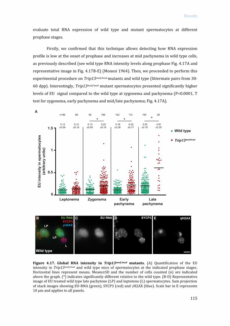
Results
115
evaluate total RNA expression of wild type and mutant spermatocytes at different
prophase stages.
Firstly, we confirmed that this technique allows detecting how RNA expression
profile is low at the onset of prophase and increases at mid pachynema in wild type cells,
as previously described (see wild type RNA intensity levels along prophase Fig. 4.17A and
representative image in Fig. 4.17B-E) (Monesi 1964). Then, we proceeded to perform this
experimental procedure on Trip13mod/mod mutants and wild type (littermate pairs from 30-
60 dpp). Interestingly, Trip13mod/mod mutant spermatocytes presented significantly higher
levels of EU signal compared to the wild type at zygonema and pachynema (P<0.0001, T
test for zygonema, early pachynema and mid/late pachynema; Fig. 4.17A).
Figure 4.17. Global RNA intensity in Trip13mod/mod mutants. (A) Quantification of the EU intensity in Trip13mod/mod and wild type mice of spermatocytes at the indicated prophase stages. Horizontal lines represent means. Means±SD and the number of cells counted (n) are indicated above the graph. (*) indicates significantly different relative to the wild type. (B-D) Representative image of EU treated wild type late pachytene (LP) and leptotene (L) spermatocytes. Sum projection of stack images showing EU-RNA (green), SYCP3 (red) and H2AX (blue). Scale bar in E represents 10 µm and applies to all panels.
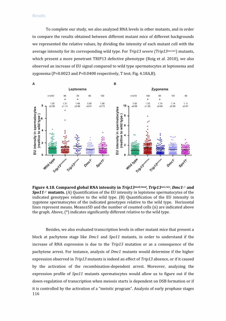
Results
116
To complete our study, we also analyzed RNA levels in other mutants, and in order
to compare the results obtained between different mutant mice of different backgrounds
we represented the relative values, by dividing the intensity of each mutant cell with the
average intensity for its corresponding wild type. For Trip13 severe (Trip13sev/sev) mutants,
which present a more penetrant TRIP13 defective phenotype (Roig et al. 2010), we also
observed an increase of EU signal compared to wild type spermatocytes at leptonema and
zygonema (P=0.0023 and P=0.0408 respectively, T test; Fig. 4.18A,B).
Figure 4.18. Compared global RNA intensity in Trip13mod/mod, Trip13sev/sev, Dmc1-/- and Spo11-/- mutants. (A) Quantification of the EU intensity in leptotene spermatocytes of the indicated genotypes relative to the wild type. (B) Quantification of the EU intensity in zygotene spermatocytes of the indicated genotypes relative to the wild type. Horizontal lines represent means. Means±SD and the number of counted cells (n) are indicated above the graph. Above, (*) indicates significantly different relative to the wild type.
Besides, we also evaluated transcription levels in other mutant mice that present a
block at pachytene stage like Dmc1 and Spo11 mutants, in order to understand if the
increase of RNA expression is due to the Trip13 mutation or as a consequence of the
pachytene arrest. For instance, analysis of Dmc1 mutants would determine if the higher
expression observed in Trip13 mutants is indeed an effect of Trip13 absence, or if it caused
by the activation of the recombination-dependent arrest. Moreover, analyzing the
expression profile of Spo11 mutants spermatocytes would allow us to figure out if the
down-regulation of transcription when meiosis starts is dependent on DSB formation or if
it is controlled by the activation of a “meiotic program”. Analysis of early prophase stages

Results
117
in both Dmc1-/- and Spo11-/- mutants, revealed that they presented similar EU expression
levels than the control mice (for leptonema: P=0.5773 and P=0.3334 respectively, T test;
for zygonema: P=0.0655 and P=0.2190 respectively, T test; Fig. 4.18A,B). Analysis of
pachytene stage was not performed because neither Dmc1-/- or Spo11-/- mutants are able
to properly synapse their homologous chromosomes. Altogether these results suggest that
Trip13 mutant cells present increased global transcription levels at zygonema and
pachynema suggesting that TRIP13 protein is necessary to control global transcription
levels at early prophase stages.
4.3.1.2 Study transcription levels in Trip13 mutants with phospho-RNA polymerase II
In order to corroborate our previous findings about the transcriptional activity in
Trip13 mutants, we examined the phosphorylated Serine2 RNA polymerase II
(phophso(S2)-RNApol II), which corresponds to the active form of the protein (Fig. 19).
Figure 4.19. Phosphorylated(S2)-RNA polymerase II in TRIP13-deficient zygotene spermatocytes. Representative zygotene spermatocytes (arrows) from (A-D) wild type mice, (E-H) Trip13mod/mod and (I-L) Trip13sev/sev stained against phosphorylated (S2) RNA polymerase II (red), SYCP3 (green) and H2AX (blue). Sum projection of stack images is shown. Scale bar in L represents 10 µm and applies to all panels.

Results
118
The RNApol II, together with other factors, forms a functional holoenzyme
required for transcription (Myer and Young 1998). The largest RNApol II subunit contains
a carboxy-terminal repeat domain (CTD) with the consensus domain sequence YSPTSPS.
The phosphorylation of the CDT determines which factors are assembled and controls the
progression of transcription. Phosphorylation occurs principally on Serine2 and Serine5 of
the repeats (Phatnani and Greenleaf 2006). After initiation, phosphorylation of Serine2
promotes elongation and at termination phosphatases allow RNApol II to be recruited to
another initiation complex (Moorhead et al. 2007).
We observed that in Trip13 mutants phospho(S2)-RNApol II staining formed
bigger clumps than in wild type spermatocytes, which was more evident in zygotene stage
(see Fig. 4.19F,J). Quantification of the phospho(S2)-RNA pol II intensity revealed that
there was a significant increase of the signal in Trip13mod/mod mutants versus the wild type
at leptonema stage (P=0.0201, T test; Fig. 4.20A). Remarkably, as it occurred in the EU
analysis, this increase was more evident at zygotene stage, where we found a significant
increase in Trip13mod/mod and Trip13sev/sev in comparison with the wild type (P=0.0435 and
P=0.0042 respectively, T test; Fig. 4.20B). Therefore, these results suggest that
transcriptional activity is increased in early prophase stages of Trip13 mutants.
Figure 4.20. Phospho (S2)-RNA polymerase II intensity quantification in Trip13 mutants. (A-B) Quantification of (phospho S2) RNA polymerase II intensity in leptotene-stage (A) and zygotene-stage (B) spermatocytes of the indicated genotypes relative to the wild type. Horizontal lines represent means. Means±SD and the number of counted cells (n) are indicated above the graph. Above, (*) indicates significantly different relative to the wild type.

Results
119
4.3.2 Specific RNA expression analysis in Trip13 mutants: RNA sequencing
Based on our preceding results, we aimed to compare global gene expression in
wild type and Trip13 mutant spermatocytes. Since our previous attempts at purifying
spermatocytes by FACS-sorting technique had failed (see section 4.1.2), we decided to use
whole testis from juvenile control and Trip13mod/mod mice. The advantage of using juvenile
mice relies in the fact that they present a first synchronous meiotic wave and that the
proportion of somatic and germ cell population remains equivalent between mutants and
control samples. For this study we used 14dpp mice in order to compare the most
equivalent samples so that Trip13mod/mod mutants were analyzed before they experienced
the activation of the recombination-dependent arrest. The proportion of cell types
remained similar, since 13.1% of wild type cells and 14.3% of Trip13 mutants cells in the
testis were spermatocytes. We corroborated by TUNEL analysis that between 13 and 16
dpp Trip13mod/mod mutants present the same apoptosis levels as wild type littermates (at
16 dpp starts to appear a slight significant increase of apoptotic cells in mutants,
P=0.0497, T test; Fig. 4.21), suggesting that the recombination-dependent arrest is not yet
implemented.
Figure 4.21. Apoptosis in juvenile Trip13mod/mod testes. (A) Quantification of the number of TUNEL-positive cells per tubule of juvenile Trip13mod/mod and wild type mouse testes at 13 dpp and 16 dpp. Horizontal lines represent the mean, which is indicated over the plot (Mean±SD). N shows the number of tubules counted per genotype. (*) indicates significantly different relative to the wild type.
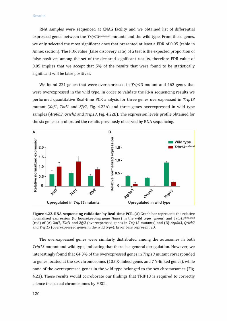
Results
120
RNA samples were sequenced at CNAG facility and we obtained list of differential
expressed genes between the Trip13mod/mod mutants and the wild type. From these genes,
we only selected the most significant ones that presented at least a FDR of 0.05 (table in
Annex section). The FDR value (false discovery rate) of a test is the expected proportion of
false positives among the set of the declared significant results, therefore FDR value of
0.05 implies that we accept that 5% of the results that were found to be statistically
significant will be false positives.
We found 221 genes that were overexpressed in Trip13 mutant and 462 genes that
were overexpressed in the wild type. In order to validate the RNA sequencing results we
performed quantitative Real-time PCR analysis for three genes overexpressed in Trip13
mutant (Xaf1, Tktl1 and Zfy2, Fig. 4.22A) and three genes overexpressed in wild type
samples (Atp8b3, Qrich2 and Trip13, Fig. 4.22B). The expression levels profile obtained for
the six genes corroborated the results previously observed by RNA sequencing.
Figure 4.22. RNA-sequencing validation by Real-time PCR. (A) Graph bar represents the relative normalized expression (to housekeeping gene Hmbs) in the wild type (green) and Trip13mod/mod (red) of (A) Xaf1, Tktl1 and Zfy2 (overexpressed genes in Trip13 mutants) and (B) Atp8b3, Qrich2 and Trip13 (overexpressed genes in the wild type). Error bars represent SD.
The overexpressed genes were similarly distributed among the autosomes in both
Trip13 mutant and wild type, indicating that there is a general deregulation. However, we
interestingly found that 64.3% of the overexpressed genes in Trip13 mutant corresponded
to genes located at the sex chromosomes (135 X-linked genes and 7 Y-linked genes), while
none of the overexpressed genes in the wild type belonged to the sex chromosomes (Fig.
4.23). These results would corroborate our findings that TRIP13 is required to correctly
silence the sexual chromosomes by MSCI.
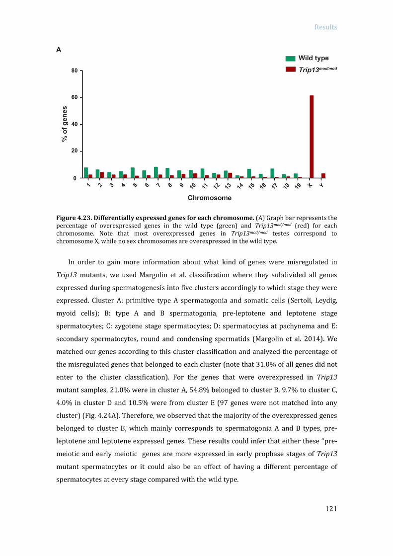
Results
121
Figure 4.23. Differentially expressed genes for each chromosome. (A) Graph bar represents the percentage of overexpressed genes in the wild type (green) and Trip13mod/mod (red) for each chromosome. Note that most overexpressed genes in Trip13mod/mod testes correspond to chromosome X, while no sex chromosomes are overexpressed in the wild type.
In order to gain more information about what kind of genes were misregulated in
Trip13 mutants, we used Margolin et al. classification where they subdivided all genes
expressed during spermatogenesis into five clusters accordingly to which stage they were
expressed. Cluster A: primitive type A spermatogonia and somatic cells (Sertoli, Leydig,
myoid cells); B: type A and B spermatogonia, pre-leptotene and leptotene stage
spermatocytes; C: zygotene stage spermatocytes; D: spermatocytes at pachynema and E:
secondary spermatocytes, round and condensing spermatids (Margolin et al. 2014). We
matched our genes according to this cluster classification and analyzed the percentage of
the misregulated genes that belonged to each cluster (note that 31.0% of all genes did not
enter to the cluster classification). For the genes that were overexpressed in Trip13
mutant samples, 21.0% were in cluster A, 54.8% belonged to cluster B, 9.7% to cluster C,
4.0% in cluster D and 10.5% were from cluster E (97 genes were not matched into any
cluster) (Fig. 4.24A). Therefore, we observed that the majority of the overexpressed genes
belonged to cluster B, which mainly corresponds to spermatogonia A and B types, pre-
leptotene and leptotene expressed genes. These results could infer that either these “pre-
meiotic and early meiotic genes are more expressed in early prophase stages of Trip13
mutant spermatocytes or it could also be an effect of having a different percentage of
spermatocytes at every stage compared with the wild type.

Results
122
Figure 4.24. Differentially expressed genes classified according to the clusters defined in Margolin et al. (2014). (A) Graph bar represents the percentage of wild type (green) and Trip13mod/mod (red) overexpressed genes corresponding to each cluster (correlated to specific cell type). Note that most overexpressed genes from Trip13mod/mod correspond to genes normally expressed in spermatogonia, pre-leptonema and leptonema cell type. (B) Graph bar showing the percentage of cells corresponding to each stage found in spermatocyte spreads from the samples used for obtaining the RNAseq data. Error bars represent SD.
In fact, we observed that Trip13mod/mod spermatocytes progressed slower through
meiosis than wild type littermates. Whereas 14 dpp wild type mice presented 45.2% of
spermatocytes at early pachynema, Trip13 mutants presented only 8.2% of pachytene-
stage spermatocytes (P<0.0001, T test) but they had significantly more zygotene–stage
spermatocytes (68.5% vs. 36.5% in the wild type, P=0.0004, T test). However, the
percentage of leptotene-stage cells was not significantly different between Trip13 mutants
(23.33%) and the wild type (18.33%, P=0.2579, T test) (Fig. 4.24B). Therefore, if we take
into account the similar percentage of leptotene-stage cells and spermatocytes (with no
apparent reduction of spermatogonial cells), our results taken together show that Trip13

Results
123
mutant spermatocytes express more “pre-meiotic/leptonema cluster genes”. Thus, despite
the limitations of RNA-sequencing technique and using whole juvenile mice testis, these
results imply that Trip13 mutant testes presented a higher expression of genes expressed
in spermatogonia, pre-leptotene and leptotene stages, than the wild type, suggesting that
TRIP13 protein is required to properly turn off the expression of this genes at the onset of
meiosis.
Regarding overexpressed genes in the wild type samples that matched into a cluster,
they classified as follow: 2.02% in cluster A, only 0.58% belonged to cluster B, 14.99%
were from cluster C, the 58.79% corresponded to cluster D and 23.63% to cluster E (there
were 115 unmatched genes) (Fig. 4.24A). Therefore, the majority of the overexpressed
genes in wild type samples belonged to cluster D (which corresponds to pachytene
spermatocytes) and some fewer to cluster E (accounting for secondary spermatocytes,
round and condensing spermatids). Then, the overexpressed genes observed in wild type
spermatocytes that by majority belong to cluster D (pachytene genes) might be a
consequence of the four times higher number of pachytene-stage cells present in wild type
testes as compared to Trip13 mutant testes.
To obtain more information, we searched the gene ontology information
corresponding to the overexpressed deregulated genes in Trip13 mutant and wild type
testis. Overexpressed genes in both wild type and Trip13 mutant testes where associated
with similar Gene Ontology terms, like development, metabolism, cell cycle, gametogenesis
and meiosis; suggesting they had similar functions. Interestingly, overexpressed genes in
Trip13 mutant samples, but not those of the wild type samples, also associated with
functions related with transcription and RNA metabolism (see Table 4.3).
In particular, the genes that highlighted transcription and RNA gene ontology
were: regulation of transcription (Nkap, Foxa2, Scml2, Irx2, Taf9b, Rhox2d, Hoxd13,
Dmrta2, Rhox2f, Rhox2g, Taf7l, Zfy2, Zfy1, Tsc22d3, Gata3, Foxd2, Fgf2, Alx3, Nkrf, Mbtps2,
Pcbd1, Rhox1, Tbx4, Zfx, Suv39h1, Esx1, Abcg4, Cdkn1c, Ebf3, Rhox13, Pbx3) and for RNA
transport and localization (Fmr1, Nxf2, Nxt2 genes). Most of the genes involved in
transcription act as transcription activators (e.g. Nkap, Irx2, Taf9b, Rhox2d/f/g, Hoxd13,
Dmrta2, Zfy1, Zfy2, Gata3, Foxd2, Fgf2, Alx3, Pcbd1, Rhox1, Tbx4, Zfx, Ebf3, Rhox13, Pbx3).
Some participate in both activation and repression (e.g. Taf7l) and only a few act as
transcriptional repressors (e.g. Scml2, Tsc22d3, Nkrf, Esx1). Regarding genes involved in
RNA transport and localization (e.g. Fmr1, Nxf2 and Nxt2), they are principally involved in
nuclear RNA export to the cytoplasm (Table 4.4). Therefore, our results obtained from
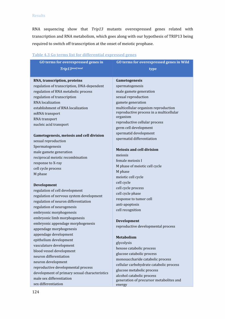
Results
124
RNA sequencing show that Trip13 mutants overexpressed genes related with
transcription and RNA metabolism, which goes along with our hypothesis of TRIP13 being
required to switch off transcription at the onset of meiotic prophase.
Table 4.3 Go terms list for differential expressed genes
GO terms for overexpressed genes in
Trip13mod/mod
GO terms for overexpressed genes in Wild
type
RNA, transcription, proteins
regulation of transcription, DNA-dependent
regulation of RNA metabolic process
regulation of transcription
RNA localization
establishment of RNA localization
mRNA transport
RNA transport
nucleic acid transport
Gametogenesis, meiosis and cell division
sexual reproduction
Spermatogenesis
male gamete generation
reciprocal meiotic recombination
response to X-ray
cell cycle process
M phase
Development
regulation of cell development
regulation of nervous system development
regulation of neuron differentiation
regulation of neurogenesis
embryonic morphogenesis
embryonic limb morphogenesis
embryonic appendage morphogenesis
appendage morphogenesis
appendage development
epithelium development
vasculature development
blood vessel development
neuron differentiation
neuron development
reproductive developmental process
development of primary sexual characteristics
male sex differentiation
sex differentiation
Gametogenesis
spermatogenesis
male gamete generation
sexual reproduction
gamete generation
multicellular organism reproduction reproductive process in a multicellular organism
reproductive cellular process
germ cell development
spermatid development
spermatid differentiation
Meiosis and cell division
meiosis
female meiosis I
M phase of meiotic cell cycle
M phase
meiotic cell cycle
cell cycle
cell cycle process
cell cycle phase
response to tumor cell
anti-apoptosis
cell recognition
Development
reproductive developmental process
Metabolism
glycolysis
hexose catabolic process
glucose catabolic process
monosaccharide catabolic process
cellular carbohydrate catabolic process
glucose metabolic process
alcohol catabolic process generation of precursor metabolites and energy

Results
125
respiratory system development reproductive structure development
lung development
respiratory tube development
limb morphogenesis
limb development
Metabolism
catecholamine metabolic process
catechol metabolic process
phenol metabolic process
diol metabolic process
regulation of cholesterol biosynthetic process oxidation reduction nitrogen compound biosynthetic process
Proteins
protein homotetramerization
prostanoid metabolic process
prostaglandin metabolic process
hexose metabolic process
carbohydrate catabolic process
monosaccharide metabolic process
glutathione metabolic process
immunoglobulin mediated immune response
organophosphate metabolic process
piRNA metabolic process
lymphocyte mediated immunity
icosanoid metabolic process
fatty acid metabolic process
unsaturated fatty acid metabolic process
peptide metabolic process cellular amino acid derivative metabolic process
coenzyme metabolic process
glycerol-3-phosphate metabolic process Immune system
B cell mediated immunity tumor necrosis factor-mediated signaling pathway adaptive immune response based on somatic recombination of immune receptors built from immunoglobulin superfamily domains
adaptive immune response
leukocyte mediated immunity
Proteins
protein folding
proteolysis
Table 4.4 Gene function of transcriptional related genes
GENE FUNCTION
Abcg4 ATP-binding cassette transporter, transport molecules across membranes
Alx3 homeobox DNA-binding that participates in transcriptional regulation in
differentiation and development
Cdkn1c
encodes p57Kip2 protein, which inhibits several G1 cyclin/Cdk complexes ((cyclin E-
CDK2, cyclin D2-CDK4, and cyclin A-CDK2) and also the mitotic cyclin B-CDC2,
negatively regulating cell cycle progression
Dmrta2 transcription factor probably involved in sexual development
Ebf3 transcription factor that activates genes involved in cell cycle arrest and apoptosis
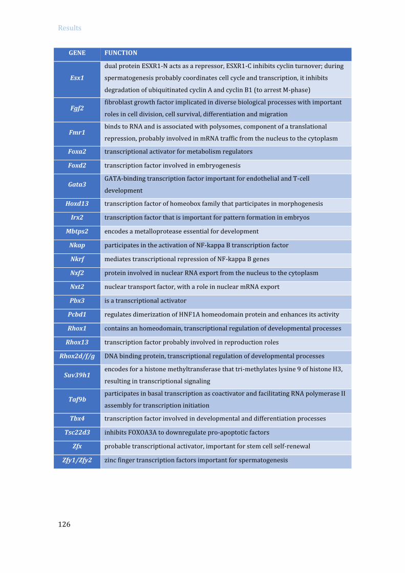
Results
126
GENE FUNCTION
Esx1
dual protein ESXR1-N acts as a repressor, ESXR1-C inhibits cyclin turnover; during
spermatogenesis probably coordinates cell cycle and transcription, it inhibits
degradation of ubiquitinated cyclin A and cyclin B1 (to arrest M-phase)
Fgf2 fibroblast growth factor implicated in diverse biological processes with important
roles in cell division, cell survival, differentiation and migration
Fmr1 binds to RNA and is associated with polysomes, component of a translational
repression, probably involved in mRNA traffic from the nucleus to the cytoplasm
Foxa2 transcriptional activator for metabolism regulators
Foxd2 transcription factor involved in embryogenesis
Gata3 GATA-binding transcription factor important for endothelial and T-cell
development
Hoxd13 transcription factor of homeobox family that participates in morphogenesis
Irx2 transcription factor that is important for pattern formation in embryos
Mbtps2 encodes a metalloprotease essential for development
Nkap participates in the activation of NF-kappa B transcription factor
Nkrf mediates transcriptional repression of NF-kappa B genes
Nxf2 protein involved in nuclear RNA export from the nucleus to the cytoplasm
Nxt2 nuclear transport factor, with a role in nuclear mRNA export
Pbx3 is a transcriptional activator
Pcbd1 regulates dimerization of HNF1A homeodomain protein and enhances its activity
Rhox1 contains an homeodomain, transcriptional regulation of developmental processes
Rhox13 transcription factor probably involved in reproduction roles
Rhox2d/f/g DNA binding protein, transcriptional regulation of developmental processes
Suv39h1 encodes for a histone methyltransferase that tri-methylates lysine 9 of histone H3,
resulting in transcriptional signaling
Taf9b participates in basal transcription as coactivator and facilitating RNA polymerase II
assembly for transcription initiation
Tbx4 transcription factor involved in developmental and differentiation processes
Tsc22d3 inhibits FOXOA3A to downregulate pro-apoptotic factors
Zfx probable transcriptional activator, important for stem cell self-renewal
Zfy1/Zfy2 zinc finger transcription factors important for spermatogenesis

127

128

129
DISCUSSION


Discussion
131
5. DISCUSSION
Meiotic prophase progression requires coordination of several processes, such as
recombination, synapsis and chromosome segregation. Germ cells undergo dramatic
reorganizations, starting with induced programmed DSBs throughout the genome. These
SPO11-generated DSBs are repaired through homologous recombination, which promote
proper homologous pairing, synapsis and eventually gives rise to CO formation (the
physical linkage that ensures correct chromosome segregation). In the presence of
deleterious events, such as unrepaired DSBs or asynapsis, meiotic surveillance
mechanisms control prophase progression and if necessary eliminate defective meiocytes
by activating programmed cell death. By avoiding genomic instability, surveillance
mechanisms guarantee successful genomic transmission to the progeny. In the recent
years, several studies have highlighted the role of the DNA damage response pathway in
the meiotic checkpoint network in several organisms (MacQueen and Hochwagen 2011).
With this work we aimed to contribute to the understanding of the meiotic surveillance
mechanism existent in mouse meiosis.
5.1 Study the components that activate the pachytene
arrest occurring in mammalian spermatocytes
One of the first objectives of this work was to determine if we could detect by the
use of biochemical tools the activation of the proteins that we had find out to participate in
the activation of the recombination-dependent pachytene arrest thanks to a genetic
approach. Our first attempt consisted in analyzing the activation of several candidates
from the DDR pathway in mutant samples by using Western blot approach from adult
mice whole testis extracts. If we could have observed the activation of CHK2 protein
(Pacheco et al. 2015) by Western blot and specifically in recombination-defective mutants,
it would have validated this technique in order discover new proteins involved in the
process. Unfortunately, we did not obtain conclusive results, possibly due to the intrinsic
limitations of comparing adult wild type testis with arrested testis, where the percentages
of the different germ cell populations extremely differ. For instance, for three of the
proteins analyzed (CHK2, E2F1 and BAX), we observed increased protein levels in both
Trip13 and Spo11 mutants. These findings are against the current knowledge which
establishes that independent mechanisms activate the recombination-dependent or the

Discussion
132
sex body-deficient arrest (Pacheco et al. 2015). The activation of the DDR and pro-
apoptotic proteins that we observed even in wild type and Spo11 mutants, probably comes
from of other cell types than spermatocytes. Adult testis contain heterogeneous cell type
populations that also activate the DDR. For instance, spermatogonial cells replicate their
DNA, which implies they are subjected to replicative stress and also activates the DDR.
Besides, it is known that the number of spermatogonial cells in the testis is auto-regulated,
in part, by apoptosis (de Rooij and Grootegoed 1998).
Next, to overcome the limitations derived from working with whole testis samples,
we decided to obtain highly enriched spermatocyte populations by using cell sorting
techniques, in order to perform Western blot or sequencing. However, by using
standardized protocols based on fluorescent labeling with Hoechst 33342 and Propidium
Iodide (Bastos et al. 2005; Getun et al. 2011), we were not able to establish defined gates
that could provide highly enriched cell type populations from either adult or juvenile
testis consistently. Our principal interest was to analyze pachytene-stage spermatocytes,
however using gates similar to the previously published ones (Bastos et al. 2005; Getun et
al. 2011; Cole et al. 2014), we obtained populations of spermatocyte with very variable
proportions of pachytene-stage cells, ranging from 15 to 50% of the cells. We also tried to
sort the first wave of meiosis in juvenile mice, but we encountered the same problem of
gating mixed cell type populations.
5.2 P53 role in meiosis
Due to the technical limitation reported above, we decided to use the classical
genetic approach and study the pachytene arrest by using mutant mice and analyzing their
meiotic phenotypes. Since it has been recently seen that the DDR pathway participates in
meiotic checkpoints, and that the MRE11 complex-ATM-CHK2 pathway participates in the
recombination dependent arrest on mouse spermatocytes, we focused on the study of p53
family as a possible downstream effector of this pachytene block. Other previous reports
support p53 role in mouse meiosis, as p53 is activated in response to SPO11-induced DSBs
(Lu et al. 2010) and Arf-/- spermatocytes experience a p53-dependent apoptosis
(Churchman et al. 2011). However, although some studies have tried to evaluate a possible
role of p53 in spermatogenesis, a clear role of p53 in mouse meiosis remained ambiguous.
Thus, first of all we performed a detailed analysis of p53 mutant meiotic phenotype.

Discussion
133
p53 mutant mice are viable and develop normally, nevertheless they are highly
prone to develop tumor malignancy already at the age of six months (Donehower et al.
1992; Jacks et al. 1994). p53 null mice are reported to be fertile, but present reduced litter
size due to embryonic lethally of a fraction of p53 null embryos. These embryos are mostly
females and display neural tube closure defects and exencephaly (Sah et al. 1995;
Armstrong et al. 1995; Gersten and Kemp 1997). We corroborated this observation, as we
only obtained very few p53 null females in our colony. Apart from the previously
mentioned results, the interest of analyzing p53 role during meiotic prophase also came
from the observation that p53 mRNA and protein are present in primary spermatocytes
and the highest levels are observed at pachynema (Sjöblom and Lähdetie 1996; Almon et
al. 1993; Schwartz et al. 1993). Additionally, p53 expression was seen to be enhanced after
irradiation in spermatocytes and spermatogonia, suggesting a role in DNA damage
response (Sjöblom and Lähdetie 1996; Beumer et al. 1998). Moreover, multinucleated
degenerative giant-cells were detected in testis histology, presumably caused by impaired
meiotic divisions. These giant cells were only observed in some mouse strains, like in p53
null mouse 129/Sv strain; but they were not observed on C57BL/6 x 129/Sv mixed
genetic background (Rotter et al. 1993). Our results are consistent with this observation,
since we do not observe any multinucleated cells in our samples (also coming from a
mixed background) and report normal appearance of p53 mutant seminiferous tubules.
Since some reports have tried to specifically answer if p53 participates in DNA
repair or recombination in mouse spermatogenesis, but results were not conclusive, we
decided to perform a more exhaustive analysis. For instance, in one study they found
increased number of γH2AX foci in p53-/- pachytene spermatocytes and absence of sex
body in some pachynema cells (in a CBA background). They also reported that Caspase-3
apoptosis was slightly increased in p53-/- mouse seminiferous tubules, accounting for
spermatogonia or secondary spermatocytes at stage XII (Paul et al. 2007). In another
work, recombination frequency analysis using four polymorphic microsatellites markers
revealed no significant difference between p53 mutants or wild type offspring (Gersten
and Kemp 1997).
In our p53 mutants we also observed a slight increase in apoptotic cells, and we
similarly observed that the TUNEL-positive cells we detected were mainly somatic cells.
These results suggest that p53 might be involved in DSB repair of testis somatic cells, and
that the observed apoptosis is p53-independent. We did not observe any synapsis defects
in p53 mutants, and no sex body defects were detected. Regarding meiotic prophase
progression, we could only observe that in absence of p53, there was an acceleration of
Discussion
134
prophase progression in the first meiotic wave, suggesting a role for p53 in checkpoint
activation. Interestingly, similar results were found for p53 mutant oocytes, which
presented faster meiotic progression (Ghafari et al. 2009). Regarding meiotic
recombination, analysis of DSB repair using γH2AX foci as recombination markers did not
reveal any difference between the wild type and mutants, in the analyzed stages (early
pachynema, mid/late pachynema and diplonema). We suggest that the fact that we
obtained different results than the previously reported (Paul et al. 2007) might be a
consequence of using H1t to differentiate early from late pachynema in our case, or may
account for the usage of different mouse genetic backgrounds. We also analyzed the
number of COs, assed by MLH1 marker, and in this case we observed a slight but
significant increase in CO formation in p53-/- spermatocytes. This result suggests that p53
may be participating in meiotic recombination in some subtle manner, possibly by
affecting CO interference. However, since the increase only represents one extra CO per
spermatocyte, we speculate that this difference might not have a relevant biological effect
in p53-/- recombination frequency, as reported before and corroborated by the fact that
p53 mutants are fertile (Gersten and Kemp 1997). Yet interestingly, studies of p53
mutation in other organisms also suggested a possible role of p53 in CO control. C. elegans
CEP-1 (p53 homologue) was found to be necessary for proper X chromosome segregation
during meiosis (Derry et al. 2001), and female flies lacking p53 although being fertile they
presented a reduced crossover rate (Lu et al. 2010). More recently, it has been proposed
that CEP-1, in collaboration with Him-5, participates in the DSB repair pathway choice
(Mateo et al. 2016). Thus, the fact that there seems to be a conserved role of p53 in
modulating the recombination outcome supports our results. Overall, the observed
participation of p53 in regulating meiotic progression and recombination suggests a
possible role of p53 in coordinating these events.
As mentioned before, p53 expression was seen to be enhanced after irradiation in
spermatocytes suggesting a role in DNA damage response (Sjöblom and Lähdetie 1996;
Beumer et al. 1998). Moreover, the physiological activation of p53 in prophase
spermatocytes was found to be SPO11-dependent (Lu et al. 2010). Similar results have
been observed in other organisms, suggesting a conserved role of p53 in responding to
DNA damage in the germinal line. For instance, Drosophila transgenic GFP-p53 expression
was observed in female oocytes in a Spo11-dependent manner (Drosophila males do not
present meiotic recombination) (Lu et al. 2010). C. elegans CEP-1 was found to be required
to activate irradiation -induced apoptosis of germ cells (although not in somatic cells), but
did not activate cell cycle arrest (Derry et al. 2001). This observation was similar to what it

Discussion
135
was found is Drosophila cells, where p53 homolog activates DNA-damaged induced
apoptosis, but not cell cycle arrest (Ollmann et al. 2000). The reason why CEP-1/p53 only
induces germ cell apoptosis can be explained as C. elegans somatic cells have a
determinate number of possible cell divisions and cannot be replaced. Therefore, while
somatic apoptosis would be extremely deleterious for the animal itself; damaged germ
cells that were not eliminated by apoptosis would give rise to defective offspring. This
logic supports the first role of p53 as a germline protector, and later evolving as a tumor
suppressor in somatic cells (Derry et al. 2001).
5.3 P53 family members p53 and TAp63 participate in
the recombination-dependent arrest
From all these evidences suggesting a role of p53 in detecting DNA damage in germ
cells, in this study we provide new insights about the functionality of p53 family members
in mouse meiotic surveillance mechanisms. One of the main goals of this thesis was to
determine if p53, or any member of the p53 family, participated in the activation of the
recombination-dependent arrest. Deficient recombination and/or sex body formation
drives mammalian spermatocytes to arrest at pachynema, which triggers programmed cell
death resulting in infertility. We previously reported that the MRE11 complex-ATM-CHK2
signaling pathway participates in the activation of the recombination-dependent arrest in
mouse spermatocytes (Pacheco et al. 2015). Moreover, others described that in females,
the activation of CHK2, p53 and TAp63 is required to eliminate mammalian oocytes with
persistent unrepaired DSBs (Bolcun-Filas et al. 2014).
In order to study the recombination-dependent arrest, we used a hypomorphic
mutation of Trip13 gene as our recombination-defective model (Li et al. 2007; Roig et al.
2010). The main interest we had in using Trip13 mutants was that although accumulating
unrepaired DSBs at pachynema, Trip13mod/mod spermatocytes are able to complete
homologous chromosome synapsis, something very unusual. Due to the strong connection
between meiotic recombination and homologous chromosome synapsis (see Introduction
for more details), most recombination defective mutants, such as Dmc1-/- or Msh4-/-, do not
reach full synapsis of their autosomes. Then, failure to complete homologous chromosome
synapsis impairs MSCI (Burgoyne et al. 2009), which activates the sex body-deficient
arrest. Therefore, most recombination defective mutants present defects that activate the
recombination-defective and the sex body-deficient arrest. The fact that Trip13 mutants

Discussion
136
completed synapsis and presented what looked like a normal sex body (Li et al. 2007; Roig
et al. 2010), made the Trip13mod/mod mice the perfect model to study the recombination-
dependent arrest. Therefore, in order to determine if any of the p53 family members
participate in the activation of the recombination-dependent arrest, we breed p53 family
members mutation in combination of Trip13 mutation, with the aim of detecting if p53,
TAp63 or p73 mutations could rescue the pachytene arrest observed in Trip13 mutants. If
we had deactivated the recombination dependent-arrest (where spermatocytes apoptose
at early pachynema), we expected to have more spermatocytes reaching late pachytene
stage (accumulating histone H1t). Moreover, the spermatocytes that bypassed the arrest
should present a higher number of unrepaired DSBs. Firstly, by analyzing the percentage
of spermatocytes that accumulated H1t we observed that while Trip13mod/mod presents a
reduction of H1t-positive cells than controls, Trip13mod/mod p53-/- and Trip13mod/mod TAp63-/-
presented a similar proportion of H1t-positive cells than wild type testes. Similar results
were obtained in Trip13 mutants also defective for either Chk2, Atm, Mre11 or NBS1
(Pacheco et al. 2015), which suggests that p53 and TAp63 might be downstream effectors
of CHK2. Besides, we observed that before the activation of the arrest, at early pachynema,
we observed similar, or even lower, numbers of unrepaired DSBs in double mutants and in
Trip13mod/mod spermatocytes. Whereas in late pachynema, Trip13mod/mod p53-/- and
Trip13mod/mod TAp63-/- spermatocytes presented increased accumulation of γH2AX foci
compared to single Trip13 mutant. Furthermore, this increase was still observable at
diplonema, so that the influence of bypassing the recombination arrest can be observed at
later stages. Contrary to these observations, Trip13mod/mod p73-/- phenotype was identical to
the single Trip13mod/mod mutant one, indicating that p73 has no role on the recombination-
dependent arrest.
On another side, we analyzed the apoptotic profile of double mutants; since
spermatocytes that present recombination failure apoptose at early pachynema (Barchi et
al. 2005; Pacheco et al. 2015), bypassing the arrest should reduce the number of cells
apoptosing at this stage (before they accumulate H1t). Accordingly to our model, while in
p73 double mutant the proportion of apoptotic H1t-negative cells was very high, similar to
Trip13 mutants, the number of H1t-negative apoptotic spermatocytes in p53 and TAp63
double mutants was greatly reduced compared to the single Trip13 mutant. Therefore,
similarly to what was observed in oocytes (Bolcun-Filas et al. 2014), results obtained in
this work highlight that p53 and TAp63 also participate in the activation of the
recombination-dependent arrest in mouse spermatocytes (see Fig. 5.1 and 5.2). These new
findings further support the idea that the recombination-dependent arrest at pachynema

Discussion
137
resembles the G2/M checkpoint of somatic cells, using ATM-CHK2-p53/p63 signaling
pathway (Burgoyne et al. 2007; Taylor and Stark 2001). Apart, we discarded p73
participation in this arrest. Furthermore, the resemblance in the phenotype between
Trip13 mutants and Trip13 p73 double mutants, emphasizes the participation of p53 and
TAp63 in the activation of the recombination-dependent arrest.
Figure 5.1. Schematic represtation of Trip13mod/mod recombination-depednent arrest bypassed by p53 and TAp63 mutation in spermatocytes.Both recombination-dependent and sex body-defiecient arrest result in a block of spermatogensis at pachytene stage (epithelial stage IV arrest). However, they are activated by different mechanisms and their spermatocytes apoptose at different timings. While recombination-defective spermatocytes (e.g. Dmc1-/-, Msh5-/-) apoptose at early pachynema (before H1t incorporation), spermatocytes that arrest due to synapsis defects (e.g. Spo11-/-) apoptose at late pachynema (after accumulating H1t, blue bar). Most Trip13mod/mod spermatocytes arrest before incorporating H1t, but still some cells manage to escape this arrest, incorporate H1t and complete meiosis. Note that when in Trip13mod/mod mice the recombination-dependent arrest is inactivated mutating Chk2, p53 or TAp63 (in Trip13mod/mod double mutants), most spermatocytes apoptose once they have incorporated H1t into their chromatin. As previously, a subset of cells is able to escape the arrest and complete meiosis.
With these data we shed light on the importance of the p53 family in the pachytene
arrest in male mouse meiosis -since previous reports presented contradictory results. A
first study observed that deletion of p53 partially rescued the phenotype of mutant
spermatocytes that failed to repair DSBs, suggesting the involvement of p53 in the
recombination dependent pachytene stage arrest (Barlow et al. 1997b). Nevertheless, two
subsequent studies denied these finding. Firstly, another group analyzed the same
mutants and could not find any signs of rescue (Ashley et al. 2004). And secondly,

Discussion
138
another study reported that p53 ablation had no impact on the meiotic progression of
Sycp3-/- spermatocytes, that fail to form proper chromosome axes and also accumulate
unrepaired DSBs (Yuan et al. 2001). Thus, both studies dismissed the implication of p53 in
the so called “pachytene checkpoint”. Technical reasons may explain these differences, for
instance while the first study used H1t as a marker of meiosis progression (as we have
done in this study), the following ones did not. Furthermore, at the time these studies
were conducted the existence of two genetically independent mechanisms that trigger
pachytene arrest in male mouse was not known (Barchi et al. 2005). This is important
since ablation of the recombination-dependent arrest has no impact on the sex body
formation surveillance mechanism (Pacheco et al. 2015). Consequently, cytological
markers, like H1t, have to be used to accurately account for the time of the arrest.
Figure 5.2. Meiotic prophase surveillance mechanisms model in mouse spermatocytes. SPO11-induced DSBs are sensed by MRE11-complex, which activates ATM. ATM participates in a negative feedback loop by inhibiting further DSBs formation. Besides, ATM activates CHK2 kinase and via downstream effectors p53 and TAp63 they control cell cycle by promoting the recombination-dependent arrest. Recombination is coupled with synapsis and sex body formation in spermatocytes. TRIP13 protein necessary for synapsis and recombination progression, but importantly, is also required for silencing the asynapsed sex chromosomes and MSCI implementation, which is essential for post-pachytene progression (sex body-deficient arrest).

Discussion
139
Therefore, our data supports that the mammalian meiotic surveillance mechanism
uses p53 and TAp63 to activate the recombination-dependent arrest at pachytene stage,
participating in the surveillance control of mouse spermatocytes (Fig. 5.2). Both p53 and
TAp63 are required to activate the recombination-dependent arrest in the presence of
unrepaired deleterious DSBs. Our data seconds the notion that, from an evolutionary point
of view, protecting the germ line from DNA damage might be the ancestral function of p53
family, evolving later as tumor suppression guardian.
The fact that p53 and TAp63 double mutant spermatocytes phenotype resembles
Trip13mod/mod Chk2-/- (Pacheco et al. 2015), makes us infer that p53 and TAp63 act
downstream of CHK2 and that they do not present redundancy in the activation of the
recombination-dependent arrest. Furthermore, the observation of a similar phenotype
between p53 and TAp63 double mutants (with equivalent percentage of cells
incorporating H1t and similar number of unrepaired DSBs at mid/late pachynema and
diplonema), makes us suggest that p53 and TAp63 are both necessary to properly activate
the recombination-dependent arrest. Since both p53 and TAp63 are intimately connected
(they can promote transcription of the same genes (Yang et al. 1998) and can associate
with each other through direct interaction (Gaiddon et al. 2001)) such complexity could
explain why availability of one member can affect the ability of the other to implement an
effective arrest. We therefore define that both, p53 and TAp63, protein levels are
important to guarantee the proper activation of the recombination-dependent arrest in
spermatocytes. This point contrasts with what has been observed in females, where p53
and TAp63 act redundantly. For instance, while Chk2 mutation completely rescued
infertility in Trip13 mutants, deletion of p53 only increased mildly the number of oocytes
found in Trip13 mutant. Also, absence of TAp63 required p53 heterozygosity in order to
mildly revert the arrest observed in Trip13 mutant oocytes (Bolcun-Filas et al. 2014). All
these observations lead to infer that the meiotic recombination-arrest is more robust in
females than males, whereas females require alteration of both p53 and TAp63 in Trip13
mutants in order to resemble the stronger rescue phenotype observed in Trip13mod/mod
Chk2-/-; in males abolition of either p53 or TAp63 is sufficient to bypass the recombination-
dependent arrest.
These gender differences may be attributable to the sex dimorphism occurring in
mammalian gametogenesis. Mammalian female meiosis differs from male meiosis in
several aspects. One of them is that while in the testis, meiosis is a continuous event that
starts with sexual maturity and lasts most adulthood, in the ovaries, meiosis starts during
fetal development and oocytes arrest at the end of meiotic prophase (around 2 days post-

Discussion
140
partum in mouse). These oocytes will not resume meiosis until adulthood. These resting
oocytes are also sensitive to DNA damage caused by ionizing radiation (Bolcun-Filas et al.
2014). Importantly, it has been proposed that the same checkpoint mechanism that
controls meiotic DSB-repair progression in fetal oocytes could control DNA integrity in the
resting oocytes (Bolcun-Filas et al. 2014). Thus, the fact that the recombination-dependent
arrest machinery has to control genome integrity for a longer period in resting oocytes
may have implemented a stronger female-specific role for p53 and TAp63 in the
mechanism that senses DNA damage during meiotic prophase. These findings support the
importance of p53 family members in the control of mammalian reproduction (Levine et
al. 2011); monitoring the quality and survival of mouse oocytes and spermatocytes.
A remaining question would be to investigate the components activated
downstream of p53 and TAp63 in recombination compromised spermatocytes. In somatic
cells, after the induction of DNA damage, p53 exerts its function through the
transcriptional induction of p21 (el-Deiry et al. 1994) and pro-apoptotic genes like: Bax
(Miyashita and Reed 1995), Puma (Nakano and Vousden 2001) or Noxa (Oda et al. 2000).
Remarkably, irradiated oocytes require TAp63 to induce expression of Puma and Noxa;
and irradiated Puma-/- and Puma-/- Noxa-/- mice were protected from oocyte lost (Kerr et al.
2012).Therefore, future studies could be directed to determine if this role of PUMA and
NOXA, or other p53 family targets, is also conserved in DNA repair compromised mouse
spermatocytes.
Furthermore, we corroborated here that p53 family members are not involved in
the activation of the sex body-deficient dependent arrest. Since Trip13 mod/mod p53-/-, Trip13
mod/mod TAp63-/-, Trip13mod/mod p73-/- and Spo11-/- TAp63-/- double mutants still present arrest
at epithelial stage IV, these results show that p53 family members are dispensable for the
activation of the sex body-deficient arrest. Moreover, these findings confirm our previous
results showing that the recombination-dependent and the sex body-deficient arrest are
two genetically separable mechanisms that respond to different offenses and are
implemented by different protein pathways (Pacheco et al. 2015).

Discussion
141
5.4 TRIP13’s new role in meiotic silencing
5.4.1 TRIP13 is required to implement the meiotic sex chromosomes
inactivation
Although deactivating the recombination-dependent arrest in the Trip13mod/mod
p53-/-, Trip13mod/mod TAp63-/- and Trip13mod/mod Chk2-/- (Pacheco et al. 2015), we found that
seminiferous tubules still arrest at epithelial stage IV, corresponding to pachytene stage.
Since most of the double mutant spermatocytes apoptosed after accumulating H1t, we
speculated that the arrest could be consequence of a defective sex body formation. In
order to study the sex body in p53 and TAp63 double mutants, firstly we analyzed the
formation of the sex body and then performed RNA-FISH of two X-linked genes (Scml2 and
Zfy2) to study its functionally. RNA-FISH technique presents the advantage to visualize the
transcription of a specific transcript at a cellular level. We used TOPBP1 to identify
pachynema cells, since TOPBP1 specifically binds to the unsynapsed X and Y chromosomes
at early pachytene stage. We reported here that Trip13 p53 and Trip13 TAp63 double
mutants presented increased expression of sex-linked genes at early pachynema.
Therefore, our findings establish sex body defects in Trip13 and double mutants, resulting
in inefficient MSCI and subsequent pachytene arrest; corroborating that TRIP13
participates in the formation of the sex body (Pacheco et al. 2015).
From our results we propose a model where TRIP13 is required to properly load
ATR onto unsynapsed X and Y axes and extend its signal to the chromatin, allowing proper
H2AX phosphorylation and SUMO-1 loading over the sex body chromatin (Fig. 5.2).
TRIP13 protein is required to remove HORMAD1 and HORMAD2 proteins from the
chromosomes axes once synapsis has occurred. In wild type spermatocytes at pachynema,
HORMAD1 and HORMAD2 only accumulate on the asynapsed portions of the X and Y
chromosomes, signaling for the attraction of the silencing machinery to the sex
chromosomes. However, in Trip13 mutant pachytene-stage spermatocytes, HORMAD1 and
HORMAD2 remain retained in all synapsed autosomes, which disturbs the silencing
signaling and MSCI is not properly implemented. Indeed, correct loading of ATR along the
X and Y axes and the sex body chromatin is HORMAD1 and HORMAD2-dependent
(Wojtasz et al. 2012). Moreover, Trip13 mutant spermatocytes present a similar
misexpression level of X-linked chromosome genes than Hormad2 mutants (Wojtasz et al.
2012), which could suggest that TRIP13 and HORMAD2 act in conjunction to implement
MSCI. Altogether, with these observations we propose that TRIP13 regulation of
HORMAD1/2 unloading from the chromosome axis is required for proper loading of ATR

Discussion
142
on asynapsed axes and following activation of the silencing machinery over the sex
chromosomes.
5.4.2 TRIP13 mediates transcriptional silencing of the unsynapsed regions
at early meiotic stages
5.4.2.1 Global EU-RNA levels analysis in Trip13 mutants
The ability to separate the recombination-dependent arrest from the sex body-
deficient arrest allowed us to find out that TRIP13 was necessary for the correct silencing
of the sexual chromosomes. Since the MSCI is a manifestation of the global transcription
repression that occurs over unsynapsed chromatin (MSUC), which has been hypothesized
to promote the transcriptional silencing that is implemented at the entry of meiotic
prophase (Page et al. 2012), this lead us to hypothesize that TRIP13 could also participate
in transcriptional inactivation that occurs at early prophase stages. To investigate if
TRIP13 participates in the suppression of transcription that arises when meiosis initiates,
we analyzed global RNA transcription levels and detailed profile expression at early stages
of meiotic prophase in control and Trip13 mutant spermatocytes. To study global RNA
synthesis levels we performed two different approaches: one by using EU Click-iT method
and the other using the phosphorylated Serine2 RNA polymerase II staining as an indirect
marker.
The EU Click-It technique was first used in 2008 to label endogenous RNA in mammalian
cells ligation (Jao and Salic 2008). EU is incorporated to nascent RNA in living cells and
then EU-labelled RNA can be detected by a reaction based on a chemoselective copper-
catalyzed. This methodology has been used for RT-qPCR studies (Ideue et al. 2012) and
also for monitoring global RNA synthesis at a single cell level through microscopy
quantification in mammalian cells (Kalveram et al. 2013; Biddlestone et al. 2014). To our
knowledge, the work presented here is the first attempt to use EU Click-iT technique to
measure RNA expression in mouse spermatocytes. We adapted the conventional protocols
on cultured cells to fresh spermatocyte cell suspension, which we kept in continuous
agitation. Besides, cytological preparations were prepared in order to maintain cell
volume. When setting up the protocol, we established the EU incubation time at 2.5 hours,
since it was the minimum time where we could clearly observe higher intensity levels of
labelled-EU at pachytene stage (when transcription is very active); and used a low
concentration of 5mM which is described as non-toxic (Tani et al. 2012). Furthermore, in

Discussion
143
order to determine EU-labeled RNA levels as precisely as possible, we took stack images
every 0.5 µm and quantified the sum projection obtained. Although we were already
quantifying the integrated density that takes into account the area of the cell, we thought
this way was the best approach, since we observed that Trip13 mutant spermatocytes
were slightly larger that wild type cells (P<0.001, T test; Fig 5.3).
Figure 5.3. Trip13mod/mod spermatocytes are larger than wild type cells. (A) Quantification of cells size in wild type (green) and Trip13mod/mod mutant (red) mice. Horizontal lines represent the mean, which is indicated over the plot (Mean±SD). N shows the cells counted per genotype. (*) indicates significantly different relative to the wild type.
Since our transcriptional analysis using EU-labelled RNA showed that in wild type
cells transcription was repressed at early prophase stages and then resumed at
pachytene-stage, similarly to what was previously observed (Monesi 1964), we inferred
that these results validated our approach to study global gene expression in Trip13 mutant
spermatocytes. Single cell RNA quantification shows that leptonema and zygonema cells
have low expression levels and these start to increase at mid pachynema, corresponding
to the stage where transcription is restored. The first study that described transcriptional
activity in mouse spermatocytes, used radioactively labeled RNA detected with
autoradiography and consequently presented several disadvantages (like using
radioactive isotopes for detecting RNA) (Monesi 1964). Therefore, we show here that EU
Click-iT method is a useful tool to monitor RNA synthesis in mouse meiosis.
Analysis from EU incubated spermatocytes showed that Trip13 mutant early
prophase stages presented higher EU-labeled RNA intensity levels that the wild type. The
differences were more significant at zygotene stage, when most of the autosomes are still

Discussion
144
unsynapsed. Moreover, we found that this increase in expression is specific of Trip13
impairment, since other mutants that similarly arrest a pachynema, like Dmc1 or Spo11
mutants, presented any differences regarding the wild type. Therefore, since Dmc1
mutants present normal expression levels, the increase of RNA intensity observed in
Trip13 mutants is independent of recombination impairment. Interestingly, Spo11 mutant
results indicate that the meiotic silencing at the onset of prophase follows a meiotic
program that is independent on the formation of the DSBs. The results obtained with the
EU-labeled RNA imaging were further corroborated with phosphorylated (S2) RNA
polymerase II staining. Other studies have previously analyzed expression in mouse
spermatocytes by following the active form of the RNA polymerase II in
immunofluorescence (Turner 2002; Page et al. 2012). It is clearly observable that
phospho-RNA polymerase II levels increase at pachytene stage nuclei, with the exception
of the small area that corresponds to the sex body (which is more evident at diplonema
stage). From our results, we also observed a significant increase of phosphoS2-RNA
polymerase II levels in Trip13 mutant zygotene-stage spermatocytes. Finding the same
trend either by directly measuring nascent RNA levels or by indirectly detecting the active
form of RNA polymerase corroborates that TRIP13 is required for controlling
transcription levels at the early stages of meiotic prophase.
Nonetheless, we noticed a big variability among cells from the same stage and
genotype (as observed in figures 3.17-18 and 3.20). Importantly, other studies using EU-
labeling technique observed similar differential expressions among cells from the same
monolayer of cultured cells (Biddlestone et al. 2014). In our view, this can be explained by
an intrinsic characteristic of cells, or a technique limitation due to the time that cells
remain in culture before being fixed. We discard a limitation of EU penetrance to the cells,
since we observe the same phenotype by marking a post-transcriptional modification of
the RNA polymerase II. In addition, if we analyzed a high number of cells we
observed a clear trend on wild type cells to increase transcription as meiotic prophase
progresses and unsynapsed regions diminish. Furthermore, since all samples were treated
in parallel with their control, and we afterwards normalized the expression levels of the
mutant cells to their specific controls, we consider that differences between the mutants
and wild type may reflect a biological phenotype.
5.4.2.2 Specific gene expression analysis of Trip13 mutants
The analysis of EU-labeled RNA and phosphorylated (S2) RNA polymerase II
suggested that transcription activity was increased in Trip13 mutant early prophase

Discussion
145
stages. However since this data only offers information about global RNA expression, we
wondered which genes were overexpressed in Trip13 mutants. We speculated about
different scenarios that could originate the increased transcription observed in Trip13
mutant spermatocytes. One possibility was that the meiotic resumption at pachytene stage
was happening earlier in Trip13 mutants. In such case, we would expect to observe
expression of pachytene-related genes already in zygonema spermatocytes. A second
alternative could be that Trip13 mutants are not able to properly apply the transcription
repression implemented at the onset of prophase. Such circumstance would result in a
higher expression of pre-meiotic related genes. The best strategy to address this issue
would have been to sequence RNA samples from highly enriched meiotic prophase stages
spermatocytes. Since we could not set up the technique to do this (see above), another
strategy to address this question would have been to sequence whole testis RNA from
different time points of the first spermatogenic wave in which we would have obtained
highly enriched leptotene-stage, zygotene-stage and early pachytene-stage spermatocyte
samples. However, due to budget limitations we were not able to use this expensive
approach. Thus, in order to be able to distinguish these two possibilities, we took
advantage of a recent publication where they characterized gene expression from 6 to 38
dpp in wild type spermatocytes by RNA sequencing. Using a computational approach, they
estimated cell-type specific clusters of genes in somatic, pre-meiotic, spermatocytes and
spermatid cells (Margolin et al. 2014). Thus, we decided to sequence whole testis RNA
from control and mutant juvenile mice and classified it using the clusters defined by
Margolin et al (2014).
We decided to analyze 14 dpp mice, when they have already progressed to
zygotene stage and before the pachytene block takes place. From our RNA sequencing
results we obtained a list of 683 genes that were significantly differentially expressed in
Trip13 mutants compared to their wild type littermates. From these, 221 genes were
upregulated in Trip13 mutants, and 462 were overexpressed in the wild type. Most of the
upregulated genes (~65%) in Trip13 mutant mapped to the X chromosome. These results
agrees with our observations of TRIP13 being required to silence the sex chromosome
genes. Therefore, we can confirm that TRIP13 is required to silence the unsynapsed
regions of the X and Y chromosome. The fact that the other 35% of upregulated genes map
in the autosomes, suggests that expression of autosomal genes could also contribute to the
higher expression levels observed in Trip13 mutants. These increased expression levels
could be a consequence of synapsis failing to silence or of a lack of repression at the entry
of meiosis. The first scenario seems to be less feasible, as if it was a synapsis defect over

Discussion
146
the autosomes this occurs in a stochastic manner all over the genome, so the probability to
detect expression of these genes by RNAseq is small (taking into account that every cell
would overexpress a different set of genes)., The other possibility, would be that these
overexpressed genes are normally repressed at the onset of meiosis in wild type, but in the
Trip13 mutant they are not. Therefore, we also analyzed to which cell-type corresponded
the overexpressed genes (Margolin et al. 2014). Remarkably, most Trip13 overexpressed
genes (including autosomic and sex-linked genes) belong to spermatogonia/pre-
leptonema/leptonema cluster, where X chromosome genes are the main contributors of
this cluster. Still, about 60% of the autosomic overexpressed genes in Trip13 mutant
corresponded to somatic/pre-meiotic and spermatogonia/leptonema cluster. This
contrasts with the fact that from cytological quantification most of the spermatocytes at 14
dpp were at zygotene stage. Moreover, since 14 dpp Trip13 mutants presented the same
percentage of spermatocytes (regarding the total number of cells in the testis) as the wild
type, we discard the possibility that Trip13 mutants overexpress pre-meiotic genes
because of the presence of more somatic or pre-meiotic cells. Thus, altogether these
results could be interpreted as TRIP13 being required to retain the expression of pre-
meiotic and early meiotic genes at low levels during meiotic prophase.
Regarding overexpressed genes in the wild type, these corresponded to all
autosomes in a similar extend (no sex-linked genes were overexpressed). In addition, most
of the overexpressed genes belonged to pachynema cell-type expressed genes. This
correlates with cytological observations, where most of the spermatocytes had already
reached pachynema stage. Our results seem to discard the possibility that reactivation of
transcription is occurring earlier in Trip13 mutants, since we did not see a major
overexpression of pachytene cell-type genes in these 14dpp Trip13 mutant testis samples.
Nevertheless, we need to take into account the limitations of our analysis relating
overexpressed genes with specific cell-types, since it is only based on the clusters
previously defined by Margolin et al. (Margolin et al. 2014). So for example, we observed
some overexpressed genes that correlated with secondary spermatocytes and spermatids
cluster. However, since our samples were form 14 dpp juvenile mice, they did not contain
any post-pachytene stage cells. This observation is probably a consequence of how genes
were classified by Margolin et al. (2014). In that study, genes were found to be expressed
in various cell types, but they were classified into the cluster that showed the highest
expression. Thus, we are probably observing genes that are classified as “spermatid
related-genes” but that are also expressed in earlier stages.

Discussion
147
Overall, RNA sequencing analysis corroborated that TRIP13 is required to silence
gene expression from unsynapsed regions of the sex chromosomes and to regulate
expression of early meiotic genes. At the onset of prophase a global transcription
repression occurs, and our data suggest that TRIP13 is required to implement the
repression in the transcription of these genes.
Recent studies are increasing the evidence that TRIP13Pch2 is a key modulator of
several chromosomal processes (Vader 2015). Our results suggesting a new role for
TRIP13 in transcription/silencing control, do not seem to be evolutionary conserved.
During meiosis, Pch2 has been principally found to participate in chromosome axis
conformation, DSB formation, recombination and asynapsis checkpoint response. What
seems to be highly conserved is that HORMAD1/2Hop1 protein is one of TRIP13Pch2
principal cofactors. In mammals, TRIP13 regulates chromosomal configuration by
coupling synapsis completion with removing HORMAD1/2Hop1 localization from the axes,
where HORMAD proteins are key regulators of the silencing network (Wojtasz et al. 2009;
Roig et al. 2010). We propose that TRIP13 role on transcription, by coupling asynapsis and
silencing, could be a reminiscence of the synapsis checkpoint role observed for Pch2 in
other organisms. At least in females, altered transcription has been found to cause the
synapsis-dependent arrest. It has been proposed that mutants that fail to complete
synapsis lose their oocytes due to the activation of MSUC mechanisms that represses
essential genes required for oogenesis (Cloutier et al. 2015a). Therefore, since we have
uncovered that TRIP13 is required to implement proper MSUC mechanisms in mouse
spermatocytes, it is conceivable that it should have similar functions in the oocytes. Thus,
our findings suggest that TRIP13 may have a conserved role in the surveillance
mechanism controlling chromosome synapsis during meiotic prophase. Nonetheless,
further experiments need to be performed to confirm this hypothesis: the model predicts
that if TRIP13 is required to silence unsynapsed regions of the genome and this leads to
oocyte arrest and apoptosis, removing TRIP13 from synaptic deficient oocytes would
rescue the arrest and apoptosis of these mutants. The lab is currently testing this
hypothesis using the proposed genetic approach.
Studies in yeast do not directly correlate TRIP13Pch2 with transcription, however
Pch2 is found to be highly enriched over the nucleolus where seems to execute important
roles. The nucleolus is a transcriptional silenced structure that assembles repetitive
ribosomal DNA (rDNA) into a Sir2-dependent closed chromatin conformation, that
presumably prevents access of transcriptional and recombination factors. Pch2 nucleolar
localization requires Sir2 and Orc1 (origin recognition complex subunit) proteins, and has

Discussion
148
been found to be necessary to avoid DSBs formation at the edges of the rDNA (borders
between heterochromatin and euchromatin are potential regions for DSB formation); and
to repress interhomolog recombination in the rDNA by avoiding Hop1 nucleolar
localization (San-Segundo and Roeder 1999; Vader et al. 2011).
Besides, our work provides new data suggesting that TRIP13 could be regulating
chromatin configuration in order to keep expression of sex-linked genes and early meiotic
genes at low levels. It would be interesting to determinate if TRIP13 also requires
HORMAD proteins to perform this function at the onset of meiosis. Moreover, several
studies in yeast revealed that Pch2 interacts with other proteins than those containing
HORMA domain. For instance, Pch2 interacts with Xrs2 (component of the Mre11
complex) and Orc1 to regulate meiotic recombination (Ho and Burgess 2011; Vader et al.
2011). Also, interactomic studies, using yeast two hybrids, predicts that human TRIP13
interacts with over 100 different proteins (Rual et al. 2005; Rolland et al. 2014), opening
the door to more complexity and other interactors that might be involved in TRIP13Pch2
diverse functions.

149

150

151
CONCLUSIONS


Conclusions
153
6. CONCLUSIONS
1. Western blot analysis from adult whole testes extracts is not a valid methodology
for studying activation of DDR proteins involved in the recombination-dependent arrest.
More precisely, activation of proteins known to be involved in this arrest also appear in
wild type and Spo11 mutant samples.
2. Cell sorting technique is not reliable or reproducible enough between different
experiments. Using the already described protocols does not allow obtaining enriched
fractions of exclusively leptonema, zygonema or pachynema cells. However, it is a good
technique for collecting highly enriched diplonema cell population.
3. Mutant mice lacking p53 in C57BL/6 and 129/Sv genetic background are viable
and fertile. Spermatogenesis progression is apparently normal, with no presence of
multinucleated giant-cells as previously described in other genetic backgrounds.
4. In the absence of p53, synapsis occurs normally. However, meiotic prophase at the
first meiotic wave is slightly accelerated, probably associated with a role of p53 in
checkpoint activation.
5. Regarding homologous recombination, mutation of p53 does not affect DNA repair
in spermatocytes evaluated as γH2AX foci. However p53 seems to have a conserved role in
regulating CO formation in mammals.
6. P53 and TAp63 are required to activate the recombination-dependent arrest in the
presence of unrepaired deleterious DSBs in mouse spermatocytes. In contrast, p73 does
not participate in the activation of these arrest.
7. P53 and TAp63 are dispensable for the activation of the sex body-deficient arrest,
which determinates that the recombination-dependent and the sex body-deficient arrest
are activated by independent mechanism.
8. Our observations from bypassing the recombination-dependent arrest allowed us
to elucidate a role for TRIP13 protein in meiotic silencing. TRIP13 is required for the
formation of a functional sex body and MSCI implementation at pachytene stage
spermatocytes.
9. TRIP13 regulates global transcription at the onset of meiotic prophase,
preferentially repressing the expression of pre-meiotic genes at the initiation of meiosis.

154

155
BIBLIOGRAPHY


Bibliography
157
7. BIBLIOGRAPHY
Acevedo J, Yan S, Michael WM. 2016. Direct Binding to RPA-Coated ssDNA Allows Recruitment of
the ATR Activator TopBP1 to Sites of DNA Damage. J Biol Chem.
Ahmed EA, de Rooij DG. 2009. Staging of mouse seminiferous tubule cross-sections. Methods Mol
Biol 558: 263–77.
Allers T, Lichten M. 2001. Intermediates of yeast meiotic recombination contain heteroduplex DNA.
Mol Cell 8: 225–231.
Allocati N, Di Ilio C, De Laurenzi V. 2012. p63/p73 in the control of cell cycle and cell death. Exp Cell
Res 318: 1285–90.
Almon E, Goldfinger N, Kapon A, Schwartz D, Levine AJ, Rotter V. 1993. Testicular Tissue-Specific
Expression of the p53 Suppressor Gene. Dev Biol 156: 107–116.
Armstrong JF, Kaufman MH, Harrison DJ, Clarke AR. 1995. High-frequency developmental
abnormalities in p53-deficient mice. Curr Biol 5: 931–6.
Ashley T, Westphal C, Plug-de Maggio A, de Rooij DG. 2004. The mammalian mid-pachytene
checkpoint: meiotic arrest in spermatocytes with a mutation in Atm alone or in combination
with a Trp53 (p53) or Cdkn1a (p21/cip1) mutation. Cytogenet Genome Res 107: 256–62.
Baart EB, de Rooij DG, Keegan KS, de Boer P. 2000. Distribution of Atr protein in primary
spermatocytes of a mouse chromosomal mutant: a comparison of preparation techniques.
Chromosoma 109: 139–47.
Banerjee R, Russo N, Liu M, Basrur V, Bellile E, Palanisamy N, Scanlon CS, van Tubergen E, Inglehart
RC, Metwally T, et al. 2014. TRIP13 promotes error-prone nonhomologous end joining and
induces chemoresistance in head and neck cancer. Nat Commun 5: 4527.
Banin S, Moyal L, Shieh S, Taya Y, Anderson CW, Chessa L, Smorodinsky NI, Prives C, Reiss Y, Shiloh
Y, et al. 1998. Enhanced phosphorylation of p53 by ATM in response to DNA damage. Science
281: 1674–7.
Barchi M, Mahadevaiah S, Di Giacomo M, Baudat F, de Rooij DG, Burgoyne PS, Jasin M, Keeney S.
2005. Surveillance of different recombination defects in mouse spermatocytes yields distinct
responses despite elimination at an identical developmental stage. Mol Cell Biol 25: 7203–15.
Barchi M, Roig I, Di Giacomo M, De Rooij DG, Keeney S, Jasin M. 2008. ATM promotes the obligate XY
crossover and both crossover control and chromosome axis integrity on autosomes. PLoS
Genet 4: e1000076.
Barlow C, Brown KD, Deng CX, Tagle DA, Wynshaw-Boris A. 1997a. Atm selectively regulates
distinct p53-dependent cell-cycle checkpoint and apoptotic pathways. Nat Genet 17: 453–6.

Bibliography
158
Barlow C, Liyanage M, Moens PB, Deng CX, Ried T, Wynshaw-Boris A. 1997b. Partial rescue of the
prophase I defects of Atm-deficient mice by p53 and p21 null alleles. Nat Genet 17: 462–6.
Barlow C, Liyanage M, Moens PB, Tarsounas M, Nagashima K, Brown K, Rottinghaus S, Jackson SP,
Tagle D, Ried T, et al. 1998. Atm deficiency results in severe meiotic disruption as early as
leptonema of prophase I. Development 125: 4007–17.
Bartrand AJ, Iyasu D, Marinco SM, Brush GS. 2006. Evidence of meiotic crossover control in
Saccharomyces cerevisiae through Mec1-mediated phosphorylation of replication protein A.
Genetics 172: 27–39.
Bastos H, Lassalle B, Chicheportiche A, Riou L, Testart J, Allemand I, Fouchet P. 2005. Flow
cytometric characterization of viable meiotic and postmeiotic cells by Hoechst 33342 in
mouse spermatogenesis. Cytometry A 65: 40–9.
Baudat F, Buard J, Grey C, Fledel-Alon A, Ober C, Przeworski M, Coop G, de Massy B. 2010. PRDM9 is
a major determinant of meiotic recombination hotspots in humans and mice. Science 327:
836–40.
Baudat F, Manova K, Yuen JP, Jasin M, Keeney S. 2000. Chromosome Synapsis Defects and Sexually
Dimorphic Meiotic Progression in Mice Lacking Spo11. Mol Cell 6: 989–998.
Bellani MA, Romanienko PJ, Cairatti DA, Camerini-Otero RD. 2005. SPO11 is required for sex-body
formation, and Spo11 heterozygosity rescues the prophase arrest of Atm-/- spermatocytes. J
Cell Sci 118: 3233–45.
Belyi VA, Ak P, Markert E, Wang H, Hu W, Puzio-Kuter A, Levine AJ. 2010. The origins and evolution
of the p53 family of genes. Cold Spring Harb Perspect Biol 2: a001198.
Beretta C, Chiarelli A, Testoni B, Mantovani R, Guerrini L. 2005. Regulation of the cyclin-dependent
kinase inhibitor p57Kip2 expression by p63. Cell Cycle 4: 1625–31.
Beumer TL, Roepers-Gajadien HL, Gademan IS, van Buul PP, Gil-Gomez G, Rutgers DH, de Rooij DG.
1998. The role of the tumor suppressor p53 in spermatogenesis. Cell Death Differ 5: 669–77.
Bhalla N, Dernburg AF. 2005. A conserved checkpoint monitors meiotic chromosome synapsis in
Caenorhabditis elegans. Science 310: 1683–6.
Biddlestone J, Druker J, Shmakova A, Ferguson G, Swedlow JR, Rocha S. 2014. Analysis of global RNA
synthesis at the single cell level following hypoxia. J Vis Exp e51420.
Bieging KT, Mello SS, Attardi LD. 2014. Unravelling mechanisms of p53-mediated tumour
suppression. Nat Rev Cancer 14: 359–70.
Bishop DK, Zickler D. 2004. Early decision; meiotic crossover interference prior to stable strand
exchange and synapsis. Cell 117: 9–15.
Bolcun-Filas E, Rinaldi VD, White ME, Schimenti JC. 2014. Reversal of female infertility by Chk2

Bibliography
159
ablation reveals the oocyte DNA damage checkpoint pathway. Science 343: 533–6.
Borde V, de Massy B. 2013. Programmed induction of DNA double strand breaks during meiosis:
setting up communication between DNA and the chromosome structure. Curr Opin Genet Dev
23: 147–55.
Börner GV, Barot A, Kleckner N. 2008. Yeast Pch2 promotes domainal axis organization, timely
recombination progression, and arrest of defective recombinosomes during meiosis. Proc Natl
Acad Sci U S A 105: 3327–32.
Börner GV, Kleckner N, Hunter N. 2004. Crossover/noncrossover differentiation, synaptonemal
complex formation, and regulatory surveillance at the leptotene/zygotene transition of
meiosis. Cell 117: 29–45.
Bourdon J-C, Fernandes K, Murray-Zmijewski F, Liu G, Diot A, Xirodimas DP, Saville MK, Lane DP.
2005. p53 isoforms can regulate p53 transcriptional activity. Genes Dev 19: 2122–37.
Braun RE. 2001. Packaging paternal chromosomes with protamine. Nat Genet 28: 10–2.
Brick K, Smagulova F, Khil P, Camerini-Otero RD, Petukhova G V. 2012. Genetic recombination is
directed away from functional genomic elements in mice. Nature 485: 642–5.
Brodsky MH, Nordstrom W, Tsang G, Kwan E, Rubin GM, Abrams JM. 2000. Drosophila p53 binds a
damage response element at the reaper locus. Cell 101: 103–13.
Burgoyne PS, Mahadevaiah SK, Turner JMA. 2009. The consequences of asynapsis for mammalian
meiosis. Nat Rev Genet 10: 207–16.
Burgoyne PS, Mahadevaiah SK, Turner JMA. 2007. The management of DNA double-strand breaks in
mitotic G2, and in mammalian meiosis viewed from a mitotic G2 perspective. Bioessays 29:
974–86.
Candi E, Agostini M, Melino G, Bernassola F. 2014. How the TP53 Family Proteins TP63 and TP73
Contribute to Tumorigenesis: Regulators and Effectors. Hum Mutat 35: 702–714.
Canman CE, Lim DS, Cimprich KA, Taya Y, Tamai K, Sakaguchi K, Appella E, Kastan MB, Siliciano JD.
1998. Activation of the ATM kinase by ionizing radiation and phosphorylation of p53. Science
281: 1677–9.
Carballo JA, Johnson AL, Sedgwick SG, Cha RS. 2008. Phosphorylation of the axial element protein
Hop1 by Mec1/Tel1 ensures meiotic interhomolog recombination. Cell 132: 758–70.
Carballo JA, Panizza S, Serrentino ME, Johnson AL, Geymonat M, Borde V, Klein F, Cha RS. 2013.
Budding yeast ATM/ATR control meiotic double-strand break (DSB) levels by down-
regulating Rec114, an essential component of the DSB-machinery. PLoS Genet 9: e1003545.
Chaturvedi P, Eng WK, Zhu Y, Mattern MR, Mishra R, Hurle MR, Zhang X, Annan RS, Lu Q, Faucette
LF, et al. 1999. Mammalian Chk2 is a downstream effector of the ATM-dependent DNA

Bibliography
160
damage checkpoint pathway. Oncogene 18: 4047–54.
Chen C, Jomaa A, Ortega J, Alani EE. 2013. Pch2 is a hexameric ring ATPase that remodels the
chromosome axis protein Hop1. Proc Natl Acad Sci 111: E44–E53.
Cheng CY, Mruk DD. 2010. The biology of spermatogenesis: the past, present and future. Philos
Trans R Soc Lond B Biol Sci 365: 1459–63.
Choi E, Lee J, Oh J, Park I, Han C, Yi C, Kim DH, Cho B-N, Eddy EM, Cho C. 2007. Integrative
characterization of germ cell-specific genes from mouse spermatocyte UniGene library. BMC
Genomics 8: 256.
Chu S, Herskowitz I. 1998. Gametogenesis in yeast is regulated by a transcriptional cascade
dependent on Ndt80. Mol Cell 1: 685–96.
Churchman ML, Roig I, Jasin M, Keeney S, Sherr CJ. 2011. Expression of Arf tumor suppressor in
spermatogonia facilitates meiotic progression in male germ cells. PLoS Genet 7: e1002157.
Cimprich KA, Cortez D. 2008. ATR: an essential regulator of genome integrity. Nat Rev Mol Cell Biol
9: 616–27.
Cloutier JM, Mahadevaiah SK, ElInati E, Nussenzweig A, Tóth A, Turner JMA. 2015a. Histone H2AFX
Links Meiotic Chromosome Asynapsis to Prophase I Oocyte Loss in Mammals. PLoS Genet 11:
e1005462.
Cloutier JM, Mahadevaiah SK, ElInati E, Tóth A, Turner J. 2015b. Mammalian meiotic silencing
exhibits sexually dimorphic features. Chromosoma.
Cole F, Baudat F, Grey C, Keeney S, de Massy B, Jasin M. 2014. Mouse tetrad analysis provides
insights into recombination mechanisms and hotspot evolutionary dynamics. Nat Genet 46:
1072–80.
Cole F, Kauppi L, Lange J, Roig I. 2012. Homeostatic control of recombination is implemented
progressively in mouse meiosis. Nat cell … 14: 424–430.
Cortez D, Guntuku S, Qin J, Elledge SJ. 2001. ATR and ATRIP: partners in checkpoint signaling.
Science 294: 1713–6.
Daniel K, Lange J, Hached K, Fu J, Anastassiadis K, Roig I, Cooke HJ, Stewart AF, Wassmann K, Jasin
M, et al. 2011. Meiotic homologue alignment and its quality surveillance are controlled by
mouse HORMAD1. Nat Cell Biol 13: 599–610.
de Rooij DG, Grootegoed JA. 1998. Spermatogonial stem cells. Curr Opin Cell Biol 10: 694–701.
de Vries FAT, de Boer E, van den Bosch M, Baarends WM, Ooms M, Yuan L, Liu J-G, van Zeeland AA,
Heyting C, Pastink A. 2005. Mouse Sycp1 functions in synaptonemal complex assembly,
meiotic recombination, and XY body formation. Genes Dev 19: 1376–89.
de Vries SS, Baart EB, Dekker M, Siezen A, de Rooij DG, de Boer P, te Riele H. 1999. Mouse MutS-like

Bibliography
161
protein Msh5 is required for proper chromosome synapsis in male and female meiosis. Genes
Dev 13: 523–31.
Derheimer FA, Kastan MB. 2010. Multiple roles of ATM in monitoring and maintaining DNA
integrity. FEBS Lett 584: 3675–81.
Derry WB, Putzke AP, Rothman JH. 2001. Caenorhabditis elegans p53: role in apoptosis, meiosis,
and stress resistance. Science 294: 591–5.
Di Giacomo M, Barchi M, Baudat F, Edelmann W, Keeney S, Jasin M. 2005. Distinct DNA-damage-
dependent and -independent responses drive the loss of oocytes in recombination-defective
mouse mutants. Proc Natl Acad Sci U S A 102: 737–42.
Dohn M, Zhang S, Chen X. 2001. p63alpha and DeltaNp63alpha can induce cell cycle arrest and
apoptosis and differentially regulate p53 target genes. Oncogene 20: 3193–205.
Donehower LA, Harvey M, Slagle BL, McArthur MJ, Montgomery CA, Butel JS, Bradley A. 1992. Mice
deficient for p53 are developmentally normal but susceptible to spontaneous tumours. Nature
356: 215–21.
Dötsch V, Bernassola F, Coutandin D, Candi E, Melino G. 2010. p63 and p73, the ancestors of p53.
Cold Spring Harb Perspect Biol 2: a004887.
Drabent B, Bode C, Bramlage B, Doenecke D. 1996. Expression of the mouse testicular histone gene
H1t during spermatogenesis. Histochem Cell Biol 106: 247–51.
Dym M, Kokkinaki M, He Z. 2009. Spermatogonial stem cells: mouse and human comparisons. Birth
Defects Res C Embryo Today 87: 27–34.
Edelmann W, Cohen PE, Kane M, Lau K, Morrow B, Bennett S, Umar A, Kunkel T, Cattoretti G,
Chaganti R, et al. 1996. Meiotic pachytene arrest in MLH1-deficient mice. Cell 85: 1125–34.
Edelmann W, Cohen PE, Kneitz B, Winand N, Lia M, Heyer J, Kolodner R, Pollard JW, Kucherlapati R.
1999. Mammalian MutS homologue 5 is required for chromosome pairing in meiosis. Nat
Genet 21: 123–7.
el-Deiry WS, Harper JW, O’Connor PM, Velculescu VE, Canman CE, Jackman J, Pietenpol JA, Burrell
M, Hill DE, Wang Y. 1994. WAF1/CIP1 is induced in p53-mediated G1 arrest and apoptosis.
Cancer Res 54: 1169–74.
Eytan E, Wang K, Miniowitz-Shemtov S, Sitry-Shevah D, Kaisari S, Yen TJ, Liu S-T, Hershko A. 2014.
Disassembly of mitotic checkpoint complexes by the joint action of the AAA-ATPase TRIP13
and p31(comet). Proc Natl Acad Sci U S A 111: 12019–24.
Falck J, Mailand N, Syljuåsen RG, Bartek J, Lukas J. 2001. The ATM-Chk2-Cdc25A checkpoint
pathway guards against radioresistant DNA synthesis. Nature 410: 842–7.
Feijoo C, Hall-Jackson C, Wu R, Jenkins D, Leitch J, Gilbert DM, Smythe C. 2001. Activation of

Bibliography
162
mammalian Chk1 during DNA replication arrest: a role for Chk1 in the intra-S phase
checkpoint monitoring replication origin firing. J Cell Biol 154: 913–23.
Flores ER, Sengupta S, Miller JB, Newman JJ, Bronson R, Crowley D, Yang A, McKeon F, Jacks T. 2005.
Tumor predisposition in mice mutant for p63 and p73: evidence for broader tumor
suppressor functions for the p53 family. Cancer Cell 7: 363–73.
Flores ER, Tsai KY, Crowley D, Sengupta S, Yang A, McKeon F, Jacks T. 2002. p63 and p73 are
required for p53-dependent apoptosis in response to DNA damage. Nature 416: 560–4.
Friedman PN, Chen X, Bargonetti J, Prives C. 1993. The p53 protein is an unusually shaped tetramer
that binds directly to DNA. Proc Natl Acad Sci U S A 90: 3319–23.
Fukuda T, Daniel K, Wojtasz L, Toth A, Höög C. 2010. A novel mammalian HORMA domain-
containing protein, HORMAD1, preferentially associates with unsynapsed meiotic
chromosomes. Exp Cell Res 316: 158–71.
Fukuda T, Pratto F, Schimenti JC, Turner JMA, Camerini-Otero RD, Höög C. 2012. Phosphorylation of
chromosome core components may serve as axis marks for the status of chromosomal events
during mammalian meiosis. PLoS Genet 8: e1002485.
Fulco M, Costanzo A, Merlo P, Mangiacasale R, Strano S, Blandino G, Balsano C, Lavia P, Levrero M.
2003. p73 is regulated by phosphorylation at the G2/M transition. J Biol Chem 278: 49196–
202.
Gaiddon C, Lokshin M, Ahn J, Zhang T, Prives C. 2001. A subset of tumor-derived mutant forms of
p53 down-regulate p63 and p73 through a direct interaction with the p53 core domain. Mol
Cell Biol 21: 1874–87.
Garcia V, Phelps SEL, Gray S, Neale MJ. 2011. Bidirectional resection of DNA double-strand breaks
by Mre11 and Exo1. Nature 479: 241–4.
Gersten KM, Kemp CJ. 1997. Normal meiotic recombination in p53-deficient mice. Nat Genet 17:
378–9.
Getun I V, Torres B, Bois PRJ. 2011. Flow cytometry purification of mouse meiotic cells. J Vis Exp.
Ghafari F, Pelengaris S, Walters E, Hartshorne GM. 2009. Influence of p53 and genetic background
on prenatal oogenesis and oocyte attrition in mice. Hum Reprod 24: 1460–72.
Gonfloni S, Di Tella L, Caldarola S, Cannata SM, Klinger FG, Di Bartolomeo C, Mattei M, Candi E, De
Felici M, Melino G, et al. 2009. Inhibition of the c-Abl-TAp63 pathway protects mouse oocytes
from chemotherapy-induced death. Nat Med 15: 1179–85.
Gotta M, Strahl-Bolsinger S, Renauld H, Laroche T, Kennedy BK, Grunstein M, Gasser SM. 1997.
Localization of Sir2p: the nucleolus as a compartment for silent information regulators. EMBO
J 16: 3243–55.
Bibliography
163
Gottlieb S, Esposito RE. 1989. A new role for a yeast transcriptional silencer gene, SIR2, in
regulation of recombination in ribosomal DNA. Cell 56: 771–6.
Griswold MD, Oatley JM. 2013. Concise review: Defining characteristics of mammalian
spermatogenic stem cells. Stem Cells 31: 8–11.
Handel MA, Schimenti JC. 2010. Genetics of mammalian meiosis: regulation, dynamics and impact
on fertility. Nat Rev Genet 11: 124–136.
Hanson PI, Whiteheart SW. 2005. AAA+ proteins: have engine, will work. Nat Rev Mol Cell Biol 6:
519–29.
Harper JW, Adami GR, Wei N, Keyomarsi K, Elledge SJ. 1993. The p21 Cdk-interacting protein Cip1
is a potent inhibitor of G1 cyclin-dependent kinases. Cell 75: 805–16.
Hassold T, Hall H, Hunt P. 2007. The origin of human aneuploidy: where we have been, where we
are going. Hum Mol Genet 16: R203–R208.
Hillers KJ. 2004. Crossover interference. Curr Biol 14: R1036–7.
Ho H-C, Burgess SM. 2011. Pch2 acts through Xrs2 and Tel1/ATM to modulate interhomolog bias
and checkpoint function during meiosis. PLoS Genet 7: e1002351.
Hollingsworth NM, Goetsch L, Byers B. 1990. The HOP1 gene encodes a meiosis-specific component
of yeast chromosomes. Cell 61: 73–84.
Hu W. 2009. The role of p53 gene family in reproduction. Cold Spring Harb Perspect Biol 1:
a001073.
Hu W, Feng Z, Teresky AK, Levine AJ. 2007. p53 regulates maternal reproduction through LIF.
Nature 450: 721–4.
Ichijima Y, Ichijima M, Lou Z, Nussenzweig A, Camerini-Otero RD, Chen J, Andreassen PR,
Namekawa SH. 2011. MDC1 directs chromosome-wide silencing of the sex chromosomes in
male germ cells. Genes Dev 25: 959–71.
Ideue T, Adachi S, Naganuma T, Tanigawa A, Natsume T, Hirose T. 2012. U7 small nuclear
ribonucleoprotein represses histone gene transcription in cell cycle-arrested cells. Proc Natl
Acad Sci U S A 109: 5693–8.
Inagaki A, Schoenmakers S, Baarends WM. 2010. DNA double strand break repair, chromosome
synapsis and transcriptional silencing in meiosis. Epigenetics 5: 255–266.
Inoue S, Tomasini R, Rufini A, Elia AJ, Agostini M, Amelio I, Cescon D, Dinsdale D, Zhou L, Harris IS,
et al. 2014. TAp73 is required for spermatogenesis and the maintenance of male fertility. Proc
Natl Acad Sci U S A 111: 1843–8.
Inselman A, Eaker S, Handel MA. 2003. Temporal expression of cell cycle-related proteins during
spermatogenesis: establishing a timeline for onset of the meiotic divisions. Cytogenet Genome

Bibliography
164
Res 103: 277–84.
Jacks T, Remington L, Williams BO, Schmitt EM, Halachmi S, Bronson RT, Weinberg RA. 1994.
Tumor spectrum analysis in p53-mutant mice. Curr Biol 4: 1–7.
Jao CY, Salic A. 2008. Exploring RNA transcription and turnover in vivo by using click chemistry.
Proc Natl Acad Sci U S A 105: 15779–84.
Jaramillo-Lambert A, Harigaya Y, Vitt J, Villeneuve A, Engebrecht J. 2010. Meiotic errors activate
checkpoints that improve gamete quality without triggering apoptosis in male germ cells. Curr
Biol 20: 2078–89.
Jin S, Antinore MJ, Lung FD, Dong X, Zhao H, Fan F, Colchagie AB, Blanck P, Roller PP, Fornace AJ, et
al. 2000. The GADD45 inhibition of Cdc2 kinase correlates with GADD45-mediated growth
suppression. J Biol Chem 275: 16602–8.
Joerger AC, Rajagopalan S, Natan E, Veprintsev DB, Robinson C V, Fersht AR. 2009. Structural
evolution of p53, p63, and p73: implication for heterotetramer formation. Proc Natl Acad Sci U
S A 106: 17705–10.
Joshi N, Barot A, Jamison C, Börner GV. 2009. Pch2 links chromosome axis remodeling at future
crossover sites and crossover distribution during yeast meiosis. PLoS Genet 5: e1000557.
Jost CA, Marin MC, Kaelin WG. 1997. p73 is a simian [correction of human] p53-related protein that
can induce apoptosis. Nature 389: 191–4.
Joyce EF, McKim KS. 2009. Drosophila PCH2 is required for a pachytene checkpoint that monitors
double-strand-break-independent events leading to meiotic crossover formation. Genetics
181: 39–51.
Joyce EF, Pedersen M, Tiong S, White-Brown SK, Paul A, Campbell SD, McKim KS. 2011. Drosophila
ATM and ATR have distinct activities in the regulation of meiotic DNA damage and repair. J
Cell Biol 195: 359–67.
Jung MS, Yun J, Chae HD, Kim JM, Kim SC, Choi TS, Shin DY. 2001. p53 and its homologues, p63 and
p73, induce a replicative senescence through inactivation of NF-Y transcription factor.
Oncogene 20: 5818–25.
Kalveram B, Lihoradova O, Indran S V, Head JA, Ikegami T. 2013. Using click chemistry to measure
the effect of viral infection on host-cell RNA synthesis. J Vis Exp e50809.
Kastan MB, Lim DS. 2000. The many substrates and functions of ATM. Nat Rev Mol Cell Biol 1: 179–
86.
Keeney S, Giroux CN, Kleckner N. 1997. Meiosis-Specific DNA Double-Strand Breaks Are Catalyzed
by Spo11, a Member of a Widely Conserved Protein Family. Cell 88: 375–384.
Kerr JB, Hutt KJ, Michalak EM, Cook M, Vandenberg CJ, Liew SH, Bouillet P, Mills A, Scott CL, Findlay

Bibliography
165
JK, et al. 2012. DNA damage-induced primordial follicle oocyte apoptosis and loss of fertility
require TAp63-mediated induction of Puma and Noxa. Mol Cell 48: 343–52.
Khanna KK, Keating KE, Kozlov S, Scott S, Gatei M, Hobson K, Taya Y, Gabrielli B, Chan D, Lees-Miller
SP, et al. 1998. ATM associates with and phosphorylates p53: mapping the region of
interaction. Nat Genet 20: 398–400.
Kim D-A, Suh E-K. 2014. Defying DNA double-strand break-induced death during prophase I
meiosis by temporal TAp63α phosphorylation regulation in developing mouse oocytes. Mol
Cell Biol 34: 1460–73.
Kim S-T, Xu B, Kastan MB. 2002. Involvement of the cohesin protein, Smc1, in Atm-dependent and
independent responses to DNA damage. Genes Dev 16: 560–70.
Kleckner N. 1996. Meiosis: how could it work? Proc Natl Acad Sci U S A 93: 8167–74.
Kneitz B, Cohen PE, Avdievich E, Zhu L, Kane MF, Hou H, Kolodner RD, Kucherlapati R, Pollard JW,
Edelmann W. 2000. MutS homolog 4 localization to meiotic chromosomes is required for
chromosome pairing during meiosis in male and female mice. Genes Dev 14: 1085–97.
Kouznetsova A, Benavente R, Pastink A, Höög C. 2011. Meiosis in mice without a synaptonemal
complex. PLoS One 6: e28255.
Krogh BO, Symington LS. 2004. Recombination proteins in yeast. Annu Rev Genet 38: 233–71.
Kurita T, Cunha GR, Robboy SJ, Mills AA, Medina RT. 2005. Differential expression of p63 isoforms
in female reproductive organs. Mech Dev 122: 1043–55.
Lam I, Keeney S. 2015. Mechanism and regulation of meiotic recombination initiation. Cold Spring
Harb Perspect Biol 7: a016634.
Lammers JH, Offenberg HH, van Aalderen M, Vink AC, Dietrich AJ, Heyting C. 1994. The gene
encoding a major component of the lateral elements of synaptonemal complexes of the rat is
related to X-linked lymphocyte-regulated genes. Mol Cell Biol 14: 1137–46.
Lane DP, Crawford L V. 1979. T antigen is bound to a host protein in SY40-transformed cells. Nature
278: 261–263.
Lange J, Pan J, Cole F, Thelen MP, Jasin M, Keeney S. 2011. ATM controls meiotic double-strand-
break formation. Nature 479: 237–40.
Lao JP, Hunter N. 2010. Trying to avoid your sister. PLoS Biol 8: e1000519.
Lau CPY, Ng PKS, Li MS, Tsui SKW, Huang L, Kumta SM. 2013. p63 regulates cell proliferation and
cell cycle progression‑associated genes in stromal cells of giant cell tumor of the bone. Int J
Oncol 42: 437–443.
Lavin MF. 2007. ATM and the Mre11 complex combine to recognize and signal DNA double-strand
breaks. Oncogene 26: 7749–58.

Bibliography
166
Lavin MF, Kozlov S. 2007. ATM activation and DNA damage response. Cell Cycle 6: 931–42.
Leu JY, Roeder GS. 1999. The pachytene checkpoint in S. cerevisiae depends on Swe1-mediated
phosphorylation of the cyclin-dependent kinase Cdc28. Mol Cell 4: 805–14.
Levine AJ, Tomasini R, McKeon FD, Mak TW, Melino G. 2011. The p53 family: guardians of maternal
reproduction. Nat Rev Mol Cell Biol 12: 259–65.
Li R, Albertini DF. 2013. The road to maturation: somatic cell interaction and self-organization of
the mammalian oocyte. Nat Rev Mol Cell Biol 14: 141–52.
Li XC, Li X, Schimenti JC. 2007. Mouse pachytene checkpoint 2 (trip13) is required for completing
meiotic recombination but not synapsis. PLoS Genet 3: e130.
Lin AW, Lowe SW. 2001. Oncogenic ras activates the ARF-p53 pathway to suppress epithelial cell
transformation. Proc Natl Acad Sci U S A 98: 5025–30.
Lin Y-L, Sengupta S, Gurdziel K, Bell GW, Jacks T, Flores ER. 2009. p63 and p73 transcriptionally
regulate genes involved in DNA repair. PLoS Genet 5: e1000680.
Linzer DI, Levine AJ. 1979. Characterization of a 54K dalton cellular SV40 tumor antigen present in
SV40-transformed cells and uninfected embryonal carcinoma cells. Cell 17: 43–52.
Livera G, Petre-Lazar B, Guerquin M-J, Trautmann E, Coffigny H, Habert R. 2008. p63 null mutation
protects mouse oocytes from radio-induced apoptosis. Reproduction 135: 3–12.
Lu W-J, Chapo J, Roig I, Abrams JM. 2010. Meiotic recombination provokes functional activation of
the p53 regulatory network. Science 328: 1278–81.
MacQueen AJ, Hochwagen A. 2011. Checkpoint mechanisms: The puppet masters of meiotic
prophase. Trends Cell Biol 21: 393–400.
Mahadevaiah SK, Bourc’his D, de Rooij DG, Bestor TH, Turner JMA, Burgoyne PS. 2008. Extensive
meiotic asynapsis in mice antagonises meiotic silencing of unsynapsed chromatin and
consequently disrupts meiotic sex chromosome inactivation. J Cell Biol 182: 263–76.
Mahadevaiah SK, Costa Y, Turner JMA. 2009. Using RNA FISH to study gene expression during
mammalian meiosis. Methods Mol Biol 558: 433–44.
Mahadevaiah SK, Turner JM, Baudat F, Rogakou EP, de Boer P, Blanco-Rodríguez J, Jasin M, Keeney
S, Bonner WM, Burgoyne PS. 2001. Recombinational DNA double-strand breaks in mice
precede synapsis. Nat Genet 27: 271–6.
Marco-Sola S, Sammeth M, Guigó R, Ribeca P. 2012. The GEM mapper: fast, accurate and versatile
alignment by filtration. Nat Methods 9: 1185–1188.
Margolin G, Khil PP, Kim J, Bellani MA, Camerini-Otero RD. 2014. Integrated transcriptome analysis
of mouse spermatogenesis. BMC Genomics 15: 39.
Martini E, Diaz RL, Hunter N, Keeney S. 2006. Crossover homeostasis in yeast meiosis. Cell 126:
Bibliography
167
285–95.
Mateo A-RF, Kessler Z, Jolliffe AK, McGovern O, Yu B, Nicolucci A, Yanowitz JL, Derry WB. 2016. The
p53-like Protein CEP-1 Is Required for Meiotic Fidelity in C. elegans. Curr Biol.
Matsuoka S, Ballif BA, Smogorzewska A, McDonald ER, Hurov KE, Luo J, Bakalarski CE, Zhao Z,
Solimini N, Lerenthal Y, et al. 2007. ATM and ATR substrate analysis reveals extensive protein
networks responsive to DNA damage. Science 316: 1160–6.
Matsuoka S, Rotman G, Ogawa A, Shiloh Y, Tamai K, Elledge SJ. 2000. Ataxia telangiectasia-mutated
phosphorylates Chk2 in vivo and in vitro. Proc Natl Acad Sci U S A 97: 10389–94.
Melino G, Bernassola F, Ranalli M, Yee K, Zong WX, Corazzari M, Knight RA, Green DR, Thompson C,
Vousden KH. 2004. p73 Induces apoptosis via PUMA transactivation and Bax mitochondrial
translocation. J Biol Chem 279: 8076–83.
Merlo P, Fulco M, Costanzo A, Mangiacasale R, Strano S, Blandino G, Taya Y, Lavia P, Levrero M.
2005. A role of p73 in mitotic exit. J Biol Chem 280: 30354–60.
Miller MP, Amon A, Ünal E. 2013. Meiosis I: when chromosomes undergo extreme makeover. Curr
Opin Cell Biol 25: 687–96.
Mills AA, Zheng B, Wang XJ, Vogel H, Roop DR, Bradley A. 1999. p63 is a p53 homologue required
for limb and epidermal morphogenesis. Nature 398: 708–13.
Miyashita T, Reed JC. 1995. Tumor suppressor p53 is a direct transcriptional activator of the human
bax gene. Cell 80: 293–9.
Moens PB, Chen DJ, Shen Z, Kolas N, Tarsounas M, Heng HH, Spyropoulos B. 1997. Rad51
immunocytology in rat and mouse spermatocytes and oocytes. Chromosoma 106: 207–15.
Momand J, Wu HH, Dasgupta G. 2000. MDM2--master regulator of the p53 tumor suppressor
protein. Gene 242: 15–29.
Monesi V. 1964. RIBONUCLEIC ACID SYNTHESIS DURING MITOSIS AND MEIOSIS IN THE MOUSE
TESTIS. J Cell Biol 22: 521–32.
Montgomery SB, Sammeth M, Gutierrez-Arcelus M, Lach RP, Ingle C, Nisbett J, Guigo R, Dermitzakis
ET. 2010. Transcriptome genetics using second generation sequencing in a Caucasian
population. Nature 464: 773–7.
Moorhead GBG, Trinkle-Mulcahy L, Ulke-Lemée A. 2007. Emerging roles of nuclear protein
phosphatases. Nat Rev Mol Cell Biol 8: 234–44.
Murray-Zmijewski F, Lane DP, Bourdon J-C. 2006. p53/p63/p73 isoforms: an orchestra of isoforms
to harmonise cell differentiation and response to stress. Cell Death Differ 13: 962–72.
Myer VE, Young RA. 1998. RNA polymerase II holoenzymes and subcomplexes. J Biol Chem 273:
27757–60.
Bibliography
168
Nakano K, Vousden KH. 2001. PUMA, a novel proapoptotic gene, is induced by p53. Mol Cell 7: 683–
94.
Neale MJ, Keeney S. 2006. Clarifying the mechanics of DNA strand exchange in meiotic
recombination. Nature 442: 153–8.
O’Donnell L. 2015. Mechanisms of spermiogenesis and spermiation and how they are disturbed.
Spermatogenesis 4: e979623.
Oatley JM, Brinster RL. 2012. The germline stem cell niche unit in mammalian testes. Physiol Rev 92:
577–95.
Oda E, Ohki R, Murasawa H, Nemoto J, Shibue T, Yamashita T, Tokino T, Taniguchi T, Tanaka N.
2000. Noxa, a BH3-only member of the Bcl-2 family and candidate mediator of p53-induced
apoptosis. Science 288: 1053–8.
Ollmann M, Young LM, Di Como CJ, Karim F, Belvin M, Robertson S, Whittaker K, Demsky M, Fisher
WW, Buchman A, et al. 2000. Drosophila p53 is a structural and functional homolog of the
tumor suppressor p53. Cell 101: 91–101.
Oren M. 1994. Relationship of p53 to the control of apoptotic cell death. Semin Cancer Biol 5: 221–7.
Osada M, Ohba M, Kawahara C, Ishioka C, Kanamaru R, Katoh I, Ikawa Y, Nimura Y, Nakagawara A,
Obinata M, et al. 1998. Cloning and functional analysis of human p51, which structurally and
functionally resembles p53. Nat Med 4: 839–43.
Pacheco S, Marcet-Ortega M, Lange J, Jasin M, Keeney S, Roig I. 2015. The ATM Signaling Cascade
Promotes Recombination-Dependent Pachytene Arrest in Mouse Spermatocytes ed. M.
Lichten. PLOS Genet 11: e1005017.
Page J, de la Fuente R, Manterola M, Parra MT, Viera A, Berríos S, Fernández-Donoso R, Rufas JS.
2012. Inactivation or non-reactivation: what accounts better for the silence of sex
chromosomes during mammalian male meiosis? Chromosoma 121: 307–26.
Page J, Suja JA, Santos JL, Rufas JS. 1998. Squash procedure for protein immunolocalization in
meiotic cells. Chromosome Res 6: 639–42.
Page SL, Hawley RS. 2003. Chromosome choreography: the meiotic ballet. Science 301: 785–9.
Page SL, Hawley RS. 2004. The genetics and molecular biology of the synaptonemal complex. Annu
Rev Cell Dev Biol 20: 525–58.
Panizza S, Mendoza MA, Berlinger M, Huang L, Nicolas A, Shirahige K, Klein F. 2011. Spo11-
accessory proteins link double-strand break sites to the chromosome axis in early meiotic
recombination. Cell 146: 372–83.
Parvanov ED, Petkov PM, Paigen K. 2010. Prdm9 controls activation of mammalian recombination
hotspots. Science 327: 835.

Bibliography
169
Pateras IS, Apostolopoulou K, Niforou K, Kotsinas A, Gorgoulis VG. 2009. p57KIP2: “Kip”ing the cell
under control. Mol Cancer Res 7: 1902–19.
Paul C, Povey JE, Lawrence NJ, Selfridge J, Melton DW, Saunders PTK. 2007. Deletion of genes
implicated in protecting the integrity of male germ cells has differential effects on the
incidence of DNA breaks and germ cell loss. PLoS One 2: e989.
Paull TT. 2015. Mechanisms of ATM Activation. Annu Rev Biochem.
Peng CY, Graves PR, Thoma RS, Wu Z, Shaw AS, Piwnica-Worms H. 1997. Mitotic and G2 checkpoint
control: regulation of 14-3-3 protein binding by phosphorylation of Cdc25C on serine-216.
Science 277: 1501–5.
Perera D, Perez-Hidalgo L, Moens PB, Reini K, Lakin N, Syväoja JE, San-Segundo PA, Freire R. 2004.
TopBP1 and ATR colocalization at meiotic chromosomes: role of TopBP1/Cut5 in the meiotic
recombination checkpoint. Mol Biol Cell 15: 1568–79.
Petitjean A, Mathe E, Kato S, Ishioka C, Tavtigian S V, Hainaut P, Olivier M. 2007. Impact of mutant
p53 functional properties on TP53 mutation patterns and tumor phenotype: lessons from
recent developments in the IARC TP53 database. Hum Mutat 28: 622–9.
Phatnani HP, Greenleaf AL. 2006. Phosphorylation and functions of the RNA polymerase II CTD.
Genes Dev 20: 2922–36.
Pittman DL, Cobb J, Schimenti KJ, Wilson LA, Cooper DM, Brignull E, Handel MA, Schimenti JC. 1998.
Meiotic prophase arrest with failure of chromosome synapsis in mice deficient for Dmc1, a
germline-specific RecA homolog. Mol Cell 1: 697–705.
Plug AW, Xu J, Reddy G, Golub EI, Ashley T. 1996. Presynaptic association of Rad51 protein with
selected sites in meiotic chromatin. Proc Natl Acad Sci U S A 93: 5920–4.
Qiao H, Prasada Rao HBD, Yang Y, Fong JH, Cloutier JM, Deacon DC, Nagel KE, Swartz RK, Strong E,
Holloway JK, et al. 2014. Antagonistic roles of ubiquitin ligase HEI10 and SUMO ligase RNF212
regulate meiotic recombination. Nat Genet 46: 194–9.
Refolio E, Cavero S, Marcon E, Freire R, San-Segundo PA. 2011. The Ddc2/ATRIP checkpoint protein
monitors meiotic recombination intermediates. J Cell Sci 124: 2488–500.
Reinhardt HC, Schumacher B. 2012. The p53 network: cellular and systemic DNA damage responses
in aging and cancer. Trends Genet 28: 128–36.
Reynolds A, Qiao H, Yang Y, Chen JK, Jackson N, Biswas K, Holloway JK, Baudat F, de Massy B, Wang
J, et al. 2013. RNF212 is a dosage-sensitive regulator of crossing-over during mammalian
meiosis. Nat Genet 45: 269–78.
Robert T, Nore A, Brun C, Maffre C, Crimi B, Bourbon H-M, de Massy B. 2016. The TopoVIB-Like
protein family is required for meiotic DNA double-strand break formation. Science (80- ) 351:
943–949.

Bibliography
170
Robinson MD, McCarthy DJ, Smyth GK. 2010. edgeR: a Bioconductor package for differential
expression analysis of digital gene expression data. Bioinformatics 26: 139–40.
Robinson MD, Oshlack A. 2010. A scaling normalization method for differential expression analysis
of RNA-seq data. Genome Biol 11: R25.
Roeder GS, Bailis JM. 2000. The pachytene checkpoint. Trends Genet 16: 395–403.
Roig I, Dowdle JA, Toth A, de Rooij DG, Jasin M, Keeney S. 2010. Mouse TRIP13/PCH2 is required for
recombination and normal higher-order chromosome structure during meiosis. PLoS Genet 6.
Rolland T, Taşan M, Charloteaux B, Pevzner SJ, Zhong Q, Sahni N, Yi S, Lemmens I, Fontanillo C,
Mosca R, et al. 2014. A Proteome-Scale Map of the Human Interactome Network. Cell 159:
1212–1226.
Romanienko PJ, Camerini-Otero RD. 2000. The Mouse Spo11 Gene Is Required for Meiotic
Chromosome Synapsis. Mol Cell 6: 975–987.
Roos WP, Kaina B. 2006. DNA damage-induced cell death by apoptosis. Trends Mol Med 12: 440–50.
Rotter V, Schwartz D, Almon E, Goldfinger N, Kapon A, Meshorer A, Donehower LA, Levine AJ. 1993.
Mice with reduced levels of p53 protein exhibit the testicular giant-cell degenerative
syndrome. Proc Natl Acad Sci U S A 90: 9075–9.
Royo H, Polikiewicz G, Mahadevaiah SK, Prosser H, Mitchell M, Bradley A, de Rooij DG, Burgoyne PS,
Turner JMA. 2010. Evidence that meiotic sex chromosome inactivation is essential for male
fertility. Curr Biol 20: 2117–23.
Royo H, Prosser H, Ruzankina Y, Mahadevaiah SK, Cloutier JM, Baumann M, Fukuda T, Höög C, Tóth
A, de Rooij DG, et al. 2013. ATR acts stage specifically to regulate multiple aspects of
mammalian meiotic silencing. Genes Dev 27: 1484–94.
Rual J-F, Venkatesan K, Hao T, Hirozane-Kishikawa T, Dricot A, Li N, Berriz GF, Gibbons FD, Dreze M,
Ayivi-Guedehoussou N, et al. 2005. Towards a proteome-scale map of the human protein-
protein interaction network. Nature 437: 1173–8.
Russell LD, Ettlin RA, Hikim APS, Clegg ED. 1993. Histological and Histopathological Evaluation of
the Testis. Int J Androl 16: 83–83.
Sah VP, Attardi LD, Mulligan GJ, Williams BO, Bronson RT, Jacks T. 1995. A subset of p53-deficient
embryos exhibit exencephaly. Nat Genet 10: 175–80.
Sancar A, Lindsey-Boltz LA, Unsal-Kaçmaz K, Linn S. 2004. Molecular mechanisms of mammalian
DNA repair and the DNA damage checkpoints. Annu Rev Biochem 73: 39–85.
Sanchez Y. 1997. Conservation of the Chk1 Checkpoint Pathway in Mammals: Linkage of DNA
Damage to Cdk Regulation Through Cdc25. Science (80- ) 277: 1497–1501.
San-Segundo PA, Roeder GS. 1999. Pch2 links chromatin silencing to meiotic checkpoint control.

Bibliography
171
Cell 97: 313–24.
Schwartz D, Goldfinger N, Rotter V. 1993. Expression of p53 protein in spermatogenesis is confined
to the tetraploid pachytene primary spermatocytes. Oncogene 8: 1487–94.
Serrentino M-E, Chaplais E, Sommermeyer V, Borde V. 2013. Differential association of the
conserved SUMO ligase Zip3 with meiotic double-strand break sites reveals regional
variations in the outcome of meiotic recombination. PLoS Genet 9: e1003416.
Sherr CJ, Weber JD. 2000. The ARF/p53 pathway. Curr Opin Genet Dev 10: 94–9.
Sjöblom T, Lähdetie J. 1996. Expression of p53 in normal and gamma-irradiated rat testis suggests a
role for p53 in meiotic recombination and repair. Oncogene 12: 2499–505.
Sogame N, Kim M, Abrams JM. 2003. Drosophila p53 preserves genomic stability by regulating cell
death. Proc Natl Acad Sci U S A 100: 4696–701.
Stevens C, Smith L, La Thangue NB. 2003. Chk2 activates E2F-1 in response to DNA damage. Nat Cell
Biol 5: 401–9.
Stracker TH, Roig I, Knobel PA, Marjanović M. 2013. The ATM signaling network in development
and disease. Front Genet 4: 37.
Su X, Paris M, Gi YJ, Tsai KY, Cho MS, Lin Y-L, Biernaskie JA, Sinha S, Prives C, Pevny LH, et al. 2009.
TAp63 prevents premature aging by promoting adult stem cell maintenance. Cell Stem Cell 5:
64–75.
Subramanian V V, Hochwagen A. 2014. The meiotic checkpoint network: step-by-step through
meiotic prophase. Cold Spring Harb Perspect Biol 6: a016675.
Suh E-K, Yang A, Kettenbach A, Bamberger C, Michaelis AH, Zhu Z, Elvin JA, Bronson RT, Crum CP,
McKeon F. 2006. p63 protects the female germ line during meiotic arrest. Nature 444: 624–8.
Taketo T, Naumova AK. 2013. Oocyte heterogeneity with respect to the meiotic silencing of
unsynapsed X chromosomes in the XY female mouse. Chromosoma 122: 337–49.
Talos F, Nemajerova A, Flores ER, Petrenko O, Moll UM. 2007. p73 suppresses polyploidy and
aneuploidy in the absence of functional p53. Mol Cell 27: 647–59.
Tani H, Mizutani R, Salam KA, Tano K, Ijiri K, Wakamatsu A, Isogai T, Suzuki Y, Akimitsu N. 2012.
Genome-wide determination of RNA stability reveals hundreds of short-lived noncoding
transcripts in mammals. Genome Res 22: 947–56.
Taylor WR, Stark GR. 2001. Regulation of the G2/M transition by p53. Oncogene 20: 1803–15.
Thacker D, Mohibullah N, Zhu X, Keeney S. 2014. Homologue engagement controls meiotic DNA
break number and distribution. Nature 510: 241–6.
Tingen C, Kim A, Woodruff TK. 2009. The primordial pool of follicles and nest breakdown in
mammalian ovaries. Mol Hum Reprod 15: 795–803.

Bibliography
172
Tipton AR, Wang K, Oladimeji P, Sufi S, Gu Z, Liu S-T. 2012. Identification of novel mitosis regulators
through data mining with human centromere/kinetochore proteins as group queries. BMC
Cell Biol 13: 15.
Toh WH, Nam SY, Sabapathy K. 2010. An essential role for p73 in regulating mitotic cell death. Cell
Death Differ 17: 787–800.
Tomasini R, Tsuchihara K, Tsuda C, Lau SK, Wilhelm M, Ruffini A, Tsao M, Iovanna JL, Jurisicova A,
Melino G, et al. 2009. TAp73 regulates the spindle assembly checkpoint by modulating BubR1
activity. Proc Natl Acad Sci U S A 106: 797–802.
Tomasini R, Tsuchihara K, Wilhelm M, Fujitani M, Rufini A, Cheung CC, Khan F, Itie-Youten A,
Wakeham A, Tsao M-S, et al. 2008. TAp73 knockout shows genomic instability with infertility
and tumor suppressor functions. Genes Dev 22: 2677–91.
Touati SA, Wassmann K. 2016. How oocytes try to get it right: spindle checkpoint control in meiosis.
Chromosoma 125: 321–35.
Tucker PA, Sallai L. 2007. The AAA+ superfamily--a myriad of motions. Curr Opin Struct Biol 17:
641–52.
Turner JMA. 2002. Meiotic sex chromosome inactivation in male mice with targeted disruptions of
Xist. J Cell Sci 115: 4097–4105.
Turner JMA. 2015. Meiotic Silencing in Mammals. Annu Rev Genet 49: 395–412.
Turner JMA, Aprelikova O, Xu X, Wang R, Kim S, Chandramouli GVR, Barrett JC, Burgoyne PS, Deng
C-X. 2004. BRCA1, histone H2AX phosphorylation, and male meiotic sex chromosome
inactivation. Curr Biol 14: 2135–42.
Turner JMA, Mahadevaiah SK, Fernandez-Capetillo O, Nussenzweig A, Xu X, Deng C-X, Burgoyne PS.
2005. Silencing of unsynapsed meiotic chromosomes in the mouse. Nat Genet 37: 41–7.
Vader G. 2015. Pch2(TRIP13): controlling cell division through regulation of HORMA domains.
Chromosoma 124: 333–9.
Vader G, Blitzblau HG, Tame MA, Falk JE, Curtin L, Hochwagen A. 2011. Protection of repetitive DNA
borders from self-induced meiotic instability. Nature 477: 115–9.
van den Hurk R, Zhao J. 2005. Formation of mammalian oocytes and their growth, differentiation
and maturation within ovarian follicles. Theriogenology 63: 1717–51.
Vernole P, Neale MH, Barcaroli D, Munarriz E, Knight RA, Tomasini R, Mak TW, Melino G, De
Laurenzi V. 2009. TAp73alpha binds the kinetochore proteins Bub1 and Bub3 resulting in
polyploidy. Cell Cycle 8: 421–9.
Vogelstein B, Lane D, Levine AJ. 2000. Surfing the p53 network. Nature 408: 307–10.
Wang K, Sturt-Gillespie B, Hittle JC, Macdonald D, Chan GK, Yen TJ, Liu S-T. 2014. Thyroid hormone

Bibliography
173
receptor interacting protein 13 (TRIP13) AAA-ATPase is a novel mitotic checkpoint-silencing
protein. J Biol Chem 289: 23928–37.
Wang S, Zickler D, Kleckner N, Zhang L. 2015. Meiotic crossover patterns: obligatory crossover,
interference and homeostasis in a single process. Cell Cycle 14: 305–14.
White-Cooper H, Bausek N. 2010. Evolution and spermatogenesis. Philos Trans R Soc Lond B Biol Sci
365: 1465–80.
Wojtasz L, Cloutier JM, Baumann M, Daniel K, Varga J, Fu J, Anastassiadis K, Stewart AF, Reményi A,
Turner JMA, et al. 2012. Meiotic DNA double-strand breaks and chromosome asynapsis in
mice are monitored by distinct HORMAD2-independent and -dependent mechanisms. Genes
Dev 26: 958–73.
Wojtasz L, Daniel K, Roig I, Bolcun-Filas E, Xu H, Boonsanay V, Eckmann CR, Cooke HJ, Jasin M,
Keeney S, et al. 2009. Mouse HORMAD1 and HORMAD2, two conserved meiotic chromosomal
proteins, are depleted from synapsed chromosome axes with the help of TRIP13 AAA-ATPase.
PLoS Genet 5: e1000702.
Wu H-Y, Burgess SM. 2006. Two distinct surveillance mechanisms monitor meiotic chromosome
metabolism in budding yeast. Curr Biol 16: 2473–9.
Yan S, Michael WM. 2009. TopBP1 and DNA polymerase-alpha directly recruit the 9-1-1 complex to
stalled DNA replication forks. J Cell Biol 184: 793–804.
Yang A, Kaghad M, Caput D, McKeon F. 2002. On the shoulders of giants: p63, p73 and the rise of
p53. Trends Genet 18: 90–5.
Yang A, Kaghad M, Wang Y, Gillett E, Fleming MD, Dötsch V, Andrews NC, Caput D, McKeon F. 1998.
p63, a p53 Homolog at 3q27–29, Encodes Multiple Products with Transactivating, Death-
Inducing, and Dominant-Negative Activities. Mol Cell 2: 305–316.
Yang A, Walker N, Bronson R, Kaghad M, Oosterwegel M, Bonnin J, Vagner C, Bonnet H, Dikkes P,
Sharpe A, et al. 2000. p73-deficient mice have neurological, pheromonal and inflammatory
defects but lack spontaneous tumours. Nature 404: 99–103.
Yazdi PT, Wang Y, Zhao S, Patel N, Lee EY-HP, Qin J. 2002. SMC1 is a downstream effector in the
ATM/NBS1 branch of the human S-phase checkpoint. Genes Dev 16: 571–82.
Yoon M-K, Ha J-H, Lee M-S, Chi S-W. 2015. Structure and apoptotic function of p73. BMB Rep 48:
81–90.
Yoshida K, Kondoh G, Matsuda Y, Habu T, Nishimune Y, Morita T. 1998. The mouse RecA-like gene
Dmc1 is required for homologous chromosome synapsis during meiosis. Mol Cell 1: 707–18.
Yuan L, Liu JG, Hoja MR, Lightfoot DA, Höög C. 2001. The checkpoint monitoring chromosomal
pairing in male meiotic cells is p53-independent. Cell Death Differ 8: 316–7.

Bibliography
174
Zakharyevich K, Ma Y, Tang S, Hwang PY-H, Boiteux S, Hunter N. 2010. Temporally and
biochemically distinct activities of Exo1 during meiosis: double-strand break resection and
resolution of double Holliday junctions. Mol Cell 40: 1001–15.
Zanders S, Alani E. 2009. The pch2Δ Mutation in Baker’s Yeast Alters Meiotic Crossover Levels and
Confers a Defect in Crossover Interference ed. G.P. Copenhaver. PLoS Genet 5: e1000571.
Zhu J, Jiang J, Zhou W, Chen X. 1998. The potential tumor suppressor p73 differentially regulates
cellular p53 target genes. Cancer Res 58: 5061–5.
Zickler D, Kleckner N. 1999. Meiotic chromosomes: integrating structure and function. Annu Rev
Genet 33: 603–754.
Zickler D, Kleckner N. 2015. Recombination, Pairing, and Synapsis of Homologs during Meiosis. Cold
Spring Harb Perspect Biol 7.

175

176

177
ANNEX


Annex
179
8. ANNEX
Table 8.1 Overexpressed genes in Trip13 mutant
Gene name logFC P-Value FDR Chromoso
me Cluster
Hoxd13 -4.3039918 3.8518E-64 2.087E-60 2 #N/A
1700080O16Rik -2.7140702 2.1804E-38 7.0885E-35 X E
RP23-280E8.2 -1.544953 2.3767E-32 6.4389E-29 X #N/A
Ccnb3 -1.2059372 2.5392E-31 5.8963E-28 X B
Gm7173 -1.9039167 5.6655E-25 6.7687E-22 X #N/A
Zfy2 -1.4992904 5.8297E-25 6.7687E-22 Y #N/A
Zfy1 -1.4746743 5.5148E-25 6.7687E-22 Y #N/A
Gm5753 -3.1651237 1.248E-24 1.3525E-21 X #N/A
Gm4911 -1.8372354 6.1093E-22 6.2067E-19 X #N/A
Pet2 -1.1682045 1.2643E-21 1.1418E-18 X B
Tktl1 -0.8028177 3.797E-19 2.4688E-16 X B
Tsga8 -2.1589536 6.2277E-18 3.4908E-15 X E
Usp9y -0.9167251 1.2612E-17 6.6134E-15 Y B
Alx3 -2.0922106 1.3376E-17 6.7944E-15 3 #N/A
3830403N18Rik -1.450128 3.0591E-17 1.4625E-14 X #N/A
Magea10 -1.1413396 5.9474E-17 2.7621E-14 X B
Supt20-ps -1.2733882 1.4977E-16 6.5798E-14 X #N/A
RP23-328P19.4 -1.4648749 4.6937E-16 1.9563E-13 3 #N/A
Nxt2 -1.0146956 7.2261E-16 2.9365E-13 X B
Prame -1.9711242 8.9509E-16 3.5487E-13 X #N/A
Ccdc177 -1.9438347 3.637E-15 1.3749E-12 12 #N/A
Cdkn1c -0.8553643 5.4475E-15 1.925E-12 7 A
Gm9 -1.7899282 8.6489E-15 2.8774E-12 X E
H2bfm -1.4126567 1.6705E-14 5.2219E-12 X #N/A
Rhox2d -1.6287591 1.9693E-14 6.0398E-12 X #N/A
Xk -0.8162722 2.8726E-14 8.647E-12 X B
Foxa2 -1.9692298 4.6617E-14 1.3531E-11 2 #N/A
Abcd1 -0.7286285 4.9616E-13 1.1202E-10 X C
Otud6a -1.2183344 6.7245E-13 1.4574E-10 X #N/A
Pcdh8 -1.5759375 1.2834E-12 2.589E-10 X B
1700013H16Rik -0.8060756 1.2901E-12 2.589E-10 14 E
Mageb17-ps -1.2225431 1.3396E-12 2.6235E-10 X #N/A
Apob -1.7209116 1.4465E-12 2.7635E-10 12 #N/A
Maoa -0.7042803 1.4621E-12 2.7635E-10 X B
Gm371 -0.8874973 2.4991E-12 4.5643E-10 X #N/A
Gm5071 -1.8630904 3.025E-12 5.4635E-10 X #N/A
Elavl3 -0.8847715 7.1767E-12 1.228E-09 9 C
3830417A13Rik -1.6654976 1.4151E-11 2.3472E-09 X E
Npr2 -0.666628 2.0982E-11 3.4451E-09 4 A
Gm773 -0.8006855 2.5085E-11 4.0775E-09 X B
Dmrtc1c1 -0.8312288 4.5576E-11 7.1235E-09 X #N/A
Dmrtc1c2 -0.8203094 8.8015E-11 1.2774E-08 X #N/A
Gm650 -1.1910767 1.6456E-10 2.306E-08 X #N/A
Abcb4 -1.7575296 3.4678E-10 4.6975E-08 5 #N/A
Usp11 -0.5654829 6.1695E-10 7.8348E-08 X A

Annex
180
Fmr1 -0.4837301 7.1181E-10 8.9004E-08 X B
Fndc3c1 -1.289788 8.38E-10 1.032E-07 X #N/A
Taf9b -0.5193741 1.0723E-09 1.2631E-07 X B
Usp26 -0.470217 1.0986E-09 1.2847E-07 X B
Ltk -1.3474641 1.1472E-09 1.3319E-07 2 #N/A
Srgap1 -0.8440006 1.3761E-09 1.5642E-07 10 E
Pmaip1 -0.7691144 1.5749E-09 1.7656E-07 18 B
BC023829 -0.4953352 1.707E-09 1.8875E-07 X B
5730457N03Rik -1.6333142 2.8651E-09 3.064E-07 6 #N/A
Rhox2g -1.5877296 3.9752E-09 4.1688E-07 X #N/A
Slc35a2 -0.5516061 4.1389E-09 4.3127E-07 X C
Scml1 -0.9231293 5.029E-09 5.1738E-07 X #N/A
Scml2 -0.5199983 5.3462E-09 5.4656E-07 X B
Tex16 -0.4573251 9.7405E-09 9.3136E-07 X B
Rhox2f -1.2912115 1.099E-08 1.0447E-06 X #N/A
A830018L16Rik -1.2487204 1.6499E-08 1.5325E-06 1 #N/A
Xlr -0.8365351 1.9669E-08 1.7962E-06 X #N/A
Rhox2h -1.4185101 3.1329E-08 2.6662E-06 X #N/A
Cyp4f17 -1.1476196 3.7502E-08 3.1205E-06 17 #N/A
Gm595 -1.0323419 3.7626E-08 3.1205E-06 X E
Rhox13 -0.4945382 4.0315E-08 3.3097E-06 X #N/A
Foxd2 -1.0685051 5.8906E-08 4.7168E-06 4 #N/A
Tex264 -0.4172813 8.869E-08 6.931E-06 9 D
Gm6038 -0.8319467 1.203E-07 9.138E-06 X #N/A
Gm6207 -0.7942627 1.222E-07 9.1962E-06 X #N/A
Piga -0.5737855 1.2616E-07 9.4067E-06 X A
AU022751 -0.5701061 1.2609E-07 9.4067E-06 X B
Wdfy1 -1.0405665 1.3021E-07 9.6646E-06 1 E
Magea4 -0.9522146 1.3301E-07 9.8277E-06 X #N/A
Irx2 -1.1680003 1.444E-07 1.0573E-05 13 #N/A
Mageb18 -0.8736122 1.4708E-07 1.0721E-05 X #N/A
Xaf1 -1.4447872 1.6105E-07 1.1584E-05 11 #N/A
Dmrta2 -1.1768323 1.6437E-07 1.177E-05 4 #N/A
Gm6215 -0.7715882 2.252E-07 1.5577E-05 X #N/A
Tfap2e -1.2300774 2.5091E-07 1.7209E-05 4 #N/A
Gabrb3 -0.9214693 2.5798E-07 1.762E-05 7 #N/A
Phf2 -0.4639373 3.0069E-07 2.0281E-05 13 D
Abcg4 -0.8892219 3.035E-07 2.0386E-05 9 A
Alpk3 -1.1895536 3.0929E-07 2.069E-05 7 #N/A
Ube2a -0.5682127 3.2313E-07 2.1527E-05 X B
Dnajc12 -0.6882517 3.5478E-07 2.3443E-05 10 C
Esx1 -0.4531886 4.3803E-07 2.8255E-05 X B
Slc6a8 -0.5745561 4.4849E-07 2.8815E-05 X A
Prps1 -0.4053128 4.5212E-07 2.8934E-05 X B
Uba1y -0.3630229 4.566E-07 2.8992E-05 Y #N/A
Efhc2 -0.8211616 4.6461E-07 2.9272E-05 X E
Tbx4 -0.9780916 5.9707E-07 3.6903E-05 11 #N/A
Gm1140 -0.7259651 6.1422E-07 3.7676E-05 X #N/A
Taf7l -0.4324687 6.7085E-07 4.0841E-05 X B
Gm6228 -0.7461949 7.3021E-07 4.4289E-05 X #N/A
Mageb1 -0.9818932 8.0397E-07 4.8582E-05 X #N/A
Gm14692 -0.7123616 8.5297E-07 5.1056E-05 X #N/A

Annex
181
Mageb2 -0.9214531 8.9693E-07 5.3405E-05 X #N/A
Magea2 -1.1231655 1.0846E-06 6.3191E-05 X #N/A
Tfcp2l1 -0.4223449 1.4084E-06 8.1183E-05 1 B
Eif2s3y -0.3802877 1.5513E-06 8.8792E-05 Y B
Atp11c -0.3739301 1.5747E-06 8.9498E-05 X B
Tmem121 -1.1127808 1.5907E-06 9.0093E-05 12 #N/A
Vbp1 -0.4228854 2.0372E-06 0.00011187 X B
Gm15205 -0.9310918 2.0942E-06 0.00011423 X #N/A
Plxna3 -0.6513312 2.2423E-06 0.0001215 X A
Slc10a3 -0.4808646 2.5291E-06 0.00013568 X B
Nsdhl -0.4375281 3.4424E-06 0.00017935 X A
Gm5345 -1.0151072 4.1539E-06 0.00021367 8 #N/A
Gm16441 -0.8884913 5.2192E-06 0.00026347 X #N/A
Fgfrl1 -0.3574525 5.7066E-06 0.00028542 5 B
Pramel3 -0.8498621 6.3696E-06 0.00031567 X #N/A
Bcap31 -0.3699434 8.4116E-06 0.00040688 X B
Rbmx2 -0.3755748 1.7172E-05 0.00078851 X B
Txlng -0.3384033 1.7517E-05 0.00079981 X #N/A
Pcbd1 -0.9873301 1.7854E-05 0.00081068 10 #N/A
Cetn2 -0.4054205 2.0504E-05 0.00091814 X A
Tex11 -0.2863764 2.1406E-05 0.0009533 X B
Psd2 -0.7702852 2.6409E-05 0.00114474 18 #N/A
Dlg3 -0.4794546 3.3307E-05 0.00141729 X C
Nyx -1.0318834 3.5574E-05 0.00150196 X #N/A
Magea6 -0.7768176 3.7945E-05 0.0015938 X #N/A
Suv39h1 -0.3010231 4.2542E-05 0.0017641 X B
Magea5 -0.6199273 4.2709E-05 0.00176532 X #N/A
Clcf1 -0.7049515 4.3076E-05 0.00177265 19 #N/A
Faxc -0.5305015 4.3742E-05 0.00179554 4 #N/A
Cldn34b1 -0.7469308 4.5574E-05 0.00186602 X #N/A
Tmem56 -0.4189872 4.9167E-05 0.0019881 3 E
Fam46d -0.4638479 5.0131E-05 0.00201701 X #N/A
Slc7a2 -0.3462948 5.5776E-05 0.00221671 8 B
Tbc1d8b -0.3651556 5.9994E-05 0.00236699 X B
Ppef1 -1.0932674 6.4746E-05 0.00253603 X E
Magea8 -0.6306112 6.564E-05 0.00255869 X #N/A
Uty -0.3595678 7.1105E-05 0.00276512 Y B
Hprt -0.3273468 7.1909E-05 0.00278969 X B
Ikbip -0.3911422 7.8372E-05 0.00300455 10 A
Ap1s2 -0.3440613 8.1317E-05 0.00311012 X B
Gm2098 -0.3244258 9.0744E-05 0.00343833 Y #N/A
Rpgr -0.4471613 9.4062E-05 0.00355578 X B
Pabpc1l2b-ps -0.7805428 0.00010041 0.00376088 X #N/A
Ribc1 -0.5710336 0.00010439 0.00389193 X E
Gpx8 -0.4603716 0.00011184 0.00415049 13 A
BC067074 -0.5291431 0.00011897 0.00438532 13 A
Ercc6l -0.3446594 0.00012776 0.00468791 X A
AV320801 -0.5798586 0.00012808 0.00468907 X #N/A
Gyg -0.4055514 0.00014365 0.00521226 3 A
Bex1 -0.4788206 0.00015002 0.00539516 X A
Tsc22d3 -0.2991701 0.00015398 0.00552531 X B
Ryr2 -0.8216779 0.00015919 0.00566946 X #N/A
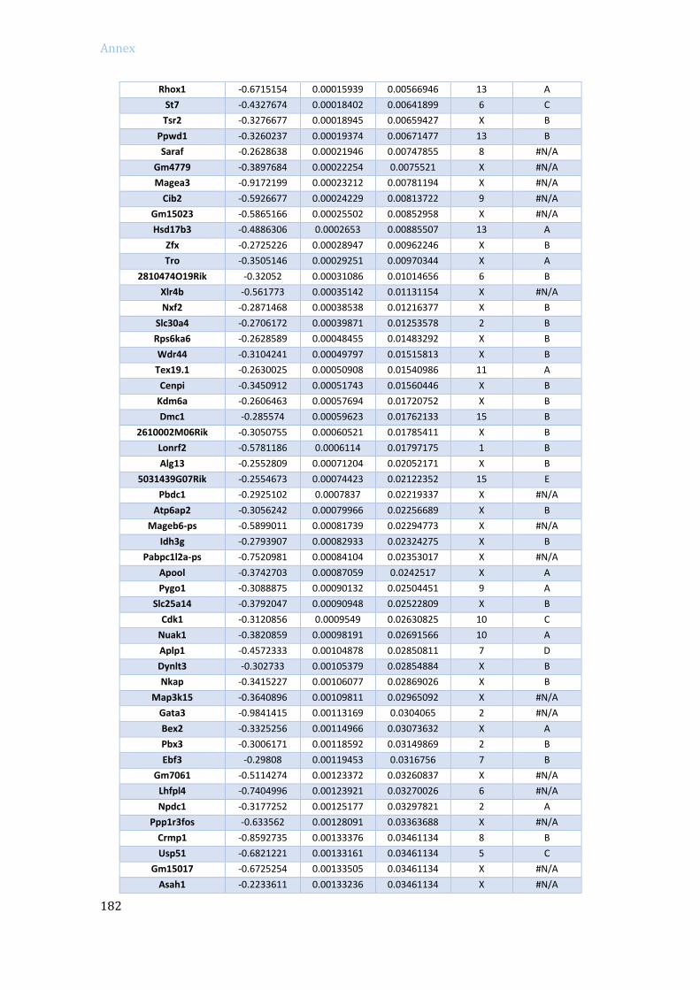
Annex
182
Rhox1 -0.6715154 0.00015939 0.00566946 13 A
St7 -0.4327674 0.00018402 0.00641899 6 C
Tsr2 -0.3276677 0.00018945 0.00659427 X B
Ppwd1 -0.3260237 0.00019374 0.00671477 13 B
Saraf -0.2628638 0.00021946 0.00747855 8 #N/A
Gm4779 -0.3897684 0.00022254 0.0075521 X #N/A
Magea3 -0.9172199 0.00023212 0.00781194 X #N/A
Cib2 -0.5926677 0.00024229 0.00813722 9 #N/A
Gm15023 -0.5865166 0.00025502 0.00852958 X #N/A
Hsd17b3 -0.4886306 0.0002653 0.00885507 13 A
Zfx -0.2725226 0.00028947 0.00962246 X B
Tro -0.3505146 0.00029251 0.00970344 X A
2810474O19Rik -0.32052 0.00031086 0.01014656 6 B
Xlr4b -0.561773 0.00035142 0.01131154 X #N/A
Nxf2 -0.2871468 0.00038538 0.01216377 X B
Slc30a4 -0.2706172 0.00039871 0.01253578 2 B
Rps6ka6 -0.2628589 0.00048455 0.01483292 X B
Wdr44 -0.3104241 0.00049797 0.01515813 X B
Tex19.1 -0.2630025 0.00050908 0.01540986 11 A
Cenpi -0.3450912 0.00051743 0.01560446 X B
Kdm6a -0.2606463 0.00057694 0.01720752 X B
Dmc1 -0.285574 0.00059623 0.01762133 15 B
2610002M06Rik -0.3050755 0.00060521 0.01785411 X B
Lonrf2 -0.5781186 0.0006114 0.01797175 1 B
Alg13 -0.2552809 0.00071204 0.02052171 X B
5031439G07Rik -0.2554673 0.00074423 0.02122352 15 E
Pbdc1 -0.2925102 0.0007837 0.02219337 X #N/A
Atp6ap2 -0.3056242 0.00079966 0.02256689 X B
Mageb6-ps -0.5899011 0.00081739 0.02294773 X #N/A
Idh3g -0.2793907 0.00082933 0.02324275 X B
Pabpc1l2a-ps -0.7520981 0.00084104 0.02353017 X #N/A
Apool -0.3742703 0.00087059 0.0242517 X A
Pygo1 -0.3088875 0.00090132 0.02504451 9 A
Slc25a14 -0.3792047 0.00090948 0.02522809 X B
Cdk1 -0.3120856 0.0009549 0.02630825 10 C
Nuak1 -0.3820859 0.00098191 0.02691566 10 A
Aplp1 -0.4572333 0.00104878 0.02850811 7 D
Dynlt3 -0.302733 0.00105379 0.02854884 X B
Nkap -0.3415227 0.00106077 0.02869026 X B
Map3k15 -0.3640896 0.00109811 0.02965092 X #N/A
Gata3 -0.9841415 0.00113169 0.0304065 2 #N/A
Bex2 -0.3325256 0.00114966 0.03073632 X A
Pbx3 -0.3006171 0.00118592 0.03149869 2 B
Ebf3 -0.29808 0.00119453 0.0316756 7 B
Gm7061 -0.5114274 0.00123372 0.03260837 X #N/A
Lhfpl4 -0.7404996 0.00123921 0.03270026 6 #N/A
Npdc1 -0.3177252 0.00125177 0.03297821 2 A
Ppp1r3fos -0.633562 0.00128091 0.03363688 X #N/A
Crmp1 -0.8592735 0.00133376 0.03461134 8 B
Usp51 -0.6821221 0.00133161 0.03461134 5 C
Gm15017 -0.6725254 0.00133505 0.03461134 X #N/A
Asah1 -0.2233611 0.00133236 0.03461134 X #N/A

Annex
183
Rad21l -0.3355452 0.00138315 0.03574419 2 #N/A
Fgf2 -0.3589929 0.00145801 0.03744828 3 #N/A
Mbtps2 -0.2697081 0.00147832 0.03778311 X B
Rdh11 -0.3610988 0.00150435 0.03832786 12 D
Yipf6 -0.262077 0.00151522 0.03848414 X B
A830080D01Rik -0.2467052 0.00161471 0.04056753 X B
Syngr2 -0.2370591 0.0016248 0.04075779 11 C
Trim52 -0.3219522 0.00171865 0.04285684 14 B
Hspa13 -0.2649786 0.0017281 0.04295145 16 #N/A
Cyp46a1 -0.5610384 0.0017572 0.04340916 12 C
Pdzd4 -0.4544886 0.00179147 0.04405495 X A
Nkrf -0.3078992 0.00182543 0.04468737 X B
Ifngr1 -0.266646 0.00183996 0.04496348 10 C
Gmds -0.2821265 0.00185318 0.04509501 13 B
Lrch2 -0.3225741 0.00187616 0.04558587 X A
Asb1 -0.2661504 0.00194191 0.04683347 1 C
Mmgt1 -0.2948245 0.00195687 0.04705458 X B
Xlr4c -0.5109194 0.00197953 0.04752915 X #N/A
Aldh1a2 -0.2079807 0.00198641 0.04753122 9 D
Cdh4 -0.7187891 0.00204228 0.0487477 2 #N/A
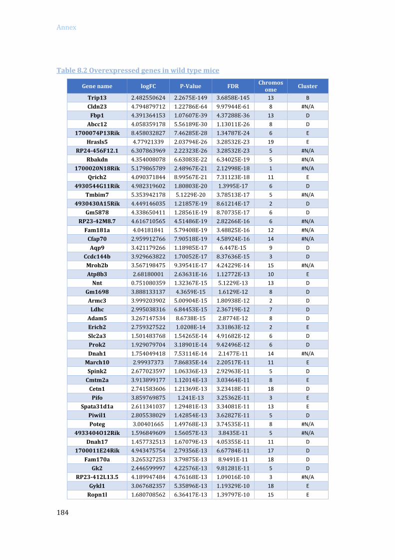
Annex
184
Table 8.2 Overexpressed genes in wild type mice
Gene name logFC P-Value FDR Chromos
ome Cluster
Trip13 2.482550624 2.2675E-149 3.6858E-145 13 B
Cldn23 4.794879712 1.22786E-64 9.97944E-61 8 #N/A
Fbp1 4.391364153 1.07607E-39 4.37288E-36 13 D
Abcc12 4.058359178 5.56189E-30 1.13011E-26 8 D
1700074P13Rik 8.458032827 7.46285E-28 1.34787E-24 6 E
Hrasls5 4.77921339 2.03794E-26 3.28532E-23 19 E
RP24-456F12.1 6.307863969 2.22323E-26 3.28532E-23 5 #N/A
Rbakdn 4.354008078 6.63083E-22 6.34025E-19 5 #N/A
1700020N18Rik 5.179865789 2.48967E-21 2.12998E-18 1 #N/A
Qrich2 4.090371844 8.99567E-21 7.31123E-18 11 E
4930544G11Rik 4.982319602 1.80803E-20 1.3995E-17 6 D
Tmbim7 5.353942178 5.1229E-20 3.78513E-17 5 #N/A
4930430A15Rik 4.449146035 1.21857E-19 8.61214E-17 2 D
Gm5878 4.338650411 1.28561E-19 8.70735E-17 6 D
RP23-42M8.7 4.616710565 4.51486E-19 2.82266E-16 6 #N/A
Fam181a 4.04181841 5.79408E-19 3.48825E-16 12 #N/A
Cfap70 2.959912766 7.90518E-19 4.58924E-16 14 #N/A
Aqp9 3.421179266 1.18985E-17 6.447E-15 9 D
Ccdc144b 3.929663822 1.70052E-17 8.37636E-15 3 D
Mroh2b 3.567198475 9.39541E-17 4.24229E-14 15 #N/A
Atp8b3 2.68180001 2.63631E-16 1.12772E-13 10 E
Nnt 0.751080359 1.32367E-15 5.1229E-13 13 D
Gm1698 3.888133137 4.3659E-15 1.6129E-12 8 D
Armc3 3.999203902 5.00904E-15 1.80938E-12 2 D
Ldhc 2.995038316 6.84453E-15 2.36719E-12 7 D
Adam5 3.267147534 8.6738E-15 2.8774E-12 8 D
Erich2 2.759327522 1.0208E-14 3.31863E-12 2 E
Slc2a3 1.501483768 1.54265E-14 4.91682E-12 6 D
Prok2 1.929079704 3.18901E-14 9.42496E-12 6 D
Dnah1 1.754049418 7.53114E-14 2.1477E-11 14 #N/A
March10 2.99937373 7.86835E-14 2.20517E-11 11 E
Spink2 2.677023597 1.06336E-13 2.92963E-11 5 D
Cmtm2a 3.913899177 1.12014E-13 3.03464E-11 8 E
Cetn1 2.741583606 1.21369E-13 3.23418E-11 18 D
Pifo 3.859769875 1.241E-13 3.25362E-11 3 E
Spata31d1a 2.611341037 1.29481E-13 3.34081E-11 13 E
Piwil1 2.805538029 1.42854E-13 3.62827E-11 5 D
Poteg 3.00401665 1.49768E-13 3.74535E-11 8 #N/A
4933404O12Rik 1.596849609 1.56057E-13 3.8435E-11 5 #N/A
Dnah17 1.457732513 1.67079E-13 4.05355E-11 11 D
1700011E24Rik 4.943475754 2.79356E-13 6.67784E-11 17 D
Fam170a 3.265327253 3.79875E-13 8.9491E-11 18 D
Gk2 2.446599997 4.22576E-13 9.81281E-11 5 D
RP23-412L13.5 4.189947484 4.76168E-13 1.09016E-10 3 #N/A
Gykl1 3.067682357 5.35896E-13 1.19329E-10 18 E
Ropn1l 1.680708562 6.36417E-13 1.39797E-10 15 E
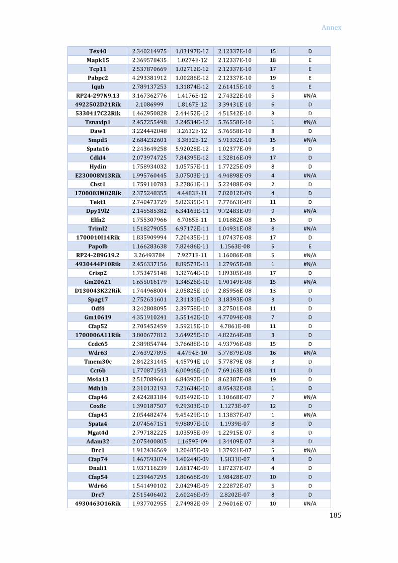
Annex
185
Tex40 2.340214975 1.03197E-12 2.12337E-10 15 D
Mapk15 2.369578435 1.0274E-12 2.12337E-10 18 E
Tcp11 2.537870669 1.02712E-12 2.12337E-10 17 E
Pabpc2 4.293381912 1.00286E-12 2.12337E-10 19 E
Iqub 2.789137253 1.31874E-12 2.61415E-10 6 E
RP24-297N9.13 3.167362776 1.4176E-12 2.74322E-10 5 #N/A
4922502D21Rik 2.1086999 1.8167E-12 3.39431E-10 6 D
5330417C22Rik 1.462950828 2.44452E-12 4.51542E-10 3 D
Tsnaxip1 2.457255498 3.24534E-12 5.76558E-10 1 #N/A
Daw1 3.224442048 3.2632E-12 5.76558E-10 8 D
Smpd5 2.684232601 3.3832E-12 5.91332E-10 15 #N/A
Spata16 2.243649258 5.92028E-12 1.02377E-09 3 D
Cdkl4 2.073974725 7.84395E-12 1.32816E-09 17 D
Hydin 1.758934032 1.05757E-11 1.77225E-09 8 D
E230008N13Rik 1.995760445 3.07503E-11 4.94898E-09 4 #N/A
Chst1 1.759110783 3.27861E-11 5.22488E-09 2 D
1700003M02Rik 2.375248355 4.4483E-11 7.02012E-09 4 D
Tekt1 2.740473729 5.02335E-11 7.77663E-09 11 D
Dpy19l2 2.145585382 6.34163E-11 9.72483E-09 9 #N/A
Elfn2 1.755307966 6.7065E-11 1.01882E-08 15 D
Triml2 1.518279055 6.97172E-11 1.04931E-08 8 #N/A
1700010I14Rik 1.835909994 7.20435E-11 1.07437E-08 17 D
Papolb 1.166283638 7.82486E-11 1.1563E-08 5 E
RP24-289G19.2 3.26493784 7.9271E-11 1.16086E-08 5 #N/A
4930444P10Rik 2.456337156 8.89573E-11 1.27965E-08 1 #N/A
Crisp2 1.753475148 1.32764E-10 1.89305E-08 17 D
Gm20621 1.655016179 1.34526E-10 1.90149E-08 15 #N/A
D130043K22Rik 1.744968004 2.05825E-10 2.85956E-08 13 D
Spag17 2.752631601 2.31131E-10 3.18393E-08 3 D
Odf4 3.242808095 2.39758E-10 3.27501E-08 11 D
Gm10619 4.351910241 3.55142E-10 4.77094E-08 7 D
Cfap52 2.705452459 3.59215E-10 4.7861E-08 11 D
1700006A11Rik 3.800677812 3.64925E-10 4.82264E-08 3 D
Ccdc65 2.389854744 3.76688E-10 4.93796E-08 15 D
Wdr63 2.763927895 4.4794E-10 5.77879E-08 16 #N/A
Tmem30c 2.842231445 4.45794E-10 5.77879E-08 3 D
Cct6b 1.770871543 6.00946E-10 7.69163E-08 11 D
Ms4a13 2.517089661 6.84392E-10 8.62387E-08 19 D
Mdh1b 2.310132193 7.21634E-10 8.95432E-08 1 D
Cfap46 2.424283184 9.05492E-10 1.10668E-07 7 #N/A
Cox8c 1.390187507 9.29303E-10 1.1273E-07 12 D
Cfap45 2.054482474 9.45429E-10 1.13837E-07 1 #N/A
Spata4 2.074567151 9.98897E-10 1.1939E-07 8 D
Mgat4d 2.797182225 1.03595E-09 1.22915E-07 8 D
Adam32 2.075400805 1.1659E-09 1.34409E-07 8 D
Drc1 1.912436569 1.20485E-09 1.37921E-07 5 #N/A
Cfap74 1.467593074 1.40244E-09 1.5831E-07 4 D
Dnali1 1.937116239 1.68174E-09 1.87237E-07 4 D
Cfap54 1.239467295 1.80666E-09 1.98428E-07 10 D
Wdr66 1.541490102 2.04294E-09 2.22872E-07 5 D
Drc7 2.515406402 2.60246E-09 2.8202E-07 8 D
4930463O16Rik 1.937702955 2.74982E-09 2.96016E-07 10 #N/A

Annex
186
Adam2 1.896465641 2.88684E-09 3.06703E-07 14 D
4930465K10Rik 1.826987173 3.91868E-09 4.13624E-07 18 D
Tdrd6 2.226286882 4.80027E-09 4.96996E-07 17 D
Trim11 0.684320316 5.69326E-09 5.74807E-07 11 C
Ypel1 0.878810691 5.65821E-09 5.74807E-07 16 D
Tcp10a 2.233788059 5.97571E-09 5.99599E-07 17 D
Spag16 1.40527158 6.70079E-09 6.68229E-07 1 D
Stambp 0.774835514 6.83598E-09 6.77554E-07 6 C
Gstt2 1.299849918 7.11577E-09 6.98476E-07 2 D
4932414N04Rik 2.358911538 7.13301E-09 6.98476E-07 10 C
Ccdc146 1.61905353 8.08372E-09 7.86832E-07 5 E
Gm27045 2.438492965 8.50431E-09 8.22842E-07 8 #N/A
Lin7a 1.970705672 9.43524E-09 9.07513E-07 10 D
Krt8 2.094520539 1.25403E-08 1.18513E-06 15 #N/A
Fabp9 1.851002139 1.26829E-08 1.19168E-06 3 D
Gm14244 3.529590904 1.31237E-08 1.22601E-06 2 #N/A
Bspry 1.031831161 1.72836E-08 1.59627E-06 4 E
Ccdc89 1.951632454 1.84738E-08 1.69656E-06 7 #N/A
Nup210l 1.065249793 2.10537E-08 1.90127E-06 16 #N/A
Cfap44 1.715357748 2.10327E-08 1.90127E-06 3 #N/A
Spag8 1.448974334 2.19538E-08 1.96077E-06 4 D
Zmynd10 1.882834103 2.18856E-08 1.96077E-06 9 D
Cers3 1.30034982 2.34746E-08 2.08513E-06 7 #N/A
Lypd4 1.581059443 2.42172E-08 2.13941E-06 7 E
Lrguk 1.393824535 2.58612E-08 2.27229E-06 6 D
Dnah7c 1.261191314 2.64843E-08 2.31453E-06 1 #N/A
Gtf2a1l 1.739071772 2.6645E-08 2.31612E-06 17 E
Dnah7b 0.887080771 2.82724E-08 2.44451E-06 1 #N/A
Fam184a 1.321167799 2.98987E-08 2.57145E-06 10 #N/A
Ptchd3 1.78186377 3.12252E-08 2.66623E-06 11 D
AY702103 2.932429882 3.29054E-08 2.78582E-06 17 #N/A
Lrrc23 2.059357516 3.36369E-08 2.83299E-06 6 D
Mroh4 2.262576313 3.40328E-08 2.85156E-06 15 #N/A
Iqcd 1.754951599 3.87532E-08 3.19763E-06 5 D
Ift172 0.729430469 4.06831E-08 3.32314E-06 5 D
Gm13814 2.33390412 4.82348E-08 3.92028E-06 2 #N/A
Dnah6 2.12010391 5.74111E-08 4.63509E-06 4 D
1700042G15Rik 4.195632488 5.76E-08 4.63509E-06 6 #N/A
Ppp3r2 1.776519636 6.11996E-08 4.87647E-06 4 D
Cuzd1 3.074000021 6.21368E-08 4.92699E-06 7 E
4931429I11Rik 1.605979807 6.80976E-08 5.37343E-06 9 D
Ttc30a2 1.252753501 8.17251E-08 6.41759E-06 2 D
Slc2a5 1.465829152 9.00705E-08 7.00524E-06 4 E
M5C1000I18Rik 1.627703667 9.37462E-08 7.2564E-06 9 #N/A
Slc9c1 1.88864348 9.9909E-08 7.69678E-06 16 #N/A
Ttc25 1.245851275 1.15641E-07 8.86671E-06 11 E
Fhad1 1.344813614 1.18401E-07 9.03571E-06 4 E
Chgb 1.37020938 1.2159E-07 9.19275E-06 2 D
1700008K24Rik 1.542124336 1.3996E-07 1.02944E-05 17 #N/A
Lrriq1 1.516796747 1.54269E-07 1.11948E-05 10 D
Agbl2 1.322376684 1.57684E-07 1.13918E-05 2 D
Slco2a1 1.187447254 1.75124E-07 1.24853E-05 9 A

Annex
187
Ak8 1.361503018 1.89179E-07 1.34284E-05 2 E
Dnah2 0.867293955 1.94334E-07 1.37344E-05 11 D
Acrbp 1.083180922 1.96195E-07 1.38058E-05 6 E
Nsun7 1.133930841 2.04225E-07 1.43003E-05 17 D
Ldhal6b 1.364076994 2.04981E-07 1.43003E-05 5 E
Krt18 2.052018459 2.09341E-07 1.4542E-05 15 A
1700102P08Rik 1.131973389 2.37986E-07 1.63918E-05 9 C
Spats1 1.700997077 2.67762E-07 1.82112E-05 17 D
Fam229b 1.162994028 2.94133E-07 1.99214E-05 10 E
Ccdc108 2.072683841 3.40369E-07 2.25824E-05 1 D
Gramd1c 0.998019688 3.56854E-07 2.34845E-05 16 D
Dnajb13 2.127405467 3.62658E-07 2.37702E-05 7 E
Gm29050 1.328938267 3.75169E-07 2.44914E-05 17 #N/A
Acr 2.782723459 4.10756E-07 2.67073E-05 15 E
Tmprss12 1.387526574 4.21582E-07 2.7302E-05 15 D
Lrrc6 1.425921293 4.55661E-07 2.89924E-05 15 D
1700040L02Rik 1.162099098 4.62706E-07 2.92657E-05 10 D
Pom121l2 1.65747905 5.14903E-07 3.23156E-05 13 #N/A
Ankrd36 1.530595705 5.19324E-07 3.24677E-05 11 D
Lca5l 0.944196943 5.35698E-07 3.33631E-05 16 D
Dynlrb2 2.186086633 5.87225E-07 3.64326E-05 8 D
Mroh8 1.204019954 6.05331E-07 3.72714E-05 2 #N/A
Prom1 2.094028354 6.24596E-07 3.81685E-05 5 E
Tkfc 0.644243147 8.24505E-07 4.96382E-05 19 D
Cpn1 1.200131667 8.54329E-07 5.10556E-05 19 D
1700018B08Rik 1.502276501 9.33827E-07 5.53991E-05 8 E
Tdrd5 0.740206727 9.77417E-07 5.77742E-05 1 C
Ybx2 0.805501381 9.95806E-07 5.84362E-05 1 D
Ankar 1.928915959 9.93898E-07 5.84362E-05 11 D
Ccdc40 0.865332629 1.07874E-06 6.30751E-05 11 D
Pdcl2 1.316460974 1.10375E-06 6.40765E-05 5 D
6430531B16Rik 1.320969604 1.14545E-06 6.62606E-05 7 D
Abo 1.439918286 1.48314E-06 8.51886E-05 2 D
Dkkl1 1.55335047 1.5576E-06 8.88379E-05 7 E
Dnah14 1.330419641 1.60966E-06 9.08508E-05 1 D
Axdnd1 1.200612425 1.62124E-06 9.11876E-05 1 #N/A
4933411G11Rik 1.380993168 1.7888E-06 0.000100265 5 D
Dnhd1 0.818459322 1.80433E-06 0.000100788 7 #N/A
4933415F23Rik 1.181540944 1.83828E-06 0.000102333 1 D
Lrrc61 0.837912993 1.86424E-06 0.000103424 6 D
Ttc29 1.401838039 2.00064E-06 0.000110614 8 D
Gpx3 1.107874998 2.02602E-06 0.000111637 11 C
Mif4gd 0.584883679 2.09151E-06 0.000114231 11 C
Fam186b 1.696150421 2.21065E-06 0.000120181 15 D
Mag 1.451479732 2.28805E-06 0.000123562 7 D
Smim24 1.292156833 2.52009E-06 0.000135643 10 D
Psma8 0.88555175 2.6466E-06 0.000141515 18 C
Rfx4 2.165011461 2.75037E-06 0.000146581 10 D
Ccdc39 1.062458919 2.85196E-06 0.000151499 3 C
Gm11992 1.006863517 2.92302E-06 0.000154768 11 D
Dnah8 1.205982318 3.01865E-06 0.000159312 17 D
Cfap57 1.346087185 3.07819E-06 0.000161929 4 #N/A
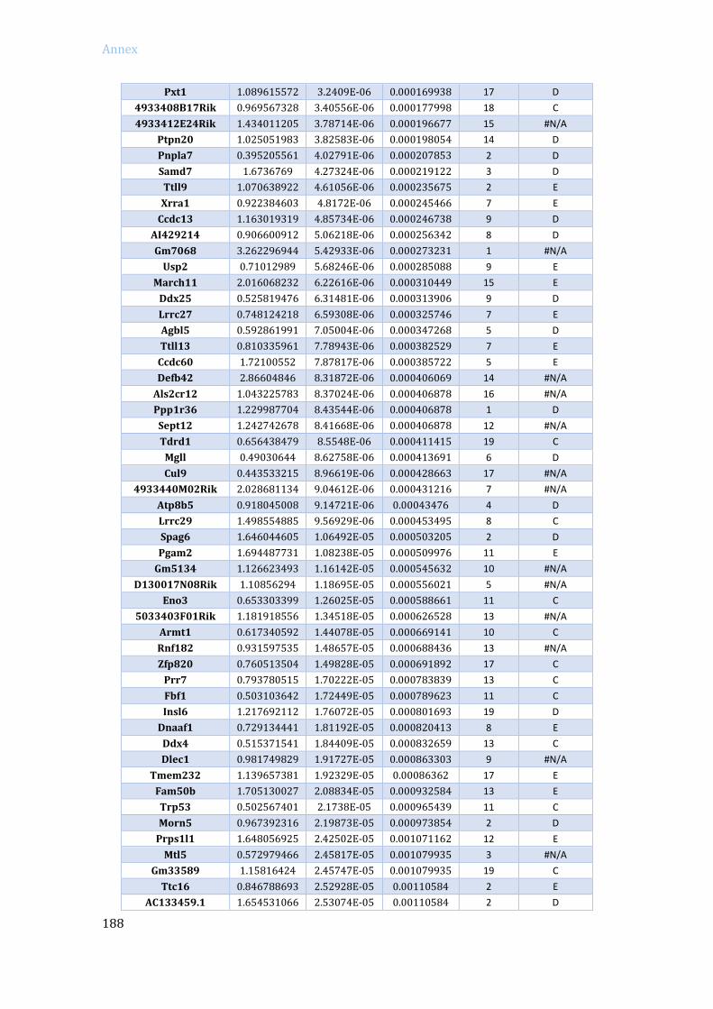
Annex
188
Pxt1 1.089615572 3.2409E-06 0.000169938 17 D
4933408B17Rik 0.969567328 3.40556E-06 0.000177998 18 C
4933412E24Rik 1.434011205 3.78714E-06 0.000196677 15 #N/A
Ptpn20 1.025051983 3.82583E-06 0.000198054 14 D
Pnpla7 0.395205561 4.02791E-06 0.000207853 2 D
Samd7 1.6736769 4.27324E-06 0.000219122 3 D
Ttll9 1.070638922 4.61056E-06 0.000235675 2 E
Xrra1 0.922384603 4.8172E-06 0.000245466 7 E
Ccdc13 1.163019319 4.85734E-06 0.000246738 9 D
AI429214 0.906600912 5.06218E-06 0.000256342 8 D
Gm7068 3.262296944 5.42933E-06 0.000273231 1 #N/A
Usp2 0.71012989 5.68246E-06 0.000285088 9 E
March11 2.016068232 6.22616E-06 0.000310449 15 E
Ddx25 0.525819476 6.31481E-06 0.000313906 9 D
Lrrc27 0.748124218 6.59308E-06 0.000325746 7 E
Agbl5 0.592861991 7.05004E-06 0.000347268 5 D
Ttll13 0.810335961 7.78943E-06 0.000382529 7 E
Ccdc60 1.72100552 7.87817E-06 0.000385722 5 E
Defb42 2.86604846 8.31872E-06 0.000406069 14 #N/A
Als2cr12 1.043225783 8.37024E-06 0.000406878 16 #N/A
Ppp1r36 1.229987704 8.43544E-06 0.000406878 1 D
Sept12 1.242742678 8.41668E-06 0.000406878 12 #N/A
Tdrd1 0.656438479 8.5548E-06 0.000411415 19 C
Mgll 0.49030644 8.62758E-06 0.000413691 6 D
Cul9 0.443533215 8.96619E-06 0.000428663 17 #N/A
4933440M02Rik 2.028681134 9.04612E-06 0.000431216 7 #N/A
Atp8b5 0.918045008 9.14721E-06 0.00043476 4 D
Lrrc29 1.498554885 9.56929E-06 0.000453495 8 C
Spag6 1.646044605 1.06492E-05 0.000503205 2 D
Pgam2 1.694487731 1.08238E-05 0.000509976 11 E
Gm5134 1.126623493 1.16142E-05 0.000545632 10 #N/A
D130017N08Rik 1.10856294 1.18695E-05 0.000556021 5 #N/A
Eno3 0.653303399 1.26025E-05 0.000588661 11 C
5033403F01Rik 1.181918556 1.34518E-05 0.000626528 13 #N/A
Armt1 0.617340592 1.44078E-05 0.000669141 10 C
Rnf182 0.931597535 1.48657E-05 0.000688436 13 #N/A
Zfp820 0.760513504 1.49828E-05 0.000691892 17 C
Prr7 0.793780515 1.70222E-05 0.000783839 13 C
Fbf1 0.503103642 1.72449E-05 0.000789623 11 C
Insl6 1.217692112 1.76072E-05 0.000801693 19 D
Dnaaf1 0.729134441 1.81192E-05 0.000820413 8 E
Ddx4 0.515371541 1.84409E-05 0.000832659 13 C
Dlec1 0.981749829 1.91727E-05 0.000863303 9 #N/A
Tmem232 1.139657381 1.92329E-05 0.00086362 17 E
Fam50b 1.705130027 2.08834E-05 0.000932584 13 E
Trp53 0.502567401 2.1738E-05 0.000965439 11 C
Morn5 0.967392316 2.19873E-05 0.000973854 2 D
Prps1l1 1.648056925 2.42502E-05 0.001071162 12 E
Mtl5 0.572979466 2.45817E-05 0.001079935 3 #N/A
Gm33589 1.15816424 2.45747E-05 0.001079935 19 C
Ttc16 0.846788693 2.52928E-05 0.00110584 2 E
AC133459.1 1.654531066 2.53074E-05 0.00110584 2 D

Annex
189
Wdr93 0.927286874 2.59586E-05 0.001131251 7 D
Ptpdc1 0.487198318 2.6132E-05 0.001135766 13 D
Ube2d2b 1.197170176 2.67388E-05 0.001155956 5 #N/A
Eps8l3 0.981451228 2.7538E-05 0.001187347 3 D
Rnf32 0.839218057 2.89241E-05 0.001242221 7 D
Fank1 1.036947417 2.89635E-05 0.001242221 5 D
Ces1d 1.040632333 2.99333E-05 0.001280435 8 E
Clgn 1.031179963 3.14294E-05 0.001340903 8 D
Ankef1 0.960777346 3.47572E-05 0.001474931 2 E
Susd5 1.05455107 3.4843E-05 0.001474931 9 #N/A
AI464131 0.954572523 3.67529E-05 0.001547715 4 D
C2cd4b 1.350429832 3.91982E-05 0.001642181 9 #N/A
Efhc1 0.843962631 4.13406E-05 0.001727483 1 D
Gm37389 1.591890818 4.17279E-05 0.001739196 3 #N/A
Rsph9 0.841652143 4.22915E-05 0.00175818 17 E
Morc2b 1.78606934 4.27891E-05 0.001765322 17 D
Mok 0.611319651 4.65099E-05 0.001894783 4 C
Fgr 1.076476767 4.64014E-05 0.001894783 12 E
Csl 1.074367777 4.74021E-05 0.001926302 10 E
Ttll6 1.358653004 4.83576E-05 0.001960233 11 E
Cfap43 0.746468081 4.95245E-05 0.001997571 19 E
Gm15881 1.367449215 5.17171E-05 0.002075709 8 #N/A
Arsk 0.475162252 5.38597E-05 0.002156379 13 B
Ubxn11 0.678260458 5.43017E-05 0.002168734 4 #N/A
Lrrc36 0.998744487 5.5124E-05 0.002196179 8 E
Crem 0.681284266 5.68341E-05 0.002253265 18 E
Ift140 0.397076608 5.99585E-05 0.002366989 17 D
Kif6 0.677325362 6.21098E-05 0.00244454 17 D
Cyct 0.645729477 6.29907E-05 0.002473223 2 C
Ccdc87 0.982955419 6.54533E-05 0.002557555 19 D
Ccdc113 1.409129624 7.35743E-05 0.0028475 8 D
Crtam 1.412619127 7.57312E-05 0.002924016 9 D
Trp63 0.784285985 7.62707E-05 0.002937868 16 #N/A
Spef2 1.169331364 7.82473E-05 0.003004553 15 D
1700009C05Rik 2.067663118 8.46885E-05 0.003231482 6 #N/A
Adamts6 0.643114044 8.64808E-05 0.003292144 13 A
1700029M20Rik 0.901226627 8.74261E-05 0.003320353 4 #N/A
Cdc42ep3 0.728446738 9.54042E-05 0.003598132 17 D
Tcte2 0.88456668 9.64312E-05 0.003628446 17 D
Rnls 0.615951773 0.000100274 0.003760881 19 #N/A
Pex11g 0.720414045 0.000103375 0.003862902 8 D
Ccdc136 0.605850217 0.000108844 0.004048662 6 E
Atp11a 0.361384368 0.000116454 0.004311999 8 C
Mum1 0.399482254 0.000118931 0.004385322 10 D
Shcbp1l 0.603686881 0.000121515 0.004468829 1 D
Cchcr1 0.516531404 0.000129969 0.004738965 17 D
Phf7 0.579727768 0.000130026 0.004738965 14 D
Wdfy4 0.645408217 0.000135103 0.004912976 14 D
Pnmal1 1.186308553 0.000145044 0.005250968 7 D
Ptprq 0.966690699 0.000147443 0.005325973 10 D
Ggnbp1 0.482754614 0.000147791 0.0053267 17 E
Ankrd45 0.6661053 0.000154461 0.005530299 1 C
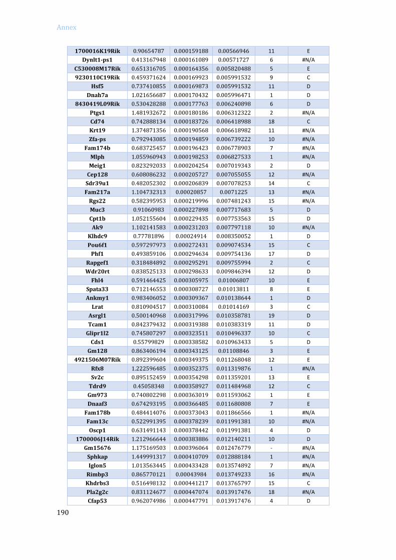
Annex
190
1700016K19Rik 0.90654787 0.000159188 0.00566946 11 E
Dynlt1-ps1 0.413167948 0.000161089 0.00571727 6 #N/A
C530008M17Rik 0.651316705 0.000164356 0.005820488 5 E
9230110C19Rik 0.459371624 0.000169923 0.005991532 9 C
Hsf5 0.737410855 0.000169873 0.005991532 11 D
Dnah7a 1.021656687 0.000170432 0.005996471 1 D
8430419L09Rik 0.530428288 0.000177763 0.006240898 6 D
Ptgs1 1.481932672 0.000180186 0.006312322 2 #N/A
Cd74 0.742888134 0.000183726 0.006418988 18 C
Krt19 1.374871356 0.000190568 0.006618982 11 #N/A
Zfa-ps 0.792943085 0.000194859 0.006739222 10 #N/A
Fam174b 0.683725457 0.000196423 0.006778903 7 #N/A
Mlph 1.055960943 0.000198253 0.006827533 1 #N/A
Meig1 0.823292033 0.000204254 0.007019343 2 D
Cep128 0.608086232 0.000205727 0.007055055 12 #N/A
Sdr39u1 0.482052302 0.000206839 0.007078253 14 C
Fam217a 1.104732313 0.00020857 0.0071225 13 #N/A
Rgs22 0.582395953 0.000219996 0.007481243 15 #N/A
Muc3 0.91060983 0.000227898 0.007717683 5 D
Cpt1b 1.052155604 0.000229435 0.007753563 15 D
Ak9 1.102141583 0.000231203 0.007797118 10 #N/A
Klhdc9 0.77781896 0.00024914 0.008350052 1 D
Pou6f1 0.597297973 0.000272431 0.009074534 15 C
Phf1 0.493859106 0.000294634 0.009754136 17 D
Rapgef1 0.318484892 0.000295291 0.009755994 2 C
Wdr20rt 0.838525133 0.000298633 0.009846394 12 D
Fhl4 0.591464425 0.000305975 0.01006807 10 E
Spata33 0.712146553 0.000308727 0.01013811 8 E
Ankmy1 0.983406052 0.000309367 0.010138644 1 D
Lrat 0.810904517 0.000310084 0.01014169 3 C
Asrgl1 0.500140968 0.000317996 0.010358781 19 D
Tcam1 0.842379432 0.000319388 0.010383319 11 D
Glipr1l2 0.745807297 0.000323511 0.010496337 10 C
Cds1 0.55799829 0.000338582 0.010963433 5 D
Gm128 0.863406194 0.000343125 0.01108846 3 E
4921506M07Rik 0.892399604 0.000349375 0.011268048 12 E
Rfx8 1.222596485 0.000352375 0.011319876 1 #N/A
Sv2c 0.895152459 0.000354298 0.011359201 13 E
Tdrd9 0.45058348 0.000358927 0.011484968 12 C
Gm973 0.740802298 0.000363019 0.011593062 1 E
Dnaaf3 0.674293195 0.000366485 0.011680808 7 E
Fam178b 0.484414076 0.000373043 0.011866566 1 #N/A
Fam13c 0.522991395 0.000378239 0.011991381 10 #N/A
Oscp1 0.631491143 0.000378442 0.011991381 4 D
1700006J14Rik 1.212966644 0.000383886 0.012140211 10 D
Gm15676 1.175169503 0.000396064 0.012476779 - #N/A
Sphkap 1.449991317 0.000410709 0.012888184 1 #N/A
Iglon5 1.013563445 0.000433428 0.013574892 7 #N/A
Rimbp3 0.865770121 0.00043984 0.013749233 16 #N/A
Khdrbs3 0.516498132 0.000441217 0.013765797 15 C
Pla2g2c 0.831124677 0.000447074 0.013917476 18 #N/A
Cfap53 0.962074986 0.000447791 0.013917476 4 D
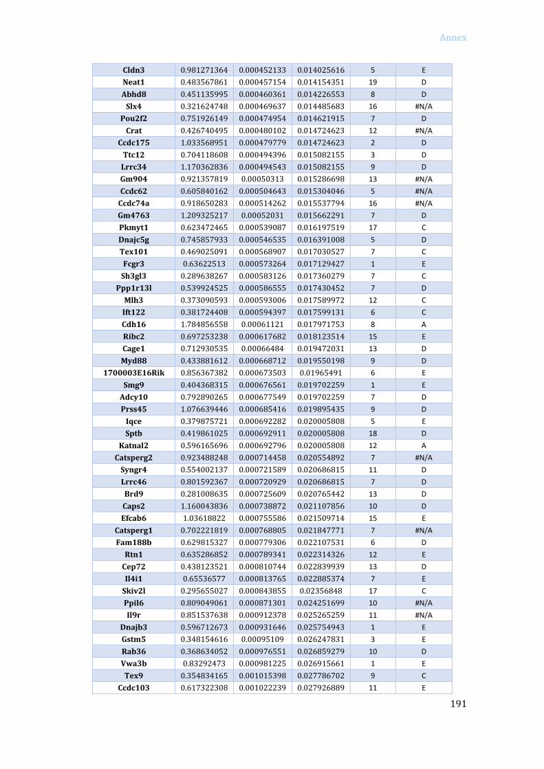
Annex
191
Cldn3 0.981271364 0.000452133 0.014025616 5 E
Neat1 0.483567861 0.000457154 0.014154351 19 D
Abhd8 0.451135995 0.000460361 0.014226553 8 D
Slx4 0.321624748 0.000469637 0.014485683 16 #N/A
Pou2f2 0.751926149 0.000474954 0.014621915 7 D
Crat 0.426740495 0.000480102 0.014724623 12 #N/A
Ccdc175 1.033568951 0.000479779 0.014724623 2 D
Ttc12 0.704118608 0.000494396 0.015082155 3 D
Lrrc34 1.170362836 0.000494543 0.015082155 9 D
Gm904 0.921357819 0.00050313 0.015286698 13 #N/A
Ccdc62 0.605840162 0.000504643 0.015304046 5 #N/A
Ccdc74a 0.918650283 0.000514262 0.015537794 16 #N/A
Gm4763 1.209325217 0.00052031 0.015662291 7 D
Pkmyt1 0.623472465 0.000539087 0.016197519 17 C
Dnajc5g 0.745857933 0.000546535 0.016391008 5 D
Tex101 0.469025091 0.000568907 0.017030527 7 C
Fcgr3 0.63622513 0.000573264 0.017129427 1 E
Sh3gl3 0.289638267 0.000583126 0.017360279 7 C
Ppp1r13l 0.539924525 0.000586555 0.017430452 7 D
Mlh3 0.373090593 0.000593006 0.017589972 12 C
Ift122 0.381724408 0.000594397 0.017599131 6 C
Cdh16 1.784856558 0.00061121 0.017971753 8 A
Ribc2 0.697253238 0.000617682 0.018123514 15 E
Cage1 0.712930535 0.00066484 0.019472031 13 D
Myd88 0.433881612 0.000668712 0.019550198 9 D
1700003E16Rik 0.856367382 0.000673503 0.01965491 6 E
Smg9 0.404368315 0.000676561 0.019702259 1 E
Adcy10 0.792890265 0.000677549 0.019702259 7 D
Prss45 1.076639446 0.000685416 0.019895435 9 D
Iqce 0.379875721 0.000692282 0.020005808 5 E
Sptb 0.419861025 0.000692911 0.020005808 18 D
Katnal2 0.596165696 0.000692796 0.020005808 12 A
Catsperg2 0.923488248 0.000714458 0.020554892 7 #N/A
Syngr4 0.554002137 0.000721589 0.020686815 11 D
Lrrc46 0.801592367 0.000720929 0.020686815 7 D
Brd9 0.281008635 0.000725609 0.020765442 13 D
Caps2 1.160043836 0.000738872 0.021107856 10 D
Efcab6 1.03618822 0.000755586 0.021509714 15 E
Catsperg1 0.702221819 0.000768805 0.021847771 7 #N/A
Fam188b 0.629815327 0.000779306 0.022107531 6 D
Rtn1 0.635286852 0.000789341 0.022314326 12 E
Cep72 0.438123521 0.000810744 0.022839939 13 D
Il4i1 0.65536577 0.000813765 0.022885374 7 E
Skiv2l 0.295655027 0.000843855 0.02356848 17 C
Ppil6 0.809049061 0.000871301 0.024251699 10 #N/A
Il9r 0.851537638 0.000912378 0.025265259 11 #N/A
Dnajb3 0.596712673 0.000931646 0.025754943 1 E
Gstm5 0.348154616 0.00095109 0.026247831 3 E
Rab36 0.368634052 0.000976551 0.026859279 10 D
Vwa3b 0.83292473 0.000981225 0.026915661 1 E
Tex9 0.354834165 0.001015398 0.027786702 9 C
Ccdc103 0.617322308 0.001022239 0.027926889 11 E
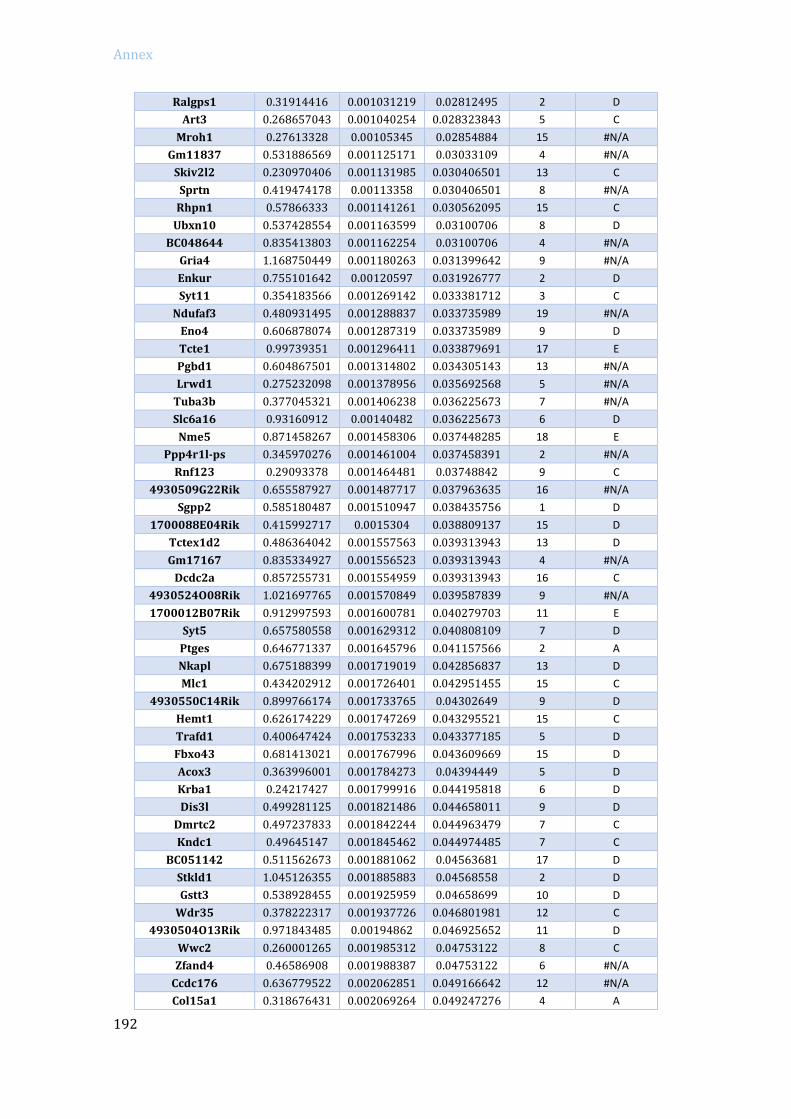
Annex
192
Ralgps1 0.31914416 0.001031219 0.02812495 2 D
Art3 0.268657043 0.001040254 0.028323843 5 C
Mroh1 0.27613328 0.00105345 0.02854884 15 #N/A
Gm11837 0.531886569 0.001125171 0.03033109 4 #N/A
Skiv2l2 0.230970406 0.001131985 0.030406501 13 C
Sprtn 0.419474178 0.00113358 0.030406501 8 #N/A
Rhpn1 0.57866333 0.001141261 0.030562095 15 C
Ubxn10 0.537428554 0.001163599 0.03100706 8 D
BC048644 0.835413803 0.001162254 0.03100706 4 #N/A
Gria4 1.168750449 0.001180263 0.031399642 9 #N/A
Enkur 0.755101642 0.00120597 0.031926777 2 D
Syt11 0.354183566 0.001269142 0.033381712 3 C
Ndufaf3 0.480931495 0.001288837 0.033735989 19 #N/A
Eno4 0.606878074 0.001287319 0.033735989 9 D
Tcte1 0.99739351 0.001296411 0.033879691 17 E
Pgbd1 0.604867501 0.001314802 0.034305143 13 #N/A
Lrwd1 0.275232098 0.001378956 0.035692568 5 #N/A
Tuba3b 0.377045321 0.001406238 0.036225673 7 #N/A
Slc6a16 0.93160912 0.00140482 0.036225673 6 D
Nme5 0.871458267 0.001458306 0.037448285 18 E
Ppp4r1l-ps 0.345970276 0.001461004 0.037458391 2 #N/A
Rnf123 0.29093378 0.001464481 0.03748842 9 C
4930509G22Rik 0.655587927 0.001487717 0.037963635 16 #N/A
Sgpp2 0.585180487 0.001510947 0.038435756 1 D
1700088E04Rik 0.415992717 0.0015304 0.038809137 15 D
Tctex1d2 0.486364042 0.001557563 0.039313943 13 D
Gm17167 0.835334927 0.001556523 0.039313943 4 #N/A
Dcdc2a 0.857255731 0.001554959 0.039313943 16 C
4930524O08Rik 1.021697765 0.001570849 0.039587839 9 #N/A
1700012B07Rik 0.912997593 0.001600781 0.040279703 11 E
Syt5 0.657580558 0.001629312 0.040808109 7 D
Ptges 0.646771337 0.001645796 0.041157566 2 A
Nkapl 0.675188399 0.001719019 0.042856837 13 D
Mlc1 0.434202912 0.001726401 0.042951455 15 C
4930550C14Rik 0.899766174 0.001733765 0.04302649 9 D
Hemt1 0.626174229 0.001747269 0.043295521 15 C
Trafd1 0.400647424 0.001753233 0.043377185 5 D
Fbxo43 0.681413021 0.001767996 0.043609669 15 D
Acox3 0.363996001 0.001784273 0.04394449 5 D
Krba1 0.24217427 0.001799916 0.044195818 6 D
Dis3l 0.499281125 0.001821486 0.044658011 9 D
Dmrtc2 0.497237833 0.001842244 0.044963479 7 C
Kndc1 0.49645147 0.001845462 0.044974485 7 C
BC051142 0.511562673 0.001881062 0.04563681 17 D
Stkld1 1.045126355 0.001885883 0.04568558 2 D
Gstt3 0.538928455 0.001925959 0.04658699 10 D
Wdr35 0.378222317 0.001937726 0.046801981 12 C
4930504O13Rik 0.971843485 0.00194862 0.046925652 11 D
Wwc2 0.260001265 0.001985312 0.04753122 8 C
Zfand4 0.46586908 0.001988387 0.04753122 6 #N/A
Ccdc176 0.636779522 0.002062851 0.049166642 12 #N/A
Col15a1 0.318676431 0.002069264 0.049247276 4 A

Annex
193