Mammalian transglutaminases
17
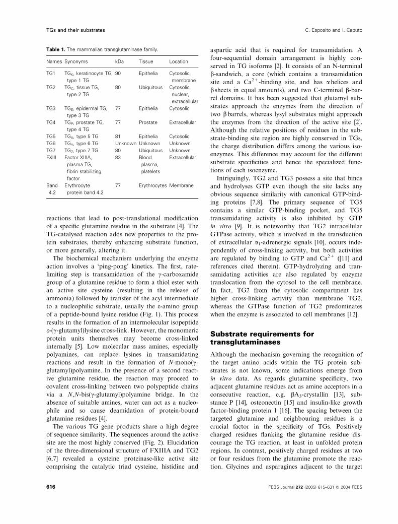
REVIEW ARTICLE Mammalian transglutaminases Identification of substrates as a key to physiological function and physiopathological relevance Carla Esposito and Ivana Caputo Department of Chemistry, University of Salerno, Italy Mammalian transglutaminases and their catalytic activity Transglutaminases (TGs; EC 2.3.2.13) are encoded by a family of structurally and functionally related genes. Nine TG genes have been identified, eight of which encode active enzymes [1]. Only six TG enzymes have been isolated and characterized at the protein level. The TG enzyme family (Table 1) comprises: (a) the intracellular TG1, TG3 and TG5 isoforms, which are expressed mostly in epithelial tissue; (b) TG2, which is expressed in various tissue types and occurs in an intracellular and an extracellular form; (c) TG4, which is expressed in prostate gland; (d) factor XIII (FXIII), which is expressed in blood; (e) TG6 and TG7, whose tissue distribution is unknown; and (f) band 4.2, which is a component protein of the membrane that has lost its enzymatic activity, and serves to maintain erythrocyte membrane integrity [2]. In addition to diversity at the genetic level, TGs undergo a number of post-translational modifications, i.e. phosphoryla- tion, nitrosylation, fatty acylation and proteolytic clea- vage [2,3]. In most instances, TGs catalyse the post-transla- tional modification of proteins, a process that results in the formation of polymerized cross-linked proteins [3]. TGs catalyse the formation of isopeptide linkages between the c-carboxamide group of the protein- bound glutamine residue and the e-amino group of the protein-bound lysine residue, so that the reaction prod- uct results in stable, insoluble macromolecular com- plexes. In addition, TGs catalyse a number of distinct Keywords post-translational modification; protein substrates; proteomics; transglutaminase Correspondence C. Esposito Department of Chemistry, University of Salerno Via S. Allende, 84081 Baronissi, Salerno, Italy Fax: +39 089 965296 Tel: +39 089 965298 E-mail: [email protected] (Received 27 July 2004, revised 3 November 2004, accepted 10 November 2004) doi:10.1111/j.1742-4658.2004.04476.x Transglutaminases form a large family of intracellular and extracellular enzymes that catalyse the Ca 2+ -dependent post-translational modification of proteins. Despite significant advances in our understanding of the biolo- gical role of most mammalian transglutaminase isoforms, recent findings suggest new scenarios, most notably for the ubiquitous tissue transglutami- nase. It is becoming apparent that some transglutaminases, normally expressed at low levels in many tissue types, are activated and ⁄ or over- expressed in a variety of diseases, thereby resulting in enhanced concentra- tions of cross-linked proteins. As applies to all enzymes that exert their metabolic function by modifying the properties of target proteins, the iden- tification and characterization of the modified proteins will cast light on the functions of transglutaminases and their involvement in human dis- eases. In this paper we review data on the properties of mammalian trans- glutaminases, particularly as regards their protein substrates and the relevance of transglutaminase-catalysed reactions in physiological and dis- ease conditions. Abbreviations CE, cell envelope; ECM, extracellular matrix; FXIII, factor XIII; GAPDH, glyceraldehyde-3-phosphate dehydrogenase; GTP, guanosine triphoshate; PAI-2, plasminogen activator inhibitor 2; SPR, small proline-rich protein; SV, seminal vesicle; TG, transglutaminase. FEBS Journal 272 (2005) 615–631 ª 2004 FEBS 615
-
Upload
independent -
Category
Documents
-
view
0 -
download
0
Transcript of Mammalian transglutaminases

REVIEW ARTICLE
Mammalian transglutaminases
Identification of substrates as a key to physiological function andphysiopathological relevance
Carla Esposito and Ivana Caputo
Department of Chemistry, University of Salerno, Italy
Mammalian transglutaminases andtheir catalytic activity
Transglutaminases (TGs; EC 2.3.2.13) are encoded by
a family of structurally and functionally related genes.
Nine TG genes have been identified, eight of which
encode active enzymes [1]. Only six TG enzymes have
been isolated and characterized at the protein level.
The TG enzyme family (Table 1) comprises: (a) the
intracellular TG1, TG3 and TG5 isoforms, which are
expressed mostly in epithelial tissue; (b) TG2, which is
expressed in various tissue types and occurs in an
intracellular and an extracellular form; (c) TG4, which
is expressed in prostate gland; (d) factor XIII (FXIII),
which is expressed in blood; (e) TG6 and TG7, whose
tissue distribution is unknown; and (f) band 4.2, which
is a component protein of the membrane that has
lost its enzymatic activity, and serves to maintain
erythrocyte membrane integrity [2]. In addition to
diversity at the genetic level, TGs undergo a number
of post-translational modifications, i.e. phosphoryla-
tion, nitrosylation, fatty acylation and proteolytic clea-
vage [2,3].
In most instances, TGs catalyse the post-transla-
tional modification of proteins, a process that results
in the formation of polymerized cross-linked proteins
[3]. TGs catalyse the formation of isopeptide linkages
between the c-carboxamide group of the protein-
bound glutamine residue and the e-amino group of the
protein-bound lysine residue, so that the reaction prod-
uct results in stable, insoluble macromolecular com-
plexes. In addition, TGs catalyse a number of distinct
Keywords
post-translational modification; protein
substrates; proteomics; transglutaminase
Correspondence
C. Esposito Department of Chemistry,
University of Salerno Via S. Allende, 84081
Baronissi, Salerno, Italy
Fax: +39 089 965296
Tel: +39 089 965298
E-mail: [email protected]
(Received 27 July 2004, revised 3 November
2004, accepted 10 November 2004)
doi:10.1111/j.1742-4658.2004.04476.x
Transglutaminases form a large family of intracellular and extracellular
enzymes that catalyse the Ca2+-dependent post-translational modification
of proteins. Despite significant advances in our understanding of the biolo-
gical role of most mammalian transglutaminase isoforms, recent findings
suggest new scenarios, most notably for the ubiquitous tissue transglutami-
nase. It is becoming apparent that some transglutaminases, normally
expressed at low levels in many tissue types, are activated and ⁄or over-
expressed in a variety of diseases, thereby resulting in enhanced concentra-
tions of cross-linked proteins. As applies to all enzymes that exert their
metabolic function by modifying the properties of target proteins, the iden-
tification and characterization of the modified proteins will cast light on
the functions of transglutaminases and their involvement in human dis-
eases. In this paper we review data on the properties of mammalian trans-
glutaminases, particularly as regards their protein substrates and the
relevance of transglutaminase-catalysed reactions in physiological and dis-
ease conditions.
Abbreviations
CE, cell envelope; ECM, extracellular matrix; FXIII, factor XIII; GAPDH, glyceraldehyde-3-phosphate dehydrogenase; GTP, guanosine
triphoshate; PAI-2, plasminogen activator inhibitor 2; SPR, small proline-rich protein; SV, seminal vesicle; TG, transglutaminase.
FEBS Journal 272 (2005) 615–631 ª 2004 FEBS 615
reactions that lead to post-translational modification
of a specific glutamine residue in the substrate [4]. The
TG-catalysed reaction adds new properties to the pro-
tein substrates, thereby enhancing substrate function,
or more generally, altering it.
The biochemical mechanism underlying the enzyme
action involves a ‘ping-pong’ kinetics. The first, rate-
limiting step is transamidation of the c-carboxamide
group of a glutamine residue to form a thiol ester with
an active site cysteine (resulting in the release of
ammonia) followed by transfer of the acyl intermediate
to a nucleophilic substrate, usually the e-amino group
of a peptide-bound lysine residue (Fig. 1). This process
results in the formation of an intermolecular isopeptide
e-(c-glutamyl)lysine cross-link. However, the monomeric
protein units themselves may become cross-linked
internally [5]. Low molecular mass amines, especially
polyamines, can replace lysines in transamidating
reactions and result in the formation of N-mono(c-glutamyl)polyamine. In the presence of a second react-
ive glutamine residue, the reaction may proceed to
covalent cross-linking between two polypeptide chains
via a N,N-bis(c-glutamyl)polyamine bridge. In the
absence of suitable amines, water can act as a nucleo-
phile and so cause deamidation of protein-bound
glutamine residues [4].
The various TG gene products share a high degree
of sequence similarity. The sequences around the active
site are the most highly conserved (Fig. 2). Elucidation
of the three-dimensional structure of FXIIIA and TG2
[6,7] revealed a cysteine proteinase-like active site
comprising the catalytic triad cysteine, histidine and
aspartic acid that is required for transamidation. A
four-sequential domain arrangement is highly con-
served in TG isoforms [2]. It consists of an N-terminal
b-sandwich, a core (which contains a transamidation
site and a Ca2+-binding site, and has a helices and
b sheets in equal amounts), and two C-terminal b-bar-rel domains. It has been suggested that glutamyl sub-
strates approach the enzymes from the direction of
two b barrels, whereas lysyl substrates might approach
the enzymes from the direction of the active site [2].
Although the relative positions of residues in the sub-
strate-binding site region are highly conserved in TGs,
the charge distribution differs among the various iso-
enzymes. This difference may account for the different
substrate specificities and hence the specialized func-
tions of each isoenzyme.
Intriguingly, TG2 and TG3 possess a site that binds
and hydrolyses GTP even though the site lacks any
obvious sequence similarity with canonical GTP-bind-
ing proteins [7,8]. The primary sequence of TG5
contains a similar GTP-binding pocket, and TG5
transamidating activity is also inhibited by GTP
in vitro [9]. It is noteworthy that TG2 intracellular
GTPase activity, which is involved in the transduction
of extracellular a1-adrenergic signals [10], occurs inde-
pendently of cross-linking activity, but both activities
are regulated by binding to GTP and Ca2+ ([11] and
references cited therein). GTP-hydrolyzing and tran-
samidating activities are also regulated by enzyme
translocation from the cytosol to the cell membrane.
In fact, TG2 from the cytosolic compartment has
higher cross-linking activity than membrane TG2,
whereas the GTPase function of TG2 predominates
when the enzyme is associated to cell membranes [12].
Substrate requirements fortransglutaminases
Although the mechanism governing the recognition of
the target amino acids within the TG protein sub-
strates is not known, some indications emerge from
in vitro data. As regards glutamine specificity, two
adjacent glutamine residues act as amine acceptors in a
consecutive reaction, e.g. bA3-crystallin [13], sub-
stance P [14], osteonectin [15] and insulin-like growth
factor-binding protein 1 [16]. The spacing between the
targeted glutamine and neighbouring residues is a
crucial factor in the specificity of TGs. Positively
charged residues flanking the glutamine residue dis-
courage the TG reaction, at least in unfolded protein
regions. In contrast, positively charged residues at two
or four residues from the glutamine promote the reac-
tion. Glycines and asparagines adjacent to the target
Table 1. The mammalian transglutaminase family.
Names Synonyms kDa Tissue Location
TG1 TGK, keratinocyte TG,
type 1 TG
90 Epithelia Cytosolic,
membrane
TG2 TGC, tissue TG,
type 2 TG
80 Ubiquitous Cytosolic,
nuclear,
extracellular
TG3 TGE, epidermal TG,
type 3 TG
77 Epithelia Cytosolic
TG4 TGP, prostate TG,
type 4 TG
77 Prostate Extracellular
TG5 TGX, type 5 TG 81 Epithelia Cytosolic
TG6 TGY, type 6 TG Unknown Unknown Unknown
TG7 TGZ, type 7 TG 80 Ubiquitous Unknown
FXIII Factor XIIIA,
plasma TG,
fibrin stabilizing
factor
83 Blood
plasma,
platelets
Extracellular
Band
4.2
Erythrocyte
protein band 4.2
77 Erythrocytes Membrane
TGs and their substrates C. Esposito and I. Caputo
616 FEBS Journal 272 (2005) 615–631 ª 2004 FEBS

glutamine may favour substrate accessibility [17,18].
Proline residues seem to be important in the recogni-
tion of a given glutamine residue by the enzyme. In
fact, a glutamine residue is not recognized as a sub-
strate by the enzyme if it occurs between two proline
residues [19].
Arentz-Hansen et al. examined the selectivity of
human TG2 for glutamine residues, in gliadin peptides,
in the generation of epitopes recognized by coeliac
lesion CD4+ lymphocytes [20]. This was a challenging
study because gliadin is an excellent TG2 substrate
being comprised of � 30–50 mol% of glutamine (Q),
15 mol% of proline (P) and 19 mol% of hydrophobic
amino acids [21]. TG2 specifically deamidated Q65
(underlined) in the 57–68 peptide (QLQPFPQPQLPY)
of A-gliadin. Therefore, in most cases the enzyme
recognized QxP (where x represents a variable amino
acid, and indicates the distance between glutamine and
Fig. 1. TG-catalysed acyl transfer reactions.
The c-carboxamide group of a glutamine
residue (Q-donor) forms a thiol ester with
the active site cysteine, and ammonia is
released. (A) e-(c-Glutamyl)lysine cross-link
formation; (B) N-mono(c-glutamyl)polyamine
formation; (C) deamidation of protein-bound
glutamine residue.
Fig. 2. Comparison of the amino acid sequences of human TGs around the active site (black box). Dashes indicate gaps inserted to optimize
sequence alignment. Boxed regions are regions in which amino acids are conserved in at least four gene products. Grey columns indicate
the presence of conserved amino acids in all TGs.
C. Esposito and I. Caputo TGs and their substrates
FEBS Journal 272 (2005) 615–631 ª 2004 FEBS 617

proline), rather than QP or QxxP [22]. Moreover, to
act as TG substrates, glutamine residues must be
exposed at the surface of the protein or, more gener-
ally, located in terminal extensions protruding from
the compactly folded domains, where they can be
accessible to covalent modification; N- and C-terminal
glutamine residues are not recognized by the enzyme
[19]. Therefore, it appears likely that the secondary
and ⁄or tertiary structure of the protein, rather than
the location of the glutamine within the primary struc-
ture itself, determines where cross-linking occurs [18].
This is supported by evidence that distinct TGs recog-
nize distinct glutamine residues in the same protein;
for instance, several typical FXIIIA substrates may
also serve as substrates for TG2, albeit with a much
lower affinity [23].
TGs are much less selective toward amine donor
lysine residues than toward glutamine residues. For
example, Lys148, 176, 183, 230, 413 and 457 in the
Aa chain of fibrinogen cross-linked to only glutam-
ines 83 and 86 in plasminogen activator inhibitor 2
(PAI-2) during cross-linking by TG2 and FXIIIA
[24]. As in the case of TG recognition of glutamine
residues, the nature of the amino acids directly pre-
ceding the lysine may influence the latter’s reactivity
[25]. Indeed, uncharged, basic polar and small ali-
phatic residues enhance reactivity, whereas aspartic
acid, glycine, proline and histidine residues reduce
reactivity [26]. An exception to this rule is Lys191 in
glyceraldehyde-3-phosphate dehydrogenase (GAPDH)
which is preceded by a glycine that adversely affects
the TG reaction [27]. Moreover, other GAPDH
lysine residues are not amine donors even though
they are located in regions with sequences that
should enhance their reactivity. These observations
suggest that the steric hindrance between enzyme
and substrate prevents TG recognition of specific
lysine residues. As a result, only a limited number of
lysine residues in lysine-rich peptides ⁄proteins are
able to act as an amine donor for TG, e.g. one of
five lysyl residues in b-endorphin [28], six of nine in
seminal vesicle (SV) protein IV [5], and one of ten
in both aB-crystallin [29,30] and S100A11 [31].
However, conformational changes in the native pro-
tein induced by protein–protein interactions may
affect the ability of some lysine residues to serve as
TG substrates. Lys191, 268 and 331 of the 26 lysine
residues in GAPDH are reactive amine donor sites
that form cross-links with substance P, which bears
the simplest Qn domain (n ¼ 2). Other GAPDH
lysine residues (Lys248, 251, 256, 257 and 260) were
recognized by TG2 in the presence of the polyQ17
and polyQ43 peptides, thus indicating that the
polyQn–GAPDH interaction makes GAPDH a better
TG2 substrate in vitro [32].
Techniques for identifyingtransglutaminase substrates
The intrinsic cross-linking activity of TGs tends to
convert target proteins into massive, probably disor-
dered, insoluble aggregates of multiple proteins. Con-
sequently, it is difficult to identify individual protein
substrates and to investigate alterations in their prop-
erties. Nevertheless, biochemical and functional prote-
omic studies in both in vitro and cellular systems have
furthered our understanding of TG-modified proteins.
However, although numerous TG substrates (both glu-
tamine and lysine donors) have been identified in vitro,
fewer have proved to be substrates in vivo.
The detection of polymer formation by SDS ⁄PAGE
and ⁄or western blot, and protein-to-protein cross-link-
ing inhibition by amine- or glutamine-rich peptide
incorporation is the most widely used indirect method
of identifying TG protein substrates. Various proce-
dures are used to identify TG substrates and the
protein domains that function as acceptors in the
cross-linking process, i.e. TG-catalysed labelling of iso-
lated peptides ⁄proteins with radioactive amines [33],
monodansylcadaverine, fluoresceincadaverine [34] and
5-biotinamidopentylamine [35], or with dansylated or
biotinylated glutamine-containing peptides such as
dansyl-e-aminocaproyl-QQIV, -TVQQEL [29] and dan-
syl-substance P [27].
The reactivity of TG to protein substrates in vitro
does not necessarily mean that the proteins are sub-
strates in vivo. Cross-linking in vivo can be evaluated by
conducting in situ assays with whole cells ⁄ tissue. With
an in situ assay it is possible not only to determine the
amine acceptor ⁄donor substrates in vivo, but also to
assess the affinity of a TG for the interaction with the
protein substrate in the presence of physiologically
occurring alternative substrates. This procedure also
yields information about the specific functions of a TG
isoform, and about the physiological consequences of
TG-catalysed post-translational modification of the
protein substrate. It entails use of cell-penetrating syn-
thetic TG substrates that do not interfere with normal
cell processes. The donor-carrying reporter groups used
are dansylated or biotinylated amines (e.g. 5-biotin-
amidopentylamine, 3-[Na[Ne-[-2¢,4¢-dinitrophenyl]-amino-n-hexanoyl-l-lysyl-amido]propane-1-ol) [36] or
glutamine-containing peptides (e.g. penetratin-1-linked
peptide) [37]. The advantage of this strategy is protein
separation via affinity chromatography followed by
identification of the labelled TG-reactive protein.
TGs and their substrates C. Esposito and I. Caputo
618 FEBS Journal 272 (2005) 615–631 ª 2004 FEBS

Depending on the probe used, the labelled substrates
can be visualized by direct fluorescence microscopy,
fluorography and western blot analysis, and identified
by N-terminal sequencing or by MS. FAB ⁄MS has
yielded data on TG-mediated cross-links in the small
purified monomeric proteins substance P [14], b-endo-rphin [28] and SV-IV [5]. Currently, TG protein
substrates are identified using a procedure that combines
gel electrophoresis separation with MS-based analyses.
Tandem MS based on data-dependent analyses [38] has
led to functional proteomic strategies in which TG
protein substrates and the enzyme-sensitive amino acid
site are identified in mixtures that have not undergone
gel electrophoretic separation.
Identification of protein substratesfor transglutaminase-catalysedcross-linkage
The recently created TRANSIT database (http://
crisceb.unina2.it/ASC/) lists � 150 protein sequences
that function as TG substrates [39]. The TRANSIT
database also lists protein substrates from food, yeast
and viruses. Our review focuses on mammalian TG pro-
tein substrates.
TG1, TG3 and TG5
Mammalian epidermis harbours at least four TG iso-
forms (TG1, TG2, TG3 and TG5). These play con-
secutive and complementary roles in the formation of
a specialized structure known as the cornified cell
envelope (CE) [40] on the intracellular surface of the
plasma membrane of keratinocytes undergoing ter-
minal differentiation. These TGs induce cross-linking
of the various proteins that constitute the CE. TG2 is
expressed only in the basal layer, whereas TG1, TG3
and TG5 are expressed in the upper layers [41]. Mem-
brane-bound TG1 is the most abundant TG isoenzyme
and is predominantly involved in epithelial differenti-
ation [42]. Moreover, TG1 catalyses the ester linkage
of specialized ceramides to CE proteins [43]. Numerous
CE proteins are substrates cross-linked by TGs: invo-
lucrin [41,44], loricrin [41,45], small proline-rich pro-
teins (SPR) [46], cystatin a [47], trichohyalin [48],
keratins [49], cornifin [50], sciellin [51], S100A11 [31],
filaggrin [45], elafin [45,52], desmoplakin [45], envopla-
kin [53], periplakin [48] and suprabasin intermediate
filaments [45,54] (Table 2). In vitro, loricrin, SPR 1, -2
and -3, and trichohyalin functioned as complete sub-
strates for TG1 and TG3 [48]. In addition, each
Table 2. TG1, TG3 and TG5 protein substrates. IF, Intermediate filaments; SPRs, small proline-rich proteins. Protein substrates were identi-
fied by functional proteomics. RL, radiolabelling; CL, cross-linking; P, proteolysis; L, labelling; S, sequencing; WB, western blot.
Protein substrate Reactive Qa Reactive Ka Method of identification Reference
Cornifin ? ? Epidermal extract; WB [50]
Cystatin ab — ? L ⁄WB [47]
Desmoplakinb 1646 ? Epidermal extract; P ⁄ S [45]
Elafin 2, 59 6, 58, 60 Epidermal extract; P ⁄ S [45,52]
Envoplakin ? ? Epidermal extract; WB [53]
Epiplakin ? ? Hair follicle; P ⁄ S [48]
Filaggrin 246, 247 ? Epidermal extract; P ⁄ S [45]
IFb ? ? Epidermal extract; P ⁄ S [45]
Involucrin 465, 496 468 RL ⁄ P ⁄ S [41,44]
Keratins ? 9, 32, 71, 73, Epidermal extract; L ⁄ P ⁄ S [49]
Loricrin 3, 6, 10, 153, 156
215, 216, 219,
225, 305, 306
4, 5, 88,
307, 315
Epidermal extract, L ⁄ P ⁄ S [41, 45]
Repetin ? ? Hair follicle; P ⁄ S [48]
S100A7b 5 ? Epidermal extract ⁄WB [31]
S100A10b 4 ? L ⁄ P ⁄ SS100A11b 102 3
Sciellin ? ? Epidermal extract; WB [51]
SPRs 4, 5, 16, 17, 18,
19, 87, 167
6, 21, 71, 164,
166, 168
Epidermal extract; P ⁄ S [46]
Suprabasin ? ? CL ⁄WB [54]
Trichohyalin ? ? L ⁄WB [48]
a Q? and K? indicate that reactive glutamine and ⁄ or lysine are present but that the specific residue is not known; – indicates a lack of
evidence for the presence of reactive glutamine. b Also in vitro TG2 substrate.
C. Esposito and I. Caputo TGs and their substrates
FEBS Journal 272 (2005) 615–631 ª 2004 FEBS 619

isoenzyme preferred selected reactive glutamine and
lysine residues on the same substrate in vivo. However,
like S100 proteins, which are a family of calcium-
dependent signal transduction mediators, both TG1
and TG2 modify the same sites on S100A11 (i.e.
Q102) and the rank order of reactivity of the three
S100 proteins (A7, A10 and A11) is the same regard-
less of which TG is involved [31]. Key substrates such
as loricrin, involucrin and SPR3 are cross-linked by
TG5 in the initial stage of epidermal differentiation.
The small oligomers formed are cross-linked to the CE
structure by the cytosolic TG3 isoenzyme and subse-
quently by the membrane-bound TG1 enzyme [41].
Derangement of the mechanisms that lead to ter-
minal keratinocyte differentiation might be involved in
lamellar ichthyosis, in hyperkeratinization conditions
such as psoriasis, and in some dermatitis disorders
(e.g. herpetiform disorders through autoimmunity
against TG3). Research is underway to develop drugs
based on natural retinoids and synthetic retinoid-like
agents that will regulate expression of TGs in the skin
[55].
TG2
A large body of data is available for TG2. The results
obtained in structural and functional proteomic studies
are summarized in Tables 3 and 4, respectively. Both
intracellular and extracellular proteins are recognized
and post-translationally modified by TG2. Despite the
lack of a leader sequence, TG2 is externalized from
cells into the extracellular space where it has been
implicated in the stabilization of the extracellular mat-
rix (ECM) and in cell–ECM interactions by cross-link-
ing matrix proteins [56]. Under ‘normal conditions’
TG2 externalized from cells becomes tightly bound to
fibronectin and forms ternary complexes with collagens
that function as a cementing substance in the ECM.
This mechanism probably serves to clear TG2 from
the circulation to prevent it inducing adverse effects.
Fibronectin, a protein abundant in the extracellular
space, is a major TG2 substrate in vitro and in vivo
[57,58]. The other proteins involved in the assembly,
remodelling and stabilization of the ECM are fibrino-
gen ⁄fibrin [24], von Willebrand factor [59], vitronectin
[60], lipoprotein(a) [61], laminin and nidogen [17]. All
have been identified as TG2 substrates in vitro
(Table 3). The reversible interactions between mole-
cules that form heteromeric complexes in the ECM of
specific tissues, e.g. laminin–nidogen [17], fibronectin–
collagen [62–64] and osteonectin–vitronectin [65], are
stabilized by TG2 [42]. Perturbation of ECM forma-
tion has been implicated in such diseases as liver, renal
and pulmonary fibrosis, as well as atherosclerosis [66].
It is noteworthy that TG2 activity is increased and the
number of e-(c-glutamyl)lysine cross-links is enhanced
in all fibrotic disorders characterized by excessive scar
tissue. Furthermore, TG2 contributes to the organiza-
tion of the ECM by stabilizing the dermo-epidermal
junction via cross-linking of the basement membrane
components fibrillin-1, the major protein of micro-
fibrils, microfibril-associated glycoprotein-1 and latent
transforming growth factor binding protein [67,68].
Latent transforming growth factor binding protein-1 is
particularly interesting because only after its TG2-cata-
lysed linkage to the matrix does it release the active
transforming growth factor b. Consequently, TG2 is
presumed to be involved in the pathogenesis of chronic
inflammatory diseases such as rheumatoid arthritis and
osteoarthritis via regulation of the availability of this
cytokine in the matrix [69]. In addition, extracellular
TG2 might play a role in tissue mineralization by cata-
lyzing the formation of the cross-linked clusters of the
Ca2+-binding proteins osteonectin and osteopontin at
the cell surface [70–72].
More intracellular proteins have been identified as
TG2 acyl-donor and ⁄or acyl-acceptor substrates in
in vitro studies (Table 3) than in functional proteomic
studies (Table 4). However, functional proteomics is a
promising tool with which to identify differently
labelled cellular proteins in relation to physiology and
disease. Indeed, this technique allows one to explore
the cross-linking pattern in such conditions as normal
vs. neoplastic or metastatic cells, and normal vs. prolif-
erating or necrotic ⁄ apoptotic cells, as well as to screen
for differences in TG substrates between quiescent and
differently stimulated cells. A large number of TG2
substrates are proteins involved in the organization of
the cytoskeleton. In the cytoskeleton, the TG2 isoform
colocalizes with stress fibres and, by virtue of its auto-
catalytic activity, it cross-links to myosin. Upon activa-
tion by Ca2+, TG2 contributes to the organization of
the cytoskeleton by cross-linking various cytoskeletal
proteins, i.e. microtubule protein tau [73–75], b-tubulin[76], actin [36,77], myosin [78], spectrin [78], thymo-
sin b [77,79], troponin T [80,81] and vimentin [82].
This extensive polymerization, which occurs during the
final steps of apoptosis, stabilizes the structure of the
dying cells thereby preventing release of cell compo-
nents that might give rise to inflammatory or auto-
immune responses [83]. Interestingly, actin is a TG2
substrate during apoptosis in vivo [74]. Also the retino-
blastoma gene product is a TG2 substrate during
apoptosis in vivo and its polymerization has been indi-
cated as a key signal for the initiation of apoptosis
[84]. Moreover, nuclear proteins such as core histones
TGs and their substrates C. Esposito and I. Caputo
620 FEBS Journal 272 (2005) 615–631 ª 2004 FEBS

Table 3. TG2 protein substrates identified by structural proteomics. BHMT, betaine-homocysteine S-methyltransferase; EMP b-3, erythrocyte
membrane protein band 3; ERM, ezrin–radixin–moesin binding phosphoprotein 50; KGDHC, a-ketoglutarate dehydrogenase; IGFBP-1, insulin-
like growth factor-binding protein 1; MAGP-1, microfibril associated glycoprotein-1; MBP, myelin basic protein; NSB, nuclease sensitive ele-
ment binding protein-1; PGD, phosphoglycerate dehydrogenase; PLA2, phospholipase A2; Pro-CpU, procarboxypeptidase U; PSA, prostate-
specific antigen; RAP, receptor-associated protein; ROCK-2, Rho-associated coiled-coil-containing protein kinase 2; UV RAD23, UV excision
repair protein RAD23; VIP, vasoactive intestinal peptide. RL, radiolabelling; CL, cross-linking; P, proteolysis; MS, mass spectrometry; L, label-
ling; S, sequencing; WB, western blot; M, mutagenesis.
Protein substrate Reactive Qa Reactive Ka
Method of
identificationb Reference
Actin 41, 167 ? CL ⁄ P ⁄MS [77]
Aldolase ? — L ⁄ P ⁄ S [35]
Amyloid bA4 15 16, 28 L ⁄ P ⁄ S [86,87]
BHMT ? ? RL [104]
BiP protein ? — L ⁄ in gel P ⁄MS [78]
C1 inhibitor 453 — L ⁄ P ⁄ S [105]
C-CAM ? ? CL ⁄WB [106]
Cementoin ? ? CL ⁄WB [52]
Chaperonin subunit 3 — ? L ⁄ in gel P ⁄MS [78]
Clathrin heavy chain ? — L ⁄ in gel P ⁄MS [78]
Collagen III, V, XI 14, ?, ? ? L ⁄ P ⁄ S [63,64]
Crystallin bA3 23, 24 17 L ⁄ P ⁄ S ⁄M [13,30]
Crystallin bB2 9 —
Crystallin bB3 21 —
Crystallin aB ? 175
Cythocrome c 42 — L ⁄ P ⁄ S [33]
Dihydropyrimidinase-2 — ? L ⁄ in gel P ⁄MS [78]
DNase c ? — L ⁄MS [78]
Elongation factor 1a — ? L ⁄ in gel P ⁄MS [78]
Elongation factor 1c — ? L ⁄ in gel P ⁄MS [78]
EMP b3 30 ? L ⁄ P ⁄ S [107]
b-Endorphin 11 29 L ⁄ P ⁄MS [28]
ERM — ? L ⁄ in gel P ⁄MS [78]
Fatty acid synthase ? — L ⁄ in gel P ⁄MS [78]
F-box only protein — ? L ⁄ in gel P ⁄MS [78]
Fibrinogen A 366, 398, 399 148,176, 183, 457 CL ⁄ P ⁄ S [24]
Galectin-3 ? ? L ⁄WB [101]
GAPDH ? 191, 248, 251, 256,
257, 260, 268, 331
L ⁄ P ⁄MS [32]
Glucagon 3, 20 — L ⁄ P ⁄ S [108]
a 2HS-glycoprotein ? ? L ⁄ P ⁄ S [72]
Heat shock 60 kDa — ? L ⁄ in gel P ⁄MS [78]
Heat shock 70 kDa ? ? L ⁄ in gel P ⁄MS [78]
Heat shock 70 ⁄ 90 ? ? L ⁄ in gel P ⁄MS [78]
Heat shock 90 kDa ? ? L ⁄ in gel P ⁄MS [78]
Huntingtin ? — L ⁄WB [90]
Histone H1 ? ? L ⁄ P ⁄ S [85,92]
Histone 2B 22, 95 —
Histone 3B 5, 19, 125 —
Histone 4B 27, 93 —
Histone 2A 24, 104, 112 —
IGFBP-1 66, 67 ? M ⁄CL ⁄WB [16]
Importin b1 subunit — ? L ⁄ in gel P ⁄MS [78]
Insulin A chain 5, 15 — L ⁄ P ⁄ S [108]
KGDHC — ? L ⁄WB [93]
Lipocortin I 19, 23 ? L ⁄M [98]
MAGP-1 20 — RL ⁄M [68]
MBP 74,122,146,
149
? L ⁄WB [89]
C. Esposito and I. Caputo TGs and their substrates
FEBS Journal 272 (2005) 615–631 ª 2004 FEBS 621

are able to act as acyl-donor TG2 substrates during
cell death [85].
Amyloid b-A4 peptide [86], a synuclein [86,87], the
microtubule-associated tau protein [88] and myelin
basic protein [89], which are all TG2 substrates in vitro,
are major components of protein aggregates in the
cytosol and nuclei, and in extracellular compartments
in the brains of patients affected by degenerative
neurological diseases. Consequently, TG-mediated
cross-linking has been implicated in the pathogenesis of
Alzheimer’s disease, Parkinson’s disease and in progres-
sive suprabulbar palsy in which the abnormal accumu-
lation of insoluble proteinaceous aggregates cause
progressive neuronal death [66]. A body of evidence
implicates TG2 in the aetiology of (CAG)n ⁄Qn-diseases
such as Huntington’s disease, i.e. elevated TG2 activity
in the affected regions of diseased brains, colocalization
of TG2 and proteinaceous complexes in cells expressing
truncated huntingtin, c-glutaminyl-lysyl cross-links in
nuclear inclusions in brain, and the finding that TG2
in vitro interacts with the polyglutamine domains to
form cross-links with polypeptides containing lysyl
groups [90–92]. Notably, GAPDH and a-ketoglutaratedehydrogenase, which are involved in energy metabo-
lism, bind tightly to both huntingtin and several pro-
teins involved in polyglutamine expansion disease [93].
This observation suggested that a slow decline in
energy metabolism of neuronal cells may trigger the
degenerative process that leads to cell death.
TG2 is involved in the activation of members of the
Rho-GTPase family [94–97]. In response to retinoic
acid, TG2 causes transamidation of RhoA and
Table 3. (Continued).
Protein substrate Reactive Qa Reactive Ka
Method of
identificationb Reference
Midkine 42, 44, 95 ? RL ⁄M [100]
Myosin — ? L ⁄ P ⁄ S [78]
Nidogen 726 — L ⁄ P ⁄ S [17]
NSB — ? L ⁄ in gel P ⁄MS [78]
Osteonectin 3, 4 — L ⁄ P ⁄ S [15]
Osteopontin 34, 36 — L ⁄ P ⁄ S [71]
Periplakin ? ? L ⁄M [109]
PGD — ? L ⁄ in gel P ⁄MS [78]
Phosphorylase kinase ? ? CL ⁄ S [110]
PLA2 4 10 CL ⁄WB [111]
PSA ? — RL [113]
RAP 26 ? L ⁄ P ⁄ S [117]
RhoA 52, 63, 136 — L ⁄ P ⁄ S ⁄MS [94]
40S Ribosomal SA — ? L ⁄ in gel P ⁄MS [78]
ROCK-2 ? — L ⁄ in gel P ⁄MS [78]
Sialoprotein ? ? L ⁄ P ⁄ S [72]
Spectrin a ? — L ⁄ in gel P ⁄MS [78]
Statherin ? ? CL ⁄ S [119]
Substance P 5,6 — L ⁄MS [14]
Synapsin ? — RL [114]
a-Synuclein 79 80 CL ⁄ S [87]
Tau 351, 424 163, 174,180,190, 225,
234, 240
L ⁄ P ⁄ S [88]
T-complex protein ? ? L ⁄ in gel P ⁄MS [78]
Thymosin b4 23, 36 3, 18, 38 L ⁄CL ⁄ P ⁄MS [77,79]
Thyroglobulin ? ? RL ⁄CL ⁄WB [118]
b-Tubulin ? — RL ⁄CL ⁄WB [76]
Tumour rejection ag-1 ? ? L ⁄ in gel P ⁄MS [78]
Uteroglobin ? 43 RL ⁄CL [115]
UV RAD23 — ? L ⁄ in gel P ⁄MS [78]
Valosin ? — L ⁄ in gel P ⁄MS [78]
Vigilin ? — L ⁄ in gel P ⁄MS [78]
Vimentin 453, 460 97, 104, 294, 439 L ⁄ P ⁄ S [82]
VIP 16 21 L ⁄ P ⁄MS [116]
a Q? and K? indicate that reactive glutamine and ⁄ or lysine are present but that the specific residue is not known; – indicates that there is no
evidence for the presence of reactive glutamine and ⁄ or lysine.
TGs and their substrates C. Esposito and I. Caputo
622 FEBS Journal 272 (2005) 615–631 ª 2004 FEBS

formation of the RhoA-Rho-associated coiled-coil-con-
taining protein kinase 2, a complex that promotes the
formation of stress fibres and focal adhesion com-
plexes. RhoA-Rho-associated coiled-coil-containing
protein kinase 2, like the ezrin ⁄ radixin ⁄moesin intracel-
lular signalling proteins and elongation factors that are
critical for the assembly of junctional proteins and
actin-cytoskeleton organization in intestinal epithelia,
was shown to be a TG2 substrate [78]. These findings
support the notion that TG2 acts as a signal transduc-
tion protein by altering the function of signalling
growth ⁄differentiation factors such as the CD38 trans-
membrane enzyme [96], dual leucine zipper-bearing
kinase [97], insulin-like growth factor-binding protein 1
[16], lipocortin I [98] and the extracellular midkine
[99–101] that are TG2 substrates in vivo (Table 4).
Another interesting aspect of TG2 function is its
involvement in receptor-mediated endocytosis in various
cellular systems [102]. In vitro, valosin and clathrin,
which are implicated in transport processes, are gluta-
mine-donor substrates, whereas importin is a lysine
donor [78]. Phosphoglycerate dehydrogenase and fatty
acid synthase, which are involved in different metabolic
processes, are TG2 substrates in vitro [78,103–119]
(Table 3).
Finally, the presence of autoantibodies against
TG2 and its protein substrates in autoimmune
diseases such as coeliac disease suggests that TG2
may cross-link potential autoantigens to itself and to
other protein substrates so triggering the humoral
response in autoimmune diseases [66,120]. In this
scenario, TG2–protein complexes formed in vivo may
function as hapten–carrier complexes [120]. An
immune reaction was observed against the well-
known TG2 substrates actin, myosin, tubulin, lipo-
cortin I and histone H2B in patients with systemic
lupus erythematosus, and against collagen and myelin
basic protein in bullous pemphigoid and multiple
sclerosis, respectively [66].
Besides its involvement in protein cross-linking,
within the intracellular compartment, TG2 is more
likely to catalyse the incorporation of polyamines into
specific acyl-donor substrates especially when the con-
centration of polyamines in the cell ⁄ tissue is in the
millimolar range. Numerous proteins are covalently
modified by polyamination in intact cells, and poly-
amines can modulate the function and metabolism of
the protein substrate. For example, TG2-catalysed
polyamination of phospholipases A2 increased activity
of the enzyme in vitro [111], polyamination of micro-
tubule-associated protein tau inhibits calpain-mediated
proteolysis [73], and modification of substance P by
spermine and spermidine incorporation protects the
peptide against proteolysis [121].
Table 4. TG2 protein substrates identified by functional proteomics. AChE, acetylcholine esterase; GST, glutathione S-transferase; IGFBP-1,
insulin-like growth factor-binding protein 1; LTBP-1, latent transforming growth factor-b binding protein-1; pRB, retinoblastoma; CL, cross-link-
ing; WB, western blot; RL, radiolabelling; IP, immunoprecipitation; L, labelling; AC, affinity chromatography; S, sequencing.
Protein
substrate Function Localization
Experimental
model
Method of
identification Reference
AchEa Neurotransmission Membrane ECM Myotubes CL ⁄WB [103]
Actin Cell morphology Cytosol HL-60 cells L ⁄AC ⁄ S [74]
CD38a Signalling Membrane HL-60 cells RL ⁄ IP ⁄WB [96]
Collagen V ⁄ XI Fibrillar organization ECM A204 cells CL ⁄WB [64]
DLK Signalling Cytosol NIH 3T3 cells CL ⁄WB [97]
GSTa Cytoprotection Cytosol Neuroblastoma cells IP ⁄WB ⁄ S [75]
Fibrillin-1 Myofibrils formation ECM Amniotic membranes CL ⁄ S [67]
Fibronectin Matrix assembly Extracellular ECV304 cells L ⁄WB [58]
IGFBP-1 Signalling Extracellular Fibroblasts CL ⁄WB [16]
Lipocortin I Signalling Cytosol
membrane
A431 cells CL ⁄WB [98]
LTBP-1 Signalling
ECM deposition
Extracellular HT 1080 cells IP ⁄WB ⁄CL [69]
Midkine Signalling EC Neurons L ⁄WB [99]
Osteonectin Bone mineralization ECM Organ culture tracheae L ⁄WB [70]
pRB Cell cycle control Nucleus U937 cells L ⁄WB [84]
RhoA Signalling Cytosol HeLa cells L ⁄WB [95]
Tau Cytoskeleton organization Cytoskeleton Brain tissue CL ⁄WB [73]
Troponin Ta Cell morphology Cytoskeleton Isolated hearts perfused CL ⁄WB [81]
a Also an in vitro TG2 substrate.
C. Esposito and I. Caputo TGs and their substrates
FEBS Journal 272 (2005) 615–631 ª 2004 FEBS 623

TG4
TG4 is the only TG with prostate-specific and andro-
gen-regulated expression. In rodents, TG4 is secreted
by the anterior lobe of the prostate, also called ‘coagu-
lating gland’, and induces the postmating formation of
a vaginal coagulatory plug by cross-linking the major
coagulating proteins, SV proteins I–V, which are secre-
ted by the SV epithelium [122,123]. The SV I–V pro-
teins are TG4 substrates, and SV IV was one of the
first TG substrates in which glutamines and lysine resi-
dues were identified by MS [5] (Table 5). TG4-cata-
lysed polymeric forms of SV IV suppress epididymal
sperm immunogenicity. Although no physiological
function has yet been assigned to human TG4, the
functions identified in the rat enzyme could apply to
the human isoform because TG4 activity occurs both
in human seminal plasma and on the spermatozoon
surface [124]. Moreover, the major gel-forming pro-
teins in human semen, semenogelin I and II, which
correspond to rat SV proteins, are substrates for TG4
[125]. However, even though the rat and human
enzymes are synthesized in the same organ and are
unconventionally secreted, there are several differences
between the rodent enzyme and the human homologue
[126]. Human TG4 is expressed at a much lower level
than the rat enzyme, and the two sequences share an
amino acid identity of no more than 53%. Rat TG4 is
very complex [127]. In fact, it is highly glycosylated
and possesses a lipid anchor that is retained during
enzyme apocrine secretion. It binds GTP, which acts
as a negative modulator [128], and it is positively influ-
enced by phosphatidic acids and SDS [127]. Finally,
rat prostate secretion contains a kinesin-like protein
able to act as an efficient acyl donor substrate for the
enzyme in vitro. This protein substrate may be import-
ant for the correct extrusion of TG4 from the coagula-
ting gland [129].
FXIII
Coagulation FXIII is a plasma TG, and circulates in
blood as a heterotetramer consisting of two catalytic A
(XIIIA) and two noncatalytic B (XIIIB) subunits
Table 5. Protein substrates of TG4. SV IV, seminal vesicle I–V; CL,
cross-linking; P, proteolysis; MS, mass spectrometry; L, labelling;
WB, western blot.
Substrate
protein
Reactive
Qa
Reactive
Ka
Method of
identification Reference
Kinesin-like ? ? L ⁄ in gel P ⁄MS [129]
Semenogelin I–II ? ? CL ⁄WB [125]
SV II–IV–V ? ? L ⁄CL [123]
SV IVb 9, 86 2, 4, 59,
78, 79, 80
L ⁄ P ⁄MS [5]
a Q? and K? indicate that reactive glutamine and ⁄ or lysine are
present but that the specific residue is not known. b Also an in vitro
TG2 substrate.
Table 6. Protein substrates of Factor XIII. Pro-CpU, procarboxypeptidase U. Proteins shown in bold have been identified by functional pro-
teomics. RL, radiolabelling; CL, cross-linking; P, proteolysis; L, labelling; S, sequencing; WB, western blot.
Substrate protein Reactive Qa Reactive Ka Method of identification Reference
a2-Antiplasminb 2 ? CL ⁄P ⁄MS [24]
Collagen XVI ? ? RL ⁄CL ⁄WB [132]
Fibronectinb 3 — L ⁄ P ⁄ S [57]
Fibrinogen A 398, 399 148, 176, 208, 219,
224, 230, 413, 418,
427, 429, 448, 508,
539, 556, 580, 601,
606
CL ⁄P ⁄ S [24,130]
Filaminb ? ? CL ⁄WB [134]
Lipoprotein(a)b ? ? RL ⁄CL ⁄WB [61]
a-Macroglobulin 670 — L ⁄ P ⁄ S [131]
PAI-2b 83, 84,86 ? CL ⁄P ⁄MS [133]
Pro-CpUb 2, 5, 292 ? RL ⁄P ⁄MS [112]
S19 ribosomal protein ? ? CL ⁄Coomassie [137]
Thrombospondin ? ? RL ⁄CL [135]
Vinculin ? ? CL ⁄WB [134]
Vitronectinb 73, 84, 86, 93 ? RL ⁄CL ⁄MS [60]
VonWillebrand factor 313,509,560,
634
— L ⁄ P ⁄ S [59]
a Q? and K? indicate that reactive glutamine and ⁄ or lysine are present but that the specific residue is not known; indicate that there is no
evidence for the presence of reactive glutamine and ⁄ or lysine. b Also an in vitro TG2 substrate.
TGs and their substrates C. Esposito and I. Caputo
624 FEBS Journal 272 (2005) 615–631 ª 2004 FEBS

(A2B2). FXIII is a proenzyme that is activated by
Ca2+ and thrombin generated in the final stage of the
blood coagulation cascade. The A subunit of coagula-
tion FXIII (an FXIII–A dimer) has been identified in
the cytoplasm of platelets, megakaryocytes and mono-
cytes–macrophages [2,3]. FXIIIA catalyses the cross-
linking of the fibrin(ogen) c-chains to form c–c dimers
involving glutamine 398 or 399 of one chain and
Lys406 of another fibrin. Successively, the enzyme sta-
bilizes the blood clot by cross-linking lysine and gluta-
mine of the a and c chains of fibrin molecules [24].
Sobel and Gawinowicz [130] found that Lys556 and
Lys580 of fibrinogen accounted for 50% of the total achain donor cross-linking activity observed; Lys539,
508, 418 and 448 accounted for 28% and Lys601, 606,
427, 429, 208, 224 and 219 each accounted for between
2 and 5% (Table 6). TG2 can also cross-link the Aachain of fibrinogen to PAI-2, but it does so by medi-
ating cross-linking to Lys183 and Lys457 instead of
Lys230 and Lys413 [24].
FXIII plays an important role in haemostasis,
wound healing and the maintenance of pregnancy. It
protects clots from plasminolysis by covalently linking
a2-antiplasmin and a2-macroglobulin to the a chain of
fibrin [131]. Cross-linking of fibrin with fibronectin and
collagen at the site of injury may facilitate wound heal-
ing by providing a scaffold for fibroblasts to prolifer-
ate and spread [57,132]. Other plasma and tissue
proteins such as vitronectin [60], PAI-2 [133], lipopro-
tein(a), platelet vinculin [134], thrombospondin [135],
von Willebrand factor [136], procarboxypeptidase U
[112] and S19 ribosomal protein [137] are reported to
be substrates of FXIIIA (Table 6), however, the role
of the enzyme-mediated modification is not always
clear. The procoagulant proteins von Willebrand fac-
tor, thrombospondin, fibronectin, a2-antiplasmin and
fibrinogen are anchored, via the TG-mediated addition
of serotonin, on the surface of activated platelets,
where both TG2 and FXIII are present, thereby form-
ing ‘COAT-platelets’ [138]. These platelets may be
formed under circumstances of extreme haemostatic
need, but may cause thrombosis if inappropriately
elicited.
Conclusion
In most cases, the cross-linking of proteins mediated
by TG isoforms results in well-defined functions in
various biological processes: cross-linking of fibrin in
haemostasis, of semenogelin in semen coagulation and
of involucrin in keratinocyte CE formation. An excep-
tion is the ubiquitous TG2, designated the ‘bete noire’
of the TG family [2], whose physiological role remains
largely obscure. TG2 appears to be involved in biologi-
cal phenomena ranging from ECM stabilization to
intracellular signalling and apoptosis, as well as in
neurodegenerative and autoimmune diseases. Conse-
quently, its function may depend on its subcellular and
cellular localization and on access to proteins able to
act as substrates. Thus far, many more substrate
proteins have been identified for TG2 than for the
other TG isoforms. However, TG2-mediated changes
to a substrate protein have not always been linked to a
given function. In order to make this correlation, there
is a need to identify as many in vivo TG substrates as
possible. The combined structural and functional pro-
teomic approach is a promising procedure with which
to verify the identification of TG2 protein substrates
in vivo.
Finally, the identification of TG substrates may lead
to novel drug targets and new diagnostic markers for
several TG-related diseases. In this context, peptides or
recombinant proteins that inhibit TGs or their pepti-
domimetic derivatives are potential therapeutic ⁄pro-phylactic agents.
Acknowledgements
We would like to acknowledge Professor Carlo Berga-
mini for his comments to the manuscript, Drs Frances-
co Facchiano and Angelo Facchiano for their help in
consulting the TRANSIT database, and Dr Eleonora
Candi for her contribution in the preparation of
Table 2. We are grateful to Jean Gilder for text edit-
ing. This work was supported by grant from MURST
40% grant to CE.
References
1 Grenard P, Bates MK & Aeschlimann D (2001) Evolu-
tion of transglutaminase genes: identification of a
transglutaminase gene cluster on human chromosome
15q15. Structure of the gene encoding transglutaminase
x and a novel gene family member, transglutaminase z.
J Biol Chem 276, 33066–33078.
2 Lorand L & Graham M (2003) Transglutaminases:
crosslinking enzymes with pleiotropic functions. Nat
Rev 4, 140–156.
3 Aeschlimann D & Paulsson M (1994) Transglutami-
nases: protein crosslinking enzymes in tissues and body
fluids. Thromb Haemost 71, 402–415.
4 Folk JE & Finlayson JS (1977) The epsilon-(gamma-
glutamyl) lysine crosslink and the catalytic role of
transglutaminases. Adv Protein Chem 31, 1–133.
5 Porta R, Esposito C, Metafora S, Malorni A, Pucci P,
Siciliano R & Marino G (1991) Mass spectrometric
C. Esposito and I. Caputo TGs and their substrates
FEBS Journal 272 (2005) 615–631 ª 2004 FEBS 625

identification of the amino donor and acceptor sites in
a transglutaminase protein substrate secreted from rat
seminal vesicles. Biochemistry 30, 3114–3120.
6 Yee VC, Pedersen LC, Le Trong I, Bishop PD, Stenk-
amp RE & Teller DC (1994) Three-dimensional struc-
ture of a transglutaminase: human blood coagulation
factor XIII. Proc Natl Acad Sci USA 91, 17296–17300.
7 Liu S, Cerione RA & Clardy J (2002) Structural basis
for the guanine nucleotide-binding activity of tissue
transglutaminase and its regulation of transamidation
activity. Proc Natl Acad Sci USA 99, 2743–2747.
8 Ahvazi B, Boeshans KM, Idler W, Naxa U, Steinert
PM & Rastinejad F (2004) Structural basis for the
coordinated regulation of transglutaminase 3 by gua-
nine nucleotides and calcium ⁄magnesium. J Biol Chem
279, 7180–7192.
9 Candi E, Paradisi A, Terrinoni A, Pietroni V, Oddi S,
Cadot B, Jogini V, Meiyappan M, Clardy J, Finazzi-
Agro A, et al. (2004) Transglutaminase 5 is regulated
by guanine–adenine nucleotides. Biochem J 381, 313–
319.
10 Nakaoka H, Perez DM, Baek KJ, Das T, Husain A,
Misono K, Im M-J & Graham RM (1994) Gh: a GTP
binding protein with transglutaminase activity and
receptor signaling function. Science 264, 1593–1596.
11 Griffin M, Casadio R & Bergamini CM (2002) Trans-
glutaminases: nature’s biological glues. Biochem J 368,
377–396.
12 Park H, Park ES, Lee HS, Yun HY, Kwon NS & Baek
KJ (2001) Distinct characteristic of Galpha(h) (trans-
glutaminase II) by compartment: GTPase and transglu-
taminase activities. Biochem Biophys Res Commun 284,
496–500.
13 Berbers GA, Feenstra RW, Van den Bos R, Hoekman
WA, Bloemendal H & de Jong WW (1984) Lens trans-
glutaminase selects specific b-crystallin sequences as
substrate. Proc Natl Acad Sci USA 81, 7017–7020.
14 Porta R, Esposito C, Metafora S, Pucci P, Malorni A
& Marino G (1988) Substance P as a transglutaminase
substrate: identification of the reaction products by fast
atom bombardment mass spectrometry. Anal Biochem
172, 499–503.
15 Hohenadl C, Mann K, Mayer U, Timpl R, Paulsson
M & Aeschlimann D (1995) Two adjacent N-terminal
glutamines of BM-40 (Osteonectin, SPARC) act as
amine acceptor sites in transglutaminasec-catalyzed
modification. J Biol Chem 270, 23415–23420.
16 Sakai K, Busby WH, Clarke JB & Clemmons DR
(2001) Tissue transglutaminase facilitates the polymeri-
zation of insulin-like growth factor-binding protein 1
(IGFBP-1) and leads to loss of IGFBP-1’s ability to
inhibit insulin-like growth factor-I-stimulated protein
synthesis. J Biol Chem 276, 8740–8745.
17 Aeschlimann D, Paulsson M & Mann K (1992) Identi-
fication of Gln726 in nidogen as the amine acceptor in
transglutaminase-catalyzed crosslinking of laminin–
nidogen complexes. J Biol Chem 267, 11316–11321.
18 Coussons PJ, Price NC, Kelly SM, Smith B & Sawyer
L (1992) Factors that govern the specificity of transglu-
taminase-catalysed modification of proteins and pep-
tides. Biochem J 282, 929–930.
19 Pastor MT, Diez A, Perez-Paya E & Abad C (1999)
Addressing substrate glutamine requirements for tissue
transglutaminase using substance P analogues. FEBS
Lett 451, 231–234.
20 Arentz-Hansen H, McAdam SN, Molberg O, Flecken-
stein B, Lundin KEA, Jorgensen TJD, Jung G,
Roepstorff P & Sollid LM (2002) Celiac lesion T cells
recognize epitopes that cluster in regions of gliadin rich
in proline residues. Gastroenterology 123, 803–809.
21 Porta R, Gentile V, Esposito C, Mariniello L & Auric-
chio S (1990) Cereal dietary proteins with sites for
cross-linking by transglutaminase. Phytochemistry 29,
2801–2804.
22 Piper JL, Gray GM & Khosla C (2002) High selectivity
of human tissue transglutaminase for immunoreactive
gliadin peptides: implications for celiac sprue. Biochem-
istry 41, 386–393.
23 McDonagh J & Fukue H (1996) Determinants of
substrate specificity for Factor XIII. Semin Thromb
Hemost 67, 60–62.
24 Ritchie H, Lawrie LC, Crombie PW, Mosesson MW &
Booth NA (2000) Cross-linking of plasminogen activa-
tor inhibitor 2 and a2-antiplasmin to fibrinin(ogen).
J Biol Chem 275, 24915–24920.
25 Grootjans JJ, Groenen PJTA & de Jong WW (1995)
Substrate requirements for transglutaminases. Influence
of the amino acid residue preceding the amine donor
lysine in a native protein. J Biol Chem 270, 22855–22858.
26 Groenen PJTA, Smulders RHPH, Peters RFR, Grootj-
ans NHL, Van Den Ijssel PRL, Bloemendal H & de
Jong WW (1994) The amino-donor substrate specificity
of tissue-type transglutaminase. Influence of amino acid
residues flanking the amino-donor lysine residue. Eur J
Biochem 220, 795–799.
27 Orru S, Ruoppolo M, Francese S, Vitagliano L, Marino
G & Esposito C (2002) Identification of tissue transglu-
taminase-reactive lysine residues in glyceraldehyde-3-
phosphate dehydrogenase. Protein Sci 11, 137–146.
28 Pucci P, Malorni A, Marino G, Metafora S, Esposito
C & Porta R (1988) b-Endorphin modification by
transglutaminase in vitro: identification by FAB ⁄MS of
glutamine-11 and lysine-29 as acyl donor and acceptor
sites. Biochem Biophys Res Commun 154, 735–740.
29 Groenen PJTA, Bloemendal H & de Jong WW (1992)
The carboxy-terminal lysine of aB-crystallin is an
amine-donor substrate for tissue transglutaminase. Eur
J Biochem 205, 671–674.
30 Groenen PJTA, Grootjans NHL, Bloemendal H & de
Jong WW (1994) Lys-17 is the amino-donor substrate
TGs and their substrates C. Esposito and I. Caputo
626 FEBS Journal 272 (2005) 615–631 ª 2004 FEBS

site for transglutaminase in bA3-crystallin. J Biol Chem
269, 831–833.
31 Ruse M, Lambert A, Robinson N, Ryan D, Shon K-J
& Eckert RL (2001) S100A7, S100A10, and S100A11
are transglutaminase substrates. Biochemistry 40, 3167–
3173.
32 Ruoppolo M, Orru S, Francese S, Caputo I & Esposito
C (2003) Structural characterization of transglutami-
nase-catalyzed cross-linking between glyceraldehydes
3-phosphate dehydrogenase and polyglutamine repeats.
Protein Sci 12, 170–179.
33 Butler SJ & Landon M (1981) Transglutaminase-cata-
lyzed incorporation of putrescine into denaturated
cytochrome c. Biochim Biophys Acta 670, 214–221.
34 Lajemi M, Demignot S, Borge L, Thenet-Gauci S &
Adolphe M (1997) The use of fluoresceincadaverine for
detecting amine acceptor protein substrates accessible
to active transglutaminase in living cells. Histochem J
29, 593–606.
35 Lee KN, Maxwell MD, Patterson MK, Birckbichler PJ
& Conway E (1992) Identification of transglutaminase
substrates in HT29 colon cancer cells: use of 5-biotin-
amidopentylamine as a transglutaminase-specific probe.
Biochim Biophys Acta 1136, 12–16.
36 Nemes Z Jr, Adany R, Balazs M, Boross P & Fesus L
(1997) Identification of cytoplasmatic actin as an abun-
dant glutaminyl substrate for tissue transglutaminase in
HL-60 and U937 cells undergoing apoptosis. J Biol
Chem 272, 20577–20583.
37 Csosz E, Keresztessy Z & Fesus L (2002) Transgluta-
minase substrates: from test tube experiments to living
cells and tissues. Minerva Biotec 14, 149–153.
38 Ruoppolo M, Orru S, D’Amato A, Francese S, Rovero
P, Marino G & Esposito C (2003) Analysis of transglu-
taminase protein substrates by functional proteomics.
Protein Sci 12, 1290–1297.
39 Facchiano AM, Facchiano A & Facchiano F (2003)
Active sequences collection (ASC) database: a new tool
to assign functions to protein sequences. Nucleic Acids
Res 31, 379–382.
40 Kalinin A, Marekov LN & Steinert PM (2001) Assem-
bly of the epidermal cornified cell envelope. J Cell Sci
117, 3069–3070.
41 Candi E, Oddi S, Terrinoni A, Paradisi A, Ranalli M,
Finazzi-Agro A & Melino G (2001) Transglutaminase
5 cross-links loricrin, involucrin and SPRs in vitro.
J Biol Chem 276, 35014–35023.
42 Greenberg CS, Birckbichler PJ & Rice RH (1991)
Transglutaminases: multifunctional cross-linking
enzymes that stabilize tissues. FASEB J 5, 3071–3077.
43 Nemes Z, Marekov LN, Fesus L & Steinert PM (1999)
A novel function for the transglutaminase 1 enzyme:
attachment of long chain x-hydroxyceramides to invo-
lucrin by ester bond formation. Proc Natl Acad Sci
USA 96, 8402–8407.
44 Simon M & Green H (1988) The glutamine residues
reactive in transglutaminase-catalyzed cross-linking of
involucrin. J Biol Chem 263, 18093–18098.
45 Steinert PM & Marekov LN (1995) The proteins
elafin, filaggrin, keratin intermediate filaments,
loricrin, and small proline-rich proteins 1 and 2 are
isopeptide cross-linked components of the human epi-
dermal cornified cell envelope. J Biol Chem 270,
17702–17711.
46 Steinert PM, Candi E, Tarcsa E, Marekov LN, Sette
M, Paci M, Ciani B, Guerrieri P & Melino G (1999)
Transglutaminase crosslinking and structural studies of
the human small proline rich 3 protein. Cell Death Dif-
fer 6, 916–930.
47 Zeeuwen PL, Van Vlijmen-Willems IM, Jansen BJ,
Sotiropoulou G, Curfs JH, Meis JF, Janssen JJ, Van
Ruissen F & Schalkijk J (2001) Cystatin M ⁄Eexpression is restricted to differentiated epidermal
keratinocytes and sweat glands: a new skin-specific
proteinase inhibitor that is a target for cross-linking
by transglutaminase. J Invest Dermatol 116, 693–
701.
48 Steinert PM, Parry DAD & Marekov LN (2003)
Trichohyalin mechanically strengthens the hair follicle.
Multiple cross-bridging roles in the inner root sheath.
J Biol Chem 278, 41409–41419.
49 Candi E, Tarcsa E, Digiovanna JJ, Compton JG, Elias
PM, Marekov LN & Steinert PM (1998) A highly con-
served lysine residue on the head domain of type II
keratins is essential for the attachment of keratin inter-
mediate filaments to the cornified cell envelope through
isopeptide crosslinking by transglutaminases. Biochem-
istry 95, 2067–2072.
50 Marvin KW, George MD, Fujimoto W, Saunders NA,
Bernacki SH & Jetten AM (1992) Cornifin, a cross-
linked envelope precursor in keratinocytes that is
down-regulated by retinoids. Proc Natl Acad Sci USA
89, 11026–11030.
51 Champliaud MF, Burgeson RE, Jin W, Baden HP &
Olson PF (1998) cDNA cloning and characterization
of sciellin, a LIM domain protein of the keratinocyte
cornified envelope. J Biol Chem 273, 31547–31554.
52 Nara K, Ito S, Ito T, Suzuki Y, Ghoneim MA, Tachib-
ana S & Hirose S (1994) Elastase inhibitor elafin is a
new type of proteinase inhibitor which has a transglu-
taminase-mediated anchoring sequence termed ‘cement-
oin’. J Biochem 115, 441–448.
53 Ruhrberg C, Hajibagheri MA, Simon M, Dooley TP &
Watt FM (1996) Envoplakin, a novel precursor of the
cornified envelope that has homology to desmoplakin.
J Cell Biol 134, 715–729.
54 Park GT, Lim SE, Jang SI & Morasso MI (2002)
Suprabasin, a novel epidermal differentiation marker
and potential cornified envelope precursor. J Biol Chem
277, 45195–45202.
C. Esposito and I. Caputo TGs and their substrates
FEBS Journal 272 (2005) 615–631 ª 2004 FEBS 627

55 Collighan R, Cortez J & Griffin M (2002) The biotech-
nological applications of transglutaminases. Minerva
Biotec 14, 143–148.
56 Aeschlimann D & Thomazy V (2000) Protein crosslink-
ing in assembly and remodelling of extracellular
matrices: the role of transglutaminases. Connect Tissue
Res 41, 1–27.
57 McDonagh RP, McDonagh J, Petersen TE, Thogersen
HC, Skorstengaard K, Sottrup-Jensen L, Magnusson
S, Dell A & Morris HR (1981) Amino acid sequence of
the factor XIIIa acceptor site in bovine plasma fibro-
nectin. FEBS Lett 127, 174–178.
58 Jones RA, Nicholas B, Mian S, Davies PJA &
Griffin M (1997) Reduced expression of tissue trans-
glutaminase in a human endothelia cell line leads to
changes in cell spreading, cell adhesion and reduced
polymerization of fibronectin. J Cell Sci 110, 2461–
2472.
59 Takagi J, Aoyama T, Ueki S, Ohba H, Saito Y &
Lorand L (1995) Identification of factor-XIIIa-reactive
glutaminyl residues in the propolypeptide of bovine
von Willebrand factor. Eur J Biochem 232, 773–777.
60 Skorstengaard K, Halkier T, Hojrup P & Mosher D
(1990) Sequence location of a putative transglutaminase
cross-linking site in human vitronectin. FEBS Lett 262,
269–274.
61 Borth EW, Chang V, Bishop P & Harpel PC (1991)
Lipoprotein(a) is a substrate for Factor XIIIa and tis-
sue transglutaminase. J Biol Chem 266, 18149–18153.
62 Mosher DF (1984) Cross-linking of fibronectin to col-
lagenous proteins. Mol Cell Biochem 58, 63–68.
63 Bowness JM, Folk JE & Timpl R (1987) Identification
of a substrate site for liver transglutaminase on the
aminopropeptide of type III collagen. J Biol Chem 262,
1022–1024.
64 Kleman J-P, Aeschlimann D, Paulsson M & van der
Rest M (1995) Transglutaminase-catalyzed cross-link-
ing of fibrils of collagen V ⁄XI in A204 rhabdomyosar-
coma cells. Biochemistry 34, 13768–13775.
65 Rosenblatt S, Bassuk JA, Alpers CE, Sage EH, Timpl
R & Preissner KT (1997) Differential modulation of
cell adhesion by interaction between adhesive and
counter-adhesive proteins: characterization of the bind-
ing of vitronectin to osteonectin (BM40, SPARC).
Biochem J 324, 311–319.
66 Kim S-Y, Jeitner TM & Steinert PM (2002) Transglu-
taminases in disease. Neurochem Int 40, 85–103.
67 Qian R-Q & Glanville RW (1997) Alignment of fibrillin
molecules in elastic microfibrils is defined by transglu-
taminase-derived cross-links. Biochemistry 36, 15841–
15847.
68 Trask BC, Broekelmann T, Ritty TM, Trask TM, Tis-
dale C & Mecham RP (2001) Posttranslational modifi-
cations of microfibril associated glycoprotein-1
(MAGP-1). Biochemistry 40, 4372–4380.
69 Nunes I, Gleizes P-E, Metz CN & Rifkin DB (1997)
Latent transforming growth factor-b binding protein
domains involved in activation and transglutaminase-
dependent cross-linking of latent transforming growth
factor-b. J Cell Biol 136, 1151–1163.
70 Aeschlimann D, Kaupp O & Paulsson M (1995) Trans-
glutaminase-catalyzed matrix cross-linking in differen-
tiating cartilage: identification of osteonectin as a
major glutaminyl substrate. J Cell Biol 129, 881–892.
71 Sorensen ES, Rasmussen LK, Moller L, Jensen PH,
Hojrup P & Petersen TE (1994) Localization of trans-
glutaminase-reactive glutamine residues in bovine
osteopontin. Biochem J 304, 13–16.
72 Kaartinen MT & McKee MD (2002) Tissue transgluta-
minase and its substrates in calcified tissues: bone and
teeth. Minerva Biotec 14, 206.
73 Tucholski J, Kuret J & Johnson GVW (1999) Tau is
modified by tissue transglutaminase in situ. J Neuro-
chem 73, 1871–1880.
74 Nemes Z Jr, Adany R, Balazs M, Boross P & Fesus L
(1997) Identification of cytoplasmatic actin as an abun-
dant glutaminyl substrate for tissue transglutaminase in
HL-60 and U937 cells undergoing apoptosis. J Biol
Chem 272, 20577–20583.
75 Piredda L, Farrace MG, Lo Bello M, Malorni W, Me-
lino G, Petruzzelli R & Piacentini M (1999) Identifica-
tion of ‘tissue’ transglutaminase binding proteins in
neural cells committed to apoptosis. FASEB J 13, 355–
364.
76 Maccioni RB & Arechaga J (1986) Transglutaminase
(TG) involvement in early embryogenesis. Exp Cell Res
167, 266–270.
77 Safer D, Sosnick TR & Elzinga M (1997) Thymosin b4binds actin in an extended conformation and contacts
both the barbed and pointed ends. Biochemistry 36,
5806–5816.
78 Orru S, Caputo I, D’Amato A, Ruoppolo M & Espo-
sito C (2003) Proteomics identification of acyl-acceptor
and acyl-donor substrates for transglutaminase in a
human intestinal epithelial cell line. Implication for
celiac disease. J Biol Chem 278, 31766–31773.
79 Huff T, Ballweber E, Humeny A, Bonk T, Becker C-
M, Muller CSG, Mannherz HG & Hannappel E (1999)
Thymosin b4 serves as a glutaminyl substrate of trans-
glutaminase. Labelling with fluorescent dansylcadaver-
ine does not abolish interaction with G-actin. FEBS
Lett 464, 14–20.
80 Bergamini CM, Signorini M, Barbato R, Menabo R,
Di Lisa F, Gorza L & Beninati S (1995) Transglutami-
nase-catalyzed polymerization of troponin in vitro.
Biochem Biophys Res Commun 206, 201–206.
81 Gorza L, Menabo R, Vitadello M, Bergamini C &
Di Lisa F (1996) Cardiomyocyte troponin T immunor-
eactivity is modified by cross-linking resulting from intra-
cellular calcium overload.Circulation 95, 1896–1904.
TGs and their substrates C. Esposito and I. Caputo
628 FEBS Journal 272 (2005) 615–631 ª 2004 FEBS

82 Clement S, Velasco PT, Murthy SNP, Wilson JH,
Lukas TJ, Goldman RD & Lorand L (1998) The inter-
mediate filament protein, vimentin, in the lens is a tar-
get for cross-linking transglutaminase. J Biol Chem
273, 7604–7609.
83 Fesus L & Piacentini M (2002) Transglutaminase 2: an
enigmatic enzyme with diverse functions. Trends Bio-
chem Sci 27, 534–539.
84 Oliviero S, Amendola A, Di Sano F, Farrace MG,
Fesus L, Nemes Z, Piredda L, Spinedi A & Piacentini
M (1997) Tissue transglutaminase-dependent posttran-
slational modification of the retinoblastoma gene
product in promonocytic cells undergoing apoptosis.
Mol Cell Biol 17, 6040–6048.
85 Ballestar E, Abad C & Franco L (1996) Core histones
are glutaminyl substrates for tissue transglutaminase.
J Biol Chem 271, 18817–18824.
86 Rasmussen LK, Esben SS, Torben EP, Gliemann J &
Jensen PH (1994) Identification of glutamine and lysine
residues in Alzheimer amyloid b-A4 peptide responsible
for transglutaminase-catalyzed homopolymerization and
cross-linking to aM receptor. FEBS Lett 338, 161–166.
87 Jensen PH, Sorensen ES, Petersen TE, Glieman J &
Rasmussen LK (1995) Residue in the synuclein consen-
sus motif of the alpha-synuclein fragment, NAC, parti-
cipate in transglutaminase-catalyzed cross-linking to
Alzheimer-disease amyloid beta A4 peptide. Biochem J
310, 91–94.
88 Murthy SN, Wilson JH, Lukas TJ, Kuret J & Lorand
L (1998) Cross-linking sites of the human tau protein,
probed by reactions with human transglutaminase.
J Neurochem 71, 2607–2614.
89 Selkoe DJ, Abraham C & Ihara Y (1982) Brain trans-
glutaminase: in vitro crosslinking of human neurofila-
ment proteins into insoluble polymers. Proc Natl Acad
Sci USA 79, 6070–6074.
90 Kahlem P, Green H & Dijan P (1998) Transglutami-
nase action imitates Huntington’s disease: selective
polymerization of huntingtin containing expanded
polyglutamine. Mol Cell 1, 595–601.
91 Cooper AJ, Jeitner TM & Blass JP (2002) The role of
transglutaminases in neurodegenerative diseases: over-
view. Neurochem Int 40, 1–5.
92 Cooper AJ, Wang J, Pasternack R, Fuchsbauer HL,
Sheu RK & Blass JP (2000) Lysine-rich histone (H1) is
a lysyl substrate of tissue transglutaminase: possible
involvement of transglutaminase in the formation of
nuclear aggregates in (CAG)(n) ⁄Q(n) expansion dis-
eases. Dev Neurosci 22, 404–417.
93 Cooper AJ, Sheu RK, Burke JR, Onodera O, Strittmat-
ter WJ, Roses AD & Blass JP (1997) Transglutaminase-
catalyzed inactivation of glyceraldehydes 3-phosphate
dehydrogenase and a-ketoglutarate dehydrogenase com-
plex by polyglutamine domains of pathological length.
Proc Natl Acad Sci USA 94, 12604–12609.
94 Schmidt G, Selzrer J, Lerm M & Aktories K (1998)
The Rho-deamidating cytotoxic necrotizing factor 1
from Escherichia coli possesses transglutaminase activ-
ity. Cysteine 866 and histidine 881 are essential for
enzymatic activity. J Biol Chem 273, 13669–13674.
95 Singh US, Kunar MT, Kao Y-L & Baker KM (2001)
Role of transglutaminase II in retinoic acid-induced
activation of RhoA-associated kinase-2. EMBO J 20,
2413–2423.
96 Umar S, Malavasi F & Mehta K (1996) Post-transla-
tional modification of CD38 protein into a high mole-
cular weight form alters its catalytic properties. J Biol
Chem 271, 15922–15927.
97 Hebert SS, Daviau A, Grondin G, Latreille M, Aubin
RA & Blouin R (2000) The mixed lineage kinase DLK
is oligomerized by tissue transglutaminase during apop-
tosis. J Biol Chem 275, 32482–32490.
98 Ando Y, Imamura S, Owada MK & Kannagi R (1991)
Calcium-induced intracellular cross-linking of lipocor-
tin I by tissue transglutaminase in A431 cells. Augmen-
tation by membrane phospholipids. J Biol Chem 266,
1101–1118.
99 Perry MJM, Mahoney S-A & Haynes LW (1995)
Transglutaminase c in cerebellar granule neurons: regu-
lation and localization of substrate cross-linking. Neu-
roscience 65, 1063–1076.
100 Kojima S, Inui T, Muramatsu H, Suzuki Y, Kadoma-
tsu K, Yoshizawa M, Hirose S, Kimura T, Sakakibara
S & Muramatsu T (1997) Dimerization of midkine by
tissue transglutaminase and its functional implication.
J Biol Chem 272, 9410–9416.
101 Mahoney S-A, Wilkinson M, Smith S & Haynes LW
(2000) Stabilization of neurites in cerebellar granule
cells by transglutaminase activity: identification of mid-
kine and galactin-3 as substrates. Neuroscience 101,
141–155.
102 Davies PJA, Davies DR, Levitzki A, Maxfield FR,
Milhaud P, Willingham MC & Pastan IH (1980) Trans-
glutaminase is essential in receptor-mediated endocyto-
sis of a2-macroglobulin and polypeptide hormones.
Nature 283, 162–167.
103 Hand D, Dias D & Haynes LW (2000) Stabilization
of collagen-tailed acethylcholinesterase in muscle cells
through extracellular anchorage by transglutaminase-
catalyzed cross-linking. Mol Cell Biochem 204, 65–
76.
104 Ichikawa A, Ohashi Y, Terada S, Natsuka Y & Ikura
K (2004) In vitro modification of betaine-homocysteine
S-methyltransferase by tissue-type transglutaminase. Int
J Biochem Cell Biol 36, 1991–2002.
105 Hauert J, Patston PA & Schapira M (2000) C1 inhibi-
tor cross-linking by tissue transglutaminase. J Biol
Chem 275, 14558–14562.
106 Hunter I, Sigmundsson K, Beauchemin N & Obrink B
(1998) The cell adhesion molecule C-CAM is a
C. Esposito and I. Caputo TGs and their substrates
FEBS Journal 272 (2005) 615–631 ª 2004 FEBS 629

substrate for tissue transglutaminase. FEBS Lett 425,
141–144.
107 Murthy SN, Wilson JH, Zhang Y & Lorand L (1994)
Residue Gln-30 of human erythrocyte anion transpor-
ter is a prime site for reaction with intrinsic transgluta-
minase. J Biol Chem 269, 22907–22911.
108 Folk JE & Cole PW (1965) Structural requirements of
specific substrates for guinea pig liver transglutaminase.
J Biol Chem 240, 2951–2960.
109 Aho S (2004) Many faces of periplakin: domain-specific
antibodies detect the protein throught the epidermis,
explaining the multiple protein–protein interactions.
Cell Tissue Res 316, 87–97.
110 Nadeau OW, Traxler KW & Carlson GM (1998) Zero-
length crosslinking of the beta subunit of phosphory-
lase kinase to the N-terminal half of its regulatory
alpha subunit. Biochem Biophys Res Commun 251,
637–641.
111 Cordella-Miele E, Miele L & Mukherjee AB (1990) A
novel transglutaminase-mediated post-translational
modification of phospholipase A2 dramatically
increases its catalytic activity. J Biol Chem 265, 17180–
17188.
112 Valnickova Z & Enghild JJ (1998) Human procarboxy-
peptidase U, or thrombin-activable fibrinolysis inhibi-
tor, is a substrate for transglutaminases. Evidence for
transglutaminase-catalyzed cross-linking to fibrin.
J Biol Chem 273, 27220–27224.
113 Quash GA, Paret MJ, Wilson MB, Poncet A, Joly MO
& Andre J (2000) Tissue transglutaminase (tTG) inserts
polyamines into seminal fluid (sf) prostate specific anti-
gen (PSA) enzymatically active sfPSA has bound poly-
amines. Conference on Transglutaminase and Protein
Crosslinking Reactions, September 16–19, Lyon,
France.
114 Facchiano F, Benfenati F, Valtorta F & Luini A
(1993) Covalent modification of synapsin I by tetanus
toxin-activated transglutaminase. J Biol Chem 268,
4588–4591.
115 Mukherjee AB, Cordella-Miele E, Kikukawa T &
Miele L (1988) Modulation of cellular response to anti-
gen by uteroglobin and transglutaminase. Adv Exp
Med Biol 231, 135–152.
116 Esposito C, Cozzolino A, Mariniello L, Stiuso P, De
Maria S, Metafora S, Ferranti P & Cartenı-Farina M
(1999) Enzymatic synthesis of vasoactive intestinal pep-
tide analogs by transglutaminase. J Peptide Res 53,
626–632.
117 Rasmussen LK, Ellgaard L, Jensen PH & Sorensen ES
(1999) Localization of a single transglutaminase-reac-
tive glutamine in the third domain of RAP, the alpha2-
macroglobulin receptor-associated protein. J Protein
Chem 18, 69–73.
118 Saber-Lichtenberg Y, Brix K, Schmitz A, Heuser JE,
Wilson JH, Lorand L & Herzog V (2000) Covalent
cross-linking of secreted bovine thyroglobulin by trans-
glutaminase. FASEB J 14, 1005–1014.
119 Yao Y, Lamkin MS & Oppenheim FG (2000) Pellicle
precursor protein crosslinking characterization of an
adduct between acidic proline-rich protein (PRP-1) and
statherin generated by transglutaminase. J Dent Res 79,
930–938.
120 Sollid LM, Molberg O, McAdam S & Lundin KE
(1997) Autoantibodies in celiac disease: tissue transglu-
taminase – guilt by association? Gut 41, 851–852.
121 Esposito C, Costa C, Amoresano A, Mariniello L,
Sommella MG, Caputo I & Porta R (1999) Transgluta-
minase-mediated amine incorporation into substance P
protects the peptide against proteolysis in vitro. Regul
Peptides 84, 75–80.
122 Williams-Ashman HG (1984) Transglutaminases and
the clotting of mammalian seminal fluids. Mol Cell Bio-
chem 58, 51–61.
123 Fawell SE & Higgins SJ (1987) Formation of rat copu-
latory plug: purified seminal vesicle secretory proteins
serve as transglutaminase substrates. Mol Cell Endocri-
nol 53, 49–52.
124 Porta R, Esposito C, De Santis A, Fusco A, Iannone
M & Metafora S (1986) Sperm maturation in human
semen: role of transgutaminase-mediated reactions. Biol
Reprod 35, 965–970.
125 Peter A, Lilja H, Lundwall A & Malm J (1998) Seme-
nogelin I and semenogelin II, the major gel-forming
proteins in human semen, are substrates for transgluta-
minase. Eur J Biochem 252, 216–221.
126 Romijn JC, Teubel WJ & Weber RFA (2002) Prostate-
specific transglutaminase in human seminal plasma: a
role in male fertility? Minerva Biotec 14, 221.
127 Esposito C, Pucci P, Amoresano A, Marino G, Cozzo-
lino A & Porta R (1996) Transglutaminase from rat
coagulating gland secretion. Post-translational modifi-
cations and activation by phosphatidic acids. J Biol
Chem 271, 27416–27423.
128 Mariniello L, Esposito C, Caputo I, Sorrentino A &
Porta R (2003) N-Terminus end of rat prostate trans-
glutaminase is responsible for its catalytic activity and
GTP binding. Int J Biochem Cell Biol 35, 1098–1108.
129 Esposito C, Mariniello L, Cozzolino A, Amoresano A,
Orru S & Porta R (2001) Rat coagulating gland secre-
tion contains a kinesin heavy chain-like protein acting
as a type IV transglutaminase substrate. Biochemistry
40, 4966–4971.
130 Sobel JH & Gawinowicz MA (1996) Identification of
the a-chain lysine donor sites involved in factor XIIIa
fibrin cross-linking. J Biol Chem 271, 19288–19297.
131 Mortensen SB, Sottrup-Jensen L, Hansen HF, Rider
D, Petersen TE & Magnusson S (1981) Sequence loca-
tion of a putative transglutaminase crosslinking site in
human alpha 2-macroglobulin. FEBS Lett 129, 314–
317.
TGs and their substrates C. Esposito and I. Caputo
630 FEBS Journal 272 (2005) 615–631 ª 2004 FEBS

132 Akagi A, Tajima S, Ishibashi A, Matsubara Y, Takeh-
ana M, Kobayashi S & Yamaguchi, N. (2002) Type XVI
collagen is expressed in factor XIIIa+ monocyte-derived
dermal dendrocytes and constitutes a potential substrate
for factor XIIIa. J Invest Dermatol 118, 267–274.
133 Jensen PH, Schuler E, Woodrow G, Richardson M,
Goss N, Hojrup P, Petersen TE & Rasmussen LK
(1994) A unique interhelical insertion in plasminogen
activator inhibitor-2 contains three glutamines, Gln83,
Gln84, Gln86, essential for transglutaminase-mediated
cross-linking. J Biol Chem 269, 15394–15398.
134 Serrano K & Devine DV (2002) Intracellular factor
XIII crosslinks platelet cytoskeletal elements upon pla-
telet activation. Thromb Haemost 88, 315–320.
135 Lynch GW, Slayter HS, Miller BE & McDonagh J
(1987) Characterization of thrombospondin as a sub-
strate for factor XIII transglutaminase. J Biol Chem
262, 1772–1778.
136 Ichinose A (2002) Factor XIII: state of the art. Minerva
Biotec 14, 121–128.
137 Nishiura H, Shibuya Y, Matsubara S, Tanase S,
Kambara T & Yamamoto T (1996) Monocyte chemo-
tactic factor in rheumatoid arthritis synovial tissue.
Probably a cross-linked derivative of S19 ribosomal
protein. J Biol Chem 271, 878–882.
138 Dale GL, Friese P, Batar P, Hamilton SF, Reed GL,
Jackson KW, Clementson KJ & Alberio L (2002) Sti-
mulated platelets use serotonin to enhance their reten-
tion of procoagulant proteins on the cell surface.
Nature 415, 175–179.
C. Esposito and I. Caputo TGs and their substrates
FEBS Journal 272 (2005) 615–631 ª 2004 FEBS 631