KLRG+ invariant natural killer T cells are long-lived effectors

© 2002 Blackwell Science Ltd
Microreview
Yersinia effectors target mammalian signalling pathways
focal adhesion complexes within the cell at points ofcontact with bacteria, although this has not been demon-strated conclusively. The formation of these focal adhesioncomplexes coupled with signalling events, including the tyrosine phosphorylation of proteins in these com-plexes, results in rearrangements in the actin cytoskeletonvia the activation of the Rho family of GTPases, whichincludes Rho, Rac and Cdc42 (Aspenstrom, 1999a;Schoenwaelder and Burridge, 1999; Chimini and Chavrier,2000). Activation of the Rho GTPases induces their inter-actions with downstream signalling effectors that areinvolved in actin organization (Aspenstrom, 1999b). Theseproteins are ideal targets for bacterial virulence factors, asinactivation of these signalling networks would blockphagocytosis and allow the survival of the bacteria.
The inflammatory cytokines, including tumour necrosisfactor a (TNFa), interleukin-8 (IL-8) and interleukin-1 (IL-1), are involved in activation of the adaptive immuneresponse, macrophage activation and inflammation. Reg-ulation of these cytokines is controlled at the transcrip-tional level via transcription factors including NF-kB, thecyclic AMP-responsive element-binding protein (CREB)and AP-1. The NF-kB transcription factor is held in thecytoplasm through interaction with the protein IkB (Karin,1999; Mercurio and Manning, 1999). Translocation of NF-kB to the nucleus requires the destruction of bound IkB,which occurs through phosphorylation of IkB by the IkBkinase complex (IKK) and subsequent ubiquitination ofIkB (Karin, 1999). CREB and AP-1 transcription factorsare activated through phosphorylation via mitogen-activated protein (MAP) kinase signalling cascades(Pearson et al., 2001). One can envisage that inhibitionof these signalling cascades, perhaps through effects onthese regulatory factors, would be an ideal mechanism fora pathogen to use in order to escape the host inflamma-tory response.
Yersinia infection
Yersinia pestis causes bubonic plague, whereas Yersiniapseudotuberculosis and Yersinia enterocolitica bothcause gastrointestinal disorders. EnteropathogenicYersinia (Y. pseudotuberculosis and Y. enterocolitica)enter the lymphoid system of their hosts via passage
Cellular Microbiology (2002) 4(4), 201–211
Stephen J. Juris,† Feng Shao† and Jack E. Dixon*University of Michigan, 1301 East Catherine, 4433Medical Science I, Ann Arbor, MI 48109-0606, USA.
Summary
Animals have an immune system to fight off chal-lenges from both viruses and bacteria. The first lineof defence is innate immunity, which is composed ofcells that engulf pathogens as well as cells thatrelease potent signalling molecules to activate aninflammatory response and the adaptive immunesystem. Pathogenic bacteria have evolved a set ofweapons, or effectors, to ensure survival in the host.Yersinia spp. use a type III secretion system totranslocate these effector proteins, called Yops, intothe host. This report outlines how Yops thwart the signalling machinery of the host immune system.
Introduction
Innate immunity and signalling
Innate immunity is the first line of defence against bacte-rial and viral infection (for a review of innate immunity, seeMedzhitov and Janeway, 2000). The cells involved in the immune response use a wide variety of signalling cascades to activate processes such as phagocytosis,cytokine production and release and production of reac-tive oxygen species. In addition to eliminating pathogens,the innate immune system activates the adaptive immuneresponse through the presentation of foreign peptides toT cells.
Phagocytosis is an essential component of the innateimmune system (May and Machesky, 2001). Rearrange-ment of the actin cytoskeleton during phagocytosis ismediated by the recruitment of proteins that function inactin rearrangement; these include paxillin, p130Cas andfocal adhesion kinase (FAK) (Greenberg et al., 1990;1993; Allen and Aderem, 1996). These proteins may form
Received 19 October, 2001; revised 4 January, 2002; accepted 9January, 2002. *For correspondence. E-mail [email protected]; Tel.(+1) 734 764 8192; Fax (+1) 734 763 6492. †The first two authorscontributed equally to this work.
through M cells (microfold cells), which are specializedcells that take up foreign antigens, in intestinal Peyer’spatches (Autenrieth and Firsching, 1996). Recognition of Yersinia by M cells is mediated via b1 integrins on theM-cell plasma membrane and the invasin protein ofYersinia (Marra and Isberg, 1997; Clark et al., 1998).Once Yersinia has penetrated the M cells, it can localizein the lymphoid tissue of its host.
The recently sequenced genome of Y. pestis (Parkhillet al., 2001) suggests that many of the genes that Yersiniauses during infection and invasion, including adhesins,secretion systems and insecticidal toxins, have beenacquired from other bacteria and viruses. Furthermore, all three pathogenic species of Yersinia harbour an extrachromosomal plasmid of 70 kb that is essential forvirulence (Portnoy and Martinez, 1985). This plasmid con-tains the genes of a type III secretion system, a translo-cation apparatus highly conserved among pathogenicGram-negative bacteria (for a review, see Cornelis, 1998).This secretion system is responsible for the translocationof the Yersinia effectors, termed Yops, into the host cell.Once inside the host cell, Yops carry out disruption of sig-nalling cascades that activate the processes of phagocy-tosis, cytokine release and respiratory burst. Six Yopeffectors have been identified (YopH, YopE, YopJ/P,YpkA/YopO, YopT and YopM). Some of these effectors areknown to function in the downregulation of critical sig-nalling cascades of the immune system (Fig. 1). The activ-ities of these effectors and the pathways they target aredescribed below.
YopH
YopH was identified as an essential Yersinia virulence
factor that inhibits macrophage phagocytosis (Bolin and Wolf-Watz, 1988; Rosqvist et al., 1988). The 51 kDaYopH protein contains C-terminal sequences similar to the protein tyrosine phosphatase (PTPase) superfamily(Guan and Dixon, 1990). These sequences includeresidues located within the active-site (D(X)nHC(X)5R) ofthe PTPases (Fig. 2A). YopH catalyses phosphate hydrol-ysis from tyrosine-phosphorylated peptides and proteins(Guan and Dixon, 1990). Bliska et al. (1991) demon-strated that bacterially produced YopH can translocateinto macrophages. The translocation requires cell contactbetween the bacteria and the macrophage. Once insidethe mammalian cell, the bacterial phosphatase is capableof dephosphorylating host phosphotyrosine-containingproteins, leading to a reduction in invasin-mediated bacterial engulfment. Furthermore, the catalytic activity ofYopH is necessary for its role in the virulence of Yersinia(Bliska et al., 1991), suggesting that the targets of YopHwithin the host cell are important in regulatingmacrophage signalling during the process of phagocyto-sis during infection. YopH dephosphorylates p130Cas,focal adhesion kinase (FAK), paxillin and the Fyn-bindingprotein (FBP), thereby inhibiting the formation of focaladhesion complexes (Fig. 1) (Black and Bliska, 1997;Persson et al., 1997; Black et al., 1998; Hamid et al.,1999). YopH bears a similar three-dimensional structureto the mammalian PTPases (Stuckey et al., 1994), suggesting that YopH most probably catalyses tyrosinephosphate hydrolysis via a similar mechanism to themammalian PTPases.
It is well established that tyrosine phosphorylation playsa role in respiratory burst induction (Yamaguchi et al.,1995). Therefore, it is not surprising that YopH down-
© 2002 Blackwell Science Ltd, Cellular Microbiology, 4, 201–211
202 S. J. Juris, F. Shao and J. E. Dixon
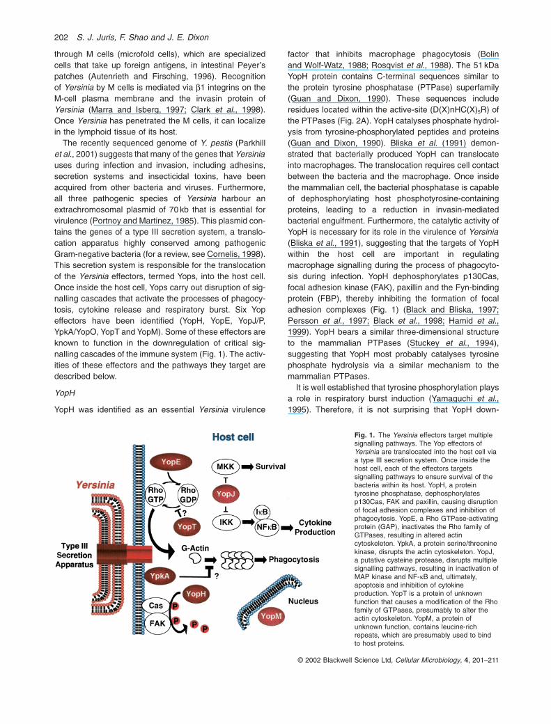
Fig. 1. The Yersinia effectors target multiplesignalling pathways. The Yop effectors ofYersinia are translocated into the host cell viaa type III secretion system. Once inside thehost cell, each of the effectors targetssignalling pathways to ensure survival of thebacteria within its host. YopH, a proteintyrosine phosphatase, dephosphorylatesp130Cas, FAK and paxillin, causing disruptionof focal adhesion complexes and inhibition ofphagocytosis. YopE, a Rho GTPase-activatingprotein (GAP), inactivates the Rho family ofGTPases, resulting in altered actincytoskeleton. YpkA, a protein serine/threoninekinase, disrupts the actin cytoskeleton. YopJ,a putative cysteine protease, disrupts multiplesignalling pathways, resulting in inactivation ofMAP kinase and NF-kB and, ultimately,apoptosis and inhibition of cytokineproduction. YopT is a protein of unknownfunction that causes a modification of the Rhofamily of GTPases, presumably to alter theactin cytoskeleton. YopM, a protein ofunknown function, contains leucine-richrepeats, which are presumably used to bindto host proteins.

Yops target mammalian signalling pathways 203
regulates signalling cascades required for the activationof Fc-mediated respiratory burst in macrophages (Bliskaand Black, 1995) and neutrophils (Ruckdeschel et al.,1996). This inhibition of respiratory burst induction by Yersinia requires the tyrosine phosphatase activity of YopH (Bliska and Black, 1995). However, Zymosan-induced oxidative burst inhibition by Y. enterocolitica in
murine bone marrow-derived macrophages was notdependent on the catalytic activity of YopH (Green et al.,1995). These results suggest a redundancy in Yop func-tion in the inhibition of zymosan-induced oxidative burst.The proteins that YopH dephosphorylates to inhibit induc-tion of the Fc-mediated respiratory burst have not been identified.
© 2002 Blackwell Science Ltd, Cellular Microbiology, 4, 201–211
Fig. 2. Sequence alignments and structural diagrams of the Yop effectors and their homologues.A. YopH harbours a protein tyrosine phosphatase (PTPase) domain at the carboxy-terminus, which is represented by the presence of thecatalytic D(X)28HC(X)5R motif.B. The GAP domains of YopE, SptP and ExoS show significant similarity to each other. However, this domain differs from any of the knowneukaryotic GAP domains, except for the presence of the mammalian Rho GAP consensus motif GxxRxSG (in red). The arginine plays acritical catalytic role in GTP hydrolysis activity.C. Sequence comparison of YopJ, adenovirus protease (AVP) and Ulp1, a yeast ubiquitin-like protein protease. YopJ shows significantsimilarity to AVP within the protease domain and shares the catalytic triad including His (in yellow), Asp/Glu (in green) and Cys (in red) forprotease activity and an invariant Gln (in blue) located in the oxyanion hole of both AVP and Ulp1.D. Sequence alignment of the kinase domain VI, VII and VII between YpkA and canonical Ser/Thr kinase (PKC1, CKIIa and Akt1) and Tyrkinase (EGFR, c-ABL and Ack). Invariant residues essential for kinase activity, including the catalytic Asp and Asn, Asp-Phe-Gly chelating Mg2+
and stabilization loop Ala-Pro-Glu, are highlighted in yellow. The lysine (in blue) within the catalytic loop renders YpkA to be a Ser/Thr kinase.The lysine (blue) or arginine (green) is believed to play a role in stabilization of the transition-state intermediate during catalysis.E. Representation of the leucine-rich repeat (LRR)-containing type III secreted virulence factors from Yersinia (YopM), Shigella (IpaH family),Salmonella (SspHs and Slrp) and symbiotic Rhizobium (Y4FR) and Bradyrhizobium (ID431). LRR is represented by the blue box. Thenumbers after the diagram indicate the number of LRRs present in each gene or gene family.F. YopT exhibits sequence similarity to the C-terminus of surface antigen (p76) from Haemophilus somnus, filamentous haemagglutinin-likeprotein Lsp2 from Haemophilus ducreyi and PfhB2 from Pasteurella multocida. Several conserved residues that may suggest catalytic activityare highlighted in blue.
YopH has recently been implicated in inhibiting theadaptive immune response. The phosphatase activity ofYopH was shown to be essential for suppressing both T-cell cytokine production and expression of the B-cell co-stimulatory receptor B7.2 (Yao et al., 1999). Thisdownregulation of T-cell and B-cell signalling was mostprobably caused by dephosphorylation of tyrosineresidues on proteins in both the T-cell receptor and the B-cell receptor complexes (Yao et al., 1999). These datasuggest that Yersinia may function to inhibit both innateand adaptive immunity, although there is no evidence forinhibition of the adaptive immune response by Yersinia invivo.
It should also be noted that other pathogenic bacteriaproduce a protein tyrosine phosphatase effector. Salmonella spp. produce the protein SptP, which containsa YopE-like domain (discussed below) fused to a YopH-likedomain (Kaniga et al., 1996). However, the substrates ofthe phosphatase domain of SptP have not been identified.
YopE
The 23 kDa YopE was one of the earliest identified Yop effectors (Forsberg and Wolf-Watz, 1988) and is anessential virulence determinant. Targeting YopE into bothepithelial cells and macrophages leads to a cytotoxicresponse, in which cells round up and contract away fromthe extracellular matrix (Rosqvist et al., 1990). Microin-jection studies show that this effect results from disrup-tion of the actin microfilament network (Rosqvist et al.,1991a). Mutation of YopE impairs the ability of Yersiniato resist phagocytosis (Rosqvist et al., 1990; Ruckdeschelet al., 1996), suggesting that YopE contributes to theantiphagocytic capability of Yersinia.
YopE contains a carboxyl-terminal effector domain(Sory et al., 1995; Schesser et al., 1996) that shares ahigh degree of similarity to the amino-terminal domain ofexoenzyme S (ExoS) of Pseudomonas aeruginosa andSptP from Salmonella typhimurium (Fig. 2B). Fu andGalan (1999) noted that this domain harbours a con-served ‘arginine finger’ used by mammalian Rho GTPase-activating proteins (GAP) for catalysis (Fig. 2B) and alsodemonstrated that the amino-terminal half of SptP hasGAP activity towards Rac and Cdc42 in vitro. Recently,YopE and ExoS were also demonstrated to possess GAP activity towards Rho family GTPases (RhoA, Rac and Cdc42) (Fig. 1) (Goehring et al., 1999; Black andBliska, 2000; Von Pawel-Rammingen et al., 2000). Activation of Rho GTP-binding proteins is a pivotal signalling event connecting receptor activation tocytoskeletal reorganization during phagocytosis in pro-fessional and non-professional phagocytic cells (Chiminiand Chavrier, 2000; May and Machesky, 2001). The
mechanism of inhibition of phagocytosis by YopE resultsfrom its Rho GAP activity, as substitution of the essentialarginine with alanine destroys the ability of YopE to inter-fere with phagocytosis in cultured HeLa cells (Black andBliska, 2000). In addition, Yersinia strains harbouring onlythe mutant version (R144A) of YopE are avirulent in amouse infection model (Black and Bliska, 2000). Thecrystal structures of the GAP domain of ExoS and SptP(Stebbins and Galan, 2000; Wurtele et al., 2001) revealthat this effector domain mimics the mammalian Rho GAPdomain by providing a similar interface to contact RhoGTPases and positioning the catalytic arginine in a posi-tion that allows for stabilization of the transitionstateintermediate. This structure bears no similarity to thestructure of the Rho GAPs (Stebbins and Galan, 2000),suggesting that the bacterial and mammalian GAP activ-ities arose independently.
Unlike SptP, YopE exhibits no preference towardsRhoA, Rac and Cdc42 as its substrate in vitro (Black and Bliska, 2000). It will be interesting to unravel whichRho GTPase is preferred under physiological conditions.RhoA and Rac are the two most probable candidates documented in the literature. RhoA is the small GTPaseswitch controlling stress fibre formation in fibroblasts aswell as in macrophages (Hall, 1998). The morphology ofactin stress fibre disruption induced by YopE implies thatRhoA might be the target of YopE in vivo (Rosqvist et al.,1991b). This is substantiated by the observation that over-expression of RhoAV14, an activated form of RhoA, prevents the cytotoxic phenotype induced by YopE in cultured HeLa cells (Black and Bliska, 2000). In addition,overexpression of molecules involved in the yeast Rho1pathway, the RhoA homologue in yeast, is able to coun-teract the growth inhibition phenotype in yeast caused byYopE (Von Pawel-Rammingen et al., 2000). However, thishypothesis was challenged by recent morphology studiesin cultured endothelial cells (Andor et al., 2001). Mem-brane ruffling triggered by Cdc42-dependent Rac activa-tion is abrogated by YopE translocation, suggesting thatYopE selectively targets Rac (Andor et al., 2001). Thisresult is consistent with the observation that overexpres-sion of Rac1V12, an active form of Rac1, interferes withthe antiphagocytic activity of YopE in cultured HeLa cells(Black and Bliska, 2000). Taken together, one can spec-ulate that YopE might target both RhoA and Rac for dif-ferent purposes. More information about the substrate ofYopE in vivo awaits further analysis, including the high-resolution crystal structure of YopE and the precise cellu-lar localization of YopE.
Recent studies have demonstrated that selective acti-vation of RhoA, Rac and Cdc42 is required in differentphagocytosis processes (Chimini and Chavrier, 2000;May and Machesky, 2001), suggesting that the action of
© 2002 Blackwell Science Ltd, Cellular Microbiology, 4, 201–211
204 S. J. Juris, F. Shao and J. E. Dixon

Yops target mammalian signalling pathways 205
YopE would allow Yersinia to inhibit these multipleprocesses. Indeed, Y. pseudotuberculosis and Y. entero-colitica are able to inhibit b1 integrin-mediated uptake in cultured epithelial cells (Black and Bliska, 2000). Y.pseudotuberculosis is capable of inhibiting Fc receptor-mediated phagocytosis in professional phagocytic cells(Fallman et al., 1995). Given that Yersinia-infected HeLacells lose the ability to phagocytose not only Yersiniaitself, but also other microorganisms (Mecsas et al.,1998), Yersinia might also antagonize complement recep-tor (b2 integrin)-mediated phagocytosis. Studies on themechanism used by different phagocytosis pathways andidentification of physiological targets of YopE will aid in theunderstanding of not only which GTPases are involved indistinct phagocytosis events, but also how YopE acts toinhibit these processes.
As stated earlier, Yersinia encode for a type III secre-tion apparatus that is essential for the translocation of the Yops into the host cell (Cornelis, 1998). The structure of this apparatus from Shigella and Salmonella resemblesa needle, which probably functions to allow transportacross the inner and outer membranes of the bacteria and into the cytoplasm of the host cell (Blocker et al.,1999; Kubori et al., 2000; Tamano et al., 2000). One model suggests that the Yops function in part toprevent a pore from forming in the host cell plasma membrane resulting from insertion of the apparatus.Indeed, mutant strains of Yersinia defective for multipleYop effectors contain a higher activity of pore formationcaused by the type III secretion apparatus than Yersiniawild-type strains (Hakansson et al., 1996a; Neyt and Cornelis, 1999; Tardy et al., 1999), suggesting that one ormore of the Yop effectors prevents pore formation andlysis of the host cell. The GAP activity of YopE is essen-tial in the prevention of pore formation (Viboud and Bliska,2001), suggesting that pore formation requires activatedRho GTPases and actin polymerization. These data alsosuggest that the prevention of pore formation by YopE isnot simply the occupation of YopE within the pore, butrather involves the downregulation of the Rho GTPaseswithin the host cell.
YopJ
The 32.5 kDa YopJ/YopP was first identified in Y. pestis(YopJ) (Straley and Bowmer, 1986) and later in Y.pseudotuberculosis and Y. enterocolitica (YopP) (Corneliset al., 1987). YopJ was first shown to be dispensable forthe virulence of Y. pseudotuberculosis (Galyov et al.,1994). However, it was later demonstrated that the apop-tosis induced by YopJ aids in the establishment of a sys-temic infection in vivo (Monack et al., 1998). So far, YopJis the only Yop effector that has been shown to have an
anti-inflammatory role and to be responsible for the induc-tion of apoptosis in macrophages.
After the transient induction of multiple signalling path-ways (MAPK and NF-kB pathway) by lipopolysaccharide(LPS), Yersinia infection results in a severe inhibition ofmultiple MAPK signalling pathways (Erk, JNK and p38) inmacrophages (Ruckdeschel et al., 1997a). Translocationof YopJ through the type III secretion machinery isrequired for this inhibition (Boland and Cornelis, 1998;Palmer et al., 1998). In addition, YopJ alone is sufficientto cause downregulation of multiple MAPK kinases(Fig. 1) (Palmer et al., 1999). YopJ can also block NF-kBactivation through the inhibition of IkB degradation andsubsequent NF-kB translocation in both macrophagesand epithelial cells (Ruckdeschel et al., 1998; 2001;Schesser et al., 1998). It was difficult to understand howone protein could simultaneously block a multiplicity ofsignalling pathways that have no apparent common com-ponents. Orth et al. (1999) made the key observation thatYopJ can bind to multiple members of the MAPK kinasesuperfamily, including MKKs and IKKb, and suppress theiractivation. The interaction between YopJ and IKKbwas also confirmed by co-immunoprecipitation frommacrophages (Denecker et al., 2001; Ruckdeschel et al.,2001).
Yersinia infection causes macrophage apoptosis(Ruckdeschel et al., 1997b) as well as the inhibition of cytokine production and an inflammatory response(Nakajima and Brubaker, 1993; Beuscher et al., 1995;Schulte et al., 1996). These processes allow the pathogento survive and multiply extracellularly in the host lymphoidtissue. Studies on the function of YopJ have offeredinsights into the mechanism underlying the inhibition ofcytokine production. YopJ was shown to be necessary forthe inhibition of TNFa and IL-8 production (Boland andCornelis, 1998; Palmer et al., 1998; Schesser et al.,1998). Inactivation of multiple MAPK pathways and theNF-kB pathway parallels the decrease in TNFa produc-tion (Ruckdeschel et al., 1997a). Considering that theTNFa and IL-8 promoters harbour NF-kB and AP-1binding sites (Roebuck, 1999; Liu et al., 2000), andMAPKs and NF-kB signals converge to the transcriptionfactors AP-1 and NF-kB, inhibition of cytokine productionis probably the result of disruption of MAPK and NF-kBactivation.
YopJ is the only Yop effector required for the inductionof apoptosis in murine macrophages (Mills et al., 1997;Monack et al., 1997). However, induction of apoptosisseems to be independent of the role of YopJ in perturb-ing signalling pathways, as YopJ does not induce apop-tosis in fibroblasts even though it disrupts the samesignalling pathways within fibroblasts. Recent studiessuggest that the apoptotic signalling upstream of Bid
© 2002 Blackwell Science Ltd, Cellular Microbiology, 4, 201–211
cleavage and cytochrome c release, presumably throughcaspase 8 activation, is responsible for the YopJ-inducedapoptosis in macrophages (Denecker et al., 2001).Macrophage apoptosis caused by YopJ most probablyresults from inactivation of NF-kB, as inhibition of NF-kBleads the macrophage to undergo apoptosis (Pagliariet al., 2000).
Recent reports from Orth et al. (2000) revealed thatYopJ may function as a cysteine protease. The observa-tion arose from the finding that the predicted secondarystructure of YopJ was similar to the observed secondarystructure determined from the X-ray structure of the ade-novirus protease. Furthermore, critical catalytic residuesof protease activity were conserved in both YopJ and theadenovirus protease (Fig. 2C). Mutation of the predictedcatalytic residues required for cysteine protease activityabolished the ability of YopJ to disrupt MAPK signallingpathways (Orth et al., 2000). This hypothesis was con-firmed further by the inability of mutants in the predictedcatalytic residues of AvrBst, a predicted YopJ homologuein the plant pathogen Xanthomonas campestris, to inducea plant hypersensitive response (Orth et al., 2000). Theseauthors also suggest that YopJ might be a ubiquitin-like protein protease based on the sequence similaritybetween YopJ and Ulp1 (Fig. 2C), a yeast ubiquitin-likeprotein protease (Li and Hochstrasser, 1999). These data,taken together with the initial observations that YopJ canblock MAPK activation via phosphorylation (Orth et al.,1999), suggest that ubiquitin-like protein modificationplays an unidentified regulatory role in MAPK signalling.A growing family of ubiquitin-like proteins has recentlybeen shown to be involved in modulating eukaryotic signalling pathways (reviewed by Yeh et al., 2000). The suggestion that YopJ inhibits signalling by theremoval of ubiquitin-like proteins casts new light on deciphering the mechanism of Yersinia pathogenesis as well as eukaryotic signalling pathways. Importantly,however, demonstration of in vitro protease activity byYopJ and identification of the substrate(s) of YopJ areneeded.
YopJ shows similarity to other type III secreted effec-tors including AvrA from Salmonella, AvrRxv, AvrBst, XopJand AvrXv4 from X. campestris, AvrPpiG1and ORF5 fromPseudomonas syringae, ORFB from Erwinia amylovoraand y4lo from Rhizobium. Thus, YopJ may function by acommon catalytic mechanism across both animal andplant kingdoms (Orth et al., 2000). This suggests that thesignalling pathways disrupted by YopJ family effectors areconserved in both plants and animals. Indeed, both YopJand AvrBst can induce apoptosis in macrophages andplants respectively (Mills et al., 1997; Monack et al., 1997;Orth et al., 2000). However, it has been demonstrated thatAvrA from Salmonella does not inhibit the same pathwaysas YopJ (Schesser et al., 2000), suggesting that some of
the YopJ homologues may function differently mechanis-tically. However, the similarity among the family doessuggest a common catalytic activity.
YpkA
The Yersinia protein kinase YpkA was first identified as asecreted protein that is capable of autophosphorylation onserine residues (Galyov et al., 1993). YpkA contains adomain bearing similarity to the eukaryotic serine/threo-nine protein kinases (Fig. 2D) (Galyov et al., 1993). Thisstudy also demonstrated that YpkA is an essential viru-lence determinant for Yersinia infection, as disruption ofthe YpkA gene in Yersinia results in an avirulent strain ofbacteria in an animal model. This was the first identifica-tion of a bacterial protein kinase playing a direct role invirulence.
Once inside the host cell, YpkA causes a change inmorphology distinct from those mediated by YopE, in thatthe cells containing YpkA round up but do not detach fromthe extracellular matrix (Hakansson et al., 1996b). Fur-thermore, YpkA is translocated to the plasma membrane(Fig. 1) (Hakansson et al., 1996b), suggesting that thesubstrates for YpkA may be localized at the plasma mem-brane as well. Interestingly, YpkA produced by bacteriaexhibits no detectable kinase activity unless a eukaryotickinase activator is present. Purification of this eukaryoticactivator led to its identification as actin (Juris et al.,2000). The C-terminus of YpkA, which is predicted to forman amphipathic helix, is required for YpkA kinase activityand for the interaction between YpkA and actin (Juriset al., 2000). Interaction between YpkA and actin sug-gested that the morphological changes induced by YpkAmight be caused by changes in the actin cytoskeleton.Indeed, YpkA induces disruption of actin stress fibres inHeLa cells in transient transfection experiments (Fig. 1)(Juris et al., 2000). Disruption of the actin cytoskeleton byYpkA might be mediated by its ability to interact with the small GTPase RhoA, which plays an important role in actin dynamics within the cell (Barz et al., 2000;Dukuzumuremyi et al., 2000). Although actin can bephosphorylated in vitro (Juris et al., 2000), the in vivo sub-strate is not clear. Phosphorylation of actin by YpkA is notlikely to be physiologically significant because of theabundance of actin in a cell. YpkA most probably func-tions by phosphorylating proteins that are important in theregulation of the actin cytoskeleton within the host cell.The substrates of YpkA probably have an important rolein RhoA signalling. Through its ability to disrupt the actincytoskeleton, YpkA may have an effect on the ability of amacrophage to phagocytose Yersinia and to move toareas of infection.
It is intriguing that several pathogenic species of bacteria encode proteins with domains that resemble
© 2002 Blackwell Science Ltd, Cellular Microbiology, 4, 201–211
206 S. J. Juris, F. Shao and J. E. Dixon
Yops target mammalian signalling pathways 207
eukaryotic-like serine/threonine protein kinase domains(Leonard et al., 1998; Avenue-Gay and Everett, 2000).These proteins control development and stress re-sponses within the bacteria. Only YpkA and a proteinkinase from Pseudomonas aeruginosa have been shown to be essential virulence determinants (Galyovet al., 1993; Wang et al., 1998). Yersinia, and other path-ogenic bacteria may use these eukaryotic-like proteinkinases to tip the balance of signalling cascades withintheir hosts by phosphorylating endogenous substratesthat regulate immune responses to levels that completelyshift the state of those signalling cascades in favour ofbacterial survival.
YopM
The Yersinia protein YopM is a 41 kDa protein that is anecessary virulence determinant of Y. pestis (Leung et al.,1990). YopM is composed almost entirely of sequencesresembling leucine-rich repeats (LRRs). The LRR domainis found in a diverse number of proteins, including the RNase inhibitor, the Toll receptors, proteoglycans and platelet glycoproteins (for a review, see Kobe andDeisenhofer, 1994). This domain is believed to be aprotein–protein interaction motif functional in a multitudeof signalling pathways both within the cell and in the extra-cellular milieu.
The diversity of signalling pathways governed by LRR-containing proteins has made it difficult to ascertain theexact pathways that YopM may affect. Initial characteri-zation of YopM demonstrated its ability to interact with thrombin and inhibit thrombin-induced platelet aggre-gation (Reisner and Straley, 1992), suggesting that YopM may actually function in the extracellular environ-ment of the host. However, YopM has also been shown to be translocated into macrophages via the typeIII secretion apparatus (Boland et al., 1996), making the exact location of YopM during Yersinia infection controversial. Recent studies have shown that YopM isnot only translocated into HeLa cells during Yersinia infec-tion, but also localizes to the nucleus of HeLa cells, traf-ficking via a vesicle-associated pathway (Skrzypek et al.,1998).
Other LRR-containing proteins have been identified inboth pathogenic and symbiotic bacteria. These includethe IpaH family from Shigella flexneri, SspH 1, SspH 2 and Slrp from Salmonella typhimurium, y4fr from Rhizobium and ID431 from Bradyrhizobium (Fig. 2E). Thefunctions of all these LRR-containing proteins are un-known. These proteins may have been acquired by bothpathogenic and symbiotic bacteria to thwart similar pathways within the host. The structure of YopM revealsa parallel b-sheet on its concave face, which is typical ofthe solved structures of other LRR-containing proteins
(Evdokimov et al., 2001). However, in other LRR-containing proteins, such as the RNase inhibitor (Kobeand Deisenhofer, 1993) and internalin B (Marino et al.,1999), the convex surface is typically helical in structure,whereas on YopM, this face contains an extended struc-ture (Evdokimov et al., 2001). This may be caused by theshort length of the LRR of YopM. Furthermore, the YopMstructure suggests that it forms a tetramer, which createsa channel with a pore of 35 Å (Evdokimov et al., 2001).
YopT
The function of YopT is largely unknown; however,translocation of YopT into host cells results in cell round-ing and disruption of cytoskeleton structure, a cytotoxicresponse resembling that of YopE (Iriarte and Cornelis,1998; Zumbihl et al., 1999; Boyd et al., 2000). YopT issecreted in lower amounts than YopE, although botheffectors appear to function by altering the host cytoskele-ton structures (Iriarte and Cornelis, 1998). Why doesYersinia have two cytotoxins (YopT and YopE) to abrogatecytoskeleton structures? The answer to this question isunknown, although there are several possibilities. UnlikeYopE mutants, YopT mutants colonize intestinal Peyer’spatches as in wild type (Iriarte and Cornelis, 1998), sug-gesting that YopT and YopE may have different roles inaltering cytoskeleton structure during the course of aYersinia infection. Similarly, in in vitro experiments, YopEmutant strains of Y. pseudotuberculosis exhibit a signifi-cant decrease in cytotoxicity (Rosqvist et al., 1991b),whereas YopT mutant strains of Y. enterocolitica are ascytotoxic as the wild-type strains (Hartland et al., 1994;Iriarte and Cornelis, 1998). Thus, YopT and YopE mayaffect different signalling pathways to synergize the cyto-toxic effect and secure the survival of the pathogen in lymphoid tissues. Recently, Zumbihl et al. (1999) demon-strated that infection of a YopT strain causes an uniden-tified modification of RhoA (Fig. 1). The GTPase showsan acidic shift in its pI after infection with YopT. Modifica-tion of RhoA by YopT may inhibit its ability to alter thecytoskeleton during phagocytosis, allowing for the sur-vival of Yersinia.
Sequence comparisons show that YopT is similar to theC-terminus of surface antigen p76 from Haemophilussomnus and a small portion of the filamentous haemagglutinin-like protein from Haemophilus ducreyicalled Lsp1 and Lsp2 as well as PfhB1 and PfhB2 fromPasteurella multocida (Fig. 2F). The function of these proteins is unknown. The several conserved residuesamong these proteins suggest a similar catalytic activity.However, none of the proteins similar to YopT has beenshown to modify RhoA. Further studies on the function ofthe YopT-like domain in these proteins will also provideadditional insights into YopT pathogenesis.
© 2002 Blackwell Science Ltd, Cellular Microbiology, 4, 201–211
Concluding remarks
The Yersinia species of bacteria contain six characterizedYop effectors to disrupt vital signalling cascades that arerequired for innate immunity. Studies on the functions ofthese effectors have revealed the signalling pathwaysthey target and have also shed light on novel signallingpathways used by the host to induce an immuneresponse. Yersinia has probably acquired some (or all) ofits effectors via horizontal gene transfer between bacteriaand fine tuned these effectors to regulate signalling path-ways within its host. Those signalling pathways targetedby the effectors play crucial roles in the elimination ofpathogens. Identification of the functions of new effectors,as well as the targets or substrates of these effectors, willaid in the understanding of bacterial pathogenesis as wellas innate immunity.
Acknowledgements
We thank Matthew Wishart and Victor DiRita for their helpful sug-gestions and critical evaluation of this manuscript. This work was funded by a grant from the National Institutes of Health(J.E.D. and F.S.), an NIH Molecular Mechanisms of MicrobialPathogenesis predoctoral training grant (S.J.J.) and the WaltherCancer Institute (J.E.D.).
References
Allen, L.A., and Aderem, A. (1996) Molecular definition of dis-tinct cytoskeletal structures involved in complement- and Fcreceptor-mediated phagocytosis in macrophages. J Exp Med184: 627–637.
Andor, A., Trulzsch, K., Essler, M., Roggenkamp, A., Wiedemann,A., Heesemann, J., and Aepfelbacher, M. (2001) YopE ofYersinia, a GAP for Rho GTPases, selectively modulates Rac-dependent actin structures in endothelial cells. Cell Microbiol 3: 301–310.
Aspenstrom, P. (1999a) The Rho GTPases have multiple effectson the actin cytoskeleton. Exp Cell Res 246: 20–25.
Aspenstrom, P. (1999b) Effectors for the Rho GTPases. CurrOpin Cell Biol 11: 95–102.
Autenrieth, I.B., and Firsching, R. (1996) Penetration of M cellsand destruction of Peyer’s patches by Yersinia enterocolitica:an ultrastructural and histological study. J Med Microbiol 44:285–294.
Av-Gay, Y., and Everett, M. (2000) The eukaryotic-like Ser/Thr protein kinases of Mycobacterium tuberculosis. TrendsMicrobiol 8: 238–244.
Barz, C., Abahji, T.N., Trulzsch, K., and Heesemann, J. (2000)The Yersinia Ser/Thr protein kinase YpkA/YopO directly inter-acts with the small GTPases RhoA and Rac-1. FEBS Lett 482:139–143.
Beuscher, H.U., Rodel, F., Forsberg, A., and Rollinghoff, M.(1995) Bacterial evasion of host immune defense: Yersiniaenterocolitica encodes a suppressor for tumor necrosis factoralpha expression. Infect Immun 63: 1270–1277.
Black, D.S., and Bliska, J.B. (1997) Identification of p130Cas as a substrate of Yersinia YopH (Yop51), a bacterial protein
tyrosine phosphatase that translocates into mammalian cellsand targets focal adhesions. EMBO J 16: 2730–2744.
Black, D.S., and Bliska, J.B. (2000) The RhoGAP activity of the Yersinia pseudotuberculosis cytotoxin YopE is required for antiphagocytic function and virulence. Mol Microbiol 37:515–527.
Black, D.S., Montagna, L.G., Zitsmann, S., and Bliska, J.B.(1998) Identification of an amino-terminal substrate-bindingdomain in the Yersinia tyrosine phosphatase that is requiredfor efficient recognition of focal adhesion targets. Mol Microbiol 29: 1263–1274.
Bliska, J.B., and Black, D.S. (1995) Inhibition of the Fc receptor-mediated oxidative burst in macrophages by the Yersiniapseudotuberculosis tyrosine phosphatase. Infect Immun 63:681–685.
Bliska, J.B., Guan, K.L., Dixon, J.E., and Falkow, S. (1991) Tyro-sine phosphate hydrolysis of host proteins by an essentialYersinia virulence determinant. Proc Natl Acad Sci USA 88:1187–1191.
Blocker, A., Gounon, P., Larquet, E., Niebuhr, K., Cabiaux, V.,Parsot, C., and Sansonetti, P. (1999) The tripartite type IIIsecretion of Shigella flexneri inserts IpaB and IpaC into hostmembranes. J Cell Biol 147: 683–693.
Boland, A., and Cornelis, G.R. (1998) Role of YopP in suppres-sion of tumor necrosis factor alpha release by macrophagesduring Yersinia infection. Infect Immun 66: 1878–1884.
Boland, A., Sory, M.P., Iriarte, M., Kerbourch, C., Wattiau, P., andCornelis, G.R. (1996) Status of YopM and YopN in the YersiniaYop virulon: YopM of Y. enterocolitica is internalized inside thecytosol of PU5-1.8 macrophages by the YopB, D, N deliveryapparatus. EMBO J 15: 5191–5201.
Bolin, I., and Wolf-Watz, H. (1988) The plasmid-encoded Yop2bprotein of Yersinia pseudotuberculosis is a virulence determi-nant regulated by calcium and temperature at the level of tran-scription. Mol Microbiol 2: 237–245.
Boyd, A.P., Grosdent, N., Totemeyer, S., Geuijen, C., Bleves, S.,Iriarte, M., et al. (2000) Yersinia enterocolitica can deliver Yopproteins into a wide range of cell types: development of a deliv-ery system for heterologous proteins. Eur J Cell Biol 79:659–671.
Chimini, G., and Chavrier, P. (2000) Function of Rho family pro-teins in actin dynamics during phagocytosis and engulfment.Nature Cell Biol 2: E191–E196.
Clark, M.A., Hirst, B.H., and Jepson, M.A. (1998) M-cell surfacebeta1 integrin expression and invasin-mediated targeting ofYersinia pseudotuberculosis to mouse Peyer’s patch M cells.Infect Immun 66: 1237–1243.
Cornelis, G.R. (1998) The Yersinia Yop virulon, a bacterialsystem to subvert cells of the primary host defense. FoliaMicrobiol (Praha) 43: 253–261.
Cornelis, G.R., Vanooteghem, J.C., and Sluiters, C. (1987) Transcription of the yop regulon from Y. enterocolitica requirestrans acting pYV and chromosomal genes. Microb Pathog 2:367–379.
Denecker, G., Declercq, W., Geuijen, C.A., Boland, A., Benabdillah, R., van Gurp, M., et al. (2001) Yersinia entero-colitica YopP-induced apoptosis of macrophages involves theapoptotic signaling cascade upstream of bid. J Biol Chem 276:19706–19714.
Dukuzumuremyi, J.M., Rosqvist, R., Hallberg, B., Akerstrom, B.,Wolf-Watz, H., and Schesser, K. (2000) The Yersinia proteinkinase A is a host factor inducible RhoA/Rac-binding virulencefactor. J Biol Chem 275: 35281–35290.
Evdokimov, A.G., Anderson, D.E., Routzahn, K.M., and Waugh,
© 2002 Blackwell Science Ltd, Cellular Microbiology, 4, 201–211
208 S. J. Juris, F. Shao and J. E. Dixon
Yops target mammalian signalling pathways 209
D.S. (2001) Unusual molecular architecture of the Yersiniapestis cytotoxin YopM: a leucine-rich repeat protein with theshortest repeating unit. J Mol Biol 312: 807–821.
Fallman, M., Andersson, K., Hakansson, S., Magnusson, K.E.,Stendahl, O., and Wolf-Watz, H. (1995) Yersinia pseudotu-berculosis inhibits Fc receptor-mediated phagocytosis in J774cells. Infect Immun 63: 3117–3124.
Forsberg, A., and Wolf-Watz, H. (1988) The virulence proteinYop5 of Yersinia pseudotuberculosis is regulated at transcrip-tional level by plasmid-plB1-encoded trans-acting elementscontrolled by temperature and calcium. Mol Microbiol 2:121–133.
Fu, Y., and Galan, J.E. (1999) A salmonella protein antagonizesRac-1 and Cdc42 to mediate host-cell recovery after bacterialinvasion (see comments). Nature 401: 293–297.
Galyov, E.E., Hakansson, S., Forsberg, A., and Wolf-Watz, H.(1993) A secreted protein kinase of Yersinia pseudotuberculo-sis is an indispensable virulence determinant. Nature 361:730–732.
Galyov, E.E., Hakansson, S., and Wolf-Watz, H. (1994) Charac-terization of the operon encoding the YpkA Ser/Thr proteinkinase and the YopJ protein of Yersinia pseudotuberculosis. JBacteriol 176: 4543–4548.
Goehring, U.M., Schmidt, G., Pederson, K.J., Aktories, K., andBarbieri, J.T. (1999) The N-terminal domain of Pseudomonasaeruginosa exoenzyme S is a GTPase-activating protein forRho GTPases. J Biol Chem 274: 36369–36372.
Green, S.P., Hartland, E.L., Robins-Browne, R.M., and Phillips,W.A. (1995) Role of YopH in the suppression of tyrosine phosphorylation and respiratory burst activity in murinemacrophages infected with Yersinia enterocolitica. J LeukocBiol 57: 972–977.
Greenberg, S., Burridge, K., and Silverstein, S.C. (1990) Colo-calization of F-actin and talin during Fc receptor-mediatedphagocytosis in mouse macrophages. J Exp Med 172:1853–1856.
Greenberg, S., Chang, P., and Silverstein, S.C. (1993) Tyrosinephosphorylation is required for Fc receptor-mediated phagocytosis in mouse macrophages. J Exp Med 177: 529–534.
Guan, K.L., and Dixon, J.E. (1990) Protein tyrosine phosphataseactivity of an essential virulence determinant in Yersinia.Science 249: 553–556.
Hakansson, S., Schesser, K., Persson, C., Galyov, E.E.,Rosqvist, R., Homble, F., and Wolf-Watz, H. (1996a) The YopBprotein of Yersinia pseudotuberculosis is essential for thetranslocation of Yop effector proteins across the target cellplasma membrane and displays a contact-dependent mem-brane disrupting activity. EMBO J 15: 5812–5823.
Hakansson, S., Galyov, E.E., Rosqvist, R., and Wolf-Watz, H.(1996b) The Yersinia YpkA Ser/Thr kinase is translocated andsubsequently targeted to the inner surface of the HeLa cellplasma membrane. Mol Microbiol 20: 593–603.
Hall, A. (1998) Rho GTPases and the actin cytoskeleton. Science279: 509–514.
Hamid, N., Gustavsson, A., Andersson, K., McGee, K., Persson,C., Rudd, C.E., and Fallman, M. (1999) YopH dephosphory-lates Cas and Fyn-binding protein in macrophages. MicrobPathog 27: 231–242.
Hartland, E.L., Green, S.P., Phillips, W.A., and Robins-Browne,R.M. (1994) Essential role of YopD in inhibition of the respira-tory burst of macrophages by Yersinia enterocolitica. InfectImmun 62: 4445–4453.
Iriarte, M., and Cornelis, G.R. (1998) YopT, a new Yersinia Yop
effector protein, affects the cytoskeleton of host cells. MolMicrobiol 29: 915–929.
Juris, S.J., Rudolph, A.E., Huddler, D., Orth, K., and Dixon, J.E.(2000) A distinctive role for the Yersinia protein kinase: actinbinding, kinase activation, and cytoskeleton disruption. ProcNatl Acad Sci USA 97: 9431–9436.
Kaniga, K., Uralil, J., Bliska, J.B., and Galan, J.E. (1996) Asecreted protein tyrosine phosphatase with modular effectordomains in the bacterial pathogen Salmonella typhimurium.Mol Microbiol 21: 633–641.
Karin, M. (1999) The beginning of the end: IkappaB kinase (IKK) and NF-kappaB activation. J Biol Chem 274: 27339–27342.
Kobe, B., and Deisenhofer, J. (1993) Crystal structure of porcineribonuclease inhibitor, a protein with leucine-rich repeats.Nature 366: 751–756.
Kobe, B., and Deisenhofer, J. (1994) The leucine-rich repeat: aversatile binding motif. Trends Biochem Sci 19: 415–421.
Kubori, T., Sukhan, A., Aizawa, S.I., and Galan, J.E. (2000) Mol-ecular characterization and assembly of the needle complex ofthe Salmonella typhimurium type III protein secretion system.Proc Natl Acad Sci USA 97: 10225–10230.
Leonard, C.J., Aravind, L., and Koonin, E.V. (1998) Novel fami-lies of putative protein kinases in bacteria and archaea: evo-lution of the ‘eukaryotic’ protein kinase superfamily. GenomeRes 8: 1038–1047.
Leung, K.Y., Reisner, B.S., and Straley, S.C. (1990) YopM inhibitsplatelet aggregation and is necessary for virulence of Yersiniapestis in mice. Infect Immun 58: 3262–3271.
Li, S.J., and Hochstrasser, M. (1999) A new protease required forcell-cycle progression in yeast. Nature 398: 246–251.
Liu, H., Sidiropoulos, P., Song, G., Pagliari, L.J., Birrer, M.J.,Stein, B., et al. (2000) TNF-alpha gene expression inmacrophages: regulation by NF-kappa B is independent of c-Jun or C/EBP beta. J Immunol 164: 4277–4285.
Marino, M., Braun, L., Cossart, P., and Ghosh, P. (1999) Struc-ture of the lnlB leucine-rich repeats, a domain that triggers hostcell invasion by the bacterial pathogen L. monocytogenes. MolCell 4: 1063–1072.
Marra, A., and Isberg, R.R. (1997) Invasin-dependent andinvasin-independent pathways for translocation of Yersiniapseudotuberculosis across the Peyer’s patch intestinal epithe-lium. Infect Immun 65: 3412–3421.
May, R.C., and Machesky, L.M. (2001) Phagocytosis and theactin cytoskeleton. J Cell Sci 114: 1061–1077.
Mecsas, J., Raupach, B., and Falkow, S. (1998) The YersiniaYops inhibit invasion of Listeria, Shigella and Edwardsiellabut not Salmonella into epithelial cells. Mol Microbiol 28:1269–1281.
Medzhitov, R., and Janeway, C., Jr (2000) Innate immunity. NEngl J Med 343: 338–344.
Mercurio, F., and Manning, A.M. (1999) Multiple signals con-verging on NF-kappaB. Curr Opin Cell Biol 11: 226–232.
Mills, S.D., Boland, A., Sory, M.P., van der Smissen, P., Kerbourch, C., Finlay, B.B., and Cornelis, G.R. (1997) Yersinia enterocolitica induces apoptosis in macrophages by aprocess requiring functional type III secretion and translo-cation mechanisms and involving YopP, presumably acting as an effector protein. Proc Natl Acad Sci USA 94: 12638–12643.
Monack, D.M., Mecsas, J., Ghori, N., and Falkow, S. (1997)Yersinia signals macrophages to undergo apoptosis and YopJis necessary for this cell death. Proc Natl Acad Sci USA 94:10385–10390.
© 2002 Blackwell Science Ltd, Cellular Microbiology, 4, 201–211

Monack, D.M., Mecsas, J., Bouley, D., and Falkow, S. (1998)Yersinia-induced apoptosis in vivo aids in the establishment ofa systemic infection in mice. J Exp Med 188: 2127–2137.
Nakajima, R., and Brubaker, R.R. (1993) Association betweenvirulence of Yersinia pestis and suppression of gamma inter-feron and tumor necrosis factor alpha. Infect Immun 61: 23–31.
Neyt, C., and Cornelis, G.R. (1999) Insertion of a Yop transloca-tion pore into the macrophage plasma membrane by Yersiniaenterocolitica: requirement for translocators YopB and YopD,but not LcrG. Mol Microbiol 33: 971–981.
Orth, K., Palmer, L.E., Bao, Z.Q., Stewart, S., Rudolph, A.E.,Bliska, J.B., and Dixon, J.E. (1999) Inhibition of the mitogen-activated protein kinase kinase superfamily by a Yersinia effec-tor. Science 285: 1920–1923.
Orth, K., Xu, Z., Mudgett, M.B., Bao, Z.Q., Palmer, L.E., Bliska,J.B., et al. (2000) Disruption of signaling by Yersinia effectorYopJ, a ubiquitin-like protein protease (see comments).Science 290: 1594–1597.
Pagliari, L.J., Perlman, H., Liu, H., and Pope, R.M. (2000)Macrophages require constitutive NF-kappaB activation tomaintain A1 expression and mitochondrial homeostasis. MolCell Biol 20: 8855–8865.
Palmer, L.E., Hobbie, S., Galan, J.E., and Bliska, J.B. (1998)YopJ of Yersinia pseudotuberculosis is required for the inhibi-tion of macrophage TNF-alpha production and downregulationof the MAP kinases p38 and JNK. Mol Microbiol 27: 953–965.
Palmer, L.E., Pancetti, A.R., Greenberg, S., and Bliska, J.B.(1999) YopJ of Yersinia spp. is sufficient to cause downregu-lation of multiple mitogen-activated protein kinases in eukary-otic cells. Infect Immun 67: 708–716.
Parkhill, J., Wren, B.W., Thomson, N.R., Titball, R.W., Holden,M.T.G., Prentice, M.B., et al. (2001) Genome sequence ofYersinia pestis, the causative agent of plague. Nature 413:523–527.
Pearson, G., Robinson, F., Beers Gibson, T., Xu, B.E.,Karandikar, M., Berman, K., and Cobb, M.H. (2001) Mitogen-activated protein (MAP) kinase pathways: regulation and physiological functions. Endocr Rev 22: 153–183.
Persson, C., Carballeira, N., Wolf-Watz, H., and Fallman, M.(1997) The PTPase YopH inhibits uptake of Yersinia, tyrosinephosphorylation of p130Cas and FAK, and the associatedaccumulation of these proteins in peripheral focal adhesions.EMBO J 16: 2307–2318.
Portnoy, D.A., and Martinez, R.J. (1985) Role of a plasmid in thepathogenicity of Yersinia species. Curr Topics MicrobiolImmunol 118: 29–51.
Reisner, B.S., and Straley, S.C. (1992) Yersinia pestis YopM:thrombin binding and overexpression. Infect Immun 60:5242–5252.
Roebuck, K.A. (1999) Regulation of interleukin-8 gene expres-sion. J Interferon Cytokine Res 19: 429–438.
Rosqvist, R., Bolin, I., and Wolf-Watz, H. (1988) Inhibition ofphagocytosis in Yersinia pseudotuberculosis: a virulenceplasmid-encoded ability involving the Yop2b protein. InfectImmun 56: 2139–2143.
Rosqvist, R., Forsberg, A., Rimpilainen, M., Bergman, T., andWolf-Watz, H. (1990) The cytotoxic protein YopE of Yersiniaobstructs the primary host defence. Mol Microbiol 4: 657–667.
Rosqvist, R., Forsberg, A., and Wolf-Watz, H. (1991a) Microin-jection of the Yersinia YopE cytotoxin in mammalian cellsinduces actin microfilament disruption. Biochem Soc Trans 19:1131–1132.
Rosqvist, R., Forsberg, A., and Wolf-Watz, H. (1991b) Intracel-lular targeting of the Yersinia YopE cytotoxin in mammalian
cells induces actin microfilament disruption. Infect Immun 59:4562–4569.
Ruckdeschel, K., Roggenkamp, A., Schubert, S., and Heesemann, J. (1996) Differential contribution of Yersiniaenterocolitica virulence factors to evasion of microbicidalaction of neutrophils. Infect Immun 64: 724–733.
Ruckdeschel, K., Machold, J., Roggenkamp, A., Schubert, S.,Pierre, J., Zumbihl, R., et al. (1997a) Yersinia enterocoliticapromotes deactivation of macrophage mitogen-activatedprotein kinases extracellular signal-regulated kinase-1/2, 38,and c-Jun NH2-terminal kinase. Correlation with its inhibitoryeffect on tumor necrosis factor-alpha production. J Biol Chem272: 15920–15927.
Ruckdeschel, K., Roggenkamp, A., Lafont, V., Mangeat, P.,Heesemann, J., and Rouot, B. (1997b) Interaction of Yersiniaenterocolitica with macrophages leads to macro-phage celldeath through apoptosis. Infect Immun 65: 4813–4821.
Ruckdeschel, K., Harb, S., Roggenkamp, A., Hornef, M.,Zumbihl, R., Kohler, S., et al. (1998) Yersinia enterocoliticaimpairs activation of transcription factor NF-kappaB: involve-ment in the induction of programmed cell death and in the sup-pression of the macrophage tumor necrosis factor alphaproduction. J Exp Med 187: 1069–1079.
Ruckdeschel, K., Mannel, O., Richter, K., Jacobi, C.A., Trulzsch,K., Rouot, B., and Heesemann, J. (2001) Yersinia outer proteinP of Yersinia enterocolitica simultaneously blocks the nuclearfactor-kappa B pathway and exploits lipopolysaccharide sig-naling to trigger apoptosis in macrophages. J Immunol 166:1823–1831.
Schesser, K., Frithz-Lindsten, E., and Wolf-Watz, H. (1996)Delineation and mutational analysis of the Yersinia pseudotu-berculosis YopE domains which mediate translocation acrossbacterial and eukaryotic cellular membranes. J Bacteriol 178:7227–7233.
Schesser, K., Spiik, A.K., Dukuzumuremyi, J.M., Neurath, M.F.,Pettersson, S., and Wolf-Watz, H. (1998) The yopJ locus isrequired for Yersinia-mediated inhibition of NF-kappaB activa-tion and cytokine expression: YopJ contains a eukaryotic SH2-like domain that is essential for its repressive activity. MolMicrobiol 28: 1067–1079.
Schesser, K., Dukuzumuremyi, J.M., Cilio, C., Borg, S., Wallis,T.S., Pettersson, S., and Galyov, E.E. (2000) The SalmonellaYopJ-homologue AvrA does not possess YopJ-like activity.Microb Pathog 28: 59–70.
Schoenwaelder, S.M., and Burridge, K. (1999) Bidirectional sig-naling between the cytoskeleton and integrins. Curr Opin CellBiol 11: 274–286.
Schulte, R., Wattiau, P., Hartland, E.L., Robins-Browne, R.M.,and Cornelis, G.R. (1996) Differential secretion of interleukin-8 by human epithelial cell lines upon entry of virulent or non-virulent Yersinia enterocolitica. Infect Immun 64: 2106–2113.
Skrzypek, E., Cowan, C., and Straley, S.C. (1998) Targeting ofthe Yersinia pestis YopM protein into HeLa cells and intracel-lular trafficking to the nucleus. Mol Microbiol 30: 1051–1065.
Sory, M.P., Boland, A., Lambermont, I., and Cornelis, G.R. (1995)Identification of the YopE and YopH domains required forsecretion and internalization into the cytosol of macrophages,using the cyaA gene fusion approach. Proc Natl Acad Sci USA92: 11998–12002.
Stebbins, C.E., and Galan, J.E. (2000) Modulation of host signaling by a bacterial mimic: structure of the Salmonellaeffector SptP bound to Rac1. Mol Cell 6: 1449–1460.
Straley, S.C., and Bowmer, W.S. (1986) Virulence genes regu-
© 2002 Blackwell Science Ltd, Cellular Microbiology, 4, 201–211
210 S. J. Juris, F. Shao and J. E. Dixon
Yops target mammalian signalling pathways 211
lated at the transcriptional level by Ca2+ in Yersinia pestisinclude structural genes for outer membrane proteins. InfectImmun 51: 445–454.
Stuckey, J.A., Schubert, H.L., Fauman, E.B., Zhang, Z.Y., Dixon,J.E., and Saper, M.A. (1994) Crystal structure of Yersiniaprotein tyrosine phosphatase at 2.5 A and the complex withtungstate (see comments). Nature 370: 571–575.
Tamano, K., Aizawa, S., Katayama, E., Nonaka, T., Imajoh-Ohmi,S., Kuwae, A., et al. (2000) Supramolecular structure of theShigella type III secretion machinery: the needle part ischangeable in length and essential for delivery of effectors.EMBO J 19: 3876–3887.
Tardy, F., Homble, F., Neyt, C., Wattiez, R., Cornelis, G.R.,Ruysschaert, J.M., and Cabiaux, V. (1999) Yersinia enteroco-litica type III secretion-translocation system: channel formationby secreted Yops. EMBOJ 18: 6793–6799.
Viboud, G.I., and Bliska, J.B. (2001) A bacterial type III secretionsystem inhibits actin polymerization to prevent pore formationin host cell membranes. EMBO J 20: 5373–5382.
Von Pawel-Rammingen, U., Telepnev, M.V., Schmidt, G., Aktories, K., Wolf-Watz, H., and Rosqvist, R. (2000) GAP activ-ity of the Yersinia YopE cytotoxin specifically targets the Rhopathway: a mechanism for disruption of actin microfilamentstructure. Mol Microbiol 36: 737–748.
Wang, J., Li, C., Yang, H., Mushegian, A., and Jin, S. (1998) A novel serine/threonine protein kinase homologue ofPseudomonas aeruginosa is specifically inducible within thehost infection site and is required for full virulence in neu-tropenic mice. J Bacteriol 180: 6764–6768.
Wurtele, M., Wolf, E., Pederson, K.J., Buchwald, G., Ahmadian,M.R., Barbieri, J.T., and Wittinghofer, A. (2001) How thePseudomonas aeruginosa ExoS toxin downregulates Rac.Nature Struct Biol 8: 23–26.
Yamaguchi, M., Oishi, H., Araki, S., Saeki, S., Yamane, H.,Okamura, N., and Ishibashi, S. (1995) Respiratory burst andtyrosine phosphorylation by vanadate. Arch Biochem Biophys323: 382–386.
Yao, T., Mecsas, J., Healy, J.I., Falkow, S., and Chien, Y. (1999)Suppression of T and B lymphocyte activation by a Yersiniapseudotuberculosis virulence factor, yopH. J Exp Med 190:1343–1350.
Yeh, E.T., Gong, L., and Kamitani, T. (2000) Ubiquitin-like pro-teins: new wines in new bottles. Gene 248: 1–14.
Zumbihl, R., Aepfelbacher, M., Andor, A., Jacobi, C.A., Ruckdeschel, K., Rouot, B., and Heesemann, J. (1999) Thecytotoxin YopT of Yersinia enterocolitica induces modificationand cellular redistribution of the small GTP-binding proteinRhoA. J Biol Chem 274: 29289–29293.
© 2002 Blackwell Science Ltd, Cellular Microbiology, 4, 201–211
Copyright © 2022 FDOKUMEN