Osteoclast-associated intracellular ITAM signalling molecules ...
351
OSTEOCLAST-ASSOCIATED INTRACELLULAR ITAM SIGNALLING MOLECULES IN HUMAN PERI-IMPLANT OSTEOLYSIS AND RHEUMATOID ARTHRITIS Ekram Alias BSc. (Biomedical Sc.), BHSc. (Hons.) DISCIPLINE OF ANATOMY AND PATHOLOGY SCHOOL OF MEDICAL SCIENCES Thesis submitted for the Doctor of Philosophy in Medicine in The School of Medical Sciences at The University of Adelaide November 2013
-
Upload
khangminh22 -
Category
Documents
-
view
0 -
download
0
Transcript of Osteoclast-associated intracellular ITAM signalling molecules ...
OSTEOCLAST-ASSOCIATED INTRACELLULAR
ITAM SIGNALLING MOLECULES
IN HUMAN PERI-IMPLANT OSTEOLYSIS
AND RHEUMATOID ARTHRITIS
Ekram Alias
BSc. (Biomedical Sc.), BHSc. (Hons.)
DISCIPLINE OF ANATOMY AND PATHOLOGY
SCHOOL OF MEDICAL SCIENCES
Thesis submitted for the Doctor of Philosophy in Medicine
in The School of Medical Sciences at The University of Adelaide
November 2013

i
TABLE OF CONTENTS
!
TABLE!OF!CONTENTS ...................................................................................................................... i!
ABSTRACT.........................................................................................................................................vi!
STATEMENT!OF!ACCESS ............................................................................................................ viii!
DECLARATION!OF!ORIGINALITY ............................................................................................... ix!
ACKNOWLEDGEMENT.................................................................................................................... x!
PUBLICATIONS ...............................................................................................................................xii!
SCIENTIFIC!COMMUNICATIONS .............................................................................................. xiii!
ABBREVIATIONS...........................................................................................................................xvi!
LIST!OF!FIGURES...........................................................................................................................xix!
LIST!OF!TABLES........................................................................................................................... xxii!
1.! LITERATURE!REVIEW ............................................................................................................1!1.1.! Bone!Remodeling...........................................................................................................................2!1.2.! InflammationGAssociated!Bone!Diseases...............................................................................3!1.2.1.! Peri)implant!Osteolysis ......................................................................................................................... 4!1.2.2.! Rheumatoid!Arthritis ............................................................................................................................. 6!1.2.3.! Osteoarthitis.............................................................................................................................................11!
1.3.! OsteoclastsG!The!Bone!Resorbing!Cells................................................................................ 13!1.3.1.! Differentiation!and!Maturation!of!Osteoclasts..........................................................................14!1.3.1.1.! Recruitment!and!Targeting!of!Osteoclasts.......................................................................................... 15!1.3.1.2.! Multinucleation............................................................................................................................................... 17!1.3.1.3.! Osteoclast)associated!Cell!Markers ....................................................................................................... 18!
1.3.2.! Mechanism!of!Bone!Resorption.......................................................................................................19!1.3.3.! Osteoclasts!in!Pathology!of!Inflammation)Mediated!Bone!Loss .......................................22!1.3.4.! Regulation!on!Osteoclastogenesis!and!Bone!Resorbing!Activity!by!Cytokines ..........25!1.3.4.1.! Tumor!Necrosis!Factor)Alpha!(TNFα).................................................................................................. 27!1.3.4.2.! Interleukin)1!(IL)1)....................................................................................................................................... 29!1.3.4.3.! Macrophage)Colony!Stimulating!Factor!(M)CSF) ............................................................................ 31!
1.3.5.! Molecular!Regulation!and!Intracellular!Signalling!in!Osteoclastogenesis ....................32!
ii
1.3.5.1.! RANK/RANKL)dependent!Pathway!in!Osteoblast/Osteoclasts!Interaction.........................32!1.3.5.1.1.! RANKL........................................................................................................................................................33!1.3.5.1.2.! Receptor!Activator!of!NF)κB!(RANK) ...........................................................................................34!1.3.5.1.3.! Osteoprotegerin!(OPG) .......................................................................................................................35!1.3.5.1.4.! OPG/RANKL/RANK!System..............................................................................................................36!1.3.5.1.5.! Intracellular!Signalling!Post)RANK!Activation.........................................................................37!1.3.5.1.6.! NFATc1)!The!Key!Transcriptional!Factor!in!Osteoclastogenesis.....................................38!1.3.5.1.7.! Pathological!Relevance!of!OPG/RANKL/RANK!System .......................................................41!
1.3.5.2.! ITAM)mediated!Signalling!Pathway .......................................................................................................44!1.3.5.2.1.! TREM2!and!DAP12 ...............................................................................................................................49!1.3.5.2.2.! OSCAR!and!FcRγ ....................................................................................................................................54!
1.4.! Study!Outline ................................................................................................................................64!1.4.1.! Hypotheses............................................................................................................................................... 64!1.4.2.! General!Aims ........................................................................................................................................... 65!1.4.2.1.! Aim!1 ....................................................................................................................................................................65!1.4.2.2.! Aim!2 ....................................................................................................................................................................65!1.4.2.3.! Aim!3 ....................................................................................................................................................................65!1.4.2.4.! Aim!4 ....................................................................................................................................................................65!
1.4.3.! Significance!of!the!Study .................................................................................................................... 66!
2.! EXPRESSION!OF!NFATc1,!OSTEOCLASTGRELATED!ITAMGRELATED!MOLECULES!
AND!MARKERS!IN!HUMAN!IN!TISSUES!ADJACENT!TO!PERIGIMPLANT!OSTEOLYSIS
! 69!2.1.! Introduction..................................................................................................................................70!
2.1.1.! Hypothesis................................................................................................................................................ 73!2.1.2.! Aims ............................................................................................................................................................ 73!
2.2.! Methods..........................................................................................................................................74!2.2.1.! Subjects...................................................................................................................................................... 74!2.2.2.! Immunohistochemistry!and!Reagents ......................................................................................... 75!2.2.2.1.! Antibodies!and!Reagents.............................................................................................................................77!2.2.2.2.! TRAP!Staining!and!Serial!Labeling!of!Osteoclast!Cell!Markers...................................................78!2.2.2.3.! Scoring!of!Immunostaining!Results........................................................................................................79!
2.2.3.! Quantitative!Real!Time!Reverse!Transcription!Polymerase!Chain!Reaction.............. 79!2.2.3.1.! RNA!Extraction!from!Frozen!Tissues!and!Spectrophotometry ..................................................79!2.2.3.2.! Reverse!Transcription ..................................................................................................................................81!2.2.3.3.! Real!Time!Polymerase!Chain!Reaction..................................................................................................82!
2.2.4.! Statistical!Analysis ................................................................................................................................ 83!2.3.! Results ............................................................................................................................................84!
iii
2.3.1.! Histological!Features!of!Peri)Implant!Osteolysis!and!Osteoarthritis!Tissues .............84!2.3.2.! Expression!of!NFATc1!and!osteoclast!ITAM)associated!molecules!in!peri)implant!osteolysis!tissues!in!comparison!to!osteoarthritis!tissues...................................................................86!2.3.2.1.! NFATc1............................................................................................................................................................... 86!2.3.2.2.! TREM2!and!DAP12........................................................................................................................................ 89!2.3.2.3.! OSCAR!and!FcRγ ............................................................................................................................................. 92!
2.3.3.! Detection!of!osteoclast)cell!lineage!expressing!TREM2!and!OSCAR!in!peri)prosthetic!tissues!with!TRAP!and!cathepsin!K..........................................................................................95!2.3.4.! Detection!of!polyethylene!particles!within!immunostained!multinucleated!cells....98!2.3.5.! mRNA!expression!of!osteoclast!ITAM)associated!molecules!in!peri)implant!osteolysis!and!osteoarthritic!tissues...........................................................................................................101!
2.4.! Discussions .................................................................................................................................103!2.5.! Conclusions.................................................................................................................................115!
3.! EFFECT!OF!POLYETHYLENE!ON!THE!EXPRESSION!OF!ITAMGRELATED!
MOLECULES!IN!OSTEOCLASTS!IN#VITRO .............................................................................117!3.1.! Introduction ...............................................................................................................................118!3.1.1.! Hypothesis .............................................................................................................................................122!3.1.2.! Aims ..........................................................................................................................................................122!
3.2.! Methods........................................................................................................................................123!3.2.1.! Preparation!of!PE!Particles.............................................................................................................123!3.2.2.! Cell!Culture ............................................................................................................................................123!3.2.3.! TRAP!Staining.......................................................................................................................................124!3.2.4.! Dentine!Pit!Resorption!Assay ........................................................................................................125!3.2.5.! qRT)!PCR.................................................................................................................................................125!3.2.5.1.! RNA!Extraction..............................................................................................................................................126!3.2.5.2.! Reverse)Transcription...............................................................................................................................126!3.2.5.3.! Real!Time!PCR ...............................................................................................................................................126!
3.3.! Results ..........................................................................................................................................126!3.3.1.! Effect!of!PE!particles!exposure!on!osteoclast!formation ...................................................127!3.3.2.! Effect!of!PE!particles!exposure!on!the!osteoclast!resorption!activity..........................133!3.3.3.! Effect!PE!particles!on!gene!expression!of!osteoclast!ITAM)related!molecules!in!PBMC)derived!osteoclasts ...............................................................................................................................139!
3.4.! Discussions .................................................................................................................................146!3.5.! Conclusions.................................................................................................................................159!
4.! DETECTION!OF!NFATc1!AND!OSTEOCLAST!ITAMGRELATED!MOLECULES!IN!
HUMAN!RHEUMATOID!ARTHRITIS ......................................................................................163!
iv
4.1.! Introduction............................................................................................................................... 164!4.1.1.! Hypothesis..............................................................................................................................................166!4.1.2.! Aims ..........................................................................................................................................................166!
4.2.! Methods....................................................................................................................................... 166!4.2.1.! Subjects....................................................................................................................................................166!4.2.2.! Immunohistochemistry ....................................................................................................................169!4.2.2.1.! Antibodies!and!Reagents..........................................................................................................................169!4.2.2.2.! Scoring!of!tissue!immunostaining ........................................................................................................170!
4.2.3.! ELISA!of!OSCAR!in!Synovial!Fluids!from!RA!and!OA!Patients .........................................171!4.2.4.! Statistical!Analysis ..............................................................................................................................172!
4.3.! Results ......................................................................................................................................... 172!4.3.1.! Immunostaining!of!cathepsin!K ....................................................................................................173!4.3.2.! Expression!of!NFATc1!by!immunohistochemistry!in!RA!and!control!tissues ..........177!4.3.3.! Expression!of!ITAM)related!molecules!in!RA!synovial!tissues .......................................179!4.3.3.1.! TREM2!and!DAP12......................................................................................................................................179!4.3.3.2.! OSCAR!and!FcRγ...........................................................................................................................................184!4.3.3.3.! Immunostaining!of!OSCAR!and!TREM2!Associated!with!Vasculature .................................189!4.3.3.3.1.! TREM2 ....................................................................................................................................................191!4.3.3.3.2.! OSCAR .....................................................................................................................................................193!
4.3.4.! Level!of!soluble!OSCAR!levels!in!synovial!fluids!of!RA!and!OA .......................................195!4.4.! Discusssion................................................................................................................................. 197!4.5.! Conclusion .................................................................................................................................. 207!
5.! REGULATION!OF!OSCAR!EXPRESSION!IN!ENDOTHELIAL!CELLS......................... 211!5.1.! Introduction............................................................................................................................... 212!5.1.1.! Hypothesis..............................................................................................................................................214!5.1.2.! Aims ..........................................................................................................................................................215!
5.2.! Methods....................................................................................................................................... 215!5.2.1.! Cell!Culture.............................................................................................................................................215!5.2.2.! Real!Time!qRT)PCR ............................................................................................................................216!5.2.2.1.! RNA!Isolation!and!Spectrophotometry ..............................................................................................217!5.2.2.2.! Real!Time!Reverse!Transcription .........................................................................................................217!5.2.2.3.! Polymerase!Chain!Reaction!(PCR) .......................................................................................................217!
5.2.3.! ELISA!of!OSCAR!and!OPG!in!Cell!Culture!Supernatant........................................................217!5.2.4.! Immunofluoresence!on!Monolayer!Cell!Culture....................................................................218!5.2.5.! Statistical!Analysis ..............................................................................................................................219!
5.3.! Results ......................................................................................................................................... 219!
v
5.3.1.! OSCAR!and!OPG!mRNA!Expression!in!HUVECs .....................................................................219!5.3.2.! Stimulation!of!OSCAR!and!OPG!mRNA!expression!in!BMEC!by!cytokines!TNFα!and!IL)1ß! 223!5.3.3.! Expression!of!soluble!OSCAR!following!TNFα!and!IL)1β!treatment ............................231!5.3.4.! Detection!of!OSCAR!expression!in!BMEC!in#situ ....................................................................233!
5.4.! Discussions .................................................................................................................................235!5.5.! Conclusion...................................................................................................................................241!
6.! SUMMARY!AND!CONCLUDING!REMARKS....................................................................242!
REFERENCE!LIST.........................................................................................................................247!
APPENDICES.................................................................................................................................303!
vi
ABSTRACT
Peri-implant osteolysis (PO) and rheumatoid arthritis (RA) are examples of local
inflammation-mediated bone loss, in which osteoclasts are believed to mediate the osteolysis.
Besides the well-established OPG/RANK/RANKL system, ITAM-mediated signalling
pathway has been found to be the co-stimulatory intracellular pathway mediating osteoclast
differentiation and activity. TREM2, DAP12, FcRγ and OSCAR are components of the
ITAM-mediated signalling pathway identified in osteoclasts. Another important molecule in
the osteoclasts regulation is NFATc1, the key transcriptional factor mediating
osteoclastogesis. Despite their known importance in the regulation of osteoclast and bone
resorption, little is known if there any alteration in the expression of these molecules could be
associated with the progression of bone loss in PO and RA.
In relation to study in context of PO, the expression of ITAM-related molecules, TREM2,
DAP12, OSCAR and FcRγ, along with NFATc1 and osteoclast cell marker cathepsin K in PO
tissues in comparison to OA tissues was examined at protein level through
immunohistochemistry as well as at mRNA level using qRT-PCR. The effects of PE particles,
a common PO-induced wear particles, on osteoclast formation and resorption activity as well
as mRNA expression of NFATc1 and ITAM-associated molecules were studied in vitro using
a novel collagen gel PBMC assay. As for studies on RA, the expression of all those molecules
in RA (active and inactive) tissues was compared to OA and normal tissues. The levels of
soluble OSCAR in synovial fluids from RA and OA patients was also measured through
ELISA and compared. Following observation on immunostaining of RA tissues, the
regulation on the expression of OSCAR in endothelial cells following TNFα and IL-1β
stimulation was studied in BMEC culture in vitro. OSCAR protein expression was analysed
through immunofluoresence and ELISA on the cell culture supernatants meanwhile mRNA
level was measured using qRT-PCR.
Higher level of protein and mRNA for all those ITAM-associated molecules and cathepsin K
was found in PO compared to OA tissues. Closer examination on tissue immunostaining
found presence of PE particles inside and close to some cells positive for ITAM-related
vii
molecules. Investigation on the effect of PE in culture of PBMC-derived osteoclast cells
found that the particles promote more osteoclasts formed and higher resoprtion activity. The
PE particles also appeared to stimulate the mRNA expression of cathepsin K and all ITAM-
associated molecules studied. Examination on the immunostaining indicated that highest
number of cells positive for NFATc1, TREM2, DAP12, OSCAR and FcRγ in active RA
tissues compared to inactive RA, OA and normal tissues. High concentration of soluble
OSCAR was found in synovial fluids of both RA and OA groups. Study on the expression
OSCAR in BMEC demonstrated that TNFα and IL-1β could upregulate the expression of
mRNA and protein in secreted form.
In general the expression of NFATc1, TREM2, DAP12, OSCAR and FcRγ was found high in
PO and RA. Induction in expression of ITAM-associated molecules by PE particles and
stimulation of OSCAR expression in endothelial cells by pro-inflammatory cytokines may
suggest that these molecules may have role in the progression of PO and OA.

viii
STATEMENT OF ACCESS
I, undersigned, the author of this thesis, understand that the University of Adelaide will make
it available for use within the University library. All users consulting this thesis will have to
sign the following.
“In consulting this thesis I agree not to copy or closely paraphrase it in whole or in part
without the written consent of the author, and make proper written acknowledgement for any
assistance I have obtained for it.”
Beyond this, I do not wish to place any restriction on access to this thesis.
Ekram Alias

ix
DECLARATION OF ORIGINALITY
I certify that this work contains no material which has been accepted for the award of any
other degree or diploma in any university or other tertiary institution in my name and, to the
best of my knowledge and belief, contains no material published or written by another person,
except where due reference has been made in the text. In addition, I certify that no part of this
will, in the future, be used in a submission in my name, for any other degree or diploma in
any university without the prior approval of the University of Adelaide and where applicable,
any partner institution responsible for the joint-award of this degree.
I give consent to this copy of my thesis when deposited in the University Library, being made
available for loan and photocopying, subject to the provisions of the Copyright Act 1968.
The author acknowledges that copyright of published works contained within this thesis
resides with the copyright holder(s) of those works.
I also give permission for the digital version of my thesis to be made available on the web, via
the University’s digital research repository, the Library catalogue and also through web
search engines, unless permission has been granted by the University to restrict access for a
period of time.
Ekram Alias
x
ACKNOWLEDGEMENT
This study was supported by the Adelaide Fee Scholarship International (AFSI) from The
University of Adelaide, Public Institutions of Higher Leaning Academic Training Scheme
(SLAI) from Ministry of Higher Education Malaysia and The National University of
Malaysia. Funding for the experimental work of this study was obtained through research
grants from the National Health and Medical Research Council (NHMRC). I would also like
to express gratitude to my current employer, The National University of Malaysia for granting
me a leave for pursuing study at the PhD level.
My highest appreciation to my principal supervisor, Associate Professor Dr. David R. Haynes
for being a wonderful and caring supervisor, as well as for all the continuing guidance and
advices that I need throughout the PhD candidature.
I would like to express my special acknowledgement to my co-supervisors Dr. Tania Narelle
Crotti and Dr. Kencana Dharmapatni, who have been very supportive supervisors. Their
opinions, ideas and helps as well as expertise have been great assets towards my completion
of project and PhD. I would like to acknowledge them for their contribution in efforts and
precious time spent for consultations, from SQA scoring, statistics to guidance in the writing
of the thesis right until the end. Their constant concern on my life has inspired and motivated
me as well as made the long journey for PhD wonderful, sweet and memorable.
I would like to acknowledge Christopher A. Holding for his work and some of the data
presented in Chapter 3 of this thesis (as indicated in some particular sections in the chapter).
As part of this PhD project, the work has been extended to analysis on mRNA expression as
well as on the images and earlier data provided by him. I would also like to extend my
gratitude to him for all the helps, teachings and being a great technical “guru” even before the
commencement of my PhD project.
I would like to thank Professor Malcolm D. Smith from Rheumatology unit in the
Repatriation Hospital in Daw Park, SA for supplying his precious sample collection of tissues
and synovial fluids from patients.

xi
Many thanks go to Helen Weedon for her valuable assistance in preparing tissue sections,
synovial fluids and RNA samples as well as for sharing information with us.
I owe a big credit to Professor Andrew C. W. Zannetino for his very supportive collaboration
and brilliant ideas. His enthusiasm towards research has taught me a lot of lessons in my life.
My high gratitude to Jenny Drew from SA Pathology for giving a lot of helps and support,
particularly for the in vitro work on endothelial cells presented in Chapter 5. Hope she enjoys
her retirement days.
A credit to Dr. Susan Neale in the Discipline of Orthopaedic and Trauma, The University of
Adelaide, for arranging the tissue collection used and presented in Chapter 2 together with the
corresponding patient information. I would also like to acknowledge Ali Shah for his summer
vacation scholarship 2006/07 work on testing several OSCAR antibody, which has given
some insights towards the project.
I am very grateful to Dr. Ghafar Sarvestani and his colleagues from Detmold Imaging Facility
in SA Pathology, for the assistance on Nanozoomer Digital Pathology facility used for
capturing images for the immunohistochemistry work presented in thesis.
Also thank to Dale Caville from The Discipline of Anatomy and Pathology, School of
Medical Sciences, The University of Adelaide, for his assistance with photography and time
you spent for printing my diagrams. My acknowledgement also goes to Tavik Morgestern for
helping me in compiling the thesis together.
Many thanks to my colleagues and peers, especially Melissa Cantley, Syahrul Fitri Zawawi
and Arshad Sidek for their continuing morale support and help as well as for the friendship. I
did enjoy the fun we had together. A big thank also for the other member of School Medical
Sciences for the continuing support since the first day I came to the school.
Lastly, I would like to specially dedicate my success in completing the PhD project and thesis
to my beloved wife, Haniza Hassan who has been beside me and encouraging me during
those hard days and to my inspiring daughter, Iffah. Also to my beloved parents, siblings and
in-law family for their continuing prayer, support and the long wait for my success during my
8-years stay in this wonderful city Adelaide, Australia.
xii
PUBLICATIONS
Alias E*, Dharmapatni ASSK*, Holding AC, Atkins GJ, Findlay DM, Howie DW, Crotti TN,
Haynes DR. Polyethylene particles stimulate expression of ITAM-related molecules in peri-
implant tissues and when stimulating osteoclastogenesis in vitro. Acta Biomater 2012;
8:3104-12
Crotti TN, Dharmapatni ASSK*, Alias E*, Zannettino ACW, Smith MD, Haynes DR. The
immunoreceptor tyrosine-based activation motif (ITAM)-related factors are increased in
synovial tissue and vasculature of rheumatoid arthritic joints. Arthritis Research and Therapy
2012; 14:R245
* These authors contributed equally to this work
xiii
SCIENTIFIC COMMUNICATIONS
Alias E*, Dharmapatni K, Smith MD, Weedon H, Crotti TN, Haynes DR. ELEVATED
NUMBER OF CELLS EXPRESSING NFATc1 IN THE HUMAN ACTIVE
RHEUMATOID ARTHRITIS TISSUES. Australia and New Zealand Orthopaedics
Research Society (ANZORS), Adelaide, Australia 2009
(Oral Presentation)
Alias E, Dharmapatni AASK, Smith MD, Weedon H, Crotti TN, Haynes DR*. THE
EXPRESSION OF NFATc1 AND OSCAR IN THE SYNOVIAL TISSUE OF
PATIENTS WITH RHEUMATOID ARTHRITIS. Australian Rheumatology Association
SA Branch Meeting, Adelaide, Australia 2009
(Oral Presentation)
Alias E*, Dharmapatni K, Smith MD, Weedon H, Crotti TN, Haynes DR. THE
EXPRESSION OF NFATc1 AND ITAM-RELATED MOLECULES IN THE
SYNOVIAL TISSUE OF PATIENTS WITH RHEUMATOID ARTHRITIS. Australian
Rheumatology Association (ARA), Melbourne, Australia 2010
(Oral Presentation)
Alias E*, Dharmapatni AASSK, Neale SD, Crotti TN and Haynes DR. HIGHER
EXPRESSION OF ITAM-RELATED OSTEOCLASTOGENESIS CO-
STIMULATORY FACTORS IN PERI-IMPLANT OSTEOLYSIS TISSUES IN
COMPARISON TO OSTEOARTHRITIS TISSUES. Australian and New Zealand Bone
Mineral Society (ANZBMS), Adelaide, Australia 2010
(Poster Presentation)
Crotti TN*, Alias E, Dharmapatni AASK, Weedon H, Smith MD, Haynes DR. THE
INCREASED EXPRESSION OF ITAM FACTORS IN ACTIVE RA MAY BE
INVOLVED IN MODULATING THE PATHOPHYSIOLOGICAL PROCESS OF
xiv
RHEUMATOID ARTHRITIS. ANZORS-Australian Health and Medical Research
Congress (AHMRC), Melbourne, Australia 2010
(Poster Presentation)
Haynes DR*, Alias E, Dharmapatni ASSK, Holding CA, Shah A, Howie DW, Findlay DM,
Crotti TN. ELEVATED EXPRESSION OF OSCAR, TREM-2 AND THEIR ADAPTOR
MOLECULES MOLECULES IN HUMAN PERI-IMPLANT TISSUES ADJACENT
TO FOCAL BONE RESORPTION. 7th Combined Meeting of the Orthopaedics Research
Societies, Kyoto, Japan 2010
(Oral Presentation)
Alias E*, Holding CA, Dharmapatni ASSK, Neale SD, Crotti TN and Haynes DR. HIGHER
EXPRESSION OF OSTEOCLAST ITAM-RELATED MOLECULES IS
ASSOCIATED WITH HUMAN POLYETHYLENE (PE)-INDUCED PERI-
PROSTHETIC OSTEOLYSIS. Australia and New Zealand Orthopaedics Research Society,
Brisbane, Australia 2011
(Poster Presentation)
Crotti TN*, Alias E, Dharmapatni AASK, Weedon HM, Zannettino A, Smith MD, Haynes
DR. EVIDENCE THAT ITAM RELATED FACTORS IN SYNOVIAL TISSUE AND
VASCULATURE OF ACTIVE RHEUMATOID ARTHRITIS MAY BE INVOLVED
IN MODULATING LOCAL BONE EROSION. American Society for Bone and Mineral
Research, San Diego, US 2011.
(Poster Presentation)
Dharmapatni K, Findlay DM*, Alias E, Holding CA, Atkins GJ, Howie DW Crotti TN,
Haynes DR. POLYETHYLENE PARTICLES STIMULATE OSTEOCLASTS IN VIVO
AND IN VITRO IN ASSOCIATION WITH ENHANCED EXPRESSION OF ITAM-
RELATED MOLECULES. 39th Annual Congress of the European-Calcified-Tissue-Society
(ECTS), Stockholm, Sweden 2012
(Poster Presentation)
Crotti TN*, Alias E, Dharmapatni AASK, Zannettino ACW, Howie DW, Haynes DR.
EXPRESSION OF OSCAR BY HUMAN ENDOTHELIAL CELLS FOLLOWING
xv
INDUCTION BY PRO-INFLAMMATORY CYTOKINES. 8th Clare Valley Bone
Meeting, South Autralia, Australia 2012
(Poster Presentation)
Crotti TN*, Dharmapatni K, Alias E, Weedon H, Zannettino A, Smith MD. EVIDENCE
THAT VASCULAR EXPRESSION OF THE ITAM RECEPTOR, OSCAR, IN
ACTIVE RHEUMATOID ARTHRITIS IS MODULATED BY INFLAMMATORY
MEDIATORS. Australian Rheumatology Association, Brisbane, Australia 2012
(Poster Presentation)
* All scientific communications were presented by the indicated (*) authors
xvi
ABBREVIATIONS
ACPA anticitrullinated protein antibodies ACR American College of Rheumatology AEC 3-amino-9-ethylcarbzole AP1 activator protein-1 APTS aminopropyltriethoxy-silane Atpv0d2 ATPase V0 domain BMD bone mass density BMEC bone marrow endothelial cell line BMMs bone marrow-derived monocyte/macrophage cells BMUs basic multicellular units BSA bovine serum albumin Ca2+ calcium CCR C-C chemokine receptor cDNA complementary deoxyribonucleic acid ChIP chromatin immunoprecipitation CO2 carbon dioxide gas COCr cobalt chromium cpTi commercially pure titanium CRP C-reactive protein CsA cyclosporin A CT comparative threshold CTR calcitonin receptor CVD cardiovascular disease DAP12 DNAx-protein 12kDa DAPI 4’, 6-Diamidino-2-phenylindole DC-STAMP dendritic-cell transmembrane protein DEPC diethylpyrocarbonate DMARD disease modifying antirheumatic drug DNA deoxyribonucleic acid DPX dibutyl phthalate xylene DTT dithiothreitol ECM extracellular matrix EDTA ethylenediaminetetraacetic acid EGF endothelial cell growth factor ELISA enzyme-linked immunoabsorbant assay ESR erythrocyte sedimentation rate EULAR European League Against Rheumatism FBGCs foreign-body giant cells FBLC fibroblast-like cell FBS fetal bovine serum FcRγ Fc receptor common gamma-subunit chain FCS fetal calf serum g gram GAPDH glyceraldehyde-3-phosphate dehydrogenase
xvii
GM-CSF granulocyte-macrophage colony-stimulating factor H&E hemotoxylin eosin hARP human acidic ribosomal protein HBSS Hank’s balanced salt solution HMVEC human microvascular endothelial cells HRP horse radish peroxidase HUVECs human umbilical vein endothelial cells ICAM intracellular adhesion molecules IgG immunoglobulin G IL interleukin IL-1R IL-1 receptor IL-1ra IL-1 receptor antagonist IP3 inositol triphosphate ITAM immunoreceptor tyrosine-based activation motif ITIM immunoreceptor tyrosine-based inhibitory motif LDL low density lipoprotein LPS lipopolysaccharide M-CSF macrophage colony stimulating factor mAb monoclonal antibody MCP-1 monocyte chemoattractant protein-1 MDL-1 myeloid DAP12-associated lectin-1 mg milligram MIP-1 macrophage inflammatory protein 1 MIP-1α macrophage inhibitory factor-1α MIP1γ macrophage inflammatory protein 1-gamma MITF microphthalmia transcription factor ml milliliter mm milimeter MMPs matrix metalloproteinases MNC multinucleated cell mRNA messenger ribosomal nucleic acid MTX methotrexate NFATc1 nuclear factor activated T-cell 1 NFκB nuclear factor-kappa-B ng nanogram NK natural killer NO nitric oxide NRS normal rabbit serum NSAIDs non-steroidal anti-inflammatory drugs OA osteoarthritis OCT Optimal Cutting Temperature medium OPG osteoprotogerin OPG-Fc OPG-fusion protein ORO Oil Red O OSCAR osteoclasts-associated receptor PBMC peripheral blood mononuclear cells PBS phosphate buffer saline PCR polymerase chain reaction PE polyethylene PECAM-1 platelet-endothelial cell adhesion molecule 1
xviii
PGE2 prostaglandin E2 PIAS3 protein inhibitor of activated STAT 3 PIR-A paired Ig receptor-A PLCγ phospholipase Cγ PLOSL polycystic lipomembranous osteodysplasia with sclerosis leukoencephalopathy PMMA polymethylmethacrylate PO peri-implant osteolysis PP peri-prosthetic qRT-PCR quantitive reverse-transcription polymerase chain reaction RA rheumatoid arthritis RANK receptor activator of NF kappa B RANK-Fc RANK fusion protein RANKL receptor activator of NF kappa B ligand RANTES regulated upon activation, normal T cell expressed and secreted RF rheumatoid factor RGD tripeptide arginine-glycine-aspartic acid RNA ribosomal nucleic acid rpm rotations per minute RT reverse-transcription SDF-1 stromal-cell derived factor-1 SEM standard error of mean SH2 Src homology 2 SIRPβ signal regulatory protein β sOSCAR soluble/ secreted form of OSCAR SQA semiquantitaive analysis sRANKL soluble RANKL TACE TNF-α converting enzyme TMB 3, 3', 5, 5'-tetramethylbenzidine TNF tumor necrosis factor TNFR TNF receptor TNFα tumor necrosis factor-α TRAF TNF receptor activating factor TRAP tartrate-resistance acid phosphotase TREM2 triggering receptor expressed by myeloid cells-2 USFs upstream stimulating factors V-ATPase vacuolar (H+) ATPase VCAM-1 vascular cell adhesion molecule 1 VEGF vascular endothelial growth factor ΔCT delta/difference in the comparative threshold
xix
LIST OF FIGURES
Figure 1.1: Main stages in osteoclast differentiation................................................................15!Figure 1.2: Transportation of bone resorbing agents through a trans-Golgi trafficking network
in osteoclasts and release across the ruffle border via exocytosis ....................................21!Figure 1.3: Overview on the interaction between pre-osteoclasts and osteoblasts, involving
molecules like RANKL and OPG, which regulate the osteoclastogenesis process..........36!Figure 1.4: ITAM signalling plays role as costimulatory pathway involved in osteoclast
differentiation ...................................................................................................................47!Figure 1.5: Polypeptide structure and domains of human OSCAR structure and domains......55!Figure 1.6: Positive feedback loop between NFATc1 and OSCAR expression and activity...59!Figure 1.7: Binding sites for transcription factors to bind on human OSCAR gene ................60!Figure 2.1: Representative histology of tissues from PO and OA groups following H&E
staining..............................................................................................................................85!Figure 2.2: NFATc1 expression in human peri-implant osteolysis and osteoarthritis tissues..88!Figure 2.3: TREM2 expression in human peri-implant osteolysis and osteoarthritis tissues...90!Figure 2.4: Representative pictures of DAP12 immunostaining in human peri-prosthetic and
osteoarthritis tissues studied .............................................................................................91!Figure 2.5: OSCAR expression in human peri-prosthetic and osteoarthritis tissues................93!Figure 2.6: FcRγ expression in human peri-implant osteolysis and osteoarthritis tissues .......94!Figure 2.7: Serial tissue immunolabeling for TREM2 and OSCAR with osteoclast cell
markers cathepsin K and TRAP........................................................................................97!Figure 2.8: Detection of polyethylene particles within multinucleated cells positively stained
for osteoclasts-associated ITAM related molecules in human peri-prosthetic tissues .....99!Figure 2.9: Detection of PE particles in close proximity of multinucleated cells expressing
cathepsin K (A) and TRAP (B).......................................................................................100!Figure 2.10: Relative mRNA expression for NFATc1, TREM2, OSCAR, DAP12 and FcRγ in
PO and OA tissues ..........................................................................................................102!
xx
Figure 3.1: TRAP staining of PBMC-derived osteoclast cell culture at different time points
following 3-days exposure with and without PE particles (between columns) in collagen
gel. ..................................................................................................................................128!Figure 3.2: TRAP staining of PBMC-derived osteoclast culture from each donor (between
rows) at 14 days post 3-days collagen gel with and without PE particles (between
columns) .........................................................................................................................129!Figure 3.3: Detection of PE particles within and in close proximity to TRAP-positive
mutlinucleated PBMC-derived osteoclast-like cells at 14 days post 3-days exposure to
PE in collagen gel ...........................................................................................................130!Figure 3.4: Counting of TRAP-positive multinucleated osteoclast-like cells following 14 days
post 3-day culture in collagen gel ...................................................................................131!Figure 3.5: Representative pictures of TRAP staining in response to different doses of
RANKL...........................................................................................................................132!Figure 3.6: Dentine resorption pit assay of PBMC-derived osteoclast cell culture at different
time points following 3-day exposure (beteen rows) with and without PE particles
(between columns) in collagen gel .................................................................................134!Figure 3.7: Dentine resorption pit assay of PBMC-derived osteoclast culture from each donor
(between rows) at 14 days post collagen gel with and without PE particles (between
columns) .........................................................................................................................135!Figure 3.8: Total resorption areas on dentine assay by PBMC-derived osteoclast-like cells
following 14 days post 3-day culture in collagen gel in response to stimulation with and
without PE ......................................................................................................................136!Figure 3.9: Representative picture of dentine resorption pits in response to different doses of
RANKL...........................................................................................................................137!Figure 3.10: Expression level of genes investigated in three donors at 10, 12 and 14 days post
collagen gel .....................................................................................................................141!Figure 3.11: Expression level of genes investigated at 10, 12 and 14 days post collagen gel in
three donors ....................................................................................................................143!Figure 3.12: Expression level of genes investigated in three donors at 14 days post collagen
gel in response to different dose of RANKL ..................................................................145!Figure 4.1: Cathepsin K expression in human RA, OA and normal tissues...........................174!Figure 4.2: Panel of cathepsin K immunostaining in fibroblast-like synoviocytes in human
active (A) and inactive RA (B), OA (C) and normal tissues (D). ..................................176!Figure 4.3: NFATc1 expression in human RA, OA and normal tissues ................................178!
xxi
Figure 4.4: TREM2 expression in human RA, OA and normal tissues..................................181!Figure 4.5: DAP12 expression in human RA, OA and normal tissues...................................183!Figure 4.6: OSCAR expression in human RA, OA and normal tissues .................................187!Figure 4.7: Panel of FcRγ immunostaining in human RA, OA and normal tissues ...............188!Figure 4.8: Immunostaining of TREM2 and OSCAR associated with blood vessel structures
across different groups of human tissues........................................................................190!Figure 4.9: TREM immunostaining associated with vasculature ...........................................192!Figure 4.10: Immunostaining of OSCAR in human tissues of RA and OA...........................194!Figure 4.11: Level of soluble OSCAR in synovial fluid of patients with RA and OA ..........196!Figure 5.1: Fold change of OPG and OSCAR mRNA level in 3 donors of primary HUVECs
over 48 hours period post-treatment with cytokines TNFα and IL-1β ...........................220!Figure 5.2: OSCAR mRNA level in relative to GAPDH in in 3 donors primary HUVECs over
48 hours period post-treatment in 3 treatment groups ....................................................222!Figure 5.3: OPG and OSCAR mRNA expression in BMEC following treatment with either
5ng/mL or 20ng/mL of TNFα or IL-1β ..........................................................................224!Figure 5.4: OPG and OSCAR mRNA expression in BMEC in response to 5ng/mL of TNFα or
IL-1β up to 72 hours post-treatment ...............................................................................226!Figure 5.5: OPG and OSCAR mRNA expression in BMEC culture (in triplicates) in response
to treatment with 5ng/mL TNFα or IL-1β ......................................................................228!Figure 5.6: OPG (top) and OSCAR mRNA expression (bottom) in BMEC in response to
treatment with 5ng/mL TNFα or IL-1β in 3 different experiments ................................230!Figure 5.7: Expression of soluble OSCAR by BMEC culture following treatment with TNFα
and IL-1β ........................................................................................................................232!Figure 5.8: Detection of OPG and OSCAR expression by BMEC culture following 48 hours
treatment with 5ng/mL of TNFα or IL-1β through immunofluoresence........................234!
xxii
LIST OF TABLES
Table 1.1: ACR 1987 Revised Criteria for RA...........................................................................8!Table 1.2: The 2010 ACR-European League Against Rheumatism (EULAR) classification
criteria for RA.....................................................................................................................9!Table 1.3: Traditional format of ACR Classification Criteria for OA of the Hip ....................12!Table 1.4: Alignment of the conserved amino acids sequences of ITAM motifs in DAP12 and
FcRγ between species .......................................................................................................45!Table 1.5: Factors regulating OSCAR mRNA expression in osteoclasts.................................61!Table 2.1: Demographic details of PO and OA patients for immunohistochemistry study .....75!Table 2.2: Secondary and tertiary antibodies as well antigen retrieval buffer corresponding to
the primary antibodies used in immunohistochemistry ....................................................77!Table 2.3: Primary antibodies used for immunohistochemistry...............................................78!Table 2.4: Demographic details of patients in which PO and OA mRNA samples taken from
..........................................................................................................................................80!Table 2.5: Primer sequences of each gene investigated ...........................................................83!Table 2.6: SQA scores for immnostaining of NFATc1 and ITAM-related molecules in PO and
OA tissues.........................................................................................................................86!Table 2.7: SQA scores for immunostaining of TREM2, OSCAR, cathepsin K and TRAP in
PO and OA tissues ............................................................................................................96!Table 2.8: Mean of relative mRNA expression (-ΔCT) of TREM2, OSCAR, TRAP and
cathepsin K in PO and OA tissue groups........................................................................101!Table 4.1: Summary of the demographic information of patients where the synovial tissues
obtained from for tissue immunostaining .......................................................................167!Table 4.2: Demographic details of active RA and OA patients where synovial fluids were
used in OSCAR ELISA. .................................................................................................168!Table 4.3: List of antibodies and antigen retrieval buffer used for paraffin-embedded tissue
immunostaining ..............................................................................................................170!Table 4.4: SQA scores of cathepsin K immunostaining in human active, inactive RA, OA and
normal tissues .................................................................................................................173!
xxiii
Table 4.5: SQA scores of NFATc1 immunostaining in human active, inactive RA, OA and
normal tissues .................................................................................................................177!Table 4.6: SQA scores of TREM2 immunostaining in human active, inactive RA, OA and
normal tissues .................................................................................................................180!Table 4.7: SQA scores of DAP12 immunostaining in human active, inactive RA, OA and
normal tissues .................................................................................................................182!Table 4.8: SQA scores of OSCAR immunostaining in human active, inactive RA, OA and
normal tissues .................................................................................................................185!Table 4.9: SQA scores of FcRγ immunostaining in human active, inactive RA, OA and
normal tissues .................................................................................................................186!


1
1. LITERATURE REVIEW
2
1.1. Bone Remodeling
Bone is a very important structure that plays numerous crucial roles in human as well as
animal biology. The bone plays essential roles in protecting the brain, lungs and bone marrow
as well as storage site for calcium, phosphorus and quite number of proteins. In addition, it
also functions as the site for harboring stem cells and commited hematopoietic and immune
cells. Lastly, it is also well acknowledged that bone is a very important structure for
mechanical support in the body (Parfitt 1987; Ross & Christiano 2006).
Bone is a dynamic tissue that undergoes physiological remodeling throughout life even
though skeletal growth and modelling have been completed. Dynamic bone remodelling is
essential for adaptation to mechanical constraints and to maintain appropriate levels of
calcium, phosphorus and proteins stored in bone, as reviewed in Grimaud et al. (2003),
besides its other roles in tooth eruption and removing fracture as well as renewing old bone
(Vaananen et al. 2000).
Bone remodelling involves two tightly coupled events, which are bone resorption by
osteoclasts and bone formation by osteoblasts. The remodeling of bone occurs in discrete
small packets of cells called basic multicellular units (BMUs) (Frost 1964) throughout the
skeleton. These BMUs consists of osteoclasts, osteoblasts and their precursors. Interaction
between osteoclasts and osteoblasts either through cell-to-cell contact or paracrine method is
in a bidirectional fashion (Matsuo & Irie 2008). Once the process of bone resorption is
completely accomplished, it is believed that there are factors or “signals” sent and sensed by
osteoblast to start bone formation process. Osteoblasts perform their function in bone
formation by laying down unmineralized matrix composed of type I collagen and
proteoglycans, which later become mineralised, onto the area where the bone has been
resorbed. Besides, there are large amount of previous studies that also collectively indicate
that osteoblasts regulate the differentiation and function of osteoclasts during normal bone
turnover (Suda et al. 2001).
In healthy adult bone remodelling the processes of bone resorption and formation counter
balances each other (reviewed in Grimaud et al. 2003) resulting in no net change in overall
3
bone volume. However in pathological bone loss diseases, the balance between bone
formation and resorption is believed to be disrupted in favour of the latter, leading to
excessive localised or net bone degradation. For instance, previous literature has indicated
that there is an imbalance between bone formation and resorption in rheumatoid arthritis (RA)
(as described in 1.3.3 below) and adjacent to prosthetic implants that leads to the bone loss
and joint destruction or implant loosening (Goldring 2003; Gravallese et al. 2000; Redlich et
al. 2002b).
1.2. Inflammation-Associated Bone Diseases
Bone loss is a serious public health problem in our growing, aging global population. In
general, bone loss could be divided into two main categories; systemic and local bone loss. A
well known example of systemic bone loss is osteoporosis, a disease characterised by
decrease in bone mass and density which leads to increase in the bone fragility and hence
susceptibility to bone fractures.
Besides systemic bone loss, diseases associated with localised bone loss also receive
significant attention in orthopaedic research considering the high prevalence of the diseases
and morbidity. Commonly described diseases associated with local bone loss include peri-
implant osteolysis (PO), RA and periodontal disease. RA, for example, has been reported to
affect about half million of the Australian population in 2007 (Economics 2007). However, in
this review, focus will be placed on the pathology of PO and RA only.
There are quite a number of similarities shared between PO and RA. As the name of chronic
inflammation-mediated local bone loss implies, in the pathology of these diseases, the bone
loss occurred is highly associated with inflammation in the tissue microenvironment.
Research over decades found that there is very close relationship between the bone loss and
inflammation in these diseases. Due to this fact, a new term of osteoimmunology was first
used to describe the close relation between bone homeostasis and immune systems (Arron &
Choi 2000).
Osteoimmunology is becoming more relevant term in normal physiology and pathology as it
demonstrates the complex crosstalk between immune system and bone remodeling unit
4
involving coordination of lymphocytes, mast cells and macrophages on osteoclasts,
osteoblasts and osteocytes (Takayanagi 2007a; Walsh et al. 2006). This crosstalk becomes
more obvious in the context of inflammation-mediated bone loss like particle-induced PO and
RA. By virtue of this, it is interesting to look at the inflammation-mediated bone loss from the
view point of osteoimmunology. Regulating immunomodulatory factors that play role in bone
homeostasis will potentially be a therapeutical approach that will cover both bone and
immunological sides in those diseases of inflammation-mediated bone loss.
1.2.1. Peri-implant Osteolysis
The invention and application of prostheses has been widely acknowledged as one of the most
successful achievement in medical history. Nevertheless, implant failure is becoming a big
problem and major concern haunting and shadowing this wonderful application. It was
estimated that between 5 to 30% of total joint replacement require revision surgery within 15
to 20 years time after prostheses were primarily implanted (Allami et al. 2006; Callaghan et
al. 1998; Fender et al. 1999; Graves et al. 2004). The loss in bone stock around the prostheses
not only leads towards loss of fixation of the prostheses, but also revision surgery becomes
more complicated and expensive with considerable risk of morbidity and even mortality. This
problem becomes more significant when considering the community is aging due to medical
advancement and improved lifestyle. Whilst the benefits offered by prostheses allow the
application to become more widely accepted in the community, it is becoming a great concern
that prostheses are being introduced to the younger community group. Data from Kavanagh et
al. (1994) suggests that people from the younger group of age when the prostheses were first
implanted have higher risk for implant failure ( 27% for < 59 years old, 13% for 59-65 years
old and 7.5% for 65-70 years old). Together, all these factors contribute towards the
increasing need for hip and knee replacement, reported to be between 5 to 10 % each year
(Graves et al. 2004), leading to growing concern regarding implant failure as a critical health
problem awaiting the community.
There is a number of factors identified as contributing towards aseptic prosthetic loosening
such as fracture, infection, poor surgical technique, stress shielding and mechanical failure
(Aspenberg & van der Vis 1998; Holding et al. 2006; Skripitz & Aspenberg 2000). However,
studies over the few decades found that aseptic prosthetic loosening is the most common
5
reason for prosthetic implant joint failure requiring surgical revision (Graves et al. 2004;
Kesteris et al. 1998; Malchau et al. 1993).
Aseptic loosening following PO is a complication that occurs as a result of granulomatous
inflammation in the soft tissues in the vicinty of the prostheses, characterised by massive
infiltration of inflammatory cells into the tissues. This lesion is characterised by the presence
of inflammatory cells like macrophages, foreign-body giant cells, lymphocytes, fibroblasts
and dendritic cells. The granulomatous lesion is also associated with the formation of
synovial-like pseudomembrane predominated by macrophages at the interface of bone-cement
around loosening implants (Goldring et al. 1983). The inflammation around the prostheses
involves the release of inflammation-triggering mediators and chemoattractants for recruiting
inflammatory cells by macrophages and other phagocytic cells following the phagocytosis of
particle wear debris from the implanted prostheses (Baumann et al. 2004; Holding et al. 2006;
Horowitz & Gonzales 1997; Matthews et al. 2001; Murray & Rushton 1990; Rader et al.
1999; Ren et al. 2008). This is most widely accepted theory for aseptic loosening by scientific
community and this is further discussed in the following chapters. This inflammation
condition, denoted as an increase in the release of pro-inflammatory cytokines within the
tissue (Jiranek et al. 1993) will eventually in favour an increase in the bone resorption activity
and lysis on bones around the prosthesis, a phenomenon known as PO (further details see
section 1.3.3 below).
Studies over decades have suggested that there is an association between risk for aseptic
loosening and implant wear rates. Patients with implant wear rates of more than 0.15mm/year
are classified as having high risk of aseptic loosening (Sochart 1999). Meanwhile in general,
normal wear rate for prostheses of well functioning prostheses is about 0.05mm/year
(Ilchmann et al. 1998). It has been discovered from accumulating amount of studies that
several types of materials liberated from components of prothesis have ability to regulate
bioactivity of interacting cells and believed to play role in triggering particle-induced
osteolysis. Those commonly-associated particles includes metals (Haynes et al. 1998; Haynes
et al. 1993; Kaufman et al. 2008), polymethylmethacrylate (PMMA) cement (Jones et al.
2001) and polyethylene (PE) (Holding et al. 2006), a material usually used as lining in
acetabular cup to reduce friction against the hard metal or ceramic femoral head. However, in
this thesis, discussion is made to focus on PE only as it is seen as the most damaging particles
being intimately associated with PO. This could be simply evaluated from the facts that
6
i) there is strong correlation between PE wear rates and the extent of osteolysis
(Howie et al. 2007; Kadoya et al. 1998; Oparaugo et al. 2001; Orishimo et al.
2003),
and
ii) highest concentration of PE particles at the sites of osteolysis (Kobayashi et al.
1997).
The generation of PE particles is inevitable result from wear even in normal functioning
prostheses, therefore the effect of PE particles on tissue and cellular activity should be
considered as a major concern and this will discussed in the Chapter 2 and more thoroughly in
the following Chapter 3. There are accumulating publications studying different materials as
strategy to reduce the occurance of PO.
To date the diagnosis for assessing the extent of PO is only carried out through radiographic
evaluation. Nevertheless, this approach is hindered by several limitations. Through this
technique, the detection on the onset of osteolysis is limited by frequency of radiographs
taken post-implantation. It is also a concern that standard anteroposterior radiographs oftenly
undermine the extent of osteolysis lesion around metal-backed acetabular component. While
there have been ideas related to detecting free particles liberated, such as PE, from implants as
a strategy to detect the onset of PO, it seems to be not a practical approach due to the
limitation in detecting the particles, which obviously be in very small amount in the early
stage of wear and PO. Therefore, identifying biomarkers for diagnostic tool could provide
better option for detecting and assessing early stage of PO. Studies relating to PO is the main
theme covered in the following 2 chapters.
1.2.2. Rheumatoid Arthritis
RA is a systemic autoimmune disease with predominant clinical manifestation in the joint.
Pathologies of RA joints includes chronic inflammation of the synovium (synovitis),
thickening of the synovium and pannus formation which leads to cartilage and bone erosion.
Hence RA is a typical example of chronic inflammation-induced localised bone loss.
It has been estimated that, in Northern American and North European regions, RA accounts
for prevalence of 0.5-1% with a mean annual occurrance of 0.02 to 0.05% (Alamanos &
7
Drosos 2005). Despite the high number of patients having this disease, the main cause for the
disease is still unknown and under study, however studies in the past have pointed out quite a
number of factors associated with RA such as infection and autoimmunity (Weyand &
Goronzy 1997), environment as well as genetics (Stahl & Raychaudhuri 2012).
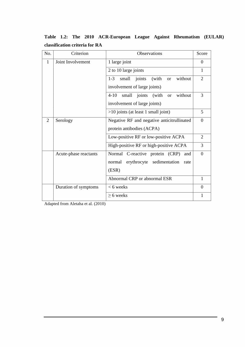
The classification of acute RA has been widely made based on American College of
Rheumatology (ACR) 1987 criteria (Arnett et al. 1988), as presented in the following Table
1.1. Patients are diagnosed as RA if at least 4 criteria listed are fulfilled, with the first four (1-
4) criteria present for at least 6 weeks. Nonetheless, a much more recent criteria for RA
(Aletaha et al. 2010), presented in the following Table 1.2, has been developed and becomes
current guideline for identifying RA, with a total score of 6 out of 10 gives classification as
RA (Table 1.2).
One of the hallmark clinical features of RA is the inflammation of synovium (or known as
synovitis) associated with the infiltration of inflammatory cells into the synovium. The
massive infiltration of inflammatory cells, which includes the likes of macrophages and T-
cells, is believed to lead to the hyperproliferation observed in the synovial membrane. It is
believed that the inflammatory mediators released by those cells contribute towards the
destruction of bone and cartilage tissue, as reviewed in Walsh et al. (2005). This conclusion is
based on the results shown in many studies, including in animal models (Ji et al. 2002; Shealy
et al. 2002; van den Berg 2001; Zwerina et al. 2004).
Research in the past has also characterised angiogenesis as another common pathological
feature of RA. Angiogenesis could be defined as the physiological process of the formation of
new blood capillaries from the pre-existing vessels. In RA angiogenesis occurs in response to
support and maintain the inflammation state by transporting inflammatory cells to the
inflammation site and to accommodate demands nutrients and oxygen by the growing pannus.

8
Table 1.1: ACR 1987 Revised Criteria for RA
No. Criterion Definition
1 Morning stiffness Morning stiffness in and around joints lasting at least
1 hour before maximal improvement
2 Arthritis of 3 or more joint
areas
Soft tissue swelling of at least 3 joint areas observed
by physician.
3 Arthritis of hand joints At least 1 swollen area in wrist, proximal
interphalangeal and metacarpophalangeal.
4 Symmetric swelling Simultaneous swelling on joint areas refered in both
sides of the body.
5 Rheumatoid nodules Subcutaneous nodules over bony prominences or
extensor surfaces, or in juxtaarticular regions, as
observed by physician.
6 Rheumatoid Factor (RF) Detection of abnormal level of serum RF.
7 Radiographic changes Radiographic changes typical of rheumatoid arthritis
on posteroanterior hand and wrist radiographs, which
must include erosions or unequivocal bony
decalcification localized in or most marked adjacent
to the involved joints
Adapted from Arnett et al. (1988)
9
Table 1.2: The 2010 ACR-European League Against Rheumatism (EULAR)
classification criteria for RA
No. Criterion Observations Score
1 large joint 0
2 to 10 large joints 1
1-3 small joints (with or without
involvement of large joints)
2
4-10 small joints (with or without
involvement of large joints)
3
1
Joint Involvement
>10 joints (at least 1 small joint) 5
Negative RF and negative anticitrullinated
protein antibodies (ACPA)
0
Low-positive RF or low-positive ACPA 2
2 Serology
High-positive RF or high-positive ACPA 3
Normal C-reactive protein (CRP) and
normal erythrocyte sedimentation rate
(ESR)
0 Acute-phase reactants
Abnormal CRP or abnormal ESR 1
< 6 weeks 0 Duration of symptoms
≥ 6 weeks 1
Adapted from Aletaha et al. (2010)
10
Bone loss in RA, another feature that may be observed in the disease, is targeted primarily to
the diarthrodial joints (free-moving joints formed from two opposing bone surfaces with
articular cartilage in the middle) and is characterised by progressive erosion of bone and
cartilage extracellular matrices, which occasionally becomes apparent even in early stage of
RA (Zhao et al. 2011). RA also has pathological characteristics of formation of pannus
(thickened synovial tissue) resulting from the hyperproliferation of the synovial membrane.
This structure of pannus is believed to “invade” bone and cartilage resulting the bone erosion
following the release of pro-inflammatory cytokines and matrix metalloproteinases (MMPs).
From the view point of bone erosion, there have been three forms of bone loss described in
RA; focal bone erosions, periarticular bone loss and general osteopenia (Goldring 2009;
Schett 2007; Walsh et al. 2005).
While those predominant features described above appear to be more associated with a
localised disease, RA is nevertheless a systemic disease with the associated syptoms
manifested throughout the body like rheumatoid nodules, vasculitis and Felty syndrome
(Schneider et al. 1985). With the features such as synovitis and formation of pannus are
considered as primary in RA, in general, this disease is also associated with secondary
osteoporosis (Gough et al. 1998).
At cellular level, the initiation and progression of RA results from the interaction between
cells such as T and B lymphocytes, cells from monocytes/macrophages lineage and both type
A (macrophage-like) and B (fibroblast-like) synoviocytes, which produce and release
products like chemokines and cytokines that recruit other cell types into tissues and enzymes
such as MMPs, cathepsins and mast cell proteinases that directly cause damage on cartilage
and bone. CD4-positive T-cells are thought to be the main player ochestrating the immune
response in synovial inflammation (Rittner et al. 1997). There is also evidence that
osteoclasts, the bone-resorbing cells, are involved in mediating the bone erosion observed in
RA tissues (Bromley & Woolley 1984; Fujikawa et al. 1996; Gravallese et al. 1998; Shen et
al. 2006) (to be described in 1.3.3 below).
11
1.2.3. Osteoarthitis
Osteoarthritis (OA) is the most common type of arthritis (Hedbom & Hauselmann 2002).
Nonetheless like RA, the etiology is still unknown. The most common early symptom
associated with osteoarthritis is pain at the affected joints, which can be worsened by load
bearing and relieved following rest. Studies have pointed out quite a number of associated
factors identified as contributing towards the onset of OA like aging, body weight load or
obesity, mechanical stress on joints, probably due to misalignment of bones (Brandt et al.
2008) as well as genetics (Valdes & Spector 2008).
This type of arthritis is mainly characterized by slow but progressive degeneration of articular
cartilage, which leads to pain and disability (Buckwalter et al. 2005). Unlike PO and RA as
described earlier, this disease is not considered as a bone loss disease, indeed the subchondral
bone underneath the degraded cartilage is generally observed in OA as thickening (Gevers et
al. 1989). However, OA is reviewed in this thesis as the synovial tissues from OA patients
were used in the study as controls (described further in Chapter 2 and 4). The primary process
of cartilage degradation is believed to be due to increase in the synthesis and activity of
extracellular degrading proteinases, mainly MMPs followed by secondary process of
synovitis (inflammation of synovium). Due to that, OA is also considered to be an
inflammatory disease following the release of pro-inflammatory cytokines found within the
tissues (Haywood et al. 2003; Hussein et al. 2008; Nakamura et al. 1999).
Another hallmark feature that could only be uniquely found in OA is the formation of
osteophytes (Felson et al. 2005), which is osteochondral nodule-like structure formed in the
bone near chondro-synovial junction probably from progenitor cells inside the perichondrium
(Matyas et al. 1997). The function of osteophytes still remains unknown, however they are
believed to be associated with stabilizing the joints affected by OA.
Currently, the diagnosis criteria for OA widely used are based on the ACR criteria developed
by Altman and colleagues (Altman et al. 1986). The classification for OA according to the
guideline depends on the joint affected. Taking hip as example, considering tissues from OA
12
group used in this study were from hip, characterization for OA disease is based on the
criteria listed in Table 1.3 below.
Table 1.3: Traditional format of ACR Classification Criteria for OA of the Hip
Main Criterion Additional criteria
with at least 2 must be fulfilled
1. ESR< 20mm/hour
2.Radiographic femoral or acetabular
osteophytes
Hip pain
3.Narrowing of joint space on radigraphs
Adapted from Altman et al. (1986)
As mentioned above, previously OA is mainly viewed as a disease of cartilage. Cartilage is a
structure cushioning between bones at joints for frictionless motion and resists compressive
forces during loading. In terms of the structure composition, the matrix is mainly consisted of
collagen (60%) and proteoglycan (25%) as well as non-collagenous proteins (Buckwalter et
al. 2005). Chondrocytes are the only cell type exists in cartilage and only accounts for 2% of
cartilage volume (Poole et al. 2001). Since there is no blood vessels or nerves in cartilage,
synovial fluid is really crucial in providing them nutrition and oxygen. Chondrocytes are the
key players for homeostasis or maintenance of cartilage as they produce collagen and
proteoglycan during anabolic phase and metalloproteinases for catabolic phase of cartilage
homeostasis.
Studies suggested that the degradation of cartilage has link to increase in cell death of
chondrocytes in OA (Dai et al. 2006; Wei et al. 2006). Besides cell death, there are also
thoughts that the reduction in chondrocytes has association with phenotype alteration of the
cells into more fibroblast-like cells, and this is known as dedifferentiation (Aigner et al. 1993;
Sandell & Aigner 2001). There is also evidence associating the initiation and progression of
OA with the senescence activity of chondrocytes, which is closely linked to aging (Martin &
Buckwalter 2001a, 2001b)
Studies on OA synovial tissues indicate that inflammation could be observed in all stages of
OA (Smith et al. 1997). Synovitis, which means inflammation in the synovium, can be found
13
in the early and late stage OA, however it becomes more obvious in the later stages (Smith et
al. 1997). This inflammation in tissues is characterized by thickening of lining layer,
increased vascularity and massive infiltration of inflammatory cells into the thickening layer
synovial membranes (Smith et al. 1997). Besides RA, OA also is another example of disease
that could be characterised as having pathological feature of synovial angiogenesis (Walsh et
al. 2007), as this could be seen from the high vascularity observed in OA synovial tissues
(Smith et al. 1997). In comparison to late stage OA, it was found that there was more vascular
formation and tissue infiltration of CD4 (T-cells) and CD68-positive cells (activated
macrophages) as well as greater expression of vascular endothelial growth factor (VEGF) and
intracellular adhesion molecules-1 (ICAM-1) in early OA (Benito et al. 2005).
1.3. Osteoclasts- The Bone Resorbing Cells
Osteoclasts are terminally differentiated multinucleated cells (normally ranged between 3 to
20 nuclei, as reviewed in Roodman and Windle (2005), formed from the fusion of marrow-
derived mononucleated hemopoietic progenitor cells (Coccia et al. 1980; Walker 1975). Upon
fusion, each nucleus within the multinucleated osteoclasts has been reported to have a limited
lifespan of approximately 12.5 days or 2 weeks, therefore new osteoclast precursors need to
be kept recruited and fused into the existing mature osteoclasts for longer resorption activity
(Jaworski et al. 1981; Manolagas 2000).
As mentioned earlier, osteoclasts are cells uniquely specialized for resorbing bone. The
significance of osteoclasts in resorbing bone could be seen from the occurrance of
osteopetrosis (generally characterized as significantly higher bone mass and reduced bone
marrow cavities) as well as defected hematopoietic and bone marrow cell development (Villa
et al. 2006). From the physiological perspective, osteoclasts are very important in initiating
bone remodeling and hence may affect other function of bone remodeling such as for calcium
homeostasis.
The precursors to osteoclasts, referred to as pre-osteoclasts, are present in both peripheral
circulation and bone marrow, however, they only differentiate into mature functional
osteoclasts in the tissue in which they are recruited (Collin-Osdoby et al. 2001; Gravallese et
al. 1998) and when in contact with bone (McHugh et al. 2007; Shen et al. 2006). Fully
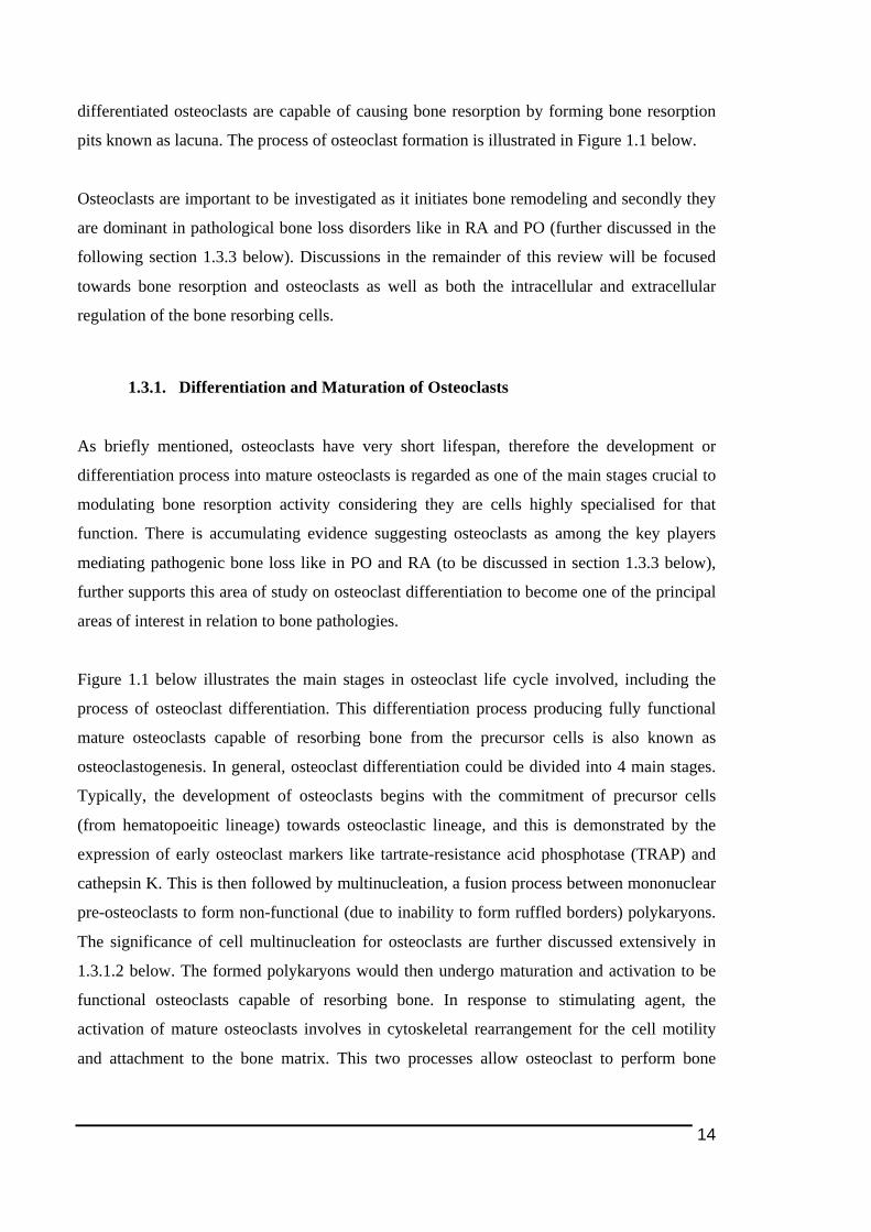
14
differentiated osteoclasts are capable of causing bone resorption by forming bone resorption
pits known as lacuna. The process of osteoclast formation is illustrated in Figure 1.1 below.
Osteoclasts are important to be investigated as it initiates bone remodeling and secondly they
are dominant in pathological bone loss disorders like in RA and PO (further discussed in the
following section 1.3.3 below). Discussions in the remainder of this review will be focused
towards bone resorption and osteoclasts as well as both the intracellular and extracellular
regulation of the bone resorbing cells.
1.3.1. Differentiation and Maturation of Osteoclasts
As briefly mentioned, osteoclasts have very short lifespan, therefore the development or
differentiation process into mature osteoclasts is regarded as one of the main stages crucial to
modulating bone resorption activity considering they are cells highly specialised for that
function. There is accumulating evidence suggesting osteoclasts as among the key players
mediating pathogenic bone loss like in PO and RA (to be discussed in section 1.3.3 below),
further supports this area of study on osteoclast differentiation to become one of the principal
areas of interest in relation to bone pathologies.
Figure 1.1 below illustrates the main stages in osteoclast life cycle involved, including the
process of osteoclast differentiation. This differentiation process producing fully functional
mature osteoclasts capable of resorbing bone from the precursor cells is also known as
osteoclastogenesis. In general, osteoclast differentiation could be divided into 4 main stages.
Typically, the development of osteoclasts begins with the commitment of precursor cells
(from hematopoeitic lineage) towards osteoclastic lineage, and this is demonstrated by the
expression of early osteoclast markers like tartrate-resistance acid phosphotase (TRAP) and
cathepsin K. This is then followed by multinucleation, a fusion process between mononuclear
pre-osteoclasts to form non-functional (due to inability to form ruffled borders) polykaryons.
The significance of cell multinucleation for osteoclasts are further discussed extensively in
1.3.1.2 below. The formed polykaryons would then undergo maturation and activation to be
functional osteoclasts capable of resorbing bone. In response to stimulating agent, the
activation of mature osteoclasts involves in cytoskeletal rearrangement for the cell motility
and attachment to the bone matrix. This two processes allow osteoclast to perform bone

15
resorption (detailed in section 1.3.2 below), and once the osteoclasts have performed their
function, they will then undergo apoptosis.
Figure 1.1: Main stages in osteoclast differentiation
Adapted from Boyle et al. (2003)
Osteoclastogenesis is believed to take place at or in close proximity to the site of bone
resorption (Collin-Osdoby et al. 2001; McHugh et al. 2007). Generally, osteoclasts
differentiate from monocytes, most likely to be CD14-positive cells (Atkins et al. 2006;
Massey & Flanagan 1999; Nicholson et al. 2000; Nose et al. 2009; Shalhoub et al. 2000;
Sorensen et al. 2007). This means that osteoclasts share similar progenitor to macrophages
and dendritic cells, two types of immune cells derived from monocytes as well (Miyamoto et
al. 2001). However, in some cases like in PO, in which there has been evidence that
osteoclasts could be differentiated from macrophages (see section 1.3.3 below) as wells as
from dendritic cells as indicated by a quite recent study (Gallois et al. 2010).
In general, the total of net bone resorption depends on two factors, which are the rate of cells
recruitment and the bone resorbing capacity of each multinucleated cell (Zou et al. 2010).
These two factors, with the latter is closely associated with multinucleation and cytoskeletal
organisation (Teitelbaum 2011), are going to be discussed in detail below.
1.3.1.1. Recruitment and Targeting of Osteoclasts
The mechanism of how osteoclasts are recruited to specific site on the bone surfaces for
resorption is not really clearly understood. However, it is suggested that the recruitment of
pre-osteoclasts to site of bone resorption is on the basis of chemotaxis.
16
This is based on the initial work done by Malone et al. (1982), which showed that three
constituents of bone matrix, collagen type I peptides, α2HS glycoprotein and osteocalcin
appear to exhibit chemotactic effects on human monocytes, the cell type that osteoclasts are
derived from. The chemotactic mobilization of osteoclasts could also be initiated by
osteoblasts as the bone-forming cells are also known to produce collagen and osteocalcin
(Malone et al. 1982). Osteoblasts also release stromal-cell derived factor-1 (SDF-1) that is
thought to contribute in the migration and homing of osteoclasts (Wright et al. 2005; Yu et al.
2003).
It is also thought that osteocytes, the most abundant cells within bone matrix, may play a role
in the recruitment of osteoclasts, especially in the context of initiating bone remodelling at a
particular site of bone, for example at fractured site. It is hypothesized that dying osteocytes
(by apoptosis) in the close proximity to the microcracks may transmit chemotactic signals to
osteoclasts requesting for bone remodeling for fracture removal (Noble et al. 2003). Another
good example of such signal transmission would be osteopontin, an osteoclast
chemoattractant important for initiating bone remodeling following mechanical loading
(Fujihara et al. 2006; Gross et al. 2005; Terai et al. 1999). It was found that the expression of
osteopontin by osteocytes is upregulated in hypoxic condition (Gross et al. 2005). Indeed
besides recruiting osteoclast, there is accumulating evidence pointing out that osteocytes are
the cells that support osteoclast formation (Kurata et al. 2006; Xiong & O'Brien 2012; Zhao et
al. 2002) (to be further discussed in section 1.3.5.1.4 below).
Besides those chemoattractants of osteoclasts described above, there is quite a number of
other chemokines being associated with the recruitment of osteoclast precursor cells. Such
common examples include monocyte chemoattractant protein-1 (MCP-1) (Hashimoto et al.
1998; Lu et al. 1998), C-C chemokine receptor 2 (CCR2) (Gao et al. 1997), macrophage
inflammatory protein 1 (MIP-1) (Boring et al. 1997) and regulated upon activation, normal T
cell expressed and secreted (RANTES) (Yano et al. 2005). Another example of osteoclast-
associated chemokines is macrophage inhibitory factor-1α (MIP-1α)(Hashimoto et al. 1998),
which has been shown to increase osteoclast motility and its receptor CCR1 expressed on
osteoclast precursor cells (Yu et al. 2004).

17
1.3.1.2. Multinucleation
The multinucleated feature of osteoclasts is believed to be associated with optimisation of
bone resorption capability and efficiency (Boissy et al. 2002). This cell-cell fusion process
was found to be influenced by factors like tumor necrosis factor-α (TNFα) and
lipopolysaccharide (LPS) (Lee et al. 2006b). It is hypothesized that multinucleation provides
synergy in terms of resorption efficiency. Bone resorption by one multinucleated osteoclast is
greater than the total bone resorption by the total number of mononuclear osteoclasts
(equivalent to the number of nuclei in the single multinucleated osteoclast) (Bar-Shavit 2007;
Boissy et al. 2002). However, findings by Lees et al. (2001) suggest that the total bone
resorption is proportionate to the number of nuclei within osteoclasts. Inability to
multinucleate in mutant mice-derived osteoclasts was found to result in less dentine
resorption activity (Humphrey et al. 2004; Paloneva et al. 2003).
There is a number of proteins identified as crucially required in the process of multinucleation
of osteoclasts and absence of those proteins severely affect the osteoclast resorption capacity.
Such example is dendritic-cell transmembrane protein (DC-STAMP) (Yagi et al. 2005). Study
by Yagi and co-workers (2005) demonstrated that cells deficient in DC-STAMP, had reduced
resorption efficiency per nuclei.
Another protein that was found to be important in the process of cell multinucleation is the d2
isoform of vacuolar (H+) ATPase (V-ATPase) V0 domain (Atpv0d2) (Lee et al. 2006b). The
multinucleated state of the osteoclasts is believed to enable osteoclasts to possess a high
number of mitochondria, which is essential for the production of high amount of energy
needed by the ATPase pump for the secretion of protons in the bone resorption as reviewed
by Vaananen (2005).
The large size of osteoclasts in comparison to macrophage allows them to cover a large area
on the bone surfaces, which further enhancing the efficiency of bone resorption (Bar-Shavit
2007). In addition, it was found that large sized osteoclasts are more active in comparison to
the small sized ones (Lees et al. 2001). Examination at the protein level found that the
expression levels of proteins that play crucial role in osteoclast signalling, attachment, cell
fusion and matrix degradation (refer to following sections) are relatively higher in large sized
osteoclasts than the small ones (Trebec et al. 2007).
18
1.3.1.3. Osteoclast-associated Cell Markers
As pre-osteoclasts diferentiate into mature osteoclasts, the pre-osteoclasts begin to express a
number of proteins. The expression of these proteins is very useful as markers in determining
or estimating the stage of the osteoclast differentiation (see Figure 1.1). Commonly used
osteoclast gene markers are ctsk (encoding cathepsin K) (Drake et al. 1996), Acp5 (encoding
TRAP) (Janckila et al. 2005; Janckila et al. 2001), calcr (calcitonin receptor (Lee et al. 1995)
and osteoclasts-associated receptor (OSCAR)-encoding gene (Kim et al. 2002).
Cathepsin K (catK) is a protein found to be expressed at high level in osteoclasts from all
species studied thus far (Drake et al. 1996; Littlewood-Evans et al. 1997; Troen 2003;
Yamaza et al. 1998). Cathepsin K appears to be a main bone-matrix degrading enzyme as
mice deficient in cathepsin K exhibited osteopetrosis (Gowen et al. 1999; Saftig et al. 1998).
This is further proven in a study in which a significant reduction in bone resorption activity
following treatment with a cathepsin K inhibitor in a rat model was observed (Xia et al.
1999). Meanwhile in human, mutation on cathepsin K gene leads to pycnodysostosis disease
(Gelb et al. 1996).
Cathepsin K has been found to be able to degrade collagen type I and II as well as bone
matrix protein osteonectin (Bossard et al. 1996; Garnero et al. 1998; Kafienah et al. 1998).
The unique feature of cathepsin K in comparison to other mamalian proteinase is that it
cleaves collagen type I molecules both sides of in and outside helical region (Garnero et al.
1998). Cathepsin K is regulated by microphthalmia transcription factor (MITF) (Motyckova
et al. 2001). As osteoclast differentiates, the amount of cathepsin K produced increases, which
is believed to be as a result of receptor activator NF kappa B ligand (RANKL) stimulation
(Corisdeo et al. 2001) (RANKL to be discussed in detail in section 1.3.5.1).
TRAP is a widely used histochemical marker for the detection of osteoclasts as well as for
bone resorption activity (Halleen 2003; Halleen et al. 2006; Rissanen et al. 2008). TRAP is an
enzyme expressed by osteoclasts with a role in bone resorbing activity. In TRAP-knockout
mice, osteoclasts were still found to be formed, however, accompanied by a reduction in bone
resorptive activity (Hayman et al. 1996). Meanwhile, overexpression of TRAP was found to
19
lead to an increase in bone turnover (Angel et al. 2000). TRAP is found in various types of
tissues, nevertheless high TRAP activity is found to be predominant in bone, spleen, liver and
thymus as it is expressed abundantly by osteoclasts, macrophages, multinucleated foreign-
body giant cells and dendritic cells (Chun et al. 1999; Halleen et al. 2006; Hayman & Cox
2003). TRAP is an early marker for the detection of cells committed towards osteoclast-
lineage as it is expressed in the early stage of commitment towards osteoclast lineage (before
mutinucleation of pre-osteoclasts takes place). Like cathepsin K, TRAP is also regulated by
MITF (Luchin et al. 2000).
Even though cathepsin K and TRAP has been widely accepted as osteoclast cell markers, it
appears that researchers need to be cautious over reliance on TRAP and cathepsin K as solely
osteoclastic markers as these two markers are also found to be expressed in dendritic cells,
another cell type sharing the same precursor with osteoclasts, even though in low quantity
(Kim et al. 2002). Cathepsin K has also been found to be expressed in fibroblasts (Hou et al.
2002; Hou et al. 2001), macrophages and smooth muscle in atheroma (Sukhova et al. 1998)
and a number of cell types in lung tissues (Buhling et al. 2000).
It was suggested that the newly discovered osteoclast-specific receptor named OSCAR, to be
discussed in further detail in section 3.5.2) to be more reliable as an osteoclast gene marker as
this receptor was understood to be not expressed in both dendritic cell and monocyte-derived
macrophage in mice (Kim et al. 2002). In addition, the expression of OSCAR was also
detected in RAW264.7 macrophage cell line-derived osteoclast-like cells, with peak
expression upon the completion of the osteoclast differentiation following four days of
RANKL stimulation (Kim et al. 2002).
1.3.2. Mechanism of Bone Resorption
The resorption of bone by osteoclasts begins with the attachment of the mature osteoclast to
the surface of the bone. The attachment of bone and osteoclasts is largely mediated by
membrane-bound molecules known as integrins. To date, there are at least five types of
integrins identidfied as being expressed in osteoclasts; α2β1, αvβ1, αvβ3, αvβ5 (Nesbitt et al.
1993) and the recently discovered α9β1 (Rao et al. 2006), however, only αvβ3 and αvβ5
integrins are of particular interesting in relation to osteoclast bone resorption activity as it
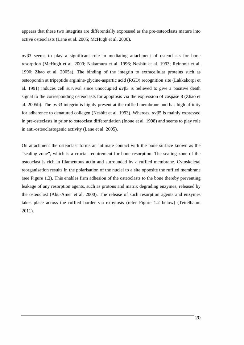
20
appears that these two integrins are differentially expressed as the pre-osteoclasts mature into
active osteoclasts (Lane et al. 2005; McHugh et al. 2000).
αvβ3 seems to play a significant role in mediating attachment of osteoclasts for bone
resorption (McHugh et al. 2000; Nakamura et al. 1996; Nesbitt et al. 1993; Reinholt et al.
1990; Zhao et al. 2005a). The binding of the integrin to extracellular proteins such as
osteopontin at tripeptide arginine-glycine-aspartic acid (RGD) recognition site (Lakkakorpi et
al. 1991) induces cell survival since unoccupied αvβ3 is believed to give a positive death
signal to the corresponding osteoclasts for apoptosis via the expression of caspase 8 (Zhao et
al. 2005b). The αvβ3 integrin is highly present at the ruffled membrane and has high affinity
for adherence to denatured collagen (Nesbitt et al. 1993). Whereas, αvβ5 is mainly expressed
in pre-osteclasts in prior to osteoclast differentiation (Inoue et al. 1998) and seems to play role
in anti-osteoclastogenic activity (Lane et al. 2005).
On attachment the osteoclast forms an intimate contact with the bone surface known as the
“sealing zone”, which is a crucial requirement for bone resorption. The sealing zone of the
osteoclast is rich in filamentous actin and surrounded by a ruffled membrane. Cytoskeletal
reorganisation results in the polarisation of the nuclei to a site opposite the ruffled membrane
(see Figure 1.2). This enables firm adhesion of the osteoclasts to the bone thereby preventing
leakage of any resorption agents, such as protons and matrix degrading enzymes, released by
the osteoclast (Abu-Amer et al. 2000). The release of such resorption agents and enzymes
takes place across the ruffled border via exoytosis (refer Figure 1.2 below) (Teitelbaum
2011).
21
Figure 1.2: Transportation of bone resorbing agents through a trans-Golgi trafficking
network in osteoclasts and release across the ruffle border via exocytosis
Adapted from Vaananen et al. (2000).
The bone degradation process could be divided into two main phases; inorganic (bone
demineralisation) and organic (matrix degradation) phases (Abu-Amer et al. 2000). The
demineralisation of bone occurs before the degradation of matrix protein. The process of
demineralisation of bone occurs by the acidification of the sealed region and is mediated by
the vacuolar H+-ATPase pump located in the osteoclast ruffled membrane (Blair et al. 1989;
Mattsson et al. 1994; Vaananen et al. 1990). Protons (H+), produced by cytoplasmic carbonic
anhydrase II within the osteoclasts (Gay & Mueller 1974), are pumped out of the osteoclasts
into the area between the cells and the bone causing a drop in pH to ~4.5 (Schlesinger et al.
1997) that leads to the dissolution of bone mineral (the solid hydroxyapatite, Ca10(PO4)6(OH)2
to free Ca2+, HPO42- and water, H2O). The build up of alkaline intracellular condition as a
result of pumping protons out from the cells is neutralized by the chloride/bicarbonate
exchange pump (Hall & Chambers 1989; Schlesinger et al. 1997; Teti et al. 1989).
The degradation of matrix protein occurs through the secretion of lysosomal proteases, MMPs
and a variety of other matrix degrading enzymes, which are released from the osteoclasts by
22
the process of exocytosis. The high expression of cathepsin K (Drake et al. 1996), MMP9
(Tezuka et al. 1994) and type IV collagenase/gelatinase B (Wucherpfennig et al. 1994) in
osteoclasts suggests that these enzymes play roles in the organic phase of the bone resorption.
Screening on patients with pycnodysostosis, a rare disease which has a feature of increased
bone density, showed that the disease is linked to mutation on cathepsin K gene, which may
indicate that this enzyme may play important role in bone resorption (Gelb et al. 1996; Ho et
al. 1999; Johnson et al. 1996; Motyckova & Fisher 2002). Meanwhile, knockout on MMPs
results in defects in bone resorption (Delaisse et al. 2003). The cumulative effect of the bone
demineralization and matrix degradation is the formation of lacuna pits on the surface of the
bone.
Products produced from the degradation activity by the osteoclasts are removed from the
sealing-zone confined microenvironment to the extracellular space via transcytosis through
the osteoclast itself from the ruffled membrane to the opposite functional secretory domain
(refer Figure 1.2) (Nesbitt & Horton 1997; Salo et al. 1997). A study by Halleen et al. (1999)
found that TRAP is localised together with matrix degradation products in the transcytotic
vesicles and may play a role in further fragmentation of the endocytosed materials by
producing reactive oxygen species. The transcytotic secretory system of the matrix-degraded
components in the osteoclasts seems to share similarity with the antigen presentation
presentation mechanism in macrophage, the immune cell derived from the same progenitor
cells for osteoclasts (Vaananen et al. 2000).
The mechanism by which osteoclast-mediated bone resorption is stopped or arrested is poorly
understood. However, there are a couple of theories suggested. First, the bone resorption
process could stop by itself considering osteoclasts have limited lifespan. Secondly, the
resorption process might be arrested as a consequence of inhibition on the osteoclasts by the
accumulated high concentration of calcium resulted from the bone mineral dissolution (Zaidi
1990) or TGF-β cytokine or other products released from the bone matrix (Pfeilschifter et al.
1990a; Pfeilschifter et al. 1990b).
1.3.3. Osteoclasts in Pathology of Inflammation-Mediated Bone Loss
23
As briefly mentioned earlier in this chapter, osteoclasts become one of the central theme
discussed in this thesis since studies over decades found that they present in pathological bone
disorders like PO and RA as well as being associated with the initiation and progression of
those diseases.
In PO, it has been found that there is increased osteoclast bone resorbing activity that is
partially compensated by high osteoblast activity (in terms of mineral apposition and bone
formation rate), which was indicated by the increased remodelling of peri-implant bone and
immature bone formation around loosened prosthetic hip implants (Takagi et al. 2001).
There is also accumulating evidence pointing out the increase in the recruitment and
differentiation of osteoclast precursor cells in PO tissues as might be the main factor
responsible for wear particle-induced osteolysis (Eftekhar et al. 1985; Greenfield et al. 2002;
Kadoya et al. 1996). Examination on the interface tissues around failed prostheses found high
level of cathepsin K, which is an osteoclast enzyme, in the PO tissues (Konttinen et al. 2001).
A more detail investigation by Shen and colleagues (Shen et al. 2006) found cells with PE
particles inside, both mononucleated and multinucleated, were positive for osteoclast-
associated markers cathepsin K and TRAP. Previous work carried out in this laboratory found
that RANKL, which has been demonstrated to increase osteoclast bone resorbing activity (see
1.3.5.1 below), to be significantly higher in PO tissues compared to OA tissues (Crotti et al.
2004). A recent study by Qin and coworkers (2012) demonstrated that treatment with an
osteoclast vATPase inhibitor, saliphenylalamide was able to attenuate particle-induced
osteolysis in a murine calvarial model.
As mentioned earlier, macrophages have been found to be a dominant player involved in the
disease progression of PO (refer 1.2.1 above). Macrophages are also dominant in PO tissues
in term of cell population, which has been indicated by higher level of macrophage cell
marker, in PO tissues as compared to the controls (Kadoya et al. 1996; Sabokbar et al. 1997).
Those high number of macrophages found in the tissues are also thought to be potential
osteoclast precursor cells (Haynes et al. 2001b; Sabokbar et al. 1997). There has been
evidence demonstrating PE-associated multinucleated osteoclast-like cells in PO tissues were
found to be positive for macrophage cell marker CD68 (Shen et al. 2006). It has also been
shown in the past that these cells (i.e macrophages) isolated from peri-prosthetic tissues
24
around loosening implants are capable of differentiating into bone-resorbing osteoclasts in
vitro (Haynes et al. 2001b; Neale et al. 2000; Sabokbar et al. 1997). However, there has been
contradiction underlying the capacity of those tissue-isolated cells to readily differentiate into
osteoclasts as some studies reported that such ex vivo differentiation required presence of
osteoblasts (Neale et al. 2000; Sabokbar et al. 1997), while another study (Haynes et al.
2001b) indicated presence of osteoblasts is not necessary for the differentiation process.
The thought that macrophages can be differentiated into osteoclasts is further supported by
the data that reported expression of osteoclast cell markers by macrophages in the
pseudomembrane surrounding the loose prostheses (Kadoya et al. 1994; Kadoya et al. 1996;
Neale & Athanasou 1999). With the capability of macrophages to get differentiated into
osteoclasts, there is possibility that the bone erosion occurring in PO results from the presence
of massive number of osteoclasts derived from large population of macrophages dominating
in the tissues.
In view of the increased recruitment of osteoclast precursors into PO tissues, it has been
found that the presence of biomaterial debris has effect on the expression of osteoclasts-
associated chemokines (refer 1.3.1.1 above). Stimulation with ultra-high molecular weight
polyethylene (UHMWPE) particles in vivo was found to increase the expression level of
MCP-1 (Rhodes et al. 1997). Meanwhile, stimulation by PMMA and titanium alloy in an in
vitro monocyte/macrophage model demonstrated increased in the expression of MCP-1 and
MIP-1α, but not RANTES (Nakashima et al. 1999a). Study on human PO tissues (Haynes et
al. 2004) supported these earlier findings, in which there was higher level of MCP-1 and MIP-
1α demonstrated in those tissues. It has been suggested that different types of biomaterial give
different response in C-C chemokines expression (Nakashima et al. 1999a). Based on the
expression profile of chemokines, biomaterial debris has also been found to affect cells
activity. For instance, PE was suggested to have effect on the activation and migration of
macrophages (Ishiguro et al. 1997).
Studies in the past has described the formation of osteoclasts as a key process in inflammatory
bone erosion occuring in RA (Durand et al. 2011) besides imbalance in regulation of bone
remodeling resulted from excessive bone resorption and decrease in bone formation (Goldring
2003; Gravallese et al. 2000; Redlich et al. 2002b). High disease activity, which is a common
25
marker for RA disease progression, was found to be correlated with osteoclastogenesis (Zhao
et al. 2011).
Through examinations on the human tissue and the arthritic animal models, it appears
osteoclasts are the cells mediating the pathological bone loss present in RA patients (Bromley
& Woolley 1984; Fujikawa et al. 1996; Gravallese et al. 1998; Gravallese et al. 2000;
Kuratani et al. 1998; Redlich et al. 2002b; Suzuki et al. 1998). Mature osteoclasts could be
found in the close proximity to the bone surface in the joints of the RA patients (Bromley &
Woolley 1984; Gravallese et al. 1998). In addtion, it has been found that
monocytes/macrophages isolated from human rheumatoid arthritis synovial tissues could and
is more potent to differentiate into bone resorbing osteoclasts in vitro (Fujikawa et al. 1996;
Nose et al. 2009; Vandooren et al. 2009).
1.3.4. Regulation on Osteoclastogenesis and Bone Resorbing Activity by
Cytokines
There is quite a number of factors that have been shown in the past as having influence on the
regulation of osteoclast differentiation and bone resorbing activity. Age, genders (Jevon et al.
2002), hormones (Faucheux et al. 2002; Huang et al. 2004; Rodan & Martin 1981), cytokines
(Fujikawa et al. 2001; Tsuboi et al. 1999; Tsuda et al. 1997) are among factors known to
incluence bone loss. Of all those factors, release of cytokines appeared to be most studied,
and since local inflammation-mediated bone loss becomes the scope of this study, this section
of this thesis is going to discuss the influence of cytokines, especially the major pro-
inflammatory cytokines in on the regulation of osteoclast differentiation and activity in
inflammation mediated localised bone loss,.
Numerous cytokines have been identified and reported as capable of altering and modulating
the rate of osteoclastogenesis and they are believed to play significant role in the pathogenesis
of those diseases described above. However, in this review, the cytokines that are going to be
discussed are mainly TNFα and interleukin-1 (IL-1), which are prominently and widely
reported to be involved in local inflammation-mediated bone loss. The discussion in this
thesis is limited to TNFα and IL-1 only since those two cytokines are involved in the study
presented in this thesis (see Chapter 5).
26
It appears from previous literature that TNFα and IL-1 are the predominant cytokines as they
are abundant in inflamed tissues as well as in the sera and synovial fluid in RA patients
(Buchan et al. 1988; Feldmann & Maini 2001; Maini et al. 1995; Tak & Bresnihan 2000).
Examination on tissues taken from arthritic patients, for instance, found that these two pro-
inflammatory cytokines are mainly expressed by activated macrophages and activated
synoviocytes in the lining layer (Danning et al. 2000; MacNaul et al. 1990) and the synovial
fibroblasts (Farahat et al. 1993; Niki et al. 2001). It has also been found that endothelial cells
also express TNFα in RA tissues (Farahat et al. 1993).
In the context of PO, receptors of TNFα and IL-1 are among cytokine receptors detected to be
expressed by macrophages, foreign body giant cells and osteoclasts isolated from peri-
prosthetic tissues (Merkel et al. 1999; Neale & Athanasou 1999; Xu et al. 1996) and
interaction between these cytokine receptors and their ligands is suggested to play role in
osteoclast formation and bone loss (Chiba et al. 1994b; Jiranek et al. 1993). The release of
those two cytokines by activated macrophages could be stimulated by various types of
prosthetic wear particles in vitro (Haynes et al. 1998; Merkel et al. 1999). TNFα and IL-1β
does not only promote osteoclastogenesis by directly affect osteoclast precursors, but they
also promote osteoclast formation through regulation on osteoblasts (Haynes et al. 1997).
Meanwhile, in OA the presence of TNFα and IL-1 has also been reported (Smith et al. 1997).
A study comparing between stages of OA indicated that there was greater proportion of cells
expressing TNFα and IL-1β in early OA than in late OA (Benito et al. 2005). It has also been
reported that there was a pattern of lower level of IL-1 receptor antagonist (IL-1ra) and
declining ratio IL-1ra/IL-1 as the disease progresses (Fernandes et al. 2002; Smith et al.
1997), and this may give a picture on the significance of IL-1 in OA progression. Meanwhile
for TNFα, the significance of this cytokine towards OA progression could be seen from the
fact that there is higher level of p55 TNFα receptor (refer section 1.3.4.1 below) being
expressed in OA than in normal tissues (Alaaeddine et al. 1997; Fernandes et al. 2002; Smith
et al. 1997).
It has been suggested that TNFα and IL-1 play role in the progression of OA by promoting
the production of nitric oxide (NO) and prostaglandin E2 (PGE2) (Alaaeddine et al. 1997;
Amin & Abramson 1998), which in turn stimulate other cellular changes in OA (Hedbom &
27
Hauselmann 2002) such as decline in collagen and proteoglycan production, increased
apoptosis of cells including chondrocytes and inhibiton of β1 integrin-dependant adhesion to
the extracellular matrix (Amin & Abramson 1998). Previous literature also indicates that
TNFα and IL-1 significantly stimulate the expression of MMP such as MMP-3, which would
obviously involved in the degradation of cartilage in OA (Fernandes et al. 2002). It appears
that these two cytokines were likely to regulate inflammation in the tissue, as local injection
with anti-TNFα and anti-IL-1 results in reduction in recruitment of inflammatory cells and in
the number of osteoclasts by 80% and 67% respectively, as well as accompanied by a
decrease in bone loss (60%) in comparison to the control (Assuma et al. 1998).
Considering TNFα and IL-1 as the most dominant pro-inflammatory cytokines in mediating
the progression of these diseases, the regulation of bone resorption by these two cytokines
will be discussed in more detail below. There is another osteoclast-regulating cytokine
described in this section 1.3.4, which is macrophage-colony stimulating factor (M-CSF) as
this cytokine was also used in study reported in this thesis (Chapter 3).
1.3.4.1. Tumor Necrosis Factor-Alpha (TNFα)
It has been long known that TNFα is a promoter for bone resorption and inhibitor for bone
formation expressed and secreted by activated macrohages (Bertolini et al. 1986). Taking RA
as an example of local inflammation-mediated bone loss, TNFα is reported to be the principal
cytokine responsible for initiating bone loss in the disease (Shealy et al. 2002; Teitelbaum
2006).
Through histological examination on human RA tissues, it has been found that there is
massive number of cells in the synovial membrane as well as in pannus-cartilage junction
expressing TNF-α (Chu et al. 1991). The significant contribution TNFα has towards the
pathogenesis of rheumatoid arthritis could be seen from in vivo study on a human (h) TNFα
(h-TNFα) transgenic mouse (Tg197), which found out that the animal model developed
severe progressive polyarthritis as well as histological features commonly found in RA
(Keffer et al. 1991). Treatment in the same model using an anti-TNFα antibody allowed the
repair of the joint damage and reduced the bone erosion in the transgenic mouse (Redlich et
al. 2002a; Shealy et al. 2002). In occurance with this in a TNFα-transgenic serum transfer
28
arthritis-induced mouse, blockade of TNFα using infliximab (anti-mouse TNFα) results in a
significant reduction in osteoclast number (Redlich et al. 2002a; Zwerina et al. 2004).
Treatment with etanercept, which is a decoy recombinant human TNFα receptor, currently
has been widely applied and has been reported to provide an effective therapy for RA
(Gomez-Puerta et al. 2004; Wallenius et al. 2005).
As mentioned above, TNFα is also reported to be among the most important pro-
inflammatory mediators in PO (Chiba et al. 1994b; Jiranek et al. 1993). Like in RA, TNFα
was found to be expressed widely by high number of cells that include macrophages and
foreign-body giant cells (FBGCs) isolated from PO tissues (Holding et al. 2006; Neale &
Athanasou 1999; Xu et al. 1996). TNFα was also detected be expressed by fibroblasts,
endothelial cells and other cells present in the pseudosynovial membrane (Xu et al. 1996).
There is also data suggesting that TNFα could also be expressed by osteoclasts reported in a
previous study carried out in the laboratory (Holding et al. 2006), in which TNFα expression
was found to be strongly associated with multinucleated osteoclast-like cells containing PE
particles.
TNFα has been shown to be important soluble mediator in murine in vitro model of PMMA-
induced osteolysis as there was increase in gene and protein expression of TNFα upon
stimulation by PMMA cement and knockout of TNFα receptors resulted inhibition on
PMMA-induced osteolysis in vivo (Merkel et al. 1999). It has also been reported that TNFα
was among increased cytokines following exposure to PE particles in vitro (Horowitz &
Gonzales 1997).
TNFα can stimulate osteoclastogenesis either by directly on the pre-osteoclasts (Fuller et al.
2002; Kim et al. 2005c; Kobayashi et al. 2000; Komine et al. 2001; Lam et al. 2000; Zhang et
al. 2001) or indirectly by upregulating the expression of RANKL and M-CSF in the
interacting osteoblasts (Wei et al. 2005). TNFα is also thought to prolong the survival of
osteoclasts, thereby leads to increasing the bone resorption volume (Holding et al. 2006). It
has been shown that TNFα was able to inhibit the apoptotic effect of bisphophonates targeted
on osteoclasts (Zhang et al. 2005)
TNF-α interact with osteoclasts via two membrane receptors, which are TNF receptor type 1
(p55r) and type 2 (p75r) (Abu-Amer et al. 2000; Suda et al. 2001). However, it is believed
29
that p55r, which is expressed by each of pre-osteoclasts and marrow stromal cells, (Wei et al.
2005; Zhang et al. 2001) is the one which mediates osteoclastogenic signalling by TNFα
(Abu-Amer et al. 2000; Zhang et al. 2001). An examination of the expression Fas in human
osteoblasts, the bone-forming cells, has suggested that TNFα does not only promote bone loss
by inducing osteoclastogenesis, but also by stimulating apoptosis of the cells following
stimulation on p55r (Tsuboi et al. 1999). This implies that TNFα does not only promote bone
resorption by enhancing osteoclastogenesis, but might also by inducing the apoptosis of
osteoblasts, which eventually leads to decrease in the rate of bone formation. Meanwhile,
Abu-Amer et al. (2000) found that ligation of the TNF molecule on p75 on opposite appeared
to result in reduction in the TRAP intensity indicating suppression of osteoclastogenesis,
despite its well known role in inducing cell proliferation.
1.3.4.2. Interleukin-1 (IL-1)
IL-1 was the first cytokine (and even the first immune cell-derived product) recognized to be
shown to have an effect on osteoclast activation (Gowen et al. 1985). In the presence of M-
CSF, IL-1 was reported to be able to prevent the apoptosis, hence enhance the survival of
purified osteoclasts isolated from osteoblasts/marrow stromal cells (Jimi et al. 1995) as well
as involved in the resorptive function of osteoclasts (Kobayashi et al. 2000; Suda et al. 2001).
IL-1 was also reported to directly induce the multinucleation and activation of osteoclasts
(Jimi et al. 1999) and involves in a bidirectional interaction between pre-osteoclasts and
osteoblastic cells during osteoclastogenesis process (Atkins et al. 2000; Haynes et al. 1999).
Like TNFα, IL-1α has been shown to prolong the survival of osteoclasts (Lee et al. 2002).
The involvement of IL-1 in the pathogenesis of rheumatoid arthritis could be seen from a
number of studies using IL-1 transgenic animals which have shown that the overexpression of
IL-1α (Niki et al. 2004; Niki et al. 2001) and IL-1β (Ghivizzani, Kang et al. 1997) led to the
onset of arthritic features of bone and cartilage degradation. This is further supported by
findings from numerous studies investigating the blocking effect on IL-1 in animal models of
arthritis using anti-IL-1 monoclonal antibodies, endogenous IL-1 receptor antagonist, or
decoy soluble IL-1 type II receptors. Blocking IL-1 in all those animal models markedly
reduced bone erosion and the cartilage degradation (Abramson & Amin 2002). Meanwhile
knockout of IL-1 receptor diminished signs of arthritis in mice in which arthritogenic serum
30
was transferred (Ji et al. 2002). Like TNFα, IL-1 might also promote further bone destruction
in RA by reducing the rate of bone formation by inducing Fas-mediated apoptosis of the
bone-forming osteoblast cells (Tsuboi et al. 1999).
In PO IL-1 β is among important pro-inflammatory mediators involved in the progression of
the pathology (Chiba et al. 1994b; Jiranek et al. 1993). Investigation on a murine model of
prosthesis failure found that expression of IL-1β was common at the joints (Yang et al. 2007).
Higher level of IL-1β was reported in synovial fluids from knees of patient with PO than in
synovial fluids from patients with primary arthroplasty (Kovacik et al. 2000; Kovacik et al.
1997). Higher expression of IL-1, based on the level measured in the supernatant of
subcultured medium, was also reported in PO tissues with evidence of femoral osteolysis than
in PO tissues without sign of bone erosion (Chiba et al. 1994a).
Through immunohistochemistry, IL-1β was detected as being expressed by macrophages,
FBGCs and osteoclasts isolated from PO tissues, besides TNFα (Neale & Athanasou 1999).
Earlier study reported detection of IL-1β mRNA in macrophage-like cells in human PO
tissues (Jiranek et al. 1993). Analysis on the cytokine expression profile found that there was
more expression of cytokines including IL-1β by peripheral blood mononuclear cells
(PBMCs) from patient undergoing implant revision surgery than cells from patient
undergoing primary hip replacement (Waddell et al. 2005). Through an in vitro model of
monocyte/macrophage culture, Kamikawa and colleagues (2001) demonstrated PE particles
could cause the upregulation of IL-1β expression in PE-induced PO.
The increased expression of IL-1β in wear particle induced PO, as suggested from all those
studies, may suggest that this cytokine plays important role in mediating pathogenesis of
particle-induced PO. This is also further supported by other study showing knockout of IL-1
receptor in a murine model challenged with TiAlV particles caused reduction in the
infllammatory response such decreased expression of IL-6 and MCP-1 besides diminshed
formation of periprosthetic fibro-inflammatory membrane (Epstein et al. 2005). Introducing
IL-1 receptor antagonist into a murine in vivo model was found to give protective effects from
PE-particle induced osteoysis (Yang et al. 2004).
In terms of the role IL-1 has in the narrow context of mediating osteoclastogenesis, the
significance of IL-1 could be seen from the study by Zwerina et al. (2004), which showed that
31
blockade of IL-1 using anakinra resulted in reduction in osteoclast number. IL-1 and TNFα
seem to have an additive effect in vivo mouse model since the combination of anakinra
(which blocks IL-1R) and infliximab (which have blocking effect on TNFα) resulted in a
greater reduction in osteoclast number (almost complete suppression on osteoclastogenesis)
as compared to treatment with single antibody to either IL-1 or TNFα (Zwerina et al. 2004). A
later finding revealed that IL-1 is also involved in mediating TNFα-induced
osteoclastogenesis through the action on the osteoblasts (Wei et al. 2005). This is
demonstrated from the inhibition of TNFα-induced RANKL expression by osteoblasts as a
result of IL-1 receptor (IL-1R) blockade or gene-knockout.
1.3.4.3. Macrophage-Colony Stimulating Factor (M-CSF)
Unlike TNFα and IL-1 that are classified as pro-inflammatory cytokines and very relevant to
be discussed in the context of inflammation-associated bone diseases like PO and RA, M-CSF
is not considered to be pro-inflammatory. However, this cytokine is a very important factor
regulating osteoclast development and survival, therefore it has been considered to be
necessary component in many osteoclast in vitro culture, including in an experimental setup
for study presented in this thesis (see Chapter 3).
M-CSF appears to stimulate proliferation of the osteoclast pool via the binding of M-CSF
expressed by stromal or osteoblastic cells to its receptor c-Fms on the surface of pre-
osteoclasts (Fujikawa et al. 2001; Lee et al. 1994). It appears that the interaction between M-
CSF and c-Fms is a crucial lineage-fate determining factor into osteoclastic lineage instead of
dendritic cell (Lee et al. 1994; Miyamoto et al. 2001). M-CSF also exerts an autocrine effect
by mediating RANKL upregulation, thereby further promoting osteoclastogenesis (Udagawa
et al. 2002). The M-CSF expanded pool of pre-osteoclasts are then stimulated by subsequent
RANKL-RANK interaction to differentiate into mature osteoclasts as reviewed by
Teitelbaum (2007).
As the name of M-CSF (also known as colony-stimulating factor-1) implies, the main
function of this cytokine is promoting cell proliferation. In in vitro cell culture, M-CSF is
considered to be very important component to be included in an in vitro model of
monocyte/macrophage-associated cells as it will promote cells to proliferate before
32
differentiate into the particular cell type to be studied, therefore allowing sufficient amount of
biomolecules such as RNA, DNA and proteins to be harvested and measured in further tests.
Besides cell proliferation, M-CSF has also been found to regulate cell spreading and
migration (Faccio et al. 2003c). In context of osteoclast cell culture, M-CSF is usually
expressed by osteoblasts to support the survival of pre-osteoclasts (Fuller et al. 1993; Jimi et
al. 1995; Lagasse & Weissman 1997; Woo et al. 2002; Yoshida et al. 1990). However, in cell
culture of RAW264.7, which is a cell line commonly used as an in vitro model of osteoclasts,
M-CSF is not required as they are reported to produce M-CSF in autocrine manner
(Humphrey et al. 2004)
The essential role M-CSF plays in osteoclastogenesis is exemplified by the osteopetrotic
feature as a consequence of defect in the differentiation and survival in the M-CSF-mutated
mice (Yoshida et al. 1990). Meanwhile in the context of bone pathology, M-CSF has been
detected to be present in synovial-like membrane (Xu et al. 1997) and in synovial fluid taken
from patients with aseptic loosening (Takei et al. 2000). Besides PO tissues, M-CSF was also
detected in synovial fluids taken from RA and OA patients, even though at lower level (Takei
et al. 2000). There is also evidence of elevation of M-CSF level in tissues of PO in
comparison to the tissues taken from patients undergoing primary hip replacement (Xu et al.
1997) suggesting M-CSF might have influence in pathogenesis of peri-implant osteolysis.
The significance of M-CSF has in PO could be seen from the excretion of M-CSF in co-
cultures of arthroplasty-derived monocytes and osteoblastic cells (Neale & Athanasou 1999;
Quinn et al. 1998). It seems activated macrophages are major source of M-CSF release in
peri-prosthetic tissues (Haynes et al. 2001b; Xu et al. 1997).
1.3.5. Molecular Regulation and Intracellular Signalling in Osteoclastogenesis
1.3.5.1. RANK/RANKL-dependent Pathway in Osteoblast/Osteoclasts
Interaction
The close interaction between osteoclasts and osteoblasts has been recognised for decades in
terms of the regulation on osteoclastogenesis (or osteoclast differentiation). The inter-
relationship between the osteoblasts and the pre-osteoclasts was first hypothesized in 1981 by
Rodan and Martin, when it was proposed that the immature osteoblasts (Atkins et al. 2003)
33
express an important factor in mediating osteoclastogenesis. This was subsequently supported
in work by Udagawa and colleagues (Takahashi et al. 1988; Udagawa et al. 1990) who
showed that the differentiation of bone marrow-derived monocyte/macrophage cells (BMMs)
into mature osteoclasts was achieveable in vitro in the presence of osteoblasts. However, the
nature of the interaction between the osteoblasts and pre-osteoclasts was not understood until
the discovery and identification of the OPG/RANK/RANKL system by a number of groups of
researchers in the late 1990’s (Lacey et al. 1998; Simonet et al. 1997; Tsuda et al. 1997;
Wong et al. 1997b; Yasuda et al. 1999). The discovery of these molecules should be regarded
as the starting point for osteoimmunology area as these molecules, whose significance was
earlier identified in the immunology field, has also been shown to be very important in bone
homeostasis, particularly in the regulation of osteoclast differentiation. Further literature
review on this system of OPG/RANK/RANKL is as documented below.
1.3.5.1.1. RANKL
RANKL is a member of the TNF superfamily. RANKL is also known as osteaclast
differentiation factor (ODF), OPG ligand (OPGL) and TNF-related activation-induced
cytokine (TRANCE) (Anderson et al. 1997; Lacey et al. 1998; Wong et al. 1997a; Wong et al.
1997b; Yasuda et al. 1998b). The membrane-bound human RANKL is 317 amino acids in
length and is in trimeric form as a type 2 transmembrane glycoprotein (carbox-terminus on
the extracellular side) characterized by a long extracellular stalk region together with a
receptor-binding core domain (Wong et al. 1997b). A smaller monomer soluble form,
reported to be less efficient than membrane-bound RANKL at stimulating osteoclast
formation, also exists and is obtained through the cleavage of membrane-bound RANKL at
the Phe 139 site within the ectodomain by a metalloproteases called TNF-α converting
enzyme (TACE) (Clohisy & Harris 1999; Nakashima et al. 2000). In PO tissues for instance,
it has been found that expression of TACE colocalised on RANKL-positive cells (Horiki et al.
2004), and this might indicate the secretion of soluble form in situ.
In context of bone homeostasis, RANKL is expressed by stromal cells and osteoblasts (Lacey
et al. 1998; Suda et al. 1999; Yasuda et al. 1998b) as well as in osteocytes (Atkins et al. 2009;
Nakashima et al. 2011; Xiong & O'Brien 2012). RANKL has been shown to be highly
expressed in bone, bone marrow and also lymphoid tissues (Lacey et al. 1998; Yasuda et al.
34
1998b) with fibroblasts (Mandelin et al. 2005; Quinn et al. 2000; Sakai et al. 2002), mammary
epithelial cells (Theill et al. 2002) and lymphocytes (Horwood et al. 1999; Kong et al. 1999;
Kotake et al. 2001; Wong et al. 1997b) including B-cells (Manabe et al. 2001) and activated
T-cells (Anderson et al. 1997; Horwood et al. 1999) are among other cell types reported to
express RANKL.
One of the major roles of RANKL is stimulation of osteoclast differentiation. The work by
Kong et al. (1999) showed the importance of RANKL in osteoclastogenesis by demonstrating
osteopetrosis and complete absence of osteoclasts in a RANKL-knockout mouse. In vitro
studies have shown that in the presence of M-CSF, soluble RANKL is sufficient to induce the
differentiation of the pre-osteoclast into an active osteoclast (Lacey et al. 1998; Yasuda et al.
1998b).
In addition to promoting the differentiation of the pre-osteoclast into a mature osteoclast,
RANKL was also found to be involved in enhancing the resorptive activity of the mature
osteoclasts (Faust et al. 1999; Lacey et al. 1998). The enhanced resorption activity induced by
RANKL was reported to be due to induction of rapid reorganization of cytoskeleton of
osteoclasts into actin rings (hence forming the sealing zone) and stimulation of mature
osteoclasts to perform multiple cycles of resorption (Faust et al. 1999).
1.3.5.1.2. Receptor Activator of NF-κB (RANK)
The discovery of RANKL as a crucial factor in osteoclastogenesis enabled the identification
of RANK as the receptor for RANKL on the pre-osteoclasts and osteoclasts (Abu-Amer et al.
2000; Khosla 2001; Yasuda et al. 1998a). Human RANK, a 616 amino acid long protein
belonging to the TNF receptor (TNFR) family first discovered in dendritic cells, is composed
of a 28 amino acid extracellular domain, followed by a 21 amino acid long transmembrane
domain, with the remainder of the RANK protein occupying the large cytoplasm (Anderson et
al. 1997). This large cytoplasmic domain is important in triggering the intracellular signalling
cascade that occurs upon the binding of RANKL to the extracellular domain.
RANK, which is also known as TRANCE-receptor or osteoclast differentiation and activation
receptor (ODAR), has also been identified as expressed in dendritic cells, endothelial cells,
35
fibroblasts and both T and B lymphocytes, besides in mature osteoclasts or precursors
(Haynes et al. 2001b).
RANK-knockout mice was found to have severe osteopetrosis due to complete absence of
osteoclasts (Dougall et al. 1999). Meanwhile in genetically modified mouse model the
expression of RANK-Fc fusion protein (which blocks interaction between RANK and its
ligand) was found to cause severe osteopetrosis, which is a phenotype usually characterized
by deficiency in osteoclasts and greater bone volume than normal (Hsu et al. 1999).
1.3.5.1.3. Osteoprotegerin (OPG)
It is now well established that osteoprotegerin (OPG) is a decoy receptor of RANKL (Yasuda
et al. 1998b). OPG is a 380 amino acid long protein (401 amino acid in inactive pro-peptide
form with 21-residue signal sequence (Kwon et al. 1998) expressed by osteoblasts (Simonet
et al. 1997). OPG has been found to be expressed in marrow stromal cells,
monocytes/marophages, dendritic cells, fibroblasts, both T and B lymphocytes (Simonet et al.
1997; Tsuda et al. 1997), endothelial cells (Collin-Osdoby et al. 2001; Crotti et al. 2003;
Crotti et al. 2004; Haynes et al. 2003) and most recently shown to be expressed in
mesenchymal stem cells (Oshita et al. 2011).
OPG was discovered before RANKL, therefore RANKL was also known as OPG ligand
(OPGL) (Simonet et al. 1997; Tsuda et al. 1997). OPG is a member of the TNF superfamily
of proteins that are secreted by the cells. It lacks a transmembrane and cytoplasmic domain
and is secreted as a soluble protein (Simonet et al. 1997; Yasuda et al. 1998a). OPG is
identical to osteoclasts inhibitory factor (OCIF) (Simonet et al. 1997; Yasuda et al. 1998a;
Yasuda et al. 1998b) and TR1 (Kwon et al. 1998; Tan et al. 1997). OPG-knockout mice were
observed to develop osteoporosis as well as other negative effects on the circulation system
(Bucay et al. 1998; Mizuno et al. 1998). Conversely, transgenic mice over-expressing OPG
demonstrated signs of suppresion on bone resorption (Stolina et al. 2007). It was concluded
from these studies that OPG plays a major role in bone metabolism because uncontrolled
osteoclast activity occurred in its absence. The involvement of RANKL-RANK interaction in
the osteoclastogenic process and the confirmation of OPG as an antagonist for their
interaction have been demonstrated in numerous subsequent studies.
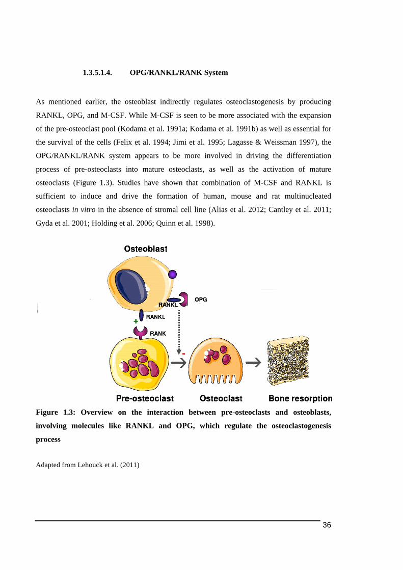
36
1.3.5.1.4. OPG/RANKL/RANK System
As mentioned earlier, the osteoblast indirectly regulates osteoclastogenesis by producing
RANKL, OPG, and M-CSF. While M-CSF is seen to be more associated with the expansion
of the pre-osteoclast pool (Kodama et al. 1991a; Kodama et al. 1991b) as well as essential for
the survival of the cells (Felix et al. 1994; Jimi et al. 1995; Lagasse & Weissman 1997), the
OPG/RANKL/RANK system appears to be more involved in driving the differentiation
process of pre-osteoclasts into mature osteoclasts, as well as the activation of mature
osteoclasts (Figure 1.3). Studies have shown that combination of M-CSF and RANKL is
sufficient to induce and drive the formation of human, mouse and rat multinucleated
osteoclasts in vitro in the absence of stromal cell line (Alias et al. 2012; Cantley et al. 2011;
Gyda et al. 2001; Holding et al. 2006; Quinn et al. 1998).
Figure 1.3: Overview on the interaction between pre-osteoclasts and osteoblasts,
involving molecules like RANKL and OPG, which regulate the osteoclastogenesis
process
Adapted from Lehouck et al. (2011)

37
Briefly, the regulation of osteoclastogenesis by OPG/RANKL/RANK system could be
summarised in Figure 1.3 above. RANKL expressed by either osteoblasts or osteocytes binds
to its natural receptor RANK (Anderson et al. 1997; Nakashima et al. 2011; Xiong & O'Brien
2012; Yasuda et al. 1998a) on the surface of the pre-osteoclast. There have been controversies
over the cells expressing the RANKL supporting for osteoclast formation as there have been
reports in the early days of the OPG/RANK/RANKL system discovery indicating osteoblasts
or stromal cells are the cells providing such RANKL. However more recent studies indicating
that osteocytes actually perform the function of releasing the RANKL for promoting
osteoclastogenesis instead of osteoblasts (Kurata et al. 2006; Nakashima et al. 2011; Xiong &
O'Brien 2012; Zhao et al. 2002).
The binding of RANKL to RANK triggers an intracellular signalling pathway, which begins
with the activation of JNK, followed by recruitment and activation of a number of TNF
receptor activating factor (TRAF) members, especially TRAF6 adaptor protein (Wong et al.
1998), which then activates nuclear factor-kappaB (NFκB). The activated NFκB plays an
important role in inducing the activation of nuclear factor of activated T-cells 1 (NFATc1).
The activated NFATc1 co-operates with other transcription factors to induce the transcription
of osteoclastic-specific genes (Asagiri and Takayanagi 2007). The crucial role of NFATc1 in
osteoclastogenesis process is presented in detail in section 1.3.5.1.6 below. The presence of
OPG, the negative regulator of osteoclastogenesis, prevents such events from occurring by
binding to RANKL, thereby preventing RANKL from interacting with RANK (Lacey et al.
1998; Yasuda et al. 1998b) (refer Figure 1.3).
1.3.5.1.5. Intracellular Signalling Post-RANK Activation
TRAF6 has been shown to play a crucial role in the intracellular signalling pathway of
RANKL-induced osteoclast activation rather than differentiation. Even though
osteoclastogenesis still occurs, TRAF6-knockout mice was found to have an osteopetrotic
phenotype following defect in cytoskeletal organization (Armstrong et al. 2002; Lomaga et al.
1999). This is supported by another study by Naito and colleagues (1999) who demonstrated
that lack of TRAF6 appeared to cause defective osteoclastogenesis. Subsequent work done by
Gohda et al. (2005) suggested that, following induction on the RANK in RANKL-induced
osteoclastogenesis, TRAF6 transduces the intrinsic signal which later lead to the increase in
38
the gene expression of the newly known key regulator of osteoclastogenesis, NFATc1. It has
been suggested that activation of TRAF6 could lead to the dephosphorylation of NFATc1
mediated by JNK, p38 and NFκB1 (p50/p105) and NFκB2 (p52/p100) as the activation of
those molecules could not be observed in TRAF6-deficient mouse (Hirotani et al. 2004). This
is further supported by work done by Franzoso et al. (1997) and Iotsova et al. (1997) in which
gene-knockout on both p50 and p52 was demonstrated to lead to osteopetrosis due to a lack of
osteoclasts, suggesting an essential role of NFκB molecule in mediating osteoclastogenesis.
Another pathway believed to relay the intracellular signal from RANKL stimulation besides
TRAF6 is via the expression of c-fos, another crucial component of osteoclastogenesis
(Grigoriadis et al. 1994). c-fos is a factor that heterodimerizes with c-Jun to form the another
important transcription factor, activator protein-1 (AP-1).
1.3.5.1.6. NFATc1- The Key Transcriptional Factor in
Osteoclastogenesis
NFATc1 has been discovered as the master transcriptional factor and key regulator involved
in mediating the osteoclastogenesis process. The discovery of NFATc1 as a key regulator in
osteoclastogenesis (Ishida et al. 2002; Takayanagi et al. 2002) has opened up a new era in the
understanding of osteoclast cellular biology. Since this high impact discovery, there have
been numerous studies, providing great insights into osteoclast biology and the field of
osteoimmunology.
NFAT transcription factor family was first identified in T-cells (Shaw et al. 1988). Currently
there are 5 members identified in this transcription factor family, which are NFAT1
(NFATc2), NFAT2 (NFATc1), NFAT3 (NFATc4), NFAT4 (NFATc3) and NFAT5 (Rao et
al. 1997; Takayanagi 2007b). It seemed to be very challenging to study the function of
NFATc1 in vivo in early days as targeted deletion on NFATc1 resulted in embryonic lethality
as it is also important in cardiac morphological formation (de la Pompa et al. 1998; Ranger et
al. 1998). The essential role NFATc1 plays in osteoclastogenesis could be seen from the
retardation of osteoclastogenesis in vitro resulting from the suppression of NFATc1 (Ishida et
al. 2002) and knockout of NFATc1 in embryonic stem (ES) cells (Takayanagi et al. 2002).
39
Dephosphorylation of the hyperphosphorylated cytoplasmic NFATc1 allows the transcription
factor to be activated and translocated into the nucleus (Hirotani et al. 2004). The activation
of phospholipase C by TRAF6 produces inositol triphosphate (IP3) which mediates the
increase in the intracellular calcium level that in turn leads to the activation of calcineurin,
which is responsible for dephoshorylation of NFATc1 (Hirotani et al. 2004). Another
mechanism that is thought to mediate the dephosphorylation of NFATc1 is through the
inhibition of glycogen synthase kinase-3, the principal inhibitor for NFATc1 translocations
into the nucleus (Beals et al. 1997), resulting from the activation of TRAF6.
NFATc1 expression was notably upregulated in response to RANKL stimulation during
osteoclast differentiation period (Hirotani et al. 2004; Ishida et al. 2002; Takayanagi et al.
2002). Meanwhile, the over-expression (Takayanagi et al. 2002) or ectopic expression of
constitutively active NFATc1 (Hirotani et al. 2004; Kim et al. 2005b) is sufficient to bypass
the requirement of RANKL for osteoclastogenesis in vitro (as indicated by formation of
TRAP-positive multinucleated cells), implying that up-regulation of NFATc1 expression is
crucial to mediate the RANKL stimulation. In model of RAW264.7 cell line, stimulation by
soluble RANKL protein was reported to increase the mRNA expression of NFATc1, NFATc2
and NFATc3. Nevertheless, only the induction of NFATc1 mRNA expression was found to
be more than two-fold, occurring in less than 5 hours and remained high until 24 hour post
RANKL stimulation and the nuclear localisation of NFATc1 was found to take place after 24
hour post- RANKL stimulation (Ishida et al. 2002).
NFATc1 has been reported to be able to auto-amplify its own expression following the
induction for osteoclastogenesis and the auto-amplification lasts until the end of the
differentiation process (Asagiri et al. 2005). Regulation at epigenetic level was believed to
underlie this mechanism of NFATc1 auto-amplification (Asagiri et al. 2005). It was also
found out that the process of auto-amplification of NFATc1 is initiated by temporal high
expression NFATc2 occuring at the early stage following RANKL stimulation (Asagiri et al.
2005). An earlier study by Ikeda and co-workers (2004) indicated that overexpression of
NFATc2 was able to enhance osteoclastogenesis. It appears that NFATc2 is involved in the
mechanism by directly binds to the promoter of NFATc1, as evidenced from discovery of
NFATc2 binding site on NFAc1 promoter region (Zhou et al. 2002), and upregulating the
expression of NFATc1 (Asagiri et al. 2005). This NFATc1 auto-amplification during the
osteoclastogenesis (Asagiri et al. 2005) supported by sustaining calcineurin-mediated Ca2+
40
oscillation following RANKL stimulation (Takayanagi et al. 2002) (which promote nuclear
localisation for its own and other gene transcription), indicates that NFATc1 actively plays a
central role in the molecular mechanism underlying osteoclastogenesis, particularly on
osteoclast cell fate determination in late stage of the process.
NFATc1 is believed to work with other transcription factors in synergy to initiate gene
transcription (Kim et al. 2005d). NFATc1 is believed to co-operate with c-Fos, an upstream
component of AP-1, in inducing osteoclast-specific genes (Takayanagi et al. 2002) by binding
on the promoter regions of those genes. NFAT binding sites have been identified in the gene
promoter regions of those osteoclast-specific markers such as Acp5 (TRAP-encoding gene)
(Takayanagi et al. 2002), Calcr (expressing calcitonin receptor) (Matsuo et al. 2004;
Takayanagi et al. 2002), Ctsk (encoding cathepsin K) (Matsumoto et al. 2004; Takayanagi et
al. 2002), and OSCAR-encoding gene (Kim et al. 2005b; Kim et al. 2005d) (OSCAR is going
to be discussed in further detail in the following 1.3.5.2.2 section). Another such example of
osteoclast-inducible gene targeted by NFATc1 is the β3-integrin encoding-gene (Crotti et al.
2006; Crotti et al. 2008), a cell attachment-facilitated receptor which facilitates the adhesion
and motility of osteoclasts required for the osteoclast differentiation and bone resorption
process (as described earlier in 1.3.2). NFATc1 cooperatively binds with PU.1, a member of
the ETS family of transcription factors to induce the gene expression of the β3-integrin (Crotti
et al. 2008).
NFATc1 was also found to be involved in osteoclast multinucleation process by directly
regulates the mRNA expression of a couple of cell fusion-mediating molecules, which are
dendritic-cell transmembrane protein (DC-STAMP) and Atp6vod2 (described earlier in
section 1.3.1.2 above) with the discovery of the NFAT-binding sites within the promoter
regions of those protein-encoding genes (Lee et al. 2006b). Inhibition of NFATc1 nuclear
localisation by cyclosporin A (CsA) appeared to retard the multinucleation process in
response to RANKL stimulation (Ishida et al. 2002). Overexpression of DC-STAMP and
Atp6vod2 was found to drive and restore cell-cell fusion process even under inhibition by
CsA (Lee et al. 2006b). Even though the intracellular localisation of NFAT family members
is a very strictly controlled mechanism (Crabtree & Olson 2002; Macian et al. 2001), however
excessive NFATc1 level is believed to be able to overcome the tight regulation (Takayanagi
et al. 2002).
41
1.3.5.1.7. Pathological Relevance of OPG/RANKL/RANK System
In pathological bone loss it is believed that there is an alteration in bone metabolism resulting
in excessive osteoclast formation and hence bone resorption following a shift in the
RANKL/OPG ratio (Crotti et al. 2003; Crotti et al. 2004; Haynes et al. 2001a) as well as a
possible suppression of osteoblast differentiation and activity (Schett 2009; Walsh &
Gravallese 2010).
In the context of inflammation-mediated bone loss, the inflammation (presence of pro-
inflammatory cytokines) within the tissue results in alteration in the expression level of
RANKL and OPG on the osteoblasts towards in favour of increase in osteoclastogenesis,
which eventually leads to excessive bone resorption (Boyle et al. 2003; Crotti et al. 2003;
Crotti et al. 2004; Crotti et al. 2002; Haynes et al. 2003; Haynes et al. 2001a; Haynes et al.
2001b; Lerner 2006). This imbalance in RANKL and OPG does not only affect the
osteoclastogenesis directly, however it could also be associated with the increase recruitment
of inflammatory cells including osteoclast precursor cells through the regulation of associated
chemokines. For instance, RANKL has been shown to upregulate the expression of MIP-1α,
which is one of the chemokines shown to increase osteoclast motility and its receptor CCR1
expressed on osteoclast precursor cells (Yu et al. 2004). In addition, high titre of RANKL is
also believed to enhance the osteoclast bone resorbing activity and this could be seen in vitro
data indicated that higher dose of RANKL (50 instead of 10ng/ml) led to formation of more
pits on dentine (Holding et al. 2006). The study also reported synergistic effect on the volume
of dentine resorbed when combining RANKL and pro-inflammatory cytokine TNFα in the
treatment on the culture osteoclast-like cells (Holding et al. 2006).
There is quite a number of convincing evidence indicating a shift in the RANKL/OPG ratio in
tissues of local inflammation-mediated bone loss disease is associated with the severity of the
diseases such as in peri-implant osteolysis (Gehrke et al. 2003; Haynes et al. 2001b; Haynes
et al. 2004; Mandelin et al. 2003) and RA (Crotti et al. 2002; Haynes et al. 2003;
Vanderborght et al. 2004). Some studies suggest that the shift in the RANKL/OPG ratio is
resulted from the elevated expression of RANKL (Crotti et al. 2004; Crotti et al. 2002; Kawai
et al. 2006; Wara-aswapati et al. 2007), while others indicate that the alteration in the ratio is
42
also accompanied with decrease in OPG expression as well (Crotti et al. 2003; Haynes et al.
2001b; Lu et al. 2006).
The sources of high RANKL levels in PO is believed to include osteoblasts (Pioletti &
Kottelat 2004), fibroblasts (Horiki et al. 2004; Quinn et al. 2000), activated T-lymphocytes,
giant cells and macrophages (Gehrke et al. 2003; Haynes et al. 2001b; Haynes et al. 2004;
Mandelin et al. 2003). However, the main cell type thought to be responsible for the release of
high level of RANKL in the tissue around the prostheses are macrophages following
phagocytosis of wear particles (Crotti et al. 2004; Holding et al. 2006).
In the context of PO pathology, it has been reported that wear debris, even at low
concentration, was shown to be able to increase the expression of RANKL and M-CSF by
osteoblast culture (Pioletti & Kottelat 2004). Meanwhile in vivo, metals and PE wear debris
has been shown to increase RANKL/OPG ratio in mouse calvarial model (Masui et al. 2005).
Kim and colleagues (2001) found that there was lower level of OPG in synovial fluid of PO in
comparison to OA. More interestingly, this low level of OPG could be associated with
osteoclast formation and activity in PO as it was found that addition of the synovial fluid
taken from PO patients into an osteoclast culture enhanced the formation of osteoclasts and
addition of exogenous OPG could counteract the effect (Kim et al. 2001).
It is interesting to note that cells isolated PO tissues have the ability to readily form
osteoclasts in the absence of RANKL as demonstrated by a number of studies (Haynes et al.
2001b; Neale & Athanasou 1999; Sabokbar et al. 2003). The ability of
monocytes/macrophages isolated from PO tissues to readily differentiate into osteoclasts
capable of resorbing bone in the absence of support from osteoblasts or RANKL
supplementation (Haynes et al. 2001b; Sabokbar et al. 2003) might be explained by the ability
of the cells to express RANKL and RANK besides abundance of TNFα themselves (Holding
et al. 2006). This theory is supported by the immunohistochemistry data from the examination
on the PO tissues, in which higher expression of RANKL and RANK in PO tissues than in
OA and normal tissues was reported, with the expression to be mainly in cytoplasm of
aggregated cells (Crotti et al. 2004; Haynes et al. 2001b). About half population of the cells
expressing RANKL was reported to be positive for CD68, which is a cell marker for activated
macrophages (Crotti et al. 2004; Sabokbar et al. 1997). It has also been noticed that in human
43
PO tissues, RANK was found to be expressed by similar cells that also expressing RANKL,
including strongly in multinucleated osteoclast-like cells containing PE particles (Crotti et al.
2004; Holding et al. 2006). Addition of OPG was found able to inhibit osteoclast formation
from cells isolated from around failed prostheses (Itonaga et al. 2000).
Similar to PO, in RA there have been numerous studies indicating shift in the expression of
RANKL and OPG associated with the progression of the disease. A number of studies have
demonstrated that RANKL was highly expressed, both at mRNA and protein level, in
synovial tissues taken from RA patients (Crotti et al. 2002; Gravallese et al. 2000; Horwood
et al. 1999; Kotake et al. 2001; Shigeyama et al. 2000). The data indicating higher expression
of RANK in active RA tissues than in normal and OA groups (Haynes et al. 2001a) may
further support the idea that the OPG/RANK/RANKL system has association with RA
disease progression. This high expression of RANKL in the inflamed tissue in RA is believed
to be one of the main factors that is responsible for the increase in osteoclastogenesis which
eventually leads to excessive bone resorption (Haynes et al. 2001a). This is supported by the
finding that there is positive correlation between severity of synovitis in the RA patients and
the increase in RANKL (Crotti et al. 2002) accompanied with drop in the OPG expression
(Haynes et al. 2003).
A study using an RANKL-knockout mouse model demonstrated the failure of getting any
expected sign of arthritis following arthritic serum transfer (Pettit et al. 2001). Similarly, in a
rat model of collagen-induced arthritis treatment with OPG-fusion protein (OPG-Fc) was able
to arrest the bone erosion in the model by reducing the number of osteoclasts formed by over
75% (Romas et al. 2002). This is further supported by another in vitro study which
demonstrated that treatment with OPG alone is sufficient to significantly arrest TNFα-
mediated bone destruction by limiting the number osteoclasts formed (Redlich et al. 2002b).
There is accumulating evidence suggesting that activated T-cells (Haynes et al. 2003;
Horwood et al. 1999; Kim et al. 2000; Kong et al. 1999; Kotake et al. 2001; Weitzmann et al.
2001) and synovial fibroblasts (Quinn et al. 2000; Takayanagi et al. 2000a) as might be the
main source for the high expression of RANKL in the RA inflamed joints. Even though B
lymphocytes are also associated with RANKL production and may play role in
osteoclastogenesis (Manabe et al. 2001), this appeared to not to be in the context of RA
tissues (Kotake et al. 2001).
44
Having established that the increase in RANKL plays a major role in RA bone resorption, it
was suggested that treatment with OPG or molecules mimicking the protein might have
potential to overcome or reduce the excessive bone erosion occurs in RA. The potential of this
strategy appears to be promising as studies by Cheng et al. (2004), using a peptide that
functions like OPG, and Onyia et al. (2004) who used a molecules that modulates endogenous
OPG expression managed to demonstrate the ability of those tested molecules in reversing the
bone loss in ovariectomised mouse as well as in amine-induced adjuvant arthritis (Cheng et
al. 2004; Onyia et al. 2004).
Besides PO and RA, there are also other diseases being associated with alteration in
expression of OPG/RANK/RANKL. Mutations in RANK is associated with excessive
osteoclast activity resulting bone abnormality as been observed in familial expansile
osteolysis (Hughes et al. 2000) and familial Paget disease (Whyte & Mumm 2004; Whyte et
al. 2002). Meanwhile, diseases like idiopathic hyperphosphatasia (Cundy et al. 2002) and
juvenile Paget disease (Whyte & Mumm 2004; Whyte et al. 2002) was reported to have
relationship with functional mutation in OPG. On the other hand, OPG gene polymorhism
seemed to be associated with inreased risk of osteoporotic fracture (Arko et al. 2005; Arko et
al. 2002). The relevance RANK/RANKL/OPG system has in clinical sense has opened up
windows for becoming good therapeutic target for the treatment of bone loss-associated
diseases.
1.3.5.2. ITAM-mediated Signalling Pathway
It seems like there is another concurrent intracellular signalling pathway involved in driving
osteoclastogenesis since it is less likely that the stimulation of the RANK receptor, which
belongs to the tumor necrosis family receptor family (Anderson et al. 1997; Wong et al.
1997b), directly induce calcium signalling (Kim et al. 2005d). A number of publications have
suggested that ITAM signalling might be involved co-stimulating osteoclastogenesis along
together with RANK-RANKL pathway (Humphrey et al. 2006; Koga et al. 2004; Mocsai et
al. 2004). The involvement of this co-stimulating pathway is thought to enable a fine-tune
regulation of osteoclastogenesis (Herman et al. 2008).
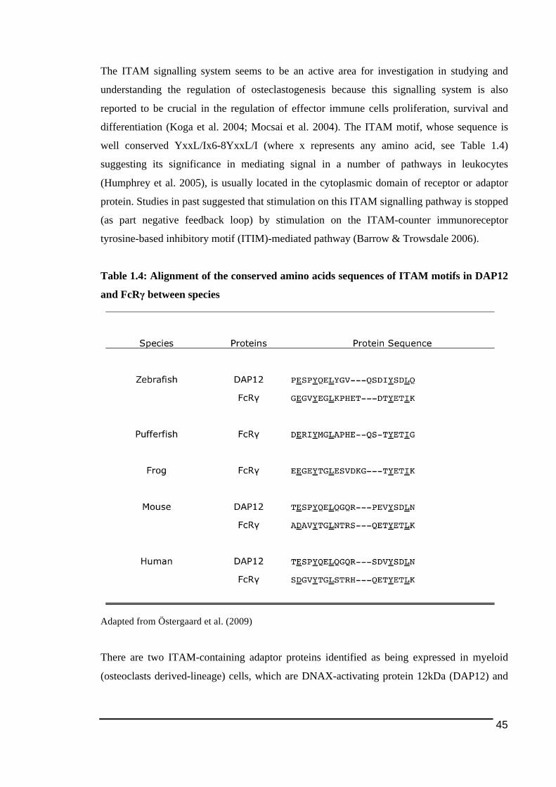
45
The ITAM signalling system seems to be an active area for investigation in studying and
understanding the regulation of osteclastogenesis because this signalling system is also
reported to be crucial in the regulation of effector immune cells proliferation, survival and
differentiation (Koga et al. 2004; Mocsai et al. 2004). The ITAM motif, whose sequence is
well conserved YxxL/Ix6-8YxxL/I (where x represents any amino acid, see Table 1.4)
suggesting its significance in mediating signal in a number of pathways in leukocytes
(Humphrey et al. 2005), is usually located in the cytoplasmic domain of receptor or adaptor
protein. Studies in past suggested that stimulation on this ITAM signalling pathway is stopped
(as part negative feedback loop) by stimulation on the ITAM-counter immunoreceptor
tyrosine-based inhibitory motif (ITIM)-mediated pathway (Barrow & Trowsdale 2006).
Table 1.4: Alignment of the conserved amino acids sequences of ITAM motifs in DAP12
and FcRγ between species
Adapted from Östergaard et al. (2009)
There are two ITAM-containing adaptor proteins identified as being expressed in myeloid
(osteoclasts derived-lineage) cells, which are DNAX-activating protein 12kDa (DAP12) and
46
Fc receptor common gamma subunit (FcRγ) (Humphrey et al. 2005). The significance of
ITAM-containing adaptor proteins in the development of functional osteoclasts appears to be
equivalent to the essential role RANKL/RANK axis plays and this is shown in several studies
(Humphrey et al. 2004; Koga et al. 2004) (to be discussed in further in the following section).
Due to lack or minimal extracellular domain for ligand binding and hence unlikely direct
binding to the ligands (Abram & Lowell 2007; Lanier & Bakker 2000; Takai et al. 1994),
DAP12 and FcRγ must pair to other receptors in order to sense and receive signals from the
extracellular environment (Lanier & Bakker 2000; Takai et al. 1994). OSCAR and triggering
receptor expressed by myeloid cells (TREM)-2 are examples of receptors that have been
found to pair with FcRγ and DAP12 respectively via ionic interaction (positively charged
transmembrane residue on receptor binds to the negative charge on adaptor proteins
contributed from the amino acids) (Bouchon et al. 2001; Daws et al. 2001; Daws et al. 2003;
Hamerman et al. 2006; Humphrey et al. 2005; Ishikawa et al. 2004; Lanier & Bakker 2000;
Long & Humphrey 2012). A single mutation on any of those amino acids is sufficient to
disrupt the ionic interaction (Lanier et al. 1998a; Wu et al. 2000). These transmembrane
receptors, together with their corresponding ITAM-containing adaptor proteins, are going to
be discussed further below.
The ITAM signalling system involves intracellular signal transduction following the
stimulation on the ITAM-adaptor associated receptor, resulting phosphorylation of tyrosine
within the ITAM on cytoplasmic domains of the adapter molecules (Humphrey et al. 2005).
In osteoclast precursors, the phosphorylation of tyrosine within the ITAM allows the
recruitment of Syk to the motif, which serves as the docking site (McVicar et al. 1998). This
is then followed by the activation of the Syk kinase (Faccio et al. 2003c; Koga et al. 2004;
Mocsai et al. 2004; Zou et al. 2007) through binding of the Src homology 2 (SH2) domain of
the Syk’s C-terminal to the ITAM motif (Futterer et al. 1998; Mocsai et al. 2004) and
autophosphorylation of phospholipase Cγ (PLCγ) (Pitcher & van Oers 2003) that in turn
mediates several other intracellular pathways that take place during osteoclastogenesis
process. It has been reported that deficiency or inhibition of Syk tyrosine kinase resulted in
defect in osteoclast differentiation (Koga et al. 2004; Mocsai et al. 2004) and function (Zou et
al. 2007).
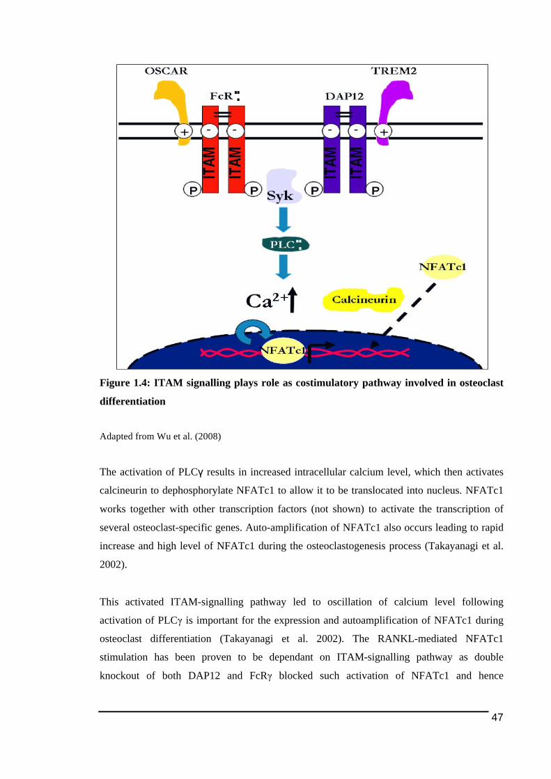
47
Figure 1.4: ITAM signalling plays role as costimulatory pathway involved in osteoclast
differentiation
Adapted from Wu et al. (2008)
The activation of PLCγ results in increased intracellular calcium level, which then activates
calcineurin to dephosphorylate NFATc1 to allow it to be translocated into nucleus. NFATc1
works together with other transcription factors (not shown) to activate the transcription of
several osteoclast-specific genes. Auto-amplification of NFATc1 also occurs leading to rapid
increase and high level of NFATc1 during the osteoclastogenesis process (Takayanagi et al.
2002).
This activated ITAM-signalling pathway led to oscillation of calcium level following
activation of PLCγ is important for the expression and autoamplification of NFATc1 during
osteoclast differentiation (Takayanagi et al. 2002). The RANKL-mediated NFATc1
stimulation has been proven to be dependant on ITAM-signalling pathway as double
knockout of both DAP12 and FcRγ blocked such activation of NFATc1 and hence
48
osteoclastogenesis (Koga et al. 2004). The RANKL-induced osteoclastogenesis in the DAP12-
/- FcRγ-/- model could be restored by ectopic expression of NFATc1 (Koga et al. 2004).
Not only important in mediating the process of osteoclast differentiation, ITAM signalling
pathway has also been found to play significant role in influencing the activity or capacity of
osteoclasts as well. Furthermore, both ITAM-mediated pathways in osteoclasts, which include
OSCAR/FcRγ and TREM2/DAP12, seems to work in synergy and coherent manner in
regulating the resorption activity or capacity of oscteoclasts. This could be seen from the
mutation in both DAP12 and FcRγ that appeared to result in more severe osteopetrosis as well
as reduction in bone resorption in vivo as well as more defect on osteoclast activity and
formation in vitro than mutation in either DAP12 or FcRγ alone (Koga et al. 2004; Mocsai et
al. 2004). The theory has also been supported by data from Kim et al. (2005) (Kim et al.
2005d), which indicated that the blockade of both TREM2 and OSCAR led to greater
suppression on osteoclasts than the inhibition on either one of those receptors. This theory
was also further supported by a recent study in mouse in vivo indicating double knockout on
both OSCAR and DAP12 that led to reduction in TRAP-positive osteoclasts total number and
bone loss by about a half as compared to cells lack in DAP12 or OSCAR alone (Barrow et al.
2011).
There are a few possibilities on the mechanism of regulation on osteoclast bone-resorbing
capacity by the ITAM signalling pathway in osteoclasts. The first one would possibly be
through the cell polarisation (during cell attachment to the bone, see section 1.3.2) as it was
found that DAP12 and FcRγ appeared to mediate αvβ3-induced cytoskeletal organisation (Zou
et al. 2007; Zou et al. 2010). The second mechanism ITAM signalling pathway might affect
osteoclast activity or capacity is through the stage of cell multinucleation, which has been
suggested based on data from several previous studies. In vitro examination on osteoclast-like
cells derived from DAP12 and FcRγ-deficient mice demonstrated a defect in multinucleation
of osteoclasts as well as retardation in the expression of NFATc1 (Koga et al. 2004; Mocsai et
al. 2004). The defect in the multinucleation of DAP12 and FcRγ-deficient osteoclasts appears
to be resulted from the failure in expressing NFATc1 as introduction and transduction of
NFATc1 was able to restore the capability of the cell to differentiate appropriately (Koga et
al. 2004). It is quite interesting to note that deficiency of DAP12 in macrophages (cell type
sharing similar progenitor to osteoclasts) in DAP12-knockout mice also impaired the ability
of the cells to fuse and form multinucleated giant cells (Helming et al. 2008). Considering
49
multinucleation is related to the degree of osteoclast bone resorbing activity (Boissy et al.
2002), in this study, it is generally hypothesized that regulation of ITAM-related molecules
may have significant impact in the increase of bone resorption observed in PO and RA (see
1.4.1 below).
Collectively, all those studies described above suggested that there is crucial role ITAM
signalling pathway has in mediating osteoclastogenesis beside RANKL-induced signalling
pathway. Considering this, it would be interesting and worthwhile to study this signalling
pathway by looking at the associated molecules such as TREM2 and OSCAR more closely.
Those osteoclast ITAM-associated molecules are further discussed below.
1.3.5.2.1. TREM2 and DAP12
DAP12 is a membrane adaptor molecule containing ITAM identified as expressed in
osteoclasts couples together with TREM2. DAP12 is also known as tyrosine kinase binding
protein (TYROBP) or KARAP (Lanier et al. 1998b; Paloneva et al. 2000). Comparison on the
homologs between species found DAP12 in human shares 75% sequence homology with the
ones in mouse.
DAP12, which was first discrovered in natural killer (NK) cells as dimer of adaptor
protein/receptor complex (covalently attached to NIC-receptor) (Lanier & Bakker 2000), is
also identified as being expressed in many other myeloid cell types, such as macrophages,
monocytes, dendritic cells, granulocytes as well as some T and B lymphocytes (Chen et al.
2009; Goronzy et al. 2005; Humphrey et al. 2004; Lanier & Bakker 2000; Tomasello et al.
1998).
DAP12 is a transmembrane protein identified as an important component in transducing
activating signals in NK cells for targeting cells to be lysed (Campbell & Colonna 1999;
Colonna 1998; Lanier et al. 1998b; McVicar et al. 1998). It seems like DAP12 play
significant role in monocyte differentiation in general, as it appears that the adaptor protein
was found to modulate and expressed during monocyte differentiation into macrophages
(Aoki et al. 2000) and dendritic cells (Bouchon et al. 2001), other cell types sharing similar
progenitor osteoclasts. In dendritic cells, DAP12 also appears to play important role in cell
50
maturation and prolonging cell survival by enhancing antigen presentation through the
upregulation of CD40, CD86 and MHC class II (Bouchon et al. 2001).
Even though DAP12 has been reported to play various important roles in numerous cell types,
the signficance of this molecule in bone homeostasis, in the context of osteoclasts in
particular, should also be acknowledged. As briefly described earlier (see 1.3.5.2 above),
DAP12 appears to play role in bone resorption, and this could be seen from DAP12-deleted
mice exhibiting higher bone mass of femur and tibia in addition to increased trabecular
thickness (Humphrey et al. 2004). As mentioned previously, it has been reported that DAP12-
deficient mice developed severe osteopetrosis and in vitro works demonstrated that the
osteoclast precursor cells isolated from the mice did not undergo differentiation (Koga et al.
2004; Mocsai et al. 2004; Takai et al. 2005). It was suggested that the suppression on the
osteoclastogenesis in the DAP12-/- mice-derived cells occurred between the intermediate and
late stage as mononuclear cells derived from these mice induced into osteoclastogenesis
following RANKL stimulation in vitro were still found to be TRAP-positive and expressed
osteoclast-specific marker like cathepsin K, β3-integrin, calcitonin receptor, RANK and
OSCAR, nevertheless lost its capability of resorbing bone (Humphrey et al. 2004; Mocsai et
al. 2004). Very recent study by Despars and coworkers (2013) found that DAP12
overexpression could promote osteoclastogenesis by increasing the proliferation rate for the
osteoclast progenitors. It turns out that DAP12 is critical in osteoclastogenesis as retroviral
introduction of DAP12 into DAP12-/-/FcRγ-/- is able to restore the capability of the osteoclast
precursor cells to get multinucleated and differentiated into mature functional osteoclasts
(Humphrey et al. 2004; Mocsai et al. 2004; Paloneva et al. 2003).
DAP12 also has role in osteoclast bone resorbing activity through cyoskeletal reorganisation
(Zou et al. 2010) triggered by M-CSF (Otero et al. 2009). This DAP12-induced cytoskeletal
reorganisation was found to be mediated by Syk phosphotyrosine (Faccio et al. 2003c).
Targeted deletion on Syk, which is downstream of DAP12 (Koga et al. 2004), led to the
phenotype of failure in cytoskeletal dysfunction that led to inability to resorb mineralised
substrate in vitro (Zou et al. 2007). Besides failure in actin ring and ruffle border formation,
the dysfunction of cytoskeletal reorganization in DAP12-/- cells were further supported by
inability of the mutant osteoclasts formed to transmigrate through osteoblast layer (Zou et al.
2010). The signficance of this TREM2/DAP12-mediated ITAM signalling pathway in
osteoclast cytoskeletal reorganisation is redemonstrated by Faccio and colleagues (2005),

51
whom reported that deficiency in Vav3, another candidate target downstream of those ITAM
molecules, led to failure in actin-cytoskeleton organisation and defect in resorption activity.
Besides TREM2, DAP12 could also couple with other receptors such as myeloid DAP12-
associated lectin (MDL-1), TREM1, TREM3, NKGD2 and signal regulatory protein β
(SIRPβ) (Daws et al. 2001; Humphrey et al. 2004; Joyce-Shaikh et al. ; Long & Humphrey
2012). However, considering TREM2 is the particular immunoreceptor that paired to DAP12
in osteoclasts (Daws et al. 2001; Humphrey et al. 2006), the scope of these studies will focus
on TREM2. In addition, it has been suggested by quite a number of studies that TREM2 is
linked to osteoclastogenesis.
TREM2 is a member the TREMs family (Bouchon et al. 2000) that also consists of TREM1,
TREM3 and recently identified TREM-like 4 (Hemmi et al. 2009). TREM2 are reported to be
widely expressed by cells from myeloid lineage such as dendritic cells, macrophages,
microglial cells and osteoclasts (Bouchon et al. 2000; Daws et al. 2001; Paradowska-Gorycka
& Jurkowska 2013). TREM2 was first discovered in human monocyte-derived dendritic cells
and mouse macrophage RAW264.7 cell line (Bouchon et al. 2001; Daws et al. 2001). Of note,
TREM2 expression level appears to be relatively constant regardless of stimulation by
RANKL and regulation of the interaction seems to dominantly influenced by the expression
of the ligand (Kim et al. 2005d). To date, the ligand for TREM2, including the one expressed
in osteoclasts, is still unknown. Humphrey et al. (2006) (Humphrey et al. 2006) suggested that
TREM2 signalling might be activated by negatively charged inorganic bone matrix, hence
may play role in mediating osteoclast development in bone microenvironment. TREM2 has
been shown to bind to anionic ligands possibly through pattern recognition (Daws et al. 2003;
Roodman 1999). A more recent study suggested plexin A1, a receptor for semaphorins, as
potential activating ligands in TREM2/DAP12-mediated signalling pathway during
osteoclastogenesis as stimulation of osteoclast precursors with cells deficient in plexin A in
vitro led to reduction in osteoclast formation and in vivo mouse model lack of plexin A1
manifested osteosclerosis together with increased bone mass (Takegahara et al. 2006). In the
latest discovery on ligand of TREM2, TREM2 was reported to bind bacterial LPS (Quan et al.
2008) and has been suggested to function as phagocytic receptor for bacteria in macrophage
(N'Diaye et al. 2009).
52
Deficiency in TREM2 was found to result in arrest in the differentiation of human pre-
osteoclasts into functional osteoclasts (Cella et al. 2003; Paloneva et al. 2003). In the
RAW264.7 cell line, blockade of TREM2 and its interaction with its ligand led to the
inhibition of osteoclastogenesis including on the ability of osteoclasts to resorb bone
efficiently as well as their capability to migrate (Humphrey et al. 2006). On the other hand,
stimulation of TREM2 mouse pre-osteoclasts appeared to promote osteoclast multinucleation
and migration/chemotaxis (Helming et al. 2008; Humphrey et al. 2006). However, in a study
by Paloneva et al. (2003) it was reported that mutations in DAP12 and TREM2 did not lead to
defect in the osteoclastogenesis, but only resulted in delay in osteoclasts differentiation and
decrease in bone resorption assessed based on dentine pit analysis.
Experiments in TREM2/DAP12 non-knockout models also demonstrated the significance of
TREM2 and DAP12 in mediating osteoclastogenesis. Addition of TREM2-Fc protein , which
provided competition for binding to ligand, into an in vitro osteoclast-osteoblast co-culture
seemed to give inhibitory effect on osteoclasts (Kim et al. 2005d). Suppression of TREM2
through RNA silencing was found to inhibit RANKL-driven osteoclastogenesis in RAW264.7
cell line and mature osteoclast formation and bone resorbing activity (Humphrey et al. 2006;
Park-Min et al. 2009). Meanwhile stimulation of TREM2 in those cells and murine primary
BMMs via antibody cross-linking promoted osteoclast-like cells formation by three-fold in
the presence of RANKL and M-CSF with DAP12 appeared to be essential in mediating the
process (Humphrey et al. 2006).
Unlike OSCAR (to be discussed in the following section), TREM2 appears not to be
regulated by NFATc1, based on the findings that there is no putative NFAT binding site on
TREM2 promoter region and treatment with FK506 (which hence inhibits nuclear localization
of NFATc1) does not significantly affect TREM-2 gene expression (Kim et al. 2005d).
Indeed, there have been more recent studies using in vitro model of PBMC-derived osteoclast
culture suggesting that TREM2 is upstream of NFATc1 expression in the RANKL-induced
CaMK-MEK-ERK the cell signalling pathway (Kalliolias et al. 2010; Park-Min et al. 2009).
They also demonstrated elevation in TREM2 mRNA expression level taking place during
osteoclastogenesis period, in parallel to RANK mRNA expression on the human PBMC-
derived pre-osteoclastic cells. Nonetheless, for mouse TREM2 the gene seems to be regulated
by MITF, a basic helix-loop-helix transcription factor and PU.1 as the promoter region of this
gene contains the binding sites for these transcription factors (Kim et al. 2005d).
53
In the context of human pathology, DAP12 deficiency, as a result of genetic mutation on
chromosome 19 leading to premature termination of the protein, is believed to be the cause of
an uncommon (reported to be among Japanese, Finnish and Italian population thus far),
genetically recessive human disease called polycystic lipomembranous osteodysplasia with
sclerosis leukoencephalopathy (PLOSL), which also known as Nasu-Hakola disease (Kondo
et al. 2002; Nakamagoe et al. 2011; Paloneva et al. 2000; Paloneva et al. 2003; Soragna et al.
2003; Thrash et al. 2009). There is also evidence of the genetic disease associated with
mutation in TREM2 splicing (Numasawa et al. 2011; Thrash et al. 2009). Recent studies also
pointed out TREM2 as strongly involved in Alzheimer’s disease (Fenoglio et al. 2007;
Neumann & Daly 2013).
PLOSL is characterized by osteopenia, which lead to early onset of osteoporosis, multiple
lipid filled bony cysts restricted to wrists and ankles, trabecular bone loss and cortical bone
demineralisation, loss of brain white matter leading to presenile dementia and premature
death before age 50 (Paloneva et al. 2001; Verloes et al. 1997). There is also evidence
associating the disease to the fragility of bone manifested by repeated bone fracture in a
Japanese patient (Nakamagoe et al. 2011) believed to resulted from bone spongy texture
following formation of multiple lipid-filled bone cysts. This disease was suggested to be
associated with deficiency of DAP12, as this could be seen from in vivo data by Kaifu et al.
(2003) indicating that DAP12-deleted mice were shown to manifest bone and brain
abnormalities displayed in PLOSL. However a very recent data by Despars and colleagues
(2013) indicated otherwise as the study reported that osteopenia, which could be seen in
PLOSL, was manifested by transgenic mice overexpressing DAP12. Mutation on TREM2 in
PLOSL is believed to cause brain abnormality in the disease by introducing defect on the
neuronal apoptotic clearance by microglial as TREM2 is found to be important in the
phagocytosis process by microglials (Hsieh et al. 2009; Prada et al. 2006). It is quite
interesting to note that, even though the function of DAP12 is mainly described in NK cells as
important component in mediating signals for targeting cells to be lysed (Campbell &
Colonna 1999; Colonna 1998; Lanier et al. 1998b; McVicar et al. 1998), there was other
study reported no defect on NK cell function in PLOSL patients (Paloneva et al. 2000),
suggesting cellular function is affected by mutation in DAP12/TREM2 more obvious in
monocyte/macrophage lineage (such as microglials (Cuadros & Navascues 1998) and
osteoclasts).
54
Even though there are accumulating studies indicating the significance of TREM2 and
DAP12 in bone homeostasis, there is controversy surrounding the specific or exact role the
activation of these molecules play to be resolved. While majority of those studies above
suggested TREM2/DAP12 is involved in co-stimulating osteoclast differentiation process, a
very recent study by Otero et al. (2012), using a mouse model, in contrast found that
deficiency in TREM2 leads to “accelarated” differentiation of osteoclast accompanied with
earlier experssion of osteoclast-specific markers. Otero et. al. (2012) suggested that the
activation of TREM2/DAP12 signalling pathway may play important role in early stage of
osteoclast development, most likely during the stage of proliferation of precursor cells before
differentiation. Based on the data obtained, they speculated that deletion of TREM2 led
towards earlier cell cycle arrest for proliferation resulting earlier osteoclast differentiation
(Otero et al. 2012). This is supported by earlier findings that reported expression of TREM2
in macrophages in culture containing M-CSF (supplement mainly for cell proliferation) only
(Turnbull et al. 2006).
Such contradictions among all these findings might be attributed by differences in cell types
and models used as well as cellular response in respond to range of stimulating ligands,
avidity of ligands and spectrum of associated-receptors (related to DAP12) (Otero et al.
2012). Therefore, further studies should be carried out in the future on DAP12 and TREM2
molecules in order to gain better understanding on those molecules, not only in the context of
cell and molecular biology, but also more importantly the signficance and relevance of the
protein at higher level of physiology and pathology, if there any. Thus far, DAP12 and
TREM2 are not explored yet in studies investigating local inflammation-mediated bone loss
tissue such as in RA and PO.
1.3.5.2.2. OSCAR and FcRγ
Early in this century, an immunoglobulin-like receptor expressed in murine osteoclasts known
as OSCAR was discovered, thanks to the work done by Kim et al. (2002). OSCAR discovered
via the PCR-Subtraction, was found to play an important role in osteoclastogenesis (Kim et
al. 2002). Human and mouse OSCAR share 73% sequence homology (Kim et al. 2002) and
the expression of OSCAR in bone is highly conserved among various species (Nemeth et al.
55
2011), suggesting a significant role it plays in bone function or homeostasis. OSCAR, which
is a novel member of leukocyte receptor complex (LRC)-encoded protein, was thought to be
expressed at both gene and protein predominantly in mature osteoclasts only in murine bone
tissues and RAW264.7 cells (Kim et al. 2002). However unlike in murine cells, OSCAR
could also be found to be expressed in monocytes (the precursor cell of osteoclasts), dendritic
cells (which is also derived from monocytes (Miyamoto et al. 2001)) in human tissues (Merck
et al. 2005; Merck et al. 2004; Merck et al. 2006; Tenca et al. 2005) and very recently in
endothelial cells (Goettsch et al. 2011; Nemeth et al. 2011).
OSCAR was also reported to be expressed in RAW264.7 cell line-derived osteoclast-like cells
(common model or cell line of osteoclasts used for study purpose as it has been shown to
differentiate into osteoclast-like cells in vitro following stimulation of RANKL and regulation
of NFATc1 (Hirotani et al. 2004; Sinningen et al. 2013), making OSCAR as a good candidate
of marker for osteoclasts (Humphrey et al. 2004; Kim et al. 2002). The expression of OSCAR
is thought to start in the late stage of osteoclast differentiation after the formation of TRAP-
positive pre-osteoclasts (Kim et al. 2002) and NFATc1 expression (Kim et al. 2005b).
OSCAR is a type I transmembrane protein with full length cDNA of 282 amino acids (Kim et
al. 2002; Nemeth et al. 2011) and possesses a positively charged arginine residue in the
transmembrane domain (Kim et al. 2002), which suggests that this immunoreceptor might be
coupled with other transmembrane protein. Based on protein structure modeling, human
OSCAR is expected to contain a signal peptide (Nemeth et al. 2011) (see Figure 1.5)
speculating its secretory function and supporting the idea of soluble isoform (Herman et al.
2008; Khan 2007).
Figure 1.5: Polypeptide structure and domains of human OSCAR structure and
domains
Taken from Nemeth et al. (2011)
56
Figure 1.5 above is a schematic diagram representing full-length encoded human OSCAR
polypeptide. The polypeptide could be divided into four distinct domains, which include a
signal peptide (SP), two Ig-like motifs and transmembrane domain (TM) in between those
two Ig-like domains. Presence of SP supports the possibility of OSCAR present in secreted
form. For the Ig-like motif, when the receptor is bound to the cell membrane, there is one
located in extracellular (EC) and this domain spans from amino acid 38-121. This domain
contains two cysteine residues (C) essential for disulphide bonds for protein stabilisation and
a N-glycosylation site (N), which allows the protein to present as monomer (Merck et al.
2004). The other one located in intracelullar (IC) spans from amino acid 135 to 215 and is
important for mediating signal transduction as it is connected to a short cytoplasmic tail. The
transmembrane domain (TM) consists of 282 amino acids long, including an arginine residue
(positively charged, represented as R) that is essential to get the receptor associated with
FcRγ (Nemeth et al. 2011).
In its function as an immunoreceptor, OSCAR is physically associated with FcRγ chain, an
adaptor protein for Fc receptors (Ishikawa et al. 2004; Koga et al. 2004). The FcRγ chain has
been demonstrated to play a role in facilitating or upregulating the expression of OSCAR on
the cell surface (Ishikawa et al. 2004) in addition to mediating signal transduction (Koga et al.
2004). Besides OSCAR, FcRγ has also been found to couple with other receptor such as
FcRIII (Athanasou & Quinn 1990) and paired Ig receptor-A (PIR-A)(Maeda et al. 1998;
Taylor & McVicar 1999).
A ligand for OSCAR has only recently been identified as an extracellular matrix collagen
(Barrow et al. 2011). Earlier, it has been suggested that the ligand of OSCAR might be
expressed by osteoblasts since it has been demonstrated by Kim and colleagues (2002) that
osteoblasts stained well with OSCAR-Ig fusion protein. This data was also supported by later
study (Kim et al. 2005d) which indicated detection of OSCAR ligand on osteoblast upon
stimulation by osteotropic factors 1, 25-(OH)2 vitamin D3 and PGE2 as well as RANKL.
Herman et al. (2008) later reported that the introduction of OSCAR-Fc protein competed for
OSCAR ligand in homogenous cell culture that eventually led to decrease in osteoclast
formation. This suggested that ligand of OSCAR is expressed in PBMC-cell culture, hence
may further speculate role in cellular amplifying response. Nonetheless, the finding from
Barrow et al. (2011) does not contradict data provided by earlier studies (Kim et al. 2002;
57
Koga et al. 2004) as extracellular matrix (ECM) collagen are expressed by osteoblastic bone-
lining cells (Andersen et al. 2009; Hauge et al. 2001; Miller & Jee 1987). Indeed in his study,
Barrow and colleagues (2011) demonstrated interaction between mononucleated osteoclast-
expressing OSCAR and collagen expressed by bone-lining osteoblasts. The finding of
collagen type I as the ligand for OSCAR may suggest that this receptor might be responsible
for osteoclast differentiation process in close proximity to bone instead in tissues and
circulation.
OSCAR has been shown to play role in immune system (both innate and adaptive) through
dendritic cell maturation and activation (Merck et al. 2005; Merck et al. 2004), MHC class II-
mediated antigen presentation for adaptive immune system (Merck et al. 2004) as well as in
augmenting pro-inflammatory reaction in monocytes and neutrophils by inducing the
secretion of cytokines like granulocyte-macrophage colony-stimulating factor (GM-CSF) and
IL-4 that sustain T-cell proliferation (Merck et al. 2006; Tenca et al. 2005). Besides, OSCAR
also prolongs cell survival through inhibition of apoptosis in dendritic cells, denoted by
reduction in numbers of apoptotic cells and increase in the expression of anti-apoptotic
molecule Bcl-2 (Merck et al. 2005; Tenca et al. 2005). Unlike in osteoclasts, the expression of
OSCAR in dendritic cells is independent of the cell differentiation stage (Merck et al. 2004).
It was suggested that OSCAR has a more specific role in co-stimulating osteoclastogenesis
process based on data documented by a number of studies (Kim et al. 2002; Koga et al. 2004).
An upregulation in the expression of OSCAR has been observed during osteoclastogenesis
(Ishikawa et al. 2004) and downregulation in the gene expression is resulted following
inhibition of the calcineurin-mediated pathway (which also regulates NFATc1 nuclear
translocation) using FK506 (Kim et al. 2005d). Recently, Barrow et al. (2011) demonstrated
that interaction between OSCAR and ligand (collagen) enhanced osteoclastogenesis in vitro
and blocking the interaction would inhibit such enhancement in osteoclastogenesis. More
interestingly, activation of OSCAR- FcRγ was also found to inhibit apoptosis in M-CSF and
RANKL-induced osteoclastogenesis (Zou et al. 2010).
As mentioned earlier, OSCAR gene expression could be directly regulated by NFATc1 like
other genes of osteoclast-specific marker as this has been demonstrated through direct
chromatin immunoprecipitation (ChIP) assay and gene reporter assay (Kim et al. 2005b; Kim
et al. 2005d). Constitutively active NFATc1 was found to promote the expression of OSCAR
58
(Kim et al. 2005b). There are two NFATc1-binding sites identified on mouse OSCAR
promoter region, however the regulation of OSCAR expression occurred mostly on only one
of those binding sites (Kim et al. 2005b). While NFATc1 also has the capability of directly
inducing OSCAR gene expression (Kim et al. 2005d), at the same time NFATc1 has also
been reported to be upregulated downstream of calcium signalling mediated by OSCAR
(Koga et al. 2004). This implies that a positive feedback loop may be involved in the
interaction between OSCAR and NFATc1 (refer Figure 1.6).
59
Figure 1.6: Positive feedback loop between NFATc1 and OSCAR expression and activity
Adapted from Kim et al. (2005b), Kim et al. (2005d) and Koga et al. (2004)
The ligation of RANK to its ligand RANKL triggers a cascade of intracellular signalling pathway that
eventually produces NFATc1, the key transcriptional factor for switching on the gene expression of a
number of osteoclast-specific cells, including OSCAR (Kim et al. 2005b; Kim et al. 2005d). The
transcription of those osteoclast-specific genes by NFATc1 is important in allowing the
osteoclastogenesis process to progress. Meanwhile, the stimulation of OSCAR by its ligand causes the
activation of ITAM in the associated adapter molecules FcRγ, which eventually leads to the activation
of PLCγ and increase in intracellular calcium level. This increase in calcium level is important for the
nuclear localisation of NFATc1 (Koga et al. 2004) and hence promoting further transcription of
osteoclast-specific genes, including OSCAR. Therefore, this interaction between NFATc1 and
OSCAR based on their regulation of expression and activity forms a positive feedback loop.
60
Studies on the mouse promoter region of the OSCAR gene indicate that transcription factors
MITF and PU.1 also play a role in regulating transcription of the OSCAR gene (So et al.
2003), like TREM2 gene (Kim et al. 2005d). It was reported that the stimulation of OSCAR
gene expression by NFATc1, like TRAP (Kim et al. 2005d) and cathepsin K (Matsumoto et
al. 2004), could be further enhanced in synergy with a combination of MITF and PU.1
transcription factors (Kim et al. 2005d; So et al. 2003). Other transcription factors also known
to regulate OSCAR expression include USFs (promote OSCAR gene expression) (Kim et al.
2008) (Figure 1.7) and inihibitory transcription factors inhibitor of differentiation (ID) 2 (Lee
et al. 2006a) and MafB (Kim et al. 2007a). Figure 1.7 represents a schematic diagram of
illustrating the loci on human gene where the transcription factors are bound for initiating
gene transcription. There is a spelling error in the diagram, where MIFT is supposed to be
corrected to MITF.
Figure 1.7: Binding sites for transcription factors to bind on human OSCAR gene
Taken from Nemeth et al. (2011)
Besides transcription factor, there is quite a number of other factors identified to date as able
to regulate the transcription of OSCAR gene, which are compiled and listed in Table 1.5
below.
Further evidence of the significance of OSCAR in mediating osteoclastogenesis could be
observed from the retardation of the osteoclast differentiation process as a result of down-
regulation in the expression of OSCAR and NFATc1 as a consequence of the over-expression
of protein inhibitor of activated STAT 3 (PIAS3) (see Table 1.5) (Kim et al. 2007b). PIAS3 is
postulated to mediate the inhibitory effect on the OSCAR expression by recruiting histone
deacetylase 1 to the promoter of OSCAR (Kim et al. 2007b).
61
Table 1.5: Factors regulating OSCAR mRNA expression in osteoclasts
Category Factors Function
RANKL Cytokine
IL-1 Cytokine
FcRγ Adaptor molecule
NFATc1 Transcription factor
MITF Transcription factor
PU.1 Transcription factor
Inducer
USFs Transcription factor
MafB Transcription factor
ID Transcription factor
PIAS3 Repressor of transcription
KR62776 PPAR-γ
Silibinin Polyphenolic flavonoid
Repressor
MHC CIITA Non-DNA binding
coactivator
Adapted from Nemeth et al. (2011)
Surprisingly, mice deficient in FcRγ, the OSCAR-associated ITAM-containing adaptor
protein, do not exhibit signs of osteopetrosis, suggesting that the role of FcRγ chain could be
compensated by other mechanism or pathway in vivo (Ishikawa et al. 2004; Koga et al. 2004;
Mocsai et al. 2004). The significance of the contribution FcRγ gives towards mediating bone
loss, however, still should not be undermined since mice deficient in both DAP12 and FcRγ
showed more severe osteopetrosis as compared to those which lacked of DAP12 alone even
though in vitro examination demonstrated that either deficiency of DAP12 alone and double
knockout of both DAP12 and FcRγ led to failure for multinucleation (Koga et al. 2004;
Mocsai et al. 2004; Takai et al. 2005). The significant role of FcRγ in compensating the
osteoclastogenesis in DAP12-deficient cells (Kim et al. 2002; Koga et al. 2004; Mocsai et al.
2004) could also be seen from the partial increase in the size of DAP12-deficient osteoclasts
formed, besides increasing osteoclast formation following the activation of OSCAR-FcRγ
(Zou et al. 2010). Recently Barrow et al. (2011) demonstrated that stimulation of OSCAR or
FcRγ-mediated pathway was able to compensate the reduction in osteoclastogenesis in
62
DAP12-deficient cells. Together findings from all these studies imply that the contribution in
osteoclast development or bone resorption by FcRγ should not be neglected.
Studies on the ITAM-containing adaptor proteins also suggest that DAP12 and FcRγ are
cooperatively involved in transducing signals for osteoclastogenesis (Kim et al. 2005d). This
can be seen from studies in which pre-osteoclasts with double mutations in both DAP12 and
FcRγ could not differentiate into multinucleated osteoclasts even in the presence of
osteoblasts, however mutation on DAP12 alone still enabled the ability of the pre-osteoclasts
to multinucleate provided the differentiation process took place in the presence of osteoblasts
(Koga et al. 2004; Mocsai et al. 2004). This fits well in the context of human pathology of
PLOSL (described in previous section 1.3.5.2.1 above), which is also characterised by
trabecular bone loss besides bone cysts (Paloneva et al. 2000), suggesting that dysfunction in
TREM2-DAP12 pathway still allows osteoclastogenesis to occur (Chouery et al. 2008). And
recently, it has been shown that the interaction of OSCAR, the FcRγ-associated
transmembrane receptor, with its ligand, provide the compensatory mechanism for the
impairment in osteoclastogenesis resulted from deficiency in DAP12 or TREM2 observed in
both in vivo and in vitro studies in mouse cells and human cells from PLOSL patients
(Barrow et al. 2011; Koga et al. 2004; Mocsai et al. 2004).
Bringing OSCAR and FcRγ into the perspective of bone physiology and pathology, currently
there has been a few studies investigating the expression of these ITAM-associated molecules
in those contexts. Recently Herman et al. (2008) revealed that the gene and protein expression
of OSCAR is upregulated in synovial tissues of rheumatoid arthritis patients. It is interesting
to note that the upregulation of OSCAR expression in rheumatoid arthritis occurs not only
locally in mononuclear monocytes prior to differentiation into osteoclasts at the inflamed
synovium, but also in the circulating monocytes before the entry of the cells into synovium
(Herman et al. 2008). Thus, it is thought that the increase in the expression of OSCAR could
also be associated with systemic inflammation (Herman et al. 2008).
Besides RA, OSCAR has been found to be associated with the pathology of OA and other
bone diseases. Incubation of cells with synovial fluid taken from OA patients in vitro was
found to give increase in the mRNA expression of OSCAR and NFATc1, suggesting that
there are unkown factors in those synovial fluids that stimulate the expression of OSCAR.
Study on a Korean population found that a single nucleotide polymorphism (OSCAR-
63
2322A>G that affect CREB binding site in OSCAR promoter region) is associated with low
bone mass density (BMD) (Kim et al. 2005a). In another more recent study, Wu et al. (2007)
demonstrated deletion in both FcRγ and DAP12 in mice leads to reduction in bone loss
resulted from acute estrogen deficiency-induced osteoporosis (following ovariactomised).
Considering the OSCAR-encoded gene is a newly-discovered target of NFATc1 and OSCAR
itself is a more recently discovered osteoclast-specific protein, little is known about its
regulation and the role it plays in osteoclastogenesis, therefore this area need to be explored
further, particularly in correlation to the context of pathological bone loss such as the ones
associated with localised inflammation. Scientific exploration on OSCAR and FcRγ in this
area would be worthwhile as it could lead towards interesting finding in field of
osteoimmunology.

64
1.4. Study Outline
In this thesis, those osteoclast ITAM-associated molecules discussed above were studied in
the context of pathology of PO and RA. This study is novel in looking at the ITAM-
associated molecules from the pathological angle of view on bone loss, particularly in PO that
has not been looked at before.
There are four main chapters included in this thesis. Following this Chapter 1, Chapter 2
describes about the examination on the protein and mRNA expression of TREM2, DAP12,
OSCAR and FcRγ in human tissues of PO and compared to OA tissues. In connection to the
data presented in Chapter 2 which indicate that the expression of all those ITAM-related
molecules in PE-containing cells, including osteoclast-like cells in the tissues, Chapter 3
investigates closely on the effect of PE particles might have on the mRNA expression of
ITAM-related molecules and on the formation and activity of osteoclasts in the in vitro cell
culture.
Quite similar to Chapter 2, the Chapter 4 is about examination on the expression of those
ITAM-related molecules in RA in comparison to OA and normal human synovial tissues.
Besides tissues, study was also carried out on samples of synovial fluid taken from RA and
OA for measuring and comparing the levels of soluble OSCAR. In Chapter 5, following
observation of OSCAR immunostaining in blood vessels, the expression of OSCAR was
investigated closely in endothelial cell lines in vitro.
1.4.1. Hypotheses
The expression of NFATc1 and osteoclast ITAM-signalling associated molecules is
upregulated in PO and RA and play role in the pathogenesis of bone loss in those diseases by
modulating the human osteoclast activity
65
1.4.2. General Aims
The hypothesis will be investigated by addressing the following four general aims and each
chapter in this thesis is organised according to the aims to be achieved:
1.4.2.1. Aim 1
To investigate protein and gene expression of ITAM receptors and adaptor molecules and
NFATc1 in soft tissue retrieved from osteolytic sites adjacent to PE-implant in comparison
with synovial tissues obtained from OA patients.
1.4.2.2. Aim 2
To investigate the effect of PE particles have on the bone resorption and gene expression of
ITAM factors and NFATc1 in an in vitro model of human osteoclasts.
1.4.2.3. Aim 3
To investigate and compare the protein and mRNA expression of ITAM receptors and adaptor
molecules and NFATc1 in synovial tissues from patients with active, inactive RA, OA and
healthy controls.
1.4.2.4. Aim 4
To investigate if the expression and regulation of OSCAR in endothelial cell in vitro is
regulated by pro-inflammatory cytokines TNF-α and IL-1β.
66
1.4.3. Significance of the Study
While the relevance of OPG/RANKL/RANK system in the context of inflammation-mediated
bone loss is documented here (see section 1.3.5.1.7), there are very limited studies on ITAM-
associated molecules in human tissues associated with PO and RA.
Current pharmacological treatments for bone diseases, usually osteoperosis in adults, using
bisphosphonates seems to effectively regulate down bone resorption activity, mainly by
inducing the apoptosis of osteoclasts (Marini 2003). However, by taking a case documented
by Whyte et al. (2003), in which reported that a boy treated with high doses of pamidronate (a
common example of bisphosphonates) exhibited signs of osteopetrosis (Odvina et al. 2005),
as an example, it appears that alternative strategies addressing bone loss should be looked
forward. In addition, there is accumulating evidence that bisphosphonate treatment in children
with high bone fragility would introduce the risk of bone becoming brittle rather than better
(Marini 2003). It also appears that administration of bisphosphonates might not be effective in
controlling local bone loss like they do in systemic bone loss. In a study on rheumatoid
arthritis by Eggelmeijer et al. (1996), even though there was increase in bone mass density,
the treatment failed to inhibit bone focal erosion. Even though the application of
bisphosphonates has already been established as treatment drugs in systemic bone loss like
osteoperosis and Paget’s disease, the effectiveness of the drugs in treating arthritis is still
controversial and questionable (Herrak et al. 2004; Redlich et al. 2002a).
At the moment, in treating inflammation-associated bone diseases like RA many drugs widely
used are mainly anti-inflammatory agents aiming to abrogate the inflammation within the
tissues. Even though this treatment option in a certain extent is regarded to provide successful
therapeutical outcome, there have been reports indicating that some of the existing drugs also
provide unwanted side effects. For instance, etanercept (an antagonist to TNFα, see section
1.3.4.1 above) even though regarded as an efficient drug for RA treatment (Gomez-Puerta et
al. 2004; Wallenius et al. 2005), there have been studies reporting the side effects associated
with this drug such as development of tuberculosis (Gomez-Reino et al. 2007), psoriasis
(Sfikakis et al. 2005), interstatial granulomatous dermatitis (Deng et al. 2006) and
glomerulonephritis (Stokes et al. 2005).
67
Therefore, further research with the goal of achieving a complete understanding on the
pathology of bone loss in addition to those existing studies needs to be carried on in order to
identify better strategies and treatment options for the inflammation-associated localised bone
loss. To date, investigation on the regulation of osteoclastogenesis still remains as an essential
field in this bone research and looking at the signalling pathway and protein level as well as
gene modulation in osteoclasts in the context of osteoimmunology appears to be a promising
approach. Based on the literature discussed above, this will not only give insights into the
regulation of osteoclastogenesis, but also more importantly provide strategies aimed at
suppressing both the bone loss as well as inflammation, especially in the context of
inflammation-related localised bone loss.

68
69
2. EXPRESSION OF NFATc1, OSTEOCLAST-RELATED
ITAM-RELATED MOLECULES AND MARKERS IN
HUMAN IN TISSUES ADJACENT TO PERI-IMPLANT
OSTEOLYSIS
Some of the data presented in this chapter has been published in the following journal article:
Alias E, Dharmapatni ASSK, Holding AC, Atkins GJ, Findlay DM, Howie DW, Crotti TN,
Haynes DR. Polyethylene particles stimulate expression of ITAM-related molecules in peri-
implant tissues and when stimulating osteoclastogenesis in vitro. Acta Biomater 2012;
8:3104-12
70
2.1. Introduction
PO, as mentioned earlier in 1.2.1 above, has been found to be the most common cause for
implants failure that leads to the need for revision surgery (Graves et al. 2004; Kesteris et al.
1998; Malchau et al. 1993). PO not only causes implant loosening and loss of fixation, but
also makes the revision surgery more complicated and expensive (Harris 1995) due to loss of
bone stock around the loosening prostheses.
PO is an example of local inflammation-mediated bone pathology that is oftenly associated
with increase in osteoclast formation and activity that eventually results in excessive bone
loss (Goldring et al. 1983; Greenfield et al. 2002; Haynes et al. 2004). This can be seen on
radiological examination in the form of radiolucent zones, a radiographic hallmark features of
aseptic loosening, at the interface between loosened implant and adjacent bone (Clohisy &
Harris 1999; Gruen et al. 1979; Santavirta et al. 1990; Zicat et al. 1995).
Immunohistochemical examination on pseudomembrane surrounding the failed protheses
indicated that the PO tissues were heavily populated with cells expressing osteoclast markers
(Kadoya et al. 1994; Konttinen et al. 2001; Neale & Athanasou 1999). In other study, the
progression of bone erosion in this disease appears to be dominated by macrophages with
large numbers (identified through CD11b cell marker) found at the site of osteolysis in PO
tissues (Kadoya et al. 1996). Many of these macrophages are believed to be the progenitors
for osteoclasts seen in the tissues (Haynes et al. 2001b; Sabokbar et al. 1997). In consistent
with fact, macrophages isolated from the PO tissues were found to have capability to become
osteoclasts ex vivo either with (Neale et al. 2000; Sabokbar et al. 1997) or without the support
of osteoblasts or stromal cells (Haynes et al. 2001b).
The most widely accepted theory for the initiation of PO suggests that the pathogenesis of the
disease is due to chronic inflammation within the tissues triggered by activated macrophages
and FBGCs (Haynes et al. 2001b; Holding et al. 2006; Howie 1990; Shen et al. 2006). The
inflammatory reaction in response to particle wear debris is initiated from the phagocytosis of
the particles by macrophages, most likely infiltrating macrophages (Ren et al. 2010), leading
71
to release of pro-inflammatory mediators that stimulate osteoclast differentiation amongst the
macrophages (Holding et al. 2006; Horowitz & Gonzales 1997; Murray & Rushton 1990).
The pro-inflammatory mediators released by macrophages and foreign-body giant cells
following phagocytosis of wear debris during inflammation within the tissues do not just only
lead to increased recruitment of osteoclast precursor cells into tissues (as evident from the
accumulation of pre-osteoclast in the form of macrophages in the granulomatous lesions
between bone and loosened implant (Goldring et al. 1983; Santavirta et al. 1990) and release
of osteoclast-associated chemokines (Nakashima et al. 1999a; Rhodes et al. 1997), see section
1.3.1.1 above), but also increases osteoclast differentiation process (Eftekhar et al. 1985;
Greenfield et al. 2002; Kadoya et al. 1996). Furthermore, such released cytokines following
the phagocytosis of wear debris includes RANKL, the crucial factor for osteoclastogenesis,
besides TNFα and IL-1 (Baumann et al. 2004; Ishiguro et al. 1997; Matthews et al. 2001;
Rader et al. 1999).
Through either one or both mechanisms described above (increased differentiation of
osteoclast precursors within the tissue into mature functional osteoclasts and more recruitment
of osteoclast precursor cells from blood circulation into the tissues), the outcome of increased
in osteoclast differentation in the PO tissues appears to be definite (Purdue et al. 2006).
Therefore, looking at the molecules that might be involved or regulated during
osteoclastogenesis might be a good approach in gaining a better understanding on the
pathology of PO.
In summary, the initiation process of particle-induced PO could be divided into 3 stages (Zhu
et al. 2001), which are:
i) generation of particles
This will vary depending on the types and composition of biomaterials used.
ii) migration of particles into tissues
The presence of liberated particles is not only detected in the joint capsule, but also in
all tissues surrounding the joint. Taking PE as an example, oftenly the particles were
found to migrate to the cement-bone interface (Schmalzried et al. 1992b).
72
iii) cellular response to foreign-body particles
This is the area where most research on biomaterial is carried out. The ultimate goal to
achieve in this research area is to reduce foreign-body particle-induced bone lysis.
This area becomes the main emphasis covered in Chapter 3 of this thesis.
There is accumulating evidence indicating that PE wear debris is the major and most common
factor associated with the pathogenesis of peri-implant osteolysis, particularly around hip
prostheses. There has been correlation between the prevalence of osteolysis and PE wear rate
described in the past (Holding et al. 2006; Looney et al. 2002; Orishimo et al. 2003;
Wilkinson et al. 2005). This is further supported by the fact that there is higher incidence of
peri-implant osteolysis in patients with PE-containing prostheses than those with metal-on-
metal hip arthroplasty (Migaud et al. 2004; Migaud et al. 2011). Introduction of PE particle
into in vivo model led to increase in osteoclast-like formation and activity in a particle
concentration-dependent manner (Brooks et al. 2000; Kim et al. 1998). Another reason of
why PE becomes important biomaterial to be studied is because PE was found to be the most
abundant wear debris in PO tissues (Campbell et al. 1995; Hirakawa et al. 1996; Shanbhag et
al. 1994b; Willert et al. 1990) (section 3.1 below for more information associating PE
particles and osteolysis).
There have been quite a number of studies that suggests direct association between PE
particles and the regulation on osteoclast-like cells in human PO tissues. Shen and colleagues
(2006) demonstrated presence of PE particles within and in close proximity of multinucleated
osteoclast-like cells expressing osteoclast phenotypic markers such as TRAP, cathepsin K and
calcitonin receptor (CTR). Presence of PE particles was also detected in multinucleated cells
positive for RANK (Holding et al. 2006), supporting an earlier finding of RANK mRNA
expression in cells that had ingested PE particles (Crotti et al. 2004). This indicates that the
expression of RANK in osteoclastic or/and pre-osteoclastic cells in response to PE might
stimulate formation of osteoclasts in PO tissues. While study by Holding and colleagues
(2006) demonstrated direct association between PE particles and RANK expression on
osteoclasts in PO tissues, to date, there has been no study investigating the connection
between PE and other osteoclast co-stimulatory receptors, such as TREM2 and OSCAR,
reported. Indeed, thus far, there is very limited number of studies investigating the expression
73
of osteoclast ITAM-associated molecules in the context of human localized bone loss,
including PO.
Considering the significance of ITAM signalling during osteoclast development as described
in 1.3.5.2 above, it is surprising that more is not known about the role of these molecules play
in pathogenic bone loss. Like OPG/RANK/RANKL system, the role of these molecules may
be important. Two chapters in this thesis will cover the regulation of ITAM-related molecules
in association with osteoclast formation in the context of PE-induced PO. This chapter
investigates the expression of osteoclast ITAM-associated molecules and NFATc1 in human
PO tissues from patients undergoing revision surgery. PO tissues were compared to OA
tissues taken from those undergoing primary surgery for prostheses implantation. Expression
of these molecules was investigated at both protein (through immunohistochemistry, see 2.2.2
below) and mRNA level (via real-time RT-PCR, see 2.2.3 below). In regard to the analysis on
the protein expression, particular attention was given to multinucleated osteoclast-like cells
positive for NFATc1 and ITAM-associated molecules in the PO tissues and observations
were made as to the presence of PE particles in relation to levels and proximity of expression.
In addressing the interest on the expression of these molecules in osteoclasts on the tissues,
the immunostaining for those investigated molecules was compared with immunostaining for
osteoclast phenotypic cell markers.
2.1.1. Hypothesis
NFATc1 and osteoclast ITAM-related molecules are expressed in human tissues of peri-
implant osteolysis induced by PE and associated with the disease progression.
2.1.2. Aims
• To compare protein expression of NFATc1 and osteoclast ITAM-related molecules in
human PO and OA tissues
• To identify TREM2 and OSCAR-expressing multinucleated cells in human PO tissues
as osteoclastic cells
• To compare the mRNA level of NFATc1 and those ITAM-associated molecules
between human PO and OA tissues
74
2.2. Methods
2.2.1. Subjects
Tissue samples used in the study were obtained from Department of Orthopaedics and
Trauma of Royal Adelaide Hospital, South Australia. The study was granted ethical approval
from the Royal Adelaide Hospital Human Ethics Committee, in accordance with the National
Health and Medical Research Council of Australia guidelines. Informed consent was obtained
from patient prior to each tissue collection.
For PO tissue group, a total of 12 (4 males and 8 females) soft tissue samples were taken from
around sites adjacent to osteolysis (refer Holding et al. (2006) for illustration of the site where
tissues were collected). Patients undergoing surgery for removal or replacement of failed hip
(majority samples) or knee prostheses had age ranged between 56 to 83 years old with mean
of 70.67 ± 10.99 years. Material composition of the failed prostheses was recorded and
verified for PE-containing material for the purpose of the study designed here. All PO tissue
samples were screened for presence of PE using polarized filter under which PE particles are
viewed as birefringence in the dark polarized view under light microscope.
Controls included 12 synovial tissue samples (2 male and 10 female) obtained from OA joints
of patients undergoing primary hip or knee replacement surgery. OA subjects were within age
range between 60 to 81 years at the time of procedure. All samples from OA group fulfilled
published criteria for the classification of OA (Altman et al. 1986). Recorded data incated that
these OA tissues showed no radiological evidence of osteolytic lesions. Details of patient’s
demographics are summarised as in Table 2.1.
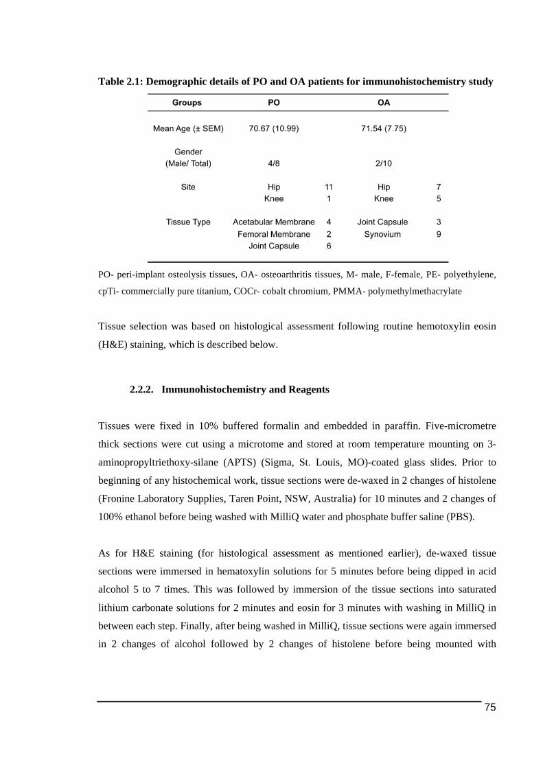
75
Table 2.1: Demographic details of PO and OA patients for immunohistochemistry study
PO- peri-implant osteolysis tissues, OA- osteoarthritis tissues, M- male, F-female, PE- polyethylene,
cpTi- commercially pure titanium, COCr- cobalt chromium, PMMA- polymethylmethacrylate
Tissue selection was based on histological assessment following routine hemotoxylin eosin
(H&E) staining, which is described below.
2.2.2. Immunohistochemistry and Reagents
Tissues were fixed in 10% buffered formalin and embedded in paraffin. Five-micrometre
thick sections were cut using a microtome and stored at room temperature mounting on 3-
aminopropyltriethoxy-silane (APTS) (Sigma, St. Louis, MO)-coated glass slides. Prior to
beginning of any histochemical work, tissue sections were de-waxed in 2 changes of histolene
(Fronine Laboratory Supplies, Taren Point, NSW, Australia) for 10 minutes and 2 changes of
100% ethanol before being washed with MilliQ water and phosphate buffer saline (PBS).
As for H&E staining (for histological assessment as mentioned earlier), de-waxed tissue
sections were immersed in hematoxylin solutions for 5 minutes before being dipped in acid
alcohol 5 to 7 times. This was followed by immersion of the tissue sections into saturated
lithium carbonate solutions for 2 minutes and eosin for 3 minutes with washing in MilliQ in
between each step. Finally, after being washed in MilliQ, tissue sections were again immersed
in 2 changes of alcohol followed by 2 changes of histolene before being mounted with
76
coverslips with dibutyl phthalate xylene (DPX) (Labchem, PA, USA) as the mounting
medium.
For immunohistochemistry work, following de-waxing tissue sections were processed for
antigen retrieval by heating up in either 10mM Tris-EDTA buffer pH 9.0 or 10mM sodium
citrate pH 6.0 (refer Table 2.2) at 92-95oC for 10-20 minutes. Selection of antigen retrieval
buffer was based on the preliminary work on antibody optimisation prior to the actual
immunostaining. In this thesis, immunohistochemical staining was detected based on three-
step immunoperoxidase method previously published (Crotti et al. 2002; Kraan et al. 2000;
Tak et al. 1995) with minor modifications, as described below. Since immunoperoxidase
method was employed in the work, in order to prevent unspecific staining, endogenous
peroxidase activity was inhibited by 0.1% sodium azide (NaN3) and 0.3% (w/v) hydrogen
peroxidase (H2O2) (Merck, Victoria, Australia).
The primary antibodies diluted in PBS and 1% bovine serum albumin (BSA) were applied at
optimized final concentrations overnight in wet chamber at room temperature. The antibody
working concentrations (see Table 2.3) were determined by applying a range of
concentrations, with manufacturer recommended concentration as start points followed by
series of 1 in 2 serial dilutions. Sections were then incubated with a horse-radish peroxidase
(HRP)-conjugated secondary antibody diluted in PBS containing 1%BSA normal human
serum for 30 minutes, followed by incubation with an HRP-conjugated tertiary antibody
against the secondary antibody. HRP activity was detected using hydrogen peroxide as the
substrate and 3-amino-9-ethylcarbzole (AEC) as the dye. There was a minimum three washes
with PBS between each step to wash off any unbound antibody or protein for avoiding
unspecific binding and hence staining. Sections were counterstained in Harris hematoxylin for
10 seconds, and then washed with MilliQ before immediately immersed in saturated lithium
carbonate and mounted with aquamount (Microscopy Aquatex, Darmstadt, Germany).
Negative controls included tissue incubated with an isotype specific antibody for the
monoclonal antibody or serum of corresponding species for polyclonal antibodies at
equivalent concentrations to the primary antibody. Staining for each antibody was carried on
all tissue samples involved at the same time to reduce staining variability due to experimental
setup.
77
2.2.2.1. Antibodies and Reagents
The following antibodies used for the immunohistochemistry, which are polyclonal goat anti-
human OSCAR (catalogue no. SC34233), polyclonal rabbit anti-human DAP12 (SC20783)
and monoclonal antibody mouse anti-human NFATc1 (clone 7A6, SC7294) were purchased
from Santa Cruz Biotechnology Inc. (CA, USA). Polyclonal rabbit anti-human FC€R1G (LS-
B2169), which was chosen based on personal communication as FC€R1γ is equivalent to
FcRγ (Ishikawa et al. 2004; Zou et al. 2010), was purchased from Lifespan Biosciences Inc.
(Seattle, WA, USA). For immunostaining of TREM2, polyclonal rabbit anti human TREM2
(HPA010917) purchased from Sigma-Aldrich Pty. Ltd (Castle Hill, NSW, Australia) was
used for the study. For the detection of osteoclast cells in the tissues, one of the cell markers
used was cathepsin K and the antibody used for the immunostaining was a monoclonal
antibody mouse anti-human Cathepsin K (MAB3324 clone 182-12G5) purchased from
Millipore (Billerica, MA, USA).
Table 2.2: Secondary and tertiary antibodies as well antigen retrieval buffer
corresponding to the primary antibodies used in immunohistochemistry
EDTA- ethylenediaminetetraacetic acid
78
Table 2.3: Primary antibodies used for immunohistochemistry
IgG1- immunoglobulin subtype 1
Either affinity purified, HRP-conjugated goat anti-mouse or goat anti-rabbit IgG (depends on
the species in which primary antibodies were raised) was used as secondary antibodies
(DakoCytomation, Glostrup, Denmark). For the tertiary antibodies, either HRP-conjugated
swine-anti-goat or rabbit anti-swine IgG purchased from Biosource (Camerillo, CA, USA)
were applied to the tissues before staining was carried out using AEC that was purchased
from Sigma -Aldrich (St Louis, MO, USA).
2.2.2.2. TRAP Staining and Serial Labeling of Osteoclast Cell Markers
TRAP staining was performed on tissue sections using a commercial kit (386A, Sigma, St.
Louis, MO, USA) as per manufacturer’s instructions. Tissue sections on APTS-coated slides
were incubated at 37°C for 2 hours in dark conditions with the filtered kit solution containing
0.5% Fast Green Garnet salt and 4% of each acetate, tartrate solution and Naphtol AS-BI
Phosphate. As per the immunostaining procedure described earlier, tissue sections were
counterstained with Methyl Green and mounted with aquamount before being viewed under
microscopy for further analysis.
Serial sections of tissues, which had been immunostained for TREM2, OSCAR and osteoclast
marker cathepsin K were compared with TRAP staining on tissue sections from
corresponding patients. Particular attention was given to multinucleated cells for the
identification of positively stained osteoclasts.
79
2.2.2.3. Scoring of Immunostaining Results
The immunohistochemically stained slides were scanned using Nanozoomer Digital
Pathology (Hamamatsu, Shizouka, Japan). In order to evaluate the extent of positive staining
on the tissues, three random areas (2mm by 2mm squares) were drawn in each section, in
which semiquantitative analysis (SQA) scoring was carried out by two-blinded independent
observer. The proportion of positive-stained cells was graded using a scale of 5 ranging from
0 to 4 adapted from previously published studies (Crotti et al. 2004; Kraan et al. 1999; Tak et
al. 1995). A minimal grade of 0 is indicated by 5% of positive cells or less, 1 for 6 to 10%, 2
for cell immunostaining positivity between 11 to 25%, 3 for 26 to 50% and 4 for more than
50% positively stained cells.
2.2.3. Quantitative Real Time Reverse Transcription Polymerase Chain
Reaction
Quantitive reverse-transcription polymerase chain reaction (qRT-PCR) was employed in this
study to measure the level of mRNA expression of the investigated genes. This is described in
detail below.
2.2.3.1. RNA Extraction from Frozen Tissues and Spectrophotometry
Tissues from some patients were snap-frozen in Optimal Cutting Temperature (OCT) medium
(Tissue TekR Zoeterwoude, The Netherlands) using liquid nitrogen and stored in -80°C before
used. A total of 12 PO and 6 OA frozen tissues, which was a subset of corresponding patient
groups whom the tissue samples were selected for immunohistochemical study described
earlier (see in the beginning of section 2.2.2), were selected for mRNA isolation and qRT-
PCR analysis (samples are detailed as in Table 2.4 below). Those frozen tissues were chosen
for RT-PCR based on histological assessment screening for multinucleated cells for PO and
high cellularity OA tissues. Tissues were sectioned at a minimum of 20µm thickness in total
using a cryostat (Leica Microsystems, Weitzlar, Germany).
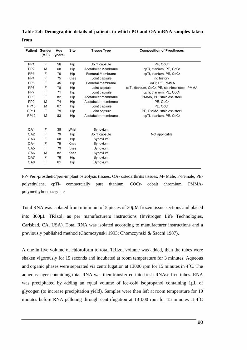
80
Table 2.4: Demographic details of patients in which PO and OA mRNA samples taken
from
PP- Peri-prosthetic/peri-implant osteolysis tissues, OA- osteoarthritis tissues, M- Male, F-Female, PE-
polyethylene, cpTi- commercially pure titanium, COCr- cobalt chromium, PMMA-
polymethylmethacrylate
Total RNA was isolated from minimum of 5 pieces of 20µM frozen tissue sections and placed
into 300µL TRIzol, as per manufacturers instructions (Invitrogen Life Technologies,
Carlsbad, CA, USA). Total RNA was isolated according to manufacturer instructions and a
previously published method (Chomczynski 1993; Chomczynski & Sacchi 1987).
A one in five volume of chloroform to total TRIzol volume was added, then the tubes were
shaken vigorously for 15 seconds and incubated at room temperature for 3 minutes. Aqueous
and organic phases were separated via centrifugation at 13000 rpm for 15 minutes in 4˚C. The
aqueous layer containing total RNA was then transferred into fresh RNAse-free tubes. RNA
was precipitated by adding an equal volume of ice-cold isopropanol containing 1µL of
glycogen (to increase precipitation yield). Samples were then left at room temperature for 10
minutes before RNA pelleting through centrifugation at 13 000 rpm for 15 minutes at 4˚C
81
followed by aspiration of supernatant. The RNA pellet was washed in 500µL of 70% ethanol
and centrifuged at 7500 rpm for 5 minutes at 4˚C. Supernatant was discarded and pellets were
allowed to air dry. The obtained RNA pellet was resuspended in 20µL of diethyl
pyrocarbonate (DEPC) (Sigma-Aldrich, St Louis, MO, USA)-treated water (DEPC water) and
heated to 65˚C for 5 minutes.
RNA concentration was determined using a UV spectrophotometer (Shimadzu Corporation,
Kyoto, Japan). RNA Concentraion was calculated according to the formula:
RNA Concentration (ng/ml) = OD260 x Dilution Factor x RNA Constant
with RNA constant = 40ng/µl
RNA isolated was kept in -80°C until needed for cDNA synthesis.
2.2.3.2. Reverse Transcription
One microgram of total RNA was combined with 500µM dNTPs (Bioline, London, United
Kingdom), 1µg random hexamer (GeneWorks, Adelaide, SA, Australia) and DEPC water to a
final volume of 14.75µl in a 0.2mL flat cap tube. Tubes were then heated to 65˚C for 5
minutes before being briefly cooled to 4˚C. This pre-reverse-transcription mix was then added
with 200 units SuperScript™ III Reverse Transcriptase, 1x First Strand Buffer and 1.25mM
dithiothreitol (DTT) (all those three reagents from Invitrogen Life Technologies, Carlsbad,
CA, USA) for a final volume of 20µL. The reverse transcription process consisted of
incubation at 25˚C for 5 minutes, followed by 30 minutes at 50˚C and finally 70˚C for 5
minutes. The step of reverse transcription (RT) for complementary deoxyribonucleic acid
(cDNA) sysnthesis was performed using a Rotor-Gene 3000 (Corbett Life Science, Mortlake,
NSW, Australia). All synthesized cDNA was stored at -80˚C until use.
82
2.2.3.3. Real Time Polymerase Chain Reaction
Similar to the reverse transcription process, the cDNA amplification via polymerase chain
reaction (PCR) was carried out in the same machine Rotor-Gene 3000 (Corbett Life Science).
Reaction mixtures contained 1µl of 1 in 5 pre-diluted cDNA, 7.5µl Platinum SYBR® Green
qPCR Supermix-UDG, 300nM each of forward and reverse primer and DEPC-treated water
to a final volume of 15µl.
In every PCR performed, samples were analysed in triplicate to obtain consistency in data and
to eliminate any false results obtained through inaccurate pipetting. The PCR procedure began
with an incubation at 50˚C for 2 minutes to activate UDG, which is an enzyme in the
Platinum® SYBR® Green qPCR SuperMix-UDG (Invitrogen Inc.) used to destroy potential
carry-over PCR contamination (Longo et al. 1990). Each cycle of amplification involved a
three-step thermocycling procedure with DNA denaturation, annealing and elongation
occurring at 95˚C, 60˚C and 72˚C respectively, with each step held for 20 seconds, for a total
of 40 cycles. The SYBR® Green fluorescence measurement was taken at the completion of
each elongation step.
All PCR reactions were validated by the presence of a single peak in the melt curve analysis,
which was obtained from PCR melting protocol upon completion 40 cycles of PCR
amplification. This involves heating the PCR product at the end of the procedure at a rate of
1˚C every 5 seconds from 72˚C to 95˚C and collecting fluorescence readings at every degree.
Primer3Plus online application (Untergasser et al. 2007) was used to design oligonucleotide
primers to human OSCAR, FcRγ TREM2 and DAP12 in house, aimed to flank intron-exon
boundaries. Primer sequences for NFATc1 were obtained based on a previous publication
(Granfar et al. 2005). The endogenous reference gene human acidic ribosomal protein (hARP)
was used to normalize the PCR data, with the primer sequence was taken from a published
work (Franssen et al. 2005). Primers were purchased from Geneworks (Thebarton, SA,
Australia). Primer sequences used for each gene in this study are as shown in Table 2.5.

83
Table 2.5: Primer sequences of each gene investigated
hARP- human acidic ribosomal protein, S- sense, AS- anti-sense, Tm- melting temperature, bp- base
pair
Relative expression of the target genes to the housekeeping gene (hARP) in the studied
samples of in vitro assay was obtained using the difference in the comparative threshold
(ΔCT) method, which is a real time PCR calculation and analysis method documented by
Livak and Schmittgen (2001). The levels of expression of the target genes in the studied
samples as compared with the reference gene were calculated as 2 power-ΔCT (Livak &
Schmittgen 2001).
2.2.4. Statistical Analysis
Non-parametric tests were employed to analyse and compare the mean ranks of the SQA’s
data between the two groups. Kruskal-Wallis test was used to find any significant difference
between any of the groups before performing Mann-Whitney U test to identify groups with
statistical significant data, with a p<0.05 was considered to be statistically different.
84
As for the measurement of gene expression, differences in relative mRNA levels between two
groups was also analysed using student t-test and p<0.05 was considered significant
statistically. All statistical analysis in this study was performed using SPSS 11 (SPSS Inc,
Chicago, IL, USA) and GraphPad Prism (Graphpad Software, CA, USA).
2.3. Results
2.3.1. Histological Features of Peri-Implant Osteolysis and Osteoarthritis
Tissues
Prior to any immunostaining, routine tissue staining using H&E was carried for the
histological assessment as described in the Methods 2.2.1 above. Acellular tissues consisting
mainly of connective tissues were avoided to ensure that data generated (cell immunostaining)
would be meaningful and could be interpretable in context of cellular and molecular biology.
By far the majority of tissues were largely cellular and tested. High cellularity in PO tissues
also gives indication of the tissues are in active inflammatory stage of disease progression
rather than scar tissues. In a number of PO tissue samples studied, there was mix of very
cellular and fibrous tissues (that may give indication those tissues in later stage of PO
progression).
As predicted in the PO group, all tissues displayed signs of granulomatous inflammatory
reaction (see Figure 2.1A) that is commonly observed in PO tissues (see 1.2.1 above). All PO
tissues were dominated by high number of multinucleated cells, possibly could be osteoclasts
and foreign body giant cells. All samples demonstrated the presence of PE particles to some
extent. These particles were observed as birefringent material under polarized microscopic
view (arrowed in Figure 2.1A) and ranged in size, from just visible under microscope to
approximately a milimeter in size. In addition, in a few tissues we noted presence of metal
particles, seen as black debris on the tissues. It was also noted that multinucleated osteoclast-
like cells were commonly associated with or in close proximity to smaller sized PE particles,
whereas foreign body giant like cells appear associated with larger PE debris.
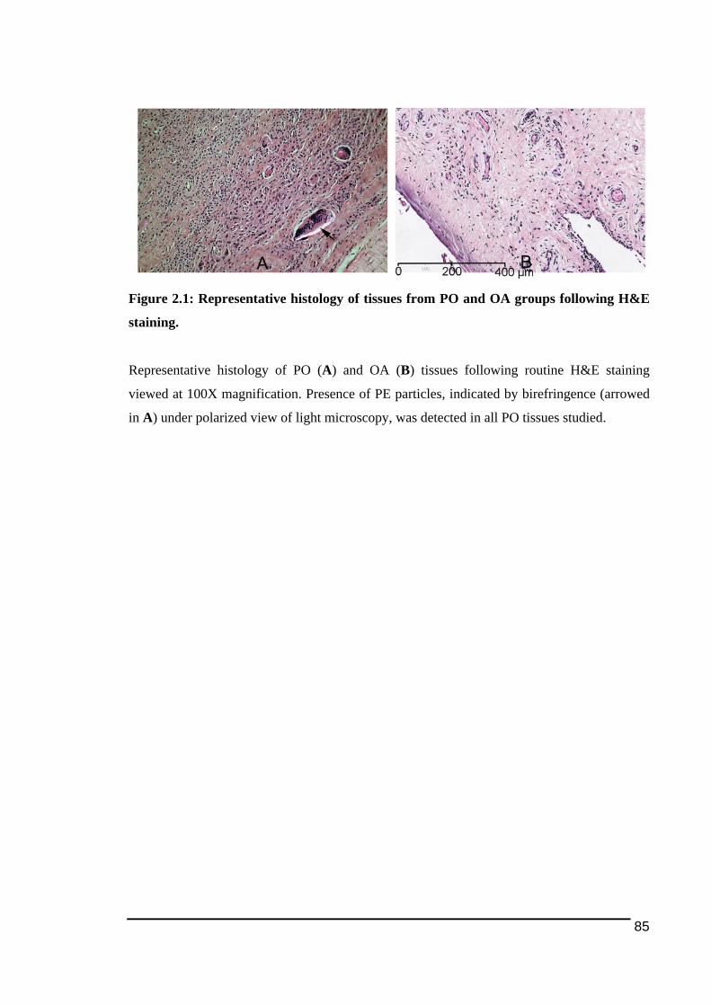
85
Figure 2.1: Representative histology of tissues from PO and OA groups following H&E
staining.
Representative histology of PO (A) and OA (B) tissues following routine H&E staining
viewed at 100X magnification. Presence of PE particles, indicated by birefringence (arrowed
in A) under polarized view of light microscopy, was detected in all PO tissues studied.
86
Generally, the histology of OA tissues showed majority cells were in the lining layer of the
synovial tissues. Presence of vasculature was occasionally seen in the OA synovial tissues.
Some of the OA tissue samples demonstrated very mild infiltration of inflammatory cells
composed of mononuclear, probably macrophage and lymphocyte-like, cells particularly
around blood vessels. Overall there was minimal inflammatory reaction seen. Multinucleated
cells were not detected in any of these sections.
2.3.2. Expression of NFATc1 and osteoclast ITAM-associated molecules in peri-
implant osteolysis tissues in comparison to osteoarthritis tissues
SQA was carried out, as described earlier in section 2.2.2.3 above. The SQA scores for
immunostaining of NFATc1 and each ITAM-associated molecules are summarised as in
Table 2.6 below.
Table 2.6: SQA scores for immnostaining of NFATc1 and ITAM-related molecules in
PO and OA tissues
significantly higher in PO than in OA if p<0.05
Observations on detection of each molecule are detailed in the following sections below.
2.3.2.1. NFATc1
In PO tissues, strong NFATc1 immunostaining was predominantly observed in cells with a
lymphocyte morphology and only faint staining noted in a few number multinucleated cells
(Figure 2.2A). Interestingly, NFATc1 was expressed in nuclei and peri-nuclei on
87
multinucleated cells, with stronger or more intensed red colour staining was observed on
nuclei than in the cytoplasmic region (Figure 2.2D).
In OA, the majority of NFATc1 immunostaining was detected in mononuclear cells with
lymphocyte-like morphology located mainly perivascular layer (Figure 2.2B). There was no
positive staining found on the blood vessels.
There was no significant difference (p>0.05) in the proportion of cells expressing NFATc1
between in PO and OA tissues, as shown in Table 2.6 above.
There was no positive immunostaining found in peri-prosthetic tissues incubated with primary
antibody isotype (mouse IgG1 as negative control) (Figure 2.2C) indicating no unspecific or
false positive NFATc1 immmunostaining on the tissues.
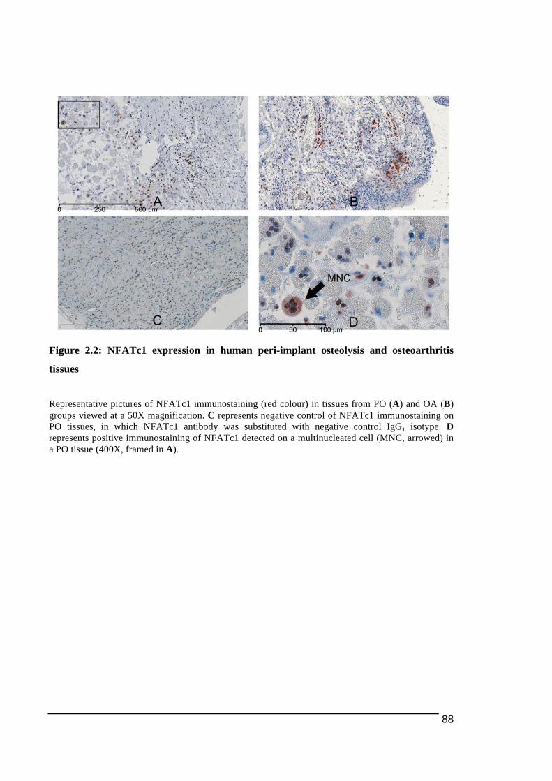
88
Figure 2.2: NFATc1 expression in human peri-implant osteolysis and osteoarthritis
tissues
Representative pictures of NFATc1 immunostaining (red colour) in tissues from PO (A) and OA (B) groups viewed at a 50X magnification. C represents negative control of NFATc1 immunostaining on PO tissues, in which NFATc1 antibody was substituted with negative control IgG1 isotype. D represents positive immunostaining of NFATc1 detected on a multinucleated cell (MNC, arrowed) in a PO tissue (400X, framed in A).
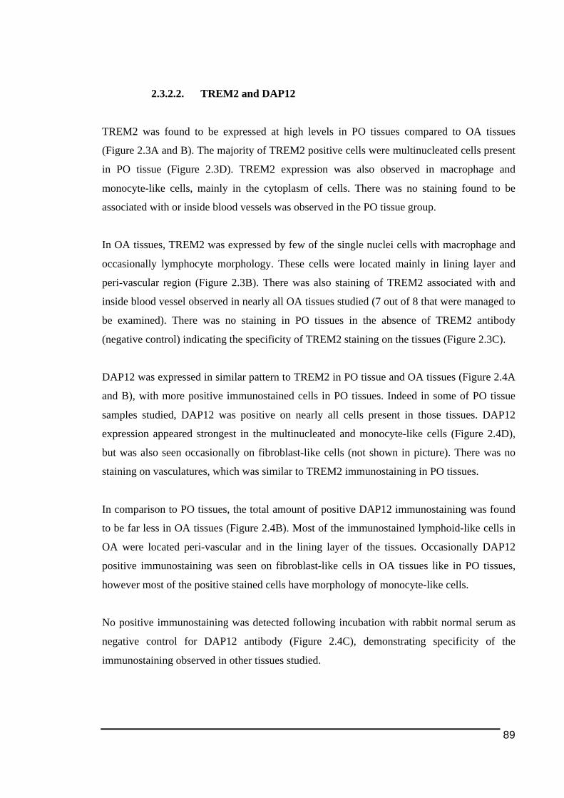
89
2.3.2.2. TREM2 and DAP12
TREM2 was found to be expressed at high levels in PO tissues compared to OA tissues
(Figure 2.3A and B). The majority of TREM2 positive cells were multinucleated cells present
in PO tissue (Figure 2.3D). TREM2 expression was also observed in macrophage and
monocyte-like cells, mainly in the cytoplasm of cells. There was no staining found to be
associated with or inside blood vessels was observed in the PO tissue group.
In OA tissues, TREM2 was expressed by few of the single nuclei cells with macrophage and
occasionally lymphocyte morphology. These cells were located mainly in lining layer and
peri-vascular region (Figure 2.3B). There was also staining of TREM2 associated with and
inside blood vessel observed in nearly all OA tissues studied (7 out of 8 that were managed to
be examined). There was no staining in PO tissues in the absence of TREM2 antibody
(negative control) indicating the specificity of TREM2 staining on the tissues (Figure 2.3C).
DAP12 was expressed in similar pattern to TREM2 in PO tissue and OA tissues (Figure 2.4A
and B), with more positive immunostained cells in PO tissues. Indeed in some of PO tissue
samples studied, DAP12 was positive on nearly all cells present in those tissues. DAP12
expression appeared strongest in the multinucleated and monocyte-like cells (Figure 2.4D),
but was also seen occasionally on fibroblast-like cells (not shown in picture). There was no
staining on vasculatures, which was similar to TREM2 immunostaining in PO tissues.
In comparison to PO tissues, the total amount of positive DAP12 immunostaining was found
to be far less in OA tissues (Figure 2.4B). Most of the immunostained lymphoid-like cells in
OA were located peri-vascular and in the lining layer of the tissues. Occasionally DAP12
positive immunostaining was seen on fibroblast-like cells in OA tissues like in PO tissues,
however most of the positive stained cells have morphology of monocyte-like cells.
No positive immunostaining was detected following incubation with rabbit normal serum as
negative control for DAP12 antibody (Figure 2.4C), demonstrating specificity of the
immunostaining observed in other tissues studied.
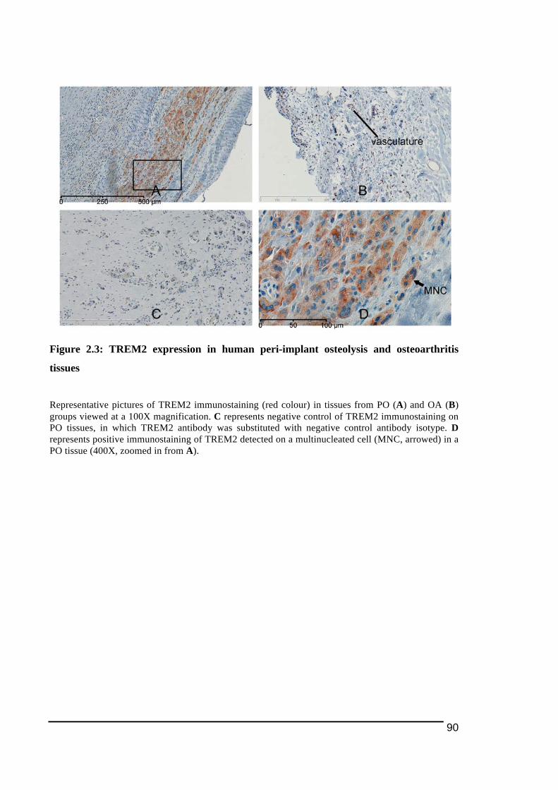
90
Figure 2.3: TREM2 expression in human peri-implant osteolysis and osteoarthritis
tissues
Representative pictures of TREM2 immunostaining (red colour) in tissues from PO (A) and OA (B) groups viewed at a 100X magnification. C represents negative control of TREM2 immunostaining on PO tissues, in which TREM2 antibody was substituted with negative control antibody isotype. D represents positive immunostaining of TREM2 detected on a multinucleated cell (MNC, arrowed) in a PO tissue (400X, zoomed in from A).

91
Figure 2.4: Representative pictures of DAP12 immunostaining in human peri-prosthetic
and osteoarthritis tissues studied
Representative pictures of DAP12 immunostaining (red colour) in tissues from PO (A) and OA (B) groups viewed at a 100X magnification. C represents negative control of DAP12 immunostaining on PO tissues, in which DAP12 antibody was substituted with negative control antibody isotype. D represents positive immunostaining of DAP12 detected on a multinucleated cell in a PO tissue (400X, zoomed in from A).
92
SQA data of immunostaining of both TREM2 and its adaptor molecule, DAP12,
demonstrated a higher proportion of positive cells in PO tissues compared to OA tissues
(Table 2.6), which further supported the observations of tissue staining between those two
groups.
2.3.2.3. OSCAR and FcRγ
There were notably more OSCAR positively stained cells in PO tissues in comparison to OA
tissues (Figure 2.5A-B). In PO tissues, OSCAR immunostaining was clearly visible in
majority of the multinucleated cells (Figure 2.5A and D), regardless of their proximity to
wear particles (discussed later in 2.3.4 below). Staining was mostly confined to the cytoplasm
although some cells occasionally demonstrated nuclear staining (Figure 2.5D for closer view).
There was also some staining on fibroblasts occasionally found on some of the tissues (not
shown). There was no or minimal positive OSCAR staining associated with vasculature.
In about half of the OA tissues studied, apart from positive staining seen mainly on
mononuclear cells, OSCAR immunostaining was also observed on the blood vessel walls as
well as inside the lumens (arrowed in Figure 2.5B). The majority of the OSCAR-positive cells
in the OA tissue group were found to be distributed in the lining layer, though in some OA
tissues, stained cells were also found to be scattered in the tissues.
Similar to OSCAR, the FcRγ-positively stained cells appeared more in PO tissues compared
to in OA tissues (see Figure 2.6A-B). FcRγ was highly expressed by monocyte-like cells on
cell membrane rather than in the cytoplasm (shown clearly in Figure 2.6D). FcRγ was also
detected in the cytoplasm of multinucleated cells (arrowed in Figure 2.6D).
In some OA tissues, majority of the FcRγ positive-immunostained cells had monocyte and
lymphocyte-like morphology. Generally, strong expression of FcRγ was seen in cells
scattered on tissues (Figure 2.6B), however in some of the OA tissues, expression in the
lining layer of the tissues as well as in perivascular areas was also noted. However, unlike
OSCAR immunostaining described above, there was no staining for FcRγ associated with
vasculature.
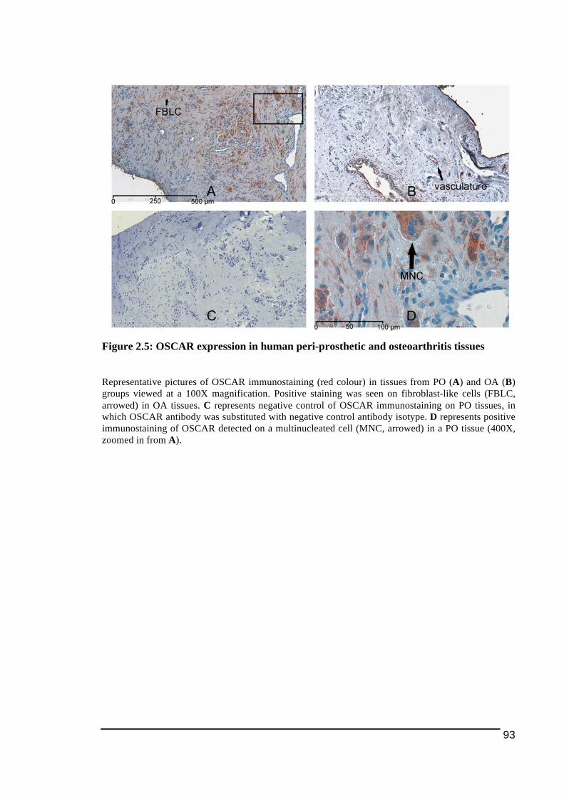
93
Figure 2.5: OSCAR expression in human peri-prosthetic and osteoarthritis tissues
Representative pictures of OSCAR immunostaining (red colour) in tissues from PO (A) and OA (B) groups viewed at a 100X magnification. Positive staining was seen on fibroblast-like cells (FBLC, arrowed) in OA tissues. C represents negative control of OSCAR immunostaining on PO tissues, in which OSCAR antibody was substituted with negative control antibody isotype. D represents positive immunostaining of OSCAR detected on a multinucleated cell (MNC, arrowed) in a PO tissue (400X, zoomed in from A).
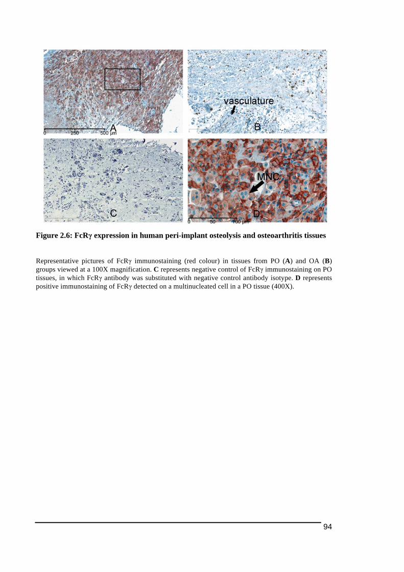
94
Figure 2.6: FcRγ expression in human peri-implant osteolysis and osteoarthritis tissues
Representative pictures of FcRγ immunostaining (red colour) in tissues from PO (A) and OA (B) groups viewed at a 100X magnification. C represents negative control of FcRγ immunostaining on PO tissues, in which FcRγ antibody was substituted with negative control antibody isotype. D represents positive immunostaining of FcRγ detected on a multinucleated cell in a PO tissue (400X).
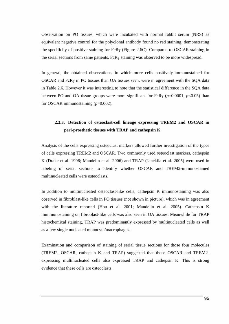
95
Observation on PO tissues, which were incubated with normal rabbit serum (NRS) as
equivalent negative control for the polyclonal antibody found no red staining, demonstrating
the specificity of positive staining for FcRγ (Figure 2.6C). Compared to OSCAR staining in
the serial sections from same patients, FcRγ staining was observed to be more widespread.
In general, the obtained observations, in which more cells positively-immunostained for
OSCAR and FcRγ in PO tissues than OA tissues seen, were in agreement with the SQA data
in Table 2.6. However it was interesting to note that the statistical difference in the SQA data
between PO and OA tissue groups were more significant for FcRγ (p=0.0001, p<0.05) than
for OSCAR immunostaining (p=0.002).
2.3.3. Detection of osteoclast-cell lineage expressing TREM2 and OSCAR in
peri-prosthetic tissues with TRAP and cathepsin K
Analysis of the cells expressing osteoclast markers allowed further investigation of the types
of cells expressing TREM2 and OSCAR. Two commonly used osteoclast markers, cathepsin
K (Drake et al. 1996; Mandelin et al. 2006) and TRAP (Janckila et al. 2005) were used in
labeling of serial sections to identify whether OSCAR and TREM2-immunostained
multinucleated cells were osteoclasts.
In addition to multinucleated osteoclast-like cells, cathepsin K immunostaining was also
observed in fibroblast-like cells in PO tissues (not shown in picture), which was in agreement
with the literature reported (Hou et al. 2001; Mandelin et al. 2005). Cathepsin K
immmunostaining on fibroblast-like cells was also seen in OA tissues. Meanwhile for TRAP
histochemical staining, TRAP was predominantly expressed by multinucleated cells as well
as a few single nucleated monocyte/macrophages.
Examination and comparison of staining of serial tissue sections for those four molecules
(TREM2, OSCAR, cathepsin K and TRAP) suggested that those OSCAR and TREM2-
expressing multinucleated cells also expressed TRAP and cathepsin K. This is strong
evidence that these cells are osteoclasts.
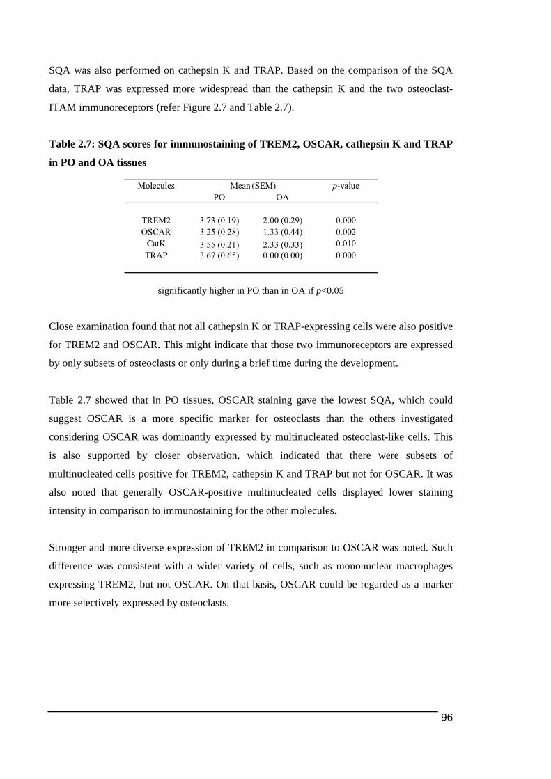
96
SQA was also performed on cathepsin K and TRAP. Based on the comparison of the SQA
data, TRAP was expressed more widespread than the cathepsin K and the two osteoclast-
ITAM immunoreceptors (refer Figure 2.7 and Table 2.7).
Table 2.7: SQA scores for immunostaining of TREM2, OSCAR, cathepsin K and TRAP
in PO and OA tissues
significantly higher in PO than in OA if p<0.05
Close examination found that not all cathepsin K or TRAP-expressing cells were also positive
for TREM2 and OSCAR. This might indicate that those two immunoreceptors are expressed
by only subsets of osteoclasts or only during a brief time during the development.
Table 2.7 showed that in PO tissues, OSCAR staining gave the lowest SQA, which could
suggest OSCAR is a more specific marker for osteoclasts than the others investigated
considering OSCAR was dominantly expressed by multinucleated osteoclast-like cells. This
is also supported by closer observation, which indicated that there were subsets of
multinucleated cells positive for TREM2, cathepsin K and TRAP but not for OSCAR. It was
also noted that generally OSCAR-positive multinucleated cells displayed lower staining
intensity in comparison to immunostaining for the other molecules.
Stronger and more diverse expression of TREM2 in comparison to OSCAR was noted. Such
difference was consistent with a wider variety of cells, such as mononuclear macrophages
expressing TREM2, but not OSCAR. On that basis, OSCAR could be regarded as a marker
more selectively expressed by osteoclasts.
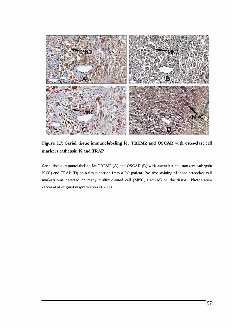
97
Figure 2.7: Serial tissue immunolabeling for TREM2 and OSCAR with osteoclast cell
markers cathepsin K and TRAP
Serial tissue immunolabeling for TREM2 (A) and OSCAR (B) with osteoclast cell markers cathepsin
K (C) and TRAP (D) on a tissue section from a PO patient. Positive staining of those osteoclast cell
markers was detected on many multinucleated cell (MNC, arrowed) on the tissues. Photos were
captured at original magnification of 200X.

98
2.3.4. Detection of polyethylene particles within immunostained multinucleated
cells
By taking the fact that polyethylene particles were detected in all studied PO tissues into
account, it was of particular interest to investigate if the expression of NFATc1 and ITAM-
associated molecules studied in multinucleated osteoclast-like cells could be associated with
the presence of polyethylene particles.
Generally, PE particles in varying sizes were observed as scattered throughout the PO tissues
with the majority were in close proximity to the cells positively stained for all those ITAM-
related molecules. Indeed in some cases, as shown in Figure 2.8 above, presence of PE
particles was detected within multinucleated cells expressing TREM2, DAP12, OSCAR and
FcRγ.
Examination on tissue sections stained for common osteoclast cell markers, cathepsin K and
TRAP (Figure 2.9) showed some of the immunostained multinucleated cells were associated
with or in close proximity of the polyethylene particles. Those multinucleated cells could
possibly be pre-osteoclasts, as indicated from the work by Shen and colleagues (2006) (Shen
et al. 2006).
Generally, it was found that multinucleated cells associated with PE particles displayed weak
cathepsin K staining, as shown in Figure 2.9A, even though occasionally there were some PE-
associated multinucleated cells showing strong cathepsin K immunostaining observed. For
TRAP histochemical staining, there was no noticeable difference in the intensity of positive
staining found between PE-associated and non-associated positive multinucleated cells.
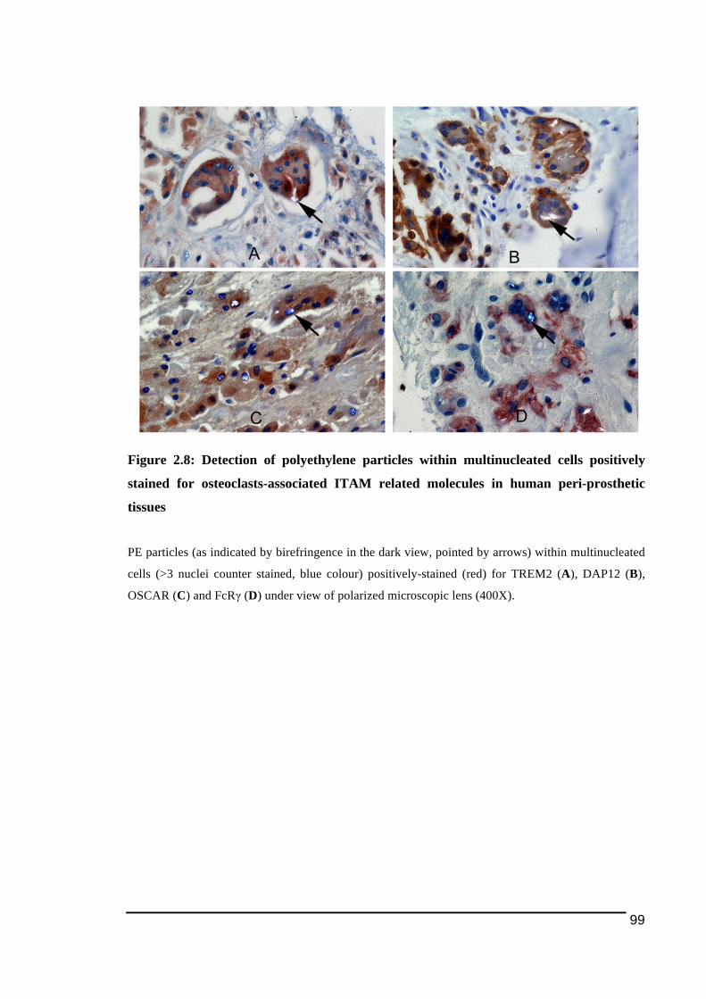
99
Figure 2.8: Detection of polyethylene particles within multinucleated cells positively
stained for osteoclasts-associated ITAM related molecules in human peri-prosthetic
tissues
PE particles (as indicated by birefringence in the dark view, pointed by arrows) within multinucleated
cells (>3 nuclei counter stained, blue colour) positively-stained (red) for TREM2 (A), DAP12 (B),
OSCAR (C) and FcRγ (D) under view of polarized microscopic lens (400X).
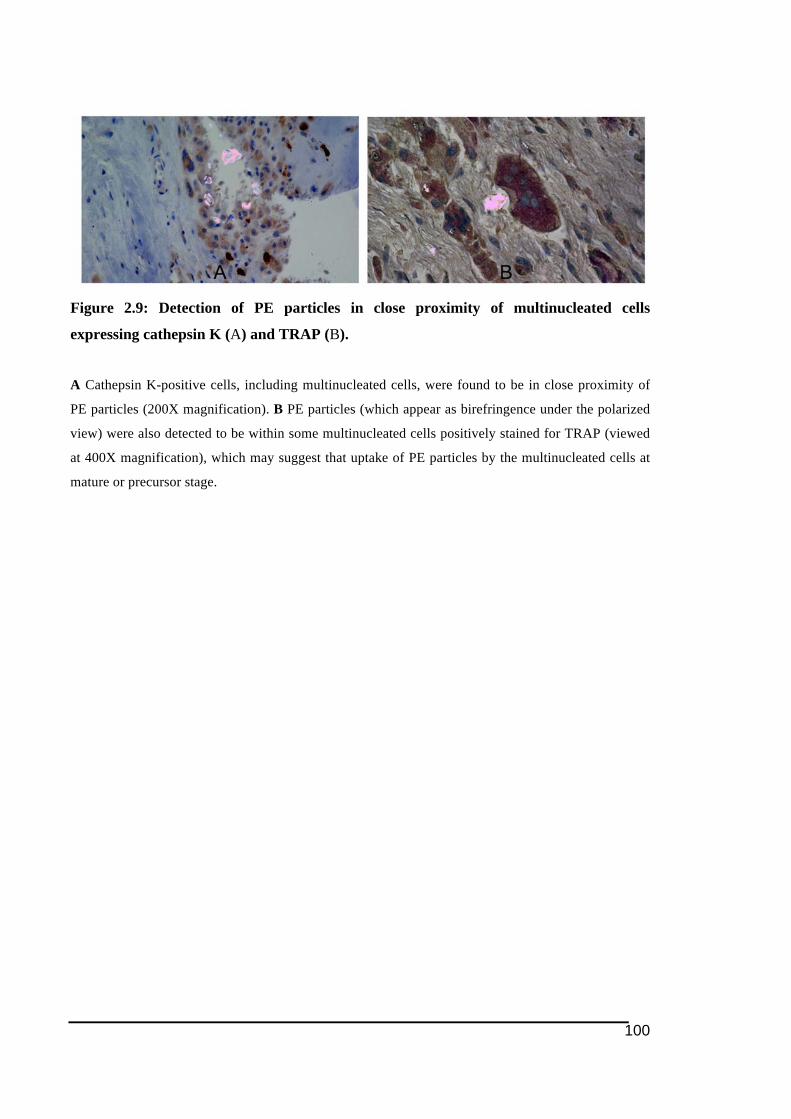
100
Figure 2.9: Detection of PE particles in close proximity of multinucleated cells
expressing cathepsin K (A) and TRAP (B).
A Cathepsin K-positive cells, including multinucleated cells, were found to be in close proximity of
PE particles (200X magnification). B PE particles (which appear as birefringence under the polarized
view) were also detected to be within some multinucleated cells positively stained for TRAP (viewed
at 400X magnification), which may suggest that uptake of PE particles by the multinucleated cells at
mature or precursor stage.

101
2.3.5. mRNA expression of osteoclast ITAM-associated molecules in peri-
implant osteolysis and osteoarthritic tissues
Quantitative real-time RT-PCR was performed to investigate the expression of NFATc1 and
the ITAM-associated molecules at the mRNA level. In agreement with the
immunohistochemical observations, there was no statistical difference in NFATc1 mRNA
level between the two groups, even though it seemed there was a trend of higher NFATc1
mRNA expression in PO than in OA. Like NFATc1, there was a pattern towards higher levels
of mRNAs of ITAM immunoreceptor OSCAR in PO compared to OA supporting the
immunohistochemical findings, surprisingly there was no significant difference in the relative
mRNA level achieved (p>0.05, see Table 2.8). The mRNA expression of TREM2, DAP12
and FcRγ was statistically higher in PO than in OA tissue (p<0.05) consistent with the
immunohistochemical results. The relative levels of genes investigated in PO compared to
OA tissues are summarized as in Figure 2.10.
The relative mRNA expression of the osteoclast cell markers, TRAP and cathepsin K, was
significantly higher in PO tissue group than OA (Table 2.8 below), in agreement with the
immunohistochemical SQA data presented ealier (Table 2.6). This is also in consistent with
other published data (Koulouvaris et al. 2008) that showed higher mRNA level of cathepsin K
in PO than in OA tissues. The relative mRNA expression of these two osteoclast-associated
cell markers generally appeared higher than OSCAR. This agreed with the SQA data of
protein expression obtained.
Table 2.8: Mean of relative mRNA expression (-ΔCT) of TREM2, OSCAR, TRAP and
cathepsin K in PO and OA tissue groups
significantly higher in PO than in OA if p<0.05

102
Figure 2.10: Relative mRNA expression for NFATc1, TREM2, OSCAR, DAP12 and
FcRγ in PO and OA tissues
Mean mRNA expression in relative to hARP mRNA level (-ΔCT, see 2.2.3.3 above) (represented by
the y-axis) of NFATc1, ITAM immunoreceptors, TREM2 and OSCAR (top), and corresponding
adaptor molecules, DAP12 and FcRγ (bottom). The error bars shown on the graphs represent the
standard error of mean (SEM) for the collective relative mRNA expression in those two tissue groups.

103
2.4. Discussions
This and the following chapter of this thesis are studies on the regulation of osteoclast
formation in the context of PO pathology by looking at the molecules reported by literature as
mediating osteoclastogenesis. The findings, for the first time, show several important
molecules could be associated with excessive osteoclast formation and expression of
osteoclast-associated cell markers (Kadoya et al. 1996; Neale & Athanasou 1999) in PO
tissues. The findings support the contention that pre-osteoclasts and cells that have commited
into osteoclastic lineage are among those recruited to form the granulomatous tissues (Haynes
et al. 2001a; Neale et al. 2000; Sabokbar et al. 1997). This contention is further supported by
the notion that the cells isolated from revision tissue readily form bone resorbing osteoclasts
in vitro were found to have higher resorbing capability (Haynes et al. 2001b; Roato et al.
2010; Sabokbar et al. 1997).
Studies in this chapter were designed to investigate the expression of NFATc1 and osteoclast
ITAM-related molecules in human PO tissues and compare to in OA tissues. As NFATc1 and
ITAM-associated molecules have been shown to be crucial for intracelullar signalling in
osteoclast differentiation, it was hypothesized that these molecules are expressed in PO
tissues and involved in the excessive formation of osteoclasts. It was observed that there were
changes in the expression profile towards higher level of those molecules at both protein and
mRNA (or gene) level in PO compared to OA. The higher expression of these molecules in
PO group suggests they play role in increased osteoclast number and activity in tissues and
hence the progression of PO.
One of the strength of this study was that the samples used in the qRT-PCR were taken from
the same group of patients used for the tissue immunostaining. In addition considering all PO
tissues included in this study were taken from patients undergoing revision surgery who had
primary hip or knee replacement following OA, the OA tissues could be regarded as an ideal
control group to be compared with. Differences in expression profile observed between those
two tissue groups could be associated with progression of PO.
104
The original hypothesis that NFATc1 and osteoclast ITAM-related molecules are expressed in
human tissues of peri-implant osteolysis induced by PE and associated with the diseases was
supported by the data presented in this Chapter 2. Being osteoclast-associated
immunoreceptors that mediate the co-stimulatory signalling for osteoclast development and
activity (Humphrey et al. 2005; Koga et al. 2004; Mocsai et al. 2004), TREM2 and OSCAR,
like RANK (Crotti et al. 2004), alongside with the other ITAM-associated adaptor molecules,
were found to be strongly expressed in PE-associated multinucleated cells in PO tissues in
this study. While the presence of these factors here is largely observational and descriptive
research the mechanisms by which these factors induce PO needs to be established. This data
may suggest their role in the formation of osteoclasts in PO tissues (see 2.3.1 above) and this
is going to be further investigated and discussed in following Chapter 3 below.
Before proceeding to the examination on the expression of those molecules through
immunohistochemistry work, it is necessary to discuss the histology of the tissues used. The
fact that OA tissues were used as the control tissue group was largely due to unavailability of
normal tissues. Besides, OA tissue group is also an appropriate control to be compared with
PO tissues as majority of patients from whom the PO tissues were collected had primary
surgery for joint replacement and implanting the prostheses following OA. Hence, differences
between those two tissue groups observed are appropriate to be considered as attributed by
peri-implant osteolysis pathology and disease progression.
The most obvious histological difference between the OA and PO tissues was the presence of
granulomatous lesion dominated by inflammatory cells, particularly macrophages, and
foreign-body giant cells (Santavirta et al. 1990). Indeed, examination over PO tissues
suggested that most of the PO tissues in this study were taken at the granulomatous stage,
where numerous cells infiltrated. This granulomatous lesion in PO often leads to the
formation of pseudosynovium-like structure, in which cells are organized into lining layer, in
the membranous tissues adjacent to the failed implant surface (Goldring et al. 1983).
Nevertheless, there were also parts of tissues that were more fibrous and collagenous,
possibly scar tissues that gave indication for late stage of PO. It is believed that an
inflammatory reaction in this granulomatous lesion is initiated in attempt for the particle
clearance within the tissues. This inflammatory reaction within the tissues is also identified as
factor contributing towards the promotion of osteoclast formation (Greenfield et al. 2002;
Holding et al. 2006; Murray & Rushton 1990).
105
Throughout the immunohistochemistry work done, there were quite a number of
immunostained tissue sections that could not be analysed due to damage of the tissues. This
damage was mainly caused by treatment for retrieval of antigen epitopes that relied on heated
non-neutral buffers (sodium citrate and Tris-EDTA, see Table 2.2). Unfortunately, this could
not be avoided since the antigen epitope retrieval treatment was found to be necessary for
reducing background staining while enhancing more specific staining.
Besides the osteoclast ITAM-related molecules another focus in this study was NFATc1 as it
has been described by the literature as the key trascription factor that mediate the
osteocastogenesis process (refer section 1.3.5.1.6). Therefore, if PO progression has been
associated with excessive osteoclast formation, it is likely that this is associated with
upregulation of this transcription factor. Thus, in the beginning of this project, it was
hypothesized that there was significantly more cells expressing NFATc1 in PO tissues than in
OA. However the SQA data of NFATc1 did not support this. In fact, the smaller proportion of
cells expressing NFATc1 in PO tissues than predicted might suggest that the expression of the
key transcriptional factor for osteoclastogenesis is tightly regulated, which is very important
towards well-balanced bone homeostasis. In addition, it is possible that high levels of
NFATc1 are only expressed briefly during the late stages of osteoclast formation, a stage
most cells in the PO have passed or yet to encounter. This may be consistent with the fact that
many of the NFATc1-immunostained mononuclear cells observed were single nucleated,
which could be precursor cells of osteoclasts such as macrophages (Minematsu et al. 2007). It
was interesting to note that NFATc1 immunostaining could also be detected in some
multinucleated osteoclast-like cells. In these cells strong intensed red colour staining was
localized on nuclei and peri-nuclei rather than in the cytoplasmic region (Figure 2.2D). This
might suggest that in those cells, most of NFATc1 has been activated and recruited into
nucleus to activate transcriptions of other genes.
Nonetheless, the majority of cells detected as expressing NFATc1 were observed as to have
lymphocyte-like morphology. Such NFATc1 expression by lymphocytes has been reported in
other conditions (Takayanagi 2005). However few of these cells were seen in the PO tissues
consistent with the fact that lymphocyte infiltrates are not normally associated with PE-
related PO (Crotti et al. 2004; Jiranek et al. 1993; Sandhu et al. 1998). Crotti et al. (2004)
showed very few CD3-T cells in PO tissues and earlier Jiranek and colleagues (1993)
106
demonstrated T-cells make up less than 10% of total cell population. This may provide
another explanation for lower NFATc1 expression, both at protein and mRNA, than predicted
found in PO tissues.
Despite the small proportion of T-lymphocytes among the total cell population, the
significance this cell type might have in PO progression should not be underestimated.
Previous studies have reported detection of T-cells infiltrating membrane vessels and in close
proximity to osteolcast-like cells, as well as higher T cell population in PO tissues than in the
normal tissues (Hercus & Revell 2001; Roato et al. 2010; Sandhu et al. 1998). Other study
found an increased ratio of CD8 to CD4-positive T-lymphocytes in human PO tissues
containing PE and metal particles in comparison to OA tissues (Landgraeber et al. 2009). It is
believed that those NFATc1-positive mononuclear cells observed in PO tissues are activated
T-cells, considering most of NFATc1 positive staining localized mainly in nucleus (Figure
2.2). It is thought that these cells may play role in promoting osteoclastogenesis in early stage
of PO possibly through RANK/RANKL pathway (Roato et al. 2010). Literature has suggested
that activated T-cells have elevated expression of membrane-bound RANKL in the cells
(Kiesel et al. 2009; Roato et al. 2010). This is commonly seen in RA (Haynes et al. 2003;
Horwood et al. 1999; Kim et al. 2000; Kong et al. 1999; Kotake et al. 2001; Weitzmann et al.
2001) and will be further discussed in Chapter 4.
The significance of activated T-cells towards promoting osteoclastogenesis is still arguable as
it has also been found that activated T-cells could have suppressive effect on
osteoclastogenesis by secreting IFNγ (Takayanagi et al. 2000b) and IL-4 (Mirosavljevic et al.
2003). Data by Sandhu and colleagues (1998) suggested that T-cells may play role in
modulating inflammation in the tissues only and are not directly regulated by UHMWPE
particles.
TREM2 and DAP12 are thought to be important in mediating osteoclast formation and
resorption activity through several mechanisms such as cell multinucleation (Helming et al.
2008; Humphrey et al. 2006), cytoskeltal organisation and hence migration/chemotaxis
(Humphrey et al. 2006) and promoting cell proliferation by regulating cell cycle (Otero et al.
2012). In the context of human pathology TREM2 or DAP12 has only begun to be studied in
diseased tissues other than in Nasu-Hakola disease (Numasawa et al. 2011; Paloneva et al.
107
2003) and very recently in Alzheimer’s disease (Melchior et al. 2010). Therefore,
investigating these two molecules in PO tissues as well as in OA tissues is novel.
It is quite interesting to note that TREM2 was expressed in multinucleated cells (section
2.3.2.2 above) that could be either osteoclasts or FBGCs, in PO tissues. It is difficult to
differentiate between these cell types and, at time, their functions may overlap. If FBGCs
were among cell types expressing TREM2, then this would be a novel finding since this has
not been reported before. However it is more likely that those TREM2-positive
multinucleated cells were osteoclasts or pre-osteoclasts in the late stages of differentiation in
PO tissues. The strong expression of TREM2 in multinucleated osteoclasts could indicate that
this immunoreceptor has an important role in osteoclast formation or/and activity. In addition
to multinucleated cells, TREM2 expression was also observed strongly in macrophages and
monocyte-like cells in the PO tissues. Expression of TREM2 in macrophages has been well
documented and is closely associated in the activation of these cells (Turnbull et al. 2006).
With TREM2 has also been suggested to be macrophage phagocytic receptor for bacterial
products (N'Diaye et al. 2009; Quan et al. 2008), it would be interesting to know if TREM2 is
directly or associated with the phagocytic receptor for biomaterials, such as PE or other types
of biomaterial debris in PO. Phagocytosis of biomaterials in mononuclear cells has been
shown to promote osteoclast formation (Sabokbar et al. 1998). Furthermore macrophages
associated with biomaterial were able to directly resorb bones (Quinn et al. 1992) and this was
further supported by the discovery of degradative enzyme for biomaterial substrate, such as
cholesterol esterase, within macrophages (Al-Saffar et al. 1997; Kadoya et al. 1994; Labow et
al. 1998).
In OA tissues, TREM2 immunostaining was noticed in monocyte/macrophage-like cells
mainly around peri-vascular areas and on blood vessels. The positive TREM2
immunostaining on the vasculature is consistent with the finding on expression of TREM2 in
endothelial cells that has been documented earlier (Chen et al. 2008b). TREM2
immunostaining was also occasionally spotted on lymphocyte-like cells in some OA tissues,
however to date there has been no study indicating the expression of TREM2 in lymphocytes,
further investigation needs to be carried out for confirmation.
It is interesting to note that the immunostaining for TREM2-associated adaptor molecules,
DAP12 in PO tissues was similar as TREM2 immunostaining, in which the cells expressing

108
the molecules dominated by the likes of multinucleated cells and macrophages. In
macrophages DAP12 is thought to be associated with cell fusion for the formation of
multinucleated cells (Helming et al. 2008; Humphrey et al. 2004). In explaining the findings
demonstrating the ability of cells isolated from PO tissues to readily form osteoclast-like cells
(Haynes et al. 2001b; Neale et al. 2000; Sabokbar et al. 1997), the demonstration of DAP12
expression in mononuclear macrophages cells reported in this study may suggest that the
DAP12 expression could have role in promoting osteoclastogenesis in PO tissues.
DAP12 was mainly expressed in mononuclear cells that were mostly located in lining layer of
the OA tissues. Such observation raised up possibility of DAP12 might be expressed by type
A synoviocyte, a cell type derived from macrophage lineage and usually dominate lining layer
of synovial tissues. Expression of DAP12 in cells, majority with mononuclear monocyte and
macrophage morphology, might indicate that this molecule is also involved in the activation
of those cells (Turnbull et al. 2005) and hence in the progression of OA.
Although DAP12 appeared to be expressed mostly by monocyte/macrophage-like cells in
both PO and OA tissues, DAP12 immunostaining was also noted on fibroblast-like cells (with
elongated morphology). To date, there has been no literature reporting DAP12 expression in
fibroblasts and immunostaining of DAP12 along with fibroblast cell markers should be done
in future to confirm this observation. Overall the findings of differences in TREM2 and
DAP12 expression between PO and OA tissue groups could suggest the intracellular
TREM2/DAP12-mediated signal may have role in the initiation and progression of PO at the
molecular level.
Another novel finding in this study is the demonstration of OSCAR expression in
multinucleated osteoclast-like cells in PO tissues. Previously expression of OSCAR in
specifically osteoclast-like cells has been shown in human RA (Herman et al. 2008; Herman
et al. 2007) suggesting its role in RA progression. As discussed earlier in the previous Chapter
1, OSCAR has been suggested to be another co-stimulatory receptor mediating
osteoclastogenesis (Kim et al. 2002; Koga et al. 2004). In the pathology of PLOSL, the
intracellular signal through OSCAR/FcRγ is also thought to provide compensatory pathway
for bone loss resulting the formation of bone cysts and trabecular bone loss observed in the
TREM2/DAP12–deficient disease (Barrow et al. 2011; Chouery et al. 2008).
109
In both PO and OA tissue groups, there were several cell types expressing OSCAR. Unlike in
murine bone tissues and RAW264.7 lineage, in which the expression of OSCAR is only
limited to the osteoclasts (Kim et al. 2002) the cell types expressing the immunoreceptor also
include monocyte/macrophages (Merck et al. 2005), dendritic (Merck et al. 2004; Tenca et al.
2005), and most recently shown in endothelial cells (Goettsch et al. 2011) in human tissues.
OSCAR immunostaining in the control OA tissues was similar to TREM2 as it was mainly
detected in mononuclear cells in lining layer, which could be synoviocytes type 1
(macrophage-derived). It was also noted occasionally on blood vessels in some OA tissues
consistent with the recent reports that endothelial cells express the ITAM immunoreceptor
(Goettsch et al. 2011).
Meanwhile in PO tissues, OSCAR was expressed by multinucleated osteoclast-like cells as
expected and interestingly occasionally in fibroblast-like cells. There has been no literature on
the expression of OSCAR by fibroblast being reported to date. It is quite interesting to find
OSCAR immunostaining on vessels in some OA tissues, but not in PO tissues. Andersson et
al. (2007) found that synovial fluid from 5 of 6 patients with OA stimulated the mRNA
expression of OSCAR and NFATc1 in mouse calvarial implants in vitro, while mRNA
expressions of DAP12 and FcRγ were not affected by synovial fluid from either revision or
OA patient groups. The authors suggested that perhaps OSCAR and NFATc1 mRNA might
be regulated by soluble factors present in OA synovial fluid, however DAP12 and FcRγ were
not regulated in the same way. Synovial fluids from peri-implant patients are known to
produce many inflammatory factors that stimulate bone resorption, such as IL-1α, IL-1β, IL-
6, IL-8, IL-11, oncostatin M, TNF-α and M-CSF (Clarke et al. 2001; Nivbrant et al. 1999;
Sypniewska et al. 2002). However in this study it should be noted that such factors in the
synovial fluid were human proteins that were tested on mouse in vivo. In addition, it should
be noted that there are differences in cell types expressing OSCAR between species as
indicated earlier.
FcRγ adaptor molecule was mainly expressed by monocyte-like cells in PO and type 1
synoviocytes (which are related to monocyte/macrophage lineage) in OA tissues. Similar to
OSCAR, FcRγ was also expressed in multinucleated cells in PO tissues. However larger
numbes of FcRγ-positive cells were seen compared to OSCAR in PO tissues (as reflected in
the SQA score, Table 2.6). Although expressed by similar cells the higher expression of FcRγ
110
compared to OSCAR could raise possibility of OSCAR might be, in part, explained by the
fact that FcRγ has been shown to be able to stimulate the expression of OSCAR later
(Ishikawa et al. 2004).
In general the differences in mRNA expression of the ITAM-related molecules between PO
and OA (2.3.5 above) supported the data from immunohistochemical studies of protein
expression, even though no significant difference was obtained in OSCAR mRNA levels
between the two tissue groups. As SQA data indicates proportion number of positive cells
rather than amount of protein expression level, the non-significant higher OSCAR mRNA
level in PO tissues could be explained by generally low intensity of immunostaining observed
in PO tissues. Nonetheless, the data of OSCAR protein and mRNA expression presented in
this study is in agreement with Kim et al. (2002) that reported detection of OSCAR mRNA in
osteoclast-rich tissues, hence supporting the hypothesis.
Cells markers are widely used in identifying cell types in both in vitro and in vivo studies as it
is more reliable than looking at the cell morphology alone. For osteoclasts, cell markers such
as cathepsin K and TRAP have widely been used to identify osteoclasts in past (refer section
1.3.1.3 above), including on PO tissues (Crotti et al. 2004; Kadoya et al. 1994; Shen et al.
2006).
While dual immunolabeling with cell markers is an ideal approach to identify those
expressing multinucleated cells as osteoclasts, there was difficulty in obtaining clean positive
dual-immunolabeling encountered on paraffin-embeded tissues using this method in the
study. While the use of serial immunostaining, although is not equivalent, is the best
alternative.
Immunostaining for cathepsin K and TRAP demonstrated large number of osteoclasts and
pre-osteoclasts in PO tissues compared to OA (see Table 2.7). The SQA data for in situ TRAP
histochemical staining comparing PO and control tissues was consistent with previous
publications (Crotti et al. 2004; Kadoya et al. 1994). While Hansen et al. (2001) reported an
absence of cathepsin K staining in tissues obtained during primary hip surgery, in this study
staining of cathepsin K was observed in OA tissues mainly on fibroblast-like cells, which has
been previously shown to express the enzyme (Hou et al. 2001; Mandelin et al. 2005),
although in much less extent than in PO tissues.
111
The serial section analyses indicated multinucleated cells expressing TREM2 and OSCAR
were also positive for cathepsin K and TRAP, which indicates that those two
immunoreceptors were expressed in osteoclasts, rather than FBGCs in PO tissues. While
other cell population may express cathepsin K and TRAP those cells were very likely to be
osteoclasts as they were multinucleated cells. Cathepsin K has also been shown to be
expressed in osteoblasts (Mandelin et al. 2006) and fibroblasts (Hou et al. 2001; Mandelin et
al. 2005). This expression of cathepsin K by fibroblasts is consistent with these cells ability to
resorb bone independent of osteoclasts (Pap et al. 2003) and stimulate osteoclastogenesis in
PO by providing RANKL (Koreny et al. 2006; Sakai et al. 2002). TRAP, on the other hand, is
thought to be expressed by foreign-body giant cells in PO tissues (Kadoya et al. 1994) besides
in osteoclasts-closely related cells, macrophages and monocytes (Efstratiadis & Moss 1985;
Takeshita et al. 2000).
There have been several reports indicating the expression of these osteoclast cell markers
along with the ITAM-related molecules. To date this study is the first to demonstrate
expression of TREM2 in osteoclast-like cells in human tissues. Deletion in DAP12 was
reported as not affecting expression of known osteoclast cell markers like TRAP, CTR and
cathepsin K (Faccio et al. 2003c; Humphrey et al. 2004), suggesting that the expression of
those osteoclast markers is independent of DAP12 or the expression of those molecules
downstream of those osteoclast markers. Nonetheless, besides significant higher level of
osteoclast markers in PO tissues as compared to in OA tissues shown here, the expression of
DAP12 and other studied ITAM-related molecules were also demonstrated to be more in PO
tissues than in the OA tissues. Recently, Barrow et al. (2011) demonstrated the expression of
OSCAR in not just only multinucleated cells, but also in TRAP-positive mononuclear
osteoclasts in healthy human biopsies.
As described in Chapter 1, the expression of TREM2 and OSCAR has been demonstrated in
multinucleated osteoclast-like cells in a number of publications (Barrow et al. 2011; Herman
et al. 2008; Humphrey et al. 2006). However this is the first study to propose TREM2 is
closely associated with the osteoclast phenotype in context of human tissue and pathology.
OSCAR has been regarded as osteoclast markers as documented in a number of publications
(Heinemann et al. 2011; Humphrey et al. 2004; Mabilleau et al. 2011). Indeed OSCAR was
proposed to be a more specific osteoclast marker than cathepsin K and TRAP as the latter two
112
had mRNA detected in other tissues not containing in mouse (Kim et al. 2002), whereas
OSCAR mRNA was reported to be detected in osteoclast-rich tissues only. Through the
present study, TREM2 and OSCAR appeared to be expressed by subsets of multinucleated
cells expressing cathepsin K and TRAP, and this could speculate that OSCAR and TREM2
are only expressed at certain stages of osteoclast development. Data from Kim et al. (2002)
indicated that OSCAR expression was possibly more in mature osteoclasts and it was
suggested that OSCAR is expressed mainly at the later stages of osteoclastogenesis (Kim et
al. 2002). Future studies in assessing OSCAR potential to be accepted as late stage-osteoclast
marker (Kim et al. 2005d) should include double label studies using between OSCAR and
other more definitive osteoclast marker for more developed osteoclasts such as CTR
(Hattersley & Chambers 1989; Takeshita et al. 2000).
There has been a large number of studies carried out on the cellular responses following the
exposure and interaction with variety types of biomaterials. Throughout the literature, it has
been found that different types of particles induce different responses on osteoclasts and
related cells (Haynes et al. 1998; Haynes et al. 1993; Shanbhag et al. 1994a, 1995). Therefore,
this study should be interpreted only in the context of the major biomaterial present in the PO
tissues, which is PE.
It is suggested that osteolysis could be triggered by a threshold number of particles
approximately 1 x 10¹⁰ particles per gram of tissues (Revell et al. 1997). This may suggest
that 1mg of PE wear, for instance, could cause osteolysis considering that amount of PE wear
could generate number of particles estimated about 1.3 x 10¹⁰ (Tipper et al. 2000). There has
been compelling evidence indicating strong correlation between PE wear and PO particularly
around hip prostheses, described in past (Brooks et al. 2000; Eftekhar et al. 1985; Holding et
al. 2006; Kim et al. 1998; Looney et al. 2002; Orishimo et al. 2003; von Knoch et al. 2005;
Wilkinson et al. 2005) (to be further discussed in detail in the following chapter 3.1 below).
This is further supported by the fact that there is generally a higher incidence of peri-implant
osteolysis in patients with PE-containing prostheses than those with metal-on-metal hip
arthroplasty (Migaud et al. 2004; Migaud et al. 2011). For that reason, the study documented
in this thesis placed PE as the chosen biomaterial to be investigated. It needs to be
acknowledged that other biomaterials were also present on the tissues, besides PE particles.
Besides PE, metal particles, which is the second most abundant wear debris type commonly
113
found in PO tissues (Hirakawa et al. 1996), could also be observed (seen as black particles,
not shown in photo) in some of the tissue.
There is a number of previous studies that have characterized the PE particles isolated from
human PO tissues (Campbell et al. 1996; Mabrey et al. 2002; Mabrey et al. 2001;
Schmalzried et al. 1997; Wirth et al. 1999). Data from a number of studies indicates that there
were differences in morphology and size of particles isolated from different sites depending
on whether they were isolated from hip, knee tissue specimen and synovial fluid (Mabrey et
al. 2002; Mabrey et al. 2001; Schmalzried et al. 1997; Wirth et al. 1999). For instance, PE
particles isolated from hip tissues appeared to be smaller and more globular compared to the
particles for the hip. There was also differerence in concentration of PE particles between in
tissues and in synovial fluid (Mabrey et al. 2001). A study analyzing the presence of PE
particles in peri-implant tissues indicated that the mean number of particles is 1.4 x 10⁹ per
one-gram tissue (ranging from 7.5x10⁷ to 1.1x10¹⁰ particles per gram tissue) (Koseki et al.
2005). Besides PE presence in tissues around failed prostheses, there is also concern that PE
particles could also be detected in other organs such liver and spleen, however it is currently
non-achievable to investigate this due to limitation on sensitivity and specificity of detecting
devices available (Urban et al. 2000).
The presence of PE particles on the human PO tissues was detected using polarized lens
placed on the light microscope. Visualising using polarized light is a common technique to
detect and view PE debris. With the aid of polarized light, PE debris could be visualized
taking advantage of the birefringency of the crystalline components within the particle
(Hansen et al. 2001). PE particles in paraffin embedded tissues could also be detected using
Oil Red O (ORO) stain (Hansen et al. 2001; Holding et al. 2006; Schmalzried et al. 1993),
which is able to detect small sized PE particles that this study may not have detected using
conventional polarized light. Even though Hansen and colleagues (2001) argued ORO
staining is more sensitive than by birefringence of polarized lens only because common
immunostaining dye may diminish the brightness of tiny birefringence, there is also other
study suggesting otherwise (Schmalzried et al. 1993).
PE particles were detected not only around, but also more interestingly within mononuclear
and multinucleated cells. PE seen within mononuclear cells believed to be macrophages
suggested that they actively phagocytose PE particles in the PO tissues adjacent to osteolytic
114
sites (Schmalzried et al. 1992a). The concept of macrophages engulfing foreign-body
particles has been well established (Nakashima et al. 1999b; Voronov et al. 1998; Xing et al.
2002).
The observations obtained in this study agree with a previous study carried out in the same
laboratories, which indicated the presence of PE particles associated with macrophages and
multinucleated cells (Holding et al. 2006). Early studies believed that osteoclasts do not
necessarily phagocytose wear particles in PO tissues (Athanasou 1996), even though more
recent ones suggested the vice versa (Wang et al. 1997a, 1997b). There is also speculation
that osteoclasts could phagocytose biomaterial particles while maintaining the capability of
resorbing bone (Crotti et al. 2004; Wang et al. 1997a). It is also possible that the
multinucleated osteoclast-like cells containing PE particles originated from macrophages that
had earlier ingested those wear particles (Athanasou 1996; Pandey et al. 1996; Purdue et al.
2006; Sabokbar et al. 1998; Voronov et al. 1998). Previous studies had demonstrated that
cells associated with PE particles in PO tissues were positive for CD68, a cell marker for
tissue macrophage as well as osteoclasts (Hansen et al. 2001; Shen et al. 2006). The presence
of PE particles (characterized as predominantly in submicron size (Benz et al. 2001; Campbell
et al. 1996; Campbell et al. 1995) within the cells seemed to be non-toxic (Benz et al. 2001;
Santerre et al. 2000; Xing et al. 2002) unlike some metal particles. It is possible that
macrophages are unable to degrade the particles resulting a “frustrated phagocytosis” baby
macrophages that then differentiate into osteoclasts with capability of resorbing bone (Pandey
et al. 1996; Sabokbar et al. 1998; Wang et al. 1997a, 1997b). There is a further discussion on
this in the following chapter).
Shen and colleagues (2006) demonstrated presence of PE particles around and within
multinucleated cells expressing osteoclast phenotypic markers such as TRAP, cathepsin K
and CTR. The expression of cathepsin K and TRAP in PE-associated multinucleated cells
demonstrated in this study is in agreement with the findings in Shen et al. (2006). Similar to
the previous studies (Hansen et al. 2001; Shen et al. 2006), this study also found PE-
associated multinucleated cells in general displayed weaker immunostaining for cathepsin K
than in non-PE containng cells. This may suggest the expression of cathepsin K is lower in
PE-containing multinucleated cells than in the one without PE. It is thought that PE-
containing cells undergo cell reprogramming rather than performing osteoclastic bone
115
resorbing function through reduction in expression of bone degrading enzyme such as
cathepsin K (Catelas et al. 1998; Catelas et al. 1999b).
Besides osteoclast cell markers, the expression of important molecules regulating
osteoclastogenesis such as RANK has also been studied in PE-containing multinucleated cells
on PO tissues. Using immunohistochemistry, Holding et al. (2006) showed multinucleated
cells containing PE particles were positive for RANK, supporting the earlier finding of
RANK mRNA expression in cells that had engulfed PE particles (Crotti et al. 2004). This
may indicate that the expression of RANK in macrophages or/and pre-osteoclasts is in
response to direct stimulation from PE coming into direct contact with cells or following
particle engulfment by the cells. This suggests PE might stimulate the expression of the key
osteoclastogenesis-mediating receptor, which then promotes formation of osteoclasts in PO
tissues.
The stimulation of RANK expression in macrophages in contact with PE particles in PO
tissues (Holding et al. 2006) could be consistent with this occuring similarly for TREM2 and
OSCAR in this current study. Upregulation of these co-stimulatory receptors would
exacerbate the pro-osteoclastogenic effects of the upregulation of RANK and stimulation of
the RANK/RANKL pathway. Based on suggestion by Crotti and colleagues (2004), it is
believed that the high expression of RANK could be the mechanism underlying the ability of
cells isolated from PO tissues to readily form osteoclasts (Sabokbar et al. 1997). Shen and
colleagues (2006) suggested that osteoclast cell markers expressed in later stage of
osteoclastogenesis are not expressed in cells associated with PE. However the observation of
OSCAR and TREM2, along with their associated adaptor molecules shown here could
suggest that these molecules might be regulated by PE particles. This will be will be covered
in the following Chapter 3.
2.5. Conclusions
Generally there was consistent pattern of higher expression tissues of NFATc1 and all those
ITAM-associated molecules at both protein and gene level in PO than OA. This may suggest
that these molecules may play role in the pathogenesis of bone erosion in PO. This also
implies that the hypothesis outlined in this chapter was supported by the data. These findings
116
raise interest in the regulation of those molecules by PE particles and led to more functional
studies that will be discussed more in the following Chapter 3.
While the signifcance of ITAM-associated molecules has been largely established in the
context of bone biology and an immunological point of view, the uniqueness of the study
documented here is that those osteoclast-ITAM related molecules were studied from the point
of view of human bone pathology of peri-implant osteolysis. In addition this study is a
comprehensive investigation of all known molecules of ITAM-related pathway and NFATc1.
Following all this meaningful data obtained from study documented in this chapter pointing
towards the signficance ITAM-associated molecules might have in the pathology of PO, new
target for drug therapy could be proposed in effort of developing better treatment for PO. To
date, there have been few drugs designed to target the ITAM-associated components of
osteoclast described in literature.
117
3. EFFECT OF POLYETHYLENE ON THE EXPRESSION
OF ITAM-RELATED MOLECULES IN OSTEOCLASTS
IN VITRO
Some of the data presented in this chapter has been published in the following journal article:
Alias E, Dharmapatni ASSK, Holding AC, Atkins GJ, Findlay DM, Howie DW, Crotti TN,
Haynes DR. Polyethylene particles stimulate expression of ITAM-related molecules in peri-
implant tissues and when stimulating osteoclastogenesis in vitro. Acta Biomater 2012;
8:3104-12
118
3.1. Introduction
PE is a common biomaterial used in either knee or hip prostheses. There is a number of
physical properties of PE giving an advantage over other materials. Such physical properties
of PE include low coefficient friction, good wettability and excellent energy absorption
(Purdue et al. 2006).
Nevertheless, despite all these great properties for implant designing, there is growing
concern over the application of PE as an increase in osteoclast formation and activity
following inflammation triggered by exposure to PE particles has been reported. Besides, PE
is also actively liberated from prostheses, as PE has been identified as the most abundant wear
debris in PO tissues (Campbell et al. 1995; Hirakawa et al. 1996; Shanbhag et al. 1994b;
Willert et al. 1990). A previous study has indicated that the penetration rate of femoral head
into the PE-lining acetabular cup is 0.2mm/year on average and a small increase to more than
0.3mm/year is associated with osteolysis after 10 years post-implantation (Dowd et al. 2000).
However, it is more concerning that metal head–on-PE prostheses has the highest wear rate as
compared to other types of implants with wear rate reported to be between 0.075 to 0.4
mm/year (Sochart 1999).
Due to the undesired effects by PE particles, alternative biomaterials have been looked for
and there have been move towards using PE-free prostheses, such as ceramic-on-ceramic and
metal-on-metal prostheses, or modifying the PE to reduce wear. However, the move for
alternative types of prostheses will not necessarily improve the situation. For example,
alumina-on-alumina bearing (which is an example of ceramic-on-ceramic implant) have
shown no significant difference with cobalt chromium-on polyethylene bearing in clinical
performance in terms of wear rates (Capello et al. 2005). The move for using ceramic-on-
ceramic implant is also over-shadowed by other complications it may bring (Barrack et al.
2004). Such concerns include chipping of the liner and the fragilty of ceramic for fracture. In
addition, there is also concern about microseparation of ceramic-on-ceramic prostheses that
may result a unique pattern stripe of wear. From the economic point of view, revision could
become more difficult and costly since ceramic heads can not be used for second time and

119
ceramic prostheses are 20% more expensive than metal-on polyethylene prostheses (Purdue
2006).
As for the metal-on-metal prostheses, there are concerns on such implants like more
expensive, higher risk of instability and metal hypersensitivity of patients (MacDonald 2004).
There is also evidence that use of such implants is accompanied with systemic release of
cobalt ions (Brodner et al. 1997; MacDonald et al. 2003). This raises concerns about the risks
of the implants being carcinogenic even though this is not proven yet (Cuckler 2005). In
addition, metal particles are generally smaller and present in higher numbers in tissues,
therefore raising a concern that they may trigger stronger cellular responses. However, there
is no evidence supporting such concern in particular to date (MacDonald 2004).
In conclusion the use of PE-containing prostheses in the community is still common, and as a
consequence more studies need to be carried out to investigate the cellular response following
the liberation and exposure to PE particles and their roles or involvement in the progression of
PO.
There are several factors that may influence PE wear or particle generation from prostheses.
Those factors include the implant design, such as modularity, geometry and implant fixation
(Zhu et al. 2001), the femoral head size and the composition of the PE itself. PE volumetric
wear increases as the head diameter increases. In addition to that, the femoral head size need
to acccomodate a minimum 6mm (or ideally 8mm) thickness of PE liner (Bartel et al. 1986).
As for the composition of the PE particles, highly crosslinked PE has been produced since it
is more wear resistant (Muratoglu et al. 2001). However the highly crosslinked PE was found
to still show signs of wear, therefore the problem may still present in patients (Engh et al.
2006; Leung et al. 2007; Schroder et al. 2011), even though at reduced rate of early incidence
following prostheses implantation (Glyn-Jones et al. 2008; Thomas et al. 2011). Interestingly,
a study by Endo and colleagues (2002) found that even though crosslinked PE particles
demonstrated 30 percent lower rate of wear, there was higher percentage of small PE particles
ranging from 0.1 to 1µm size, which were more biologically active and could be
phagocytosed. This was further supported by a finding of elevated secretion of TNFα by
macrophages following phagocytosis crosslinked PE as compared to non-crosslinked PE
(Illgen et al. 2008), suggesting that crosslinked PE is more potent than the non-crosslinked PE
in promoting inflammation.
120
Different types of prostheses wear debris were found to give different responses in cultures of
macrophages or osteoclasts (Haynes et al. 1998; Haynes et al. 1993; Shanbhag et al. 1994a,
1995). For example, a previous study carried out in this laboratory indicated that there were
differences in the level and pattern of mRNA and protein expression of osteoclastogenic
molecules in response to stimulation by a number of types of wear particles (Haynes et al.
2001b). Even small differences in chemical composition could make differences in biological
response. For instance, as shown by Rogers and coworkers (1997) titanium alloy containing
vanadium instead of niobium stimulated higher levels of secretion of osteolytic agents like IL-
1, IL-6 and prostaglandins E2. As mentioned earlier (refer section 1.2.1), studies have looked
at quite numbers of different types of materials used in prostheses including PE (Holding et
al. 2006), metals (Haynes et al. 1998; Haynes et al. 1993), ceramics (Kaufman et al. 2008)
and PMMA cement (Jones et al. 2001).
However, this present study focused on PE since PE is considered to elicit damaging
osteolytic effects and there is a strong correlation between PE wear rates and the extent of
osteolysis (Howie et al. 2007; Kadoya et al. 1998; Looney et al. 2002; Oparaugo et al. 2001;
Orishimo et al. 2003). This is further supported by the finding of higher numbers of PE
particles detected in peri-implant tissues with osteolysis than ones without osteolysis
(Kobayashi et al. 1997; Koseki et al. 2005). The significance of PE particles as the causative
agent for osteolysis in PO has been demonstrated in in vitro (Baumann et al. 2004; Horowitz
& Gonzales 1997; Matthews et al. 2001; Rader et al. 1999) as well as in vivo animal models
(Barrow et al. 2011; Howie et al. 1988; Kim et al. 1998; Ren et al. 2004). A recent study
performed in this laboratory indicated that there was a correlation between “defect size”
(volume of peri-implant osteolysis lesions) and the amount of PE debris (based on numbers of
PE debris present on PO tissues) (Holding et al. 2006). Other study using an in vivo murine
model demonstrated in the number of cells infiltrating the air pouch in response to UHMWPE
was higher than other biomaterials TiAlV, CoCr and PMMA (Wooley et al. 2002).
There is accumulating evidence suggesting the modulation of PE-induced osteolysis is
through osteoclast-associated cells. Study by Shen and colleagues (2006) reported cells
containing PE wear particles found on human peri-implant tissues expressed osteoclast
phenotypic markers, such as tartrate resistant acid phosphatase (TRAP), cathepsin K and
CTR. In addition these genes are further induced by interaction between osteoclasts and the
121
bone matrix (Shen et al. 2006). In vitro studies have shown RANK mRNA expression in
human monocytic (the cell lineage from which osteoclasts are derived) cell line was
upregulated in response to stimulation by PE particles (Baumann et al. 2004). PE was also
found to activate and stimulate cytokine release by monocyte/macrophage-lineage cells. A
study by Chiba and colleagues (2001) showed that PE stimulated monocytes from PO tissues
to release cytokines like IL-1β, IL-6 and TNF-α. In macrophages, PE was reported to be able
to activate the cells (Boynton et al. 1991; Horowitz & Gonzales 1997; Willert et al. 1990) and
stimulate TNFα release in a macrophage cell line (Catelas et al. 1998; Catelas et al. 1999a).
Dendritic cells, a related cell type that has been shown to be able to differentiate into
osteoclasts (Gallois et al. 2010), was demonstrated as could be activated by UHMWPE
particles to increase secretion of IL-1β and IL-6 (Pal et al. 2011).
There is evidence that PE may also promote bone loss through regulation of osteoblasts,
either by suppressing bone formation (Sacomen et al. 1998), like ALP activity (Atkins et al.
2009; Chiu et al. 2009; Dean et al. 1999; Pal et al. 2011) or promoting osteoclastogenesis
through the release of increased pro-osteoclastogenesis mediators such as RANKL (Granchi
et al. 2004; Pal et al. 2011). Nonetheless, Pal et al. (2011) indicated that the effect of PE
particles was less on osteoblasts and bone formation compared to the effect on promoting
osteoclastogenesis. This ground further supports approach of this thesis to choose osteoclast
as model to study PE particles in vitro.
The focus on osteoclast effects is further supported by the fact that a number of treatments
have been suggested to reduce PE-induced PO by targeting osteoclasts. Several types of
bisphosphonates (which target osteoclast bone resorbing activity), such as alendronate
(Millett et al. 2002; Shanbhag et al. 1997), TRK-530 (Iwase et al. 2002) and pamidronate
(Horowitz & Gonzales 1997; O'Hara et al. 2004) have been used to treat the bone loss in in
vivo PE-induced PO models with some success. Treatment with the RANKL inhibitor, OPG,
reduced osteolysis in a mouse calvarial model using PE (von Knoch et al. 2005). Similarly,
treatment with RANK-Fc (RANK-fusion protein, antagonist to RANKL composed of
extracellular region of RANK with Fc portion of human IgG1) also showed promise in a
particle-induced PO model (Childs et al. 2002).
The discovery of the presence of TREM2 and OSCAR-expressing multinucleated cells
associated with PE particles as described in Chapter 2 has prompted studies in this chapter to
122
investigate on the direct association between PE particles and the expression of those
immunoreceptors as other ITAM-related molecules. As discussed in the previous chapter, the
observation of PE-associated multinucleated cells positive for TREM2 and OSCAR was
similar to the observation described in Holding et al (2006) for RANK expression and earlier
studies has also indicated direct link between PE and expression of RANK (Baumann et al.
2004; Crotti et al. 2004). Therefore, this present study was aimed at investigating the direct
effect of PE on osteoclastogenesis and the expression of co-stimulatory ITAM-associated
molecules as well as the osteoclast markers. There has been no study investigating on the
effect of PE on the mRNA expression of those molecules in human osteoclast assays being
reported. The in vitro model used in this study was adapted from a novel 3D-collagen model
described by Atkins and colleagues (2009).
3.1.1. Hypothesis
There is increase in osteoclast formation and bone resorbing activity as well as the mRNA
expression of ITAM-associated factors in response to the stimulation by PE particles.
3.1.2. Aims
• To investigate the effect of PE particles on the formation of multinucleated osteoclast-
like cells derived from PBMC
• To study the effect of PE particles on bone-resorption activity of PBMC-derived
osteoclasts
• To investigate the effect of PE particles on the mRNA expression of ITAM-associated
molecules and osteoclast markers in the PBMC-derived osteoclasts
123
3.2. Methods
3.2.1. Preparation of PE Particles
PE particles used in this in vitro study were Ceridust 3615 polyethylene powder (Clariant,
Victoria, Australia), which have been previously characterized to be in size between 0.3 to
10µm (Matsusaki et al. 2007; von Knoch et al. 2004). This falls within the biologically active
size range to stimulate macrophage (Green et al. 1998; Pal et al. 2011).
The particles were washed and sterilized by resuspending those particles at a concentration of
1mg/mL in 70% ethanol and then left incubated at room temperature for 48 hours (Hitchins &
Merritt 1999). To remove the ethanol but yet maintaining that the PE particles to be
introduced into the in vitro cell cuture system endotoxin-free, the particles were washed with
endotoxin-free PBS (SA Pathology, SA, Australia). The particles were also sonicated for 30
minutes prior to addition into the collagen gel to avoid any clumps in the collagen gel.
3.2.2. Cell Culture
Studies indicate that cells recruited to peri-prosthetic tissues in response to wear particles in
vivo are monocyte/macrophage lineage. Therefore, adherent PBMCs, in which monocytes
make up the main cell population, were used in these in vitro studies to investigate the direct
effect of PE particles on osteoclast formation and function. Osteoclast culture assays and
analysis, including TRAP staining and resorption pit assays, were originally carried out by a
research assistant in the laboratory (Christopher Holding). For the purpose of my Ph.D. I
assessed gene expression of ITAM molecules in these cells.
PBMCs were extracted from human buffy coats obtained from three healthy donors at the
Red Cross Transfusion Service (Adelaide, South Australia) as described previously (Holding
et al. 2006). Buffy coats were diluted one in four with Hank’s balanced salt solution (HBSS)
(Gibco, BRL). PBMCs were then isolated through centrifugation at 1500rpm for 30 minutes
using Ficoll hypaque separation gradient (Pharmacia Biotech, Uppsala, Sweden) and washed
124
three times with HBSS. PBMCs were then resuspended and seeded at 1 X 10⁸ cells/mL into
12-well culture dishes (Nalge Nunc International, Rochester, NY) in 1ml volume per well
with complete medium composed of α-MEM (Invitrogen, Melbourne, Victoria, Australia),
10% fetal calf serum (FCS) (Gibco, BRL) and 1% of each penicillin, streptomycin
(Invitrogen) and L-glutamine (Invitrogen). The cell culture was incubated with complete
medium containing 25ng/ml M-CSF (Chemicon, Millipore, Victoria, Australia) and soluble
RANKL (50ng/ml, 10ng/mL or none accordingly) (Chemicon, Milipore, Victoria, Australia)
in type I collagen gel (Cellmatrix Type 1-A, Nitta, Tokyo, Japan) with or without 500 µg/mL
PE particles for 3 days in 5% of carbon dioxide (CO2) (BOC Gas and Gear, South Australia,
Australia) at 37°C in humidified condition. The protocols for preparation of the collagen gels
and adding the PBMCs were as described in Atkins et al. (2009).
After 3 days of cells being exposed to the particles, the collagen in the cell culture system was
digested using 0.2% w/v collagenase A (Roche, NSW, Australia) for 1 hour at 37°C in 5%
CO2 (with occasional mixing to aid digestion every 5 to 10 minutes) to allow the cells to be
reseeded onto new plates for the remaining non-gel culture period. Following the digestion of
the collagen gels, cells were pelleted and then reseeded at concentration of 1 X 10⁶ cells/mL
Cells were plated on 12-well plates (Falcon; Becton Dickenson Labware, New Jersey) for
mRNA extraction in duplicate, or seeded onto coverslips for TRAP staining and dentine slices
for resorption assay. M-CSF and soluble RANKL (sRANKL) was included in the subsequent
medium changes up to day 17. Medium was changed every 2 to 3 days. Experiments were
performed from 3 patients in duplicate. After 13, 15 and 17 days of growth (10, 12 and 14
days since in collagen) cells were fixed for TRAP staining and dentine was processed for
resorption analysis. TRIZol was added and cellular RNA was isolated using manufacturers
instructions for qRT-PCR assessment.
3.2.3. TRAP Staining
To confirm the presence of osteoclast cell lineage in the cell culture system, a portion of
PBMCs were also cultured on 8-well chamber slides (Nalge Nunc International, Rochester,
NY) for TRAP staining. Cells were fixed after 13, 15 and 17 days of cells culture using ice-
cold acetone/methanol (1:1 ratio). TRAP staining was performed in single run using the
standard TRAP staining kit described above (see section 2.2.2.2 above). Cells were left
125
incubated with 1mL volume per well of TRAP staining solutions (containing acetate,
naphthol AS-BI phosphate and tartaric acid solution, see section 2.2.2.2 above) for one hour.
Following several washes with distilled water, cells were counter stained with the nuclear
stain Methyl Green. Excess Methyl Green stain was removed by washing with distilled water
three times before air-dried for overnight. Finally stained cells were mounted with Aquatex
mounting medium (Merck, KGaA, Germany).
Photos of the TRAP-stained cell culture were captured at magnification of 100X using Nikon
FXA Research Light Microscope. 2 or 3 photos were captured for each samples and the total
number of purple-coloured TRAP-stained multinucleated cells with more than 3 nuclei were
counted as osteoclast-like cells formed in each photos before being averaged out to be the
final data for TRAP staining.
3.2.4. Dentine Pit Resorption Assay
To confirm the formation of functional osteoclast and to assess the effect of exposure to PE
particles on the bone resorption activity cells were seeded onto dentine slices of whale tooth.
Culture medium and adherent cells were washed with HBSS and completely removedby
trypsinisation (0.1% v/v in PBS/EDTA) for 1 hour (37°C, 5% CO2) at day 17. After washing
with distilled water and left air-dried overnight, dentine slices were mounted onto stubs and
then carbon gold-coated for SEM viewing using tungsten filament Phillips XL20 Scanning
Electron Microscopy (Philips, Amsterdam, Netherlands). Quantitation of the area of
resorption was performed using Adobe Photoshop CS4 (Adobe, San Jose, CA) and Image J
(National Institutes of Health, Bethesda, Maryland) software analysis. Three random fields
(viewed at objective 100X magnification on scanning electron microscopy) were chosen for
the analysis and percentage area of resorption was calculated.
3.2.5. qRT- PCR
Assessment of the change in the mRNA expression of NFATc1 and osteoclast ITAM-related
molecules was performed using quantitative real-time RT-PCR as described in 2.2.3
previously.

126
3.2.5.1. RNA Extraction
To isolate RNA from PBMC-derived osteoclast culture 600µl of TRIzol was added to each
duplicate samples. The duplicates were then pooled. Similar protocols for RNA isolation and
quantitation as described in section 2.2.3.1 were employed.
3.2.5.2. Reverse-Transcription
The protocol for generating cDNA from RNA extracted was carried out as described in the
previous chapter. All reagents and protocols were as detailed in section 2.2.3.2. 1µg of RNA
was reverse transcribed for each samples.
3.2.5.3. Real Time PCR
Real-time PCR was as described earlier in Chapter 2 shown in section 2.2.3.3. The primers
are shown in Table 2.5 above. As described in the previous chapter, the mRNA levels for all
genes investigated were normalized to hARP mRNA. To reduce variations between samples
and runs, the threshold was set to be at 0.1 and the CT values (discussed in section 2.2.3.3) for
each sample were used based on the threshold.
3.3. Results
To assess the effect of PE particles on expression of NFATc1and ITAM-associated
molecules, PBMC-derived osteoclasts were cultured in the presence and absence of PE
particles. The effect of PE on ITAM-associated molecules was assessed during the later stages
of culture (13, 15 and 17 days with the first three days in collagen together with the PE
particles).
127
3.3.1. Effect of PE particles exposure on osteoclast formation
Based on the immunohistochemistry data as presented earlier in 2.3.4 showing PE particles
within multinucleated osteoclast-like cells (Figure 2.9), this section of the study was carried
out to investigate whether these PE particles can stimulate more osteoclast formation in vitro
similar to that observed on PO tissues. To demonstrate the formation of osteoclasts upon
exposure to PE in vitro, TRAP staining was performed at the time points indicated.
Examination of the cell cultures from the all three donors included in this study showed that
PE particles increased in the number of TRAP-positive multinucleated cells. In particular, it
appears that the largest increase was observed at 14 days post collagen gel (Figure 3.1). For
the purpose of consistency, the remaining panel of representative pictures presented below
would be on cell culture at 14 days post collagen gel.
The TRAP staining at 14 days post collagen gel from each donor were compared and
presented in the panel below (Figure 3.2). As indicated in the panel, there was difference in
response between donors, however all donors showed increase in the number of osteoclast-
like cells formed following exposure to PE particles.
The TRAP-stained culture viewed under polarised lens verified the presence of PE particles
associated with the TRAP-positive osteoclast-like cells (Figure 3.3). In addition, the particles
also appeared to be within the cells consistent with their phagocytosis.
Quantitative analysis on the number of TRAP-positive multinucleated cells revealed that there
were more TRAP positive cells in cultures with PE than without PE on Day 17 (Figure 3.4).
All results described above were from cell culture system with an optimized dose of RANKL
at 50ng/mL. This was determined from experiments of cell culture with and without PE
particles grown in the presence of routine dose (50ng/mL), low dose (10ng/mL) or no
RANKL (Figure 3.5).
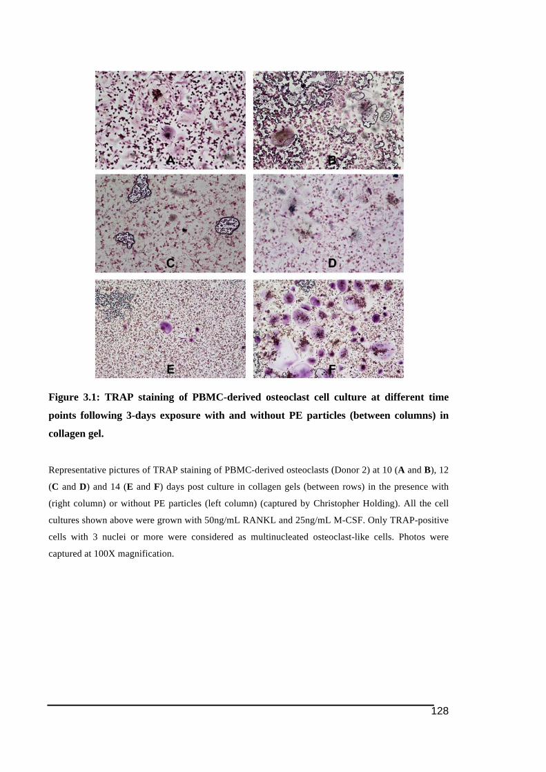
128
Figure 3.1: TRAP staining of PBMC-derived osteoclast cell culture at different time
points following 3-days exposure with and without PE particles (between columns) in
collagen gel.
Representative pictures of TRAP staining of PBMC-derived osteoclasts (Donor 2) at 10 (A and B), 12
(C and D) and 14 (E and F) days post culture in collagen gels (between rows) in the presence with
(right column) or without PE particles (left column) (captured by Christopher Holding). All the cell
cultures shown above were grown with 50ng/mL RANKL and 25ng/mL M-CSF. Only TRAP-positive
cells with 3 nuclei or more were considered as multinucleated osteoclast-like cells. Photos were
captured at 100X magnification.
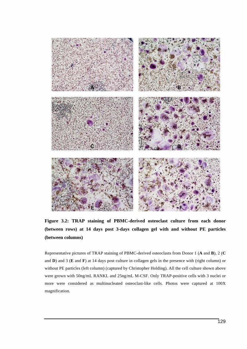
129
Figure 3.2: TRAP staining of PBMC-derived osteoclast culture from each donor
(between rows) at 14 days post 3-days collagen gel with and without PE particles
(between columns)
Representative pictures of TRAP staining of PBMC-derived osteoclasts from Donor 1 (A and B), 2 (C
and D) and 3 (E and F) at 14 days post culture in collagen gels in the presence with (right column) or
without PE particles (left column) (captured by Christopher Holding). All the cell culture shown above
were grown with 50ng/mL RANKL and 25ng/mL M-CSF. Only TRAP-positive cells with 3 nuclei or
more were considered as multinucleated osteoclast-like cells. Photos were captured at 100X
magnification.
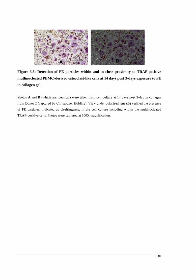
130
Figure 3.3: Detection of PE particles within and in close proximity to TRAP-positive
mutlinucleated PBMC-derived osteoclast-like cells at 14 days post 3-days exposure to PE
in collagen gel
Photos A and B (which are identical) were taken from cell culture at 14 days post 3-day in collagen
from Donor 2 (captured by Christopher Holding). View under polarized lens (B) verified the presence
of PE particles, indicated as birefringence, in the cell culture including within the multinucleated
TRAP-positive cells. Photos were captured at 100X magnification.
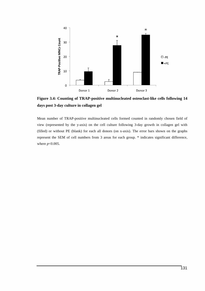
131
Figure 3.4: Counting of TRAP-positive multinucleated osteoclast-like cells following 14
days post 3-day culture in collagen gel
Mean number of TRAP-positive multinucleated cells formed counted in randomly chosen field of
view (represented by the y-axis) on the cell culture following 3-day growth in collagen gel with
(filled) or without PE (blank) for each all donors (on x-axis). The error bars shown on the graphs
represent the SEM of cell numbers from 3 areas for each group. * indicates significant difference,
where p<0.005.
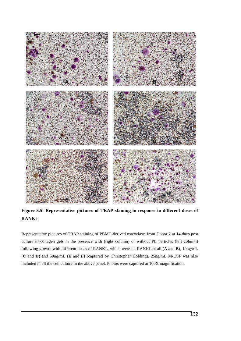
132
Figure 3.5: Representative pictures of TRAP staining in response to different doses of
RANKL
Representative pictures of TRAP staining of PBMC-derived osteoclasts from Donor 2 at 14 days post
culture in collagen gels in the presence with (right column) or without PE particles (left column)
following growth with different doses of RANKL, which were no RANKL at all (A and B), 10ng/mL
(C and D) and 50ng/mL (E and F) (captured by Christopher Holding). 25ng/mL M-CSF was also
included in all the cell culture in the above panel. Photos were captured at 100X magnification.
133
From the panel in Figure 3.5 above, it appeared to be clear that the dose of RANKL had
influence on PE-induced formation of osteoclast-like cells from PBMC, with 50ng/mL
became the optimum dose required.
3.3.2. Effect of PE particles exposure on the osteoclast resorption activity
Representative pictures captured from scanning electron microscope scans are arranged in
panels in the following figures for comparison on the effect of PE particles on osteoclast
resorption function (Figure 3.6 and Figure 3.7).
Quantitative analysis of pit resorption indicates that there was greater resorption of dentine
discs occurred in cells with PE exposure compared to dentine discs with no PE particles
(Figure 3.8). It also appeared that RANKL dose had big influence on the total resorption
activity looking at the minimal amount of total resorption on the dentine discs for low dose
and no RANKL samples as compared the 50ng/mL RANKL (Figure 3.9).
134
Figure 3.6: Dentine resorption pit assay of PBMC-derived osteoclast cell culture at
different time points following 3-day exposure (beteen rows) with and without PE
particles (between columns) in collagen gel
Representative pictures of resorption pits on dentine slices by PBMC-derived osteoclasts (from Donor
2 only) at 10 (A and B), 12 (C and D) and 14 (E and F) days post culture in collagen gels (between
rows) in the presence with (right column) or without PE particles (left column) (scanning electron
microscopic scans by Christopher Holding). All the cell cultures shown above were grown with
50ng/mL RANKL and 25ng/mL M-CSF. Photos were captured at 100X magnification.
135
Figure 3.7: Dentine resorption pit assay of PBMC-derived osteoclast culture from each
donor (between rows) at 14 days post collagen gel with and without PE particles
(between columns)
Representative pictures of resorption pits on dentine slices by PBMC-derived osteoclasts from Donor
1 (A and B), 2 (C and D) and 3 (E and F) at 14 days post culture in collagen gels in the presence with
(right column) or without PE particles (left column) (scanning electron microscopic scans by
Christopher Holding). All the cell cultures shown above were grown with 50ng/mL RANKL and
25ng/mL M-CSF. Photos were captured at 100X magnification.
136
Figure 3.8: Total resorption areas on dentine assay by PBMC-derived osteoclast-like
cells following 14 days post 3-day culture in collagen gel in response to stimulation with
and without PE
The graph shows mean percentage of areas on dentine slices resorbed by osteoclast-like cells in
randomly chosen field of view on the cell culture (represented by the y-axis) following 3-day growth
in collagen gel with (filled) or without (blank) for each all donors (on x-axis). The error bars shown on
the graphs represent the SEM of resorption areas from 3 fields for each donor.
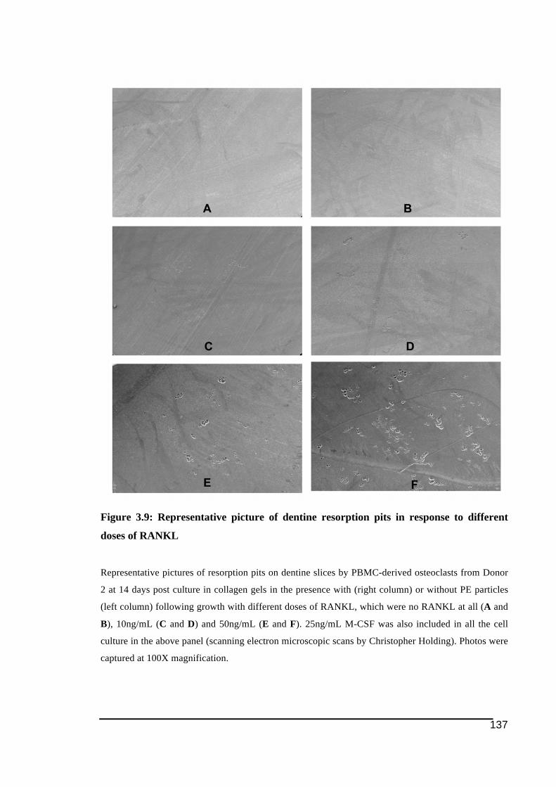
137
Figure 3.9: Representative picture of dentine resorption pits in response to different
doses of RANKL
Representative pictures of resorption pits on dentine slices by PBMC-derived osteoclasts from Donor
2 at 14 days post culture in collagen gels in the presence with (right column) or without PE particles
(left column) following growth with different doses of RANKL, which were no RANKL at all (A and
B), 10ng/mL (C and D) and 50ng/mL (E and F). 25ng/mL M-CSF was also included in all the cell
culture in the above panel (scanning electron microscopic scans by Christopher Holding). Photos were
captured at 100X magnification.

138
139
3.3.3. Effect PE particles on gene expression of osteoclast ITAM-related
molecules in PBMC-derived osteoclasts
The expression levels of ITAM-related molecules and NFATc1 in response to exposure to PE
particles were studied at the gene level. The mRNA expression levels of these molecules were
also compared and the expression of cathepsin K was used as the osteoclast marker. The
results are shown in Figure 3.10 and Figure 3.11.
There was an inconsistent response to PE in the earlier time points (Day 10 and 12 post
collagen gel) between donors (Figure 3.10). However, at 10 days following 3 days of collagen
gel there was a trend for lower mRNA expression of ITAM-associated molecules, TREM2,
OSCAR and FcRγ, in cells exposed to PE particles in the collagen gel in comparison to the
controls. However, it seemed to be a turnover in the mRNA expression as there was a trend
towards higher mRNA level of OSCAR, TREM2 and FcRγ in samples incubated with
polyethylene particles at Day 14 post collagen gel and this was consistently observed in three
donors assessed in this study (Figure 3.11).
Changes in NFATc1 mRNA expression seemed to follow no common trend between donors.
While a decrease in the gene expression over the time in response to PE particle exposure in
Donor 1 and Donor 2 was observed the opposite occurred with Donor 3. The relative mRNA
expression of cathepsin K appeared to be higher from Day 12 to Day 14 (even though not
statistically significant) in cells exposed to PE than the controls in every donor. As an
osteoclast cell marker, this higher mRNA expression of cathepsin K in cells mixed with PE
particles, particularly at 14 days post collagen gel was consistent with the data for TRAP
staining (Figure 3.2 and Figure 3.4) and dentine pits resorption assay (Figure 3.7 and Figure
3.8) presented earlier in this Chapter 3.
140
The mRNA expression level (in relative to hARP mRNA) of ITAM-related molecules (TREM2,
DAP12, OSCAR and FcRγ) together with transcription factor NFATc1 and osteoclast cell marker
cathepsin K (between rows) in PBMC-derived osteoclast-like cells in response to exposure to PE
particles (filled) as compared to the controls (blank). All the cell culture shown above were grown
with 50ng/mL RANKL and 25ng/mL M-CSF. The graphs were sorted according to the time point post
3-day collagen gel (in columns). Statistical difference, p<0.05 is indicated by * on the graphs.
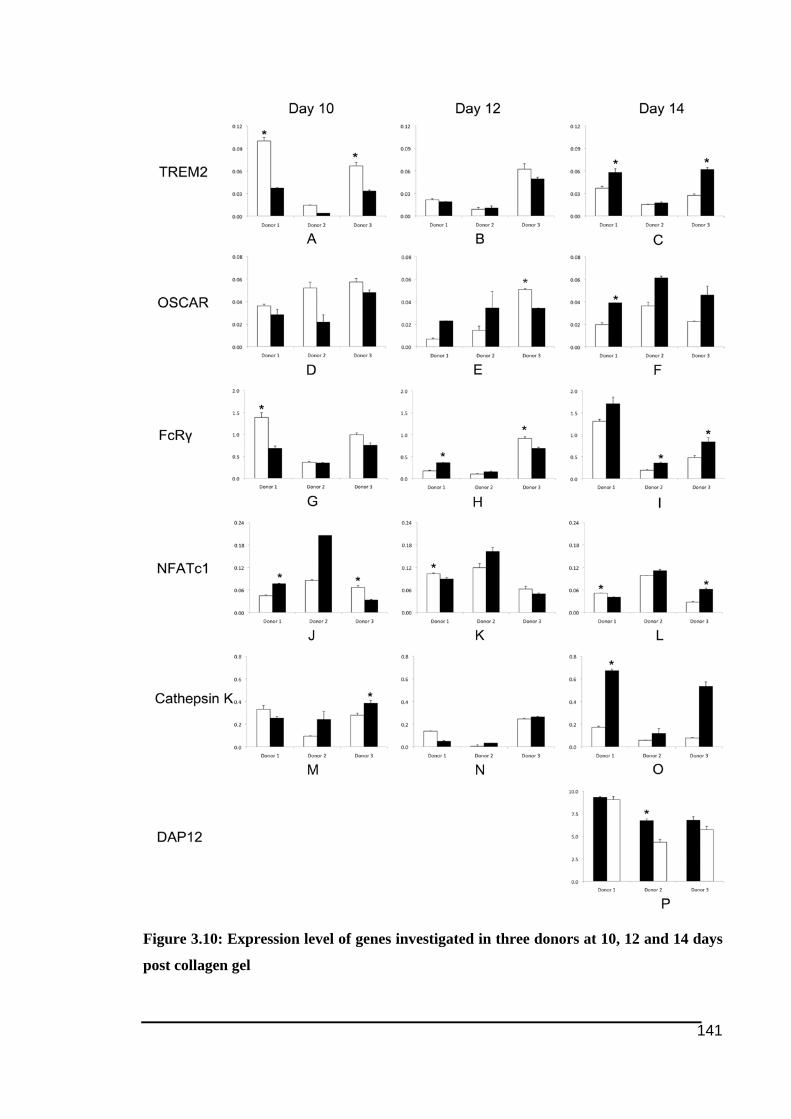
141
Figure 3.10: Expression level of genes investigated in three donors at 10, 12 and 14 days
post collagen gel

142
The mRNA expression level (in relative to hARP mRNA) of ITAM-related molecules (TREM2,
DAP12, OSCAR and FcRγ) together with transcription factor NFATc1 and osteoclast cell marker
cathepsin K (between rows) in PBMC-derived osteoclast-like cells in response to exposure to PE
particles (filled) as compared to the controls (blank). All the cell culture shown above were grown
with 50ng/mL RANKL and 25ng/mL M-CSF. The graphs were sorted according to the donors the
cells from (in columns). Statistical difference, p<0.05 is represented as * on the graphs.
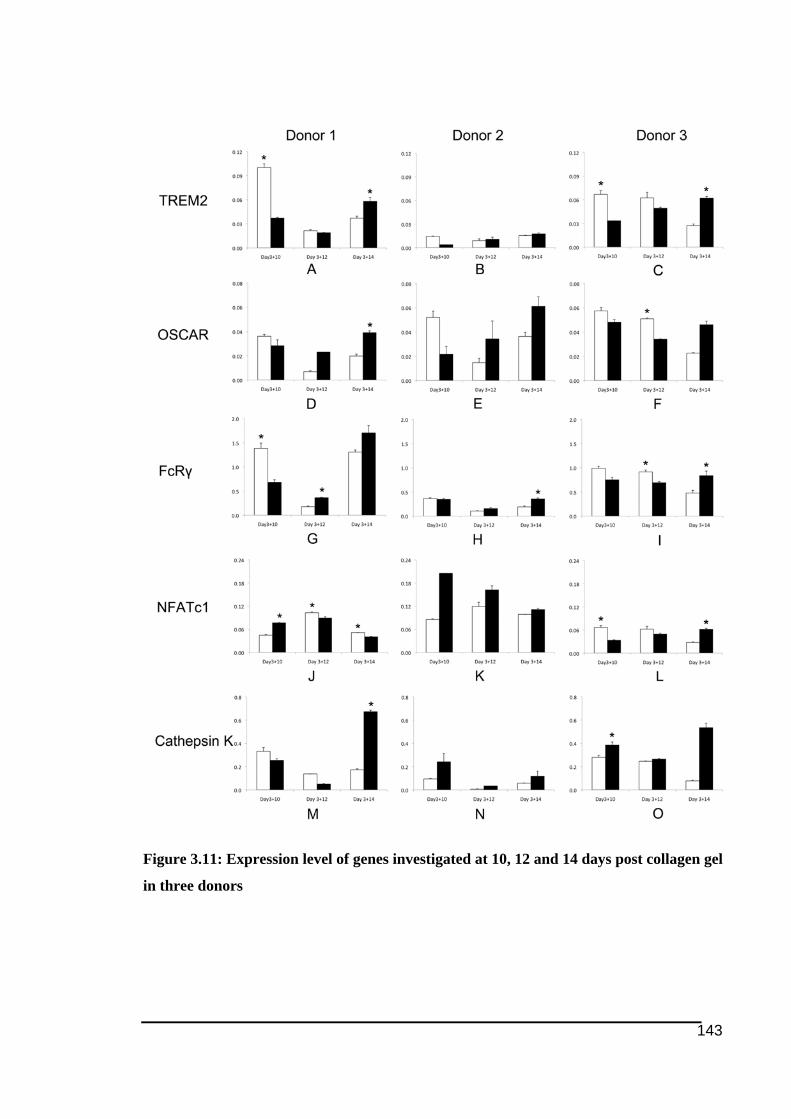
143
Figure 3.11: Expression level of genes investigated at 10, 12 and 14 days post collagen gel
in three donors

144
The mRNA expression level (in relative to hARP mRNA) of ITAM-related molecules (TREM2,
OSCAR and FcRγ) together with transcription factor NFATc1 and osteoclast cell marker cathepsin K
(between rows) in PBMC-derived osteoclast-like cells (both exposed to PE particles (filled) and the
controls (blank) in all donors studied. All the cell culture shown above were grown with 25ng/mL M-
CSF, but differently in term of the doses of RANKL (between columns), which included no RANKL,
10 and 50ng/mL concentration. Statistical difference, p<0.05 is represented as * on the graphs.
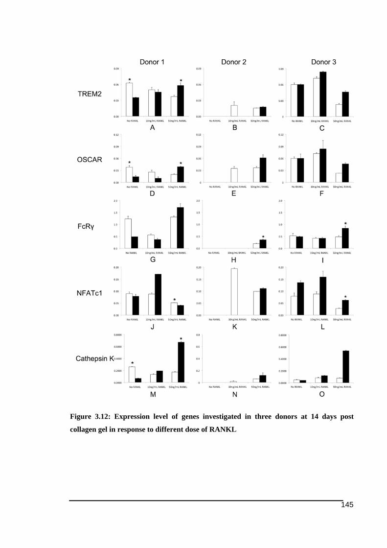
145
Figure 3.12: Expression level of genes investigated in three donors at 14 days post
collagen gel in response to different dose of RANKL
146
The mRNA levels of each individual donor across the time point are shown in Figure 3.11. In
general, there was no consistent trend in mRNA expression between donors, which may be
due to variability between donors.
The data of mRNA expression in response to different dose of RANKL was presented in
Figure 3.12 below. For this particular pool of data, comparison between sample groups was
difficult to low and no RANKL samples groups (below the level of detection), which often
seemed with Donor 2. For the other two donors, there was no particular trend on the
regulation of mRNA expression in response to PE particles seen in the 50ng/mL RANKL
group. Nevertheless, it was quite interesting to note a similar pattern of mRNA expression
level of between molecules except NFATc1 (between rows) in each sample group in both
Donor 1 and 3.
3.4. Discussions
The work presented in this Chapter 3 was carried out as a functional study to better
understand the findings in the previous Chapter 2, which suggested that the expression of
osteoclast ITAM-related molecules (TREM2, DAP12, OSCAR and FcRγ) may be involved in
PE-induced osteoclastogenesis.
The novel aspect of this study is not just the in vitro model setup for osteoclast culture with
particles, but also the investigation of osteoclast ITAM-related molecule expression upon
stimulation of particles like PE. The rationale of having collagen gel to overcome
hydrophobicity of the PE particles solved the problem of low density of PE particles that
normally results in particles floating in the culture media (Atkins et al. 2009; Voronov et al.
1998). Collagen was included in the in vitro model to take advantage of collagen trapping the
hydrophobic and low-densed PE particles and hence promote more contact between those
particles and the cells seeded at the bottom of the culture plate wells (Voronov et al. 1998).
The collagen gel also allows to better present presence of PE in PO tissues. In the tissues
collagen type I is found in abundance in bone microenvironment and in inflammatory
membrane (Voronov et al. 1998). This collagen gel model has allowed us to investigate the
effect of PE on osteoclastogenesis in vitro.
147
There has been quite a number of earlier studies employing various methods to overcome
those limitations of PE properties in aquoes media in order to optimize the interation between
PE particles and the seeded cells (Voronov et al. 1998). Voronov and colleagues (1998)
introduced PE in their in vitro cell culture system by coating the particles onto coverslips,
which were then placed at the bottom of wells of cell culture plates. Meanwhile Rao and co-
workers (1999) used a different approach by applying inverted cell culture system. However
the best way to mimic the in vivo condition in vitro could be the collagen model used here and
the demonstration that cells were able to phagocytose PE particles is a good indication of this
(Voronov et al. 1998). The method used here resulted in harvesting of large numbers of
healthy cells containing particles which is consistent with work by Santerre and colleagues
(2000), who reported on monocytes exposed to PE had markedly increased the cell life span
from 10 days to 30 days (Santerre et al. 2000).
For this novel in vitro model of osteoclasts, PBMC–derived osteoclast-like cells were chosen
since they are a very well established in vitro model for osteoclasts, in which the expression
of osteoclast markers have been described earlier (Faust et al. 1999). It is believed that CD14-
positive cells are the most likely the precursor cells for osteoclasts (Atkins et al. 2006;
Massey & Flanagan 1999; Nicholson et al. 2000; Nose et al. 2009; Shalhoub et al. 2000;
Sorensen et al. 2007). However, other study suggested CD11b-positive cells as among those
that can serve as osteoclast precursors (Li et al. 2004).
The main focus of this chapter is on the effects of PE on osteoclastogenesis. It is often
misunderstood that PE is a less damaging biomaterial. From the literature, it was found that
the uptake of PE by macrophage cells did not cause any cytotoxicity to the engulfing cells,
but indeed prolonged the survival of the cells (Benz et al. 2001; Santerre et al. 2000; Xing et
al. 2002). However, a number of previous studies reported significant increase in the release
of proinflammatory cytokines TNFα, IL-1 and IL-8 by macrophages after being challenged
with PE particles (Boynton et al. 2000; Rader et al. 1999).
It has been shown that size and shape might influence the biological effects of PE particles
(Ren et al. 2003). For instance, in a recent study Pal et al. (2011) had found that the size of PE
particles liberated could affect the degree of osteoclast formation (Pal et al. 2011). The study
indicated that PE particle of size 0.2µm or smaller induced more osteoclast formation and if
148
particles of similar size were used in the present study, it might have given more significant
differences.
In this study Ceridust 3615 of a size range of within 0.3 to 10µm was used (Matsusaki et al.
2007; von Knoch et al. 2004). Studies using smaller size ranged PE particles will be irrelevant
because the particles present in tissues surrounding failed prostheses are mostly in the size
range of 0.5-3µm (Hirakawa et al. 1996; Howie 1990; Lee et al. 1992; Shanbhag et al.
1994b), with nearly all PE particles detected in PO tissues are reported to be less than 2µm
(Jacobs et al. 2008; Koseki et al. 2005; Shanbhag et al. 1995; Shanbhag et al. 1994b; Visentin
et al. 2004). This is further supported by more recent studies (Jacobs et al. 2008; Pal et al.
2011), which indicated that about 90% of PE particles liberated are less than 1µm size. Other
studies have confirmed that very small size are outside of the range of optimal biological
active size of polyethylene (Green et al. 1998; Matthews et al. 2000). Furthermore more
recently Pal et al. (2011) found murine macrophages did not respond to PE particles with size
less than 0.2µm. It is important to make sure that the size of the particles introduced was less
than 10µm as those are phagocytosed by macrophages (osteoclast precursors) while the larger
ones are surrounded by the FBGCs derived from macrophages (Ingham & Fisher 2005;
Santerre et al. 2000; Voronov et al. 1998).
Ceridust 3615 has been widely used in other previous studies (Elfick et al. 2004; Green et al.
1998; Guo et al. 2012; Matsusaki et al. 2007). Ceridust 3615 is used for in vitro or in vivo
studies as they are commercially available, cost effective and are of a size comparable to
particles isolated from tissues (von Knoch et al. 2004). Regarding the physical properties,
Ceridust 3615 has been shown to have a rougher surface, shorter length and more consistent
size than PE particles isolated from the tissues (von Knoch et al. 2004). Previous studies have
indicated that rough surface PE particles may stimulate stronger pro-inflammatory effect
(represented by TNFα and IL-1β expression in murine model) in comparison to a smoother
surface particles (Sieving et al. 2003; Yang et al. 2002b). Interestingly Pal et al. (2011)
showed that exposure to PE particles with wider range of sizes resulted in higher number of
osteoclast-like cells formed compared to exposure to PE particle of a more homogenous size.
500µg/mL was the chosen concentration Ceridust 3615 PE particles to be used in this
experiment as it was optimised in a previous study (Matsusaki et al. 2007). This concentration
149
of PE particles in the in vitro system was also been optimised in preliminary work carried out
in the laboratory (by Christopher Holding).
RANKL is another important component of the model that should be discussed in this
Chapter 3. The hypothesis proposed by Rodan and Martin (1981), and later supported by
Udagawa and colleagues (Takahashi et al. 1988; Udagawa et al. 1990) has indicated that the
differentiation of BMMs into mature osteoclasts could be achieved in vitro in the presence of
osteoblasts. Further studies revealed that supplementation of RANKL and M-CSF (Kong et
al. 1999; Lacey et al. 1998; Nicholson et al. 2000; Quinn et al. 1998; Yasuda et al. 1998b),
which are expressed by osteoblasts, is sufficient to compensate the need for the presence of
osteoblasts in driving osteoclastogenesis in vitro. Matsuzaki et al. (1998) also described that
addition of soluble RANKL, along with M-CSF could compensate absence of stromal cells
for formation of osteoclast-like cells in vitro.
From previous studies, it appears that RANKL plays an essential role in PE-induced
osteolysis as of inhibition of PE-induced osteolysis in vivo was achieved following therapy
with anti-RANKL (Childs et al. 2002; Tsutsumi et al. 2008). The importance of RANKL in
osteoclastogenesis in vitro stimulation of ITAM-pathway in the absence of RANKL did not
induce osteoclast formation (Koga et al. 2004). RANKL is also important in promoting the
survival and motility of osteoclasts (Lacey et al. 1998). It would be a particular interest to
study cells derived from PO tissues in this model, as it has been shown that cells derived from
PO tissues were able to differentiate into osteoclasts in absence of RANKL (Sabokbar et al.
2003). Nonetheless, considering the large number of sample groups required to fulfil the
objectives of the study, the use of human PBMCs became the more realistic option for this
study.
In this in vitro study, M-CSF was also used as it serves quite a number of important roles for
the osteoclast cell culture. Based on previous studies, it has been known that M-CSF, released
by osteoblasts, prolongs survival of osteoclasts and induces osteoclastic activity in vitro
(Fuller et al. 1993; Lagasse & Weissman 1997; Yoshida et al. 1990). It is also needed for the
cell spreading, migration and proliferation of the osteoclasts cultured (Faccio et al. 2003c).
Supplementation of RANKL alone without M-CSF may not be sufficient to promote
osteoclastogenesis in vitro (Quinn et al. 1998) as M-CSF is required to stimulate the
expression of RANK in monocytes (progenitor for osteoclasts)(Arai et al. 1999). In the
150
context of PO pathogenesis, it has been found that RANK, possibly induced by M-CSF, is
elevated in PO pseudosynovial membranes (Holding et al. 2006; Xu et al. 1997) suggesting
the importance of M-CSF in this model. In PO tissues, a study by Neale and colleagues
(1999) suggested that M-CSF was able to stimulate osteoclast formation and resorption
activity in the tissues (Neale et al. 1999). There is also data indicating that M-CSF is
particularly important for cell multinucleation (Faccio et al. 2003c). It is widely accepted that
the optimal concentration of RANKL and M-CSF in an in vitro model of osteoclast is around
50ng/ml and 25ng/ml respectively. This concentration of RANKL and M-CSF has been used
in several numbers of studies involving osteoclast assay (Baumann et al. 2004; Herman et al.
2008), including this study.
Atkins et al. (2009) have shown, in similar 3D collagen gel model, that osteoblast cells
exposed to PE particles have striking effect in increasing expression of osteoclastogenic genes
including for M-CSF and RANKL. This study here indicates PE elevated mRNA expression
of osteoclast ITAM-related molecules, osteoclast formation and resorption activity in the
absence of osteoblasts. It might be interesting to investigate if co-culturing osteoblasts-
osteoclasts would have an additive effect towards osteoclastogenesis.
Based on the data documented in this chapter (in both sections 3.3.1 and 3.3.2), it was clearly
demonstrated that the PE particles (Ceridust 3615) directly increased the formation of
multinucleated PBMC-derived osteoclast-like cells and their bone resorption activity.
Together this data was in consistent with other previous studies reported (Anderson et al.
2001; Granchi et al. 2004; Holding et al. 2006; Pal et al. 2011) even though the experimental
setup was different between studies. It is quite interesting to note that the PE-induced increase
in the formation of TRAP-positive osteoclasts seemed to require RANKL (refer to Figure
3.5).
While the study by Anderson et al. (2001) reported increase in both bone resorption activity
and TRAP-positive cell formation from macrophages in response to biomaterials including
PE, the study used animal systems, which is different from this current work that used human
PBMCs. Even though there are close similarities with the work by Pal and co-workers (2011),
which have already shown increase in the formation of TRAP-positive cells (viewed in 2
fields of visions at 4X magnification for SQA by two independent observers) upon exposure
to PE particle in vitro, this study took a more extensive approach looking at the gene
151
expression of osteoclasts, particularly ITAM-associated molecules to illustrate the role they
play in osteoclastogenesis in the context of peri-implant osteolysis. Besides, unlike the study
that used PE wear debris isolated from PO tissues, the current presented work instead applied
commercially available Ceridust 3615, which is physically more homogenous particles and
hence could promisingly yield reproducible data.
In relation to the formation of TRAP-positive cells following stimulation by the PE particles,
Pal et al. (2011) reported that even three days post-treatment with RANKL, giant (with size of
more than 200µm) TRAP-positive multinucleated cells started to be observed as already being
formed in response to the presence of PE particles. This increase in PE-induced osteoclast
formation was also reported as becoming greater up to nearly five times after 10 day of cell
culture than the control, which is quite in agreement with the data obtained from this study. It
is important to note that, in applying TRAP as an osteoclast cell marker, counting the
osteoclast-like cells should be only on multinucleated osteoclast-like cells only as TRAP has
also been reported as could be expressed in mononuclear cells as well (Humphrey et al.
2004).
It was particularly interesting to note the detection of PE particles within the multinucleated
TRAP-positive osteoclast-like cells formed in the cell culture (refer Figure 3.3). Such PE
phagocytosis-like phenomenon has also been described earlier in a culture of macrophage
cell-line by Matsusaki and colleagues (2007), who used similar PE particles (Ceridust 3615)
in their work. While majority believes macrophages that have phagocytosed PE particles play
role in promoting more osteoclast formation in the tissues by secreting pro-osteoclastogenic
cytokines (Hirashima et al. 2001; Voronov et al. 1998), there is another line of opinion
suggesting that those macrophages become the precursor cells for the multinucleated cells
formed (Pandey et al. 1996; Sabokbar et al. 1998; Wang et al. 1997a, 1997b) (refer section
2.3.4 above). Indeed, the data of this study, including ones presented in the previous chapter
(refer section 2.3.4 and the corresponding discussion 2.4), is in favour of the latter theory.
Another important component of this current study that distinguishes it from the work by Pal
et al. (2011) is the addition of dentine pits resorption assay as a mean of looking on the
activity of osteoclasts formed on top of total osteoclasts formed in response to exposure to PE
particles. Inclusion of dentine pits resorption assay into an osteoclast in vitro assay should be
regarded as important as it could give more meaningful data that would represent the extent of
152
bone resorption compared to counting of osteoclasts formed. Besides this assay also provides
important information in assessing osteoclast functionality and activity. Indeed, having a
mineralised substrate such as pieces of bone or dentine intact to the osteoclasts formed could
influence regulation of the activity of the cells. It has been shown that interaction between
osteoclast and bone substrate could affect “genetic programming”, phenotype and activity of
osteoclast formed (McHugh et al. 2000; McHugh et al. 2007; McHugh et al. 2010; Shen et al.
2006), for instance higher expression of osteoclasts cell markers such as TRAP and cathepsin
K as this has been shown earlier by Shen and colleagues (2006). It would also be interesting
to investigate the expression of other ITAM-related molecules, considering ITAM-related
molecules such as OSCAR is believed to be expressed in late stage of osteoclastogenesis
(Kim et al. 2002; Kim et al. 2005d).
Based on the data obtained, which clearly showed that the presence of PE particles would
result in significant increase in the percentage of resorption pits areas particularly after two
weeks post-exposure to PE, it could be concluded that PE has effect in enhancing bone
resorption activity (refer to Figure 3.6). Nonetheless, the amount of RANKL appeared to have
significant influence on the PE-induced increase in resorption pits. Data on the PE-induced
increase in resorption pits in response to different dose of RANKL presented in this chapter
was consistent with the earlier study carried out in this lab (Holding et al. 2006), which has
demonstrated that 50ng/mL RANKL produced more pits on dentine than 10ng/mL dose. This
gives indication that high amount of RANKL along with PE particles in the tissues could
enhance the activity of osteoclasts. It is also important to note in Figure 3.9 that there was no
(or very minimal numbers of) resorption pit formed on the dentine in the absence of RANKL
even at two weeks post exposure to PE, and this may indicate that RANKL is an essential
component for the bone resorption activity of the multinucleated cells formed in the model.
As indicated earlier, the investigation on the mRNA expression of ITAM-related molecules,
as well as NFATc1 and cathepsin K, in response to PE stimulation becomes the main focus
being looked at in this chapter. This focus of the study provide novel discovery as there has
been no literature testing on the effect of biomaterials on the regulation of mRNA expression
of ITAM –related molecules, either through in vitro or in vivo, being reported to date.
In comparison to the data from the TRAP staining and dentine pits resorption assay, the
analysis on the mRNA expression was more difficult due to inconsistent trend of expression
153
between donors, as this could be seen from the graphs in the Results 3.3.3 (refer to Figure
3.11). It has been well acknowledged in studies in the past that there were differences in
cellular response to foreign-body particles between donors or patients, including in PBMC in
vitro model (Sethi et al. 2003; Yagil-Kelmer et al. 2004). However as being mentioned earlier
in the Results (section 3.3.3 above), yet still there are some similar trends that could be
deduced, which included in the observation that PE-induced change in mRNA expression
level was in similar fashion between TREM2, OSCAR, FcRγ and cathepsin K (refer to Figure
3.11). This also may suggest that the PE-induced change in mRNA expression of TREM2,
OSCAR, FcRγ and cathepsin K was in similar fashion, which would further speculate that the
regulation of these molecules mRNA expression could share a common pathway. Such
similar regulation pattern of mRNA expression between these molecules may also indicate
that the induction of the mRNA expression of these molecules by PE occurred in similar stage
during osteoclastogenesis.
While there has been very limited amount of studies investigating on NFATc1 in the context
of peri-implant osteolysis, this thesis (Chapter 2 and 3), to the author’s knowledge, is the first
one to explore on NFATc1 in conjunction to PE-induced osteolysis in particular.
While Yamanaka and coworkers (Yamanaka et al. 2008; Yamanaka et al. 2013) demonstrated
that NFATc1 is upregulated in PMMA-induced osteoclastogenesis data from this study was
unable to show any marked pattern of regulation (either suppression or induction) by PE
particles. Differences in cell types, biomaterial and size of particles tested might provide an
explanation for the discrepancies in the response observed (Horowitz & Gonzales 1996). It
could also be that high levels of NFATc1 expression during PE-induced osteoclast formation
are transient with respect to the stage of osteoclast development.
Unlike the other molecules where their mRNA expression had common pattern of response
following PE stimulation, there was no consistent in the change of NFATc1 mRNA
expression. Response time for regulation of mRNA expression following PE stimulation or
earlier expression could provide an explanation for such observation. Being a master
transcriptional factor, change in the mRNA expression of NFATc1 is expected to take place
earlier than the other molecules.
154
As for the ITAM-associated molecules, based on Figure 3.10, it appeared that at both 10 and
12 days post-collagen gel there was lower expression of TREM2 (even though only some
were statistically different) in PE-exposed sample groups compared to non-PE ones
consistently between donors. Interestingly it seemed that there was turnover in the mRNA
expression as there was trend of higher level in PE-containing samples, which might give
indication that PE-induced TREM2 mRNA expression took place after 14 days post-collagen
gel. For DAP12, there was only data for the mRNA expression at 14 days post-collagen gel in
the presence of 50ng/mL RANKL (in Figure 3.10). The real time PCR was not carried out for
the other sample groups (earlier time points and samples with less than 50ng/mL RANKL)
due to limitation on the amount of cDNA remaining. Nevertheless, at 14 days post-collagen
gel, like TREM2, generally there appeared to be increase in the mRNA expression following
exposure to PE in the gel. The increase in mRNA expression of TREM2 and DAP12 with
higher number of TRAP-positive cells and resorption pits on dentine may suggest that these
ITAM-related molecules might mediate those two PE-induced response.
As discussed earlier, findings from previous studies that reported the higher number of
multinucleated cells formed in response to exposure to PE particles could possibly be in
connection to higher mRNA expression of DAP12 in those samples. Previous studies have
suggested that ITAM-related molecules including TREM2 and DAP12 are involved in
osteoclastogenesis (Humphrey et al. 2006; Koga et al. 2004; Otero et al. 2012; Paloneva et al.
2003). However, this study on these molecules in vitro documented in this thesis is the first
reported in the context of PO. Humphrey and colleagues (2006) reported that the addition of
both M-CSF and RANKL resulted in increase in the expression of TREM2 accompanied with
increase in the formation of osteoclast–like cells from RAW264.7 cells. Their data also
suggested that there was a positive correlation between the expression of surface TREM2 and
the potency of RAW264.7 cells for differentiation into osteoclasts. There is also evidence
indicating that activation on TREM2 appeared to enhance M-CSF-promoted cell migration
and spreading (Humphrey et al. 2006), and hence possibly on the resorption activity of the
osteoclasts. There have also been studies reporting on increase in TREM2 mRNA expression
in in vitro model of human PBMC-derived osteoclasts, therefore the data obtained (increasing
TREM2 mRNA level throughout the osteoclastogenesis period) was an expected study
outcome (Kalliolias et al. 2010; Park-Min et al. 2009). Park-Min and colleagues (2009)
through their findings in human PBMC-derived osteoclast culture had suggested that
elevation of TREM2 mRNA expression could take place quite quickly after addition of
155
RANKL. Inhibition on osteoclastogenesis in human CD14-positive PBMC also showed
decreased mRNA expression of TREM2, besides NFATc1, and this may suggest that increase
in TREM2 mRNA expression is necessary during osteoclastogenesis.
There have been debates as whether lack of DAP12 could lead to arrest in osteoclast
formation or not. DAP12 has been shown to be upregulated by PU.1 transcription factor, a
crucial transcription factor in osteoclast differentiation (Henkel et al. 2002). Humphrey and
coworkers (2004) reported that deletion in DAP12 as not affecting expression of known
osteoclast cell markers like TRAP, CTR and cathepsin K, suggesting there is no direct
connection between DAP12 and the expression of those cell markers. Nonetheless through
this study it was shown that increase in cathepsin K mRNA expression, alongside with great
increase in osteoclast formation (from TRAP staining) at 14 days post-exposure to PE, is
accompanied with a trend of higher level of DAP12 mRNA expression, therefore this could
still leave room of connection between DAP12 and osteoclastogenesis (and expression of
osteoclast cell markers) open.
There has been evidence suggesting that DAP12 deficiency could arrest osteoclastogenesis
induced by RANKL and M-CSF (Koga et al. 2004; Mocsai et al. 2004). Data from Humphrey
et al. (2004) suggested that DAP12 plays important in osteoclast multinucleation during
differentiation process. It was reported that pre-osteoclasts from DAP12-deficient mice were
unable to multinucleate in vitro, nonetheless they still possessed dentine-resorptive capacity
even though at a reduced extend. Together this data suggests the trend of increase of DAP12
mRNA expression in response to exposure to PE particles is more relevant to the increased
formation of multinucleated cells than the resorption activity. Humphrey et al. (2004)
indicated that the activation of DAP12 was also able to increase the size of the multinucleated
cells formed. However, such increase in multinucleated osteoclast formation and size
appeared to require RANKL since Humphrey et al. (2004) reported that there was no
multinucleated TRAP-positive cells formed following DAP12 activation in absence of
RANKL.
Meanwhile, there is also evidence suggesting that DAP12 is more associated to the regulation
of osteoclast bone resorbing activity than in the osteoclast formation itself. For instance,
Kaifu et al. (2003) reported reduced resorption activity in DAP12-deficient cells, yet those
cells were still able to multinucleate even though at much lesser extend. Zou et al. (2010)
156
found that osteoclastogenesis still occured in presence of M-CSF and RANKL despite the
lack of DAP12, nevertheless there was defect in cytoskeletal organization that is important in
ruffled border and actin ring formation, which subsequently affect osteoclast bone-resorbing
capacity (Boissy et al. 2002; Faccio et al. 2003c; Humphrey et al. 2004; Paloneva et al. 2003;
Teitelbaum 2007; Zou et al. 2010), therefore the increase in the DAP12 mRNA expression in
PE-containing samples could also be associated with the higher area of dentine pits. DAP12 is
also believed to mediate the β3 (interaction with bone substrate)-induced osteoclast
cytoskeletal organization (Faccio et al. 2003b; McHugh et al. 2000).
For OSCAR and FcRγ, as what could be seen from Figure 3.10 and Figure 3.11, there was
much more consistent trend of mRNA expression between between all donors across the three
time points. At 10 days post-exposure to PE particles, the mRNA expression of the samples
was generally lower than their corresponding controls (without PE). However, on the
following time point (12 days post-collagen gel), there appeared to be a turnover in the
mRNA expression and by 14 days post-PE stimulation, the mRNA expression of OSCAR and
FcRγ in all donors appeared to be generally higher than the controls (even though not all
statistically different). Like for TREM2 and DAP12, those higher trends of mRNA level of
OSCAR and FcRγ at 14 days post-exposure to PE compared to the controls could be
connected to the high PE-induced increase in the formation of TRAP-positive multinucleated
osteoclast-like cells and total area of resorption pits.
To date, there has been very minimal information on the influence of OSCAR and FcRγ
towards the bone resorbing activity of the osteoclasts. Another interesting data regarding the
significance of OSCAR/ FcRγ in osteoclast function would be from Zou et al. (2010), which
indicated that activation of OSCAR/FcRγ in osteoclasts grown on bone could partially
compensate for the defect in cytoskeletal reorganisation in DAP12-deficient cells. In a
previous study carried out in this laboratory, it was found that reduction in the resorption
activity of PBMC-derived osteoclasts following inhibition by drugs in vitro was also
accompanied by lower expression of OSCAR mRNA at a late time point (Cantley et al.
2011). These studies may suggest that obtaining study outcomes of higher in both total area of
resorption pits (Figure 3.8) and mRNA level for OSCAR and FcRγ in PE-exposed cells at 14
days post-collagen gel (Figure 3.10) was not a coincidence, indeed may lead to hint of a
possible connection.
157
Based on literature, the expression of OSCAR is seen to be more associated with further
differentiation of monocyte/macrophage cell lineage in the development of dendritic cells
(Merck et al. 2005; Merck et al. 2004) as well as osteoclastogenesis (Herman et al. 2008) in
human. For instance, there has been a study reported that human PBMC-derived osteoclasts
formed following LPS-induced osteoclastogenesis as expressing OSCAR and other osteoclast
markers, however unable to produce resorption lacunar (Mabilleau et al. 2011), suggesting
that the expression of OSCAR has more connection to the capability of forming the bone
resorbing cells.
There are more studies that may provide better link between higher OSCAR and FcRγ
expression (Figure 3.10) and more osteoclast formation in vitro (Figure 3.4). For example, on
its discovery, it was found that inhibition of OSCAR by the addition of soluble form of
OSCAR in a co-culture with osteoblasts reduced the formation of osteoclasts (Kim et al.
2002). Recently, Barrow et al. (2011) demonstrated that interaction between OSCAR and the
ligand (collagen) enhanced osteoclastogenesis in vitro and blocking on the interaction would
inhibit such enhancement in osteoclastogenesis. Barrow et al. (2011) also discovered
reduction in the mRNA expression of NFATc1 and osteoclast markers like TRAP, cathepsin
K and calcitonin receptor in mouse BMMs lacking either OSCAR or FcRγ. In DAP12-
deficient cells (in the context of PLOSL), OSCAR and FcRγ has significance in the
osteoclastogenesis, and this could be seen from the activation of these molecules, which was
reported as able to partially increase the size of osteoclasts formed (Zou et al. 2010).
In contrast to the majority studies, Ishikawa et al. (2004) suggested FcRγ is not really crucial
in osteoclast formation following their observation of no difference in the number of TRAP-
positive cells between FcRγ-deficient (with low level of OSCAR detected) and the wild type
cells. Nonetheless, there is possibiliity of the absence of FcRγ being compensated by another
pathway in mediating the osteoclastogenesis.
In relation to the point in which OSCAR being nominated as a good candidate for osteoclast
cell marker, it is quite interesting to note that the induction of the mRNA expression of
cathepsin K by PE exposure in Donor 2 was very small, particularly at 14 days post PE
exposure in comparison to the other two donors (refer Figure 3.11), where as the data from
TRAP staining and dentine resorption pits assay indicated the increase in the number of
osteoclast-like cells formed (refer Figure 3.4) and the functionality of osteoclast formed
158
resulted from 3-day exposure to PE (refer Figure 3.8) was huge. When comparing the data on
mRNA expression against TRAP staining and resorption assay, it seemed that OSCAR
mRNA expression was more consistent with the trend of data from those two assays as
compared to cathepsin K (refer Figure 3.11). This may put on another credit for considering
OSCAR as a good osteoclast cell marker (see section 2.4).
In corresponding to the investigation on whether the PE-induced elevation in OSCAR and
FcRγ mRNA expression is RANKL-dependent or not, it appeared that such of increase could
be consistently (between donors) observed only at the dose 50ng/mL RANKL (as partly
discussed in this section). Herman et al. (2008) suggested these two pathways are independent
of each other (M-CSF and RANKL did not increase OSCAR mRNA expression), however
indeed, they together contribute towards osteoclastogenesis process in an additive manner.
However, there are some supports from other earlier studies that these intracellular pathways
may be interconnected, even though to date there is quite limited amount of information
directly on the relationship between OSCAR/FcRγ and RANKL-mediated pathway. There are
quite numbers of studies which have shown RANKL to mediate increase in mRNA
expression. For instance, the study by Kim et al. (2005d) reported increase in OSCAR mRNA
expression up to 1200-fold following 3 days-growth of murine BMMs in the presence of
RANKL and M-CSF. Indeed, OSCAR was discovered following its marked increase in
expression post RANKL stimulation (Kim et al. 2002). The RANKL-mediated increase in
OSCAR mRNA expression was reported to be upregulated by USFs (Kim et al. 2008) but
negatively regulated by MafB (Kim et al. 2007a) and PIAS3 (Kim et al. 2007b) (refer Table
1.5).
As it applies to TREM2 and DAP12 (see Discussions 3.4 earlier) there was issue of very low
gene transcription for OSCAR, FcRγ, NFATc1 and cathepsin K (some below detection limit)
in some sample groups with low and no RANKL, particularly in Donor 2 (refer Figure 3.12).
There are questions as whether cells for those groups actually viable at 14 days post collagen
gel as they might required RANKL to allow them to differentiate, which then allow them to
survive at that time point. In this in vitro model, in the presence of M-CSF but without
RANKL, the cells will keep proliferating as monocytes without really going committed into
osteoclast lineage. As a result of that, considering the time point of the extraction of cell
159
culture (13, 15 and 17 days) and short lifespan of monocytes/macrophages than osteoclasts,
the cells were probably already undergoing apoptosis.
3.5. Conclusions
In summary, through this 3D collagen gel model all PE-induced stimulation for osteoclast-
like formation (based on TRAP staining), bone resorbing activity (as indicated from dentine
pits resorption assay) and more importantly mRNA expression of ITAM-related molecules
and cathepsin K generally took place at 14 days after being exposed to the biomaterial. With
all the data indicating the increase in response to PE occurring at 14 days post-collagen gels
documented in this chapter, supported by higher expression of those molecules in peri-
implant tissues as discussed earlier in Chapter 2, together they may also suggest that the
investigated ITAM-related molecules could be involved in mediating the PE-induced increase
in osteoclastogenesis and bone resorption.
Therefore, in addition to the improvements on the implant design, therapies targeting
osteoclastogenic molecules like osteoclast ITAM-osteoclast associated molecules, which may
be involved in the regulation peri-implant osteolysis as described here, could provide an
excellent approach of treatment for the disease. There have been examples of studies in the
past that showed targeting osteoclastogenic molecules, such as RANK/RANKL/OPG axis
(Childs et al. 2002; Goater et al. 2002; Ulrich-Vinther et al. 2002; von Knoch et al. 2005;
Yang et al. 2002a) could offer promising therapy potential in the treatment of peri-implant
osteolysis.
Despite the great advantages in this novel in vitro model of osteoclasts, there are unavoidable
limitations noticed throughout the study. In order to keep supplementing the cells with fresh
media to avoid depletion of cell growth and differentiation factors over the long period of
culture (up to 17 days), the collagen gel has to be digested to allow for media change, leaving
the exposure of the cells to PE particles lasted for 3 days only. Improvement on the model in
the future should consider for possible options of longer particles-containing collagen gel.
Another aspect that could be improved for future studies would be on the effort to have a
better in vitro model of osteoclastogenesis mimicking the in vivo microenvironment. For
160
example, it is believed that osteoclastogenesis occurs in close proximity to bone
microenvironment, which could be explained by the feedback provided from the interaction
between β3 integrin and the substrate (McHugh et al. 2000; McHugh et al. 2007; McHugh et
al. 2010; Shen et al. 2006). This aspect should be highly taken into account, particularly in the
investigation on molecules like late stage osteoclast markers like cathepsin K (Shen et al.
2006) and ITAM-related molecules as there have been studies suggesting the significance of
ITAM-associated molecules on such interaction between the cells and bone or mineralised
substrates. For instance, β3-induced osteoclast cytoskeletal organization (McHugh et al. 2000)
(following the interaction with the bone substrate) is believed to be mediated by DAP12
(Faccio et al. 2003a; Faccio et al. 2003c). Therefore, an example of improvement that could
be done on the model would be inclusion of bone or other similar mineralized substrate for all
sample groups of the cell culture including for the TRAP staining and RNA extraction.
The in vitro model wil be also better if it is not just a homogenous culture of osteoclasts only,
but instead a co-culture of osteoblast and osteoclasts, which will better reflect BMU in bone
microenvironment in in vitro model. Pal et al. (2011) found that non-significant inhibition on
osteoblast proliferation following exposure to PE become significant in context of co-culture
of osteoblast-osteoclast. In addition, there appeared to be a close interaction between
osteoclast and osteoblast following exposure to PE particles as it has been observed that, in
co-culture of those two cells, osteoclasts had been observed to be present around osteoblasts
(Pal et al. 2011).
A possible future work that could complement and support the conclusion made from the
outcome of this study would be investigation on the effect of OSCAR and TREM2 over-
expression or activation on PE-induced osteoclastogenesis. Other possible future studies
should also look on the combination of materials as combination of biomaterials may act
synergistically in amplifying cellular response, cytokine release and osteclast formation and
activity since oftenly there are more than one type of biomaterials found in PO tissues, like
metals beside PE as documented in Chapter 2 here. While this study has used homogenous
commercially available Ceridust 3615 PE particles, it also leaves a window for other
interesting potential future studies to use PE particles isolated from PO tissues. It has been
reported that PE particles retrieved from PO tissues could results in greater cell response or
stimulation in comparison to Ceridust 3615 PE particles (Matsusaki et al. 2007). It would also
be a particular interest to study cells derived from PO tissues in this model, which is more

161
appropriate to the context of PO as it has been shown that those cells derived from PO tissues
were able to get differentiated into osteoclasts in absence of RANKL (Sabokbar et al. 2003).

162
163
4. DETECTION OF NFATc1 AND OSTEOCLAST ITAM-
RELATED MOLECULES IN HUMAN RHEUMATOID
ARTHRITIS
Some of the data presented in this chapter has been published in the following journal article:
Crotti TN, Dharmapatni ASSK, Alias E, Zannettino ACW, Smith MD, Haynes DR. The
immunoreceptor tyrosine-based activation motif (ITAM)-related factors are increased in
synovial tissue and vasculature of rheumatoid arthritic joints. Arthritis Research and Therapy
2012; 14:R245
164
4.1. Introduction
RA is a systemic autoimmune disease with predominant clinical manifestation in the joint.
The etiology of rheumatoid arthritis is not exactly identified yet, however it is believed to be
due to combinations of factors such as an autoimmune response of the body, genetics and
environmental and biological triggers like infection and hormonal change (Firestein 2009;
Jefferies 1998; McInnes & Schett 2011).
With synovium becoming the primary target organ for therapy in RA (Vieira-Sousa et al.
2011), studies of the synovial tissues are considered to be important in gaining better
understanding of RA. The synovial tissues in RA have a massive infiltration of inflammatory
cells thought responsible for many of the pathologic features of RA joints (Kraan et al. 1999;
Tak et al. 1997). With regard to soluble factors in the development of joint pathology in RA,
the synovial fluid is thought to provide better indication of the disease process rather than
serum that reflect the systemic condition of the subjects (Lettesjo et al. 1998).
It is interesting to note that there is a number of similar pathologic features or changes seen in
OA and RA synovial tissues. Such examples include increase in number of blood vessels
formed (angiogenesis) and thickening in the lining layer due to infiltration of inflammatory
cells into tissues. Therefore, given these similarities between RA and OA, it would be of
interest to study the difference at the molecular level that may explain the difference in the
disease outcome. This thesis studies the regulation of ITAM-associated molecules in the
context of pathology and this becomes one of the main objectives in this Chapter 4. How RA
and OA tissues differ from the normal tissues in regard to expression of those molecules will
give us a further insight into the pathology of both diseases.
In regard to bone loss in RA (refer 1.2.2), previous literature has indicated that the imbalance
between bone formation and resorption in rheumatoid arthritis leads to the bone loss
(Goldring 2003; Gravallese et al. 2000; Redlich et al. 2002b). The role of osteoclasts in
mediating the bone loss in the disease has long been elucidated (Gravallese et al. 1998) (refer
1.3.3). With mature osteoclasts present only close to bone surface of joint in RA patients
(Gravallese et al. 1998) and the ligand of OSCAR has recently been identified collagen
165
(Barrow et al. 2011), there is possibility that OSCAR may play significant role towards the
late stage of osteoclast formation in RA. This is supported by work carried out by Herman
and colleagues (2008), who also demonstrated high expression of OSCAR in osteoclast-like
cells close to bone microenvironment, where abundance of fibrillar collagen is expressed both
in the soft tissues and on the bone surfaces (Chow & Chambers 1992; Miller et al. 1980). This
concept is supported by Barrow et al. (2011), who also reported the interaction between
mononucleated osteoclast-expressing OSCAR and collagen on bone surface using
immunohistochemistry. Herman et al. (2008) also found higher expression of OSCAR in the
mononuclear cells from RA patients than in controls and associated high expression of
OSCAR with more potent precursor osteoclast cells.
While the expression of OSCAR has been shown to be higher in RA tissues and lower in
serum compared to the normal (Herman et al. 2008), further more extensive studies should
also be carried out to gain better understanding on the role of the immunoreceptor in the
disease. As indicated in Chapter 1, aside from OSCAR, there has been no study reporting
about any other osteoclast ITAM-associated molecules (TREM2, DAP12 and FcRγ) in
association to bone loss in RA. However in the wider context of RA particularly on aspect of
inflammation, DAP12 is acknowledged as component mediating signal for positive regulation
for cytotoxic activity in NK cells (Snyder et al. 2003) and T-cell activation (Snyder et al.
2004).
Unlike osteoclast ITAM-related molecules, there are many studies investigating NFATc1 in
relation to RA. For examples, there is a number of studies reporting the reduction of arthritic
signs observed in animal arthritis models following treatment with a range of compounds was
also accompanied with decrease in expression of NFATc1 (Hsu et al. 2011; Kubota et al.
2007; Li et al. 2012). However, even though disease progression of RA appears to be strongly
associated with the regulation of NFATc1 (Miyazaki et al. 2007; Urushibara et al. 2004), until
to date there has been no study reporting on the expression of NFATc1 in RA tissues (Sitara
& Aliprantis 2010).
RA is a much more complex disease that provide a wider scope to look at the role of
inflammation and the immune system in bone disease. The complexity of RA pathogenesis
should be seen from wider scope than just bone loss as it is a systemic autoimmune disease.
Cells such as endothelial cells, are important in regulating cell infiltration into the tissues and
166
immune cells, including T-cells, known to regulate both inflammatory cells and osteoclasts,
are vital to the complex pathology of RA. Therefore, the experimental design for tissue
analysis aims to take these features into account and investigate not just RA and OA tissues
but also include the study of normal and inactive RA synovial tissues (see 4.2.1 below). In
addition, the levels of OSCAR in the synovial fluid will also be analysed for the first time.
4.1.1. Hypothesis
NFATc1 and osteoclast ITAM-related molecules are highly expressed in human RA tissues
and associated with the progression of the disease.
4.1.2. Aims
• To compare protein expression of NFATc1 and osteoclast ITAM-related molecules in
tissues from active RA, inactive RA, OA and normal
• To investigate the level of soluble OSCAR in the synovial fluids of RA and OA
patients
4.2. Methods
4.2.1. Subjects
Synovial tissue samples for immunostaining were collected from patients undergoing knee
arthroscopy or total knee replacement (for OA group) at the Rheumatology unit in the
Repatriation General Hospital, Daw Park, South Australia. Informed consent was obtained
from the patients in prior to the procedures and the ethical approval was given by the
Repatriation General Hospital Human Ethics Committee.
All patients diagnosed with RA fulfilled the ACR criteria for RA (Arnett et al. 1988), while
for OA patients, the criteria is based on the one outlined by Altman and colleagues (1986).
Patients diagnosed with active RA exhibited presence of active joint inflammation. Patients
classified as inactive RA were in remission after successful treatment for active RA with
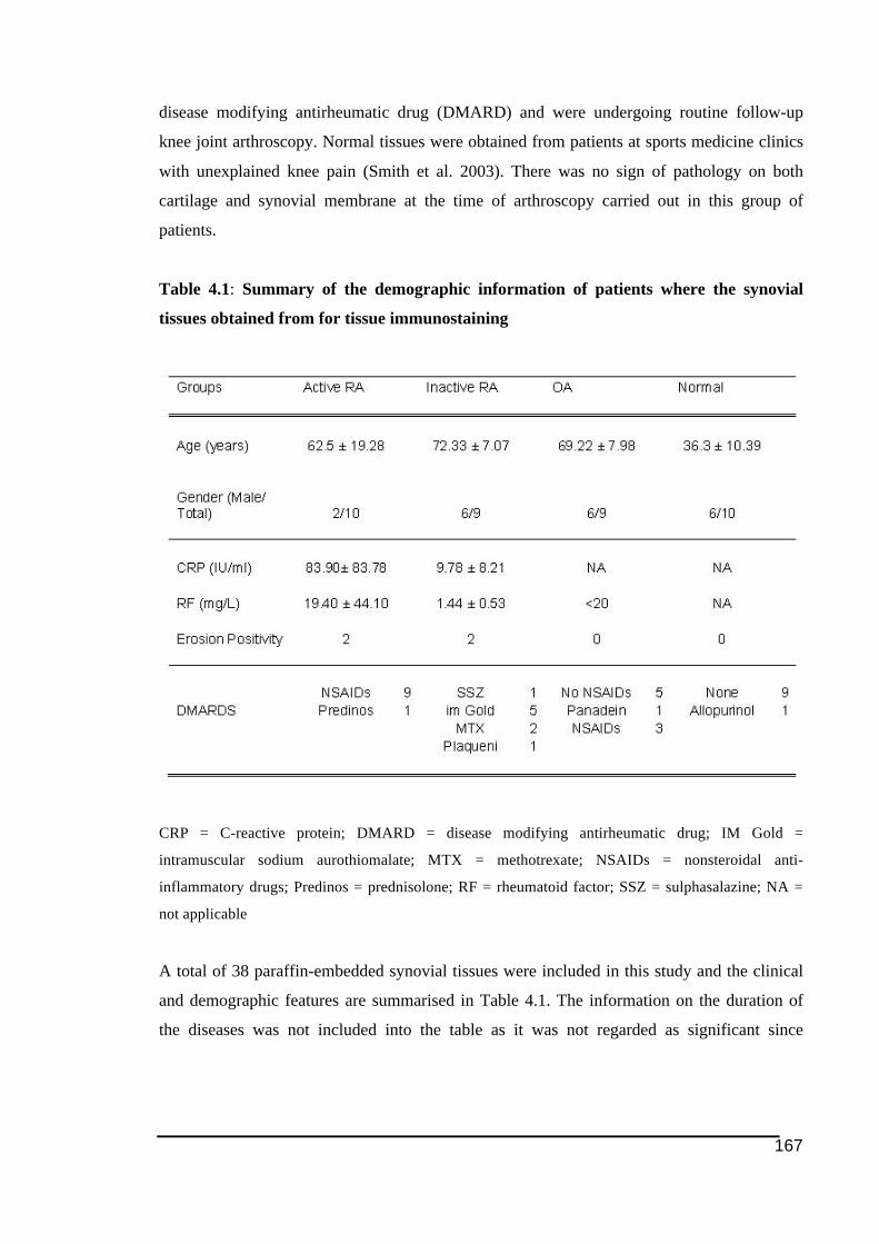
167
disease modifying antirheumatic drug (DMARD) and were undergoing routine follow-up
knee joint arthroscopy. Normal tissues were obtained from patients at sports medicine clinics
with unexplained knee pain (Smith et al. 2003). There was no sign of pathology on both
cartilage and synovial membrane at the time of arthroscopy carried out in this group of
patients.
Table 4.1: Summary of the demographic information of patients where the synovial
tissues obtained from for tissue immunostaining
CRP = C-reactive protein; DMARD = disease modifying antirheumatic drug; IM Gold =
intramuscular sodium aurothiomalate; MTX = methotrexate; NSAIDs = nonsteroidal anti-
inflammatory drugs; Predinos = prednisolone; RF = rheumatoid factor; SSZ = sulphasalazine; NA =
not applicable
A total of 38 paraffin-embedded synovial tissues were included in this study and the clinical
and demographic features are summarised in Table 4.1. The information on the duration of
the diseases was not included into the table as it was not regarded as significant since
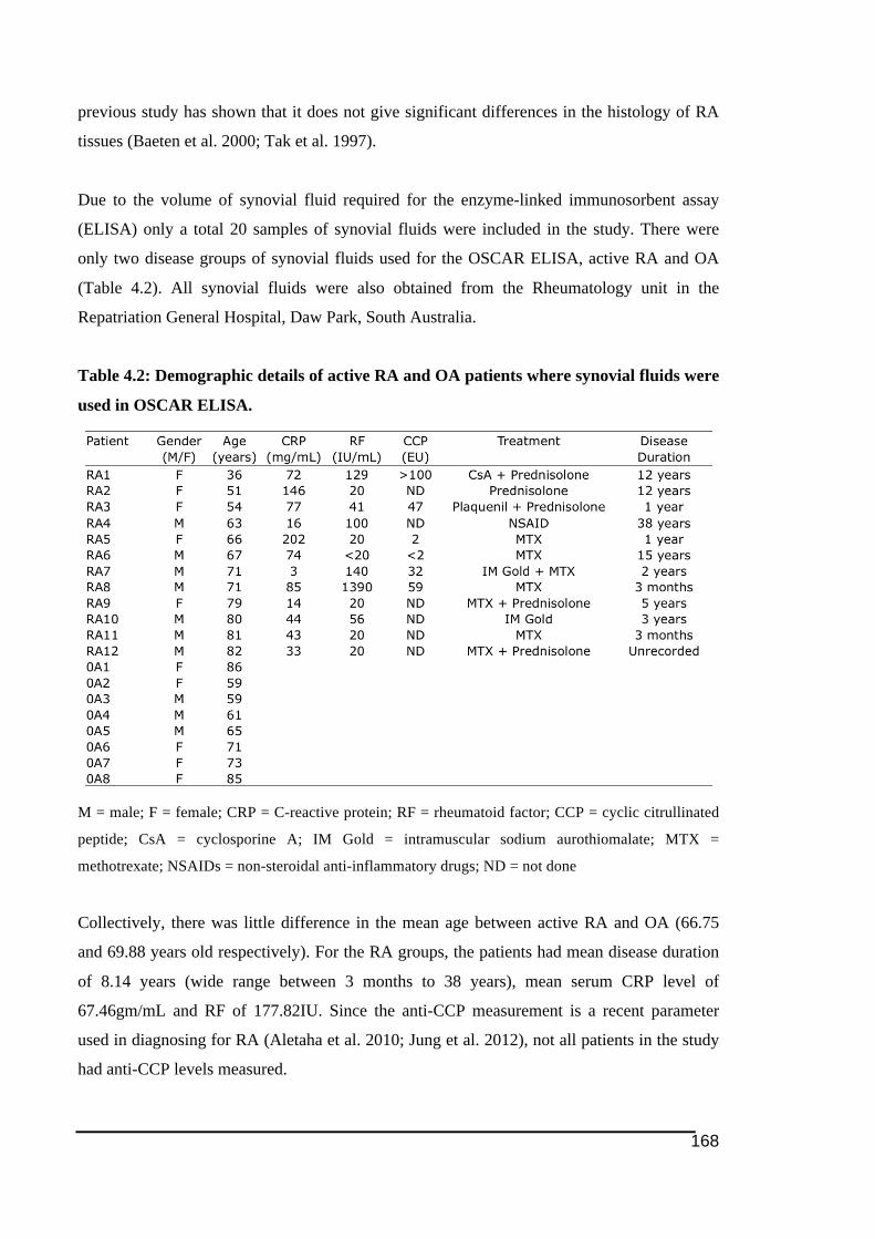
168
previous study has shown that it does not give significant differences in the histology of RA
tissues (Baeten et al. 2000; Tak et al. 1997).
Due to the volume of synovial fluid required for the enzyme-linked immunosorbent assay
(ELISA) only a total 20 samples of synovial fluids were included in the study. There were
only two disease groups of synovial fluids used for the OSCAR ELISA, active RA and OA
(Table 4.2). All synovial fluids were also obtained from the Rheumatology unit in the
Repatriation General Hospital, Daw Park, South Australia.
Table 4.2: Demographic details of active RA and OA patients where synovial fluids were
used in OSCAR ELISA.
M = male; F = female; CRP = C-reactive protein; RF = rheumatoid factor; CCP = cyclic citrullinated
peptide; CsA = cyclosporine A; IM Gold = intramuscular sodium aurothiomalate; MTX =
methotrexate; NSAIDs = non-steroidal anti-inflammatory drugs; ND = not done
Collectively, there was little difference in the mean age between active RA and OA (66.75
and 69.88 years old respectively). For the RA groups, the patients had mean disease duration
of 8.14 years (wide range between 3 months to 38 years), mean serum CRP level of
67.46gm/mL and RF of 177.82IU. Since the anti-CCP measurement is a recent parameter
used in diagnosing for RA (Aletaha et al. 2010; Jung et al. 2012), not all patients in the study
had anti-CCP levels measured.
169
4.2.2. Immunohistochemistry
All the immunohistochemical methods used in this chapter were similar to that described in
the previous Chapter 2 (refer 2.2.2). Briefly, 5µm thick tissue sections were mounted on
APTS-coated glass slides. Tissue sections were deparafinised using two changes of histolene
followed by two changes of 95 to 100% alcohol, then washing with MiliQ water. The
dewaxed tissue sections were pre-treated with either sodium citrate pH 6.0 or
ethylenediamine-tetraacetic acid (EDTA) pH 9.0 (refer Table 4.3) at 90-95˚C for 20 minutes
as procedure for antigen retrieval before subsequent immunohistochemistry performed.
Tissue slides were immersed in PBS as washing protocol between the immunohistochemical
steps. To block unspecific staining due to endogenous peroxidase activity, tissues were
treated with 0.1% sodium azide and 1% hydrogen peroxide in PBS buffer. Three-step
peroxidase-based immunostaining technique, as described in Haynes et al. (2003), was
employed. Counter staining using Harris haemotoxylin (Scharlau Chemie, Sentmenat, Spain)
and saturated lithium carbonate were applied on tissues before mounted in Gurr Aquamount
(BDH, Poole, UK). The staining colour was allowed to develop for 20 minutes using AEC
(K3469, Dako, CA, USA).
Staining for all groups of tissue samples was performed at the same time for each antibody to
avoid any differences between groups due variability in the immunohistochemistry procedure.
Negative controls used were isotype-matched antibody controls for monoclonal antibodies
(mouse IgG1kappa for mouse IgG1) and equivalent antibody-raised serum for polyclonal
antibodies (either normal rabbit or goat serum).
4.2.2.1. Antibodies and Reagents
All antibodies used for tissue immnostaining work have been described in Chapter 2
(presented in Table 2.3 on page 78). The optimal working concentrations were determined in
prior to the tissue immunostaining. Three primary antibodies purchased from the Santa Cruz
Biotechnology Inc. (CA, USA) were rabbit polyclonal anti-DAP12 (sc-20783, used at
2µg/mL), goat polyclonal anti-OSCAR (sc-34233, used at 10 µg/mL) and mouse monoclonal
IgG1 anti-NFATc1 (sc-7294 used at 4µg/mL).
170
TREM2 was detected using a rabbit polyclonal antibody (HPA012571, Sigma Life Sciences)
at a working concentration 0.8µg/mL. The antibody used to detect FcRγ was a rabbit
polyclonal (LS-B2169, Lifespan Bioscience, chosen from personal communication) was used
at 1.25µg/mL. A mouse monoclonal IgG1 anti-cathepsin K (MAB3324, Milipore) was used at
a working concentration of 2µg/mL to detect any presence of osteoclasts.
There were four different polyclonal HRP-conjugated immunoglobulins used as either
secondary or tertiary appropriate antibodies (Table 4.3). Swine anti-goat immunoglobulins
(ACI3404), used at working concentration of 7 µg/mL, was purchased from Invitrogen Life
Technology (CA, USA). The other antibodies were purchased from Dako Cytomation
(Glostrup, Denmark); goat anti-rabbit immunoglobulins (P0448), goat anti-mouse
immunoglobulins (P0447) and rabbit anti-swine immunoglobulins (P0164) were used at
working concentrations of 3, 10 and 13 µg/mL respectively.
Table 4.3: List of antibodies and antigen retrieval buffer used for paraffin-embedded
tissue immunostaining
EDTA- ethylenediaminetetraacetic acid
4.2.2.2. Scoring of tissue immunostaining
For consistency, the SQA scoring system for the measurement of immunohistochemistry
results was performed by two “blinded” observers (EA and AASSK for NFATc1
immunostaining and EA and TNC for the immunostaining of other molecules) using a 5-scale
score ranging from 0 to 4 described previously in Chapter 2 (refer 2.2.2.3) (exception for
NFATc1 immunostaining). To aid the semiquantitative quantitation as well as for the purpose
171
consistency in the view field chosen, the SQA were carried on the scanned image of the
tissues captured using the Nanozoomer Digital Pathology (Hamamatsu, Shizouka, Japan)
technology.
To assess the amount of immunostaining of the ITAM-associated molecules and cathepsin K
(in 2mm2 view area on tissues as well, counted as the percentage of positive stained cells), a
score of 0 represented 0 to 5% positive stained cells, 6 to 10% of positive cells were indicated
by a score of 1, 2 for 11 to 25% positive cells, 3 indicated positive cells between 26 and 50%
of total cell population viewed and finally, a score of 4 indicated more than 50% of positive
cells (adapted from Tak et al. (1995)).
Different SQA scoring method was placed on the NFATc1 immunostaining, in which the
number of positive stained cell aggregations (defined as more than 25 cells) was used as the
parameter for grading (adapted from Kraan et al. (1999)). In a viewed 2mm2 area, a score of 0
represented no positive stained cell aggregation, score of 1 indicated 1 to 3 positive cell
aggregations, score of 2 indicated total positive cell aggregation between 4 to 6, 7 to 9
positive cell aggregations gave score of 3 and score of 4 indicated more than 9 positive cell
aggregations presented.
4.2.3. ELISA of OSCAR in Synovial Fluids from RA and OA Patients
The patient details of the synovial fluid used in this study (for both RA and OA groups) are as
displayed in Table 4.2 above. The level of soluble OSCAR in the synovial fluid was measured
using the ELISA Kit for human OSCAR purchased from USCN Life Science Incorporation
(Wuhan, China). The detection limit for the OSCAR ELISA kit assay was 0.108ng/mL.
Prior to assay all samples of synovial fluid were clarified through centrifugation at 13 000
rpm at 4°C for five minutes. Generally the supernatants of the samples were pre-diluted 1 in 5
using the sample diluent from the ELISA kit. This pre-dilution of the sample was performed
as a measure to avoid any outcome of obtaining readings beyond the concentration range of
the provided protein standards (0 to 20ng/mL). Sample pre-dilution was also carried out for
the purpose of reducing the thickness of the synovial fluids, which may affect the accuracy of
pipetting during the sample loading for the ELISA.

172
The OSCAR ELISA was performed according to the manufacturer’s instructions. Briefly, a
volume of 100µL of the pre-diluted synovial fluids was loaded into each well along with the
standards and then left incubated for two hours. This was then followed by one-hour
incubations with the provided reagent A and 30 minutes with reagent B, with washing steps
done in between those two procedures and afterwards. All incubation took place at 37°C.
Finally, 90µL volume of 3,3',5,5'-tetramethylbenzidine (TMB) substrate solution in the kit
was added to each well and the colour development (from blue to yellow) was monitored
every five minutes. Stop solution (containing sulphuric acid) was added after 30 minutes. The
absorbance reading at the 450nm wavelength was measured using an ELISA plate reader. The
levels of soluble OSCAR in the synovial fluids were determined based on a standard curve
generated.
4.2.4. Statistical Analysis
Statistical analysis was performed using SPSS version 11.5 (Chicago, IL, USA). Kruskal
Wallis analysis was performed to compare the mean of the SQA scores between groups.
Comparison was made between each two groups using Mann Whitney analysis. The
significant difference and a p-value< 0.05 was accepted as statistically significant.
For the data from the OSCAR ELISA work, the level of soluble OSCAR in the synovial fluid
was compared between the two groups (RA and OA) through Student t-test using GraphPad
Prism version 5.0 (GraphPad Software Inc. La Jolla, SA, USA). A p-value <0.05 was
considered to be significantly different.
4.3. Results
Unlike in Chapter 2, there was no histological assessment carried out on every tissue in the
collection due to limited amount sections available. The limited number of sections for every
tissue samples also became the ground for not having negative control for each
immunostaining procedure.
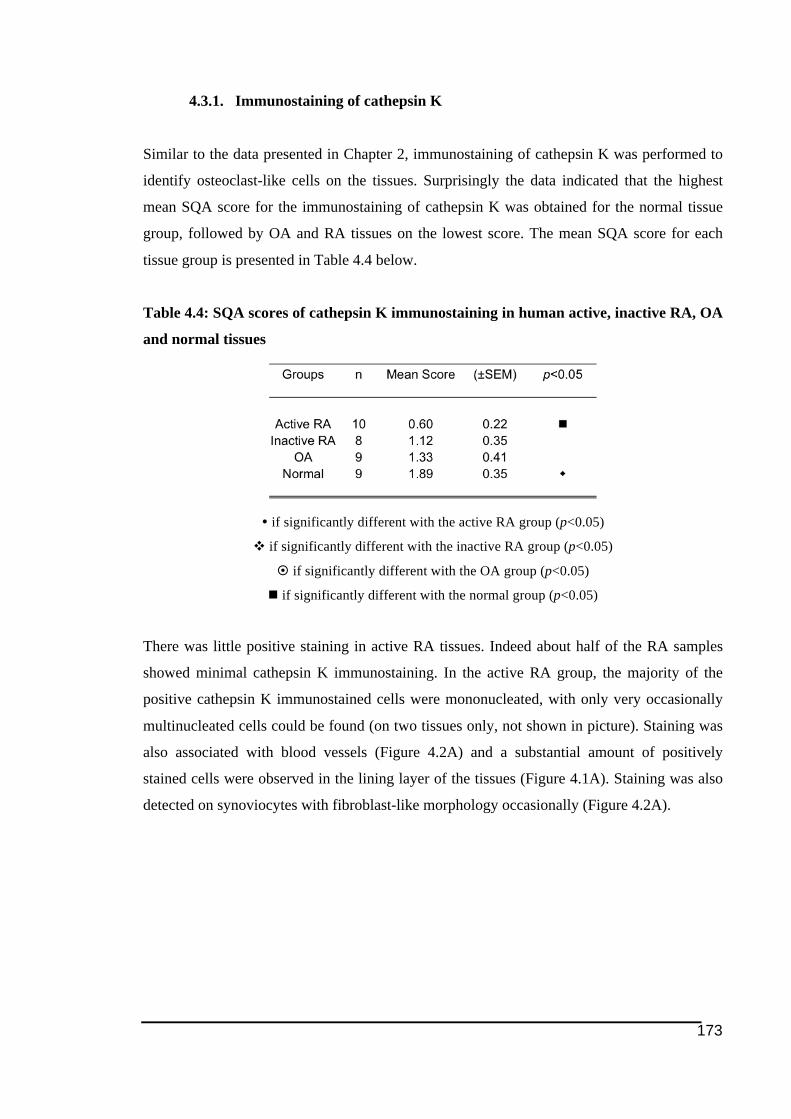
173
4.3.1. Immunostaining of cathepsin K
Similar to the data presented in Chapter 2, immunostaining of cathepsin K was performed to
identify osteoclast-like cells on the tissues. Surprisingly the data indicated that the highest
mean SQA score for the immunostaining of cathepsin K was obtained for the normal tissue
group, followed by OA and RA tissues on the lowest score. The mean SQA score for each
tissue group is presented in Table 4.4 below.
Table 4.4: SQA scores of cathepsin K immunostaining in human active, inactive RA, OA
and normal tissues
! if significantly different with the active RA group (p<0.05)
" if significantly different with the inactive RA group (p<0.05)
# if significantly different with the OA group (p<0.05)
$ if significantly different with the normal group (p<0.05)
There was little positive staining in active RA tissues. Indeed about half of the RA samples
showed minimal cathepsin K immunostaining. In the active RA group, the majority of the
positive cathepsin K immunostained cells were mononucleated, with only very occasionally
multinucleated cells could be found (on two tissues only, not shown in picture). Staining was
also associated with blood vessels (Figure 4.2A) and a substantial amount of positively
stained cells were observed in the lining layer of the tissues (Figure 4.1A). Staining was also
detected on synoviocytes with fibroblast-like morphology occasionally (Figure 4.2A).
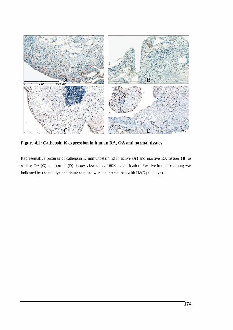
174
Figure 4.1: Cathepsin K expression in human RA, OA and normal tissues
Representative pictures of cathepsin K immunostaining in active (A) and inactive RA tissues (B) as
well as OA (C) and normal (D) tissues viewed at a 100X magnification. Positive immunostaining was
indicated by the red dye and tissue sections were counterstained with H&E (blue dye).
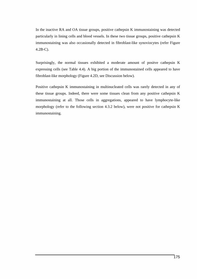
175
In the inactive RA and OA tissue groups, positive cathepsin K immunostaining was detected
particularly in lining cells and blood vessels. In these two tissue groups, positive cathepsin K
immunostaining was also occasionally detected in fibroblast-like synoviocytes (refer Figure
4.2B-C).
Surprisingly, the normal tissues exhibited a moderate amount of positve cathepsin K
expressing cells (see Table 4.4). A big portion of the immunostained cells appeared to have
fibroblast-like morphology (Figure 4.2D, see Discussion below).
Positive cathepsin K immunostaining in multinucleated cells was rarely detected in any of
these tissue groups. Indeed, there were some tissues clean from any positive cathepsin K
immunostaining at all. Those cells in aggregations, appeared to have lymphocyte-like
morphology (refer to the following section 4.3.2 below), were not positive for cathepsin K
immunostaining.
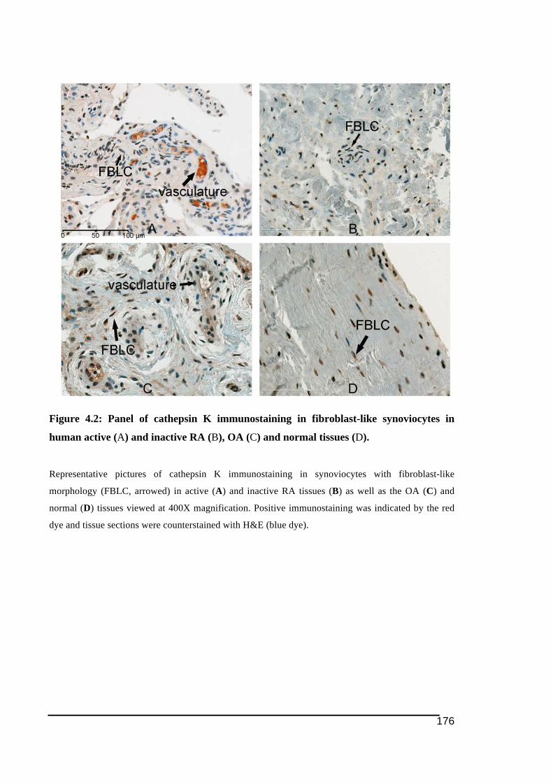
176
Figure 4.2: Panel of cathepsin K immunostaining in fibroblast-like synoviocytes in
human active (A) and inactive RA (B), OA (C) and normal tissues (D).
Representative pictures of cathepsin K immunostaining in synoviocytes with fibroblast-like
morphology (FBLC, arrowed) in active (A) and inactive RA tissues (B) as well as the OA (C) and
normal (D) tissues viewed at 400X magnification. Positive immunostaining was indicated by the red
dye and tissue sections were counterstained with H&E (blue dye).
177
4.3.2. Expression of NFATc1 by immunohistochemistry in RA and control
tissues
Overall NFATc1 immunostaining positivity was highest in active RA tissues compared to
other tissue groups. This is shown in the SQA scores for the NFATc1 immunostaining
displayed in Table 4.5.
Table 4.5: SQA scores of NFATc1 immunostaining in human active, inactive RA, OA
and normal tissues
! if significantly different with the active RA group (p<0.05)
" if significantly different with the inactive RA group (p<0.05)
# if significantly different with the OA group (p<0.05)
$ if significantly different with the normal group (p<0.05)
Majority cells expressing NFATc1 had a T-lymphoyte-like morphology (small-sized cells
with large nuclei (Grossi et al. 1978), particularly in RA tissues. NFATc1 immunostaining
was seen mainly in the nuclei of cells presented in aggregates of inflammatory-like cells,
particularly in RA tissues. Due to the patchy nature of the staining pattern, the SQA scoring
method was modified to be different from the one that has been reported (Kraan et al. 1999).
This method was based on the number of aggregations of cells immunostained.
In active RA tissues, large numbers of positively stained cell aggregations were seen,
generally more than 4 cell aggregations per 2mm2 view field. In some tissues most of the
NFATc1-immunostained cells appeared to be synoviocytes in the lining layer of the tissues.
No NFATc1 immunostaining was seen associated with the blood vasculature. Despite
indication of very minimal amount of osteoclast-like cells in the tissues (see 4.3.1 above),
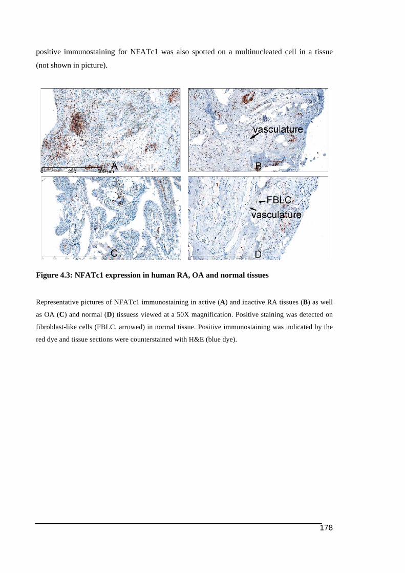
178
positive immunostaining for NFATc1 was also spotted on a multinucleated cell in a tissue
(not shown in picture).
Figure 4.3: NFATc1 expression in human RA, OA and normal tissues
Representative pictures of NFATc1 immunostaining in active (A) and inactive RA tissues (B) as well
as OA (C) and normal (D) tissuess viewed at a 50X magnification. Positive staining was detected on
fibroblast-like cells (FBLC, arrowed) in normal tissue. Positive immunostaining was indicated by the
red dye and tissue sections were counterstained with H&E (blue dye).
179
In the inactive RA group, a few cell aggregates expressed NFATc1, however these were
fewer than in active RA. Nearly all of the tissue samples within the inactive RA group give
score of 0 or 1. The NFATc1 immunostaining pattern generally appears to be similar between
active and inactive RA tissues. In these tissues NFATc1 immunostaining was not seen in the
vasculature meanwhile positive immunostaining was mainly observed in synoviocytes located
in the tissue lining layers. Nonetheless, there was no NFATc1 immunostaining on
multinucleated-like cell was observed.
OA tissues, like the inactive RA tissues, gives low number of cell aggregations. Unlike in RA
tissue, OA tissues appear to have more scattered distribution of NFATc1 positive cells. Strong
immunostaining in the OA tissues could be seen mostly on cells in the lining layer. Quite
surprisingly, positive NFATc1 immunostaining was seen in quite a number of cells with
multinucleated appearance (not shown in picture). No positive immunostaining was seen in
the vasculature in OA tissues.
Finally, the normal tissue generally had minimal positive NFATc1 immunostaining. Indeed,
the normal tissue section shown in the picture (Figure 4.3D) has the highest score of SQA
among other normal tissues. In majority of the normal tissues, NFATc1 immunostaining was
seen in tissue lining synoviocytes though the intensity of the staining was weak. Fibroblast-
like synoviocytes scattered on the tissues were other examples of NFATc1-immunostained
seen in a one or two normal tissues. Generally there was no immunostaining observed in the
vasculature in the tissue group.
4.3.3. Expression of ITAM-related molecules in RA synovial tissues
4.3.3.1. TREM2 and DAP12
Immunohistochemistry staining for TREM2 and DAP12 demonstrated that both molecules
could be detected in all tissue groups. Generally it was found that there was more positive
immunostaining (statistically different) in the active RA tissues compared to the other tissue
groups (refer Table 4.6 and Table 4.7 below). Interestingly there were also significant
differences in the mean SQA score of TREM2 and DAP12 immunostaining obtained between
180
OA and the normal tissues. However, there was no significant difference in the SQA scores
for both TREM2 and DAP12 immunostaining seen between the inactive RA and OA tissue
groups.
Table 4.6: SQA scores of TREM2 immunostaining in human active, inactive RA, OA
and normal tissues
! if significantly different with the active RA group (p<0.05)
" if significantly different with the inactive RA group (p<0.05)
# if significantly different with the OA group (p<0.05)
$ if significantly different with the normal group (p<0.05)
TREM2 immunostaining was observed on massive amount of cells in all tissues in the active
RA tissues. TREM2 immunostaining was detected mainly in synoviocyte-like cells in the
lining layer and in aggregations (with T-lymphocytes morphology as being mentioned in
4.3.2 above) in the active RA group. TREM2-immunostained mononucleated cells were also
seen scattered on the tissues. Positive TREM2 immunostaining was seen in the multinucleated
cells (similar outcome in 4.3.1 earlier) in some of the active RA tissues (not shown in
picture).
181
Figure 4.4: TREM2 expression in human RA, OA and normal tissues
Representative pictures of TREM2 immunostaining in active (A), inactive RA (B), OA (C) and
normal tissues (D) viewed at a 100X magnification. Fibroblast-like cells (FBLC, arrowed) were
among cells observed positive for TREM2. Positive immunostaining was indicated by the red dye and
tissue sections were counterstained with H&E (blue dye).
182
In the inactive RA and OA tissue groups, the staining was seen more on the lining layer of the
tissues. Strong TREM2 immunostaining was observed in these tissue groups on cells in the
lining layer. TREM2 immunostaining was also seen associated with vasculature and
fibroblast-like synoviocytes. Similar to the active RA group, multinucleated cells in the
inactive and OA tissues were also positive for TREM2. In the inactive RA tissues, TREM2
was also expressed in a subpopulation cells in the lymphocyte aggregations.
In the normal tissues the expression of TREM2 was weak (Figure 4.4D). Most of the positive
TREM2 immunostaining was observed in fibroblast-like synoviocytes. TREM2
immunostaining was also seen associated with the vasculature in some tissues in the group
(not shown in picture). Unlike the other tissue groups, generally there was no obvious
TREM2 immunostaining seen in cells in the tissue-lining layer.
Table 4.7: SQA scores of DAP12 immunostaining in human active, inactive RA, OA and
normal tissues
! if significantly different with the active RA group (p<0.05)
" if significantly different with the inactive RA group (p<0.05)
# if significantly different with the OA group (p<0.05)
$ if significantly different with the normal group (p<0.05)
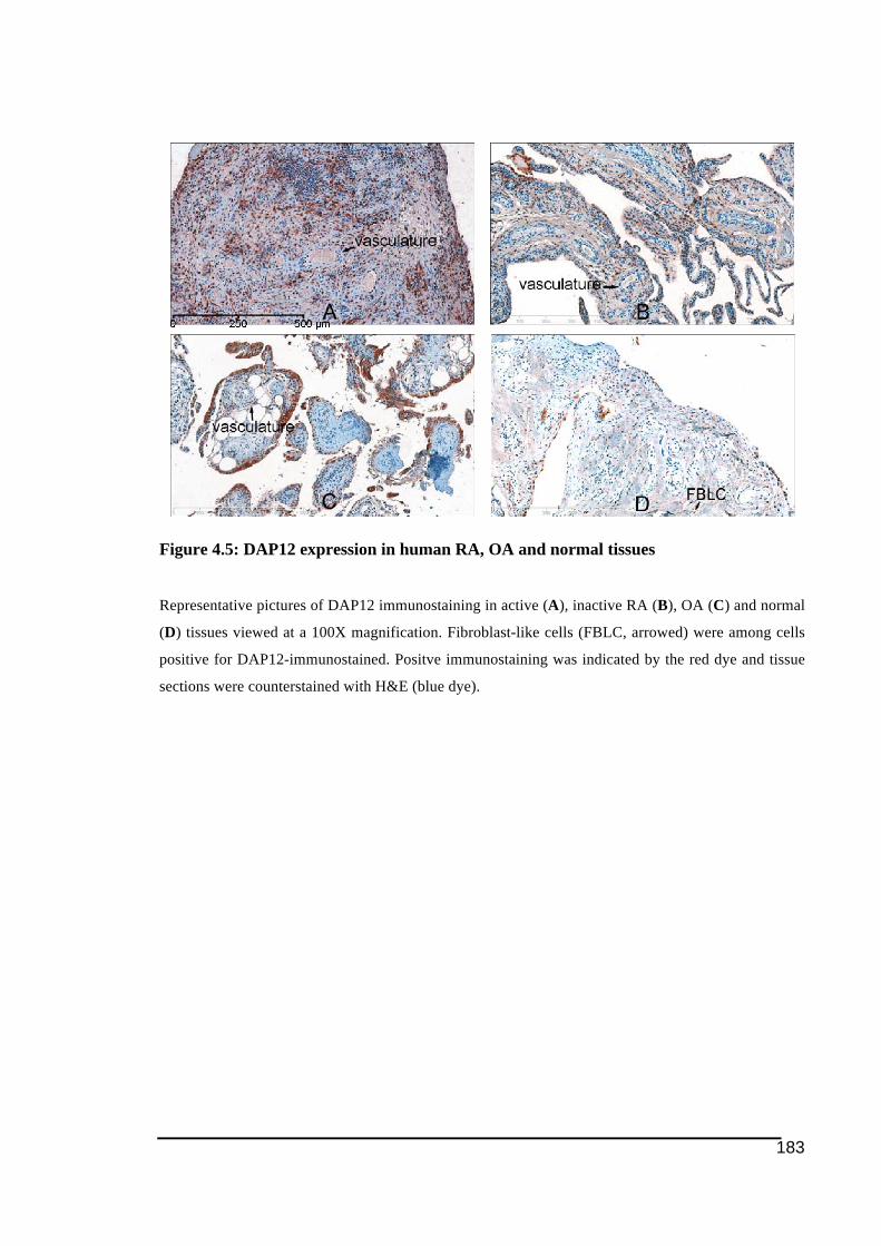
183
Figure 4.5: DAP12 expression in human RA, OA and normal tissues
Representative pictures of DAP12 immunostaining in active (A), inactive RA (B), OA (C) and normal
(D) tissues viewed at a 100X magnification. Fibroblast-like cells (FBLC, arrowed) were among cells
positive for DAP12-immunostained. Positve immunostaining was indicated by the red dye and tissue
sections were counterstained with H&E (blue dye).
184
Generally there appeared to be a similar pattern of immunostaining for DAP12 and TREM2.
However the SQA score for DAP12 immunostaining was markedly lower than for TREM2
(compare Table 4.6 and Table 4.7). DAP12-immunostained cells appeared to be tissue-lining
synoviocytes and perivascular cells, however, there was no DAP12 immunostaining seen
associated with the vasculature in this tissue group. In some of the active RA tissues, in which
there appeared to be massive infiltration of T-lymphocyte-like cells (refer 4.3.2 above), most
cells were negative for DAP12 resulting in a lower SQA score than it first appeared.
Similar observations were seen in both inactive RA and OA tissues, however DAP12
immunostaining was reduced (see Table 4.7). While a significant number of DAP12-
immunostained cells were scattered throughout tissues in those two groups, most of the
stained cells appeared to be in the tissue lining layer. Similar to that seen in the active RA
tissues, multinucleated cells immunostained for DAP12 were seen in a number of inactive RA
and OA tissues (not shown). However, there was no sign of DAP12 immunostaining on
vasculature in both tissue groups. In some OA tissues, DAP12 immunostaining was also
detected in synovial fibroblast-like cells (not shown in picture).
In normal tissues, few DAP12-immunostained cells were seen, however the staining intensity
appeared to be quite weak. Fibroblast-like and tissue-lining synoviocytes were among the
cells expressing DAP12 (Figure 4.5D). DAP12 immunostaining associated with the
vasculature was also seen in a normal tissue (not shown in picture).
4.3.3.2. OSCAR and FcRγ
Generally the extent of positive immunostaining of OSCAR (in all tissue groups) was less
than the other molecules investigated, as shown in Table 4.8 below. From the comparison on
the mean SQA score for OSCAR immunostaining between tissue groups, significant
difference could only be seen between the normal control group against both active RA and
OA tissues.
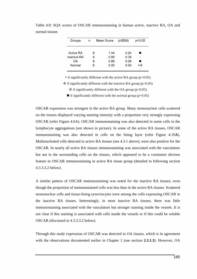
185
Table 4.8: SQA scores of OSCAR immunostaining in human active, inactive RA, OA and
normal tissues
! if significantly different with the active RA group (p<0.05)
" if significantly different with the inactive RA group (p<0.05)
# if significantly different with the OA group (p<0.05)
$ if significantly different with the normal group (p<0.05)
OSCAR expression was strongest in the active RA group. Many mononuclear cells scattered
on the tissues displayed varying staining intensity with a proportion very strongly expressing
OSCAR (refer Figure 4.6A). OSCAR immunostaining was also detected in some cells in the
lymphocyte aggregations (not shown in picture). In some of the active RA tissues, OSCAR
immunostaining was also detected in cells on the lining layer (refer Figure 4.10A).
Multinucleated cells detected in active RA tissues (see 4.3.1 above), were also positive for the
OSCAR. In nearly all active RA tissues immunostaining was associated with the vasculature
but not in the surrounding cells on the tissues, which appeared to be a consistent obvious
feature in OSCAR immunostaining in active RA tissue group (detailed in following section
4.3.3.3.2 below).
A similar pattern of OSCAR immunostaining was noted for the inactive RA tissues, even
though the proportion of immunostained cells was less than in the active RA tissues. Scattered
mononuclear cells and tissue-lining synoviocytes were among the cells expressing OSCAR in
the inactive RA tissues. Interestingly, in most inactive RA tissues, there was little
immunostaining associated with the vasculature but stronger staining inside the vessels. It is
not clear if this staining is associated with cells inside the vessels or if this could be soluble
OSCAR (discussed in 4.3.3.3.2 below).
Through this study expression of OSCAR was detected in OA tissues, which is in agreement
with the observations documented earlier in Chapter 2 (see section 2.3.1.3). However, OA
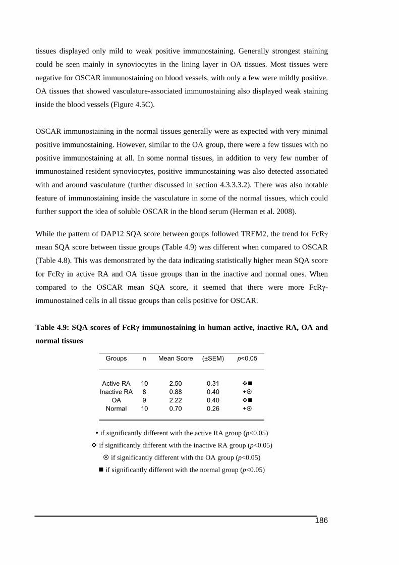
186
tissues displayed only mild to weak positive immunostaining. Generally strongest staining
could be seen mainly in synoviocytes in the lining layer in OA tissues. Most tissues were
negative for OSCAR immunostaining on blood vessels, with only a few were mildly positive.
OA tissues that showed vasculature-associated immunostaining also displayed weak staining
inside the blood vessels (Figure 4.5C).
OSCAR immunostaining in the normal tissues generally were as expected with very minimal
positive immunostaining. However, similar to the OA group, there were a few tissues with no
positive immunostaining at all. In some normal tissues, in addition to very few number of
immunostained resident synoviocytes, positive immunostaining was also detected associated
with and around vasculature (further discussed in section 4.3.3.3.2). There was also notable
feature of immunostaining inside the vasculature in some of the normal tissues, which could
further support the idea of soluble OSCAR in the blood serum (Herman et al. 2008).
While the pattern of DAP12 SQA score between goups followed TREM2, the trend for FcRγ
mean SQA score between tissue groups (Table 4.9) was different when compared to OSCAR
(Table 4.8). This was demonstrated by the data indicating statistically higher mean SQA score
for FcRγ in active RA and OA tissue groups than in the inactive and normal ones. When
compared to the OSCAR mean SQA score, it seemed that there were more FcRγ-
immunostained cells in all tissue groups than cells positive for OSCAR.
Table 4.9: SQA scores of FcRγ immunostaining in human active, inactive RA, OA and
normal tissues
! if significantly different with the active RA group (p<0.05)
" if significantly different with the inactive RA group (p<0.05)
# if significantly different with the OA group (p<0.05)
$ if significantly different with the normal group (p<0.05)
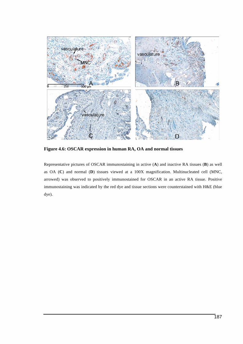
187
Figure 4.6: OSCAR expression in human RA, OA and normal tissues
Representative pictures of OSCAR immunostaining in active (A) and inactive RA tissues (B) as well
as OA (C) and normal (D) tissues viewed at a 100X magnification. Multinucleated cell (MNC,
arrowed) was observed to positively immunostained for OSCAR in an active RA tissue. Positive
immunostaining was indicated by the red dye and tissue sections were counterstained with H&E (blue
dye).
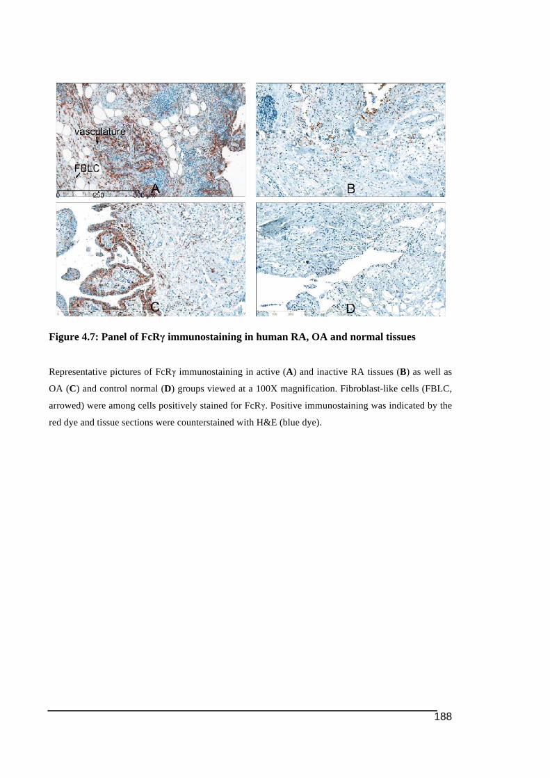
188
Figure 4.7: Panel of FcRγ immunostaining in human RA, OA and normal tissues
Representative pictures of FcRγ immunostaining in active (A) and inactive RA tissues (B) as well as
OA (C) and control normal (D) groups viewed at a 100X magnification. Fibroblast-like cells (FBLC,
arrowed) were among cells positively stained for FcRγ. Positive immunostaining was indicated by the
red dye and tissue sections were counterstained with H&E (blue dye).
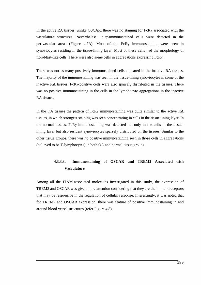
189
In the active RA tissues, unlike OSCAR, there was no staining for FcRγ associated with the
vasculature structures. Nevertheless FcRγ-immunostained cells were detected in the
perivascular areas (Figure 4.7A). Most of the FcRγ immunostaining were seen in
synoviocytes residing in the tissue-lining layer. Most of these cells had the morphology of
fibroblast-like cells. There were also some cells in aggregations expressing FcRγ.
There was not as many positively immunostained cells appeared in the inactive RA tissues.
The majority of the immunostaining was seen in the tissue-lining synoviocytes in some of the
inactive RA tissues. FcRγ-positive cells were also sparsely distributed in the tissues. There
was no positive immunostaining in the cells in the lymphocyte aggregations in the inactive
RA tissues.
In the OA tissues the pattern of FcRγ immunostaining was quite similar to the active RA
tissues, in which strongest staining was seen concentrating in cells in the tissue lining layer. In
the normal tissues, FcRγ immunostaining was detected not only in the cells in the tissue-
lining layer but also resident synoviocytes sparsely distributed on the tissues. Similar to the
other tissue groups, there was no positive immunostaining seen in those cells in aggregations
(believed to be T-lymphocytes) in both OA and normal tissue groups.
4.3.3.3. Immunostaining of OSCAR and TREM2 Associated with
Vasculature
Among all the ITAM-associated molecules investigated in this study, the expression of
TREM2 and OSCAR was given more attention considering that they are the immunoreceptors
that may be responsive in the regulation of cellular response. Interestingly, it was noted that
for TREM2 and OSCAR expression, there was feature of positive immunostaining in and
around blood vessel structures (refer Figure 4.8).
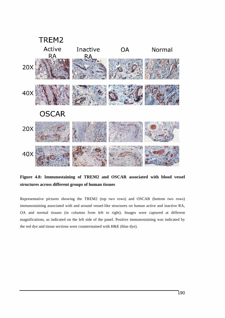
190
Figure 4.8: Immunostaining of TREM2 and OSCAR associated with blood vessel
structures across different groups of human tissues
Representative pictures showing the TREM2 (top two rows) and OSCAR (bottom two rows)
immunostaining associated with and around vessel-like structures on human active and inactive RA,
OA and normal tissues (in columns from left to right). Images were captured at different
magnifications, as indicated on the left side of the panel. Positive immunostaining was indicated by
the red dye and tissue sections were counterstained with H&E (blue dye).
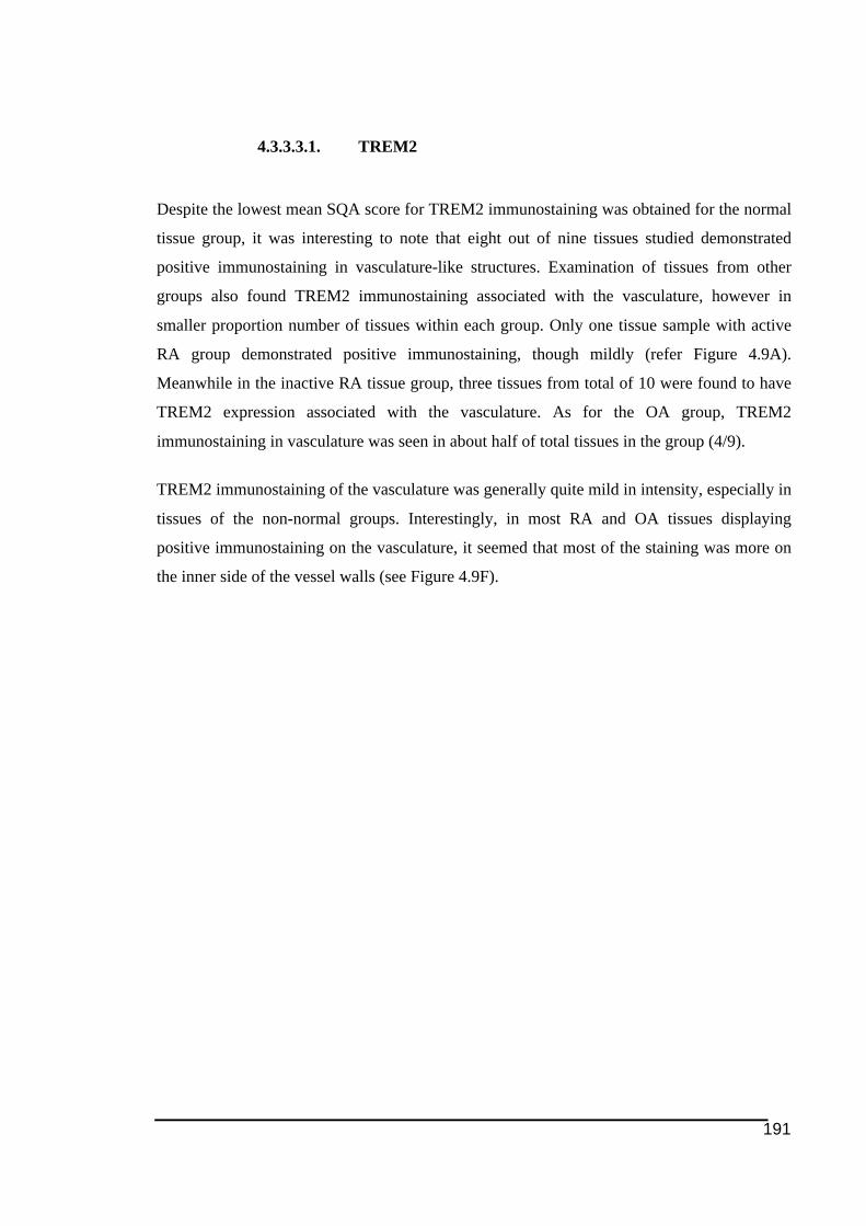
191
4.3.3.3.1. TREM2
Despite the lowest mean SQA score for TREM2 immunostaining was obtained for the normal
tissue group, it was interesting to note that eight out of nine tissues studied demonstrated
positive immunostaining in vasculature-like structures. Examination of tissues from other
groups also found TREM2 immunostaining associated with the vasculature, however in
smaller proportion number of tissues within each group. Only one tissue sample with active
RA group demonstrated positive immunostaining, though mildly (refer Figure 4.9A).
Meanwhile in the inactive RA tissue group, three tissues from total of 10 were found to have
TREM2 expression associated with the vasculature. As for the OA group, TREM2
immunostaining in vasculature was seen in about half of total tissues in the group (4/9).
TREM2 immunostaining of the vasculature was generally quite mild in intensity, especially in
tissues of the non-normal groups. Interestingly, in most RA and OA tissues displaying
positive immunostaining on the vasculature, it seemed that most of the staining was more on
the inner side of the vessel walls (see Figure 4.9F).
192
Figure 4.9: TREM immunostaining associated with vasculature
Representative pictures showing the TREM2 immunostaining associated with vasculature in active
(A), inactive RA tissues (B) as well as by in OA (C) and normal tissues (D) captured at 200X
magnification. E represents TREM2 immunostaining in an OA tissue at lower magnification (100X)
to show notable specific staining on vasculature on tissues. F demonstrates TREM2 immunostaining
in vasculature was more in the inner side of the vasculature (400X). TREM2 immunostaining was
indicated by the red dye and tissue sections were counterstained with H&E (blue dye).
193
4.3.3.3.2. OSCAR
In comparison to TREM2, positive immunostaining associated with blood vessels was more
notable for OSCAR, particularly in the RA tissues. In both active and inactive RA groups,
OSCAR immunostaining was detected in vasculature-like structures in nearly all (8/9) tissues
in those groups. The intensity of the immunostaining in the vessel-like structures varied
between tissues, with majority of tissues in active RA group in particular displaying strong
staining. In addition, there appeared to be positive staining in cells inside blood vessels (refer
Figure 4.10C) in one tissue in the active RA. This sort of observation is in agreement with the
findings of Herman and colleagues (2008), who showed that circulating monocytes can
express OSCAR prior to entry into tissues. This could be further supported by an observation
that cells appeared to be marginalising from the vessels displaying OSCAR immunostaining
(refer Figure 4.10D).
For the OA tissues, only few tissues within the group (4/9) showed positive OSCAR
immunostaining associated with blood vessel-like structures. Furthermore, the OSCAR
immunostaining associated with the blood vessels was very mild in those tissues. Like in RA
tissues, there was also staining inside blood vessels observed in most OA tissues (7/9).
OSCAR immunostaining for the normal tissues in and around blood vessel-like structures was
quite similar to the OA group. Nearly all normal tissues (8/9) showed OSCAR
immunostaining inside the blood vessel-like structure, however only 2 of those tissues
displayed staining in wall of the vasculature. In most of the normal tissues immunostained for
OSCAR, the staining inside the vasculature appeared to be stronger than in the other tissue
groups.

194
Figure 4.10: Immunostaining of OSCAR in human tissues of RA and OA
OSCAR-positive immunostaining was seen in lining cells in OA tissues (A). OSCAR immunostaining
was detected associated with vasculuture in active RA tissues, as clearly shown in B. C shows positive
immunostaining of OSCAR was also detected in cells within the blood vessels. D Examination on
other part of the tissues shown in B showed appearance of migrated OSCAR-positive cells from blood
vessel (shown by arrow in panel D). All pictures were captured at 400X magnification.
195
4.3.4. Level of soluble OSCAR levels in synovial fluids of RA and OA
The observation of OSCAR immunostaining within and inside the blood vessel walls in RA
tissues particularly could also suggest the presence of soluble OSCAR in the serum, as this
has been demonstrated in previous studies (Herman et al. 2008; Zhao et al. 2011). As level of
soluble OSCAR in serum has been reported in previous studies (Herman et al. 2008; Zhao et
al. 2011), synovial fluid was tested for soluble OSCAR for the first time. Due to the
unavailability of synovial fluid from normal subjects, synovial fluids from OA patients were
used as the controls. The concentrations of soluble OSCAR in the synovial fluids of RA,
alongside with OA sample group, are presented in Figure 4.11 below.
Interestingly the levels of soluble OSCAR in serum of RA shown in previous studies
(Herman et al. 2008; Zhao et al. 2011) appeared to be markedly lower than in the synovial
fluids in RA patients used in this study. Surprisingly, it appeared that soluble OSCAR was not
only detectable in synovial fluid from OA patients, but the level measured (mean
130.73ng/ml) was slightly higher than in RA samples (mean 111.74ng/mL). Moreover, the
level of soluble OSCAR in synovial fluids from OA group was more consistent than in the
RA groups, which include one outlier.
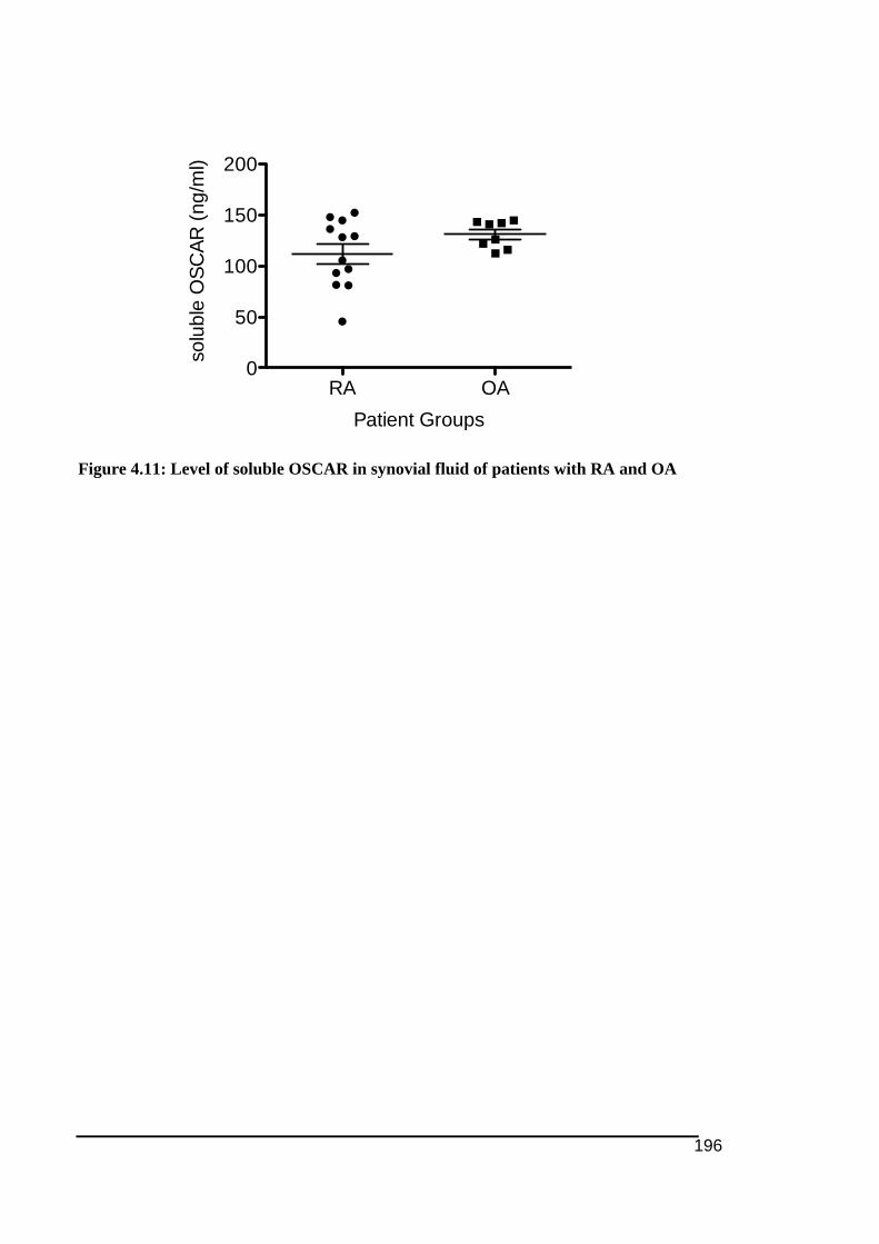
196
Figure 4.11: Level of soluble OSCAR in synovial fluid of patients with RA and OA
RA OA0
50
100
150
200
Patient Groups
solu
ble
OSC
AR (n
g/m
l)
197
4.4. Discusssion
In contrast to the previous two chapters that are themed on peri-implant osteolysis, this
chapter, together with the following Chapter 5, investigates rheumatoid arthritis. It is
interesting to note the differences in ITAM-associated molecules involved in promoting
osteoclast formation in peri-implant osteolysis and rheumatoid arthritis. The study presented
in this chapter can be compared to Chapter 2 as it investigated tissue expression of ITAM-
related molecules (including TREM2, DAP12, OSCAR and FcRγ), alongside with the
osteoclast key transcriptional factor NFATc1 and cell marker cathepsin K, in RA tissues (and
synovial fluid for OSCAR only) in comparison to the other groups.
The synovial membrane is believed to be an active site for osteoclastogenesis in RA
(Gravallese et al. 2000). Those tissue samples used in this study were obtained from a well-
characterised tissue collection, which is a unique feature of this study in comparison to the
others. Another aspect that made the tissue collection unique was the presence of an inactive
RA tissue group from patients in remission after successful treatment with DMARD. Since
OA and normal tissues were also included in this study it made multi-groups comparisons
possible rather than just only 2 groups comparison as in Chapter 2. The inclusion of OA and
RA tissues allowed comparison to see how these different diseases varied. Tissues were
collected through biopsy performed by a rheumatologist (Professor Malcolm D. Smith) and
therefore consistent procedures and tissue site sampling were the key factors in obtaining this
homogenous tissue library. In addition, this tissue collection has also been used in many
published studies (Crotti et al. 2003; Crotti et al. 2002; Haynes et al. 2003; Smith et al. 2003).
The inclusion of inactive RA tissue group in the study has made information such as history
of medication relevant to be recorded (see Table 4.1). The type of medication could be a
confounding factor that may influence protein expression in the tissues (Vieira-Sousa et al.
2011). Unfortunately, complete information on the ESR and DAS28 was not recorded for all
patients. Therefore the findings in this thesis could not be directly compared to the study by
Herman and colleagues (2008) since these subject parameters could become factors that may
give different outcome between studies.
198
Unlike in Chapter 2, there was no assessment on the tissue histology using H&E staining due
to limited number of tissue sections from the small tissue samples. In addition, there was also
no examination on tissue mRNA level carried out due to the very limited amount of frozen
tissue (from the same collection) available for RNA extraction.
Similar to Chapter 2, dual immunolabeling was not feasible to be performed due to technical
limitation associated with paraffin-embedded tissue sections. In addition serial
immunolabeling with cell markers was not possible due to the limited number of tissue
sections available. As a consequence, the tissue immunostaining described in this chapter
could only interpreted based on the cell morphology alone.
Based on the SQA score obtained for cathepsin K immunostaining in this study (refer Table
4.4), surprisingly it appeared that the number of cells expressing cathepsin K was
significantly lower in active RA tissues in comparison to the normal tissues. In addition there
was no significant difference in the SQA score between RA and OA tissue groups. Both of
these outcomes were in contradiction to the earlier findings made by Hou and co-workers
(2002), which showed more cathepsin K-positive cells in RA than in OA tissues. This
difference could probably be explained by variabilities such as tissue sites sampled and the
progression of the disease of individual patients. Nonetheless, there was consistency between
these two studies in terms of the phenotype of cells expressing cathepsin K seen in all tissue
groups.
The immunostaining for cathepsin K did not reveal the presence of significant numbers of
multinucleated osteoclast-like cells in the RA tissues (see Figure 4.1A and B). The presence
of osteoclasts in RA tissues is believed to lead to bone erosion in the disease (Gravallese et al.
1998). However the fact that only 2 active RA tissues that showed positive signs of bone
erosion (refer Table 4.1) (Crotti et al. 2012), in addition to the very rare detection of
osteoclast-like cells in the active RA tissues found in this study, could suggest that most RA
tissues studied were not from chronic RA patients. Another possibility is the tissues were
collected from the synovial membranes that were not juxtaposing bone where osteoclasts
would be present (Shen et al. 2006). It would be expected that collection of pannus would
reveal presence of larger number of osteoclast-like cells. Future work using tissues sampled
from bone-juxtaposed tissues or pannus in chronic RA patients may give more meaningful
199
outcome and allow the original research question of the study (in context of osteoclast) to be
properly addressed.
In addition to the RA tissues, multinucleated osteoclast-like cells were also detected in an OA
synovial tissue. This observation is consistent with several previous findings (Dodds et al.
1999; Hou et al. 2002). Even though there was minimal number of multinucleated osteoclast-
like cells that could be seen, there are still possibility that the other cathepsin K-positive cells
were macrophage-like cells (Haywood et al. 2003; Hummel et al. 1998), which could become
osteoclast precursors (Dodds et al. 1999; Gravallese et al. 1998; Hou et al. 2002). Dodds and
colleagues (1999) through their work found that mononuclear cells expressing cathepsin K in
OA tissues were also positive for TRAP, suggesting that they were possibly osteoclast
precursor cells.
It is also possible that cathepsin K is expressed by non-myeloid cell types in RA synovial
tissues. For example, the observation in which cathepsin K-positive cells were among cells in
lymphocyte aggregates seen in this study was consistent with a previous report that found
cells containing cathepsin K mRNA transcripts in the areas of lymphocyte infiltration in RA
tissues (Hummel et al. 1998).
Cathepsin K immunostaining in the diseased and normal tissues could also be seen in cells
with fibroblast-like morphology (see Figure 4.2). This was consistent with previous literature
reporting on the expression of cathepsin K in synovial fibroblasts, detected at both mRNA
and protein level (Bottini & Firestein 2013; Hou et al. 2002; Hou et al. 2001). Through their
observation in the normal tissues, Hou and colleagues (2002) suggested that cathepsin K is
constitutively expressed by synovial fibroblasts.
Hummel and coworkers (1998) reported that cathepsin K-positive cells found at cartilage-
pannus junction in RA tissues appeared to have macrophage or fibroblast-like morphology,
leaving speculation of cathepsin K role in cartilage invasion and erosion (Li et al. 2000). This
is because cathepsin K has been identified as able to degrade both types I (in bone) and II
collagens (in cartilage) (Bossard et al. 1996; Kafienah et al. 1998). In addition, an earlier
study found that synovial fibroblasts from human RA tissues (as well as from OA tissues)
were able to cause cartilage degradation (Scott et al. 1997). The examination on tissues from a
human fetus with pycnodysostosis (resulted from cathepsin K deficiency, refer section
200
1.3.1.3), which found presence of undigested collagen fibrils accumulated in lysosomal
vacuoles of fibroblast (Everts et al. 2003), provides further evidence of role of cathepsin K in
fibroblastic cells in collagen degradation. In an in vivo animal model, treatment with a
cathepsin K inhibitor managed to reduce the extent of bone and cartilage erosion in mice
subjected to collagen-induced arthritis (Svelander et al. 2009).
In RA synovial fibroblasts may play several roles in the disease progression. The pannus is
made up from an over-abundance of syovial fibroblasts (Bottini & Firestein 2013). These
cells can also produce a range of molecules that are able to modulate cell growth,
angiogenesis, synovitis and stimulate production inflammatory mediators in other immune
cells. In addition, they can also express proteolytic enzymes, including cathepsin K and
MMPs that are important in both bone and cartilage erosion, and release cytokines that
promote and support osteoclastogenesis in RA (Gravallese et al. 1998; Hase et al. 2008;
Hwang et al. 2012b; Kim et al. 2012).
In comparison to RA, much less is known about the role of fibroblastic synoviocytes in OA.
However, there is evidence associating the formation of fibroblast-like synoviocytes occurs at
the expense of chondrocyes in OA (Aigner et al. 1993; Sandell & Aigner 2001) (see cell
phenotypic alteration in section 1.2.3). A recent study by Zhang and colleagues (2012)
reported that the inhibition on cathepsin K expression could slow down the process.
As stated earlier in the Introduction (section 4.1) even though a number of studies suggesting
NFATc1 as might have role in RA disease progression and could be a good therapeutical
target (Miyazaki et al. 2007; Urushibara et al. 2004), it appeared that there has been no study
on NFATc1 expression in RA tissues being reported so far (Sitara & Aliprantis 2010). Indeed
to the author’s knowledge, there has been only one previous study demonstrating in vivo
model of arthritis in NFAT-deficient mice (Gerth et al. 2004). Therefore, the data presented in
this section is the first to report expression of NFATc1 protein in human RA tissues.
Interestingly as indicated in the results section, most of the positive NFATc1-immunostained
cells were seen to be in aggregations with closer inspection suggesting they were infiltrating
T-lymphocytes. Detection of NFATc1 on T-lymphocyte-like cells, particlarly in RA tissues is
not an unexpected observation considering NFATc1 is a transcription factor expressed, and
indeed was first discovered (Shaw et al. 1988), in the T-cells. NFATc1 in the activated T-
201
lymphocytes regulates the cell differentiation and activation (Pan et al. 2007) and may further
stimulate osteoclastogenesis by stimulating RANKL expression in T-cells (Kotake et al.
2001). Previous work carried out in this laboratory has found high level of RANKL in human
RA tissues (Crotti et al. 2002), therefore high NFATc1 expression in RA tissues (compared to
other tissue groups) is not unexpected since NFATc1 has been shown to be induced following
RANKL stimulation (Ishida et al. 2002; Kim et al. 2012; Takayanagi et al. 2002). Lower SQA
score for NFATc1 in the inactive as compared to the active RA tissues could probably be
explained by study by Urushibara and colleagues (2004) demonstrating that treatment with
DMARD could reduce the expression of NFATc1.
Similar to RA, there has been no study investigating NFATc1 in the context of OA (Sitara &
Aliprantis 2010). However recently, there are few studies suggesting a connection between
OA disease progression and another NFAT family member, which is NFAT1 (distinct from
NFATc1). Wang and colleagues (2009) found that reduced expression of NFAT1 in
chondrocytes could result in development of OA. Further studies by that particular group
(Rodova et al. 2011) suggested that NFAT1 may regulate cartilage homeostasis and could be
associated with epigenetic modifications that could only occur in elderly or adult population.
While OA has traditionally been considered as a non-inflammatory disease, there is
accumulating evidence indicating T–lymphocytes are involved and contribute to the disease
progression. The presence of T-lymphocytes in OA has actually been reported long ago
(Kuryliszyn-Moskal 1995; Lindblad & Hedfors 1987; Reidbord & Osial 1987; Revell et al.
1988) and recently the role of lymphocytes in OA disease has been actively discussed. Similar
to RA, the detection of CD3, CD4 and CD8-positive T-cells in lining, sublining and
occasionally in deep layer of OA synovial tissues has been demonstrated (Ishii et al. 2002;
Nakamura et al. 1999; Sakkas et al. 1998). There are also other similarities with RA observed
in OA tissues such as accumulation of activated T-cells (CD45RO-positive) (Ezawa et al.
1997) and increase in the CD4/CD8 T-cell ratio (Kuryliszyn-Moskal 1995). Indeed Leheita
and colleagues (2005) reported that there was no significant difference in the CD4/CD8 cell
ratio between in OA and RA. This alteration in T-cell populations in OA tissues suggests such
cell populations have a role in the pathogenesis of OA (Hussein et al. 2008). Several
mechanisms relating to the involvement of T-lymphocytes have been suggested (Sakkas &
Platsoucas 2007). For example, CD4-positive T-helper cells promote osteoclast formation in
202
OA tissues by expressing cytokine macrophage inflammatory protein 1-gamma (MIP1γ)
(Shen et al. 2011).
Even though it has been a while since TREM2 and DAP12 was discovered and believed to be
involved as co-stimulatory receptor (and adaptor molecule) for promoting osteoclastogenesis
(Humphrey et al. 2006; Humphrey et al. 2004; Otero et al. 2012), the significance of these
molecules in bone-associated diseases has only been seen the pathogenesis of PLOSL (Cella
et al. 2003; Numasawa et al. 2011; Soragna et al. 2003) and osteoporosis (Chouery et al.
2008; Hopwood et al. 2009). The author is unaware of any study investigating TREM2 (or
DAP12) in the context arthritis being reported to date, therefore the findings discussed in this
section could be regarded as novel.
It appeared that nearly all cells positive for TREM2 were mononucleated cells (see Figure
4.4). However positive immunostaining was also detected occasionally in multinucleated-like
cells in some tissues. Like some other molecules studied here, it is possible that among the
TREM2-stained mononuclear cells there were osteoclasts precursors as TREM2 might play a
significant role during the cell proliferation stage of pre-osteoclastic cells that occurs before
switching to osteoclast differentiation (Otero et al. 2012).
It would be an interesting topic to study TREM2 expression since besides in co-stimulation
for osteoclastogenesis, the significance of TREM2 is also widely discussed in the context of
inflammation as it also appears to play important role in a range of immune cell types (refer
earlier sections 1.3.5.2.1). Recently there was evidence suggesting that TREM2 is involved in
mediating inflammation, based on the evidence from TREM2-knockout mice had attenuated
inflammatory response following stroke (Sieber et al. 2013). TREM2 is reported to induce
cytokine production in macrophage (Hamerman et al. 2005) and involved in type II
hypersensitivity-associated inflammation (Turnbull et al. 2006). In microglias (macrophages
in brain), TREM2 is important in regulating phagocytosis mechanism for clearance of
apoptotic neurons (Takahashi et al. 2005).
Recently there are studies linking RA with high expression of another TREM family member,
TREM1 (Chen et al. 2008a; Collins et al. 2009; Kuai et al. 2009; Murakami et al. 2009). It
has been reported that there was higher level of TREM1 being expressed in RA tissues as
well as in the synovial fluids of RA patients as compared to the OA (Collins et al. 2009; Kuai

203
et al. 2009). However, it is unlikely to be associated with bone loss in RA since TREM1 is not
expressed by osteoclasts (Humphrey et al. 2004).
DAP12 immunostaining followed the same pattern as for the TREM2, however the SQA
scores for DAP12 immunostaining (for all tissue groups) were generally lower compared to
TREM2 (compare Table 4.6 and Table 4.7). Probably one good explanation for such outcome
would be DAP12 are expressed in narrower range of cell types than TREM2 in synovial
tissues. DAP12 has been well known expressed in NK cells besides T-cells (Chen et al. 2009;
Goronzy et al. 2005) (as discussed in detail earlier in section 1.3.5.2.1). DAP12 is a
transmembrane protein identified as important component in transducing activating signals in
NK cells for targeting cells to be lysed (Colonna 1998; McVicar et al. 1998). Deletion of
dap12or blockade of either TREM2 using sTREM2 suppressed dendritic cell-mediated
activation of NK cells (Terme et al. 2004).
The discovery of OSCAR (Kim et al. 2002) and its role as co-stimulatory immunoreceptor in
osteoclastogenesis has prompted several research groups to study this molecule in bone-
associated diseased tissues, including in RA (Herman et al. 2008; Zhao et al. 2011). These
studies demonstrated that the level of OSCAR expression in RA tissues was higher than in the
control groups. The data obtained from this presented study is in agreement with these reports
(see Table 4.8).
This study surprisingly found that there were not as many OSCAR-positive cells in RA
tissues as expected (where mean SQA score was 1.44 ± 0.24, see Table 4.8). One possible
reason for this is that the active RA tissues in this study were from early stage of RA in which
bone erosion may not yet be detected (radiographically seen bone erosion could be detected in
only two active RA patients) and low expression of OSCAR may be expected since OSCAR
was reported to be expressed in late stage of osteoclast formation (Kim et al. 2005d). This is
unlike the study by Herman and colleagues (2008) in which all RA tissues used were
collected from patients with chronic and longstanding RA.
There are several other differences from the study reported by Herman et al. (2008) that make
this study unique and novel. Here there is a wider comparison of tissue groups that included
inactive RA and normal tissues that may be a more appropriate control, as opposed to Herman
et al. (2008) that used OA tissues. In addition, the inclusion of an inactive RA group in this
204
study could provide information on whether successful DMARD treatment leads to
differences in the expression of OSCAR and hence indicate that OSCAR is a marker for RA
disease progression. Earlier, Zhao and colleagues (2011) reported higher levels of soluble
OSCAR in patient sera in RA controlled stage than ones in the active stage. This observation
was taken to indicate a relationship between OSCAR levels and successful treatment. Herman
et al. (2008), found that an increased level of soluble OSCAR in RA patient sera coincided
with therapy with TNFα blocker.
This presented study also provides other additional insights. For example, it could be deduced
that the expression of OSCAR in RA tissues is not only limited to osteoclasts but also in other
cell types (very limited number of osteoclasts detected in early stage RA tissues). RA is an
autoimmune disease that involves interaction between various immune cells. Unlike in
mouse, OSCAR has been reported to be reported to be expressed by other cell types besides
osteoclasts in human tissues (Merck et al. 2005; Merck et al. 2004; Merck et al. 2006),
therefore discovery of OSCAR immunostaining in cells with no osteoclast-like morphology
was not something unexpected. The data presented in this chapter here is also in agreement
with findings from earlier unpublished work carried out in the laboratory using other IgG2A
OSCAR antibody (O7) (Ali Shah’s summer vacation scholarship 2006/07), which found that
OSCAR staining was mainly associated with cells of the monocyte lineage, with cell
morphology of macrophages and/or dendritic cells. Nevertheless, unlike in NFATc1
immunostaining, in which majority of the staining was observed as from T-lymphocyte-like
cells, OSCAR was not expressed by lymphocytes (see Figure 4.6), which is in agreement with
an earlier report (Merck et al. 2004). Based on previous literature, OSCAR may have role in
inflammation and this is consistent with its expression on inflammatory-like cells noted in RA
and OA tissues. For instance, Merck and colleagues (2006) demonstrated that cross-linking of
OSCAR activated monocytes and neutrophils to release numerous pro-inflammatory
cytokines. Earlier, the same group suggested OSCAR is an endocytic receptor involved with
MHC class II antigen presentation on dendritic cells (Merck et al. 2004) and may play role in
the maturation and prolonging survival of the dendritic cells (Merck et al. 2005) and
monocytes (Merck et al. 2006).
The work on the immunostaining of FcRγ and the comparison between RA, OA and normal
tissue groups in this current study is novel. Like DAP12, the discovery of FcRγ as part
ITAM-mediated signalling pathway in osteoclastogenesis as well as in some inflammatory

205
cells have made it quite interesting to be studied in inflammation-mediated bone disease like
RA. FcRγ is known to be involved in mediating cell signalling in numerous inflammatory cell
types including T-lymphocytes (Juvet et al. 2013), NK cells (Hwang et al. 2012a), neutrophils
(Mocsai et al. 2006) and macrophages (Ito et al. 2012). The wide range of cell types
expressing FcRγ compared to OSCAR contributed to the higher mean SQA score for FcRγ
immunostaining compared to OSCAR.
Another significant novel finding of this study is the positive immunostaining of TREM2 and
OSCAR associated with vasculature in human synovial tissues (see Figure 4.8). This sort of
observation has not been reported in the study by Herman and colleagues (2008), which
limited their findings of OSCAR-positive immunostaining to cells with monocyte-like
morphology only in close proximity to synovial vessels.
Due to technical limitations relating to dual immunolabeling on paraffin-embedded tissues,
vasculature-like structures could no be dual-immunostained with the vasculature marker
Factor VIII (Haynes et al. 2003), making it difficult to perform SQA scoring of
TREM2/OSCAR-immunostained blood vessels observers usually can easily identify
vasculature-like structures in tissues.
It is quite interesting to note more normal tissues displayed TREM2 immunostaining
associated with vasculature-like structures compared to the more inflamed tissue groups,
particularly the active RA (mentioned in 4.3.3.3.1). There are previous literatures suggesting
TREM2 is more associated with anti-inflammatory function (Hamerman et al. 2006; Klesney-
Tait et al. 2006). This is quite different to another member in TREM family, TREM1 that was
found to be associated with inflammation in arthritis (Chen et al. 2008a; Kuai et al. 2009;
Murakami et al. 2009). There is a suggestion that the expression of TREM2 could be down-
regulated and this could be supported by a study investigating TREM mRNA expression in
mice. In this study hepatic endothelial cells were treated with TNFα and IL-1β resulting in a
reduction in TREM2 mRNA levels (Chen et al. 2008b).
In the early time during the experiments, the observation of OSCAR immunostaining
associated with vasculature, particularly in RA tissues (see Figure 4.10B), was unexpected
finding as there has been no previous study reporting OSCAR expression in endothelial cells
until it quite recently in the context of vascular system and atherosclerosis (Goettsch et al.
206
2011; Nemeth et al. 2011). Nonetheless, the observation obtained from this presented study is
novel in demonstrating in situ OSCAR expression in human tissues.
It is interesting to note that a number of OSCAR-immunostained cells were seen around the
vasculature, and among these cells expressing OSCAR there were some cells that seemed to
be infiltrating into the tissues from the vasculature (refer Figure 4.10D). This might be
consistent with the findings of Herman and co-workers (2008) reporting monocytes in the
peripheral circulation, particularly those that can differentiate to become osteoclasts, express
high levels of OSCAR (Herman et al. 2008). It may be these cells expressing OSCAR
observed in close proximity to the vasculature are osteoclast precursors.
It is also interesting to note that OSCAR immunostaining associated with the vasculature was
observed in nearly all RA tissues (both active and inactive ones) as well as in some OA
tissues studied. However, this was very rarely seen in normal tissue group (mentioned in
4.3.3.3.2). This sort of observation has prompted further investigation documented in Chapter
5 of thesis. In Chapter 5 the expression of OSCAR in endothelial cells was investigated in
regard to inflammation such that seen in RA and OA. The very recent work by Goettsch and
colleagues (2013) suggests that in endothelial cells, OSCAR may play a role in inflammation
by inducing the expression of monocyte adhesion molecules such as ICAM-1 and thus
promoting the recruitment of inflammatory cells into tissues.
In comparison to the RA and OA tissues, OSCAR immunostaining around the vasculatures in
normal tissues appeared to show more OSCAR immunostaining inside blood vessels but not
in the wall of blood vessels as seen in the other tissues (see in Figure 4.8). This could indicate
OSCAR, possibly in secreted form, is present in serum. This observation is in agreement with
previous findings reporting higher levels of soluble OSCAR in normal sera compared to sera
from RA patients (Herman et al. 2008; Zhao et al. 2011). The findings in regard to OSCAR
immunostaining on vasculature in this study is similar to previous work in the laboratory on
OPG, which showed OPG expression associated with vasculature in PO, OA and normal
tissues (Crotti et al. 2004; Haynes et al. 2003).
To date levels of soluble OSCAR have not been studied in human disease except in the sera
of RA patients and normal healthy donors (Herman et al. 2008; Zhao et al. 2011). In this
chapter synovial fluids from OA patients were used as the control as synovial fluids from
207
normal healthy individuals could not be obtained due to ethical concerns. The levels of factors
such as OSCAR in synovial fluid is believed to be important in promoting osteoclastogenesis
and underlying bone erosion in joints (Adamopoulos et al. 2006a). It is interesting to note that
macrophages in synovial fluid, which is more in RA, are able to differentiate into fully
functional osteoclasts (Adamopoulos et al. 2006b).
It is quite interesting to note that the levels of soluble OSCAR reported in RA and normal
serum by two different studies (Herman et al. 2008; Zhao et al. 2011) differed much. The
mean level of soluble OSCAR in RA versus normal serum reported by Zhao et al. (2011) was
1.259 ± 0.450ng/mL and 1.754 ± 0.426ng/mL respectively, however the earlier study by
Herman and colleagues (2008) found a much higher level at 21 ± 12ng/mL for RA and 112 ±
56ng/mL for normal. Here through this study the OSCAR concentrations in both RA and OA
synovial fluids were more than 100ng/mL (refer Figure 4.11). Examination on soluble
OSCAR in serum was not carried out in this study as this would only be a repeatition to the
previous works (Herman et al. 2008; Zhao et al. 2011).
OSCAR was discovered as exists in soluble form about half decade after the discovery of
OSCAR (Khan 2007; Kim et al. 2002). The presence of a soluble form of OSCAR is
supported by the discovery of signal peptide as part of protein structure of human OSCAR
(Nemeth et al. 2011) (see Figure 1.5). Following this, researchers such as Herman and co-
workers interested in bone biology and pathology have included soluble OSCAR as part of
their investigation. Further studies on the origin and function of soluble OSCAR would be
interesting. The possibility of relating levels of soluble OSCAR in the synovial fluid and
disease activity might prove valuable in predicting disease outcomes.
4.5. Conclusion
Chapter 4 of this thesis presents investigation on the expression of osteoclast ITAM-related
molecules as well as NFATc1 and cathepsin K in RA (both active and inactive classes), OA
and control normal synovial membrane. These studies were complemented by novel
investigations into the level of soluble OSCAR in synovial fluids from RA and OA patients.
208
Importantly it was found that there were markedly more cells positive for all ITAM-
associated molecules (TREM2, DAP12, OSCAR and FcRγ) in active RA and OA tissue
groups compared to the normal tissues. Statistically higher mean SQA scores in active
compared to the inactive RA tissues were noted for TREM2, DAP12 and FcRγ
immunostaining. NFATc1 was associated with lymphoid aggregates and markedly more
aggregations of NFATc1-positive cells were seen in active RA tissues than the other groups.
The osteoclast marker, cathepsin K was highly expressed in normal tissues in comparison to
the other tissue groups. As cathepsin K was associated with synovial fibroblast-like cells this
expression is unlikely to be associated with osteoclasts in this instance. This is further
supported by the fact that multinucleated osteoclast-like cells were only occasionally detected
in a few tissues from RA and OA groups. This indicates that this marker is not a valid
indicator of osteoclastogenesis in synovial tissues.
The novel findings in regard to TREM2 and OSCAR detection in vasculature were
interesting. While TREM2 immunostaining associated with vasculature was observed more in
the normal tissue groups, the opposite trend applied for OSCAR immunostaining with more
positive vessel staining was seen in both the active and inactive RA tissues. The observations
of positive immunostaining associated with vasculature, particularly OSCAR in RA tissues,
suggests that the expression of the ITAM-associated immunoreceptor in endothelial cells
could be associated with a role in the progression of inflammation-mediated diseases like
arthritis. This speculation is also based on finding on recent studies proposing OSCAR
expression in endothelial cells is associated with the inflammation during the development of
artherosclerosis (Goettsch et al. 2012; Goettsch et al. 2011).
Complementary work investigating on the level of soluble OSCAR in synovial fluids taken
from patients demonstrated high level of the secreted-form OSCAR in both RA and OA
samples. Since this was the first study to measure soluble OSCAR in synovial fluid,
comparison and analysis was only possible to be drawn between data from this work and
findings on soluble OSCAR level in sera of RA patients from previous studies (Herman et al.
2008; Zhao et al. 2011). Unfortunately level of soluble TREM2 that has been discovered to
exist in secreted form (Piccio et al. 2008) in those synovial fluids was not studied due to the
limited volume of the samples. Another limitation that may confound the investigations on
209
soluble OSCAR levels in those synovial fluids could be related to the accuracy of titration
during pipetting the “very thick” synovial fluids.
Overall this chapter illustrates expression of all investigated molecules (NFATc1, TREM2,
DAP12, OSCAR and FcRγ) between patient groups, which could provide insights on the
basis of underlying progression of inflammation-associated bone diseases like RA and OA.
Higher expression of NFATc1 and ITAM-associated molecules in RA tissues for instance
indicates that these molecules could have role in the progression of the disease. These factors
may have important role in the progression of RA (as well as OA) and could become
promising therapeutical targets for better treatment of these diseases in the future.

210

211
5. REGULATION OF OSCAR EXPRESSION IN
ENDOTHELIAL CELLS
Some of the data presented in this chapter has been published in the following journal article:
Crotti TN, Dharmapatni ASSK, Alias E, Zannettino ACW, Smith MD, Haynes DR. The
immunoreceptor tyrosine-based activation motif (ITAM)-related factors are increased in
synovial tissue and vasculature of rheumatoid arthritic joints. Arthritis Research and Therapy
2012; 14:R245
212
5.1. Introduction
The vasculature and endothelial cells play an important role in the pathogenesis of RA.
Endothelial cells regulate the migration of inflammatory cells from the circulation into
specific sites like synovial fluid and synovial membrane (Middleton et al. 2004). Previous
research has reported endothelial cells in RA patients become activated and more permeable
(Middleton et al. 2004; Szekanecz et al. 2010). The activated endothelial cells phenotype is
also associated with the expression of chemokines and adhesion molecules that enhances
leukocytes recruitment and extravasation into the inflamed tissue sites. Infiltration of
leukocytes into synovial tissue is critical for the initiation and progression of RA resulting in
changes in the sublining layer of the tissues, which are among the earliest types of changes
observed in RA.
The findings from the previous Chapter 4, in which OSCAR was seen associated with the
vasculature in tissues from OA and more obviously in RA patients, had prompted study in
this chapter to investigation on the expression of this ITAM-associated immunoreceptor in
endothelial cells in vitro. This approach is justified and supported by other recent discovery of
detection of OSCAR expression in endothelial cells in vitro (Goettsch et al. 2011). While the
work conducted by Goettsch and colleagues (2011) was on OSCAR expression in response to
oxidised low density lipoprotein (LDL) in arthrogenic endothelial cells, the studies presented
here explore the response on OSCAR expression following treatment with pro-inflammatory
cytokines TNFα and IL-1β (a model that better fits with RA). To date, there are very few
studies showing the association between pro-inflammatory cytokines (such as TNFα and IL-
1β) and the expression of OSCAR (Herman et al. 2008; Kim et al. 2009), however none has
been done in endothelial cells. Findings from previous studies (Goettsch et al. 2012; Goettsch
et al. 2011; Herman et al. 2008) have given strong justification for furthering investigation on
the influence of pro-inflammatory cytokines, like TNFα, on OSCAR expression in endothelial
as documented in this Chapter 5.
The significance of pro-inflammatory cytokines TNFα and IL-1β in RA has been well
documented in the literature (refer section 1.3.4). There have been many studies reporting
these two cytokines as the key cytokines expressed in tissues, sera and synovial fluids of RA
213
patients (Altomonte et al. 1992; Buchan et al. 1988; Feldmann & Maini 2001; Maini et al.
1995; Tak & Bresnihan 2000). TNFα is considered as a significant cytokine in RA pathology
since it has been found to act as a key inflammatory regulator and could induce the release or
production of other pro-inflammatory cytokine. This was demonstrated in studies which
found out that therapy with anti-TNFα, such as infliximab, in RA patients reduced the levels
of IL-1β and IL-6 in the sera (Musacchio et al. 2009; Popa et al. 2009).
TNFα and IL-1β play significant roles in propagating inflammation in RA either through
induction of other chemokines, such as MCP-1 (Hachicha et al. 1993; Lisignoli et al. 1999;
Schmidt et al. 2013; Taylor et al. 2000) or directly affecting the vasculature causing leukocyte
extravasation into the inflammatory sites (Issekutz et al. 1994). While the main focus of this
particular chapter is on endothelial cells, the role of these pro-inflammatory cytokines in
promoting leukocyte recruitment into tissues in RA appears to be relevant to be discussed in
more detail. The possible connection between OSCAR and atherosclerosis-associated
inflammation suggested following the discovery of the regulation of OSCAR expression in
endothelial cells by oxidised LDL (Goettsch et al. 2012; Goettsch et al. 2011) could provides
another good justification for investigation on the regulation of the expression of the
immunoreceptor by pro-inflammatory cytokines. Previous reports on the human endothelial
cell culture in vitro found that there was increase in the expression of cell adhesion molecules
such as E-selectin, P-selectin, ICAM-1, ICAM-2, vascular cell adhesion molecule 1 (VCAM-
1), platelet-endothelial cell adhesion molecule 1 (PECAM-1) in response to TNFα treatment
(Gerritsen et al. 1993; To et al. 1996). In addition, in an in vivo mouse model, in which
synovial tissues taken from RA patients was transplanted, injection of TNFα was found to
stimulate the expression of cell adhesion molecules (Proudman et al. 1999; Wahid et al.
2000). This induced expression of cell adhesion molecules subsequently increases the
retention of mononuclear cells and promotes the recruitment of the cells from blood
circulation into the tissues (Jorgensen et al. 1996).
As indicated earlier, the expression of OSCAR in vasculature in arthiritic tissues appears
similar to the observation in a previous work carried out in the laboratory on OPG expression
in vasculature in human tissues (Haynes et al. 2003). OPG shares few common similarities
with OSCAR, such as expression in endothelial cells (Collin-Osdoby et al. 2001) and
existence in secreted form, apart from its well-known role in bone homeostasis. Early studies
on OPG-knockout mice not only found symptoms of osteoporosis indicating OPG role in
214
protecting bone, but also identified signs of arterial calcification, demonstrating its
significance in vascular disease (Bucay et al. 1998; Min et al. 2000). However, a vascular
protecting role of OPG might also be argued in human (Venuraju et al. 2010) since OPG level
appears to be associated with the progression of several diseases. OPG was upregulated in
patients with arthrosclerosis and is associated with the stability of the arthrosclerotic plaques
(Golledge et al. 2004). Previous studies have reported higher levels of serum OPG in patients
with diabetes and hypertension-associated cardiovascular diseases (CVD) as well as renal
failure (Blazquez-Medela et al. 2012; Eleftheriadis et al. 2013; Golledge et al. 2004)
(Knudsen et al. 2003). A more recent study has also suggested serum OPG can be used as a
biomarker for arthrosclerosis in diabetes (Augoulea et al. 2013). High levels of OPG in serum
have also been reported in patients with renal failure and undergoing hemodialysis (Gonnelli
et al. 2005; Kazama et al. 2002).
Considering OPG expression in endothelial cells has been well established in the literature,
the expression (mRNA and protein) of OPG in the in vitro endothelial cell culture model was
also examined besides OSCAR. This allows comparison on the validity of cell culture model
used with others that have been reported could be made. There have been several numbers in
the past narrating OPG expression in endothelial cells. Haynes and colleagues (2003) were
among the first to report OPG immunostaining associated with vasculature in synovial tissues
of patients with arthritis (Haynes et al. 2003). In the context of regulation by pro-
inflammatory cytokines TNFα and IL-1β for instance, stimulation with those cytokines in
human umbilical vein endothelial cells (HUVECs) was found to promote increased OPG
expression at both mRNA and protein levels. Similarly in human microvascular endothelial
cells (HMVEC) Collin-Osbody et al. (2001) (Collin-Osdoby et al. 2001) also found induction
in OPG mRNA expression following TNFα treatment.
5.1.1. Hypothesis
The expression of OSCAR in endothelial cells is upregulated by treatment with pro-
inflammatory cytokines TNFα and IL-1β in vitro
215
5.1.2. Aims
• To investigate changes in OSCAR mRNA expression following treatment with either
TNFα or IL-1β in in vitro culture of HUVECs
• To investigate changes in OSCAR mRNA expression following treatment with either
TNFα or IL-1β in in vitro culture of BMEC
• To measure the level of soluble OSCAR in the supernatants of TNFα or IL-1β-treated
and untreated BMEC culture
• To investigate changes in OSCAR protein expression in situ in TNFα or IL-1β treated
and untreated BMEC through immunofluoresence
5.2. Methods
5.2.1. Cell Culture
In this chapter the investigation on the expression of OSCAR in endothelial cells was carried
out in two in vitro models, which were primary HUVECs and human bone marrow
endothelial cell (BMEC) line. Prior to the commencement of the experiment those cells were
grown up in culture flasks (pre-coated with gelatine for better adherence of cells on the flask
walls), with passaging to further grow the total number required for the experiments (Maciag
et al. 1981).
Endothelial cells were grown in media known as HUVE media consisting of M199 media
(JRH, Brooklyn, Vic, Australia) and 10% fetal bovine serum (FBS) (Thermo Fisher
Scientific, Scoresby, Vic, Australia) (Maciag et al. 1982). The media was supplemented with
additional components, all purchased from Invitrogen Life Technologies (Carlsbad, CA,
USA) that included HEPES buffer solution (0.02M), L-Glutamine (200mM), non-essential
amino acids (0.1mM), sodium pyruvate (1mM), 7.5% sodium bicarbonate, penicillin and
streptomycin. Cells were also fed with endothelial cell growth factor (EGF) (BD Biosciences)
and heparin sodium salt (Sigma Aldrich, St. Louis, MO), which were freshly added to the
media before used.
216
During the stage of rapid cell proliferation cells were only passaged once or twice depending
on the total number of cells required, therefore the cells used in the experiment were from
passage 3 or 4. During cell passaging the old media was removed and remaining cells were
washed with PBS containing 0.01M EDTA before cells were subjected to incubation with
trypsin at 37°C for 1 minute. The trypsin was later neutralised by applying the HUVE media
(enzyme inactivation by FBS in HUVE media). The harvested cell suspension (following
trypsinisation) was washed through centrifugation at 1500rpm for 5 minutes at 20°C. The cell
pellet was resuspended with fresh HUVE medium to be ready for reseeding or splitting into
new gelatine pre-coated flasks.
HUVECs cells were seeded at concentration 2 X 10⁵cells per well (with 100µL media) in
triplicates into 12-well plates (Falcon; Becton Dickenson Labware, New Jersey). Cells were
cultured over a period of 48 hours for HUVECs in the presence and absence of 5ng/mL of
human recombinant pro-inflammatory cytokines, TNFα (210-TA, R&D Systems, MN, USA)
and IL-1β (201-LB-005, R&D Systems, MN, USA). RNA from the HUVECs culture was
collected at time points 0, 2, 6, 12, 24 and 48 hours post-treatment with cytokines.
For the BMEC cultures, the most suitable concentration of cytokines (TNFα and IL-1β) was
tested at two different concentrations (5ng/mL and 20ng/mL) and the mRNA expression of
OPG and OSCAR was determined. Based on the results obtained (see section Results 5.3.2),
BMEC were left growing for up to 72 hours in duplicates either with or without 5ng/mL
TNFα and IL-1β. The total RNA (for qRT-PCR, section 5.2.2 below) was collected at 0, 6,
12, 24, 48 and 72 hours (for BMEC only) post-treatment with those cytokines. The BMEC
cell culture supernatant (section 5.2.3) was also collected and immediately kept at -20°C to be
used for OSCAR ELISA.
5.2.2. Real Time qRT-PCR
In general, the procedure for performing qRT-PCR was as described in detail in Chapter 2
and 3 of this thesis (sections 2.2.3 and 3.2.5).
217
5.2.2.1. RNA Isolation and Spectrophotometry
The cell RNA was isolated following addition of 200µL TRIzol reagent per well as per
manufacturers instructions (Invitrogen Life Technologies, Carlsbad, CA, USA). Triplicates
were pooled together, and at each time point RNA was harvested using routine procedure for
RNA extraction from cell culture (section 2.2.3.1) and RNA concentration was measured
using a spectrophotometer (section 2.2.3.1).
5.2.2.2. Real Time Reverse Transcription
cDNA was generated from 1µg RNA per reaction using Superscript III Reverse Transcriptase
(Invitrogen Life Technologies, Carlsbad, CA, USA) (sections 2.2.3.1 and 3.2.5.1).
5.2.2.3. Polymerase Chain Reaction (PCR)
The PCR methods were similar to that described in earlier in this thesis (sections 2.2.3.3 and
3.2.5.3). The primer sequences for OSCAR gene were also the same as presented in the
previous chapters. In contrast to the PCR work presented in Chapter 2, which used hARP as
the reference gene, this study on mRNA expression in endothelial cell lines used
glyceraldehyde-3-phosphate dehydrogenase (GAPDH) as the housekeeping gene. GAPDH
was chosen as reference gene since it was used routinely in other similar studies (Reid et al.
2009; Zannettino et al. 2005). The OPG and GAPDH primers used were the used in Atkins et
al. (2009). All samples were in triplicates for the qPCR reactions. Unlike the earlier presented
PCR data, in this chapter the data was calculated in form of fold induction based on the ∆∆CT
method (Livak & Schmittgen 2001).
5.2.3. ELISA of OSCAR and OPG in Cell Culture Supernatant
ELISA was performed on supernatant of the cell culture to determine the levels of soluble
OSCAR. Collected supernatants were left as triplicates for each treatment and time point and
not pooled together. Similar ELISA kit (USCN Life Science Incorporation, Wuhan, China)
and protocols as described in previous Chapter 4 (refer section 4.2.3 above) were used. 100µL
218
of undiluted supernatants was vortexed before being tested alongside with the appropriate
standards. ELISA was only done for samples from the selected time points 0, 24, 48 and 72
hours post-treatment time points.
5.2.4. Immunofluoresence on Monolayer Cell Culture
The expression of OSCAR protein was examined more closely using immunofluoresence.
Cells (BMEC) were seeded in chamber slides (Nalge Nunc International, Rochester, NY,
USA) at concentration of 5 X 10⁴ cells per well. Following 48 hours of cell culture with or
without TNFα and IL-1β (at optimal 5ng/mL concentration determined in prior work), the
cells were fixed with 1:1 acetone/methanol solution (300µL per well) for 5 minutes at room
temperature. The acetone/methanol solution was replaced with filtered PBS, then cells were
air dried in a fume hood before being stored at -20°C until ready for immunofluoresence.
Before incubating with the primary antibodies, cells were washed with PBS. The fixed cells
were incubated with 200µL primary antibodies, either polyclonal anti-OSCAR (sc-34233
Santa Cruz Biotech., CA, USA) as described in page 77 at concentration of 8µg/mL or anti-
OPG (MAB805 R&D Systems, MN, USA) at 10µg/mL concentration in PBS containing 1%
BSA, per well for about 1 hour. Antibody isotype controls included normal goat serum (005-
000-121, Jackson Immunoresearch, PA, USA)(for anti-OSCAR) and 1B5 (for the OPG
antibody). Cells were then washed 3 times by 5 minutes incubation in PBS to remove the
unbound primary antibodies. The specifically bound primary antibodies were later tagged
with fluorophore-conjugated secondary antibodies, anti-goat IgG (whole-molecule)-FITC
(green) produced in rabbit (F7367, ImL, Sigma Aldrich) diluted at 1:400 as recommended by
the manufacturer and Cy3-conjugated (red) AffiniPure Fab Fragment Donkey anti-mouse IgG
(H+L) (715-167-003, Jackson Immunoresearch, PA, USA) at concentration of 300ng/mL
through incubation for 30 minutes in dark at room temperature. Similar to the previous step,
the cells were washed with PBS 3 times before and after counter-staining. The cells were
counter-stained through incubation at room temperature in dark for 10 minutes with 4’, 6-
Diamidino-2-phenylindole (DAPI) (Sigma Aldrich, MO) at 300µL per well at concentration
of 100ng/mL (diluted in PBS). Finally, the chambers were removed to allow the coverslips to
be mounted on the fixed cell culture using SlowFade Gold antifade reagent (S36937,
Invitrogen, Mulgrave, Vic, Australia).
219
The immunofluoresence of the cells was viewed with an Olympus BX51 in Adelaide
Microscopy, SA, Australia. Images were captured using Cell F software integrated into the
computer attached to the machine. The images were captured at magnification of 200X.
5.2.5. Statistical Analysis
Statistical differences in levels of soluble OSCAR as detected by ELISA between treatments
and time points were evaluated using Kruskal Wallis followed by Mann-Whitney test
(GraphPad Software Inc. La Jolla, SA, USA). Differences in the mRNA level were evaluated
using one-way ANOVA test (Graphpad Software, CA, USA). A p<0.05 was considered
statistically significant.
5.3. Results
5.3.1. OSCAR and OPG mRNA Expression in HUVECs
The experiment used 3 different donors of HUVECs. The level of mRNA expression for OPG
and OSCAR was assessed to see if there was any consistent pattern in the mRNA expression
in response to treatment with and without TNFα and IL-1β cytokines. The OPG and OSCAR
mRNA level in HUVECs at those investigated time points are presented in Figure 5.1.
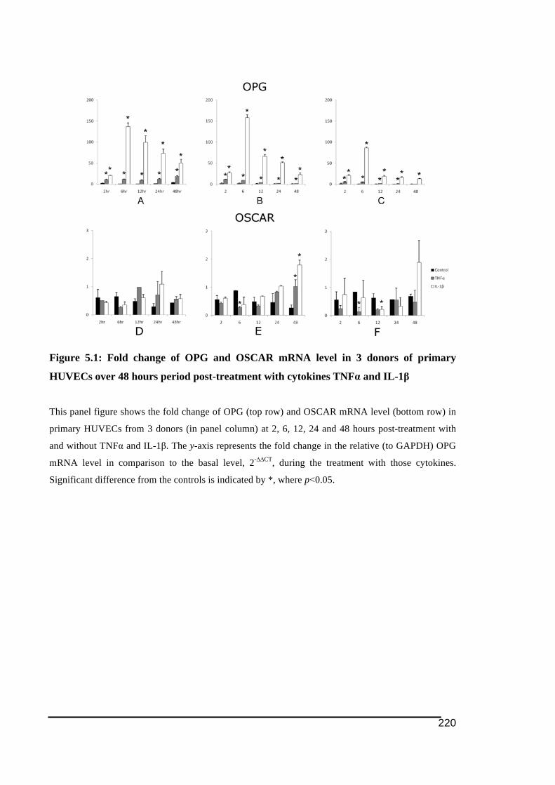
220
Figure 5.1: Fold change of OPG and OSCAR mRNA level in 3 donors of primary
HUVECs over 48 hours period post-treatment with cytokines TNFα and IL-1β
This panel figure shows the fold change of OPG (top row) and OSCAR mRNA level (bottom row) in
primary HUVECs from 3 donors (in panel column) at 2, 6, 12, 24 and 48 hours post-treatment with
and without TNFα and IL-1β. The y-axis represents the fold change in the relative (to GAPDH) OPG
mRNA level in comparison to the basal level, 2-∆∆CT, during the treatment with those cytokines.
Significant difference from the controls is indicated by *, where p<0.05.
221
Based on the results obtained as shown in Figure 5.1 (upper panel row), IL-1β appeared to be
a potent stimulator of OPG mRNA expression in all the HUVECs cultures as compared to the
TNFα. There was also a consistent pattern between donors of largest stimulation on OPG
mRNA expression seen 6 hours post-treatment with IL-1β before declining later. However,
for TNFα, even though there was significantly higher OPG mRNA expression in the
cytokine-treated samples, the stimulation appeared to be quite small compared to the IL-1β-
induced levels. Consistent with the previous report from our group (Zannettino et al. 2005), it
could be concluded that the data for OPG mRNA expression in primary HUVECs culture
obtained from this study was reproducible and consistent with the previous report.
Unlike OPG, there was little change or induction of OSCAR mRNA in response to either
TNFα or IL-1β (Figure 5.1 bottom row). In addition, there was no consistent trend in change
of mRNA between donors that could be seen. In comparison to OPG, change in the OSCAR
mRNA expression following treatment with TNFα and IL-1β in majority appeared to be very
minimal (~ 1-fold change). Looking at the data on the level of OSCAR in relative to GAPDH,
it could be concluded the level of OSCAR mRNA expression in those primary HUVECs was
very low (Figure 5.2).

222
Figure 5.2: OSCAR mRNA level in relative to GAPDH in in 3 donors primary HUVECs
over 48 hours period post-treatment in 3 treatment groups
This panel figure is consisted 3 graphs showing the relative OSCAR mRNA level (after normalised to
GAPDH) in primary HUVECs from 3 donors at 2, 6, 12, 24 and 48 hours post-treatment with and
without cytokines TNFα and IL-1β. The y-axis represents the relative (to GAPDH) OSCAR mRNA
level, 2-∆CT, during the treatment with those cytokines. Significant difference from the control is
indicated by *, where p<0.05.
223
5.3.2. Stimulation of OSCAR and OPG mRNA expression in BMEC by
cytokines TNFα and IL-1ß
Since there was no significant change in the OSCAR mRNA expression seen in culture of
primary HUVECs following treatments with pro-inflammatory cytokines TNFα and IL-1β,
further investigations were carried out in BMEC.
Based on Figure 5.3 (top and bottom rows), there appeared to be no significant difference in
both OSCAR and OPG mRNA expression between cytokine concentrations of 5 and
20ng/mL (except on OPG mRNA stimulation at 6 hours post-treatment). Therefore, it was
concluded that higher concentration 20ng/mL of either TNFα and IL-1β did not result in
significant change in the stimulation of mRNA expression. Hence, 5ng/mL of cytokine
concentration was determined to be sufficient to induce an increase in mRNA expression in
BMEC and used in the subsequent experiments on BMEC. Data from this preliminary
experiment also gave an indication of the elevation of OPG and OSCAR mRNA levels
following stimulation by either TNFα or IL-1β that be could seen particularly at the later time
points.
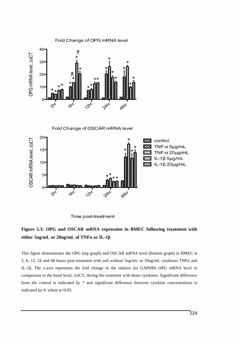
224
Figure 5.3: OPG and OSCAR mRNA expression in BMEC following treatment with
either 5ng/mL or 20ng/mL of TNFα or IL-1β
This figure demonstrates the OPG (top graph) and OSCAR mRNA level (bottom graph) in BMEC at
2, 6, 12, 24 and 48 hours post-treatment with and without 5ng/mL or 20ng/mL cytokines TNFα and
IL-1β. The y-axis represents the fold change in the relative (to GAPDH) OPG mRNA level in
comparison to the basal level, ∆∆CT, during the treatment with those cytokines. Significant difference
from the control is indicated by * and significant difference between cytokine concentrations is
indicated by #, where p<0.05.

225
In the following experiment a 72 hour post-treatment time point was added in the setup since
the finding from the earlier experiment demonstrated large change in mRNA expression
modulated by TNFα and IL-1β took place at 24 and 48 hours post-treatment. As shown in
Figure 5.4, there were large differences in the mRNA levels between treatment groups
particularly at the later time points 24 to 72 hour time points.
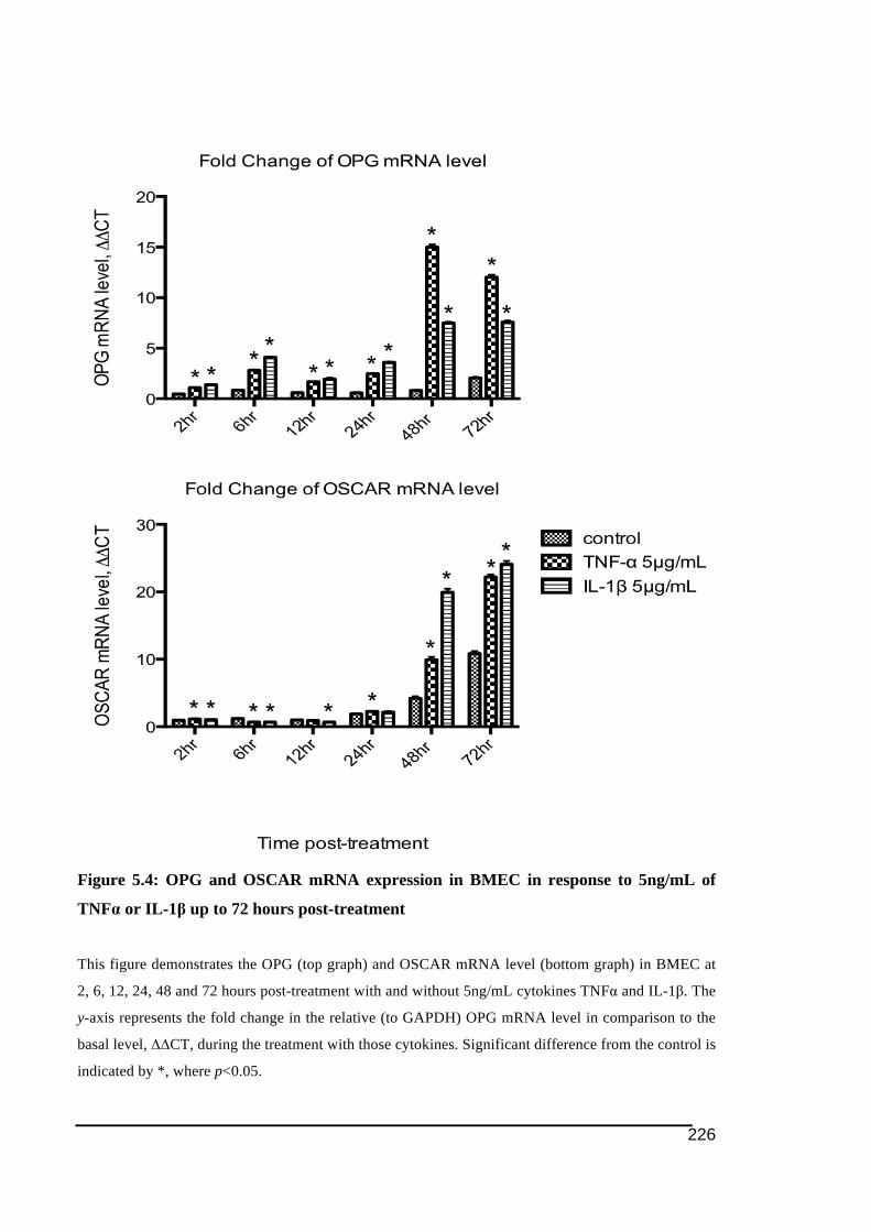
226
Figure 5.4: OPG and OSCAR mRNA expression in BMEC in response to 5ng/mL of
TNFα or IL-1β up to 72 hours post-treatment
This figure demonstrates the OPG (top graph) and OSCAR mRNA level (bottom graph) in BMEC at
2, 6, 12, 24, 48 and 72 hours post-treatment with and without 5ng/mL cytokines TNFα and IL-1β. The
y-axis represents the fold change in the relative (to GAPDH) OPG mRNA level in comparison to the
basal level, ∆∆CT, during the treatment with those cytokines. Significant difference from the control is
indicated by *, where p<0.05.

227
Another experiment incorporating samples in triplicates was carried out. However due to the
large amount of samples the number of time points in the experiment was reduced to 24, 48
and 72 hours post-treatment. For the same reason, the PCR setup was also limited to
duplicates instead of triplicates for each sample. Data from the corresponding experiment on
BMEC culture is presented in the following Figure 5.5.
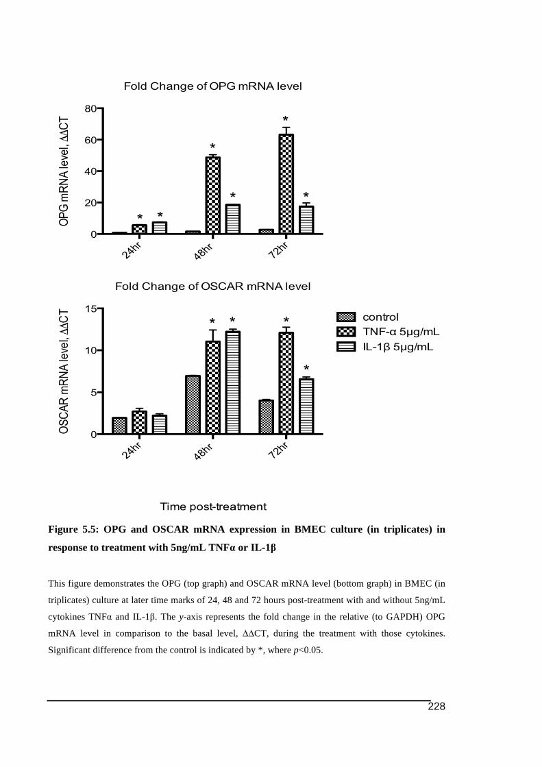
228
Figure 5.5: OPG and OSCAR mRNA expression in BMEC culture (in triplicates) in
response to treatment with 5ng/mL TNFα or IL-1β
This figure demonstrates the OPG (top graph) and OSCAR mRNA level (bottom graph) in BMEC (in
triplicates) culture at later time marks of 24, 48 and 72 hours post-treatment with and without 5ng/mL
cytokines TNFα and IL-1β. The y-axis represents the fold change in the relative (to GAPDH) OPG
mRNA level in comparison to the basal level, ∆∆CT, during the treatment with those cytokines.
Significant difference from the control is indicated by *, where p<0.05.
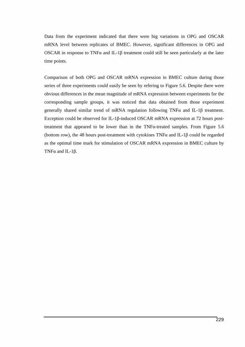
229
Data from the experiment indicated that there were big variations in OPG and OSCAR
mRNA level between replicates of BMEC. However, significant differences in OPG and
OSCAR in response to TNFα and IL-1β treatment could still be seen particularly at the later
time points.
Comparison of both OPG and OSCAR mRNA expression in BMEC culture during those
series of three experiments could easily be seen by refering to Figure 5.6. Despite there were
obvious differences in the mean magnitude of mRNA expression between experiments for the
corresponding sample groups, it was noticed that data obtained from those experiment
generally shared similar trend of mRNA regulation following TNFα and IL-1β treatment.
Exception could be observed for IL-1β-induced OSCAR mRNA expression at 72 hours post-
treatment that appeared to be lower than in the TNFα-treated samples. From Figure 5.6
(bottom row), the 48 hours post-treatment with cytokines TNFα and IL-1β could be regarded
as the optimal time mark for stimulation of OSCAR mRNA expression in BMEC culture by
TNFα and IL-1β.
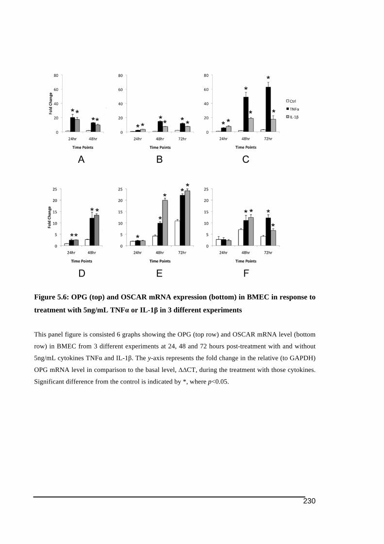
230
Figure 5.6: OPG (top) and OSCAR mRNA expression (bottom) in BMEC in response to
treatment with 5ng/mL TNFα or IL-1β in 3 different experiments
This panel figure is consisted 6 graphs showing the OPG (top row) and OSCAR mRNA level (bottom
row) in BMEC from 3 different experiments at 24, 48 and 72 hours post-treatment with and without
5ng/mL cytokines TNFα and IL-1β. The y-axis represents the fold change in the relative (to GAPDH)
OPG mRNA level in comparison to the basal level, ∆∆CT, during the treatment with those cytokines.
Significant difference from the control is indicated by *, where p<0.05.

231
5.3.3. Expression of soluble OSCAR following TNFα and IL-1β treatment
While many investigations on the regulation of OSCAR expression were carried out at the
mRNA level, examination at protein expression was also done to complement the gene
expression findings. Only BMEC cell culture supernatants from the time points 24, 48 and 72
hours post-treatment were subjected to the ELISA.
Data obtained from the OSCAR ELISA indicated that there was significantly higher level of
soluble OSCAR being secreated by BMEC culture following stimulation by 5ng/mL of TNFα
or IL-1β as compared to the controls at 24, 48 and 72 hour post-treatment. The pattern of
soluble OSCAR expression in response to TNFα or IL-1β stimulation followed a similar
pattern to the mRNA level (Figure 5.5 earlier). Both protein (Figure 5.7) and mRNA
expression (Figure 5.5 bottom row and middle column) suggested that IL-1β was more potent
in inducing OSCAR in BMEC than TNFα at 48 hours post-cytokine treatment.

232
Figure 5.7: Expression of soluble OSCAR by BMEC culture following treatment with
TNFα and IL-1β
This graph shows the level of soluble OSCAR secreated in BMEC media supernatant at 24, 48 and 72
hours treatment with and without 5ng/mL cytokines TNFα and IL-1β. The y-axis represents the fold
change in the relative (to GAPDH) OPG mRNA level in comparison to the basal level, ∆∆CT, during
the treatment with those cytokines. Error bars indicate the standard error of means obtained from 3
replicates within a sample group. Significant difference from the control is indicated by *, where
p<0.05.
233
5.3.4. Detection of OSCAR expression in BMEC in situ
OSCAR protein expression following cytokines TNFα or IL-1β treatment was also
investigated by immunofluoresence staining to detect membrane-bound OSCAR on the
BMEC. Immunofluoresence was also carried out for OPG expression, as this has not been
done in BMEC. Samples from 48 hours cytokines treatment only were included in the study
as the PCR data demonstrated maximal stimulation of both OPG and OSCAR mRNA took
place at this time point.
The immunofluorence for OPG demonstrated that the molecules could be detected in BMEC
at 48 hours cytokine treatment particularly the one treated with 5ng/mL of TNFα and IL-1β
(Figure 5.8B and C). It also appeared that there was more expression of OPG in BMEC
treated with IL-1β as compared to TNFα. However, in the control, OPG was only minimally
detected in the unstimulated BMEC suggesting that there was little expression of OPG taking
place in the BMEC culture. In antibody isotype negative control fluorescence was also
minimally detected (not shown).
As for the OSCAR expression, it appeared that there was induction of expression following
stimulation by TNFα as compared to IL-1β (Figure 5.8E). There was minimal expression of
OSCAR in non-cytokine treated BMEC (Figure 5.8D and F). This indicates that stimulation
with TNFα resulted in increased production of OSCAR protein as detected by
immunofluoresence.
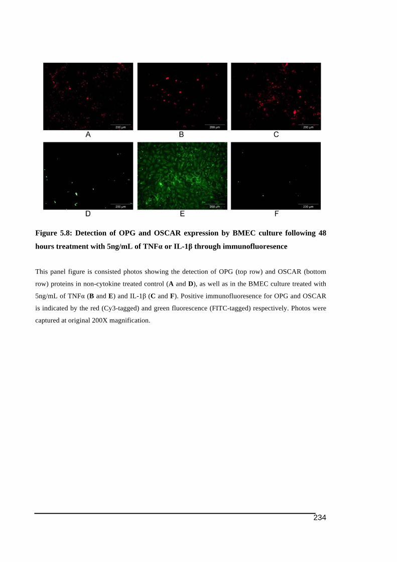
234
Figure 5.8: Detection of OPG and OSCAR expression by BMEC culture following 48
hours treatment with 5ng/mL of TNFα or IL-1β through immunofluoresence
This panel figure is consisted photos showing the detection of OPG (top row) and OSCAR (bottom
row) proteins in non-cytokine treated control (A and D), as well as in the BMEC culture treated with
5ng/mL of TNFα (B and E) and IL-1β (C and F). Positive immunofluoresence for OPG and OSCAR
is indicated by the red (Cy3-tagged) and green fluorescence (FITC-tagged) respectively. Photos were
captured at original 200X magnification.
235
5.4. Discussions
The studies presented in this chapter were designed to complement work from the previous
chapter. In Chapter 4, there was interesting finding that the vasculature was found to express
osteoclast-associated ITAM immunoreceptors, TREM2 and OSCAR especially in RA tissues.
OSCAR immunostaining of the vasculature was particularly interesting to be focused on as
TREM2 was viewed more as having anti-inflammatory function. Chen and colleagues (2008)
found treatment with TNFα and IL-1β upregulated protein expression of TREM1 and TREM3
detected by Western blotting, however, they also suppressed TREM2 in mice liver endothelial
cells. This prompted further investigation on OSCAR expression in vitro using either primary
endothelial cells or an endothelial cell line.
The observation of OSCAR immunostaining associated with vasculature structures,
particularly in human RA synovial tissues, was similar to observations of OPG
immunostaining reported about a decade ago (Crotti et al. 2004; Haynes et al. 2003).
Subsequent studies on OPG expression in endothelial cell in vitro have reported abundant
expression of OPG in HUVECs (Corallini et al. 2011; Stein et al. 2008; Zannettino et al.
2005) and the expression of OPG in endothelial cells was later found to affect various roles of
endothelial cells. There were studies suggesting that the upregulated expression of OPG by
proinflammatory cytokines could contribute towards the dysfunction of endothelial cells
(Collin-Osdoby et al. 2001; Secchiero et al. 2006). Later studies found that expression in OPG
in endothelial cells was associated with development and progression of artherosclerosis and
CVD (Goettsch et al. 2011). It was found that higher levels of OPG could be secreted by
inflammatory cells present in artherosclerotic plaques (Vik et al. 2007) and this was
associated with the dysfunction of endothelial cells by inducing the expression of adhesion
molecules like ICAM-1, VCAM-1 and E-selectin (Mangan et al. 2007; Zauli et al. 2007) that
would promote attachment and accumulation of leukocytes. Therefore investigation of OPG
expression was carried out to compare it to OSCAR expression and regulation in vitro. This
was carried out with the HUVECs model used in previous publications (Corallini et al. 2011;
Stein et al. 2008; Zannettino et al. 2005).
236
There has been a range of human endothelial cells used in in vitro experimental works done in
past. Among them are HUVECs, BMEC, microvascular endothelial cells (Collin-Osdoby et
al. 2001) and each cell type may behave differently. For instance, RANKL has been shown to
be expressed in human microvascular endothelial cells (Collin-Osdoby et al. 2001). However
there was no RANKL mRNA detected in HUVECs (Zannettino et al. 2005). In this particular
work HUVECs was the first choice for the in vitro endothelial cell model before BMEC was
also opted for the work following minimal changes in mRNA expression obtained from
HUVECs culture.
As the name implies, primary HUVECs are isolated by flushing human umbilical cord with
collagenase as reported in the literature (Gamble et al. 1985). Primary HUVECs were opted as
the first choice of in vitro endothelial cell culture model for a couple of reasons. First
HUVECs was a very commonly used in vitro endothelial cell model for studying OPG
expression in many studies (Corallini et al. 2011; Stein et al. 2008; Zannettino et al. 2005).
Following the discovery of positive OPG immunostaining on vasculature in tissues (Haynes
et al. 2003), following-up study in vitro was conducted in HUVECs, in which induction of
OPG expression following stimulation with either TNFα or IL-1β was observed (Zannettino
et al. 2005). Considering the observation of OSCAR immunostaining on vasculature mainly
in the human RA synovial tissues was quite similar to previous published finding of OPG on
vessels (Haynes et al. 2003), the use of HUVECs to study OSCAR expression in endothelial
cells in vitro following cytokine stimulation was very relevant. In addition, OSCAR
expression has also been shown in HUVECs through immunofluoresence and RT-PCR (even
though at low level) recently (Goettsch et al. 2011). Furthermore, HUVECs is a primary
endothelial cell type that is closer to the endothelial cells in tissues and in vivo, not a cell line
that more likely could undergo phenotype alternations following manipulation. All these
together support the choice of using HUVECs in this study.
BMEC used in this study is a cell line that has been used for for studying human endothelial
cells in vitro. BMEC was first isolated and characterised nearly 20 years ago (Almeida-Porada
& Ascensao 1996; Masek & Sweetenham 1994; Schweitzer et al. 1995) and was
recommended for studies investigating the role of endothelial cells for “homing” of
hematopoietic progenitor cells to the bone marrow (Rood et al. 1999). Rood and colleagues
(1999) found no difference or preferential adhesion of hematopoietic progenitor cells to
human BMEC in comparison to HUVECs. However, an earlier study found otherwise and
237
showed there was better adherence of CD34-positive to TNFα-stimulated BMEC compared to
similarly treated HUVECs (Schweitzer et al. 1997).
The expression of OSCAR and TREM2 in vasculature in rheumatoid arthritis was likely the
result of inflammation in the tissues, presumably via the effect of pro-inflammatory cytokines
like TNFα and IL-1β. These two cytokines are reported to be expressed in abundant in RA
synovial membrane and may play important role in stimulating osteoclast formation in the
tissues of RA (Chu et al. 1991). The regulation of OSCAR by either TNFα or IL-1β was first
examined at the gene or mRNA level only (therefore based on the qRT-PCR data only) before
proceeding to the investigation at the protein level. It should be noted that the qRT-PCR data
was analysed using 2-∆∆CT method (data in form of mean fold change) instead of 2-∆CT (mRNA
level in relative to the reference gene) (Livak & Schmittgen 2001) as applied for data
presented in the previous chapters 2 and 3. This was justified by the presence of 0 hour time
point (post-cytokine treatment) as the point for basal expression and data from the other
evaluated time marks will be calculated as fold change over this reference point. GAPDH was
used as the reference gene for normalisation as it is a common housekeeping gene used for
endothelial cells reported in the literature, particularly for HUVECs (Mangan et al. 2007;
Secchiero et al. 2008; Zhang et al. 2002).
In the experiment on HUVECs, cytokine concentration of 5ng/mL was chosen since this
concentration has been reported to be able to activate HUVECs without causing induction of
apoptosis (Molostvov et al. 2001). Zannettino and co-workers (2005) found that stimulation
on HUVECs with either TNFα or IL-1β at 5ng/mL elevated OPG mRNA levels by 8 to 9-
fold. This study saw the induction of OPG mRNA expression following the cytokine
treatment was inconsistent between donors, even though all of them showed the increase. The
present study also noted that 5ng/mL of IL-1β was far more potent in stimulating OPG
mRNA expression (more than 50-fold induction) than TNFα of similar concentration (about 3
to 6-fold change) at 24 hours treatment. In another previous study, it was reported that OPG
mRNA was also found to be expressed in primary HMVEC in vitro and could be upregulated
by treatment with TNFα and IL-1α (with effective cytokine concentration ranging from 0.1 to
10nM) (Collin-Osdoby et al. 2001).
With regard to the OSCAR mRNA expression in HUVECs, there was variation in responses
to the cytokines between donors. As a consequence it may be inappropriate to compare the
238
data obtained in this study to that previously reported (Goettsch et al. 2011), which did not
use multiple donors of the primary cells. Due to the inconsistent change in OSCAR mRNA
expression following TNFα or IL-1β treatment the experiment was not extended to
investigation on protein expression in HUVECs. However, it was decided that the
investigation was continued on BMEC instead of primary HUVECs. In addition, this would
be novel as there has been no report on OSCAR in BMEC.
Unlike the studies in HUVECs in which 5ng/mL is regarded as an optimum cytokine
concentration (Molostvov et al. 2001; Zannettino et al. 2005), there is not much literature
reporting suitable concentration of TNFα and IL-1β cytokines to induce cell responses` in
BMEC. Therefore in the first experiment with BMEC, the mRNA levels following induction
by TNFα and IL-1β was compared between 5ng/mL and 20ng/mL. Study outcome there was
little difference on the induction of OSCAR gene expression in BMEC at the 2 concentrations
of TNFα and IL-1β (Figure 5.3 bottom row). Therefore, 5ng/mL was chosen as it was also
used for HUVECs.
Overall in the 3 experiments with BMEC mRNA expression for both OPG and OSCAR
following cytokine stimulation was consistent between experiments. Consistent with the
findings here the induction of mRNA expression in endothelial cells has been reported as be
able to peak as early as 1 hour post-stimulation (Hashimoto et al. 1994).
While it has been quite well established that OPG is detected in wide range of endothelial cell
types such as in human HMVEC and HUVECs and could play significant role in endothelial
cells (Collin-Osdoby et al. 2001; Secchiero et al. 2006; Secchiero et al. 2008; Stein et al.
2008; Zannettino et al. 2005; Zauli et al. 2007), to the author’s knowledge, detection of OPG
expression in human BMEC cell line (both at mRNA and protein level) has never been
documented and hence could be considered as novel. Using the antibody MAB805, which has
been thought to immunostain OPG in dimeric form expressed by endothelial cells (Haynes et
al. 2003), this study demonstrated OPG expression at protein level in BMEC as detected by
immunofluoresence.
This study provides support for the work by Goettsch and colleagues (2011) demonstrating
endothelial cells as another cell type expressing OSCAR. This is also the first study to
demonstrate the upregulation of OSCAR expression in endothelial cells by both TNFα and
239
IL-1β. Earlier induction of OSCAR has been shown in human CD14-positive monocytes
(Herman et al. 2008) and in murine osteoclasts by TNFα and IL-1α respectively (Kim et al.
2009). A very recent study by Sinningen and colleagues (2013) found that treatment with
50ng/mL of TNFα resulted in stimulation of OSCAR mRNA expression in RAW264.7 cell
line by 2-fold.
Significant induction of OSCAR mRNA expression following treatment with either TNFα or
IL-1β from the first two experiments with BMEC was consistently noted at and after 24 hours
post-cytokine treatment. Such finding may indicate that the induction in the OSCAR mRNA
expression was a secondary response mediated by other pathway(s) and not directly
stimulated by those TNFα and IL-1β cytokines. This sort of observation has helped in
designing the experimental setup for the third experiment with BMEC by limiting the time
points, hence allowing more replicates of samples to be included.
While the qRT-PCR data gave an indication of largest stimulation of OSCAR mRNA
expression taking place at 48 hour post-treatment, it was quite surprising to note that only
TNFα appeared to induce expression of OSCAR protein as detected by immunofluoresence,
meanwhile data from the qPCR data indicated that IL-1β stimulated OSCAR mRNA
expression more than TNFα did.
However, it was interesting to note that there was correlation in pattern after the cytokine
treatment between the OSCAR mRNA level (refer Figure 5.6F) and the level (or
concentration) of soluble OSCAR secreted into BMEC culture supernatant (Figure 5.7).
ELISA was carried out on the supernatants of BMEC only (not HUVECs) since only the PCR
data from the BMEC culture appeared to show induction of OSCAR mRNA expression. The
high expression of soluble OSCAR in BMEC culture supernatant in response to TNFα and
IL-1β stimulation may provide insights in explaining high level of soluble OSCAR in
synovial fluids of RA patients (refer section 4.3.4). While the expression of membrane bound
OSCAR in HUVECs would be detected by immunofluoresence (Goettsch et al. 2011), this
study demonstrated protein expressed is most likely in soluble isoform of OSCAR, consistent
with that previously described (Khan 2007; Kim et al. 2002).
While the association between OSCAR and its possible role in the development of
artherosclerosis has been investigated (Goettsch et al. 2012; Goettsch et al. 2011), the present
240
study is the first in line to look into endothelial cell-expressed OSCAR in the context of
chronic inflammation. It is consistent with the idea that OSCAR should have no longer seen
as a molecule involved in bone homeostasis only, but also possibly plays other role in the
progression of inflammation-mediated bone loss like RA.
Pro-inflammatory cytokines TNFα and IL-1β are likely to contribute to high OSCAR protein
secreted in RA and OA synovial fluids described in previous Chapter 4 and in serum
described by Herman and colleagues (2008) through induction on the expression of the
molecule in the endothelial cells. Interestingly Herman et al. (2008) suggested that the level of
soluble OSCAR in serum could be associated with protective function against bone erosion in
RA, however this remains to be proven by further investigations.
It would be interesting to speculate on the role of OSCAR expressed by endothelial cells in
the recruitment and infiltration of inflammatory cells into tissues. In dendritic cells activation
of OSCAR in DC promotes expression of MCP-1 (Merck et al. 2006), which is the
chemoatttractant recruiting leukocytes to site of inflammation. Literature has indicated that
IL-1β and TNFα might be involved in promoting recruitment of osteoclast precursors (which
is monocyte/macrophage-derived cells) (Issekutz et al. 1994) through endothelial cell
activation (Kindle et al. 2006). Activated endothelial cells are believed to be important in
supporting each phase of inflammatory process (Pober & Sessa 2007). Meanwhile a recent
discovery by Goettsch and coworkers (2012) demonstrated induction in the expression of
adhesion molecules (that mediating attachment of monocytes to the endothelium) following
OSCAR overexpression by endothelial cells in vitro. Taken together, there is possibility that
OSCAR may play role not just only in mediating osteoclastogenesis, but also in promoting
the recruitment of inflammatory cells to the inflammation sites.
The findings of this chapter and the previous chapter open up new areas of research on
OSCAR to be explored in the future. It would be interesting to investigate the regulation of
the OSCAR expression in the presence of TNFα and IL-1β receptor antagonists in order to
gain insight of the receptors involved in OSCAR regulation in the endothelial cells. It would
also be interesting to investigate whether the stimulation of OSCAR expression in BMEC is
mediated by NFATc1, which was observed in human coronary artery endothelial cells
following stimulation by oxidized LDL (Goettsch et al. 2011).
241
OPG expression in endothelial cells (Haynes et al. 2003) has been associated with vascular
homestasis and pathology (Corallini et al. 2008; Reid & Holen 2009; Secchiero et al. 2006).
Similarly, cytokine regulation on OSCAR expression in endothelial cells as observed in this
study alongside with the recent discovery of possible role in the inflammation and
artherosclerosis (Goettsch et al. 2012; Goettsch et al. 2011) and the statistics of high
mortalitiy in RA patients due to CVDs (Pham et al. 2006) could provide a meaningful insight
on the role of endothelial cell-expressed OSCAR has in the context of RA.
5.5. Conclusion
This study is the first to report the regulation of OSCAR mRNA and protein expression in
response to stimulation by pro-inflammatory cytokines TNFα or IL-1β in endothelial cells in
vitro.
Despite the fact that there was induction of OPG mRNA expression observed following
stimulation by either TNFα or IL-1β, in which OPG mRNA expression peaked at 6 hours post
treatment and similar to the previous study in HUVECs (Zannettino et al. 2005), there was an
inconsistency in the change in the OSCAR mRNA level between donors of primary
HUVECs. Hence, BMEC was used in the in vitro model in the study. Data from these
experiments consistently indicated that the largest increase in the OSCAR mRNA level took
place at 48 and 72 hours post cytokine treatment.
The level of of soluble OSCAR in the supernatants of BMEC culture was consistent with the
mRNA levels in response to the cytokines. Immunofluoresence for OSCAR on the BMEC
culture at 48 hours post-treatment indicated that there was more OSCAR protein produced
following stimulation by TNFα.
Together the data suggest that the regulation on OSCAR in RA tissues may have various roles
in disease regulation. These include, adhesion and migration of cells from the vasculature,
involvement endothelial cell damage (possibly associated with atherosclerosis) and the
regulation of bone metabolism in disease. Further studies of OSCAR expression in
endothelial cells open new gate for future scientific investigations.
242
6. SUMMARY AND CONCLUDING REMARKS
Bone remodelling is regulated by tight balance between bone resorption by osteoclasts and
bone formation by osteoblasts. Imbalance between these two processes, either through
reduced rate of bone formation or excessive osteoclast activity resulting net bone resorption
could result in pathological bone loss. Peri-implant or peri-prosthetic osteolysis (PO) and
rheumatoid arthritis (RA) are two examples of pathological bone loss condtions that have
been associated with net bone resorption resulted from excessive osteoclast activity
(Gravallese et al. 1998; Holding et al. 2006; Shen et al. 2006).
As a part of strategy for therapy against pathological bone loss as observed in RA and PO, the
expression of molecules regulating of osteoclast differentiation and activity at the molecular
level has long been studied. The OPG/RANK/RANKL triad has been found to be the main
signalling pathway mediating osteoclast differentiation as well as activity and the expression
of these molecules detected in RA and PO tissues has been demonstrated to have strong
association with the feature of bone loss observed in those diseases (Crotti et al. 2002; Gehrke
et al. 2003; Haynes et al. 2003; Haynes et al. 2001b; Haynes et al. 2004; Mandelin et al. 2003;
Vanderborght et al. 2004). Studies have also indicated that regulation on these molecules has
been found to be an effective approach in limiting bone loss in pathological conditions like
RA (Pettit et al. 2001; Romas et al. 2002) and PO (Itonaga et al. 2000).
From accumulating evidence it has been strongly suggested that immunoreceptor tyrosine-
based based activation motif (ITAM)-mediated pathway, which has been discovered in
osteoclasts early in this century, plays role in mediating co-stimulatory signals for
osteoclastogenesis (Humphrey et al. 2005; Koga et al. 2004). While two immunoreceptors,
TREM2 and OSCAR together with their corresponding membrane adaptor molecules DNAX
activation protein of 12kDa (DAP12) and Fc receptor gamma (FcRγ), have been identified as
the molecules mediating the co-stimulatory ITAM-mediated signalling pathway in
osteoclasts, there have been limited studies investigating on these molecules in the context of
skeletal pathology. Hence the studies described here were designed to provide more holistic
243
investigation on osteoclast-associated ITAM-related molecules (TREM2, OSCAR, DAP12
and FcRγ), as well as the key transcriptional factor in osteoclasts nuclear factor of activated
T-cells 1 (NFATc1), in the context of PO and RA pathology. Studies presented here
investigate on the expression of those osteoclast-associated ITAM-related molecules in RA
and PO tissues (in comparison to the control tissues) followed by in vitro work used to verify
the observations on the tissues obtained earlier.
Presentation of the studies in this thesis commenced with investigation on both protein and
mRNA expression of NFATc1, TREM2, DAP12, OSCAR and FcRγ in tissues adjacent to PO
site in comparison to the osteoarthritis (OA) tissues as the controls. Generally there were
more cells positive for immunostaining for all those ITAM-associated molecules, including
multinucleated oscteoclast-like cells that were also positive for osteoclast cell markers tartrate
acid phospotase (TRAP) and cathepsin K (based on serial immunolabeling comparison), in
PO tissues as compared to the OA. Similar sort of outcome pattern was obtained for the
examination on the mRNA levels, in which there was higher level of mRNA for all those
ITAM-associated molecules in PO than in OA tissues.
Despite presence of more multinucleated osteoclast-like cells observed in PO tissues
(supported by immunostaining for osteoclast cell markers TRAP and cathepsin K), study
outcome found that there was not much positive immunostaining for NFATc1. Consistent
with this, findings from the examination on mRNA level found that NFATc1 gene expression
was not statistically higher in PO tissues in comparison to the control OA tissues.
More interestingly, polyethylene (PE) particles, a common type of wear debris causing
osteolysis in PO (Campbell et al. 1995; Dowd et al. 2000; Hirakawa et al. 1996; Shanbhag et
al. 1994b; Sochart 1999; Willert et al. 1990), were detected present in osteoclast-like cells
positive for those osteoclast cell markers TRAP and cathepsin K. PE particles were also
detected within and close to cells positive for TREM2, DAP12, OSCAR and FcRγ. All these
study outcomes appeared to support one of the hypotheses presented in this thesis, in which
ITAM-related molecules are expressed higher in human tissues of peri-implant osteolysis
induced by PE and could be associated with the disease progression.
Detection of PE particles in some cells expressing osteoclast ITAM-related molecules,
particularly in multinucleated cells, in PO tissues prompted further investigation on whether
244
those particles might affect the expression of those ITAM-related molecules in osteoclasts
and hence could be associated with PE-induced osteoclastogenesis. This investigation was
carried out in a novel in vitro model for PE-induced osteoclastogenesis assay that provided
better representation of tissue microenvironment that may affect cellular interactions with the
PE particles (Atkins et al. 2009). As part of assessment on the novel model of PBMC-derived
osteoclast culture grown in 3D-collagen gel as an in vitro model for investigating PE-induced
osteoclastogenesis, TRAP staining and dentine pit resorption assay were carried out to
confirm increase in osteoclast formation and resorption activity in response to exposure to PE
particles. Such outcomes were achieved following the assessment on the in vitro model. The
study also detected presence of PE particles within the PBMC-derived osteoclasts in the
culture. Together all these may suggest that PE particles could induce osteolysis in PO tissues
through increase in the formation of osteoclasts and total resorption activity.
In conjunction to the intent of the study to further investigate the effect of PE particles on the
expression of TREM2, DAP12, OSCAR and FcRγ in context of PE-induced
osteoclastogenesis, the mRNA levels of those molecules were studied and compared between
cells cultured with and without PE particles. The study outcome, which found that these
factors were induced by PE particles during osteoclast formation in vitro, could suggest that
the ITAM-associated receptor pathway is markedly activated by PE wear particles and may
augment RANKL-mediated osteolysis in peri-implant tissues containing PE particles. The
upregulation of osteoclast ITAM-related molecules in vitro as documented in Chapter 3,
supported by higher expression of those molecules in peri-implant tissues as discussed earlier
in Chapter 2, may indicate the significance those molecules possibly have in osteoclast
formation and activity in the disease as hypothesized in the beginning of these studies.
Meanwhile in the context of RA, similar approach was used in which the expression of
TREM2, DAP12, OSCAR and FcRγ alongside with NFATc1 and osteoclast cell marker
cathepsin K was compared between in active and inactive RA tissues as well as OA and
normal tissues following tissue immunostaining. Quite similar trend of study outcome (to PO
tissues) was obtained, which demonstrated trends of more positive immunostaining of
OSCAR, FcRγ, TREM2 and DAP12 in human RA tissues, particularly the active RA group,
in comparison to the OA and normal tissue group. However, tissue immunostaining for the
osteoclast cell marker cathepsin K indicated that those examined RA tissues were not heavily
245
populated with osteoclast-like cells. Instead, many of the cathepsin K-positive cells in the
tissues examined were seen to have fibroblast-like morphology. Therefore, the finding of
more expression of ITAM-related molecules in RA tissues could not be associated with an
increase in osteoclast formation in RA that could be responsible for bone loss observed in the
disease.
Despite the larger proportion of positively immunostained cells seen in the active RA tissues
compared to the other tissue groups, unlike the other studied molecules, outcome from
NFATc1 immunostaining found that most of the cells were in aggregations and appeared to
be T-lymphocyte-like cells. Therefore based on the study outcome obtained, NFATc1 were
mainly discussed in the context of T-lymphocytes in RA. There was no work on comparing
the mRNA expression of these molecules in the RA and control tissues since there was
technical limitation in finding appropriate tissue source for RNA extraction.
In addition to the findings that there was higher proportion of cells expressing ITAM-
associated molecules in RA tissues, there was also another interesting observation noted,
where positive immunostaining particularly for OSCAR was also obviously seen in
vasculature structure especially in RA tissues. Positive immunostaining associated with
vasculature, however, was not observed in PO tissues as described earlier. This sort of
observation has become the basis for the work presented in the Chapter 5. Positive OSCAR
immunostaining on vasculature has promoted further investigation on the existence of soluble
form of OSCAR and the level of soluble OSCAR in RA patients. However since such study
using patient sera has been reported in the literature this present work measured and compared
the level of soluble OSCAR in the synovial fluids of patients with RA and OA to demonstrate
novelty in the finding. Interesting data obtained from the work demonstrated quite high
amount of soluble OSCAR measured in both groups of RA and OA synovial fluids.
In relation to the aim for further investigation following observation of OSCAR
immunostaining on vasculature particularly in the RA tissues, experimental work was carried
out on in vitro culture of endothelial cells, which were primary human umbilical vein
endothelial cells (HUVECs) and bone marrow endothelial cell (BMEC) line. While there have
been studies reporting on stimulation on OSCAR expression by oxidized low density
lipoprotein (Goettsch et al. 2011) (more relevant to cardiovascular pathology) during the
experimental work been carried out, this study provide novel finding of upregulation of
246
OSCAR expression in endothelial cells in vitro by pro-inflammatory cytokine TNFα and IL-
1β, which is closer to the context of rheumatoid arthritis pathology. The expression of
OSCAR in endothelial cells in the context of inflammation and rheumatoid arthritis also open
a new dimension the current body of knowledge by taking OSCAR out of “monocyte-
macrophage lineage and bone loss” scope only to broader context of inflammation. Like
OPG, this study suggests bigger role and contribution OSCAR in a wider and more complex
tissue microenvironment in inflammation-mediated bone loss pathology.
The findings presented in this thesis suggest that, in inflammation-mediated bone loss, there is
alteration in the expression of osteoclast-associated ITAM-signalling molecules that may
contribute towards the progression of PO and RA. Just like the revelation on the significance
of OPG/RANK/RANKL axis in the pathology of those diseases (Crotti et al. 2004; Crotti et
al. 2002) which has opened options for therapy approach wide, the discovery of significance
on osteoclast ITAM-associated molecules in the context of inflammation-mediated bone loss
as being proposed and documented here may provide new dimension for more targeted and
specific treatment for those diseases. Further studies on ITAM-associated molecules in the
future could identify that these molecules may potentially be developed as a promising
therapy target.
247
REFERENCE LIST
Abram, CL & Lowell, CA 2007, 'The expanding role for ITAM-based signaling pathways in immune cells', Science's STKE : signal transduction knowledge environment, vol. 2007, no. 377, Mar 13, p. re2. Abramson, SB & Amin, A 2002, 'Blocking the effects of IL-1 in rheumatoid arthritis protects bone and cartilage', Rheumatology, vol. 41, no. 9, Sep, pp. 972-980. Abu-Amer, Y, Erdmann, J, Alexopoulou, L, Kollias, G, Ross, FP & Teitelbaum, SL 2000, 'Tumor necrosis factor receptors types 1 and 2 differentially regulate osteoclastogenesis', Journal of Biological Chemistry, vol. 275, no. 35, Sep 1, pp. 27307-27310. Adamopoulos, IE, Danks, L, Itonaga, I, Locklin, RM, Sabokbar, A, Ferguson, DJ & Athanasou, NA 2006a, 'Stimulation of osteoclast formation by inflammatory synovial fluid', Virchows Archiv, vol. 449, no. 1, Jul, pp. 69-77. Adamopoulos, IE, Sabokbar, A, Wordsworth, BP, Carr, A, Ferguson, DJ & Athanasou, NA 2006b, 'Synovial fluid macrophages are capable of osteoclast formation and resorption', Journal of Pathology, vol. 208, no. 1, Jan, pp. 35-43. Aigner, T, Bertling, W, Stoss, H, Weseloh, G & von der Mark, K 1993, 'Independent expression of fibril-forming collagens I, II, and III in chondrocytes of human osteoarthritic cartilage', Journal of Clinical Investigation, vol. 91, no. 3, Mar, pp. 829-837. Al-Saffar, N, Revell, PA & Kobayashi, A 1997, 'Modulation of the phenotypic and functional properties of phagocytic macrophages by wear particles from orthopaedic implants', Journal of Materials Science: Materials in Medicine, vol. 8, no. 11, Nov, pp. 641-648. Alaaeddine, N, DiBattista, JA, Pelletier, JP, Cloutier, JM, Kiansa, K, Dupuis, M & Martel-Pelletier, J 1997, 'Osteoarthritic synovial fibroblasts possess an increased level of tumor necrosis factor-receptor 55 (TNF-R55) that mediates biological activation by TNF-alpha', Journal of Rheumatology, vol. 24, no. 10, Oct, pp. 1985-1994. Alamanos, Y & Drosos, AA 2005, 'Epidemiology of adult rheumatoid arthritis', Autoimmunity Reviews, vol. 4, no. 3, Mar, pp. 130-136. Aletaha, D, Neogi, T, Silman, AJ, Funovits, J, Felson, DT, Bingham, CO, 3rd, Birnbaum, NS, Burmester, GR, Bykerk, VP, Cohen, MD, Combe, B, Costenbader, KH, Dougados, M, Emery, P, Ferraccioli, G, Hazes, JM, Hobbs, K, Huizinga, TW, Kavanaugh, A, Kay, J, Kvien, TK, Laing, T, Mease, P, Menard, HA, Moreland, LW, Naden, RL, Pincus, T, Smolen, JS, Stanislawska-Biernat, E, Symmons, D, Tak, PP, Upchurch, KS, Vencovsky, J, Wolfe, F & Hawker, G 2010, '2010 Rheumatoid arthritis classification criteria: an American College of Rheumatology/European League Against Rheumatism collaborative initiative', Arthritis and Rheumatism, vol. 62, no. 9, Sep, pp. 2569-2581. Alias, E, Dharmapatni, AS, Holding, AC, Atkins, GJ, Findlay, DM, Howie, DW, Crotti, TN & Haynes, DR 2012, 'Polyethylene particles stimulate expression of ITAM-related molecules in peri-implant tissues and when stimulating osteoclastogenesis in vitro', Acta Biomaterialia, vol. 8, no. 8, Aug, pp. 3104-3112.
248
Allami, MK, Fender, D, Khaw, FM, Sandher, DR, Esler, C, Harper, WM & Gregg, PJ 2006, 'Outcome of Charnley total hip replacement across a single health region in England. The results at ten years from a regional arthroplasty register', Journal of Bone and Joint Surgery (British Volume), vol. 88, no. 10, Oct, pp. 1293-1298. Almeida-Porada, G & Ascensao, JL 1996, 'Isolation, characterization, and biologic features of bone marrow endothelial cells', Journal of Laboratory and Clinical Medicine, vol. 128, no. 4, Oct, pp. 399-407. Altman, R, Asch, E, Bloch, D, Bole, G, Borenstein, D, Brandt, K, Christy, W, Cooke, TD, Greenwald, R, Hochberg, M & et al. 1986, 'Development of criteria for the classification and reporting of osteoarthritis. Classification of osteoarthritis of the knee. Diagnostic and Therapeutic Criteria Committee of the American Rheumatism Association', Arthritis and Rheumatism, vol. 29, no. 8, Aug, pp. 1039-1049. Altomonte, L, Zoli, A, Mirone, L, Scolieri, P & Magaro, M 1992, 'Serum levels of interleukin-1b, tumour necrosis factor-a and interleukin-2 in rheumatoid arthritis. Correlation with disease activity', Clinical Rheumatology, vol. 11, no. 2, Jun, pp. 202-205. Amin, AR & Abramson, SB 1998, 'The role of nitric oxide in articular cartilage breakdown in osteoarthritis', Current Opinion in Rheumatology, vol. 10, no. 3, May, pp. 263-268. Andersen, TL, Sondergaard, TE, Skorzynska, KE, Dagnaes-Hansen, F, Plesner, TL, Hauge, EM, Plesner, T & Delaisse, JM 2009, 'A physical mechanism for coupling bone resorption and formation in adult human bone', American Journal of Pathology, vol. 174, no. 1, Jan, pp. 239-247. Anderson, DM, Maraskovsky, E, Billingsley, WL, Dougall, WC, Tometsko, ME, Roux, ER, Teepe, MC, DuBose, RF, Cosman, D & Galibert, L 1997, 'A homologue of the TNF receptor and its ligand enhance T-cell growth and dendritic-cell function', Nature, vol. 390, no. 6656, Nov 13, pp. 175-179. Anderson, GI, MacQuarrie, R, Osinga, C, Chen, YF, Langman, M & Gilbert, R 2001, 'Inhibition of leukotriene function can modulate particulate-induced changes in bone cell differentiation and activity', Journal of Biomedical Materials Research, vol. 58, no. 4, pp. 406-414. Andersson, MK, Lundberg, P, Ohlin, A, Perry, MJ, Lie, A, Stark, A & Lerner, UH 2007, 'Effects on osteoclast and osteoblast activities in cultured mouse calvarial bones by synovial fluids from patients with a loose joint prosthesis and from osteoarthritis patients', Arthritis Research & Therapy, vol. 9, no. 1, p. R18. Angel, NZ, Walsh, N, Forwood, MR, Ostrowski, MC, Cassady, AI & Hume, DA 2000, 'Transgenic mice overexpressing tartrate-resistant acid phosphatase exhibit an increased rate of bone turnover', Journal of Bone and Mineral Research, vol. 15, no. 1, Jan, pp. 103-110. Aoki, N, Kimura, S, Takiyama, Y, Atsuta, Y, Abe, A, Sato, K & Katagiri, M 2000, 'The role of the DAP12 signal in mouse myeloid differentiation', Journal of Immunology, vol. 165, no. 7, Oct 1, pp. 3790-3796. Arai, F, Miyamoto, T, Ohneda, O, Inada, T, Sudo, T, Brasel, K, Miyata, T, Anderson, DM & Suda, T 1999, 'Commitment and differentiation of osteoclast precursor cells by the sequential expression of c-Fms and receptor activator of nuclear factor kappaB (RANK) receptors', Journal of Experimental Medicine, vol. 190, no. 12, Dec 20, pp. 1741-1754. Arko, B, Prezelj, J, Kocijancic, A, Komel, R & Marc, J 2005, 'Association of the osteoprotegerin gene polymorphisms with bone mineral density in postmenopausal women', Maturitas, vol. 51, no. 3, Jul 16, pp. 270-279.
249
Arko, B, Prezelj, J, Komel, R, Kocijancic, A, Hudler, P & Marc, J 2002, 'Sequence variations in the osteoprotegerin gene promoter in patients with postmenopausal osteoporosis', Journal of Clinical Endocrinology and Metabolism, vol. 87, no. 9, Sep, pp. 4080-4084. Armstrong, AP, Tometsko, ME, Glaccum, M, Sutherland, CL, Cosman, D & Dougall, WC 2002, 'A RANK/TRAF6-dependent signal transduction pathway is essential for osteoclast cytoskeletal organization and resorptive function', Journal of Biological Chemistry, vol. 277, no. 46, Nov 15, pp. 44347-44356. Arnett, FC, Edworthy, SM, Bloch, DA, McShane, DJ, Fries, JF, Cooper, NS, Healey, LA, Kaplan, SR, Liang, MH, Luthra, HS & et al. 1988, 'The American Rheumatism Association 1987 revised criteria for the classification of rheumatoid arthritis', Arthritis and Rheumatism, vol. 31, no. 3, Mar, pp. 315-324. Arron, JR & Choi, Y 2000, 'Bone versus immune system', Nature, vol. 408, no. 6812, Nov 30, pp. 535-536. Asagiri, M, Sato, K, Usami, T, Ochi, S, Nishina, H, Yoshida, H, Morita, I, Wagner, EF, Mak, TW, Serfling, E & Takayanagi, H 2005, 'Autoamplification of NFATc1 expression determines its essential role in bone homeostasis', Journal of Experimental Medicine, vol. 202, no. 9, Nov 7, pp. 1261-1269. Asagiri, M & Takayanagi, H 2007, 'The molecular understanding of osteoclast differentiation', Bone, vol. 40, no. 2, Feb, pp. 251-264. Aspenberg, P & van der Vis, H 1998, 'Fluid pressure may cause periprosthetic osteolysis. Particles are not the only thing', Acta Orthopaedica Scandinavica, vol. 69, no. 1, Feb, pp. 1-4. Assuma, R, Oates, T, Cochran, D, Amar, S & Graves, DT 1998, 'IL-1 and TNF antagonists inhibit the inflammatory response and bone loss in experimental periodontitis', Journal of Immunology, vol. 160, no. 1, Jan 1, pp. 403-409. Athanasou, NA 1996, 'Cellular biology of bone-resorbing cells', Journal of Bone and Joint Surgery (American Volume), vol. 78, no. 7, Jul, pp. 1096-1112. Athanasou, NA & Quinn, J 1990, 'Immunophenotypic differences between osteoclasts and macrophage polykaryons: immunohistological distinction and implications for osteoclast ontogeny and function', Journal of Clinical Pathology, vol. 43, no. 12, Dec, pp. 997-1003. Atkins, GJ, Haynes, DR, Geary, SM, Loric, M, Crotti, TN & Findlay, DM 2000, 'Coordinated cytokine expression by stromal and hematopoietic cells during human osteoclast formation', Bone, vol. 26, no. 6, Jun, pp. 653-661. Atkins, GJ, Kostakis, P, Pan, B, Farrugia, A, Gronthos, S, Evdokiou, A, Harrison, K, Findlay, DM & Zannettino, AC 2003, 'RANKL expression is related to the differentiation state of human osteoblasts', Journal of Bone and Mineral Research, vol. 18, no. 6, Jun, pp. 1088-1098. Atkins, GJ, Kostakis, P, Vincent, C, Farrugia, AN, Houchins, JP, Findlay, DM, Evdokiou, A & Zannettino, AC 2006, 'RANK Expression as a cell surface marker of human osteoclast precursors in peripheral blood, bone marrow, and giant cell tumors of bone', Journal of Bone and Mineral Research, vol. 21, no. 9, Sep, pp. 1339-1349. Atkins, GJ, Welldon, KJ, Holding, CA, Haynes, DR, Howie, DW & Findlay, DM 2009, 'The induction of a catabolic phenotype in human primary osteoblasts and osteocytes by polyethylene particles', Biomaterials, vol. 30, no. 22, Aug, pp. 3672-3681.
250
Augoulea, A, Vrachnis, N, Lambrinoudaki, I, Dafopoulos, K, Iliodromiti, Z, Daniilidis, A, Varras, M, Alexandrou, A, Deligeoroglou, E & Creatsas, G 2013, 'Osteoprotegerin as a marker of atherosclerosis in diabetic patients', International Journal of Endocrinology, vol. 2013, p. 182060. Baeten, D, Demetter, P, Cuvelier, C, Van Den Bosch, F, Kruithof, E, Van Damme, N, Verbruggen, G, Mielants, H, Veys, EM & De Keyser, F 2000, 'Comparative study of the synovial histology in rheumatoid arthritis, spondyloarthropathy, and osteoarthritis: influence of disease duration and activity', Annals of the Rheumatic Diseases, vol. 59, no. 12, Dec, pp. 945-953. Bar-Shavit, Z 2007, 'The osteoclast: a multinucleated, hematopoietic-origin, bone-resorbing osteoimmune cell', Journal of Cellular Biochemistry, vol. 102, no. 5, Dec 1, pp. 1130-1139. Barrack, RL, Burak, C & Skinner, HB 2004, 'Concerns about ceramics in THA', Clinical Orthopaedics and Related Research, no. 429, Dec, pp. 73-79. Barrow, AD, Raynal, N, Andersen, TL, Slatter, DA, Bihan, D, Pugh, N, Cella, M, Kim, T, Rho, J, Negishi-Koga, T, Delaisse, JM, Takayanagi, H, Lorenzo, J, Colonna, M, Farndale, RW, Choi, Y & Trowsdale, J 2011, 'OSCAR is a collagen receptor that costimulates osteoclastogenesis in DAP12-deficient humans and mice', Journal of Clinical Investigation, vol. 121, no. 9, Sep 1, pp. 3505-3516. Barrow, AD & Trowsdale, J 2006, 'You say ITAM and I say ITIM, let's call the whole thing off: the ambiguity of immunoreceptor signalling', European Journal of Immunology, vol. 36, no. 7, Jul, pp. 1646-1653. Bartel, DL, Bicknell, VL & Wright, TM 1986, 'The effect of conformity, thickness, and material on stresses in ultra-high molecular weight components for total joint replacement', Journal of Bone and Joint Surgery (American Volume), vol. 68, no. 7, Sep, pp. 1041-1051. Baumann, B, Rader, CP, Seufert, J, Noth, U, Rolf, O, Eulert, J & Jakob, F 2004, 'Effects of polyethylene and TiAlV wear particles on expression of RANK, RANKL and OPG mRNA', Acta Orthopaedica Scandinavica, vol. 75, no. 3, Jun, pp. 295-302. Beals, CR, Sheridan, CM, Turck, CW, Gardner, P & Crabtree, GR 1997, 'Nuclear export of NF-ATc enhanced by glycogen synthase kinase-3', Science, vol. 275, no. 5308, Mar 28, pp. 1930-1934. Benito, MJ, Veale, DJ, FitzGerald, O, van den Berg, WB & Bresnihan, B 2005, 'Synovial tissue inflammation in early and late osteoarthritis', Annals of the Rheumatic Diseases, vol. 64, no. 9, Sep, pp. 1263-1267. Benz, EB, Federman, M, Godleski, JJ, Bierbaum, BE, Thornhill, TS & Spector, M 2001, 'Transmission electron microscopy of intracellular particles of polyethylene from joint replacement prostheses: size distribution and cellular response', Biomaterials, vol. 22, no. 21, Nov, pp. 2835-2842. Bertolini, DR, Nedwin, GE, Bringman, TS, Smith, DD & Mundy, GR 1986, 'Stimulation of bone resorption and inhibition of bone formation in vitro by human tumour necrosis factors', Nature, vol. 319, no. 6053, Feb 6-12, pp. 516-518. Blair, HC, Teitelbaum, SL, Ghiselli, R & Gluck, S 1989, 'Osteoclastic bone resorption by a polarized vacuolar proton pump', Science, vol. 245, no. 4920, Aug 25, pp. 855-857. Blazquez-Medela, AM, Garcia-Ortiz, L, Gomez-Marcos, MA, Recio-Rodriguez, JI, Sanchez-Rodriguez, A, Lopez-Novoa, JM & Martinez-Salgado, C 2012, 'Osteoprotegerin is associated with cardiovascular risk in hypertension and/or diabetes', European Journal of Clinical Investigation, vol. 42, no. 5, May, pp. 548-556.
251
Boissy, P, Saltel, F, Bouniol, C, Jurdic, P & Machuca-Gayet, I 2002, 'Transcriptional activity of nuclei in multinucleated osteoclasts and its modulation by calcitonin', Endocrinology, vol. 143, no. 5, May, pp. 1913-1921. Boring, L, Gosling, J, Chensue, SW, Kunkel, SL, Farese, RV, Jr., Broxmeyer, HE & Charo, IF 1997, 'Impaired monocyte migration and reduced type 1 (Th1) cytokine responses in C-C chemokine receptor 2 knockout mice', Journal of Clinical Investigation, vol. 100, no. 10, Nov 15, pp. 2552-2561. Bossard, MJ, Tomaszek, TA, Thompson, SK, Amegadzie, BY, Hanning, CR, Jones, C, Kurdyla, JT, McNulty, DE, Drake, FH, Gowen, M & Levy, MA 1996, 'Proteolytic activity of human osteoclast cathepsin K. Expression, purification, activation, and substrate identification', Journal of Biological Chemistry, vol. 271, no. 21, May 24, pp. 12517-12524. Bottini, N & Firestein, GS 2013, 'Duality of fibroblast-like synoviocytes in RA: passive responders and imprinted aggressors', Nature Reviews. Rheumatology, vol. 9, no. 1, Jan, pp. 24-33. Bouchon, A, Dietrich, J & Colonna, M 2000, 'Cutting edge: inflammatory responses can be triggered by TREM-1, a novel receptor expressed on neutrophils and monocytes', Journal of Immunology, vol. 164, no. 10, May 15, pp. 4991-4995. Bouchon, A, Hernandez-Munain, C, Cella, M & Colonna, M 2001, 'A DAP12-mediated pathway regulates expression of CC chemokine receptor 7 and maturation of human dendritic cells', Journal of Experimental Medicine, vol. 194, no. 8, Oct 15, pp. 1111-1122. Boyle, WJ, Simonet, WS & Lacey, DL 2003, 'Osteoclast differentiation and activation', Nature, vol. 423, no. 6937, May 15, pp. 337-342. Boynton, E, Waddell, JP, Morton, J & Gardiner, GW 1991, 'Aseptic loosening in total hip implants: the role of polyethylene wear debris', Canadian Journal of Surgery, vol. 34, no. 6, Dec, pp. 599-605. Boynton, EL, Waddell, J, Meek, E, Labow, RS, Edwards, V & Santerre, JP 2000, 'The effect of polyethylene particle chemistry on human monocyte-macrophage function in vitro', Journal of Biomedical Materials Research, vol. 52, no. 2, Nov, pp. 239-245. Brandt, KD, Dieppe, P & Radin, EL 2008, 'Etiopathogenesis of osteoarthritis', Rheumatic Diseases Clinics of North America, vol. 34, no. 3, Aug, pp. 531-559. Brodner, W, Bitzan, P, Meisinger, V, Kaider, A, Gottsauner-Wolf, F & Kotz, R 1997, 'Elevated serum cobalt with metal-on-metal articulating surfaces', Journal of Bone and Joint Surgery (British Volume), vol. 79, no. 2, Mar, pp. 316-321. Bromley, M & Woolley, DE 1984, 'Chondroclasts and osteoclasts at subchondral sites of erosion in the rheumatoid joint', Arthritis and Rheumatism, vol. 27, no. 9, Sep, pp. 968-975. Brooks, RA, Sharpe, JR, Wimhurst, JA, Myer, BJ, Dawes, EN & Rushton, N 2000, 'The effects of the concentration of high-density polyethylene particles on the bone-implant interface', Journal of Bone and Joint Surgery (British Volume), vol. 82, no. 4, May, pp. 595-600. Bucay, N, Sarosi, I, Dunstan, CR, Morony, S, Tarpley, J, Capparelli, C, Scully, S, Tan, HL, Xu, W, Lacey, DL, Boyle, WJ & Simonet, WS 1998, 'osteoprotegerin-deficient mice develop early onset osteoporosis and arterial calcification', Genes and Development, vol. 12, no. 9, May 1, pp. 1260-1268.
252
Buchan, G, Barrett, K, Turner, M, Chantry, D, Maini, RN & Feldmann, M 1988, 'Interleukin-1 and tumour necrosis factor mRNA expression in rheumatoid arthritis: prolonged production of IL-1 alpha', Clinical and Experimental Immunology, vol. 73, no. 3, Sep, pp. 449-455. Buckwalter, JA, Mankin, HJ & Grodzinsky, AJ 2005, 'Articular cartilage and osteoarthritis', Instructional Course Lectures, vol. 54, pp. 465-480. Buhling, F, Waldburg, N, Gerber, A, Hackel, C, Kruger, S, Reinhold, D, Bromme, D, Weber, E, Ansorge, S & Welte, T 2000, 'Cathepsin K expression in human lung', Advances in Experimental Medicine and Biology, vol. 477, pp. 281-286. Callaghan, JJ, Forest, EE, Olejniczak, JP, Goetz, DD & Johnston, RC 1998, 'Charnley total hip arthroplasty in patients less than fifty years old. A twenty to twenty-five-year follow-up note', Journal of Bone and Joint Surgery (American Volume), vol. 80, no. 5, May, pp. 704-714. Campbell, KS & Colonna, M 1999, 'DAP12: a key accessory protein for relaying signals by natural killer cell receptors', International Journal of Biochemistry and Cell Biology, vol. 31, no. 6, Jun, pp. 631-636. Campbell, P, Doorn, P, Dorey, F & Amstutz, HC 1996, 'Wear and morphology of ultra-high molecular weight polyethylene wear particles from total hip replacements', Proceedings of the Institution of Mechanical Engineers. Part H: Journal of Engineering in Medicine, vol. 210, no. 3, pp. 167-174. Campbell, P, Ma, S, Yeom, B, McKellop, H, Schmalzried, TP & Amstutz, HC 1995, 'Isolation of predominantly submicron-sized UHMWPE wear particles from periprosthetic tissues', Journal of Biomedical Materials Research, vol. 29, no. 1, Jan, pp. 127-131. Cantley, MD, Fairlie, DP, Bartold, PM, Rainsford, KD, Le, GT, Lucke, AJ, Holding, CA & Haynes, DR 2011, 'Inhibitors of histone deacetylases in class I and class II suppress human osteoclasts in vitro', Journal of Cellular Physiology, vol. 226, no. 12, Dec, pp. 3233-3241. Capello, WN, Dantonio, JA, Feinberg, JR & Manley, MT 2005, 'Alternative bearing surfaces: alumina ceramic bearings for total hip arthroplasty', Instructional Course Lectures, vol. 54, pp. 171-176. Catelas, I, Huk, OL, Petit, A, Zukor, DJ, Marchand, R & Yahia, L 1998, 'Flow cytometric analysis of macrophage response to ceramic and polyethylene particles: effects of size, concentration, and composition', Journal of Biomedical Materials Research, vol. 41, no. 4, Sep 15, pp. 600-607. Catelas, I, Petit, A, Marchand, R, Zukor, DJ, Yahia, L & Huk, OL 1999a, 'Cytotoxicity and macrophage cytokine release induced by ceramic and polyethylene particles in vitro', Journal of Bone and Joint Surgery (British Volume), vol. 81, no. 3, May, pp. 516-521. Catelas, I, Petit, A, Zukor, DJ, Marchand, R, Yahia, L & Huk, OL 1999b, 'Induction of macrophage apoptosis by ceramic and polyethylene particles in vitro', Biomaterials, vol. 20, no. 7, Apr, pp. 625-630. Cella, M, Buonsanti, C, Strader, C, Kondo, T, Salmaggi, A & Colonna, M 2003, 'Impaired differentiation of osteoclasts in TREM-2-deficient individuals', Journal of Experimental Medicine, vol. 198, no. 4, Aug 18, pp. 645-651. Chen, CH, Liao, HT, Chen, HA, Liang, TH, Wang, CT & Chou, CT 2008a, 'Soluble triggering receptor expressed on myeloid cell-1 (sTREM-1): a new mediator involved in early ankylosing spondylitis', Journal of Rheumatology, vol. 35, no. 9, Sep, pp. 1846-1848.
253
Chen, LC, Laskin, JD, Gordon, MK & Laskin, DL 2008b, 'Regulation of TREM expression in hepatic macrophages and endothelial cells during acute endotoxemia', Experimental and Molecular Pathology, vol. 84, no. 2, Apr, pp. 145-155. Chen, X, Bai, F, Sokol, L, Zhou, J, Ren, A, Painter, JS, Liu, J, Sallman, DA, Chen, YA, Yoder, JA, Djeu, JY, Loughran, TP, Epling-Burnette, PK & Wei, S 2009, 'A critical role for DAP10 and DAP12 in CD8+ T cell-mediated tissue damage in large granular lymphocyte leukemia', Blood, vol. 113, no. 14, Apr 2, pp. 3226-3234. Cheng, X, Kinosaki, M, Takami, M, Choi, Y, Zhang, H & Murali, R 2004, 'Disabling of receptor activator of nuclear factor-kappaB (RANK) receptor complex by novel osteoprotegerin-like peptidomimetics restores bone loss in vivo', Journal of Biological Chemistry, vol. 279, no. 9, Feb 27, pp. 8269-8277. Chiba, J, Maloney, WJ, Inoue, K & Rubash, HE 2001, 'Biochemical analyses of human macrophages activated by polyethylene particles retrieved from interface membranes after failed total hip arthroplasty', Journal of Arthroplasty, vol. 16, no. 8 Suppl 1, Dec, pp. 101-105. Chiba, J, Rubash, HE, Kim, KJ & Iwaki, Y 1994a, 'The characterization of cytokines in the interface tissue obtained from failed cementless total hip arthroplasty with and without femoral osteolysis', Clinical Orthopaedics and Related Research, no. 300, Mar, pp. 304-312. Chiba, J, Schwendeman, LJ, Booth, RE, Jr., Crossett, LS & Rubash, HE 1994b, 'A biochemical, histologic, and immunohistologic analysis of membranes obtained from failed cemented and cementless total knee arthroplasty', Clinical Orthopaedics and Related Research, no. 299, Feb, pp. 114-124. Childs, LM, Paschalis, EP, Xing, L, Dougall, WC, Anderson, D, Boskey, AL, Puzas, JE, Rosier, RN, O'Keefe, RJ, Boyce, BF & Schwarz, EM 2002, 'In vivo RANK signaling blockade using the receptor activator of NF-kappaB:Fc effectively prevents and ameliorates wear debris-induced osteolysis via osteoclast depletion without inhibiting osteogenesis', Journal of Bone and Mineral Research, vol. 17, no. 2, Feb, pp. 192-199. Chiu, R, Ma, T, Smith, RL & Goodman, SB 2009, 'Ultrahigh molecular weight polyethylene wear debris inhibits osteoprogenitor proliferation and differentiation in vitro', Journal of biomedical materials research, Part A, vol. 89, no. 1, Apr, pp. 242-247. Chomczynski, P 1993, 'A reagent for the single-step simultaneous isolation of RNA, DNA and proteins from cell and tissue samples', Biotechniques, vol. 15, no. 3, Sep, pp. 532-534, 536-537. Chomczynski, P & Sacchi, N 1987, 'Single-step method of RNA isolation by acid guanidinium thiocyanate-phenol-chloroform extraction', Analytical Biochemistry, vol. 162, no. 1, Apr, pp. 156-159. Chouery, E, Delague, V, Bergougnoux, A, Koussa, S, Serre, JL & Megarbane, A 2008, 'Mutations in TREM2 lead to pure early-onset dementia without bone cysts', Human Mutation, vol. 29, no. 9, Sep, pp. E194-204. Chow, J & Chambers, TJ 1992, 'An assessment of the prevalence of organic material on bone surfaces', Calcified Tissue International, vol. 50, no. 2, Feb, pp. 118-122. Chu, CQ, Field, M, Feldmann, M & Maini, RN 1991, 'Localization of tumor necrosis factor alpha in synovial tissues and at the cartilage-pannus junction in patients with rheumatoid arthritis', Arthritis and Rheumatism, vol. 34, no. 9, Sep, pp. 1125-1132.
254
Chun, L, Yoon, J, Song, Y, Huie, P, Regula, D & Goodman, S 1999, 'The characterization of macrophages and osteoclasts in tissues harvested from revised total hip prostheses', Journal of Biomedical Materials Research, vol. 48, no. 6, pp. 899-903. Clarke, SA, Brooks, RA, Hobby, JL, Wimhurst, JA, Myer, BJ & Rushton, N 2001, 'Correlation of synovial fluid cytokine levels with histological and clinical parameters of primary and revision total hip and total knee replacements', Acta Orthopaedica Scandinavica, vol. 72, no. 5, Oct, pp. 491-498. Clohisy, JC & Harris, WH 1999, 'Primary hybrid total hip replacement, performed with insertion of the acetabular component without cement and a precoat femoral component with cement. An average ten-year follow-up study', Journal of Bone and Joint Surgery (American Volume), vol. 81, no. 2, Feb, pp. 247-255. Coccia, PF, Krivit, W, Cervenka, J, Clawson, C, Kersey, JH, Kim, TH, Nesbit, ME, Ramsay, NK, Warkentin, PI, Teitelbaum, SL, Kahn, AJ & Brown, DM 1980, 'Successful bone-marrow transplantation for infantile malignant osteopetrosis', New England Journal of Medicine, vol. 302, no. 13, Mar 27, pp. 701-708. Collin-Osdoby, P, Rothe, L, Anderson, F, Nelson, M, Maloney, W & Osdoby, P 2001, 'Receptor activator of NF-kappa B and osteoprotegerin expression by human microvascular endothelial cells, regulation by inflammatory cytokines, and role in human osteoclastogenesis', Journal of Biological Chemistry, vol. 276, no. 23, Jun 8, pp. 20659-20672. Collins, CE, La, DT, Yang, HT, Massin, F, Gibot, S, Faure, G & Stohl, W 2009, 'Elevated synovial expression of triggering receptor expressed on myeloid cells 1 in patients with septic arthritis or rheumatoid arthritis', Annals of the Rheumatic Diseases, vol. 68, no. 11, Nov, pp. 1768-1774. Colonna, M 1998, 'Immunology. Unmasking the killer's accomplice', Nature, vol. 391, no. 6668, Feb 12, pp. 642-643. Corallini, F, Celeghini, C, Rimondi, E, di Iasio, MG, Gonelli, A, Secchiero, P & Zauli, G 2011, 'Trail down-regulates the release of osteoprotegerin (OPG) by primary stromal cells', Journal of Cellular Physiology, vol. 226, no. 9, Sep, pp. 2279-2286. Corallini, F, Rimondi, E & Secchiero, P 2008, 'TRAIL and osteoprotegerin: a role in endothelial physiopathology?', Frontiers in Bioscience, vol. 13, pp. 135-147. Corisdeo, S, Gyda, M, Zaidi, M, Moonga, BS & Troen, BR 2001, 'New insights into the regulation of cathepsin K gene expression by osteoprotegerin ligand', Biochemical and Biophysical Research Communications, vol. 285, no. 2, Jul 13, pp. 335-339. Crabtree, GR & Olson, EN 2002, 'NFAT signaling: choreographing the social lives of cells', Cell, vol. 109 Suppl, Apr, pp. S67-79. Crotti, T, Smith, MD, Hirsch, R, Soukoulis, S, Weedon, H, Capone, M, Ahern, MJ & Haynes, D 2003, 'Receptor activator NF kappaB ligand (RANKL) and osteoprotegerin (OPG) protein expression in periodontitis', Journal of Periodontal Research, vol. 38, no. 4, Aug, pp. 380-387. Crotti, TN, Dharmapatni, AA, Alias, E, Zannettino, AC, Smith, MD & Haynes, DR 2012, 'The immunoreceptor tyrosine-based activation motif (ITAM) -related factors are increased in synovial tissue and vasculature of rheumatoid arthritic joints', Arthritis Research & Therapy, vol. 14, no. 6, Nov 12, p. R245.
255
Crotti, TN, Flannery, M, Walsh, NC, Fleming, JD, Goldring, SR & McHugh, KP 2006, 'NFATc1 regulation of the human beta3 integrin promoter in osteoclast differentiation', Gene, vol. 372, May 10, pp. 92-102. Crotti, TN, Sharma, SM, Fleming, JD, Flannery, MR, Ostrowski, MC, Goldring, SR & McHugh, KP 2008, 'PU.1 and NFATc1 mediate osteoclastic induction of the mouse beta3 integrin promoter', Journal of Cellular Physiology, vol. 215, no. 3, Jun, pp. 636-644. Crotti, TN, Smith, MD, Findlay, DM, Zreiqat, H, Ahern, MJ, Weedon, H, Hatzinikolous, G, Capone, M, Holding, C & Haynes, DR 2004, 'Factors regulating osteoclast formation in human tissues adjacent to peri-implant bone loss: expression of receptor activator NFkappaB, RANK ligand and osteoprotegerin', Biomaterials, vol. 25, no. 4, Feb, pp. 565-573. Crotti, TN, Smith, MD, Weedon, H, Ahern, MJ, Findlay, DM, Kraan, M, Tak, PP & Haynes, DR 2002, 'Receptor activator NF-kappaB ligand (RANKL) expression in synovial tissue from patients with rheumatoid arthritis, spondyloarthropathy, osteoarthritis, and from normal patients: semiquantitative and quantitative analysis', Annals of the Rheumatic Diseases, vol. 61, no. 12, Dec, pp. 1047-1054. Cuadros, MA & Navascues, J 1998, 'The origin and differentiation of microglial cells during development', Progress in Neurobiology, vol. 56, no. 2, Oct, pp. 173-189. Cuckler, JM 2005, 'The rationale for metal-on-metal total hip arthroplasty', Clinical Orthopaedics and Related Research, vol. 441, Dec, pp. 132-136. Cundy, T, Hegde, M, Naot, D, Chong, B, King, A, Wallace, R, Mulley, J, Love, DR, Seidel, J, Fawkner, M, Banovic, T, Callon, KE, Grey, AB, Reid, IR, Middleton-Hardie, CA & Cornish, J 2002, 'A mutation in the gene TNFRSF11B encoding osteoprotegerin causes an idiopathic hyperphosphatasia phenotype', Human Molecular Genetics, vol. 11, no. 18, Sep 1, pp. 2119-2127. Dai, SM, Shan, ZZ, Nakamura, H, Masuko-Hongo, K, Kato, T, Nishioka, K & Yudoh, K 2006, 'Catabolic stress induces features of chondrocyte senescence through overexpression of caveolin 1: possible involvement of caveolin 1-induced down-regulation of articular chondrocytes in the pathogenesis of osteoarthritis', Arthritis and Rheumatism, vol. 54, no. 3, Mar, pp. 818-831. Danning, CL, Illei, GG, Hitchon, C, Greer, MR, Boumpas, DT & McInnes, IB 2000, 'Macrophage-derived cytokine and nuclear factor kappaB p65 expression in synovial membrane and skin of patients with psoriatic arthritis', Arthritis and Rheumatism, vol. 43, no. 6, Jun, pp. 1244-1256. Daws, MR, Lanier, LL, Seaman, WE & Ryan, JC 2001, 'Cloning and characterization of a novel mouse myeloid DAP12-associated receptor family', European Journal of Immunology, vol. 31, no. 3, Mar, pp. 783-791. Daws, MR, Sullam, PM, Niemi, EC, Chen, TT, Tchao, NK & Seaman, WE 2003, 'Pattern recognition by TREM-2: binding of anionic ligands', Journal of Immunology, vol. 171, no. 2, Jul 15, pp. 594-599. de la Pompa, JL, Timmerman, LA, Takimoto, H, Yoshida, H, Elia, AJ, Samper, E, Potter, J, Wakeham, A, Marengere, L, Langille, BL, Crabtree, GR & Mak, TW 1998, 'Role of the NF-ATc transcription factor in morphogenesis of cardiac valves and septum', Nature, vol. 392, no. 6672, Mar 12, pp. 182-186. Dean, DD, Schwartz, Z, Liu, Y, Blanchard, CR, Agrawal, CM, Mabrey, JD, Sylvia, VL, Lohmann, CH & Boyan, BD 1999, 'The effect of ultra-high molecular weight polyethylene wear debris on MG63 osteosarcoma cells in vitro', Journal of Bone and Joint Surgery (American Volume), vol. 81, no. 4, Apr, pp. 452-461.
256
Delaisse, JM, Andersen, TL, Engsig, MT, Henriksen, K, Troen, T & Blavier, L 2003, 'Matrix metalloproteinases (MMP) and cathepsin K contribute differently to osteoclastic activities', Microscopy Research and Technique, vol. 61, no. 6, Aug 15, pp. 504-513. Deng, A, Harvey, V, Sina, B, Strobel, D, Badros, A, Junkins-Hopkins, JM, Samuels, A, Oghilikhan, M & Gaspari, A 2006, 'Interstitial granulomatous dermatitis associated with the use of tumor necrosis factor alpha inhibitors', Archives of Dermatology, vol. 142, no. 2, Feb, pp. 198-202. Despars, G, Pandruvada, SN, Anginot, A, Domenget, C, Jurdic, P & Mazzorana, M 2013, 'DAP12 Overexpression Induces Osteopenia and Impaired Early Hematopoiesis', PloS One, vol. 8, no. 6, p. e65297. Dodds, RA, Connor, JR, Drake, FH & Gowen, M 1999, 'Expression of cathepsin K messenger RNA in giant cells and their precursors in human osteoarthritic synovial tissues', Arthritis and Rheumatism, vol. 42, no. 8, Aug, pp. 1588-1593. Dougall, WC, Glaccum, M, Charrier, K, Rohrbach, K, Brasel, K, De Smedt, T, Daro, E, Smith, J, Tometsko, ME, Maliszewski, CR, Armstrong, A, Shen, V, Bain, S, Cosman, D, Anderson, D, Morrissey, PJ, Peschon, JJ & Schuh, J 1999, 'RANK is essential for osteoclast and lymph node development', Genes and Development, vol. 13, no. 18, Sep 15, pp. 2412-2424. Dowd, JE, Sychterz, CJ, Young, AM & Engh, CA 2000, 'Characterization of long-term femoral-head-penetration rates. Association with and prediction of osteolysis', Journal of Bone and Joint Surgery (American Volume), vol. 82-A, no. 8, Aug, pp. 1102-1107. Drake, FH, Dodds, RA, James, IE, Connor, JR, Debouck, C, Richardson, S, Lee-Rykaczewski, E, Coleman, L, Rieman, D, Barthlow, R, Hastings, G & Gowen, M 1996, 'Cathepsin K, but not cathepsins B, L, or S, is abundantly expressed in human osteoclasts', Journal of Biological Chemistry, vol. 271, no. 21, May 24, pp. 12511-12516. Durand, M, Boire, G, Komarova, SV, Dixon, SJ, Sims, SM, Harrison, RE, Nabavi, N, Maria, O, Manolson, MF, Mizianty, M, Kurgan, L & de Brum-Fernandes, AJ 2011, 'The increased in vitro osteoclastogenesis in patients with rheumatoid arthritis is due to increased percentage of precursors and decreased apoptosis - the In Vitro Osteoclast Differentiation in Arthritis (IODA) study', Bone, vol. 48, no. 3, Mar 1, pp. 588-596. Economics, A 2007, Painful realities: the economic impact of Arthritis in Australia in 2007, Access Economics, Canberra, viewed 24 September, 2013 2013, <http://apo.org.au/node/2363>. Efstratiadis, T & Moss, DW 1985, 'Tartrate-resistant acid phosphatase of human lung: apparent identity with osteoclastic acid phosphatase', Enzyme, vol. 33, no. 1, pp. 34-40. Eftekhar, NS, Doty, SB, Johnston, AD & Parisien, MV 1985, 'Prosthetic synovitis', Hip, pp. 169-183. Eggelmeijer, F, Papapoulos, SE, van Paassen, HC, Dijkmans, BA, Valkema, R, Westedt, ML, Landman, JO, Pauwels, EK & Breedveld, FC 1996, 'Increased bone mass with pamidronate treatment in rheumatoid arthritis. Results of a three-year randomized, double-blind trial', Arthritis and Rheumatism, vol. 39, no. 3, Mar, pp. 396-402. Eleftheriadis, T, Yiannaki, E, Liakopoulos, V, Antoniadi, G, Pissas, G, Galaktidou, G & Stefanidis, I 2013, 'Serum osteoprotegerin is markedly increased and may contribute to decreased blood T cell count in hemodialysis patients', International Urology and Nephrology, Jan 8.
257
Elfick, AP, Green, SM, McCaskie, AW & Birch, MA 2004, 'Opsonization of polyethylene wear particles regulates macrophage and osteoblast responses in vitro', Journal of biomedical materials research. Part B, Applied biomaterial, vol. 71, no. 2, Nov 15, pp. 244-251. Endo, M, Tipper, JL, Barton, DC, Stone, MH, Ingham, E & Fisher, J 2002, 'Comparison of wear, wear debris and functional biological activity of moderately crosslinked and non-crosslinked polyethylenes in hip prostheses', Proceedings of the Institution of Mechanical Engineers. Part H: Journal of Engineering in Medicine, vol. 216, no. 2, pp. 111-122. Engh, CA, Jr., Stepniewski, AS, Ginn, SD, Beykirch, SE, Sychterz-Terefenko, CJ, Hopper, RH, Jr. & Engh, CA 2006, 'A randomized prospective evaluation of outcomes after total hip arthroplasty using cross-linked marathon and non-cross-linked Enduron polyethylene liners', Journal of Arthroplasty, vol. 21, no. 6 Suppl 2, Sep, pp. 17-25. Epstein, NJ, Warme, BA, Spanogle, J, Ma, T, Bragg, B, Smith, RL & Goodman, SB 2005, 'Interleukin-1 modulates periprosthetic tissue formation in an intramedullary model of particle-induced inflammation', Journal of Orthopaedic Research, vol. 23, no. 3, May, pp. 501-510. Everts, V, Hou, WS, Rialland, X, Tigchelaar, W, Saftig, P, Bromme, D, Gelb, BD & Beertsen, W 2003, 'Cathepsin K deficiency in pycnodysostosis results in accumulation of non-digested phagocytosed collagen in fibroblasts', Calcified Tissue International, vol. 73, no. 4, Oct, pp. 380-386. Ezawa, K, Yamamura, M, Matsui, H, Ota, Z & Makino, H 1997, 'Comparative analysis of CD45RA- and CD45RO-positive CD4+T cells in peripheral blood, synovial fluid, and synovial tissue in patients with rheumatoid arthritis and osteoarthritis', Acta Medicinae Okayama, vol. 51, no. 1, Feb, pp. 25-31. Faccio, R, Novack, DV, Zallone, A, Ross, FP & Teitelbaum, SL 2003a, 'Dynamic changes in the osteoclast cytoskeleton in response to growth factors and cell attachment are controlled by beta3 integrin', Journal of Cell Biology, vol. 162, no. 3, Aug 4, pp. 499-509. Faccio, R, Takeshita, S, Zallone, A, Ross, FP & Teitelbaum, SL 2003b, 'c-Fms and the alphavbeta3 integrin collaborate during osteoclast differentiation', Journal of Clinical Investigation, vol. 111, no. 5, Mar, pp. 749-758. Faccio, R, Teitelbaum, SL, Fujikawa, K, Chappel, J, Zallone, A, Tybulewicz, VL, Ross, FP & Swat, W 2005, 'Vav3 regulates osteoclast function and bone mass', Nature Medicine, vol. 11, no. 3, Mar, pp. 284-290. Faccio, R, Zou, W, Colaianni, G, Teitelbaum, SL & Ross, FP 2003c, 'High dose M-CSF partially rescues the Dap12-/- osteoclast phenotype', Journal of Cellular Biochemistry, vol. 90, no. 5, Dec 1, pp. 871-883. Farahat, MN, Yanni, G, Poston, R & Panayi, GS 1993, 'Cytokine expression in synovial membranes of patients with rheumatoid arthritis and osteoarthritis', Annals of the Rheumatic Diseases, vol. 52, no. 12, Dec, pp. 870-875. Faucheux, C, Horton, MA & Price, JS 2002, 'Nuclear localization of type I parathyroid hormone/parathyroid hormone-related protein receptors in deer antler osteoclasts: evidence for parathyroid hormone-related protein and receptor activator of NF-kappaB-dependent effects on osteoclast formation in regenerating mammalian bone', Journal of Bone and Mineral Research, vol. 17, no. 3, Mar, pp. 455-464. Faust, J, Lacey, DL, Hunt, P, Burgess, TL, Scully, S, Van, G, Eli, A, Qian, Y & Shalhoub, V 1999, 'Osteoclast markers accumulate on cells developing from human peripheral blood mononuclear precursors', Journal of Cellular Biochemistry, vol. 72, no. 1, Jan 1, pp. 67-80.
258
Feldmann, M & Maini, RN 2001, 'Anti-TNF alpha therapy of rheumatoid arthritis: what have we learned?', Annual Review of Immunology, vol. 19, pp. 163-196. Felix, R, Hofstetter, W, Wetterwald, A, Cecchini, MG & Fleisch, H 1994, 'Role of colony-stimulating factor-1 in bone metabolism', Journal of Cellular Biochemistry, vol. 55, no. 3, Jul, pp. 340-349. Felson, DT, Gale, DR, Elon Gale, M, Niu, J, Hunter, DJ, Goggins, J & Lavalley, MP 2005, 'Osteophytes and progression of knee osteoarthritis', Rheumatology, vol. 44, no. 1, Jan, pp. 100-104. Fender, D, Harper, WM & Gregg, PJ 1999, 'Outcome of Charnley total hip replacement across a single health region in England: the results at five years from a regional hip register', Journal of Bone and Joint Surgery (British Volume), vol. 81, no. 4, Jul, pp. 577-581. Fenoglio, C, Galimberti, D, Piccio, L, Scalabrini, D, Panina, P, Buonsanti, C, Venturelli, E, Lovati, C, Forloni, G, Mariani, C, Bresolin, N & Scarpini, E 2007, 'Absence of TREM2 polymorphisms in patients with Alzheimer's disease and Frontotemporal Lobar Degeneration', Neuroscience Letters, vol. 411, no. 2, Jan 10, pp. 133-137. Fernandes, JC, Martel-Pelletier, J & Pelletier, JP 2002, 'The role of cytokines in osteoarthritis pathophysiology', Biorheology, vol. 39, no. 1-2, pp. 237-246. Firestein, GS 2009, 'Etiology and Pathogenesis of Rheumatoid Arthritis', Kelley's Textbook of Rheumatology, 8 edn, vol. 2, Elsevier/Saunders, Philadelphia, PA, pp. 1035-1086. Franssen, ME, Zeeuwen, PL, Vierwinden, G, van de Kerkhof, PC, Schalkwijk, J & van Erp, PE 2005, 'Phenotypical and functional differences in germinative subpopulations derived from normal and psoriatic epidermis', Journal of Investigative Dermatology, vol. 124, no. 2, Feb, pp. 373-383. Franzoso, G, Carlson, L, Xing, L, Poljak, L, Shores, EW, Brown, KD, Leonardi, A, Tran, T, Boyce, BF & Siebenlist, U 1997, 'Requirement for NF-kappaB in osteoclast and B-cell development', Genes and Development, vol. 11, no. 24, Dec 15, pp. 3482-3496. Frost, HM 1964, Dynamics of bone remodeling, Bone Biodynamics, Little Brown, Boston. Fujihara, S, Yokozeki, M, Oba, Y, Higashibata, Y, Nomura, S & Moriyama, K 2006, 'Function and regulation of osteopontin in response to mechanical stress', Journal of Bone and Mineral Research, vol. 21, no. 6, Jun, pp. 956-964. Fujikawa, Y, Sabokbar, A, Neale, S & Athanasou, NA 1996, 'Human osteoclast formation and bone resorption by monocytes and synovial macrophages in rheumatoid arthritis', Annals of the Rheumatic Diseases, vol. 55, no. 11, Nov, pp. 816-822. Fujikawa, Y, Sabokbar, A, Neale, SD, Itonaga, I, Torisu, T & Athanasou, NA 2001, 'The effect of macrophage-colony stimulating factor and other humoral factors (interleukin-1, -3, -6, and -11, tumor necrosis factor-alpha, and granulocyte macrophage-colony stimulating factor) on human osteoclast formation from circulating cells', Bone, vol. 28, no. 3, Mar, pp. 261-267. Fuller, K, Murphy, C, Kirstein, B, Fox, SW & Chambers, TJ 2002, 'TNFalpha potently activates osteoclasts, through a direct action independent of and strongly synergistic with RANKL', Endocrinology, vol. 143, no. 3, Mar, pp. 1108-1118. Fuller, K, Owens, JM, Jagger, CJ, Wilson, A, Moss, R & Chambers, TJ 1993, 'Macrophage colony-stimulating factor stimulates survival and chemotactic behavior in isolated osteoclasts', Journal of Experimental Medicine, vol. 178, no. 5, Nov 1, pp. 1733-1744.
259
Futterer, K, Wong, J, Grucza, RA, Chan, AC & Waksman, G 1998, 'Structural basis for Syk tyrosine kinase ubiquity in signal transduction pathways revealed by the crystal structure of its regulatory SH2 domains bound to a dually phosphorylated ITAM peptide', Journal of Molecular Biology, vol. 281, no. 3, Aug 21, pp. 523-537. Gallois, A, Lachuer, J, Yvert, G, Wierinckx, A, Brunet, F, Rabourdin-Combe, C, Delprat, C, Jurdic, P & Mazzorana, M 2010, 'Genome-wide expression analyses establish dendritic cells as a new osteoclast precursor able to generate bone-resorbing cells more efficiently than monocytes', Journal of Bone and Mineral Research, vol. 25, no. 3, Mar, pp. 661-672. Gamble, JR, Harlan, JM, Klebanoff, SJ & Vadas, MA 1985, 'Stimulation of the adherence of neutrophils to umbilical vein endothelium by human recombinant tumor necrosis factor', Proceedings of the National Academy of Sciences of the United States of America, vol. 82, no. 24, Dec, pp. 8667-8671. Gao, JL, Wynn, TA, Chang, Y, Lee, EJ, Broxmeyer, HE, Cooper, S, Tiffany, HL, Westphal, H, Kwon-Chung, J & Murphy, PM 1997, 'Impaired host defense, hematopoiesis, granulomatous inflammation and type 1-type 2 cytokine balance in mice lacking CC chemokine receptor 1', Journal of Experimental Medicine, vol. 185, no. 11, Jun 2, pp. 1959-1968. Garnero, P, Borel, O, Byrjalsen, I, Ferreras, M, Drake, FH, McQueney, MS, Foged, NT, Delmas, PD & Delaisse, JM 1998, 'The collagenolytic activity of cathepsin K is unique among mammalian proteinases', Journal of Biological Chemistry, vol. 273, no. 48, Nov 27, pp. 32347-32352. Gay, CV & Mueller, WJ 1974, 'Carbonic anhydrase and osteoclasts: localization by labeled inhibitor autoradiography', Science, vol. 183, no. 123, Feb 1, pp. 432-434. Gehrke, T, Sers, C, Morawietz, L, Fernahl, G, Neidel, J, Frommelt, L & Krenn, V 2003, 'Receptor activator of nuclear factor kappaB ligand is expressed in resident and inflammatory cells in aseptic and septic prosthesis loosening', Scandinavian Journal of Rheumatology, vol. 32, no. 5, pp. 287-294. Gelb, BD, Shi, GP, Chapman, HA & Desnick, RJ 1996, 'Pycnodysostosis, a lysosomal disease caused by cathepsin K deficiency', Science, vol. 273, no. 5279, Aug 30, pp. 1236-1238. Gerritsen, ME, Kelley, KA, Ligon, G, Perry, CA, Shen, CP, Szczepanski, A & Carley, WW 1993, 'Regulation of the expression of intercellular adhesion molecule 1 in cultured human endothelial cells derived from rheumatoid synovium', Arthritis and Rheumatism, vol. 36, no. 5, May, pp. 593-602. Gerth, AJ, Pham, CT & Peng, SL 2004, 'Regulation of the symmetry and intensity of immune complex-mediated synovitis by nuclear factor of activated T cells', Arthritis and Rheumatism, vol. 50, no. 10, Oct, pp. 3392-3395. Gevers, G, Dequeker, J, Geusens, P, Nyssen-Behets, C & Dhem, A 1989, 'Physical and histomorphological characteristics of iliac crest bone differ according to the grade of osteoarthritis at the hand', Bone, vol. 10, no. 3, pp. 173-177. Glyn-Jones, S, Isaac, S, Hauptfleisch, J, McLardy-Smith, P, Murray, DW & Gill, HS 2008, 'Does highly cross-linked polyethylene wear less than conventional polyethylene in total hip arthroplasty? A double-blind, randomized, and controlled trial using roentgen stereophotogrammetric analysis', Journal of Arthroplasty, vol. 23, no. 3, Apr, pp. 337-343. Goater, JJ, O'Keefe, RJ, Rosier, RN, Puzas, JE & Schwarz, EM 2002, 'Efficacy of ex vivo OPG gene therapy in preventing wear debris induced osteolysis', Journal of Orthopaedic Research, vol. 20, no. 2, Mar, pp. 169-173.
260
Goettsch, C, Kliemt, S, Sinningen, K, von Bergen, M, Hofbauer, LC & Kalkhof, S 2012, 'Quantitative proteomics reveals novel functions of osteoclast-associated receptor in STAT signaling and cell adhesion in human endothelial cells', Journal of Molecular and Cellular Cardiology, vol. 53, no. 6, Dec, pp. 829-837. Goettsch, C, Rauner, M, Sinningen, K, Helas, S, Al-Fakhri, N, Nemeth, K, Hamann, C, Kopprasch, S, Aikawa, E, Bornstein, SR, Schoppet, M & Hofbauer, LC 2011, 'The Osteoclast-Associated Receptor (OSCAR) Is a Novel Receptor Regulated by Oxidized Low-Density Lipoprotein in Human Endothelial Cells', Endocrinology, Oct 18. Gohda, J, Akiyama, T, Koga, T, Takayanagi, H, Tanaka, S & Inoue, J 2005, 'RANK-mediated amplification of TRAF6 signaling leads to NFATc1 induction during osteoclastogenesis', EMBO Journal, vol. 24, no. 4, Feb 23, pp. 790-799. Goldring, SR 2003, 'Pathogenesis of bone and cartilage destruction in rheumatoid arthritis', Rheumatology, vol. 42 Suppl 2, May, pp. ii11-16. —— 2009, 'Periarticular bone changes in rheumatoid arthritis: pathophysiological implications and clinical utility', Annals of the Rheumatic Diseases, vol. 68, no. 3, Mar, pp. 297-299. Goldring, SR, Schiller, AL, Roelke, M, Rourke, CM, O'Neil, DA & Harris, WH 1983, 'The synovial-like membrane at the bone-cement interface in loose total hip replacements and its proposed role in bone lysis', Journal of Bone and Joint Surgery (American Volume), vol. 65, no. 5, Jun, pp. 575-584. Golledge, J, McCann, M, Mangan, S, Lam, A & Karan, M 2004, 'Osteoprotegerin and osteopontin are expressed at high concentrations within symptomatic carotid atherosclerosis', Stroke, vol. 35, no. 7, Jul, pp. 1636-1641. Gomez-Puerta, JA, Sanmarti, R, Rodriguez-Cros, JR & Canete, JD 2004, 'Etanercept is effective in patients with rheumatoid arthritis with no response to infliximab therapy', Annals of the Rheumatic Diseases, vol. 63, no. 7, Jul, p. 896. Gomez-Reino, JJ, Carmona, L & Angel Descalzo, M 2007, 'Risk of tuberculosis in patients treated with tumor necrosis factor antagonists due to incomplete prevention of reactivation of latent infection', Arthritis and Rheumatism, vol. 57, no. 5, Jun 15, pp. 756-761. Gonnelli, S, Montagnani, A, Caffarelli, C, Cadirni, A, Campagna, MS, Franci, MB, Lucani, B, Gaggiotti, E & Nuti, R 2005, 'Osteoprotegerin (OPG) and receptor activator of NF-kB ligand (RANK-L) serum levels in patients on chronic hemodialysis', Journal of Endocrinological Investigation, vol. 28, no. 6, Jun, pp. 534-539. Goronzy, JJ, Henel, G, Sawai, H, Singh, K, Lee, EB, Pryshchep, S & Weyand, CM 2005, 'Costimulatory pathways in rheumatoid synovitis and T-cell senescence', Annals of the New York Academy of Sciences, vol. 1062, Dec, pp. 182-194. Gough, A, Sambrook, P, Devlin, J, Huissoon, A, Njeh, C, Robbins, S, Nguyen, T & Emery, P 1998, 'Osteoclastic activation is the principal mechanism leading to secondary osteoporosis in rheumatoid arthritis', Journal of Rheumatology, vol. 25, no. 7, Jul, pp. 1282-1289. Gowen, M, Lazner, F, Dodds, R, Kapadia, R, Feild, J, Tavaria, M, Bertoncello, I, Drake, F, Zavarselk, S, Tellis, I, Hertzog, P, Debouck, C & Kola, I 1999, 'Cathepsin K knockout mice develop osteopetrosis due to a deficit in matrix degradation but not demineralization', Journal of Bone and Mineral Research, vol. 14, no. 10, Oct, pp. 1654-1663.
261
Gowen, M, Wood, DD & Russell, RG 1985, 'Stimulation of the proliferation of human bone cells in vitro by human monocyte products with interleukin-1 activity', Journal of Clinical Investigation, vol. 75, no. 4, Apr, pp. 1223-1229. Granchi, D, Ciapetti, G, Amato, I, Pagani, S, Cenni, E, Savarino, L, Avnet, S, Peris, JL, Pellacani, A, Baldini, N & Giunti, A 2004, 'The influence of alumina and ultra-high molecular weight polyethylene particles on osteoblast-osteoclast cooperation', Biomaterials, vol. 25, no. 18, Aug, pp. 4037-4045. Granfar, RM, Day, CJ, Kim, MS & Morrison, NA 2005, 'Optimised real-time quantitative PCR assays for RANKL regulated genes', Molecular and Cellular Probes, vol. 19, no. 2, Apr, pp. 119-126. Gravallese, EM, Harada, Y, Wang, JT, Gorn, AH, Thornhill, TS & Goldring, SR 1998, 'Identification of cell types responsible for bone resorption in rheumatoid arthritis and juvenile rheumatoid arthritis', American Journal of Pathology, vol. 152, no. 4, Apr, pp. 943-951. Gravallese, EM, Manning, C, Tsay, A, Naito, A, Pan, C, Amento, E & Goldring, SR 2000, 'Synovial tissue in rheumatoid arthritis is a source of osteoclast differentiation factor', Arthritis and Rheumatism, vol. 43, no. 2, Feb, pp. 250-258. Graves, SE, Davidson, D, Ingerson, L, Ryan, P, Griffith, EC, McDermott, BF, McElroy, HJ & Pratt, NL 2004, 'The Australian Orthopaedic Association National Joint Replacement Registry', Medical Journal of Australia, vol. 180, no. 5 Suppl, Mar 1, pp. S31-34. Green, TR, Fisher, J, Stone, M, Wroblewski, BM & Ingham, E 1998, 'Polyethylene particles of a 'critical size' are necessary for the induction of cytokines by macrophages in vitro', Biomaterials, vol. 19, no. 24, Dec, pp. 2297-2302. Greenfield, EM, Bi, Y, Ragab, AA, Goldberg, VM & Van De Motter, RR 2002, 'The role of osteoclast differentiation in aseptic loosening', Journal of Orthopaedic Research, vol. 20, no. 1, Jan, pp. 1-8. Grigoriadis, AE, Wang, ZQ, Cecchini, MG, Hofstetter, W, Felix, R, Fleisch, HA & Wagner, EF 1994, 'c-Fos: a key regulator of osteoclast-macrophage lineage determination and bone remodeling', Science, vol. 266, no. 5184, Oct 21, pp. 443-448. Grimaud, E, Soubigou, L, Couillaud, S, Coipeau, P, Moreau, A, Passuti, N, Gouin, F, Redini, F & Heymann, D 2003, 'Receptor activator of nuclear factor kappaB ligand (RANKL)/osteoprotegerin (OPG) ratio is increased in severe osteolysis', American Journal of Pathology, vol. 163, no. 5, Nov, pp. 2021-2031. Gross, TS, King, KA, Rabaia, NA, Pathare, P & Srinivasan, S 2005, 'Upregulation of osteopontin by osteocytes deprived of mechanical loading or oxygen', Journal of Bone and Mineral Research, vol. 20, no. 2, Feb, pp. 250-256. Grossi, CE, Webb, SR, Zicca, A, Lydyard, PM, Moretta, L, Mingari, MC & Cooper, MD 1978, 'Morphological and histochemical analyses of two human T-cell subpopulations bearing receptors for IgM or IgG', Journal of Experimental Medicine, vol. 147, no. 5, May 1, pp. 1405-1417. Gruen, TA, McNeice, GM & Amstutz, HC 1979, '"Modes of failure" of cemented stem-type femoral components: a radiographic analysis of loosening', Clinical Orthopaedics and Related Research, no. 141, Jun, pp. 17-27. Guo, X, Peng, J, Wang, Y, Wang, A, Zhang, X, Yuan, M, Zhang, L, Zhao, B, Liu, B, Fan, M, Xue, J, Guo, Q, Xu, W, Lu, Q, Ting, K & Lu, S 2012, 'NELL1 promotes bone regeneration in polyethylene particle-induced osteolysis', Tissue engineering. Part A, vol. 18, no. 13-14, Jul, pp. 1344-1351.
262
Gyda, M, Corisdeo, S, Zaidi, M & Troen, BR 2001, 'Macrophage colony-stimulating factor suppresses osteoblast formation', Biochemical and Biophysical Research Communications, vol. 285, no. 2, Jul 13, pp. 328-334. Hachicha, M, Rathanaswami, P, Schall, TJ & McColl, SR 1993, 'Production of monocyte chemotactic protein-1 in human type B synoviocytes. Synergistic effect of tumor necrosis factor alpha and interferon-gamma', Arthritis and Rheumatism, vol. 36, no. 1, Jan, pp. 26-34. Hall, TJ & Chambers, TJ 1989, 'Optimal bone resorption by isolated rat osteoclasts requires chloride/bicarbonate exchange', Calcified Tissue International, vol. 45, no. 6, Dec, pp. 378-380. Halleen, JM 2003, 'Tartrate-resistant acid phosphatase 5B is a specific and sensitive marker of bone resorption', Anticancer Research, vol. 23, no. 2A, Mar-Apr, pp. 1027-1029. Halleen, JM, Raisanen, S, Salo, JJ, Reddy, SV, Roodman, GD, Hentunen, TA, Lehenkari, PP, Kaija, H, Vihko, P & Vaananen, HK 1999, 'Intracellular fragmentation of bone resorption products by reactive oxygen species generated by osteoclastic tartrate-resistant acid phosphatase', Journal of Biological Chemistry, vol. 274, no. 33, Aug 13, pp. 22907-22910. Halleen, JM, Tiitinen, SL, Ylipahkala, H, Fagerlund, KM & Vaananen, HK 2006, 'Tartrate-resistant acid phosphatase 5b (TRACP 5b) as a marker of bone resorption', Clínica y Laboratorio, vol. 52, no. 9-10, pp. 499-509. Hamerman, JA, Jarjoura, JR, Humphrey, MB, Nakamura, MC, Seaman, WE & Lanier, LL 2006, 'Cutting edge: inhibition of TLR and FcR responses in macrophages by triggering receptor expressed on myeloid cells (TREM)-2 and DAP12', Journal of Immunology, vol. 177, no. 4, Aug 15, pp. 2051-2055. Hamerman, JA, Tchao, NK, Lowell, CA & Lanier, LL 2005, 'Enhanced Toll-like receptor responses in the absence of signaling adaptor DAP12', Nature Immunology, vol. 6, no. 6, Jun, pp. 579-586. Hansen, T, Otto, M, Gaumann, A, Eckardt, A, Petrow, PK, Delank, KS, Kirkpatrick, CJ & Kriegsmann, J 2001, 'Cathepsin K in aseptic hip prosthesis loosening: expression in osteoclasts without polyethylene wear particles', Journal of Rheumatology, vol. 28, no. 7, Jul, pp. 1615-1619. Harris, WH 1995, 'The problem is osteolysis', Clinical Orthopaedics and Related Research, no. 311, Feb, pp. 46-53. Hase, H, Kanno, Y, Kojima, H, Sakurai, D & Kobata, T 2008, 'Coculture of osteoclast precursors with rheumatoid synovial fibroblasts induces osteoclastogenesis via transforming growth factor beta-mediated down-regulation of osteoprotegerin', Arthritis and Rheumatism, vol. 58, no. 11, Nov, pp. 3356-3365. Hashimoto, E, Kage, K, Ogita, T, Nakaoka, T, Matsuoka, R & Kira, Y 1994, 'Adenosine as an endogenous mediator of hypoxia for induction of vascular endothelial growth factor mRNA in U-937 cells', Biochemical and Biophysical Research Communications, vol. 204, no. 1, Oct 14, pp. 318-324. Hashimoto, S, Nakayama, T, Gon, Y, Hata, N, Koura, T, Maruoka, S, Matsumoto, K, Hayashi, S, Abe, Y & Horie, T 1998, 'Correlation of plasma monocyte chemoattractant protein-1 (MCP-1) and monocyte inflammatory protein-1alpha (MIP-1alpha) levels with disease activity and clinical course of sarcoidosis', Clinical and Experimental Immunology, vol. 111, no. 3, Mar, pp. 604-610. Hattersley, G & Chambers, TJ 1989, 'Calcitonin receptors as markers for osteoclastic differentiation: correlation between generation of bone-resorptive cells and cells that express calcitonin receptors in mouse bone marrow cultures', Endocrinology, vol. 125, no. 3, Sep, pp. 1606-1612.
263
Hauge, EM, Qvesel, D, Eriksen, EF, Mosekilde, L & Melsen, F 2001, 'Cancellous bone remodeling occurs in specialized compartments lined by cells expressing osteoblastic markers', Journal of Bone and Mineral Research, vol. 16, no. 9, Sep, pp. 1575-1582. Hayman, AR & Cox, TM 2003, 'Tartrate-resistant acid phosphatase knockout mice', Journal of Bone and Mineral Research, vol. 18, no. 10, Oct, pp. 1905-1907. Hayman, AR, Jones, SJ, Boyde, A, Foster, D, Colledge, WH, Carlton, MB, Evans, MJ & Cox, TM 1996, 'Mice lacking tartrate-resistant acid phosphatase (Acp 5) have disrupted endochondral ossification and mild osteopetrosis', Development, vol. 122, no. 10, Oct, pp. 3151-3162. Haynes, DR, Atkins, GJ, Loric, M, Crotti, TN, Geary, SM & Findlay, DM 1999, 'Bidirectional signaling between stromal and hemopoietic cells regulates interleukin-1 expression during human osteoclast formation', Bone, vol. 25, no. 3, Sep, pp. 269-278. Haynes, DR, Barg, E, Crotti, TN, Holding, C, Weedon, H, Atkins, GJ, Zannetino, A, Ahern, MJ, Coleman, M, Roberts-Thomson, PJ, Kraan, M, Tak, PP & Smith, MD 2003, 'Osteoprotegerin expression in synovial tissue from patients with rheumatoid arthritis, spondyloarthropathies and osteoarthritis and normal controls', Rheumatology, vol. 42, no. 1, Jan, pp. 123-134. Haynes, DR, Boyle, SJ, Rogers, SD, Howie, DW & Vernon-Roberts, B 1998, 'Variation in cytokines induced by particles from different prosthetic materials', Clinical Orthopaedics and Related Research, no. 352, Jul, pp. 223-230. Haynes, DR, Crotti, TN, Loric, M, Bain, GI, Atkins, GJ & Findlay, DM 2001a, 'Osteoprotegerin and receptor activator of nuclear factor kappaB ligand (RANKL) regulate osteoclast formation by cells in the human rheumatoid arthritic joint', Rheumatology, vol. 40, no. 6, Jun, pp. 623-630. Haynes, DR, Crotti, TN, Potter, AE, Loric, M, Atkins, GJ, Howie, DW & Findlay, DM 2001b, 'The osteoclastogenic molecules RANKL and RANK are associated with periprosthetic osteolysis', Journal of Bone and Joint Surgery (British Volume), vol. 83, no. 6, Aug, pp. 902-911. Haynes, DR, Crotti, TN & Zreiqat, H 2004, 'Regulation of osteoclast activity in peri-implant tissues', Biomaterials, vol. 25, no. 20, Sep, pp. 4877-4885. Haynes, DR, Hay, SJ, Rogers, SD, Ohta, S, Howie, DW & Graves, SE 1997, 'Regulation of bone cells by particle-activated mononuclear phagocytes', Journal of Bone and Joint Surgery. British Volume, vol. 79, no. 6, Nov, pp. 988-994. Haynes, DR, Rogers, SD, Hay, S, Pearcy, MJ & Howie, DW 1993, 'The differences in toxicity and release of bone-resorbing mediators induced by titanium and cobalt-chromium-alloy wear particles', Journal of Bone and Joint Surgery, vol. 75, no. 6, Jun, pp. 825-834. Haywood, L, McWilliams, DF, Pearson, CI, Gill, SE, Ganesan, A, Wilson, D & Walsh, DA 2003, 'Inflammation and angiogenesis in osteoarthritis', Arthritis and Rheumatism, vol. 48, no. 8, Aug, pp. 2173-2177. Hedbom, E & Hauselmann, HJ 2002, 'Molecular aspects of pathogenesis in osteoarthritis: the role of inflammation', Cellular and Molecular Life Sciences, vol. 59, no. 1, Jan, pp. 45-53. Heinemann, C, Heinemann, S, Worch, H & Hanke, T 2011, 'Development of an osteoblast/osteoclast co-culture derived by human bone marrow stromal cells and human monocytes for biomaterials testing', European cells & materials, vol. 21, pp. 80-93.
264
Helming, L, Tomasello, E, Kyriakides, TR, Martinez, FO, Takai, T, Gordon, S & Vivier, E 2008, 'Essential role of DAP12 signaling in macrophage programming into a fusion-competent state', Science signaling, vol. 1, no. 43, p. ra11. Hemmi, H, Idoyaga, J, Suda, K, Suda, N, Kennedy, K, Noda, M, Aderem, A & Steinman, RM 2009, 'A new triggering receptor expressed on myeloid cells (Trem) family member, Trem-like 4, binds to dead cells and is a DNAX activation protein 12-linked marker for subsets of mouse macrophages and dendritic cells', Journal of Immunology, vol. 182, no. 3, Feb 1, pp. 1278-1286. Henkel, GW, McKercher, SR & Maki, RA 2002, 'Identification of three genes up-regulated in PU.1 rescued monocytic precursor cells', International Immunology, vol. 14, no. 7, Jul, pp. 723-732. Hercus, B & Revell, PA 2001, 'Phenotypic characteristics of T lymphocytes in the interfacial tissue of aseptically loosened prosthetic joints', Journal of Materials Science: Materials in Medicine, vol. 12, no. 10-12, Oct-Dec, pp. 1063-1067. Herman, S, Muller, RB, Kronke, G, Zwerina, J, Redlich, K, Hueber, AJ, Gelse, H, Neumann, E, Muller-Ladner, U & Schett, G 2008, 'Induction of osteoclast-associated receptor, a key osteoclast costimulation molecule, in rheumatoid arthritis', Arthritis and Rheumatism, vol. 58, no. 10, Sep 29, pp. 3041-3050. Herman, S, Polzer, K, Zwerina, J, Redlich, K, Scheinecker, C & Schett, G 2007, 'Osteoclast-Associated-Receptor (OSCAR) and Its Role in Rheumatoid Arthritis', paper presented at American College of Rheumatology 2007 Annual Scientifc Meeting Boston, MA, 10/11/2007. Herrak, P, Gortz, B, Hayer, S, Redlich, K, Reiter, E, Gasser, J, Bergmeister, H, Kollias, G, Smolen, JS & Schett, G 2004, 'Zoledronic acid protects against local and systemic bone loss in tumor necrosis factor-mediated arthritis', Arthritis and Rheumatism, vol. 50, no. 7, Jul, pp. 2327-2337. Hirakawa, K, Bauer, TW, Stulberg, BN & Wilde, AH 1996, 'Comparison and quantitation of wear debris of failed total hip and total knee arthroplasty', Journal of Biomedical Materials Research, vol. 31, no. 2, Jun, pp. 257-263. Hirashima, Y, Ishiguro, N, Kondo, S & Iwata, H 2001, 'Osteoclast induction from bone marrow cells is due to pro-inflammatory mediators from macrophages exposed to polyethylene particles: a possible mechanism of osteolysis in failed THA', Journal of Biomedical Materials Research, vol. 56, no. 2, Aug, pp. 177-183. Hirotani, H, Tuohy, NA, Woo, JT, Stern, PH & Clipstone, NA 2004, 'The calcineurin/nuclear factor of activated T cells signaling pathway regulates osteoclastogenesis in RAW264.7 cells', Journal of Biological Chemistry, vol. 279, no. 14, Apr 2, pp. 13984-13992. Hitchins, VM & Merritt, K 1999, 'Decontaminating particles exposed to bacterial endotoxin (LPS)', Journal of Biomedical Materials Research, vol. 46, no. 3, Sep 5, pp. 434-437. Ho, N, Punturieri, A, Wilkin, D, Szabo, J, Johnson, M, Whaley, J, Davis, J, Clark, A, Weiss, S & Francomano, C 1999, 'Mutations of CTSK result in pycnodysostosis via a reduction in cathepsin K protein', Journal of Bone and Mineral Research, vol. 14, no. 10, Oct, pp. 1649-1653. Holding, CA, Findlay, DM, Stamenkov, R, Neale, SD, Lucas, H, Dharmapatni, AS, Callary, SA, Shrestha, KR, Atkins, GJ, Howie, DW & Haynes, DR 2006, 'The correlation of RANK, RANKL and TNFalpha expression with bone loss volume and polyethylene wear debris around hip implants', Biomaterials, vol. 27, no. 30, Oct, pp. 5212-5219.
265
Hopwood, B, Tsykin, A, Findlay, DM & Fazzalari, NL 2009, 'Gene expression profile of the bone microenvironment in human fragility fracture bone', Bone, vol. 44, no. 1, Jan, pp. 87-101. Horiki, M, Nakase, T, Myoui, A, Sugano, N, Nishii, T, Tomita, T, Miyaji, T & Yoshikawa, H 2004, 'Localization of RANKL in osteolytic tissue around a loosened joint prosthesis', Journal of Bone and Mineral Metabolism, vol. 22, no. 4, pp. 346-351. Horowitz, SM & Gonzales, JB 1996, 'Inflammatory response to implant particulates in a macrophage/osteoblast coculture model', Calcified Tissue International, vol. 59, no. 5, Nov, pp. 392-396. —— 1997, 'Effects of polyethylene on macrophages', Journal of Orthopaedic Research, vol. 15, no. 1, Jan, pp. 50-56. Horwood, NJ, Kartsogiannis, V, Quinn, JM, Romas, E, Martin, TJ & Gillespie, MT 1999, 'Activated T lymphocytes support osteoclast formation in vitro', Biochemical and Biophysical Research Communications, vol. 265, no. 1, Nov, pp. 144-150. Hou, WS, Li, W, Keyszer, G, Weber, E, Levy, R, Klein, MJ, Gravallese, EM, Goldring, SR & Bromme, D 2002, 'Comparison of cathepsins K and S expression within the rheumatoid and osteoarthritic synovium', Arthritis and Rheumatism, vol. 46, no. 3, Mar, pp. 663-674. Hou, WS, Li, Z, Gordon, RE, Chan, K, Klein, MJ, Levy, R, Keysser, M, Keyszer, G & Bromme, D 2001, 'Cathepsin k is a critical protease in synovial fibroblast-mediated collagen degradation', American Journal of Pathology, vol. 159, no. 6, Dec, pp. 2167-2177. Howie, DW 1990, 'Tissue response in relation to type of wear particles around failed hip arthroplasties', Journal of Arthroplasty, vol. 5, no. 4, Dec, pp. 337-348. Howie, DW, Neale, SD, Stamenkov, R, McGee, MA, Taylor, DJ & Findlay, DM 2007, 'Progression of acetabular periprosthetic osteolytic lesions measured with computed tomography', Journal of Bone and Joint Surgery (American Volume), vol. 89, no. 8, Aug, pp. 1818-1825. Howie, DW, Vernon-Roberts, B, Oakeshott, R & Manthey, B 1988, 'A rat model of resorption of bone at the cement-bone interface in the presence of polyethylene wear particles', Journal of Bone and Joint Surgery (American Volume), vol. 70, no. 2, Feb, pp. 257-263. Hsieh, CL, Koike, M, Spusta, SC, Niemi, EC, Yenari, M, Nakamura, MC & Seaman, WE 2009, 'A role for TREM2 ligands in the phagocytosis of apoptotic neuronal cells by microglia', Journal of Neurochemistry, vol. 109, no. 4, May, pp. 1144-1156. Hsu, H, Lacey, DL, Dunstan, CR, Solovyev, I, Colombero, A, Timms, E, Tan, HL, Elliott, G, Kelley, MJ, Sarosi, I, Wang, L, Xia, XZ, Elliott, R, Chiu, L, Black, T, Scully, S, Capparelli, C, Morony, S, Shimamoto, G, Bass, MB & Boyle, WJ 1999, 'Tumor necrosis factor receptor family member RANK mediates osteoclast differentiation and activation induced by osteoprotegerin ligand', Proceedings of the National Academy of Sciences of the United States of America, vol. 96, no. 7, Mar 30, pp. 3540-3545. Hsu, YC, Cheng, CP & Chang, DM 2011, 'Plectranthus amboinicus attenuates inflammatory bone erosion in mice with collagen-induced arthritis by downregulation of RANKL-induced NFATc1 expression', Journal of Rheumatology, vol. 38, no. 9, Sep, pp. 1844-1857. Huang, JC, Sakata, T, Pfleger, LL, Bencsik, M, Halloran, BP, Bikle, DD & Nissenson, RA 2004, 'PTH differentially regulates expression of RANKL and OPG', Journal of Bone and Mineral Research, vol. 19, no. 2, Feb, pp. 235-244.
266
Hughes, AE, Ralston, SH, Marken, J, Bell, C, MacPherson, H, Wallace, RG, van Hul, W, Whyte, MP, Nakatsuka, K, Hovy, L & Anderson, DM 2000, 'Mutations in TNFRSF11A, affecting the signal peptide of RANK, cause familial expansile osteolysis', Nature Genetics, vol. 24, no. 1, Jan, pp. 45-48. Hummel, KM, Petrow, PK, Franz, JK, Muller-Ladner, U, Aicher, WK, Gay, RE, Bromme, D & Gay, S 1998, 'Cysteine proteinase cathepsin K mRNA is expressed in synovium of patients with rheumatoid arthritis and is detected at sites of synovial bone destruction', Journal of Rheumatology, vol. 25, no. 10, Oct, pp. 1887-1894. Humphrey, MB, Daws, MR, Spusta, SC, Niemi, EC, Torchia, JA, Lanier, LL, Seaman, WE & Nakamura, MC 2006, 'TREM2, a DAP12-associated receptor, regulates osteoclast differentiation and function', Journal of Bone and Mineral Research, vol. 21, no. 2, Feb, pp. 237-245. Humphrey, MB, Lanier, LL & Nakamura, MC 2005, 'Role of ITAM-containing adapter proteins and their receptors in the immune system and bone', Immunological Reviews, vol. 208, Dec, pp. 50-65. Humphrey, MB, Ogasawara, K, Yao, W, Spusta, SC, Daws, MR, Lane, NE, Lanier, LL & Nakamura, MC 2004, 'The signaling adapter protein DAP12 regulates multinucleation during osteoclast development', Journal of Bone and Mineral Research, vol. 19, no. 2, Feb, pp. 224-234. Hussein, MR, Fathi, NA, El-Din, AM, Hassan, HI, Abdullah, F, Al-Hakeem, E & Backer, EA 2008, 'Alterations of the CD4(+), CD8 (+) T cell subsets, interleukins-1beta, IL-10, IL-17, tumor necrosis factor-alpha and soluble intercellular adhesion molecule-1 in rheumatoid arthritis and osteoarthritis: preliminary observations', Pathology Oncology Research, vol. 14, no. 3, Sep, pp. 321-328. Hwang, I, Zhang, T, Scott, JM, Kim, AR, Lee, T, Kakarla, T, Kim, A, Sunwoo, JB & Kim, S 2012a, 'Identification of human NK cells that are deficient for signaling adaptor FcRgamma and specialized for antibody-dependent immune functions', International Immunology, vol. 24, no. 12, Dec, pp. 793-802. Hwang, SJ, Choi, B, Kang, SS, Chang, JH, Kim, YG, Chung, YH, Sohn, DH, So, MW, Lee, CK, Robinson, WH & Chang, EJ 2012b, 'Interleukin-34 produced by human fibroblast-like synovial cells in rheumatoid arthritis supports osteoclastogenesis', Arthritis Research & Therapy, vol. 14, no. 1, p. R14. Ikeda, F, Nishimura, R, Matsubara, T, Tanaka, S, Inoue, J, Reddy, SV, Hata, K, Yamashita, K, Hiraga, T, Watanabe, T, Kukita, T, Yoshioka, K, Rao, A & Yoneda, T 2004, 'Critical roles of c-Jun signaling in regulation of NFAT family and RANKL-regulated osteoclast differentiation', Journal of Clinical Investigation, vol. 114, no. 4, Aug, pp. 475-484. Ilchmann, T, Markovic, L, Joshi, A, Hardinge, K, Murphy, J & Wingstrand, H 1998, 'Migration and wear of long-term successful Charnley total hip replacements', Journal of Bone and Joint Surgery (British Volume), vol. 80, no. 3, May, pp. 377-381. Illgen, RL, 2nd, Forsythe, TM, Pike, JW, Laurent, MP & Blanchard, CR 2008, 'Highly crosslinked vs conventional polyethylene particles--an in vitro comparison of biologic activities', Journal of Arthroplasty, vol. 23, no. 5, Aug, pp. 721-731. Ingham, E & Fisher, J 2005, 'The role of macrophages in osteolysis of total joint replacement', Biomaterials, vol. 26, no. 11, Apr, pp. 1271-1286. Inoue, M, Namba, N, Chappel, J, Teitelbaum, SL & Ross, FP 1998, 'Granulocyte macrophage-colony stimulating factor reciprocally regulates alphav-associated integrins on murine osteoclast precursors', Molecular Endocrinology, vol. 12, no. 12, Dec, pp. 1955-1962.
267
Iotsova, V, Caamano, J, Loy, J, Yang, Y, Lewin, A & Bravo, R 1997, 'Osteopetrosis in mice lacking NF-kappaB1 and NF-kappaB2', Nature Medicine, vol. 3, no. 11, Nov, pp. 1285-1289. Ishida, N, Hayashi, K, Hoshijima, M, Ogawa, T, Koga, S, Miyatake, Y, Kumegawa, M, Kimura, T & Takeya, T 2002, 'Large scale gene expression analysis of osteoclastogenesis in vitro and elucidation of NFAT2 as a key regulator', Journal of Biological Chemistry, vol. 277, no. 43, Oct 25, pp. 41147-41156. Ishiguro, N, Kojima, T, Ito, T, Saga, S, Anma, H, Kurokouchi, K, Iwahori, Y, Iwase, T & Iwata, H 1997, 'Macrophage activation and migration in interface tissue around loosening total hip arthroplasty components', Journal of Biomedical Materials Research, vol. 35, no. 3, Jun 5, pp. 399-406. Ishii, H, Tanaka, H, Katoh, K, Nakamura, H, Nagashima, M & Yoshino, S 2002, 'Characterization of infiltrating T cells and Th1/Th2-type cytokines in the synovium of patients with osteoarthritis', Osteoarthritis and Cartilage, vol. 10, no. 4, Apr, pp. 277-281. Ishikawa, S, Arase, N, Suenaga, T, Saita, Y, Noda, M, Kuriyama, T, Arase, H & Saito, T 2004, 'Involvement of FcRgamma in signal transduction of osteoclast-associated receptor (OSCAR)', International Immunology, vol. 16, no. 7, Jul, pp. 1019-1025. Issekutz, AC, Meager, A, Otterness, I & Issekutz, TB 1994, 'The role of tumour necrosis factor-alpha and IL-1 in polymorphonuclear leucocyte and T lymphocyte recruitment to joint inflammation in adjuvant arthritis', Clinical and Experimental Immunology, vol. 97, no. 1, Jul, pp. 26-32. Ito, K, Nakashima, H, Watanabe, M, Ishimura, A, Miyahara, Y, Abe, Y, Yasuno, T, Ifuku, M, Sasatomi, Y & Saito, T 2012, 'Macrophage impairment produced by Fc receptor gamma deficiency plays a principal role in the development of lipoprotein glomerulopathy in concert with apoE abnormalities', Nephrology, Dialysis, Transplantation, vol. 27, no. 10, Oct, pp. 3899-3907. Itonaga, I, Sabokbar, A, Murray, DW & Athanasou, NA 2000, 'Effect of osteoprotegerin and osteoprotegerin ligand on osteoclast formation by arthroplasty membrane derived macrophages', Annals of the Rheumatic Diseases, vol. 59, no. 1, Jan, pp. 26-31. Iwase, M, Kim, KJ, Kobayashi, Y, Itoh, M & Itoh, T 2002, 'A novel bisphosphonate inhibits inflammatory bone resorption in a rat osteolysis model with continuous infusion of polyethylene particles', Journal of Orthopaedic Research, vol. 20, no. 3, May, pp. 499-505. Jacobs, JJ, Campbell, PA & Y, TK 2008, 'How has the biologic reaction to wear particles changed with newer bearing surfaces?', Journal of the American Academy of Orthopaedic Surgeons, vol. 16 Suppl 1, pp. S49-55. Janckila, AJ, Parthasarathy, RN, Parthasarathy, LK, Seelan, RS, Hsueh, YC, Rissanen, J, Alatalo, SL, Halleen, JM & Yam, LT 2005, 'Properties and expression of human tartrate-resistant acid phosphatase isoform 5a by monocyte-derived cells', Journal of Leukocyte Biology, vol. 77, no. 2, Feb, pp. 209-218. Janckila, AJ, Takahashi, K, Sun, SZ & Yam, LT 2001, 'Tartrate-resistant acid phosphatase isoform 5b as serum marker for osteoclastic activity', Clinical Chemistry, vol. 47, no. 1, Jan, pp. 74-80. Jaworski, ZF, Duck, B & Sekaly, G 1981, 'Kinetics of osteoclasts and their nuclei in evolving secondary Haversian systems', Journal of Anatomy, vol. 133, no. Pt 3, Oct, pp. 397-405. Jefferies, WM 1998, 'The etiology of rheumatoid arthritis', Medical Hypotheses, vol. 51, no. 2, Aug, pp. 111-114.
268
Jevon, M, Sabokbar, A, Fujikawa, Y, Hirayama, T, Neale, SD, Wass, J & Athanasou, NA 2002, 'Gender- and age-related differences in osteoclast formation from circulating precursors', Journal of Endocrinology, vol. 172, no. 3, Mar, pp. 673-681. Ji, H, Pettit, A, Ohmura, K, Ortiz-Lopez, A, Duchatelle, V, Degott, C, Gravallese, E, Mathis, D & Benoist, C 2002, 'Critical roles for interleukin 1 and tumor necrosis factor alpha in antibody-induced arthritis', Journal of Experimental Medicine, vol. 196, no. 1, Jul 1, pp. 77-85. Jimi, E, Nakamura, I, Duong, LT, Ikebe, T, Takahashi, N, Rodan, GA & Suda, T 1999, 'Interleukin 1 induces multinucleation and bone-resorbing activity of osteoclasts in the absence of osteoblasts/stromal cells', Experimental Cell Research, vol. 247, no. 1, Feb 25, pp. 84-93. Jimi, E, Shuto, T & Koga, T 1995, 'Macrophage colony-stimulating factor and interleukin-1 alpha maintain the survival of osteoclast-like cells', Endocrinology, vol. 136, no. 2, Feb, pp. 808-811. Jiranek, WA, Machado, M, Jasty, M, Jevsevar, D, Wolfe, HJ, Goldring, SR, Goldberg, MJ & Harris, WH 1993, 'Production of cytokines around loosened cemented acetabular components. Analysis with immunohistochemical techniques and in situ hybridization', Journal of Bone and Joint Surgery, vol. 75, no. 6, Jun, pp. 863-879. Johnson, MR, Polymeropoulos, MH, Vos, HL, Ortiz de Luna, RI & Francomano, CA 1996, 'A nonsense mutation in the cathepsin K gene observed in a family with pycnodysostosis', Genome Research, vol. 6, no. 11, Nov, pp. 1050-1055. Jones, LC, Frondoza, C & Hungerford, DS 2001, 'Effect of PMMA particles and movement on an implant interface in a canine model', Journal of Bone and Joint Surgery (British Volume), vol. 83, no. 3, Apr, pp. 448-458. Jorgensen, C, Couret, I, Canovas, F, Bologna, C, Brochier, J, Reme, T, Lipsky, P & Sany, J 1996, 'Mononuclear cell retention in rheumatoid synovial tissue engrafted in severe combined immunodeficient (SCID) mice is up-regulated by tumour necrosis factor-alpha (TNF-alpha) and mediated through intercellular adhesion molecule-1 (ICAM-1)', Clinical and Experimental Immunology, vol. 106, no. 1, Oct, pp. 20-25. Joyce-Shaikh, B, Bigler, ME, Chao, CC, Murphy, EE, Blumenschein, WM, Adamopoulos, IE, Heyworth, PG, Antonenko, S, Bowman, EP, McClanahan, TK, Phillips, JH & Cua, DJ 'Myeloid DAP12-associating lectin (MDL)-1 regulates synovial inflammation and bone erosion associated with autoimmune arthritis', Journal of Experimental Medicine, vol. 207, no. 3, Mar 15, pp. 579-589. Jung, SJ, Kang, Y, Ha, YJ, Lee, KH, Lee, SW, Lee, SK & Park, YB 2012, 'Application of the 2010 ACR/EULAR classification criteria for rheumatoid arthritis in Korean patients with undifferentiated arthritis', Scandinavian Journal of Rheumatology, vol. 41, no. 3, May, pp. 192-195. Juvet, SC, Thomson, CW, Kim, EY, Joe, B, Adeyi, O & Zhang, L 2013, 'FcRgamma promotes T cell apoptosis in Fas-deficient mice', Journal of Autoimmunity, Jan 10. Kadoya, Y, al-Saffar, N, Kobayashi, A & Revell, PA 1994, 'The expression of osteoclast markers on foreign body giant cells', Bone and Mineral, vol. 27, no. 2, Nov, pp. 85-96. Kadoya, Y, Kobayashi, A & Ohashi, H 1998, 'Wear and osteolysis in total joint replacements', Acta Orthopaedica Scandinavica. Supplementum, vol. 278, Feb, pp. 1-16. Kadoya, Y, Revell, PA, al-Saffar, N, Kobayashi, A, Scott, G & Freeman, MA 1996, 'Bone formation and bone resorption in failed total joint arthroplasties: histomorphometric analysis with histochemical
269
and immunohistochemical technique', Journal of Orthopaedic Research, vol. 14, no. 3, May, pp. 473-482. Kafienah, W, Bromme, D, Buttle, DJ, Croucher, LJ & Hollander, AP 1998, 'Human cathepsin K cleaves native type I and II collagens at the N-terminal end of the triple helix', Biochemical Journal, vol. 331 ( Pt 3), May 1, pp. 727-732. Kaifu, T, Nakahara, J, Inui, M, Mishima, K, Momiyama, T, Kaji, M, Sugahara, A, Koito, H, Ujike-Asai, A, Nakamura, A, Kanazawa, K, Tan-Takeuchi, K, Iwasaki, K, Yokoyama, WM, Kudo, A, Fujiwara, M, Asou, H & Takai, T 2003, 'Osteopetrosis and thalamic hypomyelinosis with synaptic degeneration in DAP12-deficient mice', Journal of Clinical Investigation, vol. 111, no. 3, Feb, pp. 323-332. Kalliolias, GD, Zhao, B, Triantafyllopoulou, A, Park-Min, KH & Ivashkiv, LB 2010, 'Interleukin-27 inhibits human osteoclastogenesis by abrogating RANKL-mediated induction of nuclear factor of activated T cells c1 and suppressing proximal RANK signaling', Arthritis and Rheumatism, vol. 62, no. 2, Feb, pp. 402-413. Kamikawa, K, Harada, Y, Nagata, K & Moriya, H 2001, 'Differential effects of oxidised and non-oxidised polyethylene particles on human monocyte/macrophages in vitro', Journal of Bone and Joint Surgery (British Volume), vol. 83, no. 4, May, pp. 593-597. Kaufman, AM, Alabre, CI, Rubash, HE & Shanbhag, AS 2008, 'Human macrophage response to UHMWPE, TiAlV, CoCr, and alumina particles: analysis of multiple cytokines using protein arrays', Journal of biomedical materials research, Part A, vol. 84, no. 2, Feb, pp. 464-474. Kavanagh, BF, Wallrichs, S, Dewitz, M, Berry, D, Currier, B, Ilstrup, D & Coventry, MB 1994, 'Charnley low-friction arthroplasty of the hip. Twenty-year results with cement', Journal of Arthroplasty, vol. 9, no. 3, Jun, pp. 229-234. Kawai, T, Matsuyama, T, Hosokawa, Y, Makihira, S, Seki, M, Karimbux, NY, Goncalves, RB, Valverde, P, Dibart, S, Li, YP, Miranda, LA, Ernst, CW, Izumi, Y & Taubman, MA 2006, 'B and T lymphocytes are the primary sources of RANKL in the bone resorptive lesion of periodontal disease', American Journal of Pathology, vol. 169, no. 3, Sep, pp. 987-998. Kazama, JJ, Shigematsu, T, Yano, K, Tsuda, E, Miura, M, Iwasaki, Y, Kawaguchi, Y, Gejyo, F, Kurokawa, K & Fukagawa, M 2002, 'Increased circulating levels of osteoclastogenesis inhibitory factor (osteoprotegerin) in patients with chronic renal failure', American Journal of Kidney Diseases, vol. 39, no. 3, Mar, pp. 525-532. Keffer, J, Probert, L, Cazlaris, H, Georgopoulos, S, Kaslaris, E, Kioussis, D & Kollias, G 1991, 'Transgenic mice expressing human tumour necrosis factor: a predictive genetic model of arthritis', EMBO Journal, vol. 10, no. 13, Dec, pp. 4025-4031. Kesteris, U, Robertsson, O, Wingstrand, H & Onnerfalt, R 1998, 'Cumulative revision rate with the Scan Hip Classic I total hip prosthesis. 1,660 cases followed for 2-12 years', Acta Orthopaedica Scandinavica, vol. 69, no. 2, Apr, pp. 133-137. Khan, EA 2007, 'Characterization of a Novel Protein - 3D10 - a Secreted Receptor Form of the Human Osteoclast-Associated Receptor (OSCAR)', School of Dentistry, Dissertation thesis, Doctorate of Philosophy thesis, University of North Carolina. Khosla, S 2001, 'Minireview: the OPG/RANKL/RANK system', Endocrinology, vol. 142, no. 12, Dec, pp. 5050-5055.
270
Kiesel, JR, Buchwald, ZS & Aurora, R 2009, 'Cross-presentation by osteoclasts induces FoxP3 in CD8+ T cells', Journal of Immunology, vol. 182, no. 9, May 1, pp. 5477-5487. Kim, GS, Koh, JM, Chang, JS, Park, BL, Kim, LH, Park, EK, Kim, SY & Shin, HD 2005a, 'Association of the OSCAR promoter polymorphism with BMD in postmenopausal women', Journal of Bone and Mineral Research, vol. 20, no. 8, Aug, pp. 1342-1348. Kim, JH, Jin, HM, Kim, K, Song, I, Youn, BU, Matsuo, K & Kim, N 2009, 'The mechanism of osteoclast differentiation induced by IL-1', Journal of Immunology, vol. 183, no. 3, Aug 1, pp. 1862-1870. Kim, JH, Kim, K, Jin, HM, Youn, BU, Song, I, Choi, HS & Kim, N 2008, 'Upstream stimulatory factors regulate OSCAR gene expression in RANKL-mediated osteoclast differentiation', Journal of Molecular Biology, vol. 383, no. 3, Nov 14, pp. 502-511. Kim, K, Kim, JH, Lee, J, Jin, HM, Kook, H, Kim, KK, Lee, SY & Kim, N 2007a, 'MafB negatively regulates RANKL-mediated osteoclast differentiation', Blood, vol. 109, no. 8, Apr 15, pp. 3253-3259. Kim, K, Kim, JH, Lee, J, Jin, HM, Lee, SH, Fisher, DE, Kook, H, Kim, KK, Choi, Y & Kim, N 2005b, 'Nuclear factor of activated T cells c1 induces osteoclast-associated receptor gene expression during tumor necrosis factor-related activation-induced cytokine-mediated osteoclastogenesis', Journal of Biological Chemistry, vol. 280, no. 42, Oct 21, pp. 35209-35216. Kim, K, Lee, J, Kim, JH, Jin, HM, Zhou, B, Lee, SY & Kim, N 2007b, 'Protein inhibitor of activated STAT 3 modulates osteoclastogenesis by down-regulation of NFATc1 and osteoclast-associated receptor', Journal of Immunology, vol. 178, no. 9, May 1, pp. 5588-5594. Kim, KJ, Kobayashi, Y & Itoh, T 1998, 'Osteolysis model with continuous infusion of polyethylene particles', Clinical Orthopaedics and Related Research, no. 352, Jul, pp. 46-52. Kim, KJ, Kotake, S, Udagawa, N, Ida, H, Ishii, M, Takei, I, Kubo, T & Takagi, M 2001, 'Osteoprotegerin inhibits in vitro mouse osteoclast formation induced by joint fluid from failed total hip arthroplasty', Journal of Biomedical Materials Research, vol. 58, no. 4, pp. 393-400. Kim, KW, Kim, HR, Park, JY, Park, JS, Oh, HJ, Woo, YJ, Park, MK, Cho, ML & Lee, SH 2012, 'Interleukin-22 promotes osteoclastogenesis in rheumatoid arthritis through induction of RANKL in human synovial fibroblasts', Arthritis and Rheumatism, vol. 64, no. 4, Apr, pp. 1015-1023. Kim, N, Kadono, Y, Takami, M, Lee, J, Lee, SH, Okada, F, Kim, JH, Kobayashi, T, Odgren, PR, Nakano, H, Yeh, WC, Lee, SK, Lorenzo, JA & Choi, Y 2005c, 'Osteoclast differentiation independent of the TRANCE-RANK-TRAF6 axis', Journal of Experimental Medicine, vol. 202, no. 5, Sep 5, pp. 589-595. Kim, N, Odgren, PR, Kim, DK, Marks, SC, Jr. & Choi, Y 2000, 'Diverse roles of the tumor necrosis factor family member TRANCE in skeletal physiology revealed by TRANCE deficiency and partial rescue by a lymphocyte-expressed TRANCE transgene', Proceedings of the National Academy of Sciences of the United States of America, vol. 97, no. 20, Sep 26, pp. 10905-10910. Kim, N, Takami, M, Rho, J, Josien, R & Choi, Y 2002, 'A novel member of the leukocyte receptor complex regulates osteoclast differentiation', Journal of Experimental Medicine, vol. 195, no. 2, Jan 21, pp. 201-209. Kim, Y, Sato, K, Asagiri, M, Morita, I, Soma, K & Takayanagi, H 2005d, 'Contribution of nuclear factor of activated T cells c1 to the transcriptional control of immunoreceptor osteoclast-associated
271
receptor but not triggering receptor expressed by myeloid cells-2 during osteoclastogenesis', Journal of Biological Chemistry, vol. 280, no. 38, Sep 23, pp. 32905-32913. Kindle, L, Rothe, L, Kriss, M, Osdoby, P & Collin-Osdoby, P 2006, 'Human microvascular endothelial cell activation by IL-1 and TNF-alpha stimulates the adhesion and transendothelial migration of circulating human CD14+ monocytes that develop with RANKL into functional osteoclasts', Journal of Bone and Mineral Research, vol. 21, no. 2, Feb, pp. 193-206. Klesney-Tait, J, Turnbull, IR & Colonna, M 2006, 'The TREM receptor family and signal integration', Nature Immunology, vol. 7, no. 12, Dec, pp. 1266-1273. Knudsen, ST, Foss, CH, Poulsen, PL, Andersen, NH, Mogensen, CE & Rasmussen, LM 2003, 'Increased plasma concentrations of osteoprotegerin in type 2 diabetic patients with microvascular complications', European Journal of Endocrinology of the European Federation of Endocrine Societies, vol. 149, no. 1, Jul, pp. 39-42. Kobayashi, A, Freeman, MA, Bonfield, W, Kadoya, Y, Yamac, T, Al-Saffar, N, Scott, G & Revell, PA 1997, 'Number of polyethylene particles and osteolysis in total joint replacements. A quantitative study using a tissue-digestion method', Journal of Bone and Joint Surgery. British Volume, vol. 79, no. 5, Sep, pp. 844-848. Kobayashi, K, Takahashi, N, Jimi, E, Udagawa, N, Takami, M, Kotake, S, Nakagawa, N, Kinosaki, M, Yamaguchi, K, Shima, N, Yasuda, H, Morinaga, T, Higashio, K, Martin, TJ & Suda, T 2000, 'Tumor necrosis factor alpha stimulates osteoclast differentiation by a mechanism independent of the ODF/RANKL-RANK interaction', Journal of Experimental Medicine, vol. 191, no. 2, Jan 17, pp. 275-286. Kodama, H, Nose, M, Niida, S & Yamasaki, A 1991a, 'Essential role of macrophage colony-stimulating factor in the osteoclast differentiation supported by stromal cells', Journal of Experimental Medicine, vol. 173, no. 5, May 1, pp. 1291-1294. Kodama, H, Yamasaki, A, Nose, M, Niida, S, Ohgame, Y, Abe, M, Kumegawa, M & Suda, T 1991b, 'Congenital osteoclast deficiency in osteopetrotic (op/op) mice is cured by injections of macrophage colony-stimulating factor', Journal of Experimental Medicine, vol. 173, no. 1, Jan 1, pp. 269-272. Koga, T, Inui, M, Inoue, K, Kim, S, Suematsu, A, Kobayashi, E, Iwata, T, Ohnishi, H, Matozaki, T, Kodama, T, Taniguchi, T, Takayanagi, H & Takai, T 2004, 'Costimulatory signals mediated by the ITAM motif cooperate with RANKL for bone homeostasis', Nature, vol. 428, no. 6984, Apr 15, pp. 758-763. Komine, M, Kukita, A, Kukita, T, Ogata, Y, Hotokebuchi, T & Kohashi, O 2001, 'Tumor necrosis factor-alpha cooperates with receptor activator of nuclear factor kappaB ligand in generation of osteoclasts in stromal cell-depleted rat bone marrow cell culture', Bone, vol. 28, no. 5, May, pp. 474-483. Kondo, T, Takahashi, K, Kohara, N, Takahashi, Y, Hayashi, S, Takahashi, H, Matsuo, H, Yamazaki, M, Inoue, K, Miyamoto, K & Yamamura, T 2002, 'Heterogeneity of presenile dementia with bone cysts (Nasu-Hakola disease): three genetic forms', Neurology, vol. 59, no. 7, Oct 8, pp. 1105-1107. Kong, YY, Feige, U, Sarosi, I, Bolon, B, Tafuri, A, Morony, S, Capparelli, C, Li, J, Elliott, R, McCabe, S, Wong, T, Campagnuolo, G, Moran, E, Bogoch, ER, Van, G, Nguyen, LT, Ohashi, PS, Lacey, DL, Fish, E, Boyle, WJ & Penninger, JM 1999, 'Activated T cells regulate bone loss and joint destruction in adjuvant arthritis through osteoprotegerin ligand', Nature, vol. 402, no. 6759, Nov 18, pp. 304-309.
272
Konttinen, YT, Takagi, M, Mandelin, J, Lassus, J, Salo, J, Ainola, M, Li, TF, Virtanen, I, Liljestrom, M, Sakai, H, Kobayashi, Y, Sorsa, T, Lappalainen, R, Demulder, A & Santavirta, S 2001, 'Acid attack and cathepsin K in bone resorption around total hip replacement prosthesis', Journal of Bone and Mineral Research, vol. 16, no. 10, Oct, pp. 1780-1786. Koreny, T, Tunyogi-Csapo, M, Gal, I, Vermes, C, Jacobs, JJ & Glant, TT 2006, 'The role of fibroblasts and fibroblast-derived factors in periprosthetic osteolysis', Arthritis and Rheumatism, vol. 54, no. 10, Oct, pp. 3221-3232. Koseki, H, Matsumoto, T, Ito, S, Doukawa, H, Enomoto, H & Shindo, H 2005, 'Analysis of polyethylene particles isolated from periprosthetic tissue of loosened hip arthroplasty and comparison with radiographic appearance', Journal of Orthopaedic Science, vol. 10, no. 3, May, pp. 284-290. Kotake, S, Udagawa, N, Hakoda, M, Mogi, M, Yano, K, Tsuda, E, Takahashi, K, Furuya, T, Ishiyama, S, Kim, KJ, Saito, S, Nishikawa, T, Takahashi, N, Togari, A, Tomatsu, T, Suda, T & Kamatani, N 2001, 'Activated human T cells directly induce osteoclastogenesis from human monocytes: possible role of T cells in bone destruction in rheumatoid arthritis patients', Arthritis and Rheumatism, vol. 44, no. 5, May, pp. 1003-1012. Koulouvaris, P, Ly, K, Ivashkiv, LB, Bostrom, MP, Nestor, BJ, Sculco, TP & Purdue, PE 2008, 'Expression profiling reveals alternative macrophage activation and impaired osteogenesis in periprosthetic osteolysis', Journal of Orthopaedic Research, vol. 26, no. 1, Jan, pp. 106-116. Kovacik, MW, Gradisar, IA, Jr., Haprian, JJ & Alexander, TS 2000, 'Osteolytic indicators found in total knee arthroplasty synovial fluid aspirates', Clinical Orthopaedics and Related Research, no. 379, Oct, pp. 186-194. Kovacik, MW, Gradisar, IA, Sylvester, AM, Haprian, JJ & Alexander, TS 1997, 'A comparison of interleukin-1 beta in human synovial fluid of osteoarthritic and revision total knee arthroplasty', Biomedical Sciences Instrumentation, vol. 33, pp. 519-523. Kraan, MC, Haringman, JJ, Ahern, MJ, Breedveld, FC, Smith, MD & Tak, PP 2000, 'Quantification of the cell infiltrate in synovial tissue by digital image analysis', Rheumatology, vol. 39, no. 1, Jan, pp. 43-49. Kraan, MC, Haringman, JJ, Post, WJ, Versendaal, J, Breedveld, FC & Tak, PP 1999, 'Immunohistological analysis of synovial tissue for differential diagnosis in early arthritis', Rheumatology, vol. 38, no. 11, Nov, pp. 1074-1080. Kuai, J, Gregory, B, Hill, A, Pittman, DD, Feldman, JL, Brown, T, Carito, B, O'Toole, M, Ramsey, R, Adolfsson, O, Shields, KM, Dower, K, Hall, JP, Kurdi, Y, Beech, JT, Nanchahal, J, Feldmann, M, Foxwell, BM, Brennan, FM, Winkler, DG & Lin, LL 2009, 'TREM-1 expression is increased in the synovium of rheumatoid arthritis patients and induces the expression of pro-inflammatory cytokines', Rheumatology, vol. 48, no. 11, Nov, pp. 1352-1358. Kubota, T, Hoshino, M, Aoki, K, Ohya, K, Komano, Y, Nanki, T, Miyasaka, N & Umezawa, K 2007, 'NF-kappaB inhibitor dehydroxymethylepoxyquinomicin suppresses osteoclastogenesis and expression of NFATc1 in mouse arthritis without affecting expression of RANKL, osteoprotegerin or macrophage colony-stimulating factor', Arthritis Research & Therapy, vol. 9, no. 5, p. R97. Kurata, K, Heino, TJ, Higaki, H & Vaananen, HK 2006, 'Bone marrow cell differentiation induced by mechanically damaged osteocytes in 3D gel-embedded culture', Journal of Bone and Mineral Research, vol. 21, no. 4, Apr, pp. 616-625.
273
Kuratani, T, Nagata, K, Kukita, T, Hotokebuchi, T, Nakasima, A & Iijima, T 1998, 'Induction of abundant osteoclast-like multinucleated giant cells in adjuvant arthritic rats with accompanying disordered high bone turnover', Histology and Histopathology, vol. 13, no. 3, Jul, pp. 751-759. Kuryliszyn-Moskal, A 1995, 'Comparison of blood and synovial fluid lymphocyte subsets in rheumatoid arthritis and osteoarthritis', Clinical Rheumatology, vol. 14, no. 1, Jan, pp. 43-50. Kwon, BS, Wang, S, Udagawa, N, Haridas, V, Lee, ZH, Kim, KK, Oh, KO, Greene, J, Li, Y, Su, J, Gentz, R, Aggarwal, BB & Ni, J 1998, 'TR1, a new member of the tumor necrosis factor receptor superfamily, induces fibroblast proliferation and inhibits osteoclastogenesis and bone resorption', FASEB Journal, vol. 12, no. 10, Jul, pp. 845-854. Labow, RS, Meek, E & Santerre, JP 1998, 'Differential synthesis of cholesterol esterase by monocyte-derived macrophages cultured on poly(ether or ester)-based poly(urethane)s', Journal of Biomedical Materials Research, vol. 39, no. 3, Mar 5, pp. 469-477. Lacey, DL, Timms, E, Tan, HL, Kelley, MJ, Dunstan, CR, Burgess, T, Elliott, R, Colombero, A, Elliott, G, Scully, S, Hsu, H, Sullivan, J, Hawkins, N, Davy, E, Capparelli, C, Eli, A, Qian, YX, Kaufman, S, Sarosi, I, Shalhoub, V, Senaldi, G, Guo, J, Delaney, J & Boyle, WJ 1998, 'Osteoprotegerin ligand is a cytokine that regulates osteoclast differentiation and activation', Cell, vol. 93, no. 2, Apr 17, pp. 165-176. Lagasse, E & Weissman, IL 1997, 'Enforced expression of Bcl-2 in monocytes rescues macrophages and partially reverses osteopetrosis in op/op mice', Cell, vol. 89, no. 7, Jun 27, pp. 1021-1031. Lakkakorpi, PT, Horton, MA, Helfrich, MH, Karhukorpi, EK & Vaananen, HK 1991, 'Vitronectin receptor has a role in bone resorption but does not mediate tight sealing zone attachment of osteoclasts to the bone surface', Journal of Cell Biology, vol. 115, no. 4, Nov, pp. 1179-1186. Lam, J, Takeshita, S, Barker, JE, Kanagawa, O, Ross, FP & Teitelbaum, SL 2000, 'TNF-alpha induces osteoclastogenesis by direct stimulation of macrophages exposed to permissive levels of RANK ligand', Journal of Clinical Investigation, vol. 106, no. 12, Dec, pp. 1481-1488. Landgraeber, S, von Knoch, M, Loer, F, Brankamp, J, Tsokos, M, Grabellus, F, Schmid, KW & Totsch, M 2009, 'Association between apoptotis and CD4(+)/CD8(+) T-lymphocyte ratio in aseptic loosening after total hip replacement', International Journal of Biological Sciences, vol. 5, no. 2, pp. 182-191. Lane, NE, Yao, W, Nakamura, MC, Humphrey, MB, Kimmel, D, Huang, X, Sheppard, D, Ross, FP & Teitelbaum, SL 2005, 'Mice lacking the integrin beta5 subunit have accelerated osteoclast maturation and increased activity in the estrogen-deficient state', Journal of Bone and Mineral Research, vol. 20, no. 1, Jan, pp. 58-66. Lanier, LL & Bakker, AB 2000, 'The ITAM-bearing transmembrane adaptor DAP12 in lymphoid and myeloid cell function', Immunology Today, vol. 21, no. 12, Dec, pp. 611-614. Lanier, LL, Corliss, B, Wu, J & Phillips, JH 1998a, 'Association of DAP12 with activating CD94/NKG2C NK cell receptors', Immunity, vol. 8, no. 6, Jun, pp. 693-701. Lanier, LL, Corliss, BC, Wu, J, Leong, C & Phillips, JH 1998b, 'Immunoreceptor DAP12 bearing a tyrosine-based activation motif is involved in activating NK cells', Nature, vol. 391, no. 6668, Feb 12, pp. 703-707.
274
Lee, J, Kim, K, Kim, JH, Jin, HM, Choi, HK, Lee, SH, Kook, H, Kim, KK, Yokota, Y, Lee, SY, Choi, Y & Kim, N 2006a, 'Id helix-loop-helix proteins negatively regulate TRANCE-mediated osteoclast differentiation', Blood, vol. 107, no. 7, Apr 1, pp. 2686-2693. Lee, JM, Salvati, EA, Betts, F, DiCarlo, EF, Doty, SB & Bullough, PG 1992, 'Size of metallic and polyethylene debris particles in failed cemented total hip replacements', Journal of Bone and Joint Surgery. British Volume, vol. 74, no. 3, May, pp. 380-384. Lee, SH, Rho, J, Jeong, D, Sul, JY, Kim, T, Kim, N, Kang, JS, Miyamoto, T, Suda, T, Lee, SK, Pignolo, RJ, Koczon-Jaremko, B, Lorenzo, J & Choi, Y 2006b, 'v-ATPase V0 subunit d2-deficient mice exhibit impaired osteoclast fusion and increased bone formation', Nature Medicine, vol. 12, no. 12, Dec, pp. 1403-1409. Lee, SK, Goldring, SR & Lorenzo, JA 1995, 'Expression of the calcitonin receptor in bone marrow cell cultures and in bone: a specific marker of the differentiated osteoclast that is regulated by calcitonin', Endocrinology, vol. 136, no. 10, Oct, pp. 4572-4581. Lee, TH, Fevold, KL, Muguruma, Y, Lottsfeldt, JL & Lee, MY 1994, 'Relative roles of osteoclast colony-stimulating factor and macrophage colony-stimulating factor in the course of osteoclast development', Experimental Hematology, vol. 22, no. 1, Jan, pp. 66-73. Lee, ZH, Lee, SE, Kim, CW, Lee, SH, Kim, SW, Kwack, K, Walsh, K & Kim, HH 2002, 'IL-1alpha stimulation of osteoclast survival through the PI 3-kinase/Akt and ERK pathways', Journal of Biochemistry, vol. 131, no. 1, Jan, pp. 161-166. Lees, RL, Sabharwal, VK & Heersche, JN 2001, 'Resorptive state and cell size influence intracellular pH regulation in rabbit osteoclasts cultured on collagen-hydroxyapatite films', Bone, vol. 28, no. 2, Feb, pp. 187-194. Leheita, O, Abed Elrazek, NY, Younes, S & Mahmoud, AZ 2005, 'Lymphocytes subsets in osteoarthritis versus rheumatoid arthritis', Egyptian Journal of Immunology, vol. 12, no. 2, pp. 113-124. Lehouck, A, Boonen, S, Decramer, M & Janssens, W 2011, 'COPD, bone metabolism, and osteoporosis', Chest, vol. 139, no. 3, Mar, pp. 648-657. Lerner, UH 2006, 'Inflammation-induced bone remodeling in periodontal disease and the influence of post-menopausal osteoporosis', Journal of Dental Research, vol. 85, no. 7, Jul, pp. 596-607. Lettesjo, H, Nordstrom, E, Strom, H, Nilsson, B, Glinghammar, B, Dahlstedt, L & Moller, E 1998, 'Synovial fluid cytokines in patients with rheumatoid arthritis or other arthritic lesions', Scandinavian Journal of Immunology, vol. 48, no. 3, Sep, pp. 286-292. Leung, SB, Egawa, H, Stepniewski, A, Beykirch, S, Engh, CA, Jr. & Engh, CA, Sr. 2007, 'Incidence and volume of pelvic osteolysis at early follow-up with highly cross-linked and noncross-linked polyethylene', Journal of Arthroplasty, vol. 22, no. 6 Suppl 2, Sep, pp. 134-139. Li, P, Schwarz, EM, O'Keefe, RJ, Ma, L, Boyce, BF & Xing, L 2004, 'RANK signaling is not required for TNFalpha-mediated increase in CD11(hi) osteoclast precursors but is essential for mature osteoclast formation in TNFalpha-mediated inflammatory arthritis', Journal of Bone and Mineral Research, vol. 19, no. 2, Feb, pp. 207-213. Li, YJ, Kukita, A, Watanabe, T, Takano, T, Qu, P, Sanematsu, K, Ninomiya, Y & Kukita, T 2012, 'Nordihydroguaiaretic acid inhibition of NFATc1 suppresses osteoclastogenesis and arthritis bone destruction in rats', Laboratory Investigation, vol. 92, no. 12, Dec, pp. 1777-1787.
275
Li, Z, Hou, WS & Bromme, D 2000, 'Collagenolytic activity of cathepsin K is specifically modulated by cartilage-resident chondroitin sulfates', Biochemistry, vol. 39, no. 3, Jan 25, pp. 529-536. Lindblad, S & Hedfors, E 1987, 'Arthroscopic and immunohistologic characterization of knee joint synovitis in osteoarthritis', Arthritis and Rheumatism, vol. 30, no. 10, Oct, pp. 1081-1088. Lisignoli, G, Toneguzzi, S, Pozzi, C, Piacentini, A, Riccio, M, Ferruzzi, A, Gualtieri, G & Facchini, A 1999, 'Proinflammatory cytokines and chemokine production and expression by human osteoblasts isolated from patients with rheumatoid arthritis and osteoarthritis', Journal of Rheumatology, vol. 26, no. 4, Apr, pp. 791-799. Littlewood-Evans, A, Kokubo, T, Ishibashi, O, Inaoka, T, Wlodarski, B, Gallagher, JA & Bilbe, G 1997, 'Localization of cathepsin K in human osteoclasts by in situ hybridization and immunohistochemistry', Bone, vol. 20, no. 2, Feb, pp. 81-86. Livak, KJ & Schmittgen, TD 2001, 'Analysis of relative gene expression data using real-time quantitative PCR and the 2(-Delta Delta C(T)) Method', Methods, vol. 25, no. 4, Dec, pp. 402-408. Lomaga, MA, Yeh, WC, Sarosi, I, Duncan, GS, Furlonger, C, Ho, A, Morony, S, Capparelli, C, Van, G, Kaufman, S, van der Heiden, A, Itie, A, Wakeham, A, Khoo, W, Sasaki, T, Cao, Z, Penninger, JM, Paige, CJ, Lacey, DL, Dunstan, CR, Boyle, WJ, Goeddel, DV & Mak, TW 1999, 'TRAF6 deficiency results in osteopetrosis and defective interleukin-1, CD40, and LPS signaling', Genes and Development, vol. 13, no. 8, Apr 15, pp. 1015-1024. Long, CL & Humphrey, MB 2012, 'Osteoimmunology: the expanding role of immunoreceptors in osteoclasts and bone remodeling', Bonekey Rep, vol. 1, Apr 18. Longo, MC, Berninger, MS & Hartley, JL 1990, 'Use of uracil DNA glycosylase to control carry-over contamination in polymerase chain reactions', Gene, vol. 93, no. 1, Sep 1, pp. 125-128. Looney, RJ, Boyd, A, Totterman, S, Seo, GS, Tamez-Pena, J, Campbell, D, Novotny, L, Olcott, C, Martell, J, Hayes, FA, O'Keefe, RJ & Schwarz, EM 2002, 'Volumetric computerized tomography as a measurement of periprosthetic acetabular osteolysis and its correlation with wear', Arthritis Res, vol. 4, no. 1, pp. 59-63. Lu, B, Rutledge, BJ, Gu, L, Fiorillo, J, Lukacs, NW, Kunkel, SL, North, R, Gerard, C & Rollins, BJ 1998, 'Abnormalities in monocyte recruitment and cytokine expression in monocyte chemoattractant protein 1-deficient mice', Journal of Experimental Medicine, vol. 187, no. 4, Feb 16, pp. 601-608. Lu, HK, Chen, YL, Chang, HC, Li, CL & Kuo, MY 2006, 'Identification of the osteoprotegerin/receptor activator of nuclear factor-kappa B ligand system in gingival crevicular fluid and tissue of patients with chronic periodontitis', Journal of Periodontal Research, vol. 41, no. 4, Aug, pp. 354-360. Luchin, A, Purdom, G, Murphy, K, Clark, MY, Angel, N, Cassady, AI, Hume, DA & Ostrowski, MC 2000, 'The microphthalmia transcription factor regulates expression of the tartrate-resistant acid phosphatase gene during terminal differentiation of osteoclasts', Journal of Bone and Mineral Research, vol. 15, no. 3, Mar, pp. 451-460. Mabilleau, G, Chappard, D & Sabokbar, A 2011 'Role of the A20-TRAF6 axis in lipopolysaccharide-mediated osteoclastogenesis', Journal of Biological Chemistry, Dec 2.
276
Mabrey, JD, Afsar-Keshmiri, A, Engh, GA, Sychterz, CJ, Wirth, MA, Rockwood, CA & Agrawal, CM 2002, 'Standardized analysis of UHMWPE wear particles from failed total joint arthroplasties', Journal of Biomedical Materials Research, vol. 63, no. 5, pp. 475-483. Mabrey, JD, Afsar-Keshmiri, A, McClung, GA, 2nd, Pember, MA, 2nd, Wooldridge, TM & Mauli Agrawal, C 2001, 'Comparison of UHMWPE particles in synovial fluid and tissues from failed THA', Journal of Biomedical Materials Research, vol. 58, no. 2, pp. 196-202. MacDonald, SJ 2004, 'Metal-on-metal total hip arthroplasty: the concerns', Clinical Orthopaedics and Related Research, no. 429, Dec, pp. 86-93. MacDonald, SJ, McCalden, RW, Chess, DG, Bourne, RB, Rorabeck, CH, Cleland, D & Leung, F 2003, 'Metal-on-metal versus polyethylene in hip arthroplasty: a randomized clinical trial', Clinical Orthopaedics and Related Research, no. 406, Jan, pp. 282-296. Maciag, T, Hoover, GA, Stemerman, MB & Weinstein, R 1981, 'Serial propagation of human endothelial cells in vitro', Journal of Cell Biology, vol. 91, no. 2 Pt 1, Nov, pp. 420-426. Maciag, T, Kadish, J, Wilkins, L, Stemerman, MB & Weinstein, R 1982, 'Organizational behavior of human umbilical vein endothelial cells', Journal of Cell Biology, vol. 94, no. 3, Sep, pp. 511-520. Macian, F, Lopez-Rodriguez, C & Rao, A 2001, 'Partners in transcription: NFAT and AP-1', Oncogene, vol. 20, no. 19, Apr 30, pp. 2476-2489. MacNaul, KL, Hutchinson, NI, Parsons, JN, Bayne, EK & Tocci, MJ 1990, 'Analysis of IL-1 and TNF-alpha gene expression in human rheumatoid synoviocytes and normal monocytes by in situ hybridization', Journal of Immunology, vol. 145, no. 12, Dec 15, pp. 4154-4166. Maeda, A, Kurosaki, M & Kurosaki, T 1998, 'Paired immunoglobulin-like receptor (PIR)-A is involved in activating mast cells through its association with Fc receptor gamma chain', Journal of Experimental Medicine, vol. 188, no. 5, Sep 7, pp. 991-995. Maini, RN, Elliott, MJ, Brennan, FM, Williams, RO, Chu, CQ, Paleolog, E, Charles, PJ, Taylor, PC & Feldmann, M 1995, 'Monoclonal anti-TNF alpha antibody as a probe of pathogenesis and therapy of rheumatoid disease', Immunological Reviews, vol. 144, Apr, pp. 195-223. Malchau, H, Herberts, P & Ahnfelt, L 1993, 'Prognosis of total hip replacement in Sweden. Follow-up of 92,675 operations performed 1978-1990', Acta Orthopaedica Scandinavica, vol. 64, no. 5, Oct, pp. 497-506. Malone, JD, Teitelbaum, SL, Griffin, GL, Senior, RM & Kahn, AJ 1982, 'Recruitment of osteoclast precursors by purified bone matrix constituents', Journal of Cell Biology, vol. 92, no. 1, Jan, pp. 227-230. Manabe, N, Kawaguchi, H, Chikuda, H, Miyaura, C, Inada, M, Nagai, R, Nabeshima, Y, Nakamura, K, Sinclair, AM, Scheuermann, RH & Kuro-o, M 2001, 'Connection between B lymphocyte and osteoclast differentiation pathways', Journal of Immunology, vol. 167, no. 5, Sep 1, pp. 2625-2631. Mandelin, J, Hukkanen, M, Li, TF, Korhonen, M, Liljestrom, M, Sillat, T, Hanemaaijer, R, Salo, J, Santavirta, S & Konttinen, YT 2006, 'Human osteoblasts produce cathepsin K', Bone, vol. 38, no. 6, Jun, pp. 769-777. Mandelin, J, Li, TF, Hukkanen, M, Liljestrom, M, Salo, J, Santavirta, S & Konttinen, YT 2005, 'Interface tissue fibroblasts from loose total hip replacement prosthesis produce receptor activator of
277
nuclear factor-kappaB ligand, osteoprotegerin, and cathepsin K', Journal of Rheumatology, vol. 32, no. 4, Apr, pp. 713-720. Mandelin, J, Li, TF, Liljestrom, M, Kroon, ME, Hanemaaijer, R, Santavirta, S & Konttinen, YT 2003, 'Imbalance of RANKL/RANK/OPG system in interface tissue in loosening of total hip replacement', Journal of Bone and Joint Surgery (British Volume), vol. 85, no. 8, Nov, pp. 1196-1201. Mangan, SH, Van Campenhout, A, Rush, C & Golledge, J 2007, 'Osteoprotegerin upregulates endothelial cell adhesion molecule response to tumor necrosis factor-alpha associated with induction of angiopoietin-2', Cardiovascular Research, vol. 76, no. 3, Dec 1, pp. 494-505. Manolagas, SC 2000, 'Birth and death of bone cells: basic regulatory mechanisms and implications for the pathogenesis and treatment of osteoporosis', Endocrine Reviews, vol. 21, no. 2, Apr, pp. 115-137. Marini, JC 2003, 'Do bisphosphonates make children's bones better or brittle?', New England Journal of Medicine, vol. 349, no. 5, Jul 31, pp. 423-426. Martin, JA & Buckwalter, JA 2001a, 'Roles of articular cartilage aging and chondrocyte senescence in the pathogenesis of osteoarthritis', Iowa Orthopaedic Journal, vol. 21, pp. 1-7. —— 2001b, 'Telomere erosion and senescence in human articular cartilage chondrocytes', Journals of Gerontology. Series A: Biological Sciences and Medical Sciences, vol. 56, no. 4, Apr, pp. B172-179. Masek, LC & Sweetenham, JW 1994, 'Isolation and culture of endothelial cells from human bone marrow', British Journal of Haematology, vol. 88, no. 4, Dec, pp. 855-865. Massey, HM & Flanagan, AM 1999, 'Human osteoclasts derive from CD14-positive monocytes', British Journal of Haematology, vol. 106, no. 1, Jul, pp. 167-170. Masui, T, Sakano, S, Hasegawa, Y, Warashina, H & Ishiguro, N 2005, 'Expression of inflammatory cytokines, RANKL and OPG induced by titanium, cobalt-chromium and polyethylene particles', Biomaterials, vol. 26, no. 14, May, pp. 1695-1702. Matsumoto, M, Kogawa, M, Wada, S, Takayanagi, H, Tsujimoto, M, Katayama, S, Hisatake, K & Nogi, Y 2004, 'Essential role of p38 mitogen-activated protein kinase in cathepsin K gene expression during osteoclastogenesis through association of NFATc1 and PU.1', Journal of Biological Chemistry, vol. 279, no. 44, Oct 29, pp. 45969-45979. Matsuo, K, Galson, DL, Zhao, C, Peng, L, Laplace, C, Wang, KZ, Bachler, MA, Amano, H, Aburatani, H, Ishikawa, H & Wagner, EF 2004, 'Nuclear factor of activated T-cells (NFAT) rescues osteoclastogenesis in precursors lacking c-Fos', Journal of Biological Chemistry, vol. 279, no. 25, Jun 18, pp. 26475-26480. Matsuo, K & Irie, N 2008, 'Osteoclast-osteoblast communication', Archives of Biochemistry and Biophysics, vol. 473, no. 2, May 15, pp. 201-209. Matsusaki, T, Kawanabe, K, Ise, K, Nakayama, T, Toguchida, J & Nakamura, T 2007, 'Gene expression profile of macrophage-like U937 cells in response to polyethylene particles: a novel cell-particle culture system', Journal of Arthroplasty, vol. 22, no. 7, Oct, pp. 960-965. Matsuzaki, K, Udagawa, N, Takahashi, N, Yamaguchi, K, Yasuda, H, Shima, N, Morinaga, T, Toyama, Y, Yabe, Y, Higashio, K & Suda, T 1998, 'Osteoclast differentiation factor (ODF) induces osteoclast-like cell formation in human peripheral blood mononuclear cell cultures', Biochemical and Biophysical Research Communications, vol. 246, no. 1, May 8, pp. 199-204.
278
Matthews, JB, Besong, AA, Green, TR, Stone, MH, Wroblewski, BM, Fisher, J & Ingham, E 2000, 'Evaluation of the response of primary human peripheral blood mononuclear phagocytes to challenge with in vitro generated clinically relevant UHMWPE particles of known size and dose', Journal of Biomedical Materials Research, vol. 52, no. 2, Nov, pp. 296-307. Matthews, JB, Green, TR, Stone, MH, Wroblewski, BM, Fisher, J & Ingham, E 2001, 'Comparison of the response of three human monocytic cell lines to challenge with polyethylene particles of known size and dose', Journal of Materials Science: Materials in Medicine, vol. 12, no. 3, Mar, pp. 249-258. Mattsson, JP, Schlesinger, PH, Keeling, DJ, Teitelbaum, SL, Stone, DK & Xie, XS 1994, 'Isolation and reconstitution of a vacuolar-type proton pump of osteoclast membranes', Journal of Biological Chemistry, vol. 269, no. 40, Oct 7, pp. 24979-24982. Matyas, JR, Sandell, LJ & Adams, ME 1997, 'Gene expression of type II collagens in chondro-osteophytes in experimental osteoarthritis', Osteoarthritis and Cartilage, vol. 5, no. 2, Mar, pp. 99-105. McHugh, KP, Hodivala-Dilke, K, Zheng, MH, Namba, N, Lam, J, Novack, D, Feng, X, Ross, FP, Hynes, RO & Teitelbaum, SL 2000, 'Mice lacking beta3 integrins are osteosclerotic because of dysfunctional osteoclasts', Journal of Clinical Investigation, vol. 105, no. 4, Feb, pp. 433-440. McHugh, KP, Shen, Z, Crotti, TN, Flannery, MR, Fajardo, R, Bierbaum, BE & Goldring, SR 2007, 'Role of cell-matrix interactions in osteoclast differentiation', Advances in Experimental Medicine and Biology, vol. 602, pp. 107-111. McHugh, KP, Shen, Z, Crotti, TN, Flannery, MR, O'Sullivan, RP, Purdue, PE & Goldring, SR 2010, 'The role of cell-substrate interaction in regulating osteoclast activation: potential implications in targeting bone loss in rheumatoid arthritis', Annals of the Rheumatic Diseases, vol. 69 Suppl 1, Jan, pp. i83-85. McInnes, IB & Schett, G 2011, 'The pathogenesis of rheumatoid arthritis', New England Journal of Medicine, vol. 365, no. 23, Dec 8, pp. 2205-2219. McVicar, DW, Taylor, LS, Gosselin, P, Willette-Brown, J, Mikhael, AI, Geahlen, RL, Nakamura, MC, Linnemeyer, P, Seaman, WE, Anderson, SK, Ortaldo, JR & Mason, LH 1998, 'DAP12-mediated signal transduction in natural killer cells. A dominant role for the Syk protein-tyrosine kinase', Journal of Biological Chemistry, vol. 273, no. 49, Dec 4, pp. 32934-32942. Melchior, B, Garcia, AE, Hsiung, BK, Lo, KM, Doose, JM, Thrash, JC, Stalder, AK, Staufenbiel, M, Neumann, H & Carson, MJ 2010, 'Dual induction of TREM2 and tolerance-related transcript, Tmem176b, in amyloid transgenic mice: implications for vaccine-based therapies for Alzheimer's disease', ASN Neuro, vol. 2, no. 3, p. e00037. Merck, E, de Saint-Vis, B, Scuiller, M, Gaillard, C, Caux, C, Trinchieri, G & Bates, EE 2005, 'Fc receptor gamma-chain activation via hOSCAR induces survival and maturation of dendritic cells and modulates Toll-like receptor responses', Blood, vol. 105, no. 9, May 1, pp. 3623-3632. Merck, E, Gaillard, C, Gorman, DM, Montero-Julian, F, Durand, I, Zurawski, SM, Menetrier-Caux, C, Carra, G, Lebecque, S, Trinchieri, G & Bates, EE 2004, 'OSCAR is an FcRgamma-associated receptor that is expressed by myeloid cells and is involved in antigen presentation and activation of human dendritic cells', Blood, vol. 104, no. 5, Sep 1, pp. 1386-1395. Merck, E, Gaillard, C, Scuiller, M, Scapini, P, Cassatella, MA, Trinchieri, G & Bates, EE 2006, 'Ligation of the FcR gamma chain-associated human osteoclast-associated receptor enhances the
279
proinflammatory responses of human monocytes and neutrophils', Journal of Immunology, vol. 176, no. 5, Mar 1, pp. 3149-3156. Merkel, KD, Erdmann, JM, McHugh, KP, Abu-Amer, Y, Ross, FP & Teitelbaum, SL 1999, 'Tumor necrosis factor-alpha mediates orthopedic implant osteolysis', American Journal of Pathology, vol. 154, no. 1, Jan, pp. 203-210. Middleton, J, Americh, L, Gayon, R, Julien, D, Aguilar, L, Amalric, F & Girard, JP 2004, 'Endothelial cell phenotypes in the rheumatoid synovium: activated, angiogenic, apoptotic and leaky', Arthritis Research & Therapy, vol. 6, no. 2, pp. 60-72. Migaud, H, Jobin, A, Chantelot, C, Giraud, F, Laffargue, P & Duquennoy, A 2004, 'Cementless metal-on-metal hip arthroplasty in patients less than 50 years of age: comparison with a matched control group using ceramic-on-polyethylene after a minimum 5-year follow-up', Journal of Arthroplasty, vol. 19, no. 8 Suppl 3, Dec, pp. 23-28. Migaud, H, Putman, S, Krantz, N, Vasseur, L & Girard, J 2011, 'Cementless metal-on-metal versus ceramic-on-polyethylene hip arthroplasty in patients less than fifty years of age: a comparative study with twelve to fourteen-year follow-up', Journal of Bone and Joint Surgery (American Volume), vol. 93 Suppl 2, May, pp. 137-142. Miller, SC, Bowman, BM, Smith, JM & Jee, WS 1980, 'Characterization of endosteal bone-lining cells from fatty marrow bone sites in adult beagles', Anatomical Record, vol. 198, no. 2, Oct, pp. 163-173. Miller, SC & Jee, WS 1987, 'The bone lining cell: a distinct phenotype?', Calcified Tissue International, vol. 41, no. 1, Jul, pp. 1-5. Millett, PJ, Allen, MJ & Bostrom, MP 2002, 'Effects of alendronate on particle-induced osteolysis in a rat model', Journal of Bone and Joint Surgery (American Volume), vol. 84-A, no. 2, Feb, pp. 236-249. Min, H, Morony, S, Sarosi, I, Dunstan, CR, Capparelli, C, Scully, S, Van, G, Kaufman, S, Kostenuik, PJ, Lacey, DL, Boyle, WJ & Simonet, WS 2000, 'Osteoprotegerin reverses osteoporosis by inhibiting endosteal osteoclasts and prevents vascular calcification by blocking a process resembling osteoclastogenesis', Journal of Experimental Medicine, vol. 192, no. 4, Aug 21, pp. 463-474. Minematsu, H, Shin, MJ, Celil Aydemir, AB, Seo, SW, Kim, DW, Blaine, TA, Macian, F, Yang, J & Lee, FY 2007, 'Orthopedic implant particle-induced tumor necrosis factor-alpha production in macrophage-monocyte lineage cells is mediated by nuclear factor of activated T cells', Annals of the New York Academy of Sciences, vol. 1117, Nov, pp. 143-150. Mirosavljevic, D, Quinn, JM, Elliott, J, Horwood, NJ, Martin, TJ & Gillespie, MT 2003, 'T-cells mediate an inhibitory effect of interleukin-4 on osteoclastogenesis', Journal of Bone and Mineral Research, vol. 18, no. 6, Jun, pp. 984-993. Miyamoto, T, Ohneda, O, Arai, F, Iwamoto, K, Okada, S, Takagi, K, Anderson, DM & Suda, T 2001, 'Bifurcation of osteoclasts and dendritic cells from common progenitors', Blood, vol. 98, no. 8, Oct 15, pp. 2544-2554. Miyazaki, M, Fujikawa, Y, Takita, C & Tsumura, H 2007, 'Tacrolimus and cyclosporine A inhibit human osteoclast formation via targeting the calcineurin-dependent NFAT pathway and an activation pathway for c-Jun or MITF in rheumatoid arthritis', Clinical Rheumatology, vol. 26, no. 2, Feb, pp. 231-239.
280
Mizuno, A, Amizuka, N, Irie, K, Murakami, A, Fujise, N, Kanno, T, Sato, Y, Nakagawa, N, Yasuda, H, Mochizuki, S, Gomibuchi, T, Yano, K, Shima, N, Washida, N, Tsuda, E, Morinaga, T, Higashio, K & Ozawa, H 1998, 'Severe osteoporosis in mice lacking osteoclastogenesis inhibitory factor/osteoprotegerin', Biochemical and Biophysical Research Communications, vol. 247, no. 3, Jun 29, pp. 610-615. Mocsai, A, Abram, CL, Jakus, Z, Hu, Y, Lanier, LL & Lowell, CA 2006, 'Integrin signaling in neutrophils and macrophages uses adaptors containing immunoreceptor tyrosine-based activation motifs', Nature Immunology, vol. 7, no. 12, Dec, pp. 1326-1333. Mocsai, A, Humphrey, MB, Van Ziffle, JA, Hu, Y, Burghardt, A, Spusta, SC, Majumdar, S, Lanier, LL, Lowell, CA & Nakamura, MC 2004, 'The immunomodulatory adapter proteins DAP12 and Fc receptor gamma-chain (FcRgamma) regulate development of functional osteoclasts through the Syk tyrosine kinase', Proceedings of the National Academy of Sciences of the United States of America, vol. 101, no. 16, Apr 20, pp. 6158-6163. Molostvov, G, Morris, A, Rose, P & Basu, S 2001, 'Interaction of cytokines and growth factor in the regulation of verotoxin-induced apoptosis in cultured human endothelial cells', British Journal of Haematology, vol. 113, no. 4, Jun, pp. 891-897. Motyckova, G & Fisher, DE 2002, 'Pycnodysostosis: role and regulation of cathepsin K in osteoclast function and human disease', Current Molecular Medicine, vol. 2, no. 5, Aug, pp. 407-421. Motyckova, G, Weilbaecher, KN, Horstmann, M, Rieman, DJ, Fisher, DZ & Fisher, DE 2001, 'Linking osteopetrosis and pycnodysostosis: regulation of cathepsin K expression by the microphthalmia transcription factor family', Proceedings of the National Academy of Sciences of the United States of America, vol. 98, no. 10, May 8, pp. 5798-5803. Murakami, Y, Akahoshi, T, Aoki, N, Toyomoto, M, Miyasaka, N & Kohsaka, H 2009, 'Intervention of an inflammation amplifier, triggering receptor expressed on myeloid cells 1, for treatment of autoimmune arthritis', Arthritis and Rheumatism, vol. 60, no. 6, Jun, pp. 1615-1623. Muratoglu, OK, Bragdon, CR, O'Connor, DO, Jasty, M & Harris, WH 2001, 'A novel method of cross-linking ultra-high-molecular-weight polyethylene to improve wear, reduce oxidation, and retain mechanical properties. Recipient of the 1999 HAP Paul Award', Journal of Arthroplasty, vol. 16, no. 2, Feb, pp. 149-160. Murray, DW & Rushton, N 1990, 'Macrophages stimulate bone resorption when they phagocytose particles', Journal of Bone and Joint Surgery (British Volume), vol. 72, no. 6, Nov, pp. 988-992. Musacchio, E, Valvason, C, Botsios, C, Ostuni, F, Furlan, A, Ramonda, R, Modesti, V, Sartori, L & Punzi, L 2009, 'The tumor necrosis factor alpha-blocking agent infliximab inhibits interleukin 1beta (IL-1beta) and IL-6 gene expression in human osteoblastic cells', Journal of Rheumatology, vol. 36, no. 8, Aug, pp. 1575-1579. N'Diaye, EN, Branda, CS, Branda, SS, Nevarez, L, Colonna, M, Lowell, C, Hamerman, JA & Seaman, WE 2009, 'TREM-2 (triggering receptor expressed on myeloid cells 2) is a phagocytic receptor for bacteria', Journal of Cell Biology, vol. 184, no. 2, Jan 26, pp. 215-223. Naito, A, Azuma, S, Tanaka, S, Miyazaki, T, Takaki, S, Takatsu, K, Nakao, K, Nakamura, K, Katsuki, M, Yamamoto, T & Inoue, J 1999, 'Severe osteopetrosis, defective interleukin-1 signalling and lymph node organogenesis in TRAF6-deficient mice', Genes to Cells, vol. 4, no. 6, Jun, pp. 353-362.
281
Nakamagoe, K, Shioya, A, Yamaguchi, T, Takahashi, H, Koide, R, Monzen, T, Satoh, J & Tamaoka, A 2011, 'A Japanese case with Nasu-Hakola disease of DAP12 gene mutation exhibiting precuneus hypoperfusion', Internal Medicine, vol. 50, no. 22, pp. 2839-2844. Nakamura, H, Yoshino, S, Kato, T, Tsuruha, J & Nishioka, K 1999, 'T-cell mediated inflammatory pathway in osteoarthritis', Osteoarthritis and Cartilage, vol. 7, no. 4, Jul, pp. 401-402. Nakamura, I, Gailit, J & Sasaki, T 1996, 'Osteoclast integrin alphaVbeta3 is present in the clear zone and contributes to cellular polarization', Cell and Tissue Research, vol. 286, no. 3, Dec, pp. 507-515. Nakashima, T, Hayashi, M, Fukunaga, T, Kurata, K, Oh-Hora, M, Feng, JQ, Bonewald, LF, Kodama, T, Wutz, A, Wagner, EF, Penninger, JM & Takayanagi, H 2011, 'Evidence for osteocyte regulation of bone homeostasis through RANKL expression', Nature Medicine, vol. 17, no. 10, Oct, pp. 1231-1234. Nakashima, T, Kobayashi, Y, Yamasaki, S, Kawakami, A, Eguchi, K, Sasaki, H & Sakai, H 2000, 'Protein expression and functional difference of membrane-bound and soluble receptor activator of NF-kappaB ligand: modulation of the expression by osteotropic factors and cytokines', Biochemical and Biophysical Research Communications, vol. 275, no. 3, Sep 7, pp. 768-775. Nakashima, Y, Sun, DH, Trindade, MC, Chun, LE, Song, Y, Goodman, SB, Schurman, DJ, Maloney, WJ & Smith, RL 1999a, 'Induction of macrophage C-C chemokine expression by titanium alloy and bone cement particles', Journal of Bone and Joint Surgery (British Volume), vol. 81, no. 1, Jan, pp. 155-162. Nakashima, Y, Sun, DH, Trindade, MC, Maloney, WJ, Goodman, SB, Schurman, DJ & Smith, RL 1999b, 'Signaling pathways for tumor necrosis factor-alpha and interleukin-6 expression in human macrophages exposed to titanium-alloy particulate debris in vitro', Journal of Bone and Joint Surgery (American Volume), vol. 81, no. 5, May, pp. 603-615. Neale, SD & Athanasou, NA 1999, 'Cytokine receptor profile of arthroplasty macrophages, foreign body giant cells and mature osteoclasts', Acta Orthopaedica Scandinavica, vol. 70, no. 5, Oct, pp. 452-458. Neale, SD, Fujikawa, Y, Sabokbar, A, Gundle, R, Murray, DW, Graves, SE, Howie, DW & Athanasou, NA 2000, 'Human bone-derived cells support formation of human osteoclasts from arthroplasty-derived cells in vitro', Journal of Bone and Joint Surgery. British Volume, vol. 82, no. 6, Aug, pp. 892-900. Neale, SD, Sabokbar, A, Howie, DW, Murray, DW & Athanasou, NA 1999, 'Macrophage colony-stimulating factor and interleukin-6 release by periprosthetic cells stimulates osteoclast formation and bone resorption', Journal of Orthopaedic Research, vol. 17, no. 5, Sep, pp. 686-694. Nemeth, K, Schoppet, M, Al-Fakhri, N, Helas, S, Jessberger, R, Hofbauer, LC & Goettsch, C 2011, 'The role of osteoclast-associated receptor in osteoimmunology', Journal of Immunology, vol. 186, no. 1, Jan 1, pp. 13-18. Nesbitt, S, Nesbit, A, Helfrich, M & Horton, M 1993, 'Biochemical characterization of human osteoclast integrins. Osteoclasts express alpha v beta 3, alpha 2 beta 1, and alpha v beta 1 integrins', Journal of Biological Chemistry, vol. 268, no. 22, Aug 5, pp. 16737-16745. Nesbitt, SA & Horton, MA 1997, 'Trafficking of matrix collagens through bone-resorbing osteoclasts', Science, vol. 276, no. 5310, Apr 11, pp. 266-269. Neumann, H & Daly, MJ 2013, 'Variant TREM2 as risk factor for Alzheimer's disease', New England Journal of Medicine, vol. 368, no. 2, Jan 10, pp. 182-184.
282
Nicholson, GC, Malakellis, M, Collier, FM, Cameron, PU, Holloway, WR, Gough, TJ, Gregorio-King, C, Kirkland, MA & Myers, DE 2000, 'Induction of osteoclasts from CD14-positive human peripheral blood mononuclear cells by receptor activator of nuclear factor kappaB ligand (RANKL)', Clinical Science (London, England: 1979), vol. 99, no. 2, Aug, pp. 133-140. Niki, Y, Yamada, H, Kikuchi, T, Toyama, Y, Matsumoto, H, Fujikawa, K & Tada, N 2004, 'Membrane-associated IL-1 contributes to chronic synovitis and cartilage destruction in human IL-1 alpha transgenic mice', Journal of Immunology, vol. 172, no. 1, Jan 1, pp. 577-584. Niki, Y, Yamada, H, Seki, S, Kikuchi, T, Takaishi, H, Toyama, Y, Fujikawa, K & Tada, N 2001, 'Macrophage- and neutrophil-dominant arthritis in human IL-1 alpha transgenic mice', Journal of Clinical Investigation, vol. 107, no. 9, May, pp. 1127-1135. Nivbrant, B, Karlsson, K & Karrholm, J 1999, 'Cytokine levels in synovial fluid from hips with well-functioning or loose prostheses', Journal of Bone and Joint Surgery (British Volume), vol. 81, no. 1, Jan, pp. 163-166. Noble, BS, Peet, N, Stevens, HY, Brabbs, A, Mosley, JR, Reilly, GC, Reeve, J, Skerry, TM & Lanyon, LE 2003, 'Mechanical loading: biphasic osteocyte survival and targeting of osteoclasts for bone destruction in rat cortical bone', American Journal of Physiology: Cell Physiology, vol. 284, no. 4, Apr, pp. C934-943. Nose, M, Yamazaki, H, Hagino, H, Morio, Y, Hayashi, S & Teshima, R 2009, 'Comparison of osteoclast precursors in peripheral blood mononuclear cells from rheumatoid arthritis and osteoporosis patients', Journal of Bone and Mineral Metabolism, vol. 27, no. 1, pp. 57-65. Numasawa, Y, Yamaura, C, Ishihara, S, Shintani, S, Yamazaki, M, Tabunoki, H & Satoh, JI 2011, 'Nasu-Hakola disease with a splicing mutation of TREM2 in a Japanese family', European Journal of Neurology, vol. 18, no. 9, Sep, pp. 1179-1183. O'Hara, LJ, Nivbrant, B & Rohrl, S 2004, 'Cross-linked polyethylene and bisphosphonate therapy for osteolysis in total hip arthroplasty: a case report', Journal of Orthopaedic Surgery (Hong Kong), vol. 12, no. 1, Jun, pp. 114-121. Odvina, CV, Zerwekh, JE, Rao, DS, Maalouf, N, Gottschalk, FA & Pak, CY 2005, 'Severely suppressed bone turnover: a potential complication of alendronate therapy', Journal of Clinical Endocrinology and Metabolism, vol. 90, no. 3, Mar, pp. 1294-1301. Onyia, JE, Galvin, RJ, Ma, YL, Halladay, DL, Miles, RR, Yang, X, Fuson, T, Cain, RL, Zeng, QQ, Chandrasekhar, S, Emkey, R, Xu, Y, Thirunavukkarasu, K, Bryant, HU & Martin, TJ 2004, 'Novel and selective small molecule stimulators of osteoprotegerin expression inhibit bone resorption', Journal of Pharmacology and Experimental Therapeutics, vol. 309, no. 1, Apr, pp. 369-379. Oparaugo, PC, Clarke, IC, Malchau, H & Herberts, P 2001, 'Correlation of wear debris-induced osteolysis and revision with volumetric wear-rates of polyethylene: a survey of 8 reports in the literature', Acta Orthopaedica Scandinavica, vol. 72, no. 1, Feb, pp. 22-28. Orishimo, KF, Claus, AM, Sychterz, CJ & Engh, CA 2003, 'Relationship between polyethylene wear and osteolysis in hips with a second-generation porous-coated cementless cup after seven years of follow-up', Journal of Bone and Joint Surgery (American Volume), vol. 85-A, no. 6, Jun, pp. 1095-1099. Oshita, K, Yamaoka, K, Udagawa, N, Fukuyo, S, Sonomoto, K, Maeshima, K, Kurihara, R, Nakano, K, Saito, K, Okada, Y, Chiba, K & Tanaka, Y 2011, 'Human mesenchymal stem cells inhibit
283
osteoclastogenesis through osteoprotegerin production', Arthritis and Rheumatism, vol. 63, no. 6, Jun, pp. 1658-1667. Ostergaard, AE, Martin, SA, Wang, T, Stet, RJ & Secombes, CJ 2009, 'Rainbow trout (Oncorhynchus mykiss) possess multiple novel immunoglobulin-like transcripts containing either an ITAM or ITIMs', Developmental and Comparative Immunology, vol. 33, no. 4, Apr, pp. 525-532. Otero, K, Shinohara, M, Zhao, H, Cella, M, Gilfillan, S, Colucci, A, Faccio, R, Ross, FP, Teitelbaum, SL, Takayanagi, H & Colonna, M 2012, 'TREM2 and beta-Catenin Regulate Bone Homeostasis by Controlling the Rate of Osteoclastogenesis', Journal of Immunology, Feb 6. Otero, K, Turnbull, IR, Poliani, PL, Vermi, W, Cerutti, E, Aoshi, T, Tassi, I, Takai, T, Stanley, SL, Miller, M, Shaw, AS & Colonna, M 2009, 'Macrophage colony-stimulating factor induces the proliferation and survival of macrophages via a pathway involving DAP12 and beta-catenin', Nature Immunology, vol. 10, no. 7, Jul, pp. 734-743. Pal, N, Quah, B, Smith, PN, Gladkis, LL, Timmers, H & Li, RW 2011, 'Nano-osteoimmunology as an important consideration in the design of future implants', Acta Biomaterialia, vol. 7, no. 7, Jul, pp. 2926-2934. Paloneva, J, Autti, T, Raininko, R, Partanen, J, Salonen, O, Puranen, M, Hakola, P & Haltia, M 2001, 'CNS manifestations of Nasu-Hakola disease: a frontal dementia with bone cysts', Neurology, vol. 56, no. 11, Jun 12, pp. 1552-1558. Paloneva, J, Kestila, M, Wu, J, Salminen, A, Bohling, T, Ruotsalainen, V, Hakola, P, Bakker, AB, Phillips, JH, Pekkarinen, P, Lanier, LL, Timonen, T & Peltonen, L 2000, 'Loss-of-function mutations in TYROBP (DAP12) result in a presenile dementia with bone cysts', Nature Genetics, vol. 25, no. 3, Jul, pp. 357-361. Paloneva, J, Mandelin, J, Kiialainen, A, Bohling, T, Prudlo, J, Hakola, P, Haltia, M, Konttinen, YT & Peltonen, L 2003, 'DAP12/TREM2 deficiency results in impaired osteoclast differentiation and osteoporotic features', Journal of Experimental Medicine, vol. 198, no. 4, Aug 18, pp. 669-675. Pan, M, Winslow, MM, Chen, L, Kuo, A, Felsher, D & Crabtree, GR 2007, 'Enhanced NFATc1 nuclear occupancy causes T cell activation independent of CD28 costimulation', Journal of Immunology, vol. 178, no. 7, Apr 1, pp. 4315-4321. Pandey, R, Quinn, J, Joyner, C, Murray, DW, Triffitt, JT & Athanasou, NA 1996, 'Arthroplasty implant biomaterial particle associated macrophages differentiate into lacunar bone resorbing cells', Annals of the Rheumatic Diseases, vol. 55, no. 6, Jun, pp. 388-395. Pap, T, Claus, A, Ohtsu, S, Hummel, KM, Schwartz, P, Drynda, S, Pap, G, Machner, A, Stein, B, George, M, Gay, RE, Neumann, W, Gay, S & Aicher, WK 2003, 'Osteoclast-independent bone resorption by fibroblast-like cells', Arthritis Research & Therapy, vol. 5, no. 3, pp. R163-173. Paradowska-Gorycka, A & Jurkowska, M 2013, 'Structure, expression pattern and biological activity of molecular complex TREM-2/DAP12', Human Immunology, vol. 74, no. 6, Jun, pp. 730-737. Parfitt, AM 1987, 'Bone and plasma calcium homeostasis', Bone, vol. 8 Suppl 1, pp. S1-8. Park-Min, KH, Ji, JD, Antoniv, T, Reid, AC, Silver, RB, Humphrey, MB, Nakamura, M & Ivashkiv, LB 2009, 'IL-10 suppresses calcium-mediated costimulation of receptor activator NF-kappa B signaling during human osteoclast differentiation by inhibiting TREM-2 expression', Journal of Immunology, vol. 183, no. 4, Aug 15, pp. 2444-2455.
284
Pettit, AR, Ji, H, von Stechow, D, Muller, R, Goldring, SR, Choi, Y, Benoist, C & Gravallese, EM 2001, 'TRANCE/RANKL knockout mice are protected from bone erosion in a serum transfer model of arthritis', American Journal of Pathology, vol. 159, no. 5, Nov, pp. 1689-1699. Pfeilschifter, J, Bonewald, L & Mundy, GR 1990a, 'Characterization of the latent transforming growth factor beta complex in bone', Journal of Bone and Mineral Research, vol. 5, no. 1, Jan, pp. 49-58. Pfeilschifter, J, Wolf, O, Naumann, A, Minne, HW, Mundy, GR & Ziegler, R 1990b, 'Chemotactic response of osteoblastlike cells to transforming growth factor beta', Journal of Bone and Mineral Research, vol. 5, no. 8, Aug, pp. 825-830. Pham, T, Gossec, L, Constantin, A, Pavy, S, Bruckert, E, Cantagrel, A, Combe, B, Flipo, RM, Goupille, P, Le Loet, X, Mariette, X, Puechal, X, Schaeverbeke, T, Sibilia, J, Tebib, J, Wendling, D & Dougados, M 2006, 'Cardiovascular risk and rheumatoid arthritis: clinical practice guidelines based on published evidence and expert opinion', Joint, Bone, Spine: Revue du Rhumatisme, vol. 73, no. 4, Jul, pp. 379-387. Piccio, L, Buonsanti, C, Cella, M, Tassi, I, Schmidt, RE, Fenoglio, C, Rinker, J, 2nd, Naismith, RT, Panina-Bordignon, P, Passini, N, Galimberti, D, Scarpini, E, Colonna, M & Cross, AH 2008, 'Identification of soluble TREM-2 in the cerebrospinal fluid and its association with multiple sclerosis and CNS inflammation', Brain, vol. 131, no. Pt 11, Nov, pp. 3081-3091. Pioletti, DP & Kottelat, A 2004, 'The influence of wear particles in the expression of osteoclastogenesis factors by osteoblasts', Biomaterials, vol. 25, no. 27, Dec, pp. 5803-5808. Pitcher, LA & van Oers, NS 2003, 'T-cell receptor signal transmission: who gives an ITAM?', Trends in Immunology, vol. 24, no. 10, Oct, pp. 554-560. Pober, JS & Sessa, WC 2007, 'Evolving functions of endothelial cells in inflammation', Nature Reviews: Immunology, vol. 7, no. 10, Oct, pp. 803-815. Poole, AR, Kojima, T, Yasuda, T, Mwale, F, Kobayashi, M & Laverty, S 2001, 'Composition and structure of articular cartilage: a template for tissue repair', Clinical Orthopaedics and Related Research, no. 391 Suppl, Oct, pp. S26-33. Popa, C, Barrera, P, Joosten, LA, van Riel, PL, Kullberg, BJ, van der Meer, JW & Netea, MG 2009, 'Cytokine production from stimulated whole blood cultures in rheumatoid arthritis patients treated with various TNF blocking agents', European Cytokine Network, vol. 20, no. 2, Jun, pp. 88-93. Prada, I, Ongania, GN, Buonsanti, C, Panina-Bordignon, P & Meldolesi, J 2006, 'Triggering receptor expressed in myeloid cells 2 (TREM2) trafficking in microglial cells: continuous shuttling to and from the plasma membrane regulated by cell stimulation', Neuroscience, vol. 140, no. 4, Jul 21, pp. 1139-1148. Proudman, SM, Cleland, LG & Mayrhofer, G 1999, 'Effects of tumor necrosis factor-alpha, interleukin 1beta, and activated peripheral blood mononuclear cells on the expression of adhesion molecules and recruitment of leukocytes in rheumatoid synovial xenografts in SCID mice', Journal of Rheumatology, vol. 26, no. 9, Sep, pp. 1877-1889. Purdue, PE, Koulouvaris, P, Nestor, BJ & Sculco, TP 2006, 'The central role of wear debris in periprosthetic osteolysis', HSS Journal, vol. 2, no. 2, Sep, pp. 102-113. Quan, DN, Cooper, MD, Potter, JL, Roberts, MH, Cheng, H & Jarvis, GA 2008, 'TREM-2 binds to lipooligosaccharides of Neisseria gonorrhoeae and is expressed on reproductive tract epithelial cells', Mucosal Immunology, vol. 1, no. 3, May, pp. 229-238.
285
Quinn, J, Joyner, C, Triffitt, JT & Athanasou, NA 1992, 'Polymethylmethacrylate-induced inflammatory macrophages resorb bone', Journal of Bone and Joint Surgery (British Volume), vol. 74, no. 5, Sep, pp. 652-658. Quinn, JM, Elliott, J, Gillespie, MT & Martin, TJ 1998, 'A combination of osteoclast differentiation factor and macrophage-colony stimulating factor is sufficient for both human and mouse osteoclast formation in vitro', Endocrinology, vol. 139, no. 10, Oct, pp. 4424-4427. Quinn, JM, Horwood, NJ, Elliott, J, Gillespie, MT & Martin, TJ 2000, 'Fibroblastic stromal cells express receptor activator of NF-kappa B ligand and support osteoclast differentiation', Journal of Bone and Mineral Research, vol. 15, no. 8, Aug, pp. 1459-1466. Rader, CP, Sterner, T, Jakob, F, Schutze, N & Eulert, J 1999, 'Cytokine response of human macrophage-like cells after contact with polyethylene and pure titanium particles', Journal of Arthroplasty, vol. 14, no. 7, Oct, pp. 840-848. Ranger, AM, Grusby, MJ, Hodge, MR, Gravallese, EM, de la Brousse, FC, Hoey, T, Mickanin, C, Baldwin, HS & Glimcher, LH 1998, 'The transcription factor NF-ATc is essential for cardiac valve formation', Nature, vol. 392, no. 6672, Mar 12, pp. 186-190. Rao, A, Luo, C & Hogan, PG 1997, 'Transcription factors of the NFAT family: regulation and function', Annual Review of Immunology, vol. 15, pp. 707-747. Rao, H, Lu, G, Kajiya, H, Garcia-Palacios, V, Kurihara, N, Anderson, J, Patrene, K, Sheppard, D, Blair, HC, Windle, JJ, Choi, SJ & Roodman, GD 2006, 'Alpha9beta1: a novel osteoclast integrin that regulates osteoclast formation and function', Journal of Bone and Mineral Research, vol. 21, no. 10, Oct, pp. 1657-1665. Rao, S, Shirata, K, Furukawa, KS, Ushida, T, Tateishi, T, Kanazawa, M, Katsube, S & Janna, S 1999, 'Evaluation of cytotoxicity of UHMWPE wear debris', Bio-Medical Materials and Engineering, vol. 9, no. 4, pp. 209-217. Redlich, K, Hayer, S, Maier, A, Dunstan, CR, Tohidast-Akrad, M, Lang, S, Turk, B, Pietschmann, P, Woloszczuk, W, Haralambous, S, Kollias, G, Steiner, G, Smolen, JS & Schett, G 2002a, 'Tumor necrosis factor alpha-mediated joint destruction is inhibited by targeting osteoclasts with osteoprotegerin', Arthritis and Rheumatism, vol. 46, no. 3, Mar, pp. 785-792. Redlich, K, Hayer, S, Ricci, R, David, JP, Tohidast-Akrad, M, Kollias, G, Steiner, G, Smolen, JS, Wagner, EF & Schett, G 2002b, 'Osteoclasts are essential for TNF-alpha-mediated joint destruction', Journal of Clinical Investigation, vol. 110, no. 10, Nov, pp. 1419-1427. Reid, P & Holen, I 2009, 'Pathophysiological roles of osteoprotegerin (OPG)', European Journal of Cell Biology, vol. 88, no. 1, Jan, pp. 1-17. Reid, PE, Brown, NJ & Holen, I 2009, 'Breast cancer cells stimulate osteoprotegerin (OPG) production by endothelial cells through direct cell contact', Molecular Cancer, vol. 8, p. 49. Reidbord, HE & Osial, TA, Jr. 1987, 'Synovial lymphocyte subsets in rheumatoid arthritis and degenerative joint disease', Journal of Rheumatology, vol. 14, no. 6, Dec, pp. 1089-1094. Reinholt, FP, Hultenby, K, Oldberg, A & Heinegard, D 1990, 'Osteopontin--a possible anchor of osteoclasts to bone', Proceedings of the National Academy of Sciences of the United States of America, vol. 87, no. 12, Jun, pp. 4473-4475.
286
Ren, PG, Huang, Z, Ma, T, Biswal, S, Smith, RL & Goodman, SB 2010, 'Surveillance of systemic trafficking of macrophages induced by UHMWPE particles in nude mice by noninvasive imaging', Journal of biomedical materials research, Part A, vol. 94, no. 3, Sep 1, pp. 706-711. Ren, W, Markel, DC, Schwendener, R, Ding, Y, Wu, B & Wooley, PH 2008, 'Macrophage depletion diminishes implant-wear-induced inflammatory osteolysis in a mouse model', Journal of biomedical materials research, Part A, vol. 85, no. 4, Jun 15, pp. 1043-1051. Ren, W, Yang, SY, Fang, HW, Hsu, S & Wooley, PH 2003, 'Distinct gene expression of receptor activator of nuclear factor-kappaB and rank ligand in the inflammatory response to variant morphologies of UHMWPE particles', Biomaterials, vol. 24, no. 26, Nov, pp. 4819-4826. Ren, W, Yang, SY & Wooley, PH 2004, 'A novel murine model of orthopaedic wear-debris associated osteolysis', Scandinavian Journal of Rheumatology, vol. 33, no. 5, pp. 349-357. Revell, PA, al-Saffar, N & Kobayashi, A 1997, 'Biological reaction to debris in relation to joint prostheses', Proceedings of the Institution of Mechanical Engineers. Part H: Journal of Engineering in Medicine, vol. 211, no. 2, pp. 187-197. Revell, PA, Mayston, V, Lalor, P & Mapp, P 1988, 'The synovial membrane in osteoarthritis: a histological study including the characterisation of the cellular infiltrate present in inflammatory osteoarthritis using monoclonal antibodies', Annals of the Rheumatic Diseases, vol. 47, no. 4, Apr, pp. 300-307. Rhodes, NP, Hunt, JA & Williams, DF 1997, 'Macrophage subpopulation differentiation by stimulation with biomaterials', Journal of Biomedical Materials Research, vol. 37, no. 4, Dec 15, pp. 481-488. Rissanen, JP, Suominen, MI, Peng, Z & Halleen, JM 2008, 'Secreted tartrate-resistant acid phosphatase 5b is a Marker of osteoclast number in human osteoclast cultures and the rat ovariectomy model', Calcified Tissue International, vol. 82, no. 2, Feb, pp. 108-115. Rittner, HL, Zettl, A, Jendro, MC, Bartz-Bazzanella, P, Goronzy, JJ & Weyand, CM 1997, 'Multiple mechanisms support oligoclonal T cell expansion in rheumatoid synovitis', Molecular Medicine, vol. 3, no. 7, Jul, pp. 452-465. Roato, I, Caldo, D, D'Amico, L, D'Amelio, P, Godio, L, Patane, S, Astore, F, Grappiolo, G, Boggio, M, Scagnelli, R, Molfetta, L & Ferracini, R 2010, 'Osteoclastogenesis in peripheral blood mononuclear cell cultures of periprosthetic osteolysis patients and the phenotype of T cells localized in periprosthetic tissues', Biomaterials, vol. 31, no. 29, Oct, pp. 7519-7525. Rodan, GA & Martin, TJ 1981, 'Role of osteoblasts in hormonal control of bone resorption--a hypothesis', Calcified Tissue International, vol. 33, no. 4, pp. 349-351. Rodova, M, Lu, Q, Li, Y, Woodbury, BG, Crist, JD, Gardner, BM, Yost, JG, Zhong, XB, Anderson, HC & Wang, J 2011, 'Nfat1 regulates adult articular chondrocyte function through its age-dependent expression mediated by epigenetic histone methylation', Journal of Bone and Mineral Research, vol. 26, no. 8, Aug, pp. 1974-1986. Rogers, SD, Howie, DW, Graves, SE, Pearcy, MJ & Haynes, DR 1997, 'In vitro human monocyte response to wear particles of titanium alloy containing vanadium or niobium', Journal of Bone and Joint Surgery. British Volume, vol. 79, no. 2, Mar, pp. 311-315.
287
Romas, E, Sims, NA, Hards, DK, Lindsay, M, Quinn, JW, Ryan, PF, Dunstan, CR, Martin, TJ & Gillespie, MT 2002, 'Osteoprotegerin reduces osteoclast numbers and prevents bone erosion in collagen-induced arthritis', American Journal of Pathology, vol. 161, no. 4, Oct, pp. 1419-1427. Rood, PM, Gerritsen, WR, Kramer, D, Ranzijn, C, von dem Borne, AE & van der Schoot, CE 1999, 'Adhesion of hematopoietic progenitor cells to human bone marrow or umbilical vein derived endothelial cell lines: a comparison', Experimental Hematology, vol. 27, no. 8, Aug, pp. 1306-1314. Roodman, GD 1999, 'Mechanisms of abnormal bone turnover in Paget's disease', Bone, vol. 24, no. 5 Suppl, May, pp. 39S-40S. Roodman, GD & Windle, JJ 2005, 'Paget disease of bone', Journal of Clinical Investigation, vol. 115, no. 2, Feb, pp. 200-208. Ross, FP & Christiano, AM 2006, 'Nothing but skin and bone', Journal of Clinical Investigation, vol. 116, no. 5, May, pp. 1140-1149. Sabokbar, A, Fujikawa, Y, Neale, S, Murray, DW & Athanasou, NA 1997, 'Human arthroplasty derived macrophages differentiate into osteoclastic bone resorbing cells', Annals of the Rheumatic Diseases, vol. 56, no. 7, Jul, pp. 414-420. Sabokbar, A, Kudo, O & Athanasou, NA 2003, 'Two distinct cellular mechanisms of osteoclast formation and bone resorption in periprosthetic osteolysis', Journal of Orthopaedic Research, vol. 21, no. 1, Jan, pp. 73-80. Sabokbar, A, Pandey, R, Quinn, JM & Athanasou, NA 1998, 'Osteoclastic differentiation by mononuclear phagocytes containing biomaterial particles', Arch Orthop Trauma Surg., vol. 117, no. 3, pp. 136-140. Sacomen, D, Smith, RL, Song, Y, Fornasier, V & Goodman, SB 1998, 'Effects of polyethylene particles on tissue surrounding knee arthroplasties in rabbits', Journal of Biomedical Materials Research, vol. 43, no. 2, Summer, pp. 123-130. Saftig, P, Hunziker, E, Wehmeyer, O, Jones, S, Boyde, A, Rommerskirch, W, Moritz, JD, Schu, P & von Figura, K 1998, 'Impaired osteoclastic bone resorption leads to osteopetrosis in cathepsin-K-deficient mice', Proceedings of the National Academy of Sciences of the United States of America, vol. 95, no. 23, Nov 10, pp. 13453-13458. Sakai, H, Jingushi, S, Shuto, T, Urabe, K, Ikenoue, T, Okazaki, K, Kukita, T, Kukita, A & Iwamoto, Y 2002, 'Fibroblasts from the inner granulation tissue of the pseudocapsule in hips at revision arthroplasty induce osteoclast differentiation, as do stromal cells', Annals of the Rheumatic Diseases, vol. 61, no. 2, Feb, pp. 103-109. Sakkas, LI & Platsoucas, CD 2007, 'The role of T cells in the pathogenesis of osteoarthritis', Arthritis and Rheumatism, vol. 56, no. 2, Feb, pp. 409-424. Sakkas, LI, Scanzello, C, Johanson, N, Burkholder, J, Mitra, A, Salgame, P, Katsetos, CD & Platsoucas, CD 1998, 'T cells and T-cell cytokine transcripts in the synovial membrane in patients with osteoarthritis', Clinical and Diagnostic Laboratory Immunology, vol. 5, no. 4, Jul, pp. 430-437. Salo, J, Lehenkari, P, Mulari, M, Metsikko, K & Vaananen, HK 1997, 'Removal of osteoclast bone resorption products by transcytosis', Science, vol. 276, no. 5310, Apr 11, pp. 270-273. Sandell, LJ & Aigner, T 2001, 'Articular cartilage and changes in arthritis. An introduction: cell biology of osteoarthritis', Arthritis Res, vol. 3, no. 2, pp. 107-113.
288
Sandhu, J, Waddell, JE, Henry, M & Boynton, EL 1998, 'The role of T cells in polyethylene particulate induced inflammation', Journal of Rheumatology, vol. 25, no. 9, Sep, pp. 1794-1799. Santavirta, S, Hoikka, V, Eskola, A, Konttinen, YT, Paavilainen, T & Tallroth, K 1990, 'Aggressive granulomatous lesions in cementless total hip arthroplasty', Journal of Bone and Joint Surgery (British Volume), vol. 72, no. 6, Nov, pp. 980-984. Santerre, JP, Labow, RS & Boynton, EL 2000, 'The role of the macrophage in periprosthetic bone loss', Canadian Journal of Surgery, vol. 43, no. 3, Jun, pp. 173-179. Schett, G 2007, 'Erosive arthritis', Arthritis Research & Therapy, vol. 9 Suppl 1, p. S2. —— 2009, 'Bone formation versus bone resorption in ankylosing spondylitis', Advances in Experimental Medicine and Biology, vol. 649, pp. 114-121. Schlesinger, PH, Blair, HC, Teitelbaum, SL & Edwards, JC 1997, 'Characterization of the osteoclast ruffled border chloride channel and its role in bone resorption', Journal of Biological Chemistry, vol. 272, no. 30, Jul 25, pp. 18636-18643. Schmalzried, TP, Campbell, P, Schmitt, AK, Brown, IC & Amstutz, HC 1997, 'Shapes and dimensional characteristics of polyethylene wear particles generated in vivo by total knee replacements compared to total hip replacements', Journal of Biomedical Materials Research, vol. 38, no. 3, Fall, pp. 203-210. Schmalzried, TP, Jasty, M & Harris, WH 1992a, 'Periprosthetic bone loss in total hip arthroplasty. Polyethylene wear debris and the concept of the effective joint space', Journal of Bone and Joint Surgery (American Volume), vol. 74, no. 6, Jul, pp. 849-863. Schmalzried, TP, Jasty, M, Rosenberg, A & Harris, WH 1993, 'Histologic identification of polyethylene wear debris using Oil Red O stain', Journal of Applied Biomaterials, vol. 4, no. 2, Summer, pp. 119-125. Schmalzried, TP, Kwong, LM, Jasty, M, Sedlacek, RC, Haire, TC, O'Connor, DO, Bragdon, CR, Kabo, JM, Malcolm, AJ & Harris, WH 1992b, 'The mechanism of loosening of cemented acetabular components in total hip arthroplasty. Analysis of specimens retrieved at autopsy', Clinical Orthopaedics and Related Research, no. 274, Jan, pp. 60-78. Schmidt, EM, Davies, M, Mistry, P, Green, P, Giddins, G, Feldmann, M, Stoop, AA & Brennan, FM 2013, 'TNF receptor 1-selective blockade inhibits pro-inflammatory cytokine and chemokine production in human rheumatoid synovial membrane cell cultures', Arthritis and Rheumatism, Jun 19. Schneider, HA, Yonker, RA, Katz, P, Longley, S & Panush, RS 1985, 'Rheumatoid vasculitis: experience with 13 patients and review of the literature', Seminars in Arthritis and Rheumatism, vol. 14, no. 4, May, pp. 280-286. Schroder, DT, Kelly, NH, Wright, TM & Parks, ML 2011, 'Retrieved highly crosslinked UHMWPE acetabular liners have similar wear damage as conventional UHMWPE', Clinical Orthopaedics and Related Research, vol. 469, no. 2, Feb, pp. 387-394. Schweitzer, CM, van der Schoot, CE, Drager, AM, van der Valk, P, Zevenbergen, A, Hooibrink, B, Westra, AH & Langenhuijsen, MM 1995, 'Isolation and culture of human bone marrow endothelial cells', Experimental Hematology, vol. 23, no. 1, Jan, pp. 41-48.
289
Schweitzer, KM, Vicart, P, Delouis, C, Paulin, D, Drager, AM, Langenhuijsen, MM & Weksler, BB 1997, 'Characterization of a newly established human bone marrow endothelial cell line: distinct adhesive properties for hematopoietic progenitors compared with human umbilical vein endothelial cells', Laboratory Investigation, vol. 76, no. 1, Jan, pp. 25-36. Scott, BB, Weisbrot, LM, Greenwood, JD, Bogoch, ER, Paige, CJ & Keystone, EC 1997, 'Rheumatoid arthritis synovial fibroblast and U937 macrophage/monocyte cell line interaction in cartilage degradation', Arthritis and Rheumatism, vol. 40, no. 3, Mar, pp. 490-498. Secchiero, P, Corallini, F, Pandolfi, A, Consoli, A, Candido, R, Fabris, B, Celeghini, C, Capitani, S & Zauli, G 2006, 'An increased osteoprotegerin serum release characterizes the early onset of diabetes mellitus and may contribute to endothelial cell dysfunction', American Journal of Pathology, vol. 169, no. 6, Dec, pp. 2236-2244. Secchiero, P, Corallini, F, Rimondi, E, Chiaruttini, C, di Iasio, MG, Rustighi, A, Del Sal, G & Zauli, G 2008, 'Activation of the p53 pathway down-regulates the osteoprotegerin expression and release by vascular endothelial cells', Blood, vol. 111, no. 3, Feb 1, pp. 1287-1294. Sethi, RK, Neavyn, MJ, Rubash, HE & Shanbhag, AS 2003, 'Macrophage response to cross-linked and conventional UHMWPE', Biomaterials, vol. 24, no. 15, Jul, pp. 2561-2573. Sfikakis, PP, Iliopoulos, A, Elezoglou, A, Kittas, C & Stratigos, A 2005, 'Psoriasis induced by anti-tumor necrosis factor therapy: a paradoxical adverse reaction', Arthritis and Rheumatism, vol. 52, no. 8, Aug, pp. 2513-2518. Shalhoub, V, Elliott, G, Chiu, L, Manoukian, R, Kelley, M, Hawkins, N, Davy, E, Shimamoto, G, Beck, J, Kaufman, SA, Van, G, Scully, S, Qi, M, Grisanti, M, Dunstan, C, Boyle, WJ & Lacey, DL 2000, 'Characterization of osteoclast precursors in human blood', British Journal of Haematology, vol. 111, no. 2, Nov, pp. 501-512. Shanbhag, AS, Hasselman, CT & Rubash, HE 1997, 'The John Charnley Award. Inhibition of wear debris mediated osteolysis in a canine total hip arthroplasty model', Clinical Orthopaedics and Related Research, no. 344, Nov, pp. 33-43. Shanbhag, AS, Jacobs, JJ, Black, J, Galante, JO & Glant, TT 1994a, 'Macrophage/particle interactions: effect of size, composition and surface area', Journal of Biomedical Materials Research, vol. 28, no. 1, Jan, pp. 81-90. —— 1995, 'Human monocyte response to particulate biomaterials generated in vivo and in vitro', Journal of Orthopaedic Research, vol. 13, no. 5, Sep, pp. 792-801. Shanbhag, AS, Jacobs, JJ, Glant, TT, Gilbert, JL, Black, J & Galante, JO 1994b, 'Composition and morphology of wear debris in failed uncemented total hip replacement', Journal of Bone and Joint Surgery (British Volume), vol. 76, no. 1, Jan, pp. 60-67. Shaw, JP, Utz, PJ, Durand, DB, Toole, JJ, Emmel, EA & Crabtree, GR 1988, 'Identification of a putative regulator of early T cell activation genes', Science, vol. 241, no. 4862, Jul 8, pp. 202-205. Shealy, DJ, Wooley, PH, Emmell, E, Volk, A, Rosenberg, A, Treacy, G, Wagner, CL, Mayton, L, Griswold, DE & Song, XY 2002, 'Anti-TNF-alpha antibody allows healing of joint damage in polyarthritic transgenic mice', Arthritis Res, vol. 4, no. 5, p. R7. Shen, PC, Wu, CL, Jou, IM, Lee, CH, Juan, HY, Lee, PJ, Chen, SH & Hsieh, JL 2011, 'T helper cells promote disease progression of osteoarthritis by inducing macrophage inflammatory protein-1gamma', Osteoarthritis and Cartilage, vol. 19, no. 6, Jun, pp. 728-736.
290
Shen, Z, Crotti, TN, McHugh, KP, Matsuzaki, K, Gravallese, EM, Bierbaum, BE & Goldring, SR 2006, 'The role played by cell-substrate interactions in the pathogenesis of osteoclast-mediated peri-implant osteolysis', Arthritis Research & Therapy, vol. 8, no. 3, p. R70. Shigeyama, Y, Pap, T, Kunzler, P, Simmen, BR, Gay, RE & Gay, S 2000, 'Expression of osteoclast differentiation factor in rheumatoid arthritis', Arthritis and Rheumatism, vol. 43, no. 11, Nov, pp. 2523-2530. Sieber, MW, Jaenisch, N, Brehm, M, Guenther, M, Linnartz-Gerlach, B, Neumann, H, Witte, OW & Frahm, C 2013, 'Attenuated Inflammatory Response in Triggering Receptor Expressed on Myeloid Cells 2 (TREM2) Knock-Out Mice following Stroke', PloS One, vol. 8, no. 1, p. e52982. Sieving, A, Wu, B, Mayton, L, Nasser, S & Wooley, PH 2003, 'Morphological characteristics of total joint arthroplasty-derived ultra-high molecular weight polyethylene (UHMWPE) wear debris that provoke inflammation in a murine model of inflammation', Journal of biomedical materials research, Part A, vol. 64, no. 3, Mar 1, pp. 457-464. Simonet, WS, Lacey, DL, Dunstan, CR, Kelley, M, Chang, MS, Luthy, R, Nguyen, HQ, Wooden, S, Bennett, L, Boone, T, Shimamoto, G, DeRose, M, Elliott, R, Colombero, A, Tan, HL, Trail, G, Sullivan, J, Davy, E, Bucay, N, Renshaw-Gegg, L, Hughes, TM, Hill, D, Pattison, W, Campbell, P, Sander, S, Van, G, Tarpley, J, Derby, P, Lee, R & Boyle, WJ 1997, 'Osteoprotegerin: a novel secreted protein involved in the regulation of bone density', Cell, vol. 89, no. 2, Apr 18, pp. 309-319. Sinningen, K, Rauner, M, Goettsch, C, Al-Fakhri, N, Schoppet, M & Hofbauer, LC 2013, 'Monocytic expression of osteoclast-associated receptor (OSCAR) is induced in atherosclerotic mice and regulated by oxidized low-density lipoprotein in vitro', Biochemical and Biophysical Research Communications, Jun 28. Sitara, D & Aliprantis, AO 2010, 'Transcriptional regulation of bone and joint remodeling by NFAT', Immunological Reviews, vol. 233, no. 1, Jan, pp. 286-300. Skripitz, R & Aspenberg, P 2000, 'Pressure-induced periprosthetic osteolysis: a rat model', Journal of Orthopaedic Research, vol. 18, no. 3, May, pp. 481-484. Smith, MD, Barg, E, Weedon, H, Papengelis, V, Smeets, T, Tak, PP, Kraan, M, Coleman, M & Ahern, MJ 2003, 'Microarchitecture and protective mechanisms in synovial tissue from clinically and arthroscopically normal knee joints', Annals of the Rheumatic Diseases, vol. 62, no. 4, Apr, pp. 303-307. Smith, MD, Triantafillou, S, Parker, A, Youssef, PP & Coleman, M 1997, 'Synovial membrane inflammation and cytokine production in patients with early osteoarthritis', Journal of Rheumatology, vol. 24, no. 2, Feb, pp. 365-371. Snyder, MR, Lucas, M, Vivier, E, Weyand, CM & Goronzy, JJ 2003, 'Selective activation of the c-Jun NH2-terminal protein kinase signaling pathway by stimulatory KIR in the absence of KARAP/DAP12 in CD4+ T cells', Journal of Experimental Medicine, vol. 197, no. 4, Feb 17, pp. 437-449. Snyder, MR, Nakajima, T, Leibson, PJ, Weyand, CM & Goronzy, JJ 2004, 'Stimulatory killer Ig-like receptors modulate T cell activation through DAP12-dependent and DAP12-independent mechanisms', Journal of Immunology, vol. 173, no. 6, Sep 15, pp. 3725-3731. So, H, Rho, J, Jeong, D, Park, R, Fisher, DE, Ostrowski, MC, Choi, Y & Kim, N 2003, 'Microphthalmia transcription factor and PU.1 synergistically induce the leukocyte receptor osteoclast-
291
associated receptor gene expression', Journal of Biological Chemistry, vol. 278, no. 26, Jun 27, pp. 24209-24216. Sochart, DH 1999, 'Relationship of acetabular wear to osteolysis and loosening in total hip arthroplasty', Clinical Orthopaedics and Related Research, no. 363, Jun, pp. 135-150. Soragna, D, Papi, L, Ratti, MT, Sestini, R, Tupler, R & Montalbetti, L 2003, 'An Italian family affected by Nasu-Hakola disease with a novel genetic mutation in the TREM2 gene', Journal of Neurology, Neurosurgery and Psychiatry, vol. 74, no. 6, Jun, pp. 825-826. Sorensen, MG, Henriksen, K, Schaller, S, Henriksen, DB, Nielsen, FC, Dziegiel, MH & Karsdal, MA 2007, 'Characterization of osteoclasts derived from CD14+ monocytes isolated from peripheral blood', Journal of Bone and Mineral Metabolism, vol. 25, no. 1, pp. 36-45. Stahl, EA & Raychaudhuri, S 2012, 'Rheumatoid arthritis: Evidence for a genetic component to disease severity in RA', Nature Reviews. Rheumatology, vol. 8, no. 6, pp. 312-313. Stein, NC, Kreutzmann, C, Zimmermann, SP, Niebergall, U, Hellmeyer, L, Goettsch, C, Schoppet, M & Hofbauer, LC 2008, 'Interleukin-4 and interleukin-13 stimulate the osteoclast inhibitor osteoprotegerin by human endothelial cells through the STAT6 pathway', Journal of Bone and Mineral Research, vol. 23, no. 5, May, pp. 750-758. Stokes, MB, Foster, K, Markowitz, GS, Ebrahimi, F, Hines, W, Kaufman, D, Moore, B, Wolde, D & D'Agati, VD 2005, 'Development of glomerulonephritis during anti-TNF-alpha therapy for rheumatoid arthritis', Nephrology, Dialysis, Transplantation, vol. 20, no. 7, Jul, pp. 1400-1406. Stolina, M, Dwyer, D, Ominsky, MS, Corbin, T, Van, G, Bolon, B, Sarosi, I, McCabe, J, Zack, DJ & Kostenuik, P 2007, 'Continuous RANKL inhibition in osteoprotegerin transgenic mice and rats suppresses bone resorption without impairing lymphorganogenesis or functional immune responses', Journal of Immunology, vol. 179, no. 11, Dec 1, pp. 7497-7505. Suda, T, Kobayashi, K, Jimi, E, Udagawa, N & Takahashi, N 2001, 'The molecular basis of osteoclast differentiation and activation', Novartis Foundation Symposium, vol. 232, pp. 235-247; discussion 247-250. Suda, T, Takahashi, N, Udagawa, N, Jimi, E, Gillespie, MT & Martin, TJ 1999, 'Modulation of osteoclast differentiation and function by the new members of the tumor necrosis factor receptor and ligand families', Endocrine Reviews, vol. 20, no. 3, Jun, pp. 345-357. Sukhova, GK, Shi, GP, Simon, DI, Chapman, HA & Libby, P 1998, 'Expression of the elastolytic cathepsins S and K in human atheroma and regulation of their production in smooth muscle cells', Journal of Clinical Investigation, vol. 102, no. 3, Aug 1, pp. 576-583. Suzuki, Y, Nishikaku, F, Nakatuka, M & Koga, Y 1998, 'Osteoclast-like cells in murine collagen induced arthritis', Journal of Rheumatology, vol. 25, no. 6, Jun, pp. 1154-1160. Svelander, L, Erlandsson-Harris, H, Astner, L, Grabowska, U, Klareskog, L, Lindstrom, E & Hewitt, E 2009, 'Inhibition of cathepsin K reduces bone erosion, cartilage degradation and inflammation evoked by collagen-induced arthritis in mice', European Journal of Pharmacology, vol. 613, no. 1-3, Jun 24, pp. 155-162. Sypniewska, G, Lis, K & Bilinski, PJ 2002, 'Bone turnover markers and cytokines in joint fluid: analyses in 10 patients with loose hip prosthesis and 39 with coxarthrosis', Acta Orthopaedica Scandinavica, vol. 73, no. 5, Oct, pp. 518-522.
292
Szekanecz, Z, Besenyei, T, Paragh, G & Koch, AE 2010, 'New insights in synovial angiogenesis', Joint, Bone, Spine: Revue du Rhumatisme, vol. 77, no. 1, Jan, pp. 13-19. Tak, PP & Bresnihan, B 2000, 'The pathogenesis and prevention of joint damage in rheumatoid arthritis: advances from synovial biopsy and tissue analysis', Arthritis and Rheumatism, vol. 43, no. 12, Dec, pp. 2619-2633. Tak, PP, Smeets, TJ, Daha, MR, Kluin, PM, Meijers, KA, Brand, R, Meinders, AE & Breedveld, FC 1997, 'Analysis of the synovial cell infiltrate in early rheumatoid synovial tissue in relation to local disease activity', Arthritis and Rheumatism, vol. 40, no. 2, Feb, pp. 217-225. Tak, PP, van der Lubbe, PA, Cauli, A, Daha, MR, Smeets, TJ, Kluin, PM, Meinders, AE, Yanni, G, Panayi, GS & Breedveld, FC 1995, 'Reduction of synovial inflammation after anti-CD4 monoclonal antibody treatment in early rheumatoid arthritis', Arthritis and Rheumatism, vol. 38, no. 10, Oct, pp. 1457-1465. Takagi, M, Santavirta, S, Ida, H, Ishii, M, Takei, I, Niissalo, S, Ogino, T & Konttinen, YT 2001, 'High-turnover periprosthetic bone remodeling and immature bone formation around loose cemented total hip joints', Journal of Bone and Mineral Research, vol. 16, no. 1, Jan, pp. 79-88. Takahashi, K, Rochford, CD & Neumann, H 2005, 'Clearance of apoptotic neurons without inflammation by microglial triggering receptor expressed on myeloid cells-2', Journal of Experimental Medicine, vol. 201, no. 4, Feb 21, pp. 647-657. Takahashi, N, Akatsu, T, Udagawa, N, Sasaki, T, Yamaguchi, A, Moseley, JM, Martin, TJ & Suda, T 1988, 'Osteoblastic cells are involved in osteoclast formation', Endocrinology, vol. 123, no. 5, Nov, pp. 2600-2602. Takai, T, Inui, M, Inoue, K & Mori, Y 2005, '[Regulation of osteoclast development by immunoglobulin-like receptors]', Nihon rinsho. Japanese journal of clinical medicine, vol. 63, no. 9, Sep, pp. 1562-1568. Takai, T, Li, M, Sylvestre, D, Clynes, R & Ravetch, JV 1994, 'FcR gamma chain deletion results in pleiotrophic effector cell defects', Cell, vol. 76, no. 3, Feb 11, pp. 519-529. Takayanagi, H 2005, 'Inflammatory bone destruction and osteoimmunology', Journal of Periodontal Research, vol. 40, no. 4, Aug, pp. 287-293. —— 2007a, 'Osteoimmunology: shared mechanisms and crosstalk between the immune and bone systems', Nature Reviews: Immunology, vol. 7, no. 4, Apr, pp. 292-304. —— 2007b, 'The role of NFAT in osteoclast formation', Annals of the New York Academy of Sciences, vol. 1116, Nov, pp. 227-237. Takayanagi, H, Iizuka, H, Juji, T, Nakagawa, T, Yamamoto, A, Miyazaki, T, Koshihara, Y, Oda, H, Nakamura, K & Tanaka, S 2000a, 'Involvement of receptor activator of nuclear factor kappaB ligand/osteoclast differentiation factor in osteoclastogenesis from synoviocytes in rheumatoid arthritis', Arthritis and Rheumatism, vol. 43, no. 2, Feb, pp. 259-269. Takayanagi, H, Kim, S, Koga, T, Nishina, H, Isshiki, M, Yoshida, H, Saiura, A, Isobe, M, Yokochi, T, Inoue, J, Wagner, EF, Mak, TW, Kodama, T & Taniguchi, T 2002, 'Induction and activation of the transcription factor NFATc1 (NFAT2) integrate RANKL signaling in terminal differentiation of osteoclasts', Developmental Cell, vol. 3, no. 6, Dec, pp. 889-901.
293
Takayanagi, H, Ogasawara, K, Hida, S, Chiba, T, Murata, S, Sato, K, Takaoka, A, Yokochi, T, Oda, H, Tanaka, K, Nakamura, K & Taniguchi, T 2000b, 'T-cell-mediated regulation of osteoclastogenesis by signalling cross-talk between RANKL and IFN-gamma', Nature, vol. 408, no. 6812, Nov 30, pp. 600-605. Takegahara, N, Takamatsu, H, Toyofuku, T, Tsujimura, T, Okuno, T, Yukawa, K, Mizui, M, Yamamoto, M, Prasad, DV, Suzuki, K, Ishii, M, Terai, K, Moriya, M, Nakatsuji, Y, Sakoda, S, Sato, S, Akira, S, Takeda, K, Inui, M, Takai, T, Ikawa, M, Okabe, M, Kumanogoh, A & Kikutani, H 2006, 'Plexin-A1 and its interaction with DAP12 in immune responses and bone homeostasis', Nature Cell Biology, vol. 8, no. 6, Jun, pp. 615-622. Takei, I, Takagi, M, Ida, H, Ogino, T, Santavirta, S & Konttinen, YT 2000, 'High macrophage-colony stimulating factor levels in synovial fluid of loose artificial hip joints', Journal of Rheumatology, vol. 27, no. 4, Apr, pp. 894-899. Takeshita, S, Kaji, K & Kudo, A 2000, 'Identification and characterization of the new osteoclast progenitor with macrophage phenotypes being able to differentiate into mature osteoclasts', Journal of Bone and Mineral Research, vol. 15, no. 8, pp. 1477-1488. Tan, KB, Harrop, J, Reddy, M, Young, P, Terrett, J, Emery, J, Moore, G & Truneh, A 1997, 'Characterization of a novel TNF-like ligand and recently described TNF ligand and TNF receptor superfamily genes and their constitutive and inducible expression in hematopoietic and non-hematopoietic cells', Gene, vol. 204, no. 1-2, Dec 19, pp. 35-46. Taylor, LS & McVicar, DW 1999, 'Functional association of FcepsilonRIgamma with arginine(632) of paired immunoglobulin-like receptor (PIR)-A3 in murine macrophages', Blood, vol. 94, no. 5, Sep 1, pp. 1790-1796. Taylor, PC, Peters, AM, Paleolog, E, Chapman, PT, Elliott, MJ, McCloskey, R, Feldmann, M & Maini, RN 2000, 'Reduction of chemokine levels and leukocyte traffic to joints by tumor necrosis factor alpha blockade in patients with rheumatoid arthritis', Arthritis and Rheumatism, vol. 43, no. 1, Jan, pp. 38-47. Teitelbaum, SL 2006, 'Osteoclasts; culprits in inflammatory osteolysis', Arthritis Research & Therapy, vol. 8, no. 1, p. 201. —— 2007, 'Osteoclasts: what do they do and how do they do it?', American Journal of Pathology, vol. 170, no. 2, Feb, pp. 427-435. —— 2011, 'The osteoclast and its unique cytoskeleton', Annals of the New York Academy of Sciences, vol. 1240, Dec, pp. 14-17. Tenca, C, Merlo, A, Merck, E, Bates, EE, Saverino, D, Simone, R, Zarcone, D, Trinchieri, G, Grossi, CE & Ciccone, E 2005, 'CD85j (leukocyte Ig-like receptor-1/Ig-like transcript 2) inhibits human osteoclast-associated receptor-mediated activation of human dendritic cells', Journal of Immunology, vol. 174, no. 11, Jun 1, pp. 6757-6763. Terai, K, Takano-Yamamoto, T, Ohba, Y, Hiura, K, Sugimoto, M, Sato, M, Kawahata, H, Inaguma, N, Kitamura, Y & Nomura, S 1999, 'Role of osteopontin in bone remodeling caused by mechanical stress', Journal of Bone and Mineral Research, vol. 14, no. 6, Jun, pp. 839-849. Terme, M, Tomasello, E, Maruyama, K, Crepineau, F, Chaput, N, Flament, C, Marolleau, JP, Angevin, E, Wagner, EF, Salomon, B, Lemonnier, FA, Wakasugi, H, Colonna, M, Vivier, E & Zitvogel, L 2004, 'IL-4 confers NK stimulatory capacity to murine dendritic cells: a signaling pathway
294
involving KARAP/DAP12-triggering receptor expressed on myeloid cell 2 molecules', Journal of Immunology, vol. 172, no. 10, May 15, pp. 5957-5966. Teti, A, Blair, HC, Teitelbaum, SL, Kahn, AJ, Koziol, C, Konsek, J, Zambonin-Zallone, A & Schlesinger, PH 1989, 'Cytoplasmic pH regulation and chloride/bicarbonate exchange in avian osteoclasts', Journal of Clinical Investigation, vol. 83, no. 1, Jan, pp. 227-233. Tezuka, K, Nemoto, K, Tezuka, Y, Sato, T, Ikeda, Y, Kobori, M, Kawashima, H, Eguchi, H, Hakeda, Y & Kumegawa, M 1994, 'Identification of matrix metalloproteinase 9 in rabbit osteoclasts', Journal of Biological Chemistry, vol. 269, no. 21, May 27, pp. 15006-15009. Theill, LE, Boyle, WJ & Penninger, JM 2002, 'RANK-L and RANK: T cells, bone loss, and mammalian evolution', Annual Review of Immunology, vol. 20, pp. 795-823. Thomas, GER, Simpson, DJ, Mehmood, S, Taylor, A, McLardy-Smith, P, Gill, HS, Murray, DW & Glyn-Jones, S 2011, 'The seven-year wear of highly cross-linked polyethylene in total hip arthroplasty: a double-blind, randomized controlled trial using radiostereometric analysis', Journal of Bone and Joint Surgery (American Volume), vol. 93, no. 8, Apr 20, pp. 716-722. Thrash, JC, Torbett, BE & Carson, MJ 2009, 'Developmental regulation of TREM2 and DAP12 expression in the murine CNS: implications for Nasu-Hakola disease', Neurochemical Research, vol. 34, no. 1, Jan, pp. 38-45. Tipper, JL, Ingham, E, Hailey, JL, Besong, AA, Fisher, J, Wroblewski, BM & Stone, MH 2000, 'Quantitative analysis of polyethylene wear debris, wear rate and head damage in retrieved Charnley hip prostheses', Journal of Materials Science: Materials in Medicine, vol. 11, no. 2, Feb, pp. 117-124. To, SS, Newman, PM, Hyland, VJ, Robinson, BG & Schrieber, L 1996, 'Regulation of adhesion molecule expression by human synovial microvascular endothelial cells in vitro', Arthritis and Rheumatism, vol. 39, no. 3, Mar, pp. 467-477. Tomasello, E, Olcese, L, Vely, F, Geourgeon, C, Blery, M, Moqrich, A, Gautheret, D, Djabali, M, Mattei, MG & Vivier, E 1998, 'Gene structure, expression pattern, and biological activity of mouse killer cell activating receptor-associated protein (KARAP)/DAP-12', Journal of Biological Chemistry, vol. 273, no. 51, Dec 18, pp. 34115-34119. Trebec, DP, Chandra, D, Gramoun, A, Li, K, Heersche, JN & Manolson, MF 2007, 'Increased expression of activating factors in large osteoclasts could explain their excessive activity in osteolytic diseases', Journal of Cellular Biochemistry, vol. 101, no. 1, May 1, pp. 205-220. Troen, BR 2003, 'Molecular mechanisms underlying osteoclast formation and activation', Experimental Gerontology, vol. 38, no. 6, Jun, pp. 605-614. Tsuboi, M, Kawakami, A, Nakashima, T, Matsuoka, N, Urayama, S, Kawabe, Y, Fujiyama, K, Kiriyama, T, Aoyagi, T, Maeda, K & Eguchi, K 1999, 'Tumor necrosis factor-alpha and interleukin-1beta increase the Fas-mediated apoptosis of human osteoblasts', Journal of Laboratory and Clinical Medicine, vol. 134, no. 3, Sep, pp. 222-231. Tsuda, E, Goto, M, Mochizuki, S, Yano, K, Kobayashi, F, Morinaga, T & Higashio, K 1997, 'Isolation of a novel cytokine from human fibroblasts that specifically inhibits osteoclastogenesis', Biochemical and Biophysical Research Communications, vol. 234, no. 1, May 8, pp. 137-142. Tsutsumi, R, Hock, C, Bechtold, CD, Proulx, ST, Bukata, SV, Ito, H, Awad, HA, Nakamura, T, O'Keefe, RJ & Schwarz, EM 2008, 'Differential effects of biologic versus bisphosphonate inhibition of
295
wear debris-induced osteolysis assessed by longitudinal micro-CT', Journal of Orthopaedic Research, vol. 26, no. 10, Oct, pp. 1340-1346. Turnbull, IR, Gilfillan, S, Cella, M, Aoshi, T, Miller, M, Piccio, L, Hernandez, M & Colonna, M 2006, 'Cutting edge: TREM-2 attenuates macrophage activation', Journal of Immunology, vol. 177, no. 6, Sep 15, pp. 3520-3524. Turnbull, IR, McDunn, JE, Takai, T, Townsend, RR, Cobb, JP & Colonna, M 2005, 'DAP12 (KARAP) amplifies inflammation and increases mortality from endotoxemia and septic peritonitis', Journal of Experimental Medicine, vol. 202, no. 3, Aug 1, pp. 363-369. Udagawa, N, Kotake, S, Kamatani, N, Takahashi, N & Suda, T 2002, 'The molecular mechanism of osteoclastogenesis in rheumatoid arthritis', Arthritis Res, vol. 4, no. 5, pp. 281-289. Udagawa, N, Takahashi, N, Akatsu, T, Tanaka, H, Sasaki, T, Nishihara, T, Koga, T, Martin, TJ & Suda, T 1990, 'Origin of osteoclasts: mature monocytes and macrophages are capable of differentiating into osteoclasts under a suitable microenvironment prepared by bone marrow-derived stromal cells', Proceedings of the National Academy of Sciences of the United States of America, vol. 87, no. 18, Sep, pp. 7260-7264. Ulrich-Vinther, M, Carmody, EE, Goater, JJ, K, Sb, O'Keefe, RJ & Schwarz, EM 2002, 'Recombinant adeno-associated virus-mediated osteoprotegerin gene therapy inhibits wear debris-induced osteolysis', Journal of Bone and Joint Surgery (American Volume), vol. 84-A, no. 8, Aug, pp. 1405-1412. Untergasser, A, Nijveen, H, Rao, X, Bisseling, T, Geurts, R & Leunissen, JA 2007, 'Primer3Plus, an enhanced web interface to Primer3', Nucleic Acids Research, vol. 35, no. Web Server issue, Jul, pp. W71-74. Urban, RM, Jacobs, JJ, Tomlinson, MJ, Gavrilovic, J, Black, J & Peoc'h, M 2000, 'Dissemination of wear particles to the liver, spleen, and abdominal lymph nodes of patients with hip or knee replacement', Journal of Bone and Joint Surgery (American Volume), vol. 82, no. 4, Apr, pp. 457-476. Urushibara, M, Takayanagi, H, Koga, T, Kim, S, Isobe, M, Morishita, Y, Nakagawa, T, Loeffler, M, Kodama, T, Kurosawa, H & Taniguchi, T 2004, 'The antirheumatic drug leflunomide inhibits osteoclastogenesis by interfering with receptor activator of NF-kappa B ligand-stimulated induction of nuclear factor of activated T cells c1', Arthritis and Rheumatism, vol. 50, no. 3, Mar, pp. 794-804. Vaananen, HK, Karhukorpi, EK, Sundquist, K, Wallmark, B, Roininen, I, Hentunen, T, Tuukkanen, J & Lakkakorpi, P 1990, 'Evidence for the presence of a proton pump of the vacuolar H(+)-ATPase type in the ruffled borders of osteoclasts', Journal of Cell Biology, vol. 111, no. 3, Sep, pp. 1305-1311. Vaananen, HK, Zhao, H, Mulari, M & Halleen, JM 2000, 'The cell biology of osteoclast function', Journal of Cell Science, vol. 113 ( Pt 3), Feb, pp. 377-381. Vaananen, K 2005, 'Mechanism of osteoclast mediated bone resorption--rationale for the design of new therapeutics', Advanced Drug Delivery Reviews, vol. 57, no. 7, May 25, pp. 959-971. Valdes, AM & Spector, TD 2008, 'The contribution of genes to osteoarthritis', Rheumatic Diseases Clinics of North America, vol. 34, no. 3, Aug, pp. 581-603. van den Berg, WB 2001, 'Uncoupling of inflammatory and destructive mechanisms in arthritis', Seminars in Arthritis and Rheumatism, vol. 30, no. 5 Suppl 2, Apr, pp. 7-16.
296
Vanderborght, A, Linsen, L, Thewissen, M, Geusens, P, Raus, J & Stinissen, P 2004, 'Osteoprotegerin and receptor activator of nuclear factor-kappaB ligand mRNA expression in patients with rheumatoid arthritis and healthy controls', Journal of Rheumatology, vol. 31, no. 8, Aug, pp. 1483-1490. Vandooren, B, Melis, L, Veys, EM, Tak, PP & Baeten, D 2009, 'In vitro spontaneous osteoclastogenesis of human peripheral blood mononuclear cells is not crucially dependent on T lymphocytes', Arthritis and Rheumatism, vol. 60, no. 4, Apr, pp. 1020-1025. Venuraju, SM, Yerramasu, A, Corder, R & Lahiri, A 2010, 'Osteoprotegerin as a predictor of coronary artery disease and cardiovascular mortality and morbidity', Journal of the American College of Cardiology, vol. 55, no. 19, May 11, pp. 2049-2061. Verloes, A, Maquet, P, Sadzot, B, Vivario, M, Thiry, A & Franck, G 1997, 'Nasu-Hakola syndrome: polycystic lipomembranous osteodysplasia with sclerosing leucoencephalopathy and presenile dementia', Journal of Medical Genetics, vol. 34, no. 9, Sep, pp. 753-757. Vieira-Sousa, E, Gerlag, DM & Tak, PP 2011, 'Synovial tissue response to treatment in rheumatoid arthritis', The open rheumatology journal, vol. 5, pp. 115-122. Vik, A, Mathiesen, EB, Noto, AT, Sveinbjornsson, B, Brox, J & Hansen, JB 2007, 'Serum osteoprotegerin is inversely associated with carotid plaque echogenicity in humans', Atherosclerosis, vol. 191, no. 1, Mar, pp. 128-134. Villa, A, Vezzoni, P & Frattini, A 2006, 'Osteopetroses and immunodeficiencies in humans', Current Opinion in Allergy and Clinical Immunology, vol. 6, no. 6, Dec, pp. 421-427. Visentin, M, Stea, S, Squarzoni, S, Antonietti, B, Reggiani, M & Toni, A 2004, 'A new method for isolation of polyethylene wear debris from tissue and synovial fluid', Biomaterials, vol. 25, no. 24, Nov, pp. 5531-5537. von Knoch, F, Heckelei, A, Wedemeyer, C, Saxler, G, Hilken, G, Brankamp, J, Sterner, T, Landgraeber, S, Henschke, F, Loer, F & von Knoch, M 2005, 'Suppression of polyethylene particle-induced osteolysis by exogenous osteoprotegerin', Journal of biomedical materials research, Part A, vol. 75, no. 2, Nov 1, pp. 288-294. von Knoch, M, Sprecher, C, Barden, B, Saxler, G, Loer, F & Wimmer, M 2004, '[Size and shape of commercially available polyethylene particles for in-vitro and in-vivo-experiments]', Zeitschrift für Orthopädie und Ihre Grenzgebiete, vol. 142, no. 3, May-Jun, pp. 366-370. Voronov, I, Santerre, JP, Hinek, A, Callahan, JW, Sandhu, J & Boynton, EL 1998, 'Macrophage phagocytosis of polyethylene particulate in vitro', Journal of Biomedical Materials Research, vol. 39, no. 1, Jan, pp. 40-51. Waddell, J, Pritzker, KP & Boynton, EL 2005, 'Increased cytokine secretion in patients with failed implants compared with patients with primary implants', Clinical Orthopaedics and Related Research, no. 434, May, pp. 170-176. Wahid, S, Blades, MC, De Lord, D, Brown, I, Blake, G, Yanni, G, Haskard, DO, Panayi, GS & Pitzalis, C 2000, 'Tumour necrosis factor-alpha (TNF-alpha) enhances lymphocyte migration into rheumatoid synovial tissue transplanted into severe combined immunodeficient (SCID) mice', Clinical and Experimental Immunology, vol. 122, no. 1, Oct, pp. 133-142. Walker, DG 1975, 'Bone resorption restored in osteopetrotic mice by transplants of normal bone marrow and spleen cells', Science, vol. 190, no. 4216, Nov 21, pp. 784-785.
297
Wallenius, M, Rodevand, E & Skomsvoll, JF 2005, '[Clinical experience with TNF-alpha inhibitors in rheumatoid arthritis]', Tidsskrift for den Norske Laegeforening, vol. 125, no. 12, Jun 16, pp. 1664-1666. Walsh, DA, Bonnet, CS, Turner, EL, Wilson, D, Situ, M & McWilliams, DF 2007, 'Angiogenesis in the synovium and at the osteochondral junction in osteoarthritis', Osteoarthritis and Cartilage, vol. 15, no. 7, Jul, pp. 743-751. Walsh, MC, Kim, N, Kadono, Y, Rho, J, Lee, SY, Lorenzo, J & Choi, Y 2006, 'Osteoimmunology: interplay between the immune system and bone metabolism', Annual Review of Immunology, vol. 24, pp. 33-63. Walsh, NC, Crotti, TN, Goldring, SR & Gravallese, EM 2005, 'Rheumatic diseases: the effects of inflammation on bone', Immunological Reviews, vol. 208, Dec, pp. 228-251. Walsh, NC & Gravallese, EM 2010, 'Bone remodeling in rheumatic disease: a question of balance', Immunological Reviews, vol. 233, no. 1, Jan, pp. 301-312. Wang, J, Gardner, BM, Lu, Q, Rodova, M, Woodbury, BG, Yost, JG, Roby, KF, Pinson, DM, Tawfik, O & Anderson, HC 2009, 'Transcription factor Nfat1 deficiency causes osteoarthritis through dysfunction of adult articular chondrocytes', Journal of Pathology, vol. 219, no. 2, Oct, pp. 163-172. Wang, W, Ferguson, DJ, Quinn, JM, Simpson, AH & Athanasou, NA 1997a, 'Biomaterial particle phagocytosis by bone-resorbing osteoclasts', Journal of Bone and Joint Surgery (British Volume), vol. 79, no. 5, Sep, pp. 849-856. —— 1997b, 'Osteoclasts are capable of particle phagocytosis and bone resorption', Journal of Pathology, vol. 182, no. 1, May, pp. 92-98. Wara-aswapati, N, Surarit, R, Chayasadom, A, Boch, JA & Pitiphat, W 2007, 'RANKL upregulation associated with periodontitis and Porphyromonas gingivalis', Journal of Periodontology, vol. 78, no. 6, Jun, pp. 1062-1069. Wei, L, Sun, XJ, Wang, Z & Chen, Q 2006, 'CD95-induced osteoarthritic chondrocyte apoptosis and necrosis: dependency on p38 mitogen-activated protein kinase', Arthritis Research & Therapy, vol. 8, no. 2, p. R37. Wei, S, Kitaura, H, Zhou, P, Ross, FP & Teitelbaum, SL 2005, 'IL-1 mediates TNF-induced osteoclastogenesis', Journal of Clinical Investigation, vol. 115, no. 2, Feb, pp. 282-290. Weitzmann, MN, Cenci, S, Rifas, L, Haug, J, Dipersio, J & Pacifici, R 2001, 'T cell activation induces human osteoclast formation via receptor activator of nuclear factor kappaB ligand-dependent and -independent mechanisms', Journal of Bone and Mineral Research, vol. 16, no. 2, Feb, pp. 328-337. Weyand, CM & Goronzy, JJ 1997, 'Pathogenesis of rheumatoid arthritis', Medical Clinics of North America, vol. 81, no. 1, Jan, pp. 29-55. Whyte, MP & Mumm, S 2004, 'Heritable disorders of the RANKL/OPG/RANK signaling pathway', Journal of Musculoskeletal & Neuronal Interactions, vol. 4, no. 3, Sep, pp. 254-267. Whyte, MP, Obrecht, SE, Finnegan, PM, Jones, JL, Podgornik, MN, McAlister, WH & Mumm, S 2002, 'Osteoprotegerin deficiency and juvenile Paget's disease', New England Journal of Medicine, vol. 347, no. 3, Jul 18, pp. 175-184.
298
Whyte, MP, Wenkert, D, Clements, KL, McAlister, WH & Mumm, S 2003, 'Bisphosphonate-induced osteopetrosis', New England Journal of Medicine, vol. 349, no. 5, Jul 31, pp. 457-463. Wilkinson, JM, Hamer, AJ, Stockley, I & Eastell, R 2005, 'Polyethylene wear rate and osteolysis: critical threshold versus continuous dose-response relationship', Journal of Orthopaedic Research, vol. 23, no. 3, May, pp. 520-525. Willert, HG, Bertram, H & Buchhorn, GH 1990, 'Osteolysis in alloarthroplasty of the hip. The role of ultra-high molecular weight polyethylene wear particles', Clinical Orthopaedics and Related Research, no. 258, Sep, pp. 95-107. Wirth, MA, Agrawal, CM, Mabrey, JD, Dean, DD, Blanchard, CR, Miller, MA & Rockwood, CA, Jr. 1999, 'Isolation and characterization of polyethylene wear debris associated with osteolysis following total shoulder arthroplasty', Journal of Bone and Joint Surgery (American Volume), vol. 81, no. 1, Jan, pp. 29-37. Wong, BR, Josien, R, Lee, SY, Sauter, B, Li, HL, Steinman, RM & Choi, Y 1997a, 'TRANCE (tumor necrosis factor [TNF]-related activation-induced cytokine), a new TNF family member predominantly expressed in T cells, is a dendritic cell-specific survival factor', Journal of Experimental Medicine, vol. 186, no. 12, Dec 15, pp. 2075-2080. Wong, BR, Josien, R, Lee, SY, Vologodskaia, M, Steinman, RM & Choi, Y 1998, 'The TRAF family of signal transducers mediates NF-kappaB activation by the TRANCE receptor', Journal of Biological Chemistry, vol. 273, no. 43, Oct 23, pp. 28355-28359. Wong, BR, Rho, J, Arron, J, Robinson, E, Orlinick, J, Chao, M, Kalachikov, S, Cayani, E, Bartlett, FS, 3rd, Frankel, WN, Lee, SY & Choi, Y 1997b, 'TRANCE is a novel ligand of the tumor necrosis factor receptor family that activates c-Jun N-terminal kinase in T cells', Journal of Biological Chemistry, vol. 272, no. 40, Oct 3, pp. 25190-25194. Woo, KM, Kim, HM & Ko, JS 2002, 'Macrophage colony-stimulating factor promotes the survival of osteoclast precursors by up-regulating Bcl-X(L)', Experimental and Molecular Medicine, vol. 34, no. 5, Nov 30, pp. 340-346. Wooley, PH, Morren, R, Andary, J, Sud, S, Yang, SY, Mayton, L, Markel, D, Sieving, A & Nasser, S 2002, 'Inflammatory responses to orthopaedic biomaterials in the murine air pouch', Biomaterials, vol. 23, no. 2, Jan, pp. 517-526. Wright, LM, Maloney, W, Yu, X, Kindle, L, Collin-Osdoby, P & Osdoby, P 2005, 'Stromal cell-derived factor-1 binding to its chemokine receptor CXCR4 on precursor cells promotes the chemotactic recruitment, development and survival of human osteoclasts', Bone, vol. 36, no. 5, May, pp. 840-853. Wu, J, Cherwinski, H, Spies, T, Phillips, JH & Lanier, LL 2000, 'DAP10 and DAP12 form distinct, but functionally cooperative, receptor complexes in natural killer cells', Journal of Experimental Medicine, vol. 192, no. 7, Oct 2, pp. 1059-1068. Wu, Y, Humphrey, MB & Nakamura, MC 2008, 'Osteoclasts - the innate immune cells of the bone', Autoimmunity, vol. 41, no. 3, Apr, pp. 183-194. Wu, Y, Torchia, J, Yao, W, Lane, NE, Lanier, LL, Nakamura, MC & Humphrey, MB 2007, 'Bone microenvironment specific roles of ITAM adapter signaling during bone remodeling induced by acute estrogen-deficiency', PloS One, vol. 2, no. 7, p. e586.
299
Wucherpfennig, AL, Li, YP, Stetler-Stevenson, WG, Rosenberg, AE & Stashenko, P 1994, 'Expression of 92 kD type IV collagenase/gelatinase B in human osteoclasts', Journal of Bone and Mineral Research, vol. 9, no. 4, Apr, pp. 549-556. Xia, L, Kilb, J, Wex, H, Li, Z, Lipyansky, A, Breuil, V, Stein, L, Palmer, JT, Dempster, DW & Bromme, D 1999, 'Localization of rat cathepsin K in osteoclasts and resorption pits: inhibition of bone resorption and cathepsin K-activity by peptidyl vinyl sulfones', Biological Chemistry, vol. 380, no. 6, Jun, pp. 679-687. Xing, S, Santerre, JP, Labow, RS & Boynton, EL 2002, 'The effect of polyethylene particle phagocytosis on the viability of mature human macrophages', Journal of Biomedical Materials Research, vol. 61, no. 4, Sep 15, pp. 619-627. Xiong, J & O'Brien, CA 2012, 'Osteocyte RANKL: new insights into the control of bone remodeling', Journal of Bone and Mineral Research, vol. 27, no. 3, Mar, pp. 499-505. Xu, JW, Konttinen, YT, Lassus, J, Natah, S, Ceponis, A, Solovieva, S, Aspenberg, P & Santavirta, S 1996, 'Tumor necrosis factor-alpha (TNF-alpha) in loosening of total hip replacement (THR)', Clinical and Experimental Rheumatology, vol. 14, no. 6, Nov-Dec, pp. 643-648. Xu, JW, Konttinen, YT, Waris, V, Patiala, H, Sorsa, T & Santavirta, S 1997, 'Macrophage-colony stimulating factor (M-CSF) is increased in the synovial-like membrane of the periprosthetic tissues in the aseptic loosening of total hip replacement (THR)', Clinical Rheumatology, vol. 16, no. 3, May, pp. 243-248. Yagi, M, Miyamoto, T, Sawatani, Y, Iwamoto, K, Hosogane, N, Fujita, N, Morita, K, Ninomiya, K, Suzuki, T, Miyamoto, K, Oike, Y, Takeya, M, Toyama, Y & Suda, T 2005, 'DC-STAMP is essential for cell-cell fusion in osteoclasts and foreign body giant cells', Journal of Experimental Medicine, vol. 202, no. 3, Aug 1, pp. 345-351. Yagil-Kelmer, E, Kazmier, P, Rahaman, MN, Bal, BS, Tessman, RK & Estes, DM 2004, 'Comparison of the response of primary human blood monocytes and the U937 human monocytic cell line to two different sizes of alumina ceramic particles', Journal of Orthopaedic Research, vol. 22, no. 4, Jul, pp. 832-838. Yamanaka, Y, Abu-Amer, W, Foglia, D, Otero, J, Clohisy, JC & Abu-Amer, Y 2008, 'NFAT2 is an essential mediator of orthopedic particle-induced osteoclastogenesis', Journal of Orthopaedic Research, vol. 26, no. 12, Dec, pp. 1577-1584. Yamanaka, Y, Clohisy, JC, Ito, H, Matsuno, T & Abu-Amer, Y 2013, 'Blockade of JNK and NFAT pathways attenuates orthopedic particle-stimulated osteoclastogenesis of human osteoclast precursors and murine calvarial osteolysis', Journal of Orthopaedic Research, vol. 31, no. 1, Jan, pp. 67-72. Yamaza, T, Goto, T, Kamiya, T, Kobayashi, Y, Sakai, H & Tanaka, T 1998, 'Study of immunoelectron microscopic localization of cathepsin K in osteoclasts and other bone cells in the mouse femur', Bone, vol. 23, no. 6, Dec, pp. 499-509. Yang, SY, Mayton, L, Wu, B, Goater, JJ, Schwarz, EM & Wooley, PH 2002a, 'Adeno-associated virus-mediated osteoprotegerin gene transfer protects against particulate polyethylene-induced osteolysis in a murine model', Arthritis and Rheumatism, vol. 46, no. 9, Sep, pp. 2514-2523. Yang, SY, Ren, W, Park, Y, Sieving, A, Hsu, S, Nasser, S & Wooley, PH 2002b, 'Diverse cellular and apoptotic responses to variant shapes of UHMWPE particles in a murine model of inflammation', Biomaterials, vol. 23, no. 17, Sep, pp. 3535-3543.
300
Yang, SY, Wu, B, Mayton, L, Mukherjee, P, Robbins, PD, Evans, CH & Wooley, PH 2004, 'Protective effects of IL-1Ra or vIL-10 gene transfer on a murine model of wear debris-induced osteolysis', Gene Therapy, vol. 11, no. 5, Mar, pp. 483-491. Yang, SY, Yu, H, Gong, W, Wu, B, Mayton, L, Costello, R & Wooley, PH 2007, 'Murine model of prosthesis failure for the long-term study of aseptic loosening', Journal of Orthopaedic Research, vol. 25, no. 5, May, pp. 603-611. Yano, S, Mentaverri, R, Kanuparthi, D, Bandyopadhyay, S, Rivera, A, Brown, EM & Chattopadhyay, N 2005, 'Functional expression of beta-chemokine receptors in osteoblasts: role of regulated upon activation, normal T cell expressed and secreted (RANTES) in osteoblasts and regulation of its secretion by osteoblasts and osteoclasts', Endocrinology, vol. 146, no. 5, May, pp. 2324-2335. Yasuda, H, Shima, N, Nakagawa, N, Mochizuki, SI, Yano, K, Fujise, N, Sato, Y, Goto, M, Yamaguchi, K, Kuriyama, M, Kanno, T, Murakami, A, Tsuda, E, Morinaga, T & Higashio, K 1998a, 'Identity of osteoclastogenesis inhibitory factor (OCIF) and osteoprotegerin (OPG): a mechanism by which OPG/OCIF inhibits osteoclastogenesis in vitro', Endocrinology, vol. 139, no. 3, Mar, pp. 1329-1337. Yasuda, H, Shima, N, Nakagawa, N, Yamaguchi, K, Kinosaki, M, Goto, M, Mochizuki, SI, Tsuda, E, Morinaga, T, Udagawa, N, Takahashi, N, Suda, T & Higashio, K 1999, 'A novel molecular mechanism modulating osteoclast differentiation and function', Bone, vol. 25, no. 1, Jul, pp. 109-113. Yasuda, H, Shima, N, Nakagawa, N, Yamaguchi, K, Kinosaki, M, Mochizuki, S, Tomoyasu, A, Yano, K, Goto, M, Murakami, A, Tsuda, E, Morinaga, T, Higashio, K, Udagawa, N, Takahashi, N & Suda, T 1998b, 'Osteoclast differentiation factor is a ligand for osteoprotegerin/osteoclastogenesis-inhibitory factor and is identical to TRANCE/RANKL', Proceedings of the National Academy of Sciences of the United States of America, vol. 95, no. 7, Mar 31, pp. 3597-3602. Yoshida, H, Hayashi, S, Kunisada, T, Ogawa, M, Nishikawa, S, Okamura, H, Sudo, T & Shultz, LD 1990, 'The murine mutation osteopetrosis is in the coding region of the macrophage colony stimulating factor gene', Nature, vol. 345, no. 6274, May 31, pp. 442-444. Yu, X, Collin-Osdoby, P & Osdoby, P 2003, 'SDF-1 increases recruitment of osteoclast precursors by upregulation of matrix metalloproteinase-9 activity', Connective Tissue Research, vol. 44 Suppl 1, pp. 79-84. Yu, X, Huang, Y, Collin-Osdoby, P & Osdoby, P 2004, 'CCR1 chemokines promote the chemotactic recruitment, RANKL development, and motility of osteoclasts and are induced by inflammatory cytokines in osteoblasts', Journal of Bone and Mineral Research, vol. 19, no. 12, Dec, pp. 2065-2077. Zaidi, M 1990, '"Calcium receptors" on eukaryotic cells with special reference to the osteoclast', Bioscience Reports, vol. 10, no. 6, Dec, pp. 493-507. Zannettino, AC, Holding, CA, Diamond, P, Atkins, GJ, Kostakis, P, Farrugia, A, Gamble, J, To, LB, Findlay, DM & Haynes, DR 2005, 'Osteoprotegerin (OPG) is localized to the Weibel-Palade bodies of human vascular endothelial cells and is physically associated with von Willebrand factor', Journal of Cellular Physiology, vol. 204, no. 2, Aug, pp. 714-723. Zauli, G, Corallini, F, Bossi, F, Fischetti, F, Durigutto, P, Celeghini, C, Tedesco, F & Secchiero, P 2007, 'Osteoprotegerin increases leukocyte adhesion to endothelial cells both in vitro and in vivo', Blood, vol. 110, no. 2, Jul 15, pp. 536-543.

301
Zhang, J, Fu, M, Myles, D, Zhu, X, Du, J, Cao, X & Chen, YE 2002, 'PDGF induces osteoprotegerin expression in vascular smooth muscle cells by multiple signal pathways', FEBS Letters, vol. 521, no. 1-3, Jun 19, pp. 180-184. Zhang, Q, Badell, IR, Schwarz, EM, Boulukos, KE, Yao, Z, Boyce, BF & Xing, L 2005, 'Tumor necrosis factor prevents alendronate-induced osteoclast apoptosis in vivo by stimulating Bcl-xL expression through Ets-2', Arthritis and Rheumatism, vol. 52, no. 9, Sep, pp. 2708-2718. Zhang, Y, Li, J, Zhu, J, Zhou, G, Zhang, WJ, Cao, Y & Liu, W 2012, 'Enhanced cartilage formation by inhibiting cathepsin K expression in chondrocytes expanded in vitro', Biomaterials, vol. 33, no. 30, Oct, pp. 7394-7404. Zhang, YH, Heulsmann, A, Tondravi, MM, Mukherjee, A & Abu-Amer, Y 2001, 'Tumor necrosis factor-alpha (TNF) stimulates RANKL-induced osteoclastogenesis via coupling of TNF type 1 receptor and RANK signaling pathways', The Journal of biological chemistry., vol. 276, no. 1, 2001 Jan 5, pp. 563-568. Zhao, H, Kitaura, H, Sands, MS, Ross, FP, Teitelbaum, SL & Novack, DV 2005a, 'Critical role of beta3 integrin in experimental postmenopausal osteoporosis', Journal of Bone and Mineral Research, vol. 20, no. 12, Dec, pp. 2116-2123. Zhao, H, Ross, FP & Teitelbaum, SL 2005b, 'Unoccupied alpha(v)beta3 integrin regulates osteoclast apoptosis by transmitting a positive death signal', Molecular Endocrinology, vol. 19, no. 3, Mar, pp. 771-780. Zhao, S, Guo, YY, Ding, N, Yang, LL & Zhang, N 2011, 'Changes in serum levels of soluble osteoclast-associated receptor in human rheumatoid arthritis', Chinese Medical Journal, vol. 124, no. 19, Oct, pp. 3058-3060. Zhao, S, Zhang, YK, Harris, S, Ahuja, SS & Bonewald, LF 2002, 'MLO-Y4 osteocyte-like cells support osteoclast formation and activation', Journal of Bone and Mineral Research, vol. 17, no. 11, Nov, pp. 2068-2079. Zhou, B, Cron, RQ, Wu, B, Genin, A, Wang, Z, Liu, S, Robson, P & Baldwin, HS 2002, 'Regulation of the murine Nfatc1 gene by NFATc2', Journal of Biological Chemistry, vol. 277, no. 12, Mar 22, pp. 10704-10711. Zhu, YH, Chiu, KY & Tang, WM 2001, 'Review Article: Polyethylene wear and osteolysis in total hip arthroplasty', Journal of Orthopaedic Surgery (Hong Kong), vol. 9, no. 1, Jun, pp. 91-99. Zicat, B, Engh, CA & Gokcen, E 1995, 'Patterns of osteolysis around total hip components inserted with and without cement', Journal of Bone and Joint Surgery (American Volume), vol. 77, no. 3, Mar, pp. 432-439. Zou, W, Kitaura, H, Reeve, J, Long, F, Tybulewicz, VL, Shattil, SJ, Ginsberg, MH, Ross, FP & Teitelbaum, SL 2007, 'Syk, c-Src, the alphavbeta3 integrin, and ITAM immunoreceptors, in concert, regulate osteoclastic bone resorption', Journal of Cell Biology, vol. 176, no. 6, Mar 12, pp. 877-888. Zou, W, Zhu, T, Craft, CS, Broekelmann, TJ, Mecham, RP & Teitelbaum, SL 2010, 'Cytoskeletal dysfunction dominates in DAP12-deficient osteoclasts', Journal of Cell Science, vol. 123, no. Pt 17, Sep 1, pp. 2955-2963. Zwerina, J, Hayer, S, Tohidast-Akrad, M, Bergmeister, H, Redlich, K, Feige, U, Dunstan, C, Kollias, G, Steiner, G, Smolen, J & Schett, G 2004, 'Single and combined inhibition of tumor necrosis factor, interleukin-1, and RANKL pathways in tumor necrosis factor-induced arthritis: effects on synovial

302
inflammation, bone erosion, and cartilage destruction', Arthritis and Rheumatism, vol. 50, no. 1, Jan, pp. 277-290.

303
APPENDICES
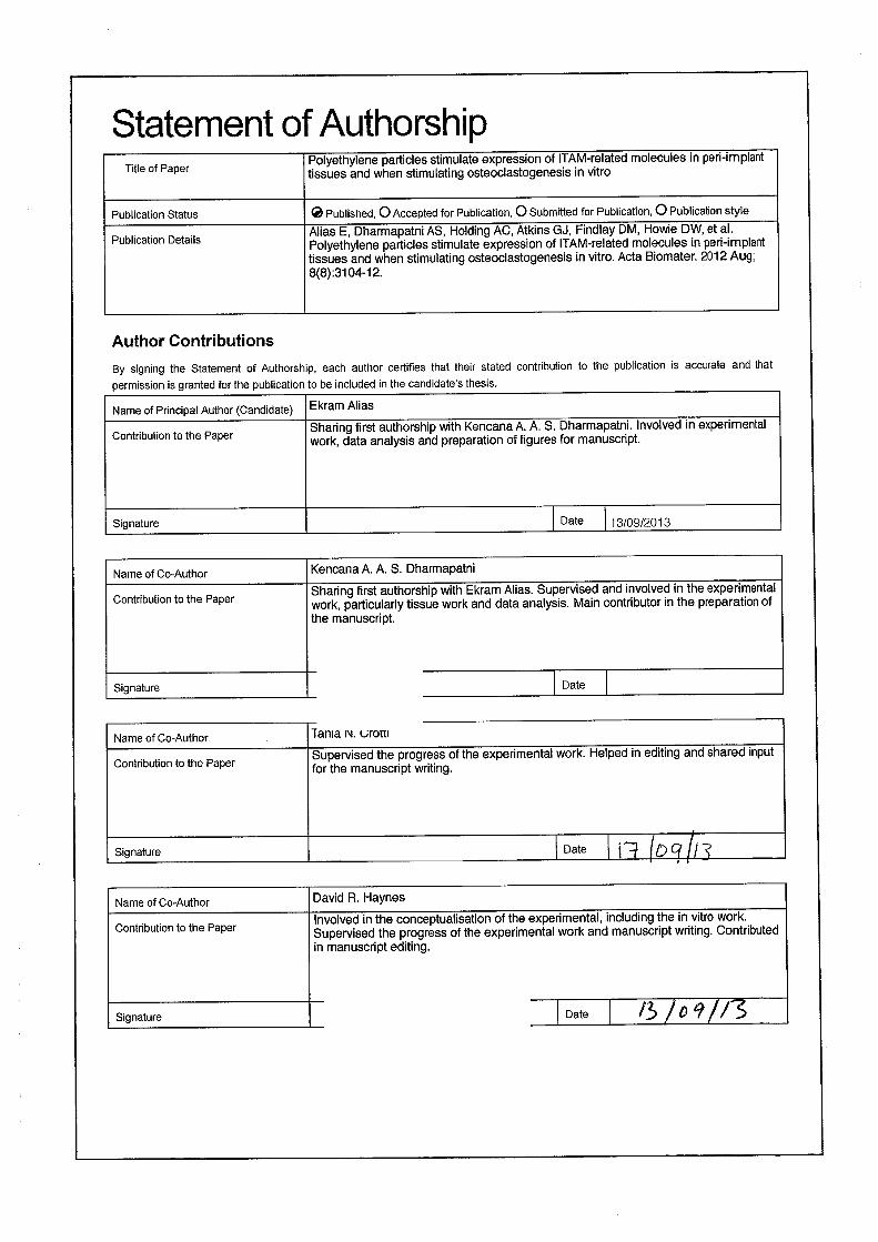
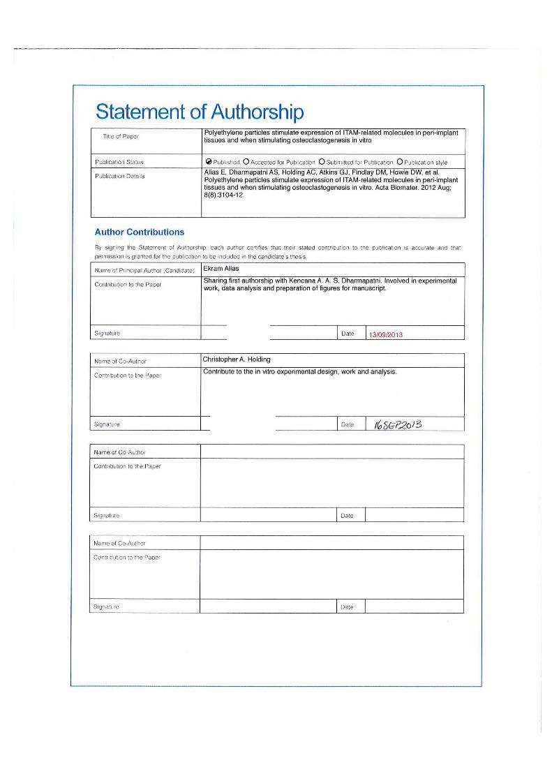


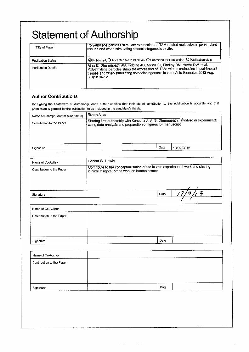
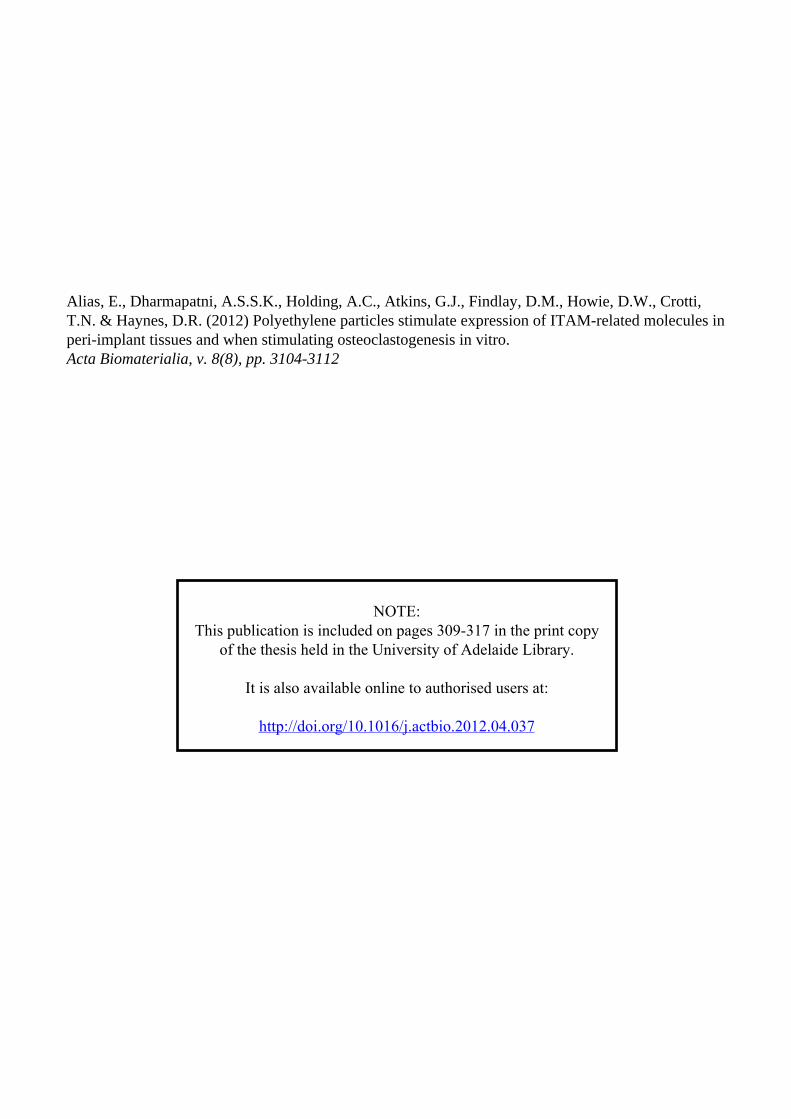
A Alias, E., Dharmapatni, A.S.S.K., Holding, A.C., Atkins, G.J., Findlay, D.M., Howie, D.W., Crotti, T.N. & Haynes, D.R. (2012) Polyethylene particles stimulate expression of ITAM-related molecules in peri-implant tissues and when stimulating osteoclastogenesis in vitro. Acta Biomaterialia, v. 8(8), pp. 3104-3112
NOTE:
This publication is included on pages 309-317 in the print copy of the thesis held in the University of Adelaide Library.
It is also available online to authorised users at:
http://doi.org/10.1016/j.actbio.2012.04.037

RESEARCH ARTICLE Open Access
The immunoreceptor tyrosine-based activationmotif (ITAM) -related factors are increased insynovial tissue and vasculature of rheumatoidarthritic jointsTania N Crotti1*, Anak ASSK Dharmapatni1†, Ekram Alias1,2†, Andrew CW Zannettino3,4, Malcolm D Smith5,6 andDavid R Haynes1
Abstract
Introduction: The immunoreceptor tyrosine-based activation motif (ITAM) pathway provides osteoclast co-stimulatory signals and regulates proliferation, survival and differentiation of effector immune cells. In theosteoclast, the receptors Triggering Receptor Expressed on Myeloid cells 2 (TREM2) and Osteoclast AssociatedReceptor (OSCAR) and their respective adaptor proteins, DAP12 and FcRg mediate ITAM signals and induce calciumsignaling and the crucial transcription factor, NFATc1. In rheumatoid arthritis (RA), OSCAR expression by monocytesis inversely correlated with disease activity. Additionally, serum levels of OSCAR are reduced in RA patients versushealthy controls suggesting that expression and secretion or cleavage of soluble (s) OSCAR is immune modulated.Recent data suggest that endothelial cells may also be a source of OSCAR.
Methods: ITAM receptors, their adaptor proteins, and NFATc1 and cathepsin K were detected in human synovialtissues by immunohistochemistry. Synovial tissues from patients with active RA were compared with tissue frompatients in remission, osteoarthritis (OA) patients and healthy individuals. OSCAR was measured by immunoassay insynovial fluids recovered from active RA and OA patients. Endothelial cells were cultured with or without 5 ng/mLTNF-a or IL-1b over 72 hours. Temporal expression of OSCAR mRNA was assessed by qRT PCR and OSCAR proteinin the supernatant was measured by ELISA.
Results: Significantly higher (P < 0.05) NFATc1-positive inflammatory cell aggregates were found in active RA tissuesthan in healthy synovial tissue. Similarly, the percentage of OSCAR, FcRg, DAP12 and TREM2 positive cells wassignificantly higher in active RA tissues compared to the healthy synovial tissue. Notably, OSCAR was stronglyexpressed in the microvasculature of the active RA tissues (9/9), inactive RA (8/9) weakly in OA (4/9) but only in thelumen of healthy synovial tissue (0/8). OSCAR levels were detected in synovial fluids from both RA (47 to 152 ng/mL)and OA (112 to 145 ng/mL) patients. Moreover, OSCAR mRNA expression and soluble OSCAR release was stimulatedby TNF-a and IL1-b in cultured endothelial cells.
Conclusions: Increased levels of ITAM related factors were present in synovial tissue from active RA jointscompared to OA and healthy joints. OSCAR was strongly expressed by the vasculature of active RA patients andmembrane bound and soluble OSCAR was stimulated by inflammatory mediators in endothelial cells in vitro.
* Correspondence: [email protected]† Contributed equally1Discipline of Anatomy and Pathology, The University of Adelaide, FromeRd., Adelaide, SA 5005, AustraliaFull list of author information is available at the end of the article
Crotti et al. Arthritis Research & Therapy 2012, 14:R245http://arthritis-research.com/content/14/6/R245
© 2012 Crotti et al.; licensee BioMed Central Ltd. This is an open access article distributed under the terms of the Creative CommonsAttribution License (http://creativecommons.org/licenses/by/2.0), which permits unrestricted use, distribution, and reproduction inany medium, provided the original work is properly cited.
IntroductionRheumatoid arthritis (RA) is an autoimmune disease thatinvolves dysregulated immune cell functions. It is charac-terized by joint damage and systemic bone loss associatedwith excessive osteoclast activity [1-4]. Human studiesshow that mature osteoclasts are in close proximity to thebone surface in affected joints of patients with RA [2].Identifying factors that regulate the differentiation andactivity of osteoclasts is crucial in identifying potential tar-gets to abrogate this local and systemic bone loss in RA.A key molecule known to stimulate osteoclast differen-
tiation and activity is receptor activator NF kappa Bligand (RANKL). Elevated RANKL in active RA relativeto its inhibitor osteoprotegerin (OPG) is associated withincreased osteoclast differentiation and resorption [5]. Inactive RA synovial tissue we, and others, have demon-strated that RANKL expression is significantly increasedin lymphocytes and fibroblasts [6,7]. RANKL binds to itsreceptor, RANK, on osteoclast precursors instigating thedifferentiation of monocytes into multinucleated matureosteoclasts via activation of key signaling cascades invol-ving the transcription factor, nuclear factor of activated Tcells (NFATc1) (reviewed by Asagiri et al. [8]).NFATc1 is not only crucial in the regulation of terminal
osteoclast formation, but also plays a role in the immunesystem [8,9]. NFATc1 also regulates T cell differentiationand activation [10], such as that seen in inflammatory dis-eases such as RA. In osteoclasts, NFATc1 directly inducesearly and late stage osteoclast specific gene expression inthe absence of RANKL [9,11-14]. To our knowledge thedistribution of NFATc1 expression in synovial tissue fromactive RA joints has not been previously characterized.Recent studies suggest RANK-RANKL induced osteo-
clastogenesis is enhanced by co-stimulatory signalsmediated by immunoreceptor tyrosine-based activationmotif (ITAM) harboring adaptors [15-17]. ITAM signal-ling is also involved in the regulation of effector immunecells proliferation, survival and differentiation [16,17].DNAX-activating protein 12kDa (DAP12) and Fc recep-tor common g chain (FcRg) are similar ITAM-containingadaptor proteins relevant to osteoclast formation in phy-siological bone turnover [17-19]. In pre-osteoclasts andosteoclasts the ITAM adaptor proteins DAP12 and FcRgassociate with innate immune receptors; in particular,Triggering Receptor Expressed on Myeloid cells 2(TREM2) [15,20-22] and Osteoclast Associated Receptor(OSCAR) [17,23], respectively, to activate calcium, induceNFATc1 and convey ITAM signaling [17].Mice deficient in both DAP12 and FcRg develop severe
osteopetrosis [16] and exhibit an osteoclast multinuclea-tion defect that is restored upon introduction of NFATc1[17]. Evidence suggests DAP12 and TREM2 are requiredfor differentiation and migration of osteoclasts as boneresorption is reduced in osteoclasts derived from mice
with mutations in DAP12 and/or TREM2 in vitro[15,20,21]. In the context of RA, activation of macro-phages by FcRg is reported to induce cartilage destruc-tion independent of inflammation [24]. It was thussuggested that interaction of FcRg with immune com-plexes drives inflammation and induces bone loss indir-ectly [24]. We have recently demonstrated increasedlevels of OSCAR, FcRg, TREM2 and DAP12 in peri-implant tissues [25]. To our knowledge the expression ofFcRg, DAP12 and TREM2 has not yet been demonstratedin human RA.A single-nucleotide polymorphism within the promo-
ter of the OSCAR gene has been linked to increasedrisk of postmenopausal osteoporosis [26]. In RA,OSCAR has been related to disease activity [27] and thepotential of cells to differentiate into osteoclasts [28].These findings support the contention that OSCARplays a role in human bone turn over [26,28,29]. Inosteoclasts the crucial transcription factor, NFATc1,induces OSCAR gene expression [30]. Furthermore,ligand-activated OSCAR interacts with FcRg to producean increase in intracellular calcium [31] that further sti-mulates NFATc1 expression. This establishes a positivefeedback loop that results in marked elevation of bothOSCAR and NFATc1 expression in terminal stages ofosteoclast formation [8]. Administration of an OSCAR-Ig fusion protein inhibits osteoclastogenes in vitro[30,32]. These findings establish OSCAR as not only animportant immune modulator but also a major player inthe regulation of osteoclastogenesis.OSCAR is expressed by osteoclasts as well as dendritic
cells in humans and is involved in antigen presentationand activation of dendritic cells [33,34]. OSCAR has alsobeen reported to be associated with osteoclasts near sitesof erosion and the mononuclear cells adjacent to themicrovasculature in RA patients [28]. Interestingly, morerecent studies have identified OSCAR expression byendothelial cells [35]. This is similar to our observationthat OPG expression and release by endothelial cells isregulated by inflammatory cytokines [36]. However, pro-duction and possible release of soluble OSCAR byendothelial cells in response to inflammatory cytokinespresent in arthritides has not yet been reported.High levels of OSCAR have recently been demon-
strated in the synovial tissues and monocytes isolatedfrom RA patients, with these cells having a greater pro-pensity for differentiation into osteoclasts [28]. Tumornecrosis factor (TNF)-a was found to induce OSCARexpression in monocytes isolated from RA patients [28].However, serum levels of OSCAR were lower in RApatients compared with normal controls. These findingsfurther suggest cell associated and soluble OSCAR isregulated by inflammatory cytokines that play a signifi-cant role in the pathogenesis of RA.
Crotti et al. Arthritis Research & Therapy 2012, 14:R245http://arthritis-research.com/content/14/6/R245
Page 2 of 13
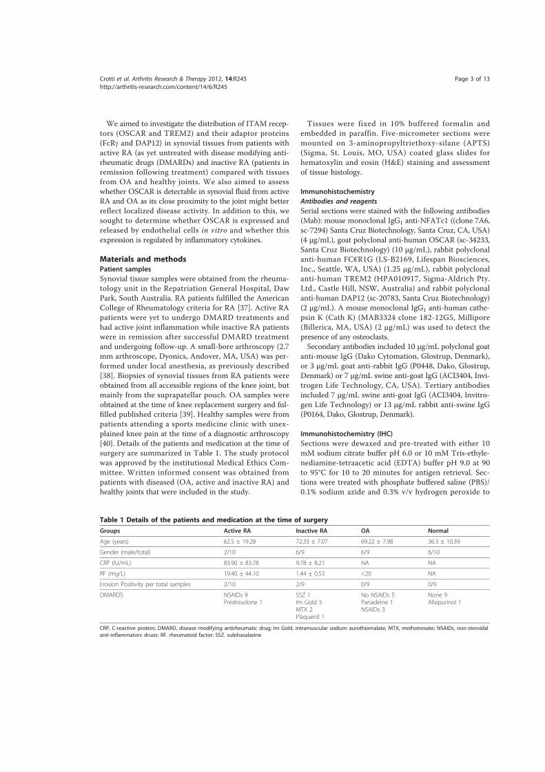
We aimed to investigate the distribution of ITAM recep-tors (OSCAR and TREM2) and their adaptor proteins(FcRg and DAP12) in synovial tissues from patients withactive RA (as yet untreated with disease modifying anti-rheumatic drugs (DMARDs) and inactive RA (patients inremission following treatment) compared with tissuesfrom OA and healthy joints. We also aimed to assesswhether OSCAR is detectable in synovial fluid from activeRA and OA as its close proximity to the joint might betterreflect localized disease activity. In addition to this, wesought to determine whether OSCAR is expressed andreleased by endothelial cells in vitro and whether thisexpression is regulated by inflammatory cytokines.
Materials and methodsPatient samplesSynovial tissue samples were obtained from the rheuma-tology unit in the Repatriation General Hospital, DawPark, South Australia. RA patients fulfilled the AmericanCollege of Rheumatology criteria for RA [37]. Active RApatients were yet to undergo DMARD treatments andhad active joint inflammation while inactive RA patientswere in remission after successful DMARD treatmentand undergoing follow-up. A small-bore arthroscopy (2.7mm arthroscope, Dyonics, Andover, MA, USA) was per-formed under local anesthesia, as previously described[38]. Biopsies of synovial tissues from RA patients wereobtained from all accessible regions of the knee joint, butmainly from the suprapatellar pouch. OA samples wereobtained at the time of knee replacement surgery and ful-filled published criteria [39]. Healthy samples were frompatients attending a sports medicine clinic with unex-plained knee pain at the time of a diagnostic arthroscopy[40]. Details of the patients and medication at the time ofsurgery are summarized in Table 1. The study protocolwas approved by the institutional Medical Ethics Com-mittee. Written informed consent was obtained frompatients with diseased (OA, active and inactive RA) andhealthy joints that were included in the study.
Tissues were fixed in 10% buffered formalin andembedded in paraffin. Five-micrometer sections weremounted on 3-aminopropyltriethoxy-silane (APTS)(Sigma, St. Louis, MO, USA) coated glass slides forhematoxylin and eosin (H&E) staining and assessmentof tissue histology.
ImmunohistochemistryAntibodies and reagentsSerial sections were stained with the following antibodies(Mab): mouse monoclonal IgG1 anti-NFATc1 ((clone 7A6,sc-7294) Santa Cruz Biotechnology, Santa Cruz, CA, USA)(4 μg/mL), goat polyclonal anti-human OSCAR (sc-34233,Santa Cruz Biotechnology) (10 μg/mL), rabbit polyclonalanti-human FC€R1G (LS-B2169, Lifespan Biosciences,Inc., Seattle, WA, USA) (1.25 μg/mL), rabbit polyclonalanti-human TREM2 (HPA010917, Sigma-Aldrich Pty.Ltd., Castle Hill, NSW, Australia) and rabbit polyclonalanti-human DAP12 (sc-20783, Santa Cruz Biotechnology)(2 μg/mL). A mouse monoclonal IgG1 anti-human cathe-psin K (Cath K) (MAB3324 clone 182-12G5, Millipore(Billerica, MA, USA) (2 μg/mL) was used to detect thepresence of any osteoclasts.Secondary antibodies included 10 μg/mL polyclonal goat
anti-mouse IgG (Dako Cytomation, Glostrup, Denmark),or 3 μg/mL goat anti-rabbit IgG (P0448, Dako, Glostrup,Denmark) or 7 μg/mL swine anti-goat IgG (ACI3404, Invi-trogen Life Technology, CA, USA). Tertiary antibodiesincluded 7 μg/mL swine anti-goat IgG (ACI3404, Invitro-gen Life Technology) or 13 μg/mL rabbit anti-swine IgG(P0164, Dako, Glostrup, Denmark).
Immunohistochemistry (IHC)Sections were dewaxed and pre-treated with either 10mM sodium citrate buffer pH 6.0 or 10 mM Tris-ethyle-nediamine-tetraacetic acid (EDTA) buffer pH 9.0 at 90to 95°C for 10 to 20 minutes for antigen retrieval. Sec-tions were treated with phosphate buffered saline (PBS)/0.1% sodium azide and 0.3% v/v hydrogen peroxide to
Table 1 Details of the patients and medication at the time of surgeryGroups Active RA Inactive RA OA Normal
Age (years) 62.5 ± 19.28 72.33 ± 7.07 69.22 ± 7.98 36.3 ± 10.39
Gender (male/total) 2/10 6/9 6/9 6/10
CRP (IU/mL) 83.90 ± 83.78 9.78 ± 8.21 NA NA
RF (mg/L) 19.40 ± 44.10 1.44 ± 0.53 <20 NA
Erosion Positivity per total samples 2/10 2/9 0/9 0/9
DMARDS NSAIDs 9Prednisolone 1
SSZ 1Im Gold 5MTX 2Plaquenil 1
No NSAIDs 5Panadeine 1NSAIDs 3
None 9Allopurinol 1
CRP, C-reactive protein; DMARD, disease modifying antirheumatic drug; Im Gold, intramuscular sodium aurothiomalate; MTX, methotrexate; NSAIDs, non-steroidalanti-inflammatory drugs; RF, rheumatoid factor; SSZ, sulphasalazine
Crotti et al. Arthritis Research & Therapy 2012, 14:R245http://arthritis-research.com/content/14/6/R245
Page 3 of 13
inhibit endogenous peroxidase activity. A three-step per-oxidase-based immunostaining technique, as previouslydescribed [41], with minor modifications, was employed.Sections were incubated with the primary antibodies(concentrations as described above) diluted in PBS and1% bovine serum albumin (BSA) overnight at room tem-perature in a wet chamber. Sections were incubated withthe appropriate HRP-conjugated secondary antibodiesfollowed by incubation with the relevant HRP- conju-gated tertiary swine anti-goat IgG or rabbit anti-swineIgG. HRP activity was detected using hydrogen peroxideas the substrate and 3-amino-9-ethylcarbazole (AEC)(K3469, Dako, Carpinteria, CA, USA) as the dye. Sec-tions were counter-stained with Harris hematoxylin andlithium carbonate and mounted with GurrAquamount(British Drug House, Poole, UK). Negative controlsincluded isotype-matched antibody controls (mouseIgG1kappa for mouse IgG1) and antibody-raised serumfor polyclonal antibody (normal rabbit serum or goatserum) with equivalent concentration to the primaryantibodies.
Semi quantitative scoring analysis (SQA) of IHC resultSections were scanned at high resolution using a Nano-Zoomer (Hamatsu, Shizouka, Japan), Digital Pathology,to enable quantification and archival of the IHC results.Three areas of 2 mm2 were randomly selected and sec-tions were assessed in random order by two blindedobservers. Semi-quantitative assessment (SQA) ofOSCAR, FcRg, DAP12 and TREM2 and Cath K stainingwas scored using a 5-scale (0 to 4) scoring system[25,42]. Assessment was according to the percentage ofpositive stained cells as follows; 0 represented 0 to 5%,1 for 6 to 10%, 2 for 11 to 25%, 3 indicated between 26and 50% and a score of 4 indicated more than 50% ofpositive cells (adapted from [42]) within the sublining ofthe synovial tissue. For NFATc1 immunostaining, thenumber of positive stained cell aggregations (defined asmore than 25 cells) was used as a parameter for grading(adapted from [43]). A score of 0 represented no positivestained cell aggregation, a score of 1 indicated one tothree positive cell aggregations, score of 2 indicated totalpositive cell aggregation between four and six, seven tonine positive cell aggregations gave a score of 3 and ascore of 4 indicated more than nine positive cell aggrega-tions were present. Observations were made as to thepresence of positive cells in the vessels or lining.
Endothelial cell in vitro culturesBone marrow endothelial cells (BMEC) were cultured intriplicate in the presence and absence of 5 ng/mL TNF-aand IL1-b over a 72-hour time period. Expression ofOSCAR and OPG mRNA levels by BMECs were assessedat 0, 6, 12, 24, 48 and 72 hours by quantitative reverse
transcriptase polymerase chain reaction (QRT PCR).Only data from 0, 24, 48 and 72 are presented here.Supernatants were collected at 0, 24, 48 and 72 hours toassess soluble OSCAR and were stored at -20°C until usein ELISA assay. BMECs were also cultured on chamberslides for immunofluorescent detection of OSCAR andOPG.
ELISA analysis of soluble OSCARSynovial fluid was obtained from patients at the rheuma-tology unit in the Repatriation General Hospital, DawPark, South Australia. The patient cohort consisted ofsynovial fluid from active RA (average age 67, CRP 67.4IU/mL and RF 165 mg/L, 7 male/5 female, 4/12 with ero-sion) and OA joints (average age 70, 3 male/5 female).Levels of OSCAR were assessed by ELISA kit followingmanufacturer’s instructions (USCN Life Science, Inc.,Wuhan, China). Samples (synovial fluids and supernatants)were clarified by centrifugation at 13,000 rpm for five min-utes at 4°C. The supernatant was transferred to 1.5 mLeppendorf tubes and diluted 1/5 for synovial fluid and1/10 for supernatant in the sample diluent provided in thekit. One hundred microliters of pre-diluted samples wereloaded into each well along with the protein standardsprovided. Assays were carried out in duplicate and theOSCAR protein concentration in each sample was deter-mined based on the standard curve generated.
RNA extraction and cDNA synthesisTotal RNA was isolated from in vitro BMEC cultures fol-lowing the addition of 500 μL TRIzol reagent per well, asper the manufacturer’s instructions (Invitrogen Life Tech-nologies, Carlsbad, CA, USA). Complementary DNA(cDNA) was synthesized from 1 μg RNA per reactionusing Superscript III Reverse Transcriptase (InvitrogenLife Technologies), as previously described [44].
Quantitative real time reverse-transcription polymerasechain reaction (QRT-PCR)Real-time PCR was performed using Platinum SYBRGreen qPCRSupermix-UDG (Invitrogen Life Technolo-gies), as per the manufacturer’s recommendations. Ampli-fication was carried out in a Rotor-Gene 3000. Reactionmixtures contained 1 μl of 1 in 5 pre-diluted cDNA, 7.5 μlPlatinum SYBR Green qPCR Supermix-UDG, 300 nMeach of forward and reverse primers and diethyl pyrocar-bonate (DEPC)-treated water to a final volume of 15 μl.Primer3Plus freeware [45] was used to design oligonucleo-tide primers to human OSCAR Forward ‘CCC AGC TTCATA CCA CCC TA’ and Reverse: ‘GAA GAG AAG GGGAGC GAT CT’ [46]. Primer sequences for OPG and theendogenous reference gene glyceraldehyde 3-phosphatedehydrogenase (GAPDH) were designed as described pre-viously [47]. All samples were investigated in triplicate
Crotti et al. Arthritis Research & Therapy 2012, 14:R245http://arthritis-research.com/content/14/6/R245
Page 4 of 13
QRTPCR reactions. Fold induction was calculated as ameasure of 2ddCT [48].
Immunofluoresence of BMECBMECs were grown for 40 hours in the absence or pre-sence of 5 ng/mL IL-1b or TNF-a. Cells were fixed with1:1 methanol:acetone for five minutes and washed withPBS. Primary antibodies OPG MAB805 (10 μg/mL) orpolyclonal goat anti-human OSCAR (8 μg/mL, sc-34233,Santa Cruz Biotechnology) were diluted in PBS with 1%BSA and incubated overnight at room temperature. Tocontrol for non-specific staining of the OSCAR antibody,wells were incubated with normal goat serum in theabsence of the antibody to OSCAR. Following washingwith PBS, OSCAR and OPG antibodies were detectedwith secondary antibodies to goat conjugated with FITC(green) and mouse conjugated to cy3 (red) (both fromSouthern Biotechnology Associates, Inc., Birmingham, AL,USA), respectively, diluted in PBS plus 1% BSA for 30 min-utes then washed three times with PBS.
Statistical analysisPower calculations demonstrate sufficient power to detectdifferences between the healthy subjects and joints withactive RA for each of the molecules detected (92.9 to100%). To assess the SQAs assigned by analysis of the IHCstaining statistical analysis was performed using SPSS ver-sion 11.5 (SPSS Inc, Chicago, IL, USA). A non-parametricKruskal-Wallis analysis was used to compare the mean ofthe SQA score between the groups. A Mann-Whitney-Utest was used to examine the significant differencebetween two groups, with a P-value <0.05 accepted asstatistically significant.Differences in the soluble OSCAR levels in synovial
fluids between the two groups were analyzed by Student’st-test and P < 0.05 was considered significant. Statisticalsignificance between treatments and time points wasdetermined using Kruskal-Wallis followed by the Mann-Whitney test using GraphPad Prism version 5.0d (Graph-Pad Software Inc. La Jolla, CA, USA).Differences in mRNA levels between the groups at each
time point were analyzed using a Two-way Anova test.
ResultsExpression of ITAM modulatory factors in active RAWhile NFATc1 expression was localized in isolated cellsthroughout the tissue, NFATc1 was mainly expressed bycells within lymphocyte aggregates in tissues fromuntreated active RA joints (Figure 1). The SQA gradingfor NFATc1 immunostaining was, therefore, based onthe number of cell aggregates expressing NFATc1(described in Methods). The number of NFATc1-positivecell aggregations was found to be significantly more in
the active RA group compared to all other groups (P <0.05) (Table 2).Cathepsin K (Cath K) is routinely used as a marker of
osteoclast-like cells as it is expressed at high levels inosteoclasts [49,50] and is the main bone-matrix degradingenzyme [51,52]. Immunostaining for Cath K was found tobe predominantly in the sublining of the synovium andexpressed by synovial fibroblasts (images not shown). Theproportion of Cath K positive cells was not significantlydifferent between groups.OSCAR has been reported to be increased in RA com-
pared to that in OA tissues [28]. OSCAR expression wasassociated with macrophage-like cells in the sublining ofthe synovium of inactive and active RA patients as wellas in the lining of OA patients (Figure 1). Strongly posi-tive OSCAR staining was associated with the microvascu-lature of the synovium in patients with active RA, nine ofnine (Figure 2A) and with inactive RA (eight of nine)(Figure 2B). OSCAR was weakly expressed in four out ofeight of the tissue samples from OA patients (Figure 2C)and was absent in the microvasculature of the healthy tis-sues (zero of nine) (Figure 2D). Interestingly, OSCARappeared in the lumen of the microvasculature of thesynovium from healthy patients that was suggestive ofcleaved or soluble protein in the serum.TREM2 was highly expressed throughout tissue from
active RA patients (Figure 1). Many types of cellsappeared to express TREM2, including mononuclearcells in lymphoid aggregates and fibroblasts with expres-sion significantly greater than all other groups (Table 2).TREM2 expression was also associated with the micro-vasculature of active and inactive RA patients.FcRg was also highly expressed in active RA tissues
and OA patient tissues (Figure 1). This was significantlyhigher in comparison to the inactive RA and the controltissues (P < 0.05) (Table 2). FcRg was strongly expressedin macrophage-like synovial lining cells particularly inthe OA patients. In active RA, FcRg appeared to beexpressed by macrophage and fibroblast-like cellsthroughout the tissue but was absent in the lymphoidaggregates and not associated with the microvasculature.DAP12 expression was significantly higher in active
RA compared with all other groups (P < 0.05) (Table 2).DAP12 appeared predominantly associated with macro-phage-like cells in the sublining of the synovial tissueand the macrophage-like lining cells of the OA group(Figure 1). Of note, the microvasculature was negativein all groups.
Soluble OSCAR in OA and RA synovial fluidSoluble OSCAR was detected in serum from RApatients and normal individuals by Herman [28], sug-gesting a cleaved or released form of OSCAR. To more
Crotti et al. Arthritis Research & Therapy 2012, 14:R245http://arthritis-research.com/content/14/6/R245
Page 5 of 13
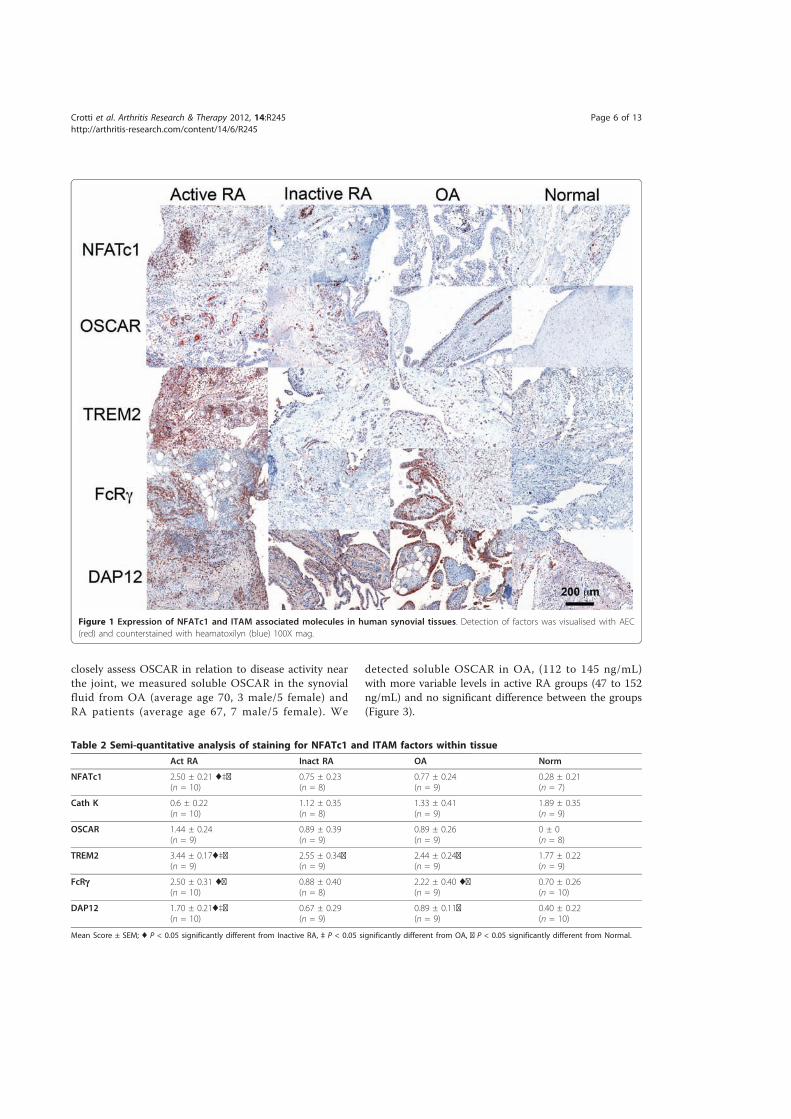
closely assess OSCAR in relation to disease activity nearthe joint, we measured soluble OSCAR in the synovialfluid from OA (average age 70, 3 male/5 female) andRA patients (average age 67, 7 male/5 female). We
detected soluble OSCAR in OA, (112 to 145 ng/mL)with more variable levels in active RA groups (47 to 152ng/mL) and no significant difference between the groups(Figure 3).
Figure 1 Expression of NFATc1 and ITAM associated molecules in human synovial tissues. Detection of factors was visualised with AEC(red) and counterstained with heamatoxilyn (blue) 100X mag.
Table 2 Semi-quantitative analysis of staining for NFATc1 and ITAM factors within tissueAct RA Inact RA OA Norm
NFATc1 2.50 ± 0.21 ♦‡◘(n = 10)
0.75 ± 0.23(n = 8)
0.77 ± 0.24(n = 9)
0.28 ± 0.21(n = 7)
Cath K 0.6 ± 0.22(n = 10)
1.12 ± 0.35(n = 8)
1.33 ± 0.41(n = 9)
1.89 ± 0.35(n = 9)
OSCAR 1.44 ± 0.24(n = 9)
0.89 ± 0.39(n = 9)
0.89 ± 0.26(n = 9)
0 ± 0(n = 8)
TREM2 3.44 ± 0.17♦‡◘(n = 9)
2.55 ± 0.34◘(n = 9)
2.44 ± 0.24◘(n = 9)
1.77 ± 0.22(n = 9)
FcRg 2.50 ± 0.31 ♦◘(n = 10)
0.88 ± 0.40(n = 8)
2.22 ± 0.40 ♦◘(n = 9)
0.70 ± 0.26(n = 10)
DAP12 1.70 ± 0.21♦‡◘(n = 10)
0.67 ± 0.29(n = 9)
0.89 ± 0.11◘(n = 9)
0.40 ± 0.22(n = 10)
Mean Score ± SEM; ♦ P < 0.05 significantly different from Inactive RA, ‡ P < 0.05 significantly different from OA, ◘ P < 0.05 significantly different from Normal.
Crotti et al. Arthritis Research & Therapy 2012, 14:R245http://arthritis-research.com/content/14/6/R245
Page 6 of 13
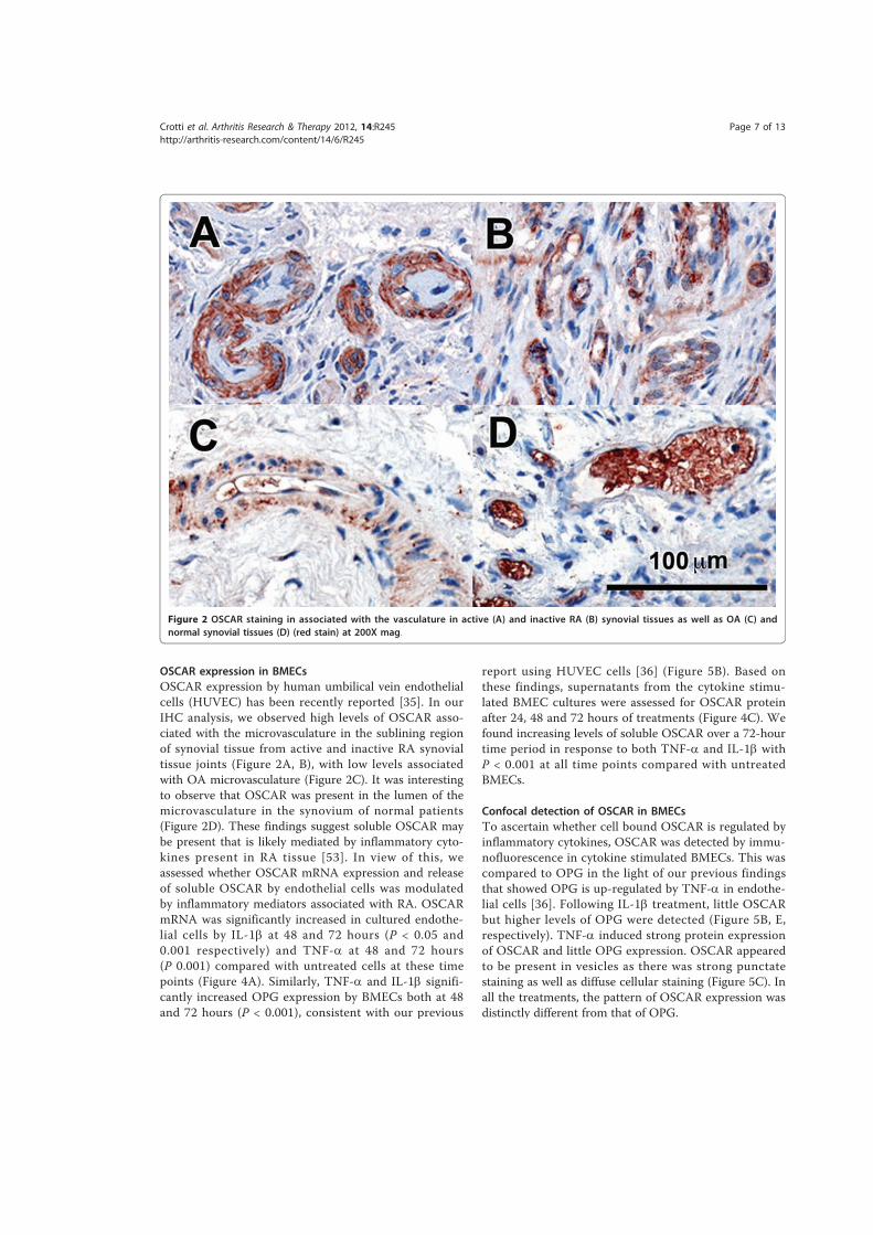
OSCAR expression in BMECsOSCAR expression by human umbilical vein endothelialcells (HUVEC) has been recently reported [35]. In ourIHC analysis, we observed high levels of OSCAR asso-ciated with the microvasculature in the sublining regionof synovial tissue from active and inactive RA synovialtissue joints (Figure 2A, B), with low levels associatedwith OA microvasculature (Figure 2C). It was interestingto observe that OSCAR was present in the lumen of themicrovasculature in the synovium of normal patients(Figure 2D). These findings suggest soluble OSCAR maybe present that is likely mediated by inflammatory cyto-kines present in RA tissue [53]. In view of this, weassessed whether OSCAR mRNA expression and releaseof soluble OSCAR by endothelial cells was modulatedby inflammatory mediators associated with RA. OSCARmRNA was significantly increased in cultured endothe-lial cells by IL-1b at 48 and 72 hours (P < 0.05 and0.001 respectively) and TNF-a at 48 and 72 hours(P 0.001) compared with untreated cells at these timepoints (Figure 4A). Similarly, TNF-a and IL-1b signifi-cantly increased OPG expression by BMECs both at 48and 72 hours (P < 0.001), consistent with our previous
report using HUVEC cells [36] (Figure 5B). Based onthese findings, supernatants from the cytokine stimu-lated BMEC cultures were assessed for OSCAR proteinafter 24, 48 and 72 hours of treatments (Figure 4C). Wefound increasing levels of soluble OSCAR over a 72-hourtime period in response to both TNF-a and IL-1b withP < 0.001 at all time points compared with untreatedBMECs.
Confocal detection of OSCAR in BMECsTo ascertain whether cell bound OSCAR is regulated byinflammatory cytokines, OSCAR was detected by immu-nofluorescence in cytokine stimulated BMECs. This wascompared to OPG in the light of our previous findingsthat showed OPG is up-regulated by TNF-a in endothe-lial cells [36]. Following IL-1b treatment, little OSCARbut higher levels of OPG were detected (Figure 5B, E,respectively). TNF-a induced strong protein expressionof OSCAR and little OPG expression. OSCAR appearedto be present in vesicles as there was strong punctatestaining as well as diffuse cellular staining (Figure 5C). Inall the treatments, the pattern of OSCAR expression wasdistinctly different from that of OPG.
Figure 2 OSCAR staining in associated with the vasculature in active (A) and inactive RA (B) synovial tissues as well as OA (C) andnormal synovial tissues (D) (red stain) at 200X mag.
Crotti et al. Arthritis Research & Therapy 2012, 14:R245http://arthritis-research.com/content/14/6/R245
Page 7 of 13
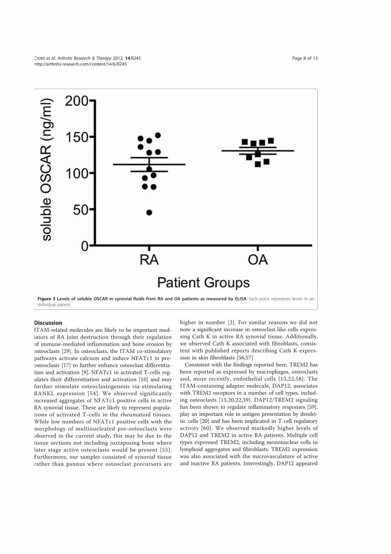
DiscussionITAM-related molecules are likely to be important med-iators of RA joint destruction through their regulationof immune-mediated inflammation and bone erosion byosteoclasts [29]. In osteoclasts, the ITAM co-stimulatorypathways activate calcium and induce NFATc1 in pre-osteoclasts [17] to further enhance osteoclast differentia-tion and activation [9]. NFATc1 in activated T-cells reg-ulates their differentiation and activation [10] and mayfurther stimulate osteoclastogenesis via stimulatingRANKL expression [54]. We observed significantlyincreased aggregates of NFATc1 positive cells in activeRA synovial tissue. These are likely to represent popula-tions of activated T-cells in the rheumatoid tissues.While low numbers of NFATc1 positive cells with themorphology of multinucleated pre-osteoclasts wereobserved in the current study, this may be due to thetissue sections not including juxtaposing bone wherelater stage active osteoclasts would be present [55].Furthermore, our samples consisted of synovial tissuerather than pannus where osteoclast precursors are
higher in number [2]. For similar reasons we did notnote a significant increase in osteoclast-like cells expres-sing Cath K in active RA synovial tissue. Additionally,we observed Cath K associated with fibroblasts, consis-tent with published reports describing Cath K expres-sion in skin fibroblasts [56,57]Consistent with the findings reported here, TREM2 has
been reported as expressed by macrophages, osteoclastsand, more recently, endothelial cells [15,22,58]. TheITAM-containing adapter molecule, DAP12, associateswith TREM2 receptors in a number of cell types, includ-ing osteoclasts [15,20,22,59]. DAP12/TREM2 signalinghas been shown to regulate inflammatory responses [59],play an important role in antigen presentation by dendri-tic cells [20] and has been implicated in T cell regulatoryactivity [60]. We observed markedly higher levels ofDAP12 and TREM2 in active RA patients. Multiple celltypes expressed TREM2, including mononuclear cells inlymphoid aggregates and fibroblasts. TREM2 expressionwas also associated with the microvasculature of activeand inactive RA patients. Interestingly, DAP12 appeared
Figure 3 Levels of soluble OSCAR in synovial fluids from RA and OA patients as measured by ELISA. Each point represents levels in anindividual patient.
Crotti et al. Arthritis Research & Therapy 2012, 14:R245http://arthritis-research.com/content/14/6/R245
Page 8 of 13
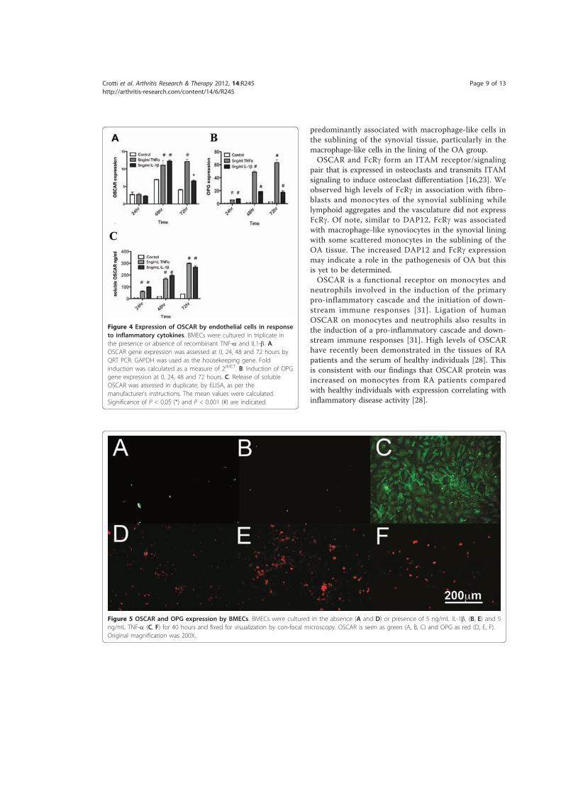
predominantly associated with macrophage-like cells inthe sublining of the synovial tissue, particularly in themacrophage-like cells in the lining of the OA group.OSCAR and FcRg form an ITAM receptor/signaling
pair that is expressed in osteoclasts and transmits ITAMsignaling to induce osteoclast differentiation [16,23]. Weobserved high levels of FcRg in association with fibro-blasts and monocytes of the synovial sublining whilelymphoid aggregates and the vasculature did not expressFcRg. Of note, similar to DAP12, FcRg was associatedwith macrophage-like synoviocytes in the synovial liningwith some scattered monocytes in the sublining of theOA tissue. The increased DAP12 and FcRg expressionmay indicate a role in the pathogenesis of OA but thisis yet to be determined.OSCAR is a functional receptor on monocytes and
neutrophils involved in the induction of the primarypro-inflammatory cascade and the initiation of down-stream immune responses [31]. Ligation of humanOSCAR on monocytes and neutrophils also results inthe induction of a pro-inflammatory cascade and down-stream immune responses [31]. High levels of OSCARhave recently been demonstrated in the tissues of RApatients and the serum of healthy individuals [28]. Thisis consistent with our findings that OSCAR protein wasincreased on monocytes from RA patients comparedwith healthy individuals with expression correlating withinflammatory disease activity [28].
Figure 4 Expression of OSCAR by endothelial cells in responseto inflammatory cytokines. BMECs were cultured in triplicate inthe presence or absence of recombinant TNF-a and IL1-b. A.OSCAR gene expression was assessed at 0, 24, 48 and 72 hours byQRT PCR. GAPDH was used as the housekeeping gene. Foldinduction was calculated as a measure of 2ddCT. B. Induction of OPGgene expression at 0, 24, 48 and 72 hours. C. Release of solubleOSCAR was assessed in duplicate, by ELISA, as per themanufacturer’s instructions. The mean values were calculated.Significance of P < 0.05 (*) and P < 0.001 (#) are indicated.
Figure 5 OSCAR and OPG expression by BMECs. BMECs were cultured in the absence (A and D) or presence of 5 ng/mL IL-1b, (B, E) and 5ng/mL TNF-a (C, F) for 40 hours and fixed for visualization by con-focal microscopy. OSCAR is seen as green (A, B, C) and OPG as red (D, E, F).Original magnification was 200X.
Crotti et al. Arthritis Research & Therapy 2012, 14:R245http://arthritis-research.com/content/14/6/R245
Page 9 of 13
Interestingly, OSCAR expressing monocytes were seenadjacent to microvasculature, consistent with observa-tions by Herman et al. [28]. In a previous study, theexpression of OSCAR was shown in multinucleatedosteoclast-like cells attached to the bone [28]. We couldnot confirm this as the synovial tissue specimens fromthe RA patients used in this study did not include bone[55,61]. However, we did detect positive macrophage-likecells expressing OSCAR in the lining of the OA speci-mens similar to that previously reported [28]. Hermanand colleagues have suggested that OSCAR expressionmight be regulated by pro-inflammatory cytokines [28].Therefore, increased expression of OSCAR in active RAsynovia compared to other groups is likely to be due toelevated inflammatory cytokines present in RA synovialtissues.A possible limitation of the study is that both groups
were not perfectly age- and sex-matched. It is very difficultto age- and sex-match the RA, OA and normal tissues dueto the nature of the joint diseases affecting predominantlydifferent age groups and sexes. In addition, patients pre-senting with active RA can take several years of treatmentbefore the disease is in remission and classified as inactive.While to our knowledge there is are no reports thatOSCAR expression changes with age, it is possible the dif-ferences in age may influence the results of our study.It is possible that soluble OSCAR may act as a decoy
receptor and suppress ITAM signaling. Of particularinterest is the observation that RA patients had reducedlevels of soluble OSCAR in serum compared to healthyindividuals [28]. However, we noted high levels ofOSCAR in the synovial fluid of our active RA patientsand OA patients. A possible reason for this is that in theHerman et al. study patients with RA were being treatedwith anti-TNF therapy, unlike our active RA patients. Itis also possible that levels in the joint locally are notnecessarily reflected systemically in the circulation. Thelack of significant differences between soluble OSCAR inOA and RA patients may be due to the low sample num-ber or may reflect the increased release of OSCAR inresponse to inflammatory cytokines present in both ofthese pathologies [38,53].High levels of OSCAR were associated with the micro-
vasculature of the synovium of active RA specimens aswell as tissues taken from our inactive RA patient group,who have had successful DMARD treatments. In thesynovial tissue of healthy joints, positive OSCAR stainingwas present only in the lumen of the vasculature, sug-gesting the presence of soluble OSCAR in the blood.These observations were consistent with the recent find-ing that human primary endothelial cells express OSCAR[35]. Contrary to our findings, Herman et al. did notobserve OSCAR staining of the microvasculature of hisRA patients. This again may be due to the fact that,
unlike our active RA patients, the patients in the Hermanstudy were being treated with anti-TNF therapy.The increased expression of OSCAR associated with
the microvasculature in RA compared with normal tis-sue suggests that OSCAR expression is modulated byimmune mediators or cytokines. Our previous studieshave shown a reverse pattern of OPG expression in thevasculature, with low OPG levels associated with RA,compared with high levels associated with normal tissue[41]. Additionally, our in vitro studies showed that OPGexpression in HUVEC is regulated by cytokines [36].Our in vitro studies extend the findings by Goettsch etal. [35] and demonstrate that the expression of OSCARby endothelial cells is stimulated by the inflammatorycytokines, TNF-a and IL-b, and is consistent with ourin vivo findings showing OSCAR expression was ele-vated in the untreated RA synovial vasculature. Impor-tantly, endothelial cells are a likely source of OSCARand not just binding soluble OSCAR present in theserum, as we demonstrated that the inflammatory cyto-kines stimulated both mRNA and protein in vitro. TNF-a also induces OSCAR expression in monocytes in vitro[28]. This indicates that soluble OSCAR in the serum[28] and synovial fluid in RA is released by a variety ofcells and is stimulated by inflammatory cytokines.As recombinant human OSCAR-Fc is able to act as a
decoy receptor for cell bound OSCAR [28], solubleOSCAR might provide a protective mechanism againstbone erosion in vivo by competing with a ligand for cellu-lar OSCAR and reducing signaling for inflammatory cellsand osteoclasts. We propose that successful treatment ofRA results in increased cleavage of OSCAR resulting inincreased soluble OSCAR levels [27,28]. This is consistentwith the observation that patients in remission have higherlevels of soluble OSCAR and suggests a role in modulatingosteoclastic bone resorption.While we were able to demonstrate OSCAR expression
in RA and OA synovial fluids for the first time, our dataon the relative levels suggest that it is not a good discrimi-nator between RA and OA. The data of Herman [28] andZhao [27], who measured blood levels, suggest blood levelsare lower in active RA compared to inactive disease. How-ever, induction of OSCAR release in vitro and the levels ofexpression in the tissue indicate its expression, at leastlocally, may be related to disease activity. Further studiesfollowing patients during treatment may help resolvewhether it is a marker for assessing disease activity andjoint erosion in RA.
ConclusionsHere we present evidence that NFATc1 and the ITAM-related molecules FcRg, TREM2, DAP12 and NFATc1 areup-regulated in active RA synovia in comparison tohealthy control or inactive RA and OA tissues. In addition
Crotti et al. Arthritis Research & Therapy 2012, 14:R245http://arthritis-research.com/content/14/6/R245
Page 10 of 13
to this, we find higher proportions of OSCAR positive ves-sels in both active RA patients and inactive RA patientscompared with healthy controls. Our in vitro studies alsoconfirm OSCAR expression by endothelial cells anddemonstrate regulation of production and release of solu-ble protein by inflammatory cytokines, IL-1b and TNF-a.Of interest, we were able to demonstrate OSCAR expres-sion in RA and OA synovial fluids for the first time.
AbbreviationsAEC: 3-amino-9-ethylcarbazole; APTS: 3-aminopropyltriethoxy-silane; BMEC:bone marrow endothelial cells; BSA: bovine serum albumin; Cath K:cathepsin K; DAP12: DNAX-activating protein 12kDa; DEPC: diethylpyrocarbonate; DMARDs: disease-modifying antirheumatic drugs; EDTA:ethylenediamine-tetraacetic acid; FcRγ: Fc receptor common γ chain;GAPDH: glyceraldehyde 3-phosphate dehydrogenase; H&E: hematoxylin andeosin; HRP: horse-radish peroxidase; HUVEC: human umbilical veinendothelial cells; IHC: immunohistochemistry; ITAM: immunoreceptortyrosine-based activation motif; NFATc1: nuclear factor of activated T cells;OA: osteoarthritis; OPG: osteoprotegerin; OSCAR: osteoclast associatedreceptor; PBS: phosphate-buffered saline; RA: rheumatoid arthritis; RANKL:receptor activator NFkappa B ligand; SQA: semi-quantitative assessment;TREM2: triggering receptor expressed on myeloid cells 2.
AcknowledgementsThe authors are grateful to Helen Weedon, Repatriation General Hospital,Adelaide, for sourcing the tissue and synovial samples from the patients andproviding the relevant clinical information. The authors gratefullyacknowledge Ghafar Sarvestani, Detmold Imaging Facility IMVS, Adelaide, forassistance with Nanozoomer Digital Pathology and Dale Caville, Discipline ofAnatomy and Pathology, School of Medical Sciences, The University ofAdelaide, for assistance with photography. This work was funded by TheNational Health and Medical Research Council of Australia. Ekram Alias wassupported by Adelaide Fee Scholarship International of the University ofAdelaide and the Malaysian Ministry of Higher Education.
Author details1Discipline of Anatomy and Pathology, The University of Adelaide, FromeRd., Adelaide, SA 5005, Australia. 2Department of Biochemistry, Faculty ofMedicine, National University of Malaysia, 50300 Kuala Lumpur, Malaysia.3Myeloma Research Laboratory, Centre for Cancer Biology, SA Pathology,Frome Rd, Adelaide, SA 5005, Australia. 4Discipline of Physiology, TheUniversity of Adelaide, Frome Rd., Adelaide, SA 5005, Australia. 5Departmentof Medicine, Flinders Medical Centre, Flinders Drive, Bedford Park, SA 5042,Australia. 6Repatriation General Hospital, Daws Rd., Daw Park, SA 5041,Australia.
Authors’ contributionsTC, EA and AD made substantial contributions to conception of the study,the design of experiments, the acquisition of data, data analysis andinterpretation of data. TC, EA, AD, AZ MS and DH made substantialcontributions to conception of the study, the interpretation of data andwere involved in drafting the manuscript. All authors have read andapproved the manuscript for publication.
Competing interestsThe authors declare that they have no competing interests.
Received: 4 May 2012 Revised: 14 September 2012Accepted: 6 November 2012 Published: 12 November 2012
References1. Goldring SR, Gravallese EM: Pathogenesis of bone erosions in rheumatoid
arthritis. Curr Opin Rheumatol 2000, 12:195-199.2. Gravallese EM, Harada Y, Wang JT, Gorn AH, Thornhill TS, Goldring SR:
Identification of cell types responsible for bone resorption in
rheumatoid arthritis and juvenile rheumatoid arthritis. Am J Pathol 1998,152:943-951.
3. Suzuki Y, Nishikaku F, Nakatuka M, Koga Y: Osteoclast-like cells in murinecollagen induced arthritis. J Rheumatol 1998, 25:1154-1160.
4. Walsh NC, Crotti TN, Goldring SR, Gravallese EM: Rheumatic diseases: theeffects of inflammation on bone. Immunol Rev 2005, 208:228-251.
5. Haynes DR, Crotti TN, Loric M, Bain GI, Atkins GJ, Findlay DM:Osteoprotegerin and receptor activator of nuclear factor kappaB ligand(RANKL) regulate osteoclast formation by cells in the human rheumatoidarthritic joint. Rheumatology 2001, 40:623-630.
6. Crotti TN, Smith MD, Weedon H, Ahern MJ, Findlay DM, Kraan M, Tak PP,Haynes DR: Receptor activator NF-kappaB ligand (RANKL) expression insynovial tissue from patients with rheumatoid arthritis,spondyloarthropathy, osteoarthritis, and from normal patients:semiquantitative and quantitative analysis. Ann Rheum Dis 2002,61:1047-1054.
7. Pettit AR, Walsh NC, Manning C, Goldring SR, Gravallese EM: RANKL proteinis expressed at the pannus-bone interface at sites of articular boneerosion in rheumatoid arthritis. Rheumatology 2006, 45:1068-1076.
8. Asagiri M, Takayanagi H: The molecular understanding of osteoclastdifferentiation. Bone 2007, 40:251-264.
9. Takayanagi H, Kim S, Koga T, Nishina H, Isshiki M, Yoshida H, Saiura A,Isobe M, Yokochi T, Inoue J, Wagner EF, Mak TW, Kodama T, Taniguchi T:Induction and activation of the transcription factor NFATc1 (NFAT2)integrate RANKL signaling in terminal differentiation of osteoclasts. DevCell 2002, 3:889-901.
10. Pan M, Winslow MM, Chen L, Kuo A, Felsher D, Crabtree GR: EnhancedNFATc1 nuclear occupancy causes T cell activation independent ofCD28 costimulation. J Immunol 2007, 178:4315-4321.
11. Crotti TN, Flannery M, Walsh NC, Fleming JD, Goldring SR, McHugh KP:NFATc1 regulation of the human beta3 integrin promoter in osteoclastdifferentiation. Gene 2006, 372:92-102.
12. Crotti TN, Sharma SM, Fleming JD, Flannery MR, Ostrowski MC, Goldring SR,McHugh KP: PU.1 and NFATc1 mediate osteoclastic induction of themouse beta3 integrin promoter. J Cell Physiol 2008, 215:636-644.
13. Matsuo K, Irie N: Osteoclast-osteoblast communication. Arch BiochemBiophys 2008, 473:201-209.
14. Kim K, Kim JH, Lee J, Jin HM, Lee SH, Fisher DE, Kook H, Kim KK, Choi Y,Kim N: Nuclear factor of activated T cells c1 induces osteoclast-associated receptor gene expression during tumor necrosis factor-related activation-induced cytokine-mediated osteoclastogenesis. J BiolChem 2005, 280:35209-35216.
15. Humphrey MB, Daws MR, Spusta SC, Niemi EC, Torchia JA, Lanier LL,Seaman WE, Nakamura MC: TREM2, a DAP12-associated receptor,regulates osteoclast differentiation and function. J Bone Miner Res 2006,21:237-245.
16. Mocsai A, Humphrey MB, Van Ziffle JA, Hu Y, Burghardt A, Spusta SC,Majumdar S, Lanier LL, Lowell CA, Nakamura MC: The immunomodulatoryadapter proteins DAP12 and Fc receptor gamma-chain (FcRgamma)regulate development of functional osteoclasts through the Syk tyrosinekinase. Proc Natl Acad Sci USA 2004, 101:6158-6163.
17. Koga T, Inui M, Inoue K, Kim S, Suematsu A, Kobayashi E, Iwata T, Ohnishi H,Matozaki T, Kodama T, Taniguchi T, Takayanagi H, Takai T: Costimulatorysignals mediated by the ITAM motif cooperate with RANKL for bonehomeostasis. Nature 2004, 428:758-763.
18. Humphrey MB, Lanier LL, Nakamura MC: Role of ITAM-containing adapterproteins and their receptors in the immune system and bone. ImmunolRev 2005, 208:50-65.
19. Nakashima T, Takayanagi H: Osteoimmunology: crosstalk between theimmune and bone systems. J Clin Immunol 2009, 29:555-567.
20. Bouchon A, Hernandez-Munain C, Cella M, Colonna M: A DAP12-mediatedpathway regulates expression of CC chemokine receptor 7 andmaturation of human dendritic cells. J Exp Med 2001, 194:1111-1122.
21. Paloneva J, Mandelin J, Kiialainen A, Bohling T, Prudlo J, Hakola P, Haltia M,Konttinen YT, Peltonen L: DAP12/TREM2 deficiency results in impairedosteoclast differentiation and osteoporotic features. J Exp Med 2003,198:669-675.
22. Daws MR, Lanier LL, Seaman WE, Ryan JC: Cloning and characterization ofa novel mouse myeloid DAP12-associated receptor family. Eur J Immunol2001, 31:783-791.
Crotti et al. Arthritis Research & Therapy 2012, 14:R245http://arthritis-research.com/content/14/6/R245
Page 11 of 13
23. Ishikawa S, Arase N, Suenaga T, Saita Y, Noda M, Kuriyama T, Arase H,Saito T: Involvement of FcRgamma in signal transduction of osteoclast-associated receptor (OSCAR). Int immunol 2004, 16:1019-1025.
24. van Lent PL, Grevers L, Lubberts E, de Vries TJ, Nabbe KC, Verbeek S,Oppers B, Sloetjes A, Blom AB, van den Berg WB: Fcgamma receptorsdirectly mediate cartilage, but not bone, destruction in murine antigen-induced arthritis: uncoupling of cartilage damage from bone erosionand joint inflammation. Arthritis Rheum 2006, 54:3868-3877.
25. Alias E, Dharmapatni AS, Holding AC, Atkins GJ, Findlay DM, Howie DW,Crotti TN, Haynes DR: Polyethylene particles stimulate expression ofITAM-related molecules in peri-implant tissues and when stimulatingosteoclastogenesis in vitro. Acta Biomater 2012, 8:3104-3112.
26. Kim GS, Koh JM, Chang JS, Park BL, Kim LH, Park EK, Kim SY, Shin HD:Association of the OSCAR promoter polymorphism with BMD inpostmenopausal women. J Bone Miner Res 2005, 20:1342-1348.
27. Zhao S, Guo YY, Ding N, Yang LL, Zhang N: Changes in serum levels ofsoluble osteoclast-associated receptor in human rheumatoid arthritis.Chin Med J (Engl) 2011, 124:3058-3060.
28. Herman S, Muller RB, Kronke G, Zwerina J, Redlich K, Hueber AJ, Gelse H,Neumann E, Muller-Ladner U, Schett G: Induction of osteoclast-associatedreceptor, a key osteoclast costimulation molecule, in rheumatoidarthritis. Arthritis Rheum 2008, 58:3041-3050.
29. Nemeth K, Schoppet M, Al-Fakhri N, Helas S, Jessberger R, Hofbauer LC,Goettsch C: The role of osteoclast-associated receptor inosteoimmunology. J Immunol 2010, 186:13-18.
30. Kim Y, Sato K, Asagiri M, Morita I, Soma K, Takayanagi H: Contribution ofnuclear factor of activated T cells c1 to the transcriptional control ofimmunoreceptor osteoclast-associated receptor but not triggeringreceptor expressed by myeloid cells-2 during osteoclastogenesis. J BiolChem 2005, 280:32905-32913.
31. Merck E, Gaillard C, Scuiller M, Scapini P, Cassatella MA, Trinchieri G,Bates EE: Ligation of the FcR gamma chain-associated human osteoclast-associated receptor enhances the proinflammatory responses of humanmonocytes and neutrophils. J Immunol 2006, 176:3149-3156.
32. Kim N, Takami M, Rho J, Josien R, Choi Y: A novel member of theleukocyte receptor complex regulates osteoclast differentiation. J ExpMed 2002, 195:201-209.
33. Merck E, Gaillard C, Gorman DM, Montero-Julian F, Durand I, Zurawski SM,Menetrier-Caux C, Carra G, Lebecque S, Trinchieri G, Bates EE: OSCAR is anFcRgamma-associated receptor that is expressed by myeloid cells and isinvolved in antigen presentation and activation of human dendriticcells. Blood 2004, 104:1386-1395.
34. Merck E, de Saint-Vis B, Scuiller M, Gaillard C, Caux C, Trinchieri G, Bates EE:Fc receptor gamma-chain activation via hOSCAR induces survival andmaturation of dendritic cells and modulates Toll-like receptor responses.Blood 2005, 105:3623-3632.
35. Goettsch C, Rauner M, Sinningen K, Helas S, Al-Fakhri N, Nemeth K,Hamann C, Kopprasch S, Aikawa E, Bornstein SR, Schoppet M, Hofbauer LC:The osteoclast-associated receptor (OSCAR) is a novel receptorregulated by oxidized low-density lipoprotein in human endothelialcells. Endocrinology 2011, 152:4915-4926.
36. Zannettino AC, Holding CA, Diamond P, Atkins GJ, Kostakis P, Farrugia A,Gamble J, To LB, Findlay DM, Haynes DR: Osteoprotegerin (OPG) islocalized to the Weibel-Palade bodies of human vascular endothelialcells and is physically associated with von Willebrand factor. J Cell Physiol2005, 204:714-723.
37. Arnett FC, Edworthy SM, Bloch DA, McShane DJ, Fries JF, Cooper NS,Healey LA, Kaplan SR, Liang MH, Luthra HS, et al: The AmericanRheumatism Association 1987 revised criteria for the classification ofrheumatoid arthritis. Arthritis Rheum 1988, 31:315-324.
38. Smith MD, Triantafillou S, Parker A, Youssef PP, Coleman M: Synovialmembrane inflammation and cytokine production in patients with earlyosteoarthritis. J Rheumatol 1997, 24:365-371.
39. Altman R, Asch E, Bloch D, Bole G, Borenstein D, Brandt K, Christy W,Cooke TD, Greenwald R, Hochberg M, et al: Development of criteria forthe classification and reporting of osteoarthritis. Classification ofosteoarthritis of the knee. Diagnostic and Therapeutic CriteriaCommittee of the American Rheumatism Association. Arthritis Rheum1986, 29:1039-1049.
40. Smith MD, Barg E, Weedon H, Papengelis V, Smeets T, Tak PP, Kraan M,Coleman M, Ahern MJ: Microarchitecture and protective mechanisms in
synovial tissue from clinically and arthroscopically normal knee joints.Ann Rheum Dis 2003, 62:303-307.
41. Haynes DR, Barg E, Crotti TN, Holding C, Weedon H, Atkins GJ, Zannetino A,Ahern MJ, Coleman M, Roberts-Thomson PJ, Kraan M, Tak PP, Smith MD:Osteoprotegerin expression in synovial tissue from patients withrheumatoid arthritis, spondyloarthropathies and osteoarthritis andnormal controls. Rheumatology 2003, 42:123-134.
42. Kraan MC, Haringman JJ, Post WJ, Versendaal J, Breedveld FC, Tak PP:Immunohistological analysis of synovial tissue for differential diagnosisin early arthritis. Rheumatology (Oxford) 1999, 38:1074-1080.
43. Tak PP, van der Lubbe PA, Cauli A, Daha MR, Smeets TJ, Kluin PM,Meinders AE, Yanni G, Panayi GS, Breedveld FC: Reduction of synovialinflammation after anti-CD4 monoclonal antibody treatment in earlyrheumatoid arthritis. Arthritis Rheum 1995, 38:1457-1465.
44. Dharmapatni AA, Smith MD, Crotti TN, Holding CA, Vincent C, Weedon HM,Zannettino AC, Zheng TS, Findlay DM, Atkins GJ, Haynes DR: TWEAK andFn14 expression in the pathogenesis of joint inflammation and boneerosion in rheumatoid arthritis. Arthritis Res Ther 2011, 13:R51.
45. Primer3Plus. [http://primer3plus.com/cgi-bin/dev/primer3plus.cgi].46. Zawawi MS, Dharmapatni AA, Cantley MD, McHugh KP, Haynes DR,
Crotti TN: Regulation of ITAM adaptor molecules and their receptors byinhibition of calcineurin-NFAT signalling during late stage osteoclastdifferentiation. Biochem Biophys Res Commun 2012, 427:404-409.
47. Atkins GJ, Welldon KJ, Holding CA, Haynes DR, Howie DW, Findlay DM: Theinduction of a catabolic phenotype in human primary osteoblasts andosteocytes by polyethylene particles. Biomaterials 2009, 30:3672-3681.
48. Livak KJ, Schmittgen TD: Analysis of relative gene expression data usingreal-time quantitative PCR and the 2(-Delta Delta C(T)) Method. Methods2001, 25:402-408.
49. Troen BR: Molecular mechanisms underlying osteoclast formation andactivation. Exp Gerontol 2003, 38:605-614.
50. Corisdeo S, Gyda M, Zaidi M, Moonga BS, Troen BR: New insights into theregulation of cathepsin K gene expression by osteoprotegerin ligand.Biochem Biophys Res Commun 2001, 285:335-339.
51. Gowen M, Lazner F, Dodds R, Kapadia R, Feild J, Tavaria M, Bertoncello I,Drake F, Zavarselk S, Tellis I, Hertzog P, Debouck C, Kola I: Cathepsin Kknockout mice develop osteopetrosis due to a deficit in matrixdegradation but not demineralization. J Bone Miner Res 1999,14:1654-1663.
52. Saftig P, Hunziker E, Wehmeyer O, Jones S, Boyde A, Rommerskirch W,Moritz JD, Schu P, von Figura K: Impaired osteoclastic bone resorptionleads to osteopetrosis in cathepsin-K-deficient mice. Proc Natl Acad SciUSA 1998, 95:13453-13458.
53. Youssef PP, Triantafillou S, Parker A, Coleman M, Roberts-Thomson PJ,Ahern MJ, Smith MD: Variability in cytokine and cell adhesion moleculestaining in arthroscopic synovial biopsies: quantification using colorvideo image analysis. J Rheumatol 1997, 24:2291-2298.
54. Kotake S, Udagawa N, Hakoda M, Mogi M, Yano K, Tsuda E, Takahashi K,Furuya T, Ishiyama S, Kim KJ, Saito S, Nishikawa T, Takahashi N, Togari A,Tomatsu T, Suda T, Kamatani N: Activated human T cells directly induceosteoclastogenesis from human monocytes: possible role of T cells inbone destruction in rheumatoid arthritis patients. Arthritis Rheum 2001,44:1003-1012.
55. Shen Z, Crotti TN, McHugh KP, Matsuzaki K, Gravallese EM, Bierbaum BE,Goldring SR: The role played by cell-substrate interactions in thepathogenesis of osteoclast-mediated peri-implant osteolysis. Arthritis ResTher 2006, 8:R70.
56. Quintanilla-Dieck MJ, Codriansky K, Keady M, Bhawan J, Runger TM:Expression and regulation of cathepsin K in skin fibroblasts. ExpDermatol 2009, 18:596-602.
57. Runger TM, Quintanilla-Dieck MJ, Bhawan J: Role of cathepsin K in theturnover of the dermal extracellular matrix during scar formation. JInvest Dermatol 2007, 127:293-297.
58. Chen LC, Laskin JD, Gordon MK, Laskin DL: Regulation of TREM expressionin hepatic macrophages and endothelial cells during acuteendotoxemia. Exp Mol Pathol 2008, 84:145-155.
59. Hamerman JA, Jarjoura JR, Humphrey MB, Nakamura MC, Seaman WE,Lanier LL: Cutting edge: inhibition of TLR and FcR responses inmacrophages by triggering receptor expressed on myeloid cells (TREM)-2 and DAP12. J Immunol 2006, 177:2051-2055.
Crotti et al. Arthritis Research & Therapy 2012, 14:R245http://arthritis-research.com/content/14/6/R245
Page 12 of 13
60. Goronzy JJ, Henel G, Sawai H, Singh K, Lee EB, Pryshchep S, Weyand CM:Costimulatory pathways in rheumatoid synovitis and T-cell senescence.Ann N Y Acad Sci 2005, 1062:182-194.
61. Crotti TN, O’Sullivan RP, Shen Z, Flannery MR, Fajardo RJ, Ross FP,Goldring SR, McHugh KP: Bone matrix regulates osteoclast differentiationand annexin A8 gene expression. J Cell Physiol 2011, 226:3413-3421.
doi:10.1186/ar4088Cite this article as: Crotti et al.: The immunoreceptor tyrosine-basedactivation motif (ITAM) -related factors are increased in synovial tissueand vasculature of rheumatoid arthritic joints. Arthritis Research & Therapy2012 14:R245.
Submit your next manuscript to BioMed Centraland take full advantage of:
Convenient online submission
Thorough peer review
No space constraints or color figure charges
Immediate publication on acceptance
Inclusion in PubMed, CAS, Scopus and Google Scholar
Research which is freely available for redistribution
Submit your manuscript at www.biomedcentral.com/submit
Crotti et al. Arthritis Research & Therapy 2012, 14:R245http://arthritis-research.com/content/14/6/R245
Page 13 of 13