
Impact of CD39 and purinergic signalling on the growth and metastasis of colorectal cancer

�1
Cover

�2
Summary
This dissertation takes an in depth look at the phosphoinositide pathway and the roles of the
different phosphoinositides in normal physiology. There are a broad range of physiological
roles within normal tissues which the phosphoinositides can act through. These roles are fo-
cussed around control of growth, cell cycle and apoptosis, which are aspects of cellular func-
tion which are commonly dysregulated in cancer physiology. Unsurprisingly, then, there are
multiple mutations found within this pathway in a range of different cancer types. A large pro-
portion of these do cause changes in cell growth, but this dissertation also shows that alter-
ations in the regulation of phosphoinositides can also result in various different hallmarks of
cancer. These range from potential for metastasis, angiogenesis, chromosomal instability
and dysregulation of metabolism. Evolutionary, it seems advantageous for cancers to acquire
mutations that can cause a gain in a number of these different aspects of carcinogenesis.
Furthering this, recent research has alluded to a role of phosphoinositides in resistance to
therapy in cancer. Loss of function of suppressors of this pathway is a common way through
which resistance to treatment is gained. Combinational therapy, where multiple drugs are
used to target various cellular aberrations, could hold the key to treating these patients who
have resistance to therapy. Therefore, research into understanding these mechanisms of re-
sistance is imperative. Also, the advent of new technologies which allow genetic screening of
mutations for resistance can help produce targeted approaches to therapy. This could poten-
tially open the gateway to personalised medicine.

�3
Title Page
The Role of Phosphoinositide
Signalling in Cancer

�4
Contents Page
Cover 1
Summary 2
Title Page 3
Contents Page 4
Introduction 5
Generation of phosphoinositides 5
Signalling downstream of phosphoinositides 9
Biological functions and role in normal physiology 13
Common mutations and their effects 15
Role in cancer 21
Future perspectives and conclusions 23
Glossary 26
Bibliography 28

�5
Introduction
Phosphoinositides, in their various degrees of phosphorylation on the 3’, 4’ and 5’ positions
of inositol (PI(3)P, PI(4)P, PI(5)P, PI(3,4)P2, PI(3,5)P2, PI(4,5)P2 and PI(3,4,5)P3) are fun-
damental to a range of cellular processes. They act as secondary messengers within cells to
transfer extracellular information inside, as well as messengers within cells. It has been
shown that multiple different proteins interact, with varying specificities, to phosphoinositides.
These proteins have been shown to have significance in cellular growth, cell adhesion, cell
motility and others.
There has been a shift in research into this pathway after it was revealed to influence the
progress of diseases such as cancer and diabetes. Through understanding the role of these
messengers within normal cells and the mechanistic pathways, the roles of aberrations can
be fully assessed and understood. Furthering this, it provides a platform for rational drug de-
sign and allows for understanding of mechanisms of resistance and toxicity. It also highlights
the potential personalised medicine can have within cancer.
This dissertation focuses on the relevance of phosphoinositides within cancer, with an em-
phasis on how mutations within proteins cause aberrations in the cellular levels of phospho-
inositides and relating these to their function within cancer.
Generation of phosphoinositides
The crux of the phosphoinositide signalling cascade is the phosphorylation and dephospho-
rylation of these molecules at differing positions, which is a fluid process allowing for rapid
propagation and termination of intracellular signals (Sasaki et al, 2009). Phosphoinositides
are amphiphilic molecules (neutral long fatty acid tails, and highly negatively charged phos-
phate heads), which reside within the membranes of the cells. The degree of phosphorylation

�6
is dependent upon the spatial arrangement within the different cellular compartments of the
different kinases and phosphatases (figure 1 and figure 3). The proteins that carry out these
reactions require multiple different stimuli to become recruited and activated.
Figure 1: Table depicting the different kinases (reaction arrows in black) and phos-
phatases (reaction arrows in grey), their locations within cells and the reactions
which they carry out, in reference to the cycle of the phosphoinositides. Abbrevia-
tions: GC, Golgi complex; PM, plasma membrane; ER, endoplasmic reticulum; N,
nucleus; E, endosome; LE, late endosome; Ly, lysosomes; SV, synaptic vesicles;
CCV, clathrin-coated vesicle; Mi, mitochondria; ND, not determined. Taken from De
Matteis and Godi (2004)

�7
The majority of the enzymes that carry out these processes are cytosolic, and one of the
ways in which they can be recruited to their specified compartment is through regulatory
small GTPases. One example is synaptojanin 2 requiring Rac1 for the translocation to the
plasma membrane (Malecz et al, 2000). Small GTPases can also activate the kinases and
phosphatases, as is the case with ARF 1,5 and 6 activating PI(4)P5Ks (Honda et al, 1999).
GTPases can act as molecular switches, allowing for the tight activation and termination of
these signals.
An example of the generation of phosphoinositides can be seen through the superfamily of
PI3Ks, which phosphorylate at the D3 position on the inositol ring. They can be stimulated
through multiple signals, with RTKs playing an important role. When ligands bind to their re-
spective receptors, RTKs dimerise, allowing autophosphorylation of tyrosine residues. Phos-
photyrosine residues can then bind to SH2 domain containing proteins. PI3K can either bind
directly to the activated receptors, which is the case with growth factor receptors, like
PDGFR, or they can bind via receptor substrates, like IRS-1 (Shoelson et al, 1993). The reg-
ulatory subunit of the PI3K binds to these proteins, as it contains an SH2 domain. Figure 4
also shows other ways through which PI3K can be activated. One interesting way is through
the binding of the p85 regulatory subunit (which is a subunit of the Class IA PI3Ks) to GABs.
In order for the GABs to bind to the growth factor receptors, they bind to the GRB. In the
case of FGFR, GAB1 (upon FGF stimulation), can only associate with the receptor once
GRB2 is bound. GAB1 is then phosphorylated allowing for p85 binding (Ong et al, 2001).
There are four classes of PI3K: Class IA, IB, II and III. Class IA PI3Ks consist of one regula-
tory subunit (related to p85), and one of three catalytic domains (p110α, p110β and p110δ).
Every p85 contains an nSH2 and cSH2 domain with an iSH2 domain lying between them. All
p110s contain an N-terminal ABD, an RBD, a C2 and the helical and catalytic domains (Miled
et al, 2007). p85 has a three effects upon the p110 subunit: inhibition, stabilisation and acti-
vation. Yu et al (1998) showed that insect p110 monomer activity is inhibited by the addition

�8
of p85 by up to 85%. It was shown to do this through minimal contact of the nSH2 and iSH2,
which was sufficient to suppress the basal activity of PI3K. The same paper also noted that
the dimerisation of p85 with p110 in mammalian cells caused an increase in the half life of
p110. Both these results show a dual nature of p85 before it has bound to phosphotyrosines.
It is the cSH2 and the nSH2 domains that interact with the phosphotyrosine residues on the
receptors and adaptor proteins. Yu et al (1998) and Miled et al (2007) also suggests that
there is a conformational change imposed through the p85 subunit binding, which relieves
the inhibition of the p110 subunit, allowing for its activation. The alleviation of the inhibition on
the catalytic subunit allows the phosphorylation of PI(4,5)2 into PI(3,4,5)P3. Figure 2 depicts
a suggested mechanism. Furthering this, p110 activity could be mediated through the subunit
being brought into contact with its substrate. Normally, the p85/p110 complex is found in the
cytosol. Once the p85 subunit is activated, it allows the recruitment of p110 to the membrane,
causing a higher substrate concentration.
Figure 2: A diagram describing the conformational changes that occur when PI3Kα
is activated, which lead to the phosphorylation of PI(4,5)P2 to PI(3,4,5)P3. Taken
from Burke et al (2013)

�9
Signalling downstream of phosphoinositides
Once a specific phosphoinositide has been generated, they bind to specific protein domains
to elicit a response. Figure 3 shows a summary of the phosphoinositide-binding domains,
and some examples of proteins. The domains within these proteins have overlapping speci-
ficities for the different phosphoinositide. This figure alone highlights the degree of complexity
with the signalling between the phosphoinositides and downstream proteins, as multiple dif-
ferent secondary messengers can act upon specific proteins. Differing regulatory methods,
such as phosphorylation, can control this cascade. The activation of AKT is exemplar of this,
which will be mentioned below.
Figure 3: Taken from Di Paolo & De Camilli (2006). c) shows the localisation of the different
reactions in different subcellular compartments, as well as the directionality through which
the compartments interact with each other. d) This table shows domains which bind to
phosphoinositides, examples of proteins with these domains, and the specificities of each
protein.

�10
Phosphoinositides can assemble proteins as part of complexes and activate them. These
different roles are not always separate, after recruitment to membranes, they can become
activated. TIRAP is a protein involved with the innate immune system. Protein-lipid interac-
tion assays have shown that TIRAP preferentially binds to PI(4,5)P2 (Kagan & Medzhitov,
2006), which directs the protein to the cell membrane. TIRAP mutants, with either the
PI(4,5)P2 binding domain or the N-terminal domain substituted with a Fyn SH4 domain were
generated and the function of TIRAP was monitored. This was shown to be limited, showing
that PI(4,5)P2 is integral to the action of TIRAP. The same paper notes a role of the
PI(4,5)P2 domain in the recruitment of MyD88 to the membrane, as the same SH4-TIRAP
chimeras were unable to allow the localisation of MyD88 to the membrane. This shows that
the PI(4,5)P2 domain has an important role in both binding to the phosphoinositides, but also
with interactions between proteins. In this example, it seems to be the case that TIRAP acts
as an adaptor molecule and its recruitment enables its function.
The rest of this dissertation will focus on the phosphoinositide cycle within the plasma mem-
brane (figure 3), because there are strong links with this part of the pathway and cancer. The
central part of this is the generation of PI(3,4,5)P3 from PI(4,5)P2 through the action of PI3K
as part of the PI3K/AKT axis. Figure 4 shows that PI(3,4,5)P3 then interacts with PDK1,
which phosphorylates AKT. In order for phosphorylation of AKT to happen, it is firstly recruit-
ed to the membrane, binding through its PH domain. Frech et al (1997) showed that AKT's
PH domain has a high specificity for both PI(4,5)P2 and PI(3,4,5)P3 (Kd=2.5μM, 540nM re-
spectively), showing a stark preference for PI(3,4,5)P3.

�11
In order for the kinase activity of AKT to be activated, it needs to be phosphorylated. This
happens at two residues: Ser473 and Thr308 (Alessi et al, 1997). Interestingly, it has been
shown through mutation of Ser473 to Asp473 (mimicking the phosphorylation event), that
AKT with phosphorylated Ser473 acts as a better substrate for PDK1. This suggests that the
Ser473 phosphorylation event regulates AKT, whereas the phosphorylation of Thr308 acti-
vates the kinase activity of AKT (Biondi et al, 2001). Thr308 has been shown to be phospho-
rylated by PDK1 (Alessi et al, 1997), but the agent of phosphorylation for the serine residue
Figure 4: A diagram showing the PI3K/AKT pathway, emphasising the importance of
AKT within multiple different cellular processes. Taken from Cell Signalling Technology
(2014)

�12
has remained elusive. Originally named PDK2, multiple different enzymes have since been
linked to this function. It has been shown, convincingly, that the mTOR/RICTOR complex
phosphorylates only Ser473. RNAi in human and Drosophila cell lines cause a decrease in
the levels of phosphorylation or this residue (Sarbassov et al, 2005). This has been debated
because the phosphorylation of AKT has not been shown to be inhibited by rapamycin. Ra-
pamycin inhibits the action of mTOR, and so one would expect inhibition of the phosphoryla-
tion of Ser473. Bayascas & Alessi (2005) suggest that this may be due to the presence of a
rapamycin-insensitive form of mTOR. DNA-PK has also been shown to be able to phospho-
rylate Ser473, similarly through siRNA experiments and through DNA-PK assays (Feng et al,
2004). It seems that both mTOR/RICTOR and DNA-PK can act to phosphorylate Ser473 and
one mechanism of phosphorylation may be more prominent under different cellular states. In
the case of DNA-PK, the enzyme can be activated due to DNA double strand damage, and
so its role may be more niche.
The termination of this signal can either be carried out through phosphatases, cleaving the
phosphate groups from Ser473 and Thr308 or through removing a phosphate from the D3
position of PI(3,4,5)P3. The latter is carried out by a protein called PTEN, with its activity
regulated through changes in the protein’s phosphorylation. This allows binding to the plas-
ma membrane, giving access to AKT (Das et al, 2003). PHLPP has been shown to dephos-
phorylate AKT at the Ser473 site, which decreases the AKT activity levels in vitro (Gao et al,
2005), whereas the Thr308 site is dephosphorylated by PP2 (Kuo et al, 2008). The regulation
of the dephosphorylation of AKT involves multiple different proteins such as TIP, Axin,
CaMKIV, R-Ras, RhoB as well as feedback loops, through AKT signalling and mTOR sig-
nalling (Seshacharyulu et al, 2013; Newton & Trotman, 2014).
Once AKT has been phosphorylated at Ser473 and Thr308, its serine/threonine protein ki-
nase activity becomes fully activated. From here, AKT can phosphorylate a multitude of dif-
ferent proteins (figure 4). The range of different proteins which it interacts with reflects the

�13
different physiological responses it controls. AKT alone has multiple different effects upon
cells, so the signalling through the other proteins which interact with the full range of phos-
phoinositides is even broader. The next section will describe the cellular and physiological
functions that rely upon the phosphoinositide signalling.
Biological functions and role in normal physiology
Phosphoinositides have multiple effects within cells, depending on the context. This next sec-
tion will give a brief summary of the different ways through which phosphoinositides act cellu-
larly and how this impacts the physiology of tissues and organisms.
The location of phosphoinositides within cellular membranes hint towards a role of them with-
in membrane trafficking. The phosphoinositides that are generated in the distinct compart-
ments interact with proteins, such as clathrin adaptor proteins, which allow membrane bud-
ding. The composition of phosphoinositides and proteins within membranes can act as a
code for other proteins in membrane identity. As Figure 1 shows, there are distinct composi-
tions within the different subcellular compartments. This allows for the correct direction of
membrane traffic as proteins with a high affinity with the specific compositions will bind. AP2
adaptor complex binds preferentially to PI(4,5)P2, especially the μ2-phosphorylated subunit,
and specific cargo proteins that are bound to the membrane. Binding specificity to both the
proteins and PI(4,5)P2 ensure that the correct adaptor protein binds to and dock at the cor-
rect cellular membranes (Höning et al, 2005). As a part of membrane trafficking, the phos-
phoinositides play an important role within endo- and exocytosis.
Phosphoinositides can also interact with actin machinery, which can help to control a variety
of cell movements, including membrane ruffles, motility, cytokinesis and phagocytosis.
Through PI(4,5)P2 and small GTPases, like cdc42, actin nucleation can occur because it in-
teracts with WASP. WASP activates Arp2/3 complexes, allowing for actin monomers to bind

�14
to form a new filament (Pollard & Borisy, 2003). This has implications with phagocytosis, as
actin filaments are essential for the movement of the lymphocytic cell membranes around
microbes and particles in order to rid them from the body. There is also a role of endo- and
exocytosis within neurons, with the release and recycling of neurotransmitter within synaptic
junctions.
Another context where phospholipids play a role are with regulation of membrane proteins,
such as various ion channels. In the case of KATP channels, phosphoinositides have been
shown to interact with them and increase their probability of being open. It was also shown
that the degree of phosphorylation increased the activity of the channel, with PI(4,5)P2 hav-
ing a greater effect on the activation of KATP channels compared to PI4P and PC, the former
having 5 anionic charges and the latter being a neutral molecule (Fan & Makielski, 1997).
This indicates a role of phosphoinositides, and their charge with regulation of membrane po-
tential.
As figure 4 shows, PI(3,4,5)P3 has an important role within glucose metabolism. In the ab-
sence of active AKT, glycogen is synthesised within the cells. When AKT’s inhibition of GSK-
3 is alleviated, glycogen synthase is activated. When AKT becomes activated cells switch to
glycogenolysis and glycolysis, allowing the release of glucose and glucose metabolism (re-
spectively). The latter is through the activation of PFKFB2. In this way, AKT acts like a mole-
cular switch within this pathway. AKT has been shown to have no effect on the activation of
the electron transport chain, which indicates its role within anaerobic respiration, within hy-
poxic conditions (Elstrom et al, 2004).
The role of AKT within tissue growth, cell proliferation, inhibition of apoptosis highlights its
role as the orchestrator of cell growth (figure 4). The rule of phosphoinositides in growth of
tissues is also highlighted in their role with the actin machinery, as it is needed for cytokinesis
during mitosis. Furthering this, AKT activates p70S6K, which has been shown to control the
progression of the cell cycle from G1 to S phase, as well as activating protein synthesis

�15
(Seufferlein & Rozengurt, 1996). As figure 4 shows, there are multiple different proteins that
are involved with this physiological outcome, highlighting how key the regulation of this path-
way is. For example, AKT acts to inhibit every stage of the pathway which leads to apoptosis,
phosphorylating FoxO1, Bim, Bcl-2 and Bax, which would otherwise lead to changes within
mitochondria, leading to cell death. Aberrations to this part of the pathway have profound ef-
fects in cancer growth.
Phosphoinositides have been shown to have a role within a specific type of apoptosis, called
anoikis. This happens when epithelial cells become detached from the extracellular matrix,
which signals through extracellular contacts with integrins. It has been shown that cells ex-
pressing activated PI3K were protected from anoikis (measured through DNA laddering)
when detached from the extracellular matrix. The addition of LY294002 (an inhibitor of PI3K)
induced apoptosis in these cells (Khwaja et al, 1997). These results show that the absence
of phosphoinositide signalling within cells that are detached from the extracellular membrane
causes anoikis, suggesting a protective role against metastases.
Common mutations and their effects
Cancer is the accumulation of different mutations within genes, which result in increased cell
growth, and transformation of cells so that they have the ability to invade (Figure 5 shows a
comprehensive list of the hallmarks of cancer, which define these characteristics of cancer).
As shown within the previous sections, there are a multitude of proteins where mutations in
the primary structure can arise to change their function. Mutations can either be activating or
loss of function and the types of genes that these occur in are either oncogenes or tumour
suppressors, respectively. In the case of oncogenes, only one chromosomal copy needs to
be mutated, but tumour suppressors require loss of both chromosomal copies. This section

�16
will cover the common mutations that arise within the phosphoinositide pathway and how
they cause the transformation of cells.
Samuels et al (2004) describe the high prevalence of somatic mutations that occur within
PI3KCA (which encodes p110α) within colorectal cancers (32%), glioblastomas (27%), gas-
tric cancers (25%), breast cancers (8%) and lung cancers (4%), with 80% of all of the muta-
tions occurring within the helical or kinase domains of the protein. The number of these mu-
tation and the high ratio between non-synonymous and synonymous mutations suggest that
this is a fundamental mutation in the progression of cancer. The same paper noted that at
least 75% of the mutations were found within clusters either within the helical domain or the
kinase domain hotspots have been described at H1047R, E545K and E542K, none of which
were truncating mutations, which lead the group to conclude that the mutations are predomi-
nantly increasing the kinase activity of the subunit. Despite the groups relatively small sam-
ple size, the provided some clear evidence for the alteration of phosphoinositide pathway in
cancerous cell lines. Other studies have shown that that there is an increase in the gene
copy number through using the FISH technique, with one study reporting that 7/12 ovarian
tumours had an increased relative copy number. The same study showed that in the cells
that were measured, PI3KCA was present on different chromosomes in one or two copies
(Shayesteh et al, 1999). Interestingly, in a larger scale study, PI3KCA mutations within breast
cancer occurred most commonly in hormone receptor-positive and HER2-positive tumours
(34.5% and 22.7% respectively) in comparison to triple receptor negative and basal-like
breast cancers. This may suggest a synergistic effect within the pathways, causing increased
activation of the pathway (Stemke-Hale et al, 2008). The increased expression of PI3KCA
seems to have effects on increased cell proliferation, cell survival and reduced apoptosis.
PTEN is another protein which has been shown to be mutated within some cancers. Kong et
al (1997) showed that mutations of PTEN are common in endometrial carcinomas, with 55%
(21/38) occurrence in the sample. However, there were no mutations found within pancreatic

�17
and colorectal tumours and there was one incidence of mutations found within gastric cancer.
One comprehensive review discusses the incidences of PTEN mutations found within a
number of different cancers. Adding to the observations discussed in endometrial cancer,
there are reports showing PTEN mutations in glial tumours (24%), prostate cancer (10%),
endometroid ovarian cancer (24%) and breast cancer (5%). Small incidence rates in other
cancers were found but the sample sizes were very small (Ali et al, 1999). The effect of
PTEN within tumours has been assessed through dividing tumours into low PTEN and high
PTEN activity. Low PTEN activity has increased levels of AKT phosphorylation (at both
Ser473 and Thr308), mTOR phosphorylation and p70S6K phosphorylation (Stemke-Hale et
al, 2008). These results were because of the inability of cells to cleave the phosphate from
the D3 position of PI(3,4,5)P3. The functional and physiological roles of these mutations ap-
pear similar to the mutations of PI3KCA, with increased activity of AKT, which results in cell
survival. Aberration in expression of PTEN has been linked to the overexpression of miR-21.
Increased levels of miR-21 in human hepatocellular cancer correlates with decreased PTEN
levels and increased FAK levels (a protein which is deactivated by PTEN). A clearer link was
provided by the evidence that a putative miR-21 binding motive in the 3’-UTR of PTEN, when
combined with luciferase and when anti-miR-21 was added, luciferase activity increased
(Meng et al, 2007). It is not clear what causes the changed expression of miR-21, but this
result shows that epigenetics play as an important role within cancer as mutations.
PTEN and PI3KCA mutations have been reported to be mutually exclusive in a number of
cancer cell lines such as breast cancer and various types of brain tumours (Saal et al, 2005;
Broderick et al, 2004). However, the coexistence of PTEN and PI3KCA mutations were found
in 26% (17/66) in endometrial cell lines, with the same study reporting an increased tendency
of these tumours to contain PI3KCA mutations as well (46%, 17/37) in comparison to tu-
mours that did not contain PTEN mutations (7/29, 24%) (Oda et al, 2005). The study did
point out that the latter observation was not statistically significant, however, if the sample

�18
size was larger, this may have been different. Mutations in both PI3KCA and PTEN have
been shown to be present in endometrial cancers, which have a greater frequency of PTEN
mutations compared to breast cancer. Statistically, incidence of both PI3KCA and PTEN are
more likely to occur in these cells lines compared to cancers were PTEN mutations are rarer.
This could be the reason as to why mutations of both genes appear to be mutually exclusive.
Equally, mutations in both genes does not appear to be evolutionary advantageous because
the mutations have the same physiological role.
INPP4B is a PI(3,4)P2 phosphatase, which cleaves a phosphate group from the D4 position.
shRNA was used to knockdown protein expression levels of INPP4B in mammary epithelial
cells, and these cell lines showed increased proliferation rates as well as anchorage inde-
pendent growth in vitro and in vivo. Cells also showed altered morphology, indicating disrup-
tion of the actin machinery, as well as increased ability to penetrate a Matrigel barrier (in
comparison to PTEN knockdown cells), showing that there was an increased ability to invade
tissues. It was also shown that knockdowns of INPP4B in comparison to PTEN caused an
increased magnitude and duration of AKT stimulation, through recording the phosphorylation
of AKT dependent sites on Tuberin and GSKβ (downstream targets of AKT). The incidence of
breast cancers which had lost heterozygosity of INPP4B was higher in BRCA1 mutant (60%)
and sporadic basal-like (55.6%) cancers in comparison to high grade basal-like tumours
(5%), which are the phenotypes associated with poor patient outcome. In knockdowns for
both PTEN and INPP4B, the cells entered senescence, which has been noted to occur in cell
lines where there is an superactivation of the PI3K pathway (Gewinner et al, 2009). The ef-
fect of increased pools of PI(3,4)P2 may increase in the number of substrates for PIP(5)K
(Figure 1) to increase the levels of PI(3,4,5)P3, and hence activating AKT.
As shown in figure 4, there are multiple different receptor types that can activate the PI3K/
AKT pathway, this dissertation will discuss some examples which are commonly mutated
within cancer, resulting in over activation of the phosphoinositide pathways. HER2 is an RTK,

�19
and upon activation dimerises. HER2 itself does not have any sites which can bind to PI3K’s
p85 subunit, but it has been shown that if HER2 forms heterodimers with HER3. HER3 has
binding motifs for p85, and so can activate phosphoinositide cascades intracellularly. This
results in increased levels of phosphorylated AKT within breast cancer cell lines (Tokunaga et
al, 2006). The same study quoted a 30% level of HER2 overexpression in breast cancers, as
well as 42.9% (36/84) of pAKT positive tumours also having a positive HER2 phenotype.
This indicates activation of AKT having a high incidence within breast cancers. This was also
correlated with poorer patient prognosis, reflecting the malignancy driving nature of AKT
aberrations within cancer. Another receptor, IGF-1R, has been shown to be overexpressed
within prostate cancer at the protein level and RNA level. In the case of PTEN mutants, there
was a decreased expression of IGF-1R (3/12). This was a small incidence of cases with a
limited sample size, but could indicate a feedback mechanism within cells to decrease the
level of phosphoinositides at the receptor level (Hellawell et al, 2002). These are just two ex-
amples of receptor aberrations within two specific cancers, albeit common mutations, and
they highlight the number of different ways through which phosphoinositide pathways can be
activated.
Dysregulation of PDK1 is another common way which this pathway can be activated to
cause cancer. It has been shown that in approximately 21% of breast cancers that PDK1
RNA is overexpressed, with 5 or more copies of the PDPK1 gene present. It seems that
PDK1 does not work alone in causing aberrations within this pathway, as 82% of the tumours
analysed had dysregulation of one or more of PI3KCA, HER2 or PTEN. This suggests a role
of PDK1 as potentiating the effects of these other mutations. In vivo the same study showed
that whenever xenografts with disrupted PDK1 and HER2 expression were inserted into
mammary fat pads of mice, tumours formed. Either gene working alone causing little or no
effect (Maurer et al, 2009).

�20
The prevalence of AKT mutations within cancer are surprisingly low, in comparison to other
members of the pathway. Shoji et al (2009) found mutations in the residue E17K within AKT1
in 2/89 endometrial tumours. Further analysis showed that these tumours expressed both
oestrogen receptor and progesterone receptors, with no other mutations within the PI3K/AKT
pathway. The study quoted that, at the time of writing, the same mutation had been noted in
5.9% of breast cancers (25/427), 1.6% of colorectal cancers (4/243), 0.6% of lung cancers
(4/636), 0.8% of ovarian cancers (1/130) and 0.5% in melanoma (1/202). Another study only
found the E17K AKT1 mutation in 1.4% (6/418) of the 41 breast cancer cell lines analysed,
and found no incidences of the same mutation in AKT2 and AKT3 (Stemke-Hale et al, 2008).
Despite a relatively low incidence of AKT mutations, mutations have been described in pro-
teins that dephosphorylate the active AKT. PHLPP expression was showed to be decreased
and AKT phosphorylation levels increased in 4 of 5 colon cancer cell lines (all of them had
comparable levels of AKT and PTEN). Overexpression of PHLPP within this study showed a
68% decrease in tumour size, highlighting a potential role in cancer (Gao et al, 2005). Anoth-
er study showed that PHLPP1 and PHLPP2 expression was significantly decreased in colon
cancer cell lines in 78% and 86% of the tumour cells, with loss of both a common occur-
rence. Overexpression of PHLPP1 caused an increased number of cells in G2, and the pa-
per suggests that there is a role for this isoform within the G2/M transition. PHLPP2 overex-
pression increased the G1/S ratio, suggesting a slightly different role, for this isoform in regu-
lating this transition point within the cell cycle. Knocking down of both isoforms caused an
1.5-2 times increase in the phosphorylation of AKT, resulting in increased rate of cell cycle
and a 53-66% increase in the number of cells (Liu et al, 2009). Mutations within PP2 have
not been described.

�21
Role in cancer
As the previous section showed, there are a multitude of mutations that can arise within the
phosphoinositide signalling pathway which have implications within cancer. This section will
discuss the physiological importance of these mutations with respect to the hallmarks of can-
cer (figure 5) which has been described by Hanahan and Weinberg.
In order to gain a true insight as to what the functions are of various mutations, the timing of
when the majority of these mutations arise need to be taken into account. For example, with-
in breast cancer, PI3KCA mutations have been suggested to occur early in cancer progres-
sion. Mutations of PI3KCA were compared between the in situ carcinomas and the respec-
Figure 5: The emerging hallmarks of cancer, including the different therapeutic strategies
which are currently used to tackle the various hallmarks. It also includes examples of drugs
that target against the different hallmarks. Taken from Hanahan and Weinberg (2011)
�22
tive invading carcinoma, and there was 100% (11/11) concordance between the mutations
from the two different locations (Dunlap et al, 2010). The same study also had some evi-
dence, through similar comparisons, that AKT1 mutations occur early in cancer too, but the
sample size was only 2. It seems very likely that the roles of these mutations are in increas-
ing tumour growth, causing areas of hyperplasia through resisting apoptosis and sustained
cell cycle entry.
Interestingly, increased expression of HER3 in breast tissue has been shown to increase in-
travasation (measured by the number of circulating tumour cells) as well as metastases with-
out changing the growth rate of the tumour. It was shown that this was a result of increased
chemotaxis signalling through PI3K, because inhibition of PI3K reduced chemotaxis through
HER3 signalling (Smirnova et al, 2012). Furthering this, in the previous section it was dis-
cussed how mutations within INPP4B is linked with loss of cell anchorage to the extracellular
matrix, and how these cells sustain growth despite the lack of contact. Both of these obser-
vations indicate the importance of phosphoinositides within metastases. This may be through
the roles of phosphoinositides with regulation actin machinery.
Chromosomal instability has been linked with PTEN mutants. Homozygous PTEN mutant
cells were shown to have chromosomal aberrations within metaphase. These included
breaks, fragments, translocations and fusions, as well as implications in double strand
breaks. Although this mechanism does not appear to have a role within the dysregulation of
phosphoinositides (as mutations within the phosphatase domain do not change this function),
it highlights that there could be a dual role of PTEN loss in cancer, causing an increased rate
of tumour growth and loss of chromosomal instability (Shen et al, 2007). The dual function of
proteins needs to be taken into account, especially when generating therapies because of
the implications these extra functions may have for side effects. Chromosomal instability in-
creases the likelihood of further mutations occurring, and so increases the evolutionary rate
of cancer mutations.

�23
Another hallmark of cancer, deregulation of cellular energetics, has been implicated with
phosphoinositides. AKT activation has been shown to cause a dose-dependent increase in
glucose consumption and lactate production. This is to a level that is beyond the normal cell
glucose consumption. It was shown that with the addition of the PI3K inhibitor, LY294002,
AKT phosphorylation was abolished as well as glucose uptake and lactate production. Fur-
thermore, the withdrawal of glucose in cells that has AKT constitutively active resulted in poor
survival of cells (Elstrom et al, 2004). Interestingly, the paper showed that there was not an
increased level of oxygen consumption, showing that increased AKT activity caused an in-
creased rate of glycolysis, but did not increased the use of the electron transport chain.
Increased glucose metabolism requires increased blood supply to the tumour, this is done
through generating new blood vessels through angiogenesis. Mazure et al (1997) showed
that when wortmannin (an inhibitor of p110 of PI3K) was added to mammalian cells in hypox-
ic conditions, VEGF was induced. They found that a small increase in the activation of PI3K
caused an increase in the amount of VEGF induction as well. Once VEGF is released, it acts
as a chemoattractant for the growing vasculature, and so uncontrolled levels of PI(3,4,5)P3
can lead to angiogenesis.
As shown, phosphoinositides have roles within multiple different hallmarks of cancer, which
suggest that these lipid messengers are a good target for treatment. As shown in the roles in
normal physiology, there are so many different roles that phosphoinositides play within cells,
and so it shouldn’t be too surprising that there are so many different aspects of cancer that
can be controlled by them. The diversity of these roles show that this could be important to
focus on in finding new treatments. These roles are consistent in multiple different types of
cancer and could indicate a role of a general treatment for multiple different cancer subtypes.
Future perspectives and conclusions

�24
This dissertation has highlighted the importance of phosphoinositide signalling within a broad
range normal cellular processes. Aberrations within this pathway have been shown to trans-
form cells from a normal phenotype to one that is malignant, with mutations commonly occur-
ring within the proteins that regulate the levels of phosphoinositides.
The importance of research into this pathway has been highlighted by the number of different
chemical inhibitors of various aspects of this pathway that have been created, such as wort-
mannin and LY294002 targeting PI3K and miltefosine and perifosine which target AKT. All of
these have shown within studies to decrease tumour growth as well as increasing the sensi-
tivity of tumours to radiotherapy and chemotherapy. However, there has been some issues
associated with these drugs, ranging from water solubility, short half lives and toxicity (Hen-
nessy et al, 2005).
Resistance to these drugs is also proving an issue, with a recent study showing that loss of
PTEN can ensue after treatment with the PI3Kα inhibitor, BYL719. PTEN protein expression
was lost within the lung metastasis of a patient, but not in the primary tumour (which did con-
tain a loss of heterozygosity), and the metastatic tumour growth was also sustained in the
presence of BYL719 (Juric et al, 2014). Furthering this, it has been shown that reduction of
PTEN can drive resistance to hormone-based cancer therapy, allowing for hormone inde-
pendent growth of tumours. This was shown to be through increasing activation of PI3K
through IGF-1R and HER3 (Miller et al, 2009). Resistance mechanisms highlight compen-
satory measures that cells have in place, almost in a way to maintain cancer cell ho-
meostasis.
As resistance to therapy plays such a role in the relapse of patients, further and more intense
research needs to be undertaken in this field to understand the molecular basis of resistance.
Through this, a targeted approach with combinational therapies may be able to decrease
cancer growth and spread within patients. Miller et al (2009) showed that combined inhibition
of both IGF-1R and HER3 signalling caused a decrease in cancer growth within the hor-

�25
mone-independent cell lines. Combinational therapy within itself presents issues because of
the individuality of each cancer, but with the advent of genetic screening techniques and the
potential of personalised medicine, it seems that combinational therapy will be the future of
cancer treatment.
As this dissertation has highlighted, studies have mainly focussed on the role of the PI3K/
AKT axis within cancer. The rational basis of drug development has mainly focussed on de-
veloping inhibitors of PI3K as well as AKT and the upstream receptors that activate the path-
way. Research into the pathway as a whole could open new drug targets. A novel small in-
hibitor of PI4Kα has been developed and showed to cause a decrease in the cellular levels
of PI(4)P, PI(4,5)P2 and PI(3,4,5)P3 and reduction of cell proliferation (Waring et al, 2014).
Other potential areas of research could be within finding a molecule which acts to induce cel-
lular reduction of AKT levels, through activating the kinases PHLPP and PP2A.
The role of epigenetics within cancer is a growing field as well, and further research could
reveal the true extent of regulation of phosphoinositides within normal cells, and how this be-
comes altered in cancer. Guo et al (2008) showed that miR-126 is commonly lost in colon
cancer, and this loss causes PI3K’s p85β to become activated in these cells. Restoration of
miR-126 was shown to decrease the levels of pAKT, suggesting that the loss of miR-126
could result in increased AKT activity through altered phosphoinositide levels.
To conclude, a generalised knowledge and greater depth of understanding of the extent of
the phosphoinositide pathway can open new opportunities for cancer therapy. Understanding
what happens within normal cells is as important as understanding what happens within can-
cer and provides a great foundation for rational drug design. Furthering this, new understand-
ings of cancer in itself (like the knowledge of the hallmarks of cancer, the roles of epigenet-
ics) and through technical innovations allowing for genetic screening, more efficient and pre-
cise approaches to therapy can be uncovered.
�26
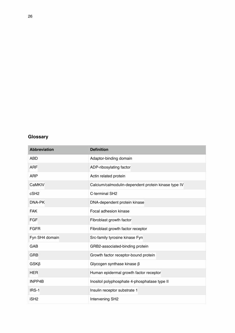
Glossary
Abbreviation Definition
ABD Adaptor-binding domain
ARF ADP-ribosylating factor
ARP Actin related protein
CaMKIV Calcium/calmodulin-dependent protein kinase type IV
cSH2 C-terminal SH2
DNA-PK DNA-dependent protein kinase
FAK Focal adhesion kinase
FGF Fibroblast growth factor
FGFR Fibroblast growth factor receptor
Fyn SH4 domain Src-family tyrosine kinase Fyn
GAB GRB2-associated-binding protein
GRB Growth factor receptor-bound protein
GSKβ Glycogen synthase kinase β
HER Human epidermal growth factor receptor
INPP4B Inositol polyphosphate 4-phosphatase type II
IRS-1 Insulin receptor substrate 1
iSH2 Intervening SH2

�27
KATP ATP-sensitive potassium channels
Kd Dissociation constant
miR Micro RNA
mTOR Mammalian target of rapamycin
MyD88 Myeloid differentiation primary response gene 88
nSH2 N-terminal SH2
PC Phosphatidylcholine
PDGFR Platelet derived growth factor receptor
PDK1 3-phosphoinositide-dependent protein kinase 1
PHLPP PH domain and leucine rich repeat protein phosphatases
PI(3,4,5)P3/PtdIns(3,4,5)P3 Phosphatidylinositol (3,4,5)-trisphosphate
PI(3,4)P2/PtdIns(3,4)P2 Phosphatidylinositol (3,4)-bisphosphate
PI(3,5)P2/PtdIns(3,5)P2 Phosphatidylinositol (3,5)-bisphosphate
PI(3)P/PtdIns(3)P Phosphatidylinositol 3-phosphate
PI(4,5)P2/PtdIns(4,5)P2 Phosphatidylinositol (4,5)-bisphosphate
PI(4)P/PtdIns(4)P Phosphatidylinositol 4-phosphate
PI(5)P/PtdIns(5)P Phosphatidylinositol 5-phosphate
PI3K Phosphoinositide 3-kinase
PTEN Phosphatase and tension homologue
RBD Ras-binding domain
RICTOR Rapamycin-insensitive companion of mammalian target of
rapamycin
RNAi Interference RNA
RTKs Receptor tyrosine kinases
SH2 Src-homology-2
shRNA Short hairpin RNA
TIP Type 2A interacting protein
TIRAP Toll/interleukin-1 receptor domain-containing adaptor protein
UTR Untranslated region
VEGF Vascular endothelial growth factor
Abbreviation Definition

�28
Bibliography
Alessi, D. R., James, S. R., Downes, C. P., Holmes, A. B., Gaffney, P. R., Reese, C. B., &
Cohen, P. (1997). Characterization of a 3-phosphoinositide-dependent protein kinase which
phosphorylates and activates protein kinase Bα. Current Biology, 7(4), 261-269
Ali, I. U., Schriml, L. M., & Dean, M. (1999). Mutational spectra of PTEN/MMAC1 gene: a tu-
mor suppressor with lipid phosphatase activity. Journal of the national cancer institute,
91(22), 1922-1932
Bayascas, J. R., & Alessi, D. R. (2005). Regulation of Akt/PKB Ser473 phosphorylation. Mol-
ecular cell, 18(2), 143-145
WASP Wiskott-Aldrich syndrom protein
Abbreviation Definition

�29
Biondi, R. M., Kieloch, A., Currie, R. A., Deak, M., & Alessi, D. R. (2001). The PIF‐binding
pocket in PDK1 is essential for activation of S6K and SGK, but not PKB. The EMBO journal,
20(16), 4380-4390
Broderick, D. K., Di, C., Parrett, T. J., Samuels, Y. R., Cummins, J. M., McLendon, R. E., ... &
Yan, H. (2004). Mutations of PIK3CA in anaplastic oligodendrogliomas, high-grade astrocy-
tomas, and medulloblastomas. Cancer research, 64(15), 5048-5050
Burke, J. E., Perisic, O., & Williams, R. L. (2013). Allosteric activation of PI3Kα by oncogenic
mutations. Oncotarget, 4(2), 180
Das, S., Dixon, J. E., & Cho, W. (2003). Membrane-binding and activation mechanism of
PTEN. Proceedings of the National Academy of Sciences, 10 0(13), 7491-7496
Di Paolo, G., & De Camilli, P. (2006). Phosphoinositides in cell regulation and membrane dy-
namics. Nature, 443(7112), 651-657
Dunlap, J., Le, C., Shukla, A., Patterson, J., Presnell, A., Heinrich, M. C., ... & Troxell, M. L.
(2010). Phosphatidylinositol-3-kinase and AKT1 mutations occur early in breast carcinoma.
Breast cancer research and treatment, 120(2), 409-418
Elstrom, R. L., Bauer, D. E., Buzzai, M., Karnauskas, R., Harris, M. H., Plas, D. R., ... &
Thompson, C. B. (2004). Akt stimulates aerobic glycolysis in cancer cells. Cancer research,
64(11), 3892-3899
Fan, Z., & Makielski, J. C. (1997). Anionic phospholipids activate ATP-sensitive potassium
channels. Journal of Biological Chemistry, 272(9), 5388-5395

�30
Frech, M., Andjelkovic, M., Ingley, E., Reddy, K. K., Falck, J. R., & Hemmings, B. A. (1997).
High affinity binding of inositol phosphates and phosphoinositides to the pleckstrin homology
domain of RAC/protein kinase B and their influence on kinase activity. Journal of Biological
Chemistry, 272(13), 8474-8481
Gao, T., Furnari, F., & Newton, A. C. (2005). PHLPP: a phosphatase that directly dephospho-
rylates Akt, promotes apoptosis, and suppresses tumor growth. Molecular cell, 18(1), 13-24
Gewinner, C., Wang, Z. C., Richardson, A., Teruya-Feldstein, J., Etemadmoghadam, D.,
Bowtell, D., ... & Cantley, L. C. (2009). Evidence that inositol polyphosphate 4-phosphatase
type II is a tumor suppressor that inhibits PI3K signaling. Cancer cell, 16(2), 115-125
Guo, C., Sah, J. F., Beard, L., Willson, J. K., Markowitz, S. D., & Guda, K. (2008). The non-
coding RNA, miR‐126, suppresses the growth of neoplastic cells by targeting phosphatidyli-
nositol 3‐kinase signaling and is frequently lost in colon cancers. Genes, Chromosomes and
Cancer, 47(11), 939-946
Hanahan, D. & Weinberg, R. A. (2011). Hallmarks of cancer: the next generation. cell, 144(5),
646-674
Hellawell, G. O., Turner, G. D., Davies, D. R., Poulsom, R., Brewster, S. F., & Macaulay, V. M.
(2002). Expression of the type 1 insulin-like growth factor receptor is up-regulated in primary
prostate cancer and commonly persists in metastatic disease. Cancer research, 62(10),
2942-2950
�31
Hennessy, B. T., Smith, D. L., Ram, P. T., Lu, Y., & Mills, G. B. (2005). Exploiting the PI3K/
AKT pathway for cancer drug discovery. Nature reviews Drug discovery, 4(12), 988-1004.
Honda, A., Nogami, M., Yokozeki, T., Yamazaki, M., Nakamura, H., Watanabe, H., ... &
Kanaho, Y. (1999). Phosphatidylinositol 4-phosphate 5-kinase α is a downstream effector of
the small G protein ARF6 in membrane ruffle formation. Cell, 99(5), 521-532
Höning, S., Ricotta, D., Krauss, M., Späte, K., Spolaore, B., Motley, A., ... & Owen, D. J.
(2005). Phosphatidylinositol-(4, 5)-bisphosphate regulates sorting signal recognition by the
clathrin-associated adaptor complex AP2. Molecular cell, 18(5), 519-531
Juric, D., Castel, P., Griffith, M., Griffith, O. L., Won, H. H., Ellis, H., ... & Scaltriti, M. (2014).
Convergent loss of PTEN leads to clinical resistance to a PI (3) K [agr] inhibitor. Nature.
Kagan, J. C., & Medzhitov, R. (2006). Phosphoinositide-mediated adaptor recruitment con-
trols Toll-like receptor signaling. Cell, 125(5), 943-955
Khwaja, A., Rodriguez‐Viciana, P., Wennström, S., Warne, P. H., & Downward, J. (1997). Ma-
trix adhesion and Ras transformation both activate a phosphoinositide 3‐OH kinase and pro-
tein kinase B/Akt cellular survival pathway. The EMBO Journal, 16(10), 2783-2793
Kong, D., Suzuki, A., Zou, T. T., Sakurada, A., Kemp, L. W., Wakatsuki, S., ... & Horii, A.
(1997). PTEN1 is frequently mutated in primary endometrial carcinomas. Nature genetics,
17(2), 143-144
Kuo, Y. C., Huang, K. Y., Yang, C. H., Yang, Y. S., Lee, W. Y., & Chiang, C. W. (2008). Regu-
lation of phosphorylation of Thr-308 of Akt, cell proliferation, and survival by the B55α regula-

�32
tory subunit targeting of the protein phosphatase 2A holoenzyme to Akt. Journal of Biological
Chemistry, 283(4), 1882-1892
Liu, J., Weiss, H. L., Rychahou, P., Jackson, L. N., Evers, B. M., & Gao, T. (2009). Loss of
PHLPP expression in colon cancer: role in proliferation and tumorigenesis. Oncogene, 28(7),
994-1004
Malecz, N., McCabe, P. C., Spaargaren, C., Qiu, R. G., Chuang, Y. Y., & Symons, M. (2000).
Synaptojanin 2, a novel Rac1 effector that regulates clathrin-mediated endocytosis. Current
Biology, 10(21), 1383-1386
Maurer, M., Su, T., Saal, L. H., Koujak, S., Hopkins, B. D., Barkley, C. R., ... & Parsons, R.
(2009). 3-Phosphoinositide–dependent kinase 1 potentiates upstream lesions on the phos-
phatidylinositol 3-kinase pathway in breast carcinoma. Cancer research, 69(15), 6299-6306
Mazure, N. M., Chen, E. Y., Laderoute, K. R., & Giaccia, A. J. (1997). Induction of vascular
endothelial growth factor by hypoxia is modulated by a phosphatidylinositol 3-kinase/Akt sig-
naling pathway in Ha-ras-transformed cells through a hypoxia inducible factor-1 transcrip-
tional element. Blood, 90(9), 3322-3331.
Meng, F., Henson, R., Wehbe–Janek, H., Ghoshal, K., Jacob, S. T., & Patel, T. (2007). Mi-
croRNA-21 regulates expression of the PTEN tumor suppressor gene in human hepatocellu-
lar cancer. Gastroenterology, 133(2), 647-658
Miled, N., Yan, Y., Hon, W. C., Perisic, O., Zvelebil, M., Inbar, Y., ... & Williams, R. L. (2007).
Mechanism of two classes of cancer mutations in the phosphoinositide 3-kinase catalytic
subunit. science, 317(5835), 239-242

�33
Miller, T. W., Pérez-Torres, M., Narasanna, A., Guix, M., Stål, O., Pérez-Tenorio, G., ... &
Arteaga, C. L. (2009). Loss of phosphatase and tensin homologue deleted on chromosome
10 engages ErbB3 and IGF-IR signaling to promote antiestrogen resistance in breast cancer.
Cancer research, 69(10), 4192
Newton, A. C., & Trotman, L. C. (2014). Turning off AKT: PHLPP as a drug target. Annual re-
view of pharmacology and toxicology, 54, 537
Oda, K., Stokoe, D., Taketani, Y., & McCormick, F. (2005). High frequency of coexistent mu-
tations of PIK3CA and PTEN genes in endometrial carcinoma. Cancer research, 65(23),
10669-10673
Ong, S. H., Hadari, Y. R., Gotoh, N., Guy, G. R., Schlessinger, J., & Lax, I. (2001). Stimula-
tion of phosphatidylinositol 3-kinase by fibroblast growth factor receptors is mediated by co-
ordinated recruitment of multiple docking proteins. Proceedings of the National Academy of
Sciences, 98(11), 6074-6079
Pollard, T. D., & Borisy, G. G. (2003). Cellular motility driven by assembly and disassembly of
actin filaments. Cell, 112(4), 453-465
Saal, L. H., Holm, K., Maurer, M., Memeo, L., Su, T., Wang, X., ... & Parsons, R. (2005).
PIK3CA mutations correlate with hormone receptors, node metastasis, and ERBB2, and are
mutually exclusive with PTEN loss in human breast carcinoma. Cancer research, 65(7),
2554-2559

�34
Samuels, Y., Wang, Z., Bardelli, A., Silliman, N., Ptak, J., Szabo, S. & Velculescu, V. E.
(2004). High frequency of mutations of the PIK3CA gene in human cancers. Science,
304(5670), 554-554.
Sarbassov, D. D., Guertin, D. A., Ali, S. M., & Sabatini, D. M. (2005). Phosphorylation and
regulation of Akt/PKB by the rictor-mTOR complex. Science, 307(5712), 1098-1101
Seshacharyulu, P., Pandey, P., Datta, K., & Batra, S. K. (2013). Phosphatase: PP2A structur-
al importance, regulation and its aberrant expression in cancer. Cancer letters, 335(1), 9-18
Shayesteh, L., Lu, Y., Kuo, W. L., Baldocchi, R., Godfrey, T., Collins, C., ... & Gray, J. W.
(1999). PIK3CA is implicated as an oncogene in ovarian cancer. Nature genetics, 21(1),
99-102
Shen, W. H., Balajee, A. S., Wang, J., Wu, H., Eng, C., Pandolfi, P. P., & Yin, Y. (2007). Es-
sential role for nuclear PTEN in maintaining chromosomal integrity. Cell, 128(1), 157-170
Shoelson, S. E., Sivaraja, M. O. H. A. N. R. A. M., Williams, K. P., Hu, P., Schlessinger, J., &
Weiss, M. A. (1993). Specific phosphopeptide binding regulates a conformational change in
the PI 3-kinase SH2 domain associated with enzyme activation. The EMBO journal, 12(2),
795
Shoji, K., Oda, K., Nakagawa, S., Hosokawa, S., Nagae, G., Uehara, Y., ... & Taketani, Y.
(2009). The oncogenic mutation in the pleckstrin homology domain of AKT1 in endometrial
carcinomas. British journal of cancer, 101(1), 145-148

�35
Smirnova, T., Zhou, Z. N., Flinn, R. J., Wyckoff, J., Boimel, P. J., Pozzuto, M., ... & Segall, J.
E. (2012). Phosphoinositide 3-kinase signaling is critical for ErbB3-driven breast cancer cell
motility and metastasis. Oncogene, 31(6), 706-715
Stemke-Hale, K., Gonzalez-Angulo, A. M., Lluch, A., Neve, R. M., Kuo, W. L., Davies, M., ...
& Hennessy, B. T. (2008). An integrative genomic and proteomic analysis of PIK3CA, PTEN,
and AKT mutations in breast cancer. Cancer research, 68(15), 6084-6091
Tokunaga, E., Kimura, Y., Oki, E., Ueda, N., Futatsugi, M., Mashino, K., ... & Maehara, Y.
(2006). Akt is frequently activated in HER2/neu‐positive breast cancers and associated with
poor prognosis among hormone‐treated patients. International journal of cancer, 118(2), 284-
289
Waring, M. J., Andrews, D. M., Faulder, P. F., Flemington, V., McKelvie, J. C., Maman, S., ...
& Wood, R. J. (2014). Potent, selective small molecule inhibitors of type III phosphatidylinosi-
tol-4-kinase α-but not β-inhibit the phosphatidylinositol signaling cascade and cancer cell
proliferation. Chemical Communications, 50(40), 5388-5390
Yu, J., Zhang, Y., McIlroy, J., Rordorf-Nikolic, T., Orr, G. A., & Backer, J. M. (1998). Regula-
tion of the p85/p110 phosphatidylinositol 3′-kinase: stabilization and inhibition of the p110α
catalytic subunit by the p85 regulatory subunit. Molecular and cellular biology, 18(3),
1379-1387
Copyright © 2022 FDOKUMEN