Secreted Frizzled-Related Protein-1 Inhibits RANKL-Dependent Osteoclast Formation
lable at ScienceDirect
Biomaterials xxx (2014) 1e11
Contents lists avai
Biomaterials
journal homepage: www.elsevier .com/locate/biomater ia ls
Osteoclast resorption of beta-tricalcium phosphate controlled bysurface architecture
Noel L. Davison a, b, *, Bas ten Harkel c, Ton Schoenmaker d, Xiaoman Luo a, b,Huipin Yuan a, b, Vincent Everts c, Florence Barr�ere-de Groot b, Joost D. de Bruijn a, b, e
a MIRA Institute for Biomedical Technology and Technical Medicine, University of Twente, 7522 NB Enschede, Netherlandsb Xpand Biotechnology BV, 3723 MB Bilthoven, Netherlandsc Department of Oral Cell Biology, Academic Centre for Dentistry Amsterdam (ACTA), MOVE Research Institute, University of Amsterdam and VU UniversityAmsterdam, 1081 BT Amsterdam, Netherlandsd Department of Periodontology, Academic Centre for Dentistry Amsterdam (ACTA), MOVE Research Institute, University of Amsterdam and VU UniversityAmsterdam, 1081 BT Amsterdam, Netherlandse School of Engineering and Materials Science (SEMS), Queen Mary University of London, E1 4NS London, United Kingdom
a r t i c l e i n f o
Article history:Received 14 March 2014Accepted 16 May 2014Available online xxx
Keywords:Calcium phosphateTopographyMicrostructureOsteoclastForeign body giant cell
* Corresponding author. Xpand BiotechnologyNetherlands.
E-mail address: [email protected]
http://dx.doi.org/10.1016/j.biomaterials.2014.05.0480142-9612/© 2014 Elsevier Ltd. All rights reserved.
Please cite this article in press as: DavisonBiomaterials (2014), http://dx.doi.org/10.101
a b s t r a c t
A resorbable bone graft substitute should mimic native bone in its capacity to support bone formationand be remodeled by osteoclasts (OCl) or other multinucleated cells such as foreign body giant cells(FBGC). We hypothesize that by changing the scale of surface architecture of beta-tricalcium phosphate(TCP), cellular resorption can be influenced. CD14þ monocyte precursors were isolated from humanperipheral blood (n ¼ 4 independent donors) and differentiated into OCl or FBGC on the surface of TCPdiscs comprising either submicron- or micron-scale surface topographical features (TCPs and TCPb,respectively). On submicrostructured TCPs, OCl survived, fused, differentiated, and extensively resorbedthe substrate; however, on microstructured TCPb, OCl survival, TRAP activation, and fusion wereattenuated. Importantly, no resorption was observed on microstructured TCPb. By confocal microscopy,OCl formed on TCPs contained numerous actin rings allowing for resorption, but not on TCPb. In com-parison, FBGC could not resorb either TCP material, suggesting that osteoclast-specific machinery isnecessary to resorb TCP. By tuning surface architecture, it appears possible to control osteoclastresorption of calcium phosphate. This approach presents a useful strategy in the design of resorbablebone graft substitutes.
© 2014 Elsevier Ltd. All rights reserved.
1. Introduction
If a bone graft substitute is meant to be replaced by new bonetissue in a bony defect, then it must be resorbable. Unlike non-resorbable materials, resorbable materials avoid the local loss ofbone density associated with stress shielding that stems from thedisproportionally high loadbearing of an implant versus the sur-rounding bone. Moreover, resorbable bone graft substitutes, such ascalcium phosphate (CaP), should allow for remodeling by boneresorbing osteoclasts [1], important for normal bone homeostasis,bone coupling, and osteogenesis [2e5].
BV, 3723 MB Bilthoven,
(N.L. Davison).
NL, et al., Osteoclast resorpt6/j.biomaterials.2014.05.048
Themodes by which a CaP bone graft substitute can be resorbedin the body are generally categorized as cell-mediated (oftentermed bioresorption or biodegradation) or passive, such asdissolution, erosion, and mechanical fragmentation [6]. Related tothese, two primary strategies for controlling the resorption rate ofCaP include modifying the physical architecture and tuning theceramic chemistry [7]. The first strategy is to incorporate aninterconnected network of pores or channels large enough for cellinfiltration (>20 mm) to promote blood perfusion and capillaryingrowth to supply and sustain cells needed for bioresorption andosteogenesis. Additionally, increasing the pore area enhances thesurface area allowing faster dissolution. The second strategy is toalter the ceramic crystalline chemistry to thermodynamically favordissolution in the body. For instance, CaP composed of beta-tricalcium phosphate (TCP) tends to dissolve more readily thanhydroxyapatite (HA) and is therefore preferred where resorbabilityis desired [1].
ion of beta-tricalcium phosphate controlled by surface architecture,

Table 1Physical characterization of TCP.
Physical parameters TCPs TCPb
Average grain diameter (mm) 0.95 ± 0.27 3.66 ± 1.05Average pore diameter (mm) 0.63 ± 0.33 1.78 ± 0.85Average peak-to-valley roughness,
Ra (mm)0.126 ± 0.003 1.287 ± 0.011
Root-mean-square peak-to-valleyroughness, RRMS (mm)
0.158 ± 0.003 1.597 ± 0.011
Porosity (%) 69.6 72.0Total pore area (m2/g) 1.477 0.769
N.L. Davison et al. / Biomaterials xxx (2014) 1e112
Other material properties may also determine CaP resorbabilitysuch as the physical surface architecture, i.e., the size and amountof surface micropores (0.1e10 mm) and grains [8,9]. However,Bohner (2012) recently emphasized that no studies have evaluatedthese factors while maintaining other material properties equal toclearly determine their effects on bioresorption or if there is anoptimal design criterion [1]. More recently, results reported by ourgroup described that despite possessing equivalent ceramicchemistry, macropores, and total porosity, TCP with submicron-scale surface grains and pores was shown to be resorbed by ~24%after implantation for 12 weeks in the dorsal muscle of dogs (anosteoinduction model) but TCP with micron-scale surface grainsand pores was appreciably un-resorbed [10]. Multinucleatedosteoclast-like cells were substantially identified on TCP withsubmicron-scale surface architecture, whereas multinucleated cellscould scarcely be found on the un-resorbed TCP with micron-scalesurface architecture. In vitro, neither TCP passively dissolved inculture medium containing serum, suggesting that multinucleatedcell-mediated resorption is likely the principal mode by which
Table 2QPCR primer sequences.
Genetarget
Sequence (50 - 30) Productsize (bp)
Accession ID
UBC gcggtgaacgccgatgattat 202 ENSG00000150991tttgccttgacattctcgatgg
TATAb ggtctgggaaaatggtgtgc 100 ENSG00000112592gctggaaaacccaacttctg
HPRT tgaccttgatttattttgcatacc 101 ENSG00000165704cgagcaagacgttcagtcct
Cyc1 gcatggtggtgaggactacg 106 ENSG00000179091ggccaggaaagtaggggttg
GAPDH tgggtgtgaaccatgagaagtatg 61 ENSG00000111640ggtgcaggaggcattgct
CATK ccatatgtgggacaggaagagagtt 149 ENSG00000143387tgcatcaatggccacagaga
TRAP cacaatctgcagtacctgcaagat 128 ENSG00000102575cccatagtggaagcgcagata
CAII tggactggccgttctaggtatt 100 ENSG00000104267tcttgccctttgttttaatggaa
AE2 ttgtgggcctctccatagttatc 103 ENSG00000164889gatcccgttaagggaggtgact
TCIRG1 gctgccaaccacttgagctt 114 ENSG00000110719caaagtgcacgtggttgaaga
DC-STAMP attttctcagtgagcaagcagtttc 101 ENSG00000164935agaatcatggataatatcttgagttcctt
VCAM-1 acaaagttggctcacaattaagaagtt 100 ENSG00000162692tgcaaaatagagcacgagaagct
ITGB1 tttccattggagatgaggttca 100 ENSG00000150093cgtaaagcccagaggcctaa
ITGB2 cgacggccgctgtca 100 ENSG00000160255tgttgttttcagccagcttgtg
ITGB3 aggctggcaggcattgtc 100 ENSG00000259207agccccaaagagggataatcc
ITGA4 ctttccagacagccaggagaa 116 ENSG00000115232gggcactccatagcaacca
ITGAV tacagcaggtccccaagtcact 100 ENSG00000138448aattcagattcatcccgcagat
Please cite this article in press as: Davison NL, et al., Osteoclast resorptBiomaterials (2014), http://dx.doi.org/10.1016/j.biomaterials.2014.05.048
these materials are degraded in vivo. However, no resorption wasobserved in vitro, most likely due to the limitations of using theRAW264.7 osteoclast model cell line. These materials inducedectopic bone formation to similar extents that they were resorbed:TCPs formed ~20% in the free area while TCPb induced none, sug-gesting that TCP resorption and bone induction may go hand inhand in a mechanism akin to bone coupling, as others have pre-viously postulated [11,12].
The identity and function of such multinucleated cells sur-rounding CaP in vivo is the subject of debate with some groupsspeculating that they are fused macrophages (i.e., foreign body gi-ant cells e FBGC) [13,14], while other groups asserting that they arespecialized bone-resorbing osteoclasts (OCl) [3,15,16]. Actually, it isplausible that both cell types are present depending the chemicalcomposition of the material, how it was fabricated (i.e., sinteringtemperature), and where it is implanted [6,15,17]. Following thehost response, monocyte/macrophages will normally infiltrate theimplantation site and adhere to the implant, providing a commonprecursor pool for both FBGC and OCl on the material surface[18,19]. Though many groups have demonstrated in vitro that OClpossess the cellular machinery necessary for resorption of CaP[20e23] and that a bony surface is not necessary for osteoclastactivation [24], whether different surface structure can substan-tially influence this process and whether FBGC can also resorb CaPis unclear.
Beyond material resorption, multinucleated cells such as OClmay be important for the osteogenic properties of CaP, in particulara class of microstructured CaP that can induce ectopic bone for-mation without exogenous stem cells or growth factors. Forinstance, depleting OCl by bisphosphonates has been repeatedlyshown to obstruct ectopic bone formation by osteoinductive CaP[12,25]. The reason for this may be due to the powerful osteogenicsignals that OCl express and secrete (e.g., S1P, Wnts, BMPs, andCTHRC1) [3,26e28]. Therefore, characterizing what material pa-rameters are promotive of OCl formation, survival, and functionmay also bear impact on the osteogenic capacity of a CaP bone graftsubstitute.
Following up on our previous findings that the scale of surfacearchitecture plays a determinant role in both the resorbability ofTCP and the presence of multinucleated cells in vivo [10], wespeculated that surface architecture might directly affect the for-mation and resorptive function of these cells. In essence, we hy-pothesized that it might be possible to control multinucleated cellformation and resorption by changing the scale of surface structure.To test this, we cultured human peripheral blood monocytes on thesurface of the same two TCP with either submicron- or micron-scale surface features and differentiated them into either OCl orFBGC by adding specific cytokines. The effects of different TCPsurface architecture on multinucleated cell formation, survival, andresorption were analyzed using a variety of in vitro assays andtechniques.
2. Materials and methods
2.1. Preparation and characterization of TCP with micron- and submicron-scalesurface architecture
Dense TCP discs were fabricated and characterized as previously described [10].TCP powders were synthesized by mixing calcium hydroxide and phosphoric acid(SigmaeAldrich) at a Ca/P ratio of 1.50. TCP powders with small (TCPs) or big (TCPb)grains and micropores in the final ceramics were prepared by wet precipitation. Thepowders were foamed with diluted H2O2 (0.1%) (Merck) at 60 �C to form micropo-rous green bodies and then dried. The dry green bodies were subsequently sinteredat 1050 �C or 1100 �C for 8 h to achieve small or big grains for TCPs and TCPb,respectively. Microporous discs (∅9 � 1 mm) were machined from the ceramicbodies using a lathe and a diamond saw microtome (Leica SP1600). Discs were ul-trasonically cleaned in successive baths of acetone, ethanol, and deionized water,dried at 60 �C, and then heat sterilized at 160 �C for 2 h for cell culture. Crystal
ion of beta-tricalcium phosphate controlled by surface architecture,

Fig. 1. Cell viability (A,B), DNA content (C), and Proliferation (D). Viability of osteoclasts (OCl, induced with M-CSF and RANKL) and foreign body giant cells (FBGC, induced with M-CSF, IL-4, and IL-13) cultured directly on TCP was roughly double on submicrostructured TCPs than on microstructured TCPb at day 25 (A). However, there was no difference inviability in either cell type cultured on tissue culture plastic adjacent to TCPs or TCPb in the same well (B). DNA content from the lysate of TCP-adherent cells at day 25 mimicked theviability data and reflected less cells present on TCPb than on TCPs (C). Proliferation of monocyte/macrophage precursors (cultured only with M-CSF), as measured by viability overtime, proceeded similarly on TCPs and TCPb through day 14 but sharply decreased by day 21 and 25 on TCPb versus TCPs (D). Cell viability and proliferation data are given inAlamarBlue relative fluorescent units normalized by the cell culture surface area (AB RFU cm�2). Data represent the mean ± s.d. of n ¼ 4 replicates from 3 independent donors.*P < 0.05, **P < 0.01, ***P < 0.001.
N.L. Davison et al. / Biomaterials xxx (2014) 1e11 3
chemistry of the materials was analyzed by X-ray diffraction (Rigaku Miniflex II)scanning the range 2q¼ 25e45� (step size¼ 0.01�, rate¼ 1�min�1) and confirmed tobe TCP.
Surface topography of the materials was analyzed using scanning electron mi-croscopy (SEM) (JEOL JSM-5600 microscope) after sputter coating them with gold
Fig. 2. Tartrate resistant acid phosphatase (TRAP) activity (A) and staining (B). TRAP activityrespectively, when formed on submicrostructured TCPs versus microstructured TCPb. TRAP aper mg total protein (mmol mg�1) (A). Representative light micrographs of TCP discs stainethan 500 mm across on the surface of TCPs versus weaker stained, sparse, smaller (<50 mm) Ofrom 2 independent donors. *P < 0.05, **P < 0.01.
Please cite this article in press as: Davison NL, et al., Osteoclast resorptBiomaterials (2014), http://dx.doi.org/10.1016/j.biomaterials.2014.05.048
for 90 s (JEOL JFC 1300). For grain and pore size analysis, at least 50 grains and poreswere measured across their vertical diameter using ImageJ software (NIH, USA).Porosity and total pore area (analogous to specific surface area) were determined bymercury intrusion testing (Micromeritics, USA). Surface roughness was computedusingMeX v5.1 software (Alicona Imaging, Austria). Briefly, SEM stereo-micrographs
measured at day 25 in the cell lysate was 7 times and 2.5 times higher in OCl and FBGC,ctivity was normalized per total protein in the lysate and given as mmol p-nitrophenold for TRAP at day 25 showed intensely stained, interconnected OCl measuring greaterCl on TCPb. Scale ¼ 500 mm. TRAP activity represents the mean ± s.d. of n ¼ 4 replicates
ion of beta-tricalcium phosphate controlled by surface architecture,

Fig. 3. Scanning electron micrographs of OCl cultured on TCP discs at day 25. (A) Representative images at low magnification (left) show massively fused OCl (~200e500 mm)homogenously populate the surface of TCPs but are less evident on TCPb (50�, scale ¼ 500 mm). At higher magnification (middle), large fused OCl (dark cells, ~300 mm across) can beseen in close proximity to one another resorbing TCPs, surrounded by un-fused precursors. Fused OCl on TCPb (light spots) are markedly smaller in comparison (<100 mm) (200�,scale ¼ 100 mm). At high magnification for detail (right), OCl actively resorb TCPs with filopodia extending to the border of the resorption lacunae (marked by white arrow heads).Small cells and no resorption lacunae are found on TCPb (1000�, scale ¼ 20 mm). (B) OCl formed on TCPs create extensive networks of resorption pits and migrate towardunresorbed material (white arrows) (top row scale ¼ 50 mm; bottom row scale ¼ 20 mm). (C) Representative scanning electron micrographs of FBGC formed on TCP at day 25. Cellsfuse to form large FBGC on TCPs (dark spots) ranging from ~40 to 100 mm across. On TCPb, cells are smaller and sparser, appearing necrotic (detail). No resorption is evident fromFBGC on either material. 50� scale ¼ 500 mm; 200� scale ¼ 100 mm; 1000� scale ¼ 20 mm.
N.L. Davison et al. / Biomaterials xxx (2014) 1e114
Please cite this article in press as: Davison NL, et al., Osteoclast resorption of beta-tricalcium phosphate controlled by surface architecture,Biomaterials (2014), http://dx.doi.org/10.1016/j.biomaterials.2014.05.048
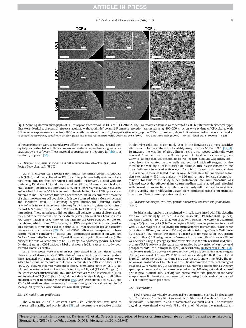
Fig. 4. Scanning electron micrographs of TCP resorption after removal of OCl and FBGC After 25 days, no resorption lacunae were detected on TCPb cultured with either cell type;discs were identical to the control reference incubated without cells (left column). Prominent resorption lacunae spanning ~100e200 mm across were evident on TCPs cultured withOCl but no resorption was evident from FBGC versus the control reference. High magnification micrographs of TCPs (right column) showed alteration of surface microstructure dueto osteoclast resorption, specifically smaller grains and increased microporosity. Overview scale (50�) ¼ 500 mm; inset scale (500�) ¼ 50 mm; detail scale (5000�) ¼ 5 mm.
N.L. Davison et al. / Biomaterials xxx (2014) 1e11 5
of the same locationwere captured at two different tilt angles (2500�, ±5�) and thendigitally reconstructed into three-dimensional surfaces for surface roughness cal-culations by the software. These material properties are all reported in Table 1, aspreviously reported [10].
2.2. Isolation of human monocytes and differentiation into osteoclasts (OCl) andforeign body giant cells (FBGC)
CD14þ monocytes were isolated from human peripheral blood mononuclearcells (PBMC) and then cultured on TCP discs. Briefly, human buffy coats (n ¼ 4 do-nors) were acquired from San Quinn Blood Bank (Amsterdam), diluted with PBScontaining 1% citrate (1:1), and then spun down (800 g, 30 min, without brake) inFicoll gradient solution. The interphase containing the PBMC was carefully collectedand washed 4 times in 0.5% bovine serum albumin buffer (2 mM EDTA, phosphate-buffered saline), then passed through a cell strainer (40 mm) to ensure the recoveryof a cleanmononuclear cell population. Cells were counted using aMuse cell counterand incubated with CD14-antibody tagged microbeads (Miltenyi Biotec)(1 � 107 cells in 20 mL microbead solution) for 15 min at 4 �C, then sorted using amanual MACS magnetic cell sorter (Miltenyi Biotec) following the manufacturer'sinstructions. These microbeads did not affect cell behavior or morphology, nor dothey need to be removed due to their extremely small size (~50 nm). Because such alow concentration is used, they do not saturate the CD14 epitopes on the cellmembrane, which makes CD14 labeling for FACs analysis after isolation possible.This method is commonly used to isolate CD14þ monocytes for use as osteoclastprecursors in the literature [29]. Purified CD14þ cells were resuspended in basicculture medium consisting of aMEM (Life Technologies) supplemented with 10%fetal calf serum (HyClone I) and 1% penicillinestreptomycin (SigmaeAldrich). Thepurity of the cells was confirmed to be 83 ± 4% by flow cytometry (Accuri C6, BectonDickinson) using a CD14 antibody label and mouse IgG2a isotype antibody (bothMiltenyi Biotec) as control.
CD14þ monocytes were seeded on TCP discs placed in 48-well tissue cultureplates at a cell density of ~500,000 cells/cm2. Immediately prior to seeding, discswere incubated with 1 mL basic medium for 2 h to equilibrate them. Cytokines wereadded to the culture medium in order to differentiate the cells into either OCl orFBGC. OCl cultures received macrophage colony-stimulating factor (M-CSF, 10 ng/mL) and receptor activator of nuclear factor kappa-B ligand (RANKL, 2 ng/mL) toinduce osteoclast differentiation. FBGC cultures receivedM-CSF, interleukin-4 (IL-4),and interleukin-13 (IL-13) (both 5 ng/mL) to induce foreign body giant cell differ-entiation, similar to previously described [30]. Cells were cultured in 5% CO2 and37 �C with medium refreshment every 3e4 days throughout the culture duration of25 days. All cytokines were purchased from R&D Systems.
2.3. Cell viability and proliferation
The AlamarBlue (AB) fluorescent assay (Life Technologies) was used tomeasure cell viability and proliferation [31]. AB measures the reductive activity
Please cite this article in press as: Davison NL, et al., Osteoclast resorptBiomaterials (2014), http://dx.doi.org/10.1016/j.biomaterials.2014.05.048
inside living cells, and is commonly used in the literature as a more sensitivealternative to formazan-based cell viability assays such as MTT and XTT [32,33].To measure the viability of disc-adherent cells, discs seeded with cells wereremoved from their culture wells and placed in fresh wells containing pre-warmed culture medium containing 5% AB reagent. Medium was gently aspi-rated from the vacated culture wells and replaced with AB reagent to alsomeasure the viability of cells cultured on tissue culture plastic adjacent to thediscs. Cells were incubated with reagent for 2 h in culture conditions and thenmedia samples were collected in an opaque 96-well plate for fluorescent detec-tion (excitation ¼ 530 nm, emission ¼ 590 nm) using a Synergy spectropho-tometer. For time course study of cell proliferation, the same procedure wasfollowed except that AB-containing culture medium was removed and refreshedwith normal culture medium, and then continuously cultured until the next timepoint. Viability and proliferation assays were conducted using 3 independentdonors and 2e4 culture replicates per donor.
2.4. Biochemical assays: DNA, total protein, and tartrate resistant acid phosphatase(TRAP)
For biochemical analysis, discs culturedwith cellswere rinsedwith PBS, placed infresh wells containing lysis buffer (0.1 M sodium acetate, 0.1% Triton X-100, pH 5.8),and then frozen at �80 �C and thawed to aid lysis. DNA in the lysate was quantifiedusing a CyQuant assay kit (Life Technologies). Briefly, sample lysate was incubatedwith GR dye reagent (1x) following the manufacturer's instructions. Fluorescence(excitation ¼ 480 nm, emission ¼ 520 nm) was detected using a Zenyth MultimodePlate Reader. Total protein was quantified using a commercial Micro BCA Proteinassay kit (Pierce) following the manufacturer's instructions. Absorbance at 405 nmwas detected using a Synergy spectrophotometer. Last, tartrate resistant acid phos-phatase (TRAP) activity in the lysate was quantified by conversion of p-nitrophenylphosphate (pNPP) to p-nitrophenol (pNP), as described by Ljusberg et al. (1999) [34].Cell lysate sample (20 mL) was collected to a 96-well plate containing assay reagent(130 mL) composed of 10 mM PNPP, 0.1 M sodium acetate (pH 5.8), 0.15 M KCl, 0.1%Triton X-100, 10 mM sodium tartrate, 1 mM ascorbic acid, and 0.1 mM FeCl6. The re-actionwas incubated for 1 h at 37 �C and then finally converted to pNP (chromogen)by adding 0.3 M NaOH (100 mL). Absorbance at 405 nmwas detected using a Synergyspectrophotometer and values were converted to mM pNP using a standard curve ofpNP (SigmaeAldrich). TRAP activity was normalized to total protein in the samesample lysate. Biochemical assays were conducted using 3 independent donors and2e4 culture replicates per donor.
2.5. TRAP staining
TRAP enzyme was visually detected using a commercial staining kit (LeukocyteAcid Phosphatase Staining Kit, SigmaeAldrich). Discs seeded with cells were firstrinsed with PBS and fixed in 2.5% glutaraldehyde overnight at 4 �C. The followingday, discs were rinsed once with PBS and stained following the manufacturer's
ion of beta-tricalcium phosphate controlled by surface architecture,

Fig. 5. Representative laser scanning confocal micrographs of OCl formed on TCP. Actinring structures (white arrows) are evident at the membrane border of OCl cultured onTCPs but not TCPb (Green ¼ phalloidin targeting F-actin). The osteoclast membraneextends >300 mm across on TCPs but remains small in comparison on TCPb(Red ¼ CD44 antibody targeting cell membrane). Inside the membrane of a singleosteoclast on TCPs, many nuclei are evident (Blue ¼ Hoechst nuclei stain).Scale ¼ 150 mm. (For interpretation of the references to color in this figure legend, thereader is referred to the web version of this article.)
N.L. Davison et al. / Biomaterials xxx (2014) 1e116
instructions. Staining was visualized using a Nikon SMZ800 stereomicroscopeequipped with a Nikon camera. TRAP staining was conducted using 3 independentdonors and 2 culture replicates per donor.
2.6. Cell morphology and resorption
Cell morphology was evaluated using SEM (JEOL JSM-5600 microscope). After25 days culture, discs cultured with cells were rinsed with PBS and fixed in 2.5%glutaraldehyde for up to 1 week. Cells were dehydrated in a graded ethanol series(60, 70, 80, 90, 95, and 100% � 2) and finally dried in HMDS (SigmaeAldrich). Discswere then mounted on metal stubs and sputter coated with gold for enhancedimaging resolution.
To analyze TCP resorption after 25 days of culture, adherent cells were removedfor qPCR analysis using RLT lysis buffer (Qiagen), and the discs were rinsed once in
Please cite this article in press as: Davison NL, et al., Osteoclast resorptBiomaterials (2014), http://dx.doi.org/10.1016/j.biomaterials.2014.05.048
70% ethanol then dried at 60 �C. Discs not seeded with cells were similarly culturedfor 25 days and served as control references. Samples were mounted on metalstubs and sputter coated with gold for SEM. Cell morphology and resorption assayswere conducted using cells from 4 independent donors and 2 culture replicates perdonor.
2.7. Fluorescent laser scanning confocal microscopy
Cell fusion and actin ring formation were analyzed using fluorescent laserscanning confocal microscopy. After culture for 25 days, cells seeded on discs wererinsed with PBS, fixed in 4% formaldehyde, blocked with 20% normal goat serum,then sequentially incubated with rat anti-mouse monoclonal primary antibodyCD44 targeting the osteoclast plasma membrane (1:100 dilution) (Cedarlane, USA),goat anti-rat Alexa467-conjugated secondary antibody (1:400) (Invitrogen),Alexa488-conjugated phalloidin targeting F-actin (1:400) (Invitrogen), and Hoechst33342 dye (1:500) (SigmaeAldrich) to stain nuclei, as previously described [35].Fluorescence was visualized using a Leica TCS-SP8-SMD DMI6000 laser scanningconfocal microscope and overlaid image stacks were generated by scanning from theapical to basal surface of the cell (Leica LAS AF software). Confocal microscopyanalysis was conducted using 2 independent donors.
2.8. Quantitative PCR (qPCR)
Total RNA was isolated using a commercial spin-column kit (Qiagen) followingthe manufacturer's instructions. Discs seeded with cells were removed from theculturewells and immediately lysed in RLT lysis buffer. The cells cultured adjacent tothe discs on tissue culture plastic in the same wells were lysed separately. Aftercolumn purification of total RNA in the cell lysate, RNA concentrationwas measuredusing a Synergy spectrophotometer. Reverse transcription of RNA was performedusing the MBI Fermentas cDNA synthesis kit (Fermentas, Lithuania), using both theOligo(dT)18 and the D(N)6 primers.
Quantitative PCR (qPCR) was performed on an ABI PRISM 7000 SequenceDetection System. The PCR reactions were performed with 60 ng cDNA in a totalvolume of 15 mL containing SYBR GreenER qPCR SuperMix (consisting of SYBRGreen 1 Dye, AmpliTaq Gold DNA polymerase, dNTPs, passive reference and buffer)(Life Technologies), and 300 nm of primer. For gene targets, the protocol consistedof an activation step (10 min, 94 �C) and 40 cycles of two-step PCR (95 �C for 15 s,60 �C for 1 min). TATAb and UBC housekeeping genes were tested using a three-step protocol (95 �C for 10 s, 55 �C for 30 s, 72 �C for 30 s). All genes were sub-jected to melt curve analysis to test if any unspecific PCR products were generated.Expression of gene targets was normalized by endogenous expression of 5 house-keeping genes (TATAb, UBC, GAPDH, cycloB, and HPRT) following the comparativeCT method [36] and presented as fold expression (2�DCT). These housekeepinggenes were shown to be stable using GeNorm software (v. 3) and their geometricmean expression was calculated for normalization of gene targets. To account forvariability between donors, fold expression levels were further normalized to thoseof cells cultured on TCPb and presented as relative fold expression (2�DDCT). QPCRPrimers were designed using Primer Express 2.0 software (Life Technologies)(Table 2), spanning at least 1 intron to avoid amplification of genomic DNA. QPCRanalysis was conducted using 4 independent donors and 2e3 culture replicates perdonor.
2.9. Statistics
Statistical comparisons between TCPs and TCPbwere performed using One-WayANOVA and Tukey's multiple comparisons when comparing more than 2 groups inGraphPad Prism 6.0. P values <0.05 were considered significant.
3. Results
3.1. Cell viability and proliferation
After culture for 25 days on TCP, OCl and FBGC viability wasroughly 100% higher on TCPs than on TCPb (P ¼ 0.0329 andP ¼ 0.0074, respectively) (Fig. 1A). In contrast, there was no dif-ference in viability of either OCl or FBGC cultured adjacent to theTCP discs on tissue culture plastic of the same culture well (Fig. 1B),indicating that the differences in disc-adherent cell viability weredue to the direct interaction with the TCP surface structure ratherthan soluble effects in the medium. In concert with these results,DNA content in the adherent cell lysate was measured and fol-lowed the same trends e ~2e3 times more DNA from cells on TCPsthan on TCPb (Fig. 1C).
To rule out the possibility that initial cell attachment was loweronTCPb or that it was cytotoxic,monocyte precursorswere cultured
ion of beta-tricalcium phosphate controlled by surface architecture,

Fig. 6. OCl RNA expression of key integrins and osteoclast markers at day 25 by qPCR. VCAM-1 was the only detectable difference in gene expression between OCl formed on TCPs and TCPb. Comparison to OCl formed on tissue cultureplastic adjacent to TCP discs emphasizes the direct effect of microstructure on gene expression. Expression levels were normalized to OCl cultured on TCPb. Data represents the mean ± s.d. of n ¼ 6 replicates from 3 independent donors.*P < 0.05, **P < 0.01.
N.L.D
avisonet
al./Biom
aterialsxxx
(2014)1e11
7
Pleasecite
thisarticle
inpress
as:Davison
NL,et
al.,Osteoclast
resorptionof
beta-tricalciumphosphate
controlledby
surfacearchitecture,
Biomaterials
(2014),http://dx.doi.org/10.1016/j.biomaterials.2014.05.048

N.L. Davison et al. / Biomaterials xxx (2014) 1e118
for 25 days only in the presence of only M-CSF for survival, and cellproliferation was assayed by measuring cell viability over time(Fig. 1D). The viability of adherent cells one day after seeding wasequivalent between the materials, indicating that the initial cellattachment efficiency was equivalent. Moreover, viability increasedthrough 14 days equivalently between thematerials, indicating thatneither material was cytotoxic and that cells were able to prolifer-ate. However, as time progressed, cell viability sharply decreased onTCPb versus TCPs resulting in 38% less metabolic activity at day 21(P¼ 0.0003) and 50% less at day 25 (P¼ 0.0016). These data indicatethat long-term cell survival was reduced on TCPb although initialcell attachment and proliferation were unhindered.
3.2. TRAP activity
TRAP, an enzymatic marker of OCl [37,38] and macrophageactivation [39,40], was both biochemically assayed and cyto-chemically stained after 25 days of culture. TRAP activity per totalprotein in OCl was more than 7 times higher after culture on TCPsthan on TCPb (P ¼ 0.0065). Similarly, TRAP activity in FBGC was 2.5times higher after culture on TCPs than on TCPb (P ¼ 0.0161),indicating that both OCl and FBGC activationwas promoted by TCPs(Fig. 2A). These results were confirmed by TRAP staining, whichshowed more intensely stained, massive multinucleated cells in aninterconnected network on TCPs, but less intensely stained, smallerand unconnected cells on TCPb. TRAP staining of FBGC was alsomore intense on TCPs versus TCPb (Fig. 2B). For both cell types,substantially more cells were present on TCPs than on TCPb, con-firming the cell viability and DNA assays.
3.3. Cell morphology and TCP resorption
Cell morphology and TCP resorption were analyzed by SEM. OnTCPs, fused OCl (>~200 mm across) extensively populated thesubmicrostructured surface and resided in prominent resorptionlacunae. Cellular processes frequently connected adjacent OCl in amassive cell network. Numerous finger-like filopodia decorated theperimeter of the membrane, extending from the cell to the materialsurface. Non-fused mononuclear cells were also observed in andoutside of the resorption lacunae. On TCPb, OCl were sparselydistributed and smaller in comparison. No resorption lacunae wereidentified on TCPb. Fewer cells were present on TCPb than on TCPs,which confirmed the cell viability, DNA content, and TRAP stainingresults (Fig. 3aeb).
In comparison, FBGC appeared substantially smaller than OClon TCPs. On TCPb, FBGC appeared apoptotic or necrotic similar to
Fig. 7. OCl RNA expression of differentially expressed gene targets over the culture period. D**P < 0.01, ***P < 0.001****P < 0.0001.
Please cite this article in press as: Davison NL, et al., Osteoclast resorptBiomaterials (2014), http://dx.doi.org/10.1016/j.biomaterials.2014.05.048
OCl cultured on the material. No resorption lacunae created byFBGC could be identified on either material (Fig. 3c).
TCP resorption was further analyzed after removing adherentOCl and FBGC after 25 days using cell lysis buffer without alteringthe native surface structure (Fig. 4). Prodigious resorption pitsranging from ~10 to 500 mm covered the surface of TCPs culturedwith OCl. In the resorption pits, the surface structure was visiblyaltered resulting in smaller grains, narrower inter-grain bound-aries, and amore polygonal grain shape (Figs. 3b and 4). In contrast,no resorption lacunae were seen on TCPb cultured with OCl; thesurface structure appeared identical to the control discs culturedwithout cells. Unlike OCl, no signs of resorption could be identifiedon either TCP cultured with FBGC. The ability of OCl to resorb TCPs,resulting in large resorption pits, but not TCPb was robustlyconfirmed across all 4 donors (Supplementary Figure).
3.4. Cell fusion and actin ring formation
Cell fusion, multinucleation, and actin ring formation werevisualized using laser confocal microscopy. Fluorescent membraneand nuclei staining clearly showed that the large OCl seen on TCPsby TRAP staining and SEM contained many nuclei (Fig. 5). Fluo-rescent staining targeting F-actin revealed numerous actin rings(~20 mm across) generally near the edge of the membrane of themultinucleated OCl. On TCPb, OCl were also fused and multinu-cleated, although to a lesser extent than on TCPs; however, clearactin ring structures were not found in OCl on TCPb. No actin ringstructures were observed in FBGC on either material (data notshown).
3.5. Gene expression
The expression of genes related to substrate adhesion, cellecelladhesion, and osteoclast differentiation and function wasanalyzed in order to explain the observed differences in osteoclastviability and resorption between the two materials. RNA wasseparately isolated from both the fraction of cells adhered to thediscs and the fraction of cells adhered to the tissue culture plasticadjacent to the discs to isolate the effects of TCP surface structurefrom potential differences induced by dissolved material presentin the media.
After 25 days culture, there was no significant difference in theexpression of key osteoclast gene markers TRAP, CATK, CAII, AE2,TCIRG1, or DC-STAMP nor were there differences in expression ofimportant integrins such as ITGB1, ITGB2, ITGB3, ITGAV, or ITGA4(Fig. 6). Only VCAM-1 (vascular cell adhesion molecule) was
ata represents the mean ± s.d. of n ¼ 3 replicates from 1 representative donor. *P < 0.05,
ion of beta-tricalcium phosphate controlled by surface architecture,

N.L. Davison et al. / Biomaterials xxx (2014) 1e11 9
expressed differently on the two materials, ~12 times higher onTCPs than on TCPb (P ¼ 0.002). OCl expression of VCAM-1 on TCPswas also significantly higher than either cells cultured adjacent toTCPs or TCPb on tissue culture plastic (P ¼ 0.017 and 0.004,respectively) suggesting that this effect was due to direct in-teractions with the surface structure (Fig. 6).
We speculated that gene expression may have followed a dy-namic pattern over time similar to cell viability patterns, so wemeasured the time-course expression of the same genes in oneadditional donor. Only ITGB1, DC-STAMP, and VCAM-1 weredifferentially expressed between OCl cultured on TCPs and TCPb attime points earlier than day 25 (Fig. 7). OCl cultured on TCPbexpressed twice as much ITGB1 than TCPs at days 6 and 11(P ¼ 0.042 and 0.042, respectively). Conversely, OCl expressedroughly 3 times as much DC-STAMP, the membrane proteinresponsible for osteoclast and macrophage fusion, on TCPs than onTCPb at day 6 (P ¼ 0.032). VCAM-1 expression was significantlyhigher in OCl cultured on TCPs versus TCPb at day 6 (~50 fold,P < 0.0001), day 11 (~13 fold, P ¼ 0.0003), day 18 (~12 fold,P ¼ 0.0430) and day 25 (~2.6 fold, P ¼ 0.015). Donor variability wasevident in DC-STAMP expression at day 25, which was significantlyhigher on TCPs than on TCPb (P ¼ 0.042) (Fig. 7) although it wasequivalent for the other 3 donors in aggregate (Fig. 6).
4. Discussion
Although modification of material physico-chemistry [41] andmacroporosity [42] have both been shown to enhance cellularresorption of CaP, little prior literature exists on the role of surfacearchitecture in this process [1]. Previously, our group reported thatin an intramuscular model of osteoinduction TCPs with submicron-scale surface structure directed the formation of multinucleatedcells and was substantially resorbed but TCPb with micron-scalesurface structure contained few multinucleated cells and was notresorbed [10]. Whether these multinucleated cells actively resorbedTCPs and why so few were present on TCPb were crucial questionsleft unanswered. Here, by differentiating human peripheral bloodmonocytes from multiple independent human donors into the twomajor types of multinucleated cells that reportedly form on CaP e
OCl and FBGC e TCPs was found to promote the survival, differen-tiation, and resorbing function of OCl, while TCPb did not. Theseresults reinforce our assertion that the scale of surface architecturemay influence cellular resorption of CaP.
Whether multinucleated cells that resorb implanted CaP arephenotypically OCl or FBGC (or both) continues to be a topic ofcontroversy. During subcutaneous implantation, both multinucle-ated OCl (TRAPþ, F4/80�) and FBGC (TRAP�, F4/80þ) are distinctlypresent on the surface of submicrostructured TCPs whereas onlyFBGC are found on the surface of microstructured TCP [43]; in themuscle tissue, only TCPs was resorbed [10]. Taken together, wespeculated that the unique presence of OCl might be directly per-forming this resorption. The present in vitro results support thishypothesis because only OCl e not FBGC e resorbed TCP. Moreover,because OCl only resorbed TCPs and not TCPb, it can be concludedthat osteoclast resorption is highly sensitive to TCP the scale ofsurface architecture, similar to what occurred in vivo.
In this study, the effects of surface structure on osteoclast for-mation and function were clear with respect to osteoclast survival,TRAP activity, multinucleation and fusion, actin ring assembly, andresorption pit creation e all of which strongly favored the sub-microstructured, osteoinductive TCPs versus microstructured, non-osteoinductive TCPb. Most prior research on the cellular effects ofsurface structure e including surface roughness, a parameter thatstems from it e is geared at improving osseointegration of animplant [44e46] with a focus on osteoblast differentiation and
Please cite this article in press as: Davison NL, et al., Osteoclast resorptBiomaterials (2014), http://dx.doi.org/10.1016/j.biomaterials.2014.05.048
mineralization [47,48]. Recognizing that osteoclast resorption andosteoblast bone-secretion physiologically go hand in hand due tobone coupling mechanisms,Webster et al. (2001) were possibly thefirst to investigate ceramic surface nano/microstructure on osteo-clast function. By imparting surface grains and pores <100 nm toalumina and HA ceramics, osteoclast activation and resorptionwere stimulated, particularly interesting findings since both ma-terials are considered to be mainly non-resorbable. Conversely,starting with TCP, which is generally considered to be highlybioactive and soluble, our data suggests that not only doessubmicron-scale surface structure promote osteoclast activationand resorption, but on the other hand, larger micron-scale surfacestructure may block osteoclast activation and resorption, thusrendering TCP appreciably non-resorbable in these experimentalconditions. Taken together, we speculate that surface nano/micro-structure may be important in stimulating osteoclastogenesisirrespective of the chemical composition or solubility of the ma-terial. Indeed, other groups have shown that varying the surfaceroughness of other materials such as titanium substantially in-fluences osteoclast activity and differentiation [49,50].
VCAM-1 is an adhesion molecule typically associated withvascular stromal cells and under certain conditions also expressedby blood monocytes [51]. Its upregulation has been shown togreatly increase osteoclast fusion in bone metastases when it bindsVLA-4 expressed by pre-OCl [52]. VCAM-1 expression was stronglyand robustly upregulated in OCl cultured on TCPs as early as day 6,so we speculated that pre-osteoclast expression of VCAM-1 mightpromote osteoclast differentiation in an autocrine loop. However,the results of a VCAM-1 antibody blockade on OCl size wereinconclusive (data not shown). Considering there was some donorvariability, further experiments with additional donors should beconducted to block these early differentially expressed genes toelucidate what role they may play in osteoclast behavior on TCPsuch as survival, fusion, and resorption.
These results show that different scales of surface architecturecan differentially affect the formation and function of OCl and thuscontrol their resorption of CaP. In the future, more materialchemistries should be similarly tested to ascertain if theseosteoclast-promotive effects are exclusively due to the materialsurface topography e that is the physical grain and pore shape andsize e or if material physico-chemistry also plays a role. Moreover,smaller surface architecture should be tested to find if there is anoptimum scale for promoting osteoclast resorption. Althoughseveral groups have attempted to deter the formation of OCl onthe surface of CaP by incorporating various inhibitory factors suchas bisphosphonates [53e55], the influence of surface structuralscale on osteoclast function may also bear significant impact onsubsequent bone formation through bone coupling mechanisms. Itis possible then that the key to designing an osteoinductive CaP isfirst to design a material surface that promotes the formation andfunction of OCl, such as submicrostructured TCPs investigatedhere.
5. Conclusion
The submicron-scale surface structure of TCP was found topromote the survival, differentiation, and resorption of OCl derivedfrom human peripheral blood monocytes; however, micron-scalesurface structure impeded OCl survival and resorption. FBGCwere unable to resorb TCP irrespective of surface structure.
Acknowledgments
The authors gratefully acknowledge the support of the TeRMSmartMix Program of the NetherlandsMinistry of Economic Affairs
ion of beta-tricalcium phosphate controlled by surface architecture,

N.L. Davison et al. / Biomaterials xxx (2014) 1e1110
and the Netherlands Ministry of Education, Culture and Science.This research forms part of the Project P2.04 BONE-IP of theresearch program of the Biomedical Materials Institute, co-fundedby the Dutch Ministry of Economic Affairs. This work was alsosupported by funding under the Seventh Research FrameworkProgram of the European Union, through the project REBORNEunder Grant agreement no. 241879. Special thanks are due to Dr.Teun de Vries and Dr. Behrouz Zandieh Doulabi (ACTA) for theirenriching discussion, and Daisy Picavet (AMC) for her help withconfocal microscopy.
Appendix A. Supplementary data
Supplementary data related to this article can be found online athttp://dx.doi.org/10.1016/j.biomaterials.2014.05.048.
References
[1] Bohner M, Galea L, Doebelin N. Calcium phosphate bone graft substitutes:failures and hopes. J Eur Ceram Soc 2012;32:2663e71.
[2] Nakahama K-I. Cellular communications in bone homeostasis and repair. CellMol Life Sci 2010;67:4001e9.
[3] Takeshita S, Fumoto T, Matsuoka K, Park K, Aburatani H, Kato S, et al. Oste-oclast-secreted CTHRC1 in the coupling of bone resorption to formation. J ClinInvest 2013;123:3914e24.
[4] Zhao C, Irie N, Takada Y, Shimoda K, Miyamoto T, Nishiwaki T, et al. Bidirec-tional ephrinB2-EphB4 signaling controls bone homeostasis. Cell Metab2006;4:111e21.
[5] Sims NA, Gooi JH. Bone remodeling: multiple cellular interactions required forcoupling of bone formation and resorption. Semin Cell Dev Biol 2008;19:444e51.
[6] Lu J, Descamps M, Dejou J, Koubi G, Hardouin P, Lemaitre J, et al. Thebiodegradation mechanism of calcium phosphate biomaterials in bone.J Biomed Mater Res 2002;63:408e12.
[7] Eggli PS, Müller W, Schenk RK. Porous hydroxyapatite and tricalcium phos-phate cylinders with two different pore size ranges implanted in the cancel-lous bone of rabbits. A comparative histomorphometric and histologic studyof bony ingrowth and implant substitution. Clin Orthop Relat Res; 1988:127e38.
[8] Okuda T, Ioku K, Yonezawa I, Minagi H, Kawachi G, Gonda Y, et al. The effect ofthe microstructure of beta-tricalcium phosphate on the metabolism of sub-sequently formed bone tissue. Biomaterials 2007;28:2612e21.
[9] Webster TJ, Ergun C, Doremus RH, Siegel RW, Bizios R. Enhanced osteoclast-like cell functions on nanophase ceramics. Biomaterials 2001;22:1327e33.
[10] Davison NL, Luo X, Schoenmaker T, Everts V, Yuan H, Barr�ere-de Groot F, et al.Submicron-scale surface architecture of tricalcium phosphate directs osteo-genesis in vitro and in vivo. Eur CellMater 2014;27:281e97 [discussion 296e7].
[11] Kondo N, Ogose A, Tokunaga K, Ito T, Arai K, Kudo N, et al. Bone formation andresorption of highly purified beta-tricalcium phosphate in the rat femoralcondyle. Biomaterials 2005;26:5600e8.
[12] Tanaka T, Saito M, Chazono M, Kumagae Y, Kikuchi T, Kitasato S, et al. Effectsof alendronate on bone formation and osteoclastic resorption after implan-tation of beta-tricalcium phosphate. J Biomed Mater Res A 2010;93:469e74.
[13] Wada T, Hara K, Ozawa H. Ultrastructural and histochemical study of beta-tricalcium phosphate resorbing cells in periodontium of dogs. J PeriodontalRes 1989;24:391e401.
[14] Dersot JM, Colombier ML, Lafont J, Baroukh B, Septier D, Saffar JL. Multinu-cleated giant cells elicited around hydroxyapatite particles implanted incraniotomy defects are not osteoclasts. Anat Rec 1995;242:166e76.
[15] Basl�e MF, Chappard D, Grizon F, Filmon R, Delecrin J, Daculsi G, et al. Osteo-clastic resorption of Ca-P biomaterials implanted in rabbit bone. Calcif TissueInt 1993;53:348e56.
[16] Wenisch S, Stahl J-P, Horas U, Heiss C, Kilian O, Trinkaus K, et al. In vivomechanisms of hydroxyapatite ceramic degradation by osteoclasts: finestructural microscopy. J Biomed Mater Res A 2003;67:713e8.
[17] Heymann D, Pradal G, Benahmed M. Cellular mechanisms of calcium phos-phate ceramic degradation. Histol Histopathol 1999;14:871e7.
[18] Anderson J, Rodriguez A, Chang D. Foreign body reaction to biomaterials.Semin Immunol 2008;20:86e100.
[19] Vignery A. Macrophage fusion: the making of osteoclasts and giant cells. J ExpMed 2005;202:337e40.
[20] Schilling AF, Linhart W, Filke S, Gebauer M, Schinke T, Rueger JM, et al.Resorbability of bone substitute biomaterials by human osteoclasts. Bio-materials 2004;25:3963e72.
[21] Gomi K, Lowenberg B, Shapiro G, Davies J. Resorption of sintered synthetichydroxyapatite by osteoclasts in vitro. Biomaterials; 1993:0e5.
Please cite this article in press as: Davison NL, et al., Osteoclast resorptBiomaterials (2014), http://dx.doi.org/10.1016/j.biomaterials.2014.05.048
[22] Xia Z, Grover LM, Huang Y, Adamopoulos IE, Gbureck U, Triffitt JT, et al.In vitro biodegradation of three brushite calcium phosphate cements by amacrophage cell-line. Biomaterials 2006;27:4557e65.
[23] Yamada S, Heymann D, Bouler JM, Daculsi G. Osteoclastic resorption ofbiphasic calcium phosphate ceramic in vitro. J Biomed Mater Res 1997;37:346e52.
[24] Fuller K, Ross JL, Szewczyk KA, Moss R, Chambers TJ. Bone is not essential forosteoclast activation. PLoS One 2010;5(9):e128.
[25] Ripamonti U, Klar RM, Renton LF, Ferretti C. Synergistic induction of boneformation by hOP-1, hTGF-beta3 and inhibition by zoledronate in macro-porous coral-derived hydroxyapatites. Biomaterials 2010;31:6400e10.
[26] Karsdal MA, Neutzsky-Wulff AV, Dziegiel MH, Christiansen C, Henriksen K.Osteoclasts secrete non-bone derived signals that induce bone formation.Biochem Biophys Res Commun 2008;366:483e8.
[27] Pederson L, Ruan M, Westendorf JJ, Khosla S, Oursler MJ. Regulation of boneformation by osteoclasts involves Wnt/BMP signaling and the chemokinesphingosine-1-phosphate. Proc Natl Acad Sci U S A 2008;105:20764e9.
[28] McCullough KA, Waits CA, Garimella R, Tague SE, Sipe JB, Anderson HC.Immunohistochemical localization of bone morphogenetic proteins (BMPs) 2,4, 6, and 7 during induced heterotopic bone formation. J Orthop Res 2007;25:465e72.
[29] Bradley E, Oursler M. Osteoclast culture and resorption assays. In:Westendorf J, editor. Osteoporos. SE e 2,, vol. 455. Humana Press; 2008.pp. 19e35.
[30] McNally AK, Anderson JM. Interleukin-4 induces foreign body giant cells fromhuman monocytes/macrophages. Differential lymphokine regulation ofmacrophage fusion leads to morphological variants of multinucleated giantcells. Am J Pathol 1995;147:1487e99.
[31] Nakayama GR, Caton MC, Nova MP, Parandoosh Z. Assessment of the AlamarBlue assay for cellular growth and viability in vitro. J Immunol Methods1997;204:205e8.
[32] Ahmed SA, Gogal RM, Walsh JE. A new rapid and simple non-radioactive assayto monitor and determine the proliferation of lymphocytes: an alternative to[3H]thymidine incorporation assay. J Immunol Methods 1994;170:211e24.
[33] Gloeckner H, Jonuleit T, Lemke HD. Monitoring of cell viability and cell growthin a hollow-fiber bioreactor by use of the dye Alamar Blue. J ImmunolMethods 2001;252:131e8.
[34] Ljusberg J, Ek-Rylander B, Andersson G. Tartrate-resistant purple acid phos-phatase is synthesized as a latent proenzyme and activated by cysteine pro-teinases. Biochem J 1999;343(Pt 1):63e9.
[35] Jansen IDC, Vermeer JAF, Bloemen V, Stap J, Everts V. Osteoclast fusion andfission. Calcif Tissue Int 2012;90:515e22.
[36] Livak KJ, Schmittgen TD. Analysis of relative gene expression data using real-time quantitative PCR and the 2(-Delta Delta C(T)) method. Methods 2001;25:402e8.
[37] Alatalo SL, Halleen JM, Hentunen TA, M€onkk€onen J, V€a€an€anen HK. Rapidscreening method for osteoclast differentiation in vitro that measurestartrate-resistant acid phosphatase 5b activity secreted into the culture me-dium. Clin Chem 2000;46:1751e4.
[38] V€a€an€anen HK, Zhao H, Mulari M, Halleen JM. The cell biology of osteoclastfunction. J Cell Sci 2000;113(Pt 3):377e81.
[39] Bianco P, Costantini M, Dearden LC, Bonucci E. Expression of tartrate-resistantacid phosphatase in bone marrow macrophages. Basic Appl Histochem1987;31:433e40.
[40] Hayman AR. Tartrate-resistant acid phosphatase (TRAP) and the osteoclast/immune cell dichotomy. Autoimmunity 2008;41:218e23.
[41] Egli RJ, Gruenenfelder S, Doebelin N, Hofstetter W, Luginbuehl R, Bohner M.Thermal treatments of calcium phosphate biomaterials to tune the physico-chemical properties and modify the in vitro osteoclast response. Adv EngMater 2011;13:B102e7.
[42] Takagi S, Chow LC. Formation of macropores in calcium phosphate cementimplants. J Mater Sci Mater Med 2001;12:135e9.
[43] Davison NL, Gamblin A-L, Layrolle P, Yuan H, de Bruijn JD, Barr�ere-de Groot F.Liposomal clodronate inhibition of osteoclastogenesis and osteoinduction bysubmicrostructured beta-tricalcium phosphate. Biomaterials 2014;35:5088e97.
[44] Zhang W, Wang G, Liu Y, Zhao X, Zou D, Zhu C, et al. The synergistic effect ofhierarchical micro/nano-topography and bioactive ions for enhancedosseointegration. Biomaterials 2013;34:3184e95.
[45] Cooper LF. A role for surface topography in creating and maintaining bone attitanium endosseous implants. J Prosthet Dent 2000;84:522e34.
[46] Mendonça G, Mendonça DBS, Arag~ao FJL, Cooper LF. Advancing dental implantsurface technologyefrom micron- to nanotopography. Biomaterials 2008;29:3822e35.
[47] Gittens RA, McLachlan T, Olivares-Navarrete R, Cai Y, Berner S, Tannenbaum R,et al. The effects of combined micron-/submicron-scale surface roughness andnanoscale features on cell proliferation and differentiation. Biomaterials2011;32:3395e403.
[48] J€ager M, Zilkens C, Zanger K, Krauspe R. Significance of nano- and micro-topography for cell-surface interactions in orthopaedic implants. J BiomedBiotechnol 2007;2007:1e19.
[49] Brinkmann J, Hefti T, Schlottig F, Spencer ND, Hall H. Response of osteoclaststo titanium surfaces with increasing surface roughness: an in vitro study.Biointerphases 2012;7:34.
ion of beta-tricalcium phosphate controlled by surface architecture,

N.L. Davison et al. / Biomaterials xxx (2014) 1e11 11
[50] Makihira S, Mine Y, Kosaka E, Nikawa H. Titanium surface roughness accel-erates RANKL-dependent differentiation in the osteoclast precursor cell line,RAW264. 7. Dent Mater J 2007;26:739e45.
[51] Thomas-Ecker S, Lindecke A, Hatzmann W, Kaltschmidt C, Z€anker KS,Dittmar T. Alteration in the gene expression pattern of primary monocytesafter adhesion to endothelial cells. Proc Natl Acad Sci U S A 2007;104:5539e44.
[52] Lu X, Mu E, Wei Y, Riethdorf S, Yang Q, Yuan M, et al. VCAM-1 promotesosteolytic expansion of indolent bone micrometastasis of breast cancer byengaging a4b1-positive osteoclast progenitors. Cancer Cell 2011;20:701e14.
Please cite this article in press as: Davison NL, et al., Osteoclast resorptBiomaterials (2014), http://dx.doi.org/10.1016/j.biomaterials.2014.05.048
[53] Fukuda C, Norihiro A, Mitsuru T, Shunsuke F, Masahi N, Takahashi N. Localapplication of alendronate on b-tricalcium phosphate accelerated induction ofosteogenesis with formation of giant osteoclast-like cell. J Biomater Nano-biotechnol 2012;03:169e77.
[54] Faucheux C, Verron E, Soueidan A, Josse S, Arshad MD, Janvier P, et al.Controlled release of bisphosphonate from a calcium phosphate biomaterialinhibits osteoclastic resorption in vitro. J Biomed Mater Res A 2009;89:46e56.
[55] Denissen H, van Beek E, L€owik C, Papapoulos S, van den Hooff A. Ceramichydroxyapatite implants for the release of bisphosphonate. Bone Miner1994;25:123e34.
ion of beta-tricalcium phosphate controlled by surface architecture,
Copyright © 2022 FDOKUMEN