Effect of reinforcement particle size onin vitro behavior of β-tricalcium phosphate-reinforced...
10
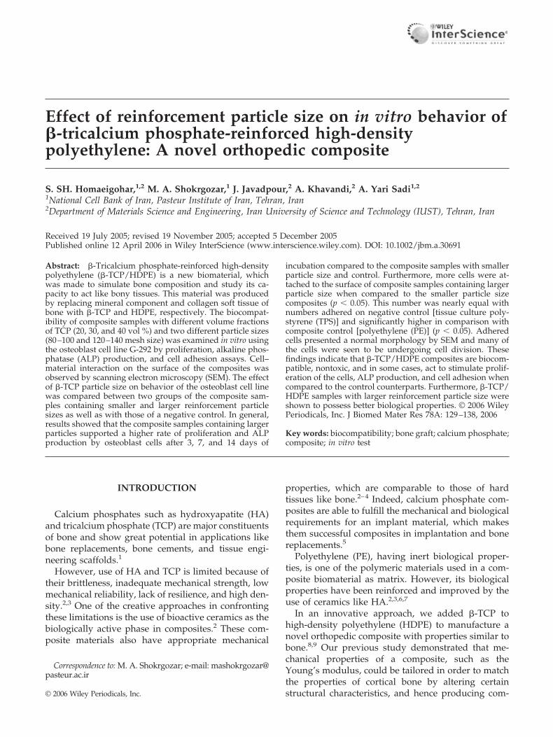
Effect of reinforcement particle size on in vitro behavior of -tricalcium phosphate-reinforced high-density polyethylene: A novel orthopedic composite S. SH. Homaeigohar, 1,2 M. A. Shokrgozar, 1 J. Javadpour, 2 A. Khavandi, 2 A. Yari Sadi 1,2 1 National Cell Bank of Iran, Pasteur Institute of Iran, Tehran, Iran 2 Department of Materials Science and Engineering, Iran University of Science and Technology (IUST), Tehran, Iran Received 19 July 2005; revised 19 November 2005; accepted 5 December 2005 Published online 12 April 2006 in Wiley InterScience (www.interscience.wiley.com). DOI: 10.1002/jbm.a.30691 Abstract: -Tricalcium phosphate-reinforced high-density polyethylene (-TCP/HDPE) is a new biomaterial, which was made to simulate bone composition and study its ca- pacity to act like bony tissues. This material was produced by replacing mineral component and collagen soft tissue of bone with -TCP and HDPE, respectively. The biocompat- ibility of composite samples with different volume fractions of TCP (20, 30, and 40 vol %) and two different particle sizes (80 –100 and 120 –140 mesh size) was examined in vitro using the osteoblast cell line G-292 by proliferation, alkaline phos- phatase (ALP) production, and cell adhesion assays. Cell– material interaction on the surface of the composites was observed by scanning electron microscopy (SEM). The effect of -TCP particle size on behavior of the osteoblast cell line was compared between two groups of the composite sam- ples containing smaller and larger reinforcement particle sizes as well as with those of a negative control. In general, results showed that the composite samples containing larger particles supported a higher rate of proliferation and ALP production by osteoblast cells after 3, 7, and 14 days of incubation compared to the composite samples with smaller particle size and control. Furthermore, more cells were at- tached to the surface of composite samples containing larger particle size when compared to the smaller particle size composites (p 0.05). This number was nearly equal with numbers adhered on negative control [tissue culture poly- styrene (TPS)] and significantly higher in comparison with composite control [polyethylene (PE)] (p 0.05). Adhered cells presented a normal morphology by SEM and many of the cells were seen to be undergoing cell division. These findings indicate that -TCP/HDPE composites are biocom- patible, nontoxic, and in some cases, act to stimulate prolif- eration of the cells, ALP production, and cell adhesion when compared to the control counterparts. Furthermore, -TCP/ HDPE samples with larger reinforcement particle size were shown to possess better biological properties. © 2006 Wiley Periodicals, Inc. J Biomed Mater Res 78A: 129 –138, 2006 Key words: biocompatibility; bone graft; calcium phosphate; composite; in vitro test INTRODUCTION Calcium phosphates such as hydroxyapatite (HA) and tricalcium phosphate (TCP) are major constituents of bone and show great potential in applications like bone replacements, bone cements, and tissue engi- neering scaffolds. 1 However, use of HA and TCP is limited because of their brittleness, inadequate mechanical strength, low mechanical reliability, lack of resilience, and high den- sity. 2,3 One of the creative approaches in confronting these limitations is the use of bioactive ceramics as the biologically active phase in composites. 2 These com- posite materials also have appropriate mechanical properties, which are comparable to those of hard tissues like bone. 2–4 Indeed, calcium phosphate com- posites are able to fulfill the mechanical and biological requirements for an implant material, which makes them successful composites in implantation and bone replacements. 5 Polyethylene (PE), having inert biological proper- ties, is one of the polymeric materials used in a com- posite biomaterial as matrix. However, its biological properties have been reinforced and improved by the use of ceramics like HA. 2,3,6,7 In an innovative approach, we added -TCP to high-density polyethylene (HDPE) to manufacture a novel orthopedic composite with properties similar to bone. 8,9 Our previous study demonstrated that me- chanical properties of a composite, such as the Young’s modulus, could be tailored in order to match the properties of cortical bone by altering certain structural characteristics, and hence producing com- Correspondence to: M. A. Shokrgozar; e-mail: mashokrgozar@ pasteur.ac.ir © 2006 Wiley Periodicals, Inc.
Transcript of Effect of reinforcement particle size onin vitro behavior of β-tricalcium phosphate-reinforced...
Effect of reinforcement particle size on in vitro behavior of�-tricalcium phosphate-reinforced high-densitypolyethylene: A novel orthopedic composite
S. SH. Homaeigohar,1,2 M. A. Shokrgozar,1 J. Javadpour,2 A. Khavandi,2 A. Yari Sadi1,2
1National Cell Bank of Iran, Pasteur Institute of Iran, Tehran, Iran2Department of Materials Science and Engineering, Iran University of Science and Technology (IUST), Tehran, Iran
Received 19 July 2005; revised 19 November 2005; accepted 5 December 2005Published online 12 April 2006 in Wiley InterScience (www.interscience.wiley.com). DOI: 10.1002/jbm.a.30691
Abstract: �-Tricalcium phosphate-reinforced high-densitypolyethylene (�-TCP/HDPE) is a new biomaterial, whichwas made to simulate bone composition and study its ca-pacity to act like bony tissues. This material was producedby replacing mineral component and collagen soft tissue ofbone with �-TCP and HDPE, respectively. The biocompat-ibility of composite samples with different volume fractionsof TCP (20, 30, and 40 vol %) and two different particle sizes(80–100 and 120–140 mesh size) was examined in vitro usingthe osteoblast cell line G-292 by proliferation, alkaline phos-phatase (ALP) production, and cell adhesion assays. Cell–material interaction on the surface of the composites wasobserved by scanning electron microscopy (SEM). The effectof �-TCP particle size on behavior of the osteoblast cell linewas compared between two groups of the composite sam-ples containing smaller and larger reinforcement particlesizes as well as with those of a negative control. In general,results showed that the composite samples containing largerparticles supported a higher rate of proliferation and ALPproduction by osteoblast cells after 3, 7, and 14 days of
incubation compared to the composite samples with smallerparticle size and control. Furthermore, more cells were at-tached to the surface of composite samples containing largerparticle size when compared to the smaller particle sizecomposites (p � 0.05). This number was nearly equal withnumbers adhered on negative control [tissue culture poly-styrene (TPS)] and significantly higher in comparison withcomposite control [polyethylene (PE)] (p � 0.05). Adheredcells presented a normal morphology by SEM and many ofthe cells were seen to be undergoing cell division. Thesefindings indicate that �-TCP/HDPE composites are biocom-patible, nontoxic, and in some cases, act to stimulate prolif-eration of the cells, ALP production, and cell adhesion whencompared to the control counterparts. Furthermore, �-TCP/HDPE samples with larger reinforcement particle size wereshown to possess better biological properties. © 2006 WileyPeriodicals, Inc. J Biomed Mater Res 78A: 129–138, 2006
Key words: biocompatibility; bone graft; calcium phosphate;composite; in vitro test
INTRODUCTION
Calcium phosphates such as hydroxyapatite (HA)and tricalcium phosphate (TCP) are major constituentsof bone and show great potential in applications likebone replacements, bone cements, and tissue engi-neering scaffolds.1
However, use of HA and TCP is limited because oftheir brittleness, inadequate mechanical strength, lowmechanical reliability, lack of resilience, and high den-sity.2,3 One of the creative approaches in confrontingthese limitations is the use of bioactive ceramics as thebiologically active phase in composites.2 These com-posite materials also have appropriate mechanical
properties, which are comparable to those of hardtissues like bone.2–4 Indeed, calcium phosphate com-posites are able to fulfill the mechanical and biologicalrequirements for an implant material, which makesthem successful composites in implantation and bonereplacements.5
Polyethylene (PE), having inert biological proper-ties, is one of the polymeric materials used in a com-posite biomaterial as matrix. However, its biologicalproperties have been reinforced and improved by theuse of ceramics like HA.2,3,6,7
In an innovative approach, we added �-TCP tohigh-density polyethylene (HDPE) to manufacture anovel orthopedic composite with properties similar tobone.8,9 Our previous study demonstrated that me-chanical properties of a composite, such as theYoung’s modulus, could be tailored in order to matchthe properties of cortical bone by altering certainstructural characteristics, and hence producing com-
Correspondence to: M. A. Shokrgozar; e-mail: [email protected]
© 2006 Wiley Periodicals, Inc.

posites that would be structurally and mechanicallycompatible with bone.8 In these investigations, ourresults along with previous reports showed that thecomposites containing smaller ceramic particles couldhave better mechanical properties, for example ahigher Young’s modulus.7,8 In addition, our previousbiological tests have demonstrated appropriate bio-logical properties and biocompatibility for such anorthopedic composite.8
In this study, the �-TCP/HDPE composite samplescontaining �-TCP with two particle sizes as reinforc-ing material were prepared and the effect of reinforce-ment particle size on proliferation, differentiation, andadhesion of the osteoblast cell line G-292 was evalu-ated.
MATERIALS AND METHODS
Raw materials
Medical-grade TCP (Merck, Germany) was used as theceramic filler material. The matrix polymer was a HDPE(0.944 g/cm3) (Arak Petrochemical Co., Iran).
Sample preparation
Depending on the phase diagram of calcium phosphatecompounds, a special heat treatment and mechanical pro-cess consisting of pressing, sintering, and grinding wereused to obtain the �-TCP with the desired particle sizedistributions namely 80–100 and 120–140.8 These two par-ticle size distributions were then used in fabrication of�-TCP/HDPE composite samples by first blending andcompounding the ceramic powder and granular PE to pro-duce a homogenous composite mixture. The primary pre-forms were then compression molded into the desiredshape.8 Ceramic filler distribution in the composite sampleswas investigated by examining the samples under scanningelectron microscopy (SEM). The composite samples contain-ing �-TCP filler with different volume fractions and particlesizes were fabricated using the above procedure (Table I).Samples with a thickness and diameter of 2 and 10 mm,respectively, were used for SEM and attachment assay,
while proliferation and differentiation assays were per-formed with samples with the same thickness but 2 mmdiameter. All the samples were sterilized by �-irradiation ata dose of 25 kGy using standard procedure for medicaldevices.
In vitro cell culture
The human primary osteogenic sarcoma cell line G-292Colon A141B1 (NCBI C-116; National Cell Bank of Iran,Pasteur Institute of Iran, Tehran, Iran) was cultured in Dul-becco’s Modified Eagle’s Medium (DMEM) (GIBCO, Scot-land) supplemented with 10% fetal calf serum (FCS) (Se-romed, Germany), 100 U/mL penicillin and 100 �g/mLstreptomycin (Sigma, USA). The cells were harvested with0.25% trypsin-EDTA solution (Sigma) in phosphate-bufferedsaline (PBS; pH 7.4) and seeded onto the composites at adensity of 2 � 103 cells/well in 96-well microtiter plates(Nunc, Denmark) for cytotoxicity/proliferation and differ-entiation assays. These cells were cultured on the surface ofthe composite samples at 8 � 103 cells/well in 24-well plates(Greiner, Germany) for attachment and cell morphologydetermination assays. The cultures were incubated at 37°Cin a humidified atmosphere with 5% CO2 for 14 days, withobservations made at 3, 7, and 14 day time points. Theculture medium was changed every 3 days.
Cell proliferation
Proliferation rate of osteoblast cells (G-292) over the com-posite samples was measured using dimethylthiazol diphe-nyl tetrazolium bromide (MTT) assay. Briefly, cells wereplated into a 96-well microtiter plate at 2 � 103 cells/well.The composite samples with different volume fractions andparticle size of �-TCP were placed on G-292 cells after anovernight incubation at 37°C with 5% CO2 in a humidifiedatmosphere. Four wells with no composite sample wereused as negative controls [tissue culture polystyrene (TPS)].Composite controls consisted of wells containing only PE.The plates were incubated for a period of 2 weeks withmedia changed every 3 days. At days 3, 7, and 14, thecomposites were removed from the wells and 10 �L of a 5mg/mL solution of MTT (Sigma) was added to each wellfollowed by incubation for 5 h at 37°C. The formazan crys-
TABLE ICharacteristics of the Composite Samples Prepared in This Study
SampleCode
TCP VolumeFraction (%)
Particle MeshSize
TCP Mass(g)
PE Mass(g)
CompositeMass (g)
CompositeDensity (g/cm3)
A 20 80–100 19.64 23.87 43.52 1.36B 20 120–140 19.64 23.87 43.52 1.36C 30 80–100 29.5 21.1 50.61 1.58D 30 120–140 29.5 21.1 50.61 1.58E 40 80–100 39.29 17.98 57.28 1.79F 40 120–140 39.29 17.98 57.28 1.79
130 HOMAEIGOHAR ET AL.
Journal of Biomedical Materials Research Part A DOI 10.1002/jbm.a
tals formed were then dissolved by addition of 100 �Lisopropanol (Sigma) per well. The plates were then incu-bated at 37°C for 10 min and transferred to 4°C for 15 minprior to absorbance measurements. The optical density (OD)was recorded on a multiwell microplate reader (ICN, Swit-zerland) at 570 nm. The experiment was repeated threetimes.
Cell differentiation
Effect of reinforcement particle size on osteoblastic phe-notype was determined biochemically through assay of ALPproduction by G-292 cells. Ninety-six-well microtiter plateswere prepared as described earlier. On incubation days 3, 7,and 14, the cells were lysed and ALP activity was deter-mined by a Cobas-Bio (Roche, UK) centrifugal analyzer us-ing p-nitrophenol phosphate in diethanolamine buffer (Ran-dox, UK) as substrate. The reaction product, p-nitrophenol,is yellow at alkaline pH (9.8) and can be quantified at awavelength of 405 nm. The experiment was repeated threetimes.
Attachment assay
G-292 cells were seeded onto composite samples placed ina 24-well plate at a concentration of 8 � 103 cells/well andwere allowed to attach to the surface of the composites.Appropriate negative and composite control wells were alsoprepared as described earlier. The plates were incubated fora period of 2 weeks with media changed every 3 days. Atvarious time intervals (3, 7, and 14 days), the adhered cellswere removed by trypsinization and live cells were enumer-ated on a hemocytometer, using 0.2% trypan blue dye ex-clusion method. The experiment was repeated three times.
Cell morphology
Twenty-four-well plates were prepared as described forattachment assay. The plates were incubated at 37°C for72 h. Then, the cells were fixed using 2.5% glutaraldehyde in0.2M PBS and they were dehydrated through a series ofalcohol concentrations (70, 80, 90, and two times in 100%).Dehydration was followed by air drying (overnight). Thesamples were next sputter-coated before examination by aJeol SEM at an accelerating voltage of 15 keV.
Statistical analysis
Statistical analysis was performed on SPSS statisticalpackage and values were considered significant at p � 0.05.The Student’s t-test was used to compare the data for thecomposite and control samples.
RESULTS
Material characteristics
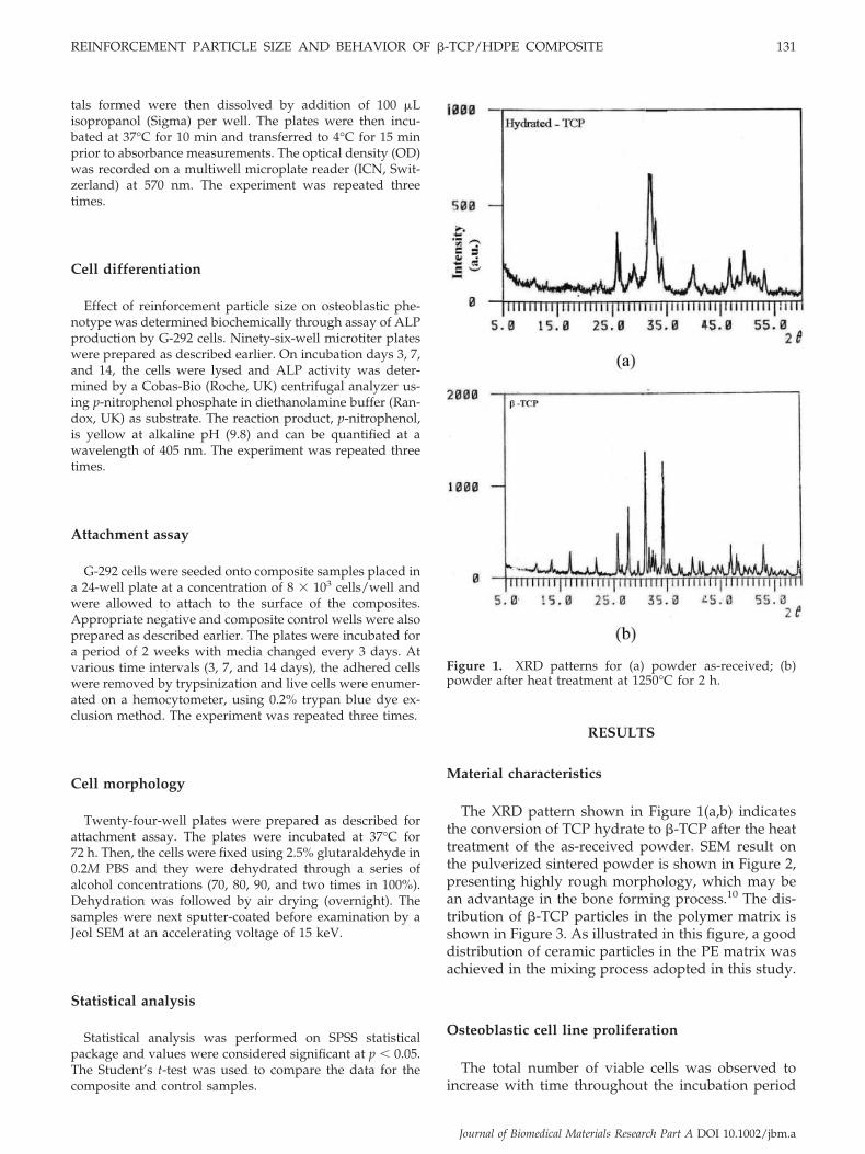
The XRD pattern shown in Figure 1(a,b) indicatesthe conversion of TCP hydrate to �-TCP after the heattreatment of the as-received powder. SEM result onthe pulverized sintered powder is shown in Figure 2,presenting highly rough morphology, which may bean advantage in the bone forming process.10 The dis-tribution of �-TCP particles in the polymer matrix isshown in Figure 3. As illustrated in this figure, a gooddistribution of ceramic particles in the PE matrix wasachieved in the mixing process adopted in this study.
Osteoblastic cell line proliferation
The total number of viable cells was observed toincrease with time throughout the incubation period
Figure 1. XRD patterns for (a) powder as-received; (b)powder after heat treatment at 1250°C for 2 h.
REINFORCEMENT PARTICLE SIZE AND BEHAVIOR OF �-TCP/HDPE COMPOSITE 131
Journal of Biomedical Materials Research Part A DOI 10.1002/jbm.a

(14 days) for all samples, including the composites,composite control (PE), and negative control (TPS).
Total cell count was seen to peak on day 14. Incomparison with PE, the composite samples show ahigher, and in some cases, an equal proliferation rate(Fig. 4). The difference between the proliferation ratesof sample A and PE was more significant on day 7 (p �
0.01) as shown in Figure 4. Also, in comparison withTPS, the composite samples support almost equal pro-liferation rate with no significant difference betweenthe composites and TPS (Fig. 4).
On days 3, 7, and 14 the measured growth rate forthe composites A and C was always higher than theother samples. In addition, the composites containinglarger particle size, such as A and C, support higherproliferation rate than the smaller ones, as indicatedby the difference in OD between samples A and B, andA and F, with significant p values of � 0.01 and � 0.05,respectively.
Differentiation
ALP production by G-292 cells over all the compos-ite samples was seen to peak on day 7. Afterwards,although the cell proliferation was still on the rise, anegligible decrease in ALP production by osteoblasticcells was observed. A similar trend in ALP productionwas also observed for the PE and TPS samples. Theresults showed that the ALP production rate is higherin almost all of the composites compared to PE, whilea similar differentiation behavior was seen in all thecomposites and TPS (Fig. 5). Interestingly, there wasan equal, or in some cases, even a higher ALP produc-tion by G-292 cells after incubation with compositesamples compared to TPS, especially in the samples Cand E with 30 and 40 vol % TCP (Fig. 5).
Figure 3. SEM micrograph showing the distribution of theTCP particles in the PE matrix (sample C with 30 vol %TCP).
Figure 2. SEM micrographs of aggregate particles (80–100mesh size).
132 HOMAEIGOHAR ET AL.
Journal of Biomedical Materials Research Part A DOI 10.1002/jbm.a

Overall, there is an unpredictable trend among var-ious samples in terms of ALP production; however, ingeneral most of the composites containing larger par-ticle size, such as A and E, show higher ALP produc-tion than the smaller size composites.
Osteoblastic cell attachment
The number of G-292 cells adhered to the compos-ites, PE, and TPS are shown in Figure 6. On day 3 ofincubation, the number of adhered cells on all of thecomposite samples was significantly higher than thoseon the PE sample (p � 0.05). This difference becameeven more significant on day 14 (p � 0.01). The attach-ment results showed a significant difference betweenthe samples A and B, A and D, and A and E (p � 0.05)on day 7. In this time, the number of adhered cells onC and E was significantly higher than those on B andD (p � 0.01). In addition, the results showed a signif-icant difference between the number of adhered cellsto sample A and TPS (p � 0.01). On day 14, thenumber of cells adhered to all composites was higherthan those on PE, as shown in Figure 6. This differencebetween PE and the composite samples A, B, and Cwas significant (p � 0.01 and � 0.05). A significantdifference between the A and D, and A and F com-
posites (p � 0.01) is shown in Figure 6. The results alsoshowed a significant increase (p � 0.05) in cell attach-ment on TPS in comparison to PE on the 3rd and 14thday of incubation.
These results indicate that the composites contain-ing larger particle size, such as A and C, supportedproliferation better than the smaller particle size com-posites.
Morphological evaluation
Cell morphology studies using SEM showed attach-ment of cells with normal osteoblast morphology; flat-tened cells with small processes attaching to the sur-rounding material [Fig. 7(a–c)]. As shown in Figure 7many of the cells were in the process of cell division[Fig. 7(d–g)].
DISCUSSION
In the present study, the effect of reinforcementparticle size on different biological properties of os-teoblast cells, such as proliferation, attachment, cellu-lar morphology, and differentiation, was investigated.
Figure 4. Cell proliferation of G-292 grown on �-TCP/HDPE composites at various time points. PE, composite control; A,20 vol % TCP (80–100); B, 20 vol % TCP (120–140); C, 30 vol % TCP (80–100); D, 30 vol % TCP (120–140); E, 40 vol % TCP(80–100); F, 40 vol % TCP (120–140); and TPS, negative control. *p � 0.05 and **p � 0.01.
REINFORCEMENT PARTICLE SIZE AND BEHAVIOR OF �-TCP/HDPE COMPOSITE 133
Journal of Biomedical Materials Research Part A DOI 10.1002/jbm.a

This effect was compared with PE and TPS, whichwere composite and negative controls, respectively.
Calcium phosphate ceramics with a highly roughsurface and structure have shown to possess strongosteogenic potential, promoting new bone formationon their surface,10,11 and this is the reason for the useof aggregated particles in the preparation of compos-ite samples in this study.
There is a simultaneous increase in both prolifera-tion and differentiation of cells until day 7. On day 14,ALP production decreased while the number of G-292cells increased. This may be due to the occupation ofthe space in the wells by the cells and consequentdecrease in differentiation rate on day 14 compared tothat on day 7. In comparison to PE sample, the bio-logical performance of composite samples was better,and in some days, the difference between compositesand PE in supporting proliferation of G-292 cells wassignificant (p � 0.05). This occurred in spite of the factthat PE with its lower density compared to that on thecomposites did not occupy much of the space in thewells.
The adherence of G-292 cells to the composites wasas high as that on TPS, and in some cases, even higher.These results demonstrate that the surface of the com-posites was sufficiently stable to support adherenceand growth of osteoblastic cells.
Among the composites, samples containing larger
particles, such as A and C, showed better results inproliferation and attachment. Similar results were ob-tained with respect to ALP production. In general, itcould be claimed that the composites with larger par-ticles show better biological performance.
Previous reports indicate that the calcium concen-tration at the interfacial layer between cells and com-posite samples favorably supports the growth of os-teoblastic cells.12 Extracellular calcium concentrationhas been considered to be a significant factor affectingthe proliferation and differentiation of osteoblasticcells.13 In previous studies, it has been demonstratedthat the high solubility of the surfaces and high releaserate of PO4 and Ca components into the medium arecrucial factors in stimulating the growth of osteoblas-tic cells.14–16 As we observed, even with the occu-pancy of a part of the well space by composites, boththe cell density and ALP activity of G-292 cells wereenhanced as compared to PE and TPS.
However, the surface of TCP ceramic is unstable andthis ceramic with its high solubility rate and pH on thesurface tends to become more acidic with incubationperiod.17 Kreiger et al. reported that the metabolic aci-dosis inhibited osteoblastic activity and decreased theirALP activity.18 However, our results showed that boththe cell density and ALP activity were enhanced on thesurface of composites as compared to PE and TPS inaccordance with a previous report.17 Since the high sol-
Figure 5. ALP activity for G-292 cells on �-TCP/HDPE composites at various time points. PE, composite control; A, 20 vol% TCP (80–100); B, 20 vol % TCP (120–140); C, 30 vol % TCP (80–100); D, 30 vol % TCP (120–140); E, 40 vol % TCP (80–100);F, 40 vol % TCP (120–140); and TPS, negative control.
134 HOMAEIGOHAR ET AL.
Journal of Biomedical Materials Research Part A DOI 10.1002/jbm.a
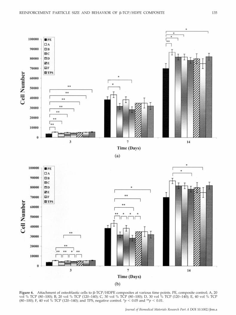
Figure 6. Attachment of osteoblastic cells to �-TCP/HDPE composites at various time points. PE, composite control; A, 20vol % TCP (80–100); B, 20 vol % TCP (120–140); C, 30 vol % TCP (80–100); D, 30 vol % TCP (120–140); E, 40 vol % TCP(80–100); F, 40 vol % TCP (120–140); and TPS, negative control. *p � 0.05 and **p � 0.01.
REINFORCEMENT PARTICLE SIZE AND BEHAVIOR OF �-TCP/HDPE COMPOSITE 135
Journal of Biomedical Materials Research Part A DOI 10.1002/jbm.a

ubility of the surface also increases the rate of the releaseof both PO4 and Ca components into the medium,17 thesurface of TCP was considered to be effective for stimu-lating the growth of osteoblastic cells. However, ex-tremely high solubility and reactivity of bioceramic sur-faces may damage the adherent cells.17,19 The highsolubility of the thin surface layer of the TCP ceramicwas found to be the dominant factor that decreased theviability of the cells by causing their rupture during theinitial anchoring phase.20 Thus, the poor biological per-
formance of composite samples with smaller particlesize may be related to the fact that smaller particles causemore contact area with the medium, and this finallyresults in higher solubility of exposed TCP particles atthe surface compared to large particle size composites.Similar observation was made by Pioletti et al. on TCPparticles used in a kind of cement.21 They reported thatthe particles could adversely affect the osteoblast func-tions as a decrease in the viability, proliferation, andproduction of extracellular matrix was observed and
Figure 7. SEM micrographs showing the cell morphology in different states of growth, including division and flattening.
136 HOMAEIGOHAR ET AL.
Journal of Biomedical Materials Research Part A DOI 10.1002/jbm.a

smaller particles produced stronger negative effects onosteoblast functions than larger ones. It was furthershown by Suzuki et al. that the moderate surface solu-bility of exposed ceramic on the surface of biomaterialcan help to promote the formation of a harmoniousinterface between the implanted material and the sur-rounding tissues, resulting in the development of thenatural bone tissue structures without inflammatory re-actions.12
Our findings, indicating more proliferation and dif-ferentiation of cells over composite samples comparedto PE and TPS and better biocompatibility of large
particle size composites, are in accordance with thereports already mentioned. It has also been suggestedthat the dissolution of Ca ions from the Ca-P surfaceresults in an interfacial supersaturated condition withthe already present Ca ions, which in turn leads to theprecipitation of carbonated apatite.22,23 The newlyformed carbonated apatite can influence the adhesionand calcification of bone cells.24,25
Morphology observation by SEM showed thatG-292 cells were able to adhere and proliferate on thematerials; therefore, the composites were demon-strated to support normal growth of osteoblast cells.
Figure 7. (Continued)
REINFORCEMENT PARTICLE SIZE AND BEHAVIOR OF �-TCP/HDPE COMPOSITE 137
Journal of Biomedical Materials Research Part A DOI 10.1002/jbm.a

CONCLUSIONS
Human osteoblast cells (G-292) were cultured on�-TCP/HDPE composites as a model to assess theeffect of reinforcement particle size, an important fac-tor in determining the mechanical properties of com-posites, on in vitro osteoconductivity. The cells wereattached to and proliferated on �-TCP/HDPE sub-strates on a scale comparable to or even higher thanTPS. The cells studied exhibited similar or better os-teoblastic differentiation on �-TCP/HDPE substratesthan TPS, as evidenced by alkaline phosphatase (ALP)activity over a period of 14 days. The composite sam-ples also showed improved behavior over the PE sam-ple, and in some cases, this was rather significant. Theabove results reflect the presence of a bioactive com-ponent on the surface of composite samples, whichstimulated the biological activity of cells in the culturemedium.
Our observations also showed that the compositesamples containing larger TCP particles had signifi-cantly enhanced biological properties among thetested composite samples. In conclusion, this novelcomposite biomaterial can fulfill both mechanical andbiological properties by choosing a proper combina-tion of particle size.
The authors thank the staff of X-ray and SEM laboratoriesof Department of Materials and Metallurgy Engineering,IUST, Milad Pathobiology Laboratory, Polymer Departmentof Amir Kabir Industrial University, Atomic Energy Orga-nization of Iran, Iran Composite Institute as well as IranMaterials and Energy Research Center. Special thanks to thestaff of National Cell Bank of Iran, Pasteur Institute of Iranfor their assistance and useful consultations. Thanks are alsodue to Dr. R. Payam Khaja Pasha of National Cell Bank ofIran, Pasteur Institute of Iran for editorial assistance.
References
1. Ramachandra RR, Roopa HN, Kannan TS. Solid state synthesisand thermal stability of HAP and HAP-�-TCP composite ce-ramic powders. J Mater Sci Mater Med 1997;8:511–518.
2. Hench LL. Bioceramics. J Am Ceram Soc 1998;81:1705–1728.3. Ramakrishna S, Mayer J, Wintermantel E, Leong KW. Biomed-
ical application of polymer-composite materials: A review.Compos Sci Tech 2001;61:1189–1224.
4. Evans SL, Gregson PJ. Composite technology in load-bearingorthopaedic implants. Biomaterials 1998;19:1329–1342.
5. Ravigavital N, Landa-Canovas AR, Gonzalez-Calbet JM, Val-let-Regf M. Structural study and stability of hydroxyapatiteand �-tricalcium phosphate: Two important bioceramics.J Biomed Mater Res 2000;51:660–668.
6. Bronzino JD. Biomedical Engineering Handbook, 2nd ed, Vol.1. Boca Raton, FL: CRC Press; 2000.
7. Wang M, Joseph R, Bonfield W. Hydroxyapatite–polyethylenecomposites for bone substitution: Effects of ceramic particlesize and morphology. Biomaterials 1998;19:2357–2366.
8. Homaeigohar SSH, Yari Sadi A, Javadpour J, Khavandi A. Theeffect of reinforcement volume fraction and particle size on themechanical properties of �-tricalcium phosphate–high densitypolyethylene composites. J Eur Cer Soc 2006;26(3):273–278.
9. Homaeigohar SSH, Shokrgozar MA, Yari Sadi A, Khavandi A,Javadpour J, Hosseinalipour M. In vitro evaluation of biocom-patibility of �-tricalcium phosphate reinforced high densitypolyethylene: An orthopedic composite. J Biomed Mater Res A2005;75:14–22.
10. Yuan H, Yang Z, Li Y, Zhang X, De Bruijn JD, De Groot K.Osteoinduction by calcium phosphate biomaterials. J Mater SciMater Med 1998;9:723–726.
11. Goshima J, Goldberg VM, Caplan AI. The osteogenic potentialof culture-expanded rat marrow mesenchymal cells assayed invivo in calcium phosphate ceramic blocks. Clin Orthop RelatRes 1991;262:298–311.
12. Suzuki T, Hukkanen M, Ohashi R, Yokogawa Y, Nishizawa K,Nagata F, Buttery L, Polka J. Growth and adhesion of osteo-blast-like cells derived from neonatal rat calvaria on calciumphosphate ceramics. J Biosci Bioeng 2000;89:18–26.
13. Loza J, Carpino L, Lawless G, Marzec N, Dziak R. Role ofextracellular calcium influx in EGF-induced osteoblastic cellproliferation. Bone 1995;16:341–347.
14. Kanatani M, Sugimoto T, Fukase M, Fugita T. Effect of elevatedextracellular calcium on the proliferation of osteoblasticMC3T3–E1 cells: Direct and indirect effects via monocytes.Biochem Biophys Res Commun 1991;181:1425–1430.
15. Ehara A, Ogata K, Imazato S, Ebisu S, Nakano T, Umakoshi Y.Effects of a TCP and TetCP on MC3T3–E1 proliferation, differ-entiation and mineralization. Biomaterials 2003;24:831–836.
16. Monchau F, Lefevre A, Descamps M, Belquin-Myrdycz A,Laffargue P, Hildebrand HF. In vitro studies of human and ratosteoclast activity on hydroxyapatite, �-tricalcium phosphate,calcium carbonate. Biomol Eng 2002;19:143–152.
17. Suzuki T, Yamamoto T, Toriyama M, Nishizawa K, YokogawaY, Mucalo RM, Nagata F, Kawamoto Y, Kameyama T. Surfaceinstability of calcium phosphate ceramics in tissue culturemedium and the effect on adhesion and growth of anchorage-dependent animal cells. J Biomed Mater Res 1997;34:507–517.
18. Kreiger NS, Sessler NE, Bunshinsky DA. Acidosis inhibitsosteoblastic and stimulates osteoclastic activity in vitro. Am JPhysiol 1992;262:442–448.
19. Suzuki T, Toriyama M, Kawamoto Y, Yokogawa Y, KawamuraS. The adhesiveness and growth of anchorage dependent ani-mal cells on biocompatible ceramic culture carriers. J FermentBioeng 1991;72:450–456.
20. Suzuki T, Ohashi R, Yokogawa Y, Nishizawa K, Nakata F,Kawamoto Y, Kameyama T, Toriyama M. Initial anchoring andproliferation of fibroblast L-929 cells on unstable surface ofcalcium phosphate ceramics. J Biosci Bioeng 1999;87:320–327.
21. Pioletti DP, Takei H, Lin T, Van Landuyt P, Jun Ma Q, Kwon SY,Paul Sung KL. The effects of calcium phosphate cement particleson osteoblast functions. Biomaterials 2000;21:1103–1114.
22. Barralet J, Best S, Bonfield W. Carbonate substitution in pre-cipitated hydroxyapatite: An investigation into the effects ofreaction temperature and bicarbonate ion concentration.J Biomed Mater Res 1998;41:79–86.
23. Hulshoff JEG, Van Digik K, De Rietveld JE, Rietvelt FJR, GinselLA, Jansen JA. Interfacial phenomena : An in vitro study of theeffect of calcium phosphate (Ca-P) ceramic on bone formation.J Biomed Mater Res 1998;40:464–474.
24. Layman DL, Ardoin RC. An in vitro system for studying os-teointegration of dental implants utilizing cells grown on densehydroxyapatite disks. J Biomed Mater Res 1998;40:282–290.
25. Maxian SH, Stefano TD, Melican MC, Tiku ML, Zawadsky JP.Bone cell behavior on Matrigel-coated Ca/P coatings of vary-ing crystallinities. J Biomed Mater Res 1998;40:171–179.
138 HOMAEIGOHAR ET AL.
Journal of Biomedical Materials Research Part A DOI 10.1002/jbm.a