Orthopedic Expansion and Protraction of the Maxilla in Cleft
9
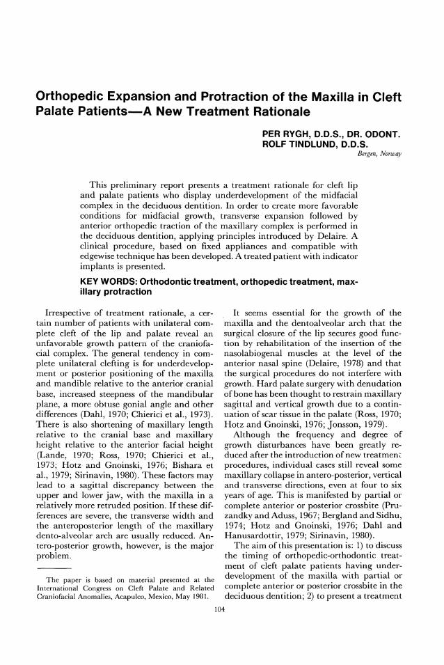
Orthopedic Expansion and Protraction of the Maxilla in Cleft Palate Patients-A New Treatment Rationale PER RYGH, D.D.S., DR. ODONT. ROLF TINDLUND, D.D.S. Bergen, Norway This preliminary report presents a treatment rationale for cleft lip and palate patients who display underdevelopment of the midfacial complex in the deciduous dentition. In order to create more favorable conditions for midfacial growth, transverse expansion followed by anterior orthopedic traction of the maxillary complex is performed in the deciduous dentition, applying principles introduced by Delaire. A clinical procedure, based on fixed appliances and compatible with edgewise technique has been developed. A treated patient with indicator implants is presented. KEY WORDS: Orthodontic treatment, orthopedic treatment, max- illary protraction Irrespective of treatment rationale, a cer- tain number of patients with unilateral com- plete cleft of the lip and palate reveal an unfavorable growth pattern of the craniofa- cial complex. The general tendency in com- plete unilateral clefting is for underdevelop- ment or posterior positioning of the maxilla and mandible relative to the anterior cranial base, increased steepness of the mandibular plane, a more obtuse gonial angle and other differences (Dahl, 1970; Chierici et al., 1973). There is also shortening of maxillary length relative to the cranial base and maxillary height relative to the anterior facial height (Lande, 1970; Ross, 1970; Chierici et al., 1973; Hotz and Gnoinski, 1976; Bishara et al., 1979; Sirinavin, 1980). These factors may lead to a sagittal discrepancy between the upper and lower jaw, with the maxilla in a relatively more retruded position. If these dif- ferences are severe, the transverse width and the anteroposterior length of the maxillary dento-alveolar arch are usually reduced. An- tero-posterior growth, however, is the major problem. The paper is based on material presented at the International Congress on Cleft Palate and Related Craniofacial Anomalies, Acapulco, Mexico, May 1981. It seems essential for the growth of the ‘maxilla and the dentoalveolar arch that the surgical closure of the lip secures good func- tion by rehabilitation of the insertion of the nasolabiogenal muscles at the level of the anterior nasal spine (Delaire, 1978) and that the surgical procedures do not interfere with growth. Hard palate surgery with denudation of bone has been thought to restrain maxillary sagittal and vertical growth due to a contin- uation of scar tissue in the palate (Ross, 1970; Hotz and Gnoinski, 1976; Jonsson, 1979). Although the frequency and degree of growth disturbances have been greatly re- duced after the introduction of new treatment procedures, individual cases still reveal some maxillary collapse in antero-posterior, vertical and transverse directions, even at four to six years of age. This is manifested by partial or complete anterior or posterior crossbite (Pru- zandky and Aduss, 1967; Bergland and Sidhu, 1974; Hotz and Gnoinski, 1976; Dahl and Hanusardottir, 1979; Sirinavin, 1980). The aim of this presentation is: 1) to discuss the timing of orthopedic-orthodontic treat- ment of cleft palate patients having under- development of the maxilla with partial or complete anterior or posterior crossbite in the deciduous dentition; 2) to present a treatment 104
-
Upload
khangminh22 -
Category
Documents
-
view
3 -
download
0
Transcript of Orthopedic Expansion and Protraction of the Maxilla in Cleft
Orthopedic Expansion and Protraction of the Maxilla in Cleft
Palate Patients-A New Treatment Rationale
PER RYGH, D.D.S., DR. ODONT.
ROLF TINDLUND, D.D.S.Bergen, Norway
This preliminary report presents a treatment rationale for cleft lip
and palate patients who display underdevelopment of the midfacial
complex in the deciduous dentition. In order to create more favorable
conditions for midfacial growth, transverse expansion followed by
anterior orthopedic traction of the maxillary complex is performed in
the deciduous dentition, applying principles introduced by Delaire. A
clinical procedure, based on fixed appliances and compatible with
edgewise technique has been developed. A treated patient with indicator
implants is presented.
KEY WORDS: Orthodontic treatment, orthopedic treatment, max-
illary protraction
Irrespective of treatment rationale, a cer-
tain number of patients with unilateral com-
plete cleft of the lip and palate reveal an
unfavorable growth pattern of the craniofa-
cial complex. The general tendency in com-
plete unilateral clefting is for underdevelop-
ment or posterior positioning of the maxilla
and mandible relative to the anterior cranial
base, increased steepness of the mandibular
plane, a more obtuse gonial angle and other
differences (Dahl, 1970; Chierici et al., 1973).
There is also shortening of maxillary length
relative to the cranial base and maxillary
height relative to the anterior facial height
(Lande, 1970; Ross, 1970; Chierici et al.,
1973; Hotz and Gnoinski, 1976; Bishara et
al., 1979; Sirinavin, 1980). These factors may
lead to a sagittal discrepancy between the
upper and lower jaw, with the maxilla in a
relatively more retruded position. If these dif-
ferences are severe, the transverse width and
the anteroposterior length of the maxillary
dento-alveolar arch are usually reduced. An-
tero-posterior growth, however, is the major
problem.
The paper is based on material presented at theInternational Congress on Cleft Palate and RelatedCraniofacial Anomalies, Acapulco, Mexico, May 1981.
It seems essential for the growth of the
‘maxilla and the dentoalveolar arch that the
surgical closure of the lip secures good func-
tion by rehabilitation of the insertion of the
nasolabiogenal muscles at the level of the
anterior nasal spine (Delaire, 1978) and that
the surgical procedures do not interfere with
growth. Hard palate surgery with denudation
of bone has been thought to restrain maxillary
sagittal and vertical growth due to a contin-
uation of scar tissue in the palate (Ross, 1970;
Hotz and Gnoinski, 1976; Jonsson, 1979).
Although the frequency and degree of
growth disturbances have been greatly re-
duced after the introduction of new treatment
procedures, individual cases still reveal some
maxillary collapse in antero-posterior, vertical
and transverse directions, even at four to six
years of age. This is manifested by partial or
complete anterior or posterior crossbite (Pru-
zandky and Aduss, 1967; Bergland and Sidhu,
1974; Hotz and Gnoinski, 1976; Dahl and
Hanusardottir, 1979; Sirinavin, 1980).
The aim of this presentation is: 1) to discuss
the timing of orthopedic-orthodontic treat-
ment of cleft palate patients having under-
development of the maxilla with partial or
complete anterior or posterior crossbite in the
deciduous dentition; 2) to present a treatment
104
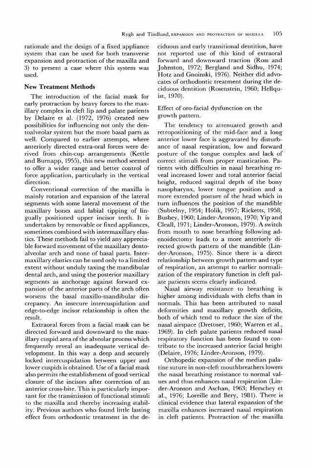
Rygh and Tindlund, EXPANSION AND PROTRACTION OF MAXILLA 10
rationale and the design of a fixed appliance
system that can be used for both transverse
expansion and protraction of the maxilla and
3) to present a case where this system was
used.
New Treatment Methods
The introduction of the facial mask for
early protraction by heavy forces to the max-
illary complex in cleft lip and palate patients
by Delaire et al. (1972, 1976) created new
possibilities for influencing not only the den-
toalveolar system but the more basal parts as
well. Compared to earlier attempts, where
anteriorly directed extra-oral forces were de-
rived from chin-cup arrangements (Kettle
and Burnapp, 1955), this new method seemed
to offer a wider range and better control of
force application, particularly in the vertical
direction.
Conventional correction of the maxilla is
mainly rotation and expansion of the lateral
segments with some lateral movement of the
maxillary bones and labial tipping of lin-
gually positioned upper incisor teeth. It is
_ undertaken by removable or fixed appliances,
sometimes combined with intermaxillary elas-
tics. These methods fail to yield any apprecia-
ble forward movement of the maxillary dento-
alveolar arch and none of basal parts. Inter-
maxillary elastics can be used only to a limited
extent without unduly taxing the mandibular
dental arch, and using the posterior maxillary
segments as anchorage against forward ex-
pansion of the anterior parts of the arch often
worsens the basal maxillo-mandibular dis-
crepancy. An insecure intercuspidation and
edge-to-edge incisor relationship is often the
result.
Extraoral forces from a facial mask can be -
directed forward and downward to the max-
illary cuspid area of the alveolar process which
frequently reveal an inadequate vertical de-
velopment. In this way a deep and securely
locked intercuspidation between upper and
lower cuspids is obtained. Use of a facial mask
also permits the establishment of good vertical
closure of the incisors after correction of an
anterior cross-bite. This is particularly impor-
tant for the transmission of functional stimuli
to the maxilla and thereby increasing stabil-
ity. Previous authors who found little lasting
effect from orthodontic treatment in the de-
3
ciduous and early transitional dentition, have
not reported use of this kind of extraoral
forward and downward traction (Ross and
Johnston, 1972; Bergland and Sidhu, 1974;
Hotz and Gnoinski, 1976). Neither did advo-
cates of orthodontic treatment during the de-
ciduous dentition (Rosenstein, 1960; Hellqu-
ist, 1970).
Effect of oro-facial dysfunction on the
growth pattern.
The tendency to attenuated growth and
retropositioning of the mid-face and a long
anterior lower face is aggravated by disturb-
ance of nasal respiration, low and forward
posture of the tongue complex and lack of
correct stimuli from proper mastication. Pa-
tients with difficulties in nasal breathing re-
veal increased lower and total anterior facial
height, reduced sagittal depth of the bony
nasopharynx, lower tongue position and a
more extended posture of the head which in
turn influences the position of the mandible
(Subtelny, 1954; Holik, 1957; Ricketts, 1958;
Bushey, 1960; Linder-Aronson, 1970; Yip and
Cleall, 1971; Linder-Aronson, 1979). A switch
from mouth to nose breathing following ad-
enoidectomy leads to a more anteriorly di-
rected growth pattern of the mandible (Lin-
der-Aronson, 1975). Since there is a direct
relationship between growth pattern and type
of respiration, an attempt to earlier normali-
zation of the respiratory function in cleft pal-
ate patients seems clearly indicated.
Nasal airway resistance to breathing is
higher among individuals with clefts than in
normals. This has been attributed to nasal
deformities and maxillary growth deficits,
both of which tend to reduce the size of the
nasal airspace (Drettner, 1960; Warren et al.,
1969). In cleft palate patients reduced nasal
respiratory function has been found to con-
tribute to the increased anterior facial height
(Delaire, 1976; Linder-Aronson, 1979).
Orthopedic expansion of the median pala-
tine suture in non-cleft mouthbreathers lowers
the nasal breathing resistance to normal val-
ues and thus enhances nasal respiration (Lin-
der-Aronson and Aschan, 1963; Herschey et
al., 1976; Loreille and Bery, 1981). There is
clinical evidence that lateral expansion of the
maxilla enhances increased nasal respiration
in cleft patients. Protraction of the maxilla
106 Cleft Palate Journal, April 1982, Vol. 19 No. 2
increases the vertical height as well as the
sagittal length of the maxilla. The influence
of protraction on the quality of respiration is
presently under investigation.
The experiments carried out by Harvold et
al. (1972, 1973) on monkeys in which the
tongue was induced to adopt a lower position
led to a lowering of the mandible and an
increase in the lower anterior facial height.
The situation in cleft lip and palate patients
with some collapse of the maxillary dental
arch is in several respects similar. The re- >
paired palatal vault is lower than in non-cleft
children and crossbite of the maxillary seg-
ments reduces the space available for the
tongue.. The vertical, transverse and labial
underdevelopment of the alveolar processes in
the upper jaw may partly explain the lower
tongue position in cleft palate children (Siri-
navin, 1980). Low posture of the tongue, due
to lack of nasal respiration and to restrained
sagittal and vertical growth of the mid-face
further favors both mandibular rotation and
maxillary retrognathism (Ross, 1970).
Expansion and protraction increase the di-
mensions of the nasal as well as the oral space,
thus permitting a higher position of the
tongue. This may break a vicious circle of
poor function leading to poor form which
further affects function.
The importance of correct function of the
primary incisor and cuspid teeth for the de-
velopment of the anterior part of the maxilla
have been clearly shown. Cleft lip and palate
patients reveal hypoplasia and underdevelop-
ment of the skeletal force-lines in the anterior
part of the face due to lack of expansive
impulses from correct occlusal forces (Delaire
and Salagnac, 1977; Sirinavin, 1980).
Anterior and posterior crossbite may cause
dental interferences which result in a "forced"
bite in young cleft patients, as can sometimes
be observed by the pattern of attrition of the
deciduous incisors. The impulses received by
both jaws during mastication and other oral
functions are abnormal, and inhibit the sag-
ittal and lateral development of the basal and -
dento-alveolar parts of the upper jaw.
Reduced treatment potential by postponing
treatment.
Collapse of segments of the maxillary den-
tal arch prevents surface growth and thereby
prevents segmental growth and arch length
increase. Since sutural growth of the upper
jaw is very active at 6-7 years of age and then
declines until the pubertal spurt (Bjork, 1966),
it seems logical to liberate the maxilla and
enable growth to take place at this time under
the influence of normal impulses from masti-
cation. The early maxillary growth peak may
explain the observation that basal maxilla
changes are observed mainly with protraction
therapy instituted before the age of 8 years in
non-cleft patients (Delaire et al., 1976). In
cleft lip and palate patients, early protraction
treatment has been found to yield far better
basal response than late treatment (Delaire et
al., 1972; Subtelny, 1980). The age factor may
explain why Friede and Lennartson (1980)
found favorable effect on only two out of four
cleft lip and palate patients treated with pro-
traction since treatment was started between
8 and 13 years of age. By expanding any part
of a collapsed maxillary dental arch the nor-
mal outwardly directed eruption of the per-
manent teeth is enhanced. Ample space is
created for the teeth in the incisor region, thus
aiding eruption.
When periostoplasty has been performed
with subsequent borie formation in the alveo-
lar region of the cleft, lateral expansion will
influence the median suture in the interincisor
area. With no bone in the cleft area, early
widening will enhance surface bone deposi-
tion in a period of rapid growth.
Treatment time in deciduous dentition.
The orthopedic-orthodontic treatment of a
cleft lip and palate patient must be based on
short periods of active, controlled, efficient
treatment and long periods of effective reten-
tion. Our treatment is based on fixed appli-
ances both during treatment and to a large
extent during retention. A survey of40 treated
patients with 1-3 years follow-up indicates
that total treatment time has not been in-
creased when compared to previous methods.
Treatment rationale
Appliances. Age six years seems the ideal
time to begin treatment, since the eruption of
the permanent maxillary incisors takes place
during the treatment and control period. The
appliance was developed to provide con-
trolled expansion, an attachment for anterior
Rygh and Tindlund, EXPANSION AND PROTRACTION OF MAXILLA
traction with the facial mask, compatibility
with edgewise appliance therapy, and good
hygiene.
A fixed appliance is preferred for segmental
expansion without tipping of teeth. An acrylic
plate would occupy more space and tend to
lower the tongue position which is already
low. During protraction a vertical component
in the cuspid region is favorable, establishing
a good cuspid intercuspatation. A fixed ap-
pliance will resist the downward-directed
traction (Figure 1).
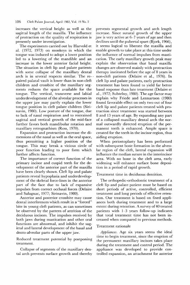
Transverse expansion.
A modified quad-helix appliance is sol-
dered to bands on the second deciduous mo-
lars and deciduous cuspids (Figure 1). The
first molar is thus locked and serves as an-
chorage as well. Maxillary first permanent
molars are used only when the second decid-
FIGURE 1. Appliance for transversal expansion andprotraction. Note hooks mesio-lingual on deciduous cus-pids. (A) Before expansion. (B) After expansion. (C) Withlingual bar supporting incisors.
107
uous molars are missing or decayed. There is
a hook for attachment to the facial mask
mesio-lingually to the cuspid bands. The total
expansion period lasts 2-3 months, with 1 or
2 activations at 5-6 weeks' interval. Expan-
sion is completed before protraction begins.
Protraction
The quad-helix is made to contact the in-
cisors (Figure 1 B) or a bar is soldered after
expansion (Figure 1 C). Additional labial
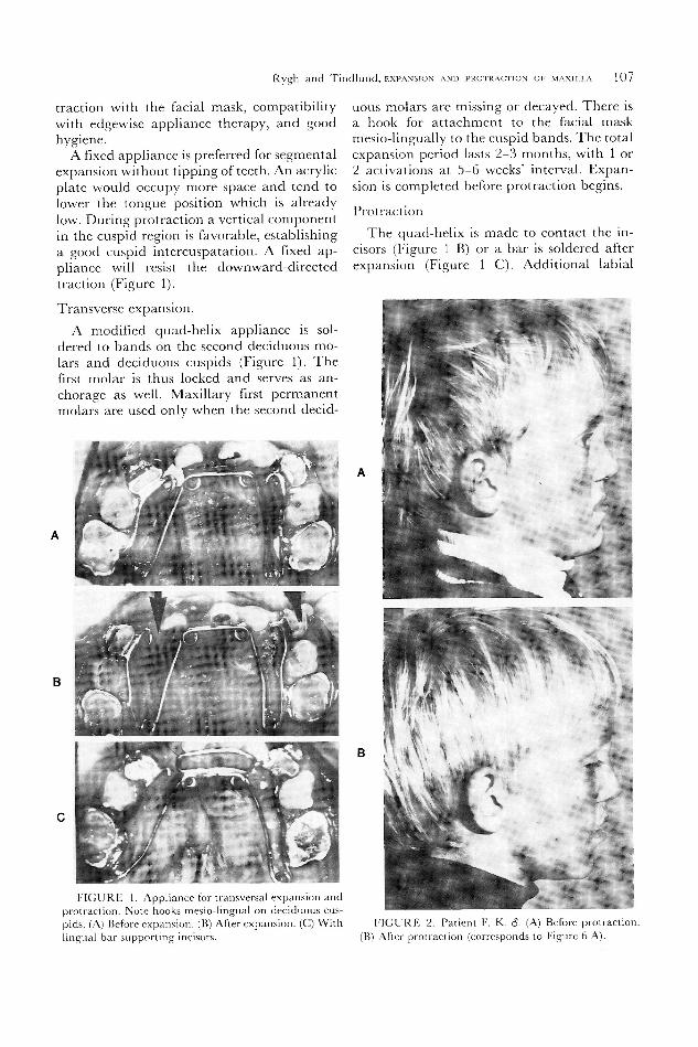
FIGURE 2. Patient F. K. d. (A) Before protraction.(B) After protraction (corresponds to Figure 6 A).
108 Cleft Palate Journal, April 1982, Vol. 19 No. 2
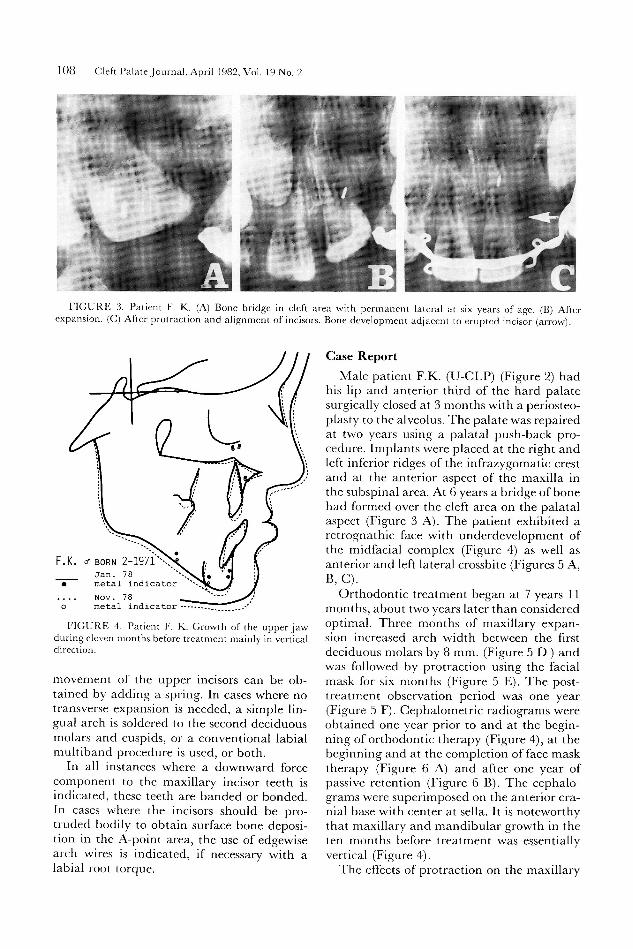
FIGURE 3. Patient F. K. (A) Bone bridge in cleft area with permanent lateral at six years of age. (B) Afterexpansion. (C) After protraction and alignment of incisors. Bone development adjacent to erupted incisor (arrow)
F.K. & BoRN 2_197i~.\\ Jan. 78e metal indicator61. Nov. 78 fo metal indicator **
FIGURE 4. Patient F. K. Growth of the upper jawduring eleven months before treatment mainly in verticaldirection.
movement of the upper incisors can be ob-tained by adding a spring. In cases where notransverse expansion is needed, a simple lin-gual arch is soldered to the second deciduousmolars and cuspids, or a conventional labialmultiband procedure is used, or both.
In all instances where a downward forcecomponent to the maxillary incisor teeth isindicated, these teeth are banded or bonded.In cases where the incisors should be pro-truded bodily to obtain surface bone deposi-tion in the A-point area, the use of edgewisearch wires is indicated, if necessary with alabial root torque.
Clase Report
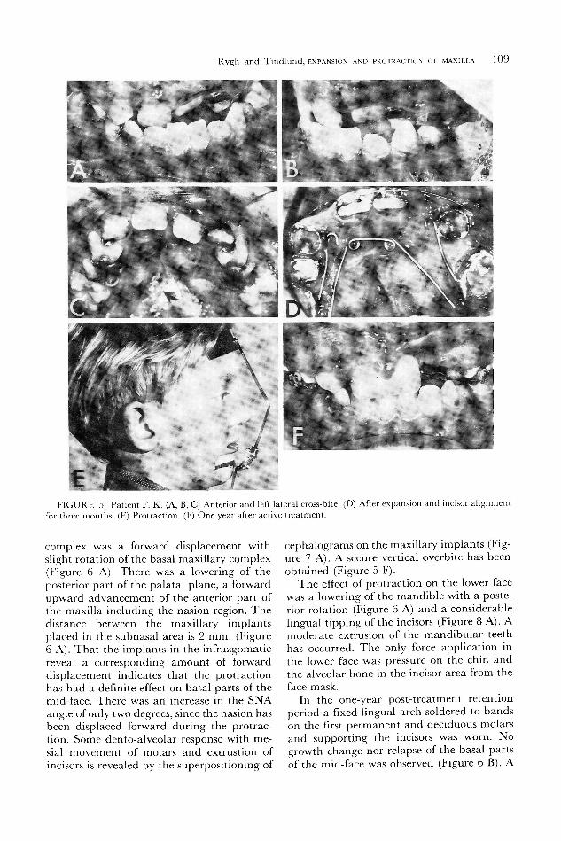
Male patient FK. (U-CLP) (Figure 2) hadhis lip and anterior third of the hard palatesurgically closed at 3 months with a periosteo-plasty to the alveolus. The palate was repairedat two years using a palatal push-back pro-cedure. Implants were placed at the right andleft inferior ridges of the infrazygomatic crestand at the anterior aspect of the maxilla inthe subspinal area. At 6 years a bridge of bonehad formed over the cleft area on the palatalaspect (Figure 3 A). The patient exhibited aretrognathic face with underdevelopment ofthe midfacial complex (Figure 4) as well asanterior and left lateral crossbite (Figures 5 A,B, C).
Orthodontic treatment began at 7 years 11months, about two years later than consideredoptimal. Three months of maxillary expan-sion increased arch width between the firstdeciduous molars by 8 mm. (Figure 5 D ) andwas followed by protraction using the facialmask for six months (Figure 5 E). The post-treatment observation period was one year(Figure 5 F). Cephalometric radiograms wereobtained one year prior to and at the begin-ning of orthodontic therapy (Figure 4), at thebeginning and at the completion of face masktherapy (Figure 6 A) and after one year ofpassive retention (Figure 6 B). The cephalo-grams were superimposed on the anterior cra-nial base with center at sella. It is noteworthythat maxillary and mandibular growth in theten months before treatment was essentiallyvertical (Figure 4).The effects of protraction on the maxillary
Rygh and Tindlund, EXPANSION AND PROTRACTION OF MAXILLA 109
FIGURE 5. Patient F. K. (A, B, C) Anterior and left lateral cross-bite. (D) After expansion and incisor alignmentfor three months. (E) Protraction. (F) One year after active treatment.
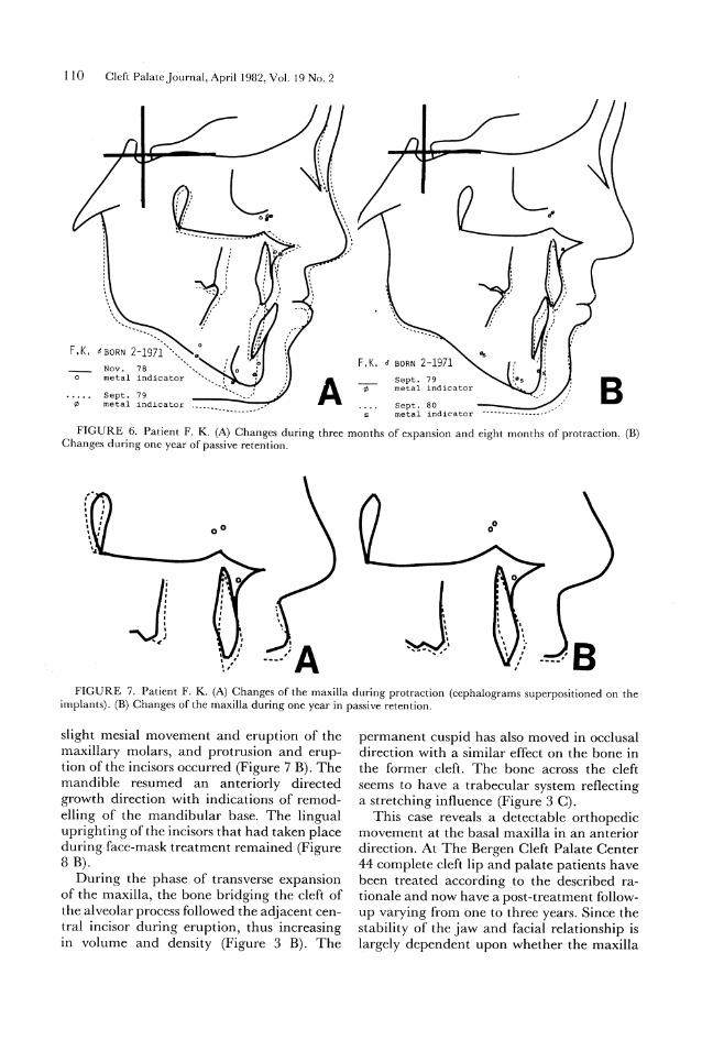
complex was a forward displacement with
slight rotation of the basal maxillary complex
(Figure 6 A). There was a lowering of the
posterior part of the palatal plane, a forward
upward advancement of the anterior part of
the maxilla including the nasion region. The
distance between the maxillary implants
placed in the subnasal area is 2 mm. (Figure
6 A). That the implants in the infrazgomatic
reveal a corresponding amount of forward
displacement indicates that the protraction
has had a definite effect on basal parts of the
mid-face. There was an increase in the SNA
angle of only two degrees, since the nasion has
been displaced forward during the protrac-
tion. Some dento-alveolar response with me-
sial movement of molars and extrustion of
incisors is revealed by the superpositioning of
cephalograms on the maxillary implants (Fig-
ure 7 A). A secure vertical overbite has been
obtained (Figure 5 F).
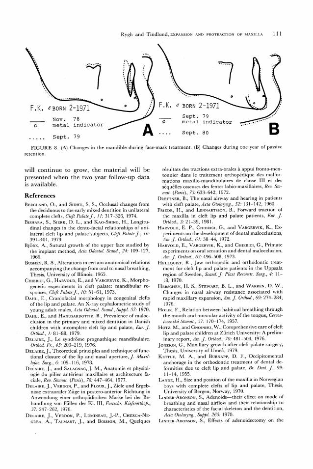
The effect of protraction on the lower face
was a lowering of the mandible with a poste-
rior rotation (Figure 6 A) and a considerable
lingual tipping of the incisors (Figure 8 A). A
moderate extrusion of the mandibular teeth
has occurred. The only force application in
the lower face was pressure on the chin and
the alveolar bone in the incisor area from the
face mask.
In the one-year post-treatment retention
period a fixed lingual arch soldered to bands
on the first permanent and deciduous molars
and supporting the incisors was worn. No
growth change nor relapse of the basal parts
of the mid-face was observed (Figure 6 B). A
Cleft Palate Journal, April 1982, Vol. 19 No. 2110
bel ~ "~~ ~
FK. « Born 2-1971 ~.,
Nov. 780 metal indicator
..... Sept. 79fed metal indicator
Sept. 79¢ metal indicator
& .. Sept. 80S metal indicator
~ ~s.=% ~ ~
F.K. e Born 2-1971
FIGURE 6. Patient F. K. (A) Changes during three months of expansion and eight months of protraction. (B)
Changes during one year of passive retention.
NM,
anm
mhum
mran
anaGe
*~<
ahusL a
FIGURE 7. Patient F. K. (A) Changes of the maxilla during protraction (cephalograms superpositioned on the
implants). (B) Changes of the maxilla during one year in passive retention.
slight mesial movement and eruption of the
maxillary molars, and protrusion and erup-
tion of the incisors occurred (Figure 7 B). The
mandible resumed an anteriorly directed
growth direction with indications of remod-
elling of the mandibular base. The lingual
uprighting of the incisors that had taken place
during face-mask treatment remained (Figure
8 B).
During the phase of transverse expansion
of the maxilla, the bone bridging the cleft of
the alveolar process followed the adjacent cen-
tral incisor during eruption, thus increasing
in volume and density (Figure 3 B). The
permanent cuspid has also moved in occlusal
direction with a similar effect on the bone in
the former cleft. The bone across the cleft
seems to have a trabecular system reflecting
a stretching influence (Figure 3 C).
This case reveals a detectable orthopedic
movement at the basal maxilla in an anterior
direction. At The Bergen Cleft Palate Center
44 complete cleft lip and palate patients have
been treated according to the described ra-
tionale and now have a post-treatment follow-
up varying from one to three years. Since the
stability of the jaw and facial relationship is
largely dependent upon whether the maxilla
Rygh and Tindlund, EXPANSION AND PROTRACTION OF MAXILLA
~~~~~
Fik. «Born 2-1971
Sept. 79
Nov.
-
78___
_ .
0 mezal indicator ® metal indicator
Sept. 79 A sept. 80
111
“““““
F.K. s Born 2-1971
FIGURE 8. (A) Changes in the mandible during face-mask treatment. (B) Changes during one year of passive
retention.
will continue to grow, the material will be
presented when the two year follow-up data
is available.
References
Brercranp, O., and StpHu, S. S., Occlusal changes from
the deciduous to the early mixed dentition in unilateral
complete clefts, Cleft Palate J., 11: 317-326, 1974.
BisHaraA, S., SiErK&, D. L., and Kao-Sninc, H., Longitu-
dinal changes in the dento-facial relationships of uni-
lateral cleft lip and palate subjects, Cleft Palate J., 16:
391-401, 1979.
Bjork, A., Sutural growth of the upper face studied by
the implant method, Acta Odontol. Scand., 24: 109-127,
1966.
Busury, R. S., Alterations in certain anatomical relations
accompanying the change from oral to nasal breathing,
Thesis, University of Illinois, 1965.
CnmIEric1, G., Harvorp, E., and Varorrvik, K., Morpho-
genetic experiments in cleft palate: mandibular re-
sponses, Cleft Palate J., 10: 51-61, 1973.
DanL, E., Craniofacial morphology in congenital clefts
of the lip and palate. An X-ray cephalometric study of
young adult males, Acta Odontol. Scand., Suppl. 57: 1970.
DanL, E., and Hanusarpotri®, B., Prevalence of maloc-
clusion in the primary and mixed dentition in Danish
children with incomplete cleft lip and palate, Eur. J.
Orthod., 1: 81-88, 1979.
DrErairE, J., Le syndrome prognathique mandibulaire.
Orthod. Fr., 45: 203-219, 1976.
DerairE, J., Theoretical principles and technique of func-
tional closure of the lip and nasal aperture, /. Maxi/-
lofac. Surg., 6: 109-116, 1978.
DeErarmE, J., and Saracnac, J. M., Anatomie et physiol-
ogie du pilier antérieur maxillaire et architecture fa-
ciale, Rev. Stomat. (Paris), 78: 447-464, 1977.
J., Verpon, P., and FrouR, J., Ziele und Ergeb-
nisse extraoraler Zuge in postero-anterior Richtung in
Anwendung einer orthopadischen Maske bei der Be-
handlung von Fallen der Kl. III, Fortschr. Kieferorthop.,
37: 241-262, 1976.
DrrairE, J., Verpon, P., LuminEavy, J.-P., Currcoa-NE-
crREA, A., TarmaNt, J., and Boisson, M., Quelques
résultats des tractions extra-orales a appui fronto-men-
tonnier dans le traitement orthopédique des malfor-
mations maxillo-mandibulaires de classe III et des
séquélles osseuses desfentes labio-maxillaires, Rev. Sto-
mat. (Paris), 73: 633-642, 1972.
DrettnNEr, B., The nasal airway and hearing in patients
with cleft palate, Acta Otolaryng., 52: 131-142, 1960.
FriEpe, H., and Lennartsson, B., Forward traction of
the maxilla in cleft lip and palate patients, Eur. J.
Orthod., 3: 21-39, 1981.
Harvorp, E. P., Cxirrict, G., and VarorrviK, K., Ex-
periments on the development of dental malocclusions,
Am. J. Orthod., 61: 38-44, 1972.
E., VARGERVIK, K., and CnuiERic1, G., Primate
experiments on oral sensation and dental malocclusion,
Am. J. Orthod., 63: 496-508, 1973.
R., Jaw orthopedic and orthodontic treat-
ment for cleft lip and palate patients in the Uppsala
region of Sweden, Scand. J. Plast Reconstr. Surg., 4: 11-
18, 1970. ,H. S., StEwart, B. L., and Warren, D. W.,
Changes in nasal airway resistance associated withrapid maxillary expansion, Am. ]. Orthod., 69: 274-284,1976.
Holik, F., Relation between habitual breathing throughthe mouth and muscular activity of the tongue, CGescos-lovenska Stomat., 57: 170-1 74, 1957.
Hotz, M., and GnomsK1, W., Comprehensive care of cleftlip and palate children at Zurich University: A prelim-inary report, Am. J. Orthod., 70: 481-504, 1976.
Jonsson, G., Maxillary growth after cleft palate surgery,Thesis, University of Umea, 1979.
KErtTtiE, M. A., and Burnapp, D. F., Occipitomentalanchorage in the orthodontic treatment of dental de-formities due to cleft lip and palate, Br. Dent. J., 99:11-14, 1955.
Lanpg, H., Size and position of the maxilla in Norwegianboys with complete clefts of lip and palate, Thesis,University of Bergen, Norway, 1970.
Linper-Aronson, S., Adenoids-their effect on mode ofbreathing and nasal airflow and their relationship tocharacteristics of the facial skeleton and the dentition,Acta Otolaryng., Suppl. 265: 1970.
Linper-Aronson, S., Effects of adenoidectomy on the

112 Cleft Palate Journal, April 1982, Vol. 19 No. 2
dentition and facial skeleton over a period of five years,Trans. 3rd Int. Orthod. Congr., Crossby LockwoodStaples, London, 85-100, 1975.
LinpeEr-Aronson, S., Respiratory function in relation tofacial morphology and the dentition, Br. J. Orthod., 6:59-71, 1979.
Linper-Aronson, S., and AscHaAN, S., Nasal resistance tobreathing and palatal height before and after expan-sion of the median palatal suture, Odontol. Revy, !4:254-270, 1963.
LorEiLLE, J.-P., and Bery, A., Modification de la venti-lation nasale par disjonction intermaxillaire, Rev. Or-thop. Dento. Fac., 15: 193-208, 1981.
Pruzansxy, S., and Aouss, H., Prevalence of arch collapseand malocclusion in complete unilateral cleft lip andpalate, Eur. Orthod. Soc. Rep. Congr., 365-382, 1967.
RickrEtTTs, R. M., Respiratory obstructions and theirrelation to tongue posture, Cleft Palate Bull., 8: 4-5,1958.
RosenstEn, S. W., Orthodontic treatment for cleft palatepatient, /. Am. Dent. Assoc., 60: 711-714, 1960.
Ross, R. B., The clinical implications of facial growth incleft lip and palate Cleft Palate J., 7: 37-47, 1970.
Ross, R. B., and Jounston, M. C., Cleft Lip and Palate,
Baltimore, Williams & Wilkins Comp., 246, 1972.Sirinavain, I., Cranio-facial and dental morphology of
six-year old Norwegian boys with complete cleft lipand palate, University of Bergen, Norway, 1-65, 1980.
SUBTELNY, J. D., The significance of adenoid tissue inorthodontia, Angle Orthod., 24: 59-69, 1954.
SusBtTELNY, J. D., Oral respiration: Facial maldevelop-ment and corrective dentofacial orthopedics, Angle Or-thod., 50: 147-164, 1980.
Warren, D. W., DuaAny, L. F., and FiscHER, N. D., Nasalpathway resistance in normal and cleft lip and palatesubjects, Cleft Palate J., 6: 449-469, 1969.
Yip, A. S. C., and CreEaLL, J. F., Cinefluorographic studyof velarpharyngeal function before and after removalof tonsils and adenoids, Angle Orthod., 41: 251-263,1971.Professor Rygh is Head of the Department of Ortho-
dontics, School of Dentistry, University of Bergen, Ber-gen, Norway. Dr. Tindlund is Orthodontist in the Nor-wegian Dental Health Service, Cleft Palate Division.Address editorial correspondence to Dr. Rygh at theDepartment of Orthodontics, School of Dentistry, Uni-versity of Bergen, Arstadveien 17, N-5000 Bergen, Nor-way.