An Innovative Method to Determine the Width of Maxillary ...
Upload
khangminh22Category
view
0download
0
International Journal of Dental Science and Innovative Research (IJDSIR)
IJDSIR : Dental Publication Service Available Online at: www.ijdsir.com Volume – 3, Issue – 2, April - 2020, Page No. : 149 - 160
Corresponding Author: Dr. Raunak Manjeet, ijdsir, Volume – 3 Issue - 2, Page No. 149 - 160
Page
149
ISSN: 2581-5989 PubMed - National Library of Medicine - ID: 101738774
Maxillary Expansion: A Review 1Dr. Raunak Manjeet , PG Student , Dept.of Orthodontics & Dentofacial Orthopaedics, KD Dental College & Hospital,
Mathura 2Dr. Kuldeep D’mello, Professor & HoD, Dept. of Orthodontics & Dentofacial Orthopaedics, KD Dental College &
Hospital, Mathura 3Dr. Atul Singh, Associate Professor, Dept. of Orthodontics & Dentofacial Orthopaedics, KD Dental College & Hospital,
Mathura 4Dr. Jitesh Wadhwa, Associate Professor, Dept of Orthodontics & Dentofacial Orthopaedics, KD Dental College &
Hospital, Mathura 5Dr. Swati Srivastava, PG Student, Dept. of Oral & Maxillofacial Surgery, KD Dental College & Hospital, Mathura
Corresponding author: Dr. Raunak Manjeet, PG Student, Dept. of Orthodontics & Dentofacial Orthopaedics, KD
Dental College & Hospital, Mathura
Citation of this Article: Dr. Raunak Manjeet, Dr. Kuldeep D’mello, Dr. Atul Singh, Dr. Jitesh Wadhwa, Dr. Swati
Srivastava, “Maxillary Expansion : A Review”, IJDSIR- April - 2020, Vol. – 3, Issue -2, P. No. 149 – 160.
Copyright: © 2020, Dr. Raunak Manjeet, et al. This is an open access journal and article distributed under the terms of
the creative commons attribution noncommercial License. Which allows others to remix, tweak, and build upon the work
non commercially, as long as appropriate credit is given and the new creations are licensed under the identical terms.
Type of Publication: Review Article
Conflicts of Interest: Nil
Abstract
Maxillary transverse discrepancy usually requires
expansion of the palate by a combination of orthopedic
and orthodontic tooth movements. Three expansion
treatment modalities are used today: rapid maxillary
expansion, slow maxillary expansion and surgically
assisted maxillary expansion. This article aims to review
the maxillary expansion by all the three modalities and a
brief on commonly used appliances & also newer
expansion modalities such as SARPE & MARPE .
Keywords : Maxillary expansion, Rapid maxillary
expansion, Slow maxillary expansion, Expansion in Cleft
Palate case, Mini Screw assisted rapid palatal expansion,
Surgically assisted maxillary expansion.
Introduction
Maxillary expansion treatments have been used for more
than a century to correct maxillary transverse deficiency.
The earliest common cited report is that of E.C. Angell
published in Dental Cosmos in 1860. The work was
discredited at the time, but the technique is now generally
accepted as a relatively simple and predictable orthodontic
therapy. Correction of the transverse discrepancy usually
requires expansion of the palate by a combination of
orthopedic and orthodontic tooth movements. Three
expansion treatment modalities are used today: rapid
maxillary expansion (RME), slow maxillary expansion
(SME) and surgically assisted maxillary expansion. Since
each treatment modality has advantages and

Dr. Raunak Manjeet, et al. International Journal of Dental Science and Innovative Research (IJDSIR)
© 2020 IJDSIR, All Rights Reserved
Page
150
Page
150
Page
150
Page
150
Page
150
Page
150
Page
150
Page
150
Page
150
Page
150
Page
150
Page
150
Page
150
Page
150
Page
150
Page
150
Page
150
Page
150
Page
150
disadvantages, controversy regarding the use of each
exists. Practitioners select treatment appliances based on
their personal experiences and on the patient’s age and
malocclusion.
Normal palatal growth is nearly complete by age 6,and
increasing interdigitation of the suture makes separation
difficult to achieve after puberty. During treatment,
transverse forces tip the buccal segments laterally and
with proper appliance design, 3rd-order moments will
induce bodily translation. If the force is strong enough,
separation occurs at the maxillary suture. The clinical
conditions indicating maxillary expansion include
crossbites, distal molar movement, functional appliance
treatment, surgical cases for instance arch coordination or
bone grafts, to aid maxillary protraction and mild
crowding. This article aims to review the maxillary
expansion and commonly used appliances.
Rapid Maxillary Expansion (RME)
Rapid maxillary expansion was first described by
Emerson Angell in 1860 and later repopularized by Haas.
The main object of RME is to correct maxillary arch
narrowness but its effects are not limited to the maxilla as
it is associated with 10 bones in the face and head.
Advocates of rapid maxillary expansion believe that it
results in minimum dental movement (tipping) and
maximum skeletal movement.
Figure 1
When heavy and rapid forces are applied to the posterior
teeth, there is not enough time for tooth movement to
occur and the forces are transferred to the sutures. When
the force delivered
by the appliance exceeds the limit needed for orthodontic
tooth movement and sutural resistance, the sutures open
up while the teeth move only minimally relative to their
supporting bone. The appliance compresses the
periodontal ligament, bends the alveolar process, tips the
anchor teeth, and gradually opens the mid palatal suture
and all the other maxillary sutures.
Figure 2
Effect of RME on Maxillary and Mandibular
Complex
Maxillary skeletal effect: When viewed occlusally, Inoue
found that the opening of the mid palatine suture was non
parallel and triangular with maximum opening at incisor
region and gradually diminishing towards the posterior
part of palate.
Viewed frontally, the maxillary suture separates supero-
inferiorly in a non parallel manner. It is pyramidal in
shape with the base of pyramid located at the oral side of
the bone.
Maxillary halves: Haas and Wertz found the maxilla to be
frequently displaced downward and forward.

Dr. Raunak Manjeet, et al. International Journal of Dental Science and Innovative Research (IJDSIR)
© 2020 IJDSIR, All Rights Reserved
Page
151
Page
151
Page
151
Page
151
Page
151
Page
151
Page
151
Page
151
Page
151
Page
151
Page
151
Page
151
Page
151
Page
151
Page
151
Page
151
Page
151
Page
151
Page
151
Figure 3
Palatal vault: Haas reported that the palatine process of
maxilla was:
lowered as a result of outward tilting of maxillary halves.
Alveolar process: Because bone is resilient, lateral
bending of the alveolar processes occurs early during
RME, which rebounds back after a few days.
Maxillary anterior teeth: From the patient’s point of view,
one of the most spectacular changes accompanying RME
is the opening of a diastema between the maxillary central
incisors. It is estimated that during active suture opening,
the incisors separate approximately half the distance the
expansion screw has been opened, but the amount of
separation between the central incisors should not be used
as an indication of the amount of suture separation. This
distema is self-corrective due to elastic recoil of the
transseptal fibers.
Maxillary posterior teeth: There is buccal tipping and
extrusion of the maxillary molars.
The posterior maxilla expands less readily because of the
resistance produced by the zygomatic buttress and
pterygoid plates.
Effect of RME on mandible: There is a concomitant
tendency for the mandible to swing downward and
backward.
RME and nasal airflow: Anatomically, there is an increase
in the width of the nasal cavity immediately following
expansion thereby improves in breathing. The nasal cavity
width gain averages of 1.9 mm, but can be as wide as 8 to
10 mm.
It is important for the clinician to remember that the main
resistance to midpalatal suture opening is probably not the
suture itself, but in the surrounding structures particularly
the sphenoid and zygomatic bones.
Indications/contraindications of RME
Rapid maxillary expansion is indicated in cases with a
transverse discrepancy equal to or greater than 4 mm, and
where the maxillary molars are already buccally inclined
to compensate for the transverse skeletal discrepancy.
Rapid palatal expansion has been used to facilitate
maxillary protraction in class III treatment by disrupting
the system of sutures, which connect the maxilla to the
cranial base, cleft lip and palate patients with collapsed
maxillae are also RME candidates.
Finally, some clinicians use the procedure to gain arch
length in patients, who have moderate maxillary
crowding. It is contraindicated in patients, who have
passed the growth spurt, have recession on the buccal
aspect of the molars, anterior open bite, steep mandibular
plane, convex profiles and who show poor compliance.
It appears that approximately 1 millimeter per week is the
maximum rate at which the tissue of the midpalatal suture
can adapt, so that tearing and hemorrhaging are minimized
compared with rapid expansion protocols.
The amount of orthopedic vs. orthodontic change depends
greatly on the patient’s age. Normal palatal growth is
nearly complete by age 6, and increasing interdigitation of
the suture makes separation difficult to achieve after
puberty.
Dr. Raunak Manjeet, et al. International Journal of Dental Science and Innovative Research (IJDSIR)
© 2020 IJDSIR, All Rights Reserved
Page
152
Page
152
Page
152
Page
152
Page
152
Page
152
Page
152
Page
152
Page
152
Page
152
Page
152
Page
152
Page
152
Page
152
Page
152
Page
152
Page
152
Page
152
Page
152
RPE appliances require frequent activations and generate
heavy forces―as much as 2-5 kg per quarter-turn with
accumulated loads of more than 9 kg.
The disadvantages of using rapid palatal expanders
include discomfort due to heavy forces used, traumatic
separation of the mid palatal suture, inability to correct
rotated molars, requirement of patient or parent
cooperation in activation of the appliance, bite opening,
relapse, micro trauma of the temporo mandibular joint and
mid palatal suture, root resorption, tissue impingement,
pain and laborintensive procedure in fabrication of the
appliance.
Clinical Management of RME
The patient/parent should be informed in advance about
the upper midline diastema during the expansion phase.
This is likely to close spontaneously during the retention
period. Patients should be instructed to turn the expansion
screw one-quarter turn twice a day (am and pm). This may
be associated with minor discomfort. Force levels tend to
accumulate following multiple turns and can be as high as
10 kg following many turns. Patients should be reviewed
weekly and some clinicians recommend that an upper
occlusal radiograph be taken one week into treatment to
ensure that the midpalatal suture has separated. If there is
no evidence of this, it is important to stop appliance
activation as there is a risk of alveolar fracture and/or
periodontal damage. Active treatment is usually required
for a period of 2-3 weeks, after which a retention period of
three months is recommended to allow for bony infilling
of the separated suture.
Appliances For RME
These are banded and bonded appliances. The banded
appliance are attached to teeth with bands on the maxillary
first molar and first premolars. The banded appliances are
hygienic as there is no palatal coverage. The banded RME
are of two types:
1.Tooth and tissue borne
2.Tooth borne
Tooth Borne RME
They consist of only bands and wires without any acrylic
covering.
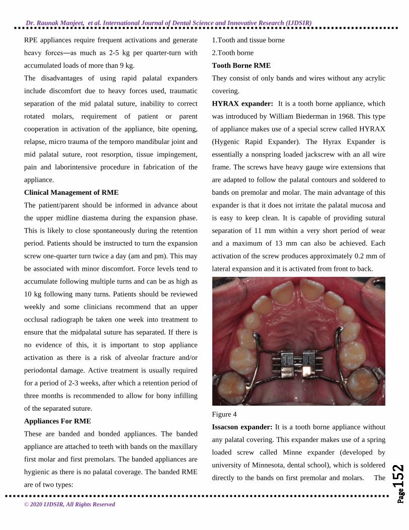
HYRAX expander: It is a tooth borne appliance, which
was introduced by William Biederman in 1968. This type
of appliance makes use of a special screw called HYRAX
(Hygenic Rapid Expander). The Hyrax Expander is
essentially a nonspring loaded jackscrew with an all wire
frame. The screws have heavy gauge wire extensions that
are adapted to follow the palatal contours and soldered to
bands on premolar and molar. The main advantage of this
expander is that it does not irritate the palatal mucosa and
is easy to keep clean. It is capable of providing sutural
separation of 11 mm within a very short period of wear
and a maximum of 13 mm can also be achieved. Each
activation of the screw produces approximately 0.2 mm of
lateral expansion and it is activated from front to back.
Figure 4
Issacson expander: It is a tooth borne appliance without
any palatal covering. This expander makes use of a spring
loaded screw called Minne expander (developed by
university of Minnesota, dental school), which is soldered
directly to the bands on first premolar and molars. The

Dr. Raunak Manjeet, et al. International Journal of Dental Science and Innovative Research (IJDSIR)
© 2020 IJDSIR, All Rights Reserved
Page
153
Page
153
Page
153
Page
153
Page
153
Page
153
Page
153
Page
153
Page
153
Page
153
Page
153
Page
153
Page
153
Page
153
Page
153
Page
153
Page
153
Page
153
Page
153
Minne expander is a heavily calibrated coil spring
expanded by turning a nut to compress the coil. Two metal
flanges perpendicular to the coil are soldered to the bands
on abutment teeth. The Minne expander may continue to
exert expansion forces after completion of the expansion
phase unless they are partly deactivated.
Figure 5
Tooth and tissue borne RME
They consist of an expansion screw with acrylic abutting
on alveolar ridges. Haas, in 1970, gave the following
advantages of tooth and tissue RME :
1. Produces more parallel expansion
2. Less relapse
3. Greater nasal cavity and apical base gain.
4. More favorable relationship of the denture bases in
width and frequently in the anteroposterior plane as well.
5. Creates more mobility of the maxilla instead of teeth.
Disadvantage of Tooth and Tissue Borne RME :
These tooth and tissue borne RME tend to have higher soft
tissue irritation.
Types of Tooth and Tissue Borne RME
Haas: The basis for the rapid expansion procedure is to
produce immediate midpalatal suture separation by
disruption of the sutural connective tissue . The rapid
palatal expander as described by Haas is a rigid appliance
designed for maximum dental anchorage that uses a
jackscrew to produce expansion in 10 to 14 days. He
believed that this will maximize the orthopedic effects and
forces produced by this appliance have been reported in
the range of 3 to 10 pounds.
Figure 6
Derichsweiler : The first premolar and molars are
banded. Wire tags are soldered to these bands and then
inserted to the split palatal acrylic, which contains the
screw.
Figure 7
bonded rapid palatal expander
The Bonded RPE were first described by Cohen and
Silverman in 1973 . It is similar to the banded version with
the exception of the method of attachment to the teeth.

Dr. Raunak Manjeet, et al. International Journal of Dental Science and Innovative Research (IJDSIR)
© 2020 IJDSIR, All Rights Reserved
Page
154
Page
154
Page
154
Page
154
Page
154
Page
154
Page
154
Page
154
Page
154
Page
154
Page
154
Page
154
Page
154
Page
154
Page
154
Page
154
Page
154
Page
154
Page
154
This appliance is constructed with an acrylic cap over the
posterior segments, which is then bonded directly to the
teeth.The bonded appliance has become increasingly
popular because of its advantages,
It can be easily cemented during the mixed dentition stage,
when retention from other appliances can be poor.
1.Number of appointments are reduced.
2.There is reduced posterior teeth tipping and extrusion.
3.The buccal capping limits molar extrusion during
treatment and, therefore improves the vertical control,
which is particularly useful in class II conditions, as molar
extrusion would cause autorotation of the mandible
backward and downward resulting in increase in facial
convexity and the vertical dimension of the lower face.
IPC Rapid Palatal Expander
IPC is designed for orthopedic expansion along with labial
alignment of incisors . As expansion occurs, the IPC
controls the NiTi open coil spring force applied to the
lingual surface of the anterior teeth. Wire around the distal
end of the lateral incisors limits the midline diastema that
often occurs during RPE treatment.
Figure 8
Slow Maxillary Expansion
SME procedures produce less tissue resistance around the
circummaxillary structures and, therefore improve bone
formation in the intermaxillary suture, which theoretically
should eliminate or reduce the limitations of RME.
Slow expansion has been found to promote greater post-
expansion stability, if given an adequate retention period.
It delivers a constant physiologic force until the required
expansion is obtained. The appliance is light and
comfortable enough to be kept in place for sufficient
retention of the expansion. Prefabrication eliminates extra
appointments for impressions and the time and expense of
laboratory fabrication.
For SME, 10 to 20 newtons of force should be applied to
the maxillary region only 450 to 900 gm of force is
generated, which may be insufficient to separate a
progressively maturing suture. Maxillary arch-width
increases ranged from 3.8 to 8.7 mm with slow expansion
of as much as 1 mm per week using 900 gm of force
Appliances In SME
Coffin Appliance
Given by Walter Coffin-1875. It is a removable appliance
capable of slow dento alveolar expansion. The appliance
consists of an omega-shaped wire of 1.25 mm thickness,
placed in the midpalatal region. The free ends of the
omega wire are embedded in acrylic covering the slopes
of the palate. The spring is activated by pulling two asides
apart manually.
Figure 9

Dr. Raunak Manjeet, et al. International Journal of Dental Science and Innovative Research (IJDSIR)
© 2020 IJDSIR, All Rights Reserved
Page
155
Page
155
Page
155
Page
155
Page
155
Page
155
Page
155
Page
155
Page
155
Page
155
Page
155
Page
155
Page
155
Page
155
Page
155
Page
155
Page
155
Page
155
Page
155
Magnets
Repulsive magnetic forces for maxillary expansion were
first described by Vardemon et al 19 87. Banded magnets
produced more pronounced skeletal; versus overall
expansion effects. The continuous force of 250-500 gm
could generate dental and skeletal movements, the degree
depending on patients status (age, growth, etc).
Disadvantage of magnets is that they tend to be oxidized
in the oral environment due to the potential formation of
corrosive products but this can be overcome by coating
magnets. The advantage of these magnets is that they
impart measured continuous force over a long period of
time, hence the risk of external root resorption is
decreased. These magnets are
quite bulky as they must be adequately stabilized and
contain stout guide rods to prevent the magnets becoming
out of line and causing unwanted rotational movements.
W-Arch
The “W”; expansion appliance was originally used by
Ricketts and his colleagues to treat cleft palate patients .
The W-arch is a fixed appliance constructed of 36 mil
steel wire soldered to molar bands. To avoid soft tissue
irritation, the lingual arch should be constructed so that it
rests 1-1.5 mm off the palatal soft tissue. It is activated
simply by opening the apices of W-arch and is easily
adjusted to provide more anterior than posterior
expansion, or vice versa if this is desired. The appliance
delivers proper force levels when opened 3-4 mm wider
than the passive width and should be adjusted to this
dimension before being inserted. Expansion should
continue at the rate of 2 mm per month until the cross bite
is slightly over corrected.
Figure 10
Quadhelix
The quadhelix appliance is a modification of Coffin’s W-
spring and was described by Ricketts . The incorporation
of four helices into the W-spring helped to increase the
flexibility and range of activation. The length of the
palatal arms of the appliance can be altered depending
upon which teeth arch in cross bite.
A new generation of prefabricated appliances, constructed
from nickel titanium, have been introduced more recently.
The advantages of using nickel titanium over stainless
steel include its more favorable force delivery
characteristics as it has super elastic properties. This may
help to produce more physiological tooth movement with
more rapid correction of cross bites.
Figure 11

Dr. Raunak Manjeet, et al. International Journal of Dental Science and Innovative Research (IJDSIR)
© 2020 IJDSIR, All Rights Reserved
Page
156
Page
156
Page
156
Page
156
Page
156
Page
156
Page
156
Page
156
Page
156
Page
156
Page
156
Page
156
Page
156
Page
156
Page
156
Page
156
Page
156
Page
156
Page
156
Mode of Action
The quadhelix appliance works by a combination of
buccal tipping and skeletal expansion in a ratio of 6:1 in
prepubertal children.
Clinical Management
The desirable force level of 400 gm can be delivered by
activating the appliance by 8 mm, which equates to
approximately one molar width. Patients should be
reviewed on a six-weekly basis. Sometimes, the appliance
can leave an imprint on the tongue, however this will
rapidly disappear following treatment. Expansion should
be continued until the palatal cusps of the upper molars
meet edge-to-edge with the buccal cusps of the
mandibular molars. A degree of overcorrection is
desirable as relapse is inevitable. A three-month retention
period, with the quadhelix in place, is recommended once
expansion has been achieved. If fixed appliances are being
used, the quadhelix can be removed once stainless steel
wires are in place.
Advantages: Good retention, a large range of action,
orthopedic effect, differential expansion, habit breaker,
fixed appliances can be incorporated, molar
rotation/torque, non-compliance and cost-effective.
Disadvantages: Molar tipping, bite opening, limited
skeletal change.
Spring Jet
The active components of the spring jet are soldered or
attached to the molar bands . The telescopic unit is placed
upto 5 mm from center of molar tubes so that the forces
pass close to the center of resistance of maxillary teeth,
but it should be 1.5 mm away from palatal tissue. Force
applied in mixed dentition is 240 gm and 400 gm in the
permanent dentition. Activation is done by moving the
lock screw horizontally along the telescopic tube. A ball
stop on the transpalatal wire allows the spring to be
compressed.
Figure 12
NiTi Expander
The Nickel Titanium Palatal Expanders were introduced
by Wendell V. It generates optimal, constant expansion
forces. The central component is made of a thermally
activated NiTi alloy and rest of component is made of
stainless steel. The expander may be used simultaneously
with conventional fixed appliances, requiring only an
additional lingual sheath on the molar bands. The action of
the appliance is a consequence of nicket titanium’s shape
memory and transition temperature effects. The nickel
titanium component has a transition temperature of 94° F.
At room temperature, the expander is too stiff to bend for
insertion. Chilling the expander softens the central
component allowing easy manipulation. Once placed,
stiffens and begins to return to its original shape. A 3 mm
increment of expansion exerts only about 350 gm of force
and the nickel titanium alloy provides relatively uniform
force levels as the expander deactivates.
Figure 13
Dr. Raunak Manjeet, et al. International Journal of Dental Science and Innovative Research (IJDSIR)
© 2020 IJDSIR, All Rights Reserved
Page
157
Page
157
Page
157
Page
157
Page
157
Page
157
Page
157
Page
157
Page
157
Page
157
Page
157
Page
157
Page
157
Page
157
Page
157
Page
157
Page
157
Page
157
Page
157
Activation Schedule (RME)
The activation schedule vary depending upon the age of
patients & form of appliance used.
Schedule By Timms
For patients upto 15 years of age : 90 degree rotation in
the morning & evening .
For patients over 15 years of age : 45 degree rotation 4
times a day.
Schedule By Zimring & Issacson
In young growing patients 2 turns each day for 4 to 5 days
& later 1 turn per day ..
In non growing patients 2 turns per days for 1 to 2 days &
then 1 turn per day for 5 to 7days & then 1 turn for
alternate days till desired expansion is achieved.
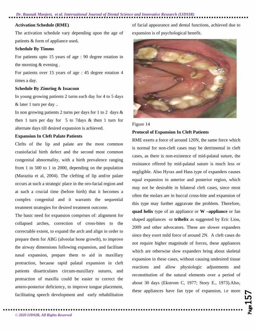
Expansion In Cleft Palate Patients
Clefts of the lip and palate are the most common
craniofacial birth defect and the second most common
congenital abnormality, with a birth prevalence ranging
from 1 in 500 to 1 in 2000, depending on the population
(Marazita et al, 2004). The clefting of lip and/or palate
occurs at such a strategic place in the oro-facial region and
at such a crucial time (before birth) that it becomes a
complex congenital and it warrants the sequential
treatment strategies for desired treatment outcome.
The basic need for expansion comprises of: alignment for
collapsed arches, correction of cross-bites to the
correctable extent, to expand the arch and align in order to
prepare them for ABG (alveolar bone growth), to improve
the airway dimensions following expansion, and facilitate
nasal expansion, prepare them to aid in maxillary
protraction, because rapid palatal expansion in cleft
patients disarticulates circum-maxillary sutures, and
protraction of maxilla could be easier to correct the
antero-posterior deficiency, to improve tongue placement,
facilitating speech development and early rehabilitation
of facial appearance and dental functions, achieved due to
expansion is of psychological benefit.
Figure 14
Protocol of Expansion In Cleft Patients
RME exerts a force of around 120N, the same force which
is normal for non-cleft cases may be detrimental in cleft
cases, as there is non-existence of mid-palatal suture, the
resistance offered by mid-palatal suture is much less or
negligible. Also Hyrax and Hass type of expanders causes
equal expansion in anterior and posterior region, which
may not be desirable in bilateral cleft cases, since most
often the molars are in buccal cross-bite and expansion of
this type may further aggravate the problem. Therefore,
quad helix type of an appliance or W –appliance or fan
shaped appliances or trihelix as suggested by Eric Liou,
2009 and other advocators. These are slower expanders
since they exert mild force of around 2N. A cleft cases do
not require higher magnitude of forces, these appliances
which are otherwise slow expanders bring about skeletal
expansion in these cases, without causing undesired tissue
reactions and allow physiologic adjustments and
reconstitution of the sutural elements over a period of
about 30 days (Ekstrom C, 1977; Story E., 1973).Also,
these appliances have fan type of expansion, i.e more

Dr. Raunak Manjeet, et al. International Journal of Dental Science and Innovative Research (IJDSIR)
© 2020 IJDSIR, All Rights Reserved
Page
158
Page
158
Page
158
Page
158
Page
158
Page
158
Page
158
Page
158
Page
158
Page
158
Page
158
Page
158
Page
158
Page
158
Page
158
Page
158
Page
158
Page
158
Page
158
anterior and less posterior expansion, which is desirable in
bilateral cleft cases.
Expansion procedures provide promising treatment
modalities for transverse deficiencies in cleft patients.
Acquaintance with these interceptive procedures helps the
clinician to understand better the final therapeutic effects
and the way the expansion appliance actually acts on the
basal bones and sutures of the craniofacial system.
Surgical Techniques
The effect of dental arch on the maxillary base diminishes
as age advances so, surgically assisted expansion
techniques can be considered.
Indications of surgical expansion are:
To widen the arch
To correct posterior crossbite when large amount (>7 mm)
of expansion is required to avoid the potential increased
risk of segmental osteotomies
To widen the arch following maxillary collapse associated
with a cleft palate, in cases with extremely thin and
delicate gingival tissue, or presence of significant buccal
gingival recession in the canine-bicuspid region of the
maxilla; and in condition, where significant nasal stenosis
is found.
The techniques available are:
Surgically assisted rapid palatal expansion (SARPE)
Segmental maxillary surgery.
Surgically assisted rapid palatal expansion (SARPE) has
gradually gained popularity as a treatment option to
correct MTD (Maxillary Transverse Deficiency). It allows
clinicians to achieve effective maxillary expansion in a
skeletally mature patient.
Segmental Maxillary Surgery–transverse expansion can be
produced during a Le Fort 1 osteotomy by creating an
additional surgical cut along the midpalatal suture. The
maxillary halves are then separated and retained in the
new position. The relative inelasticity of the palatal muco-
periosteum limits the degree of expansion that may be
achieved.
Before surgery, orthodontic treatment involves moving the
roots of the maxillary central incisors apart to improve
surgical access to the osteotomy site. This is the technique
of choice in patients, who require expansion and have
coexisting sagittal and/or vertical maxillary discrepancies.
Mini Screw Assisted Rapid Palatal Expansion
Surgically assisted rapid palatal expansion was introduced
to overcome the transverse maxillary deficiency in
skeletally mature patients, However, owing to high risks
and costs, surgical complexity, and morbidity, many
patients rejected a surgical intervention. Recently, mini
implant assisted rapid palatal expansion (MARPE) has
been suggested as an alternative to surgically assisted
rapid palatal expansion in adults to open mid palatal
suture. Orthodontic mini-implants have been proposed as
a skeletal anchorage for RPE to provide more orthopedic
expansion while reducing undesirable side effects such as
dentoalveolar tipping, periodontal dehiscence, and
marginal bone loss.
Figure 15
MARME appliance (MSE; BioMaterials Korea, Seoul,
South Korea) is used on all patients for rapid maxillary
expansion (RME). Type I appliances expand by 0.8 mm in
4 turns, but type II appliances expand by 0.8 mm in 6
turns. The body size of the MARME appliance is 13.5 X

Dr. Raunak Manjeet, et al. International Journal of Dental Science and Innovative Research (IJDSIR)
© 2020 IJDSIR, All Rights Reserved
Page
159
Page
159
Page
159
Page
159
Page
159
Page
159
Page
159
Page
159
Page
159
Page
159
Page
159
Page
159
Page
159
Page
159
Page
159
Page
159
Page
159
Page
159
Page
159
14.5 mm. It is positioned in the para midsagittal area
between the maxillary first molars. MSE has 4 holes and 4
arms that are soldered to the bands of the maxillary first
permanent molars. Four 1.5 X 11.0 mm mini-implants are
used to fenestrate the palatal base and nasal base to
generate greater expansive skeletal forces.
Figure 16
Conclusion
Expansion of the maxilla and the maxillary dentition may
be accomplished in numerous ways. The type of skeletal
and dental pattern greatly influences the type of expansion
chosen and the type of expansion selected can greatly
facilitate the overall treatment objectives.
References
1. Timms DJ. The dawn of rapid maxillary expansion.
Angle Orthod 1999 Jun;69(3):247-250.
2. Ficarelli JP. A brief review of maxillary expansion. J
Pedod 1978 Fall;3(1):29-35.
3. Bell RA. A review of maxillary expansion in relation
to rate of expansion and patient’s age. Am J Orthod
1982 Jan;81(1):32-37.
4. Majourau A, Nanda R. Biomechanical basis of
vertical dimension control during rapid palatal
expansion therapy. Am. J Orthod Dentofacial Orthop
1994 Sep;106(3):322-328.
5. Cleall JF, Bayne DI, Posen JM, Subtelny JD.
Expansion of the midpalatal suture in the monkey.
Angle Orthod 1965 Jan;35: 23-35.
6. Starnbach H, Bayne D, Cleall J, Subtelny JD.
Facioskeletal and dental changes resulting from rapid
maxillary expansion. Angle Orthod 1966
Apr;36(2):152-154.
7. Murray JM, Cleall JF. Early tissue response to rapid
maxillary expansion in the midpalatal suture of the
rhesus monkey. J Dent Res 1971 Nov-
Dec;50(6):1654-1660.
8. Storey E. Tissue response to the movement of bones.
Am J Orthod 1973 Sep;64(3):229-247.
9. Moyers RE, van der Linden FP, Riolo ML, et al.
Standards of human occlusal development. In:
Monograph 5, craniofacial growth series, Center for
Human Growth and Development,. 7th ed. University
of Michigan. Ann Arbor; 1976.
10. Persson M, Thilander B. Palatal suture closure in man
from 15 to 35 years of age. Am J Orthod 1977
Jul;72(1):42-52.
11. Handelman CS. Nonsurgical rapid maxillary alveolar
expansion in adults: a clinical evaluation, Angle
Orthod 1997;67(4): 291-305.
12. Isaacson RJ, Ingram AH. Forces produced by rapid
maxillary expansion, II. Forces present during
treatment. Angle Orthod 1964;34:261-270.
13. Starnbach HK, Cleall JF. The effects of splitting the
midpalatal suture on the surrounding structures. Am J
Orthod 1964;50.
14. Haas AJ. The treatment of maxillary deficiency by
opening the midpalatal suture. Angle Orthod 1965
Jul;35:200-217
15. Hicks EP. Slow maxillary expansion. A clinical study
of the skeletal versus dental response to low-
magnitude force. Am J Orthod 1978 Feb;73(2):121-
141.

Dr. Raunak Manjeet, et al. International Journal of Dental Science and Innovative Research (IJDSIR)
© 2020 IJDSIR, All Rights Reserved
Page
160
Page
160
Page
160
Page
160
Page
160
Page
160
Page
160
Page
160
Page
160
Page
160
Page
160
Page
160
Page
160
Page
160
Page
160
Page
160
Page
160
Page
160
Page
160
16. Ceylan I, Oktay H, Demirci M. The effect of rapid
maxillary expansion on conductive hearing loss.
Angle Orthod 1996;66(4): 301-307.
17. Bishara SE, Staley RN. Maxillary expansion: Clinical
implication. Am J Orthod Dentofacial Orthop 1987
Jan;91(1):3-14.
18. Haas AJ. Rapid expansion of the maxillary dental arch
and nasal cavity by opening the midpalatal suture.
Angle Orthod 1961;31: 73-90.
19. Wertz RA. Skeletal and dental changes accompanying
rapid midpalatal suture opening. Am J Orthod 1970
Jul;58(1):41-66.
20. Isaacson RJ, Wood JL, Ingram AH. Forces produced
by rapid maxillary expansion. I. Design of the force
measuring system. Angle Orthod 1964;34:256-260.
21. Gray LP. Results of 310 cases of rapid maxillary
expansion selected for medical reasons. J Laryngol
Otol 1975 Jun;89(6): 601-614.
22. Isaacson RJ, Wood JL, Ingram AH. Forces produced
by rapid maxillary expansion. Part II. Forces present
during treatment. Angle Orthod 1964;34: 261-269.
23. Gill D, Naini F, McNally M, Jones A. The
management of transverse maxillary deficiency. Dent
Update 2004 Nov;31(9): 516-523.
24. Bishara SE, Staley RN. Maxillary expansion: clinical
implications. Am J Orthod Dentofacial Orthop 1987
Jan;91(1): 3-14.
25. Sarver DM, Johnston MW. Skeletal changes in
vertical anterior displacement of maxilla with bonded
rapid palatal expander appliances. Am J Orthod
Dentofacial Orthop 1989 Jun;95(6) 462-466.
26. McNamara JA Jr. An orthopedic approach to the
treatment of Class III malocclusion in young patients.
J Clin Orthod 1987 Sep;21(9):598-608.
27. Bell RA, LeCompte EJ. The effects of maxillary
expansion using a quad-helix appliance during the
deciduous and mixed dentitions. Am J Orthod 1981
Feb;79(2):152-161.
28. Vardimon AD, Graber TM, Voss LR, Verrusio E.
Magnetic versus mechanical expansion with different
force thresholds and points of force application. Am J
Orthod Dentofacial Orthop 1987 Dec;92(6):455-466.
29. Ricketts, RM.; Bench, RW.; Gungino, CF., et al
Bioprogressive therapy. Rocky Mountain/
Orthodontics; 1979. p. 255-258.
30. Arndt WV. Nickel titanium palatal expander. J Clin
Orthod 1993 Mar;27(3):129-137.
31. Marzban R, Nanda R. Slow maxillary expansion with
nickel titanium. J Clin Orthod 1999 Aug;33(8):431-
441.
32. Suri L, Taneja P. Surgically assisted rapid palatal
expansion: a literature review. Am J Orthod
Dentofacial Orthop 2008 Feb;133(2):290-302.
Copyright © 2022 FDOKUMEN