Maxillary expander with differential opening vs Hyrax expander
12
Maxillary expander with differential opening vs Hyrax expander: A randomized clinical trial Arthur C esar de Medeiros Alves, a Guilherme Janson, a James A. Mcnamara Jr., b,c Jos e Roberto Pereira Lauris, d and Daniela Gamba Garib e Bauru, S~ ao Paulo, Brazil, and Ann Arbor, Mich Introduction: The aim of this 2-arm parallel trial was to compare the dentoskeletal effects of the expander with differential opening (EDO) and the Hyrax expander in the mixed dentition. Methods: Patients aged 7-11 years with maxillary dental arch constriction and Class I or Class II sagittal relationships were randomly allocated into 2 study groups. The experimental group comprised 22 patients (10 males, 12 females) with a mean age of 8.46 years treated with the EDO. The comparison group was composed of 24 patients (6 males, 18 females), mean age of 8.92 years treated with the conventional Hyrax expander. One complete turn per day for 6 days was performed for the posterior screw of the EDO and for the Hyrax expander. The anterior screw of the EDO was activated 1 complete turn per day for 10 days. The primary outcomes were the anterior opening of the midpalatal suture, changes on the interincisal diastema width, maxillary dental arch widths, arch perimeter, arch length, palatal depth, inclination of maxillary posterior teeth and on dental arch shape, and the amount of differential expansion in the anterior region compared with the posterior region of the maxillary dental arch. Computer-generated randomization was used. Allocation was concealed with sequentially, numbered, sealed, and opaque envelopes. Blinding was applicable for outcome assessment only. Occlusal radiographs of the maxilla were obtained at the end of the active expansion phase (T2). Intraoral photographs were obtained immediately pre-expansion (T1) and at T2. Digital dental models were obtained at T1 and 6 months after the active expansion period (T3). Intergroup comparisons of T1-T2 changes were performed using multiple linear regression analysis (P \ 0.05). The independent variables were both treatment and the starting forms. Bonferroni correction for multiple tests was applied. Results: The experimental group showed a significantly greater opening of the anterior region of the midpalatal suture, a greater increase of the interincisal diastema width, and greater increases of the intercanine distance and inter–first deciduous molar distance than the Hyrax expander. The experimental group showed a significant differential expansion between the anterior and posterior regions, whereas the Hyrax group produced a similar expansion in the canine and molar regions. Serious harm was not observed. Conclusions: The EDO was capable of promoting greater orthopedic and dental changes in the anterior region of the maxilla than the conventional Hyrax expander. Similarity be- tween the 2 expanders was observed for changes in the posterior region width, arch perimeter, arch length, palatal depth, and posterior teeth inclination. (Am J Orthod Dentofacial Orthop 2020;157:7-18) R apid maxillary expansion (RME) is the most common orthopedic procedure used to treat maxillary constriction and posterior crossbites. 1 The dentoskeletal effects of RME are well documented in the orthodontic literature. 2-5 Conventional RME expanders open the midpalatal suture and increase a Department of Orthodontics, Bauru Dental School, University of S~ ao Paulo, Bauru, S~ ao Paulo, Brazil. b Department of Orthodontics and Pediatric Dentistry, School of Dentistry, School of Medicine, and Center for Human Growth and Development, University of Michigan, Ann Arbor, Mich. c Private practice, Ann Arbor, Mich. d Department of Public Health, Bauru Dental School, University of S~ ao Paulo, Bauru, S~ ao Paulo, Brazil. e Department of Orthodontics, Bauru Dental School and Hospital for Rehabilita- tion of Craniofacial Anomalies, University of S~ ao Paulo, Bauru, S~ ao Paulo, Brazil. All authors have completed and submitted the ICMJE Form for Disclosure of Po- tential Conflicts of Interest, and none were reported. Registration: The trial was registered in ClinicalTrials.gov under the identifier NCT02810353. Protocol: The protocol was published on the ClinicalTrials.gov database before- trial commencement. Funding: This research was supported by the Coordination for the Improvement of Higher Education Personnel. Address correspondence to: Arthur C esar de Medeiros Alves, Department of Or- thodontics, University of S~ ao Paulo, Bauru Dental School, Al. Oct avio Pinheiro Brisolla, 9-75, Vila Santa Tereza, Bauru, S~ ao Paulo 17012 191 Brazil; e-mail, [email protected]. Submitted, April 2018; revised and accepted, July 2019. 0889-5406/$36.00 Ó 2019 by the American Association of Orthodontists. All rights reserved. https://doi.org/10.1016/j.ajodo.2019.07.010 7 RANDOMIZED CONTROLLED TRIAL
-
Upload
khangminh22 -
Category
Documents
-
view
1 -
download
0
Transcript of Maxillary expander with differential opening vs Hyrax expander
RANDOMIZED CONTROLLED TRIAL
Maxillary expander with differentialopening vs Hyrax expander: Arandomized clinical trial
Arthur C�esar de Medeiros Alves,a Guilherme Janson,a James A. Mcnamara Jr.,b,c Jos�e Roberto Pereira Lauris,d
and Daniela Gamba Garibe
Bauru, S~ao Paulo, Brazil, and Ann Arbor, Mich
aDepaBaurubDepaof MeMichicPrivadDepaBaurueDepation oAll autentiaRegisNCT0
Introduction: The aim of this 2-arm parallel trial was to compare the dentoskeletal effects of the expander withdifferential opening (EDO) and the Hyrax expander in the mixed dentition. Methods: Patients aged 7-11 yearswith maxillary dental arch constriction and Class I or Class II sagittal relationships were randomly allocated into 2study groups. The experimental group comprised 22 patients (10 males, 12 females) with a mean age of8.46 years treated with the EDO. The comparison group was composed of 24 patients (6 males, 18 females),mean age of 8.92 years treated with the conventional Hyrax expander. One complete turn per day for 6 dayswas performed for the posterior screw of the EDO and for the Hyrax expander. The anterior screw of theEDO was activated 1 complete turn per day for 10 days. The primary outcomes were the anterior opening ofthe midpalatal suture, changes on the interincisal diastema width, maxillary dental arch widths, arch perimeter,arch length, palatal depth, inclination of maxillary posterior teeth and on dental arch shape, and the amount ofdifferential expansion in the anterior region compared with the posterior region of the maxillary dental arch.Computer-generated randomization was used. Allocation was concealed with sequentially, numbered,sealed, and opaque envelopes. Blinding was applicable for outcome assessment only. Occlusal radiographsof the maxilla were obtained at the end of the active expansion phase (T2). Intraoral photographs wereobtained immediately pre-expansion (T1) and at T2. Digital dental models were obtained at T1 and 6 monthsafter the active expansion period (T3). Intergroup comparisons of T1-T2 changes were performed usingmultiple linear regression analysis (P \ 0.05). The independent variables were both treatment and thestarting forms. Bonferroni correction for multiple tests was applied. Results: The experimental group showeda significantly greater opening of the anterior region of themidpalatal suture, a greater increase of the interincisaldiastema width, and greater increases of the intercanine distance and inter–first deciduous molar distance thanthe Hyrax expander. The experimental group showed a significant differential expansion between the anteriorand posterior regions, whereas the Hyrax group produced a similar expansion in the canine and molarregions. Serious harm was not observed. Conclusions: The EDO was capable of promoting greater orthopedicand dental changes in the anterior region of the maxilla than the conventional Hyrax expander. Similarity be-tween the 2 expanders was observed for changes in the posterior region width, arch perimeter, arch length,palatal depth, and posterior teeth inclination. (Am J Orthod Dentofacial Orthop 2020;157:7-18)
Rapid maxillary expansion (RME) is the mostcommon orthopedic procedure used to treatmaxillary constriction and posterior crossbites.1
rtment of Orthodontics, Bauru Dental School, University of S~ao Paulo,, S~ao Paulo, Brazil.rtment of Orthodontics and Pediatric Dentistry, School of Dentistry, Schooldicine, and Center for Human Growth and Development, University ofgan, Ann Arbor, Mich.te practice, Ann Arbor, Mich.rtment of Public Health, Bauru Dental School, University of S~ao Paulo,, S~ao Paulo, Brazil.rtment of Orthodontics, Bauru Dental School and Hospital for Rehabilita-f Craniofacial Anomalies, University of S~ao Paulo, Bauru, S~ao Paulo, Brazil.thors have completed and submitted the ICMJE Form for Disclosure of Po-l Conflicts of Interest, and none were reported.tration: The trial was registered in ClinicalTrials.gov under the identifier2810353.
The dentoskeletal effects of RME are well documentedin the orthodontic literature.2-5 Conventional RMEexpanders open the midpalatal suture and increase
Protocol: The protocol was published on the ClinicalTrials.gov database before-trial commencement.Funding: This research was supported by the Coordination for the Improvementof Higher Education Personnel.Address correspondence to: Arthur C�esar de Medeiros Alves, Department of Or-thodontics, University of S~ao Paulo, Bauru Dental School, Al. Oct�avio PinheiroBrisolla, 9-75, Vila Santa Tereza, Bauru, S~ao Paulo 17012 191 Brazil; e-mail,[email protected], April 2018; revised and accepted, July 2019.0889-5406/$36.00� 2019 by the American Association of Orthodontists. All rights reserved.https://doi.org/10.1016/j.ajodo.2019.07.010
7
8 Alves et al
maxillary widths and the arch perimeter with aparallel-opening screw, centrally positioned in the pal-ate.6-9 Alternatively, the fan-type expander concen-trates changes in the anterior region of the dentalarch with negligible changes in the molar region.10,11
Approximately one third of patients with maxillaryconstriction have a greater transversal deficiency in the in-tercanine width than the intermolar width.12 In thesecases, conventional RME expanders would overexpandthe molar region to correct the intercanine width becausethe screws have a parallel opening. This undesirable effectcould cause a significant decrease of the buccal alveolarbone plate thickness with an increased risk of bone dehis-cences and gingival recessions.13,14 Additionally, previousstudies on the long-term stability of conventional RMEshowed greater relapse of the intercanine distance thanthe interpremolar and intermolar distances.15,16
Recently, a novel orthopedic maxillary expander wasproposed aiming to promote greater expansion on theanterior than on the posterior region.17 The expanderwith differential opening (EDO) has 2 parallel-openingscrews, 1 anteriorly and the other posteriorly positionedin the palate.17 Different amounts of activation in theanterior and posterior expansion screws determine atrapezoid-shaped opening of the appliance divergingtoward anterior.17 A recent study analyzed the dentoske-letal effects of the EDO in patients with complete bilat-eral cleft lip and palate (BCLP).18 Patients with BCLP donot have midpalatal suture, and therefore the effects ofEDO might be different in noncleft individuals. No pre-vious studies evaluated the dentoskeletal outcomes ofEDO in noncleft patients.
Specific objectives and hypotheses
The aim of this study was to compare the dentoske-letal outcomes of the EDO and the conventional Hyraxexpander during the mixed dentition in noncleft individ-uals. The null hypothesis was that there is no differencefor the dentoskeletal effects between the EDO and theHyrax expander.
MATERIAL AND METHODS
Trial design and any changes after trialcommencement
This single-center study was a randomized clinicaltrial with 2-parallel arms. This randomized clinicaltrial followed the Consolidated Standards of Report-ing Trials' statement and guidelines,19 and nochanges in methods after trial commencement wererequired.
January 2020 � Vol 157 � Issue 1 American
Participants, eligibility criteria, and settings
This study was ethically analyzed before trialcommencement by the Research Institutional Board ofthe Bauru Dental School, University of S~ao Paulo, andwas approved under protocol number 1.292.365. Addi-tionally, the protocol of this study was registered inthe ClinicalTrials.gov with the identifier NCT02810353.
Recruitment of patients occurred in the Clinic of Or-thodontics, Bauru Dental School, University of S~aoPaulo from May to November 2015. Eligibility criteriaincluded patients of both sexes in the mixed dentition,with ages ranging from 7 to 11 years diagnosed withmaxillary constriction and Class I or Class II sagittal rela-tionships. Exclusion criteria were the presence of cleft lipand palate, craniofacial syndromes, carious lesions, andhistory of previous orthodontic treatment.
Interventions
Patients who met the eligibility criteria duringrecruitment were invited to participate in the study.Written consent terms were obtained from the patientsand legal guardians. At this time, 1 researcher openedan envelope that contained a card with the name of 1of the 2 types of expanders. Therefore, patients wererandomly allocated into 1 of the 2 study groups. Thetreatment of the patients from both groups was per-formed by the same operator (ACMA).
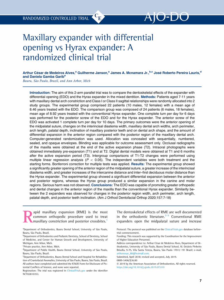
The experimental group comprised patients treatedwith the EDO (Fig 1, A-C). The EDO was composed of2 11-mm prefabricated screws, 1 posteriorly positionedon the palate at the level of the first permanent molarsand the other anteriorly positioned at the level of the firstdeciduous molars (Great Lakes Orthodontics, Tona-wanda, NY). Orthodontic bands were preferentiallyadapted on the maxillary first permanent molars, andclasps were bonded on the maxillary deciduous canines.When the maxillary first permanent molars were partiallyerupted, or the distal aspect of the crown was covered bygingiva, the maxillary second deciduous molars werebanded, and a wire extension was soldered on the palatalaspect of the first permanent molars. Both expanderscrews were concurrently activated for 6 days, with anactivation protocol of half a turn in the morning andhalf a turn in the evening. Afterward, only the anteriorscrew was activated for an extra 4-day time with thesame protocol. After the active expansion period, theexpander was kept in the mouth as a retainer for6 months. At the end of the retention phase, the ex-panders were removed, and a removable retention platewas installed.
The comparison group comprised patients who un-derwent RME using the conventional Hyrax expander
Journal of Orthodontics and Dentofacial Orthopedics
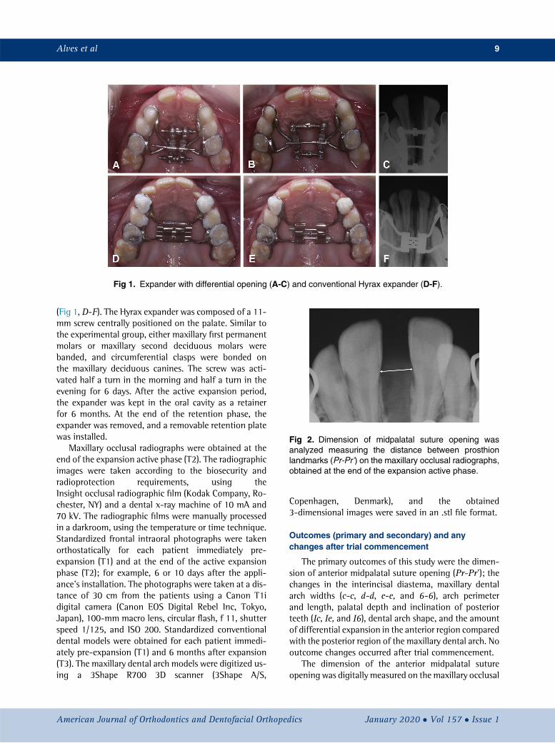
Fig 1. Expander with differential opening (A-C) and conventional Hyrax expander (D-F).
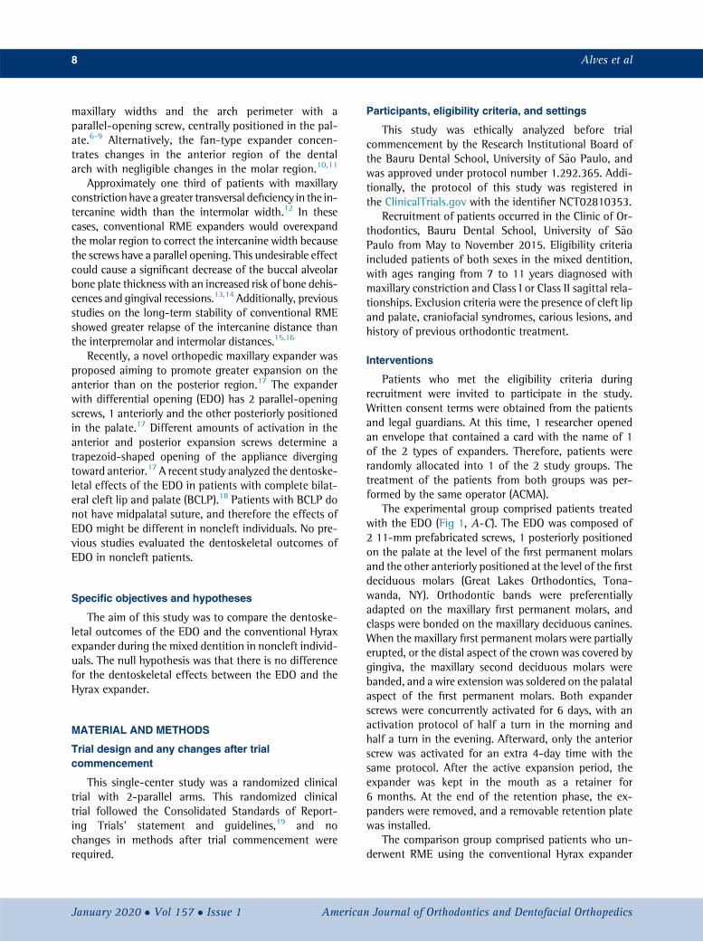
Fig 2. Dimension of midpalatal suture opening wasanalyzed measuring the distance between prosthionlandmarks (Pr-Pr’) on the maxillary occlusal radiographs,obtained at the end of the expansion active phase.
Alves et al 9
(Fig 1, D-F). The Hyrax expander was composed of a 11-mm screw centrally positioned on the palate. Similar tothe experimental group, either maxillary first permanentmolars or maxillary second deciduous molars werebanded, and circumferential clasps were bonded onthe maxillary deciduous canines. The screw was acti-vated half a turn in the morning and half a turn in theevening for 6 days. After the active expansion period,the expander was kept in the oral cavity as a retainerfor 6 months. At the end of the retention phase, theexpander was removed, and a removable retention platewas installed.
Maxillary occlusal radiographs were obtained at theend of the expansion active phase (T2). The radiographicimages were taken according to the biosecurity andradioprotection requirements, using theInsight occlusal radiographic film (Kodak Company, Ro-chester, NY) and a dental x-ray machine of 10 mA and70 kV. The radiographic films were manually processedin a darkroom, using the temperature or time technique.Standardized frontal intraoral photographs were takenorthostatically for each patient immediately pre-expansion (T1) and at the end of the active expansionphase (T2); for example, 6 or 10 days after the appli-ance's installation. The photographs were taken at a dis-tance of 30 cm from the patients using a Canon T1idigital camera (Canon EOS Digital Rebel Inc, Tokyo,Japan), 100-mm macro lens, circular flash, f 11, shutterspeed 1/125, and ISO 200. Standardized conventionaldental models were obtained for each patient immedi-ately pre-expansion (T1) and 6 months after expansion(T3). The maxillary dental arch models were digitized us-ing a 3Shape R700 3D scanner (3Shape A/S,
American Journal of Orthodontics and Dentofacial Orthoped
Copenhagen, Denmark), and the obtained3-dimensional images were saved in an .stl file format.
Outcomes (primary and secondary) and anychanges after trial commencement
The primary outcomes of this study were the dimen-sion of anterior midpalatal suture opening (Pr-Pr'); thechanges in the interincisal diastema, maxillary dentalarch widths (c-c, d-d, e-e, and 6-6), arch perimeterand length, palatal depth and inclination of posteriorteeth (Ic, Ie, and I6), dental arch shape, and the amountof differential expansion in the anterior region comparedwith the posterior region of the maxillary dental arch. Nooutcome changes occurred after trial commencement.
The dimension of the anterior midpalatal sutureopening was digitally measured on the maxillary occlusal
ics January 2020 � Vol 157 � Issue 1
Fig 3. The interincisal diastema width was analyzed measuring the distance between the pointslocated at the confluence of the mesial aspect of the maxillary central incisors with the gingival papillaon the intraoral frontal photographs obtained before (A) and after (B) the active phase of rapid maxillaryexpansion. Interincisal diastema changewas considered the difference between the values obtained atT2 and T1.
10 Alves et al
radiographs using Dolphin Imaging software, version11.0 (Dolphin Imaging and Management Solutions,Chatsworth, Calif), as shown in Figure 2.
The width of the interincisal diastema was measuredusing a modification of a method proposed in a previousstudy.20 Initially, the mesiodistal width of the clinicalcrown of the maxillary right central incisor of each pa-tient was manually measured on the pre-expansion con-ventional dental models using a caliper. Using a digitalmillimetric ruler, the photograph was resized in Micro-soft PowerPoint 2013 (Microsoft Corporation, Red-mond, Wash) according to the actual size of themeasured tooth. The interincisal diastema was measuredusing Dolphin Imaging software, as shown in Figure 3.
The measurements of maxillary dental arch widths,arch perimeter and length, palatal depth, and inclinationof posterior teeth were performed on the pre- and post-expansion digital dental models using the OrthoAnalyzer3D software (3Shape A/S), as shown in Figures 4-6.
The dental arch shape was evaluated using the Geo-magic Wrap 2015 software (Raindrop Geomagic Inc,Morrisville, NC) and the Microsoft Excel 2013 (MicrosoftCorporation). In the Geomagic Wrap 2015 software, An-drews' facial axis points21 were set on the maxillaryteeth. These points were decomposed in 3 cardinal direc-tions generating values on the x-, y-, and z-axis. Consid-ering the y-axis values referred to the depth dimension(cervico-oclusal plane), only the x-axis values (trans-versal plane) and the z-axis values (sagittal plane) weretabulated. Microsoft Excel 2013 was used to graphicallydetermine the mean maxillary dental arch shape for bothstudy groups at T1 and T2, using the interpolar function.
Sample size calculation
A minimum difference of 2 mm in the intercaninedistance, a standard deviation of 1.65, an alpha errorof 5%, and a test power of 80% were considered for
January 2020 � Vol 157 � Issue 1 American
sample size calculation.11 Twenty participants wererequired in each group.
Interim analyses and stopping guidelines
Not applicable.
Randomization (random number generation,allocation concealment, implementation)
A simple electronically generated randomization wasperformed before trial commencement using theRandom Allocation Software program.22 Randomizationensured patients' allocation in both groups with a 1:1ratio. Allocation concealment involved numbered,sealed, and opaque envelopes prepared before trialcommencement. One envelope was sequentially openedfor each participant during recruitment. Each envelopecontained a card with the name of 1 expander. The ini-tials of the name of the participant, the type of expander,and the date of allocation were identified in the externalsurface of the envelope. One operator was responsiblefor the randomization process, allocation concealment,and implementation.
Blinding
Double-blinding was not possible because the oper-ator and patients were aware of the type of expanderthat was being installed. However, blinding was accom-plished during outcome assessment once the maxillaryocclusal radiographs, the photographs, and the digitaldental models were unidentified during analysis.
Error study
One operator (ACMA) performed all the measure-ments and repeated them in 30% of the sample at least1 month later. The intraexaminer error was assessed us-ing the intraclass correlation coefficient.23
Journal of Orthodontics and Dentofacial Orthopedics
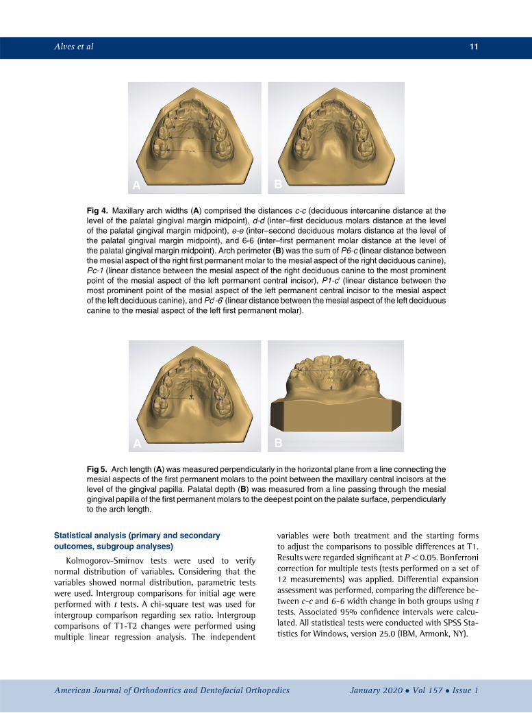
Fig 4. Maxillary arch widths (A) comprised the distances c-c (deciduous intercanine distance at thelevel of the palatal gingival margin midpoint), d-d (inter–first deciduous molars distance at the levelof the palatal gingival margin midpoint), e-e (inter–second deciduous molars distance at the level ofthe palatal gingival margin midpoint), and 6-6 (inter–first permanent molar distance at the level ofthe palatal gingival margin midpoint). Arch perimeter (B) was the sum of P6-c (linear distance betweenthe mesial aspect of the right first permanent molar to the mesial aspect of the right deciduous canine),Pc-1 (linear distance between the mesial aspect of the right deciduous canine to the most prominentpoint of the mesial aspect of the left permanent central incisor), P1-c' (linear distance between themost prominent point of the mesial aspect of the left permanent central incisor to the mesial aspectof the left deciduous canine), andPc'-6' (linear distance between themesial aspect of the left deciduouscanine to the mesial aspect of the left first permanent molar).
Fig 5. Arch length (A) was measured perpendicularly in the horizontal plane from a line connecting themesial aspects of the first permanent molars to the point between the maxillary central incisors at thelevel of the gingival papilla. Palatal depth (B) was measured from a line passing through the mesialgingival papilla of the first permanent molars to the deepest point on the palate surface, perpendicularlyto the arch length.
Alves et al 11
Statistical analysis (primary and secondaryoutcomes, subgroup analyses)
Kolmogorov-Smirnov tests were used to verifynormal distribution of variables. Considering that thevariables showed normal distribution, parametric testswere used. Intergroup comparisons for initial age wereperformed with t tests. A chi-square test was used forintergroup comparison regarding sex ratio. Intergroupcomparisons of T1-T2 changes were performed usingmultiple linear regression analysis. The independent
American Journal of Orthodontics and Dentofacial Orthoped
variables were both treatment and the starting formsto adjust the comparisons to possible differences at T1.Results were regarded significant at P\0.05. Bonferronicorrection for multiple tests (tests performed on a set of12 measurements) was applied. Differential expansionassessment was performed, comparing the difference be-tween c-c and 6-6 width change in both groups using ttests. Associated 95% confidence intervals were calcu-lated. All statistical tests were conducted with SPSS Sta-tistics for Windows, version 25.0 (IBM, Armonk, NY).
ics January 2020 � Vol 157 � Issue 1
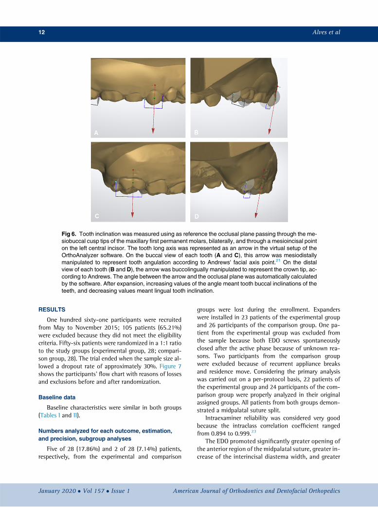
Fig 6. Tooth inclination was measured using as reference the occlusal plane passing through the me-siobuccal cusp tips of the maxillary first permanent molars, bilaterally, and through a mesioincisal pointon the left central incisor. The tooth long axis was represented as an arrow in the virtual setup of theOrthoAnalyzer software. On the buccal view of each tooth (A and C), this arrow was mesiodistallymanipulated to represent tooth angulation according to Andrews' facial axis point.21 On the distalview of each tooth (B and D), the arrow was buccolingually manipulated to represent the crown tip, ac-cording to Andrews. The angle between the arrow and the occlusal plane was automatically calculatedby the software. After expansion, increasing values of the angle meant tooth buccal inclinations of theteeth, and decreasing values meant lingual tooth inclination.
12 Alves et al
RESULTS
One hundred sixty-one participants were recruitedfrom May to November 2015; 105 patients (65.21%)were excluded because they did not meet the eligibilitycriteria. Fifty-six patients were randomized in a 1:1 ratioto the study groups (experimental group, 28; compari-son group, 28). The trial ended when the sample size al-lowed a dropout rate of approximately 30%. Figure 7shows the participants' flow chart with reasons of lossesand exclusions before and after randomization.
Baseline data
Baseline characteristics were similar in both groups(Tables I and II).
Numbers analyzed for each outcome, estimation,and precision, subgroup analyses
Five of 28 (17.86%) and 2 of 28 (7.14%) patients,respectively, from the experimental and comparison
January 2020 � Vol 157 � Issue 1 American
groups were lost during the enrollment. Expanderswere installed in 23 patients of the experimental groupand 26 participants of the comparison group. One pa-tient from the experimental group was excluded fromthe sample because both EDO screws spontaneouslyclosed after the active phase because of unknown rea-sons. Two participants from the comparison groupwere excluded because of recurrent appliance breaksand residence move. Considering the primary analysiswas carried out on a per-protocol basis, 22 patients ofthe experimental group and 24 participants of the com-parison group were properly analyzed in their originalassigned groups. All patients from both groups demon-strated a midpalatal suture split.
Intraexaminer reliability was considered very goodbecause the intraclass correlation coefficient rangedfrom 0.894 to 0.999.23
The EDO promoted significantly greater opening ofthe anterior region of the midpalatal suture, greater in-crease of the interincisal diastema width, and greater
Journal of Orthodontics and Dentofacial Orthopedics
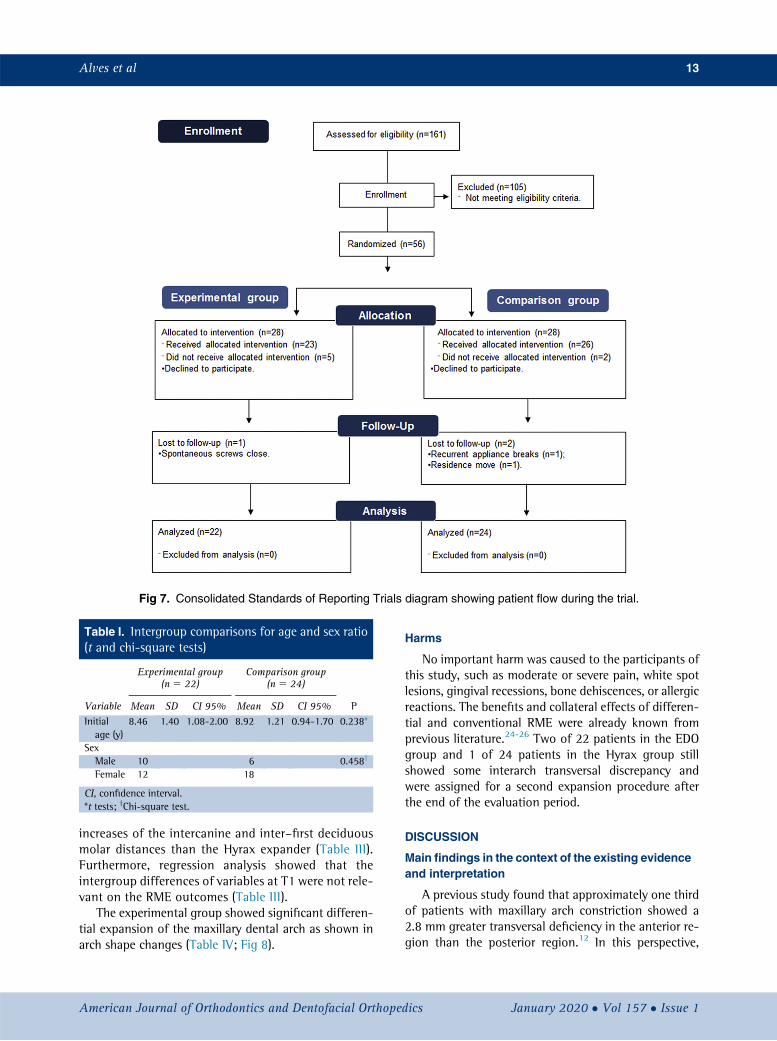
Fig 7. Consolidated Standards of Reporting Trials diagram showing patient flow during the trial.
Table I. Intergroup comparisons for age and sex ratio(t and chi-square tests)
Variable
Experimental group(n 5 22)
Comparison group(n 5 24)
PMean SD CI 95% Mean SD CI 95%Initialage (y)
8.46 1.40 1.08-2.00 8.92 1.21 0.94-1.70 0.238*
SexMale 10 6 0.458y
Female 12 18
CI, confidence interval.*t tests; yChi-square test.
Alves et al 13
increases of the intercanine and inter–first deciduousmolar distances than the Hyrax expander (Table III).Furthermore, regression analysis showed that theintergroup differences of variables at T1 were not rele-vant on the RME outcomes (Table III).
The experimental group showed significant differen-tial expansion of the maxillary dental arch as shown inarch shape changes (Table IV; Fig 8).
American Journal of Orthodontics and Dentofacial Orthoped
Harms
No important harm was caused to the participants ofthis study, such as moderate or severe pain, white spotlesions, gingival recessions, bone dehiscences, or allergicreactions. The benefits and collateral effects of differen-tial and conventional RME were already known fromprevious literature.24-26 Two of 22 patients in the EDOgroup and 1 of 24 patients in the Hyrax group stillshowed some interarch transversal discrepancy andwere assigned for a second expansion procedure afterthe end of the evaluation period.
DISCUSSION
Main findings in the context of the existing evidenceand interpretation
A previous study found that approximately one thirdof patients with maxillary arch constriction showed a2.8 mm greater transversal deficiency in the anterior re-gion than the posterior region.12 In this perspective,
ics January 2020 � Vol 157 � Issue 1
Table II. T1 and T2 variables in the sample groups
Variable
Experimental group (n 5 22) Comparison group (n 5 24)
T1 T2 T1 T2
Mean SD CI 95% Mean SD CI 95% Mean SD CI 95% Mean SD CI 95%Interincisal diastema (mm) 0.75 0.94 0.72-1.35 4.86 2.27 1.74-3.24 0.69 0.78 0.60-1.10 3.23 0.93 0.72-1.32c-c (mm) 24.45 1.90 1.43-2.81 30.75 2.24 1.64-3.09 25.50 2.44 1.82-3.72 29.03 2.50 1.86-3.80d-d (mm) 26.40 1.89 1.45-2.70 32.48 2.38 1.83-3.40 27.34 2.37 1.83-3.40 31.11 2.49 1.92-3.57e-e (mm) 29.96 2.23 1.71-3.19 35.06 2.36 1.82-3.38 31.81 2.30 1.78-3.26 35.17 2.22 1.72-3.156-6 (mm) 34.60 3.10 2.39-4.44 38.22 3.07 2.36-4.39 36.01 2.19 1.70-3.07 39.75 1.94 1.51-2.72Arch perimeter (mm) 77.21 4.25 3.27-6.07 81.68 4.06 3.12-5.80 77.73 5.05 3.92-7.09 80.20 6.81 5.29-9.56Arch length (mm) 28.40 2.12 1.63-3.03 27.58 2.28 1.76-3.27 28.50 2.12 1.65-2.98 27.71 2.06 1.60-2.89Palatal depth (mm) 13.80 2.53 1.95-3.62 12.69 2.60 2.00-3.72 14.67 1.71 1.33-2.40 14.09 2.15 1.67-3.01Ic (o) 74.99 7.34 5.94-9.62 77.47 6.70 5.41-8.59 77.25 8.85 5.09-7.89 79.32 5.29 7.20-11.50Ie (o) 75.39 4.55 4.56-7.15 79.79 5.16 4.13-6.86 76.50 6.04 5.31-8.43 80.25 5.02 4.01-6.71I6 (o) 77.03 6.52 5.51-8.46 81.59 5.26 4.33-6.68 79.37 5.78 4.81-7.24 82.22 5.08 4.23-6.37
CI, confidence interval; SD, standard deviation.
14 Alves et al
some patients may need an individualized expansionwith different amounts of screw activations in the canineandmolar regions.12 The EDO is a 2-screw expander thataims to promote a distinct amount of expansion in theanterior and posterior regions of the maxillary dentalarch.15 However, no previous clinical study had analyzedthe dentoskeletal effects of the EDO in noncleftindividuals.
An intergroup compatibility for initial age and sexdistribution was found (Table I). Despite the insignifi-cant difference between groups regarding initial age,the experimental group was slightly younger comparedwith the comparison group (Table I). However, this slightdifference is not likely to have any clinical relevance.27
These results confirm the sample homogeneity, ensurethe effectiveness of randomization and allocation ofthe patients, and decrease the risk of bias for the inter-group comparisons.28 The 6-mm intermolar amount ofexpansion was standardized in both groups to allowintergroup comparison.
The EDO promoted a significant increase of theanterior region of the midpalatal suture and the inter-incisal diastema width of 5.38 mm and 4.11 mm,respectively (Table III). These findings confirm the po-tential for an orthopedic effect of the EDO. The inter-group comparison showed that the EDO promoted agreater orthopedic effect in the anterior region of themaxilla than the conventional Hyrax expander (TableIII). These findings are probably associated with thegreater amount of activation of the anterior screw inthe EDO group. The greater the intercanine distance in-crease, the greater will be the anterior palatal suturesplit. The anterior screw of the EDO was openedapproximately 9 mm, causing a 60% orthopedic effecton the anterior region of the midpalatal suture(5.38 mm). The pattern of midpalatal suture opening
January 2020 � Vol 157 � Issue 1 American
observed for the EDO was similar to those describedin previous studies with conventional RME ex-panders,9,29 showing a triangular shape, as shown inFigure 1. Additionally, bone-tooth–borne expandersseem to produce a similar orthopedic effect comparedwith conventional Hyrax expanders.30
The experimental group showed significant increasesof the intermolar and intercanine arch widths of3.61 mm and 6.30 mm, respectively, causing a mean in-crease of 4.47 mm in arch perimeter (Table III). The inter-canine distance changes were significantly greater thanthe intermolar distance changes owing to the anteriorlydivergent opening of the EDO (Table III). A recent studyanalyzed the dentoskeletal effects of the EDO in patientswith complete bilateral cleft lip and palate and foundsignificant increases of the intermolar and intercaninearch widths of 5.57 mm and 7.68 mm, respectively,causing a mean increase of 7.66 mm in arch perimeter.18
These increased values of the maxillary dental arch widthand perimeter changes compared with our study may beassociated with a greater amount of expansion in a moreseverely constricted maxilla and with the smaller resis-tance to expansion observed in patients with BCLPbecause of the absence of the midpalatal suture.31,32
The Hyrax group showed significant increases ofintercanine and intermolar arch widths of 3.42 mmand 3.74 mm, respectively, causing a mean increase of2.46 mm in arch perimeter (Table III). In this group,the intercanine distance change was similar to the inter-molar distance change (Table IV). Similar results wereobserved in a previous study.11 This similarity betweenthe amount of expansion in the anterior and posteriorregions might be explained by the parallel-openingpattern of the conventional Hyrax screw.11,33 Theintergroup comparison showed that the EDO promoteda greater amount of expansion in the canine region
Journal of Orthodontics and Dentofacial Orthopedics
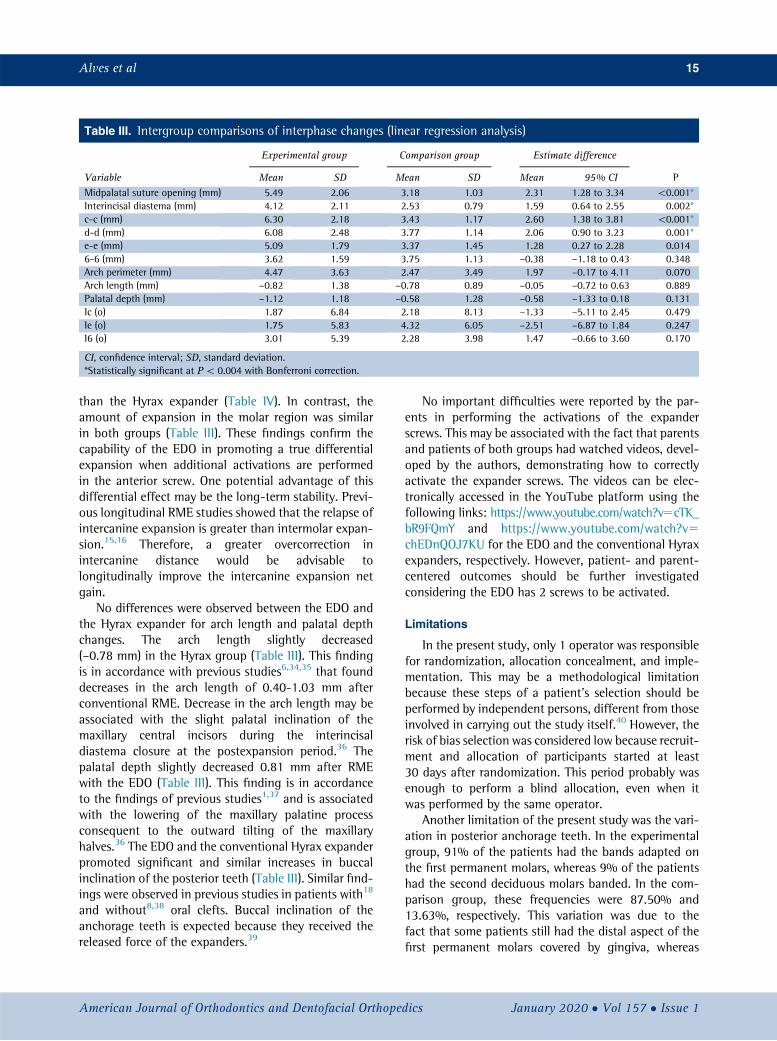
Table III. Intergroup comparisons of interphase changes (linear regression analysis)
Variable
Experimental group Comparison group Estimate difference
PMean SD Mean SD Mean 95% CIMidpalatal suture opening (mm) 5.49 2.06 3.18 1.03 2.31 1.28 to 3.34 \0.001*Interincisal diastema (mm) 4.12 2.11 2.53 0.79 1.59 0.64 to 2.55 0.002*c-c (mm) 6.30 2.18 3.43 1.17 2.60 1.38 to 3.81 \0.001*d-d (mm) 6.08 2.48 3.77 1.14 2.06 0.90 to 3.23 0.001*e-e (mm) 5.09 1.79 3.37 1.45 1.28 0.27 to 2.28 0.0146-6 (mm) 3.62 1.59 3.75 1.13 –0.38 –1.18 to 0.43 0.348Arch perimeter (mm) 4.47 3.63 2.47 3.49 1.97 –0.17 to 4.11 0.070Arch length (mm) –0.82 1.38 –0.78 0.89 –0.05 –0.72 to 0.63 0.889Palatal depth (mm) –1.12 1.18 –0.58 1.28 –0.58 –1.33 to 0.18 0.131Ic (o) 1.87 6.84 2.18 8.13 –1.33 –5.11 to 2.45 0.479Ie (o) 1.75 5.83 4.32 6.05 –2.51 –6.87 to 1.84 0.247I6 (o) 3.01 5.39 2.28 3.98 1.47 –0.66 to 3.60 0.170
CI, confidence interval; SD, standard deviation.*Statistically significant at P\ 0.004 with Bonferroni correction.
Alves et al 15
than the Hyrax expander (Table IV). In contrast, theamount of expansion in the molar region was similarin both groups (Table III). These findings confirm thecapability of the EDO in promoting a true differentialexpansion when additional activations are performedin the anterior screw. One potential advantage of thisdifferential effect may be the long-term stability. Previ-ous longitudinal RME studies showed that the relapse ofintercanine expansion is greater than intermolar expan-sion.15,16 Therefore, a greater overcorrection inintercanine distance would be advisable tolongitudinally improve the intercanine expansion netgain.
No differences were observed between the EDO andthe Hyrax expander for arch length and palatal depthchanges. The arch length slightly decreased(–0.78 mm) in the Hyrax group (Table III). This findingis in accordance with previous studies6,34,35 that founddecreases in the arch length of 0.40-1.03 mm afterconventional RME. Decrease in the arch length may beassociated with the slight palatal inclination of themaxillary central incisors during the interincisaldiastema closure at the postexpansion period.36 Thepalatal depth slightly decreased 0.81 mm after RMEwith the EDO (Table III). This finding is in accordanceto the findings of previous studies1,37 and is associatedwith the lowering of the maxillary palatine processconsequent to the outward tilting of the maxillaryhalves.36 The EDO and the conventional Hyrax expanderpromoted significant and similar increases in buccalinclination of the posterior teeth (Table III). Similar find-ings were observed in previous studies in patients with18
and without8,38 oral clefts. Buccal inclination of theanchorage teeth is expected because they received thereleased force of the expanders.39
American Journal of Orthodontics and Dentofacial Orthoped
No important difficulties were reported by the par-ents in performing the activations of the expanderscrews. This may be associated with the fact that parentsand patients of both groups had watched videos, devel-oped by the authors, demonstrating how to correctlyactivate the expander screws. The videos can be elec-tronically accessed in the YouTube platform using thefollowing links: https://www.youtube.com/watch?v5cTK_bR9FQmY and https://www.youtube.com/watch?v5chEDnQOJ7KU for the EDO and the conventional Hyraxexpanders, respectively. However, patient- and parent-centered outcomes should be further investigatedconsidering the EDO has 2 screws to be activated.
Limitations
In the present study, only 1 operator was responsiblefor randomization, allocation concealment, and imple-mentation. This may be a methodological limitationbecause these steps of a patient's selection should beperformed by independent persons, different from thoseinvolved in carrying out the study itself.40 However, therisk of bias selection was considered low because recruit-ment and allocation of participants started at least30 days after randomization. This period probably wasenough to perform a blind allocation, even when itwas performed by the same operator.
Another limitation of the present study was the vari-ation in posterior anchorage teeth. In the experimentalgroup, 91% of the patients had the bands adapted onthe first permanent molars, whereas 9% of the patientshad the second deciduous molars banded. In the com-parison group, these frequencies were 87.50% and13.63%, respectively. This variation was due to thefact that some patients still had the distal aspect of thefirst permanent molars covered by gingiva, whereas
ics January 2020 � Vol 157 � Issue 1
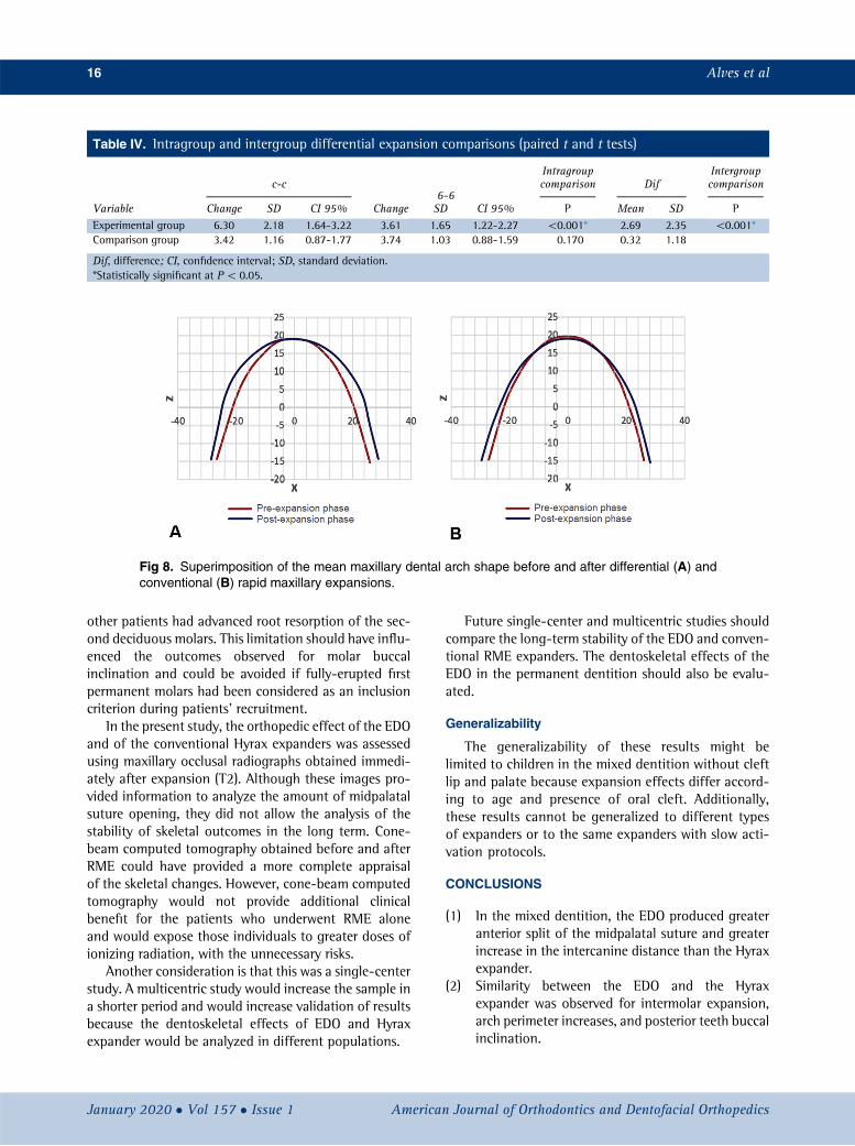
Table IV. Intragroup and intergroup differential expansion comparisons (paired t and t tests)
Variable
c-c6-6
Intragroupcomparison Dif
Intergroupcomparison
Change SD CI 95% Change SD CI 95% P Mean SD P
Experimental group 6.30 2.18 1.64-3.22 3.61 1.65 1.22-2.27 \0.001* 2.69 2.35 \0.001*Comparison group 3.42 1.16 0.87-1.77 3.74 1.03 0.88-1.59 0.170 0.32 1.18
Dif, difference; CI, confidence interval; SD, standard deviation.*Statistically significant at P\ 0.05.
Fig 8. Superimposition of the mean maxillary dental arch shape before and after differential (A) andconventional (B) rapid maxillary expansions.
16 Alves et al
other patients had advanced root resorption of the sec-ond deciduous molars. This limitation should have influ-enced the outcomes observed for molar buccalinclination and could be avoided if fully-erupted firstpermanent molars had been considered as an inclusioncriterion during patients' recruitment.
In the present study, the orthopedic effect of the EDOand of the conventional Hyrax expanders was assessedusing maxillary occlusal radiographs obtained immedi-ately after expansion (T2). Although these images pro-vided information to analyze the amount of midpalatalsuture opening, they did not allow the analysis of thestability of skeletal outcomes in the long term. Cone-beam computed tomography obtained before and afterRME could have provided a more complete appraisalof the skeletal changes. However, cone-beam computedtomography would not provide additional clinicalbenefit for the patients who underwent RME aloneand would expose those individuals to greater doses ofionizing radiation, with the unnecessary risks.
Another consideration is that this was a single-centerstudy. A multicentric study would increase the sample ina shorter period and would increase validation of resultsbecause the dentoskeletal effects of EDO and Hyraxexpander would be analyzed in different populations.
January 2020 � Vol 157 � Issue 1 American
Future single-center and multicentric studies shouldcompare the long-term stability of the EDO and conven-tional RME expanders. The dentoskeletal effects of theEDO in the permanent dentition should also be evalu-ated.
Generalizability
The generalizability of these results might belimited to children in the mixed dentition without cleftlip and palate because expansion effects differ accord-ing to age and presence of oral cleft. Additionally,these results cannot be generalized to different typesof expanders or to the same expanders with slow acti-vation protocols.
CONCLUSIONS
(1) In the mixed dentition, the EDO produced greateranterior split of the midpalatal suture and greaterincrease in the intercanine distance than the Hyraxexpander.
(2) Similarity between the EDO and the Hyraxexpander was observed for intermolar expansion,arch perimeter increases, and posterior teeth buccalinclination.
Journal of Orthodontics and Dentofacial Orthopedics
Alves et al 17
(3) The EDO may be indicated for patients with a needfor greater maxillary intercanine expansion thanintermolar expansion.
ACKNOWLEDGMENTS
The authors thank Great Lakes Orthodontics for pro-ducing the expander with differential opening (EDO) forthis study. The authors also thank the Coordination forthe Improvement of Higher Education Personnel forthe research financial support.
REFERENCES
1. Haas AJ. Rapid expansion of the maxillary dental arch and nasalcavity by opening the midpalatal suture. Angle Orthod 1961;31:73-90.
2. Bazargani F, Feldmann I, Bondemark L. Three-dimensional anal-ysis of effects of rapid maxillary expansion on facial sutures andbones. Angle Orthod 2013;83:1074-82.
3. Lagravere MO, Major PW, Flores-Mir C. Long-term dental archchanges after rapid maxillary expansion treatment: a systematicreview. Angle Orthod 2005;75:155-61.
4. Lagravere MO, Major PW, Flores-Mir C. Long-term skeletalchanges with rapid maxillary expansion: a systematic review. AngleOrthod 2005;75:1046-52.
5. Liu S, Xu T, Zou W. Effects of rapid maxillary expansion on themidpalatal suture: a systematic review. Eur J Orthod 2015;37:651-5.
6. Adkins MD, Nanda RS, Currier GF. Arch perimeter changes on rapidpalatal expansion. Am J Orthod Dentofacial Orthop 1990;97:194-9.
7. Cozzani M, Guiducci A, Mirenghi S, Mutinelli S, Siciliani G.Arch width changes with a rapid maxillary expansion appli-ance anchored to the primary teeth. Angle Orthod 2007;77:296-302.
8. Kartalian A, Gohl E, Adamian M, Enciso R. Cone-beam computer-ized tomography evaluation of the maxillary dentoskeletal com-plex after rapid palatal expansion. Am J Orthod DentofacialOrthop 2010;138:486-92.
9. da Silva Filho OG, Lara TS, de Almeida AM, da Silav HC. Evaluationof themidpalatal suture during rapid palatal expansion in children:a CT study. J Clin Pediatr Dent 2005;29:231-8.
10. Doruk C, Bicakci AA, Basciftci FA, Agar U, Babacan H. A compar-ison of the effects of rapid maxillary expansion and fan-type rapidmaxillary expansion on dentofacial structures. Angle Orthod 2004;74:184-94.
11. C€orekci B, G€oyenc YB. Dentofacial changes from fan-type rapidmaxillary expansion vs traditional rapid maxillary expansion inearly mixed dentition. Angle Orthod 2013;83:842-50.
12. Belluzzo RHL, Faltin Junior K, Lascala CE, Vianna LBR. Maxillaryconstriction: are there differences between anterior and posteriorregions? Dental Press J Orthod 2012;17:1-6.
13. Brunetto M, Andriani Jda S, Ribeiro GL, Locks A, Correa M,Correa LR. Three-dimensional assessment of buccal alveolarbone after rapid and slow maxillary expansion: a clinical trialstudy. Am J Orthod Dentofacial Orthop 2013;143:633-44.
14. Garib DG, Henriques JFC, Janson G, de Freitas MR, Fernandes AY.Periodontal effects of rapid maxillary expansion with tooth-tissue-borne and tooth-borne expanders: a computed tomography eval-uation. Am J Orthod Dentofacial Orthop 2006;129:749-58.
American Journal of Orthodontics and Dentofacial Orthoped
15. Gurel HG, Memili B, Erkan M, Sukurica Y. Long-term effects ofrapid maxillary expansion followed by fixed appliances. Angle Or-thod 2010;80:5-9.
16. Pinheiro FHSL, Garib DG, Janson G, Bombonatti R, de Freitas MR.Longitudinal stability of rapid and slow maxillary expansion. DentPress J Orthod 2014;19:70-7.
17. Garib DG, Garcia LC, Pereira V, Lauris RC, Yen S. A rapid maxillaryexpander with differential opening. J Clin Orthod 2014;48:430-5.
18. Garib D, Lauris RC, Calil LR, Alves AC, Janson G, de Almeida AM,et al. Dentoskeletal outcomes of a rapid maxillary expander withdifferential opening in patients with bilateral cleft lip and palate:a prospective clinical trial. Am J Orthod Dentofacial Orthop2016;150:564-74.
19. Schulz KF, Altman DG, Moher D, CONSORT. CONSORT 2010Statement: updated guidelines for reporting parallel group rando-mised trials. BMC Med 2010;8:18: updated Guidelines.
20. Normando D, da Silva PL, Mendes �AM. A clinical photogrammetricmethod to measure dental arch dimensions and mesio-distal toothsize. Eur J Orthod 2011;33:721-6.
21. Andrews LF. The six keys to normal occlusion. Am J Orthod 1972;62:296-309.
22. Saghaei M. Random allocation software for parallel group ran-domized trials. BMC Med Res Methodol 2004;4:26.
23. Fleiss JL. Analysis of data from multiclinic trials. Control Clin Trials1986;7:267-75.
24. De Felippe NL, Da Silveira AC, Viana G, Smith B. Influence ofpalatal expanders on oral comfort, speech, and mastication. AmJ Orthod Dentofacial Orthop 2010;137:48-53.
25. Gecgelen M, Aksoy A, Kirdemir P, Doguc DK, Cesur G, Koskan O,et al. Evaluation of stress and pain during rapid maxillary expan-sion treatments. J Oral Rehabil 2012;39:767-75.
26. Needleman HL, Hoang CD, Allred E, Hertzberg J, Berde C. Reportsof pain by children undergoing rapid palatal expansion. PediatrDent 2000;22:221-6.
27. Lu Y, Belitskaya-Levy I. The debate about p-values. Shanghai ArchPsychiatry 2015;27:381-5.
28. Berger VW, Exner DV. Detecting selection bias in randomized clin-ical trials. Control Clin Trials 1999;20:319-27.
29. Wertz RA. Skeletal and dental changes accompanying rapid mid-palatal suture opening. Am J Orthod 1970;58:41-66.
30. Gunyuz Toklu M, Germec-Cakan D, Tozlu M. Periodontal, den-toalveolar, and skeletal effects of tooth-borne and tooth-bone-borne expansion appliances. Am J Orthod Dentofacial Orthop2015;148:97-109.
31. de Medeiros Alves AC, Garib DG, Janson G, de Almeida AM,Calil LR. Analysis of the dentoalveolar effects of slow and rapidmaxillary expansion in complete bilateral cleft lip and palate pa-tients: a randomized clinical trial. Clin Oral Investig 2016;20:1837-47.
32. de Almeida AM, Ozawa TO, Alves ACM, Janson G, Lauris JRP,Ioshida MSY, et al. Slow versus rapid maxillary expansion in bilat-eral cleft lip and palate: a CBCT randomized clinical trial. Clin OralInvestig 2016;21:1789-99.
33. Weber G. The Hyrax-system according to Biederman. Quintessenz1970;21:69-70.
34. Claro CAA, Abr~ao J, Reis SAB, de Fantini SM. Correlation betweentransverse expansion and increase in the upper arch perimeter afterrapid maxillary expansion. Braz Oral Res 2006;20:76-81.
35. Mutinelli S, Cozzani M, Manfredi M, Bee M, Siciliani G. Dental archchanges following rapid maxillary expansion. Eur J Orthod 2008;30:469-76.
36. Bishara SE, Staley RN. Maxillary expansion: clinical implications.Am J Orthod Dentofacial Orthop 1987;91:3-14.
ics January 2020 � Vol 157 � Issue 1

18 Alves et al
37. Fried KH. Palate-tongue relativity. Angle Orthod 1971;41:308-23.
38. Sari Z, Uysal T, Usumez S, Basciftci FA. Rapid maxillary expansion.Is it better in the mixed or in the permanent dentition? Angle Or-thod 2003;73:654-61.
January 2020 � Vol 157 � Issue 1 American
39. Isaacson RJ, Ingram AH. Forces produced by rapid maxillaryexpansion: II. Forces present during treatment. Angle Orthod1964;34:261-70.
40. Kahan BC, Rehal S, Cro S. Risk of selection bias in randomised tri-als. Trials 2015;16:405.
Journal of Orthodontics and Dentofacial Orthopedics