ORTHOPEDIC SURGERY CASEBOOK
8
SB938.001 MIMEDX PLACENTAL-BASED ALLOGRAFTS ORTHOPEDIC SURGERY CASEBOOK VOLUME 1 | EDITION 1 TOTAL SHOULDER REPAIR WITH AMNIOFIX ® BRIDGEFIX IN ARTHROSCOPIC ROTATOR CUFF REPAIR WITH AMNIOFIX INJECTABLE ACL RECONSTRUCTION & REPAIR OF PARTIAL PATELLAR TENDON TEAR WITH AMNIOFIX REPAIR OF A PERONEAL TENDON TEAR WITH AMNIOCORD ® REPAIR OF RUPTURED ACHILLES TENDON WITH AMNIOCORD TOTAL ANKLE REPLACEMENT WITH AMNIOCORD
-
Upload
khangminh22 -
Category
Documents
-
view
1 -
download
0
Transcript of ORTHOPEDIC SURGERY CASEBOOK
SB938.001
MIMEDX PLACENTAL-BASED ALLOGRAFTS
ORTHOPEDIC SURGERY CASEBOOK
VOLUME 1 | EDITION 1
TOTAL SHOULDER REPAIR WITH AMNIOFIX®
BRIDGEFIX IN ARTHROSCOPIC ROTATOR CUFF REPAIR WITH AMNIOFIX INJECTABLE
ACL RECONSTRUCTION & REPAIR OF PARTIAL PATELLAR TENDON TEAR WITH AMNIOFIX
REPAIR OF A PERONEAL TENDON TEAR WITH AMNIOCORD®
REPAIR OF RUPTURED ACHILLES TENDON WITH AMNIOCORD
TOTAL ANKLE REPLACEMENT WITH AMNIOCORD
2 Orthopedic Surgery Casebook SB938.001
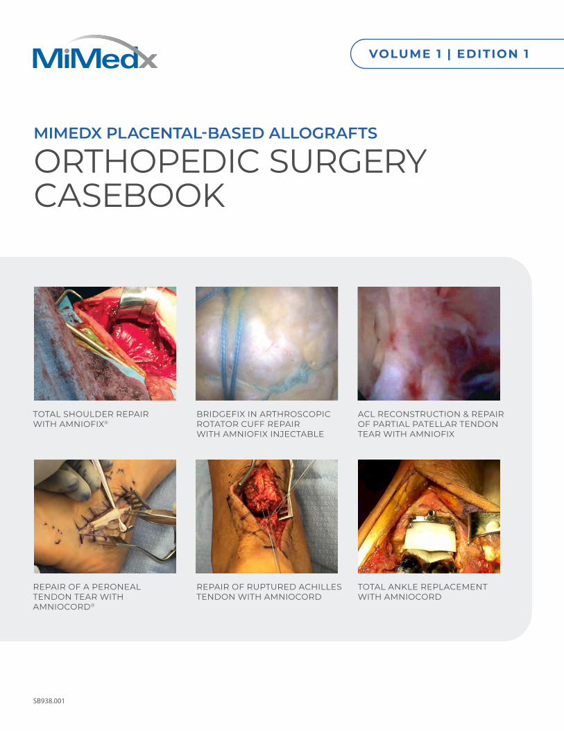
Total Shoulder Repair with AmnioFix Lonnie Paulos, MD | Orthopedic Surgery | Salt Lake City, Utah
Clinical History A 43-year-old male with a history of traumatic injury to the shoulder developed chronic osteoarthritis (OA) of the glenohumeral joint due to an injury with six prior open or arthroscopic procedures to correct it. He subsequently developed chronic pain and shoulder instability. In addition to the severe OA noted, the patient had developed significant scarring and adhesions throughout the subcutaneous shoulder capsular tissues. His range of motion was significantly reduced.
Surgical InterventionThe patient’s previously placed prosthesis was removed and replaced with a total shoulder system, and a capsular release was performed. The surgery included a pectoralis transfer to reconstruct the attenuated subscapularis muscle tendon. One 2 cm x 12 cm and two 2 cm x 3 cm AmnioFix grafts were placed under the deltoid (anteriorly, posteriorly, and laterally). Placement sites were on the superior aspect of the rotator cuff and at the proximal humerus, below the deltoid muscle. AmnioFix sheets provide a semi-permeable protective barrier that supports the healing cascade in acute and provides a human biocompatible extracellular matrix (ECM) and retains 300+ regulatory proteins.1-3
Follow-UpOn the first postoperative day, the patient’s condition was substantially improved. He was seen as an outpatient on postoperative Day 4 and noted to have a good range of motion, using his arm in a very normal, functional manner. His range of motion progressed quickly, and he was able to discontinue physical therapy after three weeks.
At his one-year visit, the shoulder was stable and pain free. Shoulder range of motion was reduced somewhat, with 20 degrees full flexion, internal rotation performed to the level of the 3rd lumbar; external rotation was 150 degrees at 0 degree abduction, and 60 degrees at 90 degree abduction. However, a better than expected outcome was achieved.
2 cm x 12 cm AmnioFix placement

MiMedx 3SB938.001
Bridgefix in Arthroscopic Rotator Cuff Repair with AmnioFix Injectable Steven Sclamberg, MD | Orthopedic Surgery | Chicago, IL
Clinical History A 61-year-old obese male (BMI 53.2) with a past medical history significant for hypertension, coronary artery disease, non-insulin dependent diabetes, and asthma, who fell onto his right shoulder and was diagnosed with a nondisplaced proximal humerus fracture and a ruptured supraspinatus tendon with four centimeters of retraction. He was treated with short-term sling immobilization, physical therapy, serial exams, and radiographs. At the four weeks’ follow-up, he had mild tenderness, diminished motion (150 active forward flexion, 45 degrees external rotation, internal rotation to L5 vertebral body), weakness (4/5 strength testing), and positive impingement signs.
A plan for BridgeFix arthroscopic rotator cuff repair with AmnioFix Injecable was made. AmnioFix Injectable is a dehydrated human amnion/chorion membrane allograft particulate with extracellular matrix (ECM) and 300+ regulatory proteins.1-3 The product is intended for the treatment of acute and chronic wounds. It may be applied as a dry powder or mixed with sterile saline to form an injection.5
Surgical InterventionAfter addressing tears observed in the supraspinatus, subscapularis and biceps tendons, attention was directed to the rotator cuff. There was a 2-tendon rotator cuff tear noted off the anterior interval in an L shape with significant retraction. The tear was repaired with a BridgeFix double-row technique. Irrigation was undertaken. A spinal needle was introduced into the repaired tendon at the midpoint of the medial and lateral rows. Suction of the subacromial space was undertaken, and AmnioFix Injectable was injected into the repair site. Wounds were closed and patient was awakened, placed in a sling, and transferred to recovery room and then home.
Follow-UpPatient was placed in a sling for two weeks with instructions to work on range of motion. At the two week follow-up, sutures were removed, the sling was discontinued, and the patient was sent for formal physical therapy per a progressive motion and strength protocol. At first postoperative visit, excellent, near normal passive range of motion was noted with a VAS pain scale rating of 2/10. Patient returned to clinic seven weeks postop for follow up visit with pain scale rating of 0/10. Examination of the shoulder revealed 170 forward elevation, 60 degrees external rotation with the arm at the side, and internal rotation to L1, symmetric measurements with contralateral uninjured shoulder. Rotator cuff strength testing was normal at 5/5. Patient was very satisfied with his results.
Double-row rotator cuff repair
Injection of AmnioFix Injectable into repair

4 Orthopedic Surgery Casebook SB938.001
Endoscopic ACL Reconstruction & Repair of Partial Patellar Tendon Tear with AmnioFix Daniel Kharrazi, MD | Orthopedic Surgery | Los Angeles, CA
Clinical History A 28-year-old female sustained a grade III ACL tear while skiing. Her examination confirmed positive Lachman, anterior drawer and pivot shift tests. She was also diagnosed with a concurrent tear of the lateral patellar tendon at its proximal attachment.
Surgical InterventionShe underwent surgical reconstruction of the left knee three months after her injury, which included endoscopic reconstruction of the ACL utilizing Achilles tendon allograft with augmented repair of a partial tear of the lateral patellar tendon using AmnioFix. The ACL Achilles tendon allograft was wrapped with AmnioFix. AmnioFix is a dehydrated human amnion/chorion membrane allograft. AmnioFix sheets provide a semi-permeable protective barrier that supports the healing cascade in acute and chronic closures. AmnioFix provides a human biocompatible extracellular matrix (ECM) and retains 300+ regulatory proteins.1-3
Follow-UpInitial outcomes were as expected, with rehabilitation based on an accelerated ACL protocol. A postoperative functional hinged brace was used for the first few weeks and switched to a functional ACL brace at two months.She was noted to have full extension with flexion of 130 degrees, with stable Lachman and anterior drawer tests with a negative pivot shift. She also had 5/5 strength in extension of her left knee. At four months postop, a light jogging program and swimming program was initiated. She was discharged from care at eight months, as she had excellent progress and recovery following ACL reconstruction, along with repair of partial tear of the lateral patellar tendon with AmnioFix augmentation.
Anatomical ACL reconstruction using augmented allograft wrapped with AmnioFix
Repair of the partial tear of the lateral patellar tendon with AmnioFix augmentation
ACL was completely ruptured from its femoral attachment

MiMedx 5SB938.001
Repair of a Peroneal Tendon Tear with AmnioCord Steven K. Neufeld, MD | Orthopedic Surgery | Falls Church, VA
Clinical History A 58-year-old male presented with long-standing tenderness and swelling along his left peroneal tendons. Past medical history is significant for multiple ankle inversion injuries all treated conservatively. He experienced pain with resisted eversion. MRI showed a complex peroneal brevis tear with associated tenosynovitis.
Surgical InterventionA complex peroneal brevis tear involving greater than 50% of the tendon and associated degeneration, scarring and tenosynovitis was noted. The severely shredded peroneal brevis tendon was debrided and repaired by connecting the intact peroneal longus tendon to the torn peroneal brevis tendon. AmnioCord was applied over the repair and sutured around the connected tendons to act as a barrier. The superior peroneal retinaculum was also repaired and augmented with AmnioCord.
AmnioCord is a dehydrated human umbilical cord allograft that provides a protective environment to support the healing process. It protects the wound bed to aid in the development of granulation tissue. The product provides a human biocompatible extracellular matrix of hyaluronic acid and collagen and retains 250+ regulatory proteins.3,4
Follow-UpThe patient was placed in a splint postoperatively and told to remain non-weight-bearing on crutches. Postop Day 10, he was placed in a weight-bearing cast. The cast was removed four weeks postop. The patient began physical therapy, and good early range of motion was observed. He was able to return to his normal activities three months after the procedure with no discomfort.
MRI
Lack of healthy tendon after removal of necrotic, torn tissue
Tenodesis of the peroneals
AmnioCord applied to repair

6 Orthopedic Surgery Casebook SB938.001
Repair of Ruptured Achilles Tendon with AmnioCord Daniel J. Cuttica, DO | Orthopedic Surgery | Falls Church, VA
Clinical History A 45-year-old male suffered an Achilles tendon rupture while playing basketball. The patient had no previous history of injury to the affected ankle. Upon physical examination, the patient had swelling, bruising, and tenderness. There was a palpable defect present in the watershed region of the Achilles tendon and an absent Thompson test. Lateral X-ray of the ankle was normal. Ultrasound was performed and revealed a complete rupture of the Achilles tendon. Due to the patient’s activity level and the complete nature of the rupture, surgical repair was the selected course of action.
Surgical InterventionA Flexor Hallucis Longus (FHL) fasciotomy was performed prior to the tendon repair to allow blood supply to the Achilles repair site and a tension-free peritenon closure. The Achilles was then repaired in standard fashion, and an epitenon suture was utilized circumferentially to reinforce the repair. A 3 cm x 5 cm AmnioCord was placed directly over the repair site.
AmnioCord is a dehydrated human umbilical cord allograft that provides a protective environment to support the healing process. It protects the wound bed to aid in the development of granulation tissue. The product provides a human biocompatible extracellular matrix of hyaluronic acid and collagen and retains 250+ regulatory proteins.3,4
AmnioCord adhered well to the tendon and did not require to be sutured in place.
Follow-UpThe patient achieved closure uneventfully. At 6 weeks postop, physical therapy was initiated.
AmnioCord placed as an onlay directly over the repair site
Placement of sutures to repair the ruptured Achilles tendon
Achilles tendon rupture repaired
MiMedx 7SB938.001
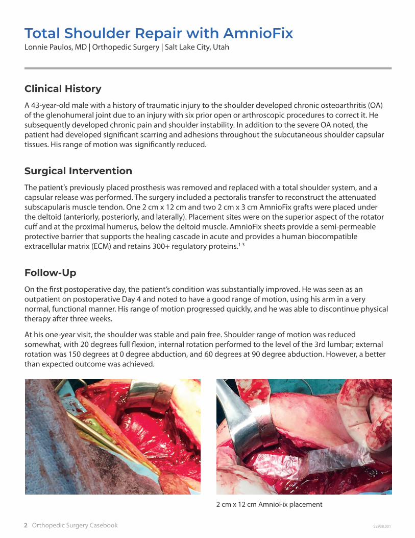
Total Ankle Replacement with AmnioCord Steven K. Neufeld, MD | Orthopedic Surgery | Falls Church, VA
Clinical History A 65-year-old male presented with long-standing tenderness, swelling and stiffness in his right ankle. He was diagnosed with ankle arthritis. He failed conservative treatment including changing shoes, taking arthritis medications, cortisone injections, custom orthotics, and physical therapy. X-rays showed arthritis, and the patient was scheduled for a total anke replacement.
Challenge The extensor tendons, specifically the extensor halluces longus tendon, frequently become “stuck” in scar tissue, resulting in a stiff and painful ankle. Additionally, there is very little soft tissue between the ankle joint and the skin, posing increased risk for wound complications.
Surgical InterventionThe patient underwent a total ankle replacement. A standard, layered closure was done: deep capsule followed by tendon sheaths, subcutaneous tissue, then skin. AmnioCord was cut and applied around the extensor tendons. The soft tissue and skin were closed over the AmnioCord, securing it in position.
AmnioCord is a dehydrated human umbilical cord allograft that provides a protective environment to support the healing process. It protects the wound bed to aid in the development of granulation tissue. The product provides a human biocompatible extracellular matrix of hyaluronic acid and collagen and retains 250+ regulatory proteins.3,4
Follow-UpThe patient was placed in a splint postoperatively and encouraged to ice and elevate the foot during the 1st postoperative week. Early range of motion exercises were started once the incision closed. He was able to return to his normal activities, including wearing normal shoes, three months after the procedure with no discomfort.
Total ankle replacement
AmnioCord applied to extensor tendons / under skin closure
Closed incision

Patents and patents pending see: www.mimedx.com/patents. AmnioCord, AmnioFix, Purion, SMR2T, and MiMedx are trademarks of MiMedx Group, Inc. ©2020 MiMedx Group, Inc. All Rights Reserved. www.mimedx.com SB932.001
Please Call: 866.477.4219 Email: [email protected]
To find out more about MiMedx® products:
REFERENCES 1. Koob, et al. J Biomed Mater Res B Appl Biomater. 2014 Aug;102(6):1353-62. 2. Lei, et al. Adv Wound Care. 2017 Feb 1;6(2):43-53. 3. MM-RD-00086, Proteome Characterization of PURION Processed Dehydrated Human Amnion Chorion Membrane (dHACM) and PURION PLUS Processed Dehydrated Human Umbilical Cord (dHUC) Allografts. 4. Bullard, et al. J Biomed Mater Res B Appl Biomater. 2019 May;107(4):1035-1046. 5. See MiMedx Product Instructions for Use for further details.
Tips for Minimally Invasive Surgical (MIS) Procedures
Common Method1. Grasp the corner of dry graft
2. Wrap the graft around the atraumatic grasper
3. Introduce through the trocar
• Amniofix sheet is the most common configuration for MIS procedures
• Cut or fold AmnioFix to desired size if needed, prior to introduction into the cannula
• Irrigate and suction / aspirate as much arthroscopic fluid as possible prior to introducing AmnioFix to prevent accidently suctioning out the graft
• AmnioFix is introduced into a cannula with an atraumatic grasper
• Ensure allograft is not hydrated/wet prior to introduction