Processing Phoneme Specific Segments for Cleft Lip ... - arXiv
Upload
independentCategory
view
2download
0
Clin Plastic Surg 31 (2004) 231–241
Cleft palate repair: art and issues
A. Michael Sadove, MD, FACS, FAAP*, John A. van Aalst, MD,John Andrew Culp, BA
Division of Plastic Surgery, Indiana University Medical Center, James Whitcomb Riley Hospital for Children,
Riley Hospital #2514, 702 Barnhill Drive, Indianapolis, IN 46202-5200, USA
The French dentist LeMonnier performed the and ethical questions about comparing techniques
first surgical repair of a congenital cleft palate in
the 1760s. The three-stage operation consisted of
passing sutures through the cleft borders, cauterizing
the cleft edges, and realigning the fresh edges. A re-
port of the operation by an observer concluded, ‘‘The
child was perfectly cured’’ [1].
Since this initial palatoplasty, the surgical manage-
ment of cleft palate and outcome evaluation have
become a complex and intricate art. The variety of
techniques used to repair cleft palates has grown
considerably. Many techniques have been developed
solely because of surgeon preference, with little ob-
jective demonstration of the long-term efficacy. By
the early 20th century, the goal in cleft palate repair
was no longer simple closure of the hard and soft
palate but included lengthening the palate to improve
speech in the cleft patient [2]. During the past few
decades, the most debated issues in cleft palate repair
have been how to achieve optimal speech develop-
ment in the cleft patient and how to avoid abnormal
maxillofacial growth after repair. These issues are
directly related to the choice of surgical repair tech-
nique and timing of the repair.
More definitive answers to questions of timing
and choice of technique in the initial repair of the
cleft palate are needed. This article highlights the
fundamental time-tested principles regarding initial
repair of the cleft palate and focuses on changes that
have occurred in the past decade, including innova-
tive techniques, new guidelines for timing of surgery,
0094-1298/04/$ – see front matter D 2004 Elsevier Inc. All right
doi:10.1016/S0094-1298(03)00136-6
* Corresponding author.
E-mail address: [email protected] (A.M. Sadove).
through clinical research.
Incidence
Oral clefts occur with an incidence of 1 in 750 live
births, making clefts the second most common con-
genital defect (following clubfoot). Within the group
of oral clefts, cleft palate has posed the greatest chal-
lenge in management. One reason for this is the com-
plex epidemiology of cleft palates. For instance, cleft
palate can occur as an isolated problem or in conjunc-
tion with cleft lip. These seem to be genetically
distinct problems. Cleft lip with or without cleft palate
occurs with a predictable racial distribution, occurring
in only 0.3 of 1000 African American live births,
1.0 in 1000 Caucasian births, 2.1 in 1000 Japanese
births, and 3.6 of 1000 Native American births.
Alternatively, isolated cleft palate occurs in about
1 in 2000 live births, without a preferential racial
distribution [3].
The epidemiology of cleft palate is further com-
plicated by the presence of an estimated 300 syn-
dromes that include some form of cleft palate in their
presentation (Table 1) [3]. Cleft palates associated
with other anomalies or syndromes tend to be iso-
lated, without lip or alveolus clefting. A syndrome is
diagnosed in one third to one half of isolated cleft
palate patients [4]. Although syndromic cleft palate
patients make up a small portion of the entire cleft
palate population, it is important for the clinician
to be suspicious of syndromes in the initial evalua-
tion. The syndromic cleft patient requires unique
management, including immediate intervention for
other problems.
s reserved.
Table 1
Syndromes commonly associated with cleft palate
Syndromes Clinical findings
Stickler Pierre-Robin sequence, ocular malformations (progressive myopia, retinal
detachment, secondary glaucoma, cataracts), hearing loss, and arthropathies
Velo-cardio-facial Cardiovascular abnormalities (tetralogy of Fallot, ventricular septal defect,
right-sided aortic arch, aberrant subclavian veins, tortuous and medially-
displaced carotid arteries); abnormal facies (long face with maxillary excess and
malar flatness, hypoplastic alar cartilages and thin upper lip with long philtrum);
characteristic 22q11 ‘‘Catch 22’’ chromosomal deletion
Van der Woude Facial clefting, lower lip paramedian sinuses
Goldenhaar Ocular dermoid/coloboma, ear tags/atresia, facial hypoplasia, kidney/vertebral
anomalies; mental retardation
Treacher-Collins Lower eyelid colobomas, down-slanting palpebral fissures, hypoplastic zygomatic
arch, low-set ears
Ectrodactyly-ectodermal
dysplasia-clefting
Lobster-claw anomaly of all four extremities, typically bilateral cleft lip/palate
Oro-facial-digital Oral clefting (lip, tongue, palate), mandibular hypoplasia, mental retardation
A.M. Sadove et al / Clin Plastic Surg 31 (2004) 231–241232
Nearly half of syndromic cleft palate presentations
are associated with Pierre-Robin sequence or velo-
cardio-facial syndrome. Pierre-Robin sequence, which
includes micrognathia, glossoptosis, and cleft palate,
is present in a number of syndromes, notably Stick-
ler syndrome, and accounts for 25% of syndromic
cleft palates [5]. Velo-cardio-facial syndrome accounts
for another 15% of syndromic cleft palates (Table 1)
[5–7].
Anatomy and classification
Embryologically, the nose, lips, and palate are di-
vided into the primary and secondary palates. The
primary palate begins to form during the fifth week of
gestation, when the nasal placodes invaginate to form
nasal pits. The ridges of tissue that form on either side
of the nasal pits are known as the medial and lateral
nasal prominences. Over the next 2 weeks of gestation,
the maxillary prominences, which are inferior and
lateral to the nasal pits, grow medially and eventually
fuse with the medial nasal prominences to form the
primary palate [8]. This involves the bony and soft
tissue elements anterior to the incisive foramen: the
nose, the lips, the prolabium (central upper lip), and the
premaxilla (triangular portion of anterior maxilla,
along with the four incisors) [9].
Normally, fusion of the primary palate is complete
at the end of the sixth week of gestation. The sec-
ondary palate begins to form during the sixth week,
when the same maxillary prominences involved in
primary palatal formation form two shelf-like out-
growths called the palatine shelves. These shelves
first grow downward on each side of the tongue; in
the seventh week of gestation, they ascend and grow
horizontally above the tongue, eventually fusing to
form the secondary palate [8]. The fusion begins at
the incisive foramen and proceeds posteriorly toward
the uvula. Normally, the shelves fuse in the midline
to form the bony hard palate; the hard palate fuses to
the vomer of the nasal septum by the ninth week of
gestation. Palatal fusion continues posteriorly with
full formation of the secondary palate by the twelfth
week of gestation [10]. This involves all elements
posterior to the incisive foramen: the posterior portion
of the maxilla, or hard palate, and the soft palate.
In addition to genetic predisposition for the de-
velopment of palatal defects, environmental hazards
can disrupt normal embryogenesis. Anticonvulsant
drugs such as phenobarbital and phenytoin (Dilantin)
increase the risk of cleft palate when taken during
pregnancy [8]. Other environmental factors that may
predispose to cleft palate formation include alcohol,
hypoxia, steroids, and retinoids (eg, vitamin A) [9].
The process of palatine fusion from anterior to pos-
terior takes about a week longer in females than
males; this added time allows for longer teratogenic
exposure and may explain the increased incidence of
isolated cleft palates in females. The preponderance
of left-sided unilateral clefts may be because the left
palatal shelf usually takes longer to reach midline in
the fusion process than the right side [9].
Anatomic features of the normal palate must be
considered in understanding cleft pathology (Fig. 1).
The palate divides the oro-pharynx and naso-phar-
ynx. The normally fused hard palate is covered with a
dense mucous membrane that is closely adherent to

Fig. 1. Superolateral view of normal and cleft anatomy of the palate. (A) Palatal anatomy of the normal newborn. (B) Anatomy
of complete cleft involving primary and secondary palates.
A.M. Sadove et al / Clin Plastic Surg 31 (2004) 231–241 233
the underlying periosteum, creating a mucoperiosteal
covering of the oral bony surface. Likewise, the nasal
surface is covered with a dense mucoperiosteum; the
vomer of the nasal septum is fused to the midline. In
a cleft of the hard palate, the free, unfused ends of the
palatine shelves are usually covered with mucoperi-
osteum; the vomer fuses to one end or is left sus-
pended between the cleft edges. At the level of the
posterior molars, along the lateral aspect of the hard
palate, the posterior palatine artery emerges from the
palatine canals; the artery is located between muco-
periosteum and bone, along the lateral edges of the
hard palate [10].
Whereas the hard palate serves a structural pur-
pose, in maintaining maxillo-facial architecture, the
soft palate serves a more functional purpose; nor-
mally, the soft palate works as an active muscular
valve, called the velopharyngeal sphincter (see Fig. 1).
This sphincter acts to raise the soft palate up against
the posterior pharyngeal wall, dynamically separating
the nose from the mouth [9]. The soft palate’s
intrinsic muscular function aids in proper breathing,
swallowing, blowing, and phonation [10]. Six pairs
of muscles comprise the soft palate: levator veli
palatini, tensor veli palatini, superior constrictor
pharyngeus, uvulus, palatopharyngeus, and palato-
glossus muscles [2]. The exact contribution of each
muscle pair to palatal function continues to be de-
bated; however, the tensor and levator veli palatini
muscles, both of which arise from the eustachian
tube, are key anatomic features in cleft palate repair.
The tensor veli palatini muscles course inferiorly and
A.M. Sadove et al / Clin Plastic Surg 31 (2004) 231–241234
wrap laterally around the pterygoid hamulus before
inserting medially into the soft palatal aponeurosis
near the junction of the soft and hard palates. These
muscles seem to control the opening of the eustachian
tube, possibly serving to aerate the middle ear
and prevent recurrent otitis media. The levator veli
palatini muscles course inferiorly and medially, in-
terdigitating in the midline and forming the bulk of
the ‘‘levator sling.’’ This sling acts to raise the palate
to the pharynx, providing much of the velopharyn-
geal sphincter’s function [3]. The palatoglossus and
palatopharyngeus muscles, originating at the midline
of the soft palate and inserting into the tongue and
lateral pharyngeal wall, respectively, also support this
sphincter function by constricting the oropharyngeal
aperture [3].
Clefts of the soft palate not only disrupt the
levator sling, but they also disturb all normal muscu-
lar insertions in the palatal aponeurosis. In the cleft
soft palate, muscles that normally join at the midline
trend anteriorly and insert on or near the poste-
rior edge of the hard palate [3]. Sphincter function
is compromised, leading to velopharyngeal insuffi-
ciency and problems with speech development.
Eustachian tube control is also lost, often leading
to chronic otitis media with the risk of permanent
hearing loss. These anatomic considerations are im-
portant for understanding palatal function and surgi-
cal landmarks.
Cleft palate classification is based upon anatomic
disruption of the primary and secondary palates and
includes the categories of complete and incomplete,
unilateral and bilateral, and submucous clefting. The
complete cleft palate involves the primary and sec-
ondary palates in which no fusion between the pala-
tal shelves has taken place. It is usually associated
with a cleft lip and alveolus. The incomplete cleft
palate involves only the secondary palate; fusion
between palatal shelves has been initiated but not
completed. Thus, the incomplete cleft may involve
only the very posterior portion of the soft palate, or it
may extend through the soft and hard palates to the
incisive foramen.
A cleft of the secondary palate is classified further
as unilateral or bilateral. With the unilateral cleft pal-
ate, only one palatal shelf fuses to the nasal septum;
this leaves the cleft defect to one side of the midline.
With the bilateral cleft palate, neither palatal shelf
fuses with the nasal septum, leaving a wider midline
cleft defect with the vomer of the nasal septum sus-
pended superiorly.
The submucous cleft involves a separation of the
intrinsic soft palate musculature while the overlying
soft palatal mucosa remains intact [3]. This type of
cleft is often difficult to diagnose because the entire
palate may seem grossly intact. Often, the only ana-
tomic clues suggesting a submucous cleft palate are
a bifid uvula, a notched posterior hard palate, or a
translucent area in the midline of the soft palate,
known as the zona pellucida, where the musculature
has failed to fuse. Often, this cleft type is not dis-
covered until a child develops velopharyngeal in-
competence, manifested as hypernasal speech [11].
Although this is the most difficult type of cleft palate
to diagnose, it is the most common type of posterior
palatal cleft [12]. The incidence of submucous cleft
palates is difficult to determine because it is usu-
ally discovered only if a patient is referred for
velopharyngeal incompetence. Attempts to measure
the incidence in the general population by anatomic
criteria have revealed rates in the range of 2 to 8 per
10,000 [11].
The syndromic cleft palate often presents as an
anatomically distinct deformity. For instance, the
typical isolated, or incomplete, cleft involving hard
and soft palates is V shaped; it is narrower anteriorly
and tends to widen toward the posterior end at the
soft palate. However, the cleft palate associated with
Pierre-Robin sequence is U shaped and much wider
than a nonsyndromic cleft palate of similar type [4].
Operative techniques
Once a patient with cleft palate is evaluated for
the presence of an associated syndrome and the cleft
is appropriately classified, strategies for primary
repair may be considered. Technique selection and
timing for repair are discussed through four repre-
sentative cases.
Incomplete cleft of the soft palate
In this case, an infant presents with a nonsyn-
dromic, isolated, and incomplete cleft palate involv-
ing only the soft palate. The primary goal of repair in
this case is restoration of velopharyngeal competence.
This is achieved by lengthening the palate, for proper
apposition of palate and posterior pharyngeal wall,
and re-organizing the palatal musculature.
Veau, in the earlier 20th century, repaired clefts of
the soft palate by bringing together the cleft edges,
with the intravelar musculature trending anteriorly
and attaching on or near the posterior edge of the hard
palate. This meant that muscle bundles were sutured
together side by side. Lateral relaxing incisions or
mucoperiosteal flaps on the hard palate were used
A.M. Sadove et al / Clin Plastic Surg 31 (2004) 231–241 235
to reduce tension and gain the mobility needed to
approximate the cleft edges [13]. Presently, the most
widely practiced methods of soft palatoplasty are
intravelar veloplasty and the Furlow double-op-
posing Z-plasty.
The intravelar veloplasty proposed by Kriens in
1969 was an improvement upon previous soft
palatoplasties [13]. Kriens’ innovation was to restore
the levator sling and palatal musculature at the
midline where they normally meet. This is accom-
plished by dissecting the anteriorly malpositioned
muscle bundles from the posterior edge of the hard
palate and repositioning them in the midline. This
technique is widely used today, although there is
much variability among surgeons in how the muscu-
lature is dissected and repositioned. Until recently,
the results of this technique had not been objectively
compared with the older standard technique advo-
cated by Veau. In 1989, Marsh et al [14] published
results from a prospective study that compared the
effects of intravelar veloplasty and traditional side-to-
side techniques upon velopharyngeal incompetence
(VPI). They found that repositioning of the levator
muscles during primary palatoplasty was no better at
improving VPI than the side-to-side veloplasty.
However, this study was limited by a small patient
population (51 patients) at a single institution. In
1995, Cutting questioned whether the intravelar velo-
plasty technique adequately dissected and reposi-
tioned the musculature [15]. It remains a challenge
to prove by prospective, well-controlled, multi-center
studies whether the intravelar veloplasty is a more
effective technique than its simpler predecessor.
The Furlow double-opposing Z-plasty technique
was unofficially introduced in 1978 and was intro-
duced in published form in 1986 [16]. Over the last
decade, it has become the veloplasty technique of
choice among many surgeons. This technique uses
two reversed Z-plasties based upon the cleft midline,
both of which draw in soft palate tissue from the sides
to close the cleft defect and restore the musculature to
its anatomic position. The inherent advantages of the
Furlow repair are that it lengthens the palate and
restores normal muscular anatomy. It also eliminates
the need for lateral relaxing incisions to gain tissue
for cleft closure. A concern shared by many surgeons,
including Millard [1], is that the Z-plasties in the soft
palate tend to pull the sides of the velum toward the
midline to lengthen the palate; this tightens the velum
in the transverse axis. Nonetheless, retrospective
studies published during the last decade have shown
that patients with Furlow repairs have reduced
hypernasality and improved articulation and speech
[17,18]. Prospective controlled trials are needed to
compare the Furlow palatoplasty to the intravelar
veloplasty and other procedures [18].
Among recent developments in the repair of soft
palate clefts, there is increased interest in manipula-
tion of the tensor veli palatini muscles to gain palatal
length. The goal of such manipulation is to release
tension on the levator sling. One way to do this is by
fracturing the pterygoid hamulus (around which the
tensor muscles are tethered) during soft palate re-
pair. This method was first used by Billroth in 1889
[1,19]. At that time, it was performed to relax the
tension in the lateral tissues of the soft palate and thus
minimize postoperative palatal dehiscence. The frac-
ture also allowed Billroth to avoid the standard lateral
relaxing incisions of the day, which tended to injure
soft palate musculature. Since that time, the technique
has continued to find favor with a small minority of
surgeons. Studies have been conducted recently to
assess the value of this technique. Kane et al [19]
conducted a randomized, prospective study com-
paring complication rates in palatoplasties with and
without hamulus fracture. No differences between the
groups were noted in perioperative morbidity (fistulas
and dehiscence) or in hearing and speech results
1 year after surgery.
The hamulus fracture technique is appealing be-
cause it relieves tension in the soft palate closure;
however, an alternative technique that severs the
tensor tendon in the space of Ernst seems to achieve
comparable or greater release of the levator sling.
Preliminary results of a prospective study using this
release method have been presented. Tendon release
was performed in conjunction with a Furlow repair
and lateral relaxing incisions on a variety of cleft
types with follow-up VPI rates of 4%, compared with
rates of 10% with the Furlow palatoplasty or intra-
velar veloplasty alone [20].
Another recent development in soft palate repair is
a uvular transposition technique [21]. This procedure
recruits tissue for soft palate lengthening from the
uvula and can be performed in conjunction with the
Furlow palatoplasty or intravelar veloplasty. A pro-
spective study evaluating speech results 4 years after
surgery showed improvement using uvular trans-
position when compared with Furlow palatoplasty
or intravelar veloplasty alone. Two of 62 patients
demonstrated significant postoperative VPI after the
uvular transposition palatoplasty [21].
In summary, an incomplete cleft of the soft palate
can be repaired with an intravelar veloplasty or
Furlow repair, depending on the surgeon’s prefer-
ence. It may be necessary to use a pushback tech-
nique from the hard palate mucoperiosteum or lateral
relaxing incisions in the soft palate to bring the

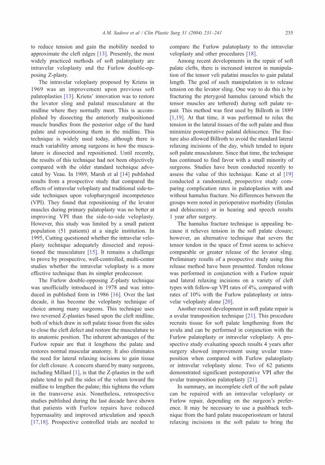
Fig. 2. Von Langenbeck palatoplasty. (A) Incomplete cleft
showing bipedicled mucoperiosteal flaps. (B) Repaired
incomplete cleft with Von Langenbeck flaps closed in
the midline.
A.M. Sadove et al / Clin Plastic Surg 31 (2004) 231–241236
cleft edges together. Alternative methods to gain
tissue and lengthen the palate include the hamu-
lus fracture, tensor veli palatini tendon release, or
uvular transposition.
Incomplete cleft of hard and soft palate
This case involves an incomplete cleft of the
secondary palate to the level of the incisive foramen.
In addition to a cleft of the entire soft palate, the hard
palate has a left unilateral cleft. The vomer is attached
to the nasal surface of the right cleft edge. The goals
of this repair are realignment of the soft palate
mucosa and musculature to restore velopharyngeal
competence and closure of the bony gap between the
edges of the hard palate to restore structural integrity
and maintain growth of the oral cavity and mid-face.
In the early 19th century, Dieffenbach introduced
lateral relaxing incisions in the secondary palate for
closure of palate defects. By the 1860s, von Langen-
beck had expanded on this idea by raising the entire
mucoperiosteum as a flap for coverage of the hard
palate cleft [13]. These flaps have remained the basis of
repair for cleft palate defects; they bring vascularized
periosteum and mucosa to cover clefts of the hard
palate and help in soft palate closure and lengthening
by pushing mucosal tissue medially and posteriorly.
Three variations of repair involving mucoperi-
osteal flaps are used: the von Langenbeck palato-
plasty, the Veau-Wardill-Kilner (V-W-K) palatoplasty,
and the two-flap palatoplasty. The first two are used
commonly to repair the incomplete cleft involving
hard and soft palates. The von Langenbeck palato-
plasty involves relaxing incisions along the lateral
edge of the hard palate, starting anteriorly near the
palatomaxillary suture line, running posteriorly just
medial to the alveolar ridge, and ending lateral to the
hamulus, about 1 cm posterior to the greater tuber-
osity of the alveolus (Fig. 2) [3,4]. The mucosa along
the edges of the cleft is also incised. The entire muco-
periosteum is then raised from the oral surface of the
hard palate; care is taken to preserve the two neuro-
vascular pedicles—the greater palatine pedicle poste-
riorly and the incisive pedicle anteriorly. Bipedicled
mucoperiosteal flaps are created on both sides of the
cleft. The nasal side of the cleft is closed first, using
redundant mucoperiosteum from the incision along
the cleft edge. Then the bipedicled flaps are approxi-
mated to cover the oral surface of the cleft. The von
Langenbeck technique works well for incomplete
clefts of the secondary palate without the presence
of cleft lip or alveolus [3].
A variation of the von Langenbeck repair, the
V-W-K repair or V-Y pushback, can also be used
for incomplete clefts involving the hard palate (Fig. 3).
The same flap design as the von Langenbeck repair is
used. Then the superior pedicle is divided, leaving a
flap on either side of the cleft based solely on the
greater palatine pedicle posteriorly. The mucoperios-
teal flaps can then be approximated directly or in a
V-Y closure at the free anterior end to actively
lengthen the soft palate. This repair technique allows
more flap advancement than the von Langenbeck
repair; however, the gain in palatal length (and
possibly improved velopharyngeal function) is at
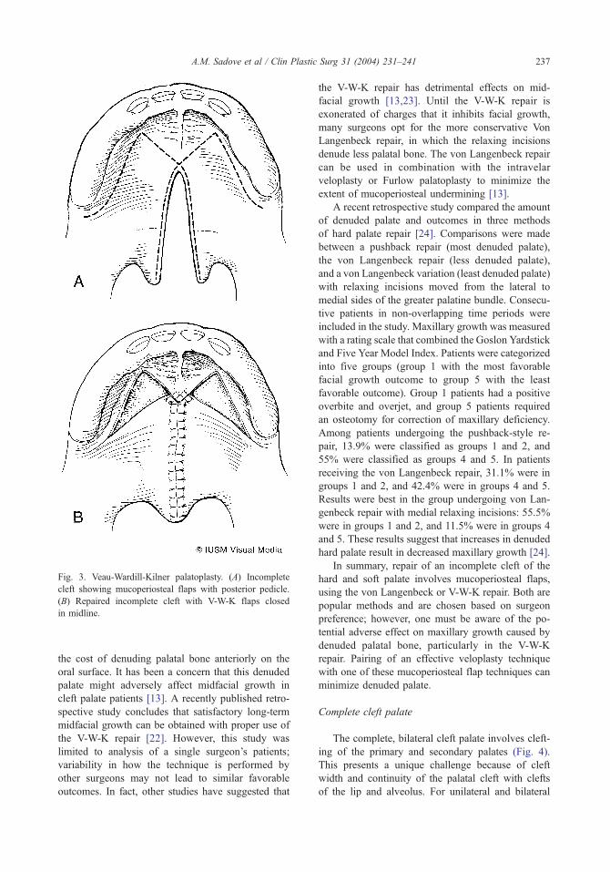
Fig. 3. Veau-Wardill-Kilner palatoplasty. (A) Incomplete
cleft showing mucoperiosteal flaps with posterior pedicle.
(B) Repaired incomplete cleft with V-W-K flaps closed
in midline.
A.M. Sadove et al / Clin Plastic Surg 31 (2004) 231–241 237
the cost of denuding palatal bone anteriorly on the
oral surface. It has been a concern that this denuded
palate might adversely affect midfacial growth in
cleft palate patients [13]. A recently published retro-
spective study concludes that satisfactory long-term
midfacial growth can be obtained with proper use of
the V-W-K repair [22]. However, this study was
limited to analysis of a single surgeon’s patients;
variability in how the technique is performed by
other surgeons may not lead to similar favorable
outcomes. In fact, other studies have suggested that
the V-W-K repair has detrimental effects on mid-
facial growth [13,23]. Until the V-W-K repair is
exonerated of charges that it inhibits facial growth,
many surgeons opt for the more conservative Von
Langenbeck repair, in which the relaxing incisions
denude less palatal bone. The von Langenbeck repair
can be used in combination with the intravelar
veloplasty or Furlow palatoplasty to minimize the
extent of mucoperiosteal undermining [13].
A recent retrospective study compared the amount
of denuded palate and outcomes in three methods
of hard palate repair [24]. Comparisons were made
between a pushback repair (most denuded palate),
the von Langenbeck repair (less denuded palate),
and a von Langenbeck variation (least denuded palate)
with relaxing incisions moved from the lateral to
medial sides of the greater palatine bundle. Consecu-
tive patients in non-overlapping time periods were
included in the study. Maxillary growth was measured
with a rating scale that combined the Goslon Yardstick
and Five Year Model Index. Patients were categorized
into five groups (group 1 with the most favorable
facial growth outcome to group 5 with the least
favorable outcome). Group 1 patients had a positive
overbite and overjet, and group 5 patients required
an osteotomy for correction of maxillary deficiency.
Among patients undergoing the pushback-style re-
pair, 13.9% were classified as groups 1 and 2, and
55% were classified as groups 4 and 5. In patients
receiving the von Langenbeck repair, 31.1% were in
groups 1 and 2, and 42.4% were in groups 4 and 5.
Results were best in the group undergoing von Lan-
genbeck repair with medial relaxing incisions: 55.5%
were in groups 1 and 2, and 11.5% were in groups 4
and 5. These results suggest that increases in denuded
hard palate result in decreased maxillary growth [24].
In summary, repair of an incomplete cleft of the
hard and soft palate involves mucoperiosteal flaps,
using the von Langenbeck or V-W-K repair. Both are
popular methods and are chosen based on surgeon
preference; however, one must be aware of the po-
tential adverse effect on maxillary growth caused by
denuded palatal bone, particularly in the V-W-K
repair. Pairing of an effective veloplasty technique
with one of these mucoperiosteal flap techniques can
minimize denuded palate.
Complete cleft palate
The complete, bilateral cleft palate involves cleft-
ing of the primary and secondary palates (Fig. 4).
This presents a unique challenge because of cleft
width and continuity of the palatal cleft with clefts
of the lip and alveolus. For unilateral and bilateral
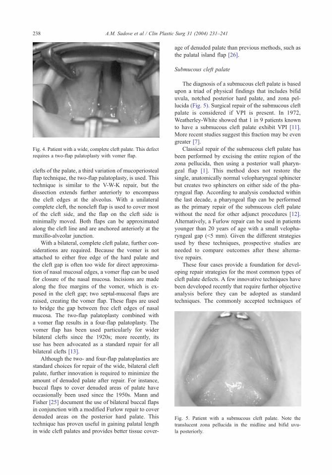
Fig. 4. Patient with a wide, complete cleft palate. This defect
requires a two-flap palatoplasty with vomer flap.
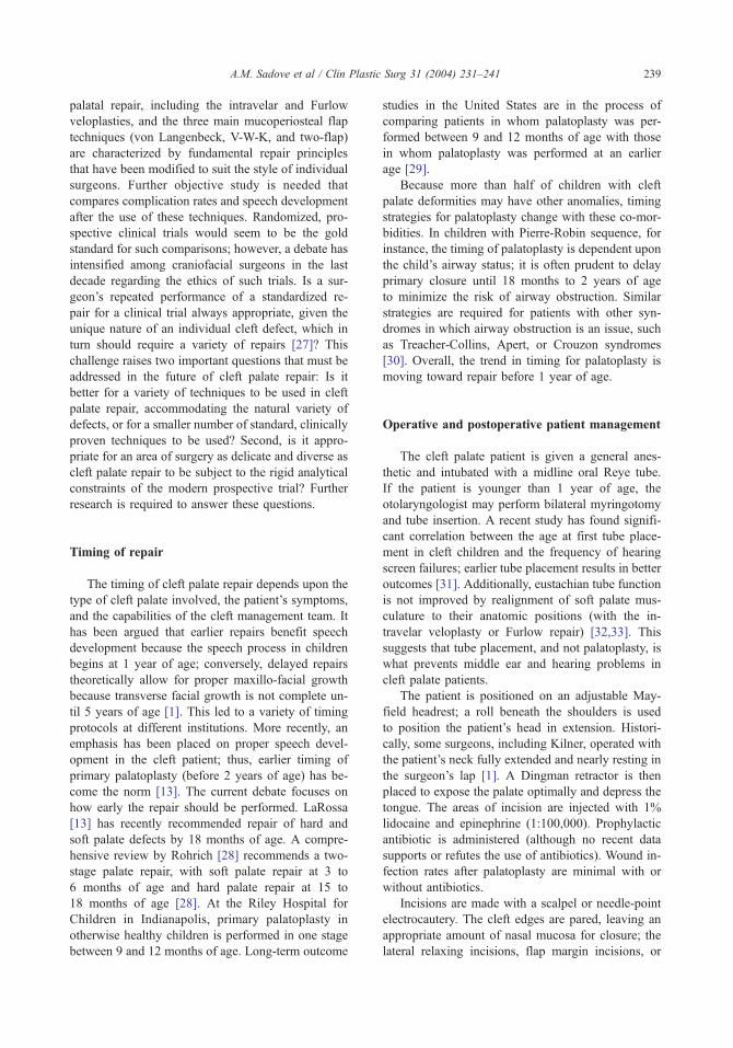
Fig. 5. Patient with a submucous cleft palate. Note the
translucent zona pellucida in the midline and bifid uvu-
la posteriorly.
A.M. Sadove et al / Clin Plastic Surg 31 (2004) 231–241238
clefts of the palate, a third variation of mucoperiosteal
flap technique, the two-flap palatoplasty, is used. This
technique is similar to the V-W-K repair, but the
dissection extends further anteriorly to encompass
the cleft edges at the alveolus. With a unilateral
complete cleft, the noncleft flap is used to cover most
of the cleft side, and the flap on the cleft side is
minimally moved. Both flaps can be approximated
along the cleft line and are anchored anteriorly at the
maxillo-alveolar junction.
With a bilateral, complete cleft palate, further con-
siderations are required. Because the vomer is not
attached to either free edge of the hard palate and
the cleft gap is often too wide for direct approxima-
tion of nasal mucosal edges, a vomer flap can be used
for closure of the nasal mucosa. Incisions are made
along the free margins of the vomer, which is ex-
posed in the cleft gap; two septal-mucosal flaps are
raised, creating the vomer flap. These flaps are used
to bridge the gap between free cleft edges of nasal
mucosa. The two-flap palatoplasty combined with
a vomer flap results in a four-flap palatoplasty. The
vomer flap has been used particularly for wider
bilateral clefts since the 1920s; more recently, its
use has been advocated as a standard repair for all
bilateral clefts [13].
Although the two- and four-flap palatoplasties are
standard choices for repair of the wide, bilateral cleft
palate, further innovation is required to minimize the
amount of denuded palate after repair. For instance,
buccal flaps to cover denuded areas of palate have
occasionally been used since the 1950s. Mann and
Fisher [25] document the use of bilateral buccal flaps
in conjunction with a modified Furlow repair to cover
denuded areas on the posterior hard palate. This
technique has proven useful in gaining palatal length
in wide cleft palates and provides better tissue cover-
age of denuded palate than previous methods, such as
the palatal island flap [26].
Submucous cleft palate
The diagnosis of a submucous cleft palate is based
upon a triad of physical findings that includes bifid
uvula, notched posterior hard palate, and zona pel-
lucida (Fig. 5). Surgical repair of the submucous cleft
palate is considered if VPI is present. In 1972,
Weatherley-White showed that 1 in 9 patients known
to have a submucous cleft palate exhibit VPI [11].
More recent studies suggest this fraction may be even
greater [7].
Classical repair of the submucous cleft palate has
been performed by excising the entire region of the
zona pellucida, then using a posterior wall pharyn-
geal flap [1]. This method does not restore the
single, anatomically normal velopharyngeal sphincter
but creates two sphincters on either side of the pha-
ryngeal flap. According to analysis conducted within
the last decade, a pharyngeal flap can be performed
as the primary repair of the submucous cleft palate
without the need for other adjunct procedures [12].
Alternatively, a Furlow repair can be used in patients
younger than 20 years of age with a small velopha-
ryngeal gap (<5 mm). Given the different strategies
used by these techniques, prospective studies are
needed to compare outcomes after these alterna-
tive repairs.
These four cases provide a foundation for devel-
oping repair strategies for the most common types of
cleft palate defects. A few innovative techniques have
been developed recently that require further objective
analysis before they can be adopted as standard
techniques. The commonly accepted techniques of
A.M. Sadove et al / Clin Plastic Surg 31 (2004) 231–241 239
palatal repair, including the intravelar and Furlow
veloplasties, and the three main mucoperiosteal flap
techniques (von Langenbeck, V-W-K, and two-flap)
are characterized by fundamental repair principles
that have been modified to suit the style of individual
surgeons. Further objective study is needed that
compares complication rates and speech development
after the use of these techniques. Randomized, pro-
spective clinical trials would seem to be the gold
standard for such comparisons; however, a debate has
intensified among craniofacial surgeons in the last
decade regarding the ethics of such trials. Is a sur-
geon’s repeated performance of a standardized re-
pair for a clinical trial always appropriate, given the
unique nature of an individual cleft defect, which in
turn should require a variety of repairs [27]? This
challenge raises two important questions that must be
addressed in the future of cleft palate repair: Is it
better for a variety of techniques to be used in cleft
palate repair, accommodating the natural variety of
defects, or for a smaller number of standard, clinically
proven techniques to be used? Second, is it appro-
priate for an area of surgery as delicate and diverse as
cleft palate repair to be subject to the rigid analytical
constraints of the modern prospective trial? Further
research is required to answer these questions.
Timing of repair
The timing of cleft palate repair depends upon the
type of cleft palate involved, the patient’s symptoms,
and the capabilities of the cleft management team. It
has been argued that earlier repairs benefit speech
development because the speech process in children
begins at 1 year of age; conversely, delayed repairs
theoretically allow for proper maxillo-facial growth
because transverse facial growth is not complete un-
til 5 years of age [1]. This led to a variety of timing
protocols at different institutions. More recently, an
emphasis has been placed on proper speech devel-
opment in the cleft patient; thus, earlier timing of
primary palatoplasty (before 2 years of age) has be-
come the norm [13]. The current debate focuses on
how early the repair should be performed. LaRossa
[13] has recently recommended repair of hard and
soft palate defects by 18 months of age. A compre-
hensive review by Rohrich [28] recommends a two-
stage palate repair, with soft palate repair at 3 to
6 months of age and hard palate repair at 15 to
18 months of age [28]. At the Riley Hospital for
Children in Indianapolis, primary palatoplasty in
otherwise healthy children is performed in one stage
between 9 and 12 months of age. Long-term outcome
studies in the United States are in the process of
comparing patients in whom palatoplasty was per-
formed between 9 and 12 months of age with those
in whom palatoplasty was performed at an earlier
age [29].
Because more than half of children with cleft
palate deformities may have other anomalies, timing
strategies for palatoplasty change with these co-mor-
bidities. In children with Pierre-Robin sequence, for
instance, the timing of palatoplasty is dependent upon
the child’s airway status; it is often prudent to delay
primary closure until 18 months to 2 years of age
to minimize the risk of airway obstruction. Similar
strategies are required for patients with other syn-
dromes in which airway obstruction is an issue, such
as Treacher-Collins, Apert, or Crouzon syndromes
[30]. Overall, the trend in timing for palatoplasty is
moving toward repair before 1 year of age.
Operative and postoperative patient management
The cleft palate patient is given a general anes-
thetic and intubated with a midline oral Reye tube.
If the patient is younger than 1 year of age, the
otolaryngologist may perform bilateral myringotomy
and tube insertion. A recent study has found signifi-
cant correlation between the age at first tube place-
ment in cleft children and the frequency of hearing
screen failures; earlier tube placement results in better
outcomes [31]. Additionally, eustachian tube function
is not improved by realignment of soft palate mus-
culature to their anatomic positions (with the in-
travelar veloplasty or Furlow repair) [32,33]. This
suggests that tube placement, and not palatoplasty, is
what prevents middle ear and hearing problems in
cleft palate patients.
The patient is positioned on an adjustable May-
field headrest; a roll beneath the shoulders is used
to position the patient’s head in extension. Histori-
cally, some surgeons, including Kilner, operated with
the patient’s neck fully extended and nearly resting in
the surgeon’s lap [1]. A Dingman retractor is then
placed to expose the palate optimally and depress the
tongue. The areas of incision are injected with 1%
lidocaine and epinephrine (1:100,000). Prophylactic
antibiotic is administered (although no recent data
supports or refutes the use of antibiotics). Wound in-
fection rates after palatoplasty are minimal with or
without antibiotics.
Incisions are made with a scalpel or needle-point
electrocautery. The cleft edges are pared, leaving an
appropriate amount of nasal mucosa for closure; the
lateral relaxing incisions, flap margin incisions, or

Box 1. Postoperative orders for palaterepair
� Elevate head of bed to 30�� 30% oxygen mist tent overnight� Continuous pulse oximetry overnight� Clear liquid diet, supplemented withintravenous lactated Ringer’s solution
� Advance diet to pureed foods as tol-erated after first postoperative night
� All hard objects (straws, nipples,thermometers) kept out of patient’smouth
� Nursing staff to record intake/output,vital signs, and oxygen saturationlevels every 4 hours
� Morphine sulfate (0.1 mg/kg), Capi-tal with codeine (1 mg/kg) as needfor pain
� Tylenol for fever (10 mg/kg)� Antibiotics for 5 days postoperatively(usually Keflex)
A.M. Sadove et al / Clin Plastic Surg 31 (2004) 231–241240
Z-plasty incisions are then made depending on choice
of repair. A freer can be used to raise the mucoperi-
osteal flaps.
Closure of the repair begins with the anterior na-
sal mucosa, working posteriorly using a running
4-0 Vicryl suture. A Castroviejo needle driver op-
timizes suture manipulation [4]. The soft palate
musculature, particularly the levator sling, is then
approximated end-to-end at the midline with inter-
rupted 4-0 Vicryl sutures. The oral mucosa is closed
last, starting posteriorly at the uvula and progress-
ing anteriorly using interrupted horizontal mattress
4-0 Vicryl sutures. In repairs using significant muco-
periosteal flaps, suspension sutures can be placed
anteriorly to fix the flaps to the alveolar ridge. To
protect the repair postoperatively, a 0-gauge silk
suture is passed through the anterior third of the
tongue and taped to the cheek; this can be removed
before discharge [30]. In addition, elbow extension
splints are placed in the operating room and are worn
by the patient until the first follow-up visit. This
prevents the child from placing fingers in the oral
cavity and disrupting the repair (Box 1).
Complications
Postoperative complications include primarily re-
pair flap dehiscence and fistula formation. A meta-
analysis in 1991 reported postpalatoplasty fistula
rates ranging from 0% to 34%. Factors that af-
fected fistula formation were the extent of clefting
(clefts involving the primary palate or more of the
secondary palate resulted in higher fistula rates), the
type of repair (pushback repairs resulted in higher
fistula rates than von Langenbeck repairs, which in
turn were higher than Furlow/intravelar veloplasty
repairs), and the surgeon repairing the palate. Of
all fistulas, 87% occur in the area of the hard pal-
ate closure, and over half of these occur imme-
diately posterior to the alveolus [34]. Age at palate
closure does not seem to significantly affect rates of
fistula formation.
Long-term palatoplasty complications are related
to velopharyngeal insufficiency or poor mid-face
growth, with the development of a cross-bite. Initial
follow-up is within 7 to 10 days after repair with the
primary surgeon. Additional, long-term follow-up is
required with multiple members of the cleft palate
team. This team includes an otolaryngologist (to
assess hearing function), an orthodontist (to coordi-
nate maxillo-facial growth), and a speech therapist (to
evaluate speech development). After the initial fol-
low-up visit, the plastic surgeon follows the patient
with yearly office visits.
Summary
Caring for the child with cleft palate requires a
multidisciplinary approach that begins with evalua-
tion for other possible congenital anomalies, decisions
about timing of repair, and choice of techniques.
Postoperative follow-up similarly requires a team ap-
proach and should include an otolaryngologist, an
orthodontist, and a speech therapist. The art of
cleft palate repair has enjoyed a decade rich in new
developments. New techniques have been developed,
and standard techniques have been refined. Most
importantly, the need for prospective, randomized
trials to objectively compare surgical techniques has
been recognized. Initiation and completion of these
trials will improve outcomes for patients with cleft
palate repairs.
References
[1] Millard DR. Alveolar and palatal deformities. Cleft
craft: the evolution of its surgery, vol. 3. Boston: Little,
Brown and Company; 1980.
[2] Nguyen PN, Sullivan PK. Issues and controversies in

A.M. Sadove et al / Clin Plastic Surg 31 (2004) 231–241 241
the management of cleft palate. Clin Plast Surg 1993;
20:671–82.
[3] Strong EB, Buckmiller LM. Management of the cleft
palate. Fac Plast Surg Clin North Am 2001;9:15–25.
[4] Vander Kolk CA. Cleft palate. In: Vander Kolk C,
editor. Craniomaxillofacial, cleft, and pediatric surgery
(plastic surgery: indications, operations and outcomes,
vol. 2). St. Louis: Mosby; 2000. p. 799–807.
[5] Coleman JR, Sykes JM. The embryology, classifi-
cation, epidemiology and genetics of facial clefting.
Fac Plast Surg Clin Nort Am 2001;9:1–13.
[6] Available at: www.cleft.ie/relatedsyndromes.
[7] Ysunza A, Pamplona C, Mendoza M, Molina F, Mar-
tinez P, Garcia-Velasco M, et al. Surgical treatment of
submucous cleft palate: a comparative trial of two mo-
dalities for palatal closure. Plast Reconstr Surg 2001;
107:9–14.
[8] Sadler TW. Langman’s medical embryology. 7th edi-
tion. Philadelphia: Williams and Wilkins; 1995.
[9] Witt PD, Marsh JL. Cleft palate deformities. In: Bentz
M, editor. Pediatric plastic surgery. New York: Apple-
ton and Lange; 1998. p. 93–105.
[10] Marks MW, Marks C. Cleft lip and palate. In: Marks
MW, Marks C, editors. Fundamentals of plastic sur-
gery. Philadelphia: W.B. Saunders; 1997. p. 156–73.
[11] Weatherley-White RCA, Sakura CY, Brenner LD,
Stewart JM, Ott JE. Submucous cleft palate: its inci-
dence, natural history and indications for treatment.
Plast Reconstr Surg 1972;49:297–304.
[12] Gosain AK, Conley SF, Marks S, Larson DL. Submu-
cous cleft palate: diagnostic methods and outcomes of
Ssurgical treatment. Plast Reconstr Surg 1996;97:
1497–507.
[13] LaRossa D. The state of the art in cleft palate surgery.
Cleft Palate Craniofac J 2000;37:225–8.
[14] Marsh JL, Grames LM, Holtman B. Intravelar velo-
plasty: a prospective study. Cleft Palate J 1989;26:
46–50.
[15] Cutting C, Rosenbaum J, Rovati L. The technique of
muscle repair in the cleft soft palate. Operative Tech
Plast Reconstr Surg 1995;2:215–22.
[16] Furlow LT. Cleft repair by double-opposing z-plasty.
Plast Reconstr Surg 1986;78:724–36.
[17] Kirschner RE, Wang P, Jawad AF, Duran M, Cohen M,
Solot C, et al. Cleft palate repair by modified Furlow
double-opposing z-plasty: the Children’s Hospital of
Philadelphia experience. Plast Reconstr Surg 1999;
104:1998–2010.
[18] McWilliams BJ, Randall P, LaRossa D, Cohen S, Yu J,
Cohen M, et al. Speech characteristics associated with
the Furlow palatoplasty as compared with other surgi-
cal techniques. Plast Reconstr Surg 1996;98:610–21.
[19] Kane AA, Lo L-J, Yen B-D, Chen Y-R, Noordhoff MS.
The effect of hamulus fracture on the outcome of palato-
plasty: a preliminary report of a prospective, alternating
study. Cleft Palate Craniofac J 2000;37:506–11.
[20] Havlik R. Total release double-opposing z-plasty in
palatal closure. Presented at the annual meeting of
the Ohio Valley Society of Plastic Surgeons. Indian-
apolis, May 30, 2003.
[21] David LR, Blalock D, Argenta LC. Uvular transposi-
tion: a new method of cleft palate repair. Plast Reconstr
Surg 1999;104:897–904.
[22] Choudary S, Cadier MAM, Shinn DL, Shekhar K,
McDowall RAW. Effect of Veau-Wardill-Kilner type
of cleft palate repair on long-term midfacial growth.
Plast Reconstr Surg 2003;111:576–84.
[23] Semb G, Shaw WC. Facial growth after different meth-
ods of surgical intervention in patients with cleft lip
and cleft palate. Acta Odont Scand 1998;56:352–5.
[24] Pigott RW, Albery EH, Hathorn IS, Atack NE, Wil-
liams A, Harland K, et al. A comparison of three meth-
ods of repairing the hard palate. Cleft Palate Craniofac
J 2002;39:383–91.
[25] Mann RJ, Fisher DM. Bilateral buccal flaps with
double-opposing Z-plasty for wider palatal clefts. Plast
Reconstr Surg 1997;100:1139–45.
[26] Seckel NG. The palatal island flap on retrospection.
Plast Reconstr Surg 1995;96:1262–70.
[27] Berkowitz S. Ethical issues in the case of surgical
repair of the cleft palate. Cleft Palate Craniofac J 1995;
32:271–81.
[28] Rohrich RJ, Love EJ, Byrd S, Johns DF. Optimal
timing of cleft palate repair. Plast Reconstr Surg 2000;
106:413–25.
[29] Peterson-Falzone SJ. The relationship between timing
of cleft palate surgery and speech outcome: what have
we learned, and where do we stand in the 1990s?
Semin Orthod 1996;2:185–91.
[30] LaRossa D. Cleft palate. In: Cohen M, editor. Mastery
of plastic and reconstructive surgery. New York: Little,
Brown and Co; 1997. p. 595–604.
[31] Broen PA, Moller KT, Carlstrom J, Doyle SS, Devers
M, Keenan KM. Comparison of the hearing histories of
children with and without cleft palate. Cleft Palate
Craniofac J 1996;33:127–33.
[32] Guneren E, Ozsoy Z, Ulay M, Eryilmaz E, Ozkul H,
Geary PM. A comparison of the effects of Veau-
Wardill-Kilner palatoplasty and Furlow double-oppos-
ing Z-plasty operations on eustachian tube function.
Cleft Palate Craniofac J 2000;37:266–70.
[33] Gunther E. Palatoplasty: Furlow’s double-reversing
Z-plasty versus intravelar veloplasty. Cleft Palate Cra-
niofac J 1998;35:546–9.
[34] Cohen SR, Kalinowski J, LaRossa D, Randall P. Cleft
palate fistulas: a multivariate statistical analysis of
prevalence, etiology, and surgical management. Plast
Recostr Surg 1991;87:1041–7.
Copyright © 2022 FDOKUMEN