Survival and complications of zygomatic implants - DiVA portal
Upload
univ-nantesCategory
view
0download
0
ARTICLE IN PRESS
Journal of Biomechanics 42 (2009) 1206–1211
Contents lists available at ScienceDirect
journal homepage: www.elsevier.com/locate/jbiomech
Journal of Biomechanics
0021-92
doi:10.1
� Corr
E-m
www.JBiomech.com
Prediction of bone density around orthopedic implantsdelivering bisphosphonate
Vincent A. Stadelmann a, Alexandre Terrier a, O. Gauthier b, J.-M. Bouler b, Dominique P. Pioletti a,�
a Laboratory of Biomechanical Orthopedics, Institute of Bioengineering, Ecole Polytechnique Federale de Lausanne, 1015 Lausanne, Switzerlandb Nantes Atlantique Universites, INSERM UMR 791, LIOAD, BP 84215, 44042 Nantes, France
a r t i c l e i n f o
Article history:
Accepted 13 March 2009The fixation of an orthopedic implant depends strongly upon its initial stability. Peri-implant bone may
resorb shortly after the surgery. This resorption is directly followed by new bone formation and
Keywords:
Mathematical model
Bone remodeling
Bisphosphonate
Drug delivery
Orthopedic implant
90/$ - see front matter & 2009 Elsevier Ltd. A
016/j.jbiomech.2009.03.024
esponding author. Tel.: +41 21693 83 41; fax:
ail address: [email protected] (D.P. P
a b s t r a c t
implants fixation strengthening, the so-called secondary fixation. If the initial stability is not reached,
the resorption continues and the implant fixation weakens, which leads to implant loosening. Studies
with rats and dogs have shown that a solution to prevent peri-implant resorption is to deliver
bisphosphonate from the implant surface.
The aims of the study were, first, to develop a model of bone remodeling around an implant
delivering bisphosphonate, second, to predict the bisphosphonate dose that would induce the maximal
peri-implant bone density, and third to verify in vivo that peri-implant bone density is maximal with the
calculated dose.
The model consists of a bone remodeling equation and a drug diffusion equation. The change in bone
density is driven by a mechanical stimulus and a drug stimulus. The drug stimulus function and the
other numerical parameters were identified from experimental data. The model predicted that a dose of
0.3mg of zoledronate on the implant would induce a maximal bone density. Implants with 0.3mg of
zoledronate were then implanted in rat femurs for 3, 6 and 9 weeks. We measured that peri-implant
bone density was 4% greater with the calculated dose compared to the dose empirically described
as best.
The approach presented in this paper could be used in the design and analysis processes of
experiments in local delivery of drug such as bisphosphonate.
& 2009 Elsevier Ltd. All rights reserved.
1. Introduction
The fate of orthopedic implants seems to be principallydetermined at an early stage. Rapid early migrations of stemshave been detected in many asymptomatic hips, often as early as 4months postoperatively (Karrholm et al., 1994). These earlymigrations have been related to an increased risk of clinicalloosening. It has been reported that peri-implant bone resorbsduring a short period after the surgery, probably in response to thesurgically induced trauma, inducing a weakening of the fixation(Venesmaa et al., 2001). In normal healing conditions, the fixationstrength increases after this initial weakening (Dhert et al., 1998),but in pathologic conditions, the lack of initial fixation promotesosteolysis via bone-implant micromotions production, debrisparticulate formation and osteoclastic resorption e.g. Stadelmannet al. (2008).
ll rights reserved.
+41 21693 86 60.
ioletti).
Based on current knowledge regarding early biological eventsat the implant interface, it has been proposed to use bispho-sphonate to improve the early implant fixation by preventing thepost-surgery osteoclastic resorption (Horowitz and Gonzales,1996). A recent clinical study showed that post-surgical systemicadministration of clodronate prevents knee prosthesis migration(Hilding and Aspenberg, 2006). Systemic administration of bi-sphosphonate presents several adverse effects, like fever, ulcersand osteonecrosis of the jaw (Dannemann et al., 2007). Sincebones are low-perfused organs, drugs diluted in blood streamhave low probabilities to reach the required locations withsufficient time or concentration to be effective. To ensure theavailability of bisphosphonate at the peri-implant area, where it ismost needed, methods for local delivery have recently beenaddressed (Wermelin et al., 2007; Peter et al., 2006, 2005).According to these studies, the effect of bisphosphonate releasedfrom implants is non-linearly dose dependent.
In order to calculate the optimal bisphosphonate dosageto obtain the best implant fixation, a biophysical theory of theevents arising around an implant used for a local delivery of

ARTICLE IN PRESS
V.A. Stadelmann et al. / Journal of Biomechanics 42 (2009) 1206–1211 1207
bisphosphonate is needed. Such a theory must relate boneremodeling to both the mechanical aspects of peri-implantsituation and the effect of the bisphosphonate. Most of theexisting models of bone remodeling are mechanically drivene.g. Huiskes et al. (2000). Few attempts exist to address the effectof systemic bisphosphonate using a model of remodeling(Hernandez et al., 2001; Pioletti and Rakotomanana, 2004).However these attempts did not take into account the spatialdiffusion of bisphosphonate when released from a local source.Therefore, the goals of the present study were triple: first, todevelop a model of bone remodeling including mechanicalstimulus as well as the stimulus of bisphosphonate diffusingfrom an implant; second, to identify the parameters of the modelfrom published data; and third to validate the model by verifyingits prediction in vivo.
Fig. 1. Simplified scheme of the experimental system: the titanium implant (Ti) is
coated with a thin layer of hydroxyapatite (HA). Bisphosphonate molecules (stars)
initially loaded in the hydroxyapatite coating are slowly released, diffuse in the
peri-implant trabecular bone (Tb) and influence the remodeling locally. The
bisphosphonate concentration and the bone density are functions of the distance x
from the coating.
2. Materials and methods
2.1. Theoretical developments
The following developments were based on an existing bone model of
remodeling previously developed in our group (Terrier et al., 2005). We extended
the initial model by adding a new internal variable for the drug concentration. The
bone density evolution law was adapted to include this new variable and finally
we introduced a diffusion law for the drug. In the initial model of remodeling, the
local bone density f varies under the influence of a mechanical stimulus c. The
choice for this stimulus was the plastic yield stress of Hill (Rakotomanana et al.,
1992). According to this choice, it has been shown that the dependence of the
stimulus in the density f could be written in the following form c ¼ Y/f4 (Terrier
et al., 2005), where Y is a mechanical function that only depends on the stress, but
may, however, depend on space and time through the stress. The bone density
evolution law was characterized by three different regimes: resorption, equili-
brium and densification, according to the stimulus level. In the present study, the
extension of the initial model was limited to the densification regime. According to
the evolution law (Eq. (1)), bone cells produce extracellular matrix when the
stimulus exceeds a densification stimulus threshold cd.
@tf ¼ udðc� cdÞ (1)
The rate of densification is given by the constant ud, which was determined from
experiment in rats (Terrier et al., 2005).
The extension of the initial model consists in adding the net effect of the drug
as an imbalance of the remodeling process, resulting in a net gain in bone mass.
This effect was introduced through a second stimulus Fdrug, called drug stimulus,
which was defined as a function of a new internal variable, the local drug
concentration k. The model was limited by the following hypothesis: (i) the drug
stimulus depends only on the drug concentration (ii) the mechanical stimulus
does not depend on the drug concentration, and (iii) the bisphosphonate diffuses
following the Fick’s law of diffusion.
The drug stimulus Fdrug(k) is expressed in (% change day�1), and the drug
concentration k in (mg/mm3). The rate of densification (Eq. (1)) becomes
@tf ¼ udðY=f4�cdÞ þFdrug ðkÞ (2)
completed by drug diffusion
@tk ¼ rðDrkÞ (3)
where D is the effective coefficient of diffusion, which takes into account the
diffusion of the drug into bone marrow, and the tortuosity of cancellous bone.
2.2. Identification of the model’s parameters
The unknown parameters of the model are the diffusion coefficient D, the
mechanical function Y(x) and the drug stimulus function Fdrug(k).
First, we experimentally estimated that Dffi800 (mm2/day) with an experi-
mental setup using C14-zoledronate diffusing through trabecular bone (Tb)
sections (Peter, 2004).
To identify the other parameters, we used previously published bone density
profiles measured around drug-releasing implants (Peter et al., 2005). These
profiles were obtained with five different doses of bisphosphonate, zoledronate in
this case (0, 0.2, 2.1, 8, 16mg/implant) corresponding to (0, 0.034, 0.35, 1.43, 2.7mg/mm3)
grafted onto the hydroxyapatite (HA) coating of cylindrical titanium implants in
rat condyles at 3 weeks post-op. We simplified the system of equations (Eqs. (2)
and (3)) with regards to this particular experimental setup, with the following
assumptions: first, we reduced the system to a one-dimensional axisymmetric
geometry, where x is the distance from the coating. Second we idealized the
boundary conditions by assuming that (i) the hydroxyapatite coating is an infinite
source of bisphosphonate, i.e., k(x ¼ 0, t)�k0, and (ii) at 2 mm from coating the
drug concentration is null i.e., k(x ¼ 2000,t)�0 (Fig. 1).
According to these assumptions we estimated the mechanical parameter Y(x),
by solving the evolution equation (Eq. (2)) for the control group without drug
stimulus. In the following calculations, we further assumed that Y(x) is the same
for all groups, which is reasonable since it only represents the stress state. Finally,
we calculated the drug stimulus Fdrug(k) from the bone density at the coating
surface, where drug concentrations can be assumed to be constant and equal to
that of the coating, for the five different drug concentrations used in the in vivo
data (Peter et al., 2005). The validity of these assumptions is discussed below.
The bone density was then calculated from the model as a function of the
distance from the coating and is represented in Fig. 2. The calculated density
profiles are very similar to the experimental data. The error between experimental
data and calculated data was estimated as the mean relative difference in bone
density for 0oxo150mm and was smaller than 5% for each bisphosphonate
concentration. Computations were processed with custom-made routines of
Mathematica (Wolfram Research, USA).
2.3. Validation of the model
The verification of the model was done in two steps. First, we evaluated the
bisphosphonate dose which theoretically induces the maximal peri-implant bone
density. We refer to this dose as the ‘‘optimal dose’’ in the following. In the second
step, we verified that this optimal dose induced in vivo the maximal peri-implant
bone density in a rat model. The in vivo methods used in this part were adapted
from (Peter et al., 2005).
2.3.1. Estimation of the optimal concentration
To calculate the optimal drug dose, we solved the theoretical model, with
zoledronate concentration as the variable, to maximize the bone density over a
thickness of 100mm from the coating: MaxkR 100 mm
0 fðx;kÞdx, and we found that
kopt ¼ 0.044mg/mm3 corresponding to 0.3mg/implant. With this drug dose we
calculated that the average bone surface/total surface (BS/TS) in the 100mm
proximity of the implant would be BS/TS ¼ 61.6, i.e., 2% greater than the highest
density measured so far with 2.1mg/implant.
2.3.2. Implants
Twelve titanium alloy cylinders (diameter 3 mm; length 5 mm) were plasma-
coated with hydroxyapatite and then six implants were soaked in aqueous
solutions of 3�10�6 and six implants in 2.25�10�5 mol L�1 of zoledronate
(Novartis Pharmaceuticals AG, Switzerland) for 48 h. The amount of zoledronate
loaded onto the implants was measured to be, respectively, 0.3 and 2.1mg.
2.3.3. Rats
Twelve female 6-month-old Wistar rats were used in this experiment.
The animals had free access to normal diet. The animals were randomly separated
into six different groups representing the two zoledronate doses: 0.3 and
2.1mg/implant and three time points: 3, 6 and 9 weeks (Table 1). Each rat
received one implant in a femoral condyle.
ARTICLE IN PRESS
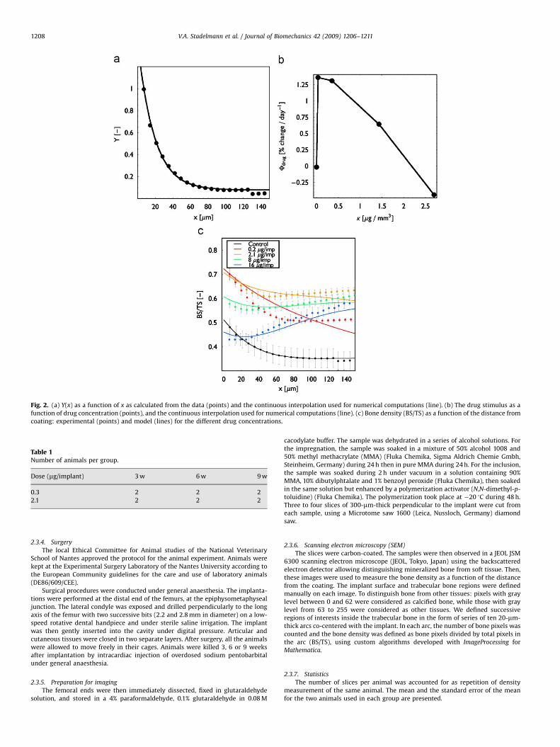
Fig. 2. (a) Y(x) as a function of x as calculated from the data (points) and the continuous interpolation used for numerical computations (line). (b) The drug stimulus as a
function of drug concentration (points), and the continuous interpolation used for numerical computations (line). (c) Bone density (BS/TS) as a function of the distance from
coating: experimental (points) and model (lines) for the different drug concentrations.
Table 1Number of animals per group.
Dose (mg/implant) 3 w 6 w 9 w
0.3 2 2 2
2.1 2 2 2
V.A. Stadelmann et al. / Journal of Biomechanics 42 (2009) 1206–12111208
2.3.4. Surgery
The local Ethical Committee for Animal studies of the National Veterinary
School of Nantes approved the protocol for the animal experiment. Animals were
kept at the Experimental Surgery Laboratory of the Nantes University according to
the European Community guidelines for the care and use of laboratory animals
(DE86/609/CEE).
Surgical procedures were conducted under general anaesthesia. The implanta-
tions were performed at the distal end of the femurs, at the epiphysometaphyseal
junction. The lateral condyle was exposed and drilled perpendicularly to the long
axis of the femur with two successive bits (2.2 and 2.8 mm in diameter) on a low-
speed rotative dental handpiece and under sterile saline irrigation. The implant
was then gently inserted into the cavity under digital pressure. Articular and
cutaneous tissues were closed in two separate layers. After surgery, all the animals
were allowed to move freely in their cages. Animals were killed 3, 6 or 9 weeks
after implantation by intracardiac injection of overdosed sodium pentobarbital
under general anaesthesia.
2.3.5. Preparation for imaging
The femoral ends were then immediately dissected, fixed in glutaraldehyde
solution, and stored in a 4% paraformaldehyde, 0.1% glutaraldehyde in 0.08 M
cacodylate buffer. The sample was dehydrated in a series of alcohol solutions. For
the impregnation, the sample was soaked in a mixture of 50% alcohol 1008 and
50% methyl methacrylate (MMA) (Fluka Chemika, Sigma Aldrich Chemie Gmbh,
Steinheim, Germany) during 24 h then in pure MMA during 24 h. For the inclusion,
the sample was soaked during 2 h under vacuum in a solution containing 90%
MMA, 10% dibutylphtalate and 1% benzoyl peroxide (Fluka Chemika), then soaked
in the same solution but enhanced by a polymerization activator (N,N-dimethyl-p-
toluidine) (Fluka Chemika). The polymerization took place at �20 1C during 48 h.
Three to four slices of 300-mm-thick perpendicular to the implant were cut from
each sample, using a Microtome saw 1600 (Leica, Nussloch, Germany) diamond
saw.
2.3.6. Scanning electron microscopy (SEM)
The slices were carbon-coated. The samples were then observed in a JEOL JSM
6300 scanning electron microscope (JEOL, Tokyo, Japan) using the backscattered
electron detector allowing distinguishing mineralized bone from soft tissue. Then,
these images were used to measure the bone density as a function of the distance
from the coating. The implant surface and trabecular bone regions were defined
manually on each image. To distinguish bone from other tissues: pixels with gray
level between 0 and 62 were considered as calcified bone, while those with gray
level from 63 to 255 were considered as other tissues. We defined successive
regions of interests inside the trabecular bone in the form of series of ten 20-mm-
thick arcs co-centered with the implant. In each arc, the number of bone pixels was
counted and the bone density was defined as bone pixels divided by total pixels in
the arc (BS/TS), using custom algorithms developed with ImageProcessing for
Mathematica.
2.3.7. Statistics
The number of slices per animal was accounted for as repetition of density
measurement of the same animal. The mean and the standard error of the mean
for the two animals used in each group are presented.

ARTICLE IN PRESS
V.A. Stadelmann et al. / Journal of Biomechanics 42 (2009) 1206–1211 1209
3. Results
3.1. In vivo verification of model’s predictions
The model predicted that a drug dose of 0.3mg/implantmaximizes bone density within 100mm layer around the implant.To verify this prediction, bone density was measured in vivo with0.3mg/implant and 2.1mg/implant zoledronate at 3, 6 and 9 weeksin rat condyles. These measures were compared to previousmeasures with 0, 0.2 2.1, 8 and 16mg/Zoledronate/implant from(Peter et al., 2005).
One rat in the 3 weeks—2.1mg zoledronate group and one ratin the 9 weeks—0.3mg-zoledronate group were excluded fromfurther analysis, as, for unknown reasons, the implant was notintegrated into bone. A total of thirty-three slices were analyzed(Table 2).
At three weeks, the model’s predictions were verified. Themean bone density of the group with optimal zoledronate dosewas 4% greater than the highest bone density obtained so far inour previously published results (Fig. 3a). The mean bone densitywith 2.1mg zoledronate/implant was in the range of our previousdata. The difference in bone density between the 0.3mg/implantgroup and the 2.1mg/implant group observed at 3 weeks, was nolonger observed at 6 and 9 weeks (Fig. 3b).
3.2. Evolution of the integration of the implant
The SEM observations confirmed implant integration compar-able to that previously described (Fig. 4a). The hydroxyapatitecoating evolved at the different time points after implantation. At
Table 2Number of slices for imaging per group.
Dose (mg/implant) 3 w 6 w 9 w
0.3 6 8 3
2.1 3 6 6
Fig. 3. (a) Mean peri-bone density (BS/TS) in the first 100mm layer. The dark bars we
implant Zoledronate. The mean bone density obtained with the optimal calculated 0.3mg
previously published results. (b) Evolution of mean bone density (BS/TS) at 3, 6 and 9
3 weeks, the bone was in contact with the coating surface, but therewas no sign of resorption in the coating and almost no bone enteringthe coating (Fig. 4b). At 6 weeks, the first signs of resorptionappeared in the coating. Approximately half of the thickness of thecoating had been resorbed and lacunae could be observed in thecoating (Fig. 4c). At 9 weeks, most of the coating had been resorbedand newly formed bone was in contact with the titanium surface.Lacunae were still present inside the coating and some speckles ofbone grew directly from the implant surface (Fig. 4d).
4. Discussion
The principal aims of this projects were first, to develop atheoretical framework of bone remodeling influenced by localrelease of bisphosphonate; second, to identify the parameters ofthe model and third, to verify the model’s predictions in vivo.
The development of the model consisted of adding the drugconcentration as a new internal variable to an existing model ofremodeling, completed by a function relating the drug concentra-tion to the remodeling stimulus. The parameters appearing in thisnew model were identified from our previously publishedexperimental data (Peter et al., 2005). Next we solved this modelto predict that a dose of 0.3mg of zoledronate grafted on theimplant coating would maximize the peri-implant bone density.This prediction was finally confirmed experimentally: with thiscalculated dose, the bone density was maximal, compared toother doses, at 3, 6 and 9 weeks post-op.
The intensity of the effect of bisphosphonate is dose depen-dent. The theoretical framework developed here shows that theinterpretation of bone density profiles has to take into account themechanical situation, the drug diffusion and the effect of drug onthe remodeling balance, modeled here by the drug stimulus Fdrug.
The drug stimulus function is the key point of the model. It canbe interpreted as the signature of the drug in a particular animalmodel. The shape of this function reflects that the drug inducesthe imbalance of the remodeling process after the decrease ofosteoclast activity. The shape of the function is concordant withwhat is observed in vitro: bisphosphonates, like most drugs, havea range of concentration with beneficial effects but can produceadverse effects at higher doses (Fleisch, 2002).
re adapted from (Peter et al.) and the light bar represents the group with 0.3mg/
/implant zoledronate was 4% greater than the highest bone density obtained in our
weeks post-op.

ARTICLE IN PRESS
Fig. 4. SEM pictures of implanted condyles: panel (a) shows the bone structure of a condyle implanted with HA-coated implant containing 2.1mg zoledronate at 3 weeks.
The implant integration is qualitatively similar to previously published results. Panel (b) shows a detail of the bone-coating interface at 3 weeks. Bone is not yet entering the
coating. Panel (c) shows the interface at 6 weeks with the first signs of coating resorption, and panel (d) shows the interface at 9 weeks with more than 50% of the coating
resorbed.
V.A. Stadelmann et al. / Journal of Biomechanics 42 (2009) 1206–12111210
The model presented here is based on several hypotheseswhich were consistent with the experimental situation addressed.We assumed that the model of a cylindrical implant could bereduced to a one-dimensional geometry. This assumption isrelevant in our situation as we analyzed only the average densityof a very thin layer of bone and we were not interested inhistomorphometry aspects. Moreover, the analyzed slices werenot taken from the extremities of the implants, which are in thecortical regions of the condyles.
Fick’s law of diffusion for bisphosphonates is certainly alimitation of the model. However, there is a lack of informationfor the diffusion aspect of bisphophonates in human bone and theuse of the experimentally determined effective coefficient ofdiffusion (Peter, 2004) can be considered as a first approach as anaverage diffusion behavior is obtained.
We identified from experimental data that the loading functionY(x) increased significantly near the implant surface. Thiscertainly reflects the mechanical stress following press-fit inser-tion and spring-back effect of bone (Kold et al., 2003). With adifferent surgical protocol, the stress distribution would certainlybe different, and Y(x) would have to be re-calculated.
In the resolution of our model, we simplified the boundaryconditions: we set the coating as an infinite source of drug atconstant concentration. The relevance of this assumption forshort-term studies was confirmed by the SEM observations: atthree weeks, the coating was indeed not significantly resorbed. Itwas probably still protected from osteoclastic resorption by itscontent in bisphosphonate. This is concordant with the very lowrelease rate of zoledronate by hydroxyapatite (Roussiere et al.,2005). However, this simplifying assumption cannot be used forlong-term studies: we observed that the hydroxyapatite coatingwas partially degraded at 6 weeks, and this degradation continuedat 9 weeks. The bisphosphonate concentration in the coating thencertainly decreases significantly after 3 weeks. With the secondboundary condition, we assumed that the drug concentration is
negligible at 2 mm from the implant surface, which correspondsto the growth plate and the bone marrow. Since these tissues arewell perfused, the drug is certainly diluted into the blood volume.
The observed degradation of the coating at 6 and 9 weeks, andthe lacunae in resorbed areas might reflect the presence ofosteocytes in the newly formed bone–implant interface. This is asign of good implant osteointegration at mid- and long-term post-op with the optimal zoledronate concentration. However, morehistology would be needed to confirm this observation.
Our results are certainly dependent on the choice of the drugand on the animal model. In the present study, we determinedthe drug stimulus function for zoledronate in rats. To extend themodel predictions to other active molecules or other animals, thefirst step would be to determine the drug stimulus function,which we called the drug signature, with the specific drug usingthe specific animal. To our knowledge, none of the required datafor such identification with other animals or with different drugshas been published.
Only a limited number of animals were used in the experi-mental part of this project. The main objective of this project wasto compare the theoretical model’s outcomes to an in vivo
situation. The experiments were therefore designed to validatethe model rather than to provide new statistically significantexperimental results, which would have required a much largernumber of animals.
The principal objective of coating implants with bispho-sphonate for local delivery is to increase the fixation of theimplant. The pullout force is somehow related to peri-implantbone density (Peter et al., 2005), but other factors, such as bonequality, also have an influence. A mathematical relationshipbetween histomorphometry of bone and its mechanical capacityto resist pullout has yet to be determined (Jakobsen et al., 2006).Thus, the mathematical model presented here only predicts bonedensity. More work will be needed to link bone density to implantfixation strength.

ARTICLE IN PRESS
V.A. Stadelmann et al. / Journal of Biomechanics 42 (2009) 1206–1211 1211
One of the most important challenges associated to orthopedicimplants is to obtain sufficient early fixation to ensure long-termstability, for patients of variable age, daily activity level or bonequality. Although several studies have shown that implantsdelivering bisphophonate improve this fixation, the bisphospho-nate molecule, the dose, or the animal model have always beenchosen empirically. So the conclusion of this article is todemonstrate that the theoretical framework presented could beof great interest to predict the bone formation around implantdelivering bisphosphonate.
Conflict of interest statement
There is no conflict of interest.
Acknowledgements
Project no. 04-P2 was supported by the AO Research Fund ofthe AO Foundation, Davos, Switzerland and the Inter-institutionalCenter for Translational Biomechanics EPFL-CHUV-DAL.
We thank B. Bujoli for the preparation of the implants, SophieSalice for her help with the sample preparation, Paul Pilet for theelectronic microscopy and Tyler Thacher for English editing.
References
Dannemann, C., Gratz, K.W., Riener, M.O., Zwahlen, R.A., 2007. Jaw osteonecrosis relatedto bisphosphonate therapy: a severe secondary disorder. Bone 40, 828–834.
Dhert, W.J.A., Thomsen, P., Blomgren, A.K., Esposito, M., Ericson, L.E., Verbout, A.J.,1998. Integration of press-fit implants in cortical bone: a study on interfacekinetics. Journal of Biomedical Materials Research 41, 574–583.
Fleisch, H., 2002. Development of bisphosphonates. Breast Cancer Research 4, 30–34.Hernandez, C.J., Beaupre, G.S., Marcus, R., Carter, D.R., 2001. A theoretical analysis
of the contributions of remodeling space, mineralization, and bone balance tochanges in bone mineral density during alendronate treatment. Bone 29,511–516.
Hilding, M., Aspenberg, P., 2006. Postoperative clodronate decreases prostheticmigration: 4-year follow-up of a randomized radiostereometric study of 50total knee patients. Acta Orthopaedica 77, 912–916.
Horowitz, S.M., Gonzales, J.B., 1996. Inflammatory response to implant particulatesin a macrophage/osteoblast coculture model. Calcified Tissue International 59,392–396.
Huiskes, R., Weinans, H., Grootenboer, H.J., Dalstra, M., Fudala, B., Sloof, T.J., 2000.Effects of mechanical forces on maintenance and adaptation of form intrabecualar bone. Nature 405, 704–706.
Jakobsen, T., Kold, S., Bechtold, J.E., Elmengaard, B., Soballe, K., 2006. Effect oftopical alendronate treatment on fixation of implants inserted with bonecompaction. Clinical Orthopaedics and Related Research, 229–234.
Karrholm, J., Borssen, B., Lowenhielm, G., Snorrason, F., 1994. Does earlymicromotion of femoral stem prostheses matter? 4–7-year stereoradiographicfollow-up of 84 cemented prostheses. Journal of Bone and Joint Surgery British76, 912–917.
Kold, S., Bechtold, J.E., Ding, M., Chareancholvanich, K., Rahbek, O., Soballe, K.,2003. Compacted cancellous bone has a spring-back effect. Acta OrthopaedicaScandinavica 74, 591–595.
Peter, B., 2004. Orthopedic implants used as drug delivery systems: numerical, invitro and in vivo studies. Ph.D. thesis #3044, EPFL.
Peter, B., Gauthier, O., Laib, S., Bujoli, B., Guicheux, J., Janvier, P., van Lenthe, G.H.,Muller, R., Zambelli, P.Y., Bouler, J.M., Pioletti, D.P., 2006. Local delivery ofbisphosphonate from coated orthopedic implants increases implants mechan-ical stability in osteoporotic rats. Journal of Biomedical Materials Research A76, 133–143.
Peter, B., Pioletti, D.P., Laib, S., Bujoli, B., Pilet, P., Janvier, P., Guicheux, J., Zambelli,P.Y., Bouler, J.M., Gauthier, O., 2005. Calcium phosphate drug delivery system:influence of local zoledronate release on bone implant osteointegration. Bone36, 52–60.
Pioletti, D.P., Rakotomanana, L.R., 2004. Can the increase of bone mineral densityfollowing bisphosphonates treatments be explained by biomechanical con-siderations? Clinical Biomechanics 19, 170–174.
Rakotomanana, R.L., Leyvraz, P.F., Curnier, A., Heegaard, J.H., Rubin, P.J., 1992. Afinite element model for evaluation of tibial prosthesis–bone interface in totalknee replacement. Journal of Biomechanics 25, 1413–1424.
Roussiere, H., Montavon, G., Samia, L.B., Janvier, P., Alonso, B., Fayon, F., Petit, M.,Massiot, D., Bouler, J.M., Bujoli, B., 2005. Hybrid materials applied tobiotechnologies: coating of calcium phosphates for the design of implantsactive against bone resorption disorders. Journal of Materials Chemistry 15,3869–3875.
Stadelmann, V.A., Terrier, A., Pioletti, D.P., 2008. Microstimulation at the bone–implant interface upregulates osteoclast activation pathways. Bone, 358–364.
Terrier, A., Miyagaki, J., Fujie, H., Hayashi, K., Rakotomanana, L., 2005. Delayof intracortical bone remodelling following a stress change: a theoreticaland experimental study. Clinical Biomechanics (Bristol, Avon) 20, 998–1006.
Venesmaa, P.K., Kroger, H.P.J., Miettinen, H.J.A., Jurvelin, J.S., Suomalainen, O.T.,Alhava, E.M., 2001. Monitoring of periprosthetic BMD after uncemented totalhip arthroplasty with dual-energy X-ray absorptiometry—a 3-year follow-upstudy. Journal of Bone and Mineral Research 16, 1056–1061.
Wermelin, K., Tengvall, P., Aspenberg, P., 2007. Surface-bound bisphosphonatesenhance screw fixation in rats—increasing effect up to 8 weeks after insertion.Acta Orthopaedica 78, 385–392.
Copyright © 2022 FDOKUMEN