All‐ceramic single crowns supported by zirconia implants
28
Zurich Open Repository and Archive University of Zurich Main Library Strickhofstrasse 39 CH-8057 Zurich www.zora.uzh.ch Year: 2019 All-ceramic single crowns supported by zirconia implants: 5-year results of a prospective multicenter study Spies, Benedikt C ; Balmer, Marc ; Jung, Ronald E ; Sailer, Irena ; Vach, Kirstin ; Kohal, Ralf-Joachim Abstract: OBJECTIVES To assess survival/success rates and patient-reported outcome of zirconia-based posterior single crowns (SCs) supported by zirconia implants in a prospective two-center study after fve years of observation. MATERIAL AND METHODS Forty-fve patients were restored with 45 zirconia implant-supported posterior SCs composed of zirconia frameworks hand-layered with a leucite-reinforced feldspathic ceramic. Survival rates of SCs were assessed and technical success was evaluated according to modifed United States Public Health Care (USPHS) criteria. Furthermore, patient-reported outcome measures (PROMs) were assessed by applying visual analog scales (VAS). Wilcoxon matched-pairs signed- rank test, mixed-efects ordered logistic regression, and linear mixed models were used to evaluate time efects on response variables. RESULTS Forty patients were available after a mean observation period of 61.0 ± 1.4 months. One SC had to be replaced, resulting in a Kaplan-Meier (KM) survival estimate for the SCs of 97.5 ± 2.47%. Since nine reconstructions showed at least in one category a major deviation from the ideal (fve major chippings, four with increased occlusal roughness, one signifcant crevice, and one pronounced over-contouring), the KM success estimate was 79.3 ± 5.8%. Incidence of chipping (n = 19) and occlusal roughness (n = 35) was frequent (p < 0.001). All PROMs at prosthetic delivery except for speech (p = 0.139) showed signifcantly improved VAS scores (81%-94%; p < 0.001) compared to pre-treatment evaluations. Thereafter, no decrease in satisfaction could be observed until the 5- year follow-up (93%-97%). CONCLUSION Veneered zirconia-based SCs supported by zirconia implants showed high survival rates and highly satisfed patients’ needs. However, signifcant incidence of technical complications is compromising the clinical long-term outcome for this indication. DOI: https://doi.org/10.1111/clr.13433 Posted at the Zurich Open Repository and Archive, University of Zurich ZORA URL: https://doi.org/10.5167/uzh-171597 Journal Article Accepted Version Originally published at: Spies, Benedikt C; Balmer, Marc; Jung, Ronald E; Sailer, Irena; Vach, Kirstin; Kohal, Ralf-Joachim (2019). All-ceramic single crowns supported by zirconia implants: 5-year results of a prospective multi- center study. Clinical Oral Implants Research, 30(5):466-475. DOI: https://doi.org/10.1111/clr.13433
-
Upload
khangminh22 -
Category
Documents
-
view
2 -
download
0
Transcript of All‐ceramic single crowns supported by zirconia implants

Zurich Open Repository andArchiveUniversity of ZurichMain LibraryStrickhofstrasse 39CH-8057 Zurichwww.zora.uzh.ch
Year: 2019
All-ceramic single crowns supported by zirconia implants: 5-year results of aprospective multicenter study
Spies, Benedikt C ; Balmer, Marc ; Jung, Ronald E ; Sailer, Irena ; Vach, Kirstin ; Kohal, Ralf-Joachim
Abstract: OBJECTIVES To assess survival/success rates and patient-reported outcome of zirconia-basedposterior single crowns (SCs) supported by zirconia implants in a prospective two-center study after fiveyears of observation. MATERIAL AND METHODS Forty-five patients were restored with 45 zirconiaimplant-supported posterior SCs composed of zirconia frameworks hand-layered with a leucite-reinforcedfeldspathic ceramic. Survival rates of SCs were assessed and technical success was evaluated accordingto modified United States Public Health Care (USPHS) criteria. Furthermore, patient-reported outcomemeasures (PROMs) were assessed by applying visual analog scales (VAS). Wilcoxon matched-pairs signed-rank test, mixed-effects ordered logistic regression, and linear mixed models were used to evaluate timeeffects on response variables. RESULTS Forty patients were available after a mean observation period of61.0 ± 1.4 months. One SC had to be replaced, resulting in a Kaplan-Meier (KM) survival estimate forthe SCs of 97.5 ± 2.47%. Since nine reconstructions showed at least in one category a major deviationfrom the ideal (five major chippings, four with increased occlusal roughness, one significant crevice, andone pronounced over-contouring), the KM success estimate was 79.3 ± 5.8%. Incidence of chipping (n= 19) and occlusal roughness (n = 35) was frequent (p < 0.001). All PROMs at prosthetic deliveryexcept for speech (p = 0.139) showed significantly improved VAS scores (81%-94%; p < 0.001) comparedto pre-treatment evaluations. Thereafter, no decrease in satisfaction could be observed until the 5-year follow-up (93%-97%). CONCLUSION Veneered zirconia-based SCs supported by zirconia implantsshowed high survival rates and highly satisfied patients’ needs. However, significant incidence of technicalcomplications is compromising the clinical long-term outcome for this indication.
DOI: https://doi.org/10.1111/clr.13433
Posted at the Zurich Open Repository and Archive, University of ZurichZORA URL: https://doi.org/10.5167/uzh-171597Journal ArticleAccepted Version
Originally published at:Spies, Benedikt C; Balmer, Marc; Jung, Ronald E; Sailer, Irena; Vach, Kirstin; Kohal, Ralf-Joachim(2019). All-ceramic single crowns supported by zirconia implants: 5-year results of a prospective multi-center study. Clinical Oral Implants Research, 30(5):466-475.DOI: https://doi.org/10.1111/clr.13433

Ac
ce
pte
d A
rti
cle
This article has been accepted for publication and undergone full peer review but has not been through the copyediting, typesetting, pagination and proofreading process, which may lead to differences between this version and the Version of Record. Please cite this article as doi: 10.1111/clr.13433 This article is protected by copyright. All rights reserved.
DR. BENEDIKT CHRISTOPHER SPIES (Orcid ID : 0000-0003-1702-1679)
DR. MARC BALMER (Orcid ID : 0000-0003-0637-8314)
DR. RONALD ERNST JUNG (Orcid ID : 0000-0003-2055-1320)
DR. IRENA SAILER (Orcid ID : 0000-0002-4537-7624)
PROF. RALF J KOHAL (Orcid ID : 0000-0001-7095-4190)
Article type : Original Research
All-ceramic single crowns supported by zirconia implants:
5-year results of a prospective multicenter study
Benedikt C. Spies, PD Dr med denta,b
(Clinical procedures; Data analysis/interpretation; Data collection; Drafting article)
Marc Balmer, Dr med dentc
(Data analysis/interpretation; Data collection; Approval of article)
Ronald E. Jung, Prof Dr med dentc
(Concept/Design; Clinical procedures; Data collection; Approval of article; Funding secured)
Irena Sailer, Prof Dr med dentd
(Concept/Design; Clinical procedures; Data collection; Approval of article; Funding secured)
Kirstin Vach, Dipl.-Math.e
(Statistical analysis)
Ralf-Joachim Kohal, Prof Dr med denta
(Concept/Design; Clinical procedures; Data collection; Approval of article; Funding secured)
Ac
ce
pte
d A
rti
cle
This article is protected by copyright. All rights reserved.
a) Department of Prosthetic Dentistry, Center for Dental Medicine, Medical Center –
University of Freiburg, Faculty of Medicine, University of Freiburg, Hugstetter Str. 55,
D-79106 Freiburg, Germany
b) Charité – Universitätsmedizin Berlin, corporate member of Freie Universität Berlin,
Humboldt-Universität zu Berlin, and Berlin Institute of Health, Department of
Prosthodontics, Geriatric Dentistry and Craniomandibular Disorders, Aßmannshauser
Str. 4-6, Berlin, Germany
c) Clinic of Fixed and Removable Prosthodontics and Dental Material Science, Center of
Dental Medicine, University of Zurich, Plattenstr. 11, CH-8032 Zurich, Switzerland
d) Division of Fixed Prosthodontics and Biomaterials, University Clinics for Dental
Medicine, University of Geneva, Geneva, 19 rue Barthélemy-Menn, CH-1205 Genf,
Switzerland
e)
Institute for Medical Biometry and Statistics, Center for Medical Biometry and
Medical Informatics, Medical Center – University of Freiburg, Faculty of Medicine,
University of Freiburg, Hebelstr. 11, D-79104 Freiburg, Germany
Corresponding author:
Benedikt Christopher Spies
Department of Prosthodontics, Geriatric Dentistry and Craniomandibular Disorders
Charité – Universitätsmedizin Berlin
Aßmannshauser Str 4-6, 14197 Berlin
Phone: +49 30 450 662546, Fax: +49 30 450 562912;
e-mail: [email protected]
Disclosure: This investigation was supported by VITA Zahnfabrik, Bad Säckingen,
Germany.
Keywords [MeSH]: Ceramics; Dental porcelain; Crown; CAD-CAM; Dental implant;
Zirconia

Ac
ce
pte
d A
rti
cle
This article is protected by copyright. All rights reserved.
Abstract
Objectives: To assess survival/success rates and patient-reported outcome of zirconia-based
posterior single crowns (SCs) supported by zirconia implants in a prospective two-center
study after five years of observation.
Material and Methods: Forty-five patients were restored with 45 zirconia implant-supported
posterior SCs composed of zirconia frameworks hand-layered with a leucite reinforced
feldspathic ceramic. Survival rates of SCs were assessed and technical success was evaluated
according to modified United States Public Health Care (USPHS) criteria. Furthermore,
patient-reported outcome measures (PROMs) were assessed by applying visual analog scales
(VAS). Wilcoxon matched-pairs signed-rank test, mixed-effects ordered logistic regression
and linear mixed models were used to evaluate time effects on response variables.
Results: Forty patients were available after a mean observation period of 61.0±1.4 months.
One SC had to be replaced, resulting in a Kaplan-Meier (KM) survival estimate for the SCs
of 97.5±2.47%. Since 9 reconstructions showed at least in one category a major deviation
from the ideal (5 major chippings, 4 with increased occlusal roughness, one significant
crevice and one pronounced over-contouring), the KM success estimate was 79.3±5.8%.
Incidence of chipping (n=19) and occlusal roughness (n=35) was frequent (p<0.001). All
PROMs at prosthetic delivery except for speech (p=0.139) showed significantly improved
VAS scores (81-94%; p<0.001) compared to pre-treatment evaluations. Thereafter, no
decrease in satisfaction could be observed until the 5-year follow-up (93-97%).
Conclusion: Veneered zirconia-based SCs supported by zirconia implants showed high
survival rates and highly satisfied patients’ needs. However, significant incidence of technical
complications is compromising the clinical long-term outcome for this indication.
German Clinical Trials Register (ID: DRKS00000226)

Ac
ce
pte
d A
rti
cle
This article is protected by copyright. All rights reserved.
Introduction
Ceramic implants were introduced to dentistry half a century ago, at about the same time with
their counterpart made from titanium. In these days, aluminum oxide was used for
manufacturing the first market-available ceramic implants like the “crystalline bone screw”
or the “Tübingen immediate implant” (Sandhaus, 1967; Schulte & Heimke, 1976). However,
reports addressing the clinical outcome of these products are sparse. Due to its reduced
fracture toughness, technical failures of dental implants made from aluminum oxide were
frequent. This resulted in concerns of most practitioners and implants made from aluminum
oxide are no more available on the market. Meanwhile, titanium became the gold-standard
material for implant manufacturing and titanium implant supported reconstructions cover the
full range of clinical indications from partial to complete fixed and removable dental
prostheses (R. E. Jung, Zembic, Pjetursson, Zwahlen, & Thoma, 2012; Kern, Kern, Wolfart,
& Heussen, 2016; Pjetursson, Thoma, Jung, Zwahlen, & Zembic, 2012). On the contrary,
ceramic dental implants might still be considered a niche product. Along with the
introduction of stabilized zirconium dioxide as promising implant bulk material and a rising
discussion on whether the reported prevalence of inflammation in tissues surrounding
titanium implants might partially associated with titanium particles resulting from
tribocorrosion (Fretwurst, Nelson, Tarnow, Wang, & Giannobile, 2018), interest in zirconia
dental implants markedly increased within the last decade.
In the first instance, research focused on the osseointegration capacity of one-piece zirconia
implants in animal investigations (Pieralli, Kohal, Lopez Hernandez, Doerken, & Spies,
2018) and, later on, in clinical trials (Pieralli, Kohal, Jung, Vach, & Spies, 2017). Whereas
osseointegration of zirconia implants was shown to be similar to titanium implants, reduced
susceptibility to peri-implant inflammatory processes still needs to be scientifically
Ac
ce
pte
d A
rti
cle
This article is protected by copyright. All rights reserved.
evidenced. Now that osseointegration seems to be sufficiently proven, research interest more
and more focusses on how to restore zirconia one-piece implants requiring cementation of the
prostheses. Besides laboratory studies evaluating cementation techniques (Zaugg, Meyer,
Rohr, Zehnder, & Zitzmann, 2018; Zaugg, Zehnder, Rohr, Fischer, & Zitzmann, 2018), there
are few data available reporting on the clinical performance of all-ceramic single crowns and
three-unit fixed dental prostheses supported by zirconia implants (Spies, Stampf, & Kohal,
2015). It seems that zirconia based bi-layered reconstructions reveal the same technical
complication range known from the restoration on titanium implants: whereas the zirconia
framework is not prone to failure, but the veneering ceramic appears to be the weakest link,
suffering from chip-off fractures and roughening of the surface.
In an earlier report, it was shown that zirconia implant supported single crowns in posterior
regions entirely survived a three-year observation period (Spies, Balmer, et al., 2017).
However, incidence of minor chippings and occlusal roughness was highly significant.
Interestingly, these minor technical complications had no impact on the highly satisfied
patient-reported outcome. It was finally concluded, that patient satisfaction and restoration
survival might decrease over the course of the follow-up appointments in case of further
progress, potentially resulting in the need for replacements in the future. Therefore, the aim
of the present report was to assess the ongoing technical and patient-reported outcomes from
the three- to five-year follow-up.

Ac
ce
pte
d A
rti
cle
This article is protected by copyright. All rights reserved.
Materials and Methods
Study design
This prospective multicenter study was performed in two centers located in Switzerland
(Center of Dental Medicine, University of Zürich, Clinic of Fixed and Removable
Prosthodontics and Dental Material Science) and Germany (Medical Center, University of
Freiburg, Department of Prosthetic Dentistry). The study protocol was approved by both local
responsible ethics committees (Freiburg: 241/08; Zürich: StV 08/10). The study was
registered in the German Clinical Trials Register (ID: DRKS00000226) and is listed in the
WHO International Clinical Trials Registry Platform. All included patients gave informed
consent prior to their inclusion (04/2010-07/2012). This prospective cohort study was
designed and performed considering the Strengthening the Reporting of Observational
Studies in Epidemiology (STROBE) Statement for cohort studies (http://www.strobe-
statement.org) and therefore in compliance with the appropriate EQUATOR guidelines.
Participants
Sixty patients with one or three adjacent missing teeth were recruited. They needed to be
compliant, aged between 20 and 70 years, show a good health status, have sufficient bone
volume in the area destined for implant installation, show a stable occlusal relationship, and
no signs of pronounced bruxism (such as attrition, pain on muscular palpation, pain-causing
joint sound, or self-reported clenching habits). Reasons for exclusion were smoking (>10
cigarettes per day), alcohol or drug abuse, known destructive habits, and health conditions not
permitting the surgical procedure.
The procedure of implant installation and assessments of the clinical outcome have been
described earlier (Balmer et al., 2018; Ronald E. Jung et al., 2016). In brief: Late implant
placement by raising a mucoperiostal flap was followed by transmucosal healing. If required,
Ac
ce
pte
d A
rti
cle
This article is protected by copyright. All rights reserved.
guided bone regeneration was performed with a bovine bone substitute and a porcine
collagen membrane. The supporting one-piece zirconia implants (ceramic.implant;
vitaclinical, VITA Zahnfabrik, Bad Säckingen, Germany) were composed of 3 mol% yttria
stabilized tetragonal zirconia polycrystals (3Y-TZP: ZrO2 93%, Y2O3 5%, HfO2 1.9%, Al2O3
0.1% [% w/w]) and provided in diameters of 4.0, 4.5, and 5.5 mm. In total, 49 implants were
installed to support 49 SCs (Spies, Balmer, et al., 2017). In the eleven remaining patients
three-unit bridges were supported by two implants. To obtain a clear indication for the
present evaluation of posterior single crowns, three patients with three anterior crowns and
the eleven patients restored with three-unit bridges were excluded from the analysis.
Clinical and laboratory procedures
Detailed information on the clinical and laboratory procedures was given in precedent
publications reporting mid-term results after 36 months of observation (Balmer et al., 2018;
Spies, Balmer, et al., 2017). In brief, implants were immediately temporized with
prefabricated provisional reconstructions. After a minimum healing period of eight (lower
jaw) or 16 weeks (upper jaw), respectively, impressions were taken with a polyether material
(Impregum; 3M Espe, Seefeld, Germany) and digitized in the dental laboratory (inEos
scanner; Sirona, Bensheim, Germany). Zirconia frameworks (In-Ceram YZ, VITA
Zahnfabrik; 3Y-TZP: ZrO2 94.9%, Y2O3 5%, Al2O3 0.1% [% w/w]) were CAD/CAM-
fabricated (Cerec inLab® software, inLab® MC XL 4-axis milling device; Sirona) and finally
hand-layered with a leucite-reinforced feldspathic ceramic (VM9, VITA Zahnfabrik)
according to the manufacturer’s instructions. Final SCs were adhesively cemented using a
dual-curing resin cement (RelyX Unicem Aplicap; 3M Espe). If a subgingival restoration
margin was present, retraction cords were placed to facilitate removal of cement remnants.
Centric and dynamic occlusions were controlled and adjusted by reducing premature
contacts, if present, to protect the SCs from any excessive forces. In all patients, anterior
Ac
ce
pte
d A
rti
cle
This article is protected by copyright. All rights reserved.
and/or canine guidance was verified with immediate posterior disclusion of replaced molars
and premolars when making lateral or protrusive movements. In each center, one master
dental technician was responsible for the manufacturing of all reconstructions.
Baseline and follow-up examinations
At baseline, after final reconstruction cementation and again after 6, 12, 24, 36 and 60
months of function, the patients were scheduled for clinical examination. Examinations
consisted of a visual and tactile inspection of the SCs, a control of static and dynamic
occlusal contacts, impression taking, and intraoral photographs of the SCs and adjacent teeth.
Biological and technical complications were recorded. A required treatment was applied, if
necessary.
Technical examination
Technical aspects were clinically evaluated according to modified USPHS (United States
Public Health Service) criteria. In brief, the reconstructions were examined for fracture of
framework or of the veneering ceramic, occlusal roughness, marginal integrity and under/
overcontouring. All parameters were rated “Alpha” in case of no problem, “Bravo” in case of
minor extent of the complication, “Charlie” if the complication was major, and “Delta” if the
reconstruction had to be replaced due to the complication (Tab. 1). In both centers, one single
examiner was responsible for the technical exams. Examiners were trained and continually
calibrated by visualization of the above mentioned parameters and possible ratings in
xemplary clinical photographs.
Patient-reported outcome
The patients` appraisal of function, esthetics and appearance, sense, speech, and self-esteem
relating to the inserted reconstructions were assessed. At study inclusion, final prosthesis
insertion, and at every follow-up appointment, all included subjects labeled a point on a line
Ac
ce
pte
d A
rti
cle
This article is protected by copyright. All rights reserved.
that corresponded with their personal satisfaction in the aforementioned categories (Visual
analog scales; VAS). The line was 100 mm in length and showed no scale. Every millimeter
of the line corresponded to 1% of satisfaction (from 0% at the left end of the line to 100% at
the right end of the line).
Statistical analyses
Sample size calculation was performed considering the radiographic outcome (expected
marginal bone loss known from the literature) and was, therefore, not primarily designed for
the evaluation of the prosthetic outcome (13). Means, medians, and standard deviations were
computed for descriptive analyses of the data. KM survival and success estimates were
calculated and graphically presented using plots. Moreover, log-rank tests were used to check
for an influence of co-variables (gender, jaw, and center). A Wilcoxon matched-pairs signed-
rank test was used to calculate for changes between prosthetic delivery and the 60 month-
follow-up (USPHS criteria, PROMs). A mixed-effects ordered logistic regression (USPHS)
and a linear mixed model (PROMs) were used to analyze a linear time trend including the
data from all measurements (Delivery, 6m, 12m, 24m, 36m, 48m, 60m).
All calculations were performed with the STATA 14.2 (StataCorp LT, College Station, TX,
USA) statistical software. The probability level for statistical significance was set to p < 0.05.
Results
Status of follow-up
Since one patient lost his implant during the healing period (considered as drop-out for the
evaluation of final single crowns), 45 out of initially 46 patients provided with 45 posterior
SCs remained for evaluation (Tab. 2). Details regarding the opposing dentition can be found
in Table 3. Final restoration delivery took place in between 04/2010 and 02/2012. Mean age

Ac
ce
pte
d A
rti
cle
This article is protected by copyright. All rights reserved.
of patients was 46.6 ± 13.1 years (range: 25–69 years) at this time point. Of the 45 included
patients, 40 patients with 40 SCs were seen at the final follow-up in between 05/2015 and
07/2017 (Fig. 1), resulting in a mean observation time of 61.0 ± 1.4 months. Five patients
refused further participation in the study in between restoration delivery (one patient refused
further participation after final prosthetic delivery without giving a reason and was
considered as drop-out for analyses) and the four-year follow-up (likewise giving no reason
for study withdrawal). For the current analyses, these four patients/SCs were considered
drop-outs.
Clinical outcome
One SC had to be replaced due to a severe chipping of the veneering ceramic (rated “Delta”),
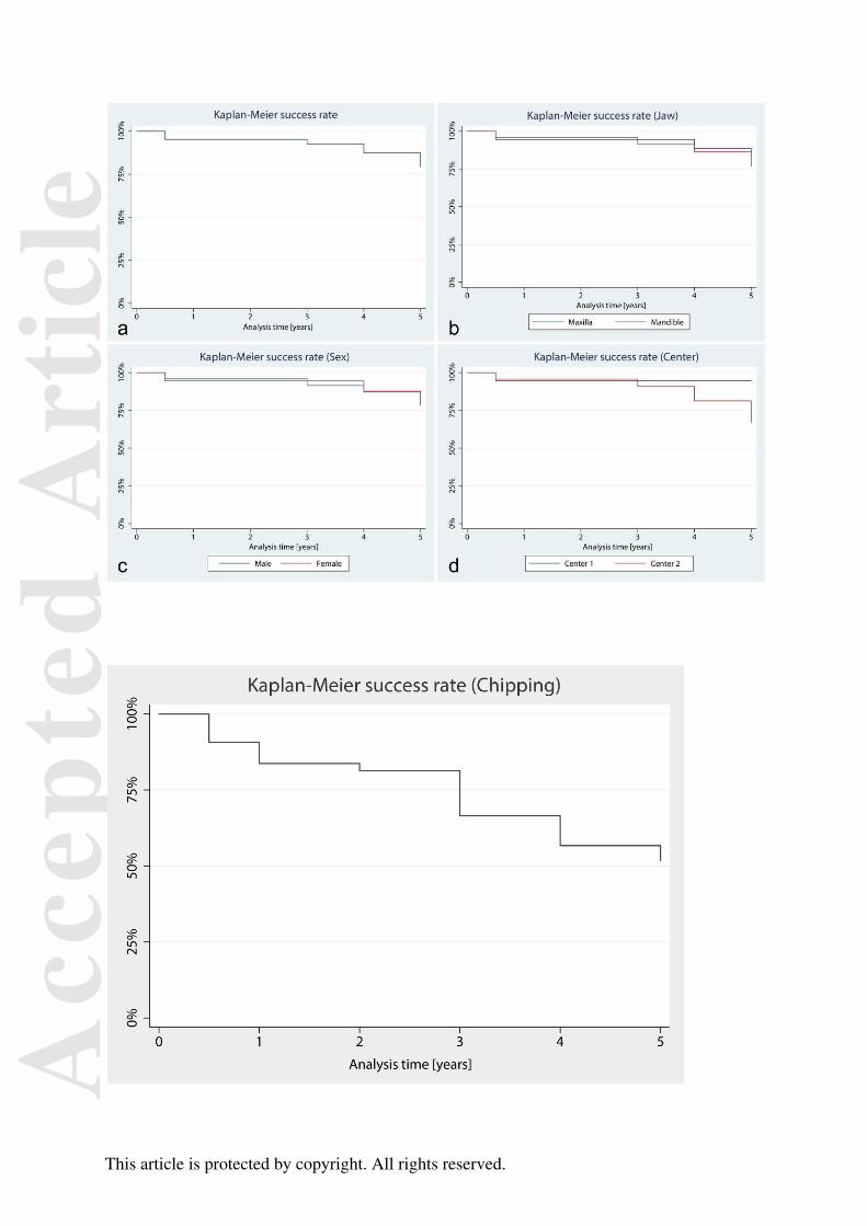
resulting in a KM survival estimate of 97.5 ± 2.47% (CI: 83.55 – 99.64; Fig. 2). Since 9
reconstructions showed at least in one category a major deviation from the ideal (rated
“Charlie”: 5 major chippings, 4 with increased occlusal roughness, one significant crevice
and one pronounced over-contouring), the KM success estimate was 79.3 ± 5.8% (Fig. 3a,
Tab. 4). Compared to baseline, incidence of chipping (n=19) and occlusal roughness (n=35)
was significant (p<0.001). Compared to the three-year follow-up, five more SCs were
affected by chipping and eight more SCs by occlusal roughening. Due to chip-off fractures,
contour of reconstructions changed over time (p<0.009). The KM estimate for a SC not to be
affected by any chipping at the end of the five-year follow-up was 51.6 ± 5.8% (Fig. 4). No
change in marginal disintegration compared to the last report after three years of observation
was found. No framework fracture or loss of retention was observed. Performed log-rank
tests revealed no statistically significant differences for the success curves regarding jaw (p =
0.681; maxilla: 82.6 ± 9.1%, mandible: 76.9 ± 9.1%; Fig. 3b) and sex (p = 0.858; females:
81.0 ± 10.0%, males: 78.4 ± 8.6%; Fig. 3c). However, the center showed to have significant
influence on the success rate (p = 0.048; center 1: 94.7 ± 5.1%, center 2: 67.0 ± 10.2%; Fig.

Ac
ce
pte
d A
rti
cle
This article is protected by copyright. All rights reserved.
3d). Regarding the absence of any type of chipping (major and minor), log-rank tests revealed
no difference regarding jaw (p = 0.855; maxilla: 50.7 ± 12.5%, mandible: 52.4 ± 10.0%), sex
(p = 0.370; females: 42.3 ± 12.0%, males: 58.3 ± 10.0%), and center (p = 0.204; center 1:
66.7 ± 11.1%, center 2: 39.7 ± 10.2%).
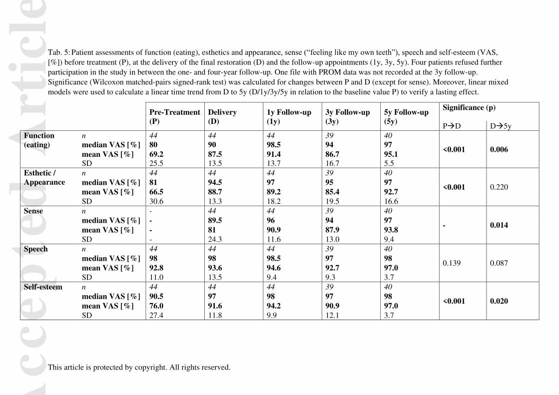
Patient-reported outcome (VAS)
Compared with baseline values prior to treatment (67-93%, sense was not assessed prior to
implant installation; Fig. 5, Tab. 5), all PROMs at prosthetic delivery except for speech
(p=0.139) showed significantly improved VAS scores (81-94%; p<0.001). Thereafter, no
decrease in satisfaction could be observed over time until the 5-year follow-up (93-97%).
Chipping incidence did not affect patient satisfaction (p≥0.140).
Discussion
The present multicenter cohort investigation revealed excellent survival of the evaluated,
zirconia implant supported, bi-layered crowns composed of a zirconia framework hand-
layered with a leucite reinforced veneering ceramic. Moreover, patients were highly satisfied
with the treatment during the entire observation period of 61 months. However, a significant
incidence of technical complications (chipping and occlusal roughness) was observed. Five
major chippings, 4 crowns with increased occlusal roughening, one significant marginal
opening and one pronounced over-contouring resulted in a reduced KM success estimate.
Including minor deviations from the ideal (e.g. minor chippings or small area roughness to be
polished intraorally), occurrence of such events was found to be highly statistically
significant. Almost half of the SCs were affected by fractures of the veneering ceramic.
Therefore, the initially raised hypothesis that ongoing technical complications after the three-
Ac
ce
pte
d A
rti
cle
This article is protected by copyright. All rights reserved.
year follow-up might result in replacements and dissatisfied patients has to be rejected (only
one replacement and highly satisfied patients were determined).
A recent systematic review and meta-analysis screening the nowadays available literature on
implant-supported all-ceramic single crowns showed, that veneered zirconia-based
reconstructions can still be considered the biggest stakeholder for all-ceramic implant-
supported replacement of single missing teeth (Rabel, Spies, Pieralli, Vach, & Kohal, 2018).
After five years of observation, the meta-analysis revealed a survival estimate of 92.0% for
zirconia-based bi-layers, ranging from 67.4 (Bömicke, Gabbert, Koob, Krisam, &
Rammelsberg, 2017) to 100% (Hosseini, Worsaae, Schiodt, & Gotfredsen, 2011; Lops,
Bressan, Chiapasco, Rossi, & Romeo, 2013; Wittneben et al., 2017). This indicated that the
survival of the present single crowns is above average. On the contrary, in the mentioned
review, the estimated occurrence of veneer fractures after five years of observation was
calculated to be 11.3% (Rabel et al., 2018), which is high but considerably lower compared to
the outcome of the present investigation. Included studies mostly applied the hand-layering
technique for veneering zirconia frameworks. Interestingly, chipping was quite a
heterogeneous outcome among included studies ranging from 0% after 12 months of
observation (Hosseini et al., 2011) up to 45.9% after 3 years of observation (Bömicke et al.,
2017). This might indicate a high technique sensitivity of different veneering procedures and
applied materials. In the present investigation, the KM estimate for chipping events was
48.4% after 5 years. As a result, the chipping frequency in the present study was significantly
higher than the estimate calculated by Rabel and collaborates (Rabel et al., 2018). One reason
for this might be considered the fact, that reconstructions of the present evaluation were
located in posterior regions, whereas multiple investigations in the meta-analyses included a
mixture of both anterior and posterior reconstructions (Chappuis et al., 2018; Nejatidanesh,
Moradpoor, & Savabi, 2016; Paolantoni, Marenzi, Blasi, Mignogna, & Sammartino, 2016;

Ac
ce
pte
d A
rti
cle
This article is protected by copyright. All rights reserved.
Tartaglia, Sidoti, & Sforza, 2015; Wittneben et al., 2017; Worni, Kolgeci, Rentsch-Kollar,
Katsoulis, & Mericske-Stern, 2015). It was shown, that single crown location has a
significant impact on the occurrence of veneer fractures in favor of reconstructions located in
the anterior region (Rabel et al., 2018). Nevertheless, since nearly half of the reconstructions
of the present evaluation were affected by minor fractures of the veneering ceramic, the
concept of bi-layered zirconia-based reconstructions for the replacement of premolars and
molars might be questioned. Since all reconstructions of the present investigation were
supported by one-piece ceramic implants, cementation of the crowns was mandatory. Screw-
retention, however, would facilitate laboratory maintenance or replacements in case of severe
technical complications or failure. Since evidence for two-piece ceramic is nowadays still
sparse, research should address this treatment option in the future.
A significant change in reconstruction contour over time that was found in the present work
might be considered a result of the high incidence of chip-off fractures requiring re-polishing,
thereby compromising the original shape of the crowns. Considering susceptibility for
fracture events, monolithic reconstructions made from lithium disilicate showed to be less
prone for this technical complication (Spies, Pieralli, Vach, & Kohal, 2017). However, data
for the monolithic type of reconstructions supported by implants is still rare (Rabel et al.,
2018).
Occlusal roughness was another technical outcome that was assessed in this investigation. At
the final follow-up appointment, all - except 5 reconstructions - were categorized to show at
least a slight roughness of the occlusal surface. Occurrence of this event was calculated to be
highly significant over time (p<0.001). Regrettably, data on roughening of veneering
ceramics is only seldom provided in the literature and was, therefore, not included as
outcome in the above mentioned review (Rabel et al., 2018). Several reasons can be assumed
for increased roughness of veneering ceramics including a crystalline phase over time in the
Ac
ce
pte
d A
rti
cle
This article is protected by copyright. All rights reserved.
oral cavity like tooth brushing (Garza, Thompson, Cho, & Berzins, 2016), environmental
conditions (Vechiato-Filho et al., 2015), abrasion due to mastication (Lawson, Bansal, &
Burgess, 2016) or attrition due to antagonistic wear (Amer, Kürklü, & Johnston, 2015). All
mentioned conditions can result in dissolution of the amorphous phase while crystalline parts
of the lattice remain at the restorations surface acting as potential abrasives. In the present
investigation, roughened surfaces were re-polished at every follow-up appointment.
However, it could be shown that intraoral polishing might not be capable to recreate the same
smooth surface finish as received from the dental laboratory after final firing (Vrochari et al.,
2015). Irrespective of the exact etiology (reasons might be multifactorial), surface roughness
is considered to be strongly related with bacterial adherence (Vo et al., 2015) and fracture
resistance (Albakry, Guazzato, & Vincent Swain, 2004; Rashid, 2014). It was already shown,
that surface roughening of veneering ceramics can be considered as a precursor for upcoming
fracture (Spies, Witkowski, Vach, & Kohal, 2018). Therefore, the results of the present
investigation suggest a need for recall-sessions on a regular basis.
In the present evaluation, the center (Zurich/Freiburg) showed to have a significant influence
on the success rate of the reconstructions. Technical procedures during fabrication of the
crowns or the examiners might have affect this outcome. While both master dental
technicians followed the manufacturers processing information to the extent possible,
potential failure might be more likely identified in an examiner calibration in between both
centers capable of improvement. A web-based training and calibration tool might help to
overcome this potential source of error in future clinical research. Particularly, differentiation
of “Bravo” and “Charlie” ratings is prone to misinterpretation and, therefore, liable in
accidentally falsifying success rates of different centers.

Ac
ce
pte
d A
rti
cle
This article is protected by copyright. All rights reserved.
Finally, it needs to be mentioned that a missing control group (e.g., monolithic
reconstructions made from lithium disilicate, hybrid ceramics or a new generation of highly
translucent zirconia) represents a major limitation of the present study. Therefore, one cannot
conclude that the presented findings show superiority or inferiority over monolithic treatment
protocols for restoring zirconia dental implants. Apparently, the patients were not aware of
the technical complications with the crowns. The highly positive patient-centered outcome
might have several other reasons than participants favoring the applied ceramic materials
like, exemplary, simply giving the patients missing posterior support. In future projects, a
more detailed and validated patient satisfaction questionnaire, like the one suggested and
modified by Walton and Layton, exploring patient-centered outcomes more comprehensively
might further improve the understanding of patient’s awareness over the years of follow-up
(Layton & Walton, 2011; Walton & Layton, 2017). Besides determining levels of satisfaction
with the appearance of the reconstruction, appearance of the soft tissues, cleansibility, cost
and overall satisfaction with the treatment by means of a VAS, the suggested questionnaire
asks whether the participant would elect to undergo the treatment again if required or would
recommend the treatment to a friend.Conclusions
Survival of the posterior zirconia-based SCs supported by zirconia oral implants was
excellent. However, the success rates were negatively influenced by high technical
complication rates. After five years of observation, these complications still had no
significant impact on patients’ satisfaction. Nevertheless, monolithic approaches might be
preferable to overcome this issue but there is a lack of scientific data. In further clinical
research, it is recommended to evaluate monolithic SCs for a time- and cost-effective
restoration of one-piece zirconia oral implants installed in posterior areas.

Ac
ce
pte
d A
rti
cle
This article is protected by copyright. All rights reserved.
Acknowledgment
This investigation was supported by VITA Zahnfabrik, Bad Säckingen, Germany.
Literature
Albakry, M., Guazzato, M., & Vincent Swain, M. (2004). Effect of sandblasting, grinding,
polishing and glazing on the flexural strength of two pressable all-ceramic dental
materials. Journal of Dentistry, 32(2), 91-99.
doi:http://dx.doi.org/10.1016/j.jdent.2003.08.006
Amer, R., Kürklü, D., & Johnston, W. (2015). Effect of simulated mastication on the surface
roughness of three ceramic systems. Journal of Prosthetic Dentistry, 114(2), 260-265.
doi:http://dx.doi.org/10.1016/j.prosdent.2015.02.018
Balmer, M., Spies, B. C., Vach, K., Kohal, R. J., Hämmerle, C. H. F., & Jung, R. E. (2018).
Three-year analysis of zirconia implants used for single-tooth replacement and three-
unit fixed dental prostheses: A prospective multicenter study. Clinical Oral Implants
Research, 29(3), 290-299. doi:10.1111/clr.13115
Bömicke, W., Gabbert, O., Koob, A., Krisam, J., & Rammelsberg, P. (2017). Comparison of
immediately loaded flapless-placed one-piece implants and flapped-placed
conventionally loaded two-piece implants, both fitted with all-ceramic single crowns,
in the posterior mandible: 3-year results from a randomised controlled pilot trial.
European Journal of Oral Implantology, 10(2), 179-195.
Chappuis, V., Rahman, L., Buser, R., Janner, S. F. M., Belser, U. C., & Buser, D. (2018).
Effectiveness of Contour Augmentation with Guided Bone Regeneration: 10-Year
Results. Journal of Dental Research, 97(3), 266-274.
doi:10.1177/0022034517737755
Fretwurst, T., Nelson, K., Tarnow, D. P., Wang, H. L., & Giannobile, W. V. (2018). Is Metal
Particle Release Associated with Peri-implant Bone Destruction? An Emerging

Ac
ce
pte
d A
rti
cle
This article is protected by copyright. All rights reserved.
Concept. Journal of Dental Research, 97(3), 259-265.
doi:10.1177/0022034517740560
Garza, L. A., Thompson, G., Cho, S. H., & Berzins, D. W. (2016). Effect of toothbrushing on
shade and surface roughness of extrinsically stained pressable ceramics. Journal of
Prosthetic Dentistry, 115(4), 489-494. doi:10.1016/j.prosdent.2015.09.013
Hosseini, M., Worsaae, N., Schiodt, M., & Gotfredsen, K. (2011). A 1-year randomised
controlled trial comparing zirconia versus metal-ceramic implant supported single-
tooth restorations. European Journal of Oral Implantology, 4(4), 347-361.
Jung, R. E., Grohmann, P., Sailer, I., Steinhart, Y.-N., Fehér, A., Hämmerle, C., . . . Kohal, R.
(2016). Evaluation of a one-piece ceramic implant used for single-tooth replacement
and three-unit fixed partial dentures: a prospective cohort clinical trial. Clinical Oral
Implants Research, 27(7), 751-761. doi:10.1111/clr.12670
Jung, R. E., Zembic, A., Pjetursson, B. E., Zwahlen, M., & Thoma, D. S. (2012). Systematic
review of the survival rate and the incidence of biological, technical, and aesthetic
complications of single crowns on implants reported in longitudinal studies with a
mean follow-up of 5 years. Clinical Oral Implants Research, 23 Suppl 6, 2-21.
doi:10.1111/j.1600-0501.2012.02547.x
Kern, J. S., Kern, T., Wolfart, S., & Heussen, N. (2016). A systematic review and meta-
analysis of removable and fixed implant-supported prostheses in edentulous jaws:
post-loading implant loss. Clinical Oral Implants Research, 27(2), 174-195.
doi:10.1111/clr.12531
Lawson, N. C., Bansal, R., & Burgess, J. O. (2016). Wear, strength, modulus and hardness of
CAD/CAM restorative materials. Dental Materials, 32(11), 275-283.
doi:10.1016/j.dental.2016.08.222
Layton, D., & Walton, T. (2011). Patient-evaluated dentistry: development and validation of
a patient satisfaction questionnaire for fixed prosthodontic treatment. The
International Journal of Prosthodontics, 24(4), 332-341.

Ac
ce
pte
d A
rti
cle
This article is protected by copyright. All rights reserved.
Lops, D., Bressan, E., Chiapasco, M., Rossi, A., & Romeo, E. (2013). Zirconia and titanium
implant abutments for single-tooth implant prostheses after 5 years of function in
posterior regions. International Journal of Oral and Maxillofacial Implants, 28(1),
281-287. doi:10.11607/jomi.2668
Nejatidanesh, F., Moradpoor, H., & Savabi, O. (2016). Clinical outcomes of zirconia-based
implant- and tooth-supported single crowns. Clinical Oral Investigations, 20(1), 169-
178. doi:10.1007/s00784-015-1479-3
Paolantoni, G., Marenzi, G., Blasi, A., Mignogna, J., & Sammartino, G. (2016). Findings of a
Four-Year Randomized Controlled Clinical Trial Comparing Two-Piece and One-
Piece Zirconia Abutments Supporting Single Prosthetic Restorations in Maxillary
Anterior Region. BioMed Research International, 8767845.
doi:10.1155/2016/8767845
Pieralli, S., Kohal, R. J., Jung, R. E., Vach, K., & Spies, B. C. (2017). Clinical Outcomes of
Zirconia Dental Implants: A Systematic Review. Journal of Dental Research, 96(1),
38-46. doi:10.1177/0022034516664043
Pieralli, S., Kohal, R. J., Lopez Hernandez, E., Doerken, S., & Spies, B. C. (2018).
Osseointegration of zirconia dental implants in animal investigations: A systematic
review and meta-analysis. Dental Materials, 34(2), 171-182.
doi:10.1016/j.dental.2017.10.008
Pjetursson, B. E., Thoma, D., Jung, R., Zwahlen, M., & Zembic, A. (2012). A systematic
review of the survival and complication rates of implant-supported fixed dental
prostheses (FDPs) after a mean observation period of at least 5 years. Clinical Oral
Implants Research, 23 Suppl 6, 22-38. doi:10.1111/j.1600-0501.2012.02546.x
Rabel, K., Spies, B. C., Pieralli, S., Vach, K., & Kohal, R. J. (2018). The clinical performance
of all-ceramic implant-supported single crowns: A systematic review and meta-
analysis. Clinical Oral Implants Research, 29 Suppl 18, 196-223.
doi:10.1111/clr.13337
Ac
ce
pte
d A
rti
cle
This article is protected by copyright. All rights reserved.
Rashid, H. (2014). The effect of surface roughness on ceramics used in dentistry: A review of
literature. European Journal of Dentistry, 8(4), 571-579. doi:10.4103/1305-
7456.143646
Sandhaus, S. (1967). Technic and instrumentation of the implant CBS (Cristalline Bone
Screw). Informatore Odonto-Stomatologico, 4(3), 19-24.
Schulte, W., & Heimke, G. (1976). Das Tübinger Sofortimplantat. Quintessenz, 27(6), 17-23.
Spies, B. C., Balmer, M., Jung, R. E., Sailer, I., Vach, K., & Kohal, R. J. (2017). All-ceramic,
bi-layered crowns supported by zirconia implants: Three-year results of a prospective
multicenter study. Journal of Dentistry, 67, 58-65. doi:10.1016/j.jdent.2017.09.008
Spies, B. C., Pieralli, S., Vach, K., & Kohal, R. J. (2017). CAD/CAM-fabricated ceramic
implant-supported single crowns made from lithium disilicate: Final results of a 5-
year prospective cohort study. Clinical Implant Dentistry and Related Research,
19(5), 876-883. doi:10.1111/cid.12508
Spies, B. C., Stampf, S., & Kohal, R. J. (2015). Evaluation of Zirconia-Based All-Ceramic
Single Crowns and Fixed Dental Prosthesis on Zirconia Implants: 5-Year Results of a
Prospective Cohort Study. Clinical Implant Dentistry and Related Research, 17(5),
1014-1028. doi:10.1111/cid.12203
Spies, B. C., Witkowski, S., Vach, K., & Kohal, R. J. (2018). Clinical and patient-reported
outcomes of zirconia-based implant fixed dental prostheses: Results of a prospective
case series 5 years after implant placement. Clinical Oral Implants Research, 29(1),
91-99. doi:10.1111/clr.13072
Tartaglia, G. M., Sidoti, E., & Sforza, C. (2015). Seven-year prospective clinical study on
zirconia-based single crowns and fixed dental prostheses. Clinical Oral
Investigations, 19(5), 1137-1145. doi:10.1007/s00784-014-1330-2
Vechiato-Filho, A. J., Dos Santos, D. M., Goiato, M. C., Moreno, A., De Medeiros, R. A.,
Kina, S., . . . Da Cruz, N. C. (2015). Surface degradation of lithium disilicate ceramic
Ac
ce
pte
d A
rti
cle
This article is protected by copyright. All rights reserved.
after immersion in acid and fluoride solutions. American Journal of Dentistry, 28(3),
174-180.
Vo, D. T., Arola, D., Romberg, E., Driscoll, C. F., Jabra-Rizk, M. A., & Masri, R. (2015).
Adherence of Streptococcus mutans on lithium disilicate porcelain specimens.
Journal of Prosthetic Dentistry, 114(5), 696-701. doi:10.1016/j.prosdent.2015.06.017
Vrochari, A. D., Petropoulou, A., Chronopoulos, V., Polydorou, O., Massey, W., & Hellwig,
E. (2015). Evaluation of Surface Roughness of Ceramic and Resin Composite
Material Used for Conservative Indirect Restorations, after Repolishing by Intraoral
Means. Journal of Prosthodontics. doi:10.1111/jopr.12390
Walton, T. R., & Layton, D. M. (2017). Satisfaction and Patient-Related Outcomes in 128
Patients with Single Implant Crowns In Situ for up to 14 Years. International Journal
of Oral and Maxillofacial Implants, 32(3), 667-674. doi:10.11607/jomi.5443
Wittneben, J. G., Gavric, J., Belser, U. C., Bornstein, M. M., Joda, T., Chappuis, V., . . .
Brägger, U. (2017). Esthetic and Clinical Performance of Implant-Supported All-
Ceramic Crowns Made with Prefabricated or CAD/CAM Zirconia Abutments: A
Randomized, Multicenter Clinical Trial. Journal of Dental Research, 96(2), 163-170.
doi:10.1177/0022034516681767
Worni, A., Kolgeci, L., Rentsch-Kollar, A., Katsoulis, J., & Mericske-Stern, R. (2015).
Zirconia-Based Screw-Retained Prostheses Supported by Implants: A Retrospective
Study on Technical Complications and Failures. Clinical Implant Dentistry and
Related Research, 17(6), 1073-1081. doi:10.1111/cid.12214
Zaugg, L. K., Meyer, S., Rohr, N., Zehnder, I., & Zitzmann, N. U. (2018). Fracture behavior,
marginal gap width, and marginal quality of vented or pre-cemented CAD/CAM all-
ceramic crowns luted on Y-TZP implants. Clinical Oral Implants Research, 29(2),
175-184. doi:10.1111/clr.13075
Ac
ce
pte
d A
rti
cle
This article is protected by copyright. All rights reserved.
Zaugg, L. K., Zehnder, I., Rohr, N., Fischer, J., & Zitzmann, N. U. (2018). The effects of
crown venting or pre-cementing of CAD/CAM-constructed all-ceramic crowns luted
on YTZ implants on marginal cement excess. Clinical Oral Implants Research, 29(1),
82-90. doi:10.1111/clr.13071
Figure legends
Fig. 1: Representative reconstructions at the five-year follow-up appointment (a: non-
successful SC due to increased occlusal roughening; b: successful SC showing a
small-area roughness on the disto-lingual cusp; c: successful SC showing a minor
chipping on the distal aspect of the occlusal surface; d: successful SC not showing any
deviation from the ideal).
Fig. 2: Kaplan-Meier survival plot.
Fig. 3: Kaplan-Meier success plots (a: overall; b–d: stratified by jaw, sex, and center).
Fig. 4: Kaplan-Meier success plot considering minor chippings as non-success.
Fig. 5: Visualization of patient-reported outcome measures (VAS score [%]) at pre-treatment,
final delivery of the prosthetic restoration and the follow-up appointments up to five
years including statistical evaluations.

Ac
ce
pte
d A
rti
cle
This article is protected by copyright. All rights reserved.
Tab. 1: Modified USPHS criteria for the success and survival analyses of the restorations
Alpha (A) Bravo (B) Charlie (C) Delta (D)
Fracture of
framework No fracture - -
Fracture (Loss of reconstruction)
Fracture of
veneering
ceramic
No fracture Minor chipping (polishable)
Major chipping (up to framework)
Fracture (Loss of reconstruction)
Occlusal
roughness No roughness
Slight roughness (Ø < 2 mm)
Obvious roughness (Ø > 2 mm)
Reconstruction needs to be replaced
Marginal
integrity
No visible or soundable gap
Marginal gap slightly soundable
Explorer penetrates a significant crevice
Reconstruction needs to be replaced
Contour of
reconstruction
Perfectly contoured
Slightly under- / overcontoured
Pronounced under- / overcontouring
Reconstruction inacceptable
Success Survival Failure
Tab. 2: Distribution of the 44 implant-supported posterior single crowns
Jaw Sex Center Region
Maxilla Mandible Female Male Freiburg Zurich Premolar Molar
18 26 19 25 19 25 17 27

Ac
ce
pte
d A
rti
cle
This article is protected by copyright. All rights reserved.
Tab. 3: Surface composition of the opposing dentition (two antagonists)
n
Natural teeth 29
At least one antagonist with a SC 9
Tooth-supported fixed dental prosthesis 3
Implant-supported fixed dental prosthesis 2
Tooth-retained removable dental prosthesis 1
Tab. 4: Results of the single crown evaluations according to the modified USPHS
criteria (Tab. 1) at prosthetic delivery and the follow-ups. Four patients refused further
participation in the study in between the one- and four-year follow-up. Significance
(Wilcoxon matched-pairs signed-rank test1) was calculated for changes between delivery and
the 60m follow-up. Furthermore, mixed-effects ordered logistic regression2 was used to
analyze a linear time trend including the data from all measurements (Delivery, 6m, 12m,
24m, 36m, 48m, 60m).
Framework
fracture
Chipping of
veneering
Occlusal
roughness
Marginal
integrity Contour
n (Alpha / Bravo / Charlie / Delta)
Delivery 44 (44/-/-/-) 44 (43/1/-/-) 44 (31/13/-/-) 44 (42/1/1/-) 44 (19/24/1/-)
6m Follow-up 44 (44/-/-/-) 44 (39/4/1/-) 44 (22/21/1/-) 44 (41/2/1/-) 44 (23/20/1/-)
12m Follow-up 44 (44/-/-/-) 44 (36/7/1/-) 44 (18/26/-/-) 44 (40/3/1/-) 44 (19/24/1/-)
24m Follow-up 42 (42/-/-/-) 42 (33/8/1/-) 42 (14/28/-/-) 42 (38/3/1/-) 42 (17/24/1/-)
36m Follow-up 40 (40/-/-/-) 40 (26/12/2/-) 40 (13/27/-/-) 40 (33/6/1/-) 40 (18/21/1/-)
48m Follow-up 40 (40/-/-/-) 40 (23/14/3/-) 40 (12/26/2/-) 40 (34/5/1/-) 40 (16/24/0/-)
60m Follow-up 40 (40/-/-/-) 40 (21/13/5/1) 40 (5/31/4/-) 40 (33/6/1/-) 40 (8/31/1/-)
Significance1 - <0.001 <0.001 0.025 0.025
Significance2 - <0.001 <0.001 0.001 0.003
Ac
ce
pte
d A
rti
cle
This article is protected by copyright. All rights reserved.
Tab. 5: Patient assessments of function (eating), esthetics and appearance, sense (“feeling like my own teeth”), speech and self-esteem (VAS,
[%]) before treatment (P), at the delivery of the final restoration (D) and the follow-up appointments (1y, 3y, 5y). Four patients refused further
participation in the study in between the one- and four-year follow-up. One file with PROM data was not recorded at the 3y follow-up.
Significance (Wilcoxon matched-pairs signed-rank test) was calculated for changes between P and D (except for sense). Moreover, linear mixed
models were used to calculate a linear time trend from D to 5y (D/1y/3y/5y in relation to the baseline value P) to verify a lasting effect.
Pre-Treatment
(P)
Delivery
(D)
1y Follow-up
(1y)
3y Follow-up
(3y)
5y Follow-up
(5y)
Significance (p)
PD D5y
Function
(eating)
n 44 44 44 39 40
median VAS [%] 80 90 98.5 94 97 <0.001 0.006
mean VAS [%] 69.2 87.5 91.4 86.7 95.1
SD 25.5 13.5 13.7 16.7 5.5
Esthetic /
Appearance
n 44 44 44 39 40
median VAS [%] 81 94.5 97 95 97 <0.001 0.220
mean VAS [%] 66.5 88.7 89.2 85.4 92.7
SD 30.6 13.3 18.2 19.5 16.6
Sense n - 44 44 39 40
median VAS [%] - 89.5 96 94 97 - 0.014
mean VAS [%] - 81 90.9 87.9 93.8
SD - 24.3 11.6 13.0 9.4
Speech n 44 44 44 39 40
median VAS [%] 98 98 98.5 97 98 0.139 0.087
mean VAS [%] 92.8 93.6 94.6 92.7 97.0
SD 11.0 13.5 9.4 9.3 3.7
Self-esteem n 44 44 44 39 40
median VAS [%] 90.5 97 98 97 98 <0.001 0.020
mean VAS [%] 76.0 91.6 94.2 90.9 97.0
SD 27.4 11.8 9.9 12.1 3.7

Ac
ce
pte
d A
rti
cle
This article is protected by copyright. All rights reserved.
Ac
ce
pte
d A
rti
cle
This article is protected by copyright. All rights reserved.

Ac
ce
pte
d A
rti
cle
This article is protected by copyright. All rights reserved.