A bioactive collagen- β tricalcium phosphate scaffold for tissue engineering
12
DOI: 10.2478/s11535-006-0005-7 Research article CEJB 1(1) 2006 61–72 A bioactive collagen-β tricalcium phosphate scaffold for tissue engineering Elena I. Oprita 1∗ , Lucia Moldovan 1 , Oana Craciunescu 1 , Wanda Buzgariu 1 , Christu Tardei 2 , Otilia Zarnescu 3 1 National Institute R&D for Biological Sciences, Spl. Independentei, 060031, P.O. Box 17-16, Bucharest, Romania 2 Advanced Researches-ICPE, Bucharest, Romania 3 Faculty of Biology, University of Bucharest, Romania Received 16 December 2005; accepted 24 January 2006 Abstract: Collagen-phosphate composites (COL/β -TCP) are novel materials that have the potential to be used as bone analogues. The aim of our study was to develop a porous bioactive material composed of type I collagen, the main bone protein and tricalcium phosphate, the mineral phase of natural bone, and investigate their in vitro biocompatibility in a human dermal fibroblast culture system. In order to obtain the bioactive materials, type I collagen was isolated from bovine tendon and characterized by physicochemical methods. β -TCP was obtained from calcium carbonate by thermal decomposition at 900 ◦ C temperature. The powder was examined with X-ray diffraction. Two variants of COL/β -TCP scaffolds (P1 and P2) were prepared and examined by scanning electron microscopy. Our results revealed a microporous structure with small white aggregates of β -TCP, non-homogenous scattered in the collagen framework without any preferential orientation. The biocompatibility of the obtained scaffolds was tested by biochemical and histological methods on human fibroblast cultures. Both materials acted as good subtrates for human dermal fibroblast proliferation and migration. c Central European Science Journals Warsaw and Springer-Verlag Berlin Heidelberg. All rights reserved. Keywords: Collagen, tricalcium phosphate, sponges, composites, cell proliferation ∗ E-mail: [email protected] Unauthenticated Download Date | 10/14/16 9:39 PM
-
Upload
independent -
Category
Documents
-
view
2 -
download
0
Transcript of A bioactive collagen- β tricalcium phosphate scaffold for tissue engineering

DOI: 10.2478/s11535-006-0005-7Research article
CEJB 1(1) 2006 61–72
A bioactive collagen-β tricalcium phosphate scaffoldfor tissue engineering
Elena I. Oprita1∗, Lucia Moldovan1, Oana Craciunescu1, WandaBuzgariu1, Christu Tardei2, Otilia Zarnescu3
1 National Institute R&D for Biological Sciences,Spl. Independentei, 060031, P.O. Box 17-16, Bucharest, Romania
2 Advanced Researches-ICPE,Bucharest, Romania3 Faculty of Biology,
University of Bucharest,Romania
Received 16 December 2005; accepted 24 January 2006
Abstract: Collagen-phosphate composites (COL/β-TCP) are novel materials that have thepotential to be used as bone analogues. The aim of our study was to develop a porous bioactivematerial composed of type I collagen, the main bone protein and tricalcium phosphate, themineral phase of natural bone, and investigate their in vitro biocompatibility in a human dermalfibroblast culture system. In order to obtain the bioactive materials, type I collagen was isolatedfrom bovine tendon and characterized by physicochemical methods. β-TCP was obtained fromcalcium carbonate by thermal decomposition at 900 ◦C temperature. The powder was examinedwith X-ray diffraction. Two variants of COL/β-TCP scaffolds (P1 and P2) were preparedand examined by scanning electron microscopy. Our results revealed a microporous structurewith small white aggregates of β-TCP, non-homogenous scattered in the collagen frameworkwithout any preferential orientation. The biocompatibility of the obtained scaffolds was testedby biochemical and histological methods on human fibroblast cultures. Both materials acted asgood subtrates for human dermal fibroblast proliferation and migration.c© Central European Science Journals Warsaw and Springer-Verlag Berlin Heidelberg. All rights reserved.
Keywords: Collagen, tricalcium phosphate, sponges, composites, cell proliferation
∗ E-mail: [email protected]
UnauthenticatedDownload Date | 10/14/16 9:39 PM

62 E.I. Oprita et al. / Central European Journal of Biology 1(1) 2006 61–72
1 Introduction
Bone grafts, obtained by tissue engineering and used to reconstruct bone defects, require
a three-dimensional scaffold that allows the growth of the human cells and does not have
a cytotoxic effect [1].
Biomaterials, either permanent or biodegradable, naturally-found or synthetic, need to
be biocompatible as well as osteoinductive, osteoconductive, integrative and mechanically
compatible with native bone, to be used in bone tissue engineering. These materials
provide cell anchorage sites, mechanical stability and structural guidance [2].
Using composite scaffolds in tissue engineering aims the amplification and control of
the naturally-found component concentrations in the autologous bone, and creation of a
rapid and strong bone healing.
From a chemical point of view, the bone is a composite porous material mainly com-
posed of an organic phase (collagen and other non-collagenic proteins), and an inorganic
or mineral phase (calcium phosphates) [3]. As a result, and because of their structural re-
semblance to the spongious bone, this material made it possible the utilization of collagen
as well as opened porosity ceramics in tissue engineering [4].
In the last two decades, remarkable advances in the field of biomaterials have led to
the development of various types of bioceramics for bone repair and prosthesis. Hydrox-
yapatite (HA), β-tricalcium phosphate (β-TCP) and their composites, are widely used
due to their good biocompatibility and osteointegrative properties [5].
They differ in the biologic response produced at the host site: porous ceramic TCP
is removed from the implant site as bone grows into the scaffolds; HA is more permanent
[6]. Also, β-TCP has a higher solubility and faster resorption kinetics than HA under
physiological conditions.
The success of applying collagen (COL) and tricalcium phosphate (β-TCP) based
biomaterials as scaffolds for generating new bone tissue results from their component’s
biocompatibility, creating a three-dimensional matrix favorable to the human cell growth.
Moreover, the collagen’s osteoinductive capacity is associated with the superior bioactivity
and osteoconduction of the β-TCP [7].
The aim of our research is to study the development of composite bioactive materials
containing collagen and the natural bone’s mineral phase (β-TCP), and to investigate
their in vitro biocompatibility in a human dermal fibroblast culture system.
2 Material and Methods
2.1 Synthesis of COL/β-TCP composites
Type I COL was isolated from bovine tendon by pepsin treatment, using dilute acetic
acid. The collagen solutions were purified by 0.7M NaCl precipitation. The following
characteristics of the collagen solution were determined: hydroxyproline [8] and hex-
osamine content [9], total nitrogen content [10] and molecular weight [11].
UnauthenticatedDownload Date | 10/14/16 9:39 PM

E.I. Oprita et al. / Central European Journal of Biology 1(1) 2006 61–72 63
β-TCP was prepared by sintering the stoichiometric mixture of NH4H2PO4 and CaCO3
(2:3 molar ratio), at 1100 ◦C. Ceramic materials (TCP and intermediate compounds) were
characterized on a wide range of temperatures, by X-ray diffraction analysis performed
on a Bruker-AXS, D8 ADVANCE high-resolution diffractometer [12].
Porous composites were prepared by mixing a COL solution (0.6 % w/v) with β-
TCP (5% w/v) dissolved in saline phosphate buffer in the presence of 0.2M HEPES and
incubated at 35 ◦C, for 24 hours. The resulting mixture was conditioned as sponges by
freeze-drying at -50 ◦C for 48 hours. A control scaffold was prepared using COL alone.
2.2 Scanning electron microscopy (SEM)
Samples of the prepared composites were processed in the low vacuum mode and visual-
ized in cross-sections, using a scanning electron microscope ESEM, XL-30, FEI (Philips).
2.3 In vitro biocompatibility evaluation
Human dermal fibroblasts, from 4-10 passage, were injected into COL/β-TCP scaffolds
(5 × 5 × 3 mm), at a cell density of 1.5 × 10⁶ and cultivated in DMEM medium sup-
plemented with 2mM L-glutamine, 1 % antibiotic mixture, 15 % fetal calf serum. The
cells were maintained in culture, at 37 ◦C, 5 % CO2 for different periods of time and
samples of the composite materials seeded with cells were subsequently analyzed by light
microscopy. At 3 and 13 days, respectively, the COL/β-TCP scaffolds were fixed in Bouin
solution and processed for paraffin inclusion. Sections of 15 μm were stained with Harris’s
Haematoxilin. The samples were visualized and photographed at a Leitz ORTHOPLAN
microscope, using a KODAK film. A collagen scaffold maintained in the same conditions
was used as control.
Cell viability was quantitatively determined by the MTT (3-(4, 5-dimethylthiazol-2-
yl)-2, 5-diphenyltetrazolium bromide) method [13]. Briefly, cells were seeded in 24-well
culture plates, at a density of 1.2 × 10⁶ cells/mL, in the presence of P1 and P2 sponges.
After 24 and 72 hours, respectively, the medium was discarded and replaced with MTT
solution (0,25 mg/mL). After 3 hours of incubation at 37 ◦C, the formazan crystals formed
in viable cells were dissolved in 1mL isopropanol. Their absorbance was monitored at
570 nm on a UV-VIS spectrometer (Jasco V-530). Cells cultured without scaffolds were
used as a control. The results were expressed as percentage, with the control value set at
100 %. The experiments were performed in triplicate.
3 Results
3.1 COL/β-TCP scaffold preparation and characterization
In the present study, we have used type I COL, extracted from mature bovine tendon by
a non-denaturing method, with optimized parameters (pH, temperature, reagent concen-
UnauthenticatedDownload Date | 10/14/16 9:39 PM

64 E.I. Oprita et al. / Central European Journal of Biology 1(1) 2006 61–72
tration). The obtained collagen solution was thoroughly characterized, by reproducible
techniques, and the analytical results are given in Table 1.
Parameter Value
Total nitrogen* 15.22Total protein* 88.40Hydroxyproline* 9.35Collagen* 85.00Aminosugars* 10.80Average molecular weight (Da) 308,000
* Results are given in g/100g dry substance
Table 1 Analytical determinations for type I collagen from bovine tendon.
β−TCP was obtained from calcium carbonate and ammonium phosphate by thermal
decomposition. X-ray diffraction analyses during the calcination process showed that
up to 540 ◦C, the mass loss was generated by the water and ammonia release after the
reagents’ decomposition. When the temperature rose to 900 ◦C, the loss was due to
the residual water and from the formation of CO2 from calcium carbonate decomposi-
tion. The total mass loss was approximately 41 % for each mole of β-TCP formed. In
the temperature range of 500◦-800 ◦C, the formation of intermediate compounds, dical-
cium phosphate and tetracalcium phosphate took place, resulting in the stoichiometric
compound, β-TCP. The X-ray diffraction patterns showed that the formation of β-TCP
started at temperatures higher than 800 ◦C when the carbonate was decomposed (Figure
1). It was observed that above 1100 ◦C, there were polymorphous transformations; at
approximately 1180 ◦C, the β form changed to the less stable α form and above 1450 ◦C,
there was a α − α’ transformation (data not shown).
Composite materials were prepared as sponges, by freeze-drying two mixtures of
COL:β-TCP, in different weight ratios, 1:1 for sample P1 and 1:2 for sample P2.
Macroscopically, both types of scaffolds were similar to the control, having a microp-
orous structure. The composite sponges had a honeycomb structure, with pores of several
microns in diameter. In higher magnification β-TCP deposits, non-homogeneous disposed
on COL fibril bundles (Figures 2, 3A-B) were observed.
3.2 Biocompatibility evaluation
The biocompatibility is determined by cellular behaviour occurring in the presence of a
biomaterial. In our experiments, the cell morphology and viability were assessed.
In order to evaluate the effect of COL-β-TCP scaffolds on the human dermal fibrob-
last, cell morphology was observed after 3 and 13 days respectively, by light microscopy.
The data demonstrated that the presence of the composite samples COL/β-TCP
(P1 and P2) in the dermal fibroblast culture was not a stress factor; the cells exhibited a
UnauthenticatedDownload Date | 10/14/16 9:39 PM

E.I. Oprita et al. / Central European Journal of Biology 1(1) 2006 61–72 65
Fig. 1 Kinetics of TCP formation analyzed by X-ray diffraction. (a) formation of interme-
diate compounds, dicalcium phosphate (◦) and tetracalcium phosphate (•); (b) presence
of intermediate compounds and β-TCP; (c) formation of β-TCP.
Fig. 2 Scanning electron microscopy image of collagen scaffold.
UnauthenticatedDownload Date | 10/14/16 9:39 PM

66 E.I. Oprita et al. / Central European Journal of Biology 1(1) 2006 61–72
A
B
Fig. 3 Scanning electron microscopy images of COL/β-TCP composites. A – P1 sample;
B – P2 sample.
favorable development, maintaining their normal morphology. After 3 days of cultivation,
small cell groups adhered at the periphery of P1 sample (Figure 4), or were integrated
into the P2 scaffold (Figure 5).
After 13 days in culture, large cell aggregates penetrated inside the COL/β-TCP
scaffolds (Figures 6, 7).
The cell proliferation was superior in P1 and P2 sponges compared to the control
(type I collagen sponge) at both periods of time (Figures 8, 9).
We have monitored the microporous scaffolds’ (P1, P2) cytotoxicity using the MTT
test, which is based on the conversion of soluble tetrazolium salts to formazan by NAD
and NADH dehydrogenases from the viable cells [14]. Human dermal fibroblasts were
seeded in a 24-well culture plate, at a density of 1.2×10⁵ cells/mL and equal size samples
(P1, P2) were added to each well.
After 24 hours, the cell viability was 95 % for P1, and 97 % for P2. After 72 hours,
the percent of viable cells was 96 % for P1 and 98 % for P2 (Figure 10). These results
confirm the low cytotoxicity of the tested scaffolds and were in agreement with histological
evaluation.
UnauthenticatedDownload Date | 10/14/16 9:39 PM

E.I. Oprita et al. / Central European Journal of Biology 1(1) 2006 61–72 67
Fig. 4 Light micrograph of fibroblast cells cultured in P1 composite for 3 days. Cells (ar-
row) were in contact with the β-TCP deposits. (Harris’s Haematoxylin staining) (×400).
Fig. 5 Light micrograph of fibroblast cells cultured in P2 composite for 3 days. Cells
(arrow) were included in the scaffold. (Harris’s Haematoxylin staining) (×400).
Fig. 6 Light micrograph of fibroblast cells cultured in P1 composite for 13 days. Cells
were organized as aggregates inside the scaffold. (Harris’s Haematoxylin staining) (×400).
UnauthenticatedDownload Date | 10/14/16 9:39 PM
68 E.I. Oprita et al. / Central European Journal of Biology 1(1) 2006 61–72
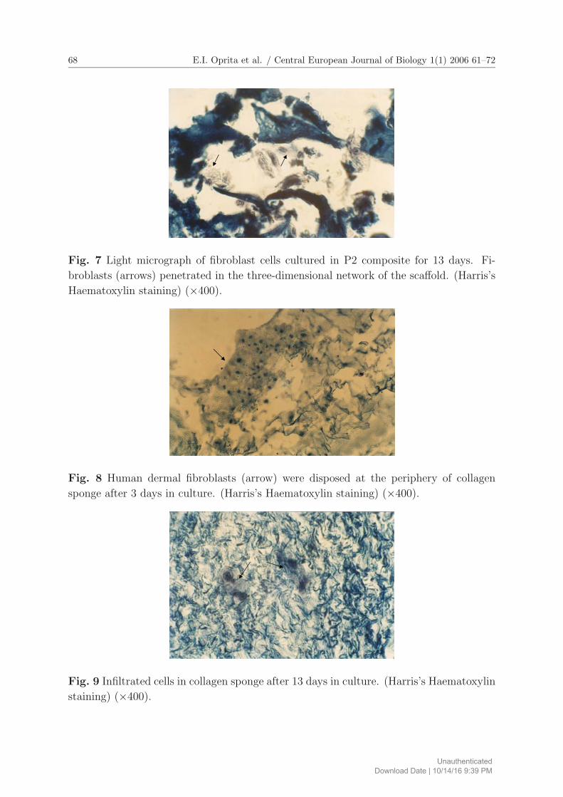
Fig. 7 Light micrograph of fibroblast cells cultured in P2 composite for 13 days. Fi-
broblasts (arrows) penetrated in the three-dimensional network of the scaffold. (Harris’s
Haematoxylin staining) (×400).
Fig. 8 Human dermal fibroblasts (arrow) were disposed at the periphery of collagen
sponge after 3 days in culture. (Harris’s Haematoxylin staining) (×400).
Fig. 9 Infiltrated cells in collagen sponge after 13 days in culture. (Harris’s Haematoxylin
staining) (×400).
UnauthenticatedDownload Date | 10/14/16 9:39 PM

E.I. Oprita et al. / Central European Journal of Biology 1(1) 2006 61–72 69
0
10
20
30
40
50
60
70
80
90
100
110
24 h 72 h
viab
ility
(%)
Control
P1 (COL/TCP=1:1)
P2 (COL/TCP=1:2)
Fig. 10 Diagram of cell viability in the case of fibroblasts cultivation in the presence
of COL/β-TCP scaffolds. Data (obtained by MTT method) are presented as the per-
centage of viable cells relative to the control, considered 100 %. The values are mean of
quadruplicate determination ± SD.
4 Discussion
A series of composite materials based on collagen and calcium salts have been designed,
and one has become available for medical use [15, 16].
Healos, launched in Europe in 2000 for medical use, is a sponge consisting of type I
collagen covered by hydroxyapatite [17].
The main criteria in obtaining collagen from animal sources is the manner in which
the soluble phase is reconstructed. By comparing the average molecular weights of the
extracted collagen with that of tropocollagen (300.000 Da), we were able to observe that
our product had a molecular weight close to that value. This showed that the enzymatic
process employed did not affect the triple helix structure of collagen.
Ozawa et al. demonstrated that β-TCP was an excellent material that induced early
bone formation. During the bone remodeling process, β-TCP itself gradually degraded
and was finally replaced by mature new bone in animal experiments [18]. The resorption
rate of β-TCP is higher, as compared to α-TCP, and to highly cristalline, sintered HA [19].
Renooji et al. demonstrated that sintered β-TCP was subject to biodegradation, whereas
hydroxyapatite was not affected by bioresorption processes 50 weeks after implantation in
dog [20]. They concluded that superior bone formation took place in the grafted β-TCP
comparable to the grafted HA.
The bioresorption properties of β-TCP make it advantageous over using hydroxyap-
atite materials, which appear to remain unremodeled even after long periods of implan-
tation.
Considering the fact that a material similar in composition and structure to native
bone can be a good implant material, the COL/β-TCP association is an appropriate
UnauthenticatedDownload Date | 10/14/16 9:39 PM

70 E.I. Oprita et al. / Central European Journal of Biology 1(1) 2006 61–72
combination for the in vitro bone formation and in vivo bone regeneration. Our study
established a method to synthesize two variants of composite materials, with a COL:β-
TCP weight ratio of 1:1 and 1:2, respectively.
In tissue engineering, it is important to develop new biomaterials suitable for cell
cultivation [21]. In our study, the biocompatibility of collagen-phosphate composites (P1
and P2) was tested on human dermal fibroblast culture, assessing cell morphology and
viability.
Histological examination clearly indicated that both variants of composites (P1 and
P2) discussed in this paper exhibited good cellular proliferation and attachment into the
sponges.
Other experiments using human osteoblast culture for demonstrating an osteoconduc-
tive effect (ALP synthesis, type I collagen synthesis, osteocalcin secretion) are a prereq-
uisite for clinical application (data not shown).
Further studies are necessary to investigate the role of different compounds, such
as bone morphogenetic proteins (BMPs) in improving our scaffolds’s properties in bone
regeneration.
We conclude that the association of COL with β-TCP determined stable composites,
which prevented β-TCP dispersion in media, resulting in an easily molded biomaterial.
These biocomposites presented a microporous structure with small aggregates of β-TCP
non-homogeneous deposited on the COL fibrous bundles, and they could be used as
scaffolds for cell proliferation and migration.
Acknowledgment
This research was supported by Project No. 269 VIASAN.
References
[1] M. Wirdmann-Al-Adham, R. Gutwald, G. Lauer, U. Huber and R. Schmelzeisen:
“How to optimize seeding and culturing of human osteoblast-like cells on various
biomaterials”, Biomaterials, Vol. 23, (2002), pp. 3319–3328.
[2] F.R.A.J. Rose and R.O.C. Oreffo: “Bone Tissue Engineering: Hope vs Hype”,
Biochem. Biophys. Res. Commun., Vol. 292, (2002), pp. 1–7.
[3] D. Gotora and J.T. Czernuszka: “Mineralisation and cross-Linking Techniques to
Improve the Mechanical Properties of Collagen-Calcium Phosphate (Coll-Cap) Com-
posites to be used as Bone Analoques”, Eur. Cell Mater., Vol. 7(1), (2004), p. 55.
[4] W. Suchanek and M. Yoshimura: “Processing and properties of hydroxiapatite-based
biomaterials for use as hard tissue replecement implants”, J. Mater. Res., Vol. 13(1),
(1998), pp. 94–119.
[5] A. Sihna, A. Ingle, K.R. Munim, S.N. Vaidya, B.P. Sharma and A.N. Bhisey: “De-
velopment of calcium phosphate based bioceramics”, Bull. Mater. Sci., Vol. 24(6),
(2001), pp. 653–657.
UnauthenticatedDownload Date | 10/14/16 9:39 PM
E.I. Oprita et al. / Central European Journal of Biology 1(1) 2006 61–72 71
[6] A.R. Vaccaro: “The Role of the Osteoconductive Scaffold in Synthetic Bone Graft”,
Orthopedics, Vol. 25(5), (2002), pp. 571–578.
[7] V. Vicente, L. Meseguer, F. Martinez, A. Galian, J. Rodriguez, M. Alcaraz and M.
Clavel: “Ultrastructural study of the osteointegration of bioceramics (whitlockite
and composite beta-TCP-collagen) in rabbit bone”, Ultrastruct. Pathol., Vol. 20(2),
(1996), pp. 179–188.
[8] G. Negroiu, L. Moldovan, M. Caloianu, N. Mirancea and D. Mirancea: “Collagen-
chondroitin sulfate substrates conditioned as sponges and membranes”, Rev. Roum.
Biochem., Vol. 29(1), (1992), pp. 23–28.
[9] L.A. Elson and W.T.J. Morgan: “Determination of total hexosamines”, Meth. Enzy-
mol., Vol. 8, (1966), pp. 20–24.
[10] G. Chirita and M. Chirita: Collagen in animal skin, Editura Tehnica, Bucharest,
1993, p. 268 (in Romanian).
[11] L. Moldovan, E.I. Oprita, O. Craciunescu, C. Tardei, D. Bojin and O. Zarnescu:
“Histochemical and scanning electron microscopic characterization of tricalcium
phosphate-collagen conjugated sponges”, Roumanian Biotech. Lett., Vol. 9(5),
(2004), pp. 1887–1893.
[12] K de Groot: “Bioceramics consisting of calcium phosphate salts”, Biomaterials, Vol.
1, (1980), pp. 47–50.
[13] T. Mosmann: “Rapid colorimetric assay for cellular growth and survival: application
to proliferation and cytotoxic assays”, J. Immunol. Methods, Vol. 65, (1983), pp.
55–63.
[14] Y. Liu, D.A. Peterson, H. Kimura and D. Schubert: “Mechanism of cellular 3-(4,5-
dimethylthiazol-2-yl)-2, 5-diphenyltetrazolium bromide (MTT) reduction”, J. Neu-
rochem., Vol. 69, (1997), pp. 581–593.
[15] A. John, L. Hong, Y. Ikada and Y. Tabata: “A trial to prepare biodegradable
collagen-hydroxiapatite composite for bone repair”, J. Biomater. Sci. Polym. Ed.,
Vol. 12(6), (2001), pp. 689–705.
[16] S.S. Liao, F.Z. Cui, W. Zhang and Q.L. Feng: “Hierarchically biomimetic bone
scaffolds materials:nano HA/collagen/PLA composite”, J. Biomed. Mater. Res. B.
Appl. Biomater., Vol. 69(2), (2004), pp. 158–165.
[17] S.H. Parkish: “Bone Graft Substitutes: Past, Present, Future”, J. Postgrad. Med.,
Vol. 48(2), (2002), pp. 142–148.
[18] M. Ozawa, K. Tanaka, S. Morikawa, M. Chazono and K. Fujii: “Clinical study of
the pure βttricalcium phosphate – Reports of 167 cases”, J. East. JPN Orthop.
Traumatol., Vol. 12, (2000), pp. 409–413.
[19] E. Fernandez, F.J. Gil, M.P. Ginebra, F.C.M. Driessens, J.A. Planell and S.M. Best:
“Calcium phosphate bone cements for clinical applications. Part I: solution chem-
istry”, J. Mater. Sci. Mater. Med., Vol. 10, (1999), pp. 169–176.
[20] W. Renooji, A. Hoogendoorn, W.J. Visser, R.H.F. Lentferkink, M.G.J. Schimitz,
H.V. Ieperen, S.J. Oldenburg, W.M. Janssen, L.M.A. Akkermans and P. Wittebol:
“Bioresoption of ceramic strontium-85-labeled calcium phosphate implants in dog
UnauthenticatedDownload Date | 10/14/16 9:39 PM

72 E.I. Oprita et al. / Central European Journal of Biology 1(1) 2006 61–72
femora”, Clin. Orthop., Vol. 197, (1985), pp. 272–285.
[21] N. Kotobuki, K. Ioku, D. Kawagoe, H. Fujimori, S. Goto and H. Ohgushi: “Ob-
servation of osteogenic differentiation cascade of living mesenchimal stem cells on
transparent hydroxyapatite ceramics”, Biomaterials, Vol. 26, (2005), pp. 779–785.
UnauthenticatedDownload Date | 10/14/16 9:39 PM