non-migratory bioactive packaging: covalent attachment of ...
144
NON-MIGRATORY BIOACTIVE PACKAGING: COVALENT ATTACHMENT OF BIOACTIVE PEPTIDES TO POLY(ETHYLENE) FOOD PACKAGING FILMS A Dissertation Presented to the Faculty of the Graduate School of Cornell University in Partial Fulfillment of the Requirements for the Degree of Doctor of Philosophy by Matthew David Steven May 2004
-
Upload
khangminh22 -
Category
Documents
-
view
0 -
download
0
Transcript of non-migratory bioactive packaging: covalent attachment of ...
NON-MIGRATORY BIOACTIVE PACKAGING:
COVALENT ATTACHMENT OF BIOACTIVE
PEPTIDES TO POLY(ETHYLENE) FOOD PACKAGING
FILMS
A Dissertation
Presented to the Faculty of the Graduate School
of Cornell University
in Partial Fulfillment of the Requirements for the Degree of
Doctor of Philosophy
by
Matthew David Steven
May 2004

© 2004 Matthew David Steven
ALL RIGHTS RESERVED
NON-MIGRATORY BIOACTIVE PACKAGING:
COVALENT ATTACHMENT OF BIOACTIVE PEPTIDES TO
POLY(ETHYLENE) FOOD PACKAGING FILMS
Matthew David Steven, Ph.D.
Cornell University 2004
Non-migratory bioactive packaging is a novel form of active packaging. It is pack-
aging which elicits a desirable biological response from food systems without the
active component migrating from the packaging into the food. Possible applications
for this include in-package enzymatic processing and non-migratory antimicrobial
packaging. Poly(ethylene glycol) (PEG) oligomers were covalently attached by
one end to surface-oxidized poly(ethylene) (PE) films using carbodiimide coupling;
bioactive peptides were covalently linked to the free terminus of the PEG chains
using the same reaction. Reactions were confirmed and monitored through contact
angle measurements, dye adsorption assays, x-ray photoelectron spectroscopy and
atomic force microscopy.
Bioactivity of the modified films depended on the peptide attached: antibac-
terial activity was found for films treated with antimicrobial peptide E14LKK; no
activity was found for lactase attached to PE films. Antibacterial activity was
demonstrated against E.coli for films to which side-chain-protected E14LKK was
attached and then deprotected.
Further investigation of the lactase coupling indicated difficulties with amino-
carboxy-PEG attachment, despite prior successes with diamino-PEG; amino-
carboxy-PEG coupling requires further optimization. Lactase attached directly
to PE was inactive, regardless of lactose inclusion in the coupling buffer, how-
ever PEGylated lactase is active: the inclusion of a PEG spacer should improve
immobilized lactase activity.
The effect of the modifications on PE properties important in packaging was
assessed through physical, mechanical and optical testing. Properties primarily
dependent on the bulk characteristics of the material were generally unaffected by
our film modifications. Properties sensitive to film surface characteristics (perme-
ability, optical properties, friction) were affected by the modifications; the changes
in these properties will need to be allowed for when designing processes and pack-
ages involving the modified films, but should not affect the utility of the film for
the standard applications of PE.
BIOGRAPHICAL SKETCH
Matthew was born, raised and educated in New Zealand prior to arriving at Cor-
nell. He received a Bachelor of Technology (Food Technology) with First Class
Honours from Massey University in May 1997, graduating top of his class. Moving
to Ithaca in August 1997 after serving three months as an Assistant Lecturer at
Massey, Matthew entered Cornell and commenced the studies leading, eventually,
to this document. Returning to New Zealand in January 2001 to marry Joanne
(nee Cuthbert), the pair returned to Ithaca together. An avid telemark skiier,
whitewater canoeist and general outdoors freak, Matthew is well known for his
enthusiasm and involvement in the outdoors community at Cornell and is looking
forward to finding some real mountains again in the near future.
iii

To Jill and David Steven, for a lifetime’s worth of guidance and support.
To Jo, for being there, keeping me going and getting me out.
iv
ACKNOWLEDGEMENTS
In any advanced degree there are numerous people who assist in various ways; it
is never possible to list them all. I would like to thank all those of you who have
assisted in my research. I truly appreciate the time and effort you have put into
assisting me. I would also like to thank all those who have made my time here
in Ithaca enjoyable: friends, colleagues and fellow outdoor nuts. You have been a
huge part of my life and have helped me stay sane when the going got tough and
the tough wanted to run like hell. There’s not much to say, but thanks.
v
TABLE OF CONTENTS
1 Introduction: Non-migratory Bioactive Packaging 11.1 Non-migratory bioactive polymers: benefits and limitations . . . . . 3
1.1.1 Benefits . . . . . . . . . . . . . . . . . . . . . . . . . . . . . 31.1.2 Limitations . . . . . . . . . . . . . . . . . . . . . . . . . . . 10
1.2 Types of Non-Migratory Bioactive Polymers . . . . . . . . . . . . . 141.2.1 Inherently bioactive synthetic polymers . . . . . . . . . . . . 141.2.2 Polymers containing entrapped bioactive compounds . . . . 181.2.3 Polymers with covalently immobilized bioactive compounds . 19
1.3 Applications of NMBP with immobilized bioactive agents . . . . . . 271.3.1 In-package processing . . . . . . . . . . . . . . . . . . . . . . 281.3.2 Antimicrobial packaging and shelf life extension . . . . . . . 301.3.3 Intelligent Packaging . . . . . . . . . . . . . . . . . . . . . . 35
2 Research Objectives 37
3 Materials and Methods 383.1 Materials . . . . . . . . . . . . . . . . . . . . . . . . . . . . . . . . 383.2 Film Preparation . . . . . . . . . . . . . . . . . . . . . . . . . . . . 40
3.2.1 Heat-Pressing LDPE Films . . . . . . . . . . . . . . . . . . . 403.2.2 Film Cleaning . . . . . . . . . . . . . . . . . . . . . . . . . . 40
3.3 Film Modification . . . . . . . . . . . . . . . . . . . . . . . . . . . . 413.3.1 Surface Oxidation of PE film using Chromic Acid . . . . . . 413.3.2 PEG attachment to PE-COOH using WSC Coupling . . . . 413.3.3 Lactase attachment to PE-PEG-COOH and PE-COOH . . . 433.3.4 E14LKK attachment to PE-PEG-COOH and PE-COOH . . 443.3.5 Deprotection of Immobilized Peptide E14LKK . . . . . . . . 45
3.4 Film Surface Analyses . . . . . . . . . . . . . . . . . . . . . . . . . 453.4.1 Contact Angle Measurement . . . . . . . . . . . . . . . . . . 453.4.2 Dye Adsorption Assays for Surface Chemistry . . . . . . . . 463.4.3 Instrumental Analysis of Film Surfaces . . . . . . . . . . . . 48
3.5 Functional Testing of Modified Films . . . . . . . . . . . . . . . . . 493.5.1 Water Vapor Transmission Rate . . . . . . . . . . . . . . . . 493.5.2 Tensile Testing . . . . . . . . . . . . . . . . . . . . . . . . . 503.5.3 Tear Propagation Resistance . . . . . . . . . . . . . . . . . . 503.5.4 Evaluation of Film Friction Coefficients . . . . . . . . . . . . 513.5.5 Transition Temperature Determination . . . . . . . . . . . . 513.5.6 Surface Gloss Evaluation . . . . . . . . . . . . . . . . . . . . 523.5.7 Film Transmittance and Haze . . . . . . . . . . . . . . . . . 52
3.6 Bioactivity Evaluations of Modified Films . . . . . . . . . . . . . . 533.6.1 Lactase activity assay . . . . . . . . . . . . . . . . . . . . . 533.6.2 Antimicrobial Efficacy Evaluations . . . . . . . . . . . . . . 53
3.7 Statistical Analysis . . . . . . . . . . . . . . . . . . . . . . . . . . . 55
vi

4 Film Modification 564.1 Film Preparation . . . . . . . . . . . . . . . . . . . . . . . . . . . . 564.2 Oxidation . . . . . . . . . . . . . . . . . . . . . . . . . . . . . . . . 574.3 PEGylation . . . . . . . . . . . . . . . . . . . . . . . . . . . . . . . 634.4 Washing . . . . . . . . . . . . . . . . . . . . . . . . . . . . . . . . . 644.5 Lactase Attachment . . . . . . . . . . . . . . . . . . . . . . . . . . . 674.6 Surface Topography . . . . . . . . . . . . . . . . . . . . . . . . . . . 674.7 Summary of Findings and Recommendations . . . . . . . . . . . . . 75
5 Bioactivity of Modified Films 765.1 Antimicrobial Peptide Modified Films . . . . . . . . . . . . . . . . . 76
5.1.1 Antimicrobial Assays . . . . . . . . . . . . . . . . . . . . . . 775.2 Lactase Modified Films . . . . . . . . . . . . . . . . . . . . . . . . . 805.3 Summary of Findings and Recommendations . . . . . . . . . . . . . 85
6 Effect of Modifications on Film Properties 866.1 Physical and Mechanical Properties . . . . . . . . . . . . . . . . . . 88
6.1.1 Transition Temperatures . . . . . . . . . . . . . . . . . . . . 886.1.2 Tensile Properties . . . . . . . . . . . . . . . . . . . . . . . . 916.1.3 Coefficients of Friction . . . . . . . . . . . . . . . . . . . . . 97
6.2 Optical Properties . . . . . . . . . . . . . . . . . . . . . . . . . . . 996.3 Moisture Permeability . . . . . . . . . . . . . . . . . . . . . . . . . 1036.4 Summary of Findings and Recommendations . . . . . . . . . . . . . 107
7 Conclusions and Recommendations 109
vii
LIST OF TABLES
4.1 Reference contact angle data . . . . . . . . . . . . . . . . . . . . . 584.2 Dye absorbance results: functional group quantification . . . . . . 614.3 Elemental composition of LDPE film surfaces . . . . . . . . . . . . 62
5.1 Antimicrobial activity of the E14LKK modified films . . . . . . . . 785.2 Lactase activity of Lactase modified LDPE films. . . . . . . . . . . 825.3 Surface composition of Lactase modified LDPE films. . . . . . . . . 83
6.1 Surface-Sensitive and Surface-Insensitive film properties . . . . . . 876.2 Reference transition temperature data . . . . . . . . . . . . . . . . 896.3 Reference data for the properties of LDPE, HDPE and PEG films. 936.4 Comparison of contact angles and WVTRs . . . . . . . . . . . . . 1046.5 Reference WVTR data for LDPE. . . . . . . . . . . . . . . . . . . 1046.6 Summary of film property data of the modified films . . . . . . . . 108
viii
LIST OF FIGURES
1.1 Non-migratory vs. migratory bioactive packaging . . . . . . . . . . 31.2 Activity of PEG-conjugated chymotrypsin and native chymotrypsin 51.3 pH stability and polymer conjugation . . . . . . . . . . . . . . . . 61.4 Bactericidal activity of hydantoins . . . . . . . . . . . . . . . . . . 171.5 In-package enzyme processing: immobilized cholesterol reductase . 29
3.1 Film modification protocol . . . . . . . . . . . . . . . . . . . . . . 423.2 Determining contact angle of droplet . . . . . . . . . . . . . . . . . 453.3 Tear patterns produced in the tear propagation experiment. . . . . 513.4 DSC Temperature Program for Melting Point Determination . . . 52
4.1 Contact angles of modified LDPE and HDPE films . . . . . . . . . 574.2 Contact angle variation with pH . . . . . . . . . . . . . . . . . . . 604.3 Adsorption of PEG by HDPE and LDPE . . . . . . . . . . . . . . 654.4 Comparison of coupling and washing methods for HDPE films. . . 664.5 Surface topography of modified and unmodified LDPE films . . . . 684.6 Surface topography of modified and unmodified HDPE films . . . . 71
6.1 Typical DSC melting-point determination curve. . . . . . . . . . . 906.2 Tensile properties of modified and unmodified films. . . . . . . . . 926.3 Tear propagation force for modified and unmodified films. . . . . . 946.4 A typical tensile test force-distance curve for HDPE. . . . . . . . . 956.5 Coefficients of friction for modified and unmodified LDPE films. . . 986.6 Optical transmittance of modified and unmodified films. . . . . . . 1006.7 Gloss of modified and unmodified films. . . . . . . . . . . . . . . . 101
ix
Chapter 1
Introduction: Non-migratory Bioactive
Packaging
Food is often prepared a significant time before, and distance away from, where
it is consumed (Branen, 1993). The basic function of food processing and pack-
aging is to preserve the food between preparation (production) and consumption,
maintaining a safe, nutritious food supply. Antimicrobial agents (preservatives)
are an important part of this preservation process and have long been paired with
packaging systems to extend the shelf life of foods. Preservatives and packaging
systems have become more intertwined as technology has progressed; similarly, the
processing and packaging of foods has become more intertwined. Non-migratory
bioactive packaging can be viewed as the end point of this evolution, the combi-
nation of processing functions (in-package enzymatic processing) or preservatives
(non-migratory antimicrobial packaging) with the packaging function in a single
material.
Non-migratory bioactive polymers (NMBP) are a class of polymers that possess
biological activity without the active components migrating from the polymer to
the substrate. This concept has primarily been applied to immobilized enzyme
processing to date (Bachler et al., 1970; Brody and Budny, 1995; Katchalski-Katzir,
1993; Mosbach, 1980; Ikada, 1994) but is now becoming of interest in packaging
applications (Appendini and Hotchkiss, 1997; Soares, 1998).
Bioactive materials are based on molecules that elicit a response from living
systems. The goal of NMBP is to create bioactive packaging for which the re-
1
2
sponse is desirable for either the package or the product, for example inhibition of
microbial growth or flavor improvement. Enzymes are classic examples of bioactive
substances, as are many peptides, proteins, and other organic compounds. A func-
tional definition of bioactive has been adopted, based upon the way the substance
interacts with living systems. Purely physical processes, for example adsorption
or diffusion, are excluded. Bioactive polymers can be formed by attachment of
bioactive molecules to synthetic polymers, as in the case of enzyme immobilization
(Appendini and Hotchkiss, 1997; Soares, 1998), or may result from an inherent
bioactive effect of the polymer structure, as with chitosan (Collins-Thompson and
Cheng-An, 2000; Tanabe et al., 2002). They have potential applications in the
packaging of food and other biological materials, in food processing equipment, on
biomedical devices (Sodhi et al., 2001; Sun and Sun, 2002) and in textiles (Edwards
and Vigo, 2001; Sun and Sun, 2002).
Non-migratory polymers are defined as those for which the bioactive component
does not migrate out of the polymer system into the surrounding medium (see
Figure 1.1). Migration, at its most basic level, is mass transfer of the bioactive
compound from the packaging material into the packaged product. Migration
can be prevented by covalent attachment of the active component to the polymer
backbone, by entrapment of the active component within the polymer matrix, or
by using inherently bioactive polymers.
3
Food
A B
Food
Figure 1.1: Non-migratory (A) vs. migratory (B) bioactive packaging.Adapted from Han (2000).
1.1 Non-migratory bioactive polymers: benefits and
limitations
In order for a new technology to be considered, it needs to have advantages over
the status quo. Typically, these advantages come with limitations, in application
or utility, and frequently with an increase in cost. Benefits and limitations may be
generic to all NMBP or specific to one type of NMBP.
1.1.1 Benefits
The benefits of NMBP can be divided into four main areas: technical benefits,
regulatory advantages, marketing aspects and the food processor’s perspective.
This list is not exhaustive: particular applications will involve some, or all, of
these plus other considerations specific to the application.
Technical benefits
The technical benefits of NMBP include improved stability of the bioactive sub-
stance and concentration of the activity at a specific locus. Improved stability is a

4
consideration for covalently immobilized bioactive substances; biological molecules,
e.g. enzymes, are typically very sensitive to environmental conditions. They are
readily denatured by some solvents, by high, and in some cases low, tempera-
tures; by high pressures, high shear or ionizing radiation; by certain pHs or in the
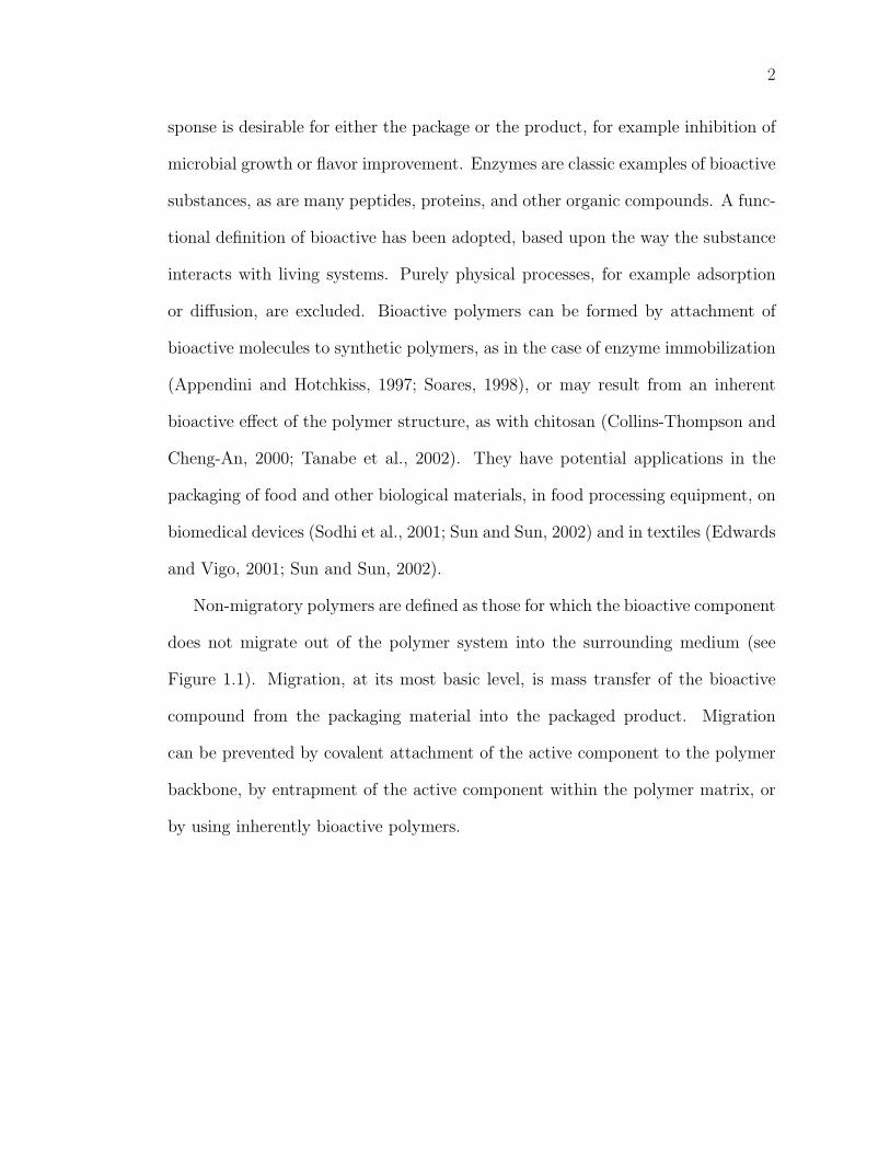
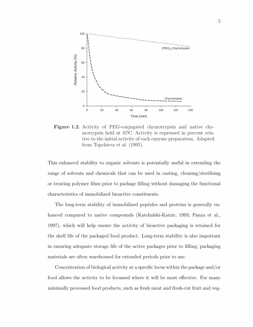
presence of high electrolyte concentrations (Richardson and Hyslop, 1985). Conju-
gation to polymer supports has been shown to dramatically enhance the stability of
these molecules. Topchieva and colleagues (Topchieva et al., 1995) demonstrated
improved thermal stability of chymotrypsin when conjugated to poly(ethylene gly-
col) (PEG) (Figure 1.2). Appendini and Hotchkiss (2001) similarly demonstrated
improved thermal stability of a small antimicrobial peptide when covalently at-
tached to a PEG-grafted poly(styrene) (PS) support. The immobilized peptide
remained active when dry-heated to 200� for 30 minutes and when autoclaved at
121� for 15 minutes. Polymers are often processed at temperatures that would
denature native proteins; improving the thermal stability of peptides is important
for their inclusion in packaging materials.
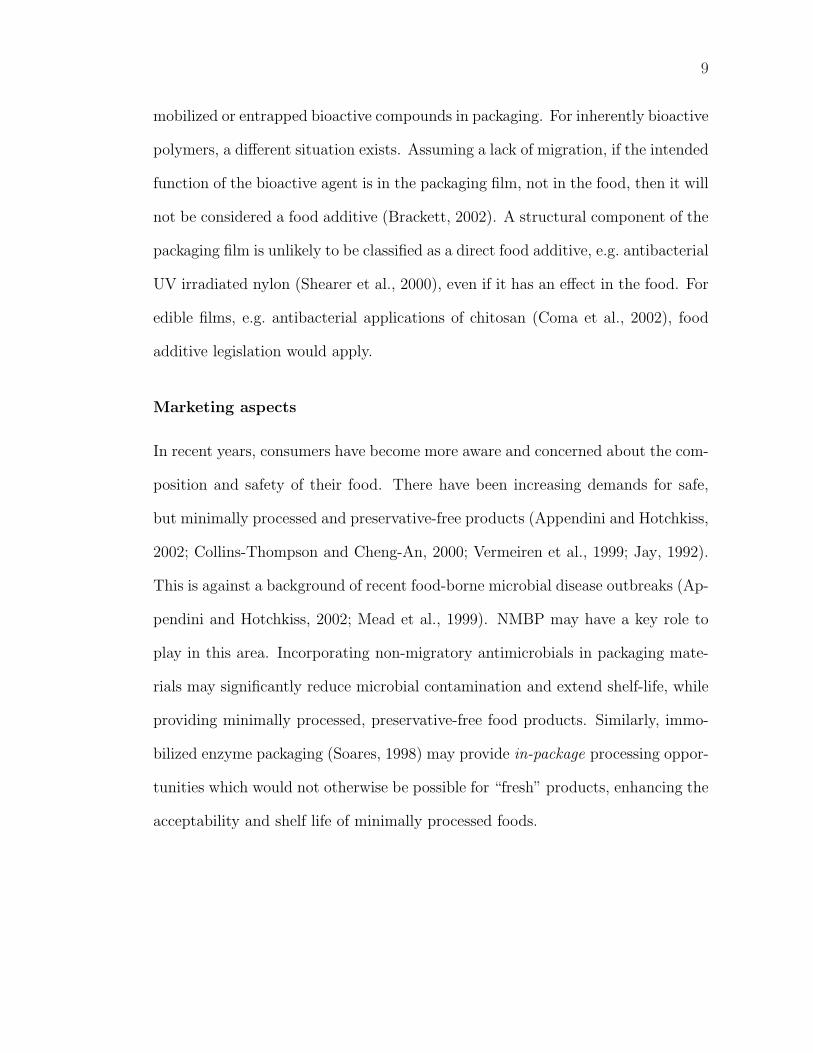
Appendini (1999) also demonstrated the improved activity of the conjugated
peptide over a range of pHs (Figure 1.3). Other authors have also reported im-
proved stability of polymer-conjugated enzymes to pH and temperature (Gaertner
and Puigserver, 1992; Yang et al., 1996, 1995a,b; Zaks and Klibanov, 1984; Chen
et al., 2000). The extended range of pH stability may provide activity in a broader
range of food products than would be the case for the native compound.
The stability of proteins to inimical media, such as organic solvents, supercrit-
ical fluids and gases, may also be improved by polymer conjugation, and applica-
tions have developed to exploit this in non-aqueous enzymology (Mabrouk, 1997;
Panza et al., 1997; Veronese, 2001; Yang et al., 1995a,b; Zaks and Klibanov, 1984).
5
Time (min)
0 20 40 60 80 100 120 140
Rel
ativ
e A
ctiv
ity (
%)
0
20
40
60
80
100
(PEG)9-Chymotrypsin
Chymotrypsin
Figure 1.2: Activity of PEG-conjugated chymotrypsin and native chy-motrypsin held at 45�. Activity is expressed in percent rela-tive to the initial activity of each enzyme preparation. Adaptedfrom Topchieva et al. (1995).
This enhanced stability to organic solvents is potentially useful in extending the
range of solvents and chemicals that can be used in casting, cleaning/sterilising
or treating polymer films prior to package filling without damaging the functional
characteristics of immobilized bioactive constituents.
The long-term stability of immobilized peptides and proteins is generally en-
hanced compared to native compounds (Katchalski-Katzir, 1993; Panza et al.,
1997), which will help ensure the activity of bioactive packaging is retained for
the shelf life of the packaged food product. Long-term stability is also important
in ensuring adequate storage life of the active packages prior to filling; packaging
materials are often warehoused for extended periods prior to use.
Concentration of biological activity at a specific locus within the package and/or
food allows the activity to be focussed where it will be most effective. For many
minimally processed food products, such as fresh meat and fresh-cut fruit and veg-
6
pH
3.5 4.0 4.5 5.0 5.5 6.0 6.5 7.0
Log 1
0 C
FU
/ml
0
2
4
6
8
Peptide PS-PEG-Peptide
Innoculum Level
Figure 1.3: E.coli 0157:H7 survival in the presence of a small syntheticpeptide in 0.1M citrate buffer across a range of pHs. Activityis shown for the native peptide (�) and peptide attached toa PS surface through a PEG spacer (�). Equivalent peptideconcentrations were used in each determination. Adapted fromAppendini (1999).
etables, the majority of microbial spoilage occurs on the product surface (Collins-
Thompson and Cheng-An, 2000; Hotchkiss, 1995). Concentrating antimicrobials
on the surface of the product, as with antimicrobial packaging, allows minimal
amounts of the active compounds to be used to maximal effect.
Regulatory advantages
Regulations relating to active food packaging are still evolving. As new technologies
develop, regulations must be modified to encompass them.
As noted by various authors (de Kruijf et al., 2002; de Kruijf and Rijk, 2003;
Meroni, 2000; Vermeiren et al., 2002, 1999), there are currently no specific EU
regulations for active or intelligent packaging. Rather, these packaging systems are
subject to the same regulations as traditional packaging. These regulations require
7
that all components used to manufacture food contact materials be on “positive
lists,” set down migration limits for both overall migration and migration of specific
components, and require that the packaging does not affect the composition of
the packaged food. The migration requirements should not be problematic for
NMBP, although a lack of migration will need to be established as detailed in the
appropriate regulations. The compounds used to manufacture NMBP will need to
be included on the relevant positive lists, however, which may be an issue for some
systems. Additionally, the entire purpose of some active packaging is to modify
the composition of the food, which directly contravenes the legislation. The key
Directive (regulation) of concern is 89/109/EEC.
An amendment was proposed to 89/109/EEC on November 17, 2003 specifi-
cally to deal with active and intelligent packaging (Anon, 2003a). This amendment
allows for the use of packaging materials that change the composition or organolep-
tic properties of food contained within them, provided the changes comply with
other EU regulations applicable to food (Anon, 2003b); the regulation clearly states
that these changes must in no way mislead consumers. 2003/0272(COD) allows
the specification of both overall and specific migration limits for active and intel-
ligent packaging separate from those set for traditional packaging. It is expected
that this regulation will be voted into effect during 2004.
In the United States, regulations relating to food contact materials can be found
in the Code of Federal Regulations (CFR) Title 21 Parts 170 through 190 (Anon,
2002a). The regulations revolve around determining whether the compounds in the
packaging materials may be considered food additives. Food additives are defined
as substances “the intended use of which results or may reasonably be expected
to result, directly or indirectly, either in their becoming a direct component of
8
food or otherwise affecting the characteristics of food”. Further, “If there is no
migration of a packaging component from the package to the food, it does not
become a component of the food and thus is not a food additive” unless it is used
“to give a different flavor, texture of other characteristic in the food”, in which
case it “may” be a food additive (21 CFR §170.3 (e) (1)). The regulations also
establish guidelines for determining limits below which migration can be considered
negligible, negating food additive classification of that substance in that specific
application.
These regulations can be interpreted as saying that any substance for which
it can be shown that there is negligible migration into a food product is not a
food additive (21 CFR §170.39). This would imply that NMBP needs to meet the
regulations required of items for food contact use, but do not need to meet the
more stringent food additive regulations, provided a lack of migration is proven.
However, additive classification may also depend on the intended function of the
component. If it is intended that a packaging component be active in the food
product, as with an immobilized antimicrobial intended to extend the shelf life of a
packaged food, then that component might be classified as a Direct Food Additive
and would be required to comply with the food additive regulations (Brackett,
2002). There is, therefore, some ambiguity as to the status of NMBP materials. If
the active components of NMBP are not considered food additives, then it could
be a significant advantage, allowing the use of substances not currently permitted
as food additives in packaging systems, provided that there is negligible migration.
The process of obtaining food contact approval for packaging materials has been
recently reviewed (Heckman and Ziffer, 2001).
The above discussion on US regulations focuses on the issues of covalently im-
9
mobilized or entrapped bioactive compounds in packaging. For inherently bioactive
polymers, a different situation exists. Assuming a lack of migration, if the intended
function of the bioactive agent is in the packaging film, not in the food, then it will
not be considered a food additive (Brackett, 2002). A structural component of the
packaging film is unlikely to be classified as a direct food additive, e.g. antibacterial
UV irradiated nylon (Shearer et al., 2000), even if it has an effect in the food. For
edible films, e.g. antibacterial applications of chitosan (Coma et al., 2002), food
additive legislation would apply.
Marketing aspects
In recent years, consumers have become more aware and concerned about the com-
position and safety of their food. There have been increasing demands for safe,
but minimally processed and preservative-free products (Appendini and Hotchkiss,
2002; Collins-Thompson and Cheng-An, 2000; Vermeiren et al., 1999; Jay, 1992).
This is against a background of recent food-borne microbial disease outbreaks (Ap-
pendini and Hotchkiss, 2002; Mead et al., 1999). NMBP may have a key role to
play in this area. Incorporating non-migratory antimicrobials in packaging mate-
rials may significantly reduce microbial contamination and extend shelf-life, while
providing minimally processed, preservative-free food products. Similarly, immo-
bilized enzyme packaging (Soares, 1998) may provide in-package processing oppor-
tunities which would not otherwise be possible for “fresh” products, enhancing the
acceptability and shelf life of minimally processed foods.
10
The food processor’s perspective
From the perspective of the food processor, NMBP could have several advantages.
A general benefit would be increased shelf life, but certain NMBP technologies and
applications may offer specific benefits. As an example, consider the production
of lactose free milk: the demand for this product is not high, although there is a
definite place for it in the market; it sells at a high price due to the high cost of pro-
duction and the low sales volume. Using lactase-active packaging, however, milk
could be packed off a normal production run and a lactose reduced product ob-
tained after a short period of storage. A migratory enzyme, or the direct addition
of lactase to milk, cannot be used in this application due to post-pasteurization
restrictions in the pasteurized milk ordinance (Anon, 1999) and the heat lability
of lactase. Similarly, for other products some of the processing may be accom-
plished in package, instead of in the processing plant, reducing processing costs
and increasing flexibility for the food processor.
1.1.2 Limitations
As noted above, any new technology must have benefits over current technologies
in order to be successful, but these benefits typically come with limitations. The
limitations of NMBP may include: a limited locus of activity, specific requirements
on the mechanism of activity of the active agent, reduced activity, availability of
appropriate technology and an increase in packaging cost.
Limited locus of activity
One possible limitation of NMBP is the need for the reaction constituents to be
transported to the package-product interface. This limits the function to areas in
11
intimate contact with the packaging material for solid and viscous liquid foods.
For low viscosity foods agitation during distribution will mix the product, so this
less problematic. For viscous liquids the high viscosity limits fluid mixing during
distribution; diffusive mixing is also limited. For solid products, diffusive migration
will be inadequate to provide the desired effect throughout the product. Even
when the product surface is the target, the need for intimate contact with the
packaging material may prevent action of the active agent within crevices and
folds on the item surface, although this can be alleviated somewhat by careful
package design and vacuum packaging. It should be noted that migratory active
packaging technologies are often similarly limited in their diffusion and mixing
requirements.
Mechanisms of action
In order for a bioactive agent to be active when covalently anchored to a packaging
material, the conformation of the immobilized active component (compared to the
free form), the location of the covalent link to the polymer, and the mechanism
by which the agent interacts with the environment must all be considered. If,
for example, an antimicrobial agent must enter the microbial cell to be effective,
then it is unlikely to be active in a tethered state, whereas an antimicrobial agent
active at the cell surface may remain active when tethered. If attachment causes
conformational changes in the bioactive compound, or an active site is altered,
then activity will be disrupted. Consider also the attachment of an enzyme that
requires a co-enzyme for activity. If this coenzyme is not present in the food or
otherwise attached along with the primary enzyme, then the primary enzyme will
be inactive. Understanding the mechanism of the active agent is key in developing
12
NMBP.
Reduced activity
One concern in immobilizing bioactive compounds is the potential for loss of
activity. In many cases, activity is reduced compared to the native compound
(Katchalski-Katzir, 1993) and may be lost completely. With appropriate coupling
methodology, however, activity can normally be retained, albeit at a lower level
than for the free compound. Appendini (1999) compared the activity of a small
antimicrobial peptide in solution and immobilized to PEG grafted PS beads and
found that it was 200–7000 times less active when covalently immobilized. It
still possessed significant antimicrobial activity, however, and was effective against
E.coli 0157:H7 at immobilized peptide concentrations of 4 µmol/ml in growth me-
dia.
Soares (1998) found that naringinase retained 23% of its free activity when
immobilized. Soares also found that at pHs less than 3.1, the immobilized naring-
inase was more active than free naringinase. This often occurs with immobilized
enzymes — their increased stability leads to higher activity than the free enzyme
under extreme conditions, even when activity is reduced under optimum condi-
tions. Mosbach (1980) also suggested that for sequential enzyme pathways, the
activity of the immobilized enzymes could be higher than that of the enzymes in
free solution if the enzymes were immobilized in close proximity.
Technology availability
The commercial availability of the technology required to produce NMBP could
limit applications. Technologies for basic polymer surface functionalisation are

13
readily available, but newer technologies allowing controlled surface functional-
isation still require development, particularly with regards the high throughput
required for packaging production. Surface functionalisation is discussed in more
detail in §1.2.3.
Other than very basic surface functionalisation, surface modification of pack-
aging films is not practised commercially. Production of most NMBP will require
significant surface modification, probably involving wet chemical treatments for im-
mobilization of active agents; adaptation of existing technologies will be required
to implement these treatments. Over time the technology will become more read-
ily available and this limitation will disappear; these issues regarding commercial
availability of required technology are true for most novel technologies.
Cost
Intensive modifications, such as the attachment of proteins, will incur significant
cost increases because of the additional processing steps required, the chemicals
used in processing, and the high cost of the active agent itself. The peptides,
proteins and enzymes involved are typically very expensive, although increased
demand should result in cost reductions in the long term. The need to recover
research and development expenses and capital equipment costs will further in-
crease finished product costs. Over time, reduced equipment costs and increased
availability, combined with reduced costs for the raw materials, should lead to
the overall cost of the films decreasing. Again, this is the typical cycle for the
introduction of new technologies.
14
1.2 Types of Non-Migratory Bioactive Polymers
1.2.1 Inherently bioactive synthetic polymers
As previously mentioned, there are three main types of NMBP: inherently bioac-
tive polymers, polymers with entrapped bioactives and polymers with covalently
immobilized bioactives. For inherently bioactive polymers, the structural polymer
itself is bioactive. This includes structural polymers with modified backbones, for
example UV irradiated nylon and naturally bioactive materials such as chitosan.
These polymers differ from those with immobilized or entrapped bioactives in that
no previously synthesized bioactive compound is incorporated into the polymer.
Several materials have been found to have inherent bioactivity (Oh et al., 2001;
Ozdemir and Sadikoglu, 1998; Shearer et al., 2000; Vigo, 1999; Vigo and Leonas,
1999) and new ones are underdevelopment (Tew et al., 2002). All the examples of
inherently bioactive polymers to date involve antimicrobial activity.
Chitosan
Chitosan is the possibly the most studied inherently bioactive NMBP (Coma et al.,
2002; Oh et al., 2001; Tanabe et al., 2002), possesses broad spectrum antimicrobial
activity in simple media, and is commercially available as an antifungal coating for
fresh fruit (Appendini and Hotchkiss, 2002; Padgett et al., 1998). Chitosan is the
deacetylated form of chitin (poly-/beta-(1,4)-N-acetyl-D-glucosamine), a common
natural biopolymer extracted from the shells of crustaceans. Production of chi-
tosan from chitin involves demineralization, deproteinization, and deacetylation
(Oh et al., 2001). The properties of chitosan films, including antimicrobial effi-
cacy, mechanical and barrier properties, are significantly affected by the degree of
15
deacetylation (Oh et al., 2001; Paulk et al., 2002). Recent research suggests that
the polycationic chitosan molecule interacts with, and disrupts, the negatively
charged outer membrane of bacteria, leading to increased membrane permeability
(Helander et al., 2001; Tsai and Su, 1999). The inhibitory activity is dependant
on the media composition and the organism. Due to its membrane effect, chitosan
can act synergistically with other compounds, for example bile acids and dyes.
Chitosan activity has been tested against a broad range of microorganisms
by researchers in many different fields, including dentistry and pharmaceuticals
(Ikinci et al., 2002), textiles (Takai et al., 2002) and food packaging (Oh et al.,
2001; Paulk et al., 2002; Tanabe et al., 2002). It has been found effective against
gram-positive and gram-negative bacteria, along with some yeasts and molds. In
food applications, chitosan has been tested with some success in mayonnaise (Oh
et al., 2001) and milk (Tsai et al., 2000).
Most research on chitosan activity has been conducted in solution, not with
chitosan films, so extrapolation to packaging applications is difficult. The high
solubility of chitosan in aqueous systems is an issue, as it results in migration into
aqueous products, which would include many foods, violating the non-migratory
principle. Chitosan is Generally Recognized as Safe (GRAS), however, so migration
and consumption is not overly problematic; it has been investigated as an edible
antimicrobial film and shows significant promise in this application (Coma et al.,
2002, 2003; Tsai et al., 2002).
UV irradiated nylon
Physico-chemical surface modification of polymers can lead to antimicrobial ac-
tivity, for example treatment of nylon with an excimer laser at UV frequencies
16
(193 nm) (Kelley et al., 1995; Ozdemir and Sadikoglu, 1998; Paik et al., 1998;
Shearer et al., 2000). The UV treatment converts amides on the nylon surface
to amines, which remain part of the polymer chains. The antimicrobial effect is
strongly dependent on the UV wavelength used, the temperature and the compo-
sition of the test media. A side effect is etching (roughening and ablation) of the
film surface. The mechanism of antimicrobial activity is assumed to be similar
to that of chitosan, poly-L-lysine and other cationic polymers: interaction with
negatively charged microbial membranes leading to membrane disruption. The
results of antimicrobial assays of UV irradiated nylons have not been definitive;
more investigation is needed to determine whether the decrease in bacterial counts
is due to bacterial adsorption to the film surface or bacterial inactivation.
Hydantoins
Recent work in textiles shows significant promise for the production of regenerable
antimicrobial polymers (Sun, 2001; Sun and Sun, 2002). Although this work was
conducted on fibrous substrates, similar technology could provide antimicrobial
food packaging films. The regenerable nature of the polymers also makes them
ideal for use on food processing equipment. Hydantoin groups are introduced to
the polymer chain by chemical modification, are activated by a dilute chlorine
bleach solution and inactivate microorganisms by oxidative processes associated
with the chloramine function. Bacterial inactivation results in loss of the chlorine
atom, which can subsequently be regenerated by another treatment with dilute
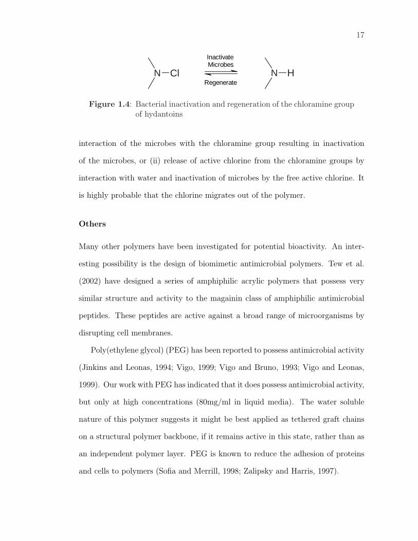
chlorine bleach (Figure 1.4).
At this point it is unclear whether this is truly a non-migratory technology, as
there are two proposed mechanisms of action for the hydantoin group: (i) direct
17
ClN HN
InactivateMicrobes
Regenerate
Figure 1.4: Bacterial inactivation and regeneration of the chloramine groupof hydantoins
interaction of the microbes with the chloramine group resulting in inactivation
of the microbes, or (ii) release of active chlorine from the chloramine groups by
interaction with water and inactivation of microbes by the free active chlorine. It
is highly probable that the chlorine migrates out of the polymer.
Others
Many other polymers have been investigated for potential bioactivity. An inter-
esting possibility is the design of biomimetic antimicrobial polymers. Tew et al.
(2002) have designed a series of amphiphilic acrylic polymers that possess very
similar structure and activity to the magainin class of amphiphilic antimicrobial
peptides. These peptides are active against a broad range of microorganisms by
disrupting cell membranes.
Poly(ethylene glycol) (PEG) has been reported to possess antimicrobial activity
(Jinkins and Leonas, 1994; Vigo, 1999; Vigo and Bruno, 1993; Vigo and Leonas,
1999). Our work with PEG has indicated that it does possess antimicrobial activity,
but only at high concentrations (80mg/ml in liquid media). The water soluble
nature of this polymer suggests it might be best applied as tethered graft chains
on a structural polymer backbone, if it remains active in this state, rather than as
an independent polymer layer. PEG is known to reduce the adhesion of proteins
and cells to polymers (Sofia and Merrill, 1998; Zalipsky and Harris, 1997).
18
Poly-l-lysine and poly(lactic acid) have both been reported to possess limited
antimicrobial activity (Appendini and Hotchkiss, 2002; Ariyapitipun et al., 2000;
Mustapha et al., 2002). Poly-l-lysine is a cationic polymer that promotes cell
adhesion and is thought to be active by a similar mechanism to chitosan (Appendini
and Hotchkiss, 2002). Low molecular weight poly(lactic acid) has been found to be
active against several organisms, although the mechanism of antimicrobial activity
is not known (Ariyapitipun et al., 2000; Mustapha et al., 2002). It has been
suggested that it releases lactic acid and the activity derives from this. Polymer
modification with quaternary ammonium and phosphonium groups to produce
polycationic biocides has also been reported (Kenawy and Mahmoud, 2003).
An important note: many of the aforementioned compounds are water-soluble
and initial trials have been conducted on the soluble form of the polymer. Activity
of the soluble form does not necessarily carry over to activity of the polymer in
a film, either as an independent layer or as a graft layer on another backbone
polymer. It is important to test film activity as well as solution activity if there is
to be any application of these new materials in food packaging.
1.2.2 Polymers containing entrapped bioactive
compounds
In some cases large compounds can be immobilized by entrapment in the polymer
matrix such that they do not migrate out of the polymer under conditions of use.
This requires an active agent larger than the channels present in the molecular
structure of the polymer and minimal polymer chain mobility, for example a poly-
mer with a high glass transition temperature or one that is highly cross-linked.
This strategy is difficult to control and more likely to result in migration of the
19
active compound than covalent immobilization or inherently bioactive polymers.
Elevated polymer processing temperatures may also result in significant migration
and loss of the active agent. This strategy was not considered in this work.
1.2.3 Polymers with covalently immobilized bioactive
compounds
The third major type of NMBP is an inactive polymer backbone to which an
active agent is covalently attached. The active agent may be a peptide, protein or
enzyme; it can be synthesized on the surface, or it can be synthesized or extracted
separately and then covalently linked to the polymer. To date there has been more
research conducted in this area than in that of inherently bioactive polymers, and
a number of examples have been commercialized, although little of this work, and
none of the commercial examples, has focussed on food packaging applications.
Enzymes or peptides that are adsorbed on polymers are often described as
being immobilized (Scannell et al., 2000), however, these compounds are not truly
immobilized as they can readily migrate out of the polymer in suitable non-reactive
solvents. The term immobilized, as used here, implies covalent attachment of
bioactive molecules to the polymer backbone. Covalent attachment of graft chains
avoids their delamination and ensures long-term chemical stability, in contrast to
physically coated or adsorbed systems (Kato et al., 2003).
Developing NMBP by immobilization
The polymer backbone is of prime concern in designing attachment schemes: if the
polymer is essentially inert, such as poly(ethylene) (PE), then reactive functional
groups need to be created on the polymer backbone to provide sites for attachment.
20
This step is termed functionalisation, and needs to be optimized to develop the
maximum number of target functional groups for the desired coupling (immobi-
lization) chemistry while minimizing polymer degradation and side reactions. For
polymers which already possess suitable functional groups in the polymer back-
bone, for example poly(acrylic acid), the coupling chemistry needs to be chosen to
target the available functional groups.
Most functionalisation and coupling chemistries are surface-centric, but mod-
ification of the bulk of the polymer can be achieved by treating the polymer as
a fine powder, then thermally processing the powder, e.g. extrusion or molding,
or by treating the polymer in solution. This results in distribution of the bioac-
tive component throughout the bulk of the polymer. Polymers can be modified
in solution using appropriate solvents, but these solvents often denature bioactive
proteins; most reactions used for surface coupling are also applicable to solution
coupling. Some proteins can be protected from solvent denaturation by conjugation
with suitable polymers, e.g. PEG oligomers. Solution modification is applicable
for water-soluble biopolymers such as chitosan, zein and poly(lactic acid). With
covalent immobilization of an active agent throughout the bulk of the polymer,
the active agent will be unable to migrate from the bulk to the polymer surface,
so food packaging applications are limited. Bulk covalent immobilization may be
of interest for constructing food-processing equipment where surface abrasion and
wear is an issue.
For surface modification, the polymer should be preformed into the final pack-
age form prior to modification, if possible, to prevent surface rearrangement dur-
ing later thermal processing/forming. Surface modification of a polymer film is
the simplest situation; more complex shapes may require different modification
21
strategies.
Solvents may swell polymers without dissolving them, allowing reagents to
penetrate the polymer matrix. Polymer swelling allows increased modification of
the polymer surface, but may render some of the active agent inaccessible if the
packaged product does not similarly swell the polymer. Careful selection of the
solvents used for the reactions can control swelling.
Polymer functionalisation For inert polymers, such as PE, the polymer back-
bone requires functionalisation prior to attaching or generating the bioactive agent
of interest. The literature contains many examples of this. The simplest methods
for laboratory use involve wet chemical oxidations of the polymer surface with, for
example, chromium trioxide, potassium hypochlorite or potassium permanganate
in concentrated sulphuric acid (Eriksson et al., 1984; Larsson et al., 1979). Al-
though these methods are simple, the hazardous nature of the reagents makes
them undesirable in commercial applications. A recent development reduces the
problems of wet chemical methods by utilizing a microwave-catalyzed reaction be-
tween solid potassium permanganate and powdered polyolefins (Mallakpour et al.,
2001a,b), but this process still produces wastewater containing high concentrations
of KMnO4.
Wet chemical oxidations introduce various carbonyl groups, predominantly car-
boxylic acids, aldehydes and ketones, to polymer surfaces. The reaction can be
optimized to produce the maximum concentration of the desired carbonyl function
(Eriksson et al., 1984; Holmes-Farley et al., 1985; Rasmussen et al., 1977). Side
reactions include incorporation of sulphate groups and surface etching/ablation.
Sulphate groups can be removed by nitric acid treatment post-oxidation; surface
22
etching can be controlled to an extent by optimizing reaction conditions. Two
recent reviews (Garbassi et al., 1994; Bergbreiter, 1994) of polymer surface modi-
fication are recommended.
The commercial application of wet chemical modifications is limited by numer-
ous safety and environmental concerns. More common commercially are “physical”
surface treatments such as flame treatment and corona discharge (Shi et al., 1998).
Corona discharge involves applying a high voltage (10–40 kV) at a high frequency
(1–4 kHz) between a discharge electrode and an earthed roller carrying the film
(Robertson, 1993). This oxidizes the surface of the film, introducing a range of
oxygen and nitrogen functional groups to the polymer backbone. Careful control is
required to prevent excessive etching. Flame treatment also produces an oxidized
film surface and introduces a range of oxygen and nitrogen functions, but is more
difficult to control than corona treatment. Both treatments require specialized
equipment, but this equipment is common in polymer processing and converting
operations. The disadvantage of both these methods is that it is very difficult
to control the exact nature of the functional groups created on the surface of the
film. It may be possible to control the surface functionalisation in corona discharge
treatment by varying the gas composition of the treatment atmosphere, although
the typical installation does not have this capability.
Control of the treatment atmosphere has allowed controlled surface functional-
isation in plasma treatment of polymer surfaces (Groning et al., 2001; Klapperich
et al., 2001; Schroder et al., 2001; Terlingen et al., 1995). The disadvantage of clas-
sic plasma processing is that it requires a high vacuum to generate a stable plasma
and, as such, is a batch process not suited to high throughput polymer converting
operations. A new development in plasma processing is the Atmospheric Pressure
23
Non-Equilibrium Plasma (APNEP) system (Shenton and Stevens, 1999). This has
been tested with a range of common polymers and various atmospheric composi-
tions (Shenton et al., 2001; Shenton and Stevens, 1999, 2001; Shenton et al., 2002);
controlled surface functionalisation appears to be possible, similar to that obtain-
able through vacuum plasmas. A disadvantage of the APNEP system is that the
plasma is at a very high temperature, so care is needed to prevent thermal degra-
dation of polymers during treatment. This is achieved by placing the films in the
downstream afterglow region of the plasma rather than in the plasma itself; the
distance from the plasma source is an important variable for this system. The
APNEP system typically causes more surface etching than vacuum plasmas.
Plasma treatment technologies, or possibly controlled atmosphere corona dis-
charge treatments, are likely to be the most useful commercial techniques for
controlled surface functionalisation of a broad range of polymers, although the
use of inherently functional polymers, e.g. poly(lactic acid), poly(acrylic acid),
poly(methacrylate) or derivatives, may be more feasible. Reviews of physical
methods for polymer surface modification/functionalisation are available (Lane
and Hourston, 1993; Ozdemir et al., 1999b,a), although these do not include the
novel APNEP technology. The review by Ozdemir et al. (1999b) is particularly
useful as it approaches surface functionalisation from the food packaging stand-
point, albeit with different intended applications.
Polymeric spacers One of the major difficulties in attaching bioactive agents
to polymeric systems is the necessity of maintaining the native conformation and
structure of the attached compound; the activity of most bioactive compounds is
closely related to their structure and may be lost if this structure is significantly
24
disturbed. The hydrophobic nature of many common polymers may disrupt the
structure of a hydrophilic bioactive compound if they are directly coupled. To
prevent this, a hydrophilic spacer molecule may be used between the bioactive
compound and the polymer backbone (Ikada, 1994). A spacer also helps reduce
steric hindrances to activity (Weetall, 1993) and allows increased mobility of the
bioactive compound. Some peptides, for example small amphiphilic antimicrobials
(e.g. magainins), require association of multiple units for activity to be evident; if
the peptides are not mobile, then they are unlikely to be active in the bound state.
A bioactive attached directly to the polymer backbone is unlikely to be mobile,
while one attached via a flexible (low Tg) spacer will be mobile and is more likely
to retain its activity.
Many different oligomers can be used as spacers, although the most common
is PEG. The main considerations in selecting a spacer for a food packaging ap-
plication are: it does not disrupt the structure of the bioactive compound; it is
approved for food contact use; and suitable chemistry exists for coupling it to
both the polymer backbone and the bioactive compound. PEG is safe, well char-
acterized (Zalipsky and Harris, 1997) and approved for food use (Anon, 2002a).
It has been extensively used for conjugation with peptides and proteins (Zalipsky
and Harris, 1997; Yang et al., 1995a; Herrwerth et al., 2003), and a large range of
derivatives are available for reaction with different functional groups (Shearwater
Corporation, 2001). Methods are well established for grafting it to polymer back-
bones, typically by attaching preformed PEG chains of defined molecular weight
using various coupling chemistries (Bae et al., 1999; Emoto et al., 1998; Kang et al.,
2001; Malmsten et al., 1998; Sofia and Merrill, 1998; Herrwerth et al., 2003). PEG
is water soluble, allowing coupling in aqueous media and increasing the probability
25
that liquid food products will swell the polymer surface, increasing the interactions
between the attached active agent and target constituents in the food.
Coupling chemistries There are many coupling chemistries available for co-
valently linking bioactive compounds to polymers and many different types of
linkage that can be formed. Amide bonds can be formed between an amino group
(on either the bioactive agent or the polymer) and a carboxylic acid group. Other
common linkages are esters and thioesters, formed by interactions between car-
boxylic acids and alcohols or thiols, respectively. All these groups are common
constituents of bioactive compounds. The coupling chemistries which have been
explored for polymer conjugation are often the same as those used for peptide
synthesis; texts on peptide synthesis are good sources of information on coupling
techniques (Bodanszky, 1993a,b; Bodanszky and Bodanszky, 1994).
The carbodiimide method is a well-established and apparently simple coupling
technique that forms amide bonds. It can be used in organic solvents or aque-
ous systems, depending on the carbodiimide used. 1,3-dicyclohexyl carbodiimide
(DCC) is typically used in organic solvents (Bodanszky and Bodanszky, 1994),
whereas 1-ethyl-3-(3-dimethylaminopropyl) carbodiimide (EDC or WSC, water
soluble carbodiimide), is the most common carbodiimide for aqueous coupling
(Bae et al., 1999; Carraway and Koshland, 1972; Herrwerth et al., 2003; Hinder
et al., 2002; Kang et al., 2001; Nakajima and Ikada, 1995; Plummer and Bohn,
2002; Valuev et al., 1998). The ideal situation for carbodiimide coupling is to
have carboxylic acid groups on the film surface react with amino functions from
the bioactive compound. The preference is for the carboxyl functions to be on
the film surface because it is the carboxylic acid that is activated by the carbodi-
26
imide; if amines are present within the structure of the activated compound, as
would be the case if activating carboxylic acids within a bioactive peptide, then the
activated carboxylic acids can spontaneously couple with them leading to inter-
and intra-molecular cross-linking and inactivation of the peptide. A similar ef-
fect can occur if a single step coupling reaction is used (Valuev et al., 1998); a
two step procedure, activation followed by coupling (Kato et al., 2003), has been
used here to avoid these problems: the film is immersed in a buffered solution
of carbodiimide to activate the carboxyl functions, removed and gently rinsed,
then immersed in a buffered solution containing the bioactive compound to be at-
tached. The exact conditions, e.g. time, temperature and pH, require fine-tuning
for each polymer-peptide combination. Peptides have previously been coupled to
carboxy-PEG coated surfaces using WSC coupling (Herrwerth et al., 2003).
A second common method for coupling bioactive agents to polymers uses glu-
taraldehyde. Glutaraldehyde has long been recognized as an efficient cross-linking
agent for use with proteins and other biological molecules (Bigi et al., 2001; Kikuchi
et al., 2002; Weissman, 1979) and has also been used to immobilize bioactive ma-
terials onto polymeric backbones (Appendini and Hotchkiss, 1997; Molday et al.,
1975; Soares, 1998). Glutaraldehyde (OHC-CH2-CH2-CH2-CHO) is a bifunctional
short-chain aldehyde that reacts with amines. It requires amines on both the sup-
port and the bioactive molecule, and forms three-dimensional, cross-linked aggre-
gates.
A third possibility for coupling reagents are succinimidyl succinate active esters
and their derivatives. These are commercially available (Shearwater Corporation,
2001) and have been extensively employed for PEG conjugation to peptides and
proteins. Succinimide esters react with free amine groups to form stable amide
27
linkages. The most commonly used derivative is the succinimidyl propionic acid
ester of PEG (PEG-SPA), which has been used to conjugate PEG with insulin
(Caliceti and Veronese, 1999) and human growth hormone antagonist (Olson et al.,
1997). The reaction conditions can be modified to suit the system of interest.
A succinimidyl ester, N-hydroxy succinimide (NHS), can also be used in an
extension of carbodiimide coupling (Hinder et al., 2002; Plummer and Bohn, 2002).
Carbodiimide activated carboxylic acids are relatively unstable and may not be
ideal for two step coupling processes, particularly if there is a significant delay
between the activation and coupling steps. Instead, the activation is performed
in the presence of NHS, creating a more stable, but still reactive, NHS ester of
the carboxylic acid. The NHS-activated film is then immersed in a solution of
the amine-containing compound to be coupled and coupling proceeds with the
formation of amide linkages. NHS active esters of PEG can also be obtained
commercially (Shearwater Corporation, 2001).
1.3 Applications of NMBP with immobilized bioactive
agents
Although there are many possible applications of immobilized bioactives in food
packaging, only a few have been explored to date. These can be broken into three
main areas: in-package processing, antimicrobial packaging/shelf life extension and
intelligent packaging.
28
1.3.1 In-package processing
In-package processing involves immobilizing an enzyme on the surface of the pack-
aging material to perform in the package what would otherwise be a processing
step in the plant prior to packaging. An example is the hydrolysis of naringin,
one of the bitter compounds in citrus juices, with immobilized naringinase (Soares
and Hotchkiss, 1998; Soares, 1998). This packaging material was able to signifi-
cantly reduce the bitter naringin content of grapefruit juice during storage, pro-
viding what was perceived as a sweeter product as storage progressed. Another
potential application exists for immobilized lactase (β-galactosidase) packaging
to produce lactose-reduced milk products at a reduced cost compared to current
methods. Reducaed-lactose milk products are required for consumers who are
lactose-intolerant and unable to consume regular milk products. Lactase immobi-
lization has been one focus of this research.
Only the number of enzymes with potential processing applications limits the
range of potential applications for in-package processing. Other examples include
hydrolases for non-thermal inactivation of shelf-life limiting enzymes; glucose iso-
merase to convert glucose to fructose, increasing the sweetness of products; and
cholesterol reductase to reduce the cholesterol content of products. The concepts
of lactase and cholesterol reductase immobilized on packaging materials were ex-
plored by Pharmacal Biotechnologies in the early 1990s (Brody and Budny, 1995),
although no commercialization is apparent.
The basic principle of in-package processing with immobilized enzymes is shown
using the example of cholesterol reductase in Figure 1.5. The interior surface of
the packaging contains immobilized cholesterol reductase in contact with the food.
The enzyme substrate, in this case cholesterol, contacts the immobilized enzyme
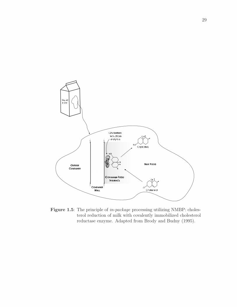
29
LiquidFood
OUTSIDECONTAINER
CONTAINERWALL
HO
HO
BULK FOOD
HO Coprosterol
Cholesterol
Cholesterolreductaseenzyme
CONTAINER-FOODINTERFACE CH3
CH3
CH3
Figure 1.5: The principle of in-package processing utilizing NMBP: choles-terol reduction of milk with covalently immobilized cholesterolreductase enzyme. Adapted from Brody and Budny (1995).
30
as a result of natural convective currents or diffusion within the product or mixing
caused by product agitation during handling and distribution. The substrate is
acted on by the enzyme, producing the desired effect, and the reaction products
are released back into the food system. Over storage, the composition of the
packaged food changes to that desired by the food processor for optimum consumer
acceptability, nutritional content, or shelf life.
1.3.2 Antimicrobial packaging and shelf life extension
The primary concern of all food processors should be the safety of the food they
produce. A recent review estimated that there were 76 million illnesses, 325,000
hospitalizations and 5000 deaths per year due to food-related illness in the United
States (Mead et al., 1999). The role of antimicrobial packaging in reducing food-
borne illness has two major aspects: sanitation of packaging materials and re-
duction of pathogen growth in packaged foods. Although less dire than food
poisoning, food spoilage due to microbial action is also a problem, resulting in
the loss of large quantities of food. Retarding microbial growth and extending
product shelf-life may allow food to be transported to places where it is needed.
Several reviews of antimicrobial food packaging have recently been published (Ap-
pendini and Hotchkiss, 2002; Collins-Thompson and Cheng-An, 2000; Han, 2000;
Vermeiren et al., 2002).
Shelf life extension and reduction of food-borne disease are the main goals of
antimicrobial packaging. Considering the later, it is clearly important to maintain
pathogen levels in food below the level that will cause illness, be it by toxin pro-
duction or food-borne infection; it is important to prevent pathogen growth and
to inactivate those present in the food. For shelf life extension, however, complete
31
growth inhibition and microbe elimination is not necessary. To extend the shelf
life of a product where microbial growth is limiting, it is only necessary to reduce
the growth rate or extend the lag phase of the organisms, i.e. to retard microbial
growth. Even where microbial growth is not the limiting factor for shelf life, im-
mobilized enzymes may have a role in extending shelf life if they can target shelf
life limiting reactions, e.g. oxidation, by removing catalysts or reactants.
Both synthetic and natural compounds have been investigated for attachment
to polymers to create non-migratory antimicrobial packaging. Appendini (1996);
Appendini and Hotchkiss (1997) investigated the attachment of lysozyme, derived
from hen egg white, to poly(vinyl alcohol), nylon and cellulose triacetate (CTA).
Although lysozyme was successfully immobilized on all materials, it’s activity on
nylon and poly(vinyl alcohol) was insufficient for commercial use. Greater activity
was retained on CTA films and significant bacterial retardation was observed in
trypticase soy broth, although there was still a significant reduction in activity
compared to free lysozyme. Activity of the CTA-lysozyme film also decreased with
repeated usage, indicating that the lysozyme was either inactivated over time/with
use or migrated out of the film.
Another natural antimicrobial system that could be used for antimicrobial pack-
aging is the lactoperoxidase system of milk. The immobilization of lactase and glu-
cose oxidase enzymes on nylon pellets has been investigated with the goal of pro-
ducing hydrogen peroxide to activate the lactoperoxidase system naturally present
in milk (Garcia-Garibay et al., 1995). The system investigated was designed for
use in a bioreactor, rather than in packaging, but the principle is equally applica-
ble to packaging. The advantage of this system is that milk regulations in many
countries prevent the addition of preservatives to milk; activating a natural antimi-
32
crobial system already present in the milk provides antimicrobial activity without
contravening regulations. Lactase and glucose oxidase were immobilized onto ny-
lon pellets using glutaraldehyde coupling with a poly(ethyleneimine) spacer. The
system resulted in reductions of 0.5–2 log cycles in the natural microflora of raw
milk. Milk samples were only exposed to the enzymes for 3 minutes, and micro-
bial counts were taken 24 hours after exposure (storage at 8�). Modifying this
system for use in a package, with prolonged exposure of the milk to the hydroper-
oxide, would probably result in greater bacterial inhibition, but might also result
in oxidation of milk components by hydroperoxides leading to undesirable sensory
characteristics.
A small synthetic antimicrobial peptide, E14LKK, has been investigated for
non-migratory antimicrobial packaging applications (Appendini, 1999; Appendini
and Hotchkiss, 2001; Haynie et al., 1995; Haynie, 1998). E14LKK is an amphi-
pathic, α-helical derivative of magainin II (Haynie et al., 1995). It has the sequence
HOOC-L-K-L-L-K-K-L-L-K-L-L-K-K-L-NH2, where L is Leucine and K is Lysine.
In the work of Appendini (1999), the peptide was synthesized attached to a PEG-
grafted PS support, but this support could not be reprocessed into useful pack-
aging. A high activity was observed for the immobilized peptide, with significant
antimicrobial activity against a broad range of microorganisms: gram positive and
gram negative bacteria, yeasts and moulds. Activity is presumed to be a result
of pore formation in the cell membrane via the barrel-stave mechanism proposed
by Ojcius and Young (1991). There is some doubt whether the peptide remained
bound to the support in the work of Appendini (1999); some evidence suggested
that the PEG spacer was hydrolyzing, releasing PEG-peptide conjugates into solu-
tion. The conditions that caused the hydrolysis were not investigated. The degree
33
of activity was found to be dependent on the test media; positive activity was
found in buffer, trypticase soy broth, apple juice and meat exudate. The original
objective of the research reported here was to extend this work to create a PE film
with immobilized, active E14LKK, although a broader outcome is now envisaged:
the system developed may potentially be used to attach any peptide to modified
PE film. Bacteriocins have also been investigated for incorporation into antimicro-
bial packaging (Scannell et al., 2000; Padgett et al., 1998; Cha et al., 2003; Cooksey
and Wu, 1998; Cooksey, 2000).
Other enzymes with indirect antimicrobial activity, typically by modifying
package atmospheres, have also been immobilized onto food packaging polymers.
Glucose oxidase was immobilized in conjunction with catalase for use as an oxygen
scavenger (de Kruijf et al., 2002; Labuza and Breene, 1989). The glucose oxidase
oxidizes glucose to produce glucono-delta-lactone and hydrogen peroxide. Hydro-
gen peroxide could lead to potential undesirable oxidations of food components, so
is degraded to water and oxygen by catalase. The net reduction in oxygen content
is half a mole per mole of reactions. A review of the mechanism and kinetics of
glucose oxidase can be found in the literature (Labuza and Breene, 1989). Alcohol
dehydrogenase has also been immobilized to polymer films and can also be used as
an oxygen scavenger (Labuza and Breene, 1989). Reduced oxygen concentration
inhibits the growth of aerobic microbes, especially yeasts and molds, however care
must be taken that anaerobic, low acid, high moisture conditions do not result, as
these may favor the growth of certain pathogens, e.g. Clostridium botulinum.
Immobilized enzymes could also be used to control carbon dioxide concentra-
tion. Non-enzymatic carbon dioxide emitters and absorbers have been developed
and commercialized (Labuza and Breene, 1989), although there has been no com-
34
mercialization of enzymatic carbon dioxide control systems to date. Other immo-
bilized enzymes could be used to produce ethanol, which has well known antimi-
crobial activity. Commercial ethanol emitters are not typically enzymatic (Labuza
and Breene, 1989), but use slow-release encapsulated ethanol. The ethanol vapors
released inhibit microbial growth, particularly that of fungi.
In addition to their indirect antimicrobial effect, many enzymes that modify
the gaseous atmosphere of packaged products can also extend product shelf life by
inhibiting non-microbial degradative mechanisms. The main application of this is
reduced-oxygen packaging that inhibits the oxidation of food components and pre-
vents the resulting negative sensory and nutritional effects. Catechin has recently
been immobilized on acrylic polymer beads as an antioxidant system (Ihara et al.,
2003). Carbon dioxide control can also be important in extending shelf life, as in
the case of packaged coffee. Enzyme systems may be used to prevent taints and
off-flavors by metabolizing compounds of concern. Care needs to be taken, how-
ever, that the taints inhibited are not indicative of microbial spoilage. Off-odors
and off-flavors can be key indicators to consumers that food is spoiled and unfit
for consumption; removing these indicators may result in consumption of spoiled,
possibly pathogenic, foods.
In all cases where indirect enzyme action is used to control microbial growth,
and in fact for all immobilized enzyme reactions in food packaging, the by-products
of the reaction must be carefully considered. As noted above, glucose oxidase
produces hydrogen peroxide, which could cause potentially detrimental oxidations
of food components. Other enzymes also produce by-products that may have
detrimental effects on the sensory characteristics or shelf life of the packaged food.
Complete understanding of the catalyzed reactions is required to ensure undesirable
35
by-products are minimized.
1.3.3 Intelligent Packaging
Intelligent Packaging1 has recently received a lot of attention (Ahvenainen, 2003;
Taoukis and Labuza, 2003; Jarvi-Kaariainen, 2003; Smolander, 2003). Immobi-
lized enzymes and antibodies are common components of intelligent packaging
systems. A range of different indicators involving immobilized bioactive com-
pounds have been developed, including time-temperature integrators (de Kruijf
et al., 2002; Labuza and Breene, 1989), spoilage indicators (Anon, 2001) and indi-
cators of chemical or other contamination (Woodaman, 2001). Time-temperature
integrators (TTIs) based on enzyme-catalyzed reactions are available commercially
(de Kruijf et al., 2002; Labuza and Breene, 1989). Although the commercial ver-
sions do not include NMBP, this is an area in which NMBP could be effective. For
microbial spoilage, enzymatic TTIs may be particularly well suited for accurately
modelling microbial growth since microbial growth depends on enzyme-catalyzed
reactions.
For indicators of microbial or toxicant contamination, there are two main meth-
ods by which immobilized bioactive compounds can be used: (i) enzyme-catalyzed
reactions requiring microbial metabolites or contaminant chemicals as substrates,
or (ii) immobilized antibodies specific to bacterial metabolites and toxins, or con-
taminating chemicals. Both the enzymes in (i) and the antibodies in (ii) could be
used to develop NMBP contamination indicators.
A final paradigm for the use of immobilized bioactive compounds in intelligent
1defined as packaging systems that monitor the condition of packaged food andcommunicate information on food quality during transport and storage (de Kruijfet al., 2002)
36
packaging is as detection units on biosensors. The incorporation of biosensor sys-
tems in packaging films is an area for future research. Biosensors may allow remote
monitoring of package conditions or point of sale testing of product condition by
interfacing with appropriate electronic devices. These may be useful for TTIs, for
the detection of contaminating bacteria or toxicants, or in other areas yet to be
explored.
Chapter 2
Research ObjectivesThe initial goal of this project was to develop a non-migratory antimicrobial pack-
aging film by covalently attaching antimicrobial peptide E14LKK to the surface of
poly(ethylene). During the course of the work, however, it became apparent that
this system has a broader potential, so the objectives were modified to reflect this:
1. Develop a method for covalently immobilizing peptides and proteins on the
surface of poly(ethylene) film.
2. Investigate the possibilities for producing lactose-reducing films by lactase
immobilization.
3. Attach peptide E14LKK to the surface of poly(ethylene) and investigate the
antimicrobial properties of the film.
4. Determine the effects of these surface modifications on the packaging-relevant
properties of poly(ethylene).
37
Chapter 3
Materials and Methods
3.1 Materials
Low density poly(ethylene) (LDPE) pellets (∼0.03g) with a specific gravity of 0.92
and an approximate molecular weight of 50,000 were purchased from Scientific
Polymer Products (Ontario, New York) and used as purchased. High density
poly(ethylene) (HDPE) film (3mil, Lot#SC00030053) blown from Sclair 19A low-
additive resin (NOVA Chemicals, Calgary, Alberta, Canada) was kindly donated
by Dupont (Wilmington, DE). HDPE film had a mean thickness (n=10) of 79.5µm
(3.13mil). Additive-free blown LDPE film was kindly donated by the Dow Chemi-
cal Company (Midland, MI). LDPE film had a mean thickness (n=10) of 100.6µm
(3.96mil).
Chromium trioxide (99.75% purity, Mallinckrodt, St Louis, MO), sulphuric and
nitric acids (AR grade, Mallinckrodt, Paris, KY), dichloromethane (AR grade,
Fischer Scientific, USA), 1-ethyl-3-(3-dimethylaminopropyl)-carbodiimide (Sigma
Chemicals, St Louis, MO), diamino-poly(ethylene glycol) 3400MW (PEG-NH2)
and ω-amino-α-carboxyl-poly(ethylene glycol) 3400MW (NH2-PEG-COOH)
(Shearwater Polymers, Hunstville, AL), toluidine blue O (Fisher Chemicals, Fair-
lawn, NJ), orange II sodium salt (Sigma-Aldrich, St Louis, MO), bromophenol
blue (BioRad, Hercules, CA), acid blue 45 (ICN Biomedicals Inc. Aurora, OH)
were used as purchased.
Antimicrobial peptide E14LKK, having the sequence LKKLLKLLKKLLKL,
was synthesized at the Sheldon Biotechnology Center (McGill University, Mon-
38
39
treal, Quebec). The peptide was received both as a free peptide and with side
chain (lysine) amines protected with the tert-butyloxycarbonyl (Boc) group. De-
protection of the protected peptide was carried out post coupling, as described
below (§3.3.5).
Lactase (GODO YNL-2, Valley Research Inc., South Bend, IN) was filtered
through a 0.45µm syringe filter, then purified by centrifugal separation using a
30,000 dalton cut off membrane (Centricon YM-30, Millipore Corporation, Bed-
ford, MA). 2ml of lactase solution was centrifuged for 2 hours at approximately
4500×g (4000RPM) using an IEC B-20A Centrifuge and a #872 Rotor (Interna-
tional Equipment Company, Needham Heights, MA). The tube was topped with
distilled water and centrifuged for a further 2 hours; more water was added and the
centrifugation repeated. This process was repeated two further times. The filtrate
was discarded, the filter inverted and centrifuged for 2 hours. The retentate was
then removed and placed in a clean test tube. 1ml of distilled water was added
to the filtrate container, the filter returned (inverted) to the centrifuge and cen-
trifuged a final 45mins to extract enzyme retained in the filter. The retentate and
the wash water were combined and the resulting solution used for lactase coupling.
Films were cut into 25×50 mm pieces using a precision strip cutter (Testing
Machines Incorporated, Amityville, NY), then stored in clean glass containers at
ambient temperature until further treatment. Film Thickness was assessed with a
Scherr-Tumico, St James, MN Dial Thickness Gauge.
40
3.2 Film Preparation
3.2.1 Heat-Pressing LDPE Films
Heat-pressed LDPE films were prepared from LDPE pellets using the method
of Al-Ati (2002). Approximately 0.3g of pellets were placed between two pieces
of mylar film and these placed between two preheated parallel metal plates in a
preheated film press (Model 341-20, Loomis Engineering and Manufacturing Com-
pany, Caldwell, NJ). The press was closed and the pellets tempered at ∼140� for
15–20 minutes. 30,000(±2000) pounds of force (133±8 kN) was then applied to the
plates with the press. The area of the smaller plate was 0.0283 m2 giving a pressure
between the plates of 4.71 MPa. The pressure was maintained for 15–20 minutes,
after which the mylar films were removed and immediately immersed in distilled
ice water. The PE film was removed from the mylar and dried under ambient
conditions.
The thickness of pressed films ranged from 1.5–4 mils (38-102µm). Thickness
variation across a single 25×50 mm piece of film was typically less than 0.5 mil
(13µm).
3.2.2 Film Cleaning
Prior to surface treatment, all PE films were first cleaned by refluxing 12–24 hours
in dichloromethane (DCM) and vacuum drying overnight at 60�, ∼27 kPa, abs.
(∼22 inches Hg vac). After cleaning, films were stored in clean glass containers at
ambient temperature until further analysis or treatment.
41
3.3 Film Modification
Figure 3.1 shows the film modification process; the detailed methodology follows.
3.3.1 Surface Oxidation of PE film using Chromic Acid
Surface oxidation was performed using methodology described by the Whitesides
group (Rasmussen et al., 1977; Holmes-Farley et al., 1985). Cleaned PE films
were immersed in chromic acid solution (CrO3:H2O:H2SO4 = 29:42:29 by weight)
at 70±2◦C for 1 minute, with agitation, followed by three successive washes in
distilled water at ambient temperature. Film samples were then transferred to 70%
nitric acid at 50� for 15 minutes followed by three further distilled water washes.
Surface oxidized films were typically analyzed, or further treated, immediately. If
storage was required, films were stored in distilled water under refrigeration to
minimize rearrangement of the film surface and loss of surface carboxyl functions
(Holmes-Farley et al., 1985).
3.3.2 PEG attachment to PE-COOH using WSC Coupling
Oxidized poly(ethylene) films were grafted with NH2-PEG-NH2 or
NH2-PEG-COOH using 1-ethyl-3-(3-aminopropyl)-carbodiimide (Water Soluble
Carbodiimide, WSC) coupling. Three films were immersed in 30 ml of 0.35 mM
WSC in degassed distilled water1 (adjusted to ∼pH 4.5 with HCl/NaOH) at room
temperature and incubated for 1 hour with agitation (Lin and Tseng, 2001; Bae
et al., 1999; Anon, 2002b,c). After incubation, the films were gently washed three
1in addition to obvious problems with buffers containing amino or carboxygroups, it has been suggested that phosphate buffers might also cause problemswith WSC coupling (Anon, 2002c). Distilled water was recommended.
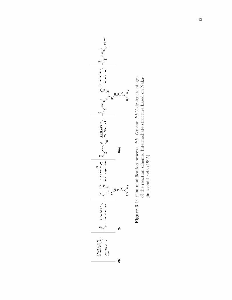
42
C OHOC
N H
OPE
GC OHO
CH3
CH 3
NCH2
CH2NH
C+
NHOCO
CH2CH
2
CH3
CN H
OPE
GC OO
CH3
CH 3
NCH2
CH2NH
C+
NHCH
2CH2
CH3
O
CN H
OPE
GN H
COpeptide
PEOx
PEG
3.2m
gWSC
,1hr
30ml
DDW
pH4.7
30ml
DDW
pH7,
24hrs
1.CrO 3
:H2SO 4
:H2O
29:29
:42,7
0°C,1
min
2.70%
HNO 3
,50°C
,15min
5.2m
gWSC
,1hr
30ml
DDW
pH4.7
4.H 2N-PE
G-C-OH
30ml
DDW
pH7
6.peptide,2
4hrs
Fig
ure
3.1
:F
ilm
modifi
cati
onpro
cess
.P
E,
Ox
and
PE
Gdes
ignat
est
ages
ofth
ere
acti
onsc
hem
e.In
term
edia
test
ruct
ure
bas
edon
Nak
a-ji
ma
and
Ikad
a(1
995)
43
times with distilled water and placed in 30 ml of 0.1 mM PEG in degassed distilled
water (adjusted to ∼pH 7), incubated for ∼24 hours at 4� with agitation, then
removed and gently rinsed in distilled water (×3). The films were washed by shak-
ing in 40 ml (per 3 films) of 10% aqueous ethanol for 1 hour, then gently rinsed in
distilled water (×3). Films were stored under moist conditions at 4� until further
analysis or modification.
3.3.3 Lactase attachment to PE-PEG-COOH and
PE-COOH
As for PEG attachment, three films were immersed in 30 ml of 0.35 mM WSC in
degassed distilled water (pH 4.5) and incubated at room temperature for 1 hour
with agitation. The films were rinsed (×3) in distilled water and added to 30 ml
of degassed distilled water (pH 7). Half the retentate from 2 ml of lactase solution,
purified as described above (§3.1), was added to each 30ml of coupling solution and
the films incubated in this at 4� for 24 hours with agitation. Films were removed,
rinsed three times in distilled water and stored under moist conditions at 4� until
further analysis or modification.
Before and after coupling, the lactase activity of the coupling solution was
tested by mixing a few drops of coupling solution with a few drops of 8 mM
o-nitrophenyl-β-D-galactopyranoside (ONPG; described in §3.6.1). The rapid de-
velopment of a yellow color indicated a positive test for lactase activity.
44
3.3.4 E14LKK attachment to PE-PEG-COOH and
PE-COOH
Coupling of E14LKK was conducted similarly to PEG and Lactase coupling. The
carboxy groups on the film were activated by agitation in 0.35 mM WSC solu-
tion (pH 4.5) for 1 hour at room temperature. The films were then placed in de-
gassed distilled water (pH 7, 3 films/30ml) to which peptide solutions had been
or were subsequently added: for E14LKK with unprotected side chain amines
(unE14LKK ), 5 mg of peptide was dissolved in the distilled water prior to adding
the films; for E14LKK with Boc protected side chains (proE14LKK ) approxi-
mately 5–10 mg of peptide previously suspended in distilled water was added after
the films. The film/peptide solutions were incubated at 4� for 24 hours with agi-
tation, then rinsed briefly in distilled water (×3). Films were stored at 4� under
moist conditions until further testing.
The bactericidal activity of the coupling solutions was assessed post-coupling
by adding 1 ml of coupling solution to 9 ml of Butterfields Buffer containing 0.1ml
of an overnight culture ( 108 cfu/ml) of Escherichia coli ATCC 25922. Samples
(triplicate) were incubated for 24hours at 37�, after which 0.1ml was spread onto
the surface of Standard Methods Agar plates. The plates were incubated at 37�
and visually assessed at 12 and 24hours. The unE14LKK coupling solution sig-
nificantly reduced the growth of the bacteria compared to the control, with the
difference most apparent at 12hours. The proE14LKK was not notably different
from the control.
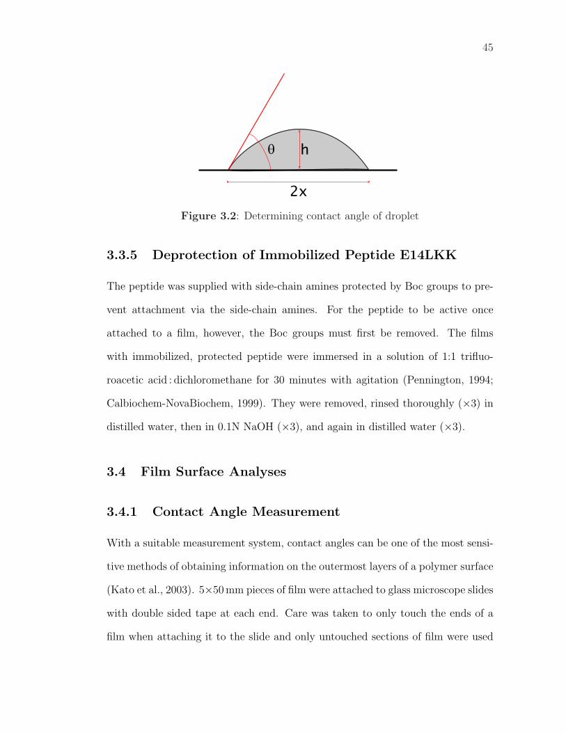
45
q
2x
h
Figure 3.2: Determining contact angle of droplet
3.3.5 Deprotection of Immobilized Peptide E14LKK
The peptide was supplied with side-chain amines protected by Boc groups to pre-
vent attachment via the side-chain amines. For the peptide to be active once
attached to a film, however, the Boc groups must first be removed. The films
with immobilized, protected peptide were immersed in a solution of 1:1 trifluo-
roacetic acid : dichloromethane for 30 minutes with agitation (Pennington, 1994;
Calbiochem-NovaBiochem, 1999). They were removed, rinsed thoroughly (×3) in
distilled water, then in 0.1N NaOH (×3), and again in distilled water (×3).
3.4 Film Surface Analyses
3.4.1 Contact Angle Measurement
With a suitable measurement system, contact angles can be one of the most sensi-
tive methods of obtaining information on the outermost layers of a polymer surface
(Kato et al., 2003). 5×50 mm pieces of film were attached to glass microscope slides
with double sided tape at each end. Care was taken to only touch the ends of a
film when attaching it to the slide and only untouched sections of film were used
46
for the contact angle measurements as skin oils alter the wetting characteristics
of the film (Holmes-Farley et al., 1985). The film was attached flat as surface
irregularities or slope alter the shape of water drops.
A 0.5µl drop was delivered to the film surface using a 2.5µl hand-held syringe
(Hamilton, #87942) with a flat-tipped, Teflon-coated needle (Hamilton, #80471).
The drop was viewed through the microscope eyepiece, with the magnification
set such that the drop size was comparable to the linear scale in the eyepiece.
The relative height and width of the drop was measured using this scale and the
contact angle calculated from the drop dimensions (Bartell and Zuidema, 1936;
Mack, 1935):
tanθ
2=h
x(3.1)
where θ is the contact angle, h is the height and x the half-width of the drop
(Figure 3.2). The height was measured first, then the width. Drop height is
more sensitive to evaporation than is drop width. To reduce evaporation, drafts
around the apparatus were minimized and drop measurement was completed within
∼30 seconds of applying the drop. 5–7 drops were measured for each film sample
and averaged to determine the contact angle for that sample; the results from at
least three, and generally six, film samples were averaged to determine the contact
angle for a treatment.
3.4.2 Dye Adsorption Assays for Surface Chemistry
These dye tests are quantitative analyses of the acid or base functions present on
the surface of a film. They were adapted from published methods (Hu et al., 2002;
Kato and Ikada, 1996; Sano et al., 1993).
47
Toluidine Blue O Assay
Film samples were soaked in 0.1mg/ml aqueous Toluidine Blue O solution (adjusted
to pH 11 with NaOH) at room temperature for 6 hours, removed, rinsed in distilled
water, then in 0.1M NaOH and again in distilled water, then dried hanging in a
desiccator2. The dye was desorbed by sonicating each film twice for 20minutes
in separate 5ml aliquots of 1:1 Hexane : Trifluoroacetic acid (TFA)3. The aliquots
were combined and the absorbance measured at either 605 or 677nm4. The dye
concentration was calculated by comparison to a standard solution using Beer’s
Law:
A = a · b · C (3.2)
where A is the absorbance, a the absorbtivity, b the path length (constant at
10mm) and C the concentration.
Standard solutions were prepared by dissolving a known amount (∼2mg) of
Toluidine Blue in 10ml of Hexane : TFA to give a stock solution (∼0.2mg/ml),
then diluting 0.1ml of this stock solution in 9.9ml of Hexane:TFA to obtain a
standard solution (∼2µg/ml). This solution was sonicated for 20minutes prior to
absorbance measurement to duplicate sample treatment. Three separate standard
2It is important to dry the films completely prior to placing in the Hexane : TFAsolution. The presence of even minute amounts of water will cause phase separa-tion.
3Previous researchers (Hu et al., 2002; Kato and Ikada, 1996; Sano et al., 1993)used 50% aqueous Acetic Acid to desorb the toluidine blue from the film, however,this did not fully extract the dye from the film, even with sonication. Maximumdye extraction was obtained with 1:1 Hexane : Trifluoroacetic Acid, although evenafter this treatment the films retained significant color.
4The absorption maximum of toluidine blue is 677nm. Issues with one spec-trophotometer prevented use of wavelengths greater than 605nm; sufficient absorp-tion remains at this wavelength to obtain accurate results. When using an alter-native spectrophotometer, the absorbance was measured at the optimal 677nm.Samples were compared to standards measured at the same wavelength.
48
solutions were prepared on two separate occasions, with concentrations of 2.2, 2.3
and 3.3µg/ml; the absorbtivities of these solutions were averaged to calculate dye
concentrations in the desorbing solutions.
The concentration of acid groups on the film surface was calculated based
on the desorbed dye concentration, the solution volume and the film area. The
concentration was adjusted for the dye absorption found for unmodified PE film,
which was assumed to have no surface acid groups.
Acid Orange 7 Assay
Films were soaked in 1.0mg/ml aqueous Acid Orange 7 (Orange II Sodium Salt,
Sigma) solution (adjusted to pH 3 with HCl) at room temperature for 6 hours.
They were removed and rinsed in distilled water, then in 0.1M HCl and again
in distilled water. The dye was extracted by sonicating the film in 5ml of 6N
NaOH for 20 minutes. The absorbance of the extraction solution was measured
at 485nm and the dye concentration calculated, in comparison to the absorbance
of a standard, using Beer’s Law (equation 3.2). Standard solutions were prepared
with dye concentrations of 1.0, 2.0 (twice) and 3.5µg/ml; the average absorbtivity
of these four standards was used to calculate the concentration of basic groups on
film surfaces, as per the Toluidine Blue assay.
3.4.3 Instrumental Analysis of Film Surfaces
Atomic Force Microscopy (AFM)
AFM scans were conducted using a Digital Instruments Dimension 3100 Scanning
Probe Microscope (Veeco Instruments, Inc., Woodbury, NY) in tapping mode with
Ultrasharp Silicon Cantilevers (MikroMasch, Germany, part number NSC14/W2C).
49
The scan size was 10µm, with 128 lines per scan, 512 sampling points per line, a
tip velocity of 12µm/s and scan rate of 0.598 Hz.
X-Ray Photoemission Spectroscopy (XPS)
XPS analysis was conducted by Dy Michael Kelley and colleagues at Jefferson
Laboratory (Newport News, VA) using a Vacuum Generators ESCALAB Mk II
(VG Scientific, West Sussex, England) with a Mg anode (1253.6eV) operated at
15kV and 200W.
3.5 Functional Testing of Modified Films
3.5.1 Water Vapor Transmission Rate
WVTR was measured using a modification of ASTM Method E96–00 (Ameri-
can Society of Testing and Materials, 2000). Film samples measuring 25×50 mm
were prepared and mounted on metal can lids using silicon sealant (732 Multi-
purpose Sealant, Dow Corning, Midland, MI), covering two 16 mm diameter holes
previously cut in the lids. The lids were sealed over aluminum dishes containing
Drierite Indicating Desiccant (8 Mesh, W.A.Hammond Drierite Co., Xenia, OH)
using melted Bowax paraffin wax (Strohmeyer & Arpe, Millburn, NJ). Sample
dishes were incubated at 35� in a desiccator containing a saturated aqueous so-
lution of ZnSO4.7H2O in the base cavity, providing a relative humidity (RH) of
approximately 90%5, and weighed every 12–48 hours for 2–3 weeks (n=3).
5a saturated solution of ZnSO4.7H2O provides a RH of 90% at 20� (Anon,2004a); the RH is expected to decrease slightly, probably by ∼2%, as temperatureincreases (based upon data presented by Labuza (2004))
50
3.5.2 Tensile Testing
Tensile testing methodology was based on ASTM Method D882–97 (American
Society of Testing and Materials, 2000) and conducted on a Texture Technologies
(Scarsdale, NY) TA–XT2 Texture Analyzer. The method was modified for use
with 50 mm long strips of film by using an initial grip spacing of 25 mm; 5 mm
wide strips of film were tested for HDPE (n=12) and 10 mm wide strips were tested
for LDPE (n=10); sample thickness was measured at three locations on each test
film prior to testing. The initial strain rate was set at 4.2 mm/s (∼250 mm/min).
Tensile Strength, Percent Elongation at Break and Yield Strength were calculated
per the ASTM standard.
3.5.3 Tear Propagation Resistance
Tear propagation testing was conducted using a modification of ASTM Method
D1938–94 (American Society of Testing and Materials, 2000) on a Texture Tech-
nologies (Scarsdale, NY) TA–XT2 Texture Analyzer. The method was adapted for
samples measuring 25×50mm. Samples were slit lengthwise using a Precision Strip
Cutter (Testing Machines Inc, Amityville, NY) leaving 25mm of uncut material
between the tip of the slit and the end of the sample. The initial spacing of the
tensile tester grips was approximately 25 mm and the initial separation rate was
4.2 mm/s (n=6). The normalized tear propagation force was calculated by divid-
ing the area under the tear propagation force-distance curve (the work to tear the
sample) by the length of the tear and the thickness of the sample. This calculation
was used because the samples showed various tear patterns (see Figure 3.3 for
examples), so the length of the tear varied.
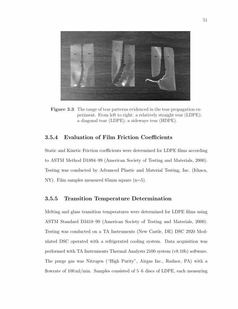
51
Figure 3.3: The range of tear patterns evidenced in the tear propagation ex-periment. From left to right: a relatively straight tear (LDPE);a diagonal tear (LDPE); a sideways tear (HDPE).
3.5.4 Evaluation of Film Friction Coefficients
Static and Kinetic Friction coefficients were determined for LDPE films according
to ASTM Method D1894–99 (American Society of Testing and Materials, 2000).
Testing was conducted by Advanced Plastic and Material Testing, Inc. (Ithaca,
NY). Film samples measured 65mm square (n=5).
3.5.5 Transition Temperature Determination
Melting and glass transition temperatures were determined for LDPE films using
ASTM Standard D3418–99 (American Society of Testing and Materials, 2000).
Testing was conducted on a TA Instruments (New Castle, DE) DSC 2920 Mod-
ulated DSC operated with a refrigerated cooling system. Data acquisition was
performed with TA Instruments Thermal Analysts 2100 system (v8.10b) software.
The purge gas was Nitrogen (“High Purity”, Airgas Inc., Radnor, PA) with a
flowrate of 100 ml/min. Samples consisted of 5–6 discs of LDPE, each measuring
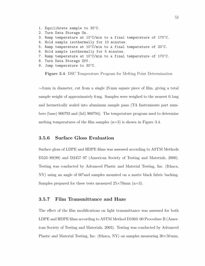
52
1. Equilibrate sample to 30◦C.2. Turn Data Storage On.
3. Ramp temperature at 10◦C/min to a final temperature of 170◦C.4. Hold sample isothermally for 10 minutes.
5. Ramp temperature at 10◦C/min to a final temperature of 20◦C.6. Hold sample isothermally for 5 minutes.
7. Ramp temperature at 10◦C/min to a final temperature of 170◦C.8. Turn Data Storage Off.
9. Jump temperature to 30◦C.
Figure 3.4: DSC Temperature Program for Melting Point Determination
∼3 mm in diameter, cut from a single 25 mm square piece of film, giving a total
sample weight of approximately 6 mg. Samples were weighed to the nearest 0.1mg
and hermetically sealed into aluminum sample pans (TA Instruments part num-
bers (base) 900793 and (lid) 900794). The temperature program used to determine
melting temperatures of the film samples (n=3) is shown in Figure 3.4.
3.5.6 Surface Gloss Evaluation
Surface gloss of LDPE and HDPE films was assessed according to ASTM Methods
D523–89(99) and D2457–97 (American Society of Testing and Materials, 2000).
Testing was conducted by Advanced Plastic and Material Testing, Inc. (Ithaca,
NY) using an angle of 60°and samples mounted on a matte black fabric backing.
Samples prepared for these tests measured 25×70mm (n=3).
3.5.7 Film Transmittance and Haze
The effect of the film modifications on light transmittance was assessed for both
LDPE and HDPE films according to ASTM Method D1003–00 Procedure B (Amer-
ican Society of Testing and Materials, 2003). Testing was conducted by Advanced
Plastic and Material Testing, Inc. (Ithaca, NY) on samples measuring 30×50 mm,
53
using CIE Illuminant C with the observer at 2°(n=3).
3.6 Bioactivity Evaluations of Modified Films
3.6.1 Lactase activity assay
Lactase activity was measured using a synthetic lactose substrate, o-nitrophenyl-
β-D-galatopyranoside (ONPG). 8 mM of ONPG solution was prepared as follows:
0.24 g of ONPG was added to 100 mL of 0.1 M potassium phosphate buffer (ad-
justed to pH 6.7 with HCl and NaOH) containing 2 mM MgCl2 and 0.06 mM EDTA.
Two 25×25 mm pieces of film were sterilized by dipping in 95% ethanol, dried over
gentle heat, then submerged in 10 ml of filter-sterilized 8 mM ONPG solution and
incubated at for ∼3 weeks and 35� with agitation. After storage, the absorbance
of the solutions was measured at 420 nM against an 8 mM ONPG standard (stored
at 4�). PE-PEG-Lactase and PE-Lactase films were compared to three controls:
no film, oxidized PE Film and PE-PEG-NH2 film (n=3). Attempts at record-
ing time series data (daily measurements) was unsuccessful due to problems with
microbial contamination leading to ONPG hydrolysis.
3.6.2 Antimicrobial Efficacy Evaluations
Film Overlay Method
To evaluate the antimicrobial efficacy of E14LKK modified films, a simple spread-
plate assay was designed. The assay is based on the method used to count bacte-
rial plates that contain ≥250 colonies; in this situation, squares measuring 10 mm2
are selected at random from the plate and the number of colonies in each square
counted. These square counts are then averaged and multiplied by 57 to determine
54
the total count for the plate (Speck, 1984). The pieces of film used in the antimicro-
bial assays measured approximately 25 mm square, so cover just over four 10 mm
squares on the surface of the petri dish; when the count per square is between 10
and 100 colonies, four random squares are averaged to determine the plate count.
A target of 20 colonies per square was selected so plate counts could be determined
from just four squares; this requires a total plate count of approximately 1.1× 10³
colonies. The effect of the film could then be evaluated by comparing the count
of colonies underneath the film (in contact with the antimicrobial surface) to the
colony count on uncovered areas of the plate. Three films could be tested per plate,
allowing control and E14LKK samples to be run side-by-side on a single culture
plate.
0.1 ml of a ∼104 cfu/ml culture of Escherichia coli (ATCC 25922) was spread
on the surface of pre-poured standard methods agar plates using a sterile plastic
‘hockey stick’. Test films were overlaid on the surface using sterile forceps and
pressed gently to ensure complete contact with the agar surface; three films were
placed on each plate. The agar plates were incubated for 24 hours at 35� and
four uncovered 10 mm squares were counted to determine the initial inoculum
concentration for the plate. Plates were then incubated a further 48 hours and four
10 mm squares were counted under each piece of film. The extra time was allowed
because it was observed that colony development was slowed under a PE film. For
each plate, the counts per square were averaged to determine the ‘plate count’
for the uncovered and covered areas of the plate. The differences were evaluated
statistically using an Analysis of Variance (ANOVA) model. Antimicrobial efficacy
evaluations were only conducted for LDPE (n=3).
55
Solution Method
The results from the Film Overlay assay were not conclusive. To further investigate
the antimicrobial activity of the films, the films used for the overlay assay were
removed from the plates, soaked in 95% aqueous ethanol for two minutes, then
placed in sterile sealed glass tubes for 3 hours until used in the solution assay.
Films were dipped in 95% ethanol, then dried over low heat and placed in
10 ml of trypticase soy broth to which 0.1 ml of a 10−4 dilution of an overnight
culture ( 108 cfu/ml) of E.coli ATCC 25922 had previously been added. Films
were shaken to fully immerse in the growth broth, then incubated 37� with gentle
agitation (90 RPM). After 24 hours, serial dilution were prepared from the test
cultures (n=3) and counted using duplicate pour plates (Speck, 1984) of the 10−5,
10−6 and 10−7 dilutions. Plates were incubated 24 hours at 37�prior to counting.
Culture tubes with no film and with neither film nor inoculum served as controls,
in addition to a range of control films.
3.7 Statistical Analysis
Statistical evaluations were conducted using Minitabr (v13.1 or v14.0, Minitab
Inc., State College, PA). Mathematical manipulations of data, prior to and fol-
lowing statistical analysis, were conducted in either Minitab or Microsoft Excel
2002 (Microsoft Corporation, Seattle, WA). Graphical analysis was conducted with
Sigmaplotr (v8.02, SPSS Inc., Chicago, IL). Error bars on all graphs are the
standard deviations. Error values given in the text are 95% confidence intervals
calculated using the student’s t-distribution. Specific statistical methods used for
each experiment are detailed in the text.
Chapter 4
Film Modification
The first stage in polymer film modification is to develop and test a modification
protocol. Rather than reinvent new coupling chemistry, the protocol developed
here has been assembled from complementary existing chemistries. A similar cou-
pling protocol was recently described for a poly(acrylic acid) grafted poly(ethylene
terephthalate) surface (Kim et al., 2000), whereby PEG was attached to surface
carboxy groups using WSC coupling, and a peptide was then attached to the PEG,
again using WSC coupling.
The detailed chemistries for each step of the modification process are outlined
in Chapter 3; an overall schematic of the protocol is shown in Figure 3.1. This
discussion focusses on the changes observed in the film at each step of the mod-
ification process. These changes allow a better understanding the effect of the
modifications, allow predictions of the effect of the modifications on film proper-
ties and provide evidence that the desired chemical changes are occurring on the
surface of the film.
4.1 Film Preparation
The surface of the film as received or as pressed is fouled by environmental contam-
inants and processing aids (e.g. oils); these need to be removed prior to working
with the film. To this end the films were refluxed in dichloromethane (DCM), then
heated to 60� under vacuum to remove the DCM. This caused a slight increase in
contact angle (CA) and a reduction in CA variability (Figure 4.1). The treatment
56
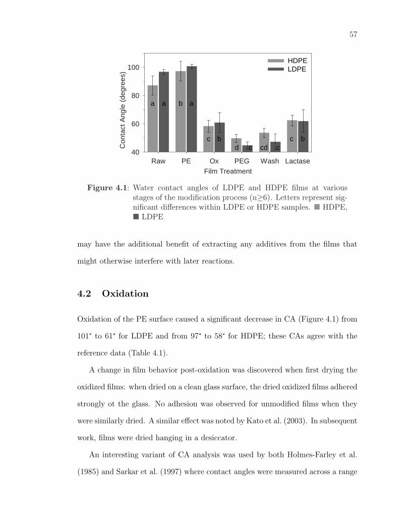
57
Film TreatmentRaw PE Ox PEG Wash Lactase
Con
tact
Ang
le (
degr
ees)
40
60
80
100HDPE LDPE
a b
cd cd
a a
b bc c
c
Figure 4.1: Water contact angles of LDPE and HDPE films at variousstages of the modification process (n≥6). Letters represent sig-nificant differences within LDPE or HDPE samples. � HDPE,� LDPE
may have the additional benefit of extracting any additives from the films that
might otherwise interfere with later reactions.
4.2 Oxidation
Oxidation of the PE surface caused a significant decrease in CA (Figure 4.1) from
101° to 61° for LDPE and from 97° to 58° for HDPE; these CAs agree with the
reference data (Table 4.1).
A change in film behavior post-oxidation was discovered when first drying the
oxidized films: when dried on a clean glass surface, the dried oxidized films adhered
strongly ot the glass. No adhesion was observed for unmodified films when they
were similarly dried. A similar effect was noted by Kato et al. (2003). In subsequent
work, films were dried hanging in a desiccator.
An interesting variant of CA analysis was used by both Holmes-Farley et al.
(1985) and Sarkar et al. (1997) where contact angles were measured across a range
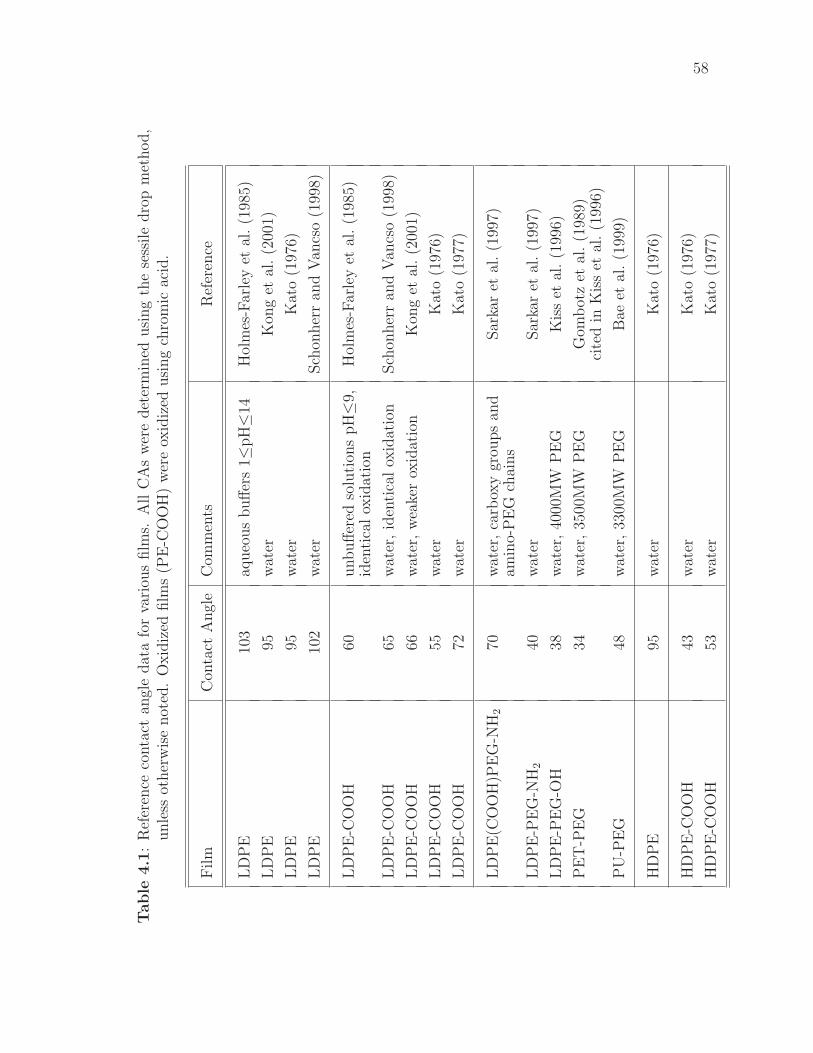
58
Table
4.1
:R
efer
ence
conta
ctan
gle
dat
afo
rva
riou
sfilm
s.A
llC
As
wer
edet
erm
ined
usi
ng
the
sess
ile
dro
pm
ethod,
unle
ssot
her
wis
enot
ed.
Oxid
ized
film
s(P
E-C
OO
H)
wer
eox
idiz
edusi
ng
chro
mic
acid
.
Film
Con
tact
Angl
eC
omm
ents
Ref
eren
ce
LD
PE
103
aqueo
us
buff
ers
1≤pH≤1
4H
olm
es-F
arle
yet
al.
(198
5)
LD
PE
95w
ater
Kon
get
al.
(200
1)
LD
PE
95w
ater
Kat
o(1
976)
LD
PE
102
wat
erSch
onher
ran
dV
ancs
o(1
998)
LD
PE
-CO
OH
60unbuff
ered
solu
tion
spH≤9
,H
olm
es-F
arle
yet
al.
(198
5)id
enti
cal
oxid
atio
n
LD
PE
-CO
OH
65w
ater
,id
enti
cal
oxid
atio
nSch
onher
ran
dV
ancs
o(1
998)
LD
PE
-CO
OH
66w
ater
,w
eake
rox
idat
ion
Kon
get
al.
(200
1)
LD
PE
-CO
OH
55w
ater
Kat
o(1
976)
LD
PE
-CO
OH
72w
ater
Kat
o(1
977)
LD
PE
(CO
OH
)PE
G-N
H2
70w
ater
,ca
rbox
ygr
oups
and
Sar
kar
etal
.(1
997)
amin
o-P
EG
chai
ns
LD
PE
-PE
G-N
H2
40w
ater
Sar
kar
etal
.(1
997)
LD
PE
-PE
G-O
H38
wat
er,
4000
MW
PE
GK
iss
etal
.(1
996)
PE
T-P
EG
34w
ater
,35
00M
WP
EG
Gom
bot
zet
al.
(198
9)ci
ted
inK
iss
etal
.(1
996)
PU
-PE
G48
wat
er,
3300
MW
PE
GB
aeet
al.
(199
9)
HD
PE
95w
ater
Kat
o(1
976)
HD
PE
-CO
OH
43w
ater
Kat
o(1
976)
HD
PE
-CO
OH
53w
ater
Kat
o(1
977)
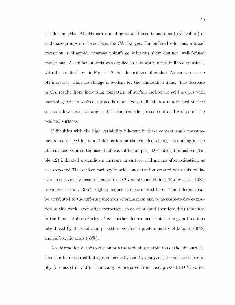
59
of solution pHs. At pHs corresponding to acid-base transitions (pKa values) of
acid/base groups on the surface, the CA changes. For buffered solutions, a broad
transition is observed, whereas unbuffered solutions show distinct, well-defined
transitions. A similar analysis was applied in this work, using buffered solutions,
with the results shown in Figure 4.2. For the oxidized films the CA decreases as the
pH increases, while no change is evident for the unmodified films. The decrease
in CA results from increasing ionization of surface carboxylic acid groups with
increasing pH; an ionized surface is more hydrophilic than a non-ionized surface
so has a lower contact angle. This confirms the presence of acid groups on the
oxidized surfaces.
Difficulties with the high variability inherent in these contact angle measure-
ments and a need for more information on the chemical changes occurring at the
film surface required the use of additional techniques. Dye adsorption assays (Ta-
ble 4.2) indicated a significant increase in surface acid groups after oxidation, as
was expected.The surface carboxylic acid concentration created with this oxida-
tion has previously been estimated to be 2.7 nmol/cm2 (Holmes-Farley et al., 1985;
Rasmussen et al., 1977), slightly higher than estimated here. The difference can
be attributed to the differing methods of estimation and to incomplete dye extrac-
tion in this work: even after extraction, some color (and therefore dye) remained
in the films. Holmes-Farley et al. further determined that the oxygen functions
introduced by the oxidation procedure consisted predominantly of ketones (40%)
and carboxylic acids (60%).
A side reaction of the oxidation process is etching or ablation of the film surface.
This can be measured both gravimetrically and by analyzing the surface topogra-
phy (discussed in §4.6). Film samples prepared from heat pressed LDPE varied
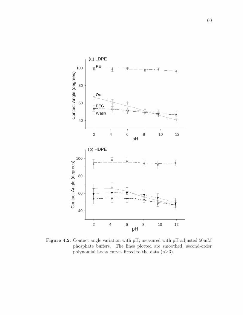
60
(a) LDPE
pH2 4 6 8 10 12
Con
tact
Ang
le (
degr
ees)
40
60
80
100 PE
Ox
PEG
Wash
(b) HDPE
pH2 4 6 8 10 12
Con
tact
Ang
le (
degr
ees)
40
60
80
100
Figure 4.2: Contact angle variation with pH; measured with pH adjusted 50mMphosphate buffers. The lines plotted are smoothed, second-orderpolynomial Loess curves fitted to the data (n≥3).
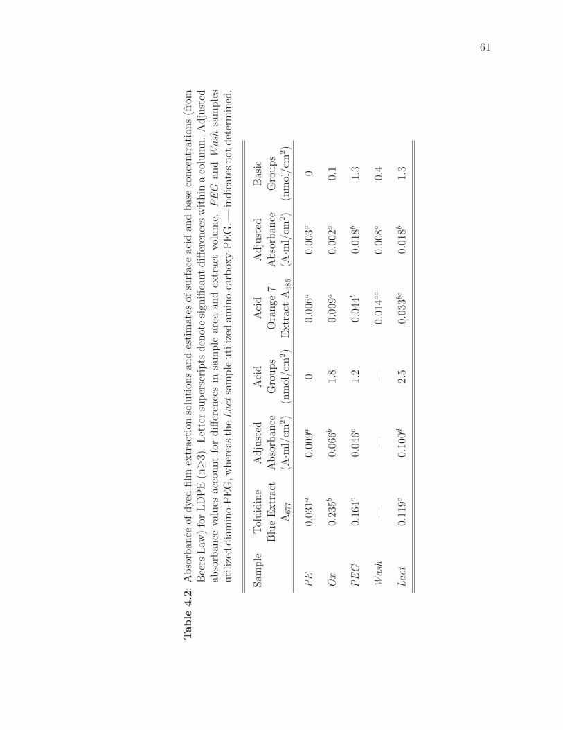
61
Table
4.2
:A
bso
rban
ceof
dye
dfilm
extr
acti
onso
luti
ons
and
esti
mat
esof
surf
ace
acid
and
bas
eco
nce
ntr
atio
ns
(fro
mB
eers
Law
)fo
rL
DP
E(n≥3
).L
ette
rsu
per
scri
pts
den
ote
sign
ifica
nt
diff
eren
ces
wit
hin
aco
lum
n.
Adju
sted
abso
rban
ceva
lues
acco
unt
for
diff
eren
ces
insa
mple
area
and
extr
act
volu
me.
PE
Gan
dW
ash
sam
ple
suti
lize
ddia
min
o-P
EG
,wher
eas
the
Lac
tsa
mple
uti
lize
dam
ino-
carb
oxy-P
EG
.—in
dic
ates
not
det
erm
ined
.
Sam
ple
Tol
uid
ine
Adju
sted
Aci
dA
cid
Adju
sted
Bas
ic
Blu
eE
xtr
act
Abso
rban
ceG
roups
Ora
nge
7A
bso
rban
ceG
roups
A677
(A·m
l/cm
2)
(nm
ol/c
m2)
Extr
act
A485
(A·m
l/cm
2)
(nm
ol/c
m2)
PE
0.03
1a0.
009a
00.
006a
0.00
3a0
Ox
0.23
5b0.
066b
1.8
0.00
9a0.
002a
0.1
PE
G0.
164c
0.04
6c1.
20.
044b
0.01
8b1.
3
Was
h—
——
0.01
4ac
0.00
8a0.
4
Lac
t0.
119c
0.10
0d2.
50.
033bc
0.01
8b1.
3
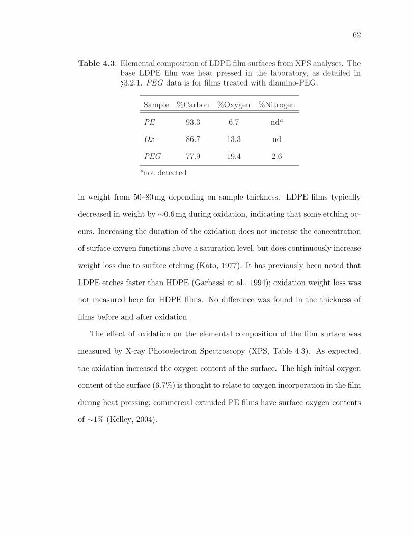
62
Table 4.3: Elemental composition of LDPE film surfaces from XPS analyses. Thebase LDPE film was heat pressed in the laboratory, as detailed in§3.2.1. PEG data is for films treated with diamino-PEG.
Sample %Carbon %Oxygen %Nitrogen
PE 93.3 6.7 nda
Ox 86.7 13.3 nd
PEG 77.9 19.4 2.6
anot detected
in weight from 50–80 mg depending on sample thickness. LDPE films typically
decreased in weight by ∼0.6 mg during oxidation, indicating that some etching oc-
curs. Increasing the duration of the oxidation does not increase the concentration
of surface oxygen functions above a saturation level, but does continuously increase
weight loss due to surface etching (Kato, 1977). It has previously been noted that
LDPE etches faster than HDPE (Garbassi et al., 1994); oxidation weight loss was
not measured here for HDPE films. No difference was found in the thickness of
films before and after oxidation.
The effect of oxidation on the elemental composition of the film surface was
measured by X-ray Photoelectron Spectroscopy (XPS, Table 4.3). As expected,
the oxidation increased the oxygen content of the surface. The high initial oxygen
content of the surface (6.7%) is thought to relate to oxygen incorporation in the film
during heat pressing; commercial extruded PE films have surface oxygen contents
of ∼1% (Kelley, 2004).
63
4.3 PEGylation
Key changes observed after PEGylation were further increased hydrophilicity, as
shown by the decreased contact angle (Figure 4.1); further increase in the surface
oxygen content (Table 4.3); the presence of basic groups on the film surface, as
shown by dye adsorption assays (Table 4.2); and the presence of nitrogen on the
film surface (Table 4.3). Neither surface nitrogen nor basic groups had previously
been detected, but both are consistent with the presence of primary amines at the
free terminus of tethered diamino-PEG chains. The combination of this evidence
confirms the PEG coupling reaction was successful.
Considering the effect of pH on contact angle, the shape of the curve differs
for the PEGylated film compared to the oxidized film. Although there is still an
overall decreasing trend for contact angle as pH increases, suggesting the presence
of unreacted acid groups, the difference between contact angles at low and high
pH is reduced for PEGylated films. The presence of unreacted acid groups on
the PEGylated film surface is confirmed by the dye adsorption results (Table 4.2).
There is the suggestion of a maximum on the contact angle curve for PEGylated
films, indicating increasing hydrophilicity (decreasing CA) at low pH, consistent
with the ionization of primary amine groups under acid conditions; this is most
apparent on the HDPE curve. The presence of unreacted acid groups and the
greater initial hydrophilicity of the PEGylated film may obscure the effect of amine
ionization on contact angle.
64
4.4 Washing
In early experiments investigating the stability of the PEGylated film surfaces, it
quickly became apparent that washing the PEGylated film caused increases in CA,
indicating some loss of the hydrophilic PEG coating. Even after vigorous washing,
however, the CA did not increase back to the level of the oxidized films. It was
hypothesized that some PEG was adsorbed to the surface, while the remainder was
covalently immobilized. This was confirmed by repeating the coupling procedures
without the activating agent (WSC) and with non-reactive PEG in the presence
of WSC and observing a decrease in contact angle both times (Figure 4.3). The
presence of adsorbed PEG is problematic as any active agent attached to adsorbed
PEG could readily migrate from the packaging material, violating non-migratory
principle. To prevent this, the adsorbed PEG must be removed prior to attaching
the active agent.
Three different wash solutions were tested: water (data not shown), pH 5 phos-
phate buffer and 10% aqueous ethanol. The washing protocol involved shaking
three test films for 1 hour in 30 ml of test solution at room temperature (Fig-
ure 4.4). Water washing resulted in a change similar to washing with pH 5 buffer.
Ethanol (10%, aq.) was found to be the most effective wash solution. More severe
treatments were not tested as it was considered they might adversely effect the
retention of the covalently immobilized PEG. Future testing with a pH 3 solution
would be advisable if the final film is to be used with acidic food products.
Figure 4.4 also allows the comparison of three different coupling methods based
on contact angle differences before and after washing. Unfortunately, a large vari-
ability was associated with these results, so statistically valid comparisons were
not possible. These results do indicate that higher levels of carbodiimide are not
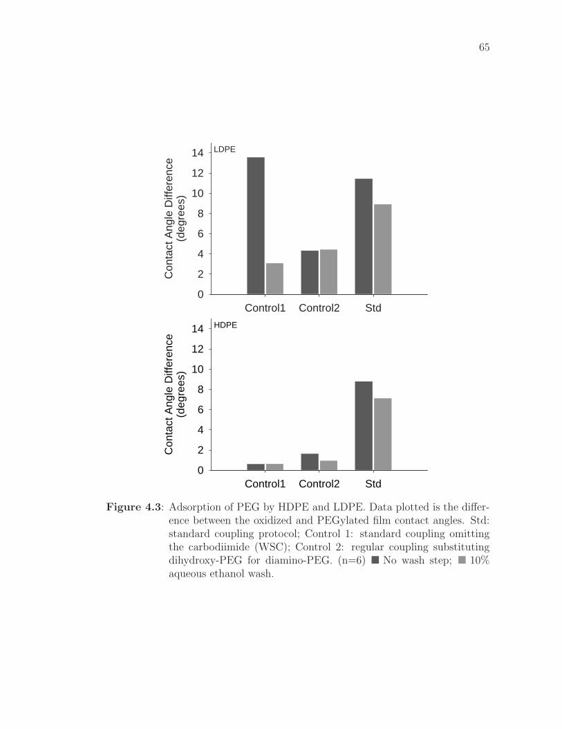
65
LDPE
Control1 Control2 Std
Con
tact
Ang
le D
iffer
ence
(deg
rees
)
0
2
4
6
8
10
12
14
HDPE
Control1 Control2 Std
Con
tact
Ang
le D
iffer
ence
(deg
rees
)
0
2
4
6
8
10
12
14
Figure 4.3: Adsorption of PEG by HDPE and LDPE. Data plotted is the differ-ence between the oxidized and PEGylated film contact angles. Std:standard coupling protocol; Control 1: standard coupling omittingthe carbodiimide (WSC); Control 2: regular coupling substitutingdihydroxy-PEG for diamino-PEG. (n=6) � No wash step; � 10%aqueous ethanol wash.
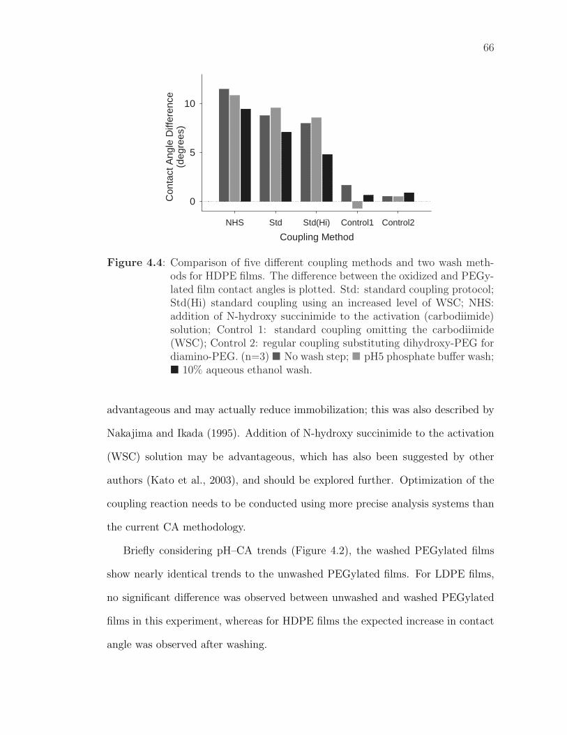
66
Coupling Method
NHS Std Std(Hi) Control1 Control2
Con
tact
Ang
le D
iffer
ence
(deg
rees
)
0
5
10
Figure 4.4: Comparison of five different coupling methods and two wash meth-ods for HDPE films. The difference between the oxidized and PEGy-lated film contact angles is plotted. Std: standard coupling protocol;Std(Hi) standard coupling using an increased level of WSC; NHS:addition of N-hydroxy succinimide to the activation (carbodiimide)solution; Control 1: standard coupling omitting the carbodiimide(WSC); Control 2: regular coupling substituting dihydroxy-PEG fordiamino-PEG. (n=3) � No wash step; � pH5 phosphate buffer wash;� 10% aqueous ethanol wash.
advantageous and may actually reduce immobilization; this was also described by
Nakajima and Ikada (1995). Addition of N-hydroxy succinimide to the activation
(WSC) solution may be advantageous, which has also been suggested by other
authors (Kato et al., 2003), and should be explored further. Optimization of the
coupling reaction needs to be conducted using more precise analysis systems than
the current CA methodology.
Briefly considering pH–CA trends (Figure 4.2), the washed PEGylated films
show nearly identical trends to the unwashed PEGylated films. For LDPE films,
no significant difference was observed between unwashed and washed PEGylated
films in this experiment, whereas for HDPE films the expected increase in contact
angle was observed after washing.
67
4.5 Lactase Attachment
After attachment of lactase to the washed, PEGylated PE film, the CA (Figure 4.1)
increased to 62° for both LDPE and HDPE. These films were indistinguishable from
oxidized films on the basis of CAs. The dye adsorption assays, however, allowed
a distinction to be made (Table 4.2); lactase coupling significantly increased the
number of both acidic and basic groups present on the film surface. The basic
groups present on the lactase-modified film do not originate from the PEG, as it
may initially appear. The PEGylated film analyzed for the dye-adsorption assays
was treated with diamino-PEG, providing the (free amine) basic groups found in
the dye adsorption assays; the PEGylated film used to create the lactase-modified
films, however, utilized amino-carboxy-PEG with a carboxylic acid free terminus.
Hence, the basic groups found on the lactase modified film can be assigned to the
lactase and not the underlying PEG.
Lactase attachment is discussed further in Chapter 5.
4.6 Surface Topography
Surface topography was investigated using Atomic Force Microscopy; it is of inter-
est both because it provides further evidence to the progress of the reactions and
because changes in film surface structure may have direct or indirect effects on
other analytical results, on film properties and on the activity of an immobilized
compound. Representative AFM images are shown in Figures 4.5 (LDPE) and 4.6
(HDPE).
It is immediately clear that there are significant differences between LDPE and
HDPE. The differences relate to the microstructure of the poly(ethylene) in each of
68
Figure 4.5: Surface topography of modified and unmodified LDPE films shownusing Atomic Force Microscopy. A: PE ; B: Ox ; C: PEG ; D: Wash.
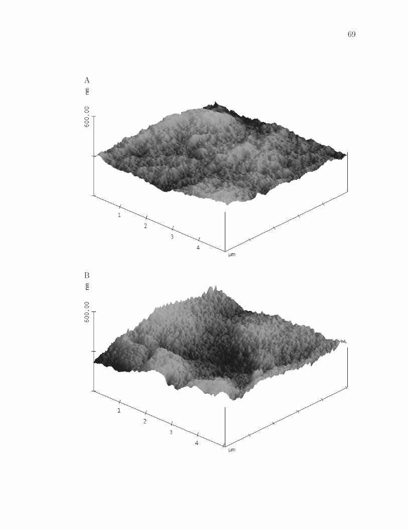
69
A
B
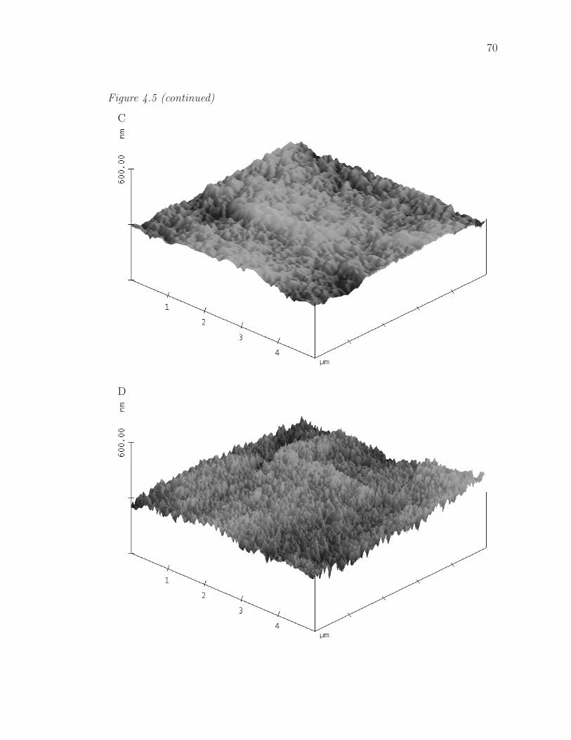
70
Figure 4.5 (continued)
C
D
71
Figure 4.6: Surface topography of modified and unmodified HDPE films shownusing Atomic Force Microscopy. A: PE ; B: Ox ; C: PEG ; D: Wash.

72
A
B
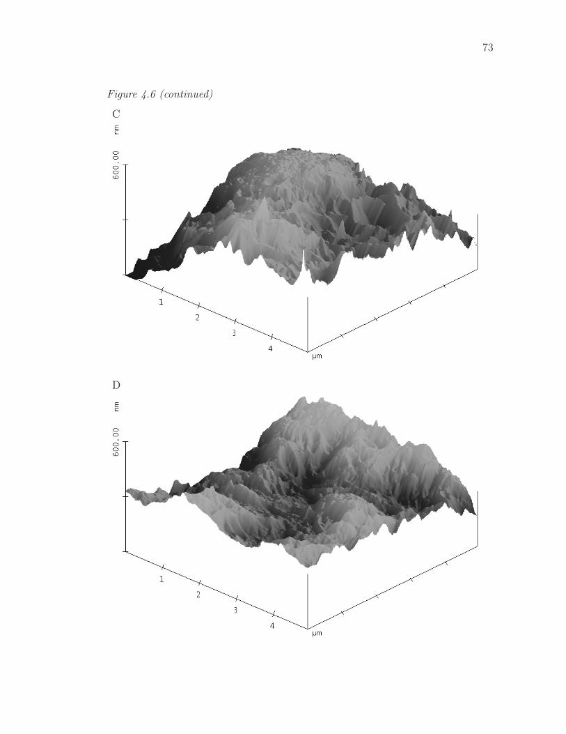
73
Figure 4.6 (continued)
C
D
74
these materials. LDPE is predominantly an amorphous material, whereas HDPE
has a high crystalline content — as can be seen from the large obtrusions and the
general angular nature of the surfaces visible in Figure 4.6.
The effects of the surface modifications are less obvious than the contrast be-
tween LDPE and HDPE, but are still distinct. These differences will first be
discussed for LDPE, for which they are most apparent. The unmodified film sur-
face is made up of broad, fairly low-angle peaks. After oxidation, however, the
surface is very different: it has a more regular pattern, with an increased number
of taller, steeper peaks. The change is due to etching and ablation of the film
surface by the chromic acid.
Most previously published images of chromic-acid etched poly(ethylene) films
are from Scanning Electron Microscopy (SEM) and do not directly correspond
to AFM images due to the differing aspects and sample preparation. Based on
the SEM images, however, the etching and surface structuring found here appears
to be similar to what has previously been observed (Kato, 1977; Schonherr and
Vancso, 1998; Lin and Tseng, 2001).
The unwashed PEGylated film surface is virtually flat, consisting of broad,
low-angle peaks. The troughs present on the oxidized film appear to have been
filled in by the PEG.
The washed PEGylated film is a different picture again, showing a similar
number of peaks to the oxidized film, but the peaks appear to be taller (greater
vertical scale) and steeper. The PEG which had previously filled in the troughs
between the peaks was, apparently, adsorbed to the surface, not covalently attached
and was removed by washing. To account for the increased height and steepness of
the peaks, it is hypothesized that the PEG chains preferentially attach to the tips
75
of the peaks on the oxidized film; the tips would be more accessible to PEG chains
in solution than the troughs (steric hindrance). PEG attachment at the tips of the
peaks would result in them becoming taller and steeper, as observed.
It has previously been noted (Kato et al., 2003) that polymer chains grafted
to an otherwise smooth surface tend to attach in clusters, leading to a surface
apparently coated with freestanding pillars. This is favorable from a thermody-
namic viewpoint1 and a similar process occurring at the most accessible points
(the tips of the peaks) on the oxidized LDPE film provides an additional reason
for preferential PEG attachment to the tips of the peaks present on the oxidized
film.
Similar differences to those just discussed for LDPE are somewhat apparent for
HDPE, but are largely obscured by the facetted, crystalline nature of the surface;
for HDPE it is more difficult to distinguish differences between the images.
4.7 Summary of Findings and Recommendations
Overall, the reaction sequence appears to be successful for covalent immobilization
of peptides on poly(ethylene) film, although it does require further optimization.
Optimization will require ready access to better analytical facilities than have been
available to date. Such facilities could consist of a more precise means for measuring
contact angles or reliable access to either XPS or ATR-FTIR. It is possible that
addition of NHS to the carbodiimide activation solution will improve the coupling
yield; this needs further investigation.
1reduction in the surface area created, compared to independent attachment ofgraft chains
Chapter 5
Bioactivity of Modified Films
The objective of this research was to attach bioactive peptides to poly(ethylene)
film to create non-migratory bioactive food packaging films. It has already been
shown (Chapter 4) that the coupling protocol appears to be effective at attach-
ing first PEG and then the peptide to PE film. In the following discussion, the
bioactivity of E14LKK and lactase modified films is investigated and the film mod-
ification reactions are examined further.
5.1 Antimicrobial Peptide Modified Films
The goal of antimicrobial films should be shelf-life extension. In this function they
must inhibit the growth of microbes, but are not required to totally eliminate them.
Antimicrobial films are less suited to food-safety applications requiring complete
pathogen elimination.
Shelf-life extension of microbially-limited products can occur in two ways: ex-
tension of the microbial lag period or reduction of the maximal microbial growth
rate. Antimicrobial films should be considered a hurdle technology in the food pro-
cessing and packaging mix; they are not a solve-all ”silver bullet”. An advantage of
antimicrobial packaging over current technologies could be in reducing the required
severity of processing conditions and/or reducing use of “artificial” preservatives
in the product formulation. There are also possible regulatory advantages and
technical benefits from isolating the antimicrobial effect at the packaging surface.
These advantages were discussed in more detail in Chapter 1.
76
77
An antimicrobial film was successfully developed (Table 5.1). Clear antibacte-
rial activity was evidenced in the solution assay for the deprotected film to which
side-chain-protected E14LKK had been attached.
5.1.1 Antimicrobial Assays
The method used to assess antimicrobial activity always needs to be carefully
considered when testing potentially antimicrobial films. Key considerations include
migration of the antimicrobial, mechanism of antimicrobial activity and similarity
of the assay to the target application of the films.
In this work, three separate assays for film antimicrobial activity were per-
formed using two different methods: a agar film-overlay method and a solution
method. The second overlay assay and the solution assay were performed using
the same set of films (Table 5.1). The first assay, a film overlay assay (data not
shown), was unsuccessful as the plate inoculum was too low — there were insuf-
ficient colonies per square centimeter, and the colonies were unevenly ditributed.
This demonstrated two difficulties with this method — the need for a very accu-
rate count of the inoculum and the need for uniform colony distribution across the
plates. These factors were understood prior to conducting the assay, but, despite
significant care, problems still arose.
In the second film overlay assay (Table 5.1) the inoculum was ideal for the
method. The uncovered colonies on the film plates could not be counted, however,
due to colony overgrowth and merging; a shorter incubation time prior to counting
the uncovered colonies or a lower incubation temperature would have prevented
this. Fortunately, a control plate was produced using a reduced inoculum from the
same culture, so an accurate ”uncovered” count was available.
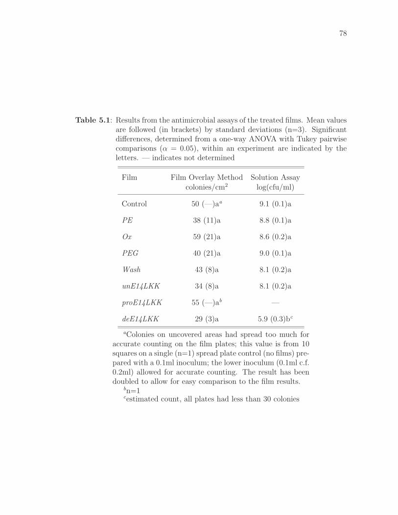
78
Table 5.1: Results from the antimicrobial assays of the treated films. Mean valuesare followed (in brackets) by standard deviations (n=3). Significantdifferences, determined from a one-way ANOVA with Tukey pairwisecomparisons (α = 0.05), within an experiment are indicated by theletters. — indicates not determined
Film Film Overlay Method Solution Assay
colonies/cm2 log(cfu/ml)
Control 50 (—)aa 9.1 (0.1)a
PE 38 (11)a 8.8 (0.1)a
Ox 59 (21)a 8.6 (0.2)a
PEG 40 (21)a 9.0 (0.1)a
Wash 43 (8)a 8.1 (0.2)a
unE14LKK 34 (8)a 8.1 (0.2)a
proE14LKK 55 (—)ab —
deE14LKK 29 (3)a 5.9 (0.3)bc
aColonies on uncovered areas had spread too much foraccurate counting on the film plates; this value is from 10squares on a single (n=1) spread plate control (no films) pre-pared with a 0.1ml inoculum; the lower inoculum (0.1ml c.f.0.2ml) allowed for accurate counting. The result has beendoubled to allow for easy comparison to the film results.
bn=1cestimated count, all plates had less than 30 colonies
79
Despite a relatively even colony distribution on the plates and an ideal colony
count, the film-overlay assay failed to show statistical significance between controls
and test samples with an ANOVA. The high variability of the control samples
suggests that, despite the appearance of uniformity, the bacterial distribution on
the test plates was inadequately uniform, suggesting a major shortcoming of this
method. By grouping the results from the control films (PE, Ox, PEG, Wash)
together and comparing the mean of the controls to the mean for each of the test
films (unE14LKK and deE14LKK ) with one-way t-tests (α = 0.05), the colony
count for deE14LKK was found to be significantly lower than the control counts.
The difference between unE14LKK and the control films was not significant at the
95% confidence level, but was at the 90% confidence level. This statistical analysis,
however, is not as valid as an ANOVA test.
These differences prompted the final solution-based antimicrobial assay, which
showed definitive differences between deE14LKK and the control films using an
ANOVA (Table 5.1). This suggests that the film overlay assay developed here
is insufficiently sensitive to adequately evaluate the antibacterial action of the
modified films. It does show some promise, but a completely uniform bacterial
distribution would be required for it to be useful. A uniform distribution may be
possible using an automated plate seeding technique as opposed to the manual
inoculation and spreading conducted here. The film-overlay assay was designed to
mimic the antibacterial effect of films used for bacterial inhibition on the surface
of solid foods. The solution assay was effective and is most suited to evaluating
films for use packaging liquid food products.
80
5.2 Lactase Modified Films
Lactase active films could produce reduced-lactose fluid milk products without re-
quiring special milk processing, potentially leading to significant cost savings and
simplified production for these products. A lower price for reduced-lactose milk
could significantly expand this market: according to the National Institutes of
Health (2003), 30–50 million Americans (10–18%) suffer from lactose intolerance,
with the rate significantly higher for certain ethnic groups (African-Americans,
up to 75%; Asian-Americans, up to 90%). Internationally, caucasians have the
lowest rate of lactose intolerance (between 2% and 25% depending on country),
with rates generally above 50% for Indians, South Americans, Africans and Asians;
incidences as high as 100% have been reported for some Asian populations (Sahi,
1994; Vesa et al., 2000). Worldwide it is estimated that two thirds of the adult
human population has some degree of lactose intolerance. Clearly the potential
market for reduced-lactose dairy products is significant, yet consumption of such
remains low. It may be postulated that this is in part due to the higher price
and lower availability of these products. Reduced-lactose milk is often 2–3 times
more expensive than regular milk and is frequently unavailable in smaller markets;
very few reduced-lactose processed dairy products are available. Lowering pro-
duction hurdles and decreasing production costs could significantly improve the
marketability of these products and potentially enrich the lives of many people.
The bioactivity of lactase modified films was measured by storage with a syn-
thetic lactose substrate, o-nitrophenyl-β-D-galactopyranoside (ONPG). Lactase
hydrolyses ONPG, releasing galactose and yellow o-nitrophenol; activity can be
monitored by measuring the color change of the solution with a visible spectropho-
tometer at 420nm. For free lactase the color change occurs extremely quickly,
81
however to allow for possible reduced activity of immobilized lactase, a three week
storage trial was conducted. Daily measurements could not be conducted without
compromising the sterility of the sample solutions — various bacteria are also able
to hydrolyze ONPG so their growth will also cause the solution to turn yellow —
so a single point measurement was used to investigate the possibility of activity.
Unfortunately, no activity was found for lactase-modified films, even after three
weeks of storage, regardless of PEG pre-treatment or the inclusion of lactose in
the coupling solution (Table 5.2).
To verify the coupling reactions and confirm the presence of lactase on the
films, XPS surface analysis was conducted on samples from the same reaction
batch as those used for the lactase activity assay, with some interesting results
(Table 5.3).The presence of lactase on the films was confirmed (distinct increase
in %N and %O), however no PEG appears to have been immobilized during the
previous modification step; there was no significant increase in oxygen content1
on PEG treatment. Although a small amount of nitrogen was observed, this
was significantly less than observed previously when PEG attachment was con-
firmed (Table 4.3). The previous analysis was conducted on PEG films treated
with diamino-PEG, whereas the coupling progression for peptide attachment uses
amino-carboxy-PEG. Diamino-PEG was used in early work because it was avail-
able and was significantly less expensive than amino-carboxy-PEG. It had been
assumed that the reactions would proceed equivalently for the two compounds, but
this was clearly not the case. The differences between the two PEGs prevented the
1in the first XPS analyses, heat pressed films with a high initial %O were used.In the later analysis shown in Table 5.3 commercially extruded LDPE films wereused; a typical surface oxygen content of commercial LDPE films is 1% (Kelley,2004) so an oxidized value of 7.6% confirms the oxidation. Untreated samples werenot analyzed.
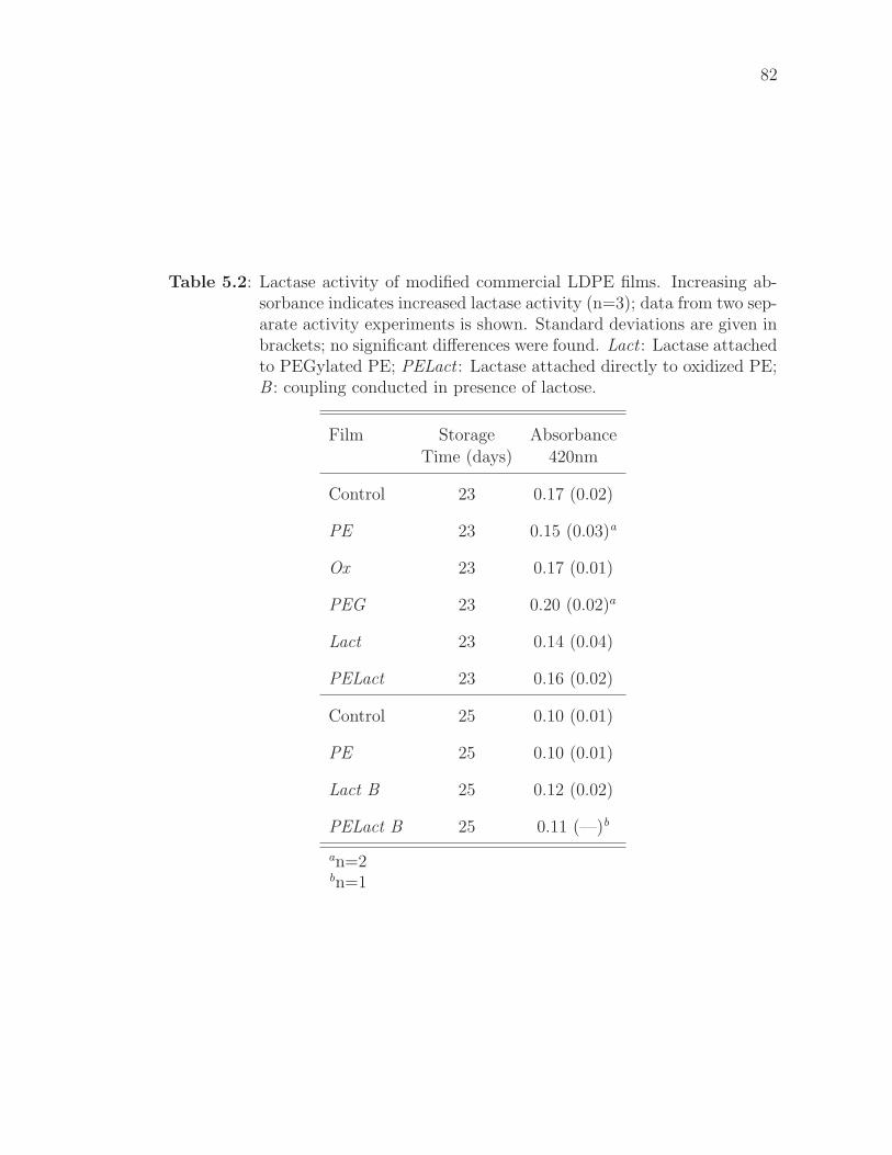
82
Table 5.2: Lactase activity of modified commercial LDPE films. Increasing ab-sorbance indicates increased lactase activity (n=3); data from two sep-arate activity experiments is shown. Standard deviations are given inbrackets; no significant differences were found. Lact : Lactase attachedto PEGylated PE; PELact : Lactase attached directly to oxidized PE;B : coupling conducted in presence of lactose.
Film Storage Absorbance
Time (days) 420nm
Control 23 0.17 (0.02)
PE 23 0.15 (0.03)a
Ox 23 0.17 (0.01)
PEG 23 0.20 (0.02)a
Lact 23 0.14 (0.04)
PELact 23 0.16 (0.02)
Control 25 0.10 (0.01)
PE 25 0.10 (0.01)
Lact B 25 0.12 (0.02)
PELact B 25 0.11 (—)b
an=2bn=1
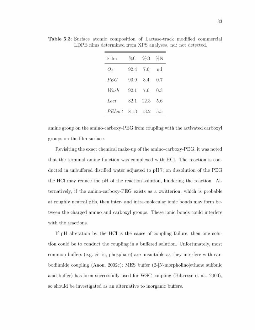
83
Table 5.3: Surface atomic composition of Lactase-track modified commercialLDPE films determined from XPS analyses. nd: not detected.
Film %C %O %N
Ox 92.4 7.6 nd
PEG 90.9 8.4 0.7
Wash 92.1 7.6 0.3
Lact 82.1 12.3 5.6
PELact 81.3 13.2 5.5
amine group on the amino-carboxy-PEG from coupling with the activated carboxyl
groups on the film surface.
Revisiting the exact chemical make-up of the amino-carboxy-PEG, it was noted
that the terminal amine function was complexed with HCl. The reaction is con-
ducted in unbuffered distilled water adjusted to pH 7; on dissolution of the PEG
the HCl may reduce the pH of the reaction solution, hindering the reaction. Al-
ternatively, if the amino-carboxy-PEG exists as a zwitterion, which is probable
at roughly neutral pHs, then inter- and intra-molecular ionic bonds may form be-
tween the charged amino and carboxyl groups. These ionic bonds could interfere
with the reactions.
If pH alteration by the HCl is the cause of coupling failure, then one solu-
tion could be to conduct the coupling in a buffered solution. Unfortunately, most
common buffers (e.g. citric, phosphate) are unsuitable as they interfere with car-
bodiimide coupling (Anon, 2002c); MES buffer (2-[N-morpholino]ethane sulfonic
acid buffer) has been successfully used for WSC coupling (Biltresse et al., 2000),
so should be investigated as an alternative to inorganic buffers.
84
If zwitterion formation is interfering with the coupling, a solution could be
to adjust the pH of the coupling solution; an optimal pH may exist where the
zwitterion form of the molecule is not predominant, but the coupling reaction
still proceeds. The use of buffered solutions is recommended if pH modification is
investigated. As well as pH, other reaction conditions can also be varied to enhance
PEG attachment. Kingshott et al. (2002, 2003) conducted PEG immobilization
at elevated temperatures and salt concentrations in what they term “cloud-point”
grafting. The temperature selected was the lower critical solution temperature
of the PEG (60�); under these conditions PEG solubility is reduced and surface
coupling is enhanced. This should also be investigated. Before any further reaction
optimization can be conducted, however, ready access to appropriate equipment
needs to be arranged to monitor the reaction outcomes; contact angles have proven
insufficient for this task.
In summary, lactase was successfully attached directly to PE film, but no activ-
ity was found in this state. Activity was not improved by including lactose in the
coupling buffer. As previously mentioned, the purpose of hydrophilic spacers is to
separate the hydrophilic peptides from the hydrophobic PE to prevent disruptions
to the peptide structure (§1.2.3). It is assumed that such disruptions are the rea-
son for the lack of lactase activity. The effectiveness of the spacer at ameliorating
this problem, however, cannot be experimentally evaluated at this time due to the
difficulties with amino-carboxy-PEG attachment. It is likely that inclusion of a
PEG spacer will improve lactase activity, as other research has shown excellent
activity for PEGylated-lactase (Talbert, 2004).
85
5.3 Summary of Findings and Recommendations
Lactase immobilization without PEGylation did not produce a lactase-active LDPE
film, regardless of lactose inclusion in the coupling buffer. Amino-carboxy-PEG
was not successfully attached to the oxidized LDPE surface, despite success with
diamino-PEG attachment. Further investigation of the coupling reactions is re-
quired to optimize coupling of amino-carboxy-PEG. It is unknown if lactase im-
mobilized on a PEGylated LDPE film will be active, but this is considered likely
based the confirmed activity of PEGylated lactase (Talbert, 2004).
An antibacterial LDPE film was successfully produced. The film was prepared
by immobilizing antimicrobial peptide E14LKK (with Boc-protected side-chain
amines) onto PEG-treated LDPE film, then deprotecting the peptide. Antibacte-
rial activity was demonstrated against E.coli in a solution assay.
Chapter 6
Effect of Modifications on Film
Properties
When modifying an existing material for enhanced performance, e.g. by providing
targeted bioactivity, it is necessary to consider the original properties and charac-
teristics of the material and how they relate to its production, use and disposal.
Typically, the intended applications of the new material are similar to current
applications of the base material; for the modified material to be suitable for exist-
ing applications, key characteristics of the original material must not be adversely
affected by the modifications, or, if they are affected, the changes must be under-
stood and adjusted for during process or package design. This chapter discusses
the affect of these film surface modifications on characteristics important to the
production, use and disposal of PE packaging films.
Each of the tests conducted is related to important properties of PE film in
real packages: transition (melting) temperatures are important in many processing
operations and for heat sealing; tensile properties are important when a film is a
structural component of a package and also in many processing operations; coeffi-
cients of friction are important in film processing and also in the transport, display
and handling of packages; optical properties are important for the communication
(marketing) function of the package; and moisture permeability is important for
the preservation of many food products.
Material properties can be classified as either surface-sensitive or surface-
insensitive. Surface-sensitive properties are ones significantly affected by nanoscale
86
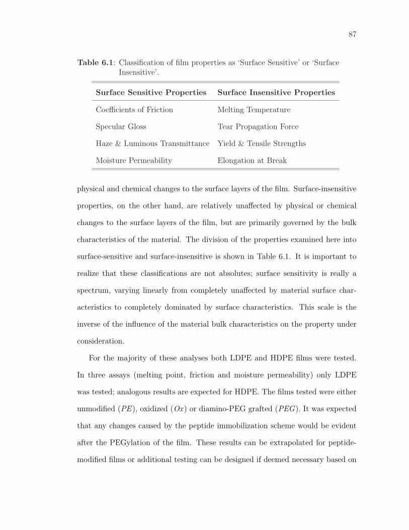
87
Table 6.1: Classification of film properties as ‘Surface Sensitive’ or ‘SurfaceInsensitive’.
Surface Sensitive Properties Surface Insensitive Properties
Coefficients of Friction Melting Temperature
Specular Gloss Tear Propagation Force
Haze & Luminous Transmittance Yield & Tensile Strengths
Moisture Permeability Elongation at Break
physical and chemical changes to the surface layers of the film. Surface-insensitive
properties, on the other hand, are relatively unaffected by physical or chemical
changes to the surface layers of the film, but are primarily governed by the bulk
characteristics of the material. The division of the properties examined here into
surface-sensitive and surface-insensitive is shown in Table 6.1. It is important to
realize that these classifications are not absolutes; surface sensitivity is really a
spectrum, varying linearly from completely unaffected by material surface char-
acteristics to completely dominated by surface characteristics. This scale is the
inverse of the influence of the material bulk characteristics on the property under
consideration.
For the majority of these analyses both LDPE and HDPE films were tested.
In three assays (melting point, friction and moisture permeability) only LDPE
was tested; analogous results are expected for HDPE. The films tested were either
unmodified (PE ), oxidized (Ox ) or diamino-PEG grafted (PEG). It was expected
that any changes caused by the peptide immobilization scheme would be evident
after the PEGylation of the film. These results can be extrapolated for peptide-
modified films or additional testing can be designed if deemed necessary based on
88
these results.
Results were analyzed using General Linear Models with the film (LDPE or
HDPE), the treatment (PE , Ox or PEG) and the interaction term as predictor
variables. Significant differences were evaluated using Tukey pairwise comparisons
with an overall confidence level of 95% (α = 0.05). In all bar charts, letters within
the bars indicate significant differences within each group of bars: the same letter
denotes no significant difference; different letters denote significant differences. For
groups of treatments with no significant differences, the values given in the text
for that group are the mean across all treatments within the group.
6.1 Physical and Mechanical Properties
Most of the physical and mechanical properties of polymer films are dependent
predominantly on the bulk properties of the material; these properties can therefore
be considered surface insensitive. In other words, they are relatively independent
of the state of the film surface, so it was hypothesized that they would be minimally
affected by these surface modifications. The exception to this, amongst the physical
and mechanical properties examined, are the coefficients of friction, which are
inherently surface sensitive; it was hypothesized that the modifications would affect
the coefficients of friction.
6.1.1 Transition Temperatures
For a mixed polymer system with distinct, homogenous regions, as in the case of
a brush-grafted polymer such as PEG-grafted PE, multiple transition tempera-
tures may be observed; the individual transition temperatures depend on the bulk
properties of the different homogenous regions. It would not, therefore, be unex-
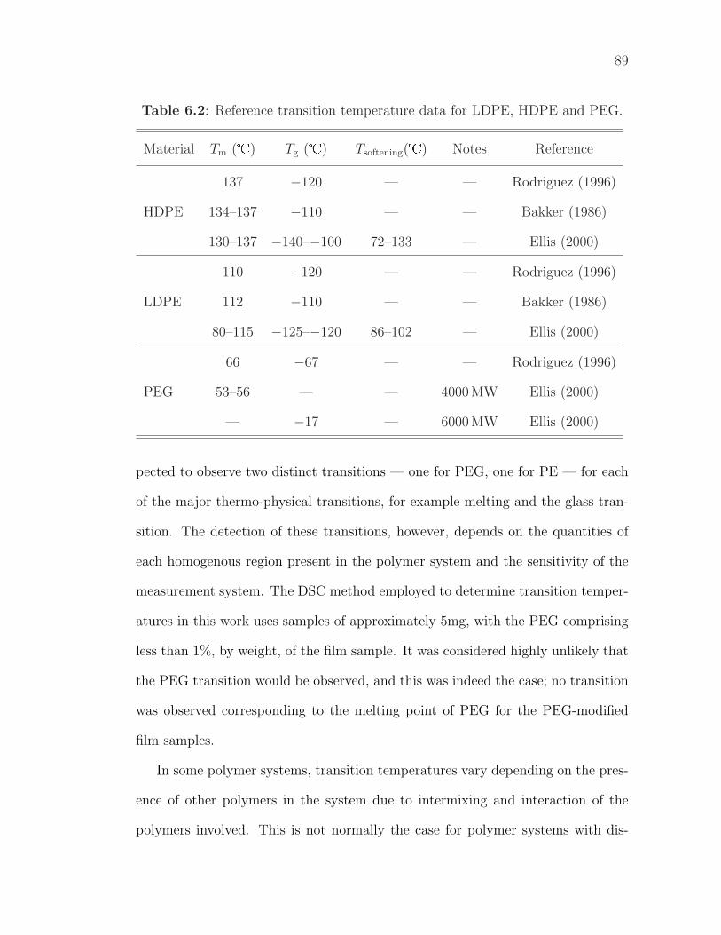
89
Table 6.2: Reference transition temperature data for LDPE, HDPE and PEG.
Material Tm (�) Tg (�) Tsoftening(�) Notes Reference
137 −120 — — Rodriguez (1996)
HDPE 134–137 −110 — — Bakker (1986)
130–137 −140–−100 72–133 — Ellis (2000)
110 −120 — — Rodriguez (1996)
LDPE 112 −110 — — Bakker (1986)
80–115 −125–−120 86–102 — Ellis (2000)
66 −67 — — Rodriguez (1996)
PEG 53–56 — — 4000 MW Ellis (2000)
— −17 — 6000 MW Ellis (2000)
pected to observe two distinct transitions — one for PEG, one for PE — for each
of the major thermo-physical transitions, for example melting and the glass tran-
sition. The detection of these transitions, however, depends on the quantities of
each homogenous region present in the polymer system and the sensitivity of the
measurement system. The DSC method employed to determine transition temper-
atures in this work uses samples of approximately 5mg, with the PEG comprising
less than 1%, by weight, of the film sample. It was considered highly unlikely that
the PEG transition would be observed, and this was indeed the case; no transition
was observed corresponding to the melting point of PEG for the PEG-modified
film samples.
In some polymer systems, transition temperatures vary depending on the pres-
ence of other polymers in the system due to intermixing and interaction of the
polymers involved. This is not normally the case for polymer systems with dis-
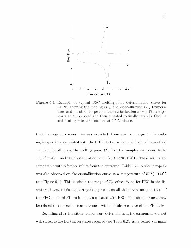
90
Temperature (°C)160100806040
-1
120 14020
0
2
1
Tpc
B
A
Tpm
Figure 6.1: Example of typical DSC melting-point determination curve forLDPE, showing the melting (Tm) and crystallization (Tpc tempera-tures and the shoulder-peak on the crystallization curve. The samplestarts at A, is cooled and then reheated to finally reach B. Coolingand heating rates are constant at 10�/minute.
tinct, homogenous zones. As was expected, there was no change in the melt-
ing temperature associated with the LDPE between the modified and unmodified
samples. In all cases, the melting point (Tpm) of the samples was found to be
110.9(±0.4)� and the crystallization point (Tpc) 93.9(±0.4)�. These results are
comparable with reference values from the literature (Table 6.2). A shoulder-peak
was also observed on the crystallization curve at a temperature of 57.8(±0.4)�
(see Figure 6.1). This is within the range of Tm values found for PEG in the lit-
erature, however this shoulder peak is present on all the curves, not just those of
the PEG-modified PE, so it is not associated with PEG. This shoulder-peak may
be related to a molecular rearrangement within or phase change of the PE lattice.
Regarding glass transition temperature determination, the equipment was not
well suited to the low temperatures required (see Table 6.2). An attempt was made
91
to determine glass transitions using manual liquid nitrogen cooling of the sample
cell, however the sample could not be cooled sufficiently and the cooling rate was
too slow and too variable for an accurate determination to be made. An estimated
glass transition (n=1) was found at −26.9� (Tmg) for unmodified LDPE film.
This does not approximate the reference values and may be a ‘phantom peak’; it
may have resulted from the inability to adequately control sample temperature.
It was considered very unlikely that a Tg would be observed for the PEG based
on the Tm results and given that a secondary transition (Tg) is typically harder to
detect than a primary transition (Tm). As such, no further attempts were made to
measure Tg values.
6.1.2 Tensile Properties
Tensile properties of a material, and structural properties in general, are dominated
by the bulk characteristics of the material. Surface specific changes typically have
negligible effect, although surface roughness can affect stress concentration and,
correspondingly, mechanical properties; significant differences were not expected
between the properties of the modified and unmodified films. Significant differences
were expected between LDPE and HDPE films, and this was indeed the case for
all the tensile properties examined.
For LDPE, no significant differences were found between modified and un-
modified films for Tensile Strength (12.6±0.4 MPa, Figure 6.2), Yield Strength
(10.2±0.2 MPa), Elongation at Break (530±50 %) or Normalized Tear Propagation
Force (32.7±0.9 N/mm, Figure 6.3), as was expected. All properties were measured
in the machine direction. The measured Tensile and Yield Strengths are within
the ranges presented by the reference values (Table 6.3). The Elongation at Break
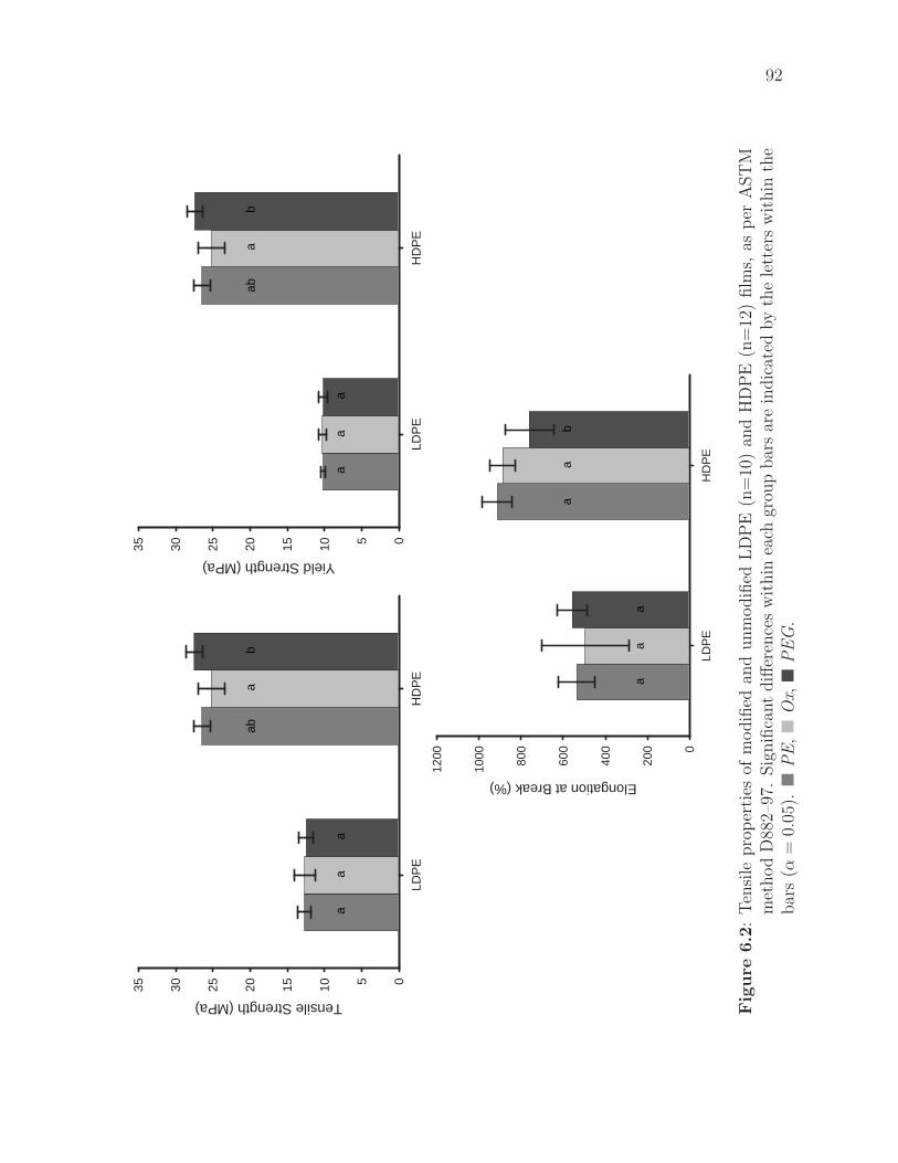
92
LDP
EH
DP
E
Tensile Strength (MPa)
05101520253035
aa
a
aba
b
LDP
EH
DP
E
Yield Strength (MPa)
05101520253035
aa
a
aba
b
LDP
EH
DP
E
Elongation at Break (%)
0
200
400
600
800
1000
1200
aa
a
aa
b
Fig
ure
6.2
:T
ensi
lepro
per
ties
ofm
odifi
edan
dunm
odifi
edL
DP
E(n
=10
)an
dH
DP
E(n
=12
)film
s,as
per
AST
Mm
ethod
D88
2–97
.Sig
nifi
cant
diff
eren
ces
wit
hin
each
grou
pbar
sar
ein
dic
ated
by
the
lett
ers
wit
hin
the
bar
s(α
=0.
05).�
PE
,�
Ox,�
PE
G.
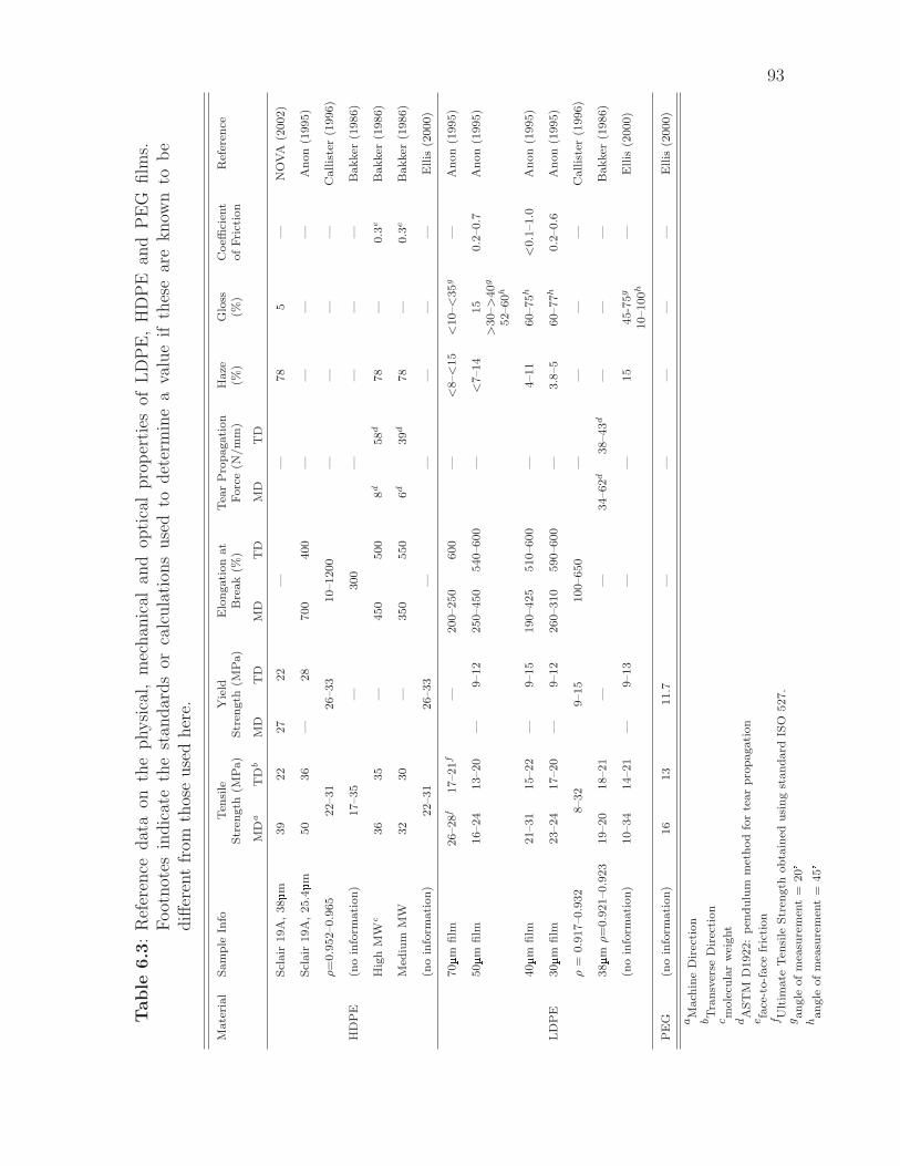
93
Table
6.3
:R
efer
ence
dat
aon
the
physi
cal,
mec
han
ical
and
opti
cal
pro
per
ties
ofL
DP
E,
HD
PE
and
PE
Gfilm
s.F
oot
not
esin
dic
ate
the
stan
dar
ds
orca
lcula
tion
suse
dto
det
erm
ine
ava
lue
ifth
ese
are
know
nto
be
diff
eren
tfr
omth
ose
use
dher
e.
Mate
rial
Sam
ple
Info
Ten
sile
Yie
ldE
lon
gati
on
at
Tea
rP
rop
agati
on
Haze
Glo
ssC
oeffi
cien
tR
efer
ence
Str
ength
(MP
a)
Str
ength
(MP
a)
Bre
ak
(%)
Forc
e(N
/m
m)
(%)
(%)
of
Fri
ctio
n
MD
aT
Db
MD
TD
MD
TD
MD
TD
Scl
air
19A
,38µm
39
22
27
22
——
78
5—
NO
VA
(2002)
Scl
air
19A
,25.4µm
50
36
—28
700
400
——
——
An
on
(1995)
ρ=
0.9
52–0.9
65
22–31
26–33
10–1200
——
——
Call
iste
r(1
996)
HD
PE
(no
info
rmati
on
)17–35
—300
——
——
Bakker
(1986)
Hig
hM
Wc
36
35
—450
500
8d
58d
78
—0.3
eB
akker
(1986)
Med
ium
MW
32
30
—350
550
6d
39d
78
—0.3e
Bakker
(1986)
(no
info
rmati
on
)22–31
26–33
——
——
—E
llis
(2000)
70µm
film
26–28
f17–21f
—200–250
600
—<
8–<
15
<10–<
35
g—
An
on
(1995)
50µm
film
16–24
13–20
—9–12
250–450
540–600
—<
7–14
15
0.2
–0.7
An
on
(1995)
>30–>
40g
52–60
h
40µm
film
21–31
15–22
—9–15
190–425
510–600
—4–11
60–75h
<0.1
–1.0
An
on
(1995)
LD
PE
30µm
film
23–24
17–20
—9–12
260–310
590–600
—3.8
–5
60–77h
0.2
–0.6
An
on
(1995)
ρ=
0.9
17–0.9
32
8–32
9–15
100–650
——
——
Call
iste
r(1
996)
38µm
ρ=
0.9
21–0.9
23
19–20
18–21
——
34–62d
38–43d
——
—B
akker
(1986)
(no
info
rmati
on
)10–34
14–21
—9–13
——
15
45-7
5g
—E
llis
(2000)
10–100h
PE
G(n
oin
form
ati
on
)16
13
11.7
——
——
—E
llis
(2000)
aM
ach
ine
Dir
ecti
on
bT
ran
sver
seD
irec
tion
cm
ole
cula
rw
eight
dA
ST
MD
1922:
pen
du
lum
met
hod
for
tear
pro
pagati
on
efa
ce-t
o-f
ace
fric
tion
f Ult
imate
Ten
sile
Str
ength
ob
tain
edu
sin
gst
an
dard
ISO
527.
gan
gle
of
mea
sure
men
t=
20°
han
gle
of
mea
sure
men
t=
45°
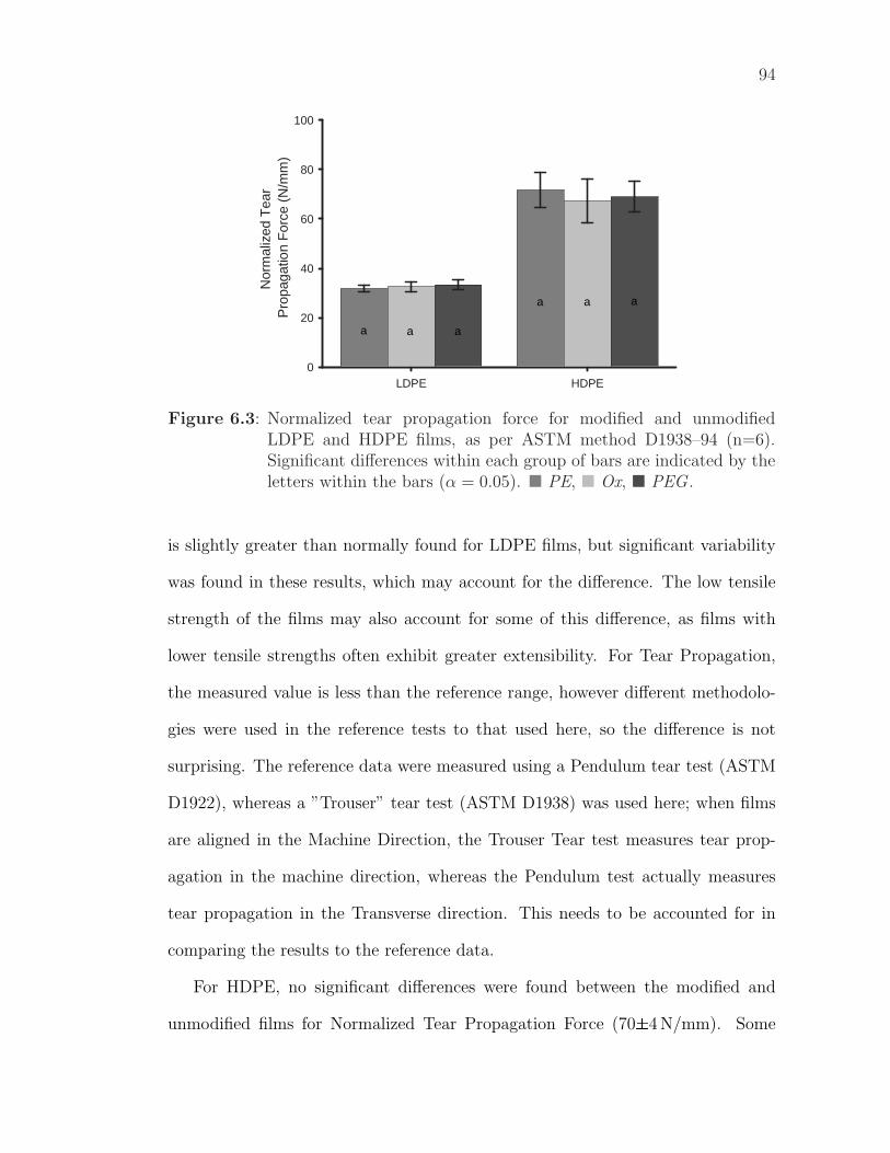
94
LDPE HDPE
Nor
mal
ized
Tea
r P
ropa
gatio
n F
orce
(N
/mm
)
0
20
40
60
80
100
a a a
a a a
Figure 6.3: Normalized tear propagation force for modified and unmodifiedLDPE and HDPE films, as per ASTM method D1938–94 (n=6).Significant differences within each group of bars are indicated by theletters within the bars (α = 0.05). � PE, � Ox, � PEG .
is slightly greater than normally found for LDPE films, but significant variability
was found in these results, which may account for the difference. The low tensile
strength of the films may also account for some of this difference, as films with
lower tensile strengths often exhibit greater extensibility. For Tear Propagation,
the measured value is less than the reference range, however different methodolo-
gies were used in the reference tests to that used here, so the difference is not
surprising. The reference data were measured using a Pendulum tear test (ASTM
D1922), whereas a ”Trouser” tear test (ASTM D1938) was used here; when films
are aligned in the Machine Direction, the Trouser Tear test measures tear prop-
agation in the machine direction, whereas the Pendulum test actually measures
tear propagation in the Transverse direction. This needs to be accounted for in
comparing the results to the reference data.
For HDPE, no significant differences were found between the modified and
unmodified films for Normalized Tear Propagation Force (70±4 N/mm). Some
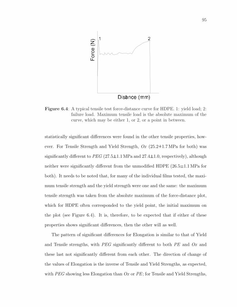
95
Distance (mm)
1 2
Figure 6.4: A typical tensile test force-distance curve for HDPE. 1: yield load; 2:failure load. Maximum tensile load is the absolute maximum of thecurve, which may be either 1, or 2, or a point in between.
statistically significant differences were found in the other tensile properties, how-
ever. For Tensile Strength and Yield Strength, Ox (25.2±1.7 MPa for both) was
significantly different to PEG (27.5±1.1 MPa and 27.4±1.0, respectively), although
neither were significantly different from the unmodified HDPE (26.5±1.1 MPa for
both). It needs to be noted that, for many of the individual films tested, the maxi-
mum tensile strength and the yield strength were one and the same: the maximum
tensile strength was taken from the absolute maximum of the force-distance plot,
which for HDPE often corresponded to the yield point, the initial maximum on
the plot (see Figure 6.4). It is, therefore, to be expected that if either of these
properties shows significant differences, then the other will as well.
The pattern of significant differences for Elongation is similar to that of Yield
and Tensile strengths, with PEG significantly different to both PE and Ox and
these last not significantly different from each other. The direction of change of
the values of Elongation is the inverse of Tensile and Yield Strengths, as expected,
with PEG showing less Elongation than Ox or PE ; for Tensile and Yield Strengths,
96
PEG had higher strength values than Ox.
The slight increase in the tensile and yield strengths between the oxidized
and PEGylated films may be explained from the surface topographies and layered
structures of these films. First, consider the surface topography: a rough surface
could lead to a reduction in film strength by providing more sites for tear initiation
(sharp concavities where the stress will concentrate) than would a smooth surface.
The greater the number of initiation sites, the greater the probability that the film
will tear at a lower load. Additionally, the sharper the concavities, the greater the
stress concentration and the increased probability of tear initiation. Considering
the films examined here and comparing the tensile data to the surface topography
(AFM analysis, §4.6), it is clear that the oxidized surface is both weaker and
significantly rougher, with an increased number of sharper peaks, and assumably
sharper valleys, than the PEGylated film (pre-washing). From the tensile data, it
appears that the oxidized film is slightly weaker (lower tensile and yield strengths)
than the untreated film, and that the untreated film is slightly weaker than the
PEGylated film, although these differences are not statistically significant. These
trends match the topographical trends shown in the AFM analysis of the film
surfaces.
Although surface roughness is one plausible explanation of the differences in
film strengths, the microstructure of the films should also be considered. The
important concept here is effect of layering on film strength and the layering present
in the PEGylated films, which have a PE core coated on both sides with a thin
layer of PEG. If a film is coated with complete layers of a material with differing
tensile properties, then the overall properties of the film may be effected, even if
the coating is thin. The important considerations are the strength of the coating
97
layers and the ease of tear initiation in these layers compared to the bulk of the
film. Given that PEG films are typically weaker1 than PE films, PEG coating will
not increase the strength of PE films by tensile reinforcement. If tear initiation is
more difficult in PEG than in PE, however, then the PEG coating could lead to
stronger films. No data on this was available.
Comparing the measured HDPE data to reference values, for all treatments
we see that the measured values for Tensile and Yield strengths are similar to the
reference data, regardless of significant differences between treatments. The mea-
sured Elongation at Break is slightly higher than the reference range, however the
differences are not considered significant; as for LDPE these differences are most
likely related to the high variability and relatively low tensile strength of the test
films. Even when there are differences between the modified and unmodified films,
these differences are not significant compared to the normal range of HDPE prop-
erties and should not affect the application of the modified films. The differences
may, however, need to be accounted for during package and process design. As
for LDPE, the measured Tear Propagation Force does not fall within the range of
the reference data; again, this is most likely related to the differing measurement
techniques used.
6.1.3 Coefficients of Friction
Coefficients of friction are important in processing situations when webs of film
are being run through and over equipment. The static coefficient of friction is
a measure of the force that must be overcome to start a stationary film moving
over a surface, so is important for process start-up. The kinetic coefficient of
1personal observation
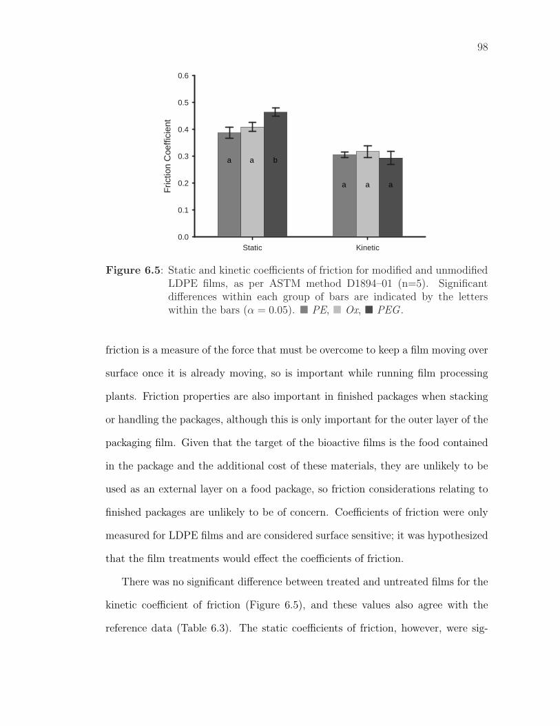
98
Static Kinetic
Fric
tion
Coe
ffici
ent
0.0
0.1
0.2
0.3
0.4
0.5
0.6
a a b
a a a
Figure 6.5: Static and kinetic coefficients of friction for modified and unmodifiedLDPE films, as per ASTM method D1894–01 (n=5). Significantdifferences within each group of bars are indicated by the letterswithin the bars (α = 0.05). � PE, � Ox, � PEG .
friction is a measure of the force that must be overcome to keep a film moving over
surface once it is already moving, so is important while running film processing
plants. Friction properties are also important in finished packages when stacking
or handling the packages, although this is only important for the outer layer of the
packaging film. Given that the target of the bioactive films is the food contained
in the package and the additional cost of these materials, they are unlikely to be
used as an external layer on a food package, so friction considerations relating to
finished packages are unlikely to be of concern. Coefficients of friction were only
measured for LDPE films and are considered surface sensitive; it was hypothesized
that the film treatments would effect the coefficients of friction.
There was no significant difference between treated and untreated films for the
kinetic coefficient of friction (Figure 6.5), and these values also agree with the
reference data (Table 6.3). The static coefficients of friction, however, were sig-
99
nificantly different for the different film treatments, although the measured values
were all within the range of the reference data2.
Coefficients of friction are affected by surface roughness and the material (e.g.
PE or PEG) of the surface layer; environmental variables are controlled in the test
protocol. There are significant differences in the surface roughness of the treated
films (see §4.6) and the PEGylated film has an external PEG layer3, so there was
expected to be differences in the coefficients of friction for the variously treated
films.
The lack of difference in the kinetic coefficient of friction between the native
and PEG treated films is of some concern, as this could be explained by abrasion or
deformation (Kato et al., 2003) of the PEG layer due to the shear forces developed
in sliding the film over the test surface. Any loss of or damage to the PEG layer
could also lead to loss of an attached bioactive compound and a reduction in film
bioactivity. This needs to be further investigated before commercialization of the
modified films.
6.2 Optical Properties
Optical properties are affected by interfaces between materials with differing re-
fractive indices, variations in surface roughness and the existence of irregularities
or inhomogeneities in the film greater in size than the wavelength of visible light
(Robertson, 1993; Jenkins and Harrington, 1991; Briston, 1983). PEG has a differ-
ent refractive index to PE, so the PEG layer is likely to alter the optical properties
2no distinction was made between static and kinetic coefficients in the referenceliterature
3although no data was found for the friction properties of PEG, it is anticipatedthat these are different to those of PE.
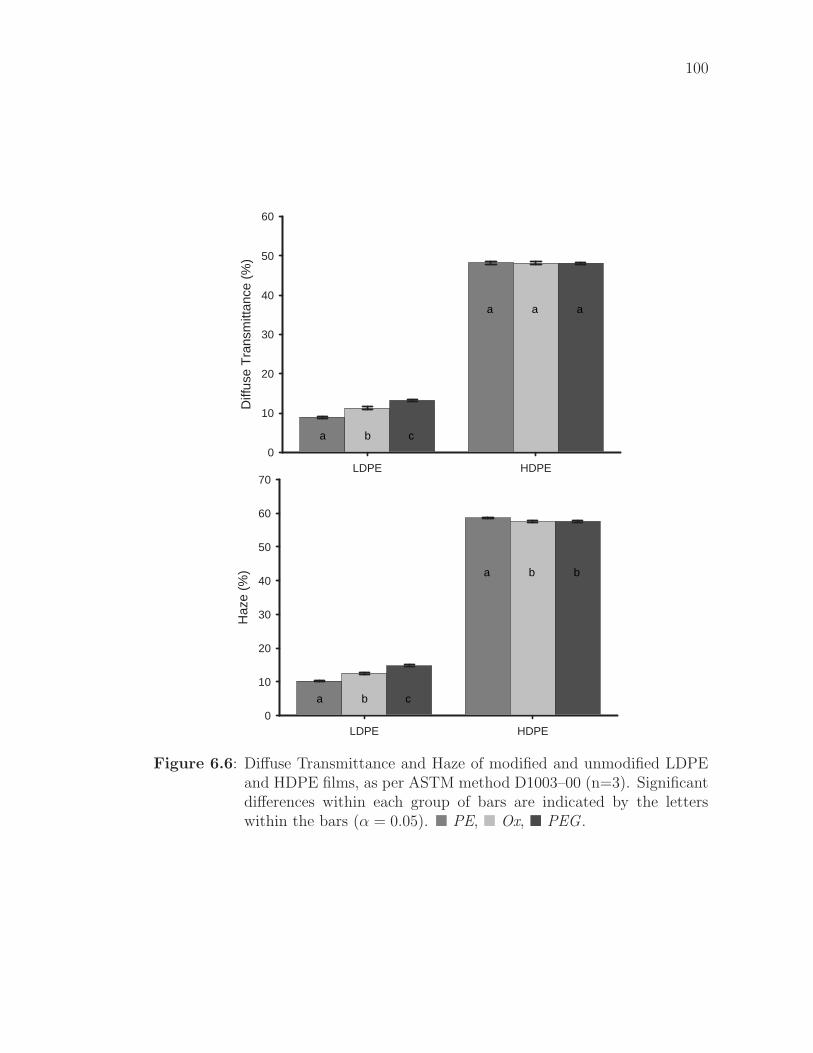
100
LDPE HDPE
Diff
use
Tra
nsm
ittan
ce (
%)
0
10
20
30
40
50
60
a a
a
a
b c
LDPE HDPE
Haz
e (%
)
0
10
20
30
40
50
60
70
a b
a
b
b c
Figure 6.6: Diffuse Transmittance and Haze of modified and unmodified LDPEand HDPE films, as per ASTM method D1003–00 (n=3). Significantdifferences within each group of bars are indicated by the letterswithin the bars (α = 0.05). � PE, � Ox, � PEG .
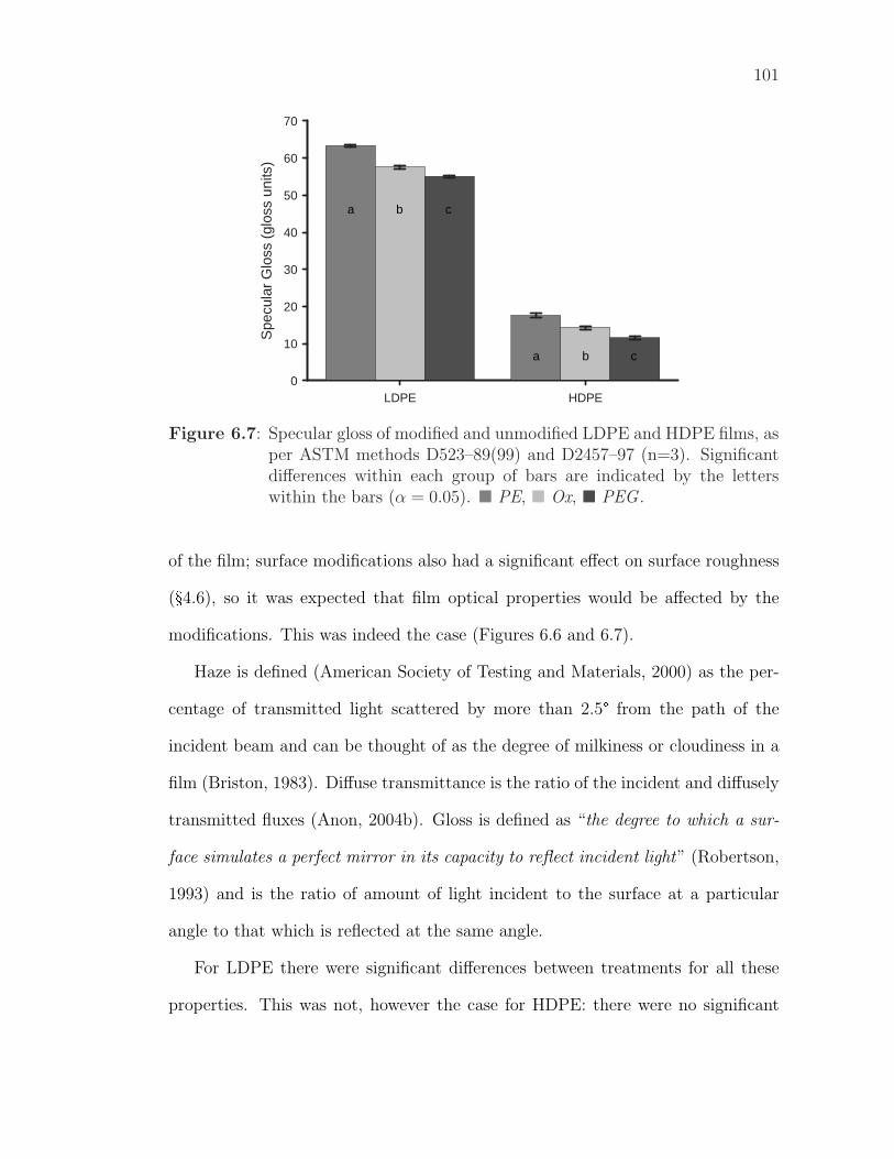
101
LDPE HDPE
Spe
cula
r G
loss
(gl
oss
units
)
0
10
20
30
40
50
60
70
a
a
b c
b c
Figure 6.7: Specular gloss of modified and unmodified LDPE and HDPE films, asper ASTM methods D523–89(99) and D2457–97 (n=3). Significantdifferences within each group of bars are indicated by the letterswithin the bars (α = 0.05). � PE, � Ox, � PEG .
of the film; surface modifications also had a significant effect on surface roughness
(§4.6), so it was expected that film optical properties would be affected by the
modifications. This was indeed the case (Figures 6.6 and 6.7).
Haze is defined (American Society of Testing and Materials, 2000) as the per-
centage of transmitted light scattered by more than 2.5° from the path of the
incident beam and can be thought of as the degree of milkiness or cloudiness in a
film (Briston, 1983). Diffuse transmittance is the ratio of the incident and diffusely
transmitted fluxes (Anon, 2004b). Gloss is defined as “the degree to which a sur-
face simulates a perfect mirror in its capacity to reflect incident light” (Robertson,
1993) and is the ratio of amount of light incident to the surface at a particular
angle to that which is reflected at the same angle.
For LDPE there were significant differences between treatments for all these
properties. This was not, however the case for HDPE: there were no significant
102
differences found in Diffuse Transmittance between the different film treatments
and there was no significant difference in Haze between oxidized and PEGylated
films. The lack of differences for HDPE, while the expected differences were found
for LDPE, can be related to the initial states of the two materials. Unmodified
HDPE is a hazy, somewhat opaque film, whereas LDPE is relatively clear. For
HDPE, the slight change caused by the modifications is insignificant compared to
the initial Haze of the films and, as such is undetectable.
Comparing the measured data to reference values (Table 6.3), it can be seen
that the measured Haze is slightly below and the Gloss slightly above the reference
values for HDPE; no reference values were found for Diffuse Transmittance. These
HDPE reference values, however, are point estimates and do not provide ranges,
yet these properties will vary depending on the molecular characteristics of the
polymer and the method used to produce the films. The reference values are
sufficiently similar to the measured values that the difference is not significant.
The differences in Gloss and Haze found between the modified HDPE films are not
apparent to the human eye.
For LDPE, the measured Haze values for both modified and unmodified films
are within the range provided by the reference values, so are within the normal
spectrum for LDPE films. As such, the increased Haze of the modified films is
unlikely to be a problem in most applications, although it may be in issue in
applications requiring exceptional clarity. It should be noted that the difference
in Haze between the unmodified and oxidized films is apparent to the human eye,
albeit barely.
The Gloss values are more difficult to evaluate as Gloss values depend on the
angle at which they are measured, the value typically increasing as the angle in-
103
creases. Most of the literature values were measured at lower angles than used here.
Extrapolating from these lower angle measurements, it appears that the measured
values are comparable to reference values. The differences in Gloss between the
modified and unmodified LDPE films was not visually apparent.
6.3 Moisture Permeability
Moisture permeation through a polymer film consists of first sorption of the per-
meant on one side of the polymer film, then diffusion through the polymer and
finally desorption of the permeant from the opposite side of the film (Krochta,
2003). Diffusion is a bulk property and will not be affected by surface modifica-
tions. Sorption is defined as the initial penetration and dispersal of the permeant
into the polymer matrix (Robertson, 1993) and is surface-sensitive, so will be af-
fected by chemical changes at the film surface. As seen previously from contact
angle analysis (Table 4.1), the surface hydrophobicity is altered by the film treat-
ments; as surface hydrophobicity decreases, moisture sorption to the surface will
increase, leading to increases in moisture permeability.
As expected, the hydrophilic modified films showed statistically significant in-
creases in WVTR compared to the hydrophobic unmodified LDPE film (Table 6.4).
Similarly, the PEGylated film (pre-washing) had a higher WVTR and lower CA
than the washed PEGylated films, although these differences were not statistically
significant for either CA or WVTR. The unexpected result was that the WVTR of
the oxidized film was higher than the WVTRs of the PEGylated films, even though
the contact angles of the PEGylated films was lower than that of the oxidized film.
The difference in transmission rates between PEGylated films and oxidized
films may relate to moisture diffusion through the PEG layer. No data was found
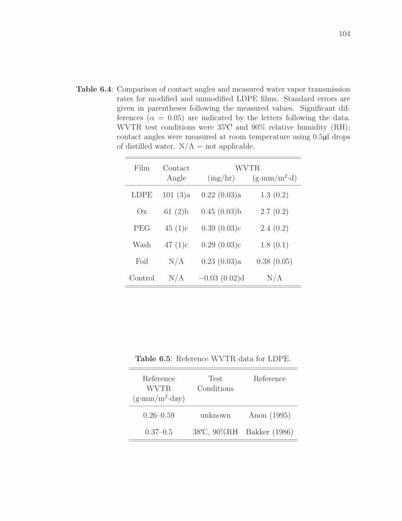
104
Table 6.4: Comparison of contact angles and measured water vapor transmissionrates for modified and unmodified LDPE films. Standard errors aregiven in parentheses following the measured values. Significant dif-ferences (α = 0.05) are indicated by the letters following the data.WVTR test conditions were 35� and 90% relative humidity (RH);contact angles were measured at room temperature using 0.5µl dropsof distilled water. N/A = not applicable.
Film Contact WVTR
Angle (mg/hr) (g·mm/m2·d)
LDPE 101 (3)a 0.22 (0.03)a 1.3 (0.2)
Ox 61 (2)b 0.45 (0.03)b 2.7 (0.2)
PEG 45 (1)c 0.39 (0.03)c 2.4 (0.2)
Wash 47 (1)c 0.29 (0.03)c 1.8 (0.1)
Foil N/A 0.23 (0.03)a 0.38 (0.05)
Control N/A −0.03 (0.02)d N/A
Table 6.5: Reference WVTR data for LDPE.
Reference Test Reference
WVTR Conditions
(g·mm/m2·day)
0.26–0.59 unknown Anon (1995)
0.37–0.5 38�, 90%RH Bakker (1986)
105
relating to the permeability of water through PEG films, however, PEG is a polar,
water soluble polymer; the affinity of water molecules for the polar PEG chains may
reduce the diffusivity of water through an immobilized PEG matrix. If the PEG
coating has a lower water diffusivity than the bulk LDPE, then the presence of PEG
coatings on both sides of the PE in PEGylated films could reduce the moisture
permeability compared to the (pure PE) oxidized films. The higher permeability
of the PEGylated films compared to the unmodified PE does not preclude this; it
would indicate that the increased sorption of the PEGylated films dominates over
the decreased diffusion through the PEG coating.
The measured WVTR of the unmodified film was significantly higher than
the reference data for LDPE (Table 6.5). This is likely related to differences in
the methods used: a modified method was used in this work to allow for smaller
film samples. In the modified method, the sample was sealed across two 5/8 inch
(15.9 mm) diameter holes in a metal can lid using silicon sealant. The metal can
lid was then sealed to the test dish (containing desiccant) using paraffin wax. The
Control sample was an intact metal lid similarly sealed to a sample dish. The
rate of weight change of the Control sample4 indicates that there was negligible
moisture permeation through the wax.
A second control (Foil)was used where a sample of aluminum foil was prepared
identically to the polymer films. The significant WVTR recorded for Foil (theo-
retically the value for foil should approach be zero) indicates that there is moisture
permeation through the silicon sealant sealing the films to the lid5. The measured
WVTR of the foil could also be due to pinholes present in the test foil — pinholes
are common in foils thinner than 25.4 µm (Robertson, 1993) — however, given a
4not significantly different from zero5early trials found problems using silicon sealant to seal the lid to the test dish
106
foil thickness of 27.9 µm, pinholes are unlikely to be present. Assuming that the
moisture permeation measured for foil is due to permeation through the silicon,
the WVTR for the unmodified LDPE film can be adjusted by this amount to
compensate, yet this still yields a WVTR significantly greater than the reference
data.
Permeation through the silicon from the edges of the films inwards to the holes
in the can lid, as would be the case for the impermeable foil, is unlikely to be the
sole cause of the difference. If moisture is assumed to permeate through the silicon
from the edge of the film, then it can also permeate through the film into the
silicon and from there into the test dish. The effective area of permeation for the
film would then be significantly greater than the area of the holes (assumed as the
area for permeation in the WVTR calculations). Moisture could permeate through
the entire area of the film, although the rate of permeation would decrease as the
distance from the holes in the lid increased. This is the most probable explanation
of the differences between the measured WVTRs and the reference data. To avoid
these uncertainties in future work, it is recommended that wax be used to seal the
films to the metal lid. For improved precision, the film thickness of each sample
should also be measured prior to testing.
The uncertainties just described prevent ready comparisons of the modified
film WVTRs to the reference data. The numerical range of the measured data
can also not be compared directly to the range of the reference data as the effect
of the silicon permeability will vary depending on the WVTR of the film. A
rough comparison can be made, however, using the ratios of the highest and lowest
measured WVTRs (2.7/1.3 = 2.1) and the highest and lowest reference values
(0.59/0.26 = 2.3)). Given that the ratio for the measured data is less than the
107
ratio for the reference data, it is thought unlikely that the higher WVTR induced
by these surface modifications will be problematic in PE packaging applications
of the modified films. The difference in WVTR of the modified films compared to
unmodified films will, however, need to be accounted for during package design,
particularly if PE is the main water barrier in the package.
Finally, a note on the statistical analysis. Using the SAS statistical package
(SAS Institute Inc., Cary, NC) to properly control for covariates (Minitab was not
able to do this), a statistically significant effect was found for the effect of time on
the rate of the transmission; there was a slight reduction in the rate measured over
the course of the experiment. This effect, however, was three orders of magnitude
less than the effect of the different treatments, so will not have affected WVTR
comparison between the different treatments. The most probable cause of the
time effect was incomplete equilibration of the samples to a steady state before
measuring the data used in the rate calculations.
6.4 Summary of Findings and Recommendations
The measured properties of the modified and unmodified LDPE and HDPE films
are summarized in Table 6.6. For many properties, no changes were caused by the
modification. Even when differences were observed, the properties remain within
(or very close to) the normal range for PE film, as defined by the literature. Some
of the altered properties of modified films will need to be accounted for in the
package design process, but the changes should not affect the suitability of the
modified films for the normal applications of PE.
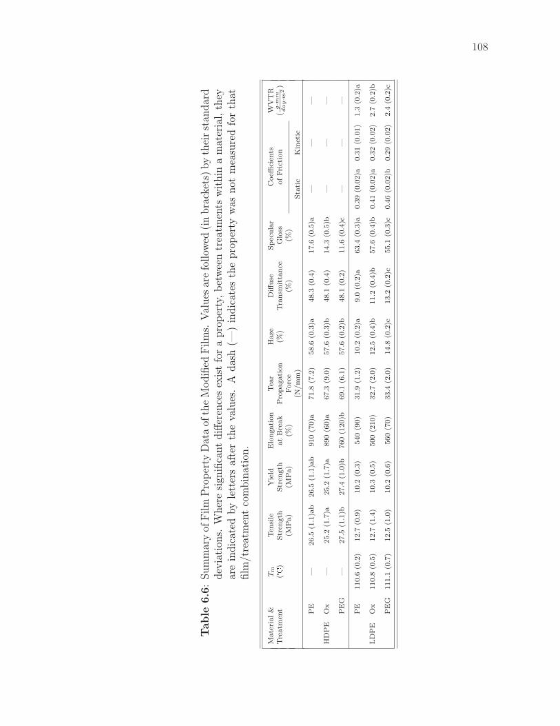
108
Table
6.6
:Sum
mar
yof
Film
Pro
per
tyD
ata
ofth
eM
odifi
edF
ilm
s.V
alues
are
follow
ed(i
nbra
cket
s)by
thei
rst
andar
ddev
iati
ons.
Wher
esi
gnifi
cant
diff
eren
ces
exis
tfo
ra
pro
per
ty,
bet
wee
ntr
eatm
ents
wit
hin
am
ater
ial,
they
are
indic
ated
by
lett
ers
afte
rth
eva
lues
.A
das
h(—
)in
dic
ates
the
pro
per
tyw
asnot
mea
sure
dfo
rth
atfilm
/tre
atm
ent
com
bin
atio
n.
Mate
rial
&T
mT
ensi
leY
ield
Elo
ngati
on
Tea
rH
aze
Diff
use
Sp
ecu
lar
Coeffi
cien
tsW
VT
RT
reatm
ent
(�)
Str
ength
Str
ength
at
Bre
ak
Pro
pagati
on
(%)
Tra
nsm
itta
nce
Glo
ssof
Fri
ctio
n(g·mm
day·m
2)
(MP
a)
(MP
a)
(%)
Forc
e(%
)(%
)(N
/m
m)
Sta
tic
Kin
etic
PE
—26.5
(1.1
)ab
26.5
(1.1
)ab
910
(70)a
71.8
(7.2
)58.6
(0.3
)a48.3
(0.4
)17.6
(0.5
)a—
——
HD
PE
Ox
—25.2
(1.7
)a25.2
(1.7
)a890
(60)a
67.3
(9.0
)57.6
(0.3
)b48.1
(0.4
)14.3
(0.5
)b—
——
PE
G—
27.5
(1.1
)b27.4
(1.0
)b760
(120)b
69.1
(6.1
)57.6
(0.2
)b48.1
(0.2
)11.6
(0.4
)c—
——
PE
110.6
(0.2
)12.7
(0.9
)10.2
(0.3
)540
(90)
31.9
(1.2
)10.2
(0.2
)a9.0
(0.2
)a63.4
(0.3
)a0.3
9(0
.02)a
0.3
1(0
.01)
1.3
(0.2
)a
LD
PE
Ox
110.8
(0.5
)12.7
(1.4
)10.3
(0.5
)500
(210)
32.7
(2.0
)12.5
(0.4
)b11.2
(0.4
)b57.6
(0.4
)b0.4
1(0
.02)a
0.3
2(0
.02)
2.7
(0.2
)b
PE
G111.1
(0.7
)12.5
(1.0
)10.2
(0.6
)560
(70)
33.4
(2.0
)14.8
(0.2
)c13.2
(0.2
)c55.1
(0.3
)c0.4
6(0
.02)b
0.2
9(0
.02)
2.4
(0.2
)c
Chapter 7
Conclusions and Recommendations
� An antibacterial LDPE film was produced. Antibacterial activity was achieved
by immobilization of side-chain protected peptide E14LKK on the surface of
a PEG-treated, oxidized LDPE film, followed by peptide deprotection.
� For many film properties, no differences were observed between the modi-
fied and unmodified films. Where differences were observed, the properties
remain within (or very close to) the normal range associated with PE film.
� Where properties were changed by the modification, the changes will need
to be accounted for in the package design process, but are unlikely to affect
the suitability of the modified films for the normal applications of PE.
� Lactase immobilized directly to LDPE, without a PEG spacer, was not active.
� Although oxidized-poly(ethylene) was successfully modified, the use of a
functional graft polymer, such as poly(acrylic acid) or poly(lactic acid), at-
tached to the poly(ethylene) surface should be explored as a method of in-
creasing the number of available grafting sites on the film surface. The use of
these functional polymers as independent films should also be investigated.
� Attachment of amino-carboxy-PEG was not successful, despite the success
with diamino-PEG. Further optimization of the coupling parameters for the
heterobifunctional PEG is necessary. Initial avenues for investigation would
be the use of buffered coupling solutions and variation of pH, temperature
and salt levels. This optimization requires ready access to suitable analytical
109
110
equipment.
� The use of N-hydroxy succinimide in the WSC activation solution should be
explored as it shows potential to improve the coupling yield.
� A solution-based antimicrobial assay was effective at demonstrating antimi-
crobial activity; it models use of the modified films with liquid food products.
BIBLIOGRAPHY
Ahvenainen, R. (2003). Active and intelligent packaging: an introduction. In
R. Ahvenainen (Ed.), Novel food packaging techniques, Chapter 2, pp. 5–21.
Cambridge, England: Woodhead Publishing Ltd.
Al-Ati, T. A. (2002). Role of CO2/O2 permselectivity of ionomer films in modified
atmosphere packaging of fresh-cut fruits. Ph.D. thesis, Cornell University, Ithaca,
NY.
American Society of Testing and Materials (2000). Annual Book of Standards.
Philadelphia, PA: American Society for Testing and Materials.
American Society of Testing and Materials (2003). Annual Book of Standards.
Philadelphia, PA: ASTM International.
Anon (1995). Permeability and Other Film Properties of Platics and Elastomers
(1st ed.). PDL Handbook Series. Norwich, NY: William Andrew, Inc./Plastics
Design Library.
Anon (1999). Grade ‘A’ Pasteurized Milk Ordinance, 1999 revision. Website: http:
//vm.cfsan.fda.gov/∼ear/p-nci.html.
Anon (2001). Plastic wrap detects spoiled food. Journal of Scientific and Industrial
Research 60 (2), 171.
Anon (2002a). Code of Federal Regulations , Title 21, Parts 170-199. Website:
http://www.access.gpo.gov/nara/cfr/waisidx 02/21cfrv3 02.html; updated Apr 1,
2002; accessed June 29, 2002.
111
112
Anon (2002b). Sigma-Aldrich. Website: http://www.sigmaaldrich.com/; accessed
2002.
Anon (2002c). Preparation and handling of peptides. Current Protocols in Pro-
tein Science, Chapter 18. Website: http://www.mrw2.interscience.wiley.com/
cponline/; accessed Feb, 2003.
Anon (2003a). Active and intelligent food packaging: Commission proposes
amendment to EU law on materials which come into contact with food. Web-
site: http://europa.eu.int/rapid/start/cgi/guesten.ksh?p action.gettxt=gt&doc=
IP/03/1554|0|RAPID&lg=EN&display=; updated 17 Nov 2003; accessed 21 Jan,
2004.
Anon (2003b). COM(2003) 0689 Proposal for a regulation of the european pailia-
ment and council on materials and articles intended to come into contact with
food. 2003/0272 (COD). updated 17 Nov 2003. Website: http://europa.eu.int/
eur-lex/en/com/pdf/2003/com2003 0689en01.pdf; accessed 21 Jan, 2004.
Anon (2004a). Other Useful Tables — Creating constant air humidity in closed
vessels. Merck Chemical Databases . Website: http://chemdat.merck.de/cdrl/
services/labtools/en/table airvessl.html; accessed January 10, 2004.
Anon (2004b). The Photonics Dictionary . Website: http://www.photonics.com/
dictionary/; accessed January 10, 2004.
Appendini, P. (1996). Immobilization of Lysozyme on synthetic polymers for ap-
plication to food packages. Ph.D. thesis, Cornell University, Ithaca, NY.
Appendini, P. (1999). Synthetic antimicrobial peptide. Potential application in
113
foods and food packaging materials. Ph.D. thesis, Cornell University, Ithaca,
NY.
Appendini, P. and J. H. Hotchkiss (1997). Immobilisation of lysozyme on food
contact polymers as potential anti-microbial films. Packaging Technology and
Science 10, 271–279.
Appendini, P. and J. H. Hotchkiss (2001). Surface modification of poly(styrene)
by attachment of an antimicrobial peptide. Journal of Applied Polymer Sci-
ence 81 (3), 609–616.
Appendini, P. and J. H. Hotchkiss (2002). Review of antimicrobial food packaging.
Innovative Food Science and Emerging Technologies 3 (2), 113–126.
Ariyapitipun, T., A. Mustapha, and A. D. Clarke (2000). Survival of Listeria
monocytogenes Scott A on vacuum-packaged raw beef treated with polylactic
acid, lactic acid, and nisin. Journal of Food Protection 63 (1), 131–136.
Bachler, M. J., G. W. Strandbe, and K. L. Smiley (1970). Starch conversion by
immobilized glucoamylase. Biotechnology and Bioengineering 12 (1), 85.
Bae, J. S., E. J. Seo, and I. K. Kang (1999). Synthesis and characterization of
heparinized polyurethanes using plasma glow discharge. Biomaterials 20 (6),
529–537.
Bakker, M. (Ed.) (1986). The Wiley Encyclopedia of Packaging Technology. New
York: John Wiley & Sons.
Bartell, F. E. and H. H. Zuidema (1936). Wetting characteristics of solids of low
surface tension such as talc, waxes and resins. Journal of the American Chemical
Society 58 (8), 1449–1454.
114
Bergbreiter, D. E. (1994). Polyethylene surface-chemistry. Progress in Polymer
Science 19 (3), 529–560.
Bigi, A., G. Cojazzi, S. Panzavolta, K. Rubini, and N. Roveri (2001). Mechanical
and thermal properties of gelatin films at different degrees of glutaraldehyde
crosslinking. Biomaterials 22 (8), 763–768.
Biltresse, S., D. Descamps, T. Boxus, and J. Marchand-Brynaert (2000). Attach-
ment of bis-(trifluoromethyl)aryl labels onto the chain ends of poly(ethylene
terephthalate) (PET) track-etched membranes and films by surface wet chem-
istry. Journal of Polymer Science Part A – Polymer Chemistry 38, 3510–3520.
Bodanszky, M. (1993a). Peptide Chemistry: A Practical Textbook (2nd ed.). Berlin:
Springer-Verlag.
Bodanszky, M. (1993b). Principles of Peptide Synthesis (2nd ed.). Berlin:
Springer-Verlag.
Bodanszky, M. and A. Bodanszky (1994). The Practice of Peptide Synthesis.
Berlin: Springer-Verlag.
Brackett, R. E. (2002). Regulatory perspective on antimicrobials in packaging.
Presentation at IFT 2002 Annual Meeting , June 15–19, Anaheim, CA.
Branen, A. L. (1993). Introduction to use of antimicrobials. In P. M. Davidson
and A. L. Branen (Eds.), Antimicrobials in Foods (2nd ed.)., Food Science and
Technology, pp. 1–9. New York: Marcel Dekker.
Briston, J. H. (1983). Plastics Films . Essex, England: Longman Scientific and
Technical.
115
Brody, A. and J. Budny (1995). Enzymes as active packaging agents. In M. Rooney
(Ed.), Active food packaging, pp. 174–192. Glasgow: Blackie Academic & Pro-
fessional.
Calbiochem-NovaBiochem Corporation (1999). Novabiochem 1999 catalog and pep-
tide synthesis handbook .
Caliceti, P. and F. M. Veronese (1999). Improvement of the physicochemical and
biopharmaceutical properties of insulin by poly(ethylene glycol) conjugation.
STP Pharma Sciences 9 (1), 107–113.
Callister, W. D., Jr. (1996). Materials Science and Engineering — An Introduction
(4th ed.). New York: John Wiley & Sons, Inc.
Carraway, K. L. and D. E. Koshland (1972). Carbodiimide modification of proteins.
Methods in Enzymology 28, 616–623.
Cha, D., K. Cooksey, M. Chinnan, and H. Park (2003). Release of nisin from
various heat-pressed and cast films. Lebensmittel-Wissenschaft und Technologie
- Food Science and Technology 36 (2), 209–213.
Chen, Y., E. T. Kang, K. G. Neoh, and K. L. Tan (2000). Covalent immobilization
of invertase onto surface-modified poly(aniline) from graft copolymerization with
acrylic acid. European Polymer Journal 36, 2095–2103.
Collins-Thompson, D. and H. Cheng-An (2000). Chilled storage of foods — pack-
aging with antimicrobial properties. In R. K. Robinson, C. A. Batt, and P. D.
Patel (Eds.), Encyclopedia of Food Microbiology, pp. 416–420. San Diego, CA:
Academic Press.
116
Coma, V., A. Deschamps, and A. Martial-Gros (2003). Bioactive packaging ma-
terials from edible chitosan polymer — antimicrobial activity assessment on
dairy-related contaminants. Journal of Food Science 68 (9), 2788–2792.
Coma, V., A. Martial-Gros, S. Garreau, A. Copinet, F. Salin, and A. Deschamps
(2002). Edible antimicrobial films based on chitosan matrix. Journal of Food
Science 67 (3), 1162–1169.
Cooksey, K. (2000). Utilization of antimicrobial packaging films for inhibition of
selected microorganisms. In S. J. Risch (Ed.), Food Packaging: Testing Methods
and Applications, ACS Symposium Series #753, pp. 17–25. Washington, DC:
American Chemical Society.
Cooksey, K. and C. Wu (1998). Utilization of nisin as a coating on packaging films
for inhibition of selected microorganisms. Abstracts of Papers of the American
Chemical Society 215 (Part 1 April 2 1998), 052–AGFD.
de Kruijf, N., M. V. Beest, R. Rijk, T. Sipilainen-Malm, P. P. Losada, and B. D.
Meulenaer (2002). Active and intelligent packaging: applications and regulatory
aspects. Food Additives and Contaminants 19, 144–162.
de Kruijf, N. and R. Rijk (2003). Legislative issues relating to active and intel-
ligent packaging. In R. Ahvenainen (Ed.), Novel Food Packaging Techniques,
Chapter 22, pp. 459–496. Cambridge, UK: Woodhead Publishing Ltd.
Edwards, J. V. and T. L. Vigo (2001). Bioactive fibers and polymers. ACS Sym-
posium Series #792. Washington, DC: American Chemical Society.
Ellis, B. (Ed.) (2000). Polymers — A Property Database. Boca Raton, FL: Chap-
man & Hall/CRC Press.
117
Emoto, K., J. M. V. Alstine, and J. M. Harris (1998). Stability of poly(ethylene
glycol) graft coatings. Langmuir 14 (10), 2722–2729.
Eriksson, J. C., C. G. Golander, A. Baszkin, and L. Terminassiansaraga (1984).
Characterization of KMnO4 H2SO4-oxidized polyethylene surfaces by means
of ESCA and Ca-45(2+) adsorption. Journal of Colloid and Interface Sci-
ence 100 (2), 381–392.
Gaertner, H. F. and A. J. Puigserver (1992). Increased activity and stability of
poly(ethylene glycol)-modified trypsin. Enzyme and Microbial Technology 14 (2),
150–155.
Garbassi, F., M. Morra, and E. Occhiello (1994). Chemical modifications. In Poly-
mer surfaces: from physics to technology, pp. 243–273. West Sussex, England:
John Wiley and Sons.
Garcia-Garibay, M., A. Luna-Salazar, and L. T. Casas (1995). Antimicrobial effect
of the lactoperoxidase system in milk activated by immobilized enzymes. Food
Biotechnology 9 (3), 157–166.
Gombotz, W. R., W. Guanghui, and A. S. Hoffman (1989). Immobilization of
poly(ethylene oxide) on poly(ethylene terephthalate) using a plasma polymer-
ization process. Journal of Applied Polymer Science 37 (1), 91–107.
Groning, P., M. C. Coen, and L. Schlapbach (2001). Polymers and cold plasmas.
Chimia 55 (3), 171–177.
Han, J. H. (2000). Antimicrobial food packaging. Food Technology 54 (3), 56–65.
Haynie, S. L. (1998). Antimicrobial composition of a polymer and a peptide forming
118
amphiphilic helices of the magainin-type. US patent 5,847,047. E.I. du Pont de
Nemours and Company, assignee.
Haynie, S. L., G. A. Crum, and B. A. Doele (1995). Antimicrobial activities of
amphiphilic peptides covalently bonded to a water-insoluble resin. Antimicrobial
Agents and Chemotherapy 39 (2), 301–307.
Heckman, J. H. and D. W. Ziffer (2001). Fathoming food packaging regulation
revisited. Food and Drug Law Journal 56 (2), 179–195.
Helander, I. M., E. L. Nurmiaho-Lassila, R. Ahvenainen, J. Rhoades, and S. Roller
(2001). Chitosan disrupts the barrier properties of the outer membrane of gram-
negative bacteria. International Journal of Food Microbiology 71 (2-3), 235–244.
Herrwerth, S., T. Rosendahl, C. Feng, J. Fick, W. Eck, M. Himmelhaus, R. Dahint,
and M. Grunze (2003). Covalent coupling of antibodies to self-assembled mono-
layers of carboxy-functionalized poly(ethylene glycol): Protein resistance and
specific binding of biomolecules. Langmuir 19, 1880–1887.
Hinder, S. J., S. D. Connell, M. C. Davies, C. J. Roberts, S. J. B. Tendler, and
P. M. Williams (2002). Compositional mapping of self-assembled monolayers
derivatized within microfluidic networks. Langmuir 18 (8), 3151–3158.
Holmes-Farley, S. R., R. H. Reamey, T. J. Mccarthy, J. Deutch, and G. M. White-
sides (1985). Acid-base behavior of carboxylic-acid groups covalently attached
at the surface of polyethylene — the usefulness of contact-angle in following the
ionization of surface functionality. Langmuir 1 (6), 725–740.
Hotchkiss, J. H. (1995). Safety considerations in active packaging. In M. Rooney
119
(Ed.), Active food packaging, pp. 238–254. Glasgow: Blackie Academic & Pro-
fessional.
Hu, S. G., C. H. Jou, and M. C. Yang (2002). Surface grafting of polyester fiber
with chitosan and the antibacterial activity of pathogenic bacteria. Journal of
Applied Polymer Science 86 (12), 2977–2983.
Ihara, N., Y. Tachibana, J. E. Chung, M. Kurisawa, H. Uyama, and S. Kobayashi
(2003). Antioxidant polymer particles — enzymatic immobilization of catechin
on polymer particles. Chemistry Letters 32 (9), 816–817.
Ikada, Y. (1994). Surface modification of polymers for medical applications. Bio-
materials 15 (10), 725–736.
Ikinci, G., S. Senel, H. Akincibay, S. Kas, S. Ercis, C. G. Wilson, and A. A. Hincal
(2002). Effect of chitosan on a periodontal pathogen porphyromonas gingivalis.
International Journal of Pharmaceutics 235 (1-2), 121–127.
Jarvi-Kaariainen, T. (2003). Integrating intelligent packaging, storage and distri-
bution. In R. Ahvenainen (Ed.), Novel food packaging techniques, Chapter 25,
pp. 535–549. Cambridge, England: Woodhead Publishing Ltd.
Jay, J. M. (1992). Modern Food Microbiology (5th ed.). New York: Chapman &
Hall.
Jenkins, W. A. and J. P. Harrington (1991). Packaging Foods With Plastics. Lan-
caster, PA: Technomic Publishing Company.
Jinkins, R. S. and K. K. Leonas (1994). Influence of a polyethylene-glycol treat-
ment on surface, liquid barrier and antibacterial properties. Textile Chemist and
Colorist 26 (12), 25–29.
120
Kang, I. K., E. J. Seo, M. W. Huh, and K. H. Kim (2001). Interaction of blood
components with heparin-immobilized polyurethanes prepared by plasma glow
discharge. Journal of Biomaterials Science – Polymer Edition 12 (10), 1091–
1108.
Katchalski-Katzir, E. (1993). Immobilized enzymes — learning from past successes
and failures. TIBTECH 11, 471–478.
Kato, K. (1976). Investigation of high-density polyethylene film surface treated
with chromic acid micture by use of 2,4-dinitrophenylhydrazine. Journal of
Applied Polymer Science 20, 2451–2460.
Kato, K. (1977). Investigation of high-density polyethylene film surface treated
with chromic acid mixture by use of 2,4-dinitrophenylhydrazine. ii. film surfaces
treated at 70�. Journal of Applied Polymer Science 21, 2735–2743.
Kato, K. and Y. Ikada (1996). Immobilization of DNA onto a polymer support and
its potentiality as immunoadsorbent. Biotechnology and Bioengineering 51 (5),
581–590.
Kato, K., E. Uchida, E.-T. Kang, Y. Uyama, and Y. Ikada (2003). Polymer surface
with graft chains. Progress in Polymer Science 28, 209–259.
Kelley, M. J. (2004). Personal Communication.
Kelley, M. J., J. D. Cohen, C. Erkenbrecher, S. L. Haynie, H. Kobsa, A. N. Roe,
and M. H. Scholla (1995). Amide to active amine conversion of nylon by deep UV
irradiation. In R. Singh, D. Norton, L. Laude, J. Narayan, and J. Cheung (Eds.),
Advanced Laser Processing of Materials – Fundamentals and Applications, pp.
121
353–356. MRS Symposium Proceedings #397. Warrendale, PA: Materials Re-
search Society.
Kenawy, E.-R. and Y. A.-G. Mahmoud (2003). Synthesis and antimicrobial activity
of some linear copolymers with quaternary ammonium and phosphonium groups.
Macromolecular Bioscience 3 (2), 107–116.
Kikuchi, M., T. Taguchi, H. N. Matsumoto, K. Takakuda, and J. Tanaka
(2002). Cross-linkage of hydroxyapatite/collagen nano-composite with 3 dif-
ferent reagents. Key Engineering Materials 218 (2), 449–452.
Kim, Y. J., I.-K. Kang, M. W. Huh, and S.-C. Yoon (2000). Surface characteriza-
tion and in vitro blood compatibility of poly(ethylene terephthalate) immobilized
with insulin and/or heparin using plasma glow discharge. Biomaterials 21 (2),
121–130.
Kingshott, P., H. Thissen, and H. J. Griesser (2002). Effects of cloud-point grafting,
chain length, and density of PEG layers on competitive adsorption of ocular
proteins. Biomaterials 23 (9), 2043–2056.
Kingshott, P., J. Wei, D. Bagge-Ravn, B. Gadegaard, and L. Gram (2003). Cova-
lent attachment of poly(ethylene glycol) to surfaces, critical for reducing bacte-
rial adhesion. Langmuir 19 (17), 6912–6921.
Kiss, E., J. Samu, A. Toth, and I. Bertoti (1996). Novel ways of covalent at-
tachment of poly(ethylene oxide) onto polyethylene: surface modification and
characterization by XPS and contact angle measurements. Langmuir 12, 1651–
1657.
122
Klapperich, C., L. Pruitt, and K. Komvopoulos (2001). Chemical and biological
characteristics of low-temperature plasma treated ultra-high molecular weight
polyethylene for biomedical applications. Journal of Materials Science — Ma-
terials in Medicine 12 (6), 549–556.
Kong, J.-S., D.-J. Lee, and H.-D. Kim (2001). Surface modification of low-densitry
polyethylene (LDPE) film and improvement of adhesion between evaporated
copper metal film and LDPE. Journal of Applied Polymer Science 82, 1677–
1690.
Krochta, J.M. (2003). Package permeability. In D. R. Heldman (Ed.), Encyclopedia
of Agricultural, Food, and Biological Engineering, pp. 720-726. New York: Marcel
Dekker, Inc.
Labuza, T. and W. Breene (1989). Applications of active packaging for improve-
ment of shelf life and nutritional quality of fresh and extended shelf life foods.
Journal of Food Processing and Preservation 13, 1–70.
Labuza, T. P. (2004). Creation of moisture sorption isotherms for hy-
groscopic materials. Website: http://www.fsci.umn.edu/Ted Labuza/PDF files/
papers/Creation Moisture Isotherms.PDF; accessed January 10, 2004.
Lane, J. M. and D. J. Hourston (1993). Surface treatments of polyolefins. Progress
in Organic Coatings 21 (4), 269–284.
Larsson, R., J. C. Eriksson, and P. Olsson (1979). Effects of surface localized sulfate
groups on the clotting time in plasma. Thrombosis Research 14 (6), 941–952.
Lin, J. C. and S. M. Tseng (2001). Surface characterization and platelet adhe-
123
sion studies on polyethylene surface with hirudin immobilization. Journal of
Materials Science – Materials in Medicine 12 (9), 827–832.
Mabrouk, P. (1997). The use of poly(ethylene glycol)-enzymes in nonaqueous
enzymology. In J. M. Harris and S. Zalipsky (Eds.), Poly(Ethylene Glycol):
Chemistry and Biological Applications, ACS Symposium Series #680, pp. 118–
133. Washington, DC: American Chemical Society.
Mack, G. L. (1935). The determination of contact angles from measurements of
the dimensions of small bubbles and drops. I - the spheriodal segment method
for acute angles. Journal of Physical Chemistry 40 (2), 159–167.
Mallakpour, S. E., A. R. Hajipour, A. R. Mahdavian, A. Zadhoush, and F. Ali-
Hosseini (2001a). Microwave assisted oxidation of polyethylene under solid-state
conditions with potassium permanganate. European Polymer Journal 37 (6),
1199–1206.
Mallakpour, S. E., A. R. Hajipour, A. Zadhoush, and A. R. Mahdavian (2001b).
Efficient and novel method for surface oxidation of polypropylene in the solid
phase using microwave irradiation. Journal of Applied Polymer Science 79 (7),
1317–1323.
Malmsten, M., K. Emoto, and J. M. V. Alstine (1998). Effect of chain density on
inhibition of protein adsorption bypoly(ethylene glycol) based coatings. Journal
of Colloid and Interface Science 202 (2), 507–517.
Mead, P. S., L. Slutsker, V. Dietz, L. F. Mccaig, J. S. Bresee, C. Shapiro, P. M.
Griffin, and R. V. Tauxe (1999). Food-related illness and death in the united
states. Emerging Infectious Diseases 5 (5), 607–625.
124
Meroni, A. (2000). Active packaging as an opportunity to create package design
that reflects the communicational, functional and logistical requirements of food
products. Packaging Technology and Science 13 (6), 243–248.
Molday, R. S., W. J. Dreyer, A. Rembaum, and S. P. S. Yen (1975). New im-
munolatex spheres — visual markers of antigens on lymphocytes for scanning
electron-microscopy. Journal of Cell Biology 64 (1), 75–88.
Mosbach, K. (1980). Immobilized enzymes. Trends in Biochemical Sciences 5 (1),
1–3.
Mustapha, A., T. Ariyapitipun, and A. D. Clarke (2002). Survival of Escherichia
Coli O157:H7 on vacuum-packaged raw beef treated with polylactic acid, lactic
acid, and nisin. Journal of Food Science 67 (1), 262–267.
Nakajima, N. and Y. Ikada (1995). Mechanism of amide formation by carbodiimide
for bioconjugation in aqueous media. Bioconjugate Chemistry 6, 123–130.
N.I.H. (2003). Lactose intolerance. National Digestive Diseases Informa-
tion Clearing House. Website: http://digestive.niddk.nih.gov/ddiseases/pubs/
lactoseintolerance/; updated March 2003; accessed 12 March, 2004. Washing-
ton, D.C.: National Institutes of Health — National Institute of Diabetes and
Digestive and Kidney Diseases
NOVA Chemicals (2002). Sclair polyethylene product data sheet: Sclair
19A. Website: http://www.novachemicals.com/OurProducts/pds/adobepds/film/
sclair/butene/19A.pdf; updated December 17, 2002; accessed Dec 30, 2003.
Oh, H. I., Y. J. Kim, E. J. Chang, and J. Y. Kim (2001). Antimicrobial charac-
125
teristics of chitosans against food spoilage microorganisms in liquid media and
mayonnaise. Bioscience Biotechnology and Biochemistry 65 (11), 2378–2383.
Ojcius, D. and J.-E. Young (1991). Cytolytic pore-forming proteins and peptides
— is there a common structural motif? Trends in Biochemical Sciences 16, 225.
Olson, K., R. Gehant, V. Mukku, K. O’connell, B. Tomlinson, K. Totpal, and
M. Winkler (1997). Preparation and characterization of poly(ethylene gly-
col)ylated human growth hormone antagonist. In J. M. Harris and S. Zalip-
sky (Eds.), Poly(Ethylene Glycol): Chemistry and Biological Applications, ACS
Symposium Series #680, pp. 170–181. Washington, DC: American Chemical
Society.
Ozdemir, M. and H. Sadikoglu (1998). A new and emerging technology: Laser-
induced surface modification of polymers. Trends in Food Science and Technol-
ogy 9 (4), 159–167.
Ozdemir, M., C. U. Yurteri, and H. Sadikoglu (1999a). Physical polymer surface
modification methods and applications in food packaging polymers. Critical
Reviews in Food Science and Nutrition 39 (5), 457–477.
Ozdemir, M., C. U. Yurteri, and H. Sadikoglu (1999b). Surface treatment of food
packaging polymers by plasma. Food Technology 53 (4), 54–58.
Padgett, T., I. Y. Han, and P. L. Dawson (1998). Incorporation of food-grade
antimicrobial compounds into biodegradable packaging films. Journal of Food
Protection 61 (10), 1330–1335.
Paik, J. S., M. Dhanasekharan, and M. J. Kelley (1998). Antimicrobial activity of
126
UV-irradiated nylon film for packaging applications. Packaging Technology and
Science 11 (4), 179–187.
Panza, J. L., K. E. Lejeune, S. Venkatasubramanian, and A. J. Russell (1997).
Incorporation of poly(ethylene glycol) proteins into polymers. In J. M. Harris
and S. Zalipsky (Eds.), Poly(Ethylene Glycol): Chemistry and Biological Appli-
cations, ACS Symposium Series #680, pp. 134–144. Washington, DC: American
Chemical Society.
Paulk, M., K. Cooksey, and J. Wiles (2002). Characterization of chitosan packaging
films with differing levels of % deacetylation. Presentation at 2002 IFT Annual
Meeting, June 15–19, Anaheim, CA.
Pennington, M. W. (1994). HF cleavage and deprotection procedures for peptides
synthesized using a Boc/Bzl strategy. In M. W. Pennington and B. M. Dunn
(Eds.), Peptide Synthesis Protocols, pp. 41–62. Totowa, NJ: Humana Press.
Plummer, S. T. and P. W. Bohn (2002). Spatial dispersion in electrochemically
generated surface composition gradients visualized with covalently bound fluo-
rescent nanospheres. Langmuir 18 (10), 4142–4149.
Rasmussen, J. R., E. R. Stedronsky, and G. M. Whitesides (1977). Introduction,
modification, and characterization of functional- groups on surface of low-density
polyethylene film. Journal of the American Chemical Society 99 (14), 4736–4745.
Richardson, T. and D. B. Hyslop (1985). Enzymes. In O. R. Fennema (Ed.), Food
Chemistry (2nd ed.)., Food Science and Technology, pp. 371–476. New York:
Marcel Dekker.
127
Robertson, G. L. (1993). Food Packaging: Principles and Practise. New York:
Marcel Dekker.
Rodriguez, F. (1996). Principles of Polymer Systems (4th ed.). Washington, DC:
Taylor & Francis.
Sahi, T. (1994). Genetics and epidemiology of adult-type hypolactasia. Scandina-
vian Journal of Gastroenterology 29 (Suppl. 202), 7–20.
Sano, S., K. Kato, and Y. Ikada (1993). Introduction of functional groups onto
the surface of polyethylene for protein immobilization. Biomaterials 14 (11),
817–822.
Sarkar, N., S. Bhattacharjee, and S. Sivaram (1997). Surface functionalization of
poly(ethylene) with succinic anhydride: preparation, modification and charac-
terization. Langmuir 13, 4142–4149.
Scannell, A. G. M., C. Hill, R. Ross, S. Marx, W. Hartmeier, and E. Arendt
(2000). Development of bioactive food packaging materials using immobilized
bacteriocins lacticin 3147 and nisaplin. International Journal of Food Microbi-
ology 60 (2-3), 241–249.
Schonherr, H. and G. J. Vancso (1998). Surface properties of oxidized LDPE by
scanning force microscopy with chemically modified probes. Journal of Polymer
Science Part B – Polymer Physics 36, 2483–2492.
Schroder, K., A. Meyer-Plath, D. Keller, W. Besch, G. Babucke, and A. Ohl (2001).
Plasma-induced surface functionalization of polymeric biomaterials in ammonia
plasma. Contributions to Plasma Physics 41 (6), 562–572.
128
Shearer, A. E. H., J. S. Paik, D. G. Hoover, S. L. Haynie, and M. J. Kelley (2000).
Potential of an antibacterial ultraviolet-irradiated nylon film. Biotechnology and
Bioengineering 67 (2), 141–146.
Shearwater Corporation (2001). Shearwater corporation catalog 2001: polyethy-
lene glycol and derivatives for biomedical applications.
Shenton, M. J., M. C. Lovell-Hoare, and G. C. Stevens (2001). Adhesion enhance-
ment of polymer surfaces by atmospheric plasma treatment. Journal of Physics
D – Applied Physics 34 (18), 2754–2760.
Shenton, M. J. and G. C. Stevens (1999). Investigating the effect of the thermal
component of atmospheric plasmas on commodity polymers. Thermochimica
Acta 332 (2), 151–160.
Shenton, M. J. and G. C. Stevens (2001). Surface modification of polymer surfaces:
Atmospheric plasma versus vacuum plasma treatments. Journal of Physics D –
Applied Physics 34 (18), 2761–2768.
Shenton, M. J., G. C. Stevens, N. P. Wright, and X. Duan (2002). Chemical-surface
modification of polymers using atmospheric pressure nonequilibrium plasmas
and comparisons with vacuum plasmas. Journal of Polymer Science Part A –
Polymer Chemistry 40 (1), 95–109.
Shi, J. L., E. T. Kang, K. G. Neoh, K. L. Tan, and D. J. Liaw (1998). Surface
graft copolymerization of low density polyethylene films and its relevance to
auto-adhesion. European Polymer Journal 34 (10), 1429–1434.
Smolander, M. (2003). The use of freshness indicators in packaging. In R. Ahve-

129
nainen (Ed.), Novel food packaging techniques, Chapter 7, pp. 127–143. Cam-
bridge, England: Woodhead Publishing Ltd.
Soares, N.d.F.F. (1998). Bitterness reduction in citrus juice through Naringase
immobilized into polymer film. Ph.D. thesis, Cornell University, Ithaca, NY.
Soares, N.d.F.F. and J. H. Hotchkiss (1998). Naringinase immobilization in pack-
aging films for reducing naringin concentration in grapefruit juice. Journal of
Food Science 63 (1), 61–65.
Sodhi, R. N. S., V. P. Sahi, and M. W. Mittelman (2001). Application of electron
spectroscopy and surface modification techniques in the development of anti-
microbial coatings for medical devices. Journal of Electron Spectroscopy and
Related Phenomena 121 (1-3), 249–264.
Sofia, S. J. and E. W. Merrill (1998). Grafting of PEO to polymer surfaces using
electron beam irradiation. Journal of Biomedical Materials Research 40 (1),
153–163.
Speck, M. L. (1984). Compendium of methods for the microbiological examination
of foods. Washington, D.C.: American Public Health Association, Inc.
Sun, G. (2001). Durable and regenerable antimicrobial textiles. In J. V. Edwards
and T. L. Vigo (Eds.), Bioactive Fibers and Poymers, ACS Syposium Seriess
#792, pp. 243–252. Washington, DC: American Chemical Society.
Sun, Y. and G. Sun (2002). Durable and regenerable antimicrobial textile ma-
terials prepared by a continuous grafting process. Journal of Applied Polymer
Science 84 (8), 1592–1599.
130
Takai, K., T. Ohtsuka, Y. Senda, M. Nakao, K. Yamamoto, J. Matsuoka, and
Y. Hirai (2002). Antibacterial properties of antimicrobial-finished textile prod-
ucts. Microbiology and Immunology 46 (2), 75–81.
Talbert, J. N. (2004). Personal Communication.
Tanabe, T., N. Okitsu, A. Tachibana, and K. Yamauchi (2002). Preparation and
characterization of keratin-chitosan composite film. Biomaterials 23 (3), 817–
825.
Taoukis, P. S. and T. P. Labuza (2003). Time-temperature indidcators (TTIs). In
R. Ahvenainen (Ed.), Novel food packaging techniques, Chapter 6, pp. 103–126.
Cambridge, England: Woodhead Publishing Ltd.
Terlingen, J. G. A., H. F. C. Gerritsen, A. S. Hoffman, and F. J. Jen (1995).
Introduction of functional-groups on polyethylene surfaces by a carbon-dioxide
plasma treatment. Journal of Applied Polymer Science 57 (8), 969–982.
Tew, G. N., D. H. Liu, B. Chen, R. J. Doerksen, J. Kaplan, P. J. Carroll, M. L.
Klein, and W. F. Degrado (2002). De Novo design of biomimetic antimicrobial
polymers. Proceedings of the National Academy of Sciences of the United States
of America 99 (8), 5110–5114.
Topchieva, I. N., N. V. Efremova, N. V. Khvorov, and N. Magretova (1995). Syn-
thesis and physicochemical properties of protein conjugates with water-soluble
poly(alkylene oxides). Bioconjugate Chemistry 6 (4), 380–388.
Tsai, G. J. and W. H. Su (1999). Antibacterial activity of shrimp chitosan against
escherichia coli. Journal of Food Protection 62 (3), 239–243.
131
Tsai, G. J., W. H. Su, H. C. Chen, and C. L. Pan (2002). Antimicrobial activity
of shrimp chitin and chitosan from different treatments and applications of fish
preservation. Fisheries Science 68 (1), 170–177.
Tsai, G. J., Z. Y. Wu, and W. H. Su (2000). Antibacterial activity of a chi-
tooligosaccharide mixture prepared by cellulase digestion of shrimp chitosan and
its application to milk preservation. Journal of Food Protection 63 (6), 747–752.
Valuev, I. L., V. V. Chupov, and L. I. Valuev (1998). Chemical modification of
polymers with physiologically activespecies using water-soluble carbodiimides.
Biomaterials 19 (1-3), 41–43.
Vermeiren, L., F. Devlieghere, M. V. Beest, N. D. Kruijf, and J. Debevere (1999).
Developments in the active packaging of foods. Trends in Food Science and
Technology 10 (3), 77–86.
Vermeiren, L., F. Devlieghere, and J. Debevere (2002). Effectiveness of some
recent antimicrobial packaging concepts. Food Additives and Contaminants 19,
163–171.
Veronese, F. M. (2001). Peptide and protein pegylation: a review of problems and
solutions. Biomaterials 22 (5), 405–417.
Vesa, T. H., P. Marteau, and R. Korpela (2000). Lactose intolerance. Journal of
the American College of Nutrition 19 (2), 165S–175S.
Vigo, T. L. (1999). Antimicrobial polymers and fibers: Retrospective end prospec-
tive. Abstracts of Papers of the American Chemical Society 218, 31–CELL.
Vigo, T. L. and J. S. Bruno (1993). Antimicrobial activity of fabrics containing
132
cross-linked polyols. Abstracts of Papers of the American Chemical Society 206,
50–CELL.
Vigo, T. L. and K. K. Leonas (1999). Antimicrobial activity of fabrics containing
crosslinked polyethylene glycols. Textile Chemist and Colorist and American
Dyestuff Reporter 1 (1), 42–46.
Weetall, H. H. (1993). Preparations of immobilized proteins covalently coupled
through silane agents to inorganic support. Applied Biochemistry and Biotech-
nology 41 (3), 157–188.
Weissman, R. C. (1979). A microscopical and mechanical study of the gelation of
collagen crosslinked with glutaraldehyde. Ph.D. thesis, Cornell University, Ithaca,
NY.
Woodaman, J. G. (2001). Detection of contaminants in food. US patent 6,270,724
B1. California South Pacific Investors, Pasadena, CA, assignee.
Yang, Z., M. Domach, R. Auger, F. X. Yang, and A. J. Russell (1996). Polyethy-
lene glycol-induced stabilization of subtilisin. Enzyme and Microbial Technol-
ogy 18 (2), 82–89.
Yang, Z., A. J. Mesiano, S. Venkatasubramanian, S. H. Gross, M. J. Harris, and
A. J. Russell (1995a). Activity and stability of enzymes incorporated into acrylic
polymers. Journal of the American Chemical Society 117, 4843–4850.
Yang, Z., D. Williams, and A. J. Russell (1995b). Synthesis of protein-containing
polymers in organic-solvents. Biotechnology and Bioengineering 45 (1), 10–17.
Zaks, A. and A. M. Klibanov (1984). Enzymatic catalysis in organic media at
100�. Science 224 (4654), 1249–1251.
133
Zalipsky, S. and J. M. Harris (1997). Introduction to chemistry and biological
applications of poly(ethylene glycol). In J. M. Harris and S. Zalipsky (Eds.),
Poly(ethylene glycol): Chemistry and Biological Applications, ACS Symposium
Series #680. Washington, DC: American Chemical Society.