Probing Protein Sequences as Sources for Encrypted Antimicrobial Peptides
Upload
khangminh22Category
view
0download
0
�����������������
Citation: Akbarian, M.; Khani, A.;
Eghbalpour, S.; Uversky, V.N.
Bioactive Peptides: Synthesis,
Sources, Applications, and Proposed
Mechanisms of Action. Int. J. Mol. Sci.
2022, 23, 1445. https://doi.org/
10.3390/ijms23031445
Academic Editors: Azzurra
Stefanucci and Adriano Mollica
Received: 7 January 2022
Accepted: 25 January 2022
Published: 27 January 2022
Publisher’s Note: MDPI stays neutral
with regard to jurisdictional claims in
published maps and institutional affil-
iations.
Copyright: © 2022 by the authors.
Licensee MDPI, Basel, Switzerland.
This article is an open access article
distributed under the terms and
conditions of the Creative Commons
Attribution (CC BY) license (https://
creativecommons.org/licenses/by/
4.0/).
International Journal of
Molecular Sciences
Review
Bioactive Peptides: Synthesis, Sources, Applications, andProposed Mechanisms of ActionMohsen Akbarian 1 , Ali Khani 2, Sara Eghbalpour 3 and Vladimir N. Uversky 4,*
1 Department of Chemistry, National Cheng Kung University, Tainan 701, Taiwan;[email protected]
2 Department of Radiation Sciences, Faculty of Applied Medicine, Iran University of Medical Sciences,Tehran 1449614535, Iran; [email protected]
3 Department of Obstetrics and Gynecology Surgery, Babol University of Medical Sciences,Babol 4717647745, Iran; [email protected]
4 Department of Molecular Medicine and Health Byrd Alzheimer’s Institute, Morsani College of Medicine,University of South Florida, Tampa, FL 33612, USA
* Correspondence: [email protected]; Tel.: +1-(813)-974-5816
Abstract: Bioactive peptides are a group of biological molecules that are normally buried in thestructure of parent proteins and become active after the cleavage of the proteins. Another groupof peptides is actively produced and found in many microorganisms and the body of organisms.Today, many groups of bioactive peptides have been marketed chemically or recombinantly. Thisarticle reviews the various production methods and sources of these important/ubiquitous anduseful biomolecules. Their applications, such as antimicrobial, antihypertensive, antioxidant activi-ties, blood-lipid-lowering effect, opioid role, antiobesity, ability to bind minerals, antidiabetic, andantiaging effects, will be explored. The types of pathways proposed for bioactive applications willbe in the next part of the article, and at the end, the future perspectives of bioactive peptides willbe reviewed. Reading this article is recommended for researchers interested in various fields ofphysiology, microbiology, biochemistry, and nanotechnology and food industry professionals.
Keywords: bioactive peptides; production of peptides; application of peptides; mechanism of application
1. Introduction
The philosophy of science is to improve the quality of human life, and for many years,a large number of people have focused on increasing this quality. Of the previous attempts,using bioactive peptides (BPs) is a particularly promising strategy. These materials, alongwith their biosafety, have medicinal, cosmetic, and even nutritional properties [1]. BPs aregenerally a group of peptides, in most cases consisting of fewer than 50 residues, that havea function in a living organism or cell. Although some of these peptides are found in abare format, many of them are hidden in the intact structure of protein molecules [2]. Thecontents of BPs’ chains in most cases comprise the amino acid proline, arginine, and lysine,along with hydrophobic residues.
From a structural point of view, there is no consensus on the architecture of BPs [3].They are classified into two main types: endogenous and exogenous peptides. Endogenouspeptides are produced in different types of cells, such as neural cells (analgesic/opioidapplication) or immune cells (role in inflammation and antimicrobial), or in various glandsthroughout the body, such as the pituitary and adrenal glands. Exogenous peptides enterthe body from various sources, such as foods, dietary supplements, and medications [4].BPs are specific components that have significant biological effects and have a positiveeffect on body function or condition. As a result, they have received a lot of attention dueto their application in increasing the quality of health and, economically, because of theiruse in the production of healthy foods, drugs, and other products [5].
Int. J. Mol. Sci. 2022, 23, 1445. https://doi.org/10.3390/ijms23031445 https://www.mdpi.com/journal/ijms
Int. J. Mol. Sci. 2022, 23, 1445 2 of 30
The different physiological roles of peptides have made them a good choice for theproduction of therapeutic compounds. Different types of physiological activity of bioactivepeptides have been reported, depending on their type, number, sequence, and the propertiesof the amino acids [2]. From a nutritional point of view, the bioavailability of peptides isgreater than that of proteins. In addition, smaller peptides have less allergenic effects thanprimary proteins, and as a result, the products of protein hydrolysis are widely used ininfant formulas [6].
Therapeutically, there are many benefits from peptides that make them more usefulthan traditional medicines. For example, bioactive peptides have more specialized activitieson the target tissue and therefore have little or no toxic effects; they are also effective evenat low concentrations. This feature is operative in treating chronic diseases. On the otherside, synthetic chemical compounds that are commonly used as drugs have a cumulativeeffect on the body. While they are still active, these chemicals may cause environmentalproblems due to their excretion.
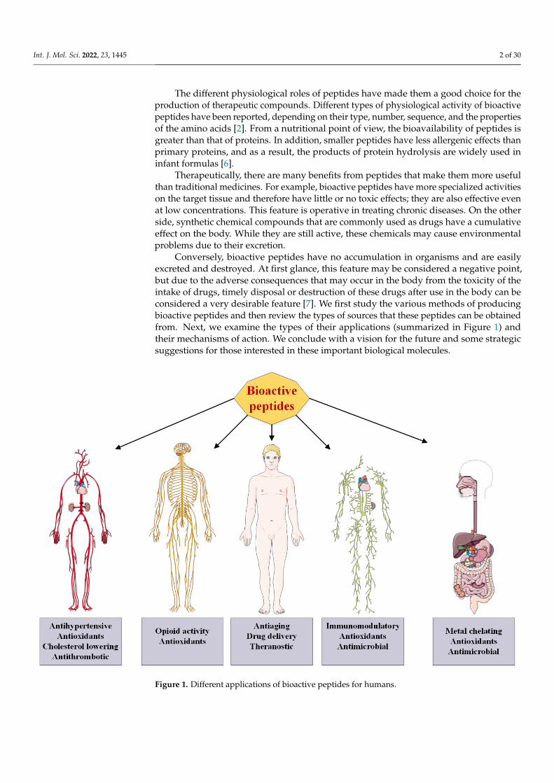
Conversely, bioactive peptides have no accumulation in organisms and are easilyexcreted and destroyed. At first glance, this feature may be considered a negative point,but due to the adverse consequences that may occur in the body from the toxicity of theintake of drugs, timely disposal or destruction of these drugs after use in the body can beconsidered a very desirable feature [7]. We first study the various methods of producingbioactive peptides and then review the types of sources that these peptides can be obtainedfrom. Next, we examine the types of their applications (summarized in Figure 1) andtheir mechanisms of action. We conclude with a vision for the future and some strategicsuggestions for those interested in these important biological molecules.
Int. J. Mol. Sci. 2022, 23, x FOR PEER REVIEW 2 of 30
attention due to their application in increasing the quality of health and, economically, because of their use in the production of healthy foods, drugs, and other products [5].
The different physiological roles of peptides have made them a good choice for the production of therapeutic compounds. Different types of physiological activity of bioac-tive peptides have been reported, depending on their type, number, sequence, and the properties of the amino acids [2]. From a nutritional point of view, the bioavailability of peptides is greater than that of proteins. In addition, smaller peptides have less allergenic effects than primary proteins, and as a result, the products of protein hydrolysis are widely used in infant formulas [6].
Therapeutically, there are many benefits from peptides that make them more useful than traditional medicines. For example, bioactive peptides have more specialized activi-ties on the target tissue and therefore have little or no toxic effects; they are also effective even at low concentrations. This feature is operative in treating chronic diseases. On the other side, synthetic chemical compounds that are commonly used as drugs have a cumu-lative effect on the body. While they are still active, these chemicals may cause environ-mental problems due to their excretion.
Conversely, bioactive peptides have no accumulation in organisms and are easily ex-creted and destroyed. At first glance, this feature may be considered a negative point, but due to the adverse consequences that may occur in the body from the toxicity of the intake of drugs, timely disposal or destruction of these drugs after use in the body can be con-sidered a very desirable feature [7]. We first study the various methods of producing bio-active peptides and then review the types of sources that these peptides can be obtained from. Next, we examine the types of their applications (summarized in Figure 1) and their mechanisms of action. We conclude with a vision for the future and some strategic sug-gestions for those interested in these important biological molecules.
Figure 1. Different applications of bioactive peptides for humans. Figure 1. Different applications of bioactive peptides for humans.
Int. J. Mol. Sci. 2022, 23, 1445 3 of 30
2. Production of Bioactive Peptides
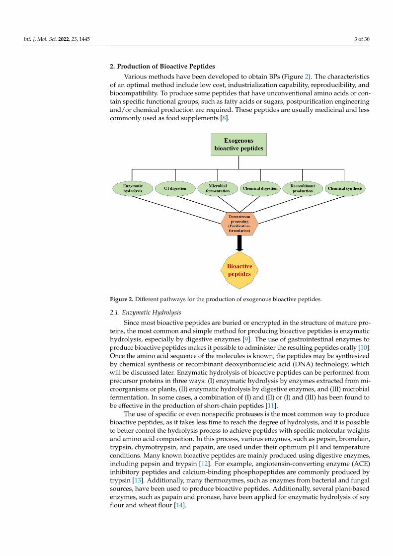
Various methods have been developed to obtain BPs (Figure 2). The characteristicsof an optimal method include low cost, industrialization capability, reproducibility, andbiocompatibility. To produce some peptides that have unconventional amino acids or con-tain specific functional groups, such as fatty acids or sugars, postpurification engineeringand/or chemical production are required. These peptides are usually medicinal and lesscommonly used as food supplements [8].
Int. J. Mol. Sci. 2022, 23, x FOR PEER REVIEW 3 of 30
2. Production of Bioactive Peptides Various methods have been developed to obtain BPs (Figure 2). The characteristics
of an optimal method include low cost, industrialization capability, reproducibility, and biocompatibility. To produce some peptides that have unconventional amino acids or con-tain specific functional groups, such as fatty acids or sugars, postpurification engineering and/or chemical production are required. These peptides are usually medicinal and less commonly used as food supplements [8].
Figure 2. Different pathways for the production of exogenous bioactive peptides.
2.1. Enzymatic Hydrolysis Since most bioactive peptides are buried or encrypted in the structure of mature pro-
teins, the most common and simple method for producing bioactive peptides is enzymatic hydrolysis, especially by digestive enzymes [9]. The use of gastrointestinal enzymes to produce bioactive peptides makes it possible to administer the resulting peptides orally [10]. Once the amino acid sequence of the molecules is known, the peptides may be syn-thesized by chemical synthesis or recombinant deoxyribonucleic acid (DNA) technology, which will be discussed later. Enzymatic hydrolysis of bioactive peptides can be per-formed from precursor proteins in three ways: (I) enzymatic hydrolysis by enzymes ex-tracted from microorganisms or plants, (II) enzymatic hydrolysis by digestive enzymes, and (III) microbial fermentation. In some cases, a combination of (I) and (II) or (I) and (III) has been found to be effective in the production of short-chain peptides [11].
The use of specific or even nonspecific proteases is the most common way to produce bioactive peptides, as it takes less time to reach the degree of hydrolysis, and it is possible to better control the hydrolysis process to achieve peptides with specific molecular weights and amino acid composition. In this process, various enzymes, such as pepsin, bromelain, trypsin, chymotrypsin, and papain, are used under their optimum pH and temperature conditions. Many known bioactive peptides are mainly produced using di-gestive enzymes, including pepsin and trypsin [12]. For example, angiotensin-converting enzyme (ACE) inhibitory peptides and calcium-binding phosphopeptides are commonly produced by trypsin [13]. Additionally, many thermozymes, such as enzymes from bac-terial and fungal sources, have been used to produce bioactive peptides. Additionally, several plant-based enzymes, such as papain and pronase, have been applied for enzy-matic hydrolysis of soy flour and wheat flour [14].
Figure 2. Different pathways for the production of exogenous bioactive peptides.
2.1. Enzymatic Hydrolysis
Since most bioactive peptides are buried or encrypted in the structure of mature pro-teins, the most common and simple method for producing bioactive peptides is enzymatichydrolysis, especially by digestive enzymes [9]. The use of gastrointestinal enzymes toproduce bioactive peptides makes it possible to administer the resulting peptides orally [10].Once the amino acid sequence of the molecules is known, the peptides may be synthesizedby chemical synthesis or recombinant deoxyribonucleic acid (DNA) technology, whichwill be discussed later. Enzymatic hydrolysis of bioactive peptides can be performed fromprecursor proteins in three ways: (I) enzymatic hydrolysis by enzymes extracted from mi-croorganisms or plants, (II) enzymatic hydrolysis by digestive enzymes, and (III) microbialfermentation. In some cases, a combination of (I) and (II) or (I) and (III) has been found tobe effective in the production of short-chain peptides [11].
The use of specific or even nonspecific proteases is the most common way to producebioactive peptides, as it takes less time to reach the degree of hydrolysis, and it is possibleto better control the hydrolysis process to achieve peptides with specific molecular weightsand amino acid composition. In this process, various enzymes, such as pepsin, bromelain,trypsin, chymotrypsin, and papain, are used under their optimum pH and temperatureconditions. Many known bioactive peptides are mainly produced using digestive enzymes,including pepsin and trypsin [12]. For example, angiotensin-converting enzyme (ACE)inhibitory peptides and calcium-binding phosphopeptides are commonly produced bytrypsin [13]. Additionally, many thermozymes, such as enzymes from bacterial and fungalsources, have been used to produce bioactive peptides. Additionally, several plant-basedenzymes, such as papain and pronase, have been applied for enzymatic hydrolysis of soyflour and wheat flour [14].

Int. J. Mol. Sci. 2022, 23, 1445 4 of 30
Many bioactive peptides, such as biogenic, opioid, immunomodulatory, salt/metal-binding, antihypertensive, and antimicrobial peptides, can be produced by enzymatichydrolysis of foods such as milk, animal and fish meat, corn, wheat, soybeans, and eggs [15].In industrial-scale conditions, the use of surface-coated enzymes is more common thanconventional soluble enzymes. Coated enzymes allow enzymatic hydrolysis under moremoderate and controlled conditions. In addition, these fixed enzymes can be recovered toprevent the production of secondary metabolites due to enzyme autolysis [16]. Anothercommon method on an industrial scale is the use of continuous hydrolysis, which is widelyused for the complete conversion of food proteins from various sources into hydrolysisproducts with improved nutritional or functional properties. This method is more efficientand less expensive under industrial conditions than conventional methods and is obtainedby using reactors equipped with ultrafiltration membranes with different components,which may be combined with other purification techniques or units [17].
The final product of enzymatic hydrolysis depends on the type of used enzyme, thetype of protein precursor, the degree of hydrolysis, and the separation method of the finalsample. Although both crude and purified peptides are used for different applications,to reduce the final price, the use of crude types of peptides is more preferred [2]. Insome cases, in addition to the physical properties of the peptide, its three-dimensionalstructure is also important. Some peptides, especially antimicrobial peptides, have a cyclicstructure (via disulfide bonds) or beta-sheets on their structure, which are necessary totheir functions [18]. During the production of such peptides, the structure of the parentprotein must not undergo harsh spatial change. On the other hand, this will be a challengein the performance of hydrolase enzymes because it may reduce the enzyme’s access to thecleavage site at a particular site of the protein.
Recently, some novel methods have been developed to overcome these conundrums.For example, it has been shown that by applying pressure (above 100 MPa) during enzy-matic hydrolysis (high hydrostatic pressure processing, HHP), the efficiency of the enzymeswill increase. Furthermore, high pressure has been shown to cause temporary and reversiblechanges in the protein structure that increase the access of hydrolase to a variety of cuttingsites on the protein surface [19,20]. Another study reviews such innovative methods, whichgenerally increase the efficiency of enzymatic hydrolysis during food processing [21].
Microbial Fermentation
There is another method, called microbial fermentation, to produce bioactive peptides,which is a type of bacterial hydrolase used to break down proteins into small peptides.In fact, this preparation of peptides is part of the enzymatic hydrolysis method that usesbacteria. Many industrially used primer cultures have high proteolytic potency. Thus,bioactive peptides can be produced by primer and nonprimer bacteria used in fermentedfood products [21]. Lactobacillales, which are a large group of beneficial bacteria in natureand are also found in our digestive system, are used to produce bioactive peptides. Theirrole in the production of fermented products is not only due to their physiological effectbut also due to their technological importance in the development of texture and taste.
The proteolytic systems of Lactobacillales, such as Lactococcus lactis [22], Lactobacillushelveticus [23], and Lactobacillus delbrueckii of the bulgaricus subspecies [24] are now wellknown. These systems consist of several proteins attached to the cell wall and severalintracellular proteins, including endopeptidases, aminopeptidases, tryptidases, and dipep-tidases [25]. Some studies have shown that the use of multiple fermentations, as well asthe combination of enzymatic hydrolysis, increases the production of bioactive peptides.A study reported that fermented milk products with a commercial mixed culture primercontaining five strains of Lactobacillales increase ACE inhibitory activity [26]. Treatment ofmilk with trypsin before fermentation with yogurt primer cultures results in the forma-tion of phosphopeptide-rich peptides. In these samples, the productions of the calciumphosphopeptides (CPP) β-caseins (β-CN, 1-25-4P) and αs1-casein (αs1-CN) (43-79)-7 were
Int. J. Mol. Sci. 2022, 23, 1445 5 of 30
reported, while the amount of proteolysis in samples that were fermented alone was notsignificant [27].
High inhibitory activity of 2,2′-azino-bis(3-ethylbenzothiazoline-6-sulfonic acid) (ABTS)and 2,2-diphenyl-1-picrylhydrazyl (DPPH) radicals of fermented camel milk with Leuconos-toc lactis has been reported [28]. In addition to living microorganisms, proteolytic enzymesisolated from Lactobacillales have been successfully used to generate bioactive peptides frommilk proteins. In one study, the ACE inhibitory activity of the casein hydrolysis productwas measured using five different commercial proteolytic enzymes. Among these enzymes,peptides produced from a protease isolated from Aspergillus aureus showed the highestinhibitory activity of ACE in vitro compared with other peptides [29]. In comparison withother methods, microbial fermentation is a cheaper way to produce peptides. This isbecause microorganisms are a cheap source of proteases and are known to be safe. The costof bacterial cultures is relatively low due to the minimum nutrient requirements and shortgrowth time. Additionally, the proteases of Lactobacillales are expressed on the cell wall,which makes extraction protocols relatively easy and inexpensive [30].
2.2. Chemical Synthesis of Peptides
In the process of chemical production, the bioactive peptides are produced fromamino acid units in a defined chemical environment. To chemical synthesis of bioactivepeptides, there are currently two major strategies, namely, soluble-phase and solid-phasesyntheses. In addition to these two methods, there is another chemical method, hybridsynthesis, which is sometimes used to produce pharmaceutical peptides. The proportion ofthe type of the used protecting groups in the structure of amino acids and the method ofdeprotection is the key step in the synthesis of bioactive peptides [31]. Chemical synthesisof peptides by soluble-phase methods was first used in 1953 to produce pharmaceuti-cal insulin peptides [32]. The basis of this method is the reaction of amino acids in asoluble medium.
Perhaps the most important advantage of soluble-phase methods is the economicjustification and purification at each stage of the synthesis since it uses less expensivematerials and equipment [33]. In this method, during the production of desired peptides,the side synthesis of intermediates is an undesirable stage. One of the limitations of themethod is the production of these intermediates. To achieve the desired pure product, eachinitial product must undergo changes to be allowed to enter the next stage and eventuallybecome an active peptide. As a result, the overall process of synthesizing an active peptidein this way is often lengthy and difficult [34]. However, using this method, therapeuticpeptides with a length of 3 to 20 amino acids have been synthesized and made their way toglobal markets [35].
The main problems of this method are the insolubility of long peptide chains inorganic solvents, long synthesis time, and the amount of chemical waste. Ten yearsafter the invention of the soluble-phase synthesis, the solid-phase synthesis method wasinvented [36]. This method is based on the reaction of amino acids that, in the presence ofinsoluble substances, become covered and unreacted in groups. This strategy is actuallyused to direct the reaction to the desired path. Initially, the first amino acid at the amineend and side-chain groups will cover and then attach to the resin bed via the carboxyl end.After binding, the protecting group washes from the amine terminus and prepares to reactwith the second amino acid. A coupling compound will be used to bind amino acids to eachother. The reactions will then be repeated to obtain the desired product/peptide [37,38].
The simplicity of this method has made it possible to mass-produce bioactive peptides,as it is simpler and faster than the solution-phase synthesis. However, the most impor-tant limitation of this method is the massive need for materials to start the process [39].Therapeutic peptides, such as ziconotide, exanatide, pramlintide, and degarelix, havebeen synthesized by this method and entered the pharmaceutical market [40]. Around1992, radio waves were used to further accelerate this method [41]. Today, this methodis widely used to produce well-known drugs, such as the antibiotic gramicidin A or the
Int. J. Mol. Sci. 2022, 23, 1445 6 of 30
glycoprotein CSF114, which are used for infectious diseases and the clinical diagnosis ofmultiple sclerosis, respectively [42].
In recent years, the chemical production of peptides by solid-phase method withthe help of fluorenylmethoxycarbonyl (Fmoc) as chemical group coating has become oneof the most widely used methods. In this method, the protective compound, Fmoc, isused to protect the amino acid side groups. With this action, some groups do not enterthe chemical reaction, and the reaction is directed in the desired direction [43]. By andlarge, the chemical methods mentioned earlier use substances that are environmentally andecologically hazardous. For example, dimethylformamide and dichloromethane, which areused in chemical methods of synthesizing peptides to protect the amino terminals of aminoacids, are very harmful to the environment. The removal of these substances from naturehas posed a significant challenge [38].
2.3. Recombinant Productions
In the recombinant production of bioactive peptides, the peptide genes are expressedin a specific expression system. Depending on whether the expression system is in vivo orin vitro, recombinant production is divided into two different groups. In the recombinantproduction of peptides by the in vivo expression system, the desired peptide gene is usuallyassociated with a protein gene, a carrier protein that can be easily purified. One of theadvantages of this method is achieving mass production of the desired peptide. Peptidesof various drugs have been obtained by this method, including ecallantide and desirudin,which have a length of 60 to 65 amino acids and are expressed in yeast [44,45].
On the other hand, the most advanced recombinant expression system is the expressionof bioactive peptides in the in vitro expression system, also called the cell-free system. In thesystem, all the necessary components for transcription and translation of a peptide gene arepresent in vitro, and in such an environment, peptide synthesis takes place in the absenceof the cell. One of the advantages of this method is the high speed of achieving the desiredproduct [46]. However, due to the cost of this method, it is used for specific peptides andmore in the laboratory and research scales. Nevertheless, many pharmaceutical peptidesare currently produced from a combination of chemical and recombinant synthesis ofpharmaceutical peptides.
In this method, first, a peptide is produced from the desired gene by the biologicalmethod. The drug peptide is then chemically modified by chemical methods [47]. One ofthe peptides that has entered the global medicine market in this way is a peptide calledliraglutide, which is used to regulate blood sugar in patients with type II diabetes. Thispeptide was approved in 2010 by the Food and Drug Administration. It is similar to thehormone glucagon and is recombinantly expressed in yeast, and is chemically added to itsamino acid No. 27, which is lysine, a 16-carbon lipid with an average of the amino acidglutamate. This action increases the functional similarity of this peptide to the hormoneglucagon [48].
The engineering of bioactive peptides is another topic that has recently attracted alot of attention. The goal of engineering peptides is to increase their efficacy and stability.Insulin can be cited as an example of the first engineered therapeutic peptide. This hormoneis often engineered by substituting one to three amino acids. The purpose of this action isto produce insulin that has a longer effect and can better play the role of natural insulin inthe body [49]. Today, a variety of drugs are produced from insulin, each of which has itsunique properties as an insulin drug and even its method of administration.
3. The Sources of Bioactive Peptides
Potentially bioactive peptides can be extracted or produced from any organism. How-ever, there are a few things to keep in mind when choosing a host. On the one hand, itshould be noted that the selection of the target host will ultimately determine the methodof extraction and purification of the peptide in question. On the other hand, it should beconsidered that the production or source of the desired peptide in the host must be so high
Int. J. Mol. Sci. 2022, 23, 1445 7 of 30
so that its production/purification will be both economical and problematically worthwhileto go on [11]. In the following section, the different types of industrialized hosts willbe described.
3.1. Animal Sources3.1.1. Marine Sources
As mentioned earlier, bioactive peptides can be obtained from a variety of sources.One of these important sources is the use of proteins and body wastes originating fromanimals to produce bioactive peptides. These peptides are the product of the enzymatichydrolysis of animal proteins. Blood is an important and rich source of animal protein thatis readily available and abundant in slaughterhouses. Other animal sources of bioactivepeptides include red meat and aquatic animals. There is growing scientific evidence thatmany hydrolyzed peptides and proteins derived from marine sources, including mollusks,crustaceans, and fishery wastes (head, intestines, skin, and fins), are capable of promotinghuman health and preventing chronic diseases [50].
So far, many studies have examined the therapeutic properties of aquatic bioactivepeptides (in vitro), and fewer studies have been performed on animal models as well ashumans. Huge volumes of fish waste are extracted annually in aquaculture processingcompanies, which account for up to 75% of the total catch weight. Converting fishery wasteinto valuable compounds is a suitable solution to reduce environmental pollution and is anoptimal use of aquatic waste [51], as the seas cover 70% of the earth’s surface, and theirbiodiversity is considerably greater than the land surface and accounts for approximately75% of all living organisms.
Recently, marine peptides have provided a new impetus for the development of phar-macy [52,53]. Peptides discovered from marine organisms stimulate cell death by variousmechanisms, including apoptosis, effect on tubulin–microtubule balance, angiogenesisinhibition, antiproliferative effects, and cytotoxic effects. These facts have introducedmarine bioactive peptides as a new choice for obtaining new compounds in biomedicalresearch [54]. Because the marine environment has more biodiversity than the terrestrialenvironment, and due to the unique adaptation of these organisms in dark, cold, and high-pressure environments during the evolution of these organisms, they have been able toexpress various proteins to overcome these environmental incompatibilities, which can be ahuge and unknown source for bioactive peptides. Many bioactive peptides with anticancerpotential have been extracted from various marine organisms, such as tonics, sponges, andmollusks [54]. Some of these products, such as aplidine, are now commercially available,and others are in various phases of clinical trials.
About 10,000 types of sponges have been found around the world [55]. To date,a wide range of bioactive peptides have been discovered in just 11 species of sponge.Between them, three genera, which include Discodermia, Petrosia, and Haliclona, can makeeffective anticancer and anti-inflammatory peptides. Although these compounds havea wide range of biological activities, they are difficult to purify in sufficient quantitiesfor pharmaceutical trials [56]. Jaspamide is a cyclic peptide isolated from sponges of thegenera Jaspis and Hemiastrella and encompasses a large 17-carbon ring and three aminoacids that can induce apoptosis in human leukemia cells (HL-60) [56,57]. About nine newcyclic peptides, homophyminics B–E and A1–E1, have been isolated from the Hamophymiasponge, which have potent cytotoxic activity. This activity has been reported against severalhuman cancer cell lines. Homophymins A1–E1, which have four amino-6 carbamoyl-2,3-dihydroxy hexanoic acid structures, have greater potency than the corresponding A-Ecompounds with the same backbone [58], indicating the importance of chemical contentsof bioactive peptides. Geodiamolide H extracted from Geodia corticostylifera sponge hasbeen shown to have anticancer activity against breast cancer by disrupting the balance ofintracellular actin. Table 1 lists some peptides with several potential therapeutic usagesextracted from marine organisms.

Int. J. Mol. Sci. 2022, 23, 1445 8 of 30
Table 1. Marine bioactive peptides.
Peptide Organism Function Ref.
Peptide extracts Bacillariophyceae Antihypertensive/antioxidant [59]
Peptide extracts Discodermiu kiiensis Antimicrobial [60]
Azonazine Aspergillus insulicola Anti-inflammatory [61]
Wewakazole L. majuscula Anticancer [62]
Mirabamide A-C-D Sponges anti-HIV [63]
Aplidine Aplidine Anticancer [64]
Arenastatin A Dysidea arenaria Anticancer [65]
Aurilide Dolabella auricularia Anticancer [66]
Didemnin Trididemnum sp. Anticancer [67]
Dolastatin Dolabella auricularia Anticancer [68]
Geodiamolide H Geodia sp. Anticancer [69]
Homophymines Homophymia sp. Anticancer [58]
Jaspamide Jaspis sp., Hemiastrella sp. Anticancer [70]
Kahalalide F Elysia rufescens, Spisulapolynyma Anticancer [65]
Keenamide A Pleurobranchus forskalii Anticancer [56]
Mollamide Didemnum molle Anticancer [71]
Phakellistatins Phakellia carteri Anticancer [72]
Tamandarins A and B Didemnum sp. Anticancer [73]
Discodermin tetradecapeptides are another group of antiseptic peptides extractedfrom the Discodermia sponge. Phakellistatin peptides isolated from Phakellia carteri spongeshave also been shown to inhibit the growth of leukemia cells. Another related compound isphakellistatin 13, which is derived from the Phakellia fusca. According to recorded observa-tions, this peptide has cell-killing properties against hepatic BEL-7404 cancer cells [74,75].On the whole, much attention has been paid to the discovery of structural, compositional,and sequence-related properties of bioactive peptides from marine sources.
3.1.2. Milk Products
Dairy products such as milk and cheese are ideal options for extracting animal bioac-tive peptides. As can be seen from the fundamental role of milk from an early age, where itacts as a source of protein and nitrogen for young mammals, it can be concluded that milkis a valuable substance in terms of protein contents. The proteins in milk have importantproperties, such as antibacterial, antioxidant, and immunoprotective activities. The numberof these properties is increasing every day, and recently, special attention has been paid tothe chaperone role of casein proteins in milk. Regarding milk peptides, opioid peptides inmilk have been reported to have morphine-like properties on the central nervous system.
Thanks to modern peptide separation and identification systems, tandem mass spec-trometry (MS/MS) and high-performance liquid chromatography–mass spectrometry(HPLC–MS), scientists today can obtain peptides with opioid properties from human milk.Other observed activities include ACE inhibitory, mineral-binding, anticarcinogenic, an-tithrombotic, and cytotoxicity [76]. In addition, lactoferrin (Lf)-protein-derived peptidesfound in the milk of all mammals play antimicrobial and immunosuppressive roles.
The fermentation of milk protein has also made it possible to access other valuablepeptides. Peptides derived from the fermentation of milk by L. helveticus LBK-16H, such asthe peptides Ile-Pro-Pro, Val-Pro-Pro, Tyr-Pro, and Lys-Val-LeuProi-Val-Pro-Gln, have anACE inhibitory effect in hypertensive animal models [77]. There are several antioxidantpeptides, such as GQGAKDMWR and EWFTFLKEAGQGAKDMWR, derived from donkeymilk [78]. Variation in the properties of milk-derived peptides has been shown to dependon factors such as the type of source protein, the hydrolysis method, and even the type ofanimal host. Today, animal hosts such as camel, mare, goat, sheep, and buffalo are used toextract proteins and peptides from their milk [79]. Some prominent examples of peptides
Int. J. Mol. Sci. 2022, 23, 1445 9 of 30
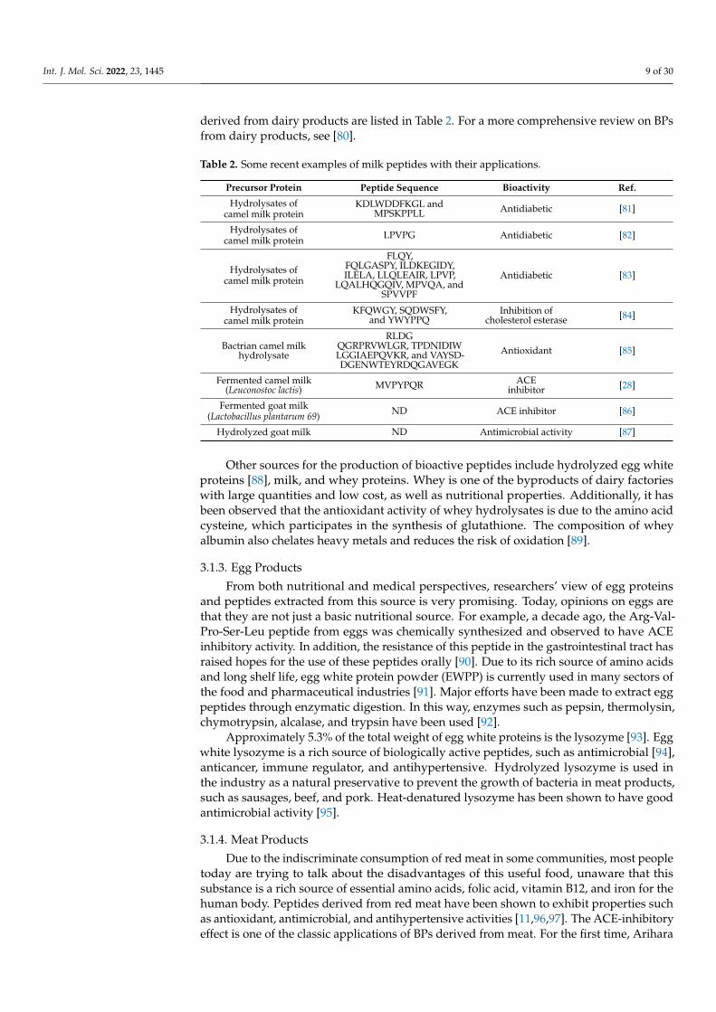
derived from dairy products are listed in Table 2. For a more comprehensive review on BPsfrom dairy products, see [80].
Table 2. Some recent examples of milk peptides with their applications.
Precursor Protein Peptide Sequence Bioactivity Ref.
Hydrolysates ofcamel milk protein
KDLWDDFKGL andMPSKPPLL Antidiabetic [81]
Hydrolysates ofcamel milk protein LPVPG Antidiabetic [82]
Hydrolysates ofcamel milk protein
FLQY,FQLGASPY, ILDKEGIDY,ILELA, LLQLEAIR, LPVP,
LQALHQGQIV, MPVQA, andSPVVPF
Antidiabetic [83]
Hydrolysates ofcamel milk protein
KFQWGY, SQDWSFY,and YWYPPQ
Inhibition ofcholesterol esterase [84]
Bactrian camel milkhydrolysate
RLDGQGRPRVWLGR, TPDNIDIWLGGIAEPQVKR, and VAYSD-DGENWTEYRDQGAVEGK
Antioxidant [85]
Fermented camel milk(Leuconostoc lactis) MVPYPQR ACE
inhibitor [28]
Fermented goat milk(Lactobacillus plantarum 69) ND ACE inhibitor [86]
Hydrolyzed goat milk ND Antimicrobial activity [87]
Other sources for the production of bioactive peptides include hydrolyzed egg whiteproteins [88], milk, and whey proteins. Whey is one of the byproducts of dairy factorieswith large quantities and low cost, as well as nutritional properties. Additionally, it hasbeen observed that the antioxidant activity of whey hydrolysates is due to the amino acidcysteine, which participates in the synthesis of glutathione. The composition of wheyalbumin also chelates heavy metals and reduces the risk of oxidation [89].
3.1.3. Egg Products
From both nutritional and medical perspectives, researchers’ view of egg proteinsand peptides extracted from this source is very promising. Today, opinions on eggs arethat they are not just a basic nutritional source. For example, a decade ago, the Arg-Val-Pro-Ser-Leu peptide from eggs was chemically synthesized and observed to have ACEinhibitory activity. In addition, the resistance of this peptide in the gastrointestinal tract hasraised hopes for the use of these peptides orally [90]. Due to its rich source of amino acidsand long shelf life, egg white protein powder (EWPP) is currently used in many sectors ofthe food and pharmaceutical industries [91]. Major efforts have been made to extract eggpeptides through enzymatic digestion. In this way, enzymes such as pepsin, thermolysin,chymotrypsin, alcalase, and trypsin have been used [92].
Approximately 5.3% of the total weight of egg white proteins is the lysozyme [93]. Eggwhite lysozyme is a rich source of biologically active peptides, such as antimicrobial [94],anticancer, immune regulator, and antihypertensive. Hydrolyzed lysozyme is used inthe industry as a natural preservative to prevent the growth of bacteria in meat products,such as sausages, beef, and pork. Heat-denatured lysozyme has been shown to have goodantimicrobial activity [95].
3.1.4. Meat Products
Due to the indiscriminate consumption of red meat in some communities, most peopletoday are trying to talk about the disadvantages of this useful food, unaware that thissubstance is a rich source of essential amino acids, folic acid, vitamin B12, and iron for thehuman body. Peptides derived from red meat have been shown to exhibit properties suchas antioxidant, antimicrobial, and antihypertensive activities [11,96,97]. The ACE-inhibitoryeffect is one of the classic applications of BPs derived from meat. For the first time, Arihara

Int. J. Mol. Sci. 2022, 23, 1445 10 of 30
et al. noticed this activity in peptides derived from porcine skeletal muscle proteins [98]. Inaddition to porcine, this biological activity has also been observed in beef-protein-derivedpeptides [99]. Additionally, the antioxidant activity of peptides derived from red meatshould not be overlooked. It is found that there is 2700 mg of carnosine per kilogramof pork meat. Carnosine shows well-documented antioxidant activity, which stems fromthe ability of the carnosine peptide to trap transition metals, such as copper, cobalt, andzinc [100].
Subsequently, after enzymatic hydrolysis (actinase E and papain) of pig protein,Saiga et al. obtained a source of antioxidant peptides [101]. Papain enzyme was also usedby Di Bernardini et al. to hydrolyze sarcoplasmic proteins from sarcoplasmic proteins.After fractionation, 10 and 3 kDa fractions showed antioxidant activities [102].
The striated muscles make up about 40% to 50% of livestock meat’s weight. Thesemuscles are made up of fibers of muscle cells. About 55% of red meat proteins containmyofibrillar proteins. These proteins are insoluble in water but are soluble in dilute salinesolutions. In the food industry, these proteins have beneficial properties. They compriserelatively large amounts of essential amino acids, which is why they have been consideredto be 70% of the nutritional value of meat. These proteins also affect the capacity of themeat emulsion so that 90% of this capacity is due to the presence of these proteins. Onthe other hand, about 97% of the storage capacity of water is due to the presence of theseproteins in the structure of red meat [103]. Sarcoplasmic proteins are given a lot of attentionin the production of BPs and makeup about 22% to 25% of the total weight of muscle tissueproteins [104]. Other proteins that receive less attention (due to their lower solubility inwater) include connective tissue proteins, such as collagen and elastin. In the food industry,the role of connective tissue proteins is not favorable as they reduce the quality of meat.
3.1.5. Venom Peptidomics: A Cure for the Deathtrap
Scientists’ views on many things have changed today. One of these cases is venom, acomposite that mainly includes peptides and other substances [105]. With new uses beingdiscovered for peptides today, some researchers are trying to figure out more applicationsto access this hidden treasury by identifying the different components of venom [106].Early efforts in this area focused on the separation of the various parts of venoms and thedetermination of their active component(s). After separating the active component andperforming some other studies, such as sequencing and structuring the active part, it willbe possible to study more applications of the discovered component. Efforts in this areahave intensified to the point that researchers are trying to build a library of ‘expressedsequence tags’ related to the venom glands of various animals. Although these studiesare expensive, they are worth the benefits that will be discovered in the future [107]. Thestudies in this field are numerous and varied, and Table 3 presents a brief classificationoverview of different families of venom peptides along with their applications.
Table 3. Different families of venom peptides.
Classification Example Host Applications Ref.
Bradykininpotentiating
peptides
TsTX-Ka andTsTX-KO Bothrops jararaca
Hypotensiveeffects, ACE
inhibitor[108]
BPPs Tityus serrulatusBothrops jararaca ACE inhibitor [109]
Antimicrobialpeptides IsCTs Opisthacanthus
madagascariensisAntimicrobial
Cytolytic activity [110]
Hormonelikepeptides Mini-Ins Conus geographus Insulin-like
activity [111]
Therapeuticpeptides Ziconotide Conus magus Pain killer [112]
Int. J. Mol. Sci. 2022, 23, 1445 11 of 30
3.1.6. Other Animals
In addition to the aforementioned sources, some other animals can be a significantsource of BPs. For example, bacterial antimicrobial peptides are called bacteriocins, whichcontain neutral or positively charged peptides and are secreted by a variety of Gram-negative and positive bacteria. These peptides are not a factor in defending against viralinfection, but help bacteria kill other bacteria in the competition for environmental supple-ments [113].
Most bacteriocins target the cell membrane with hydrophilicity or hydrophobicity,while some also inhibit the biosynthesis of biopolymers or the activity of enzymes. Thepeptides are not synthesized by ribosomes and instead undergo complex step-by-stepcompression reactions derived from peptide synthetases. Large nonribosomal (NRPS)peptides are often composed of nonprotein amino acids, including D-type amino acids,hydroxy acids, or other unusual compounds, and exhibit a wide range of inhibitory andfunctional mechanisms [114]. Bacteriocins are resistant to heat, low pH, weak organic sol-vents, cold, ice, and salts and are therefore applicable to food protection systems. Isolationand purification of these compounds are necessary to determine the exact mechanism oftheir inhibitory activity on food spoilage bacteria and foodborne bacteria. Bacteriocins aregenerally sensitive to human intestinal proteases, making them a valuable resource forfood preservation without potential harmful effects on human health [115]. Additionally,antimicrobial peptides can be extracted from the hemolymph of insects or outside theirbodies. Insects in the face of microorganisms secrete different antimicrobial peptides be-cause they can detect different types of invasive organisms and secrete the appropriateantimicrobial peptide. Antimicrobial peptides in primitive organisms are an alternative toprimary responses [116].
More attention is also being paid to BPs derived from marine microorganisms. Somemarine bioactive compounds are produced by microbes that coexist with marine species.Marine actinomycetes are a source of secondary bioactive compounds that have anticancerand antimicrobial activity. Cyclomarins A, B, and C are three ring-shaped heptapeptidesisolated from marine actinomycetes, such as Streptomyces sp. Cyclomarin A is composed ofthree common amino acids and four unusual amino acids and has shown anti-inflammatoryand anticellular activity in laboratory studies [117,118]. Salinamides A–E are types ofpeptides that have been extracted from Streptomyces sp. Salinamides A and B are twobioactive compounds with large rings that have local anti-inflammatory and antimicrobialactivity against Gram-negative bacteria and are used in the treatment of tissue inflammationand some infections. Salinamides C, D, and E are small BPs with anti-inflammatory activity.The structure of type D is similar to that of A, but contains valine residues at the isoleucinesite of type A. Salinamides E and C are also shown as a single-ring peptide [119].
Additionally, amphibians have a high level of defense system that includes innate andacquired immunity. In this group of organisms, the skin is protected by innate immunitymediated by macrophages, neutrophils, complement-mediated lysis, natural killer cells,and secreted antimicrobial peptides. Peptides of this group are synthesized in the granularglands of the skin. Due to environmental stimuli or damage to the sympathetic nerves,the peptides are activated and the contents of the glands are secreted to the surface of theskin [120]. Antimicrobial peptides in this group mostly have an alpha-helix structure. Ithas been shown that Rana frogs secrete the ranalexin antimicrobial peptide, which has acyclic structure with disulfide bridges [121].
3.2. Plant Sources
Traditionally, more attention has been paid to peptides derived from animals thanplants. Nevertheless, it should be noted that plant proteins are rich sources of proteinswithout saturated fatty acids that can carry useful ingredients. Recently, certain activitieshave been discovered in plant-derived peptides that can perform important functions inhumans. Antidiabetic, immunomodulatory, antimicrobial, hypocholesterolemic, opioid,antihypertensive, and antioxidant activities are of these benefits.
Int. J. Mol. Sci. 2022, 23, 1445 12 of 30
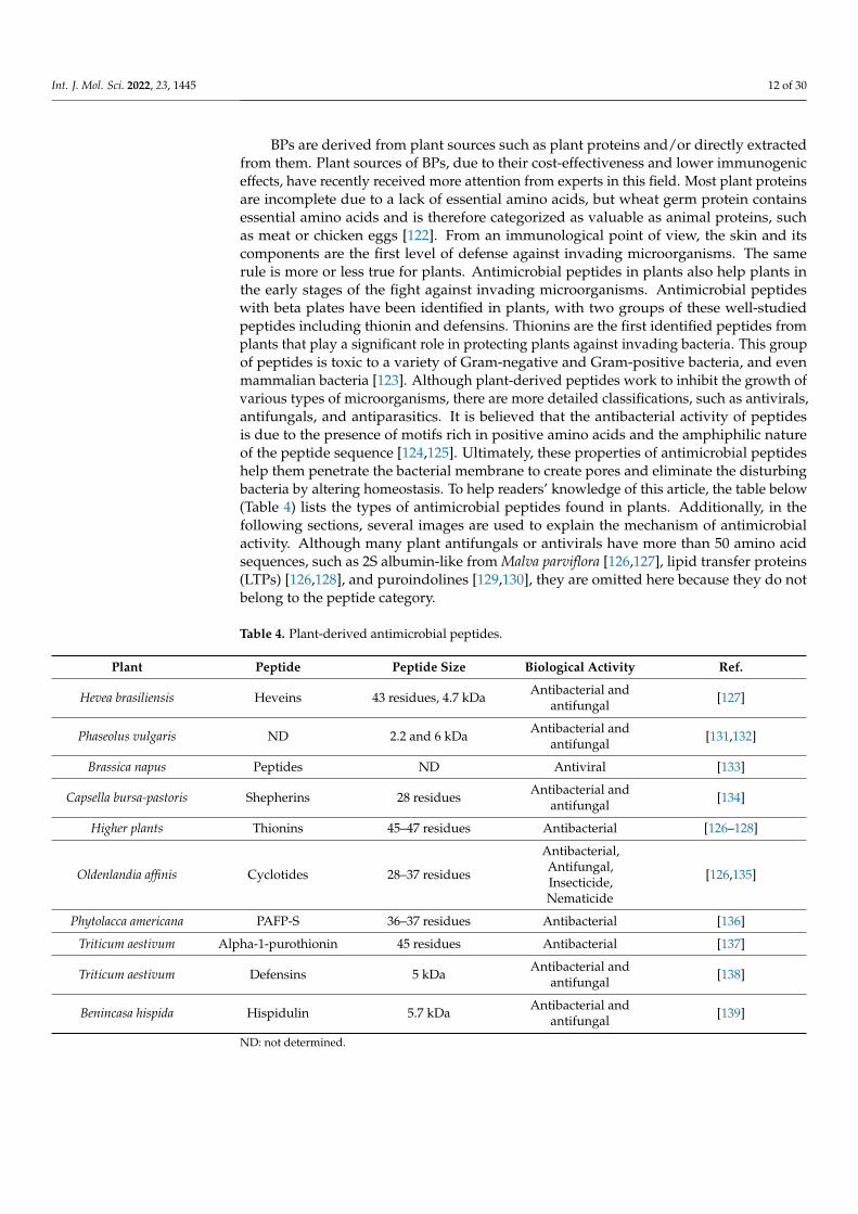
BPs are derived from plant sources such as plant proteins and/or directly extractedfrom them. Plant sources of BPs, due to their cost-effectiveness and lower immunogeniceffects, have recently received more attention from experts in this field. Most plant proteinsare incomplete due to a lack of essential amino acids, but wheat germ protein containsessential amino acids and is therefore categorized as valuable as animal proteins, suchas meat or chicken eggs [122]. From an immunological point of view, the skin and itscomponents are the first level of defense against invading microorganisms. The samerule is more or less true for plants. Antimicrobial peptides in plants also help plants inthe early stages of the fight against invading microorganisms. Antimicrobial peptideswith beta plates have been identified in plants, with two groups of these well-studiedpeptides including thionin and defensins. Thionins are the first identified peptides fromplants that play a significant role in protecting plants against invading bacteria. This groupof peptides is toxic to a variety of Gram-negative and Gram-positive bacteria, and evenmammalian bacteria [123]. Although plant-derived peptides work to inhibit the growth ofvarious types of microorganisms, there are more detailed classifications, such as antivirals,antifungals, and antiparasitics. It is believed that the antibacterial activity of peptidesis due to the presence of motifs rich in positive amino acids and the amphiphilic natureof the peptide sequence [124,125]. Ultimately, these properties of antimicrobial peptideshelp them penetrate the bacterial membrane to create pores and eliminate the disturbingbacteria by altering homeostasis. To help readers’ knowledge of this article, the table below(Table 4) lists the types of antimicrobial peptides found in plants. Additionally, in thefollowing sections, several images are used to explain the mechanism of antimicrobialactivity. Although many plant antifungals or antivirals have more than 50 amino acidsequences, such as 2S albumin-like from Malva parviflora [126,127], lipid transfer proteins(LTPs) [126,128], and puroindolines [129,130], they are omitted here because they do notbelong to the peptide category.
Table 4. Plant-derived antimicrobial peptides.
Plant Peptide Peptide Size Biological Activity Ref.
Hevea brasiliensis Heveins 43 residues, 4.7 kDa Antibacterial andantifungal [127]
Phaseolus vulgaris ND 2.2 and 6 kDa Antibacterial andantifungal [131,132]
Brassica napus Peptides ND Antiviral [133]
Capsella bursa-pastoris Shepherins 28 residues Antibacterial andantifungal [134]
Higher plants Thionins 45–47 residues Antibacterial [126–128]
Oldenlandia affinis Cyclotides 28–37 residues
Antibacterial,Antifungal,Insecticide,Nematicide
[126,135]
Phytolacca americana PAFP-S 36–37 residues Antibacterial [136]
Triticum aestivum Alpha-1-purothionin 45 residues Antibacterial [137]
Triticum aestivum Defensins 5 kDa Antibacterial andantifungal [138]
Benincasa hispida Hispidulin 5.7 kDa Antibacterial andantifungal [139]
ND: not determined.

Int. J. Mol. Sci. 2022, 23, 1445 13 of 30
4. Medicinal Applications and Proposed Mechanism of Actions of BPs4.1. Antioxidant Activity of BP and Its Mechanism of Action
In recent years, the trend of technological developments in human societies has causedfundamental changes in human lifestyle. By reducing the physical activity of people in thecommunity, which can be considered a special type of stress, the incidence of some diseases,including cardiovascular complications and different types of cancers, has increased inpeople in developed and developing societies. Looking through the literal definition ofstress, it is the general and nonspecific response of the organism to maintain homeostasisagainst any factor that threatens or impairs the body’s compensatory abilities [140]. Inits general definition, stress is a factor that interferes with a person’s physical and mentalbalance, causes psychosomatic problems, and reduces a person’s efficiency in variousaspects of life [141].
Oxidative stress is a change in the balance between pro-oxidants and antioxidants.The production of some free radicals such as superoxide can be physiologically bene-ficial, but oxidative stress occurs when the balance between the production of reactiveoxygen/nitrogen species (ROS/NOS) and the antioxidant defense system is upset. Oxida-tive stress, therefore, changes the balance between pro-oxidants/antioxidants in favor ofpro-oxidants, potentially leading to biological damages. Diseases associated with ROS pro-duction include cancer, Parkinson’s, and Alzheimer’s [142] diseases. Oxidative stress cancause serious damage to important cellular macromolecules, including lipids, nucleic acids,and proteins. In biological systems, the production of free radicals of ROS is inevitable,and the body partially neutralizes their harmful effects by designing antioxidant defensemechanisms. The most important components of the enzymatic antioxidant defense systeminclude the enzymes superoxide dismutase, glutathione peroxidase, and catalase. Antioxi-dant enzymes, which are responsible for detoxifying free radicals or repairing antioxidantmolecules, are an indicator of stress levels in cells or tissues. In addition to the primarydefense barrier created by antioxidant enzymes, the second defense barrier is created bysmall molecules (antioxidants) that react with free radicals to produce less dangerousradical compounds [143].
BPs have a strong antioxidant activity against free radicals and other reactive species.These antioxidant peptides contain 5–16 amino acids [144]. The mechanism by which pep-tides exert their antioxidant effects has not been fully elucidated, although various studieshave shown that hydrolyzed peptides and proteins prevent enzymatic and nonenzymaticoxidation by removing free radicals and chelating metal ions. Several peptides have beenfound in protein constituents that have antioxidant capacity, and their biological activitieshave been extensively studied. Although the energy of free radicals (such as hydroxyl) ishigh, in general, all 20 amino acids found in proteins can have internal interactions withfree radicals. Food-derived antioxidant peptides are safe and healthy compounds with lowmolecular weight, low cost, high activity, and easy absorption. The antioxidant propertiesof peptides are mostly related to their composition, structure, and hydrophobicity [144,145].
4.1.1. Effect of Amino Acid Contents on Antioxidant Activity of Peptides
The presence of some amino acids and their position in the peptide sequence hasan important effect on their antioxidant activity [146]. Aromatic amino acids, such astyrosine, histidine, tryptophan, and phenylalanine, and hydrophobic amino acids, suchas valine, leucine, methionine, glycine, and alanine, are essential for the antioxidant roleof the peptide. The higher oxidation of peptides compared with free amino acids isattributed to their unique chemical and physical properties by the amino acid sequenceitself. In a study, the His-Gly-Pro-Lue-Gly-Pro-Lue antioxidant peptide, the presence oftwo replicate sequences, Gly-Pro, and the placement of Lue in the carboxylic position andHis at the amine end increased the free radical scavenging property [147]. The presenceof hydrophilic amino acids such as proline, alanine, valine, and leucine in the N positionand the amino acids tyrosine, valine, methionine, leucine, isoleucine, glutamine, andtryptophan in the C-terminal position was associated with the antioxidant properties of
Int. J. Mol. Sci. 2022, 23, 1445 14 of 30
peptides [148]. Additionally, fat-soluble free radicals (peroxyl radicals) produced during theoxidation process of unsaturated fatty acids are neutralized by hydrophobic amino acidssuch as leucine, valine, alanine, and proline [149]. Amino acids such as histidine, tyrosine,methionine, and cysteine inactivate free radicals by giving them protons. Aromatic aminoacids (phenylalanine, tryptophan, and tyrosine) convert free radicals into stable moleculesby giving them electrons [150].
4.1.2. Effect of Peptide Size on Antioxidant Activity
In addition to a peptide sequence, the molecular weight of peptides can affect theirantioxidant activity [147,151]. Research has shown that the antioxidant activity of corngluten hydrolyzed protein is related to its concentration and molecular weight. Theantioxidant activity of peptides with a molecular weight of between 500–1500 Daltons isstronger than that of peptides with a molecular weight of higher than 1500 Daltons or lowerthan 500 Daltons [152]. In some cases, the higher antioxidant power of smaller peptidescompared with large chain peptides was attributed to their easier access to free radicals anda more effective removal of these radicals [153]. However, it has been repeatedly shownthat the higher the degree of hydrolysis, the lower the antioxidant activity of the peptides.This is due to the further breakdown of peptides into free amino acids that have little or noantioxidant activity [146].
4.1.3. The Role of Hydrophobicity of Peptides in Their Antioxidant Activity
Most food-derived antioxidant peptides include hydrophobic amino acids such asvaline or leucine at the N-terminal and proline, histidine, tyrosine, tryptophan, methionine,and cysteine in their sequence. Hydrophobic amino acids such as valine or leucine canincrease the affinity of peptides in the fat phase, thus facilitating access to free radicalsproduced in the fat phase [153,154].
4.2. Mechanism of Antimicrobial Activity
In the last two decades, many peptides with antibacterial, antiviral, and antifungalactivities have been identified in both vertebrates and invertebrates, which form an im-portant part of the host’s innate immune system. In most cases, the mechanism of actionof antimicrobial peptides appears to be different from that of conventional antibiotics(see Figure 3). For this reason, these peptides are very interesting as new drugs to fightinfectious agents [155]. Thus, antimicrobial peptides have opened a new chapter in thesciences, which has attracted the attention of many scientists and researchers, especiallysince they are much simpler in structure than proteins, which simplifies the study of thefunction–structure relationship and makes it possible to construct them by nonbiologicalways (such as chemical synthesis).
The factors of the effectiveness of these biologically active peptides as antimicro-bial agents depend on structural properties (e.g., peptide size, amino acid composition,or charge) [156]. However, antimicrobial peptides have some common features. Mostantimicrobial peptides are composed of fewer than 50 amino acids, 50% of which arehydrophobic [157]. Among these peptides, those with essential amino acids (lysine andarginine) have the highest antimicrobial properties [158,159]. Additionally, cationic andamphipathicity amino acids are important structural features for the antimicrobial activityof these BPs [160,161]. These peptides are also referred to as cell-penetrating peptides,protein transport domains, membrane sequences, or trojan peptides. Today, these peptidesare used to transport many membrane materials. Recent studies have shown that thesepeptides can transport a wide range of drugs, proteins, liposomes, and nanoparticles intoanimal cells [162]. In 1988, a membrane-permeable peptide was discovered from the Tatprotein of the HIV-I virus with tree sequences. This peptide was found to be able to crossthe membrane of cultured cells and accumulate in the nucleus [163,164].

Int. J. Mol. Sci. 2022, 23, 1445 15 of 30
Int. J. Mol. Sci. 2022, 23, x FOR PEER REVIEW 15 of 30
4.1.3. The Role of Hydrophobicity of Peptides in Their Antioxidant Activity Most food-derived antioxidant peptides include hydrophobic amino acids such as
valine or leucine at the N-terminal and proline, histidine, tyrosine, tryptophan, methio-nine, and cysteine in their sequence. Hydrophobic amino acids such as valine or leucine can increase the affinity of peptides in the fat phase, thus facilitating access to free radicals produced in the fat phase [153,154].
4.2. Mechanism of Antimicrobial Activity In the last two decades, many peptides with antibacterial, antiviral, and antifungal
activities have been identified in both vertebrates and invertebrates, which form an im-portant part of the host’s innate immune system. In most cases, the mechanism of action of antimicrobial peptides appears to be different from that of conventional antibiotics (see Figure 3). For this reason, these peptides are very interesting as new drugs to fight infec-tious agents [155]. Thus, antimicrobial peptides have opened a new chapter in the sci-ences, which has attracted the attention of many scientists and researchers, especially since they are much simpler in structure than proteins, which simplifies the study of the function–structure relationship and makes it possible to construct them by nonbiological ways (such as chemical synthesis).
Figure 3. (A) Types of mechanisms of action of antimicrobial peptides on bacterial cells. (B) How antimicrobial peptides penetrate the cell membrane.
The factors of the effectiveness of these biologically active peptides as antimicrobial agents depend on structural properties (e.g., peptide size, amino acid composition, or charge) [156]. However, antimicrobial peptides have some common features. Most anti-microbial peptides are composed of fewer than 50 amino acids, 50% of which are hydro-phobic [157]. Among these peptides, those with essential amino acids (lysine and argi-nine) have the highest antimicrobial properties [158,159]. Additionally, cationic and am-phipathicity amino acids are important structural features for the antimicrobial activity of these BPs [160,161]. These peptides are also referred to as cell-penetrating peptides, pro-tein transport domains, membrane sequences, or trojan peptides. Today, these peptides are used to transport many membrane materials. Recent studies have shown that these peptides can transport a wide range of drugs, proteins, liposomes, and nanoparticles into animal cells [162]. In 1988, a membrane-permeable peptide was discovered from the Tat
Figure 3. (A) Types of mechanisms of action of antimicrobial peptides on bacterial cells. (B) Howantimicrobial peptides penetrate the cell membrane.
It has been concluded that in the case of some antimicrobial peptides, although thepeptides reduce the growth of harmful microbes, they do not directly interact with the targetmicrobes or microorganisms, but do so with the help of the host immune system [165]. Forexample, milk protein hydrolyzate has been shown to stimulate the host immune system.These activities include stimulating the proliferation of the natural killer cell, stimulatingmacrophage phagocytosis, and encouraging the expression of many antibodies, cytokines,and chemokines [166].
The anti-inflammatory function of peptides is usually related to their antimicrobialactivity [167]. Inflammation is the response of the immune system to harmful stimuli(which can be invasive agents or damaged tissues) that are necessary to protect livingorganisms. In other words, inflammation is a complex biological response of host cells,vascular tissue, proteins, and other mediators to eliminate the primary causes of celldamage, tissue hemorrhage, and necrotic cells, which ultimately leads to the elimination ofinfection and treatment. During an inflammatory response, by increasing blood flow andvascular permeability, immune system components can escape from the blood vessels tothe affected area, resulting in five symptoms that may indicate inflammation: redness, heat,swelling, pain, and loss of function.
Inflammation is normally controlled and limited on its own. Inflammatory mediatorsare activated only in response to harmful stimuli and have a short lifespan, and whenthe harmful agents are removed, they are destroyed or inactivated. Additionally, at thistime, the acute inflammatory response is over, infection is removed, and damaged tissueis repaired. In addition, various anti-inflammatory mechanisms are activated. If thecausative agent cannot be eliminated quickly, it may lead to chronic inflammation that canhave serious pathological consequences. At the end of the inflammation, several differentregulatory mechanisms are activated: (1) inflammatory mediators that are short-lived aredestroyed or inactivated; (2) leukocyte migration stops; (3) the permeability of the vesselsdecreases and returns to normal; (4) the expression of proinflammatory molecules decreases,and conversely, the expression of anti-inflammatory molecules increases, which causes thetransfer of monocytes instead of neutrophils. Monocytes clean dead and damaged tissues,and tissue repair begins [168,169].
The production of natural antimicrobial agents by phagocytes has long been known.These antimicrobial peptides provide the first line of defense against pathogens in eukary-otic organisms and are generally effective against bacteria, fungi, and viruses. In addition
Int. J. Mol. Sci. 2022, 23, 1445 16 of 30
to the direct killing of microbes, these compounds also participate in processes relatedto inflammation and innate and acquired immunity. Antimicrobial peptides, which areinnate immune mediators, increase phagocytosis and trigger the release of prostaglandins.They also neutralize the shock effects of liposaccharides caused by bacteria. These peptidestransport and accumulate immune cells at the site of inflammation, induce angiogenesis,and heal wounds. The production of cytokines is also affected by these peptides [113].Antimicrobial peptides also have a chemotactic role [170]. All of these actions eliminatethe cells of bacteria. The results showed that these compounds are bactericidal at highconcentrations of mg/mL and have a safety regulatory role at lower concentrations [171].
Given all of the above, antimicrobial peptides are probably involved in all stages ofhost defense. In addition to enhancing the immune response, these compounds preventuncontrolled inflammation by suppressing proinflammatory responses. Despite the spe-cific overlap, the antimicrobial peptides interact with each other, complementing eachother to guide effective cells to the site of inflammation and modulate the local immuneresponse [172]. Phagocytes, neutrophils, and monocytes are absorbed via alpha-defensins,human neutrophil peptides 1HNP1-3, and beta-defensins such as human β-defensins2hBD3 and 3hBD4, while mast cells are adsorbed via HNP1-3, LL-37, and 4B. In addition,hBD1 and hBD3 are chemotactic for immature dendritic cells and memory T cells.
The combination of these peptides and cytokines at the site of injury will help theseimmature dendritic cells to mature and enable them to process antigens and migrate tonearby lymph nodes, where antigens are present. Antimicrobial peptides indirectly playa chemotactic role by inducing or increasing chemokine secretion. For example, LL-37induces the release of interleukin-8 by lung epithelial cells, and human defensin HNP1-3induces the activation and degranulation of mast cells. In addition, these human peptidesincrease neutrophil invasion, stimulate further transcription, and produce interleukin-8 bybronchial epithelial cells [173].
Antimicrobial peptides have a dual property: on the one hand, they protect the hostagainst harmful pathogens through antimicrobial activity, and on the other hand, theyprotect the host from the harmful effects of excessive inflammatory responses. In otherwords, these peptides stimulate the production of proinflammatory cytokines, increase theaccumulation of dendritic cells and monocytes at the site of injury, and increase phago-cytosis and maturation of dendritic cells, while simultaneously protecting the organismfrom the harmful effects of an inflammatory response. As a result, these peptides have bothproinflammatory and anti-inflammatory roles.
Cathelicidin is an important family of cationic peptides. In humans, the cathelicidingene encodes an inactive precursor protein that finally matured to active 37-amino acidpeptide (IL-37). It causes a balance between proinflammatory and anti-inflammatorysignals. Such peptides can inhibit the host’s harmful proinflammatory responses withoutlosing the beneficial innate defense [174]. Another example, α-melanocyte stimulatinghormone (α-MSH), is a neuropeptide that belongs to the melanocortin family with anti-inflammatory effects and shares several properties with antimicrobial peptides. Recentstudies indicate the direct antimicrobial activity of this peptide against fungi and pathogenicbacteria. It reduces the concentration of proinflammatory mediators and thus protectsthe brain and peripheral organs from inflammatory disorders. Therefore, α-MSH is ananti-inflammatory peptide with antimicrobial properties [175].
4.3. Antihypertensive Peptide and Its Mechanism of Action
In 2000, there were 972 million cases of hypertension in the world, and this numberis expected to reach 1.56 billion by 2025 [176]. The angiotensin-converting enzyme playsan important role in regulating and increasing blood pressure. This enzyme catalyzesthe transformation of inactive angiotensin I (decapeptide) to activate angiotensin II (oc-tapeptide), which is a strong vasoconstrictor. Angiotensin II also has a regulatory effect onthe enzyme cellular lipoxygenase, which accelerates the oxidation of low-density lipopro-
Int. J. Mol. Sci. 2022, 23, 1445 17 of 30
tein (LDL) and is associated with atherogenesis. It is also an inhibitor of bradykinin, apotent vasodilator.
Clinical studies have shown that ACE inhibitors significantly reduce mortality inpatients with myocardial infarction or heart failure [177]. Captopril and enalapril inhibitorsare used for hypertension, but they have many different side effects, including cough-ing, changes in taste, pimples, and edema, so there is a great deal of interest in usingnatural antihypertensive peptides [178]. In addition to milk proteins [179], other sourcesof antihypertensive peptides have been investigated. These sources include egg protein,mainly ovalbumin [180]; meat protein [98]; beef hemoglobin [181]; gelatin [182]; fish skinprotein [183]; and several plant proteins, such as soy [184], sesame [185], broccoli [186],buckwheat [187], and transgenic rice protein [188]. In most cases, for the body to use theantihypertensive peptides, these molecules must be absorbed intact through the intestinesand enter the bloodstream.
4.4. Mechanisms of Opioid Activity
Although pain is an important sign that there is a problem somewhere in the body,it is an unpleasant feeling that is often accompanied by severe and destructive stimuli.Chronic pain is associated with high levels of depression and anxiety. Additionally, insome physical conditions, reduced physical activity due to the generation of pain causesother diseases, such as obesity and heart harm. Chronic pain originates in the brain and/orspinal cord and is often difficult to manage [189]. Opioid drugs are currently used torelieve such pain, despite being associated with undeniable side effects. Pain is also one ofthe most important challenges in the management and/or treatment of cancer, and it hasbeen seen that the psychological effects of pain in cancer patients have a negative effecton their recovery [190,191]. The probability of pain in advanced stages of cancer is closeto 70% to 80% [192]. Pain is also seen in 90% of patients who have experienced cancermetastasis [193].
For these reasons, understanding the principles of pain and its management is criticalfor this group of patients. Pain is divided into two categories in terms of location: peripheralpain and nerve pain. Peripheral pain is pain that originates outside the central nervoussystem, including superficial pain, deep pain, or visceral pain. Nerve pain may be due to apathophysiological condition of the central nervous system, such as deep disturbance orsecretion of microbial–chemical substances and irritation due to heat or cold. Central pain,neuritis, neuralgia, and causalgia are types of nerve pain [194].
So far, various therapeutic measures have been used to control pain. Common thera-peutic measures include the administration of non-narcotic and narcotic analgesics (opioids).The types of drugs used are [195]:
(a) Muscle relaxants such as probantin and belladonna group such as atropine;(b) Vascular dilators such as papaverine hydrochloride or nitroglycerin;(c) Anti-inflammatory drugs such as indomethacin, ibuprofen, and phenylbutazone;(d) Non-narcotic analgesics such as aspirin and acetaminophen;(e) Narcotic analgesics such as Demerol and methadone hydrochloride.
Most drugs reduce blood pressure and respiratory depression, bradycardia, andconfusion [196,197]. Scientific methods of acute postoperative pain relief introduce theuse of both narcotic and non-narcotic drugs along with the use of nerve blocks as themethod of choice for complete postoperative pain relief. Oral and injectable methods(intramuscular, intravenous, and subcutaneous) for dermal or mucosal absorption orcentral or peripheral nerve blocks or without catheter placement are a variety of ways toadminister the drug [198].
As mentioned, one of the ways to control pain is to use narcotics. These drugs aregenerally peptides between 5 and 80 amino acids, which generally have two sources,endogenous and exogenous [199]. Drug peptides bind to their receptors on the surface ofnerve cells, triggering a signal that ultimately reduces pain. According to studies, mostdrug peptides act as agonists. In addition to pain, these peptides have been shown to
Int. J. Mol. Sci. 2022, 23, 1445 18 of 30
reduce stress levels. The internal sources of opioid peptides are usually in the form of eitherhormones (secreted by the glands) or a neurotransmitter that is secreted by nerve cells andacts on the terminals of other cells [200].
Enkephalin was the first known endogenous peptide. Many endogenous peptideshave been shown to have a conserved Tyr-Gly-Gly-Phe sequence at the end of their N-terminal [201,202]. Exorphins or exogenous opioid peptides with morphine-like activityenter the body from food sources, or in emergencies through drugs and supplements.Among the available food sources, dairy products are the best source for exorphins dueto the similarity of the sequence of peptides derived from them to endogenous opioidpeptides. Some observations suggest that the product of enzymatic digestion of dairyproteins, especially milk, can bind to opioid receptors on the cell surface. For example, theArg-Tyr-Leu-Gly-Tyr-Leu-Glu peptide derived from bovine milk casein alpha protein hasbeen shown to have narcotic activity [203]. It is important to note that peptides resultingfrom the digestion of digestive enzymes can be easily administered orally to humans [204].Interestingly, opioid peptides of animal origin generally bind to µ receptors, and peptidesof plant origin bind to δ receptors [205].
4.5. Mineral-Binding Peptides
Proteins interact with ions through their amino acid side chain. For example, alpha-casein and beta-casein interact with divalent and trivalent cations, such as calcium. Inaddition to proteins, peptides also have the ability to bind minerals. For example, casein-derived phosphopeptides, also known as casein phosphopeptides, have this activity [206].These phosphopeptides are involved in maintaining calcium, phosphorus, and other min-eral elements in solution in intestinal pH. This activity is due to the presence of the aminoacid phosphorylated serine, which can make salts with minerals, such as calcium. En-zymatic digestion of milk produces a diverse group of these peptides [207]. The type ofamino acid composition present in the phosphorylated region plays an essential role inthe amount of calcium-binding activity in this group of peptides [208]. These peptides arealso effective in preventing tooth decay, osteoporosis, insomnia, and hypertension. Animalstudies have shown a positive effect of these peptides on calcium absorption. A groupof researchers has shown that fermentation of whey protein with Lactobacillus holoticus iseffective in the proliferation of osteoblasts in vitro. There have also been other reports ofthe increased bioavailability of iron in rat models [209].
4.6. Blood-Lipid-Lowering Effect
Hyperlipidemia, especially high cholesterol, is one of the most important risk factorsfor cardiovascular disease. Many studies show that soy-derived peptides can lower bloodcholesterol levels in animal models of hepatotoxicity as well as in humans. Soy-rich dietshave become one of the most effective dietary treatments for high cholesterol, althoughthe mechanism has not yet been fully elucidated. It is believed that soy peptides derivedfrom protease actively cut cholesterol traveling in the gut and thus reduce cholesteroluptake [210]. Other research has shown that hydrophobic peptides derived from soyproteins are able to interact with bile acids, thereby increasing the excretion of fatty acids inthe feces [97,209].
It has been indicated that LPYPR and IAVPGEVA peptides derived from soy glycininprotein, which have structural similarity with endostatin and VPDPR, showed a cholesterol-lowering effect. These peptides inhibit 3-hydroxy-3-methylglutaryl-coenzyme, a reductase,which is a key enzyme during the biosynthesis of cholesterol [97]. Milk is another impor-tant source of BPs with a cholesterol-lowering effect. In 1999, Nagaoko et al. discovereda cholesterol-lowering peptide from digested beta-lactoglobulin hydrolase. Hydrolyzedplant protein with cholesterol-lowering activity such as soy and hydrolyzed Brassica car-inata proteins have also been reported [211]. Such effects from aquatic animals, such assardine [212] and zebrafish [213], have been shown on blood lipids. It has been shown thatpeptides that are lower in proportion to the amino acids methionine, glycine, lysine, and

Int. J. Mol. Sci. 2022, 23, 1445 19 of 30
arginine are better able to cause hyperlipidemia. However, bovine casein protein, whichhas a higher proportion of these amino acids, especially methionine and glycine, raisescholesterol levels [214].
4.7. Antiobesity Effect
Peptides can affect the absorption of nutrients in the intestines, especially the smallintestine, thereby reducing appetite. Many studies have shown that peptides derived fromdietary proteins can send satiety signals to the brain and thus prevent the consumption ofmore foods [214]. Casein-derived peptides have been shown to regulate eating in the bodyby activating the cholecystokinin A (CCK-A) receptor [215].
4.8. Antidiabetic Activity
A wide range of plant-derived peptides can help diabetics through a variety of path-ways. The pathways that have been studied so far include inhibitory properties on alpha-amylase, dipeptidyl peptidase IV, glucose transporter system, and mimicking insulinactivity [216].
4.9. Antiaging Peptides
During the aging process, the production of extracellular matrix proteins, such as colla-gen, fibronectin, elastin, and laminin, decreases, and their breakdown increases. In additionto protecting the cell structure, the extracellular matrix is effective on cellular behaviors,such as proliferation and differentiation. Such functions are controlled by small peptidesderived from the breakdown of extracellular matrix proteins called matrikine [217,218].Following the destruction of the extracellular matrix, the elasticity of the skin graduallydecreases, and the first lines of aging and wrinkles appear [219,220].
The aging process of the skin is controlled by internal and external factors. Externalfactors include exposure to ultraviolet radiation, environmental air pollution, and smoking.Exposure to ultraviolet light increases reactive oxygen species (ROS), disrupts collagensynthesis, and induces collagenase production and enzymes that break down proteins in theextracellular matrix, thereby causing cell DNA damage, and ultimately destructs the skinintegrity. Interior factors include the formation of large amounts of reactive oxygen speciesduring cellular metabolism and genetic factors that cause the destruction of extracellularmatrix proteins and reduced blood flow, and the function of skin cells is reduced [221].
Gradually, with the discovery of more functions of BPs, it was seen that these moleculesare able to rejuvenate the skin and increase its repair power. Today, peptides are usedto stimulate collagen, heal wounds, and smooth out skin wrinkles and for antioxidant,antibacterial, and skin whitening effects [222–224]. Cosmetic peptides can affect skinfreshness through several mechanisms. These pathways can be used to signal peptidesthat modulate collagen levels, such as Lys-Thr-Thr-Lys-Ser [225]; reduce IL-6 secretion andstimulate collagen and elastin synthesis, such as palmitoyl tetrapeptide-7 [226]; stimulatehuman skin fibroblasts and increase skin angiogenesis, such as Val-Gly-Val-Ala-Pro-Glypeptide; and inhibit tyrosine enzyme activity, such as Nonapeptide-1 [227].
Some peptides act as inhibitors of neurotransmitters. The mechanism of action ofneurotransmitter-inhibitory peptides similar to botulinum neurotoxin type A under thebrand names Botox® [228] and/or Dysport® [229] is the most widely used skin beautyproduct, which weakens muscle contractions and thus reduces wrinkles by inhibiting thesignal path of neurotransmitters in nerve–muscle connections. In the use of these peptides,the most important challenge being studied today is their transfer from the skin layers tothe lower and living parts of the skin.
5. Cyclic Peptides: One Step Ahead of Linear Peptides
Some studies have suggested that linear peptides have not been able to meet expecta-tions in some applications. For example, the most important drawback of linear peptidesis their low stability, which can cast a shadow on their therapeutic applications. In recent

Int. J. Mol. Sci. 2022, 23, 1445 20 of 30
decades, a new gate of research has been opened to design cyclic peptides to overcomesome of the challenges of linear peptides. Cyclic peptides have been shown to be lesssusceptible to proteolysis [230] and have a higher receptor binding capacity than theirlinear counterparts [231]. It is believed that the cyclization of peptides ultimately reducestheir spatial vibrations, provides a large surface area for interacting with the target, andconsequently strengthens their binding to the target molecule and also increases metabolicstability [232]. Although in nature, similar to the examples given in the section on marinepeptides, there are many sources of cyclic peptides, solid-phase synthesis of them has nowbecome a common method in many laboratories around the world [233]. Nearly threedecades ago, the production cost of cyclic peptides was very high, almost prohibitive formass production, but much effort in the chemical synthesis of peptides, development ofnew methods for the purification of the peptides, and significant reduction in the price ofamino acids compared with the past have led to more attention to cyclic peptides [233].The table below shows a number of cyclic peptides with their function and source (Table 5).
Table 5. Different well-known cyclic peptides along with their application and source.
Name Source Application Ref.
Gramicidin S Bacillus brevis
Antibiotic activity towardsGram-negative andGram-positive and evenseveral pathogenic fungi.
[234,235]
Tyrocidine Bacillus brevisBy antibiotic action, it candisrupt the cell membranefunction.
[236]
Plitidepsin Aplidium albicans Antitumor, antiviral, andimmunosuppressive activities. [237]
Cyclosporin A Tolypocladium inflatumAs a calcineurin inhibitor, itcan decrease the function oflymphocytes.
[238]
Alisporivir Chemically synthesized fromciclosporin
Inhibits cyclophilin A, and itis believed that it may have apotential effect onAlzheimer’s disease andhepatitis C.
[239,240]
Romidepsin Chromobacterium violaceumBy apoptotic activity, it has ananticancer activity on manytypes of malignant cell lines.
[241,242]
Ziconotide Conus magus Acts as an analgesic agent;strong pain killer. [243]
Vancomycin Amycolatopsis orientalisA board range antibacterialcompound that is used inmany bacterial infections.
[244]
6. Use of Computer-Based Techniques in Peptide Research
Computer-based methods, such as proteomic and peptidomic studies, are very helpfulin researching peptides. Using computer-based techniques, it is possible to predict theproduction of peptides from specific dietary proteins. With this method, the selectionof enzymes, proteins, and hydrolysis products, as well as the study of the secondarystructure and physical and chemical properties of the produced peptides, would be possible.The classic method for identifying and processing BPs involves in vitro digestion andchromatographic purification of the hydrolysis product. After the bioactivity test, thepeptide sequence will be usually identified. Most of the time, this process continues withthe confirmation of the biological activity of the chemically synthesized peptide sequence.
Int. J. Mol. Sci. 2022, 23, 1445 21 of 30
The main problem in the classical method is the low yield and limitation in the numberof peptide samples that are studied at one time. On the other hand, proteomics-basedmethods are based on high-efficiency protein digestion and techniques for predictingpeptide activity using computer-based systems (in silico) that provide biological andchemo-metric information about the desired peptide sequence. The key steps in thismethod are as follows: first, the protein databases are examined to select the desiredproteins with known amino acid sequences. The proteins are then digested in silico usingthe appropriate proteolytic enzymes for the selected protein. Peptides produced in silicoare then examined for structural properties and potential biological activities, includingtoxicity and allergenicity [245]. A convenient and useful list of the different databases forstructural and physical properties of peptides was recently created [246].
7. Conclusions and Future Perspectives
BPs can be identified as specific amino acid sequences that have beneficial physio-logical effects. Some of these peptides are inactively buried in the structure of proteinsand are activated by extraction from parental proteins. Technology for the production ofBPs including protein hydrolysis by microbial enzymes, plant or animal enzymes, andfermentation using different amino acids to produce peptides with separate or multiplebiological functions provided a promising way to reach a better quality of life. Today,BPs are known as products of protein hydrolysis of various foods. These peptides play avariety of biological roles, one of the most important of which is antioxidant activity. Theinverse relationship between antioxidant activity and the occurrence of diseases has beenproven in several studies. The results showed that the antioxidant power of hydrolyzedprotein was lower than standard antioxidant solutions, such as ascorbic acid and ethylene-diaminetetraacetic (EDTA), but due to the fact that natural antioxidants are usually used asalternatives in larger quantities due to their lower potency than synthetic antioxidants, inthis case too, higher doses can be recommended for greater effectiveness.
Marine BPs also showed a variety of physiological functions, such as immune stimula-tion, hypotension, antidiabetic, antioxidant stress, antiobesity, skin protection, and woundhealing. These effects have been investigated in animal and human models, and the resultsindicate the promising effect of peptides as beneficial compounds in the production offood–drug compounds and other drug supplements. Regarding BPs originating from theoceans, it is worth mentioning a few suggestions: (I) Investigation of different extracts ofdifferent marine species such as sponges and algae will identify compounds with medicinalproperties in order to produce new drugs. (II) Bioactive compounds with marine sourcesare a good alternative to land-based drugs that have shown drug resistance in humans.(III) Marine environments account for the largest area of the planet relative to land and con-tain unknown and undiscovered compositions that confirm the need for further research.
Past and current research on antimicrobial peptides has shown that these compoundshave great potential for use in the food and medical industries. Continuous discoveries ofnew antimicrobial peptides and understanding of the process, biological systems involvedin the synthesis, safety, and regulation of antimicrobial peptides have paved the way foradvances in this field with an emphasis on practical applications in the industry. Geneticengineering or chemical modification of bacteriocins to improve their functional propertieshas been considered in recent years, which has led to significant development in bacteriocintechnology. While most classical research has focused on finding antimicrobial peptideson prokaryotic sources, there is ample evidence that most life forms produce small pep-tides with antimicrobial properties. These include not only bacterial but also hydrophilicproteinlike compounds in eukaryotes, such as mammalian defensin and cathelicidins, frogmagainin, insect thanatin, and plant thionin.
In addition, a large number of antimicrobial peptides are known that have not yet beenproperly used, and their applications have not satisfactorily been discovered. Therefore,more work needs to be done to investigate the applications of these compounds, especiallyin the new sectors of the food and medical industries. For example, there is increasing
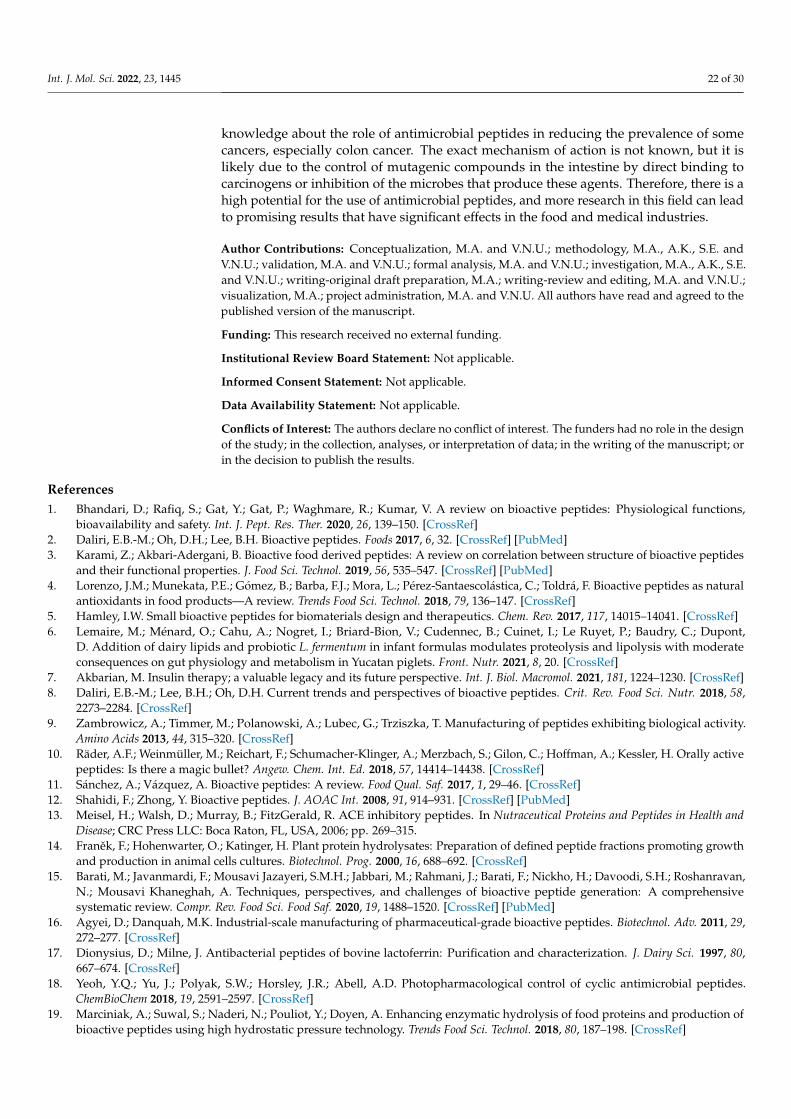
Int. J. Mol. Sci. 2022, 23, 1445 22 of 30
knowledge about the role of antimicrobial peptides in reducing the prevalence of somecancers, especially colon cancer. The exact mechanism of action is not known, but it islikely due to the control of mutagenic compounds in the intestine by direct binding tocarcinogens or inhibition of the microbes that produce these agents. Therefore, there is ahigh potential for the use of antimicrobial peptides, and more research in this field can leadto promising results that have significant effects in the food and medical industries.
Author Contributions: Conceptualization, M.A. and V.N.U.; methodology, M.A., A.K., S.E. andV.N.U.; validation, M.A. and V.N.U.; formal analysis, M.A. and V.N.U.; investigation, M.A., A.K., S.E.and V.N.U.; writing-original draft preparation, M.A.; writing-review and editing, M.A. and V.N.U.;visualization, M.A.; project administration, M.A. and V.N.U. All authors have read and agreed to thepublished version of the manuscript.
Funding: This research received no external funding.
Institutional Review Board Statement: Not applicable.
Informed Consent Statement: Not applicable.
Data Availability Statement: Not applicable.
Conflicts of Interest: The authors declare no conflict of interest. The funders had no role in the designof the study; in the collection, analyses, or interpretation of data; in the writing of the manuscript; orin the decision to publish the results.
References1. Bhandari, D.; Rafiq, S.; Gat, Y.; Gat, P.; Waghmare, R.; Kumar, V. A review on bioactive peptides: Physiological functions,
bioavailability and safety. Int. J. Pept. Res. Ther. 2020, 26, 139–150. [CrossRef]2. Daliri, E.B.-M.; Oh, D.H.; Lee, B.H. Bioactive peptides. Foods 2017, 6, 32. [CrossRef] [PubMed]3. Karami, Z.; Akbari-Adergani, B. Bioactive food derived peptides: A review on correlation between structure of bioactive peptides
and their functional properties. J. Food Sci. Technol. 2019, 56, 535–547. [CrossRef] [PubMed]4. Lorenzo, J.M.; Munekata, P.E.; Gómez, B.; Barba, F.J.; Mora, L.; Pérez-Santaescolástica, C.; Toldrá, F. Bioactive peptides as natural
antioxidants in food products—A review. Trends Food Sci. Technol. 2018, 79, 136–147. [CrossRef]5. Hamley, I.W. Small bioactive peptides for biomaterials design and therapeutics. Chem. Rev. 2017, 117, 14015–14041. [CrossRef]6. Lemaire, M.; Ménard, O.; Cahu, A.; Nogret, I.; Briard-Bion, V.; Cudennec, B.; Cuinet, I.; Le Ruyet, P.; Baudry, C.; Dupont,
D. Addition of dairy lipids and probiotic L. fermentum in infant formulas modulates proteolysis and lipolysis with moderateconsequences on gut physiology and metabolism in Yucatan piglets. Front. Nutr. 2021, 8, 20. [CrossRef]
7. Akbarian, M. Insulin therapy; a valuable legacy and its future perspective. Int. J. Biol. Macromol. 2021, 181, 1224–1230. [CrossRef]8. Daliri, E.B.-M.; Lee, B.H.; Oh, D.H. Current trends and perspectives of bioactive peptides. Crit. Rev. Food Sci. Nutr. 2018, 58,
2273–2284. [CrossRef]9. Zambrowicz, A.; Timmer, M.; Polanowski, A.; Lubec, G.; Trziszka, T. Manufacturing of peptides exhibiting biological activity.
Amino Acids 2013, 44, 315–320. [CrossRef]10. Räder, A.F.; Weinmüller, M.; Reichart, F.; Schumacher-Klinger, A.; Merzbach, S.; Gilon, C.; Hoffman, A.; Kessler, H. Orally active
peptides: Is there a magic bullet? Angew. Chem. Int. Ed. 2018, 57, 14414–14438. [CrossRef]11. Sánchez, A.; Vázquez, A. Bioactive peptides: A review. Food Qual. Saf. 2017, 1, 29–46. [CrossRef]12. Shahidi, F.; Zhong, Y. Bioactive peptides. J. AOAC Int. 2008, 91, 914–931. [CrossRef] [PubMed]13. Meisel, H.; Walsh, D.; Murray, B.; FitzGerald, R. ACE inhibitory peptides. In Nutraceutical Proteins and Peptides in Health and
Disease; CRC Press LLC: Boca Raton, FL, USA, 2006; pp. 269–315.14. Franek, F.; Hohenwarter, O.; Katinger, H. Plant protein hydrolysates: Preparation of defined peptide fractions promoting growth
and production in animal cells cultures. Biotechnol. Prog. 2000, 16, 688–692. [CrossRef]15. Barati, M.; Javanmardi, F.; Mousavi Jazayeri, S.M.H.; Jabbari, M.; Rahmani, J.; Barati, F.; Nickho, H.; Davoodi, S.H.; Roshanravan,
N.; Mousavi Khaneghah, A. Techniques, perspectives, and challenges of bioactive peptide generation: A comprehensivesystematic review. Compr. Rev. Food Sci. Food Saf. 2020, 19, 1488–1520. [CrossRef] [PubMed]
16. Agyei, D.; Danquah, M.K. Industrial-scale manufacturing of pharmaceutical-grade bioactive peptides. Biotechnol. Adv. 2011, 29,272–277. [CrossRef]
17. Dionysius, D.; Milne, J. Antibacterial peptides of bovine lactoferrin: Purification and characterization. J. Dairy Sci. 1997, 80,667–674. [CrossRef]
18. Yeoh, Y.Q.; Yu, J.; Polyak, S.W.; Horsley, J.R.; Abell, A.D. Photopharmacological control of cyclic antimicrobial peptides.ChemBioChem 2018, 19, 2591–2597. [CrossRef]
19. Marciniak, A.; Suwal, S.; Naderi, N.; Pouliot, Y.; Doyen, A. Enhancing enzymatic hydrolysis of food proteins and production ofbioactive peptides using high hydrostatic pressure technology. Trends Food Sci. Technol. 2018, 80, 187–198. [CrossRef]
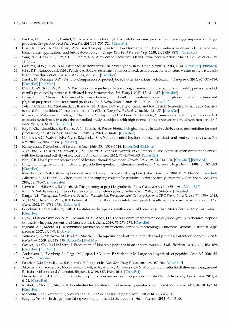
Int. J. Mol. Sci. 2022, 23, 1445 23 of 30
20. Naderi, N.; House, J.D.; Pouliot, Y.; Doyen, A. Effects of high hydrostatic pressure processing on hen egg compounds and eggproducts. Compr. Rev. Food Sci. Food Saf. 2017, 16, 707–720. [CrossRef]
21. Chai, K.F.; Voo, A.Y.H.; Chen, W.N. Bioactive peptides from food fermentation: A comprehensive review of their sources,bioactivities, applications, and future development. Compr. Rev. Food Sci. Food Saf. 2020, 19, 3825–3885. [CrossRef]
22. Song, A.A.-L.; In, L.L.; Lim, S.H.E.; Rahim, R.A. A review on Lactococcus lactis: From food to factory. Microb. Cell Factories 2017,16, 1–15.
23. Griffiths, M.W.; Tellez, A.M. Lactobacillus helveticus: The proteolytic system. Front. Microbiol. 2013, 4, 30. [CrossRef] [PubMed]24. John, R.P.; Nampoothiri, K.M.; Pandey, A. Solid-state fermentation for L-lactic acid production from agro wastes using Lactobacil-
lus delbrueckii. Process Biochem. 2006, 41, 759–763. [CrossRef]25. Sasaki, M.; Bosman, B.W.; Tan, P.S. Comparison of proteolytic activities in various lactobacilli. J. Dairy Res. 1995, 62, 601–610.
[CrossRef] [PubMed]26. Chen, G.-W.; Tsai, J.-S.; Pan, B.S. Purification of angiotensin I-converting enzyme inhibitory peptides and antihypertensive effect
of milk produced by protease-facilitated lactic fermentation. Int. Dairy J. 2007, 17, 641–647. [CrossRef]27. Lorenzen, P.C.; Meisel, H. Influence of trypsin action in yoghurt milk on the release of caseinophosphopeptide-rich fractions and
physical properties of the fermented products. Int. J. Dairy Technol. 2005, 58, 119–124. [CrossRef]28. Soleymanzadeh, N.; Mirdamadi, S.; Kianirad, M. Antioxidant activity of camel and bovine milk fermented by lactic acid bacteria
isolated from traditional fermented camel milk (Chal). Dairy Sci. Technol. 2016, 96, 443–457. [CrossRef]29. Mizuno, S.; Matsuura, K.; Gotou, T.; Nishimura, S.; Kajimoto, O.; Yabune, M.; Kajimoto, Y.; Yamamoto, N. Antihypertensive effect
of casein hydrolysate in a placebo-controlled study in subjects with high-normal blood pressure and mild hypertension. Br. J.Nutr. 2005, 94, 84–91. [CrossRef]
30. Raj, T.; Chandrasekhar, K.; Kumar, A.N.; Kim, S.-H. Recent biotechnological trends in lactic acid bacterial fermentation for foodprocessing industries. Syst. Microbiol. Biomanuf. 2021, 2, 14–40. [CrossRef]
31. Conibear, A.C.; Watson, E.E.; Payne, R.J.; Becker, C.F. Native chemical ligation in protein synthesis and semi-synthesis. Chem. Soc.Rev. 2018, 47, 9046–9068. [CrossRef]
32. Katsoyannis, P. Synthesis of insulin. Science 1966, 154, 1509–1514. [CrossRef] [PubMed]33. Vigneaud, V.D.; Ressler, C.; Swan, C.J.M.; Roberts, C.W.; Katsoyannis, P.G.; Gordon, S. The synthesis of an octapeptide amide
with the hormonal activity of oxytocin. J. Am. Chem. Soc. 1953, 75, 4879–4880. [CrossRef]34. Kent, S.B. Novel protein science enabled by total chemical synthesis. Protein Sci. 2019, 28, 313–328. [CrossRef] [PubMed]35. Bray, B.L. Large-scale manufacture of peptide therapeutics by chemical synthesis. Nat. Rev. Drug Discov. 2003, 2, 587–593.
[CrossRef]36. Merrifield, R.B. Solid phase peptide synthesis. I. The synthesis of a tetrapeptide. J. Am. Chem. Soc. 1963, 85, 2149–2154. [CrossRef]37. Albericio, F.; El-Faham, A. Choosing the right coupling reagent for peptides: A twenty-five-year journey. Org. Process Res. Dev.
2018, 22, 760–772. [CrossRef]38. Lawrenson, S.B.; Arav, R.; North, M. The greening of peptide synthesis. Green Chem. 2017, 19, 1685–1691. [CrossRef]39. Kaur, N. Solid-phase synthesis of sulfur containing heterocycles. J. Sulfur Chem. 2018, 39, 544–577. [CrossRef]40. Banga, A.K. Therapeutic Peptides and Proteins: Formulation, Processing, and Delivery Systems; CRC Press: Boca Raton, FL, USA, 2015.41. Yu, H.M.; Chen, S.T.; Wang, K.T. Enhanced coupling efficiency in solid-phase peptide synthesis by microwave irradiation. J. Org.
Chem. 1992, 57, 4781–4784. [CrossRef]42. Goodwin, D.; Simerska, P.; Toth, I. Peptides as therapeutics with enhanced bioactivity. Curr. Med. Chem. 2012, 19, 4451–4461.
[CrossRef]43. Li, W.; O’Brien-Simpson, N.M.; Hossain, M.A.; Wade, J.D. The 9-fluorenylmethoxycarbonyl (Fmoc) group in chemical peptide
synthesis—Its past, present, and future. Aust. J. Chem. 2019, 73, 271–276. [CrossRef]44. Ingham, A.B.; Moore, R.J. Recombinant production of antimicrobial peptides in heterologous microbial systems. Biotechnol. Appl.
Biochem. 2007, 47, 1–9. [PubMed]45. Antosova, Z.; Mackova, M.; Kral, V.; Macek, T. Therapeutic application of peptides and proteins: Parenteral forever? Trends
Biotechnol. 2009, 27, 628–635. [CrossRef] [PubMed]46. Ozawa, A.; Cai, Y.; Lindberg, I. Production of bioactive peptides in an in vitro system. Anal. Biochem. 2007, 366, 182–189.
[CrossRef] [PubMed]47. Andersson, L.; Blomberg, L.; Flegel, M.; Lepsa, L.; Nilsson, B.; Verlander, M. Large-scale synthesis of peptides. Pept. Sci. 2000, 55,
227–250. [CrossRef]48. Drucker, D.J.; Dritselis, A.; Kirkpatrick, P. Liraglutide. Nat. Rev. Drug Discov. 2010, 9, 267–268. [CrossRef]49. Akbarian, M.; Yousefi, R.; Moosavi-Movahedi, A.A.; Ahmad, A.; Uversky, V.N. Modulating insulin fibrillation using engineered
B-chains with mutated C-termini. Biophys. J. 2019, 117, 1626–1641. [CrossRef]50. Harnedy, P.A.; FitzGerald, R.J. Bioactive peptides from marine processing waste and shellfish: A Review. J. Funct. Foods 2012, 4,
6–24. [CrossRef]51. Rustad, T.; Storrø, I.; Slizyte, R. Possibilities for the utilisation of marine by-products. Int. J. Food Sci. Technol. 2011, 46, 2001–2014.
[CrossRef]52. Mohebbi, G.H.; Nabipour, I.; Vazirizadeh, A. The Sea, the future pharmacy. ISMJ 2014, 17, 748–788.53. King, G. Venoms to drugs: Translating venom peptides into therapeutics. Aust. Biochem. 2013, 44, 13–15.
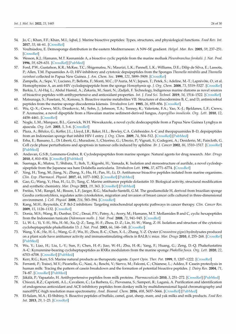
Int. J. Mol. Sci. 2022, 23, 1445 24 of 30
54. Jo, C.; Khan, F.F.; Khan, M.I.; Iqbal, J. Marine bioactive peptides: Types, structures, and physiological functions. Food Rev. Int.2017, 33, 44–61. [CrossRef]
55. Voultsiadou, E. Demosponge distribution in the eastern Mediterranean: A NW–SE gradient. Helgol. Mar. Res. 2005, 59, 237–251.[CrossRef]
56. Wesson, K.J.; Hamann, M.T. Keenamide A, a bioactive cyclic peptide from the marine mollusk Pleurobranchus forskalii. J. Nat. Prod.1996, 59, 629–631. [CrossRef] [PubMed]
57. Ford, P.W.; Gustafson, K.R.; McKee, T.C.; Shigematsu, N.; Maurizi, L.K.; Pannell, L.K.; Williams, D.E.; Dilip de Silva, E.; Lassota,P.; Allen, T.M. Papuamides A–D, HIV-inhibitory and cytotoxic depsipeptides from the Sponges Theonella mirabilis and Theonellaswinhoei collected in Papua New Guinea. J. Am. Chem. Soc. 1999, 121, 5899–5909. [CrossRef]
58. Zampella, A.; Sepe, V.; Luciano, P.; Bellotta, F.; Monti, M.C.; D’Auria, M.V.; Jepsen, T.; Petek, S.; Adeline, M.-T.; Laprévôte, O.; et al.Homophymine A, an anti-HIV cyclodepsipeptide from the sponge Homophymia sp. J. Org. Chem. 2008, 73, 5319–5327. [CrossRef]
59. Barkia, I.; Al-Haj, L.; Abdul Hamid, A.; Zakaria, M.; Saari, N.; Zadjali, F. Technology, Indigenous marine diatoms as novel sourcesof bioactive peptides with antihypertensive and antioxidant properties. Int. J. Food Sci. Technol. 2019, 54, 1514–1522. [CrossRef]
60. Matsunaga, S.; Fusetani, N.; Konosu, S. Bioactive marine metabolites VII. Structures of discodermins B, C, and D, antimicrobialpeptides from the marine sponge discodermia kiiensis. Tetrahedron Lett. 1985, 26, 855–856. [CrossRef]
61. Wu, Q.-X.; Crews, M.S.; Draskovic, M.; Sohn, J.; Johnson, T.A.; Tenney, K.; Valeriote, F.A.; Yao, X.-J.; Bjeldanes, L.F.; Crews,P. Azonazine, a novel dipeptide from a Hawaiian marine sediment-derived fungus, Aspergillus insulicola. Org. Lett. 2010, 12,4458–4461. [CrossRef]
62. Nogle, L.M.; Marquez, B.L.; Gerwick, W.H. Wewakazole, a novel cyclic dodecapeptide from a Papua New Guinea Lyngbya majuscula. Org. Lett. 2003, 5, 3–6. [CrossRef]
63. Plaza, A.; Bifulco, G.; Keffer, J.L.; Lloyd, J.R.; Baker, H.L.; Bewley, C.A. Celebesides A–C and theopapuamides B–D, depsipeptidesfrom an Indonesian sponge that inhibit HIV-1 entry. J. Org. Chem. 2009, 74, 504–512. [CrossRef] [PubMed]
64. Erba, E.; Bassano, L.; Di Liberti, G.; Muradore, I.; Chiorino, G.; Ubezio, P.; Vignati, S.; Codegoni, A.; Desiderio, M.; Faircloth, G.Cell cycle phase perturbations and apoptosis in tumour cells induced by aplidine. Br. J. Cancer 2002, 86, 1510–1517. [CrossRef][PubMed]
65. Andavan, G.S.B.; Lemmens-Gruber, R. Cyclodepsipeptides from marine sponges: Natural agents for drug research. Mar. Drugs2010, 8, 810–834. [CrossRef] [PubMed]
66. Suenaga, K.; Mutou, T.; Shibata, T.; Itoh, T.; Kigoshi, H.; Yamada, K. Isolation and stereostructure of aurilide, a novel cyclodep-sipeptide from the Japanese sea hare Dolabella auricularia. Tetrahedron Lett. 1996, 37, 6771–6774. [CrossRef]
67. Xing, H.; Tong, M.; Jiang, N.; Zhang, X.; Hu, H.; Pan, H.; Li, D. Antitumour bioactive peptides isolated from marine organisms.Clin. Exp. Pharmacol. Physiol. 2017, 44, 1077–1082. [CrossRef] [PubMed]
68. Gao, G.; Wang, Y.; Hua, H.; Li, D.; Tang, C. Marine antitumor peptide dolastatin 10: Biological activity, structural modificationand synthetic chemistry. Mar. Drugs 2021, 19, 363. [CrossRef] [PubMed]
69. Freitas, V.M.; Rangel, M.; Bisson, L.F.; Jaeger, R.G.; Machado-Santelli, G.M. The geodiamolide H, derived from brazilian spongeGeodia corticostylifera, regulates actin cytoskeleton, migration and invasion of breast cancer cells cultured in three-dimensionalenvironment. J. Cell. Physiol. 2008, 216, 583–594. [CrossRef]
70. Kang, M.H.; Reynolds, C.P. Bcl-2 inhibitors: Targeting mitochondrial apoptotic pathways in cancer therapy. Clin. Cancer Res.2009, 15, 1126–1132. [CrossRef]
71. Donia, M.S.; Wang, B.; Dunbar, D.C.; Desai, P.V.; Patny, A.; Avery, M.; Hamann, M.T. Mollamides B and C, cyclic hexapeptidesfrom the Indonesian tunicate Didemnum molle. J. Nat. Prod. 2008, 71, 941–945. [CrossRef]
72. Li, W.-L.; Yi, Y.-H.; Wu, H.-M.; Xu, Q.-Z.; Tang, H.-F.; Zhou, D.-Z.; Lin, H.-W.; Wang, Z.-H. Isolation and structure of the cytotoxiccycloheptapeptide phakellistatin 13. J. Nat. Prod. 2003, 66, 146–148. [CrossRef]
73. Wang, Y.-K.; He, H.-L.; Wang, G.-F.; Wu, H.; Zhou, B.-C.; Chen, X.-L.; Zhang, Y.-Z. Oyster (Crassostrea gigas) hydrolysates producedon a plant scale have antitumor activity and immunostimulating effects in BALB/c mice. Mar. Drugs 2010, 8, 255–268. [CrossRef][PubMed]
74. Wu, Y.; Liao, H.; Liu, L.-Y.; Sun, F.; Chen, H.-F.; Jiao, W.-H.; Zhu, H.-R.; Yang, F.; Huang, G.; Zeng, D.-Q. PhakefustatinsA–C: Kynurenine-bearing cycloheptapeptides as RXRα modulators from the marine sponge Phakellia fusca. Org. Lett. 2020, 22,6703–6708. [CrossRef] [PubMed]
75. Kerr, R.G.; Kerr, S.S. Marine natural products as therapeutic agents. Expert Opin. Ther. Pat. 1999, 9, 1207–1222. [CrossRef]76. Ferranti, P.; Traisci, M.V.; Picariello, G.; Nasi, A.; Boschi, V.; Siervo, M.; Falconi, C.; Chianese, L.; Addeo, F. Casein proteolysis in
human milk: Tracing the pattern of casein breakdown and the formation of potential bioactive peptides. J. Dairy Res. 2004, 71,74–87. [CrossRef] [PubMed]
77. Jäkälä, P.; Vapaatalo, H. Antihypertensive peptides from milk proteins. Pharmaceuticals 2010, 3, 251–272. [CrossRef] [PubMed]78. Chiozzi, R.Z.; Capriotti, A.L.; Cavaliere, C.; La Barbera, G.; Piovesana, S.; Samperi, R.; Laganà, A. Purification and identification
of endogenous antioxidant and ACE-inhibitory peptides from donkey milk by multidimensional liquid chromatography andnanoHPLC-high resolution mass spectrometry. Anal. Bioanal. Chem. 2016, 408, 5657–5666. [CrossRef] [PubMed]
79. El-Salam, M.A.; El-Shibiny, S. Bioactive peptides of buffalo, camel, goat, sheep, mare, and yak milks and milk products. Food Rev.Int. 2013, 29, 1–23. [CrossRef]

Int. J. Mol. Sci. 2022, 23, 1445 25 of 30
80. Hernández-Ledesma, B.; García-Nebot, M.J.; Fernández-Tomé, S.; Amigo, L.; Recio, I. Dairy protein hydrolysates: Peptides forhealth benefits. Int. Dairy J. 2014, 38, 82–100. [CrossRef]
81. Mudgil, P.; Kamal, H.; Yuen, G.C.; Maqsood, S. Characterization and identification of novel antidiabetic and anti-obesity peptidesfrom camel milk protein hydrolysates. Food Chem. 2018, 259, 46–54. [CrossRef]
82. Nongonierma, A.B.; Paolella, S.; Mudgil, P.; Maqsood, S.; FitzGerald, R.J. Dipeptidyl peptidase IV (DPP-IV) inhibitory propertiesof camel milk protein hydrolysates generated with trypsin. J. Funct. Foods 2017, 34, 49–58. [CrossRef]
83. Nongonierma, A.B.; Paolella, S.; Mudgil, P.; Maqsood, S.; FitzGerald, R.J. Identification of novel dipeptidyl peptidase IV (DPP-IV)inhibitory peptides in camel milk protein hydrolysates. Food Chem. 2018, 244, 340–348. [CrossRef] [PubMed]
84. Mudgil, P.; Baby, B.; Ngoh, Y.-Y.; Vijayan, R.; Gan, C.-Y.; Maqsood, S. Identification and molecular docking study of novelcholesterol esterase inhibitory peptides from camel milk proteins. J. Dairy Sci. 2019, 102, 10748–10759. [CrossRef] [PubMed]
85. Wali, A.; Yanhua, G.; Ishimov, U.; Yili, A.; Aisa, H.A.; Salikhov, S. Therapeutics, Isolation and identification of three novelantioxidant peptides from the Bactrian camel milk Hydrolysates. Int. J. Pept. Res. Ther. 2020, 26, 641–650. [CrossRef]
86. Chen, L.; Zhang, Q.; Ji, Z.; Shu, G.; Chen, H.J.L. Production and fermentation characteristics of angiotensin-I-converting enzymeinhibitory peptides of goat milk fermented by a novel wild Lactobacillus plantarum 69. Lebensm. Wiss. Technol. 2018, 91, 532–540.[CrossRef]
87. Zanutto-Elgui, M.R.; Vieira, J.C.S.; do Prado, D.Z.; Buzalaf, M.A.R.; de Magalhães Padilha, P.; de Oliveira, D.E.; Fleuri, L.F.Production of milk peptides with antimicrobial and antioxidant properties through fungal proteases. Food Chem. 2019, 278,823–831. [CrossRef]
88. Davalos, A.; Miguel, M.; Bartolome, B.; Lopez-Fandino, R. Antioxidant activity of peptides derived from egg white proteins byenzymatic hydrolysis. J. Food Prot. 2004, 67, 1939–1944. [CrossRef]
89. Walzem, R.; Dillard, C.; German, J.B. Whey components: Millennia of evolution create functionalities for mammalian nutrition:What we know and what we may be overlooking. Crit. Rev. Food Sci. Nutr. 2002, 42, 353–375. [CrossRef]
90. Yu, Z.; Yin, Y.; Zhao, W.; Wang, F.; Yu, Y.; Liu, B.; Liu, J.; Chen, F. Characterization of ACE-inhibitory peptide associated withantioxidant and anticoagulation properties. J. Food Sci. 2011, 76, C1149–C1155. [CrossRef]
91. Sun, X.; Chakrabarti, S.; Fang, J.; Yin, Y.; Wu, J. Low-molecular-weight fractions of Alcalase hydrolyzed egg ovomucin extractexert anti-inflammatory activity in human dermal fibroblasts through the inhibition of tumor necrosis factor–mediated nuclearfactor κB pathway. Nutr. Res. 2016, 36, 648–657. [CrossRef]
92. Liu, Y.-F.; Oey, I.; Bremer, P.; Carne, A.; Silcock, P. Bioactive peptides derived from egg proteins: A review. Crit. Rev. Food Sci. Nutr.2018, 58, 2508–2530. [CrossRef]
93. You, S.-J.; Udenigwe, C.C.; Aluko, R.E.; Wu, J. Multifunctional peptides from egg white lysozyme. Food Res. Int. 2010, 43, 848–855.[CrossRef]
94. Mine, Y.; Kovacs-Nolan, J. New insights in biologically active proteins and peptides derived from hen egg. World’s Poult. Sci. J.2006, 62, 87–96. [CrossRef]
95. Abdou, A.M.; Higashiguchi, S.; Aboueleinin, A.; Kim, M.; Ibrahim, H.R. Antimicrobial peptides derived from hen egg lysozymewith inhibitory effect against Bacillus species. Food Control 2007, 18, 173–178. [CrossRef]
96. Lafarga, T.; Hayes, M. Bioactive peptides from meat muscle and by-products: Generation, functionality and application asfunctional ingredients. Meat Sci. 2014, 98, 227–239. [CrossRef]
97. Möller, N.P.; Scholz-Ahrens, K.E.; Roos, N.; Schrezenmeir, J. Bioactive peptides and proteins from foods: Indication for healtheffects. Eur. J. Nutr. 2008, 47, 171–182. [CrossRef]
98. Arihara, K.; Nakashima, Y.; Mukai, T.; Ishikawa, S.; Itoh, M. Peptide inhibitors for angiotensin I-converting enzyme fromenzymatic hydrolysates of porcine skeletal muscle proteins. Meat Sci. 2001, 57, 319–324. [CrossRef]
99. Jang, A.; Lee, M. Purification and identification of angiotensin converting enzyme inhibitory peptides from beef hydrolysates.Meat Sci. 2005, 69, 653–661. [CrossRef]
100. Stadnik, J.; Keska, P. Meat and fermented meat products as a source of bioactive peptides. Acta Sci. Pol. Technol. Aliment. 2015, 14,181–190. [CrossRef]
101. Saiga, A.; Tanabe, S.; Nishimura, T. Antioxidant activity of peptides obtained from porcine myofibrillar proteins by proteasetreatment. J. Agric. Food Chem. 2003, 51, 3661–3667. [CrossRef]
102. Di Bernardini, R.; Mullen, A.M.; Bolton, D.; Kerry, J.; O’Neill, E.; Hayes, M. Assessment of the angiotensin-I-converting enzyme(ACE-I) inhibitory and antioxidant activities of hydrolysates of bovine brisket sarcoplasmic proteins produced by papain andcharacterisation of associated bioactive peptidic fractions. Meat Sci. 2012, 90, 226–235. [CrossRef]
103. Culler, R.; Smith, G.; Cross, H. Relationship of myofibril fragmentation index to certain chemical, physical and sensory character-istics of bovine longissimus muscle. J. Food Sci. 1978, 43, 1177–1180. [CrossRef]
104. Haard, N.F.; Simpson, B.K.; Pan, B.S. Sarcoplasmic proteins and other nitrogenous compounds. In Seafood Proteins; Springer:Berlin/Heidelberg, Germany, 1994; pp. 13–39.
105. Robinson, S.D.; Safavi-Hemami, H. Venom peptides as pharmacological tools and therapeutics for diabetes. Neuropharmacology2017, 127, 79–86. [CrossRef] [PubMed]
106. Robinson, S.D.; Li, Q.; Bandyopadhyay, P.K.; Gajewiak, J.; Yandell, M.; Papenfuss, A.T.; Purcell, A.W.; Norton, R.S.; Safavi-Hemami, H. Hormone-like peptides in the venoms of marine cone snails. Gen. Comp. Endocrinol. 2017, 244, 11–18. [CrossRef][PubMed]
Int. J. Mol. Sci. 2022, 23, 1445 26 of 30
107. Robinson, S.D.; Undheim, E.A.; Ueberheide, B.; King, G.F. Venom peptides as therapeutics: Advances, challenges and the futureof venom-peptide discovery. Expert Rev. Proteom. 2017, 14, 931–939. [CrossRef] [PubMed]
108. Ianzer, D.; Konno, K.; Marques-Porto, R.; Portaro, F.C.V.; Stöcklin, R.; de Camargo, A.C.M.; Pimenta, D.C. Identification of fivenew bradykinin potentiating peptides (BPPs) from Bothrops jararaca crude venom by using electrospray ionization tandem massspectrometry after a two-step liquid chromatography. Peptides 2004, 25, 1085–1092. [CrossRef]
109. Lima, M.E.D.; Martin-Eauclaire, M.-F. The toxins purified from Tityus serrulatus (Lutz & Mello) venom. J. Toxicol. Toxin Rev. 1995,14, 457–481.
110. Dai, L.; Corzo, G.; Naoki, H.; Andriantsiferana, M.; Nakajima, T. Purification, structure–function analysis, and molecularcharacterization of novel linear peptides from scorpion Opisthacanthus madagascariensis. Biochem. Biophys. Res. Commun. 2002, 293,1514–1522. [CrossRef]
111. Xiong, X.; Menting, J.G.; Disotuar, M.M.; Smith, N.A.; Delaine, C.A.; Ghabash, G.; Agrawal, R.; Wang, X.; He, X.; Fisher, S.J. Astructurally minimized yet fully active insulin based on cone-snail venom insulin principles. Nat. Struct. Mol. Biol. 2020, 27,615–624. [CrossRef]
112. Miljanich, G. Ziconotide: Neuronal calcium channel blocker for treating severe chronic pain. Curr. Med. Chem. 2004, 11, 3029–3040.[CrossRef]
113. Seo, M.-D.; Won, H.-S.; Kim, J.-H.; Mishig-Ochir, T.; Lee, B.-J. Antimicrobial peptides for therapeutic applications: A review.Molecules 2012, 17, 12276–12286. [CrossRef]
114. Pometto, A.; Shetty, K.; Paliyath, G.; Levin, R.E. Food Biotechnology; CRC Press: Boca Raton, FL, USA, 2005.115. Cheikhyoussef, A.; Pogori, N.; Chen, W.; Zhang, H. Antimicrobial proteinaceous compounds obtained from bifidobacteria: From
production to their application. Int. J. Food Microbiol. 2008, 125, 215–222. [CrossRef] [PubMed]116. Wu, Q.; Patocka, J.; Kuca, K. Insect antimicrobial peptides, a mini review. Toxins 2018, 10, 461. [CrossRef] [PubMed]117. Kamala, K.; Sivaperumal, P.; Kamath, S.M.; Thilagaraj, W.R.; Rajaram, R. Marine actinobacteria as a source for emerging
biopharmaceuticals. Encycl. Mar. Biotechnol. 2020, 4, 2095–2105.118. Mohanrasu, K.; Rao, R.G.R.; Sudhakar, M.; Raja, R.; Jeyakanthan, J.; Arun, A. Marine microbial pharmacognosy: Prospects and
perspectives. In Marine Niche: Applications in Pharmaceutical Sciences; Springer: Berlin/Heidelberg, Germany, 2020; pp. 89–110.119. D’Orazio, N.; Gammone, M.A.; Gemello, E.; De Girolamo, M.; Cusenza, S.; Riccioni, G. Marine bioactives: Pharmacological
properties and potential applications against inflammatory diseases. Mar. Drugs 2012, 10, 812–833. [CrossRef]120. Rollins-Smith, L.A.; Reinert, L.K.; O’Leary, C.J.; Houston, L.E.; Woodhams, D.C. Antimicrobial peptide defenses in amphibian
skin. Integr. Comp. Biol. 2005, 45, 137–142. [CrossRef] [PubMed]121. Abou Aleinein, R.; Hamoud, R.; Schäfer, H.; Wink, M. Molecular cloning and expression of ranalexin, a bioactive antimicrobial
peptide from Rana catesbeiana in Escherichia coli and assessments of its biological activities. Appl. Microbiol. Biotechnol. 2013, 97,3535–3543. [CrossRef] [PubMed]
122. Salas, C.E.; Badillo-Corona, J.A.; Ramírez-Sotelo, G.; Oliver-Salvador, C. Biologically active and antimicrobial peptides fromplants. BioMed Res. Int. 2015, 2015, 102129. [CrossRef]
123. Jenssen, H.; Hamill, P.; Hancock, R.E. Peptide antimicrobial agents. Clin. Microbiol. Rev. 2006, 19, 491–511. [CrossRef]124. Boman, H.G. Innate immunity and the normal microflora. Immunol. Rev. 2000, 173, 5–16. [CrossRef]125. Powers, J.-P.S.; Rozek, A.; Hancock, R.E. Structure–activity relationships for the β-hairpin cationic antimicrobial peptide
polyphemusin I. Biochim. Biophys. Acta (BBA) Proteins Proteom. 2004, 1698, 239–250. [CrossRef]126. Stotz, H.U.; Thomson, J.; Wang, Y. Plant defensins: Defense, development and application. Plant Signal. Behav. 2009, 4, 1010–1012.
[CrossRef]127. Nawrot, R.; Barylski, J.; Nowicki, G.; Broniarczyk, J.; Buchwald, W.; Gozdzicka-Józefiak, A. Plant antimicrobial peptides. Folia
Microbiol. 2014, 59, 181–196. [CrossRef] [PubMed]128. Stec, B. Plant thionins—The structural perspective. Cell. Mol. Life Sci. CMLS 2006, 63, 1370–1385. [CrossRef] [PubMed]129. Liu, Y.; Luo, J.; Xu, C.; Ren, F.; Peng, C.; Wu, G.; Zhao, J. Purification, characterization, and molecular cloning of the gene of a
seed-specific antimicrobial protein from pokeweed. Plant Physiol. 2000, 122, 1015–1024. [CrossRef]130. Terras, F.R.; Eggermont, K.; Kovaleva, V.; Raikhel, N.V.; Osborn, R.W.; Kester, A.; Rees, S.B.; Torrekens, S.; Van Leuven, F.;
Vanderleyden, J. Small cysteine-rich antifungal proteins from radish: Their role in host defense. Plant Cell 1995, 7, 573–588.131. Ye, X.; Ng, T.; Rao, P. Cicerin and arietin, novel chickpea peptides with different antifungal potencies. Peptides 2002, 23, 817–822.
[CrossRef]132. Wang, Z.; Wang, G. APD: The antimicrobial peptide database. Nucleic Acids Res. 2004, 32 (Suppl. S1), D590–D592. [CrossRef]133. del Mar Yust, M.; Pedroche, J.; Megías, C.; Girón-Calle, J.; Alaiz, M.; Millán, F.; Vioque, J. Rapeseed protein hydrolysates: A source
of HIV protease peptide inhibitors. Food Chem. 2004, 87, 387–392. [CrossRef]134. Marcus, J.P.; Green, J.L.; Goulter, K.C.; Manners, J.M. A family of antimicrobial peptides is produced by processing of a 7S
globulin protein in Macadamia integrifolia kernels. Plant J. 1999, 19, 699–710. [CrossRef]135. Craik, D.J. Discovery and applications of the plant cyclotides. Toxicon 2010, 56, 1092–1102. [CrossRef]136. Shao, F.; Hu, Z.; Xiong, Y.-M.; Huang, Q.-Z.; Wang, C.-G.; Zhu, R.-H.; Wang, D.-C. A new antifungal peptide from the seeds of
Phytolacca americana: Characterization, amino acid sequence and cDNA cloning. Biochim. Biophys. Acta (BBA) Protein Struct. Mol.Enzymol. 1999, 1430, 262–268. [CrossRef]
Int. J. Mol. Sci. 2022, 23, 1445 27 of 30
137. De Caleya, R.F.; Gonzalez-Pascual, B.; García-Olmedo, F.; Carbonero, P. Susceptibility of phytopathogenic bacteria to wheatpurothionins in vitro. Appl. Microbiol. 1972, 23, 998–1000. [CrossRef] [PubMed]
138. Hördegen, P.; Cabaret, J.; Hertzberg, H.; Langhans, W.; Maurer, V. In vitro screening of six anthelmintic plant products againstlarval Haemonchus contortus with a modified methyl-thiazolyl-tetrazolium reduction assay. J. Ethnopharmacol. 2006, 108, 85–89.[CrossRef]
139. Sharma, S.; Verma, H.N.; Sharma, N.K. Cationic bioactive peptide from the seeds of Benincasa hispida. Int. J. Pept. 2014, 2014,156060. [CrossRef]
140. Lundberg, U. Stress, subjective and objective health. Int. J. Soc. Welf. 2006, 15, S41–S48. [CrossRef]141. Pitkänen, A.; Kharatishvili, I.; Karhunen, H.; Lukasiuk, K.; Immonen, R.; Nairismägi, J.; Gröhn, O.; Nissinen, J. Epileptogenesis in
experimental models. Epilepsia 2007, 48, 13–20. [CrossRef]142. Almroth, B.C.; Albertsson, E.; Sturve, J.; Förlin, L. Oxidative stress, evident in antioxidant defences and damage products, in
rainbow trout caged outside a sewage treatment plant. Ecotoxicol. Environ. Saf. 2008, 70, 370–378. [CrossRef]143. Guo, C.; Yang, J.; Wei, J.; Li, Y.; Xu, J.; Jiang, Y. Antioxidant activities of peel, pulp and seed fractions of common fruits as
determined by FRAP assay. Nutr. Res. 2003, 23, 1719–1726. [CrossRef]144. Chen, H.-M.; Muramoto, K.; Yamauchi, F.; Fujimoto, K.; Nokihara, K. Antioxidative properties of histidine-containing peptides
designed from peptide fragments found in the digests of a soybean protein. J. Agric. Food Chem. 1998, 46, 49–53. [CrossRef]145. Seo, H.-S.; Kwak, S.-Y.; Lee, Y.-S. Antioxidative activities of histidine containing caffeic acid-dipeptides. Bioorganic Med. Chem.
Lett. 2010, 20, 4266–4272. [CrossRef]146. Aluko, R. Amino acids, peptides, and proteins as antioxidants for food preservation. In Handbook of Antioxidants for Food
Preservation; Elsevier: Amsterdam, The Netherlands, 2015; pp. 105–140.147. Mendis, E.; Rajapakse, N.; Kim, S.-K. Antioxidant properties of a radical-scavenging peptide purified from enzymatically prepared
fish skin gelatin hydrolysate. J. Agric. Food Chem. 2005, 53, 581–587. [CrossRef] [PubMed]148. Giri, A.; Ohshima, T. Bioactive marine peptides: Nutraceutical value and novel approaches. Adv. Food Nutr. Res. 2012, 65, 73–105.
[PubMed]149. Kim, S.-K.; Mendis, E. Bioactive compounds from marine processing byproducts—A review. Food Res. Int. 2006, 39, 383–393.
[CrossRef]150. Elias, R.J.; Kellerby, S.S.; Decker, E.A. Antioxidant activity of proteins and peptides. Crit. Rev. Food Sci. Nutr. 2008, 48, 430–441.
[CrossRef]151. Kim, S.-Y.; Je, J.-Y.; Kim, S.-K. Purification and characterization of antioxidant peptide from hoki (Johnius belengerii) frame protein
by gastrointestinal digestion. J. Nutr. Biochem. 2007, 18, 31–38. [CrossRef]152. Li, X.X.; Han, L.J.; Chen, L.J. In vitro antioxidant activity of protein hydrolysates prepared from corn gluten meal. J. Sci. Food
Agric. 2008, 88, 1660–1666. [CrossRef]153. Ranathunga, S.; Rajapakse, N.; Kim, S.-K. Purification and characterization of antioxidative peptide derived from muscle of
conger eel (Conger myriaster). Eur. Food Res. Technol. 2006, 222, 310–315. [CrossRef]154. Zou, T.-B.; He, T.-P.; Li, H.-B.; Tang, H.-W.; Xia, E.-Q. The structure-activity relationship of the antioxidant peptides from natural
proteins. Molecules 2016, 21, 72. [CrossRef]155. Sato, H.; Feix, J.B.; Frank, D.W. Identification of superoxide dismutase as a cofactor for the pseudomonas type III toxin, ExoU.
Biochemistry 2006, 45, 10368–10375. [CrossRef]156. Tincu, J.A.; Taylor, S.W. Antimicrobial peptides from marine invertebrates. Antimicrob. Agents Chemother. 2004, 48, 3645–3654.
[CrossRef]157. Rydlo, T.; Miltz, J.; Mor, A. Eukaryotic antimicrobial peptides: Promises and premises in food safety. J. Food Sci. 2006, 71,
R125–R135. [CrossRef]158. Cutrona, K.J.; Kaufman, B.A.; Figueroa, D.M.; Elmore, D.E. Role of arginine and lysine in the antimicrobial mechanism of
histone-derived antimicrobial peptides. FEBS Lett. 2015, 589, 3915–3920. [CrossRef] [PubMed]159. Jin, L.; Bai, X.; Luan, N.; Yao, H.; Zhang, Z.; Liu, W.; Chen, Y.; Yan, X.; Rong, M.; Lai, R. A designed tryptophan-and lysine/arginine-
rich antimicrobial peptide with therapeutic potential for clinical antibiotic-resistant Candida albicans vaginitis. J. Med. Chem. 2016,59, 1791–1799. [CrossRef] [PubMed]
160. Chou, H.-T.; Kuo, T.-Y.; Chiang, J.-C.; Pei, M.-J.; Yang, W.-T.; Yu, H.-C.; Lin, S.-B.; Chen, W.-J. Design and synthesis of cationicantimicrobial peptides with improved activity and selectivity against Vibrio spp. Int. J. Antimicrob. Agents 2008, 32, 130–138.[CrossRef]
161. Jin, Y.; Hammer, J.; Pate, M.; Zhang, Y.; Zhu, F.; Zmuda, E.; Blazyk, J. Antimicrobial activities and structures of two linear cationicpeptide families with various amphipathic β-sheet and α-helical potentials. Antimicrob. Agents Chemother. 2005, 49, 4957–4964.[CrossRef]
162. Layek, B.; Lipp, L.; Singh, J. Cell penetrating peptide conjugated chitosan for enhanced delivery of nucleic acid. Int. J. Mol. Sci.2015, 16, 28912–28930. [CrossRef]
163. Green, M.; Loewenstein, P.M. Autonomous functional domains of chemically synthesized human immunodeficiency virus tattrans-activator protein. Cell 1988, 55, 1179–1188. [CrossRef]
164. Frankel, A.D.; Pabo, C.O. Cellular uptake of the tat protein from human immunodeficiency virus. Cell 1988, 55, 1189–1193.[CrossRef]

Int. J. Mol. Sci. 2022, 23, 1445 28 of 30
165. Wittkopf, N.; Neurath, M.F.; Becker, C. Immune-epithelial crosstalk at the intestinal surface. J. Gastroenterol. 2014, 49, 375–387.[CrossRef]
166. Kayser, H.; Meisel, H. Stimulation of human peripheral blood lymphocytes by bioactive peptides derived from bovine milkproteins. FEBS Lett. 1996, 383, 18–20. [CrossRef]
167. Park, K.H.; Nan, Y.H.; Park, Y.; Kim, J.I.; Park, I.-S.; Hahm, K.-S.; Shin, S.Y. Cell specificity, anti-inflammatory activity, andplausible bactericidal mechanism of designed Trp-rich model antimicrobial peptides. Biochim. Biophys. Acta (BBA) Biomembr. 2009,1788, 1193–1203. [CrossRef] [PubMed]
168. Pahwa, R.; Goyal, A.; Bansal, P.; Jialal, I. Chronic Inflammation; StatPearls Publishing: Treasure Island, FL, USA, 2018.169. Van Linthout, S.; Tschöpe, C. Inflammation—Cause or consequence of heart failure or both? Curr. Heart Fail. Rep. 2017, 14,
251–265. [CrossRef] [PubMed]170. Dürr, M.; Peschel, A. Chemokines meet defensins: The merging concepts of chemoattractants and antimicrobial peptides in host
defense. Infect. Immun. 2002, 70, 6515–6517. [CrossRef] [PubMed]171. Izadpanah, A.; Gallo, R.L. Antimicrobial peptides. J. Am. Acad. Dermatol. 2005, 52, 381–390. [CrossRef]172. Oppenheim, J.; Biragyn, A.; Kwak, L.; Yang, D. Roles of antimicrobial peptides such as defensins in innate and adaptive immunity.
Ann. Rheum. Dis. 2003, 62 (Suppl. S2), ii17–ii21. [CrossRef]173. Auvynet, C.; Rosenstein, Y. Multifunctional host defense peptides: Antimicrobial peptides, the small yet big players in innate and
adaptive immunity. FEBS J. 2009, 276, 6497–6508. [CrossRef]174. Agier, J.; Efenberger, M.; Brzezinska-Błaszczyk, E. Cathelicidin impact on inflammatory cells. Cent.-Eur. J. Immunol. 2015, 40, 225.
[CrossRef]175. Singh, M.; Mukhopadhyay, K. Alpha-melanocyte stimulating hormone: An emerging anti-inflammatory antimicrobial peptide.
BioMed Res. Int. 2014, 2014, 874610. [CrossRef]176. Pradeepa, R.; Mohan, V. Hypertension & pre-hypertension in developing countries. Indian J. Med. Res. 2008, 128, 688.177. Messerli, F.H.; Bangalore, S.; Bavishi, C.; Rimoldi, S.F. Angiotensin-converting enzyme inhibitors in hypertension: To use or not to
use? J. Am. Coll. Cardiol. 2018, 71, 1474–1482. [CrossRef]178. Messerli, F.H. Combinations in the treatment of hypertension: ACE inhibitors and calcium antagonists. Am. J. Hypertens. 1999, 12
(Suppl. S5), 86S–90S. [CrossRef]179. Miguel, M.; Recio, I.; Ramos, M.; Delgado, M.; Aleixandre, M. Antihypertensive effect of peptides obtained from Enterococcus
faecalis-fermented milk in rats. J. Dairy Sci. 2006, 89, 3352–3359. [CrossRef]180. Miguel, M.; Aleixandre, M.; Ramos, M.; Lopez-Fandino, R. Effect of simulated gastrointestinal digestion on the antihypertensive
properties of ACE-inhibitory peptides derived from ovalbumin. J. Agric. Food Chem. 2006, 54, 726–731. [CrossRef] [PubMed]181. Qi, W.; Su, R.; He, Z. Transformation of antimicrobial into bradykinin-potentiating peptides during peptic hydrolysis of bovine
haemoglobin: Identification, release kinetics and reaction network of peptides. J. Sci. Food Agric. 2007, 87, 461–469. [CrossRef]182. Kim, S.-K.; Kim, Y.-T.; Byun, H.-G.; Nam, K.-S.; Joo, D.-S.; Shahidi, F. Isolation and characterization of antioxidative peptides from
gelatin hydrolysate of Alaska pollack skin. J. Agric. Food Chem. 2001, 49, 1984–1989. [CrossRef]183. Fujita, H.; Yoshikawa, M. LKPNM: A prodrug-type ACE-inhibitory peptide derived from fish protein. Immunopharmacology 1999,
44, 123–127. [CrossRef]184. Wang, R.; Zhao, H.; Pan, X.; Orfila, C.; Lu, W.; Ma, Y. Preparation of bioactive peptides with antidiabetic, antihypertensive, and
antioxidant activities and identification of α-glucosidase inhibitory peptides from soy protein. Food Sci. Nutr. 2019, 7, 1848–1856.[CrossRef]
185. Aondona, M.M.; Ikya, J.K.; Ukeyima, M.T.; Gborigo, T.J.A.; Aluko, R.E.; Girgih, A.T. In vitro antioxidant and antihypertensiveproperties of sesame seed enzymatic protein hydrolysate and ultrafiltration peptide fractions. J. Food Biochem. 2021, 45, e13587.[CrossRef]
186. Dang, Y.; Zhou, T.; Hao, L.; Cao, J.; Sun, Y.; Pan, D. In vitro and in vivo studies on the angiotensin-converting enzyme inhibitoryactivity peptides isolated from broccoli protein hydrolysate. J. Agric. Food Chem. 2019, 67, 6757–6764. [CrossRef]
187. Koyama, M.; Naramoto, K.; Nakajima, T.; Aoyama, T.; Watanabe, M.; Nakamura, K. Purification and identification of antihyper-tensive peptides from fermented buckwheat sprouts. J. Agric. Food Chem. 2013, 61, 3013–3021. [CrossRef]
188. Wakasa, Y.; Zhao, H.; Hirose, S.; Yamauchi, D.; Yamada, Y.; Yang, L.; Ohinata, K.; Yoshikawa, M.; Takaiwa, F. Antihypertensiveactivity of transgenic rice seed containing an 18-repeat novokinin peptide localized in the nucleolus of endosperm cells. PlantBiotechnol. J. 2011, 9, 729–735. [CrossRef] [PubMed]
189. Pruimboom, L.; Van Dam, A. Chronic pain: A non-use disease. Med. Hypotheses 2007, 68, 506–511. [CrossRef] [PubMed]190. Torrance, N.; Elliott, A.M.; Lee, A.J.; Smith, B.H. Severe chronic pain is associated with increased 10 year mortality. A cohort
record linkage study. Eur. J. Pain 2010, 14, 380–386. [CrossRef] [PubMed]191. Smith, T.J.; Saiki, C.B. Cancer pain management. Mayo Clin. Proc. 2015, 90, 1428–1439. [CrossRef] [PubMed]192. Wootton, M. Morphine is not the only analgesic in palliative care: Literature review. J. Adv. Nurs. 2004, 45, 527–532. [CrossRef]193. Haegerstam, G.A. Pathophysiology of bone pain: A review. Acta Orthop. Scand. 2001, 72, 308–317. [CrossRef]194. Orr, P.M.; Shank, B.C.; Black, A.C. The role of pain classification systems in pain management. Crit. Care Nurs. Clin. N. Am. 2017,
29, 407–418. [CrossRef]
Int. J. Mol. Sci. 2022, 23, 1445 29 of 30
195. Rakowski, J.A.; Holloway, R.W.; Ahmad, S.; Jeppson, C.N.; James, J.A.; Ghurani, G.B.; Bigsby, G.E.; Kendrick, J.E. A prospectiverandomized trial of intravenous ketorolac vs. acetaminophen administered with opioid patient-controlled analgesia in gynecologicsurgery. Gynecol. Oncol. 2019, 155, 468–472. [CrossRef]
196. Memar, R.; Farokhpur, M.; Mesripur, A. Medical Pharmacology (Nursing-Midwifery-Anesthesiology); Andishe Rafie Pub: Tehran,Iran, 2020.
197. Bousso, R.S.; Poles, K.; de Almeida Lopes Monteiro da Cruz, D. Nursing concepts and theories. Rev. Esc. Enferm. USP 2014, 48,141–145. [CrossRef]
198. Dickason, R.M.; Chauhan, V.; Mor, A.; Ibler, E.; Kuehnle, S.; Mahoney, D.; Armbrecht, E.; Dalawari, P. Racial differences in opiateadministration for pain relief at an academic emergency department. West. J. Emerg. Med. 2015, 16, 372. [CrossRef]
199. De Vega, M.J.P.; Ferrer-Montiel, A.; González-Muñiz, R. Recent progress in non-opioid analgesic peptides. Arch. Biochem. Biophys.2018, 660, 36–52. [CrossRef] [PubMed]
200. Papini, A.M. From morphine to endogenous opioid peptides, eg, endorphins: The endless quest for the perfect painkiller.Substantia 2018, 2, 81–91.
201. Takahashi, A. Enkephalin. In Handbook of Hormones; Elsevier: Amsterdam, The Netherlands, 2021; pp. 91–94.202. Cullen, J.M.; Cascella, M. Physiology, Enkephalin; StatPearls: Treasure Island, FL, USA, 2020.203. Teschemacher, H.; Koch, G.; Brantl, V. Milk protein-derived opioid receptor ligands. Pept. Sci. 1997, 43, 99–117. [CrossRef]204. Zioudrou, C.; Streaty, R.A.; Klee, W.A. Opioid peptides derived from food proteins. The exorphins. J. Biol. Chem. 1979, 254,
2446–2449. [CrossRef]205. Yoshikawa, M.; Takahashi, M.; Yang, S. Delta opioid peptides derived from plant proteins. Curr. Pharm. Des. 2003, 9, 1325–1330.
[CrossRef]206. Walther, B.; Sieber, R. Bioactive proteins and peptides in foods. Int. J. Vitam. Nutr. Res. 2011, 81, 181. [CrossRef]207. Miquel, E.; Gómez, J.Á.; Alegría, A.; Barberá, R.; Farré, R.; Recio, I. Identification of casein phosphopeptides released after
simulated digestion of milk-based infant formulas. J. Agric. Food Chem. 2005, 53, 3426–3433. [CrossRef]208. Gagnaire, V.; Pierre, A.; Molle, D.; Leonil, J. Phosphopeptides interacting with colloidal calcium phosphate isolated by tryptic
hydrolysis of bovine casein micelles. J. Dairy Res. 1996, 63, 405–422. [CrossRef]209. Korhonen, H.; Pihlanto, A. Bioactive peptides: Production and functionality. Int. Dairy J. 2006, 16, 945–960. [CrossRef]210. Ros, E. Intestinal absorption of triglyceride and cholesterol. Dietary and pharmacological inhibition to reduce cardiovascular risk.
Atherosclerosis 2000, 151, 357–379. [CrossRef]211. Athmani, N.; Dehiba, F.; Allaoui, A.; Barkia, A.; Bougatef, A.; Lamri-Senhadji, M.Y.; Nasri, M.; Boualga, A. Sardina pilchardus
and Sardinella aurita protein hydrolysates reduce cholesterolemia and oxidative stress in rat fed high cholesterol diet. J. Exp.Integr. Med. 2015, 5, 47–54. [CrossRef]
212. Affane, F.; Louala, S.; el Imane Harrat, N.; Bensalah, F.; Chekkal, H.; Allaoui, A.; Lamri-Senhadji, M. Hypolipidemic, antioxidantand antiatherogenic property of sardine by-products proteins in high-fat diet induced obese rats. Life Sci. 2018, 199, 16–22.[CrossRef]
213. Ktari, N.; Mnafgui, K.; Nasri, R.; Hamden, K.; Bkhairia, I.; Hadj, A.B.; Boudaouara, T.; Elfeki, A.; Nasri, M. Hypoglycemic andhypolipidemic effects of protein hydrolysates from zebra blenny (Salaria basilisca) in alloxan-induced diabetic rats. Food Funct.2013, 4, 1691–1699. [CrossRef] [PubMed]
214. Nagaoka, S.; Futamura, Y.; Miwa, K.; Awano, T.; Yamauchi, K.; Kanamaru, Y.; Tadashi, K.; Kuwata, T. Identification of novelhypocholesterolemic peptides derived from bovine milk β-lactoglobulin. Biochem. Biophys. Res. Commun. 2001, 281, 11–17.[CrossRef] [PubMed]
215. Pupovac, J.; Anderson, G.H. Dietary peptides induce satiety via cholecystokinin—A and peripheral opioid receptors in rats. J.Nutr. 2002, 132, 2775–2780. [CrossRef] [PubMed]
216. Patil, S.P.; Goswami, A.; Kalia, K.; Kate, A.S. Plant-derived bioactive peptides: A treatment to cure diabetes. Int. J. Pept. Res. Ther.2020, 26, 955–968. [CrossRef] [PubMed]
217. Sivaraman, K.; Shanthi, C. Matrikines for therapeutic and biomedical applications. Life Sci. 2018, 214, 22–33. [CrossRef]218. Leroux, R.; Ringenbach, C.; Marchand, T.; Peschard, O.; Mondon, P.; Criton, P. A new matrikine-derived peptide up-regulates
longevity genes for improving extracellular matrix architecture and connections of dermal cell with its matrix. Int. J. Cosmet. Sci.2020, 42, 53–59. [CrossRef]
219. Fuller-Iglesias, H.; Smith, J.; Antonucci, T.C. Theories of aging from a life-course and life-span perspective. In Annual Review ofGerontology and Geriatrics. Life-Course Perspectives on Late Life Health Inequalities; Springer: New York, NY, USA, 2009; Volume 29,Chapter 1.
220. Tosato, M.; Zamboni, V.; Ferrini, A.; Cesari, M. The aging process and potential interventions to extend life expectancy. Clin.Interv. Aging 2007, 2, 401.
221. Podolskiy, D.I.; Gladyshev, V.N. Intrinsic versus extrinsic cancer risk factors and aging. Trends Mol. Med. 2016, 22, 833–834.[CrossRef]
222. Fields, K.; Falla, T.; Rodan, K.; Bush, L. Bioactive peptides: Signaling the future. J. Cosmet. Dermatol. 2009, 8, 8–13. [CrossRef][PubMed]
223. Wieland, T.; Bodanszky, M. The World of Peptides: A Brief History of Peptide Chemistry; Springer Science & Business Media:Berlin/Heidelberg, Germany, 2012.
Int. J. Mol. Sci. 2022, 23, 1445 30 of 30
224. Rahnamaeian, M.; Vilcinskas, A. Short antimicrobial peptides as cosmetic ingredients to deter dermatological pathogens. Appl.Microbiol. Biotechnol. 2015, 99, 8847–8855. [CrossRef] [PubMed]
225. Katayama, K.; Seyer, J.M.; Raghow, R.; Kang, A.H. Regulation of extracellular matrix production by chemically synthesizedsubfragments of type I collagen carboxy propeptide. Biochemistry 1991, 30, 7097–7104. [CrossRef] [PubMed]
226. Ferreira, M.S.; Magalhães, M.C.; Sousa-Lobo, J.M.; Almeida, I.F. Trending anti-aging peptides. Cosmetics 2020, 7, 91. [CrossRef]227. Maeda, I.; Kai, S.; Taniguchi, S.; Inoue, A.; Li, H.; Kesamaru, H.; Nose, T. Angiotensin I converting enzyme-inhibiting peptides
purified from elastase-degraded elastin prepared from pig aorta. Curr. Enzym. Inhib. 2018, 14, 67–74. [CrossRef]228. Kluczyk, A.; Ludwiczak, J.; Modzel, M.; Kuczer, M.; Cebrat, M.; Biernat, M.; Bachor, R. Argireline: Needle-free botox as analytical
challenge. Chem. Biodivers. 2021, 18, e2000992. [CrossRef]229. Field, M.; Splevins, A.; Picaut, P.; Van der Schans, M.; Langenberg, J.; Noort, D.; Foster, K. AbobotulinumtoxinA (Dysport®),
onabotulinumtoxinA (Botox®), and incobotulinumtoxinA (Xeomin®) neurotoxin content and potential implications for durationof response in patients. Toxins 2018, 10, 535. [CrossRef]
230. Hussack, G.; Hirama, T.; Ding, W.; MacKenzie, R.; Tanha, J. Engineered single-domain antibodies with high protease resistanceand thermal stability. PLoS ONE 2011, 6, e28218. [CrossRef]
231. Fukuzumi, T.; Ju, L.; Bode, J.W. Chemoselective cyclization of unprotected linear peptides by α-ketoacid–hydroxylamineamide-ligation. Org. Biomol. Chem. 2012, 10, 5837–5844. [CrossRef]
232. Cini, E.; Bifulco, G.; Menchi, G.; Rodriquez, M.; Taddei, M. Synthesis of enantiopure 7-substituted azepane-2-carboxylic acids astemplates for conformationally constrained peptidomimetics. Eur. J. Org. Chem. 2012, 2012, 2133–2141. [CrossRef]
233. Tapeinou, A.; Matsoukas, M.T.; Simal, C.; Tselios, T. Review cyclic peptides on a merry-go-round; towards drug design. Pept. Sci.2015, 104, 453–461. [CrossRef] [PubMed]
234. Kondejewski, L.H.; Farmer, S.W.; Wishart, D.S.; Kay, C.M.; Hancock, R.W.; Hodges, R.S. Modulation of structure and antibacterialand hemolytic activity by ring size in cyclic gramicidin S analogs. J. Biol. Chem. 1996, 271, 25261–25268. [CrossRef] [PubMed]
235. Gause, G.F.; Brazhnikova, M.G. Gramicidin S and its use in the treatment of infected wounds. Nature 1944, 154, 703. [CrossRef]236. Qin, C.; Bu, X.; Wu, X.; Guo, Z. A chemical approach to generate molecular diversity based on the scaffold of cyclic decapeptide
antibiotic tyrocidine A. J. Comb. Chem. 2003, 5, 353–355. [CrossRef] [PubMed]237. Alonso-Álvarez, S.; Pardal, E.; Sánchez-Nieto, D.; Navarro, M.; Caballero, M.D.; Mateos, M.V.; Martín, A. Plitidepsin: Design,
development, and potential place in therapy. Drug Des. Dev. Ther. 2017, 11, 253. [CrossRef] [PubMed]238. Schreiber, S.L.; Crabtree, G.R. The mechanism of action of cyclosporin A and FK506. Immunol. Today 1992, 13, 136–142. [CrossRef]239. Gallay, P.A.; Lin, K. Profile of alisporivir and its potential in the treatment of hepatitis C. Drug Des. Dev. Ther. 2013, 7, 105.
[CrossRef]240. Dubinin, M.V.; Starinets, V.S.; Talanov, E.Y.; Mikheeva, I.B.; Belosludtseva, N.V.; Belosludtsev, K.N. Alisporivir improves
mitochondrial function in skeletal muscle of mdx mice but suppresses mitochondrial dynamics and biogenesis. Int. J. Mol. Sci.2021, 22, 9780. [CrossRef]
241. Campas-Moya, C. Romidepsin for the treatment of cutaneous T-cell lymphoma. Drugs Today 2009, 45, 787–795. [CrossRef]242. Bertino, E.M.; Otterson, G.A. Romidepsin: A novel histone deacetylase inhibitor for cancer. Expert Opin. Investig. Drugs 2011, 20,
1151–1158. [CrossRef]243. Schmidtko, A.; Lötsch, J.; Freynhagen, R.; Geisslinger, G. Ziconotide for treatment of severe chronic pain. Lancet 2010, 375,
1569–1577. [CrossRef]244. De Hoog, M.; Mouton, J.W.; van den Anker, J.N. Vancomycin: Pharmacokinetics and administration regimens in neonates. Clin.
Pharmacokinet. 2004, 43, 417–440. [CrossRef] [PubMed]245. Jenkins, C.; Orsburn, B. In silico approach to accelerate the development of mass spectrometry-based proteomics methods for
detection of viral proteins: Application to COVID-19. BioRxiv 2020. [CrossRef]246. Usmani, S.S.; Kumar, R.; Bhalla, S.; Kumar, V.; Raghava, G.P. In silico tools and databases for designing peptide-based vaccine
and drugs. Adv. Protein Chem. Struct. Biol. 2018, 112, 221–263. [PubMed]
Copyright © 2022 FDOKUMEN