Nanostructured poly(vinyl alcohol)/bioactive glass and poly(vinyl alcohol)/chitosan/bioactive glass...
12
Chemical Engineering Journal 137 (2008) 72–83 Nanostructured poly(vinyl alcohol)/bioactive glass and poly (vinyl alcohol)/chitosan/bioactive glass hybrid scaffolds for biomedical applications Herman S. Mansur ∗ , Hermes S. Costa Department of Metallurgical and Materials Engineering, Laboratory of Biomaterials and Tissue Engineering, Federal University of Minas Gerais, R. Esp´ ırito Santo 35, CEP 30160-030, Belo Horizonte, Brazil Received 18 June 2007; received in revised form 20 September 2007; accepted 21 September 2007 Abstract Bone tissue engineering is the field of intense research for developing new 3D scaffolds with hierarchical and highly interconnected porous structure, which should match the properties of the tissue that is to be replaced. These materials need to be biocompatible, ideally osteoinductive, osteoconductive and mechanically well-matched. In the present study, we report the development and characterization of novel hybrid macroporous scaffolds of poly(vinyl) alcohol (PVA)/bioactive glass (BaG) through the sol–gel route. The organic–inorganic hybrids with three concentrations of PVA (80, 70 and 60wt%) and bioactive glass (58SiO 2 –33CaO–9P 2 O 5 ) were synthesized by foaming a mixture of polymer solution and bioactive glass via sol–gel precursor solution. PVA with two degree of hydrolysis, 98.5% (high degree) and 80% (low degree) was also investigated, in order to evaluate the influence of residual acetate group present in polymer chain on the final structure and properties of 3D porous nanocomposites produced (PVA/BaG). Bioartificial polymeric hybrids were also investigated in this work by blending PVA with chitosan (chi) and then reacting with bioactive glass reagents using sol–gel processing route (PVA/Chi/BaG). The microstructure, morphology and crystallinity of the hybrid porous scaffolds were characterized through synchrotron small-angle X-ray scattering (SAXS), X-ray diffraction (XRD), fourier transform infrared spectroscopy (FTIR) and scanning electron microscopy (SEM) analysis. In addition, mechanical properties of hybrids were evaluated by uni-axial compression tests. Biocompatibility, cytotoxicity and cell viability assays were also performed by the MTT method with VERO cell culture. The results have given strong evidence that both hybrid systems of PVA/bioactive glass and PVA/chitosan/bioactive glass were successfully produced and extensively characterized with hierarchical macroporous 3D structure. FTIR spectroscopy associated with XRD and SAXS has proven to be important technique for investigating the formation of PVA/BaG and PVA/Chi/BaG hybrids as organic–inorganic network at micro-nanostructure order. Finally, the developed hybrids have presented suitable mechanical, morphological and cell viability properties for potential biological applications. © 2007 Elsevier B.V. All rights reserved. Keywords: Hybrids; Bioceramic scaffold; Nanocomposite; Bioactive glass; Polyvinyl alcohol; Bone tissue engineering; Cytotoxicity 1. Introduction Regenerative biology promises to be one of the biomedi- cal revolutions of the current century. It includes three research approaches: implantation of bioartificial tissues, cell transplanta- tion and stimulation of regeneration from residual tissues in vivo. For that reason, nanoscience and nanotechnology will certainly be of paramount importance for assisting researchers to explore the interfaces of living tissues and biomaterials. The past three ∗ Tel.: +55 31 3238 1843; fax: +55 31 3238 1815. E-mail address: [email protected] (H.S. Mansur). decades have already witnessed a tremendous increase in the use of biomaterials for bone related surgical applications [1–9]. However, a gap remains to be filled since no synthetic mate- rial used until now presents characteristics close to the natural tissue, attending both biological aspects as well as mechanical requirements. Among these materials, bioactive ceramics have been largely studied and used due to their properties of hosting bone cells and the ability of promoting the formation of a con- tinuous bone–ceramic interface, therefore allowing an implant fixation mechanism. Bioceramics usually have mechanical prop- erties quite different from those of natural tissues, particularly a high elastic modulus and a low toughness. Bioactive glasses are important bioceramic materials and have been used for the 1385-8947/$ – see front matter © 2007 Elsevier B.V. All rights reserved. doi:10.1016/j.cej.2007.09.036
Transcript of Nanostructured poly(vinyl alcohol)/bioactive glass and poly(vinyl alcohol)/chitosan/bioactive glass...

A
sosPgtpbsscraioa©
K
1
catFbt
1d
Chemical Engineering Journal 137 (2008) 72–83
Nanostructured poly(vinyl alcohol)/bioactive glass and poly(vinyl alcohol)/chitosan/bioactive glass hybrid
scaffolds for biomedical applications
Herman S. Mansur ∗, Hermes S. CostaDepartment of Metallurgical and Materials Engineering, Laboratory of Biomaterials and Tissue Engineering,
Federal University of Minas Gerais, R. Espırito Santo 35, CEP 30160-030, Belo Horizonte, Brazil
Received 18 June 2007; received in revised form 20 September 2007; accepted 21 September 2007
bstract
Bone tissue engineering is the field of intense research for developing new 3D scaffolds with hierarchical and highly interconnected poroustructure, which should match the properties of the tissue that is to be replaced. These materials need to be biocompatible, ideally osteoinductive,steoconductive and mechanically well-matched. In the present study, we report the development and characterization of novel hybrid macroporouscaffolds of poly(vinyl) alcohol (PVA)/bioactive glass (BaG) through the sol–gel route. The organic–inorganic hybrids with three concentrations ofVA (80, 70 and 60 wt%) and bioactive glass (58SiO2–33CaO–9P2O5) were synthesized by foaming a mixture of polymer solution and bioactivelass via sol–gel precursor solution. PVA with two degree of hydrolysis, 98.5% (high degree) and 80% (low degree) was also investigated, in ordero evaluate the influence of residual acetate group present in polymer chain on the final structure and properties of 3D porous nanocompositesroduced (PVA/BaG). Bioartificial polymeric hybrids were also investigated in this work by blending PVA with chitosan (chi) and then reacting withioactive glass reagents using sol–gel processing route (PVA/Chi/BaG). The microstructure, morphology and crystallinity of the hybrid porouscaffolds were characterized through synchrotron small-angle X-ray scattering (SAXS), X-ray diffraction (XRD), fourier transform infraredpectroscopy (FTIR) and scanning electron microscopy (SEM) analysis. In addition, mechanical properties of hybrids were evaluated by uni-axialompression tests. Biocompatibility, cytotoxicity and cell viability assays were also performed by the MTT method with VERO cell culture. Theesults have given strong evidence that both hybrid systems of PVA/bioactive glass and PVA/chitosan/bioactive glass were successfully producednd extensively characterized with hierarchical macroporous 3D structure. FTIR spectroscopy associated with XRD and SAXS has proven to be
mportant technique for investigating the formation of PVA/BaG and PVA/Chi/BaG hybrids as organic–inorganic network at micro-nanostructurerder. Finally, the developed hybrids have presented suitable mechanical, morphological and cell viability properties for potential biologicalpplications.2007 Elsevier B.V. All rights reserved.
yviny
duHrtr
eywords: Hybrids; Bioceramic scaffold; Nanocomposite; Bioactive glass; Pol
. Introduction
Regenerative biology promises to be one of the biomedi-al revolutions of the current century. It includes three researchpproaches: implantation of bioartificial tissues, cell transplanta-ion and stimulation of regeneration from residual tissues in vivo.
or that reason, nanoscience and nanotechnology will certainlye of paramount importance for assisting researchers to explorehe interfaces of living tissues and biomaterials. The past three∗ Tel.: +55 31 3238 1843; fax: +55 31 3238 1815.E-mail address: [email protected] (H.S. Mansur).
bbtfiehi
385-8947/$ – see front matter © 2007 Elsevier B.V. All rights reserved.oi:10.1016/j.cej.2007.09.036
l alcohol; Bone tissue engineering; Cytotoxicity
ecades have already witnessed a tremendous increase in these of biomaterials for bone related surgical applications [1–9].owever, a gap remains to be filled since no synthetic mate-
ial used until now presents characteristics close to the naturalissue, attending both biological aspects as well as mechanicalequirements. Among these materials, bioactive ceramics haveeen largely studied and used due to their properties of hostingone cells and the ability of promoting the formation of a con-inuous bone–ceramic interface, therefore allowing an implant
xation mechanism. Bioceramics usually have mechanical prop-rties quite different from those of natural tissues, particularly aigh elastic modulus and a low toughness. Bioactive glasses aremportant bioceramic materials and have been used for the
l Engi
rstirwtgpTosHisbtmbbbtpcdboithwibmpiydiPsioesacamprsapbgs
spatCewapsa[
fsfmatfoo(nfscstatsp
citoycw2ctpcsl
2
2P
H.S. Mansur, H.S. Costa / Chemica
epair and reconstruction of diseased bone tissues [6–11]. Theseo-called bioactive glasses promote bone tissue formation atheir surface and bond to surrounding tissue when implantedn the living body. A common characteristic of bioactive mate-ials is the formation of an apatite-like layer on their surfacehen they are in contact with physiological fluids or solu-
ions that mimic human plasma [6–12]. Nevertheless, bioactivelasses usually have low mechanical properties, especially in aorous form, compared to cortical and cancellous bone [9–12].his fact restricts the use of these materials in a wide rangef applications. One alternative that is being considered andtudied is the production of composites and hybrid systems.ence, the development of organic–inorganic (O–I) hybrids
s regarded as a promising approach for preparing bioactivecaffolds [12–15]. Composites, which include synthetic andiological polymers with a bioactive glass phase, have the poten-ial capability of combining bioactive behavior with adequate
echanical properties. More specifically, composite materialsased on biodegradable polymers associated with inorganicioactive glasses are of particular interest to engineer scaffoldsecause they offer an excellent balance between strength andoughness and also improve mechanical properties when com-ared to their individual components [11–17]. These compoundsan be synthesized through a hybridization route, in which twoifferent components, namely organic and inorganic, are com-ined. Sol–gel technology allows the incorporation of polymersf different nature into an inorganic silica bulk, thus produc-ng organic–inorganic hybrid materials [11–13]. Depending onhe strength of the interaction between the two phases, theybrids can be divided in two classes. The first one includeseak-bound components involving hydrogen or van der Waals
nteractions, whereas the second class of hybrids is characterizedy strong covalent bonds [18]. Among several choices of poly-ers, poly(vinyl alcohol) (PVA), a water-soluble polyhydroxy
olymer, has been frequently explored as an implant materialn biomedical applications such as drug delivery systems, dial-sis membranes, wound dressing, artificial skin, cardiovascularevices and surgical repairs because of its excellent mechan-cal strength, biocompatibility and non-toxicity [14,15,17,19].VA is considered as a biologically friendly material but withome reduced integration to living tissues due to its relative lim-ted biodegradability and bioactivity, compared to poly(ethylenexide) (PEO), poly(lactic-co-glycolic acid) (PLGA) and oth-rs. Therefore, it is promising to blend PVA with biopolymersuch as chitosan (chi) to produce a new polymeric systempplicable for a variety of purposes [20]. In order to over-ome the limited biological performance of synthetic polymersnd to enhance the mechanical characteristics of biopoly-ers, a new class of specially designed materials, ‘bioartificial
olymeric materials’, has been introduced [20–22]. These mate-ials based on blends of both synthetic and natural polymersuch as chitosan could be usefully employed as biomateri-ls or as low-environmental impact materials. Chitosan is a
olysaccharide-type biological polymer. Several advantages cane derived from the use of these macromolecules, for instanceood hemocompatibility, low antigenicity and biodegradabilityuitable for tissue repairing applications. They are non-toxic,75
neering Journal 137 (2008) 72–83 73
how interaction with living cells and behave as multifunctionalolymer containing large numbers of reactive amine groupsnd hydroxyl groups so that they could be used as a toolo modulate the integration into the hosting tissues [20–23].hitosan also promotes wound-healing and has bacteriostaticffects [22–24]. It was also observed that when they are blendedith synthetic polymers such as poly(vinyl alcohol), they are
ble to exert a stiffening effect to improve the mechanicalroperties of the produced materials and to improve its lowolubility in water [23]. Finally, chitosan is very abundant,nd its production is of low cost and ecologically interesting22–25].
Another aspect of vital importance on designing materialsor bone engineering is associated with the porosity and poretructure of the system. Thus, particular attention has to be paidor the synthesis of bioceramics and composites with porousorphology to allow the in-growth of bone tissue [3,6–12]. The
ppropriate porosity, pore size distribution and bioactivity allowhe adhesion, nucleation and growth of bone tissue to achieveull integration with the living bones. The chemical compositionf the tissue engineered scaffold is crucial for the resorbability,steoconductivity (able to host bone cell) and osteoinductivityable to induce the formation of bone) with a highly intercon-ected porous structure together with its internal pore structureor the vascular growth [9,13,16]. Ideally, from the bone tis-ue engineering design approach, the biomaterial should supportompressive loading and tensile or torsion stresses, presentingimilar mechanical property compatible with the natural boneissue [4,6,7,26,27]. Finally, it must also induce cell anchoragend remodel the extracellular matrix in order to integrate withhe surrounding tissue. Thus, despite of the complexity of theystem, such challenges are yet to overcome in order to achieveroper replacement of bone tissues by synthetic materials [27].
In summary, the present work aimed to synthesize andharacterize organic–inorganic hybrids based on PVA mod-fied with different contents of bioactive glass obtained viahe sol–gel method and blended with chitosan. The effectsf important synthesis parameters such as degree of hydrol-sis of PVA and different PVA/bioactive glass (PVA/BaG)omposition ratios on the final properties of compositesere investigated. Moreover, MTT (3-[4,5-dimetylthiazole--yl]-2,5-diphenyltetrazolium bromide) biocompatibility andytotoxicity assays were also conducted. To our knowledge,his is the first reported research in the literature where 3Dorous scaffolds based on tri-component hybrids of PVA,hitosan and bioactive glass (PVA/Chi/BaG) were synthe-ized and extensively characterized at micro-nanostructureevel.
. Materials and methods
.1. Synthesis of PVA/bioactive glass (PVA/BaG) andVA/bioactive glass/chitosan (PVA/BaG/Chi) hybrids
Hybrids containing PVA at three concentrations 80,0 and 60 wt% and bioactive glass with composition8SiO2–33CaO–9P2O5 were synthesized by foaming a mix-

7 l Engi
tsid9dMpwewtsw(2i(Ttcgdtcmch1
Mlbwtpsptfiwdcp
2P
22w(utwt1
2mpaPNatm
2d
opmCdNu
2c
tBd
TC
PP
6786787
4 H.S. Mansur, H.S. Costa / Chemica
ure of polymer solution and bioactive glass sol–gel precursorolution and were used for the cytotoxicity and biocompatibil-ty investigation. Two degree of hydrolysis of PVA were used:egree of hydrolysis 98.0–98.8% (Celvol 103, named PVA-8.5), average molecular weight Mw = 13,000–23,000 g/mol andegree of hydrolysis 80% (Aldrich–Sigma, named PVA-80),w = 9,000–10,000 g/mol. PVA aqueous solutions were pre-
ared by dissolving the PVA powder in a water bath at 80 ◦Cith a cover glass to minimize evaporation loss, under mod-
rate stirring, for approximately 2 h. PVA aqueous solutionsere prepared at concentration of 28 wt% in order to stabilize
he foam formation. The starting sol solution was synthe-ized from mixing tetraethoxysilane (TEOS, Sigma), de-ionizedater, triethylphosphate (TEP, Sigma) and calcium chloride
CaCl2·2H2O, Fluka) in presence of hydrochloric acid solutionN. The molar ratio H2O/TEOS used was 12:1. To this result-ng solution were added the surfactant, sodium lauryl sulphateSLS), and hydrofluoric acid (HF, Sigma, 10%, v/v solution).he mixture was foamed by vigorous agitation. HF was used
o catalyze the gelation. The abrupt change in the rheologi-al behavior was used to identify the gel point [28,29]. Theelation time (Tg) was estimated by visual inspection and wasefined as the time interval starting at the moment the addi-ion of HF catalyzer and the instant where no more fluidityould be observed by tilting the reaction vessel at approxi-ately 30◦. The foams were cast just before gelation in plastic
ontainers and sealed (cylinder of 20 mm diameter and 30 mmeight). Then, the samples were aged and dried at 40 ◦C for92 h.
Chitosan hybrid samples (Sigma, high molecular weight,w = 161,157 g/mol, viscosity 800–2000 cps, powder, deacety-
ation degree = 75.6%) were produced by a similar procedurey fully dissolving 2.5 g in 250.0 ml in double-distilled waterith 1.0% of HCl (PA, Aldrich) for approximately 4 h at room
emperature. Then, the chitosan acidic solution was added to thereviously prepared PVA, stirred until a completely homogenousolution was obtained. Then, it was added to the bioactive glassrecursors mixture (TEOS:TEP:CaCl2:SLS:HF) at 20 ◦C andhe mixture was vigorously stirred for foaming formation andnally poured into plastic moulds. The PVA/BaG/Chi hybrids
ere aged and dried at 40 ◦C for 192 h. Highly uniform tri-imensional porous scaffolds were obtained and were furtherharacterized. The molar fractions of reagents used for all com-osites preparation are presented in Table 1.Bcwλ
able 1oncentration of reagents used for the preparation of PVA/bioactive glass and PVA/c
roportion of phases (wt%)VA/inorganic phase
PVA degree ofhydrolysis (%)
Molar rat
TEOS
0/40 80.0 1.000/30 80.00/20 80.00/40 98.50/30 98.50/20 98.50/30 98.5
neering Journal 137 (2008) 72–83
.2. Characterization of PVA/bioactive glass andVA/chitosan/bioactive glass hybrids
.2.1. Characterization of structure and morphology
.2.1.1. Scanning electron microscopy (SEM). SEM imagesere taken from organic–inorganic hybrids with a JSM 6360LV
JEOL/NORAN) microscope. SEM photomicrographs weresed for the evaluation of hybrid foam morphology, microstruc-ure and porosity. Prior to examination, samples were coatedith a thin gold film by sputtering. Images of secondary elec-
rons (SE) were obtained using an accelerating voltage of0–15 kV.
.2.1.2. Brunauer–Emmett–Teller (BET)method. The BETethod was utilized to calculate the specific surface areas. The
ore volume and pore size distributions were derived from thedsorption branches of the isotherms. Before measurements,VA/bioactive glass hybrids were dried at 40 ◦C for 48 h under2 flow. Then, samples were maintained under vacuum for 12 h
t 40 ◦C for proper degassing (Micromeritics). It is importanto point out that only mesopore range was evaluated by this
ethod involving N2 adsorption–desorption isotherms.
.2.2. Crystallinity and phase characterization by X-rayiffraction
X-ray diffraction characterization (XRD) patterns werebtained from hybrid PVA/BaG and PVA/Chi/BaG samplesrepared as crushed powders and films produced from pure poly-er precursors of PVA and chitosan (PHILIPS, PW1710) usingu K� radiation with λ = 1.54056 A. XRD analyses were con-ucted in the 2θ range from 3.03 to 89.91◦ with steps of 0.06◦.arrow peaks identified within the scan range were confirmedsing previously published literature [30,31,34,45,46].
.2.3. Synchrotron small-angle X-ray scatteringharacterization (SAXS)
SAXS experiments were carried out on beamline SAS ofhe National Synchrotron Light Laboratory (LNLS, Campinas,razil) by using a fixed wavelength of 0.1488 nm and a sample toetect distance of 1131.2 mm which yield a focused X-ray beam.
i-dimensional scattering patterns were collected on a CCDamera, and the curve intensities I(q) versus q (q = 4πsinθ/λ,here q is the scattering vector, 2θ is the scattering angle andthe wavelength) were obtained by integrating the data in thehitosan/bioactive glass hybrids H2O/TEOS = 12
io
TEP CaCl2 HF PVA/Chi (%)
0.13 0.61 0.1 –0.2 –0.4 –0.1 –0.2 –0.4 –0.2 0.5

l Engi
dSptsSsHo
2
cPt(smitFt
2
bmibc2imofc
2a
isoap7acstefCrw
fls
3
3
sdgrCpaHmousPhPrt4
These results can be explained by a closer look to the sol–gelprocess, as the name implies, usually involves two stages: pre-cursors initially form high molecular weight but still solublepoly-intermediates, a sol, and the intermediates further link
H.S. Mansur, H.S. Costa / Chemica
irection of the anisotropic pattern (1.2 < q < 3.0 nm−1). EachAXS pattern corresponds to a data collection time of 120 s forure polymers (PVA or chitosan) and 300 s for hybrids. Fromhe experimental scattering intensity produced by all the studiedamples, the parasitic scattering intensity was subtracted. AllAXS patterns were treated and corrected by a public domainoftware for 2D data reduction and analysis (FIT2D, V.V12.077,ammersley, A.P., ESRF, France), for the time varying intensityf the direct synchrotron beam [30,31,34].
.2.4. Chemical characterization by FTIR spectroscopyFourier transform infrared spectroscopy (FTIR) was used to
haracterize the presence of specific chemical groups in theVA, chitosan and PVA-derived hybrid networks. FTIR spec-
ra were obtained within the range between 4000 and 400 cm−1
Perkin–Elmer, Paragon 1000), using the diffuse reflectancepectroscopy method (DRIFTS–FTIR). Hybrid samples wereilled and mixed with dried KBr powder (1.0 wt%), then placed
n a sampling cup and 64 scans were acquired at 2 cm−1 resolu-ion with the subtraction of KBr background. The transmittanceTIR spectrum was also obtained for PVA and chitosan films
hat were used as a reference.
.2.5. Characterization of mechanical behaviorThe mechanical behavior of the composites was evaluated
y compression tests. Specimens were evenly cut from theost homogeneous region of the foam to form blocks measur-
ng 10 mm × 10 mm × 7 mm. These samples were positionedetween parallel plates using equipment EMIC DL 3000 andompressed with a crosshead speed of 0.5 mm min−1 and a.0 kN load cell. At least five samples (n = 5) of each compos-te system were measured and the results averaged. The elastic
odulus was calculated as the slope of the initial linear portionf the stress–strain curve. The yield strength was determinedrom the cross point of the two tangents on the stress–strainurve at the yield point.
.3. Cytotoxicity and cellular viability activity by MTTssay
Simulated body fluid (SBF) solution [32] was used as a block-ng procedure for any remaining cytotoxicity groups from theol–gel method. So, the porous scaffolds were immersed in 75 mlf acellular SBF solution in polyethylene flasks, with surfacerea/volume ratio raging from 0.5 to 1.0 cm−1. The flasks werelace inside an incubator at controlled temperature of 37 ◦C for2 h. Then, the samples were rinsed gently in de-ionized water,nd dried at 25 ◦C for 48 h. All samples subjected to cytotoxi-ity assay were previously sterilized by exposure to saturatedteam of ethylene oxide. The cell viability was assessed byhe reduction of the MTT assay using VERO cell monolay-rs (cell culture isolated from kidney epithelial cells extracted
rom African green monkey) grown in 96-well microtiter plates.ell proliferation was measured in 2, 4, 6 and 8 days. MTTeagent was added to each sample and also to the microplateell used as the reference (control) and then incubated at 37 ◦C
Fts
neering Journal 137 (2008) 72–83 75
or 4 h. Absorbance was read on a microplate reader at wave-ength λ = 540 nm and background (control with no cells) wasubtracted from all samples.
. Results and discussion
.1. Synthesis of PVA-derived hybrids
Despite of the simplicity of the preparation method theynthesis and process control of inorganic–organic hybrids, pro-uced via sol–gel route, is a rather complex system due to thereat number of variables that are usually involved, for instanceeagents concentration and miscibility (water, TEOS, TEP andaCl2), pH changes during the reaction in aqueous medium, pro-ortion ratio between alkoxide precursor and water, temperature,nd catalyzer, just to name a few. For that reason, the molar ratio2O/TEOS and temperature were kept constant for all experi-ents. Gelation time is frequently used as a reliable indicative
f the overall contribution of processing parameters for prod-cts obtained through sol–gel route [12,15,28,29]. In Fig. 1, it ishown the influence in the gelation time by varying the relativeVA (PVA-98.5) concentration in the final PVA/bioactive glassybrid produced. It can be clearly observed that an increase inVA content from 0 to 80% (pure BaG and PVA/BaG: 80/20,espectively) has caused a simultaneous increase in the gelationime from approximately 7 min to over 25 min or approximately00%.
ig. 1. (a) Effect of PVA concentration on the gelation time for hybrids syn-hesized (PVA-98.5) and (b) photograph (no magnification) of 3D macroporouscaffold produced.

7 l Engi
tbstpiihnhpaitcttos
3
3S
psInpb(
wobdfFdwrsw7tisrmutAtcpp
fcbsfiwmnAuhprwmorrbtm
tn1hta
wp6ett
bi7hafdthsatttowg
6 H.S. Mansur, H.S. Costa / Chemica
ogether to form a three-dimensional network, a gel [28]. Hence,y increasing the PVA concentration in the mixture during theol–gel reaction, the polymer chain would limit and hinderhe interaction among silanol groups of silicates nucleus andolysiloxanes just produced by hydrolysis, causing an increasen the time in the formation of a gel (Tg). Such trend of rais-ng Tg is more pronounced on the PVA with higher degree ofydrolysis (PVA-98.5) than on the low degree (PVA-80, resultsot showed) due to the formation of hydrogen bonds betweenydroxyls from PVA chain and silanol from hydrolyzed alkoxiderecursor of SiO2 (TEOS). On the other hand, PVA-80 presentshigher amount of acetate groups, regulating the extension of
nteraction of PVA hydroxyls with silanol groups and weakeninghe hydrogen bonding. As far as the Tg is concerned no signifi-ant alteration was detected by adding the chitosan biopolymero PVA in the molar ratio PVA/chitosan = 0.5%. It was expectedhat under such diluted concentrations the chitosan would playnly a secondary role thus limiting the overall kinetics of theol–gel processing.
.2. Characterization of PVA/bioactive glass hybrids
.2.1. Characterization of structure and morphology byEM and BET method
SEM characterization was conducted in order to evaluate theorosity, pore morphology and pore size distribution of hybridcaffolds. SEM micrographs are presented in Fig. 2 (PVA-80%).t was clearly observed that 3D hybrid scaffolds with intercon-ected macropores typically ranging from 10 to 600 �m wereroduced by foaming a mixture of PVA aqueous solution andioactive glass sol–gel precursors, with the aid of the surfactantSLS).
For the same processing conditions and parameters, thereas no detectable variation in the modal macropore diametersf approximately 500 �m and the range of pore size distri-ution as the content of the inorganic glass phase (BaG) wasecreased from 40 to 30 wt%. These hybrid structures with dif-erent ratios of PVA/BaG of 60/40 and 70/30 wt% are shown inig. 2A–C, respectively. However, there was an unambiguousifference in the morphological aspect between these samples,here a significant qualitative increase in the number of pore
anging from 20 to 100 �m was found for hybrids with higherilica glass content of 40% (PVA/BaG: 60/40, Fig. 2A and B),hen compared to lower inorganic phase samples (PVA/BaG:0/30, Fig. 2C and D). Such difference is attributed to the reduc-ion in flexibility of the organic–inorganic structure formed byncreasing the glass content. Thus, the hybrid would be con-olidated into a more rigid microstructure during the sol–geleaction process stabilizing smaller bubbles present in the foamixture. SEM micrographs of hybrid foam scaffolds produced
sing PVA blended with chitosan and further reacted to bioac-ive glass precursors are shown in Fig. 2E–H (PVA/Chi/BaG).gain, despite of a similar macroporous tri-dimensional struc-
ure (Fig. 2E), there are some morphological differences whenompared to those hybrids of PVA/BaG without chitosan incor-orated (Fig. 2A–C). It seems to have lower connectivity on theore structure that could be explained by the reduction on the
mPwt
neering Journal 137 (2008) 72–83
oaming effect by the chitosan due to its intrinsic hydrophobicyclic rings. The SEM micrographs of tri-component scaffoldased on PVA/Chi/BaG did not present any evidence of phaseegregation or heterogeneity of the hybrid, even at high magni-cation as it can be observed in Fig. 2G and H. So, hybrid strutsith very uniform micro-nanostructure were produced withacropores approximately 500 �m in diameter and intercon-
ected macropore windows with diameters in excess of 100 �m.lthough the results obtained by SEM micrographs are quiteseful in comparing the macropore structure of the differentybrid materials, and how it is affected by processing and com-osition, it must be said that they are not absolute values butelative results. An important fact needs to be emphasized in thisork that is associated with the crack-free tri-dimensional highlyacroporous scaffold that was produced by the novel devel-
ped PVA/bioactive glass stoichiometry proportion via sol–geloute. This is a major achievement compared to some paperseported in the literature [12,18,29] where the brittle and fragileehavior of glass derived bioceramic and composites have dras-ically restricted their application in bone engineering due to the
echanical properties.In summary, the pore structure obtained is suitable for bone
issue engineering by exhibiting a homogeneous macroporousetwork with multimodal pore size distribution, namely in the0–500 �m range, open pores and some interconnectivity. Suchierarchical structure has potential application for future boneissue in-growth, nutrient delivery to the center of the regener-ted tissue and vascularization [6–12].
By varying the ratio of the organic to inorganic phases, weere able to modify the scaffold structural morphology andorosity. All foams exhibited high porosity (P) in the range from5 to 80%, estimated by using P = 1 − (ρ/ρ0), where the appar-nt density (ρ) of the scaffolds was determined by measuringhe mass and volume of each scaffold and (ρ0) is the density ofhe ideally non-porous material.
The specific surface area of hybrid scaffolds was estimatedy the BET method from the nitrogen adsorption–desorptionsotherms. The results for the hybrids with composition of0%PVA–30%BaG have showed that when the PVA with higherydrolysis degree (98.5%) was used during the synthesis, theverage surface area of the scaffold has decreased almost 50%,rom 3.0 to 1.6 m2 g−1, compared to sample with PVA of lowegree of hydrolysis (PVA-80.0%). Such effect can attribute tohe occurrence of a higher number of hydrogen bonds betweenydroxyl groups of PVA chain (PVA-98.5) with the remainingilanol present on the glass surface, causing the formation ofuniform polymer film “capping” the silicates domains. Thus,
hat would diminish the amount of mesopores in the nanome-er scale (2–50 nm). On the other hand, by keeping constanthe PVA/BaG ratio and choosing the PVA with lower degreef hydrolysis (PVA-80) the opposite effect is observed. Thatould represent an increase in the relative number of acetateroups raising the overall surface area due to the formation of
esopores. Thus, the balance of charge of a partially hydrolyzedVA (PVA-80) would be stabilized by adopting a conformation,here acetate groups are oriented to the center and hydroxyls tohe outer part of the molecule in a hydrophilic medium [30,31].

H.S. Mansur, H.S. Costa / Chemical Engineering Journal 137 (2008) 72–83 77
Fig. 2. SEM photomicrographs of hybrids synthesized with PVA-80: (A) PVA/BaG: 60/40, 30×; (b) PVA/BaG: 60/40, 100×; (C) PVA/BaG: 70/30/BaG, 30×; (D)PVA/BaG: 70/30, 100×; (E) PVA/BaG/Chi: 70/30, 30×; (F) PVA/BaG/Chi: 70/30, 100×; (G) PVA/BaG/Chi: 70/30, 500× and (H) PVA/BaG/Chi: 70/30, 10,000×.
78 H.S. Mansur, H.S. Costa / Chemical Engineering Journal 137 (2008) 72–83
Table 2Major FTIR vibrational bands associated with PVA, chitosan and bioactive glass
Material Wavenumber (cm−1) Group assignment References
BaG 461 Si–O–Si stretching (a)BaG 822 Symmetric Si–O–Si stretching (a)BaG 953 Si–OH bond stretching (a)BaG 1000–1220, 960 doublet at ν = 569 and ν = 603 Phosphates (PO2
−, PO3−2), (PO4
−3) (a)BaG 1080 Asymmetric Si–O–Si stretching in SiO4 tetrahedron (a)PVA 1141 C–O stretching, crystallinity (a)BaG, PVA 1634 O–H bending (molecular water) (a)PVA 2937–2870 CH stretching (a)PVA 1710–1740 C O stretching, acetate groups (a)BaG, PVA 3550–3200 O–H stretching and adsorbed water (a)Chi 3570–3200 ν OH bonded, ν N–H2 (b)Chi 2955–2845 ν C–H (asymmetric) (b)Chi 1900–1500 Amide I: νC O (b)Chi 1650–1550, 1570–1515 δN–H (I), δ N–H (II) (b)Chi 1650–1550 δN–H (I) (b)CC
(
Baib
3
Pg
afFsfdu
F(h
h83tt(ph[wothl
hi 1154, 896hi 1300–1000
a) Refs [30,31,34,36–39] and (b) Refs [40,41].
ET results were used as supporting technique for morphologynalysis (SEM). In previous papers of our group [33,34] poros-ty has been addressed through other methods, based on highioactive glass content in the hybrid structure formation.
.2.2. Chemical characterization by FTIR spectroscopyThe main vibrational bands associated with both polymers
VA and chitosan and with the inorganic phase of bioactivelass (BaG, SiO2–CaO–P2O5) are presented in Table 2.
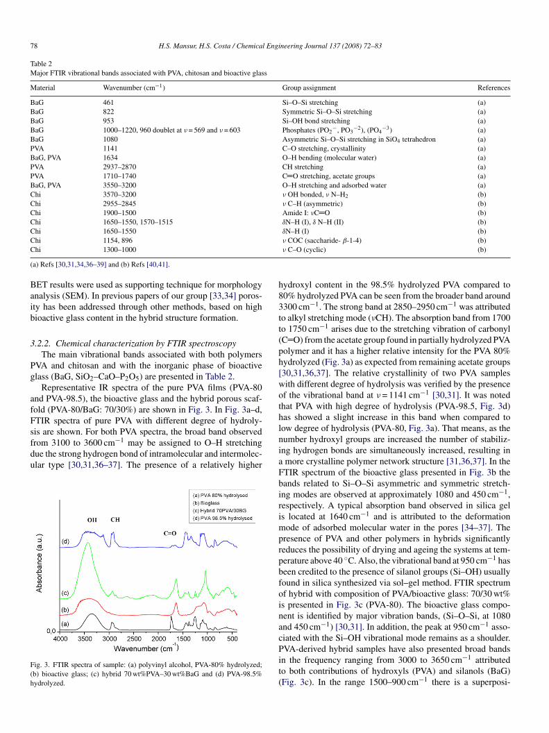
Representative IR spectra of the pure PVA films (PVA-80nd PVA-98.5), the bioactive glass and the hybrid porous scaf-old (PVA-80/BaG: 70/30%) are shown in Fig. 3. In Fig. 3a–d,TIR spectra of pure PVA with different degree of hydroly-
is are shown. For both PVA spectra, the broad band observedrom 3100 to 3600 cm−1 may be assigned to O–H stretchingue the strong hydrogen bond of intramolecular and intermolec-lar type [30,31,36–37]. The presence of a relatively higherig. 3. FTIR spectra of sample: (a) polyvinyl alcohol, PVA-80% hydrolyzed;b) bioactive glass; (c) hybrid 70 wt%PVA–30 wt%BaG and (d) PVA-98.5%ydrolyzed.
niaFbirimprpbfoinacPit(
ν COC (saccharide- β-1-4) (b)ν C–O (cyclic) (b)
ydroxyl content in the 98.5% hydrolyzed PVA compared to0% hydrolyzed PVA can be seen from the broader band around300 cm−1. The strong band at 2850–2950 cm−1 was attributedo alkyl stretching mode (νCH). The absorption band from 1700o 1750 cm−1 arises due to the stretching vibration of carbonylC O) from the acetate group found in partially hydrolyzed PVAolymer and it has a higher relative intensity for the PVA 80%ydrolyzed (Fig. 3a) as expected from remaining acetate groups30,31,36,37]. The relative crystallinity of two PVA samplesith different degree of hydrolysis was verified by the presencef the vibrational band at ν = 1141 cm−1 [30,31]. It was notedhat PVA with high degree of hydrolysis (PVA-98.5, Fig. 3d)as showed a slight increase in this band when compared toow degree of hydrolysis (PVA-80, Fig. 3a). That means, as theumber hydroxyl groups are increased the number of stabiliz-ng hydrogen bonds are simultaneously increased, resulting inmore crystalline polymer network structure [31,36,37]. In theTIR spectrum of the bioactive glass presented in Fig. 3b theands related to Si–O–Si asymmetric and symmetric stretch-ng modes are observed at approximately 1080 and 450 cm−1,espectively. A typical absorption band observed in silica gels located at 1640 cm−1 and is attributed to the deformationode of adsorbed molecular water in the pores [34–37]. The
resence of PVA and other polymers in hybrids significantlyeduces the possibility of drying and ageing the systems at tem-erature above 40 ◦C. Also, the vibrational band at 950 cm−1 haseen credited to the presence of silanol groups (Si–OH) usuallyound in silica synthesized via sol–gel method. FTIR spectrumf hybrid with composition of PVA/bioactive glass: 70/30 wt%s presented in Fig. 3c (PVA-80). The bioactive glass compo-ent is identified by major vibration bands, (Si–O–Si, at 1080nd 450 cm−1) [30,31]. In addition, the peak at 950 cm−1 asso-iated with the Si–OH vibrational mode remains as a shoulder.
VA-derived hybrid samples have also presented broad bandsn the frequency ranging from 3000 to 3650 cm−1 attributedo both contributions of hydroxyls (PVA) and silanols (BaG)Fig. 3c). In the range 1500–900 cm−1 there is a superposi-

H.S. Mansur, H.S. Costa / Chemical Engineering Journal 137 (2008) 72–83 79
F PVA–( an bio
tPb(sν
vtide
cctvBpoag
3d
geolhHsdIbyhds
ghsaIacTc
3.2.4. Characterization of PVA hybrids by X-ray scatteringWide-angle X-ray scattering (WAXS, also called X-ray
diffraction) is a powerful tool for understanding crystalline
ig. 4. FTIR spectra of samples synthesized with PVA-80: (a) hybrid of 70 wt%b) with 0.5% chitosan (PVA/Chi/BaG); Scheme 1: chemical structure of chitos
ion of the bands derived from the bioactive glass and theVA components [31,33,36,37]. Also, typical phosphate groupands at ν = 1000–1220 cm−1 (PO2
−, PO32−) and ν = 960 cm−1
PO43−). The FTIR spectra of samples containing phosphorus
howed a weak signal related to a doublet at approximately= 565 and ν = 600 cm−1, which is associated with the stretchingibrations of phosphate groups related to the presence of crys-alline phosphates in the glasses [39,45]. Although not shownn the figures, the spectra of the other hybrids produced withifferent proportion of PVA/BaG are very similar to the spectraxhibited in Fig. 3c.
FTIR spectra for hybrids of PVA/bioactive glass and PVA,hitosan and BaG are presented in Fig. 4a and b, respectively. Itan be observed the contribution of chitosan chemical groups iso the overall hybrid spectrum mainly related to amide groups atibrational bands ranging from 1000 to 1800 cm−1 [18–20,41].riefly, in the IR spectrum of chitosan (Fig. 4b), the absorptioneak of carbonyl stretching vibration in O C–NHR groups wasbserved at approximately 1660 cm−1, and the absorption peakt 1590 cm−1 was attributed to N–H bending vibration in amineroups [41].
.2.3. Crystallinity and phase characterization by X-rayiffraction
The X-ray diffraction analysis result from the pure bioactivelass is shown in Fig. 5a. As expected, it did not show the pres-nce of any crystalline phase, being totally amorphous. On thether hand, the XRD patterns from both samples of pure PVA,ow degree (PVA-80) and high degree of hydrolysis (PVA-98.5)ave shown some diffraction bands (Fig. 5b and c, respectively).ence, they have both being identified as a semi-crystalline
tructure, with slightly higher crystallinity for the PVA-98.5ue to the superior concentration of hydroxyls groups (Fig. 7c,nsert). This XRD results have endorsed the previous findingsased on FTIR spectrum of PVA with higher degree of hydrol-
sis (Section 3.2.2). The XRD curve for PVA/bioactive glassybrid (PVA/BaG: 70/30) synthesized is shown Fig. 7d. It can beirectly verified the sum up of both contributions from PVA withemi-crystalline structure and amorphous phase of bioactiveFhac
30 wt% bioactive glass; (b) hybrid of 70 wt%PVA–30 wt% bioactive glass andpolymer.
lass [34]. In the same way, the hybrid based on PVA/Chi/BaGas presented a diffraction pattern as shown in Fig. 5e. Theemi-crystalline structure was maintained when chitosan withcetylation degree of 76% was incorporated to PVA/BaG hybrid.t can be noted three peaks at approximately crystalline peakst 2θ of 10.3◦ (1 0 0), 15.2◦ (0 0 2) and 19.8◦ (0 2 0), which indi-ated some degree of crystallinity on the biopolymer network.hat would be a typical XRD pattern for the hybrid showingontribution from all components in the system [40,41].
ig. 5. XRD patterns of (a) bioactive glass SiO2–CaO–P2O5; (b) PVA 80%ydrolyzed; (c) PVA 98.5% hydrolyzed; (d) hybrid 70 wt%PVA–30 wt%BaGnd (e) hybrid 70 wt%PVA–30 wt%BaG with chitosan 0.5%; detail: PVA mainrystalline peaks.

80 H.S. Mansur, H.S. Costa / Chemical Engi
F8
pamfca
stcPifwahafTpaasPba
ig. 6. SAXS studies. SAXS patterns of (a) pure PVA-80, (b) hybrid PVA/BaG:0/20; (c) hybrid PVA/BaG: 70/30 and (d) pure bioactive glass.
hases, crystal orientations, planes and unit cell structure. Small-ngle X-ray scattering is very important to get information from
aterials structure at the nanometer scale, typically varyingrom few nanometers to 100 nm [30,31,41]. Synchrotron SAXSurves for pure PVA films and PVA/bioactive glass hybridsre presented in Fig. 6. SAXS results from PVA films have
wt
P
Fig. 7. Two-dimensional SAXS patterns of (a) pure PVA-80, (b) hybrid PVA
neering Journal 137 (2008) 72–83
hown a strong single peak with a maximum located at scat-ering vector (q) 0.54 up to 0.56 nm−1 (Fig. 6a). Such trendan be explained by assuming a semi-crystalline structure ofVA polymer sample previously described [30,31]. Consider-
ng the conditions of films preparation, that is, PVA crystallizedrom a dilute solution (5.0 wt%), one would expect that polymerill form lamellar crystals. These crystals are connected to the
morphous regions by polymer chains [30,31]. SAXS curvesave indicated a reduction in the peak (∼q = 0.55 nm−1) as themount of inorganic phase (BaG) was increased in the samples,rom pure PVA (Fig. 6a) to pure bioactive glasses (BaG, Fig. 6d).hat is associated with the chemical modification in the originalolymer network by a systematically decrease in the crystallinitys the bioactive glass concentration (amorphous) is increased,s verified by FTIR spectroscopy and XRD. Moreover, as theilane coupling reaction mechanism with hydroxyl groups ofVA occurs, it is most likely to reduce the formation of hydrogenonds and therefore, the driving force for PVA crystallization islso weakened. In other words, the nanostructure of domains
as altered by increasing the concentration of inorganic phaseo the PVA-derived hybrids [30,31].Bi-dimensional SAXS images for PVA, PVA/BaG: 80/20,
VA/BaG: 70/30 and BaG are presented in Fig. 7a–d, respec-
/BaG: 80/20; (c) hybrid PVA/BaG: 70/30 and (d) pure bioactive glass.

H.S. Mansur, H.S. Costa / Chemical Engineering Journal 137 (2008) 72–83 81
Fig. 8. Stress–strain curves obtained by compression test: (A) PVA/BaG:70/30 prepared with polymer: curve a, 80% hydrolysis grade; curve b, 98.5%hcP
toBrmt
3phFhsifpetPwcaPoih
FBi
stmuby(aisBroemso
mwb2
3a
b(pctggstm
4
ydrolysis grade and curve c, pure bioactive glass. (B) Hybrids at different con-entrations: curve a, PVA/BaG: 80/20; curve b PVA/BaG: 70/30 and curve cVA/BaG: 60/40.
ively. Again, the scattering patterns have shown strong evidencef reduction in the crystallinity of PVA (Fig. 7a) by raising theaG content in the hybrids (pure BaG, Fig. 7d). Thus, SAXS
esults have supported the proposed system of obtaining PVAodified systems at the nanoscale order by incorporating bioac-
ive glass to the newly formed hybrid structure.
.2.4.1. Characterization of mechanical behavior. The com-ression tests results of PVA/BaG (PVA/BaG: 70/30 wt%)ybrids are presented in Fig. 8, where PVA-80 is presented inig. 8A, curve a and PVA-98.5 in Fig. 8A, curve b. Both curvesave shown a similar pattern, with the compressive stress con-isted of an initial Hookean region in which stress increasedn proportion to strain due to compression of the cell elementsollowed by a plateau region representing plastic collapse ofores causing densification, with subsequent loading of the celldges and faces against one to another [42]. The averaged elas-ic moduli of hybrids were of 2.6 and 6.0 MPa for PVA-80 andVA-98.5, respectively. These values are in excellent agreementith those values reported in the literature for potential appli-
ation in repairing cancellous bone tissue [42,43]. The averagexial yield strain results were of 11% for hybrids produced with
VA-80 and 6% for PVA-98.5, which is attributed to the effectf crystallinity on the mechanical behavior. As axial yield stresss concerned the values were very similar 0.28 ± 0.02 MPa forybrids prepared with PVA with different degree of hydroly-afT
ig. 9. Relative cell viability of VERO cells cultured with extracts of PVA-80,aG and hybrid PVA/BaG samples as prepared. Simultaneous cultures were
ncubated with complete culture medium only as negative control (NC).
is. The organic phase of PVA added to the inorganic phaseo produce the hybrid material has advantageously increased the
echanical properties over the pure macroporous bioactive glasssually reported as very fragile and brittle [11,12,33]. It shoulde emphasized that the PVA-98.5 with higher degree of hydrol-sis has presented superior values of all mechanical propertiesFig. 8A, curve b) when compared to PVA-80 (Fig. 8A, curve), maintaining the PVA/BaG ratio constant. That leads to a verymportant conclusion, that in fact, the interface PVA/BaG has atronger interaction of hydroxyls from PVA and silanols fromaG as the degree of hydrolysis is increased. In addition, it is
eported in the literature that the higher the degree of hydrolysisf PVA and the polymer Mw the higher are the mechanical prop-rties [33,34]. The effect of increasing of PVA content on theechanical properties of the hybrid is evidenced by the results
hown in Fig. 8B. That is a trend of reducing the brittle behaviorf the hybrid by raising the PVA/bioactive glass ratio.
Based strictly on these mechanical properties reported, oneay assume that the PVA/BaG hybrids developed in the presentork are suitable for partial replacement of damaged cancellousone tissue which has typically a broad range of modulus fromto 12 MPa [9,43,44].
.3. Cytotoxicity and cellular viability activity by MTTssay
The cell viability, bioactivity and cytotoxicity were assessedy the MTT assay and the results are presented in histogramFig. 9). The cells treated with extracts of hybrid foams as pre-ared showed relatively good cell viability compared to theontrol used as reference. These bioactivity results based onhe in vitro MTT method have also showed that all experimentalroups were not significantly altered either by the PVA/bioactivelass ratio or the degree of hydrolysis of PVA used in the hybridynthesis. Further investigation is under way where direct con-act of cell culture spreading onto PVA/BaG hybrids aiming to
imic the in vivo environment of bone tissue replacement.
. Conclusions
Tri-dimensional porous structures composed of PVA/bio-ctive glass and PVA/chitosan/bioactive glass were success-ully synthesized by the sol–gel route and foaming method.hese organic–inorganic hybrid scaffolds exhibited a hierar-

8 l Engi
ca2birt
pon
tsbbbob
A
TRpElf
R
[
[
[
[
[
[
[
[
[
[
[
[
[
[
[
[
[
[
[
[
[
[
[
[
[
2 H.S. Mansur, H.S. Costa / Chemica
hical structure with interconnected macropores (10–500 �m)nd a mesoporous framework typical of gel–glasses (pores of–50 nm). By changing the concentration ratio from PVA toioactive glass and also the degree of hydrolysis from PVA,t was possible to modify the gelation time and, therefore, theesulting foam porosity, pore size distribution and interconnec-ivity.
FTIR spectroscopy associated with XRD and SAXS haveroven to very powerful tools for investigating the formationf PVA/BaG and PVA/Chi/BaG hybrids as organic–inorganicetwork at micro-nanostructures order.
The increase in the hydrolysis grade of PVA has improvedhe mechanical properties of the hybrid scaffolds with valuesuitable for cancellous bone repairing. In addition, the cell via-ility of all experimental groups was not significantly alteredy the PVA/bioactive glass ratio or by incorporating chitosaniopolymer into the hybrid. Hence, it opens a new windowf opportunity for developing specially designed materials foriomedical applications.
cknowledgements
The author acknowledges National Council for Scientific andechnological Development (CNPq) and State of Minas Geraisesearch Foundation (FAPEMIG) for financial support on thisroject. I would like to express my special gratitude to Prof. Dr.del F. Barbosa-Stancioli for Microbiology Laboratory and bio-
ogical assays. Also, the author thanks the important contributionrom LNLS staff and for synchrotron SAXS facilities.
eferences
[1] H.M. Kim, Ceramic bioactivity and related biomimetic strategy, Curr. Opin.Solid State Mater. Sci. 7 (4–5) (2003) 289–299.
[2] B.B. Nissan, Natural bioceramics: from coral to bone and beyond, Curr.Opin. Solid State Mater. Sci. 7 (2003) 283–288.
[3] D. Tadic, F. Beckmann, K. Schwarz, M. Epple, A novel method to producehydroxyapatite objects with interconnecting porosity that avoids sintering,Biomaterials 25 (2004) 3335–3340.
[4] J.R. Jones, L.L. Hench, Regeneration of trabecular bone using porousceramics, Curr. Opin. Solid State Mater. Sci. 7 (2003) 301–307.
[5] V. Olivier, N. Faucheux, P. Hardouin, Biomaterial challenges andapproaches to stem cell use in bone reconstructive surgery, Drug Discov.Today 9 (2004) 803–811.
[6] K.J.L. Burg, S. Porter, J.F. Kellam, Biomaterial developments for bonetissue engineering, Biomaterials 21 (2000) 2347–2359.
[7] K. Rezwan, Q.Z. Chen, J.J. Blaker, A.B. Boccaccini, Biodegradable andbioactive porous polymer, inorganic composite scaffold for bone tissueengineering, Biomaterials 27 (2006) 3413–3431.
[8] L.L. Hench, Bioceramics: from concept to clinic, J. Am. Ceram. Soc. 74(1991) 1487–1510.
[9] V.-R. Maria, G.-C. Jose Maria, Calcium phosphates as substitution of bonetissues, Prog. Solid State Chem. 32 (2004) 1–31.
10] S. Padilla, S. Sanchez-Salcedo, M. Vallet-Regı, Bioactive glass as precursorof designed-architecture scaffolds for tissue engineering, J. Biomed. Mater.Res. 81A (2007) 224–232.
11] R.J. Jones, L.M. Ehrenfried, L.L. Hench, Optimizing bioactive glass scaf-folds for bone tissue engineering, Biomaterials 27 (2005) 964–973.
12] M.M. Pereira, J.R. Jones, R.L. Orefice, L.L. Hench, Preparation of bioactiveglass-polyvinyl alcohol hybrid foams by the sol–gel method, J. Mater. Sci.:Mater. Med. 16 (2005) 1045–1050.
[
neering Journal 137 (2008) 72–83
13] C. Montserrat, J.S. Antonio, V.-R. Marıa, Amino–polysiloxane hybridmaterials for bone reconstruction, Chem. Mater. 18 (2006) 5676–5683.
14] E. Chiellini, A. Corti, S. D’antone, R. Solaro, Biodegradation of poly(vinylalcohol) based materials, Prog. Polym. Sci. 28 (2003) 963–1014.
15] A.P.V. Pereira, L.V. Wander, R.L. Orefice, Novel multicomponentsilicate–poly(vinyl alcohol) hybrids with controlled reactivity, J. Non-Cryst. Solids 273 (2000) 180–185.
16] P. Willi, P.S. Chandra, Nanoceramic matrices: biomedical applications,Am. J. Biochem. Biotechnol. 2 (2) (2006) 41–48.
17] T. Yamaoka, Y. Tabata, Y. Ikada, Comparison of body distribution ofpoly(vinyl alcohol) with other water-soluble polymers after intravenousadministration, J. Pharmaceut. Pharmacol. 47 (1995) 479–486.
18] C. Sanchez, F. Ribot, Design of hybrid organic–inorganic materials syn-thesized via sol–gel chemistry, New J. Chem. 18 (1994) 1007–1047.
19] Y.-F. Tang, Y.-M. Du, X.-W. Hu, X.-W. Shi, J.F. Kennedy, Rheologicalcharacterization of a novel thermosensitive chitosan/poly(vinyl alcohol)blend hydrogel, Carbohydr. Polym. 67 (4) (2007) 491–499.
20] L.-C. Wang, X.-G. Chen, D.-Y. Zhong, Q.-C. Xu, Study on poly(vinylalcohol)/carboxymethyl-chitosan blend film as local drug delivery system,J. Mater. Sci.: Mater. Med. 18 (6) (2007) 1125–1133.
21] S. Yang, K.-F. Leong, Z. Du, C.-K. Chua, The design of scaffolds for usein tissue engineering. Part I. Traditional factors, Tissue Eng. 7 (6) (2001)679–689.
22] M.G. Cascone, N. Barbani, C. Cristallini, P. Giusti, G. Ciardelli, L. Lazzeri,Bioartificial polymeric materials based on polysaccharides, J. Biomater.Sci. Polym. Edn. 12 (3) (2001) 267–281.
23] Y.-L. Liu, Y.-H. Su, J.-Y. Lai, In situ crosslinking of chitosanand formation of chitosan–silica hybrid membranes with using �-glycidoxypropyltrimethoxysilane as a crosslinking agent, Polymer 45(2004) 6831–6837.
24] J. Bergera, M. Reist, J.M. Mayer, O. Felt, R. Gurny, Structure and inter-actions in chitosan hydrogels formed by complexation or aggregation forbiomedical applications, Eur. J. Pharmaceut. Biopharmaceut. 57 (2004)35–52.
25] J. Berger, M. Reist, J.M. Mayer, O. Felt, N.A. Peppas, R. Gurny, Structureand interactions in covalently and ionically crosslinked chitosan hydrogelsfor biomedical applications, Eur. J. Pharmaceut. Biopharmaceut. 57 (2004)19–34.
26] M.M. Pereira, A.E. Clark, L.L. Hench, Calcium-phosphate formation onsol–gel-derived bioactive glasses in-vitro, J. Biomed. Mater. Res. 28 (1994)693.
27] V.-R. Marıa, Revisiting ceramics for medical applications, Dalton Trans.(2006) 5211–5220.
28] C.J. Brinker, G.W. Scherer, Sol–Gel Science: The Physics and Chemistryof Sol–Gel Processing, Academic Press, New York, NY, 1990.
29] A. Oki, X. Qiu, O. Alawode, B. Foley, Synthesis of organic–inorganichybrid composite and its thermal conversion to porous bioactive glassmonolith, Mater. Lett. 60 (2006) 2751–2755.
30] H.S. Mansur, R.L. Orefice, A.A.P. Mansur, Characterization of poly(vinylalcohol)/poly(ethylene glycol) hydrogels and PVA-derived hybrids bysmall-angle X-ray scattering and FTIR spectroscopy, Polymer 45 (2004)7193–7202.
31] G. Andrade, E.F. Barbosa-Stancioli1, A.A. Piscitelli Mansur, W.L.Vasconcelos, H.S. Mansur, Design of novel hybrid organic–inorganicnanostructured biomaterials for immunoassay applications, Biomed. Mater.1 (2006) 221–234.
32] T. Kokubo, Apatite formation on surfaces of ceramics, metals and polymersin body environment, Acta Mater. 46 (1998) 2519–2527.
33] M.M. Pereira, J.R. Jones, L.L. Hench, Bioactive glass and hybrid scaffoldsprepared by sol–gel method for bone tissue engineering, Adv. Appl. Ceram.104 (1) (2005) 35–42.
34] H.S. Costa, G.I. Andrade, E.F. Barbosa-Stancioli, M.M. Pereira, R.L.
Orefice, H.S. Mansur, Sol–gel derived composite from bioactiveglass–polyvinyl alcohol, J. Mater. Sci. 43 (2008) 494–502.35] W. Tao, T. Mahir, S. Gunasekaran, Selected properties of pH-sensitive,biodegradable chitosan–poly(vinyl alcohol) hydrogel, Polym. Int. 53(2004) 911–918.

l Engi
[
[
[
[
[
[
[
[
[
[
H.S. Mansur, H.S. Costa / Chemica
36] H.S. Mansur, R.L. Orefice, W.L. Vasconcelos, Z.P. Lobato, L.J. Machado,Biomaterial with chemically engineered surface for protein immobiliza-tion, J. Mater. Sci.: Mater. Med. 16 (2005) 333–340.
37] J. Coates, in: R.A. Meyers (Ed.), Encyclopedia of Analytical Chemistry:Interpretation of Infrared Spectra, a Practical Approach, John Wiley andSons Ltd., Chichester, 2000, pp. 10815–10837.
38] R.M. Almeida, C.G. Pantano, Structural investigation of silica gel films byinfrared spectroscopy, J. Appl. Phys. 68 (1990) 1225–1232.
39] J. Roman, S. Padilla, M. Vallet-Regı, Sol–gel glasses as precursors of
bioactive glass ceramics, Chem. Mater. 15 (2003) 798–806.40] Y. Zhang, Preparation of electrospun chitosan/poly(vinyl alcohol) mem-branes, Colloid Polym. Sci. 285 (2007) 855–863.
41] S.S. Silva, Functional nanostructured chitosan–siloxane hybrids, J. Mater.Chem. 15 (2005) 3952–3961.
[
neering Journal 137 (2008) 72–83 83
42] L.J. Gibson, Biomechanics of cellular solids, J. Biomech. 38 (2005)377–399.
43] D. Shi, G. Jiang, Synthesis of hydroxyapatite films on porous Al2O3 sub-strate for hard tissue prosthetics, Mater. Sci. Eng. C 6 (1998) 175–182.
44] M.O. Montjovent, L. Mathieu, B. Hinz, L.L. Applegate, P.E. Bourban, P.Y.Zambelli, J.A. Manson, D.P. Pioletti, Biocompatibility of bioresorbablepoly(l-lactic acid) composite scaffolds obtained by supercritical gas foam-ing with human fetal bone cells, Tissue Eng. 11 (2005) 1640–1649.
45] A.I. Martin, A.J. Salinas, M. Vallet-Regi, Bioactive and degradable
organic–inorganic hybrids, J. Eur. Ceram. Soc. 25 (2005) 3533–3538.46] J.D. Cho, W.S. Lyoo, S.N. Chvalun, J. Blackwell, X-ray analysis and molec-ular modeling of poly(vinyl alcohol)s with different stereoregularities,Macromolecules 32 (1999) 6236–6241.