IMPROVEMENT OF FUNCTIONAL AND BIOACTIVE ...
133
IMPROVEMENT OF FUNCTIONAL AND BIOACTIVE PROPERTIES OF CHIA SEED (SALVIA HISPANICA) PROTEIN HYDROLYSATES AND DEVELOPMENT OF BIODEGRADABLE FILMS USING CHIA SEED MUCILAGE by Uriel Carlos Urbizo Reyes A Thesis Submitted to the Faculty of Purdue University In Partial Fulfillment of the Requirements for the degree of Master of Science Department of Food Science West Lafayette, Indiana December 2019
-
Upload
khangminh22 -
Category
Documents
-
view
2 -
download
0
Transcript of IMPROVEMENT OF FUNCTIONAL AND BIOACTIVE ...

IMPROVEMENT OF FUNCTIONAL AND BIOACTIVE PROPERTIES OF
CHIA SEED (SALVIA HISPANICA) PROTEIN HYDROLYSATES AND
DEVELOPMENT OF BIODEGRADABLE FILMS USING CHIA SEED
MUCILAGE
by
Uriel Carlos Urbizo Reyes
A Thesis
Submitted to the Faculty of Purdue University
In Partial Fulfillment of the Requirements for the degree of
Master of Science
Department of Food Science
West Lafayette, Indiana
December 2019

2
THE PURDUE UNIVERSITY GRADUATE SCHOOL
STATEMENT OF COMMITTEE APPROVAL
Dr. Andrea Liceaga, Chair
Department of Food Science
Dr. Fernanda San Martin
Department of Food Science
Dr. Jose Garcia Bravo
School of Engineering Technology
Approved by:
Dr. Brian Farkas

3
In the first place, I want to thank God for all the opportunities and blessings provided to me in
these two years. I want to thank Dr. Liceaga for allowing me to do my graduate studies, sharing
knowledge, and providing support; to my committee members Dr. San Martin-González and Dr.
Garcia-Bravo, for the guidance and teachings imparted to me. Finally, I want to dedicate all this
work to my mother (Maira), father (Uriel), my two sisters (Andrea and Mayra), and friends who
supported me endlessly through this journey.

4
ACKNOWLEDGMENT OF PREVIOUS PUBLICATIONS
Some parts of this thesis contain reprint materials of “Physicochemical characteristics of chia seed
(Salvia hispanica) protein hydrolysates produced using ultrasonication followed by microwave-
assisted hydrolysis” including figures, tables and some discussions.

5
TABLE OF CONTENTS
LIST OF TABLES .......................................................................................................................... 9
LIST OF FIGURES ...................................................................................................................... 10
ABSTRACT .................................................................................................................................. 12
CHAPTER 1. INTRODUCTION ............................................................................................ 14
CHAPTER 2. LITERATURE REVIEW ................................................................................. 19
Plant protein composition, structure, and classification ............................................... 19
2.3.1 Protein structure ..................................................................................................... 19
2.3.2 Protein classification .............................................................................................. 20
Chia seeds (structure, origin, and composition) ........................................................... 23
Chia seed protein and health benefits ........................................................................... 24
Plant protein hydrolysates ............................................................................................. 26
Bioactive peptides from plant sources .......................................................................... 28
2.4.1 Antioxidant activity ................................................................................................. 29
2.4.2 Antihypertension activity ........................................................................................ 31
2.4.3 Antidiabetic ............................................................................................................. 33
2.5 Extracting bioactive peptides from chia seeds .............................................................. 34
2.5.1 Chia seed bioactive peptides ................................................................................... 35
2.5.2 Microwave-assisted proteolytic hydrolysis ............................................................. 36
2.5.3 Mucilage extraction using ultrasound .................................................................... 37
2.6 Functional Properties of Proteins .................................................................................. 40
2.6.1 Solubility ................................................................................................................. 40
2.6.2 Emulsion ................................................................................................................. 42
2.6.3 Foaming .................................................................................................................. 44
2.7 Biodegradable films: Alternative applications of plant materials. ............................... 45
2.7.1 Biodegradable films using chia seed mucilage ....................................................... 46
2.7.2 Digestion of chia seed mucilage ............................................................................. 47
2.7.3 Barrier property ...................................................................................................... 47
2.7.4 Mechanical properties ............................................................................................ 49

6
CHAPTER 3. PHYSICOCHEMICAL CHARACTERISTICS OF CHIA SEED (SALVIA
HISPANICA) PROTEIN HYDROLYSATES PRODUCED USING ULTRASONICATION
FOLLOWED BY MICROWAVE-ASSISTED HYDROLYSIS.................................................. 50
3.1 Introduction ................................................................................................................... 51
3.2 Materials and methods .................................................................................................. 53
3.2.1 Materials ................................................................................................................. 53
3.2.2 Chia seed mucilage extraction ................................................................................ 53
3.2.3 Chia seed oil extraction .......................................................................................... 54
3.2.4 Chia seed protein hydrolysate (CSPH) ................................................................... 55
3.2.5 Proximal Composition ............................................................................................ 56
3.2.6 Determination of the degree of hydrolysis .............................................................. 56
3.2.6.1 Amino Acid Analysis ...................................................................................... 57
3.2.6.2 Sodium Dodecyl Sulfate Polyacrylamide Gel Electrophoresis (SDS-PAGE) 57
3.2.7 Bioactive properties of CSPH ................................................................................. 58
3.2.7.1 2-diphenyl-2-picrylhydrazyl (DPPH) radical scavenging activity .................. 58
3.2.7.2 2,2'-azino-bis (3-ethylbenzothiazoline-6-sulphonic acid) radical scavenging
activity (ABTS) ................................................................................................................. 58
3.2.7.3 Metal Ion Chelating (MIC) .............................................................................. 59
3.2.7.4 Ferric Reducing Antioxidant Power (FRAP) .................................................. 59
3.2.7.5 Oxygen radical absorbance capacity (ORAC) ................................................. 60
3.2.7.6 Cellular antioxidant activity (CAA) ................................................................ 60
3.2.7.7 Dipeptidyl Peptidase IV (DPP-IV) Inhibitory Activity ................................... 61
3.2.7.8 Angiotensin Converting Enzyme (ACE) Inhibitory Activity .......................... 62
3.2.8 Functional properties of CSPH .............................................................................. 63
3.2.8.1 Solubility ......................................................................................................... 63
3.2.8.2 Emulsion .......................................................................................................... 63
3.2.8.3 Foaming ........................................................................................................... 64
3.2.9 Statistical Analysis .................................................................................................. 65
3.3 Results and discussion .................................................................................................. 65
3.3.1 Mucilage ultrasound separation ............................................................................. 65
3.3.2 Chia seed oil extraction .......................................................................................... 67

7
3.3.3 Enzymatic hydrolysis .............................................................................................. 67
3.3.4 Proximal Analysis ................................................................................................... 68
3.3.5 Antioxidant Activity ................................................................................................. 70
3.3.6 Correlation of antioxidant assays to CAA .............................................................. 75
3.3.7 Antidiabetic properties............................................................................................ 76
3.3.8 Anti-hypertensive activity ........................................................................................ 78
3.4 Functional Properties .................................................................................................... 79
3.4.1 Solubility ................................................................................................................. 79
3.4.2 Emulsion ................................................................................................................. 81
3.4.3 Foaming .................................................................................................................. 82
3.5 Conclusions ................................................................................................................... 84
CHAPTER 4. EFFECT OF POLYOL CONCENTRATION ON PHYSICAL AND BARRIER
CHARACTERISTICS OF EDIBLE FILMS FROM CHIA SEED (SALVIA HISPANICA)
MUCILAGE. ................................................................................................................................ 86
4.1 Introduction ................................................................................................................... 87
4.2 Materials and methods .................................................................................................. 89
4.2.1 Materials ................................................................................................................. 89
4.2.2 Chia seed mucilage extraction ................................................................................ 89
4.2.3 Proximate composition ........................................................................................... 90
4.2.4 Preparation of chia seed mucilage (CSM) films ..................................................... 90
4.2.5 Color measurement ................................................................................................. 90
4.2.6 Tensile strength and elongation at break................................................................ 91
4.2.7 Film solubility ......................................................................................................... 92
4.2.8 Water contact angle ................................................................................................ 93
4.2.9 Water vapor permeability (WVP) ........................................................................... 93
4.2.10 Raman spectroscopy ............................................................................................... 94
4.2.11 Statistical analysis .................................................................................................. 94
4.3 Results and discussion .................................................................................................. 95
4.3.1 Proximate analysis of chia seed mucilage .............................................................. 95
4.3.2 Chia seed mucilage films thickness and color ........................................................ 96
4.3.3 Mechanical properties (tensile strength and elongation at break) ......................... 98

8
4.3.4 Water vapor permeability (WVP) ......................................................................... 101
4.3.5 Water contact angle .............................................................................................. 102
4.3.6 Film solubility ....................................................................................................... 103
4.3.7 Raman analysis of chia seed mucilage and chia seed mucilage films .................. 106
4.3.8 Optimization .......................................................................................................... 111
4.4 Conclusions ................................................................................................................. 113
CHAPTER 5. OVERALL CONCLUSION AND FUTURE DIRECTIONS. ....................... 114
5.1 Overall conclusions ........................................................................................................... 114
5.2 Future directions ............................................................................................................... 114
REFERENCES ........................................................................................................................... 115
APPENDIX A. FORMS ............................................................................................................. 131

9
LIST OF TABLES
Table 1. Commonly used protein categories. ............................................................................... 21
Table 2. Total amino acid composition for chia seed (USDA, 2011). ........................................ 26
Table 3. Mucilage extraction from chia seeds. ............................................................................. 67
Table 4. Proximal analysis of chia seed protein hydrolysates. Results are reported on a dry
weight basis. .................................................................................................................................. 70
Table 5. Bioactive properties of CSPH. ....................................................................................... 73
Table 6. Total Amino acid analysis for chia seed protein and CSPH (g/100 g). ......................... 74
Table 7. Correlation analysis between antioxidant assays with cellular antioxidant analysis. .... 76
Table 8. Cellular antioxidant activity, Antidiabetic and antihypertensive properties of CSPH. .. 78
Table 9. Coded and decoded levels of polyols incorporated to chia seed mucilage films. .......... 95
Table 10. Color determination of chia seed mucilage films. ....................................................... 98
Table 11. Statistical models for response variables, as a function of independent variables grams
of glycerol (X1) and sorbitol (X2) per gram of mucilage. ........................................................... 104
Table 12. Effect of independent variables on response variables. ............................................. 105
Table 13. Raman Spectra analysis for chia seed mucilage and its films. ................................... 111
Table 14. Regression coefficients and confidence limits. .......................................................... 133

10
LIST OF FIGURES
Figure 1. Flowchart for the development of chia seed protein hydrolysates. .............................. 18
Figure 2. Microscopic structure of chia seed with mucilage exudate. ......................................... 23
Figure 3. Proteolytic hydrolysis reaction (Donohue & Osna, 2003). .......................................... 28
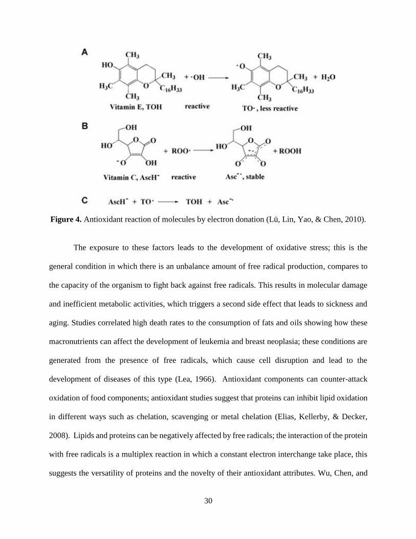
Figure 4. Antioxidant reaction of molecules by electron donation (Lü, Lin, Yao, & Chen, 2010).
....................................................................................................................................................... 30
Figure 5. Ultrasound compression and rarefaction cycles (Rostagno & Prado, 2013) –
Reproduced by permission of The Royal Society of Chemistry................................................... 38
Figure 6. Compounds liberated by microjet in ultrasound (Rostagno & Prado, 2013) –
Reproduced by permission of The Royal Society of Chemistry................................................... 39
Figure 7. Solubility curve for defatted flour of baru nuts (Guimarães et al., 2012). ................... 41
Figure 8. Role of protein in emulsions (Costello, 2017). ............................................................. 43
Figure 9. Barrier properties of PLA compared to conventional polymers at 30~C for (nitrogen,
oxygen, Carbon dioxide and Methane) (Hamad et al., 2015). ...................................................... 48
Figure 10. Diagram of mucilage separation (extraction) from chia seeds using ultrasonication
and vacuum-assisted filtration. ..................................................................................................... 54
Figure 11. Clean chia seeds after mucilage extraction using ultrasound and vacuum-filtration
separation (A); chia seeds with residual mucilage using ultrasound and centrifugation (B). ....... 66
Figure 12. Molecular weight distribution of chia seed protein hydrolysates. Lanes indicate: (1)
molecular weight markers, (2) Control: unhydrolyzed chia seed flour, (3) A-WB: chia seed
protein hydrolyzed by alcalase enzyme using water bath heating method, (4) A-MW: chia seed
protein hydrolyzed by alcalase enzyme using microwave-assisted hydrolysis, (5) AF-WB: chia
seed protein hydrolyzed by alcalase and flavourzyme enzymes using water bath heating method,
(6) AF-MW: chia seed protein sequentially hydrolyzed by alcalase and flavourzyme enzymes
using microwave-assisted hydrolysis. ........................................................................................... 71
Figure 13. Results for Solubility of chia seed protein hydrolysates. Different letters (a-d) show
significant differences (p < 0.05) between treatments. Sample codes descriptions are provided in
Table 5. ......................................................................................................................................... 80
11
Figure 14. Results for Emulsifying activity of chia seed protein hydrolysates. Different letters
(a-d) show significant differences (p < 0.05) between treatments. Sample codes descriptions are
provided in Table 5. ...................................................................................................................... 82
Figure 15. Results for foaming capacity (C) of chia seed protein hydrolysates. Different letters
(a-d) show significant differences (p < 0.05) between treatments. Sample codes descriptions are
provided in Table 5. ...................................................................................................................... 84
Figure 16. Tensile test specimen under traction. ......................................................................... 92
Figure 17. Response surface diagrams; Effect of glycerol (g) and sorbitol (g) per gram of chia
seed mucilage (CSM) on the response variables: water vapor permeability (A), solubility (B),
contact angle (C), elongation at break (D), tensile strength (E). ................................................ 100
Figure 18 . Raman spectra for chia seed mucilage. ................................................................... 106
Figure 19. Raman spectra for edible films from chia seed mucilage at different polyol
concentrations ............................................................................................................................. 107
Figure 20. Optimization of edible films from chia seed mucilage. ............................................ 112
Figure 21.Central composite design assignment for CSM films. .............................................. 131
Figure 22. Pareto charts, significance effect of polyol and their interaction on mechanical and
barrier properties of CSM films. ................................................................................................. 131
Figure 23. Ultrasonic removal of mucilage from chia seeds. .................................................... 132
Figure 24. Centrifugation as a separation method for chia seed mucilage. ............................... 132
Figure 25. Chia seed oil extraction by oil press. ........................................................................ 132

12
ABSTRACT
Chia seed (Salvia hispanica) has shown potential as an alternative source of nutrients with a high
content of fiber (36 %), protein (25%), and fat (25%). Unfortunately, the presence of a viscous
biopolymer (mucilage), surrounding the chia seed (CS), limits the accessibility of the protein and
other nutrients. Nevertheless, this biopolymer’s chemical composition makes it suitable for the
development of biodegradable films. Regarding CS protein, disulfide bonding, and nonprotein-
protein interactions often frequent in plant protein, have limited its technological application in
food matrices. Therefore, scientists have pointed at processing methods involving enzymatic
proteolysis to improve the functionality of plant protein ingredients. The objective of this study
was to establish processing techniques to exploit the functionality, extraction, and health benefits
of chia seed components. First, ultrasonication followed by vacuum-filtration was used to separate
mucilage from CS prior to fat extraction by oil press. Mucilage-free and defatted CS were treated
using conventional (enzymatic hydrolysis with alcalase) or sequential (enzymatic hydrolysis with
alcalase+flavourzyme), and under water bath or microwave-assisted hydrolysis. Chia seed protein
hydrolysates (CSPH) derived from the sequential hydrolysis with microwave treatment showed
superior (p<0.05) in vitro antioxidant activity. The highest (p<0.05) cellular antioxidant activity
was achieved by the sequential (94.76%) and conventional (93.13%) hydrolysis with microwave.
Dipeptidyl peptidase-V inhibition was higher (p<0.05) for sequential hydrolysis with water bath,
while Angiotensin-Converting Enzyme (ACE) inhibition activity increased (p<0.05) with
hydrolysis for all treatments compared to the control. Regarding functionality, sequential
hydrolysis with microwave showed higher (p<0.05) solubility at lower pH (3 and 5), while
conventional hydrolysis with microwave was better at pH 7 and 9. Emulsification properties and
foaming capacity were also higher in conventional hydrolysis with microwave, but conventional

13
hydrolysis with water bath was more stable for foaming properties only. In terms of mucilage
applicability, biodegradable films were developed by casting technique where CS mucilage was
plasticized with different polyol mixtures (sorbitol and glycerol). CS mucilage films with higher
sorbitol content showed superior tensile strength (3.23 N/mm2), and lower water vapor
permeability (1.3*109 g/ m*s*Pa) but had poor flexibility compared to other treatments.
Conversely, films with high glycerol content showed high elongation at break (67.55%) and
solubility (22.75%), but reduced water vapor permeability and tensile strength. The
hydrophobicity, measured as water contact angle, was higher (p<0.05) for mixtures containing
equal amounts of polyols. Lastly, Raman Spectroscopy analysis showed shifts from 854 to 872
cm-1 and 1061 to 1076 cm-1, which corresponded to β(CCO) modes. These shifts represent an
increase in hydrogen bonding, responsible for the high tensile strength and decreased water vapor
permeability. This study demonstrated that ultrasonication followed by vacuum filtration can
successfully separate mucilage from chia seeds; microwave-assisted and enzymatic hydrolysis
generated protein hydrolysates with improved bioactivity and functionality. Finally, chia seed
mucilage was able to form films with potential to be used in drug delivery and edible food coating
applications.
14
CHAPTER 1. INTRODUCTION
The global population is currently around 7.4 billion, twice the population number that can
sustainably inhabit the earth (Nadathur, Wanasundara, & Scanlin, 2017). To ensure food security,
food scientists must explore new alternatives to provide nutrients according to availability around
the world. Proteins are one of the most important macronutrients required for proper development.
Proteins are essential macromolecules composed of individual structures called amino acids,
which are indispensable for development and maintenance of our body (Ustunol, 2014). The
challenge for obtaining protein through conventional agriculture is enormous; it is estimated that
for the year 2050 the world will experience a population growth to 10 billion people (Nadathur et
al., 2017). For this reason, the use of emerging and sustainable protein sources has become a
trending alternative to help alleviate the environmental impact caused by the production of
conventional protein sources such as beef, poultry and dairy products. The need to change eating
patterns is necessary to provide adequate amount of protein and nutrients to the world’s population.
Meat proteins have historically been the primary protein source. Nevertheless, the amount of
resources required to produce meat from beef, poultry and pork are unsustainable, causing extreme
contamination, and increasing the emission of greenhouse gases (Müller, Wolf, & Gutzeit, 2017).
Some plants have shown to be high protein sources, with lower environmental impact. Moreover,
the current animal-derived dietary patterns have led to an increased development of chronic
diseases. Chronic disorders such as coronary heart disease, type-2 diabetes, neurodegenerative
diseases, and renal failures were responsible of 39.5 out of 54.7 million deaths in 2016 (Harris,
2019). There are different ways to address these diseases which include, diet education, smoke
prevention and excessive alcohol consumption (Harris, 2019). As food scientists, the main
approach we can contribute to alleviate the food demand and the increasing rates of chronic

15
diseases, is to develop, healthy and sustainable nutrient sources that address these issues. It is well
known that proteins have bioactive (antihypertensive, antioxidant, antidiabetic or antithrombotic)
and functional (foaming, solubility, emulsifying or gelling) properties, and that these properties
might be enhanced by protein modification using enzymatic hydrolysis or new technologies (Mine,
Li-Chan, & Jiang, 2010; Pacheco-Aguilar, Mazorra-Manzano, & Ramírez-Suárez, 2008).
Chia seed (salvia hispanica) is an emerging protein source, originated from Mesoamerica.
The use of this seed as a source of protein, is an alternative that has been explored in the past few
years. Chia seeds contain a high content of protein (15-25%) fat (30-33%) and fiber (18-30%)
(Mohd Ali et al., 2012). An issue has been encountered in the application of these seeds by the
presence of a highly viscous polymer called mucilage that limits the functionality and availability
of the protein. For example, researchers have investigated the chia seed protein and determined
that its digestibility was low compared to other plant sources, and they attributed this to the barrier
effect caused by the mucilage, which protects the seed and its components from gastric acids and
enzymes, decreasing the application and digestibility of the protein (Monroy-Torres, Mancilla-
Escobar, Gallaga-Solórzano, & Santiago-García, 2008). Multiple methods such as lyophilization
(Capitani, Ixtaina, Nolasco, & Tomás, 2013), salt extraction (Muñoz, Cobos, Diaz, & Aguilera,
2012), and oven dying (Campos et al., 2016) have been explored for mucilage extraction, but most
of them required laborious work and exhibit low yields. New technologies such as ultrasonication
have allowed us to extract highly bounded compounds from plant materials. Ultrasonication has
been previously used in the extraction of different compounds including proteins, polyphenols,
tannins and polysaccharides; however, exploration using different food matrices is still required to
address other possibilities (Vilkhu, Mawson, Simons, & Bates, 2008).
16
In this thesis, we will identify different protein extraction parameters in order to apply
ultrasound as an extraction method of mucilage from chia seeds and enzymatic proteolysis to
develop functional and bioactive peptides. Finally, we will develop a biodegradable film using
chia seed mucilage.
Research Objectives
Objective 1: Develop an improved mucilage extraction procedure by implementing ultrasound
techniques.
Hypothesis: The use of ultrasound technology will improve the extraction yield of mucilage, oil
and protein from chia seeds.
Tasks:
• Identify the conditions of separation methods (centrifugation and vacuum-assisted
filtration) and temperatures (25, 40, and 55 °C) that increase the mucilage separation yields.
Objective 2: Compare functional and bioactive properties of chia seed protein hydrolysates
(CSPH) derived from water bath and microwave-assisted hydrolysis.
Hypothesis: Enzymatic hydrolysis will create chia seed protein hydrolysates (CSPH) with
improved functional and bioactive properties.
Tasks:
• Extract protein from chia seeds using microwave energy and enzymatic treatments.
• Develop functional: EAI, FC, SOL, and bioactive assays: DPP-IV, ACE, DPPH, FRAP,
MIC, and ABTS.
• Determine structural and amino acid composition of CSPH.
Objective 3: Develop a biodegradable film using chia seed mucilage.
17
Hypothesis: Mucilage will show to have a good mechanical and barrier characteristics for film
development.
Tasks:
• Measure fundamental mechanical and barrier properties of chia seed mucilage films such
as tensile strength and elongation at break (TS, EB) and rate of water vapor permeability
(RWVP).
Chapter 3 and 4 were developed following a systematic approach (Fig 1), where the products from
chia seeds were used in the application of different products.

18
Figure 1. Flowchart for the development of chia seed protein hydrolysates.

19
CHAPTER 2. LITERATURE REVIEW
Plant protein composition, structure, and classification
Proteins are molecules essential for life, responsible of structural, functional and biological
roles in the body of all living things (Loveday, 2019). Plant proteins contain a broad variety of the
20 amino acids regarding their chemical structure and the fractions present in them. Protein is the
most indispensable macromolecule for human and animal nutrition. The amino acids composed of
unique hydrocarbon skeletons and elements (nitrogen and sulfur) can’t be replaced by any other
nutrients, mainly because sulfur and nitrogen are available nowhere else (Wu, 2016). These
components are key players in the synthesis of small molecules (glutathione, creatine, nitric oxide,
dopamine, serotonin, RNA and DNA) which are required for physiological processes (Wu, 2016).
The importance of protein is greatly appreciated in biological processes such as DNA synthesis
and replication. In general, the characteristics that define an organism’s conformation are stored
in their DNA. These molecules mainly contain information for the synthesis of proteins, and
proteins are the ones responsible of decoding or copying this information (Damodaran & Parkin,
2017).
2.3.1 Protein structure
The structure of proteins is generally classified in 4 sublevels. First, the amino acids are
covalently bonded to one another by the action of the ribosomes via amide bonding and in
accordance to the genetic code. The second level is better described as the spatial arrangement of
the polypeptide chains due to H-bonding, giving place to an arrangement in form of α- helices, β-
turns and sheets, and random coils (Nadathur, Wanasundara, & Scanlin, 2016; Nadathur et al.,
2017). The tertiary structure is often described as hydrophobic interactions between non-polar side
20
chains of amino acids and the disulfide bonds between the cysteine residues that form the
secondary structure of the protein itself, then scaffold structures arise as a 3D structure of the
polypeptide chain (Nadathur et al., 2017). Is important to mention that the tertiary structure is
crucial since it will arise to a vast number of biological roles (e.g., lock and key function in
enzymes or receptor sites on cell membranes) (Aldred & Phenols, 2009). Finally, other
arrangements exist such as quaternary protein structures, which refer to the spatial arrangement of
polypeptides or subunits within the whole structure (e.g., hemoglobulin and ion channels) (Aldred
& Phenols, 2009).
2.3.2 Protein classification
Proteins have been classified in different ways throughout the years, and due to the
complexity of these macromolecules, a single classification is not possible. Some of the common
classifications of proteins are based on solubility, conjugation, shape, function and nutritional
value as shown in Table 1. One classification that is still used today was established by Thomas
B. Osborne in 1908 (Loveday, 2019). This classification is based on solubility and is currently
known as the most common plant-protein classification, even though complex mixtures of protein
are commonly present in each group and some plant-derived protein groups overlap, it is still a
good starting point for protein fractionation (Nadathur et al., 2017).

21
Table 1. Commonly used protein categories.
Classification
criteria
Categories
Solubility Albumins: soluble in water and salt solutions
Globulins: not soluble in water, but readily soluble in salt solutions
Histones: soluble in acid solutions
Prolamins: soluble in ethanol-water solutions
Scleroproteins: insoluble in water and salt solutions
Conjugation Simple: liberates only amino acids when hydrolyzed.
Conjugated: liberates other compounds that are usually attached to its
structure (e.g., phosphate groups, carbohydrates, lipids, metal groups and
nucleic acids)
Shape Fibrous (rod, fibrous and thread-like)
Globular (spherical or ellipsoidal)
Function Catalytic, structural, regulatory/hormonal, transport, genetic, immune,
contractile and storage
Nutritional value Complete: contains all the essential amino acids
Incomplete proteins: missing one or more essential amino acids
(Nadathur et al., 2017).
Plants contain a high amount of proteins that meet biological and structural roles. For plant
seeds, the protein content is even higher varying according to the types of seeds (e.g., 10% in
cereals, 40% in oilseeds) (Shewry, Napier, & Tatham, 1995). Proteins play metabolic or structural
roles to keep the seed integrity and protect it from abiotic factors (e.g., low temperatures, bacterial
digestion, cellular respiration), nonetheless a vast portion of the storage proteins serve to store
amino acids for seedling growth and germination. Storage proteins are of importance because they
will not only determine the protein content in seeds, but also their nutritional quality (Shewry et
al., 1995). The main categories found in plants are: albumins, globulins, prolamins, and glutelins.
In the case of globulins and albumins, subclassifications are derived from ultracentrifugation-
derived Svedberg sedimentation coefficients, which are generally express as Svedberg units (S)
(Loveday, 2019). Häkkinen, Nuutila, and Ritala, (2018), identified that albumins are commonly
found as 2S whereas globulins as 7-8S and 11-12S. The most common sources of plant protein are
from grains (e.g., wheat, rice, millets and sorghum), seeds (e.g., chia, hemp), pulses (e.g., beans,

22
lentil, peas, lupins), and leaves (e.g., moringa, duckweed) (Nadathur et al., 2017). The distribution
of proteins varies according to seed varieties. Studies have shown that cereals such as wheat are
especially rich in glutelins, a subgroup of prolamins proteins. In the case of protein from rice, chia
seeds, and soy the most abundant type of proteins are the globulins 11-12S (Sandoval-Oliveros &
Paredes-López, 2013; Taylor, Taylor, Campanella, & Hamaker, 2016). Legumes are especially
abundant in albumins and globulins. Additionally, other type of proteins such as histones are
present in much lower concentration in plants but plays a crucial role in the process of post-
translational modifications modulating process like floral transition, seed germination
organogenesis and morphogenesis (Liu & Min, 2016).
The poor solubility of globulin, prolamins and glutelins is often associated to the condense
polymeric state of these fractions caused by high intermolecular disulfide bonding, which results
in low hydrophilic residue exposure (Kawakatsu & Takaiwa, 2019). This intrinsic characteristic
remains as one of the main challenges limiting the food application of these plant macromolecules
in some food matrices (e.g., non-dairy milk substitute, shakes or cereal-protein rich drinks and
beverages) (Carbonaro, Maselli, & Nucara, 2012; Clemente et al., 2000; Deshpande &
Damodaran, 1989; Ghumman, Kaur, & Singh, 2016; Loveday, 2019).
Other factors such as exogenous interaction of proteins with non-protein components (e.g.,
starches, dietary fiber, lipids, and tannins) and the high resistance of plant cell walls to digestion,
have also limited the digestibility and functionality of these proteins (Pushparaj & Urooj, 2011).
Protein structure can greatly influence the capacity of proteolytic enzymes to access the inner
structure of proteins, studies have shown that the high presence of -sheets influenced negatively
the protein digestibility of proteins whereas the protein higher ratios of -helixes, random coils to
-sheets showed a positive correlation to the digestibility values (Bai, Qin, Sun, & Long, 2016).

23
Chia seeds (structure, origin, and composition)
Throughout the years, plants have been domesticated and cultivated to obtain novel
ingredients and alternative sources of functional foods. One remarkable plant that has shown high
potential is chia (Salvia hispanica). Chia is a biannually cultivated plant; it is considered a
pseudocereal that produces purple and white flowers that eventually result in small oval shape
seeds with sizes varying from 1 to 2 mm (Mohd Ali et al., 2012). Chia seeds were first cultivated
in pre-Columbian Mesoamerica. This annual crop was used as a high-value commodity due to its
versatility, its medicinal and food applications (Cahill, 2002). The seeds are mainly divided into
two semi hemispheric structures that contain the endosperm called cotyledons. The cotyledons are
covered by the seed coat, which is composed of three layers endocarp, sclereid and columella (Fig
2) (Muñoz et al., 2012).
Figure 2. Microscopic structure of chia seed with mucilage exudate.
24
Chia seeds are predominantly consumed in Mexico and the southern part of Guatemala;
nowadays its production has increased expanding to South America, being produced by Bolivia,
Argentina, and Ecuador. Chia seeds’ nutritional composition consists of protein (15 - 25%), lipids
(30 - 33%), carbohydrates (26 - 41%), dietary fiber (18 - 30%) and minerals (4 - 5%) (Segura-
Campos, Ciau-Solís, N., Rosado-Rubio, G., Chel-Guerrero, L., & Betancur-Ancona, D. , 2014).
Some of the remarkable attributes are the high protein content, being one of the highest sources of
protein among edible plants, also containing the highest natural source of alpha-linolenic acid (ω-
3), while maintaining remarkable levels of dietary fiber and vitamins (Segura-Campos, Ciau-Solís,
N., Rosado-Rubio, G., Chel-Guerrero, L., & Betancur-Ancona, D. , 2014). The high fiber content
in chia seeds can be easily seen when the seeds are soaked in water and a copious mucilaginous
polysaccharide film forms around the seed (Weber, Gentry, Kohlhepp, & McCrohan, 1991). Chia
seed mucilage is a hydrophilic polysaccharide present in microstructures that surround the chia
seeds called “seed coats”. Mucilage content varies from cultivar to cultivar, researchers have found
these numbers varying somewhere between 5-6% (Reyes-Caudillo, Tecante, & Valdivia-López,
2008). These seeds are also a high source of phenolic compounds, including chlorogenic, caffeic
acids, quercetin and kaempferol (Sandoval-Oliveros & Paredes-López, 2013).
Chia seed protein and health benefits
The food industry is constantly aiming for the development of new food products, focusing
on superfoods that can satisfy the new tendencies regarding the nutritional needs of consumers. A
variety of studies have utilized chia seeds to enhance the healthy characteristics of food matrices.
Ayerza Jr and Coates, (2007) fed whole chia seeds and chia seed oil to rats and found that blood
serum triglycerides (TG) and low-density lipoprotein (LDL) decreased, while high-density
lipoprotein (HDL) and (ω-3) polyunsaturated fatty acids (PUFA) increased. Nieman et al., (2012)
25
analyzed the effects of chia seed supplementation (25 g/day) in overweight women, showing how
plasma α-linolenic acid and EPA increased by 58% and 39%, respectively. da Silva Marineli,
(2015) induced rats to overweight and oxidative stress to evaluate the effect of a chia seed diet on
these conditions, showing how plasma and hepatic antioxidant capacity values increased.
Newly discovered superfoods contain not only a high nutritional value, but also functional
properties. Chia seeds are not the exception, some of its components including polysaccharides,
fiber, and protein can be useful for the industry due to their functionality. Chia seeds can be used
as thickeners or to develop edible films due to its mucilage content. Chia product development has
not been vastly explored, with just a small number of products reported in literature. For example,
Sandoval-Oliveros and Paredes-López, (2013) successfully incorporated CS flour into drinks to
fortify its protein fraction. Steffolani, Martinez, León, snd Gómez, (2015) studied the effect of the
incorporation of chia seed flour to bread, finding that the incorporation of this ingredient in bread
had a sensory global score of 6 out of 9 with no difference from the control. More research using
this seed needs to be developed to fully explore its potential and applicability in a wide range of
food matrices.
Due to their rich oil content, high-quality protein, dietary fiber, vitamin, minerals and
phenolic compounds, chia seeds have been highly acknowledged and widely commercialized
(Ullah et al., 2016). All these nutrients in conjunction create a super food, which is rich in
beneficial characteristics for human health. Amino acid analysis done to chia seed protein, show
it contains the nine essential amino acids in abundant amount (Sandoval-Oliveros & Paredes-
López, 2013). A 52% of the protein fraction of chia seeds protein correspond to globulins. Globulin
structure makes it difficult to break down the protein into smaller peptides and also affects the way
the protein is digested (Sandoval-Oliveros & Paredes-López, 2013). This protein is gluten free,

26
which remarks its applicability in different products including those that cover the celiac market
(Ullah et al., 2016). The total amino acid content and the total number of peptide bonds (htot) of
chia seed protein is shown in Table 2 (USDA, 2011).
Table 2. Total amino acid composition for chia seed (USDA, 2011).
Amino Acid N g/100 g fN*N mM
Aspartic acid 1.69 10.56 79.36
Threonine 0.71 4.44 37.26
Serine 1.05 6.56 62.44
Glutamic acid 3.50 21.88 149.62
Glycine 0.95 5.94 79.06
Alanine 1.05 6.56 73.65
Valine 0.95 5.94 50.70
Cysteine 0.41 2.56 21.14
Methionine 0.59 3.69 24.72
Isoleucine 0.80 5.00 38.11
Leucine 1.37 8.56 65.26
Tryptophan 0.44 2.75 13.47
Tyrosine 0.56 3.50 19.32
Phenylalanine 1.01 6.31 38.21
Lysine 0.97 6.06 41.47
Histidine 0.53 3.31 21.34
Arginine 2.14 13.38 76.78
Proline 0.77 4.81 41.81
Total 19.49 121.81 933.72
htot/ g 9.33
The htot (meqv/g) was calculated according to (Adler-Nissen, 1986) by multiplying
nitrogen concentration (N) by Kjeldahl conversion factor (fN). The fN value used was
6.25 as suggested by (Adler-Nissen, 1986).
Plant protein hydrolysates
One of the alternatives to increase bioactivity, bioavailability, and functionality of plant
protein is through proteolysis. In this process, the molecules are cleaved into small peptides that
might have a variety of beneficial effects in the human body and food matrices (Adler-Nissen,
1986). Protein hydrolysis can be produced using acids, bases or enzymes called
proteases/peptidases; in some cases, protein catalysis can be developed by extrinsic factors such

27
as temperature. da Costa, Antonio da Rocha Gontijo, & Netto, (2007) showed the effect of heat
and enzymatic hydrolysis on whey protein and determined that an increase in heat indeed increased
the separation of peptide bonds causing protein denaturation. Considering the constant applications
of protein hydrolysates in the food industry, the best way to break down the peptides is using
enzymatic hydrolysis, controlling the extension and type of peptides produced while avoiding
hazardous chemical methods, which will lead to risk-free hydrolysates for the consumers.
There are two type of proteolytic enzymes, these include endopeptidases, which cleave
peptide bonds within the peptide chain and exopeptidases that focus on breaking down peptide
bonds that are at the end of the peptide chain. Chemical methods are most of the time used to
determine total amino acid content in food, but this process can destroy some amino acids due to
the exposure to such extreme conditions. For example, acid hydrolysis is usually done using 6M
HCl at 110°C for 16-24 h, studies have determined that these extreme conditions destroy
tryptophan (Tsugita, 1982). The enzymes used to produce peptide breakdown have substrate
specificity, and this allows them to break down only the proteins, without decreasing the nutritional
value of other compounds such as carbohydrates lipids and vitamins (Tavano, 2013). The
proteolytic reaction occurs when the enzyme breaks down the bond of the carboxyl group with the
amino group of other amino acid or peptide chain (Fig 3). Depending on the type of enzyme used,
the peptide chain will be cleaved in different parts varying in the peptide chain size.

28
Figure 3. Proteolytic hydrolysis reaction (Donohue & Osna, 2003).
The type of enzyme its specificity will determine at which position the peptide bond will
be catabolized, providing a specific arrangement of amino acids which explains the enzyme-
substrate interaction (Tavano, 2013). The resulting peptides of one particular type of protease will
differ in properties such as bioactivity, antimicrobial, and functionality.
Bioactive peptides from plant sources
Bioactive peptides (BPs) from plant sources are showing great potential. Some attributes
are antioxidant activity, metal chelating capacity, antihypertensive and antidiabetic characteristics.
The intrinsic characteristics of plant proteins (e.g., disulfide bonding, packed structures) and the
presences of nonprotein-protein interactions have made the extraction of BPs from plants
particularly difficult (Carbonaro et al., 2012; Clemente et al., 2000; Deshpande & Damodaran,
1989; Ghumman et al., 2016; Pushparaj & Urooj, 2011). For this reason, BPs of plant proteins are
generated using simultaneous hydrolysis that can break down the protein to produce low molecular
weight peptides smaller than 3 kDa (Rotimi E. Aluko, 2015). A study conducted by Segura-

29
Campos, Salazar-Vega, I. M., Chel-Guerrero, L. A., & Betancur-Ancona, D. A (2013) showed that
using simultaneous hydrolysis by alcalase and flavourzyme resulted in an increase in the degree
of hydrolysates of chia seeds, which will lead to a higher protein cleavage and better peptide
bioactivity (Segura-Campos, Salazar-Vega, I. M., Chel-Guerrero, L. A., & Betancur-Ancona, D.
A, 2013). These BPs will interact and decrease the pathological intensity of different chronic and
metabolic diseases (Aluko, 2015). Hydrolysis produced using in vitro digestion of soybean seeds,
and soy milk showed a high number of BPs, and had antimicrobial effects in food matrices
(Sánchez & Vázquez, 2017). To generate a vast extraction of BPs, the enzyme must cleave
peptides located in the primary structure of proteins; these are called encrypted peptides (Sánchez
& Vázquez, 2017). The application of the extracted peptides has motivated the food industry to
use them in beverage formulations. The antihypertensive and antioxidant capacity of a high protein
beverage from walnut, sesame seeds, oat, and soybean showed good in vitro activities, indicating
the possible application of this type of peptides (Mares-Mares et al., 2017).
2.4.1 Antioxidant activity
Free Radicals are molecular compounds that have a structure with an unpaired electron in
their last orbital (Fig 4); this causes them to be extremely reactive. The unpaired electron can
stabilize itself by donating or accepting electrons from other molecules. The electron interchange
gives origin to oxidative or reducing reactions resulting in diseases, cell disruption and diseases
that are reflected depending on environmental factors (Lobo, Patil, Phatak, & Chandra, 2010). All
the living organisms produce free radicals through metabolic process, enzymatic reactions, or
obtain them from the exposure of critical environmental conditions. Unhealthy diets, smoking,
consumption of high saturated fats, drugs, pesticides exposure and inflammation are just some of
the factors that contribute to the development of free radicals in the human body.
30
Figure 4. Antioxidant reaction of molecules by electron donation (Lü, Lin, Yao, & Chen, 2010).
The exposure to these factors leads to the development of oxidative stress; this is the
general condition in which there is an unbalance amount of free radical production, compares to
the capacity of the organism to fight back against free radicals. This results in molecular damage
and inefficient metabolic activities, which triggers a second side effect that leads to sickness and
aging. Studies correlated high death rates to the consumption of fats and oils showing how these
macronutrients can affect the development of leukemia and breast neoplasia; these conditions are
generated from the presence of free radicals, which cause cell disruption and lead to the
development of diseases of this type (Lea, 1966). Antioxidant components can counter-attack
oxidation of food components; antioxidant studies suggest that proteins can inhibit lipid oxidation
in different ways such as chelation, scavenging or metal chelation (Elias, Kellerby, & Decker,
2008). Lipids and proteins can be negatively affected by free radicals; the interaction of the protein
with free radicals is a multiplex reaction in which a constant electron interchange take place, this
suggests the versatility of proteins and the novelty of their antioxidant attributes. Wu, Chen, and

31
Shiau, (2003) determined that the antioxidant capacity of proteins has a high correlation with the
number of peptides present in food. Sustainable sources of protein such as plant proteins have
shown to have a high potential in the application as antioxidant compounds. The study of plants
like soybean, determined that the peptides delivered from enzymatic hydrolysis increase the
inhibitory effect on lipid oxidation compared to the native protein (Penta-Ramos & Xiong, 2002).
The way peptides offer antioxidant properties is very vast; some are the indirect effect of other
reactions like metal chelation or alteration of the physical and molecular structure of food. The
antioxidant capability of an amino acid relies in a significant proportion on the functional R-group
(Elias et al., 2008). Depending on the R-group exposed on a peptide chain this one might have a
higher antioxidant capacity. If the amino acids that have sulfur or an aromatic ring in their R-group
are free to react on the ends of the peptide chain, they will present a higher antioxidant capacity
due to the high availability for these hydrogens to be separated from the structure. If the protein
structure is complex, it will cause the best radical scavenging amino acids to be inaccessible, hiding
the high reactive group within the structures of the native protein. Malaypally, Liceaga, Kim, M.,
& Goforth, (2015) evaluated the influence of molecular weight on intracellular antioxidant activity
of silver carp determining how lower molecular fractions have higher intracellular antioxidant
activity due to the high amino acid interaction.
2.4.2 Antihypertension activity
Cardiovascular diseases have increased exponentially through the past years. Around
610,000 people die of heart disease in the United States every year (Centers for Disease Control
and Prevention, 2017). Globally the high blood pressure has affected around 30% of adults (Norris,
2013). Most of the heart diseases originate from a series of factors such as the constant buildup of

32
plaque in the arteries, diabetes, poor diet, physical inactivity and excessive consumption of alcohol
(Centers for Disease Control and Prevention, 2017).
Some of the most common diseases derived from these factors are atherosclerosis,
arrhythmia and myocardial infarction which might result in strokes and heart attacks if they are
not treated in the correct manner and time. Strokes are derived problems which involve the
interruption of the blood flow that is directed to the brain, while heart attacks happen when the
blood flow to the heart is blocked (American Heart Association, 2017). The American Heart
Association states that the most common strokes that patients suffer are called hemorrhagic strokes
(American Heart Association, 2017). This type of stroke is caused by uncontrolled hypertension
or high blood pressure. Hypertension is a high-risk factor that can be gradually improved by regular
exercise, healthy eating, nonsmoking, reduction in sodium intake and other environmental factors
such as stress reduction (Norris, 2013).
The incorporation of BPs has shown to reduce hypertension. Knowing the importance of
the BPs scientist have focused their efforts on finding sources, procedures or alternatives to obtain
or generate these specific peptides. Studies done in casein, and whey protein of foods such as milk,
meat, eggs, and seafood which show to release relevant BPs after hydrolysis presenting
antihypertensive benefits (Norris, 2013). The way BPs interact with the high blood pressure is by
the inhibition of angiotensin-converting enzyme (ACE), which results, a dilatation of the arteries
resulting in a reduction of hypertension. The anti-hypertension activity of a peptide will vary
depending on the peptide structure; researchers found that peptide containing proline and aromatic
amino acid structure has strong inhibitory activities against ACE (Aluko, 2015). Ketnawa, Suwal,
Huang, and Liceaga (2018) evaluated the effect of fish protein hydrolysates at 5 mg/mL, the study
33
showed a 93.48 % of ACE inhibition, suggested how bioactivity of the peptides might be improved
using enzymatic treatment.
2.4.3 Antidiabetic
Diabetes is defined as the development of a metabolic disease called hyperglycemia, which
is most of the time correlated with high levels of sugar or glucose in the blood. A hormone called
insulin regulates the absorption of glucose. The pancreas secretes this hormone, and it helps cells
to absorb glucose and use it as an energy source. The National Institute of Diabetes, Digestive and
Kidney diseases (NIDDK) explains that diabetes is developed when our body does not synthesize
enough insulin or it can’t be correctly used, this causes the glucose to stay in our bloodstream and
cause health problems (National Institute of Diabetes & Kidney Diseases, 2016). Some of the
complications that a person with diabetes might suffer are skin, eye and nerve damage. A study of
the World Health Organization (WHO) showed that the population suffering from diabetes has
quadrupled in the last four decades (WHO, 2018). An estimation showed that currently, 422
million adults have diabetes and that 1.6 million deaths are attributed to diabetes each year (WHO,
2018). Diabetes is highly correlated with obesity, and the world index showed that 1 out of 3
persons over 18 years old is in an overweight condition, and 1 out of 10 are obese (WHO, 2018).
The incorporation of specific compounds to our diet might reduce these conditions. Drugs,
synthetic and natural compounds are just some common alternatives used to treat diabetes in
traditional and modern medicine. In both cases, the use of plant molecules or compounds has
shown to be an exceptional source of medicinal ingredients, extracting active compounds that
might be used directly or indirectly to produce hypoglycemic effects (Pamunuwa, Karunaratne, &
Waisundara, 2016). Antidiabetic properties have been evaluated by the inhibition of an enzyme
called dipeptidyl peptidase-IV (DPP-IV). DPP-IV is a prolyl peptidase responsible of cleaving

34
proteins or peptides after a proline amino acid residue (Green, Flatt, & Bailey, 2006). The
relevance of DPP-IV relies on the ability of this enzyme to inactivate a wide range of peptide
hormones such as incretins (Green et al., 2006). Incretins include glucagon-like peptide (GLP-1)
and glucose-dependent insulinotropic polypeptide (GIP). These hormones contain a high insulin-
secretory activity, therefore are responsible for decreasing glucose content in the bloodstream.
Studies showed that polyphenols and proteins are the main responsible blocking or regulating
symptoms associated with diabetes. Plant proteins have a regulatory effect in numerous types of
chronic diseases, studies have been done measuring different hypoglycemic pathways by which
the proteins can fight against diabetes (Marya, Khan, Nabavi, & Habtemariam, 2018). Some plant
proteins such as bean and oat showed antidiabetic properties; these peptides interfere with the
glucose transporters GLUT1 and GLUT2 and the activity of DPP-IV (Marya et al., 2018). One of
the most remarkable studies of chia seed protein hydrolysates was done in 2013 using sequential
hydrolysis by Flavourzyme® and Alcalase® reaching DH (degree of hydrolysis) of 43.8%,
showing that ACE inhibitory activity and antioxidant effects increased as the degree of hydrolysis
increases and taught not to have an antioxidant effect when they were incorporated into foods
(Segura-Campos, Ciau-Solís, N., Rosado-Rubio, G., Chel-Guerrero, L., & Betancur-Ancona, D. ,
2014).
2.5 Extracting bioactive peptides from chia seeds
The rising level of chronic diseases throughout the years due to unhealthy diets, lack of
exercise or hereditary conditions have developed the interest of food scientists, which in response
to those conditions are focusing in the expansion and development of dietary supplements,
compounds or even foods that can improve these medical conditions. These ingredients and
supplements have been defined as bioactive compounds (BC), which are constituents present in

35
plants and high lipid sources (Kris-Etherton et al., 2002). Some of the examples of BC delivered
from plants are phenolic compounds, organosulfur compounds, plant sterols and dietary fibers
(Kris-Etherton et al., 2002). The scientific community has also revealed the bioactivity of food
proteins and peptides. BPs are easily found in dairy products such as yogurts or cheeses, products
that had to suffer proteolytic digestion (Park & Nam, 2015). It is well known that BPs may help
reduce chronic diseases that are currently causing more than 58 million premature deaths annually
(Mine et al., 2010). Some of the biological effects produced by these peptides are antioxidant, anti-
inflammatory, anti-thrombotic, antihypertensive and antidiabetic activity.
Chia seed has a high potential of being one of the top protein sources among plants.
However, to create bioactive peptides from chia seeds some considerations must be considered.
Due to some of the components, raw chia seeds must be preconditioned before using its flour to
extract BPs. As mentioned before chia seeds contain a component called mucilage which is a
hydrophilic polysaccharide. Due to this component, the chia flour gels in the presence of water
forming a barrier between the enzymes and its protein, preventing the hydrolysis from taking place.
Mucilage is separated using a method proposed by (Muñoz et al., 2012), where the extraction is
optimized considering the variables of temperature, pH, and seed water ratio and then the chia
seeds are freeze-dried and rubbed into a mesh. Although efficient, this method is costly, time-
consuming, produces low extraction yields and the quality of the mucilage extracted is low due to
the extreme manipulation.
2.5.1 Chia seed bioactive peptides
Chia seed protein was studied to determine the inhibitory activity of protein fractions
against ACE, showing that peptides from albumin and globulin fractions exhibit the highest ACE
inhibitory capacity (Orona-Tamayo, Valverde, Nieto-Rendón, & Paredes-López, 2015). Other
36
studies focused on protein isolate, which showed high antioxidant power for β-carotene
discoloration and iron reducing antioxidant power (Chim-Chi, Gallegos-Tintoré, Jiménez-
Martínez, Dávila-Ortiz, & Chel-Guerrero, 2018). Hypertensive effects of chia protein have not
been intensively studied, and they may vary depending on the proximate composition of the seed.
To our current knowledge no study has measured the antidiabetic activity as the inhibition of DPP-
IV enzyme. In terms of chia seed protein antioxidant activity, Orona-Tamayo et al. (2015)
evaluated the antioxidant activity of chia seed protein fractions and determined that the highest
antioxidant capacity was achieved by prolamin and globulin fractions. The chia seed protein
hydrolysates delivered by enzymatic proteolysis showed high antioxidant activity in white bread
(0.53–0.55 mmol/L-mg protein) and carrot cream (17.52–18.88 mmol/L-mg) (Segura-Campos,
Salazar-Vega, I. M., Chel-Guerrero, L. A., & Betancur-Ancona, D. A, 2013). Despite these two
studies, not much information is known of antioxidant mechanism of action and the effect chia
seed protein in cellular models.
2.5.2 Microwave-assisted proteolytic hydrolysis
Microwaves are a type of electromagnetic non-ionizing radiation which means it cannot
remove electrons from the atom's orbit being harmless to human beings. Other types of energy
such as X-ray energy might transfer these electrons causing molecular damage (FDA, 2017). The
use of microwaves as an alternative way for delivering energy in a fast and efficient way, although
this type of thermal energy has mostly been used for electro domestic purposes, it has become an
alternative to generate accelerated chemical and biological reactions. A vast amount of reactions
have been assessed using microwave energy. Pan, Niu, and Liu, (2003) used microwaves to aid
the extraction of polyphenols and caffeine from green tea leaves and Hua, Low, & Sze, (2006)
used microwave energy to assist the chemical digestion of proteins in order to reduce the time of
37
identification. The acceleration of the reactions is a desired attribute in the industry, increasing
productivity while cutting processing times and costs. Some bioactivities might be attributed to
the peptide size and distribution. An indirect way to measure the size of a peptide chain is using
degree of hydrolysis (DH), which gives an estimation of the portion of cleaved peptide bonds in a
protein hydrolysate (Rutherfurd, 2010). The development of high proteolytic hydrolysates
(DH>40%) of certain types of foods has been an issue and almost impossible to achieve. Multiple
attempts have been used to generate low molecular weight peptides. Pre-digestion, protein
isolation and sequential hydrolysis are only some of the common practices done to decrease
hydrolysis time. Alternative technologies have been applied to food proteins to increase the
hydrolysis. For Example, Mikhaylin, Boussetta, Vorobiev, & Bazinet, (2017) successfully
improved the protein susceptibility of β-lactoglobulin to enzymatic hydrolysis using high voltage
electrical treatments. Nguyen, Jones, Kim, San Martin-Gonzalez, & Liceaga, (2017) used
microwave to assist enzymatic hydrolysis to increase functional and antioxidant properties of
rainbow trout by products showing how this process will improve functional and antioxidant
activity compared to a conventional method. Knowing this, the application of alternative ways of
energy might help the development of enzymatic hydrolysates by generating new active sites for
the enzymes to bind and break down the covalent bonds.
2.5.3 Mucilage extraction using ultrasound
The mucilage is natural occurring polysaccharide, and it is composed of high weight
polyuronides, which consist of sugar and uronic acid units that form a viscous colloidal dispersion
in water. Due to its viscosity it can be used to encapsulate drugs, develop gels and emulsions
(Singh, Kumar, Langyan, & Ahuja, 2009). Ultrasonication has been used to assist the removal of
different molecules; one example is metabolites from plant and animal tissues, this technique also
38
increases the extraction yield and decreases the costs and gas emission (Chemat et al., 2017).
Ultrasound is defined as a mechanical wave that requires a medium to travel and spread over; they
differ from audible sounds waves by their frequency (Rostagno & Prado, 2013). The acoustic
frequencies to humans range from 16Hz and 20 kHz, while the ultrasound frequency range from
20kHz to 10MHz (Rostagno & Prado, 2013). The main characteristics to control ultrasound
attributes are power (W) the frequency (Hz) and the wavelength (cm). Other attributes such as
wave amplitude (µm) are implemented as indirect indicators of the ultrasound wave intensity.
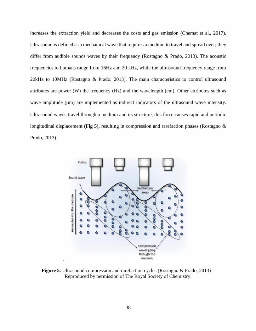
Ultrasound waves travel through a medium and its structure, this force causes rapid and periodic
longitudinal displacement (Fig 5), resulting in compression and rarefaction phases (Rostagno &
Prado, 2013).
.
Figure 5. Ultrasound compression and rarefaction cycles (Rostagno & Prado, 2013) –
Reproduced by permission of The Royal Society of Chemistry.

39
Rarefaction exerts a negative force that pulls or expands the molecules of the medium
rapidly, and this results in the development of bubbles. The cavitation bubbles surround the solid
material submerged in the medium, at that moment they are compressed, they collapse and form
micro jets the help liberate specific compounds in plants as shown in (Fig 6).
Figure 6. Compounds liberated by microjet in ultrasound (Rostagno & Prado, 2013) –
Reproduced by permission of The Royal Society of Chemistry.
The way the cavitation bubbles form depends on the ultrasound parameters such as power,
frequency, amplitude or wavelength. Knowing this, the parameters can be manipulated to obtain
optimum conditions for different plants and compounds. Ultrasound has been generally used to
address the extraction of different plant compounds such as flaxseed oil, wheat bran phenolic
compounds and rutin from euonymus alatus (Esclapez, García-Pérez, Mulet, & Cárcel, 2011).
Although ultrasound has been mainly used for phenolic compounds other authors have suggest it
for polymers from plant sources. Bernardo, Ascheri, Chávez, & Carvalho, (2018) assisted the
separation of starch from Yam using a power input of 70% for a period of 15 min.

40
2.6 Functional Properties of Proteins
The texture, appearance, and flavor are only some of the attributes consumers consider
whenever they decide if they like or not food. These characteristics are limited to the food matrix
composition. Depending on the food component different attributes might be enhanced. Some
components have to be exposed through particular conditions to achieve optimal results (e.g.,
starch must be heated to undergo gelatinization, high methoxyl pectins require low pH and the
presence of sugar to form a gel). Specific ingredients contribute to generate desirable
characteristics in a product. Some of the main functional properties are solubility, emulsification,
gelatinization, water holding capacity, and foaming capacity.
2.6.1 Solubility
The amount of nitrogen detected determines the solubility of a protein under certain
conditions, depending on the solvent, solvent concentration, pH, and charged ions (Sikorski,
1997). Some of the protein classifications rely on the solution in which they are soluble (eg.,
albumins, globulins, prolamins and glutelins). Denaturation may decrease the solubility of a
protein due to the changes in the amino acid arrangement and charged ions exposed in the ends of
the peptide chain (Sikorski, 1997). Solubility can be a handy way to determine if an ingredient will
maintain a constant suspension in a drink or beverage avoiding precipitation, deterioration or
compromising other attributes of the food or fabrication process. Protein solubility depends on the
arrangement of the amino acids in the peptide chain, some of the amino acids are very soluble in
water (e.g., proline glycine and alanine) while others don't solubilize that efficiently (e.g., cysteine
and tyrosine) (Cheung & Mehta, 2014). The individual amino acids are not soluble neither in
organic or inorganic solvents the main reason is because the solubility of a peptide chain depends
directly on the amount of polar and nonpolar structures distributed throughout the chain or
41
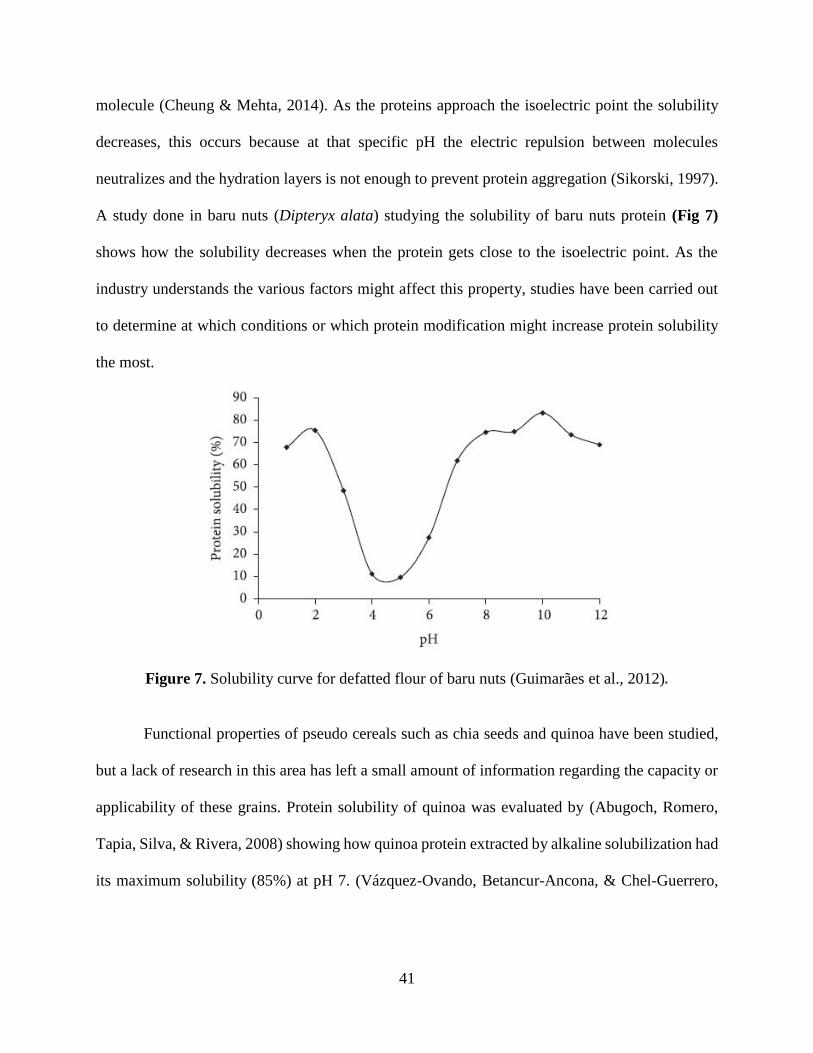
molecule (Cheung & Mehta, 2014). As the proteins approach the isoelectric point the solubility
decreases, this occurs because at that specific pH the electric repulsion between molecules
neutralizes and the hydration layers is not enough to prevent protein aggregation (Sikorski, 1997).
A study done in baru nuts (Dipteryx alata) studying the solubility of baru nuts protein (Fig 7)
shows how the solubility decreases when the protein gets close to the isoelectric point. As the
industry understands the various factors might affect this property, studies have been carried out
to determine at which conditions or which protein modification might increase protein solubility
the most.
Figure 7. Solubility curve for defatted flour of baru nuts (Guimarães et al., 2012).
Functional properties of pseudo cereals such as chia seeds and quinoa have been studied,
but a lack of research in this area has left a small amount of information regarding the capacity or
applicability of these grains. Protein solubility of quinoa was evaluated by (Abugoch, Romero,
Tapia, Silva, & Rivera, 2008) showing how quinoa protein extracted by alkaline solubilization had
its maximum solubility (85%) at pH 7. (Vázquez-Ovando, Betancur-Ancona, & Chel-Guerrero,

42
2013) studied the functional properties of a chia seed protein rich fraction determining it maximum
solubility (60%) was achieved at pH 8.
2.6.2 Emulsion
An emulsion is a way a compound links together two immiscible liquids, having one in a
generous amount. In this way, the two liquids hold together in one matrix and don't separate
anymore. The emulsion is usually done using water and oil, depending on which substance is in
predominant amount, the emulsion can be classified as oil in water (O/W) or water in oil (W/O).
To describe each of the interactions correctly, we will refer as a continuous phase to the liquid in
significantly higher amount and, dispersed phase as the substance that is present in droplets or
small amounts (McClements, 2005). Researchers have determined new possibilities and
alternatives such as the development of an emulsion within an emulsion (O/W/O) or (W/O/W).
This type of emulsion is possible by developing one emulsion first, for example O/W, and then a
second emulsion W/O which allows to obtain a slow release of specific compounds or prevent
some compounds from reacting with each other (Oh, Park, Shin, & Oh, 2004). The emulsions
might be created using high pressure, for example, milk is homogenized using a high-pressure (10-
25 Mpa) process by which the fat globules are entirely dissolved into the aqueous phase and
prevent the formation of a fatty layer on the milk. Rather than pressure, some foods need
emulsifiers that will interact with both liquid phases helping the emulsion to take place and remain
stable for a longer time. There are a variety of emulsifiers (e.g., proteins, polysaccharides,
monoglycerides and diglycerides) which work in different ways, compounds that are more soluble
in water are great agents to develop O/W emulsions and the ones that are more soluble in a fatty
matrix are better for W/O emulsions (McClements, 2005). Proteins usually offer unique
emulsifying characteristics by linking the continuous phase (water) and the disperse phase (oil).

43
They interact with the surface of a fat droplet covering it and allowing this one to be suspended in
water (Fig 8), improving the overall solubility and avoiding the separation of the fluids, if the
protein is denaturalized or deteriorates an emulsion breakdown might occur.
Figure 8. Role of protein in emulsions (Costello, 2017).
An emulsifier will affect the two critical parameters of an emulsion, which are emulsion
capacity and emulsion stability (Cano-Medina et al., 2011). Emulsifying capacity refers to the
ability of an emulsifier to form an even and well distributed oil and water matrix solution and
emulsion stability refers to the time the emulsion is stable and how it disrupts within time. Chia
seed mucilage has been well investigated for emulsion capacity and stability but research regarding
chia seed protein functionality is still missing. Vázquez-Ovando et al., (2013) investigated the
global emulsifying capacity of chia seed flour, which ranged from (50-56%) but not much detail
of chia protein has been recorded. Plant source derived proteins such as quinoa, amaranth,
buckwheat, barley, wheat, foxtail mille, rice, japanese millet and millets were also evaluated

44
showing that pseudocereals contain the highest functional and bioactive properties (Asao &
Watanabe, 2010).
2.6.3 Foaming
Foaming is a functional property of food that depends on the agent, time and air injection
at which a solution is exposed. The foaming agent is evaluated using two criteria, foaming capacity
(FC), which refers to the volume of the foam generated and foaming stability (FS), which is the
change in volume within time. Generally, foams are done by incorporating air cells or bubbles
using a thin continuous liquid layer called the lamellar phase (Zayas, 1997). For the proteins to
have a good foaming ability, they must be highly soluble in water; this will ensure a proper
formation of a cohesive film at the water and air interface, guarantying an appropriate formation
of foam (Cano-Medina et al., 2011). Some studies have evaluated different plant protein sources,
and their ability to form foams. Mao and Hua, (2012) assessed the functional properties of walnut
showing how high protein concentrations increase the foaming capacity of the solution. Vázquez-
Ovando et al., (2013) evaluated chia flour foaming properties showing a maximum foaming
capacity of 28%. Foaming capacity of different commercial soy protein isolates had been improved
by heat treatments (55°C and 85°C) (Shao, Lin, & Kao, 2016). Mune and Sogi, (2016) compared
the functional properties of bambara bean and cowpea protein concentrates derived from two
drying method (vacuum-drying and cabinet-drying), determining that cabinet-drying had the best
foaming capacity.
45
2.7 Biodegradable films: Alternative applications of plant materials.
The development of food products is one of the alternatives, but they are some other
applications of edible plants. The world is changing an extremely high rate and the use of
alternative material to reduce waste had been assessed using plant-derived products due to their
compostable and degradable capabilities. As we know plastic pollution is affecting oceans and
land fields globally due to the buoyancy and low deterioration rate of this compounds (Eriksen et
al., 2014). Ocean tides, winds, and humans spread plastics throughout bays and gulfs leading to
polluted coastlines and affected ocean fauna. The extreme use of petroleum-based products such
as plastics is leading to a high rate of accumulation of these toxic materials in the ocean. The main
affected fauna are marine animals; these can't distinguish plastic waste from their food and ingest
plastic material, which blocks in their digestive tracks, avoiding continuing eating, causing to fall
in a lack of nutrients and dying (Laist, 1987). The development of petroleum-based food packaging
is the primary source of plastic. Due to this problem, alternative ways for food packaging are
considering ingredients that increase the biodegradable characteristics. A vast quantity of
biodegradable plastics alternatives had considered showing the possibility of gradually replacing
plastic with low-cost materials and being less harmful to the environment. Psomiadou,
Arvanitoyannis, and Yamamoto, (1996) developed films made from natural resources using
microcrystalline cellulose, methylcellulose, and cornstarch showing how compounds can be used
together to create certain properties. These types of films are offering an excellent alternative for
food packaging and have proved to provide similar characteristics as petroleum-derived plastics.
Jiménez, Fabra, Talens, & Chiralt, (2012), used carbohydrates such as starch to make edible films,
showing to be an excellent mechanical barrier to hold and protect food products. Some other
alternatives for biodegradable films with less permeable characteristics are achieved using

46
Polylactic acid (PLA). The incorporation of PLA showed a general increase in mechanical
properties such as elongation, tensile strength. And also, a decrease in the water absorption, which
improved the poor mechanical properties and high permeable characteristic of conventional starch-
based films (Hu, Wang, & Tang, 2013).
2.7.1 Biodegradable films using chia seed mucilage
Using natural products to develop biodegradable films is a common practice in the pursuit of a
new alternative to petroleum-based plastics. Starch, cellulose, chitin, paper, PLA, plant fiber,
epoxidized oils are just some of the natural products used for the fabrication of biodegradable
films. Edible films have been produced using chia seed mucilage and glycerol, showing that at
higher glycerol concentrations the elongation at break (EB), water solubility and mechanical
properties of the film increased (Dick, Costa, Gomaa, Subirade, de Oliveira Rios, et al., 2015). -+
Films developed with glycerol will collapse easily within time and can only be applied to
specific products, limiting its applicability. That is why the use of PLA was considered as an
alternative plasticizer for chia seed mucilage films. PLA is a component that forms part of the
aliphatic polyester derived from lactic acid; this one is generally produced from renewable
resources. PLA is a product of starch fermentation, due to its nature it has exceptional
biodegradable and compostable characteristics (Garlotta, 2001). A study by Hamad, Kaseem,
Yang, Deri, & Ko, (2015) show the novel attributes of PLA, having better mechanical properties
(tensile Young's modulus, tensile strength, and flexural strength) compared to common petroleum-
delivered polymers such as polypropylene (PP) polystyrene (PS) and polyethylene (PE). One of
the unique characteristics of this thermoplastic is its hydrophobic nature, being suitable for the
development of biodegradable copolymer films derived from hydrophilic compounds.
47
2.7.2 Digestion of chia seed mucilage
Chia seed mucilage is a soluble fiber with a highly branched structure, this material
contains a high amount of saccharides that include xylose, glucose, arabinose, galactose,
glucuronic acid and galacturonic acid (Timilsena, Adhikari, Kasapis, & Adhikari, 2016). These
saccharides grant the material a hygroscopic behavior leading to high water holding capacities.
Tamargo, A., Cueva, C., Laguna, L., Moreno-Arribas, M. V., & Muñoz, L. A. (2018) showed that
chia seed mucilage impacts the bacterial groups, affecting predominantly the aerobes and
anaerobes at concentrations of 0.3% of chia seed mucilage. The consumption of chia seed was an
associate with an increase of the short chain fatty acids by increasing the butyric, propionic, and
acetic acid content, which are known to have beneficial effects on the health including the
promotion of gut mucosal health, and increase of apoptosis and cell differentiation of cancer cells
(Tamargo, A., Cueva, C., Laguna, L., Moreno-Arribas, M. V., & Muñoz, L. A. 2018). The main
impact of this polymer to the gut microbiota was due to an increase in viscosity affecting the
different microbial communities. Further research is needed to understand the impact of this
polysaccharide in in vivo models.
2.7.3 Barrier property
The barrier properties are crucial for the understanding of the quality and resistance of a
film, and they will also help to estimate parameters as deterioration and shelf life of the film. The
primary objective of a package or film is to keep its content protected from extrinsic factors. All
plastics films and materials are relatively permeable to molecules such as gases, water vapor, and
liquids, it all depends on the composition of the film which will determine the quality of the film
(Siracusa, Rocculi, Romani, & Rosa, 2008). For this reason, the film barrier characteristics are
evaluated using three parameters, water vapor transmission rate (WVTR), Carbon dioxide
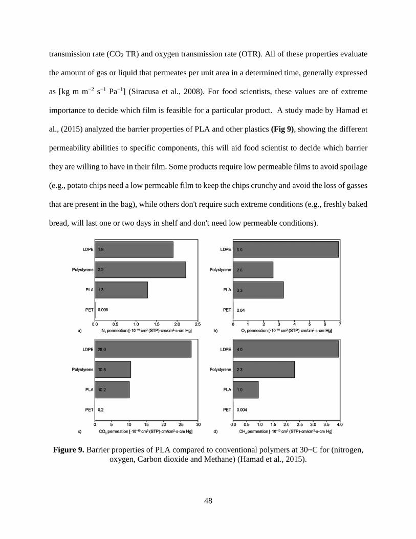
48
transmission rate (CO2 TR) and oxygen transmission rate (OTR). All of these properties evaluate
the amount of gas or liquid that permeates per unit area in a determined time, generally expressed
as [kg m m−2 s−1 Pa−1] (Siracusa et al., 2008). For food scientists, these values are of extreme
importance to decide which film is feasible for a particular product. A study made by Hamad et
al., (2015) analyzed the barrier properties of PLA and other plastics (Fig 9), showing the different
permeability abilities to specific components, this will aid food scientist to decide which barrier
they are willing to have in their film. Some products require low permeable films to avoid spoilage
(e.g., potato chips need a low permeable film to keep the chips crunchy and avoid the loss of gasses
that are present in the bag), while others don't require such extreme conditions (e.g., freshly baked
bread, will last one or two days in shelf and don't need low permeable conditions).
Figure 9. Barrier properties of PLA compared to conventional polymers at 30~C for (nitrogen,
oxygen, Carbon dioxide and Methane) (Hamad et al., 2015).

49
2.7.4 Mechanical properties
The mechanical properties help profoundly in the understanding of the polymer
architecture and conditions that might affect its integrity. To evaluate them, the conditions are
simulated by exposing the films to standard conditions of stress. Tensile test is a general analysis
that recovers information regarding the tensile strength measured in (MPa), the percent of
elongation at yield (%) and the percent elongation at break (%) these values will provide essential
information of the biodegradable polymer study, understanding it maximum stress and mechanical
capacities (Hamad et al., 2015). Other analysis done to films will determine its breaking point and
the resistance to pressure exposure which is generally measured as impact and compression
properties (Hamad et al., 2015).

50
CHAPTER 3. PHYSICOCHEMICAL CHARACTERISTICS OF
CHIA SEED (SALVIA HISPANICA) PROTEIN HYDROLYSATES
PRODUCED USING ULTRASONICATION FOLLOWED BY
MICROWAVE-ASSISTED HYDROLYSIS
Uriel Urbizo-Reyesa, M. Fernanda San Martin-Gonzáleza, Jose Garcia-Bravob, Aurelio López
Maloc, Andrea M. Liceagaa* aDepartment of Food Science, Purdue University. 745 Agriculture Mall Drive, West Lafayette, IN
47907, USA.bSchool of Engineering Technology, Purdue University. 401 Grant St 150, West
Lafayette, IN 47907, USA. cDepartamento de Ingeniería Química y Alimentos, Universidad de Las Américas Puebla, Ex-
Hacienda Santa Catarina Mártir S/N, 72810 San Andrés Cholula, Pue., México.
Reprinted with permission from Uriel Urbizo Reyes, M. Fernanda San Martin-González, Jose
Garcia Bravo, Aurelio López Malo, Andrea M. Liceaga. (2019). Physicochemical characteristics
of chia seed (Salvia hispanica) protein hydrolysates produced using ultrasonication followed by
microwave-assisted hydrolysis. Food Hydrocolloids. 97, 105187.
https://doi.org/10.1016/j.foodhyd.2019.105187
Abstract
Chia (Salvia hispanica) has gained popularity due to its high nutritional content.
Unfortunately, mucilage surrounding the chia seed (CS) limits the technological utilization of the
protein. This study evaluated the bioactivity and functionality of CS protein hydrolysates (CSPH)
produced by different treatments and a control (unhydrolyzed CS protein). Ultrasonication was
used to separate mucilage from CS (7.8% yield). Proteins in defatted-CS flour were enzymatically
hydrolyzed using conventional (enzymatic hydrolysis with alcalase) or sequential (enzymatic
hydrolysis with alcalase+flavourzyme), and under water bath or microwave-assisted hydrolysis.
CSPH from the sequential hydrolysis with microwave treatment showed superior (p<0.05) in vitro
antioxidant activity. A positive correlation (p<0.05) was established between antioxidant assays
and cellular antioxidant activity. The highest (p<0.05) cellular antioxidant activity was achieved
by the sequential (94.76 ± 1.96) and conventional (93.13 ± 1.07) hydrolysis with microwave.
Dipeptidyl peptidase-V inhibition (p<0.05) was higher for sequential hydrolysis with water bath.
Inhibition of angiotensin converting enzyme activity increased (p<0.05) with hydrolysis for all

51
treatments compared to the control. Regarding functionality, sequential hydrolysis with
microwave showed higher (p<0.05) solubility at lower pH (3 and 5), while conventional hydrolysis
with microwave was better at pH 7 and 9. Emulsification properties and foaming capacity were
also higher in conventional hydrolysis with microwave, but conventional hydrolysis with water
bath was more stable for foaming properties only. Results show that ultrasonication efficiently
separated mucilage from chia seeds. Microwave and enzymatic hydrolysis can generate protein
hydrolysates with improved bioactivity and functionality.
Keywords: Chia seed hydrolysates, ultrasonication, microwave-energy, bioactive peptides.
3.1 Introduction
Throughout the years, plants have been domesticated and cultivated to obtain novel
potential ingredients and alternative sources of functional foods. One remarkable plant that has
shown high potential is chia (Salvia hispanica). Chia is a biannually cultivated plant; it is
considered a pseudo-cereal that produces purple and white flowers that eventually result in small
oval shape seeds with sizes varying from 1 to 2 mm (Mohd Ali et al., 2012). The seeds are divided
into two semi-hemispherical structures, which contain the endosperm, called cotyledons.
Nutritional composition (wet basis) of chia seed consists of protein (15-25%), lipids (30-
33%), carbohydrates (26-41%), dietary fiber (18-30%) and minerals (4-5%) (Segura-Campos,
Ciau-Solís, N., Rosado-Rubio, G., Chel-Guerrero, L., & Betancur-Ancona, D. , 2014). Its
composition allows the seed to have remarkable attributes such as a high content of protein,
unsaturated (ω-3) fatty acids, and dietary fiber (Segura-Campos, Ciau-Solís, N., Rosado-Rubio,
G., Chel-Guerrero, L., & Betancur-Ancona, D. , 2014). The high dietary fiber content of chia seeds
can be observed when the seeds are soaked in water and a copious mucilaginous polysaccharide
coating forms around the seed. This polysaccharide is present in microstructures called
52
collumnellas that surround the chia seed and allow the formation of this gel-like material that limits
its digestibility and utilization (Muñoz et al., 2012). The implementation of technologies such as
ultrasound processing can aid in the separation of this polysaccharide, while allowing for protein
extraction (Vilkhu et al., 2008). Ultrasound methods have shown high extraction yields in a shorter
amount of time, for the separation of the polysaccharide from other plant matrices such as lingzhi
mushrooms (Ganoderma lucidum) and mutamba seeds (Guazuma ulmifolia Lam) (Kang et al.,
2019; Pereira et al., 2019).
A rising level of chronic diseases throughout the years have led to the development of food-
derived bioactive peptides that can help improve these medical conditions. Some of the biological
effects produced by these peptides are antioxidant, anti-inflammatory, anti-thrombotic, anti-
hypertensive and anti-diabetic. Some proteins, including chia seed proteins, exhibit high resistance
to proteolysis, limiting their applicability to generate bioactive peptides. For this reason, different
hydrolysis treatments such as high-voltage, electrical (Mikhaylin et al., 2017) and microwave
(Nguyen et al., 2017) treatments have been proposed to increase the protein’s susceptibility to
hydrolysis.
Studies have shown the applicability and attributes of chia seeds. For example, one study
showed how plasma α-linolenic acid and eicosapentaenoic acid increased by 58% and 39%,
respectively when chia seed was supplemented (25 g/day) in the diet of overweight women
(Nieman et al., 2012). Another study by da Silva Marineli, (2015) induced rats to overweight and
oxidative stress before evaluating the effect of a diet rich in chia seeds on these conditions, showing
how plasma and hepatic antioxidant capacity values increased. Sandoval-Oliveros & Paredes-
López, (2013) successfully incorporated chia seed into drinks to enhance the protein content. The
aim of this study was to improve the biological and functional properties of chia seed protein
53
hydrolysates by using ultrasonication to remove the mucilage and microwave-assisted enzymatic
hydrolysis to generate bioactive and functional chia seed peptides.
3.2 Materials and methods
3.2.1 Materials
Chia seeds were obtained from Healthworks® (Scottsdale, AZ, USA). Alcalase® (protease
from Bacillus licheniformis, EC 3.4.21.62) and Flavourzyme® (protease from Aspergillus oryzae,
EC 232-752-2) were purchased from Sigma Aldrich (St. Luis, MO, USA). Human Dipeptidyl
Peptidase IV (DPP-IV, ≥ 4,500 units/ µg protein) and substrate Gly-Pro p-nitroanilide
hydrochloride, Angiotensin Converting Enzyme (ACE) from human and substrate Hippuryl-L-
Histidyl-L-Leucine (HHL) were all purchased from Sigma Aldrich (St. Louis, MO, USA). All
chemicals used were reagent grade and generally obtained by three leading companies VWR
International (Radnor, PA, USA), Sigma Aldrich (St. Louis, MO, USA) and Thermo Fisher
Scientific (Waltham, MA, USA).
3.2.2 Chia seed mucilage extraction
To extract the CS mucilage (Fig 10), seeds were hydrated in distilled water (1:20 ratio by weight)
for 24 hours, under refrigerated conditions. Preliminary studies helped develop an ultrasound
treatment that offered successful mucilage separation. Hydrated seeds were pre-heated to 55±2°C,
followed by sonication at a 75% power input using an ultrasonic cell disruptor (Sonifier® Branson
S-150D Danbury, CT, USA). During sonication, the temperature increased to 60±4°C due to
molecular friction. This temperature was maintained constant using double walled beaker
connected to an immersion circulator control Lauda E100 water bath (Lauda-Königshofen,
Germany). Seed were separated using vacuum-assisted filtration. Mucilage-free CS were dried

54
using a tray dryer (Excalibur Dehydrator 3926TCDB, Sacramento, CA) held at 40°C for 12 h. The
weight of the seeds was measured to calculate mucilage extraction yield by weight difference
[Eq.1]. Ultrasound mucilage extraction was compared to conventional extraction methods using
drying oven and freeze-drying techniques following the methodology proposed by (Campos et al.,
2016) and (Capitani et al., 2013), respectively.
% yield = (Weight of dry chia seeds−Weight of dry chia seeds without mucilage)
Weight of dry chia seeds × 100 [Eq. 1]
Figure 10. Diagram of mucilage separation (extraction) from chia seeds using ultrasonication
and vacuum-assisted filtration.
3.2.3 Chia seed oil extraction
Dried, mucilage-free CS were defatted using a mechanical oil extraction method with a
Beamnova Automatic Oil Press Machine (Commercial 304 Stainless Steel Expeller, Guangzhou,
China). Seeds were pressed using a stainless-steel endless screw held at 37+2°C. The defatted chia
seed was referred to as chia flour. Percentage of oil extraction was calculated by weight difference
[Eq.2].
55
yield% = 100 ×(Weight of chia seeds−Weight of defatted chia seed flour)
Weight of chia seeds [Eq. 2]
In addition, the crude protein content of the chia flour was determined using AOAC methods
984.13 (A-D) by A&L Greatlakes laboratories Facility (Fort Wayne, Indiana, USA).
3.2.4 Chia seed protein hydrolysate (CSPH)
Chia flour was diluted in distilled water to obtain 22.5 mg protein/mL and homogenized
using a Sorvall Omni Mixer homogenizer with a macro-attachment assembly (Norwalk, CT,
U.S.A). The pH was adjusted to 8.0 using 2 M NaOH, which is the optimal condition for alcalase
activity. Proteins were enzymatically hydrolyzed using single enzyme alcalase (A) or a sequential
digestion with alcalase+flavourzyme (AF). Proteolysis occurred using conventional (WB) or
microwave-assisted (MW) hydrolysis using a microwave accelerated reaction system (MDS,
MARS‐Xpress/230/60, CEM Corporation, USA). Treatments were denoted as conventional
alcalase hydrolysis using a water bath (A-WB) and alcalase microwave-assisted hydrolysis (A-
MW). Sequentially (AF) hydrolyzed treatments were coded as AF-WB (water bath hydrolysis)
and AF-MW (microwave-assisted hydrolysis). Finally, the control (C) was non-hydrolyzed CS
protein. Samples A-WB and A-MW were hydrolyzed for 1 hour with 3% (w/w) Alcalase®. For
sequential hydrolysis different times were used, due to the high efficiency of microwave-assisted
hydrolysis the time was cut down by half to obtain similar degree of hydrolysis. AF-MW, the
reaction was initiated with 2% (w/w) of Alcalase® for 45 min followed by addition of 2% (w/w)
of Flavourzyme® for an additional 45 min. For AF-WB, the reaction was developed using 2%
(w/w) of Alcalase® for 90 min followed by 2% (w/w) of Flavourzyme® for another 90 min.
Hydrolysis was terminated by heating to 95±3°C for 15 min. The hydrolysates were cooled to 4 °C

56
and centrifuged at 17,636g for 15 min (Avanti J-26S Centrifuge, Beckman-Coulter INC. CA,
USA).
3.2.5 Proximal Composition
The moisture determination was developed by the gravitation method proposed by (ASAE, 2002)
for rapeseeds using an air convection oven. 10 grams of sample were dried for 4 hours at a
temperature of 130 ± 1 ° C. The analysis (moisture, ash, lipid, and protein) of the chia seeds was
done following the AOAC methods 920.153, 960.39, and 984.13 (A-D) respectively (AOAC,
2016).
3.2.6 Determination of the degree of hydrolysis
The degree of hydrolysis was calculated following the methodology of Adler-Nissen,
(1986) with slight modifications by Liceaga‐Gesualdo and Li‐Chan, (1999), measuring
spectrophotometrically the color formed by free amino groups reacting with
Trinitrobenzenesulforonic acid (TNBS). Aliquots (1000 µL) of the sample were added to 1000 µL
of 24% TCA solution. The samples were stirred for 1 min and then centrifuged at 21279 g for
5min. Then, Aliquots (1000 µL) of supernatant were added were mixed with 1000 µL of 2.0 M
sodium borate buffer (pH 9.2) and 500 µL of 1.0 mM TNBS. The samples were incubated at room
temperature for 30 min. The reaction was ended using 500 µL of 2M monosodium phosphate with
18mM sodium sulfide. The absorbance was measured at 420nm using polystyrene cuvette in a
Beckmann UV-Visible spectrophotometer (Irvine, CA, USA). The degree of hydrolysis (% DH)
was defined as a percent ratio of the number of peptide bonds broken (h) to the total number of
peptide bonds per unit weight (htot). The htot was calculated experimentally using the fully

57
hydrolyzed chia seed flour, obtaining a value of 9.33 meq/g. The % DH was calculated using
equation [Eq.3].
% 𝐷𝑒𝑔𝑟𝑒𝑒 𝑜𝑓 ℎ𝑦𝑑𝑟𝑜𝑙𝑦𝑠𝑖𝑠 (𝐷𝐻) = ℎ
ℎ𝑡𝑜𝑡 × 100 [Eq. 3]
3.2.6.1 Amino Acid Analysis
Total amino acid composition of CSPH was analyzed by the method described by Hall,
Jones, O'Haire, & Liceaga, (2017) by UPLC Amino Acid Analysis Solution using the AccQ Tag
Ultra Derivatization kit with UV detection (Water Corporations, Milford, MA, USA) by the
Danforth Center's Proteomics and Mass Spectrometry Facility (St. Louis, Missouri, USA).
Samples were pre-oxidized using performic acid and subject to acid hydrolysis with 0.5% phenol/6
M HCl in a vapor-phase hydrolysis vessel under vacuum. The acid hydrolysis will destroy cysteine
(Cys), Methionine (Met) and Tryptophan (Trp). The pre-oxidation step using performic acid before
the standard acid hydrolysis will yield stable formed of Cysteic acid (Cya) and methionine
sulfoxide (MetS), which can, therefore, be measured. The quantity (moles) of amino acids in each
peptide fraction was calculated using a series of standard solution runs before the samples. The
relative abundance of amino acids (mole %) will be calculated by dividing the quantity (moles) of
each amino acid by the sum of the concentration of all amino acids and multiplied by 100 and
expressed as mole %.
3.2.6.2 Sodium Dodecyl Sulfate Polyacrylamide Gel Electrophoresis (SDS-PAGE)
CSPH and control samples were dissolved to 2 mg/mL in zwitterionic-chaotrophic buffer
according to Hall, Johnson, and Liceaga, (2018) (2D-gel extraction buffer; 50 mM Tris-HCl, pH
8.8, 10 mM ethylenediaminetetraacetic acid (EDTA), 5 M urea, 2 M thiourea, 67 mM
Dithiothreitol (DTT) and stirred for 1 h at room temperature. An aliquot (50 µL) was added to 50
58
µL Laemmeli sample buffer to obtain a 1 mg/mL final concentration of protein. A sample (20 µL)
of this solution was loaded using 4–12% gradient gels (Bis-Tris, NuPAGE, ThermoScientific,
Waltham, MA) and ran with MES SDS running buffer (NuPAGE, ThermoScientific, Waltham,
MA) at 200 V for 45 min. The gel was stained overnight using Coomassie R-250 and destained
overnight using a solution of 40% (v/v) methanol and glacial acetic acid. The molecular weight
distribution of hydrolyzed peptides was determined using Precision plus ProteinTM Dual Xtra
Prestained Protein Standards (Biorad, Hercules, CA).
3.2.7 Bioactive properties of CSPH
3.2.7.1 2-diphenyl-2-picrylhydrazyl (DPPH) radical scavenging activity
The scavenging activity of the CSPH was determined according to a method described by
Bersuder, Hole, and Smith, (1998) with modifications by Hall et al., (2018). CSPH and control
(100 μL) were placed in a 96-well microplate to which 100 μL of (99.5%) ethanol and 25 μL of
DPPH solution at a concentration of 0.05% (DPPH/ethanol, w/v) was added. The solution was
incubated for 30 min at room temperature in dark conditions, and the absorbance measured at 550
nm using a microplate photometer. Radical reduction was expressed in mM TE/mg sample.
Absorbance values were corrected using a sample blank prepared using 25 µL ethanol instead of
DPPH solution.
3.2.7.2 2,2'-azino-bis (3-ethylbenzothiazoline-6-sulphonic acid) radical scavenging activity
(ABTS)
The ABTS radical scavenging activity was determined according to Ketnawa and Liceaga,
(2017) with some modifications. A solution of 7 mM of ABTS was prepared in 2.45 mM of
potassium persulphate and incubated at room temperature for 16 h. After 16 h the ABTS stock

59
solution was diluted with distilled water to obtain an absorbance at 734 nm of 0.700 ±0.02. A
CSPH sample aliquot (20 μL) was mixed with 980 μL ABTS solution and incubated in the
darkness at 30°C for 10 min, followed by absorbance reading at 734 nm. The ABTS scavenging
activity was calculated by equation [Eq.4] and results expressed as mM Trolox equivalent (TE)/mg
sample.
𝐴𝐵𝑇𝑆 𝑠𝑐𝑎𝑣𝑒𝑛𝑔𝑖𝑛𝑔 𝑎𝑐𝑡𝑖𝑣𝑖𝑡𝑦 =(𝐴𝑏𝑠 𝑜𝑓 𝑐𝑜𝑛𝑡𝑟𝑜𝑙 − 𝐴𝑏𝑠 𝑜𝑓 𝑡ℎ𝑒 𝑠𝑎𝑚𝑝𝑙𝑒)
(𝐴𝑏𝑠𝑜𝑟𝑏𝑎𝑛𝑐𝑒 𝑜𝑓 𝑐𝑜𝑛𝑡𝑟𝑜𝑙)× 100 [Eq. 4]
3.2.7.3 Metal Ion Chelating (MIC)
The MIC capacity was done following the procedure by Ketnawa and Liceaga, (2017) with
modifications. In a 96-well microplate, 200 μL of CSPH samples were mixed with 3.75 μL of 2
mM FeCl2 and 7.5 μL of 5 mM Ferrozine solution. Sample were incubated in the dark for 10 min
at room temperature, and the absorbance was read at 522 nm. The MIC capacity was Calculated
using the equation [Eq.5].
𝑀𝐼𝐶 𝑎𝑏𝑖𝑙𝑖𝑡𝑦 = [(𝐴𝑏𝑠 𝑐𝑜𝑛𝑡𝑟𝑜𝑙 − 𝐴𝑏𝑠 𝑠𝑎𝑚𝑝𝑙𝑒)
(𝐴𝑏𝑠 𝑐𝑜𝑛𝑡𝑟𝑜𝑙) ] × 100 [Eq. 5]
3.2.7.4 Ferric Reducing Antioxidant Power (FRAP)
The FRAP was done according to Girgih, Udenigwe, and Aluko, (2011) methodology with
slight modifications by Ketnawa et al., (2018). The 300 μL of protein samples were entirely mixed
with 300 μL of 1% potassium ferricyanide (0.2mMolar of phosphate buffer pH 6.6), after this the
samples were incubated at 50°C for a period of 20 minutes, 1.2 ml of 10% TCA were added and
then mixed homogeneously then centrifuged at 10,000 x g for 5 min. A 100 μL aliquot of the
mixture was pipet in a 96-well microplate with 100 μL of dd-water and finally, 20 μL of 0.1%

60
ferric chloride was added, the mixture was incubated at room temperature for 10 min, and the
absorbance was measured at 700nm. Results are expressed in mM TE/mg sample.
3.2.7.5 Oxygen radical absorbance capacity (ORAC)
ORAC was measured according to a modified methodology described by Ou, Hampsch-
Woodill, and Prior, (2001). CSPH were diluted to a protein concentration of 0.05 mg/mL in a 75
mM sodium phosphate buffer at a pH of 7.4. The experiment was carried out in a 96-well
microplate, each well containing a total volume of 205 µL. 150 µL of Fluorescein (10 nM) was
pre-incubated with 25 µL of CSPH sample solution for 15 min at 37°C in dark conditions. Then
the reaction was initiated by adding 30 µL of 2,2′-azobis(2-amidinopropane) dihydrochloride
(AAPH) and the Fluorescence was measured using a spectrophotometer (Fluoroskan Ascent FL
Microplate Fluorometer and Luminometer, Thermo Fisher Scientific, Massachusetts, United
States) every two minutes for a total time of 90 min using an excitation wavelength of 485 nm and
emission of wavelength 535 nm. The results were expressed in µM of Trolox Equivalent by
measuring the fluorescein area decay through time (AUC). The values were calculated using the
equation [Eq.6].
𝐴𝑈𝐶 = 1 +𝑓2
𝑓0+
𝑓4
𝑓0+
𝑓6
𝑓0+
𝑓8
𝑓0+ ⋯ 𝑓90/𝑓0 [Eq.6]
Where f0 represents the area under the curve at time 0 and fn the absorbance taken every minute.
3.2.7.6 Cellular antioxidant activity (CAA)
The CAA was evaluated following the methodology proposed by Malaypally et al., (2015)
and Wan, Liu, Yu, Sun, and Li, (2015). First, CSPH and control were solubilized in Dulbecco's
modified Eagle's medium (DMEM)/high modified phenol red free. Caco-2 cells (100 µL, density
of 7.6 x 10⁵ cells/mL) were placed in a 96-well black microplate and incubated for a period of 36

61
h under 5% CO2 at 37°C. After this, the growth medium was removed using needles, washed using
1x PBS (100 µL), and exposed for 1 h to 100 µL of DMEM (60 µM Dichloro-dihydro-fluorescein
diacetate DCFH-DA). CSPH was then added to a final concentration of 5 mg/mL. The solution
was removed from each well, followed by a final washed-out with 1x PBS. The cells were exposed
to an oxidizing environment by pipetting 100 µL of 500 µM AAPH into each well. Emission by
the samples was measured for every 5 min for 1 h using a fluoresce reader Spectra Max Gemini
EM spectrofluorometer (Molecular Devices, Sunnyvale, CA) with an excitation wavelength of 485
nm and an emission wavelength of 538 nm. A sample blank, positive and negative control were
required to calculate the cellular antioxidant activity. Sample blanks contained DMEM and
(DCFH-DA) without AAPH, negative control wells were prepared incubating cells with DCFH-
DA and AAPH and the positive control wells were incubated with cells treated with L-ascorbic
acid (50 µM), DCFH-DA and AAPH. Finally, the cellular antioxidant activity was measured with
equation [Eq.7], were the blank was subtracted from the sample readings. The fluorescence
emission against time data were plotted and used to, calculate the area under the curve in CAA
values (%) (Wolfe & Liu, 2007).
𝐶𝐴𝐴 𝑢𝑛𝑖𝑡 = 100 − (ʃSA
ʃCA) × 100 [Eq.7]
where ʃSA refers to the integral of the sample fluorescence vs. time and ʃCA refers to the integral
from the control sample.
3.2.7.7 Dipeptidyl Peptidase IV (DPP-IV) Inhibitory Activity
The DPP-IV inhibitory activity of CSPH was determined following the method by Hall et
al., (2018). CSPH samples were dissolved in 100 mM Tris-HCl buffer (pH 8.0) to a final
concentration of 1.25 mg/mL. Sample aliquots (25 µL) were pipetted and pre-incubated in a 96-
well microplate with 25 µL of substrate Gly-Pro p-nitroanilide hydrochloride (6 mM) at 37°C for

62
10 min. The colorimetric reaction was initiated by adding 50 µL of human DPP-IV (4.5 unit/mL),
followed by incubation at 37°C for 60 min. The reaction was stopped by adding 100 µL of 1 M
sodium acetate buffer (pH 4.0). Absorbance of released p-nitroanilide was measured at 405 nm
using a Multiskan™ FC Microplate Photometer (Waltham, MA, USA). Sample absorbance was
corrected by subtracting blanks in which DPP-IV was replaced with Tris-HCl buffer (100 mM, pH
8.0). The positive control (no inhibitor) used the buffer instead of CSPH sample. For negative
control (no DPP-IV activity), the buffer was used instead of DPP-IV solution. Percent DPP-IV
inhibition was calculated using equation [Eq.8].
𝐷𝑃𝑃 𝐼𝑉 𝑖𝑛ℎ𝑖𝑏𝑖𝑡𝑖𝑜𝑛 % = 1 − [𝐴𝑏𝑠 𝑆𝑎𝑚𝑝𝑙𝑒 − 𝐴𝑏𝑠 𝑆𝑎𝑚𝑝𝑙𝑒 𝑏𝑙𝑎𝑛𝑘
𝐴𝑏𝑠 𝑝𝑜𝑠𝑖𝑡𝑖𝑣𝑒 𝑐𝑜𝑛𝑡𝑟𝑜𝑙 − 𝐴𝑏𝑠 𝑛𝑒𝑔𝑎𝑡𝑖𝑣𝑒 𝑐𝑜𝑛𝑡𝑟𝑜𝑙] [Eq. 8]
3.2.7.8 Angiotensin Converting Enzyme (ACE) Inhibitory Activity
ACE-inhibitory activity was measured according to Hall et al., (2018). CSPH and control
samples were dissolved in sodium phosphate buffer (100 mM, pH 8.3) with NaCl (300 mM).
Aliquots (25 µL) of CSPH solutions were added to 25 µL of the substrate hippuryl-L-histidyl-L-
leucine (HHL) and incubated at 37°C for 4 min. Then, aliquots (80 µl) of human-ACE (50 mU)
were added to initiate the reaction, followed by incubation at 37°C in a water bath with constant
stirring for 2 h. Reaction was terminated by adding 50 µl of 1 M HCl; then the solution was filtered
using a 0.22 µm nylon filter. A control reaction was performed using 25 µl of buffer instead of the
inhibitor (CSPH). Hippuric acid (HA) was quantified using high-pressure liquid chromatography
(HPLC) (Model 600E, Waters Corporation, Milford MA) with a C18 analytical column (YMC
Pack ODS AM 12505-2546WT, YMC America, Inc., Allentown, PA, USA). Percentage inhibition
was calculated using equation [Eq.9]
𝐴𝐶𝐸 𝐼𝑛ℎ𝑖𝑏𝑖𝑡𝑖𝑜𝑛 % = [1 −𝐴𝑖𝑛ℎ𝑖𝑏𝑖𝑡𝑜𝑟
𝐴𝑐𝑜𝑛𝑡𝑟𝑜𝑙 ] × 100 [Eq. 9]

63
where, Ainhibitor and Acontrol represent the relative areas (A) with and without inhibitor of the HA
peaks. The half maximal inhibitory concentration (IC50) determined the potency of the samples
towards ACE inhibitory activity. IC50 was expressed in mg of protein per mL. IC50 calculations
were determined at four different CSPH concentrations (0.05, 0.5, 1, and 5 mg/mL) in triplicate.
3.2.8 Functional properties of CSPH
3.2.8.1 Solubility
Protein solubility was measured following the method described by Chobert, Bertrand-
Harb, and Nicolas, (1988) and modified by Hall et al., (2017). CSPH were diluted to 1 mg/mL in
15 mL buffers with pH 3.0 (0.1 M sodium Acetate), 5.0 (0.1 M sodium Acetate), 7.0 (7.4 mM
Phosphate) and 9.0 (0.1 M Glycine-sodium hydroxide), respectively. The solutions were stirred
for 30 min at room temperature, followed by centrifugation at 12,150 × g (25°C) for 5 min. The
protein content in the supernatant was determined using the bicinchoninic acid (BCA) protein
assay method with bovine serum albumin as standard. Protein solubility was calculated by the ratio
of protein in the supernatant to the protein content in the sample [Eq.10].
𝑆𝑜𝑙𝑢𝑏𝑖𝑙𝑖𝑡𝑦 % =𝑃𝑟𝑜𝑡𝑒𝑖𝑛 𝑐𝑜𝑛𝑡𝑒𝑛𝑡 𝑖𝑛 𝑠𝑢𝑝𝑒𝑟𝑛𝑎𝑡𝑎𝑛𝑡
𝑇𝑜𝑡𝑎𝑙 𝑝𝑟𝑜𝑡𝑒𝑖𝑛 𝑐𝑜𝑛𝑡𝑒𝑛𝑡 𝑖𝑛 𝑠𝑎𝑚𝑝𝑙𝑒× 100 [Eq. 10]
3.2.8.2 Emulsion
Emulsifying activity index (EAI) and Emulsion stability index (ESI) were measured spectro
turbidimetrically following the procedure of Pearce and Kinsella, (1978) and some modifications
by Liceaga‐Gesualdo and Li‐Chan, (1999). CSPH was diluted to 0.5% (w/v) in 0.1 M phosphate
buffer pH 7. A 3 mL aliquot was added to 1 mL of 100% pure canola oil and homogenized at
18,000 rpm for 1 minute, using Sorvall Omni Mixer with micro-attachments assembly (Norwalk,

64
CT, USA). Aliquots of 50 µL were pipetted immediately and diluted 200-fold into tubes containing
0.3% SDS solution. The tubes were inverted six times gently to ensure homogenization. The
turbidity of the mixture was measured at 500 nm using Beckmann UV-Visible spectrophotometer
(Irvine, CA, USA). EAI was calculated using equation [Eq. 11].
𝐸𝐴𝐼 =(2𝑇) × (𝐴) × (𝑑𝑓)
(∅) × (𝑐) × (10000)× 100 [Eq. 11]
Where:
T: turbidity at 500 nm wavelength at time zero.
df: dilution factor (200-fold)
L: light path in meters
ø: volume of the oil phase (0.25)
c: concentration of solids (0.5%) in the aqueous phase
Emulsion Stability Index (ESI) was measured by letting samples rest for 30, 60, and 90 min at
500 nm calculating the change in turbidity within time [Eq. 12].
𝐸𝑆𝐼 % = 100 − [𝐸𝐴𝐼0 − 𝐸𝐴𝐼𝑇
𝐸𝐴𝐼0] × 100 [Eq. 12]
Where EAIo is the emulsion activity index at zero min, and EAIt is the emulsion activity index at
30, 60, or 90 min.
3.2.8.3 Foaming
Foam capacity (FC) and foam stability (FS) were determined using the method proposed by
Waniska and KIinsella, (1979) with modifications of Pacheco-Aguilar et al., (2008). 0.75 grams
of protein were dissolved in 25 mL of dd-water (final pH 6.8) and stirred using a stir bar for 10
min at room temperature. The foam was developed by aerating the mixture exposing to air injection

65
using Sorvall Omni Mixer with a macro-attachment assembly (Norwalk, CT, USA). FC was
expressed as the percentage of increment in volume after aeration [Eq. 13].
𝐹𝐶 % = 100 × 𝑉𝑜𝑙𝑢𝑚𝑒 𝑎𝑓𝑡𝑒𝑟 𝑎𝑒𝑟𝑎𝑡𝑖𝑜𝑛 − 𝑣𝑜𝑙𝑢𝑚𝑒 𝑏𝑒𝑓𝑜𝑟𝑒 𝑎𝑒𝑟𝑎𝑡𝑖𝑜𝑛
𝑉𝑜𝑙𝑢𝑚𝑒 𝑏𝑒𝑓𝑜𝑟𝑒 𝑎𝑒𝑟𝑎𝑡𝑖𝑜𝑛 [Eq. 13]
FS was expressed as the percentage of foam remaining after 30, 60 and 90 min after the foam
formation.
3.2.9 Statistical Analysis
Results were reported as mean + standard deviation (SD) of triplicate determinations. A complete
randomized design was used as a statistical model with a Duncan separation of means p<0.05. The
correlation analysis between antioxidant assays and cellular antioxidant activity was calculated
using a p<0.05. The statistical analysis was carried out using the statistical software SAS 9.4 (Cary,
NC, USA). Experiments were done in triplicate.
3.3 Results and discussion
3.3.1 Mucilage ultrasound separation
Ultrasound treatment showed to be a novel approach for the separation of mucilage from
chia seeds. In this study the combination of vacuum-assisted filtration and high temperatures
increased mucilage separation (p < 0.05) the most (Table 3), while other tested methods involving
other physical separation methods (centrifugation) showed inefficient separation (Fig 11, B).
The highest mucilage extraction was achieved at a wave amplitude of 90 µm or a power outage of
75% and a temperature of 55°C, followed by separation using vacuum-assisted filtration. The use
of high temperature decreases the viscosity of CS-mucilage mixture. The reduction in viscosity is
attributed to an increase in molecular motion and hydrogen bonds in the polysaccharide system

66
(Timilsena, Adhikari, Kasapis, & Adhikari, 2015). The low viscosity occasioned by high
temperatures allows the sound frequency to travel in an easier manner compared to the mixtures
where the temperature was lower. The optimized treatment was compared to other conventional
methods of mucilage extraction. The mucilage extraction yield was higher (p < 0.05) when
ultrasound was used (7.65 ± 0.19%), compared to freeze-drying the CS (4.21 ± 0.29%), and oven
drying CS (3.65 ± 0.18%). The yield values obtained for conventional extractions methods in this
study were lower than the ones reported by Campos et al. (2016) and Capitani et al. (2013). Chemat
et al. (2017) showed how ultrasound developed high shear stresses in the proximity between liquid
and solid materials, causing oil to separate from basil leaves, addressing the possibility of this
application in other food matrices such as chia seed mucilage. Ultrasound frequencies generated
microjets in the chia seed surface, targeting structures called columella’s that allowed for the
physical separation of mucilage in a fast and efficient way. We can conclude that the combination
of ultrasound treatment and high temperatures followed by vacuum filtration increased the
extraction yield of mucilage from chia seeds.
Figure 11. Clean chia seeds after mucilage extraction using ultrasound and vacuum-filtration
separation (A); chia seeds with residual mucilage using ultrasound and centrifugation (B).

67
Table 3. Mucilage extraction from chia seeds.
Temperature
° C Separation method
Yield of mucilage extraction
(%)
55 Vacuum-filtration 7.66 ± 0.23a
55 Centrifugation 6.23 ± 0.56b
40 Vacuum-filtration 4.72 ± 0.17c
40 Centrifugation 3.66 ± 0.17d
25 Vacuum-filtration 3.66 ± 0.17d
25 Centrifugation 3.93 ± 0.17d
3.3.2 Chia seed oil extraction
The oil extraction yield obtained by cold press was 28.24 ± 0.06 g of oil/100 g of chia seeds
(without mucilage). The oil content in chia seeds varies between 30-33% (Sandoval-Oliveros &
Paredes-López, 2013). Cold pressing of chia seeds without mucilage allows to remove about 95.81
± 0.12 % of the total fat of the seed. Conversely, CS with mucilage lead to an extraction of only
62.90 ± 0.77 %. This is mainly attributed to the high oil holding capacity of chia seed mucilage
(Darwish, Khalifa, & El Sohaimy, 2018). The use of screw-press for oil extraction is a good
alternative to reduce the use of hazardous solvents such as isopropanol and hexane in the
extraction. Also, removal of oil from seeds (in preliminary experiments) showed to increase the
enzyme-protein interaction, facilitating the hydrolysis.
3.3.3 Enzymatic hydrolysis
Table 5 shows the degree of hydrolysis obtained from the different treatments. The highest
degree of hydrolysis (46.81 ± 0.19 and 40.68 ± 0.77 %) was obtained by sequential hydrolysis with
alcalase and flavourzyme (AF-WB) and (AF-MW), respectively. CS protein is difficult to
hydrolyze due to its high content of globulin fractions, which contain several sulfur amino acids
directly involved in maintaining the tertiary and quaternary structure of the protein (Sandoval-
Oliveros & Paredes-López, 2013). This conformation limits the access of the enzyme to cleavage
68
sites that are located within the protein fraction. This was confirmed by the amino acid analysis
were the content of sulfur-containing amino acids (Cys+Met) (Table 6) make up to 7% of the total
amino acid composition of chia seed flour (control); a similar value of 6% was reported by
(Sandoval-Oliveros & Paredes-López, 2013). The higher hydrolysis in the AF-MW treatment can
be attributed to the efficient separation of mucilage and the use of microwave energy. Singh,
Orsat, and Raghavan, (2013) evaluated the effect of electromagnetic fields in protein structure and
conformational changes, showing how magnetic forces pull dielectric charges in the protein
backbone causing protein unfolding or re-orientation. The protein unfolding exposes active sites
that allow the proteases to carry out digestion in a fast and efficient way. In addition, separation of
mucilage played an important role in the effectiveness of the hydrolysis. The minimum presence
of this polysaccharide can block the enzyme-substrate interaction by a process called enzyme
immobilization, resulting in low proteolysis. A study conducted by Monroy-Torres et al., (2008)
showed that CS flour has a low protein digestibility (79.28%) which is attributed to the presence
of mucilage. In general, the utilization of an efficient mucilage separation method and microwave
energy decreased the time required to generate higher hydrolysis (DH) and consequently produce
lower molecular weight peptides.
3.3.4 Proximal Analysis
The proximal analysis is shown in (Table 4). The content of protein varies from 40-79%.
A significant increment in protein content (p<0.05) was achieved when protein was enzymatic
hydrolyzed compared to the control, but no significant (p<0.05) difference was found between
enzymatic hydrolysis treatments. These results indicate the ability of enzymatic hydrolysis to
liberate protein and remove non protein bound components. Studies conducted in rice bran protein
have shown that protein hydrolysis is an alternative way to increase the protein extraction from

69
complex matrices using enzymes such as flavourzyme and alcalase, increasing the protein
extraction to 88 and 81% respectively (Fabian & Ju, 2011). No significant difference was found in
ash content for control and hydrolysis treatments. Other studies have shown the ash content of CS
to vary between 4-6% (Coates, 2011). Carrillo et al. (2018), study the proximal composition of
chia seeds and found the ash to be 4.82%. In this study, the ash content range between 3.74 and
7.44%, minor difference might be attributed to the difference in chia seed sources and removal of
other compounds. Ash refers to the inorganic residue that results after a food has been incinerated,
this often includes oxides, sulfates, phosphates, chloride, and silicates (Marshall, 2010). The
carbohydrate content (calculate by weight difference) ranges from 14.47 to 52.47%. this is mainly
attributed to the seed coat still present in the control. Finally, the fat content in this study ranged
between 0.37 to 2.61%, studies have assessed the fat content of chia seeds and identified their
values to range between 30 to 33% (Mohd Ali et al., 2012). The whole seeds have a fat content of
37.5%, therefore the fat extraction by cold press extracted 95.82% of the fat in the seeds.
Additionally, the fat content decreased once enzymatic hydrolyzed, and the fat content increased
as the degree of hydrolysis increases. Is known that enzymatic proteolysis helps to release bound
non protein components (eg., fats, starches, fibers), the increase in fat content for the sequential
treatment AF-MW and AF-WB, is attributed to an increase release of fat bound to protein. Similar
observations were observed when tilapia (Oreochromis niloticus) was enzymatic hydrolyzed with
neutrase and alcalase (Shirahigue et al., 2016).
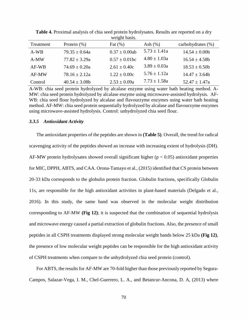
70
Table 4. Proximal analysis of chia seed protein hydrolysates. Results are reported on a dry
weight basis.
Treatment Protein (%) Fat (%) Ash (%) carbohydrates (%)
A-WB 79.35 ± 0.64a 0.37 ± 0.00ab 5.73 ± 1.41a 14.54 ± 0.00b
A-MW 77.82 ± 3.29a 0.57 ± 0.01bc 4.80 ± 1.03a 16.54 ± 4.58b
AF-WB 74.69 ± 0.20a 2.61 ± 0.40c 3.89 ± 0.03a 18.53 ± 0.50b
AF-MW 78.16 ± 2.12a 1.22 ± 0.00c 5.76 ± 1.12a 14.47 ± 3.64b
Control 40.54 ± 3.08b 2.53 ± 0.09a 7.73 ± 1.58a 52.47 ± 1.47a
A-WB: chia seed protein hydrolyzed by alcalase enzyme using water bath heating method. A-
MW: chia seed protein hydrolyzed by alcalase enzyme using microwave-assisted hydrolysis. AF-
WB: chia seed flour hydrolyzed by alcalase and flavourzyme enzymes using water bath heating
method. AF-MW: chia seed protein sequentially hydrolyzed by alcalase and flavourzyme enzymes
using microwave-assisted hydrolysis. Control: unhydrolyzed chia seed flour.
3.3.5 Antioxidant Activity
The antioxidant properties of the peptides are shown in (Table 5). Overall, the trend for radical
scavenging activity of the peptides showed an increase with increasing extent of hydrolysis (DH).
AF-MW protein hydrolysates showed overall significant higher (p < 0.05) antioxidant properties
for MIC, DPPH, ABTS, and CAA. Orona-Tamayo et al., (2015) identified that CS protein between
20-33 kDa corresponds to the globulin protein fraction. Globulin fractions, specifically Globulin
11s, are responsible for the high antioxidant activities in plant-based materials (Delgado et al.,
2016). In this study, the same band was observed in the molecular weight distribution
corresponding to AF-MW (Fig 12); it is suspected that the combination of sequential hydrolysis
and microwave energy caused a partial extraction of globulin fractions. Also, the presence of small
peptides in all CSPH treatments displayed strong molecular weight bands below 25 kDa (Fig 12),
the presence of low molecular weight peptides can be responsible for the high antioxidant activity
of CSPH treatments when compare to the unhydrolyzed chia seed protein (control).
For ABTS, the results for AF-MW are 70-fold higher than those previously reported by Segura-
Campos, Salazar-Vega, I. M., Chel-Guerrero, L. A., and Betancur-Ancona, D. A, (2013) where

71
the protein hydrolysates were produced using sequential hydrolysis and water bath. ABTS and
DPPH assays evaluated antioxidant activity by a specific mechanism called scavenging of free
radicals. Studies have shown that ability is enhanced depending on factors such as amino acid
composition, protein sequences and structural properties (Sarmadi & Ismail, 2010). AF-MW
treatment allowed for the release of a specific group of globulin fractions (G3, G4 and G5), which
compared to unhydrolyzed CS protein, showed that the globulin fraction has a higher concentration
of Phe, Tyr and His (Sandoval-Oliveros & Paredes-López, 2013).
Figure 12. Molecular weight distribution of chia seed protein hydrolysates. Lanes indicate: (1)
molecular weight markers, (2) Control: unhydrolyzed chia seed flour, (3) A-WB: chia seed
protein hydrolyzed by alcalase enzyme using water bath heating method, (4) A-MW: chia seed
protein hydrolyzed by alcalase enzyme using microwave-assisted hydrolysis, (5) AF-WB: chia
seed protein hydrolyzed by alcalase and flavourzyme enzymes using water bath heating method,
(6) AF-MW: chia seed protein sequentially hydrolyzed by alcalase and flavourzyme enzymes
using microwave-assisted hydrolysis.

72
These results lined up with the ones obtained in this study, were the content of aromatic amino
acids (His and Phe) are higher in AF-MW compared to unhydrolyzed CS protein (Table 6). It is
hypothesized that protein rotation and unfolding caused by MW and the release of encrypted
peptides cause by sequential hydrolysis, enhanced the exposure of these aromatic amino acids
responsible of a high donation of protons (Sarmadi & Ismail, 2010). The proton donations from
aromatic amino acids to molecules with electron deficiency, improves the scavenging activity
stabilizing reactive molecules (Sarmadi & Ismail, 2010). In contrast, the metal ion chelating (MIC)
capacity is generally attributed to peptides containing sulfhydryl amino acids such as Cys and Met,
which can bind heavy metals and reduce the pro-oxidant activity of some metals. The content of
Cys and Met amino acids (Table 6) was higher in the control than in other treatments. It is
hypothesized that microwave-assisted hydrolysis released encrypted sulfur peptides, making them
more bioavailable to interact with free metals, therefore enhancing the MIC capacity.
For FRAP, the not hydrolyzed protein (control) showed the highest antioxidant value, the FRAP
mechanism is to quantify the reducing potential of the BPs when it reacts with potassium
ferricyanide and ferric chloride creating the emission of light at 700 nm. Bamdad, Wu, and Chen,
(2011) showed that vast hydrolyzed proteins suffer a decrease of the reducing activity and that
medium size proteins are responsible for ferrous ion chelating activity causing the entrapping of
ferrous ions. He attributes this to the decrease of cage structure caused by the high amount of
peptide bond cleavage.

73
Table 5. Bioactive properties of CSPH.
Sample
code
Hydrolysis
time,
(%
Enzyme)
Degree
of
hydrolys
is (%)
% MIC FRAP
(µmol
TE/mg)
DPPH ABTS ORAC
Inhibition (µmol
TE/mg)
(µmol
TE/mg)
(µmol
TE/mg)
A-WB
1 h 33.64 ±
1.44c
66.93 ±
0.57c
83.45 ±
3.87c
131.74
±
17.33b
465.97
±
10.46c
1225.49
±
55.53bc 3%
A-MW
1 h 37.04 ±
2.67bc
72.76 ±
0.42b
171.03
± 4.92b
166.61
±
15.34a
435.30
±
12.96d
1535.81
± 99.16a 3%
AF-WB
3 h 46.81 ±
0.19a
74.11 ±
0.64b
180.76
± 4.19b
171.31
± 8.27a
489.09
± 3.86b
1482.48
±
114.63ab 2%
AF-MW
1.5 h 40.68 ±
0.77ab
76.85 ±
0.37a
59.93 ±
3.03c
178.02
±
15.86a
506.07
± 4.50a
1122.71
± 24.74c 2%
Control
0 h 0.00 ±
0.00d
54.43 ±
1.03d
410.26
±
29.84a
22.99 ±
1.49c
100.57
± 3.80e
816.33 ±
86.18d 0%
Values are mean + standard deviation of triplicate determinations. Different letters (a-d) indicate
significant differences (p < 0.05) between treatments (rows). A-WB: chia seed protein hydrolyzed
by alcalase enzyme using water bath heating method. A-MW: chia seed protein hydrolyzed by
alcalase enzyme using microwave-assisted hydrolysis. AF-WB: chia seed flour hydrolyzed by
alcalase and flavourzyme enzymes using water bath heating method. AF-MW: chia seed protein
sequentially hydrolyzed by alcalase and flavourzyme enzymes using microwave-assisted
hydrolysis. Control: unhydrolyzed chia seed flour.
ORAC is a common antioxidant assay based on the quantification of the fluoresce emitted by
a probe, in this case a protein called fluorescein. Fluorescein is exposed to an oxidative
environment by AAPH that causes degradation of the protein, and, consequently, a loss in
fluorescence. This assay measures the hydrophilic antioxidant capacity against certain peroxyl
radicals (Aruoma & Mutagenesis, 2003). The highest ORAC values (p<0.05) were seen in the
microwave treatments A-MW and AF-WB at, 1535.81 ± 99.16 and 1482.48 ± 114.63 µmol TE/mg
of protein, respectively. Exposure during hydrolysis of encrypted nonpolar residues such as Gly,
Ala, Ile, Trp, Tyr and Met are speculated, this in turn can cause an increase in hydrophobic
interactions of peptides with oxidizing agents. These results showed a higher ORAC value

74
compared to other digested materials such as cowpea protein (783.8 µmol/g of protein) and salmon
(1541 µmol/g of protein) (Marques et al., 2015).
Table 6. Total Amino acid analysis for chia seed protein and CSPH (g/100 g).
Amino Acid Control A-WB A-MW AF-WB AF-MW
Gly 5.128 4.278 4.465 4.231 4.354
Ala 5.405 5.196 4.994 4.972 4.940
Pro 3.573 3.518 3.533 3.477 3.708
Val 4.358 4.462 4.272 4.563 5.021
Ile 2.544 3.096 3.003 3.114 3.474
Leu 5.328 5.685 5.550 5.532 5.985
Met 4.629 3.604 3.986 3.970 3.759
Phe 3.267 3.219 3.408 3.397 3.850
His 1.417 1.358 1.362 1.332 1.358
Thr 3.964 3.608 3.502 3.520 3.567
Cys-Cys 3.046 1.536 1.843 1.797 1.804
Tyr 0.069 0.013 0.017 0.009 0.007
Glu 29.001 30.857 30.675 30.552 29.181
Arg 7.309 7.484 7.985 8.125 8.316
Ser 4.913 5.447 5.545 5.556 5.551
Asp 10.064 12.158 11.710 11.418 10.808
Lys 5.984 4.479 4.151 4.436 4.317
SCAA 7.675 5.140 5.829 5.767 5.564
AAA 4.754 4.591 4.788 4.738 5.215
PCAA 14.710 13.322 13.498 13.893 13.991
BCAA 12.230 13.243 12.825 13.208 14.480
SCAA= sulfur containing amino acids (Met+Cys). AAA= Aromatic amino acids. (Phe, His and
Tyr). PCAA= positively charged aminoacids (Arg, His and Lys). BCAA=Branched containing
amino acids (Leu, Ile and Val). Sample codes descriptions are provided in Table 5.
The cellular antioxidant activity (CAA) assay indirectly evaluates the permeability of a
compound through the cell bilayer while estimating the antioxidant capacity. In this method, a

75
fluorescent probe DCFH-DA is introduced into the Caco-2 cells, and by the action of esterase is
turned into a more polar form of DCFH which emits fluorescence under physiological conditions
(Wan et al., 2015). In order to protect DCFH from oxidation caused by AAPH solution, the CSPH
peptides must permeate and/or bind to the cell membrane and function a protective barrier against
free radicals, thus preventing molecular damage of intracellular organelles and proteins. If the
peptides have a good permeability and a high enough antioxidant capacity, they will protect the
fluorescent probe from turning into dichloro-dihydro-fluorescein (DCF) and consequently
avoiding a decrease in light emission through time (Wan et al., 2015). The highest CAA value
(Table 8) was observed for the microwave treatments AF-MW and A-MW (94.76 ± 1.96 and
93.13 ± 1.07%, respectively). Wolfe and Liu, (2007) found that the hydrophobicity of the
compounds was an important criterion to determine the antioxidant effectiveness in cell culture.
This is not the only criteria since structural conformation also plays a crucial role in the quality
and effectiveness of the antioxidant activity of these compounds (Wolfe & Liu, 2007). Conradi,
Hilgers, Ho, and Burton (1991) found that the permeability of peptide chains was significantly
correlated with the total number of hydrogen bonds a peptide can possibly form with water since
the major impediment for passive absorption is the energy required to break the peptide-water
hydrogen bonds. It is hypothesized that smaller peptides that have a weaker hydrogen bonding
capacity and a higher concentration of antioxidant amino acids on the microwave samples (A-MW
and AF-MW), are responsible for the increased cellular antioxidant activity observed in this study.
3.3.6 Correlation of antioxidant assays to CAA
A correlation analysis was carried out determining the significance of some antioxidant
assays to predict cellular antioxidant activity. The analysis presented a significant correlation
(p<0.05, r= 0.888) of DPPH with respect to CAA followed by ABTS (Table 7). No significant
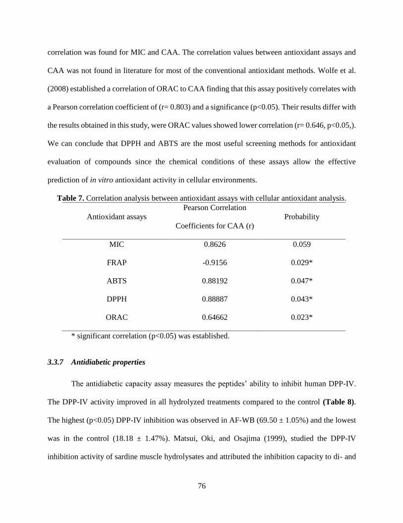
76
correlation was found for MIC and CAA. The correlation values between antioxidant assays and
CAA was not found in literature for most of the conventional antioxidant methods. Wolfe et al.
(2008) established a correlation of ORAC to CAA finding that this assay positively correlates with
a Pearson correlation coefficient of (r= 0.803) and a significance (p<0.05). Their results differ with
the results obtained in this study, were ORAC values showed lower correlation (r= 0.646, p<0.05,).
We can conclude that DPPH and ABTS are the most useful screening methods for antioxidant
evaluation of compounds since the chemical conditions of these assays allow the effective
prediction of in vitro antioxidant activity in cellular environments.
Table 7. Correlation analysis between antioxidant assays with cellular antioxidant analysis.
Antioxidant assays
Pearson Correlation
Coefficients for CAA (r)
Probability
MIC 0.8626 0.059
FRAP -0.9156 0.029*
ABTS 0.88192 0.047*
DPPH 0.88887 0.043*
ORAC 0.64662 0.023*
* significant correlation (p<0.05) was established.
3.3.7 Antidiabetic properties
The antidiabetic capacity assay measures the peptides’ ability to inhibit human DPP-IV.
The DPP-IV activity improved in all hydrolyzed treatments compared to the control (Table 8).
The highest (p<0.05) DPP-IV inhibition was observed in AF-WB (69.50 ± 1.05%) and the lowest
was in the control (18.18 ± 1.47%). Matsui, Oki, and Osajima (1999), studied the DPP-IV
inhibition activity of sardine muscle hydrolysates and attributed the inhibition capacity to di- and

77
tetra-peptides (Val-Trp and Try -Tyr -Pro -Leu) that match the structure of the substrate of the
DPP-IV enzyme. In another study, Nongonierma, Le Maux, Dubrulle, Barre, and FitzGerald
(2015) evaluated the DPP-IV inhibitory activity of the protein hydrolysates of quinoa, a similar
pseudo-cereal. The IC50 value of quinoa protein hydrolysates was of 0.88 ±0.05 mg/mL using a
porcine DPP-IV enzyme (0.0025 U/mL). Velarde-Salcedo et al., (2013) evaluated the DPP-IV
inhibition of amaranth tryptic digests using porcine DPP-IV (0.0025 U/mL) and found IC50 value
ranging between 1.2 to 2 mg/mL. To the best of authors’ knowledge, no previous report of DPP-
IV inhibition activity by CSPH using human DPP-IV (2.25 U/mL) has been reported. In this study
AF-WB CSPH had an IC50 of 1.28 mg/mL, showing the bioactive potential for CSPH, compared
to other pseudo-cereals. Lacroix and Li-Chan, (2015) compared the susceptibility of porcine and
human DPP-IV to inhibition and found that porcine DPP-IV is generally inhibited with greater
efficacy by protein derived peptides than human DPP-IV. Generally, the DPP-IV inhibition is
enhanced with the presence of lower molecular peptides, probably matching the structure of the
incretin hormones (GIP and GLP-1). The incorporation of flavourzyme might have influenced the
development of tetra- and di- peptides that match the DPP-IV active site. A similar result was
obtained for protein hydrolysates derived from Atlantic salmon (salmon salar) skin gelatin, were
the highest DPP-IV inhibition was achieved using flavourzyme (Li-Chan et al., 2012).

78
Table 8. Cellular antioxidant activity, Antidiabetic and antihypertensive properties of CSPH.
Sample
code
Cellular
antioxidant
activity %
DPP-IV inhibition
capacity
ACE inhibition
capacity
%
inhibition
per 2.5 mg
of protein
IC50
(mg/mL)
% inhibition
per mg of
protein
IC50
(mg/mL)
A-WB 87.54 ± 3.31bc 37.05 ±
0.99c 4.38 ± 0.49a 57.16 ± 1.93a 0.51 ± 0.12a
A-MW 93.13 ± 1.07ab 40.56 ±
0.42c 3.59 ± 0.31b 57.17 ± 3.42a 0.44 ± 0.09a
AF-WB 80.06 ± 2.48c 69.50 ±
1.05a 1.28 ± 0.09d 56.24 ± 0.74a 0.42 ± 0.04a
AF-
MW
94.76 ± 1.96a 53.49 ±
0.86b 2.12 ± 0.08c 45.60 ± 2.24b 0.55 ± 0.02a
Control 62.00 ± 9.08d 18.18 ±
1.47d - 11.18 ± 2.31c -
Values are mean + standard deviation of triplicate determinations. Different letters (a-d) indicate
significant differences (p < 0.05) between treatments (rows). A-WB: chia seed protein hydrolyzed
by alcalase enzyme using water bath heating method. A-MW: chia seed protein hydrolyzed by
alcalase enzyme using microwave-assisted hydrolysis. AF-WB: chia seed flour hydrolyzed by
alcalase and flavourzyme enzymes using water bath heating method. AF-MW: chia seed protein
sequentially hydrolyzed by alcalase and flavourzyme enzymes using microwave-assisted
hydrolysis. Control: unhydrolyzed chia seed flour.
3.3.8 Anti-hypertensive activity
In this study, most of the CSPH treatments had similar ACE inhibition capacity except for
AF-MW and the control which were significantly (p<0.05) lower (Table 8). Other studies have
looked at the bioactivity of CSPH incorporated into foods. A study conducted by Segura-Campos,
Salazar-Vega, I. M., Chel-Guerrero, L. A., and Betancur-Ancona, D. A, (2013) showed an
improvement in IC50 value of the ACE-inhibitory capacity of foods by incorporating 5.0 mg/g of
CSPH into carrot cream, causing a decrease from 27.67 μg protein/mL to 1.71 μg protein/ml. In
our study, the IC50 values of CSPH was around 0.40 mg/mL. In the case of the lower inhibitory
activity of AF-MW, it might be related to the formation of different structural peptides with a
lower binding capacity to the ACE active site. Additional analytical studies are needed to

79
understand the difference with peptide conformation in CSPH. It is well known that the increase
in DH and lower molecular-weight peptides improve the capacity of inhibiting ACE enzymes.
Segura-Campos, Salazar-Vega, I. M., Chel-Guerrero, L. A., and Betancur-Ancona, D. A, (2013)
evaluated the ACE inhibitory capacity of CSPH (51.64% DH) and obtained 53.8-69.3% inhibition
for purified fractions. In this study, the CSPH peptides displayed a 57% inhibition with a 46% DH.
Nevertheless, an influence of microwave energy in peptide conformation is not clear for ACE
inhibition. Ketnawa et al., (2018) evaluated the ACE inhibition of microwave-assisted hydrolyzed
peptides of rainbow trout (Oncorhynchus mykiss) and showed that peptides delivered from
microwave-assisted hydrolysis had the highest inhibition at 93.5 ± 0.24%.
3.4 Functional Properties
3.4.1 Solubility
The solubility of CSPH showed a dependency of the pH in all treatments, with solubility
increasing with increasing pH (Fig 13). This pattern is similar to what is reported for many plant
proteins and is related to the low isoelectric point (pI) of CS protein. When the pH of the solution
is above or below the pI of a protein the solubility is enhanced because the electrostatic repulsion
between molecules is greater than the hydrophobic interactions (Zayas, 2012). CS protein
extraction is more efficient at alkaline pH, reaching its maximum solubility at pH 12 (Timilsena,
Adhikari, Barrow, & Adhikari, 2016). A previous study by Timilsena et al., (2016) evaluated the
solubility of CS protein isolate, reporting 10% solubility at pH 3. A similarly low value was
obtained in this study for the solubility of the control (16%) (Fig 13). AF-MW treatment showed
the highest solubility at pH 3 (68.32%) and pH 5 (77.17%) when compared to other treatments
(Fig 13). This can be attributed to the high degree of hydrolysis of this treatment (40.68 %), where
the presence of small peptides increases the exposure of polar and ionizable groups, consequently

80
increasing their solubility (Nguyen et al., 2017). In addition, enzymatic hydrolysis by alcalase and
flavourzyme can increase the number of smaller hydrophilic polypeptides (Zhao et al., 2012). At
neutral and alkaline pH, A-MW treatment showed the highest solubility (p < 0.05), suggesting that
a medium DH (medium size peptides) could improve the solubilization of protein in neutral and
alkaline solvents. The application of microwave-assisted hydrolysis can be correlated with an
increase production of small peptides and consequently with an increased solubility (Uluko et al.,
2013). Overall, hydrolyzing the CS flour improved the solubility of the protein hydrolysates,
compared to the unhydrolyzed control at all pH range values.
Figure 13. Results for Solubility of chia seed protein hydrolysates. Different letters (a-d) show
significant differences (p < 0.05) between treatments. Sample codes descriptions are provided in
Table 5.
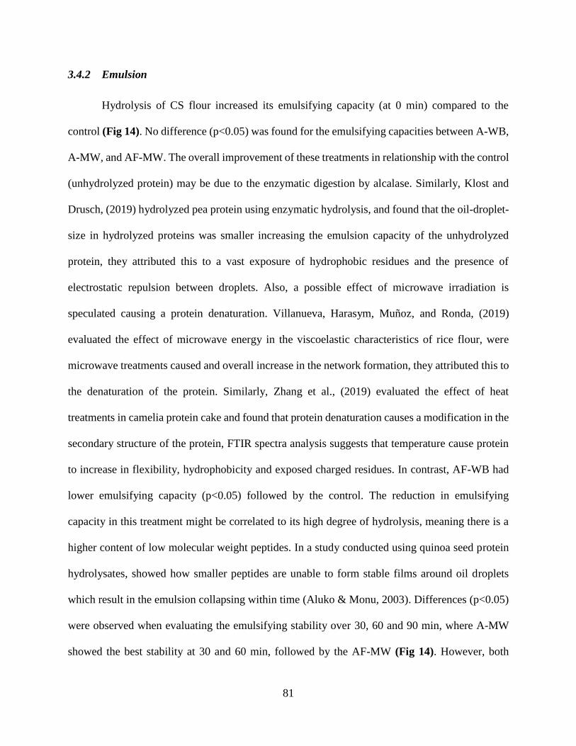
81
3.4.2 Emulsion
Hydrolysis of CS flour increased its emulsifying capacity (at 0 min) compared to the
control (Fig 14). No difference (p<0.05) was found for the emulsifying capacities between A-WB,
A-MW, and AF-MW. The overall improvement of these treatments in relationship with the control
(unhydrolyzed protein) may be due to the enzymatic digestion by alcalase. Similarly, Klost and
Drusch, (2019) hydrolyzed pea protein using enzymatic hydrolysis, and found that the oil-droplet-
size in hydrolyzed proteins was smaller increasing the emulsion capacity of the unhydrolyzed
protein, they attributed this to a vast exposure of hydrophobic residues and the presence of
electrostatic repulsion between droplets. Also, a possible effect of microwave irradiation is
speculated causing a protein denaturation. Villanueva, Harasym, Muñoz, and Ronda, (2019)
evaluated the effect of microwave energy in the viscoelastic characteristics of rice flour, were
microwave treatments caused and overall increase in the network formation, they attributed this to
the denaturation of the protein. Similarly, Zhang et al., (2019) evaluated the effect of heat
treatments in camelia protein cake and found that protein denaturation causes a modification in the
secondary structure of the protein, FTIR spectra analysis suggests that temperature cause protein
to increase in flexibility, hydrophobicity and exposed charged residues. In contrast, AF-WB had
lower emulsifying capacity (p<0.05) followed by the control. The reduction in emulsifying
capacity in this treatment might be correlated to its high degree of hydrolysis, meaning there is a
higher content of low molecular weight peptides. In a study conducted using quinoa seed protein
hydrolysates, showed how smaller peptides are unable to form stable films around oil droplets
which result in the emulsion collapsing within time (Aluko & Monu, 2003). Differences (p<0.05)
were observed when evaluating the emulsifying stability over 30, 60 and 90 min, where A-MW
showed the best stability at 30 and 60 min, followed by the AF-MW (Fig 14). However, both

82
alcalase treatments (A-MW and A-WB) had similar emulsion stability at 90 min. A study
conducted by van der Ven, Gruppen, de Bont, & Voragen, (2001) showed that whey and casein
protein hydrolysates have better emulsifying capacities compared to intact casein and whey
protein. Authors attributed this to a more uniform distribution of emulsion droplets size originated
from partially hydrolyzed peptides compared to intact protein were the droplets are bigger and
collapse faster (van der Ven et al., 2001).
Figure 14. Results for Emulsifying activity of chia seed protein hydrolysates. Different letters
(a-d) show significant differences (p < 0.05) between treatments. Sample codes descriptions are
provided in Table 5.
3.4.3 Foaming
A-MW showed the highest foaming capacity (75%) (at 0 min), followed by A-WB (66.5%) (Fig
15). This result agrees with the highest solubility at pH 7 observed in A-MW, indicating that these
peptides are more evenly distributed through the aqueous interface and consequently improve the

83
way they entrap air molecules. Similarly, Nguyen et al., (2017) developed protein hydrolysates
from rainbow trout frames using microwave-assisted hydrolysis, proving that lower molecular
weight peptides derived by microwave energy diffuse faster into the air-water interface. CSPH
showed a great potential in foaming capacity when compared to other protein sources as whey
protein hydrolysates (FC=4%) and rice protein hydrolysates (FC=6%), nevertheless different rice
sources have shown foaming capacities as high as 105% (Amagliani, O'Regan, Schmitt, Kelly, &
O'Mahony, 2019). Olivos-Lugo, Valdivia-López, & Tecante, (2010) evaluated the foaming
capacity of CS protein isolate and reported 70% foaming capacity. The purification and isolation
of the protein is suspected to be responsible of the high values reported for the CS protein isolate.
Evaluation of foam stability at 30, 60 and 90 min, showed that A-WB resulted in overall higher
foaming stability. The decrease in foaming capacity of A-MW compared to A-WB can be
attributed to the presence of smaller peptides caused by microwave treatment; smaller peptides are
known to have poor stability through time. Nevertheless, A-WB and A-WW have lower DHs
amongst all CSPH (Table 5) meaning that medium size peptides might be present. Balti et al.,
(2010) evaluated the influence of DH on foaming properties of cuttlefish and found that foaming
capacity decreased slightly as the protein hydrolysis increased. They attributed this to the loss of
cohesiveness that is achieved with high molecular-weight peptides and partially hydrolyzed
protein.
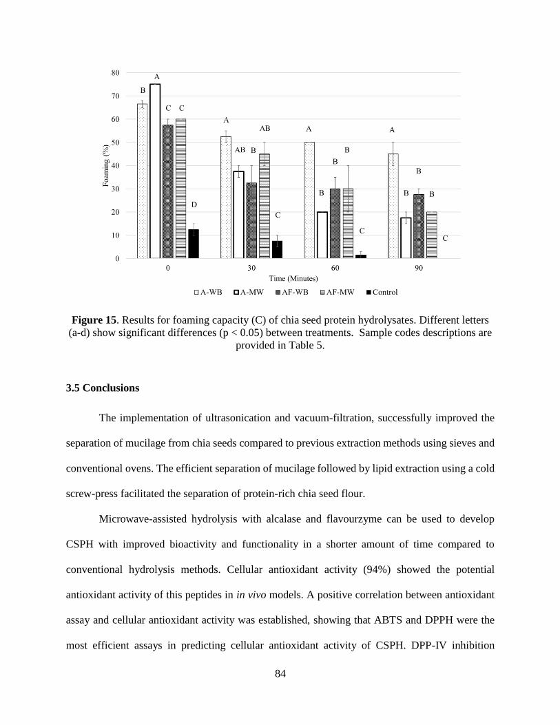
84
Figure 15. Results for foaming capacity (C) of chia seed protein hydrolysates. Different letters
(a-d) show significant differences (p < 0.05) between treatments. Sample codes descriptions are
provided in Table 5.
3.5 Conclusions
The implementation of ultrasonication and vacuum-filtration, successfully improved the
separation of mucilage from chia seeds compared to previous extraction methods using sieves and
conventional ovens. The efficient separation of mucilage followed by lipid extraction using a cold
screw-press facilitated the separation of protein-rich chia seed flour.
Microwave-assisted hydrolysis with alcalase and flavourzyme can be used to develop
CSPH with improved bioactivity and functionality in a shorter amount of time compared to
conventional hydrolysis methods. Cellular antioxidant activity (94%) showed the potential
antioxidant activity of this peptides in in vivo models. A positive correlation between antioxidant
assay and cellular antioxidant activity was established, showing that ABTS and DPPH were the
most efficient assays in predicting cellular antioxidant activity of CSPH. DPP-IV inhibition

85
increased as a function of DH, suggesting that higher hydrolysis will result in improved inhibitions.
Finally, for ACE inhibition no apparent influence was found regarding type of hydrolysis in their
inhibition capacity, suggesting the structural conformation responsible of this bioactivity remains
unchanged independently of the treatment applied. Lastly, further studying is required regarding
the structural conformation of this peptides to fully understand their inhibition mechanism.
It is known that chia seed shows to be a complex food composed of high polysaccharide,
protein, and oil content. Applying different technologies to separate its components is a novel
approach to increase yields, expand applications, and investigate bioactivities. Further research is
required in ultrasonic field to obtain an industrial scale of separation of this polysaccharide. Chia
seed components have a great applicability in food science or in the pharmaceutical sector, as
carrier, materials for encapsulation, thickeners or as a source of bioactive compounds.
86
CHAPTER 4. EFFECT OF POLYOL CONCENTRATION ON
PHYSICAL AND BARRIER CHARACTERISTICS OF EDIBLE FILMS
FROM CHIA SEED (SALVIA HISPANICA) MUCILAGE.
Uriel Urbizo-Reyesa, M. Fernanda San Martin-Gonzáleza, Jose Garcia-Bravob, Andrea M.
Liceagaa*
aDepartment of Food Science, Purdue University. 745 Agriculture Mall Drive, West Lafayette, IN
47907, USA. bSchool of Engineering Technology, Purdue University. 401 Grant St 150, West Lafayette, IN
47907, USA.
Abstract
Food packaging is one of the main contributors to the high rates of environmental
contamination; researchers have pointed at the use of biopolymers as an alternative material to
replace conventional food packaging. Chia seed (Salvia hispanica) is recognized by the
outstanding nutritional profile, and its surface is especially rich in a polysaccharide called
mucilage. The aim of this study was to evaluate the feasibility using of chia seed mucilage (CSM)
and a polyol mixture containing glycerol and sorbitol for the development of edible films. Film
formulations were optimized implementing a factorial arrangement with two replicates of the
central point according to response surface methodology. The independent variables g of glycerol
(X1), and g of sorbitol (X2) per g of CSM and their interaction (X1*X2) showed a significant effect
(p<0.05) in all mechanical and barrier properties. CSM films with higher sorbitol content showed
superior tensile strength (3.23 N/mm2), and lower water vapor permeability (1.3*10-9 g/ m*s*Pa)
but had poor flexibility compared to other treatments. Conversely, films with high glycerol content
showed high elongation at break (67.55%) and solubility (22.75%), but poor water vapor
permeability and tensile strength. The hydrophobicity, measured as water contact angle, was
higher (p<0.05) for mixtures containing equal amounts of polyols. Raman spectra analysis showed

87
shifts from 854 to 872 cm-1 and 1061 to 1076 cm-1, which corresponded to β(CCO) modes, these
shifts represent an increase in hydrogen bonding, responsible for the high tensile strength and
reduced water vapor permeability in this study. The optimum conditions of polyol concentration
were 1.3 g of glycerol and 2.0 g of sorbitol per g of CSM. CSM films showed potential as a drug
delivery, and edible food coating for use in various industry applications.
Keywords: Chia seed mucilage, biofilms, polysaccharide, optimization.
4.1 Introduction
The overuse of petroleum-derived packaging is leading to a high accumulation rate of these
toxic materials in the environment. Globally, since 1964, the use of plastics has expanded 20-fold
increasing to 322 million tons in 2015 (Wei & Zimmermann, 2017). Packaged food products are
the main culprit of single use plastics, due to the utilization of petroleum-based polymers as
primary materials in food packaging. Around 72% of the world’s plastic production is not recycled,
40% ends in landfills, and about 32% is estimated to end up in natural habitats, such as biospheres
and oceans (Andrady, 2015). Buoyancy and slow degradation rate are the reason of the extensive
damage and contamination ability of these plastic compounds (Eriksen et al., 2014). Ocean tides,
winds, and human activity are mechanisms by which plastics spread through bays and gulfs leading
to polluted coastlines and affected ocean fauna. In response to this, the food industry is in search
of alternative materials for food packaging consisting of ingredients that increase the
biodegradable characteristics of the package. In this regard, plant derived materials have shown a
great potential, offering similar structural and barrier functionalities as conventional petroleum-
based materials. Ghanbarzadeh, Almasi, and Entezami (2011) developed biodegradable films
using corn starch and showed that the mechanical properties are improved by increasing
concentrations of citric acid and carboxymethyl cellulose. Oliveira et al. (2019) fabricated films

88
from plant materials using mucilage extracted from leaves of Barbados gooseberry (Pereskia
aculeate) combined with glycerol as a cross-linker. Because of their chemical composition,
mucilaginous compounds offer a great potential alternative to plastics; very high content of
mucilaginous material is present in chia seeds (Salvia hispanica). Chia seeds have gained
popularity due to their remarkable nutritional composition, including high content of protein (15
to 25 %), fat (30-33%) and fiber (41%) (Ullah et al., 2016). Chia seed protein hydrolysates have
also shown bioactive properties such as antioxidant activity, DPP-IV and ACE-inhibition (Urbizo-
Reyes, San Martin-González, Garcia-Bravo, López Malo, & Liceaga, 2019). Around 5-7% of the
whole chia seed weight corresponds to a complex carbohydrate polymer called mucilage (Tavares,
Junqueira, de Oliveira Guimarães, & de Resende, 2018). Chia seed mucilage (CSM) has been used
as a thickening agent in a wide range of applications including the development of gluten free
pasta (Menga et al., 2017), and breads and cakes (Fernandes & de las Mercedes Salas-Mellado,
2017). A study conducted by Muñoz, Aguilera, Rodriguez-Turienzo, Cobos, and Diaz (2012)
showed that CSM and whey proteins can be combined to develop edible or biodegradable films
with improved physical properties. CSM has especially a particular high content of xylans (38%)
(Timilsena, Adhikari, Kasapis, & Adhikari, 2016), which are the mayor contributors to the
crosslinking ability of mucilage films and are known to absorb tens to hundreds of times their
weight in water. Because of this, the formation of hydrogels by mucilage-water dispersions is ideal
to rearrange the residues biaxially to form films and coatings. Also, mucilage contains a high
amount of hemicellulose, which excerpt good barrier properties such as oil resistance and low
oxygen permeability (Fu et al., 2019). Scientists have successfully developed films using xylans
and hemicelluloses by the incorporation of plasticizers into their matrices. A study showed that
incorporating glycerol into cassava starch film increased the elongation at break and water vapor

89
permeability but decreased the tensile strength of the films, showing the structural influence of
plasticizers such as polyols in the physicochemical characteristics of films (Mali, Sakanaka,
Yamashita, & Grossmann, 2005). Similarly, studies in gelatin films plasticized with polyols
showed reduced water vapor permeability properties when sorbitol was over other common
plasticizers (PEG300, EG, DEG, TEG, EA, DEA, and TEA) (Cao, Yang, & Fu, 2009). In this
aspect, the incorporation of different polyols might provide unique characteristics to edible films
and improve their physical and barrier characteristics. For this reason, the objective of this study
was to optimize the physicochemical characteristics of chia seed mucilage films, by manipulation
of polyol (glycerol and sorbitol) concentration, and to understand the structural bond conformation
using Raman spectroscopy analysis.
4.2 Materials and methods
4.2.1 Materials
Chia seeds (CS) were purchased from Healthworks® (Scottsdale, AZ, USA). Glycerin was
purchased from VWR International (Radnor, PA, USA), and sorbitol was purchased from Sigma
Aldrich (St. Louis, MO, USA).
4.2.2 Chia seed mucilage extraction
Mucilage extraction was done according to Urbizo-Reyes et al. (2019). Briefly, CS were
hydrated in distilled water (1:20 ratio by weight) for 24 hours, under refrigerated conditions.
Hydrated seeds were pre-heated to 55±2°C and sonicated at a 75% power input using an ultrasonic
cell disruptor (Sonifier® Branson S-150D Danbury, CT, USA). The temperature was maintained
constant using a double-walled beaker connected to an immersion circulator control Lauda E100
water bath (Lauda-Königshofen, Germany). Seeds were separated from the mucilage solution

90
using vacuum-assisted filtration. Finally, the mucilage solution was frozen at -85°C and
lyophilized (Labconco FreeZone 2.5 Plus, Kansas City). Dried mucilage powder was stored at 4 ±
2°C until used.
4.2.3 Proximate composition
Proximate analysis (moisture, ash, lipid, and protein) of the CSM was done following the
AOAC methods 920.153, 960.39, and 984.13 (A-D) respectively (AOAC, 2016).
4.2.4 Preparation of chia seed mucilage (CSM) films
CSM films were developed by casting methods using polyols (glycerol and sorbitol) as
plasticizers. First, the polyol mixtures were prepared according to (Table 9), then solubilized in
15 mL of distillated water and stirred for 40 min. After this, 0.3 g of lyophilized CSM were added
and stirred at 25°C until a complete homogenous suspension was achieved. Then, the mixture was
held under vacuum for 5 min to remove air that was incorporated in the solution. The films were
casted in circular aluminum plates with the following dimensions top 2-1/4" x 3/4" deep (Thomas
Scientific, Swedesboro, NJ) and dried in a tray dryer (Excalibur Dehydrator 3926TCDB,
Sacramento, CA) at 42 ±2°C for 10 h. Finally, films were peeled and stored in a desiccator for 24
hours prior analyses.
4.2.5 Color measurement
The color of the CSM films was determined using the Hunterlab ColorFlex 45°/0°
Spectrophotometer (HunterLab ColorFlex, Hunter Associates Inc., Reston, Virginia, USA). The
values were determined using the CIELAB, (L*, a* and b*), where (L*) measure the lightness and
its value range between 0 to 100 (where 0 corresponds to black color and 100 to white). The
chromaticity parameter (a*), the negative values (-a*) corresponds to green color and the positive

91
values (+a*) corresponds to red color. For the chromaticity parameter (b*), negative values (-b*)
corresponds to blue color and positive values (+b*) corresponds to yellow color. The color of the
films was measured against a white background taking 3 measurements per treatment. The total
difference in color was calculated by comparing the films to a standard white tile where (L*=93.46,
a*=-0.79, b*=0.11) according to the [Eq.14].
ΔE*= (ΔL*)2+( Δa*)2+(Δb*)2 [Eq.14]
4.2.6 Tensile strength and elongation at break
Tensile strength and elongation at break was calculated as stated by Garcia et al. (2004)
following the official ASTM standard method D882 (ASTM, 2002). First, the CSM films were cut
into 25 mm by 63.5 mm rectangles with a razor blade, and then preconditioned in a desiccator for
24 h prior analysis. The maximum tensile strength (g) and elongation at break of CSM films (Fig
16) was determined using a TA.XT Plus Texture Analyzer (Texture Technologies Corp.,
Scarsdale,NY/Stable Micro Systems, Godalming, Surrey, UK) at 25°C according to [Eq.15] and
[Eq.16], respectively.
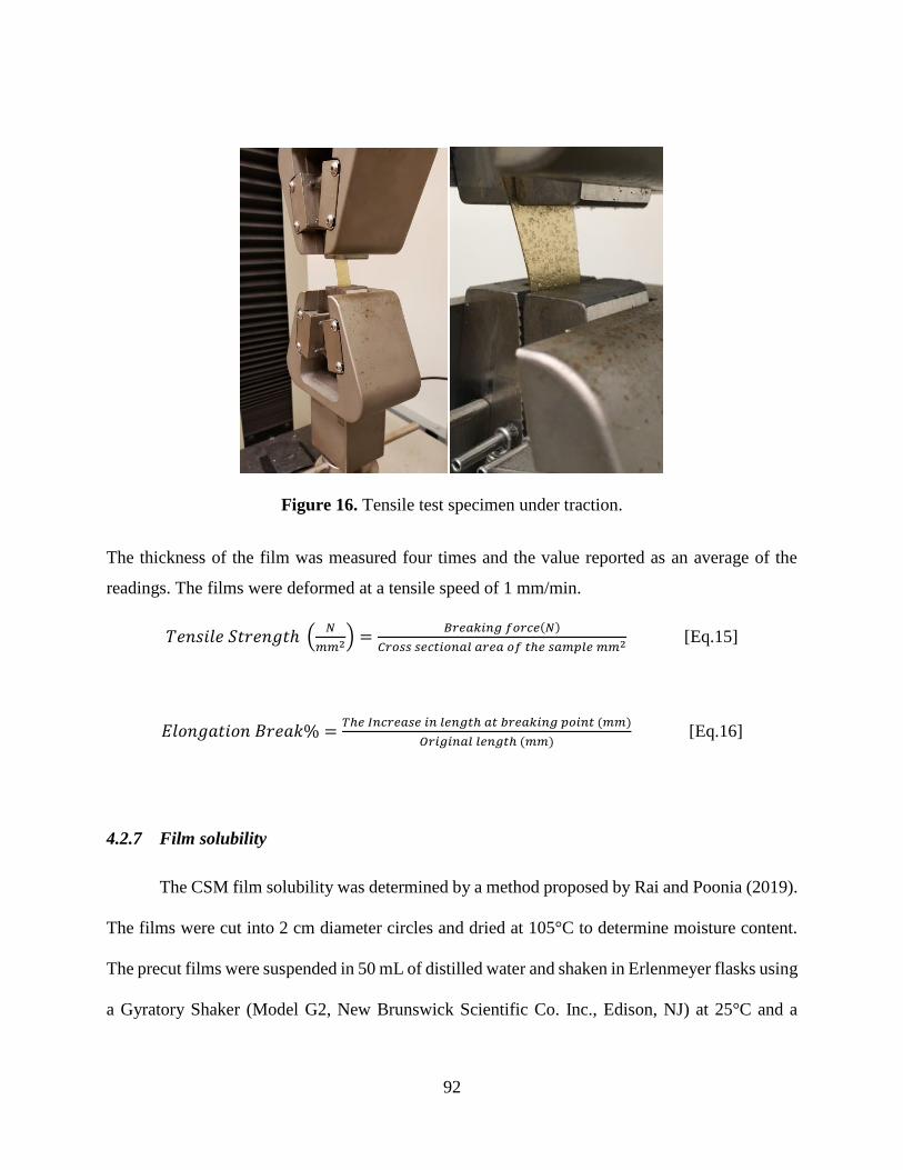
92
Figure 16. Tensile test specimen under traction.
The thickness of the film was measured four times and the value reported as an average of the
readings. The films were deformed at a tensile speed of 1 mm/min.
𝑇𝑒𝑛𝑠𝑖𝑙𝑒 𝑆𝑡𝑟𝑒𝑛𝑔𝑡ℎ (𝑁
𝑚𝑚2) =𝐵𝑟𝑒𝑎𝑘𝑖𝑛𝑔 𝑓𝑜𝑟𝑐𝑒(𝑁)
𝐶𝑟𝑜𝑠𝑠 𝑠𝑒𝑐𝑡𝑖𝑜𝑛𝑎𝑙 𝑎𝑟𝑒𝑎 𝑜𝑓 𝑡ℎ𝑒 𝑠𝑎𝑚𝑝𝑙𝑒 𝑚𝑚2 [Eq.15]
𝐸𝑙𝑜𝑛𝑔𝑎𝑡𝑖𝑜𝑛 𝐵𝑟𝑒𝑎𝑘% =𝑇ℎ𝑒 𝐼𝑛𝑐𝑟𝑒𝑎𝑠𝑒 𝑖𝑛 𝑙𝑒𝑛𝑔𝑡ℎ 𝑎𝑡 𝑏𝑟𝑒𝑎𝑘𝑖𝑛𝑔 𝑝𝑜𝑖𝑛𝑡 (𝑚𝑚)
𝑂𝑟𝑖𝑔𝑖𝑛𝑎𝑙 𝑙𝑒𝑛𝑔𝑡ℎ (𝑚𝑚) [Eq.16]
4.2.7 Film solubility
The CSM film solubility was determined by a method proposed by Rai and Poonia (2019).
The films were cut into 2 cm diameter circles and dried at 105°C to determine moisture content.
The precut films were suspended in 50 mL of distilled water and shaken in Erlenmeyer flasks using
a Gyratory Shaker (Model G2, New Brunswick Scientific Co. Inc., Edison, NJ) at 25°C and a

93
rotational speed of 100 RPM for a period of 6 hours. The un-dissolved portions of the films were
filtered using a #18 mesh and dried at 105°C. The solubility was calculated according to [Eq.17].
𝑆𝑜𝑙𝑢𝑏𝑖𝑙𝑖𝑡𝑦 (%) =𝐼𝑛𝑖𝑡𝑖𝑎𝑙 𝑑𝑟𝑦 𝑚𝑎𝑠𝑠 − 𝑓𝑖𝑛𝑎𝑙 𝑑𝑟𝑦 𝑚𝑎𝑠𝑠
𝐼𝑛𝑖𝑡𝑖𝑎𝑙 𝑑𝑟𝑦 𝑚𝑎𝑠𝑠∗ 100 [Eq. 17]
4.2.8 Water contact angle
The water contact angle was determined by the methodology proposed by Gezer, Brodsky,
Hsiao, Liu, and Kokini (2015), where a VCA Optima contact angle analyzer (AST products,
Billerica, MA) was used to photograph 30 s after 2-μL of deionized water was deposited onto the
surface of the CSM films. The stable contact angle of the water at the CSM films interface was
measured and performed in triplicate for each treatment.
4.2.9 Water vapor permeability (WVP)
The WVP was calculated according to Garcı́a, Pinotti, Martino, and Zaritzky (2004) using
the E96 method (ASTM, 2000). The films were sealed with metal clamps in 10 cm2 circular
opening PVC permeation cells containing anhydrous calcium chloride (0% RH), and stored at
25°C in a desiccator prior analysis. A 75% relative humidity (RH) gradient was established using
a saturated solution of sodium chloride in a desiccator at 25°C. The water vapor transport rate was
determined gravimetrically. Ten weight measurements were made, and the change in weight was
plotted as a function of time. A regression analysis of each sample was conducted and the slope of
the straight line (g/s) was divided by the area (m2) to calculate the water vapor transmission rate
(WVTR). Finally, the WVP was calculated according to [Eq.18] and expressed as (10-9 g/ m*s*Pa)
𝑊𝑉𝑃 = (𝑊𝑉𝑇𝑅
𝑆(𝑅1−𝑅2)) ∗ 𝑑 [Eq.18]

94
Where S: the saturation water vapor pressure at 25°C, R1: relative humidity outside the cell, R2:
relative humidity inside the cell. The driving force (S(R1-R2)) was determined to be 1753.55 (Pa).
4.2.10 Raman spectroscopy
The structure characterization of the CSM films was determined using a Bruker FT-Raman
spectrometer RFS 100/S (Bruker Optics, Lubeck, Germany) with a germanium detector cooled by
liquid nitrogen. CSM films were cut in 2.5 cm by 2.5 cm squares using a razor blade and placed
on a steel holder for Raman analysis. The analysis was conducted with an excitation wavelength
of 1064-nm from a ND: YAG laser. A total of 300 scans were collected per sample, at a 4 cm-1 of
resolution and a laser power of 220 mW. The spectra was collected at a range between 3500 and
73 cm-1. The baseline and smoothness of the spectra was first corrected using the software OPUS
6.5 (Bruker, Karlsruhe, Germany). Finally, the main peaks were identified using KnowltAll®
(Analytical Edition, ID Expert, Bio-Rad Laboratories, Inc.).
4.2.11 Statistical analysis
The statistical analysis was done using a central composite design, evaluating the influence
of the independent variables glycerol (X1) and sorbitol (X2) concentration (g) on the dependent
variables (Yn). The response surface methodology (RSM) was conducted using the statistical
software Statistica 10.0 ®. The quantities of the independent variables were coded as follows (-1,
0 and 1) whereas the central points were codified as (0, 0). The coded and decoded values are
shown in (Table 9).

95
Table 9. Coded and decoded levels of polyols incorporated to chia seed mucilage films.
Treatment
Coded variables Decoded variables
Glycerol Sorbitol Glycerol (g/g of
mucilage)
Sorbitol (g/g of
mucilage)
1* 0 0 1.67 1.67
2* 0 0 1.67 1.67
3 -1 -1 0.67 0.67
4 1 -1 2.67 0.67
5 -1 1 0.67 2.67
6 1 1 2.67 2.67
*Central point.
The number of treatments was established using [Eq.19]. The levels (g) of the polyols
were determined by preliminary data, identifying ideal central point, conditions and limitations in
the incorporation of polyols in the mucilage films. The experiments were repeated 3 times before
analysis.
𝑛 = 2𝑘 + 𝑚 [Eq. 19]
Where 2𝑘= factorial points, m=repetitions for the central point, and 𝑛 = 22 + 2 = 6 𝑇𝑅𝑇.
Finally, the independent variables were optimized using a desirability simulation of the software
Statistica 10.0 ®. A 100-step simulation was run according to the values observed in this study. In
the optimization process, the dependent variables (Yn) elongation at break, tensile strength, contact
angle and solubility were maximized, whereas the water vapor permeability was minimized.
4.3 Results and discussion
4.3.1 Proximate analysis of chia seed mucilage
The proximate composition of CSM showed content of 11.44 ±0.37% of crude protein,
0.25 ±0.05% of ash, 11.99 ±0.88% of moisture, 0.09 ±0.01% of fat and 76.35 ±0.60% of
carbohydrates (calculated by weight difference). The carbohydrate content includes acid detergent
fiber of 10.30 ±1.30%. Proximate composition of CSM, in this study falls within values reported
96
in other studies. Segura-Campos, Ciau-Solís, Rosado-Rubio, Chel-Guerrero, and Betancur-
Ancona (2014), extracted CSM and reported a higher protein (25%) and fat (26%) content, whereas
extracted and purified CSM by Timilsena et al., (2016) and reported lower content of protein
(2.6%) and higher fat content (0.6%). The compositional difference is attributed to variances
involved during mucilage extraction. Nevertheless, ultrasound-assisted mucilage extraction
method used in this study, resulted in higher extraction yield (7.8%) (Urbizo-Reyes et al., 2019)
than other physical methods (5.6%) (Timilsena et al., 2016). Ultrasound energy and the high shear
forces developed under the sonic field could be responsible for increased protein extraction within
the mucilage matrix. The molecular composition of the stored mucilage is still unknown, and a
possible intramolecular bonding of proteins in the stored mucilage matrix could be the reason why
residual proteins are still present in the extracted material. Timilsena et al. (2016), hypothesized
that a possible covalent linkage might exist between protein and mucilage. It has been widely
demonstrated that the ultrasound technique can help extract other compounds from plant matrices
such as proteins, phytochemical compounds, and minerals (Chemat et al., 2017). Recently, some
studies reported an increase on protein extraction when ultrasound was applied to spirulina (Vernès
et al., 2019). Higher protein extraction was attributed to the erosion, shear effects and
fragmentation caused during sonoporation, which generated cellular lysis that released protein
from plant cells (Vernès et al., 2019).
4.3.2 Chia seed mucilage films thickness and color
In preliminary trials, CSM begun to combust at temperatures between 180-200°C limiting
its capacity to be extruded or mixed with PLA. For this reason, its applicability on conventional
3D printing was restricted; however, the application on a bio-3D printing at lower temperatures
might be feasible and further studies are required to understand this possibility. CSM films were

97
effectively casted in aluminum plates with variating concentrations of polyols (glycerol and
sorbitol). Casting in plastic or glass surfaces made film peeling inefficient and laborious. Excessive
incorporation of polyols as plasticizers (6.0 g of polyols/g of mucilage, wet basis) resulted in
unmanageable films with lack of structure and a syrupy texture. Conversely, low concentration of
plasticizers (less than 1.0 g of polyols/g of mucilage, wet basis) resulted in films with poor
flexibility and film’s peeling from the plate became almost impossible. Film thickness depended
on treatment and ranged from 0.381 to 0.392 mm. The incorporation of polyols has shown to cause
an increase in thickness; this is attributed to an increase of free-volume when polyols are
incorporated intermolecularly (Cerqueira, Souza, Teixeira, & Vicente, 2012). The films had a
slight yellow color as indicated by the color parameters (Table 10). The natural color of these
films might serve as a barrier for UV light. This could allow the material to serve as a UV
protectant of photosensitive foods extending the shelf life of the products by decreasing photo-
oxidative degradation. Dick, Costa, Gomaa, Subirade, de Oliveira Rios, et al. (2015) evaluated
the UV absorption capacity of chia seed mucilage films and found that light transmittances was
very low for the UV regions between 200-280 nm, indicating the potential role of this material as
a UV barrier in food packaging. The L* values were significantly lower (darker) (p<0.05) for films
with higher content of glycerol. Dick, Costa, Gomaa, Subirade, de Oliveira Rios, et al. (2015)
showed similar results, where the incorporation of glycerol resulted in a decrease in lightness (L*).
An increase in glycerol also resulted in increased red (a*) and yellow (b*) colors. The low
incorporation of polyols caused overall lower (b*) values (more blueish colors). The total
difference in color (ΔE) indicated a significantly higher (p<0.05) difference for films with high
glycerol (treatment 4) from the standard white plate, and the lowest difference was achieved with
low polyol concentrations (treatment 3). This change in color is often attributed to non-enzymatic

98
browning that occurs between glycerol and residual protein in the mucilage matrix (Barrett, Briggs,
Richardson, & Reed, 1998). Nevertheless, the films retained a semi-transparent appearance as the
polyol composition changed.
Table 10. Color determination of chia seed mucilage films.
Treatments L* a* b* ΔE*
1* 50.67 ± 0.08a 1.76 ± 0.04b 15.22 ± 0.07ab 2065.76 ± 0.07b
2* 50.95 ± 0.51a 1.74 ± 0.22b 15.12 ± 0.47b 2038.41 ± 0.47bc
3 51.07 ± 0.33a 1.64 ± 0.09b 14.28 ± 0.24d 2003.95 ± 0.24c
4 49.42 ± 0.02b 2.12 ± 0.08a 15.55 ± 0.22a 2186.26 ± 0.22a
5 50.94 ± 0.66a 1.62 ± 0.19b 14.61 ± 0.12cd 2023.90 ± 0.12bc
6 50.79 ± 0.26a 1.65 ± 0.13b 14.94 ± 0.25bc 2046.18 ± 0.25bc
The treatment assignment is specified in Table 9. *: Central point.
4.3.3 Mechanical properties (tensile strength and elongation at break)
Naturally edible and biodegradable films developed using biopolymers such as proteins or
polysaccharides are brittle and collapse with manipulation. The incorporation of plasticizers is
necessary to avoid premature fracture, poor flexibility and improve mechanical properties as
tensile strength, elongation at break, and stickiness of the films (Sothornvit & Krochta, 2005).
Dick, Costa, Gomaa, Subirade, de Oliveira Rios, et al. (2015) showed that incorporating glycerol
to CSM increased the elongation and permeability and decreased the tensile strength of the films.
In this study, we determined that different polyols had a significant effect (p<0.05) in the
mechanical properties of the films made from CSM (Fig 17). The incorporation of glycerol (X1),
sorbitol (X2) and their interaction (X1*X2) had a significant (p<0.05) effect in the predicting
models (Table 11) of tensile strength (R2= 0.91) and elongation at break (R2= 0.99). Films with a
higher content of sorbitol exhibited a higher tensile strength (Table 12), whereas films with a high
content of glycerol or glycerol-sorbitol concentration showed poor strength. Glycerol is a
hydrophilic molecule that consists of a 3-carbon backbone with 3 hydroxyl groups. Because of the
small molecular nature of this polyol, the number of hydroxyl groups is higher in a given volume

99
when compared to sorbitol (Santacruz, Rivadeneira, & Castro, 2015). The high content of hydroxyl
groups contributes to the incorporation of water molecules, resulting in an increased moisture
content, that at the same time can increase the molecular mobility of the films (Mendelovici, Frost,
& Kloprogge, 2000). Similarly, Espino‐Díaz et al. (2010) developed edible films from prickly pear
(Opuntia ficus-indica) using glycerol as a plasticizer and showed a maximum tensile strength of
0.95 N/mm2. Generally edible and biodegradable films have tensile strengths that ranges, between
1-10 N/mm2 (Han, 2014). The highest tensile strength in this study was achieved with 0.67 g of
glycerol and 2.67 g of sorbitol per g of CSM (3.23 N/mm2). It seems plausible that an increase in
interactions between sorbitol-mucilage and mucilage-mucilage is responsible of the high tensile
strength of these two films. Another study using pea starch, compared different plasticizers and
found that a higher tensile strength was achieved by sorbitol compared to glycerol (Y. Zhang &
Han, 2006). For elongation at break, higher values were obtained when a higher content of glycerol
was incorporated. The small molecular nature of glycerol allows it to be inserted into adjacent
polymeric chains, resulting in decreased molecular attractions that cause an increase in molecular
mobility and decreased rigidity (Dick, Costa, Gomaa, Subirade, de Oliveira Rios, et al., 2015).
Films with a higher content of glycerol (2.67 g of glycerol and 0.67 g of sorbitol per g of CSM)
reached elongation at break values as high as 68%. Whereas in films with a high content of sorbitol
(0.67 g of glycerol and 2.67 g of sorbitol per g of CSM), the elongation at break reach a maximum
value of 21%. Talja, Helén, Roos, and Jouppila (2007) explored the mechanical properties of
edible films made with starch plasticized with different polyols. Their findings showed that films
plasticized with sorbitol had the lowest elongation at break compare to xylitol. The difference in
elongation at break has been attributed to a possible polyol crystallization, common in sorbitol and

100
xylitol. This process is also known to enhance the crosslinking within the starch network causing
differences in mechanical properties (Talja et al., 2007).
Figure 17. Response surface diagrams; Effect of glycerol (g) and sorbitol (g) per gram of chia
seed mucilage (CSM) on the response variables: water vapor permeability (A), solubility (B),
contact angle (C), elongation at break (D), tensile strength (E).

101
4.3.4 Water vapor permeability (WVP)
Glycerol, sorbitol and their interaction showed a significant (p<0.05) effect in the
predicting model (Table 11) for WVP (R2= 0.96). An increased concentration of glycerol, or
polyol mixture resulted in higher WVP (Fig 17). Similarly, Dick, Costa, Gomaa, Subirade, Rios,
et al. (2015), showed an increase in WVP with an increase in glycerol concentration. They
hypothesized that the small molecular size of glycerol, allows the polyol to penetrate the
intermolecular matrix, causing a lower amount of polysaccharide-polysaccharide interactions and
an overall increase in free volume and molecular movement. On the other hand, films plasticized
with sorbitol showed a more compact and rigid structure, limiting the permeation of water vapor
molecules through its structure. A higher molecular mobility allows water vapor molecules to
permeate more easily through the film. Hence, sorbitol being a larger molecule than glycerol,
showed a reducing effect in the WVP of the CSM films. Likewise, Antoniou, Liu, Majeed, Qazi,
and Zhong (2014) compared the WVP of edible films from tara gum (Caesalpinia spinosa) and
found that films made with sorbitol exhibited lower permeability when compared to glycerol films.
They attributed this to the lower hydrophilic nature of sorbitol compared to glycerol. In this study
the lowest WVP (Table 12) achieved was 1.30*(10-9 g/m*s*Pa), this value is lower than those
reported for tapioca starch-microcrystalline cellulose composite films 3.37*(10-8 g/m*s*Pa),
plantain seed gum 12.77*(10-9 g/m*s*Pa) and blends of cassava starch 5.05*(10-7 g/m*s*Pa)
(Niknam, Ghanbarzadeh, & Hamishehkar, 2019; Othman et al., 2019; Parra, Tadini, Ponce, &
Lugão, 2004). Nevertheless, these observations differ from other studies that evaluated the effect
of other polyols in the permeability of starch-gelatin edible films, where the highest WVP was
achieved when sorbitol was incorporated in the film matrix (Al-Hassan & Norziah, 2012). It is
speculated that a difference in polymer structure is responsible of the difference in the film

102
permeability. The WVP of CSM films is still relatively high, this also can be attributed to the
hydrophilic nature of this polysaccharide. A study conducted by Coorey, Tjoe, and Jayasena
(2014) assessed the functional properties of CSM and identified a high water-holding capacity
(267 g/g of mucilage). They associated this to the hydrophilic nature of xylans and arabinoxylans,
which are known to absorb hundreds of times their equivalent weight in water.
4.3.5 Water contact angle
Water contact angle measures directly the angle formed by a water droplet when it is
deposited on the surface of a film. This is a way of determining the hydrophobic nature of the
surface of a material. This method is based on image processing and a curve fitting technique,
analyzing the contact angle form with the baseline and the tangent of the drop boundary when
deposited on a surface (Karbowiak, Debeaufort, Champion, & Voilley, 2006). The contact angle
(θ), provides information of the wettability of a surface, a film is categorized as a hydrophilic (θ <
90) or a hydrophobic (θ >90) boundary (Karbowiak et al., 2006). In this study, glycerol, sorbitol
and their interaction showed a significant (p<0.05) effect in the predicting model (Table 11) for
contact angle (R2=0.99). An increase in both polyols caused a decreased in the contact angle,
whereas a decrease in polyols increased it (Fig 17). The contact angle was maintained between 46-
48 as the polyols increased the contact angle to reach maximum values of 48.39 at the central
points (1.67 g of glycerol and 1.67 g of sorbitol per g of CSM); nevertheless, the incorporation of
more than 3.34 g of a polyol per g of mucilage showed to decrease the water contact angle
drastically (Table 12). The low contact angle values are related to the hydrophilic nature of
mucilage fibers and polyols. CSM is a biomolecule composed of a branch of saccharides and acids
that are hydrophilic in nature, these saccharides include xylans, arabinose, glucose, galactose,
glucuronic acid and galacturonic acid (Timilsena et al., 2016). The incorporation of the polyols as

103
plasticizers may increase the hydrophilicity. Similarly, Jouki, Yazdi, Mortazavi, and Koocheki
(2013), evaluated the water contact angle of edible films made with quince seed (Cydonia oblonga)
mucilage and showed that the contact angle decreases with an increase in glycerol content. Most
authors attribute a decrease in contact angle to the hydrophilic nature of polyols, which cause a
decrease in the hydrophobicity of polysaccharide films (Jouki et al., 2013). The same behavior has
been reported for other edible films made from whey proteins, psyllium gum and tara gum
(Ahmadi, Kalbasi-Ashtari, Oromiehie, Yarmand, & Jahandideh, 2012; Antoniou et al., 2014;
Kokoszka, Debeaufort, Lenart, & Voilley, 2010).
4.3.6 Film solubility
Glycerol, sorbitol and their interaction showed a significant (p<0.05) effect in the
predicting model (Table 11) for film solubility (R2= 0.98). The increased content of polyols
resulted in a higher solubility (Fig 17). No apparent trend was observed in film solubility when
plasticized with sorbitol and glycerol. Films plasticized with a low concentration of polyol mixture
had the lowest solubility (11.61%). It seems plausible that the low quantity of polyols allowed the
films to maintain a rigid structure and higher hydrophobicity, as shown from the contact angle
values. Other researchers reported similar trends, where glycerol was relatively more efficient to
increase the solubility of the films; this was attributed to the lower molecular weight of glycerol,
that increased the exposure of hydroxyl groups and resulted in higher water binding (Ghasemlou,
Khodaiyan, & Oromiehie, 2011). However, both polyols are hydrophilic and increasing their
content will increase the water solubility of the films, with values as high as 29.34%. In preliminary
trials, the films were completely solubilized completely in 12 h. These films have high capacity to
absorb moisture due to their hydrophilic nature, thus limiting their functionality as a moisture
barrier, However, their molecular attributes make them suitable for other applications such as
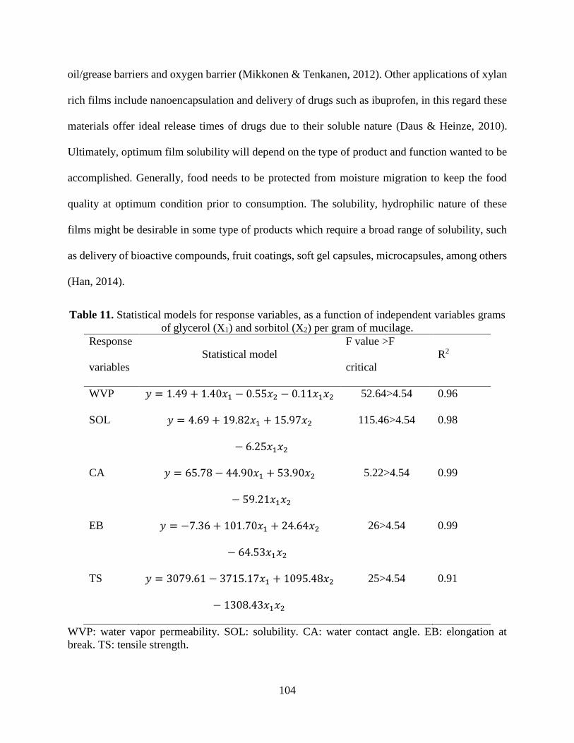
104
oil/grease barriers and oxygen barrier (Mikkonen & Tenkanen, 2012). Other applications of xylan
rich films include nanoencapsulation and delivery of drugs such as ibuprofen, in this regard these
materials offer ideal release times of drugs due to their soluble nature (Daus & Heinze, 2010).
Ultimately, optimum film solubility will depend on the type of product and function wanted to be
accomplished. Generally, food needs to be protected from moisture migration to keep the food
quality at optimum condition prior to consumption. The solubility, hydrophilic nature of these
films might be desirable in some type of products which require a broad range of solubility, such
as delivery of bioactive compounds, fruit coatings, soft gel capsules, microcapsules, among others
(Han, 2014).
Table 11. Statistical models for response variables, as a function of independent variables grams
of glycerol (X1) and sorbitol (X2) per gram of mucilage.
Response
variables
Statistical model
F value >F
critical
R2
WVP 𝑦 = 1.49 + 1.40𝑥1 − 0.55𝑥2 − 0.11𝑥1𝑥2 52.64>4.54 0.96
SOL 𝑦 = 4.69 + 19.82𝑥1 + 15.97𝑥2
− 6.25𝑥1𝑥2
115.46>4.54 0.98
CA 𝑦 = 65.78 − 44.90𝑥1 + 53.90𝑥2
− 59.21𝑥1𝑥2
5.22>4.54 0.99
EB 𝑦 = −7.36 + 101.70𝑥1 + 24.64𝑥2
− 64.53𝑥1𝑥2
26>4.54 0.99
TS 𝑦 = 3079.61 − 3715.17𝑥1 + 1095.48𝑥2
− 1308.43𝑥1𝑥2
25>4.54 0.91
WVP: water vapor permeability. SOL: solubility. CA: water contact angle. EB: elongation at
break. TS: tensile strength.

105
Table 12. Effect of independent variables on response variables.
*central points. Coded values are shown as follows (n). WVP: water vapor permeability. SOL: solubility. CA: contact angle. EB: elongation at
break. TS: tensile strength.
Treatment Glycerol (g)
Sorbitol
(g)
WVP
(10-9 g/m*s*Pa)
SOL (%) CA (θ) EB (%) TS (N/mm2)
1* 1.67 (0) 1.67 (0) 2.05 ± 0.25 19.65 ± 0.03 46.95 ± 4.65 41.84 ± 0.49 977.74 ± 11.80
2* 1.67 (0) 1.67 (0) 2.30 ± 0.13 19.60 ± 0.06 48.20 ± 3.02 41.73 ± 0.95 991.32 ± 1.60
3 0.67 (-1) 0.67 (-1) 1.62 ± 0.10 11.61 ± 0.57 48.39 ± 7.02 14.27 ± 0.26 2732.39 ± 27.87
4 2.67 (1) 0.67 (-1) 2.28 ± 0.39 22.75 ± 0.28 28.55 ± 3.66 67.55 ± 2.54 346.28 ± 8.98
5 0.67 (-1) 2.67 (1) 1.30 ± 0.16 20.44 ± 1.15 23.15 ± 3.00 21.31 ± 0.42 3232.67 ± 22.53
6 2.67 (1) 2.67 (1) 2.20 ± 0.45 29.34 ± 1.79 24.63 ± 1.37 51.35 ± 0.89 375.52 ± 3.53

106
4.3.7 Raman analysis of chia seed mucilage and chia seed mucilage films
The CSM (Fig18) and CSM films (Fig19) showed very defined spectra corresponding to
the regions of 73-600, 600-1600 and 2900-2930 cm-1. The corresponding assignment of the FT-
Raman spectra bands is shown in (Table 13).
Figure 18 . Raman spectra for chia seed mucilage.
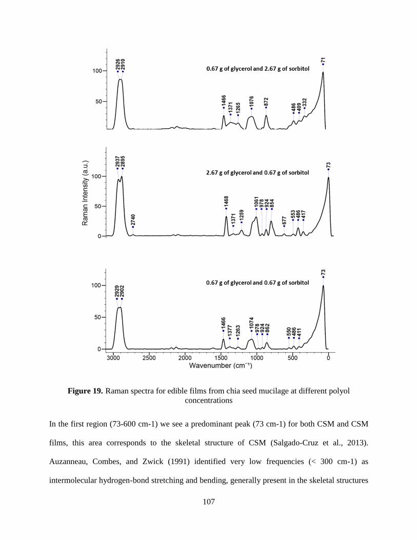
107
Figure 19. Raman spectra for edible films from chia seed mucilage at different polyol
concentrations
In the first region (73-600 cm-1) we see a predominant peak (73 cm-1) for both CSM and CSM
films, this area corresponds to the skeletal structure of CSM (Salgado-Cruz et al., 2013).
Auzanneau, Combes, and Zwick (1991) identified very low frequencies (< 300 cm-1) as
intermolecular hydrogen-bond stretching and bending, generally present in the skeletal structures
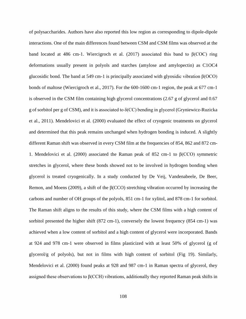
108
of polysaccharides. Authors have also reported this low region as corresponding to dipole-dipole
interactions. One of the main differences found between CSM and CSM films was observed at the
band located at 486 cm-1. Wiercigroch et al. (2017) associated this band to β(COC) ring
deformations usually present in polyols and starches (amylose and amylopectin) as C1OC4
glucosidic bond. The band at 549 cm-1 is principally associated with glyosidic vibration β(OCO)
bonds of maltose (Wiercigroch et al., 2017). For the 600-1600 cm-1 region, the peak at 677 cm-1
is observed in the CSM film containing high glycerol concentrations (2.67 g of glycerol and 0.67
g of sorbitol per g of CSM), and it is associated to δ(CC) bending in glycerol (Gryniewicz-Ruzicka
et al., 2011). Mendelovici et al. (2000) evaluated the effect of cryogenic treatments on glycerol
and determined that this peak remains unchanged when hydrogen bonding is induced. A slightly
different Raman shift was observed in every CSM film at the frequencies of 854, 862 and 872 cm-
1. Mendelovici et al. (2000) associated the Raman peak of 852 cm-1 to β(CCO) symmetric
stretches in glycerol, where these bonds showed not to be involved in hydrogen bonding when
glycerol is treated cryogenically. In a study conducted by De Veij, Vandenabeele, De Beer,
Remon, and Moens (2009), a shift of the β(CCO) stretching vibration occurred by increasing the
carbons and number of OH groups of the polyols, 851 cm-1 for xylitol, and 878 cm-1 for sorbitol.
The Raman shift aligns to the results of this study, where the CSM films with a high content of
sorbitol presented the higher shift (872 cm-1), conversely the lowest frequency (854 cm-1) was
achieved when a low content of sorbitol and a high content of glycerol were incorporated. Bands
at 924 and 978 cm-1 were observed in films plasticized with at least 50% of glycerol (g of
glycerol/g of polyols), but not in films with high content of sorbitol (Fig 19). Similarly,
Mendelovici et al. (2000) found peaks at 928 and 987 cm-1 in Raman spectra of glycerol, they
assigned these observations to β(CCH) vibrations, additionally they reported Raman peak shifts in

109
this frequency when the material was cooled down, suggesting the shift observed in this study
might be caused by hydrogen bonding. In a study on starch-based films, peaks between 920-960
cm-1 were assigned to glyosidic linkages developed by hydrogen bonding of the glycerol with
films (Nobrega, Olivato, Müller, & Yamashita, 2012). One of the main differences was observed
between the frequency range of 1061-1076 cm-1, assigned to ν(CCO). The films plasticized with
a minimum of 50% of sorbitol (g of sorbitol/g of polyols) showed peaks at a Raman frequency
higher (1074-1076 cm-1) than films with higher glycerol content (1061 cm-1). These films also
showed the highest tensile strength (Table 12), we hypothesize that an increase in network bonding
between mucilage-mucilage and mucilage-protein is responsible for these observations. Similarly,
Mendelovici et al. (2000) observed a similar Raman shift in the symmetric ν(CCO) stretching
modes (1054 cm-1), when treating glycerol cryogenically, attributing this to the increasing number
of hydrogen bonding and network formation between molecules. This band (1054 cm-1) is
commonly present in sorbitol and other polyols such as mannitol (De Veij et al., 2009).
Additionally, the presence of protein in the mucilage matrix might play a role in the formation of
highly resistant films when low polyol concentrations was present. Nobrega et al. (2012) assessed
the Raman spectrum of oat globulins and identified peaks at 1245 and 1450 cm-1 as amide III
(disordered structure) and (C-H bending) correspondingly. In this study, similar bands where
observed at 1265 and 1465 cm-1 in CSM. A slight shift in this region was observed from 1259 to
1265 cm-1 for films plasticized with at least 50% of sorbitol (g of sorbitol/g of polyols), the Raman
shift increase as sorbitol increase in concentration. This shift might suggest a protein-sorbitol
interaction, causing an increase in bond formation.
In summary, CSM films containing a low polyol concentration or a high content of sorbitol,
showed very marked band shifts at 862-872 cm-1 and 1061-1076 cm-1, corresponding to ν(CCO).

110
We hypothesize that a possible increase in interaction between sorbitol-mucilage and mucilage-
mucilage is responsible for the shift in this type of linkages and might explain the high mechanical
properties of these two films. The bands at 472, 549, 1118, and 1379 cm-1 are typically observed
in most polysaccharides such as starch and mucilage, and are attributed especially to CH2OH
(Nobrega et al., 2012). Most of these bands correspond to the glucose finger print observed in both
the CSM and its films Wiercigroch et al., 2017).
The region corresponding to 2900-2930 cm-1 has been associated with the presence of
pentose and hexoses. X. Zhang, Chen, Ramaswamy, Kim, and Xu (2017) and Kačuráková et al.
(1999) have identified the region between 2800-2900 cm-1 to be associated with high content of
D-xylose and glucose, commonly referred as v(CH)-vibrations. It is known that CSM contains
around a 38% of xylans (Timilsena et al., 2016). Xylans are hemicelluloses with a wide application
in the food industry as sweetening, thickening and emulsifying agents (Kačuráková et al., 1999).
This hemicellulose is one of the most abundant biopolymers in the plant kingdom, its structural
composition is usually described as multiple side chains bonded to a backbone of linear chains
corresponding to (1→4)-linked β-D-xylopyranosye (Ebringerová & Hromádková, 1999). Xylans
generally form a net-like cross-linked structure, and since their side chains are mainly composed
of hydroxyl groups; this provides them with high water dispersibility and water holding capacity,
making them suitable materials for encapsulation and film development applications (Fu et al.,
2019).

111
Table 13. Raman Spectra analysis for chia seed mucilage and its films.
NI: not identified, GLY: glycerol, SOR: sorbitol.
4.3.8 Optimization
The polyol content of CSM films was optimized using a desirability simulation, running a
100-step simulation according the values observed in this study. In the optimization process, the
dependent variables elongation at break, tensile strength, contact angle and solubility were
Chia seed
mucilage
2.67 g GLY +
0.67 g SOR
0.67 g GLY +
2.67 g SOR
0.67 g GLY +
0.67 g SOR Assignment
73 73 71 73 skeletal structure
(δs(C–C))
332 β(CCO), t(COHO)
417 409 411 δ(CCO)
472 β(CCO)
486 486 486
β
(COC)
549 553 550 β(OCO)
622 β(COC)
677 δ(CC)
711 γ(OH)
788
β(CCC), β(CCO),
β(OCO)
854 v(CCO)
862 v(CCO)
872 v(CCO)
924 924 β(CCH) β (COH)
978 978 ν(CO) ring, β(CCH)
985 v(CCO)
1061 ν(CCO)
1079 1076 1074 ν(CCO), β(COH)
1118 νs(COH) from C-2
1265 1259 1265 1263 Amide III
1378 1371 1371 1377 ω(CH2)
1465 1468 1466 1466 δ(CH2) δ(CCH)
1600 v(CO)v(CC)
1990 NI
2075 NI
2125 NI
2196 NI
2912 2895 2910 2902 νs(CH2)
2937 2926 2929 νs(CH2)

112
maximized, whereas the water vapor permeability was minimized. The simulation (Fig 20)
estimated the optimum conditions to be at 1.29 g of glycerol and 2.03 g of sorbitol per every g of
mucilage. This process might be useful for development of food coatings, and the desired
parameters might be adjusted accordingly to required characteristics. Other studies have optimized
film composition according to plasticizer and mucilage content (Oliveira et al. 2019). Future
studies in these films need to be conducted, evaluating the food applications, and how different
polyols interact with food (e.g. fruits and vegetables) or micro- and nano-encapsulation of
bioactive components.
Figure 20. Optimization of edible films from chia seed mucilage.

113
4.4 Conclusions
CSM, as other biomolecules, has a great potential in the development of biodegradable
materials from plants. CSM films were effectively developed by casting methods, using polyols
as plasticizers to improve their physical structure. This research showed that each polyol imparts
unique properties to the films and that the ideal content or mixture will depend on the intended use
of the material. In this regard, the utilization of both polyols, sorbitol and glycerol, in combination
showed to significantly (p<0.05) impact the physical and barrier properties of CSM films. Sorbitol
provided CSM films with higher strength and a certain degree of hydrophobicity, whereas glycerol
provided an increase in elongation at break, flexibility and hydrophilicity of the films. Raman
spectroscopy revealed marked band shifts at 862-872 cm-1 and 1061-1076 cm-1, corresponding to
(CCO), and confirming that hydrogen bonding is induced with polyol treatment, which aligns with
the mechanical observations in this study. The applications of this material might need to be
geared towards products that do not require low water vapor permeability, such as
nanoencapsulation and micro delivery carriers. Furthermore, the hygroscopic nature of CSM films
might potentially confer good barrier properties against oxygen (O2) transfer. Future studies should
focus in applying this material in such environments to understand its behavior and develop further
applications. The material begun to combust at high temperatures limiting its applicability on
conventional 3D printing. However, the application on bio-3D printing might be feasible and
further studies are required to understand this possibility.
114
114
CHAPTER 5. OVERALL CONCLUSION AND FUTURE
DIRECTIONS.
5.1 Overall conclusions
• Chia seeds (Salvia hispanica) can be used as a source of functional and nutritional
ingredients, due to their high content of fiber, high-quality fat, and protein.
• Ultrasound, along with vacuum filtration, is an excellent technology for the extraction of
mucilage from chia seed and also had a high impact on lipid separation.
• Microwave-assisted hydrolysis with sequential enzymatic treatments produced protein
with improved functionality and health benefits, especially as antioxidant agents.
• Mucilage from chia seeds showed high potential in the development of biodegradable and
edible films.
5.2 Future directions
• Evaluate the functionality of chia seed protein hydrolysates in food prototypes and evaluate
their sensory characteristics.
• Identify peptide sequences responsible for the inhibitory capacity of DPP-IV and ACE.
• Assess the bioactive properties of chia seed protein hydrolysates in vivo, especially for
antioxidant activity.
• Implement and evaluate chia seed mucilage as an edible coating for fruits, vegetables, or
even as a carrier to nano-deliver drugs.
• Evaluate the oxygen barrier properties of chia seed mucilage.
115
115
REFERENCES
Abugoch, L. E., Romero, N., Tapia, C. A., Silva, J., & Rivera, M. (2008). Study of some
physicochemical and functional properties of quinoa (Chenopodium quinoa Willd)
protein isolates. Journal of agricultural and food chemistry, 56(12), 4745-4750.
Adler-Nissen, J. (1986). Enzymic hydrolysis of food proteins. London: Elsevier.
Agriculture, 93(15), 3856-3862.
Ahmadi, R., Kalbasi-Ashtari, A., Oromiehie, A., Yarmand, M.-S., & Jahandideh, F. (2012).
Development and characterization of a novel biodegradable edible film obtained from
psyllium seed (Plantago ovata Forsk). Journal of Food Engineering, 109(4), 745-751.
Aldred, E. M., & Phenols, P. (2009). A Handbook for Complementary Healthcare Professionals.
Elsevier's Health Sciences. Philadelphia PA.
Al-Hassan, A., & Norziah, M. (2012). Starch–gelatin edible films: Water vapor permeability and
mechanical properties as affected by plasticizers. Food hydrocolloids, 26(1), 108-117.
Aluko, R. E. (2015). Structure and function of plant protein-derived antihypertensive
peptides. Current Opinion in Food Science, 4, 44-50.
Aluko, R. E., & Monu, E. (2003). Functional and bioactive properties of quinoa seed protein
hydrolysates. Journal of Food Science, 68(4), 1254-1258
Amagliani, L., O'Regan, J., Schmitt, C., Kelly, A. L., & O'Mahony, J. A. (2019).
Characterisation of the physicochemical properties of intact and hydrolysed rice protein
ingredients. Journal of Cereal Science, 88, 16-23.
American Heart Association. (2017). What is Cardiovascular Disease? Retrieved from
http://www.heart.org/HEARTORG/Conditions/What-is-Cardiovascular-
Disease_UCM_301852_Article.jsp#.Wrz-nojwbIU
Andrady, A. L. (2015). Plastics and environmental sustainability (p. 324). Hoboken, NJ: Wiley.
Antoniou, J., Liu, F., Majeed, H., Qazi, H. J., & Zhong, F. (2014). Physicochemical and
thermomechanical characterization of tara (Cesalpinia spinosa) gum edible films: effect
of polyols as plasticizers. Carbohydrate polymers, 111, 359-365.
AOAC. (2016). Official methods of analysis of AOAC International (20th ed.). Rockville, MD:
AOAC International.
Aruoma, O. I. (2003). Methodological considerations for characterizing potential antioxidant
actions of bioactive components in plant foods. Mutation Research/Fundamental and
Molecular Mechanisms of Mutagenesis, 523, 9-20.

116
116
ASAE (2002). ASAE standards. Retrieved from
https://engineering.purdue.edu/~abe305/moisture/html/page12.htm
Asao, M., & Watanabe, K. (2010). Functional and bioactive properties of quinoa and
amaranth. Food science and technology research, 16(2), 163-168.
ASTM. (2000). Standard test methods for water vapor transmission of material, E96–00. Annual
Book of American Standard Testing Methods, Philadelphia: American Society for Testing
and Materials.
ASTM. (2002). Standard test method for tensile properties of thin plastic sheeting, D882-02.
Annual Book of ASTM Standards. Philadelphia: American Society for Testing and
Materials
Auzanneau, I., Combes, D., & Zwick, A. (1991). Raman spectroscopic analysis of the effect of
polyhydric alcohols on water. Journal of Raman spectroscopy, 22(4), 227-231.
Ayerza Jr, R., & Coates, W. (2007). Effect of dietary α-linolenic fatty acid derived from chia
(Salvia hispanica) when fed as ground seed, whole seed and oil on lipid content and fatty
acid composition of rat plasma. Annals of Nutrition and Metabolism, 51(1), 27-34.
Bai, M., Qin, G., Sun, Z., & Long, G. (2016). Relationship between molecular structure
characteristics of feed proteins and protein in vitro digestibility and solubility. Asian-
Australasian journal of animal sciences, 29(8), 1159.
Balti, R., Bougatef, A., Ali, N. E. H., Zekri, D., Barkia, A., & Nasri, M. (2010). Influence of
degree of hydrolysis on functional properties and angiotensin I‐converting enzyme‐
inhibitory activity of protein hydrolysates from cuttlefish (Sepia officinalis) by‐products.
Journal of the Science of Food and Agriculture, 90(12), 2006-2014.
Bamdad, F., Wu, J., & Chen, L. (2011). Effects of enzymatic hydrolysis on molecular structure
and antioxidant activity of barley hordein. Journal of Cereal Science, 54(1), 20-28.
Barrett, A. H., Briggs, J., Richardson, M., & Reed, T. (1998). Texture and storage stability of
processed beefsticks as affected by glycerol and moisture levels. Journal of Food
Science, 63(1), 84-87.
Bernardo, C. O., Ascheri, J. L. R., Chávez, D. W. H., & Carvalho, C. W. P. (2018). Ultrasound
Assisted Extraction of Yam (Dioscorea bulbífera) Starch: Effect on Morphology and
Functional Properties. Starch‐Stärke, 70(5-6), 1700185.
Bersuder, P., Hole, M., & Smith, G. (1998). Antioxidants from a heated histidine-glucose model
system. I: Investigation of the antioxidant role of histidine and isolation of antioxidants
by high-performance liquid chromatography. Journal of the American Oil Chemists'
Society, 75(2), 181–187.
Cahill, J. P., & Provance, M. C. (2002). Genetics of qualitative traits in domesticated chia (Salvia
hispanica L.). Journal of Heredity, 93(1), 52-55.

117
117
Campos, B. E., Ruivo, T. D., da Silva Scapim, M. R., Madrona, G. S., & Bergamasco, R. D. C.
(2016). Optimization of the mucilage extraction process from chia seeds (Salvia
hispanica) and application in ice cream as a stabilizer and emulsifier. LWT-Food Science
and Technology, 65, 874-883.
Campos, S., Rubi, M., Peralta González, F., Chel Guerrero, L., & Betancur Ancona, D. (2013).
Angiotensin I-converting enzyme inhibitory peptides of chia (Salvia hispanica) produced
by enzymatic hydrolysis. International journal of food science, 2013.
Cano-Medina, A., Jiménez-Islas, H., Dendooven, L., Herrera, R. P., González-Alatorre, G., &
Escamilla-Silva, E. M. (2011). Emulsifying and foaming capacity and emulsion and foam
stability of sesame protein concentrates. Food Research International, 44(3), 684-692.
Cao, N., Yang, X., & Fu, Y. (2009). Effects of various plasticizers on mechanical and water
vapor barrier properties of gelatin films. Food hydrocolloids, 23(3), 729-735.
Capitani, M. I., Ixtaina, V. Y., Nolasco, S. M., & Tomás, M. C. (2013). Microstructure, chemical
composition and mucilage exudation of chia (Salvia hispanica L.) nutlets from Argentina.
Journal of the Science of Food Agriculture, 93(15), 3856-3862.
Carbonaro, M., Maselli, P., & Nucara, A. (2012). Relationship between digestibility and
secondary structure of raw and thermally treated legume proteins: a Fourier transform
infrared (FT-IR) spectroscopic study. Amino acids, 43(2), 911-921.
Carrillo, W., Cardenas, M., Carpio, C., Morales, D., Álvarez, M., & Silva, M. (2018). Content of
nutrients component and fatty acids in chia seeds (Salvia hispánica L.) cultivated in
Ecuador. Asian J Pharm Clin Res, 11(2), 1-4.
Centers for Disease Control and Prevention. (2017). Heart Disease in the United States.
Retrieved from https://www.cdc.gov/heartdisease/facts.htm
Cerqueira, M. A., Souza, B. W., Teixeira, J. A., & Vicente, A. A. (2012). Effect of glycerol and
corn (Zea mays) oil on physicochemical properties of polysaccharide films–A
comparative study. Food hydrocolloids, 27(1), 175-184.
Chemat, F., Rombaut, N., Sicaire, A. G., Meullemiestre, A., Fabiano-Tixier, A. S., & Abert-
Vian, M. (2017). Ultrasound assisted extraction of food and natural products.
Mechanisms, techniques, combinations, protocols and applications. A review.
Ultrasonics Sonochemistry, 34, 540-560.
Cheung, P. C. K., & Mehta, B. M. (Eds.). (2014). Handbook of Food Chemistry. Berlin,
Heidelberg: Springer Berlin Heidelberg.
Chim-Chi, Y., Gallegos-Tintoré, S., Jiménez-Martínez, C., Dávila-Ortiz, G., & Chel-Guerrero, L.
(2018). Antioxidant capacity of Mexican chia (Salvia hispanica L.) protein
hydrolyzates. Journal of Food Measurement and Characterization, 12(1), 323-331.
118
118
Chobert, J. M., Bertrand-Harb, C., & Nicolas, M. G. (1988). Solubility and emulsifying
properties of caseins and whey proteins modified enzymically by trypsin. Journal of
Agricultural and Food Chemistry, 36(5), 883-892.
Clemente, A., Vioque, J., Sánchez‐Vioque, R., Pedroche, J., Bautista, J., & Millán, F. (2000).
Factors affecting the in vitro protein digestibility of chickpea (Cicer arietinum)
albumins. Journal of the Science of Food and Agriculture, 80(1), 79-84.
Coates, W. (2011). Protein content, oil content and fatty acid profiles as potential criteria to
determine the origin of commercially grown chia (Salvia hispanica L.). Industrial Crops
and Products, 34(2), 1366-1371.
Conradi, R. A., Hilgers, A. R., Ho, N. F. H., & Burton, P. S. (1991). The Influence of Peptide
Structure on Transport Across Caco-2 Cells. Pharmaceutical Research, 8(12), 1453-
1460.
Coorey, R., Tjoe, A., & Jayasena, V. (2014). Gelling properties of chia seed (Salvia hispanica)
and flour. Journal of Food Science, 79(5), E859-E866.
Costello, R. (2017). The Science of Mixing Milk Replacer – Mixology 101. Retrieved from
https://calfsessions.com/2013/06/science-mixing-milk-replacer-mixology-101/
Cumby, N., Zhong, Y., Naczk, M., & Shahidi, F. (2008). Antioxidant activity and water-holding
capacity of canola protein hydrolysates. Food chemistry, 109(1), 144-148.
da Costa, E. L., da Rocha Gontijo, J. A., & Netto, F. M. (2007). Effect of heat and enzymatic
treatment on the antihypertensive activity of whey protein hydrolysates. International
dairy journal, 17(6), 632-640.
da Silva Marineli, R., Lenquiste, S. A., Moraes, E. A., & Maróstica Jr, M. R. (2015). Antioxidant
potential of dietary chia seed and oil (Salvia hispanica L.) in diet-induced obese rats.
Food research international, 76, 666-674.
Damodaran, S., & Parkin, K. L. (2017). Fennema’s food chemistry: CRC press.
Darwish, A. M., Khalifa, R. E., & El Sohaimy, S. A. (2018). Functional Properties of Chia Seed
Mucilage Supplemented In Low Fat Yoghurt. Alexandria Science Exchange
Journal, 39(July-September), 450-459.
Daus, S., & Heinze, T. (2010). Xylan‐based nanoparticles: Prodrugs for ibuprofen release.
Macromolecular Bioscience, 10(2), 211-220.
De Veij, M., Vandenabeele, P., De Beer, T., Remon, J. P., & Moens, L. (2009). Reference
database of Raman spectra of pharmaceutical excipients. Journal of Raman Spectroscopy,
40(3), 297-307.

119
119
Delgado, M. C. O., Nardo, A., Pavlovic, M., Rogniaux, H., Añón, M. C., & Tironi, V. A. (2016).
Identification and characterization of antioxidant peptides obtained by gastrointestinal
digestion of amaranth proteins. Food Chemistry, 197, 1160-1167.
Deshpande, S. S., & Damodaran, S. (1989). Structure‐digestibility relationship of legume 7S
proteins. Journal of Food Science, 54(1), 108-113.
Dick, M., Costa, T. M. H., Gomaa, A., Subirade, M., de Oliveira Rios, A., & Flôres, S. H.
(2015). Edible film production from chia seed mucilage: Effect of glycerol concentration
on its physicochemical and mechanical properties. Carbohydrate Polymers, 130, 198-
205.
Donohue, T. M., & Osna, N. A. (2003). Intracellular proteolytic systems in alcohol-induced
tissue injury. Alcohol Research and Health, 27, 317-324.
Ebringerová, A., & Hromádková, Z. (1999). Xylans of Industrial and Biomedical Importance.
Biotechnology and Genetic Engineering Reviews, 16(1), 325-346.
Elias, R. J., Kellerby, S. S., & Decker, E. A. (2008). Antioxidant activity of proteins and
peptides. Critical reviews in food science and nutrition, 48(5), 430-441.
Eriksen, M., Lebreton, L. C. M., Carson, H. S., Thiel, M., Moore, C. J., Borerro, J. C., . . .
Reisser, J. (2014). Plastic Pollution in the World's Oceans: More than 5 Trillion Plastic
Pieces Weighing over 250,000 Tons Afloat at Sea. PloS one, 9(12), e111913.
Esclapez, M., García-Pérez, J. V., Mulet, A., & Cárcel, J. (2011). Ultrasound-assisted extraction
of natural products. Food Engineering Reviews, 3(2), 108.
Espino‐Díaz, M., De Jesús Ornelas‐Paz, J., Martínez‐Téllez, M. A., Santillán, C., Barbosa‐
Cánovas, G. V., Zamudio‐Flores, P. B., & Olivas, G. I. (2010). Development and
characterization of edible films based on mucilage of Opuntia ficus‐Indica (L.). Journal
of Food Science, 75(6), E347-E352.
Fabian, C., & Ju, Y. H. (2011). A review on rice bran protein: its properties and extraction
methods. Critical reviews in food science and nutrition, 51(9), 816-827.
FDA. (2017). Microwave Oven Radiation. Retrieved from https://www.fda.gov/Radiation-
EmittingProducts/ResourcesforYouRadiationEmittingProducts/ucm252762.htm
Fernandes, S. S., & de las Mercedes Salas-Mellado, M. (2017). Addition of chia seed mucilage
for reduction of fat content in bread and cakes. Food chemistry, 227, 237-244.
Garcı́a, M. a. A., Pinotti, A., Martino, M. N., & Zaritzky, N. E. (2004). Characterization of
composite hydrocolloid films. Carbohydrate polymers, 56(3), 339-345.
Garcia, P. S., Grossmann, M. V. E., Shirai, M. A., Lazaretti, M. M., Yamashita, F., Muller, C. M.
O., & Mali, S. (2014). Improving action of citric acid as compatibiliser in starch/polyester
blown films. Industrial Crops and Products, 52, 305-312.

120
120
Garlotta, D. (2001). A literature review of poly (lactic acid). Journal of Polymers and the
Environment, 9(2), 63-84.
Gezer, P. G., Brodsky, S., Hsiao, A., Liu, G. L., & Kokini, J. L. (2015). Modification of the
hydrophilic/hydrophobic characteristic of zein film surfaces by contact with oxygen
plasma treated PDMS and oleic acid content. Colloids and Surfaces B: Biointerfaces,
135, 433-440.
Ghanbarzadeh, B., Almasi, H., & Entezami, A. A. (2011). Improving the barrier and mechanical
properties of corn starch-based edible films: Effect of citric acid and carboxymethyl
cellulose. Industrial Crops and Products, 33(1), 229-235.
Ghasemlou, M., Khodaiyan, F., & Oromiehie, A. (2011). Physical, mechanical, barrier, and
thermal properties of polyol-plasticized biodegradable edible film made from kefiran.
Carbohydrate polymers, 84(1), 477-483.
Ghumman, A., Kaur, A., & Singh, N. (2016). Functionality and digestibility of albumins and
globulins from lentil and horse gram and their effect on starch rheology. Food
hydrocolloids, 61, 843-850.
Girgih, A. T., Udenigwe, C. C., & Aluko, R. E. (2011). In vitro antioxidant properties of hemp
seed (Cannabis sativa L.) protein hydrolysate fractions. Journal of the American Oil
Chemists' Society, 88(3), 381-389.
Green, B. D., Flatt, P. R., & Bailey, C. J. (2006). Dipeptidyl peptidase IV (DPP IV) inhibitors: a
newly emerging drug class for the treatment of type 2 diabetes. Diabetes and vascular
disease research, 3(3), 159-165.
Gryniewicz-Ruzicka, C. M., Arzhantsev, S., Pelster, L. N., Westenberger, B. J., Buhse, L. F., &
Kauffman, J. F. (2011). Multivariate calibration and instrument standardization for the
rapid detection of diethylene glycol in glycerin by Raman spectroscopy. Applied
spectroscopy, 65(3), 334-341.
Guimarães, R. D. C. A., Favaro, S. P., Viana, A. C. A., Braga Neto, J. A., Neves, V. A., &
Honer, M. R. (2012). Study of the proteins in the defatted flour and protein concentrate of
baru nuts (Dipteryx alata Vog). Food Science and Technology, 32(3), 464-470.
Häkkinen, S. T., Nuutila, A. M., & Ritala, A. (2018). Modifying seeds to produce proteins.
In Proteins in Food Processing (pp. 413-441). Woodhead Publishing.
Hall, F. G., Jones, O. G., O'Haire, M. E., & Liceaga, A. M. (2017). Functional properties of
tropical banded cricket (Gryllodes sigillatus) protein hydrolysates. Food Chemistry, 224,
414–422.
Hall, F., Johnson, P. E., & Liceaga, A. (2018). Effect of enzymatic hydrolysis on bioactive
properties and allergenicity of cricket (Gryllodes sigillatus) protein. Food Chemistry,
262, 39–47.

121
121
Hamad, K., Kaseem, M., Yang, H. W., Deri, F., & Ko, Y. G. (2015). Properties and medical
applications of polylactic acid: A review. Express Polymer Letters, 9(5), 435–455.
Han, J. H. (2014). Edible films and coatings: a review. Innovations in food packaging (second
Edition) (pp. 213-255). Academic Press.
Harris, R. E. (2019). Epidemiology of chronic disease: global perspectives. Jones & Bartlett
Learning.
Hu, Y., Wang, Q., & Tang, M. (2013). Preparation and properties of Starch-g-PLA/poly(vinyl
alcohol) composite film. Carbohydrate polymers, 96(2), 384–388.
Hua, L., Low, T. Y., & Sze, S. K. (2006). Microwave-assisted specific chemical digestion for
rapid protein identification. Proteomics, 6(2), 586–591.
Jiménez, A., Fabra, M. J., Talens, P., & Chiralt, A. (2012). Edible and Biodegradable Starch
Films: A Review. Food and Bioprocess Technology, 5(6), 2058–2076.
Jouki, M., Yazdi, F. T., Mortazavi, S. A., & Koocheki, A. (2013). Physical, barrier and
antioxidant properties of a novel plasticized edible film from quince seed (Cydonia
oblonga) mucilage. International journal of biological macromolecules, 62, 500-507.
Kačuráková, M., Wellner, N., Ebringerova, A., Hromádková, Z., Wilson, R., & Belton, P.
(1999). Characterisation of xylan-type polysaccharides and associated cell wall
components by FT-IR and FT-Raman spectroscopies. Food hydrocolloids, 13(1), 35-41.
Kang, Q., Chen, S., Li, S., Wang, B., Liu, X., Hao, L., & Lu, J. (2019). Comparison on
characterization and antioxidant activity of polysaccharides from Ganoderma lucidum by
ultrasound and conventional extraction. International Journal of Biological
Macromolecules, 124, 1137-1144.
Karbowiak, T., Debeaufort, F., Champion, D., & Voilley, A. (2006). Wetting properties at the
surface of iota-carrageenan-based edible films. Journal of colloid and interface science,
294(2), 400-410.
Kawakatsu, T., & Takaiwa, F. (2019). Rice proteins and essential amino acids. In Rice (pp. 109-
130). AACC International Press.
Ketnawa, S., & Liceaga, A. M. (2017). Effect of microwave treatments on antioxidant activity
and antigenicity of fish frame protein hydrolysates. Food and Bioprocess Technology,
10(3), 582-591.
Ketnawa, S., Suwal, S., Huang, J. Y., & Liceaga, A. M. (2018). Selective separation and
characterisation of dual ACE and DPP‐IV inhibitory peptides from rainbow trout
(Oncorhynchus mykiss) protein hydrolysates. International Journal of Food Science &
Technology.

122
122
Khan, H., Nabavi, S. M., & Habtemariam, S. (2018). Anti-diabetic potential of peptides: Future
prospects as therapeutic agents. Life sciences, 193, 153-158.
Klost, M., & Drusch, S. (2019). Functionalisation of pea protein by tryptic hydrolysis–
Characterisation of interfacial and functional properties. Food hydrocolloids, 86, 134-
140.
Kokoszka, S., Debeaufort, F., Lenart, A., & Voilley, A. (2010). Water vapour permeability,
thermal and wetting properties of whey protein isolate based edible films. International
Dairy Journal, 20(1), 53-60.
Kris-Etherton, P. M., Hecker, K. D., Bonanome, A., Coval, S. M., Binkoski, A. E., Hilpert, K. F.,
... & Etherton, T. D. (2002). Bioactive compounds in foods: their role in the prevention of
cardiovascular disease and cancer. The American journal of medicine, 113(9), 71-88.
Lacroix, I. M., & Li-Chan, E. C. (2015). Comparison of the susceptibility of porcine and human
dipeptidyl-peptidase IV to inhibition by protein-derived peptides. Peptides, 69, 19-25.
Laist, D. W. (1987). Overview of the biological effects of lost and discarded plastic debris in the
marine environment. Marine pollution bulletin, 18(6), 319-326.
Lea, A. J. (1966). Dietary factors associated with death-rates from certain neoplasms in man. The
Lancet, 288(7458), 332–333.
Liceaga‐Gesualdo, A., & Li‐Chan, E. C. Y. (1999). Functional properties of fish protein
hydrolysate from herring (Clupea harengus). Journal of Food Science, 64(6), 1000–1004.
Li-Chan, E. C., Hunag, S. L., Jao, C. L., Ho, K. P., & Hsu, K. C. (2012). Peptides derived from
Atlantic salmon skin gelatin as dipeptidyl-peptidase IV inhibitors. Journal of Agricultural
and Food Chemistry, 60(4), 973-978.
Liu, Y., & Min, J. (2016). Structure and function of histone methylation-binding proteins in
plants. Biochemical Journal, 473(12), 1663-1680.
Lobo, V., Patil, A., Phatak, A., & Chandra, N. (2010). Free radicals, antioxidants and functional
foods: Impact on human health. Pharmacognosy reviews, 4(8), 118–126.
Loveday, S. M. (2019). Food proteins: technological, nutritional, and sustainability attributes of
traditional and emerging proteins. Annual review of food science and technology, 10,
311-339.
Lü, J. M., Lin, P. H., Yao, Q., & Chen, C. (2010). Chemical and molecular mechanisms of
antioxidants: experimental approaches and model systems. Journal of cellular and
molecular medicine, 14(4), 840-860.
123
123
Malaypally, S. P., Liceaga, A. M., Kim, K. H., Ferruzzi, M., San Martin, F., & Goforth, R. R.
(2015). Influence of molecular weight on intracellular antioxidant activity of invasive
silver carp (Hypophthalmichthys molitrix) protein hydrolysates. Journal of Functional
Foods, 18, 1158-1166.
Mali, S., Sakanaka, L. S., Yamashita, F., & Grossmann, M. (2005). Water sorption and
mechanical properties of cassava (Manihot esculenta Crantz) starch films and their
relation to plasticizing effect. Carbohydrate polymers, 60(3), 283-289.
Mao, X., & Hua, Y. (2012). Composition, structure and functional properties of protein
concentrates and isolates produced from walnut (Juglans regia L.). International journal
of molecular sciences, 13(2), 1561–1581.
Mares-Mares, E., Aguilar-Bravo, C., Herrera-Castillo, F. L. M., Sosa-Morales, M. C. E., Del
Rincón-Castro, M. C., & León-Galván, M. F. (2017). Antihypertensive and antioxidant
capacity of a high protein beverage (walnut-sesame seeds- oat-soybean). In (pp. 1):
ASABE.
Marques, M. R., Freitas, R. A. M. S., Carlos, A. C. C., Siguemoto, É. S., Fontanari, G. G., &
Arêas, J. A. G. (2015). Peptides from cowpea present antioxidant activity, inhibit
cholesterol synthesis and its solubilisation into micelles. Food Chemistry, 168, 288-293.
Marshall, M. R. (2010). Ash analysis. In Food analysis (pp. 105-115). Springer, Boston, MA.
Martínez-Hernández, C., Orona-Tamayo, D., Valverde-González, M. E., & Paredes-López, O.
(2017). Propiedades funcionales de péptidos de semillas de chía comercial (Salvia
hispanica) y silvestre (Salvia tiliifolia). Jóvenes En La Ciencia, 3(1), 139-143.
Matsui, T., Oki, T., & Osajima, Y. (1999). Isolation and identification of peptidic α-glucosidase
inhibitors derived from sardine muscle hydrolyzate. Zeitschrift für Naturforschung C,
54(3-4), 259-263.
McClements, D. J. (2005). Food emulsions: Principles, practices, and techniques / David Julian
McClements (2nd ed. ed.). Boca Raton: CRC Press.
Mendelovici, E., Frost, R. L., & Kloprogge, T. (2000). Cryogenic Raman spectroscopy of
glycerol. Journal of Raman Spectroscopy, 31(12), 1121-1126.
Menga, V., Amato, M., Phillips, T. D., Angelino, D., Morreale, F., & Fares, C. (2017). Gluten-
free pasta incorporating chia (Salvia hispanica L.) as thickening agent: an approach to
naturally improve the nutritional profile and the in vitro carbohydrate digestibility. Food
chemistry, 221, 1954-1961.
Mikhaylin, S., Boussetta, N., Vorobiev, E., & Bazinet, L. (2017). High Voltage Electrical
Treatments To Improve the Protein Susceptibility to Enzymatic Hydrolysis. ACS
Sustainable Chemistry & Engineering, 5(12), 11706–11714.
124
124
Mikkonen, K. S., & Tenkanen, M. (2012). Sustainable food-packaging materials based on future
biorefinery products: Xylans and mannans. Trends in Food Science & Technology, 28(2),
90-102.
Mine, Y., Li-Chan, E., & Jiang, B. (Eds.). (2010). Bioactive proteins and peptides as functional
foods and nutraceuticals (Vol. 29). John Wiley & Sons.
Mohd Ali, N., Yeap, S. K., Ho, W. Y., Beh, B. K., Tan, S. W., & Tan, S. G. (2012). The
promising future of chia, (Salvia hispanica L). Journal of Biomedicine & Biotechnology,
2012.
Monroy-Torres, R., Mancilla-Escobar, M. L., Gallaga-Solórzano, J. C., & Santiago-García, E. J.
(2008). Protein digestibility of chia seed Salvia hispanica L. Revista Salud Pública y
Nutrición, 9(1), 1-9.
Müller, A., Wolf, D., & Gutzeit, H. O. (2017). The black soldier fly, Hermetia illucens–a
promising source for sustainable production of proteins, lipids and bioactive
substances. Zeitschrift für Naturforschung C, 72(9-10), 351-363.
Mune, M. A. M., & Sogi, D. S. (2016). Emulsifying and Foaming Properties of Protein
Concentrates Prepared from Cowpea and Bambara Bean Using Different Drying
Methods. International journal of food properties, 19(2), 371-384.
Muñoz, L. A., Cobos, A., Diaz, O., & Aguilera, J. M. (2012). Chia seeds: Microstructure,
mucilage extraction and hydration. Journal of Food Engineering, 108(1), 216–224.
Muñoz, L., Aguilera, J., Rodriguez-Turienzo, L., Cobos, A., & Diaz, O. (2012). Characterization
and microstructure of films made from mucilage of Salvia hispanica and whey protein
concentrate. Journal of Food Engineering, 111(3), 511-518.
Nadathur, S. R., Wanasundara, J. P. D., & Scanlin, L. (2017). Proteins in the diet: Challenges in
feeding the global population. In Sustainable protein sources (pp. 1-19). Academic Press.
Nadathur, S., Wanasundara, J. P., & Scanlin, L. (Eds.). (2016). Sustainable protein sources.
Academic Press.
National Institute of Diabetes, D., & Kidney Diseases. (2016). What is Diabetes? Retrieved from
https://www.niddk.nih.gov/health-information/diabetes/overview/what-is-diabetes
Nguyen, E., Jones, O., Kim, Y. H. B., San Martin-Gonzalez, F., & Liceaga, A. M. (2017). Impact
of microwave-assisted enzymatic hydrolysis on functional and antioxidant properties of
rainbow trout Oncorhynchus mykiss by-products. Fisheries Science, 83(2), 317-331.
Nieman, D. C., Gillitt, N., Jin, F., Henson, D. A., Kennerly, K., Shanely, R. A., ... & Schwartz, S.
(2012). Chia seed supplementation and disease risk factors in overweight women: a
metabolomics investigation. The Journal of Alternative and Complementary Medicine,
18(7), 700-708.
125
125
Niknam, R., Ghanbarzadeh, B., & Hamishehkar, H. (2019). Plantago major seed gum based
biodegradable films: Effects of various plant oils on microstructure and physicochemical
properties of emulsified films. Polymer Testing, 77, 105-868.
Nobrega, M. M., Olivato, J. B., Müller, C. M., & Yamashita, F. (2012). Biodegradable starch-
based films containing saturated fatty acids: thermal, infrared and Raman spectroscopic
characterization. Polímeros, 22(5), 467-474.
Nongonierma, A. B., Le Maux, S., Dubrulle, C., Barre, C., & FitzGerald, R. J. (2015). Quinoa
(Chenopodium quinoa Willd.) protein hydrolysates with in vitro dipeptidyl peptidase IV
(DPP-IV) inhibitory and antioxidant properties. Journal of Cereal Science, 65, 112-118.
Norris, R., & FitzGerald, R. J. (2013). Antihypertensive peptides from food proteins.
In Bioactive food peptides in health and disease (pp. 45-72). InTech Publishers.
Oh, C., Park, J. H., Shin, S. i., & Oh, S. G. (2004). O/W/O Multiple Emulsions via One‐Step
Emulsification Process. Journal of Dispersion Science and Technology, 25(1), 53–62.
Oliveira, N. L., Rodrigues, A. A., Neves, I. C. O., Lago, A. M. T., Borges, S. V., & de Resende,
J. V. (2019). Development and characterization of biodegradable films based on Pereskia
aculeata Miller mucilage. Industrial Crops and Products, 130, 499-510.
Olivos-Lugo, B. L., Valdivia-López, M. Á., & Tecante, A. (2010). Thermal and Physicochemical
Properties and Nutritional Value of the Protein Fraction of Mexican Chia Seed (Salvia
hispanica L.). Food Science and Technology International, 16(1), 89-96.
Orona-Tamayo, D., Valverde, M. E., Nieto-Rendón, B., & Paredes-López, O. (2015). Inhibitory
activity of chia (Salvia hispanica L.) protein fractions against angiotensin I-converting
enzyme and antioxidant capacity. LWT - Food Science and Technology, 64(1), 236–242.
Othman, S. H., Majid, N. A., Tawakkal, I. S. M. A., Basha, R. K., Nordin, N., Shapi, I., &
Ahmad, R. (2019). Tapioca starch films reinforced with microcrystalline cellulose for
potential food packaging application. Food Science and Technology, 39(3), 605-612. .
Ou, B., Hampsch-Woodill, M., & Prior, R. L. (2001). Development and validation of an
improved oxygen radical absorbance capacity assay using fluorescein as the fluorescent
probe. Journal of Agricultural and Food Chemistry, 49(10), 4619-4626.
Pacheco-Aguilar, R., Mazorra-Manzano, M. A., & Ramírez-Suárez, J. C. (2008). Functional
properties of fish protein hydrolysates from Pacific whiting (Merluccius productus)
muscle produced by a commercial protease. Food Chemistry, 109(4), 782–789.
Pamunuwa, G., Karunaratne, D., & Waisundara, V. Y. (2016). Antidiabetic Properties, bioactive
constituents, and other therapeutic effects of scoparia dulcis. Evidence-Based
Complementary and Alternative Medicine, 2016, 8243215.

126
126
Pan, X., Niu, G., & Liu, H. (2003). Microwave-assisted extraction of tea polyphenols and tea
caffeine from green tea leaves. Chemical Engineering and Processing: Process
Intensification, 42(2), 129-133.
Park, Y. W., & Nam, M. S. (2015). Bioactive Peptides in Milk and Dairy Products: A Review.
Korean journal for food science of animal resources, 35(6), 831–840.
Parra, D., Tadini, C., Ponce, P., & Lugão, A. (2004). Mechanical properties and water vapor
transmission in some blends of cassava (Manihot esculenta Crantz) starch edible films.
Carbohydrate polymers, 58(4), 475-481.
Pearce, K. N., & Kinsella, J. E. (1978). Emulsifying properties of proteins: Evaluation of a
turbidimetric technique. Journal of agricultural and food chemistry, 26(3), 716–723.
Penta-Ramos, E. A., & Xiong, Y. L. (2002). Antioxidant Activity of Soy Protein Hydrolysates in
a Liposomal System. Journal of Food Science, 67(8), 2952–2956.
Pereira, G. A., Silva, E. K., Araujo, N. M. P., Arruda, H. S., Meireles, M. A. A., & Pastore, G.
M. (2019). Obtaining a novel mucilage from mutamba seeds exploring different high-
intensity ultrasound process conditions. Ultrasonics Sonochemistry.
Psomiadou, E., Arvanitoyannis, I., & Yamamoto, N. (1996). Edible films made from natural
resources; microcrystalline cellulose (MCC), methylcellulose (MC) and corn starch and
polyols—Part 2. Carbohydrate polymers, 31(4), 193–204.
Pushparaj, F. S., & Urooj, A. (2011). Influence of processing on dietary fiber, tannin and in vitro
protein digestibility of pearl millet. Food and Nutrition Sciences, 2(08), 895.
Rai, S., & Poonia, A. (2019). Formulation and characterization of edible films from pea (Pisum
sativum) starch and casein. Journal of Pharmacognosy and Phytochemistry, 8(2), 317-
321.
Reyes-Caudillo, E., Tecante, A., & Valdivia-López, M. (2008). Dietary fibre content and
antioxidant activity of phenolic compounds present in Mexican chia (Salvia hispanica L.)
seeds. Food chemistry, 107(2), 656-663.
Rostagno, M. A., & Prado, J. M. (Eds.). (2013). Natural product extraction: principles and
applications (No. 21). Royal Society of Chemistry.
Rutherfurd, S. M. (2010). Methodology for determining degree of hydrolysis of proteins in
hydrolysates: a review. Journal of AOAC International, 93(5), 1515-1522.
Salgado-Cruz, M., Calderón-Domínguez, G., Chanona-Pérez, J., Farrera-Rebollo, R. R.,
Méndez-Méndez, J. V., & Díaz-Ramírez, M. (2013). Chia (Salvia hispanica L.) seed
mucilage release characterisation. A microstructural and image analysis study. Industrial
Crops and Products, 51, 453-462.
127
127
Sánchez, A., & Vázquez, A. (2017). Bioactive peptides: A review. Food Quality and Safety,
1(1), 29–46.
Sandoval-Oliveros, M. R., & Paredes-López, O. (2013). Isolation and characterization of
proteins from chia seeds (Salvia hispanica L.). Journal of Agricultural and Food
Chemistry, 61(1), 193–201.
Santacruz, S., Rivadeneira, C., & Castro, M. (2015). Edible films based on starch and chitosan.
Effect of starch source and concentration, plasticizer, surfactant's hydrophobic tail and
mechanical treatment. Food hydrocolloids, 49, 89-94.
Sarmadi, B. H., & Ismail, A. (2010). Antioxidative peptides from food proteins: a review.
Peptides, 31(10), 1949-1956.
Segura-Campos, M. R., Ciau-Solís, N., Rosado-Rubio, G., Chel-Guerrero, L., & Betancur-
Ancona, D. (2014). Physicochemical characterization of chia (Salvia hispanica) seed oil
from Yucatan, Mexico. Agricultural Sciences, 05(03), 220–226.
Segura-Campos, M. R., Ciau-Solís, N., Rosado-Rubio, G., Chel-Guerrero, L., & Betancur-
Ancona, D. (2014). Chemical and functional properties of chia seed (Salvia hispanica L.)
gum. International journal of food science, 2014.
Segura-Campos, M. R., Salazar-Vega, I. M., Chel-Guerrero, L. A., & Betancur-Ancona, D. A.
(2013). Biological potential of chia (Salvia hispanica L.) protein hydrolysates and their
incorporation into functional foods. LWT - Food Science and Technology, 50(2), 723–
731.
Shao, Y. Y., Lin, K. H., & Kao, Y. J. (2016). Modification of foaming properties of commercial
soy protein isolates and concentrates by heat treatments. Journal of food quality, 39(6),
695-706.
Shewry, P. R., Napier, J. A., & Tatham, A. S. (1995). Seed storage proteins: structures and
biosynthesis. The plant cell, 7(7), 945.
Shirahigue, L. D., Silva, M. O., Camargo, A. C., Sucasas, L. F. d. A., Borghesi, R., Cabral, I. S.
R., . . . Oetterer, M. (2016). The feasibility of increasing lipid extraction in Tilapia
(Oreochromis niloticus) waste by proteolysis. Journal of Aquatic Food Product
Technology, 25(2), 265-271.
Sikorski, Z. E. (2006). Chemical and functional properties of food components. CRC press.
Singh, A., Orsat, V., & Raghavan, V. (2013). Soybean hydrophobic protein response to external
electric field: a molecular modeling approach. Biomolecules, 3(1), 168-179.
Singh, K., Kumar, A., Langyan, N., & Ahuja, M. (2009). Evaluation of Mimosa pudica seed
mucilage as sustained-release excipient. AAPS PharmSciTech, 10(4), 1121-1127.
128
128
Siracusa, V., Rocculi, P., Romani, S., & Rosa, M. D. (2008). Biodegradable polymers for food
packaging: A review. Trends in Food Science & Technology, 19(12), 634–643.
Sothornvit, R., & Krochta, J. M. (2005). Plasticizers in edible films and coatings. In Innovations
in food packaging (pp. 403-433). Academic Press.
Steffolani, E., Martinez, M. M., León, A. E., & Gómez, M. (2015). Effect of pre-hydration of
chia (Salvia hispanica L.), seeds and flour on the quality of wheat flour breads. LWT-
Food Science and Technology, 61(2), 401-406.
Talja, R. A., Helén, H., Roos, Y. H., & Jouppila, K. (2007). Effect of various polyols and polyol
contents on physical and mechanical properties of potato starch-based films.
Carbohydrate polymers, 67(3), 288-295.
Tamargo, A., Cueva, C., Laguna, L., Moreno-Arribas, M. V., & Muñoz, L. A. (2018).
Understanding the impact of chia seed mucilage on human gut microbiota by using the
dynamic gastrointestinal model simgi®. Journal of functional foods, 50, 104-111.
Tavano, O. L. (2013). Protein hydrolysis using proteases: An important tool for food
biotechnology. Journal of Molecular Catalysis B: Enzymatic, 90, 1–11.
Tavares, L. S., Junqueira, L. A., de Oliveira Guimarães, Í. C., & de Resende, J. V. (2018). Cold
extraction method of chia seed mucilage (Salvia hispanica L.): effect on yield and
rheological behavior. Journal of Food Science and Technology, 55(2), 457-466.
Taylor, J. R., Taylor, J., Campanella, O. H., & Hamaker, B. R. (2016). Functionality of the
storage proteins in gluten-free cereals and pseudocereals in dough systems. Journal of
Cereal Science, 67, 22-34.
Timilsena, Y. P., Adhikari, R., Barrow, C. J., & Adhikari, B. (2016). Physicochemical and
functional properties of protein isolate produced from Australian chia seeds (Salvia
hispanica L.). Food chemistry, 212, 648-656.
Timilsena, Y. P., Adhikari, R., Kasapis, S., & Adhikari, B. (2015). Rheological and
microstructural properties of the chia seed polysaccharide. International journal of
biological macromolecules, 81, 991-999.
Timilsena, Y. P., Adhikari, R., Kasapis, S., & Adhikari, B. (2016). Molecular and functional
characteristics of purified gum from Australian chia seeds (Salvia hispanica L.).
Carbohydrate polymers, 136, 128-136.
Tsugita, A., & Scheffler, J. J. (1982). A Rapid Method for Acid Hydrolysis of Protein with a
Mixture of Trifluoroacetic Acid and Hydrochloric Acid. European Journal of
Biochemistry, 124(3), 585–588.
Ullah, R., Nadeem, M., Khalique, A., Imran, M., Mehmood, S., Javid, A., & Hussain, J. (2016).
Nutritional and therapeutic perspectives of Chia (Salvia hispanica L.): A review. Journal
of Food Science and Technology, 53(4), 1750–1758.

129
129
Uluko, H., Zhang, S., Liu, L., Chen, J., Sun, Y., Su, Y., . . . Lv, J. (2013). Effects of microwave
and ultrasound pretreatments on enzymolysis of milk protein concentrate with different
enzymes. International Journal of Food Science & Technology, 48(11), 2250-2257.
Urbizo-Reyes, U., San Martin-González, M. F., Garcia-Bravo, J., López Malo, A., & Liceaga, A.
M. (2019). Physicochemical characteristics of chia seed (Salvia hispanica) protein
hydrolysates produced using ultrasonication followed by microwave-assisted hydrolysis.
Food hydrocolloids, 105187.
USDA. (2011). National Nutrient Database for Standard Reference, Release 24. Nutrient Data
Laboratory Home Page. Agricultural Research Service: US. Department of Agriculture.
Ustunol, Z. (2014). Introduction to Food Proteins. Applied Food Protein Chemistry, 1, 3-4.
van der Ven, C., Gruppen, H., de Bont, D. B., & Voragen, A. G. (2001). Emulsion properties of
casein and whey protein hydrolysates and the relation with other hydrolysate
characteristics. Journal of Agricultural and Food Chemistry, 49(10), 5005-5012.
Vázquez-Ovando, A., Betancur-Ancona, D., & Chel-Guerrero, L. (2013). Physicochemical and
functional properties of a protein-rich fraction produced by dry fractionation of chia seeds
(Salvia hispanica L.). CyTA-Journal of Food, 11(1), 75-80.
Velarde-Salcedo, A. J., Barrera-Pacheco, A., Lara-González, S., Montero-Morán, G. M., Díaz-
Gois, A., de Mejia, E. G., & de la Rosa, A. P. B. (2013). In vitro inhibition of dipeptidyl
peptidase IV by peptides derived from the hydrolysis of amaranth (Amaranthus
hypochondriacus L.) proteins. Food Chemistry, 136(2), 758-764
Vernès, L., Abert-Vian, M., El Maâtaoui, M., Tao, Y., Bornard, I., & Chemat, F. (2019).
Application of ultrasound for green extraction of proteins from spirulina (Arthrospira
platensis). Mechanism, optimization, modeling, and industrial prospects. Ultrasonics
sonochemistry, 54, 48-60.
Vilkhu, K., Mawson, R., Simons, L., & Bates, D. (2008). Applications and opportunities for
ultrasound assisted extraction in the food industry—A review. Innovative Food Science
& Emerging Technologies, 9(2), 161-169.
Villanueva, M., Harasym, J., Muñoz, J. M., & Ronda, F. (2019). Rice flour physically modified
by microwave radiation improves viscoelastic behavior of doughs and its bread-making
performance. Food hydrocolloids, 90, 472-481.
Wan, H., Liu, D., Yu, X., Sun, H., & Li, Y. (2015). A Caco-2 cell-based quantitative antioxidant
activity assay for antioxidants. Food Chemistry, 175, 601-608.
Waniska, R. D., & KIinsella, J. E. (1979). Foaming properties of proteins: evaluation of a
column aeration apparatus using ovalbumin. Journal of Food Science, 44(5), 1398–1402.
130
130
Weber, C. W., Gentry, H. S., Kohlhepp, E. A., & McCrohan, P. R. (1991). The nutritional and
chemical evaluation of Chia seeds (Salvia hispanica L.). Ecology of Food and Nutrition,
26(2), 119–125.
Wei, R., & Zimmermann, W. (2017). Biocatalysis as a green route for recycling the recalcitrant
plastic polyethylene terephthalate. Microbial biotechnology, 10(6), 1302-1307.
WHO. (2018). Diabetes. Retrieved from http://www.who.int/diabetes/en/
Wiercigroch, E., Szafraniec, E., Czamara, K., Pacia, M. Z., Majzner, K., Kochan, K., . . . Malek,
K. (2017). Raman and infrared spectroscopy of carbohydrates: A review. Spectrochimica
Acta Part A: Molecular and Biomolecular Spectroscopy, 185, 317-335.
Wolfe, K. L., & Liu, R. H. (2007). Cellular antioxidant activity (CAA) assay for assessing
antioxidants, foods, and dietary supplements. Journal of agricultural food chemistry,
55(22), 8896-8907.
Wolfe, K. L., Kang, X., He, X., Dong, M., Zhang, Q., & Liu, R. H. (2008). Cellular Antioxidant
Activity of Common Fruits. Journal of agricultural and food chemistry, 56(18), 8418-
8426. doi:10.1021/jf801381y
Wu, G. (2016). Dietary protein intake and human health. Food & function, 7(3), 1251-1265.
Wu, H.-C., Chen, H.-M., & Shiau, C.-Y. (2003). Free amino acids and peptides as related to
antioxidant properties in protein hydrolysates of mackerel (Scomber austriasicus). Food
research international, 36(9-10), 949–957.
Zayas, J. F. (1997). Foaming properties of proteins. In Functionality of proteins in food (pp. 260-
309). Springer, Berlin, Heidelberg.
Zayas, J. F. (2012). Functionality of proteins in food: Springer Science & Business Media.
Zhang, S., Zheng, L., Zheng, X., Ai, B., Yang, Y., Pan, Y., & Sheng, Z. (2019). Effect of steam
explosion treatments on the functional properties and structure of camellia (Camellia
oleifera Abel.) seed cake protein. Food Hydrocolloids, 93, 189-197.
Zhang, X., Chen, S., Ramaswamy, S., Kim, Y. S., & Xu, F. (2017). Obtaining pure spectra of
hemicellulose and cellulose from poplar cell wall Raman imaging data. Cellulose, 24(11),
4671-4682.
Zhang, Y., & Han, J. (2006). Plasticization of pea (Pisum sativum) starch films with
monosaccharides and polyols. Journal of Food Science, 71(6), E253-E261.
Zhao, Q., Xiong, H., Selomulya, C., Chen, X. D., Zhong, H., Wang, S., . . . Zhou, Q. (2012).
Enzymatic hydrolysis of rice dreg protein: effects of enzyme type on the functional
properties and antioxidant activities of recovered proteins. Food Chemistry, 134(3),
1360-1367.

131
131
APPENDIX A. FORMS
Figure 21.Central composite design assignment for CSM films.
Figure 22. Pareto charts, significance effect of polyol and their interaction on mechanical and
barrier properties of CSM films.

132
132
Figure 23. Ultrasonic removal of mucilage from chia seeds.
Figure 24. Centrifugation as a separation method for chia seed mucilage.
Figure 25. Chia seed oil extraction by oil press.

133
133
Table 14. Regression coefficients and confidence limits.
TS
Factor Regression
coefficient Std.Err. P
Cnf.Limt._ Cnf.Limt
+90% -90%
Mean/Inter. 3079.610 17.931 0.004 2851.770 3307.440
Glycerol -3715.170 31.117 0.005 -4110.550 -3319.800
Sorbitol 1095.480 31.117 0.018 700.110 1490.860
1L by 2L -1308.430 53.365 -24.518 0.026 -1986.490
EB
Factor Regression
coefficient Std.Err. P
Cnf.Limt._ Cnf.Limt
+90% -90%
Mean/Inter. -7.365 0.143 0.012 -9.177 -5.554
Glycerol 101.708 0.247 0.002 98.564 104.851
Sorbitol 24.641 0.247 0.006 21.498 27.784
1L by 2L -64.539 0.424 0.004 -69.930 -59.148
CA
Factor Regression
coefficient Std.Err. P
Cnf.Limt._ Cnf.Limt
+90% -90%
Mean/Inter. 65.783 0.308 0.003 61.871 69.695
Glycerol -44.903 0.528 0.007 -51.612 -38.194
Sorbitol -53.907 0.528 0.006 -60.616 -47.198
1L by 2L 59.214 0.905 0.010 47.708 70.719
SOL
Factor Regression
coefficient Std.Err. P
Cnf.Limt._ Cnf.Limt
+90% -90%
Mean/Inter. 4.699 0.151 0.020 2.783 6.616
Glycerol 19.823 0.259 0.008 16.536 23.111
Sorbitol 15.976 0.259 0.010 12.689 19.264
1L by 2L -6.254 0.444 0.045 -11.892 -0.616
WVP
Factor Regression
coefficient Std.Err. P
Cnf.Limt._ Cnf.Limt
+90% -90%
Mean/Inter. 1.499 0.007 0.003 1.415 1.583
Glycerol 1.401 0.011 0.005 1.256 1.546
Sorbitol -0.551 0.011 0.013 -0.696 -0.405
1L by 2L 0.111 0.020 0.112 -0.139 0.360
WVP: water vapor permeability. SOL: solubility. CA: water contact angle. EB: elongation at
break. TS: tensile strength.