Probing the Structural Determinants of Type II‘ β-Turn Formation in Peptides and Proteins
Probing Protein Sequences as Sources for EncryptedAntimicrobial PeptidesGuilherme D. Brand1,5, Mariana T. Q. Magalhaes1, Maria L. P. Tinoco2, Francisco J. L. Aragao2,
Jacques Nicoli3, Sharon M. Kelly4, Alan Cooper5, Carlos Bloch Jr.1*
1 Laboratorio de Espectrometria de Massa, Embrapa Recursos Geneticos e Biotecnologia, Brasılia, Distrito Federal, Brazil, 2 Laboratorio de Transferencia e Expressao de
Genes, Embrapa Recursos Geneticos e Biotecnologia, Brasılia, Distrito Federal, Brazil, 3 Departamento de Microbiologia, Instituto de Ciencias Biologicas, Universidade
Federal de Minas Gerais (UFMG), Belo Horizonte, Brazil, 4 Institute of Molecular, Cell & Systems Biology, University of Glasgow, Glasgow, Scotland, United Kingdom,
5 School of Chemistry, University of Glasgow, Glasgow, Scotland, United Kingdom
Abstract
Starting from the premise that a wealth of potentially biologically active peptides may lurk within proteins, we describe herea methodology to identify putative antimicrobial peptides encrypted in protein sequences. Candidate peptides wereidentified using a new screening procedure based on physicochemical criteria to reveal matching peptides within proteindatabases. Fifteen such peptides, along with a range of natural antimicrobial peptides, were examined using DSC and CD tocharacterize their interaction with phospholipid membranes. Principal component analysis of DSC data shows that theinvestigated peptides group according to their effects on the main phase transition of phospholipid vesicles, and that theseeffects correlate both to antimicrobial activity and to the changes in peptide secondary structure. Consequently, we havebeen able to identify novel antimicrobial peptides from larger proteins not hitherto associated with such activity, mimickingendogenous and/or exogenous microorganism enzymatic processing of parent proteins to smaller bioactive molecules. Abiotechnological application for this methodology is explored. Soybean (Glycine max) plants, transformed to include aputative antimicrobial protein fragment encoded in its own genome were tested for tolerance against Phakopsorapachyrhizi, the causative agent of the Asian soybean rust. This procedure may represent an inventive alternative to thetransgenic technology, since the genetic material to be used belongs to the host organism and not to exogenous sources.
Citation: Brand GD, Magalhaes MTQ, Tinoco MLP, Aragao FJL, Nicoli J, et al. (2012) Probing Protein Sequences as Sources for Encrypted AntimicrobialPeptides. PLoS ONE 7(9): e45848. doi:10.1371/journal.pone.0045848
Editor: Eugene A. Permyakov, Russian Academy of Sciences, Institute for Biological Instrumentation, Russian Federation
Received May 25, 2012; Accepted August 23, 2012; Published September 28, 2012
Copyright: � 2012 Brand et al. This is an open-access article distributed under the terms of the Creative Commons Attribution License, which permitsunrestricted use, distribution, and reproduction in any medium, provided the original author and source are credited.
Funding: This work was funded by the Brazilian National Counsel of Technological and Scientific Development (CNPq), Coordenacao de Aperfeicoamento dePessoal de Nıvel Superior (CAPES) and Fundacao de Apoio a Pesquisa do DF (FAPDF). The biological microcalorimetry facilities in Glasgow were originally fundedby the UK Biotechnology and Biological Sciences Research Council (BBSRC). The funders had no role in study design, data collection and analysis, decision topublish, or preparation of the manuscript.
Competing Interests: The authors have declared that no competing interests exist.
* E-mail: [email protected]
Introduction
Biologically active peptides encrypted in either precursor or
mature proteins can be unveiled by endogenous enzymatic
hydrolysis or proteolytic cleavage during digestive processes
[1,2,3,4]. A number of specific examples describing the enzymatic
release of bioactive peptides from longer polypeptide chains have
been extensively documented [5,6,7,8]. Classic examples demon-
strate the breadth and the physiological relevance of these
metabolic processes. These include the cleavage of high molecular
weight kininogen by blood plasma kallikrein in mammals, leading
to the generation of the vasoactive bradykinin [9]; frog skin
antimicrobial peptides synthesized as highly conserved prepropro-
tein, yielding mature molecules after a typical prohormone signal
processing [10]; and hemorphins, the earliest opioid-like peptides
obtained from proteolytic degradation of haemoglobin [8].
In a biotechnological context, fermentation of milk with
proteolytic starter cultures or proteolysis by enzymes derived from
microorganisms, plants and animal digestion have proven to be
efficient approaches to obtain an array of bioactive peptides.
These strategies have been applied successfully since the early days
of the dairy industry, exploiting the same principles as the
precursor molecule processing but with exogenous enzymatic
sources.
Considering the mass of genomic data now available, we
envisioned the possibility of exploring theoretical protein frag-
mentation in search of novel biologically active peptides encrypted
in expressed proteins, regardless of their structure and function in
the host organisms. The primary aim is to emulate the exogenous
proteolytic features and biotechnological principles common to the
dairy and other microorganism-based processing industries to
predict, synthesize and test new molecules that might otherwise be
latent within the three-dimensional folds of numerous proteins.
The present manuscript introduces a methodology based on the in
silico filtering of putative antimicrobial fragments of proteins in
association with biological tests that allows us to categorize groups
of peptides with different degrees of affinity to biological
membranes and to select novel antimicrobial peptide sequences
encoded within much larger proteins.
We have chosen microbicidal activity as the proof-of-principle
of this concept, since naturally occurring antimicrobial peptides
represent an ancient and pervasive part of the innate immune
system of organisms from different kingdoms, constituting the first
barrier against the invasion of pathogens [11]. Various studies
PLOS ONE | www.plosone.org 1 September 2012 | Volume 7 | Issue 9 | e45848
Ta
ble
1.
Pe
pti
de
sear
chcr
ite
ria
and
illu
stra
tive
dat
afo
rth
efi
lte
rin
go
fp
uta
tive
IAP
sfr
om
the
soyb
ean
(Gly
cin
em
ax)
ge
no
me
pro
ject
[14
].
Ka
ma
lse
arc
hcr
ite
ria
Ka
ma
lF
ilte
rin
gR
an
ge
AM
Ps
fro
mth
eA
PD
Pu
tati
ve
IAP
sfr
om
G.m
axR
an
do
mfr
ag
me
nts
fro
mG
.max
PC
Alo
ad
ing
s
PC
1P
C2
PC
3
Ne
tch
arg
e(p
H7
.0)
+1to
+62
.32
(1.7
3)
1.7
0(0
.95
)2
0.1
3(3
.17
)2
0.1
53
20
.51
80
.27
7
Mo
no
iso
top
icm
ol.
mas
s2
20
0to
32
00
Da
24
13
.95
(78
2.1
7)
26
72
.86
(26
6.1
5)
27
59
.38
(24
9.6
6)
––
–
Iso
ele
ctri
cp
oin
t(B
jellq
vist
[51
])8
to1
19
.01
(1.3
3)
9.1
3(0
.85
)6
.65
(2.5
7)
20
.21
12
0.5
13
0.2
61
Hyd
rop
ho
bic
ity
(TM
scal
e[4
9])
21
to+2
0.0
0(0
.40
)2
0.1
3(0
.19
)2
0.4
7(0
.36
)2
0.4
58
0.1
43
20
.10
3
Hyd
rop
ho
bic
mo
me
nt
(TM
scal
e[4
9])
0.3
to1
.40
.66
(0.2
6)
0.5
0(0
.12
)0
.35
(0.1
7)
20
.29
62
0.1
77
Min
hyd
rop
ath
y(K
-DH
ydro
pat
hy
plo
t[5
0])
21
.5to
12
0.2
2(0
.84
)2
0.7
9(0
.44
)2
1.2
9(0
.77
)2
0.4
31
0.1
47
20
.14
0
Max
hyd
rop
ath
y(K
-DH
ydro
pat
hy
plo
t[5
0])
+0.5
to+3
1.6
4(0
.61
)1
.36
(0.5
8)
0.9
0(0
.68
)2
0.3
71
20
.31
3
Ag
gre
gat
ion
(Nav
4SS
Ag
gre
scan
[16
])0
to+4
02
0.7
1(2
1.6
9)
16
.42
(9.6
1)
27
.15
(22
.77
)2
0.4
69
GO
RIV
Alp
ha
he
lix[4
8]
-1
8.9
2(2
0.6
9)
18
.78
(19
.49
)1
2.5
8(1
6.7
2)
20
.48
12
0.5
32
GO
RIV
Exte
nd
ed
stra
nd
[48
]-
47
.64
(14
.06
)3
9.8
7(1
1.2
2)
59
.37
(15
.04
)0
.18
20
.39
5
GO
RIV
Ran
do
mco
il[4
8]
-3
3.4
4(1
7.0
5)
41
.35
(19
.04
)2
8.0
5(1
4.1
6)
20
.23
30
.17
10
.63
8
-Par
ame
ter
no
tu
sed
.*n
oC
yso
rP
ro.
do
i:10
.13
71
/jo
urn
al.p
on
e.0
04
58
48
.t0
01
Encrypted Antimicrobial Peptides
PLOS ONE | www.plosone.org 2 September 2012 | Volume 7 | Issue 9 | e45848

have demonstrated that these peptides usually induce disturbances
in biological membranes ultimately leading to cell death [12,13].
Despite considerable efforts, we still lack a comprehensive
understanding of the major variables that underlie the interactions
of peptides with biological membranes and/or a general frame-
work to evaluate functional similarities among peptides.
We report here the experimental basis for the prediction and
evaluation of antimicrobial peptides derived from various protein
sequences from different organisms. An exploratory software
procedure, named Kamal, was developed in-house as a primary
search tool to uncover putative antimicrobial sequences from
proteins based on physicochemical similarity to a sample of known
antimicrobial peptides. Fifteen protein fragments and eleven
naturally occurring peptides were chemically synthesized and
tested for antimicrobial activities. Biophysical assays were
conducted to gain a deeper understanding of the peptide/
membrane interactions. Peptide interactions with large unilamel-
lar vesicles (LUVs) composed of DMPC and 2:1 DMPC:DMPG
were systematically investigated using differential scanning calo-
rimetry (DSC), to probe effects on the thermotropic phase
behaviour of membranes, and circular dichroism (CD) to
determine the effects of membrane interaction on peptide
secondary structure. A principal component analysis (PCA) was
applied to the resulting data. The effects produced by these
peptides on the main phase transition of model membranes were
correlated to the degree of peptide a-helical contents attained
upon titration with LUVs of the same compositions, as evaluated
by CD. The results on model membranes were also correlated to
antimicrobial potencies against the pathogenic bacteria Escherichia
coli, Staphylococcus aureus and Pseudomonas aeruginosa and the
phytopathogenic bacterium Xanthomonas axonopodis pv. glycines.
Selected peptides, including fragments of soybean proteins, were
tested for inhibitory effects on the germination of Phakopsora
pachyrhizi spores on the leaf surface of the soybean plant (Glycine
max). Additionally, G. max plants transformed with a fragment of
the enzyme D-myo-inositol 3-phosphate synthase were artificially
inoculated with P. pachyrhizi spores showing evident tolerance to
the fixation of Asian rust spores. The molecules resulting from this
procedure were named intragenic antimicrobial peptides (abbre-
viated IAPs).
Results
1. Selection of Intragenic Antimicrobial Peptides (IAPs)Candidate IAPs were selected by scanning publicly available
protein sequences using a bioinformatic tool (Kamal). Details of
the software and its implementation will be presented elsewhere
but, briefly, Kamal performs an in silico digestion of proteins over a
sliding window into peptide fragments of definable length. These
are then filtered using specific sequence-based criteria in order to
identify a set of peptide candidates with potentially desirable
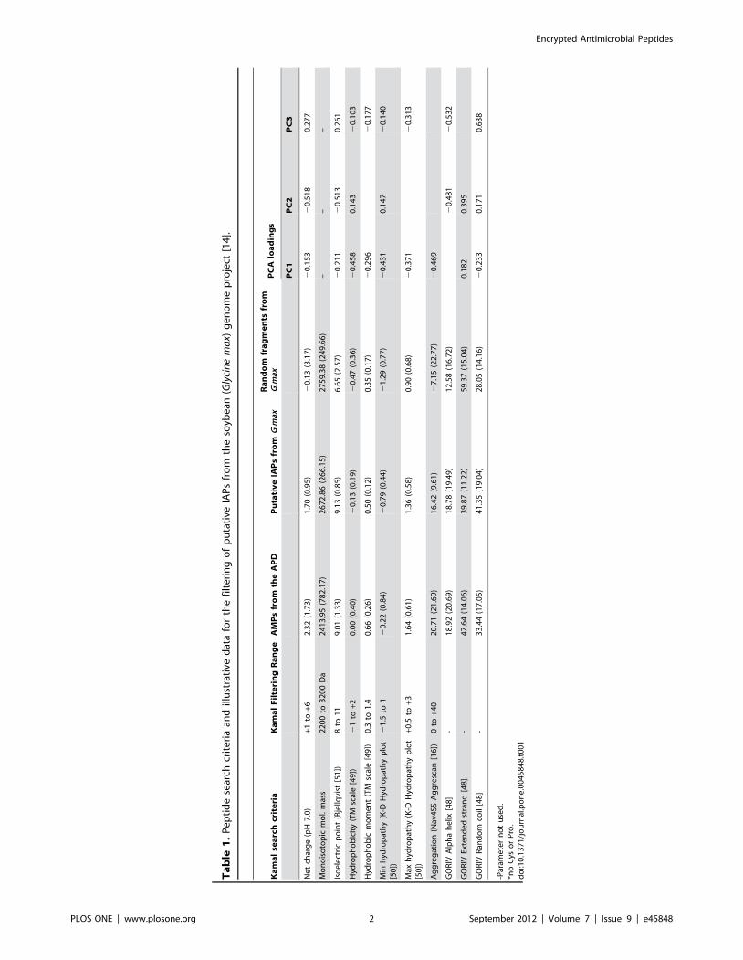
activities. This is illustrated in Table 1 by application to sequences
from the soybean (Glycine max) genome project [14]. Here, Table 1,
we compare calculated physicochemical properties of protein
fragments to a reference range of values extracted from a sample
of antimicrobial peptides (AMPs) of the Antimicrobial Peptide
Database, APD [[15]; (http://aps.unmc.edu/AP/main.php)]. The
list of reference AMPs and their calculated physicochemical
properties are available as Table S1. Putative IAPs from the fore
mentioned database were selected if all evaluated physicochemical
properties of each protein fragment were within the reference
peptide set limits, otherwise were discarded. Reference values
chosen here relate to a simple set of physicochemical properties of
relevance to peptide interactions with membranes as putative
descriptors of peptide activity [12,16,17,18]. Approximately 500
putative IAPs were filtered from the G. Max expressed sequence
tags (EST) database (Table S1). In addition, for comparison, five
hundred additional peptides from 20 to 30 amino acids were
selected at random from proteins of the same database (Table S1).
From the summary data (Table 1) it is clear that the average
estimated parameters for the filtered G. Max IAP candidates are
generally closer to the APD than to the random set of peptides, as
is inevitable from the selection criteria used by Kamal. However,
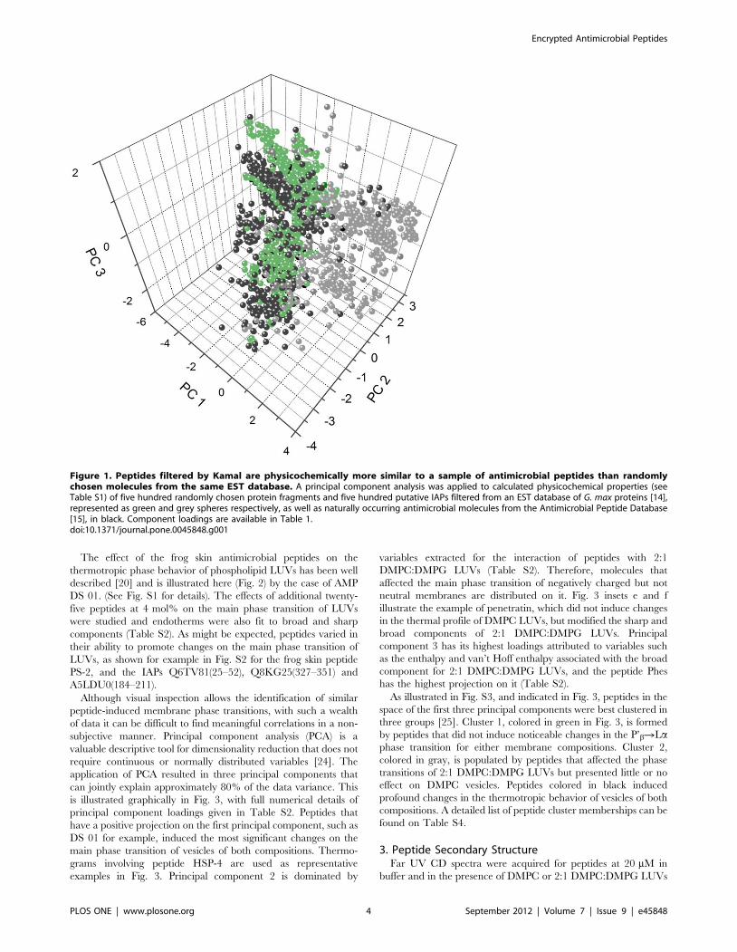
simple averages just paint a crude picture. A principal component
analysis applied to the resulting data (Fig. 1) demonstrates more
clearly how the putative IAPs filtered by Kamal (green spheres)
show superior overall physicochemical similarity to the sample of
AMPs (black spheres) than randomly chosen fragments of soy
proteins (grey spheres). Thus, if the premises are correct, these
have a higher probability of being antimicrobial. The first
principal component results mainly from various hydrophobicity-
derived physicochemical properties, while the second and third by
peptide charge and secondary structure parameters (Table 1).
Although the coincidence of the AMP and IAP clusters (Fig. 1) is
far from exact, this putative IAP set should contain promising
antimicrobial candidates.
To verify the actual antimicrobial potential of the sequences
filtered by Kamal, fifteen putative IAPs were arbitrarily selected
from a more extensive search covering a range of organisms
(Table 2), and chemically synthesized. Additionally, eleven AMPs
from frog skin secretions described by our group were used as
controls for naturally occurring molecules, together with the cell-
penetrating peptide penetratin, which is also antimicrobial [19].
Table 2 demonstrates that six out of the fifteen synthesized
putative IAPs did present a minimal inhibitory concentration
(MIC) against at least one of the tested microorganisms, the
human pathogens E. coli, S. aureus and P. aeruginosa and the
phytopathogenic bacterium X. axonopodis pv. glycines. Two peptides,
Q6TV81(25–52) from the ORF 107 of the bovine papular
stomatitis virus and A5LDU0(184–211), a fragment of the enzyme
pseudouridine synthase of Streptococcus pneumoniae SP3-BS71, had
MICs comparable to naturally occurring antimicrobial peptides.
Building on this, we have utilised biophysical assays on peptide
and membrane interactions in order to identify similarly acting
molecules and provide insights on the physicochemical require-
ments for peptide function.
2. Categorization of Peptide Interaction with ModelPhospholipid Vesicles by DSC
A number of studies elsewhere show that membrane active
compounds affect the thermotropic behaviour of phospholipid
vesicles in ways that can be related to their mechanism of action
[20,21]. Consequently we here used DSC to study the effect of
IAPs and naturally occurring antimicrobial peptides on the main
phase transition of LUVs composed of DMPC and 2:1
DMPC:DMPG.
DSC heating scans of DMPC LUVs showed the anticipated
endothermic transitions typical of the well-characterized gel to
liquid crystalline (P’bRLa) thermal phase transitions [21,22].
Thermograms were deconvoluted and fit to a non-two state model
revealing two peaks, a broad (Tm = 23.4uC) and a sharp
component (Tm = 24.1uC) in agreement with earlier work [22].
The incorporation of DMPG to the mixture, which by itself has a
transition temperature of 23.9uC, resulted in transition tempera-
tures of 23.6uC for the broad component and 24.3uC for the sharp
component as well as comparable transition enthalpies (Table S2)
[23].
Encrypted Antimicrobial Peptides
PLOS ONE | www.plosone.org 3 September 2012 | Volume 7 | Issue 9 | e45848
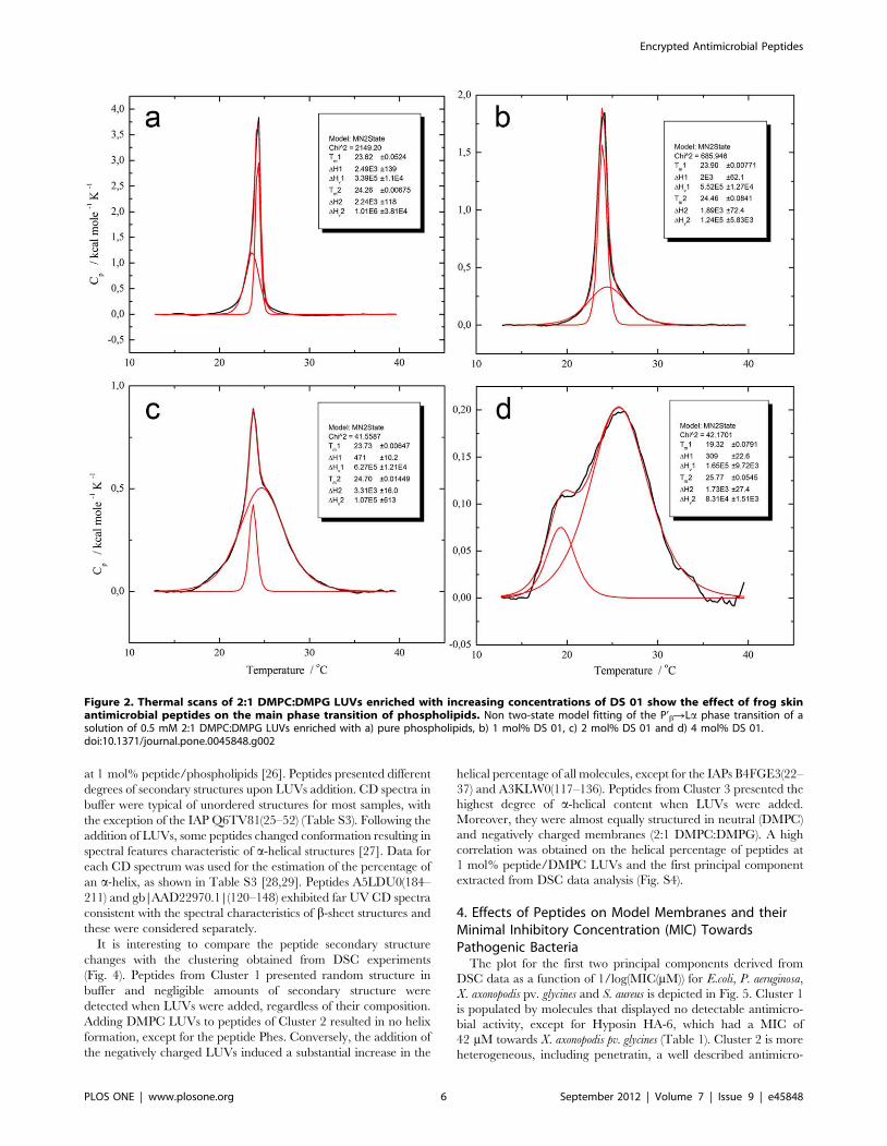
The effect of the frog skin antimicrobial peptides on the
thermotropic phase behavior of phospholipid LUVs has been well
described [20] and is illustrated here (Fig. 2) by the case of AMP
DS 01. (See Fig. S1 for details). The effects of additional twenty-
five peptides at 4 mol% on the main phase transition of LUVs
were studied and endotherms were also fit to broad and sharp
components (Table S2). As might be expected, peptides varied in
their ability to promote changes on the main phase transition of
LUVs, as shown for example in Fig. S2 for the frog skin peptide
PS-2, and the IAPs Q6TV81(25–52), Q8KG25(327–351) and
A5LDU0(184–211).
Although visual inspection allows the identification of similar
peptide-induced membrane phase transitions, with such a wealth
of data it can be difficult to find meaningful correlations in a non-
subjective manner. Principal component analysis (PCA) is a
valuable descriptive tool for dimensionality reduction that does not
require continuous or normally distributed variables [24]. The
application of PCA resulted in three principal components that
can jointly explain approximately 80% of the data variance. This
is illustrated graphically in Fig. 3, with full numerical details of
principal component loadings given in Table S2. Peptides that
have a positive projection on the first principal component, such as
DS 01 for example, induced the most significant changes on the
main phase transition of vesicles of both compositions. Thermo-
grams involving peptide HSP-4 are used as representative
examples in Fig. 3. Principal component 2 is dominated by
variables extracted for the interaction of peptides with 2:1
DMPC:DMPG LUVs (Table S2). Therefore, molecules that
affected the main phase transition of negatively charged but not
neutral membranes are distributed on it. Fig. 3 insets e and f
illustrate the example of penetratin, which did not induce changes
in the thermal profile of DMPC LUVs, but modified the sharp and
broad components of 2:1 DMPC:DMPG LUVs. Principal
component 3 has its highest loadings attributed to variables such
as the enthalpy and van’t Hoff enthalpy associated with the broad
component for 2:1 DMPC:DMPG LUVs, and the peptide Phes
has the highest projection on it (Table S2).
As illustrated in Fig. S3, and indicated in Fig. 3, peptides in the
space of the first three principal components were best clustered in
three groups [25]. Cluster 1, colored in green in Fig. 3, is formed
by peptides that did not induce noticeable changes in the P’bRLaphase transition for either membrane compositions. Cluster 2,
colored in gray, is populated by peptides that affected the phase
transitions of 2:1 DMPC:DMPG LUVs but presented little or no
effect on DMPC vesicles. Peptides colored in black induced
profound changes in the thermotropic behavior of vesicles of both
compositions. A detailed list of peptide cluster memberships can be
found on Table S4.
3. Peptide Secondary StructureFar UV CD spectra were acquired for peptides at 20 mM in
buffer and in the presence of DMPC or 2:1 DMPC:DMPG LUVs
Figure 1. Peptides filtered by Kamal are physicochemically more similar to a sample of antimicrobial peptides than randomlychosen molecules from the same EST database. A principal component analysis was applied to calculated physicochemical properties (seeTable S1) of five hundred randomly chosen protein fragments and five hundred putative IAPs filtered from an EST database of G. max proteins [14],represented as green and grey spheres respectively, as well as naturally occurring antimicrobial molecules from the Antimicrobial Peptide Database[15], in black. Component loadings are available in Table 1.doi:10.1371/journal.pone.0045848.g001
Encrypted Antimicrobial Peptides
PLOS ONE | www.plosone.org 4 September 2012 | Volume 7 | Issue 9 | e45848

Ta
ble
2.
Pe
pti
de
sp
rim
ary
stru
ctu
res,
sou
rce
pro
tein
s,o
rgan
ism
san
dm
inim
um
inh
ibit
ory
acti
vity
(MIC
)ag
ain
stp
ath
og
en
icb
acte
ria.
Pe
pti
de
na
me
*P
rim
ary
stru
ctu
res
MIC
s( m
M)
So
urc
ep
rote
inS
ou
rce
org
an
ism
Xan
tho
mo
nas
axo
no
po
dis
pv.
gly
cin
es
Esch
eri
chia
coli
Sta
ph
ylo
cocc
us
aure
us
Pse
ud
om
on
asae
rug
ino
sa
P6
14
58
(35
–6
0)
FKQ
FHFK
DFN
RA
FGFM
TR
VA
LQA
EKL
Pte
rin
-4-a
lph
a-ca
rbin
ola
min
eM
us
mu
scu
lus
40
40
ND
A8
0
B0
CZ
J3(1
04
–1
30
)IA
AA
QR
ITSG
AA
DIA
INW
AG
GLH
HA
KK
His
ton
ed
eac
ety
lase
com
ple
xLa
cca
ria
bic
olo
rN
DA
ND
AN
DA
ND
A
A4
HW
34
(18
7–
21
7)
LVQ
RFH
AY
LHK
FREA
FMN
VG
AA
AA
VEG
TK
AA
Glu
tath
ion
esy
nth
eta
seLe
ish
ma
nia
infa
ntu
m3
7N
DA
ND
AN
DA
Q8
RW
88
(70
–9
5)
GH
RG
ALK
DW
VQ
AA
GG
AV
AA
FDFT
TK
GA
lph
a-am
ylas
eC
itru
sre
ticu
lata
ND
AN
DA
ND
AN
DA
Q6
TV
81
(25
–5
2)
AA
AA
AA
AIK
MLM
DLV
NER
IMA
LNK
KA
KK
OR
F10
7vi
rio
nm
orp
ho
ge
ne
sis
Bo
vin
ep
ap
ula
rst
om
ati
tis
viru
s,
11
02
02
0
O4
33
12
(33
–6
2)
FIN
KA
GK
LQSQ
LRT
TV
VA
AA
AFL
DA
FQK
VA
Me
tast
asis
sup
pre
sso
rp
rote
in1
Ho
mo
sap
ien
s8
04
0N
DA
ND
A
Q8
KG
25
(32
7–
35
1)
FVT
NSK
RLA
EGIE
KG
VG
NSI
LIK
VN
Eno
lase
2C
hlo
rob
ium
tep
idu
mN
DA
ND
AN
DA
ND
A
P9
46
92
(92
9–
95
5)
KLK
KLL
AG
QK
DG
LLG
QIA
AM
SDLY
TK
KP
yru
vate
-fe
rre
do
xin
oxi
do
red
uct
ase
Des
ulf
ovi
bri
oa
fric
an
us
ND
AN
DA
ND
AN
DA
B4
FGE3
(22
–3
7)
KA
GLQ
FPV
GR
IAR
FLK
His
ton
eH
2A
Zea
ma
ysN
DA
ND
AN
DA
ND
A
A3
KLW
0(1
17
–1
36
)FK
ALR
ALR
LED
LRIP
TSY
IKR
ub
isco
larg
ech
ain
An
iso
ph
ylle
ap
om
ifer
aN
DA
ND
AN
DA
ND
A
Q7
YR
I0(9
–2
8)
LAK
RR
VLT
LLR
QLR
RV
SPSS
Inte
rfe
ron
alp
ha
Bo
sta
uru
s,
21
4N
DA
ND
A
gb
|AC
U2
40
18
.1|(
73
–1
01
)G
LWQ
IFSS
KEE
GK
DN
SQQ
KSK
GD
QA
KEL
Un
kno
wn
pro
tein
Gly
cin
em
ax
ND
AN
DA
ND
AN
DA
gb
|AA
D2
29
70
.1|(
12
0–
14
8)
VW
TT
AM
EKSS
AA
NFS
MSR
NQ
RR
SSLH
SLT
reh
alas
e1
GM
TR
E1G
lyci
ne
ma
xN
DA
ND
AN
DA
ND
A
Q9
XEY
7(1
20
–1
48
)SL
WK
NLS
RK
ISG
AV
KA
QP
DL
HT
LLP
LPG
ST
reh
alas
e1
GM
TR
E1G
lyci
ne
ma
xN
DA
ND
AN
DA
ND
A
A5
LDU
0(1
84
–2
11
)G
KFH
QIK
KM
FLSV
GV
KV
TSL
KR
IQFG
DF
Pse
ud
ou
rid
ine
syn
thas
eSt
rep
toco
ccu
sp
neu
mo
nia
e1
02
0N
DA
20
DS
01
GLW
STIK
QK
GK
EAA
IAA
AK
AA
GQ
AA
LGA
LN
on
eP
hyl
lom
edu
sao
rea
des
,1
11
16
Nat
tere
rin
-1G
LKD
MIK
NLA
KEA
AV
KLA
GA
VIN
KFS
PQ
PQ
No
ne
Eup
emp
hix
na
tter
eri
,1
54
01
0
PS
-2FL
SLIP
HA
INA
VST
LVH
HF
No
ne
Ph
yllo
med
usa
hyp
och
on
dri
alis
12
11
5N
DA
ND
A
DS
01
(1–
12
)G
LWST
IKQ
KG
KE*
*D
S0
1P
hyl
lom
edu
sao
rea
des
ND
AN
DA
ND
AN
DA
Syp
hax
inG
VLD
ILK
GA
AK
DLA
GH
VA
TK
VIN
KI
No
ne
Lep
tod
act
ylu
ssy
ph
ax
25
12
50
10
0
HSP
-4G
IGD
ILK
NLA
KA
AG
KA
ALH
AV
GES
LN
on
eH
ypsi
bo
as
pu
nct
atu
s,
21
91
97
7
Pse
ud
inB
GLN
TLK
KV
IQG
LHEV
IKLV
NN
HA
No
ne
Pse
ud
isb
olb
od
act
yla
,2
66
50
Ph
es
FFFD
TLK
NLA
GK
VIG
ALT
No
ne
Hyp
sib
oa
sp
un
cta
tus
33
62
62
12
5
Mag
ain
in-2
amid
eG
IGK
FLH
SAK
KFG
KA
FVG
EIM
NS
No
ne
Xen
op
us
laev
is1
31
3N
DA
26
Hyp
osi
nH
A-6
LRP
AIL
VR
VK
GK
GL
No
ne
Ph
yllo
med
usa
hyp
och
on
dri
alis
42
ND
AN
DA
ND
A
Pe
ne
trat
inR
QIK
IWFQ
NR
RM
KW
KK
An
ten
nap
ed
iaco
mp
lex
Dro
sop
hila
mel
an
og
ast
er,
27
28
2
ND
A=
No
n-d
ete
ctab
leac
tivi
ty.
*pu
blic
atio
nn
ame
s,U
nip
rotK
Bo
rg
en
eb
ank
en
trie
sfo
llow
ed
by
the
firs
tan
dla
stre
sid
ue
sin
bra
cke
ts.
**fr
ee
carb
oxy
-te
rmin
us
pe
pti
de
.d
oi:1
0.1
37
1/j
ou
rnal
.po
ne
.00
45
84
8.t
00
2
Encrypted Antimicrobial Peptides
PLOS ONE | www.plosone.org 5 September 2012 | Volume 7 | Issue 9 | e45848
at 1 mol% peptide/phospholipids [26]. Peptides presented different
degrees of secondary structures upon LUVs addition. CD spectra in
buffer were typical of unordered structures for most samples, with
the exception of the IAP Q6TV81(25–52) (Table S3). Following the
addition of LUVs, some peptides changed conformation resulting in
spectral features characteristic of a-helical structures [27]. Data for
each CD spectrum was used for the estimation of the percentage of
an a-helix, as shown in Table S3 [28,29]. Peptides A5LDU0(184–
211) and gb|AAD22970.1|(120–148) exhibited far UV CD spectra
consistent with the spectral characteristics of b-sheet structures and
these were considered separately.
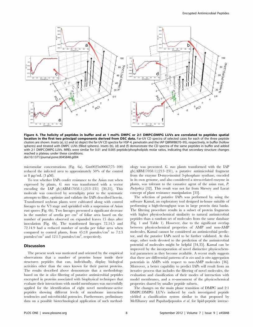
It is interesting to compare the peptide secondary structure
changes with the clustering obtained from DSC experiments
(Fig. 4). Peptides from Cluster 1 presented random structure in
buffer and negligible amounts of secondary structure were
detected when LUVs were added, regardless of their composition.
Adding DMPC LUVs to peptides of Cluster 2 resulted in no helix
formation, except for the peptide Phes. Conversely, the addition of
the negatively charged LUVs induced a substantial increase in the
helical percentage of all molecules, except for the IAPs B4FGE3(22–
37) and A3KLW0(117–136). Peptides from Cluster 3 presented the
highest degree of a-helical content when LUVs were added.
Moreover, they were almost equally structured in neutral (DMPC)
and negatively charged membranes (2:1 DMPC:DMPG). A high
correlation was obtained on the helical percentage of peptides at
1 mol% peptide/DMPC LUVs and the first principal component
extracted from DSC data analysis (Fig. S4).
4. Effects of Peptides on Model Membranes and theirMinimal Inhibitory Concentration (MIC) TowardsPathogenic Bacteria
The plot for the first two principal components derived from
DSC data as a function of 1/log(MIC(mM)) for E.coli, P. aeruginosa,
X. axonopodis pv. glycines and S. aureus is depicted in Fig. 5. Cluster 1
is populated by molecules that displayed no detectable antimicro-
bial activity, except for Hyposin HA-6, which had a MIC of
42 mM towards X. axonopodis pv. glycines (Table 1). Cluster 2 is more
heterogeneous, including penetratin, a well described antimicro-
Figure 2. Thermal scans of 2:1 DMPC:DMPG LUVs enriched with increasing concentrations of DS 01 show the effect of frog skinantimicrobial peptides on the main phase transition of phospholipids. Non two-state model fitting of the P’bRLa phase transition of asolution of 0.5 mM 2:1 DMPC:DMPG LUVs enriched with a) pure phospholipids, b) 1 mol% DS 01, c) 2 mol% DS 01 and d) 4 mol% DS 01.doi:10.1371/journal.pone.0045848.g002
Encrypted Antimicrobial Peptides
PLOS ONE | www.plosone.org 6 September 2012 | Volume 7 | Issue 9 | e45848

bial agent, as well as inactive peptides. Molecules belonging to
Cluster 3 were not only the most active towards the three assayed
microorganisms, but also had collectively the lowest MIC values
(highest 1/log(MIC(mM)), as can be seen on Table S4.
5. Inhibition of the Fixation of P. pachyrhizi Spores on theSurface of G. max Leaves and on Plants Transformed withgb|ABM17058.1|(213–231)
In order to evaluate if peptides encrypted in G. max proteins can
confer resistance to the plant’s natural pathogens, two novel IAPs
were filtered and synthesized. Gm0025x00667(75–100), primary
structure RWRFLRKISSVHMFSVKALDDFRQL, is a fragment
of the enzyme flavonoid 3-hydroxylase, while Gm0026x00785(77–
103), primary structure HKMDLHWYLRTLEEVVIR-
ALQRFQFR, is derived from the lipoate-protein ligase B. They
both inhibited the in vitro growth of X. axonopodis pv. glycines, the
causative agent of the bacterial pustule disease, at 5 and 10 mM,
respectively. IAPs were also tested ex vivo for the inhibition of the
fixation of asian rust spores on G. max leaves. In general, IAPs co-
incubated with spores of P. pachyrhizi on the surface of leaves of G.
max decreased significantly the area occupied by uredias at
Figure 3. Principal component analysis of the model fitted main phase transitions of LUVs added with peptides at 4 mol%highlight similarities on membranes thermal behaviors. A matrix describing the effect of peptides on the transition temperature (Tm),enthalpy (DH) and cooperativity (DHVH) of the broad and sharp components of DMPC and 2:1 DMPC:DMPG LUV thermal transitions was assembledand standardized (Table S2) and a PCA was applied to the resulting data. Additionally, the coordinates of peptides in the first three principalcomponents were submitted to a mixture modeling clustering algorithm, resulting in an optimal of three peptide clusters, shown here in differentcolors (Fig. S3). Thermograms demonstrating the effect of selected peptides on the main phase transition of membranes are shown as representativeexamples: HSP-4 on (a) 2:1 DMPC:DMPG and (b) DMPC LUVs represents cluster 3, in black, the IAP Q8RW88(70–95) on (c) 2:1 DMPC:DMPG and (d)DMPC LUVs represents cluster 1, in green, and penetratin added to (e) DMPC and (f) 2:1 DMPC:DMPG LUVs represents cluster 2, in grey. Groupborders are illustrational.doi:10.1371/journal.pone.0045848.g003
Encrypted Antimicrobial Peptides
PLOS ONE | www.plosone.org 7 September 2012 | Volume 7 | Issue 9 | e45848
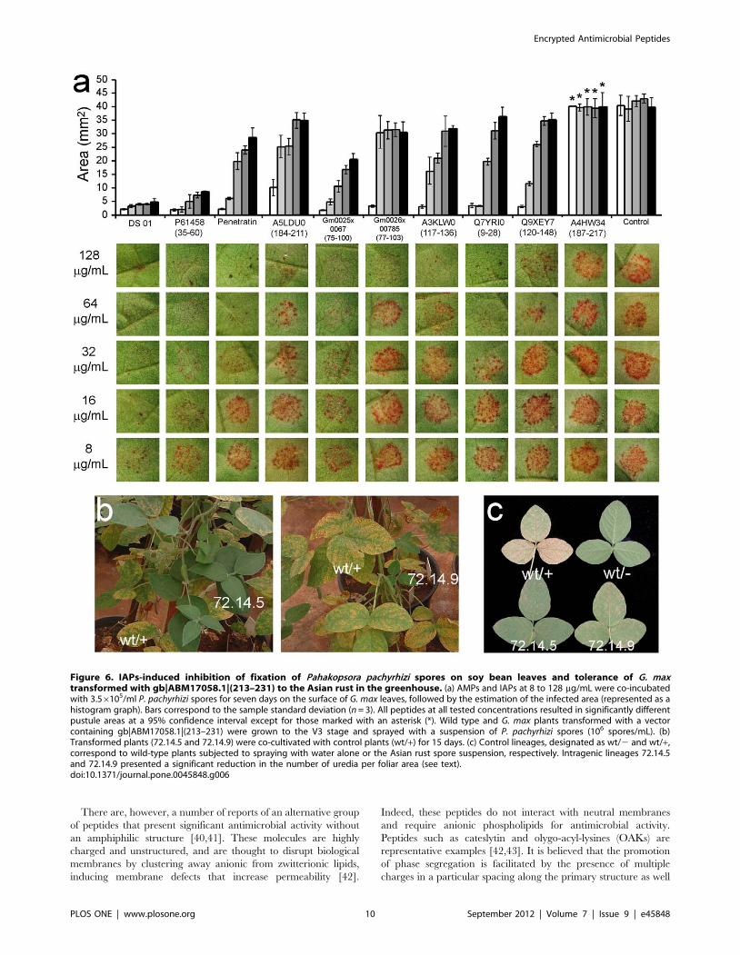
micromolar concentrations (Fig. 6a). Gm0025x00667(75–100)
reduced the infected area to approximately 50% of the control
at 8 mg/mL (3 mM).
To test whether IAPs confer resistance to the Asian rust when
expressed by plants, G. max was transformed with a vector
encoding the IAP gb|ABM17058.1|(213–231) [30,31]. This
molecule was conceived by serendipity prior to the systematic
attempts to filter, optimize and validate the IAPs described herein.
Transformed soybean plants were cultivated along with control
lineages to the V3 stage and sprinkled with a suspension of Asian
rust spores (Fig. 6b). Two lineages presented a significant decrease
in the number of uredia per cm2 of foliar area based on the
number of pustules observed on expanded leaves 15 days after
inoculation (Fig. 6b). The transformed lineages 72.14.5 and
72.14.9 had a reduced number of uredia per foliar area when
compared to control plants, from 4568 pustules/cm2 to 763
pustules/cm2 and 1263 pustules/cm2, respectively.
Discussion
The present work was motivated and oriented by the empirical
observations that a number of proteins house inside their
structures peptides that can, individually, display biological
activities other than the ones known for their parent proteins.
The results described above demonstrate that a methodology
based on the in silico filtering of putative antimicrobial peptides
encrypted in proteins associated with biophysical techniques that
evaluate their interactions with model membranes was successfully
applied for the identification of eight novel membrane-active
peptides showing different levels of membrane interaction
tendencies and microbicidal potencies. Furthermore, preliminary
data on a possible biotechnological application of such method-
ology was presented. G. max plants transformed with the IAP
gb|ABM17058.1|(213–231), a putative antimicrobial fragment
from the enzyme D-myo-inositol 3-phosphate synthase, encoded
in its own genome, and also considered a stress-related enzyme in
plants, was tolerant to the causative agent of the asian rust, P.
Pachyrhizi [32]. This result was not far from Shewry and Lucas
concept of plant resistance manipulation [33].
The selection of putative IAPs was performed by using the
software Kamal, an exploratory tool designed in-house suitable of
performing a high-throughput scan in large protein data banks.
The filtering procedure results in a subset of protein fragments
with higher physicochemical similarity to natural antimicrobial
peptides than a random set of molecules from the same database
(Fig. 1 and Table 1). However, due to the significant overlap
between physicochemical properties of AMP and non-AMP
molecules, Kamal cannot be considered an antimicrobial predic-
tor, and the putative IAPs need to be further validated. At this
stage, other tools devoted to the prediction of the antimicrobial
potential of molecules might be helpful [34,35]. Kamal can be
improved by the incorporation of novel distinctive physicochem-
ical parameters as they become available. A recent study suggests
that there are differential patterns of in vivo and in vitro aggregation
potentials in AMPs with respect to non-AMP molecules [36].
Moreover, a better capability to predict IAPs will result from an
iterative process that includes the filtering of novel molecules, the
evaluation and classification of their modes of interaction with
model membranes, and a re-assessment of the physicochemical
properties shared by smaller peptide subsets.
The changes on the main phase transition of DMPC and 2:1
DMPC:DMPG LUVs induced by each investigated peptide
yielded a classification system similar to that proposed by
McElhaney and Papahadjopoulos et al. for lipid-peptide interac-
Figure 4. The helicity of peptides in buffer and at 1 mol% DMPC or 2:1 DMPC:DMPG LUVs are correlated to peptides spatiallocation in the first two principal components derived from DSC data. Far-UV CD spectra of selected cases for each of the three peptideclusters are shown. Insets (a), (c) and (e) depict the far-UV CD spectra for HSP-4, penetratin and the IAP Q8RW88(70–95), respectively, in buffer (hollowspheres) and titrated with DMPC LUVs (filled spheres). Insets (b), (d) and (f) demonstrate the CD spectra of the same peptides in buffer and addedwith 2:1 DMPC:DMPG LUVs. MREs were similar for 0.01 and 0.005 peptide/phospholipids molar ratios, indicating that secondary structure changesreached a plateau under these conditions.doi:10.1371/journal.pone.0045848.g004
Encrypted Antimicrobial Peptides
PLOS ONE | www.plosone.org 8 September 2012 | Volume 7 | Issue 9 | e45848

tions and their relative locations at lipid bilayers [20,37,38]. This
allows one to estimate the antimicrobial potential of a given
molecule prior to antimicrobial assays. In the currently proposed
model, increasing alterations on the main phase transition of
membranes are associated with projection on the first principal
component, as presented in Fig. 3. It is interesting to notice that
peptides that interacted exclusively with 2:1 DMPC:DMPG LUVs
had less pronounced effects on the main phase transition of
membranes, consistent with the general interpretation that
interactions dominated by electrostatic effects lead to more
superficial peptide locations, an observation supported by CD
data (Fig. 4) [20]. Under these conditions, a broad and significant
antimicrobial activity can be considered a natural consequence of
a deep interaction between peptides and neutral phospholipids
bilayers, attainable by molecules with a high helical propensity in
this specific environment. Peptides from cluster 3 displayed such
properties, and therefore concur with the hypothesis that a
globally amphiphilic conformation is necessary for the disruption
of bacterial membranes [39]. Nevertheless, only three IAPs were
clustered along with these molecules, indicating that members of
this group have a distinctive balance between physicochemical
properties that is uncommon in protein fragments.
Figure 5. Peptides antimicrobial activities are correlated to their spatial location in the first two principal components derived fromDSC data. Antimicrobial activity is represented as the anti-log of the minimum inhibitory concentration (MIC) in micromoles 1/log(MIC(mM)).Peptides MICs can be verified in Table 2. Tested microorganisms were (a) Escherichia coli, (b) Staphylococcus aureus, (c) Pseudomonas aeruginosa andthe phytopathogenic bacterium (d) Xanthomonas axonopodis pv. glycines.doi:10.1371/journal.pone.0045848.g005
Encrypted Antimicrobial Peptides
PLOS ONE | www.plosone.org 9 September 2012 | Volume 7 | Issue 9 | e45848
There are, however, a number of reports of an alternative group
of peptides that present significant antimicrobial activity without
an amphiphilic structure [40,41]. These molecules are highly
charged and unstructured, and are thought to disrupt biological
membranes by clustering away anionic from zwitterionic lipids,
inducing membrane defects that increase permeability [42].
Indeed, these peptides do not interact with neutral membranes
and require anionic phospholipids for antimicrobial activity.
Peptides such as cateslytin and olygo-acyl-lysines (OAKs) are
representative examples [42,43]. It is believed that the promotion
of phase segregation is facilitated by the presence of multiple
charges in a particular spacing along the primary structure as well
Figure 6. IAPs-induced inhibition of fixation of Pahakopsora pachyrhizi spores on soy bean leaves and tolerance of G. maxtransformed with gb|ABM17058.1|(213–231) to the Asian rust in the greenhouse. (a) AMPs and IAPs at 8 to 128 mg/mL were co-incubatedwith 3.56105/ml P. pachyrhizi spores for seven days on the surface of G. max leaves, followed by the estimation of the infected area (represented as ahistogram graph). Bars correspond to the sample standard deviation (n = 3). All peptides at all tested concentrations resulted in significantly differentpustule areas at a 95% confidence interval except for those marked with an asterisk (*). Wild type and G. max plants transformed with a vectorcontaining gb|ABM17058.1|(213–231) were grown to the V3 stage and sprayed with a suspension of P. pachyrhizi spores (106 spores/mL). (b)Transformed plants (72.14.5 and 72.14.9) were co-cultivated with control plants (wt/+) for 15 days. (c) Control lineages, designated as wt/2 and wt/+,correspond to wild-type plants subjected to spraying with water alone or the Asian rust spore suspension, respectively. Intragenic lineages 72.14.5and 72.14.9 presented a significant reduction in the number of uredia per foliar area (see text).doi:10.1371/journal.pone.0045848.g006
Encrypted Antimicrobial Peptides
PLOS ONE | www.plosone.org 10 September 2012 | Volume 7 | Issue 9 | e45848

as sufficient hydrophobicity to partition into membranes [39]. We
propose that the peptides distributed along the second principal
component which present significant antimicrobial activity disrupt
membranes by such mechanism. The peptides P61458(35–60),
A5LDU0(184–211) and penetratin, for example, have borderline
hydrophobicity as well as regularly spaced positively charged
amino acid residues.
The expression of antimicrobial peptides in plants is known to
confer increased resistance to phytopathogens [44]. Indeed,
peptides such as esculentin-1, dermaseptin SI and hCAP18/
LL37 were used for the transformation of Nicotiana tabacum, Solanum
tuberosum and Brassica rapa and conferred resistance to fungal and
bacterial pathogens besides providing the plant with insecticidal
properties [44,45,46]. Preliminary results indicate that G.max
expressing the IAP gb|ABM17058.1|(213–231), a fragment of the
enzyme D-myo-inositol 3-phosphate synthase, presented increased
resistance to the Asian rust providing similar results to those
obtained with natural antimicrobial peptides (Fig. 6b). It is
plausible that IAPs with improved antimicrobial and antifungal
spectra are still left undiscovered in the soybean genome, and that
novel generations of intragenic plants that are tolerant to a wide
array of phytopathogens may be developed.
The proposed strategy of screening bioactive peptides as
fragments inside proteins inspired by the natural peptide release
and activation under enzymatic proteolysis found across various
metabolic processes appears to be universal but, to our best
understanding, restricted to certain classes of proteins. Neverthe-
less, the additional element we modestly hope to append to
nature’s magnificent evolutionary plasticity and energy effective-
ness exemplified in these processes is the introduction of the
theoretical enzyme-free cleavages concept as a complementary
mode of revealing biologically active peptides encrypted in
protein sequences to the existing physiological and microorgan-
ism-based ones. This next step of ‘‘oriented-protein processing’’
does not need to be restrained by enzymatic specificities or
optimum cleavage conditions in vivo and/or in vitro once it may
take advantage of the computational predictions capabilities,
nucleic acids and peptide synthesis methodologies currently
available. The implications of the present findings lead us to: 1.
A wider and exciting scenario for bioprospecting new molecules
using the ever-growing genomic and proteomic data banks that
could be validated by the appropriated bioassays; 2. Expanding
the range of existing biotechnological processes applied to dairy
and food processing in general; 3. The use of genomic and
physiological information from a given species as a possible inner
source of new bioactive peptides may represent an inventive
alternative to the transgenic technology, once the genetic material
to be used belongs to the host organism and not to exogenous
sources.
Materials and Methods
1. MaterialsDMPC (Dimyristoylphosphatidylcholine) and DMPG (Dimyr-
istoylphosphatidylglycerol) were purchased from Avanti (Avanti
Polar Lipids, AL, USA). Peptides are identified by their
publication names, by their UniProtKB or genebank (gb|)
accession number followed by the indication of the first and last
aminoacid residues in brackets. Penetratin, B4FGE3(22–37),
A3KLW0(117–136), Q7YRI0(9–28) were purchased from Im-
munoKontact (AMS Biotechnology, UK); gb|ACU24018.1|(73–
101), gb|AAD22970.1|(120–148) and Q9XEY7(120–148) were
purchased from JPT Peptide Technologies GmbH, Germany. The
IAPs Q8KG25(327–351), P94692(929–955), P61458(35–60),
B0CZJ3(104–130), A4HW34(187–217), Q8RW88(70–95),
Q6TV81(25–52), O43312(33–62), A5LDU0(184–211),
P83637(1–12) as well as DS 01 (P83637), DS 01(1–12), Natter-
erin-1 (P86913), Syphaxin (P85279), Phes (HQ012497) HSP-4
(JF916646), Pseudin B (P86915), PS-2 (P84567), Magainin-2
amide (P11006), Hyposin HA-6 (P86921) were synthesized in-
house by solid-phase chemistry. Peptides were purified, mass
analyzed using an Ultraflex III (Bruker Daltonics, Germany) and
quantified by their corresponding molar absorptivities or by the
method of Wadell [47].
2. Filtering of IAPsThe software Kamal v1.0 alpha was written in C++ using the
public libraries SQLite, wxWidgets and Lua. The application was
developed using the IDE code::Blocks and compiled with Mingw3.
More details will be given elsewhere. Entries under the label of
‘‘frog peptides’’ were extracted from the Antimicrobial Peptide
Database (http://aps.unmc.edu/AP/main.php), resulting in a
data set of 487 molecules (Table S1). Net charge, molecular mass,
isoelectric point (pI), average hydrophobicity, hydrophobic
moment, minimum and maximum on a Kyte-Doolittle hydrop-
athy plot, aggregation potential (Na4vSS) using the AggreScan
algorithm and the peptides secondary structure according to the
GORIV algorithm were calculated for each molecule [16,48]. The
transmembrane tendency (TM) scale was used for the calculation
of the peptides average hydrophobicity, hydrophobic moment and
the Kyte-Doolittle scale was used for the calculation of the
minimum and maximum hydrophobicities on a K-D hydropathy
plot using a 9 residue window [49,50]. Isoelectric point was
calculated according to pK values extracted from the literature
[51]. Average properties were calculated for this data set (Table
S1) and used to extract minimum and maximum values for the
filtering of IAPs from different databases (Table 1).
3. Lipid VesiclesDMPC and 2:1 DMPC:DMPG (w/w) were dissolved in
chloroform/methanol (3:1 v/v) at 10 mg/mL, dried as a thin
film on a rotary evaporator and left 3 hours under high vacuum.
Phospholipids were then dispersed in 20 mM sodium phosphate –
NaOH, 150 mM NaCl, pH 7.4 and hand-shaken until the
formation of a cloudy solution, which was then passed 19 times
through a 100 nm polycarbonate membrane at 30uC for the
formation of large unilamelar vesicles (LUVs). Phospholipid
concentration was estimated [52].
4. Differential Scanning Calorimetry (DSC)Thermograms were obtained using a VP-DSC (MicroCal Inc.,
MA, USA) at a temperature range from 10 to 40uC and a
scanning rate of 1uC/min. Blank (buffer baseline) thermograms
and 0.5 mM DMPC or 2:1 (w/w) DMPC:DMPG LUVs were
acquired as reference. Peptides were added to fresh samples of
0.5 mM LUVs at a concentration of 20 mM (0.04 mol/mol
peptide/phospholipids) at room temperature, immediately fol-
lowed by DSC data acquisition. Each sample was subjected to
repeated thermal scans until there were no distinguishable changes
in the thermal profile of the main phospholipid phase transition
(P’bRLa) between scans. Data were concentration normalized,
baseline subtracted (linear connect), and fitted to a non two-state
transition with two peaks determined by the user applying the
MicroCal OriginTM software. Re-scans for selected cases were
acquired using fresh peptide and LUV solutions to check the
reproducibility of the data.
Encrypted Antimicrobial Peptides
PLOS ONE | www.plosone.org 11 September 2012 | Volume 7 | Issue 9 | e45848

5. Circular DichroismExperiments were conducted on a Jasco-J810 spectropolarim-
eter (Jasco International Co., Japan). Spectra were acquired at
room temperature from 194 to 260 nm as an average of 4 readings
using a 0.1 cm path length cell, data pitch of 0.2 nm and a
response time of 0.5 s. Data Scans of buffer and 2 mM DMPC
and 2:1 DMPC:DMPG LUVs solutions were acquired and
subtracted from each peptide data. Peptides were scanned at a
concentration of 20 mM in buffer and then 100 fold excess of
DMPC and 2:1 DMPC:DMPG LUVs were added, resulting in a
molar ratio of 0.01 peptide/phospholipids. The spectra were
converted to mean residue ellipticity and readings at [h]222 nm
and [h]208 were used to estimate a-helix percentages according to
two different methodologies [28,29]. Proximity to equilibrium was
verified by scanning peptides added with twice the LUVs
concentration, approximately 4 mM.
6. Minimum Inhibitory Concentration Assays (MIC)MICs were determined using the M7-A6 protocol from the
Clinical Laboratory Standards Institute (CLSI). Escherichia coli
ATCC 25922, Pseudomonas aeruginosa ATCC 27853, Staphylococcus
aureus ATCC 29313 were streaked in Mueller-Hinton agar, grown
overnight at 37uC, transferred to Mueller-Hinton broth and
incubated until readings at 600 nm reached the equivalent of 0.5
in the MacFarland scale. Initial bacterium inoculum of ,105
colony forming units/mL were transferred, along with serial 2-fold
dilutions of each peptide to 96-well plates and incubated for 12 h.
Highest peptide concentrations tested was 256 mg/mL. For each
peptide concentration, optical density (OD600) readings were
subtracted from that of the growth medium and divided by the
positive control (100% bacterial growth). Inhibitory assays for X.
axonopodis pv. glycines were conducted by the same methodology
with longer incubation time (48 h).
7. In situ Germination Inhibition of PahakopsoraPachyrhizi Spores on Glycine Max Leaves’ Surface
P. pachyrhizi spores were scraped off infected G. max leaves and
were frozen at 280uC. Prior to the in situ assay, spores were
dissolved in Mili-QH water, incubated at 40uC for 40 min and
quantified using a Neubauer chamber. Fifty microliters of spore
suspension (3.56105/ml) were applied to the abaxial surface of leaf
disks (16 mm in diameter) detached from middle leaflet of the
youngest fully expanded trifoliate leaves from plants at V3 stage
(var. BR-16). Spores were then co-incubated with fifty microliters
of a peptide solution at concentrations ranging from 8 to 128 mg/
mL. Leaves were immediately placed on a Petri dish containing
moist filter papers and incubated at 21uC under 12 h-photoperiod
and photographed after 7 days. Images were used to measure the
infected area using the QUANT v1.0.1 software. Results displayed
herein correspond to the average of three separate experiments.
8. Plant Transformation and Asian Rust Tolerance AssayThe gene encoding the peptide gb|ABM17058.1|(213–231),
primary structure MIKAFKEATKVDKVVVLWTA, was syn-
thesized by Epoch Life Science Inc. (Sugar Land, TX, USA) and
cloned into the vector pBluKSPOXDCAHAS replacing the oxdc
gene [53]. The vector was used to transform soybean plants as
previously described [30,31]. Six intragenic soybean lines were
tested for tolerance to Asian rust (Phakopsora pachyrhizi). The plants
(20 plants per line at V3 stage [54]) were sprayed with a spore
suspension (106 spores/mL) and maintained in the greenhouse at
room temperature and sprinkling 4 times a day. Leaves were
photographed after 15 days and the number of pustules per square
centimeter counted.
9. Statistical AnalysesPrincipal Component Analysis was performed using the
princomp command of the R statistical software (http://www.
r-project.org). The data clustering was conducted using the
mclust command of the MCLUST R Package for normal
mixture modeling via EM, model-based clustering, discriminant
analysis and density estimation graph algorithm (http://www.stat.
washington.edu/mclust/). Three-dimensional plots were created
using Origin 7.0 (OriginLab Corp.).
Supporting Information
Figure S1 Thermal scans of 2:1 DMPC:DMPG LUVsenriched with increasing concentrations of DS 01 showthe effect of frog skin antimicrobial peptides on themain phase transition of phospholipids. Non two-state
model fitting of the P’bRLa phase transition of a solution of
0.5 mM 2:1 DMPC:DMPG LUVs enriched with a) pure
phospholipids, b) 1 mol% DS 01, c) 2 mol% DS 01 and d)
4 mol% DS 01. Compared with LUVs of the same composition,
samples enriched with 4 mol% DS 01 have a sharp component
that is shifted to lower temperatures (from 24.3 to 19.3uC) with a
lower transition enthalpy and cooperativity (DH from 2.2 to
0.3 kcal/mol and DHVH from 1000 to 160 kcal/mol). The broad
component shifts to higher temperatures (from 23.6 to 25.8uC),
has a slightly lower transition enthalpy (DH from 2.5 to 1.7 kcal/
mol), and becomes even broader (DHVH from 340 to 83 kcal/mol).
Total enthalpy associated with the main phase transition is
decreased to less than half (DH from 4.8 to 2.0 kcal/mol). These
effects are qualitatively the same as described by the McElhaney
group for other antimicrobial peptides [20].
(JPG)
Figure S2 Peptides induce distinct effects on thethermotropic phase behaviour of DMPC and 2:1DMPC:DMPG large unilamellar vesicles. The thermo-
grams for DMPC added with 4 mol% (a) PS-2 and (b)
Q6TV81(25–52) and 2:1 DMPC:DMPG added with (c)
Q8KG25(327–351) and (d) A5LDU0(184–211) are exemplified.
Insets contain the fitted parameters for the broad and sharp peak
components according to a non-two state transition model with
two manually assigned peaks. Shown thermograms were normal-
ized for the lipid sample mass.
(JPG)
Figure S3 Putative IAPs and antimicrobial peptides arebest clustered in three distinct groups. Optimal data
clustering of peptides in the first three principal components
obtained from the PCA analysis of data on Table S2 according to
the Bayesian Information criterion (BIC) is obtained when three
clusters are considered with variable volume, equal shape and
variable orientation (VEV). The ellipses superimposed to the
classification plot (on the right) correspond to the covariance of the
components.
(JPG)
Figure S4 The relative position of peptides along thefirst principal component derived from DSC data islinearly correlated to their percentual helicity at 1 mol%in DMPC LUVs. The Pearson correlation coefficient indicates a
high correlation (r2 = 0.86, p,0.0000001) between the relative
position of peptides at PC1 and their percentual helicity when
titrated with DMPC LUVs. The non-parametric Spearman’s rank
Encrypted Antimicrobial Peptides
PLOS ONE | www.plosone.org 12 September 2012 | Volume 7 | Issue 9 | e45848

correlation coefficient also pointed to a high degree of correlation
between both quantities (r= 0.72, p = 0.000018).
(JPG)
Table S1 Physicochemical properties of a sample offrog AMPs obtained from the Antimicrobial PeptideDatabase (http://aps.unmc.edu/AP/main.php) [15]
compared to a sample of putative IAPs filtered byKamal from Glycine max proteins (derived from theJoint Genome Institute - Glyma0.1c.pep.fa.gz) [14] aswell as randomly selected protein fragments from thesame G. max database.(XLSX)
Table S2 Numerical data from the fitting of the mainphase transition of DMPC and 2:1 DMPC:DMPG LUVsadded with peptides at 4 mol% to a non-two statetransition with two components (sharp and broad) andcomponent loadings of the principal component analysisapplied to the data.(XLSX)
Table S3 Peptides Mean Residue Ellipcity (MRE) inbuffer and titrated with LUVs and their percentual of a
perfect helical segment according to the methodology ofChen et al. 1974 [28] and Greenfield et al. 1969 [29].(XLSX)
Table S4 Antimicrobial activity expressed as the antilog(MIC(mM)) of peptides according to their clusters and aKruskal-Wallis statistical test.(XLSX)
Acknowledgments
We thank Flavio S. Dourado, Tatiane Iembo and Lindomar Rosendo da
Silva for the peptides Syphaxin, Nattererin-1 and Pseudin B and Luisa G.
Mayumi Arake, Rebeca C. DallAstta and Margaret Nutley (Glasgow) for
expert technical support. Yuri Sarudiansky for programming Kamal v 1.0
alpha. We also want to express our deepest gratitude to Dr. M.P
Bemquerer for making possible to have a solid-phase peptide synthesis
facility in our group.
Author Contributions
Conceived and designed the experiments: GDB CBJ. Performed the
experiments: GDB MTQM MLPT. Analyzed the data: GDB SMK AC
CBJ FJLA. Contributed reagents/materials/analysis tools: AC JN SMK
CBJ. Wrote the paper: AC GDB CBJ.
References
1. Dores RM, Lecaude S, Bauer D, Danielson PB (2002) Analyzing the evolution ofthe opioid/orphanin gene family. Mass Spectrom Rev 21: 220–243.
2. Lewis RV, Stern AS (1983) Biosynthesis of the enkephalins and enkephalin-containing polypeptides. Annu Rev Pharmacol Toxicol 23: 353–372.
3. Meisel H, Bockelmann W (1999) Bioactive peptides encrypted in milk proteins:
proteolytic activation and thropho-functional properties. Antonie Van Leeu-wenhoek 76: 207–215.
4. Phelan M, Aherme A, FitzGerald RJ, O’Brien NM (2009) Casein-derivedbioactive peptides: Biological effects, industrial uses, safety aspects and
regulatory status. International Dairy Journal 19: 643–654.
5. Ivanov VT, Karelin AA, Philippova MM, Nazimov IV, Pletnev VZ (1997)Hemoglobin as a source of endogenous bioactive peptides: the concept of tissue-
specific peptide pool. Biopolymers 43: 171–188.
6. Fukudome S, Yoshikawa M (1992) Opioid peptides derived from wheat gluten:
their isolation and characterization. FEBS Lett 296: 107–111.
7. Fukudome S, Yoshikawa M (1993) Gluten exorphin C. A novel opioid peptidederived from wheat gluten. FEBS Lett 316: 17–19.
8. Zhao Q, Garreau I, Sannier F, Piot JM (1997) Opioid peptides derived fromhemoglobin: hemorphins. Biopolymers 43: 75–98.
9. Regoli D, Barabe J (1980) Pharmacology of bradykinin and related kinins.
Pharmacol Rev 32: 1–46.
10. Vanhoye D, Bruston F, Nicolas P, Amiche M (2003) Antimicrobial peptides
from hylid and ranin frogs originated from a 150-million-year-old ancestralprecursor with a conserved signal peptide but a hypermutable antimicrobial
domain. Eur J Biochem 270: 2068–2081.
11. Tossi A, Sandri L, Giangaspero A (2000) Amphipathic, alpha-helical
antimicrobial peptides. Biopolymers 55: 4–30.
12. Yeaman MR, Yount NY (2003) Mechanisms of antimicrobial peptide action andresistance. Pharmacol Rev 55: 27–55.
13. Zasloff M (2002) Antimicrobial peptides of multicellular organisms. Nature 415:
389–395.
14. Schmutz J, Cannon SB, Schlueter J, Ma J, Mitros T, et al. (2010) Genome
sequence of the palaeopolyploid soybean. Nature 463: 178–183.
15. Wang G, Li X, Wang Z (2009) APD2: the updated antimicrobial peptidedatabase and its application in peptide design. Nucleic Acids Res 37: D933–937.
16. Conchillo-Sole O, de Groot NS, Aviles FX, Vendrell J, Daura X, et al. (2007)
AGGRESCAN: a server for the prediction and evaluation of "hot spots" of
aggregation in polypeptides. BMC Bioinformatics 8: 65.
17. Dathe M, Wieprecht T (1999) Structural features of helical antimicrobialpeptides: their potential to modulate activity on model membranes and
biological cells. Biochim Biophys Acta 1462: 71–87.
18. Dathe M, Wieprecht T, Nikolenko H, Handel L, Maloy WL, et al. (1997)
Hydrophobicity, hydrophobic moment and angle subtended by charged residuesmodulate antibacterial and haemolytic activity of amphipathic helical peptides.
FEBS Lett 403: 208–212.
19. Zhu WL, Shin SY (2009) Antimicrobial and cytolytic activities and plausible
mode of bactericidal action of the cell penetrating peptide penetratin and its lys-linked two-stranded peptide. Chem Biol Drug Des 73: 209–215.
20. Seto GW, Marwaha S, Kobewka DM, Lewis RN, Separovic F, et al. (2007)
Interactions of the Australian tree frog antimicrobial peptides aurein 1.2,citropin 1.1 and maculatin 1.1 with lipid model membranes: differential
scanning calorimetric and Fourier transform infrared spectroscopic studies.
Biochim Biophys Acta 1768: 2787–2800.
21. Jain MK, Wu NM (1977) Effect of small molecules on the dipalmitoyl lecithin
liposomal bilayer: III. Phase transition in lipid bilayer. Journal of Membrane
Biology 34: 157–201.
22. Epand RM, Sturtevant JM (1981) A calorimetric study of peptide-phospholipid
interactions: the glucagon-dimyristoylphosphatidylcholine complex. Biochemis-
try 20: 4603–4606.
23. Lewis RN, Zhang YP, McElhaney RN (2005) Calorimetric and spectroscopic
studies of the phase behavior and organization of lipid bilayer model membranes
composed of binary mixtures of dimyristoylphosphatidylcholine and dimyr-
istoylphosphatidylglycerol. Biochim Biophys Acta 1668: 203–214.
24. Jolliffe IT (2002) Principal component analysis. New York: Springer. xxix, 487 p.
25. Fraley C, Raftery A (2006) mclust Version 3 for R: Normal Mixture Modeling
and Model-based Clustering. Technical Report 504 University of Washington,
Department of Statistics.
26. Ladokhin AS, Fernandez-Vidal M, White SH (2010) CD spectroscopy of
peptides and proteins bound to large unilamellar vesicles. J Membr Biol 236:
247–253.
27. Kelly SM, Jess TJ, Price NC (2005) How to study proteins by circular dichroism.
Biochimica et Biophysica Acta - Proteins and Proteomics 1751: 119–139.
28. Chen YH, Yang JT, Chau KH (1974) Determination of the helix and beta form
of proteins in aqueous solution by circular dichroism. Biochemistry 13: 3350–
3359.
29. Greenfield N, Fasman GD (1969) Computed circular dichroism spectra for the
evaluation of protein conformation. Biochemistry 8: 4108–4116.
30. Rech EL, Vianna GR, Aragao FJL (2008) High-efficiency transformation by
biolistics of soybean, common bean and cotton transgenic plants. Nat Protocols
3: 410–418.
31. Aragao FJL, Sarokin L, Vianna GR, Rech EL (2000) Selection of transgenic
meristematic cells utilizing a herbicidal molecule results in the recovery of fertile
transgenic soybean [Glycine max (L.) Merril] plants at a high frequency.
Theoretical and Applied Genetics 101: 1–6.
32. Murphy AM, Otto B, Brearley CA, Carr JP, Hanke DE (2008) A role for inositol
hexakisphosphate in the maintenance of basal resistance to plant pathogens.
Plant J 56: 638–652.
33. Shewry PR, Lucas JA (1997) Plant Proteins that Confer Resistance to Pests and
Pathogens. In: Callow JA, editor. Advances in Botanical Research: Academic
Press. 135–192.
34. Lata S, Mishra NK, Raghava GP (2010) AntiBP2: improved version of
antibacterial peptide prediction. BMC Bioinformatics 11 Suppl 1: S19.
35. Torrent M, Di Tommaso P, Pulido D, Nogues MV, Notredame C, et al. (2012)
AMPA: an automated web server for prediction of protein antimicrobial regions.
Bioinformatics 28: 130–131.
36. Torrent M, Andreu D, Nogues VM, Boix E (2011) Connecting peptide
physicochemical and antimicrobial properties by a rational prediction model.
PLoS ONE 6: e16968.
37. McElhaney RN (1982) The use of differential scanning calorimetry and
differential thermal analysis in studies of model and biological membranes.
Chem Phys Lipids 30: 229–259.
Encrypted Antimicrobial Peptides
PLOS ONE | www.plosone.org 13 September 2012 | Volume 7 | Issue 9 | e45848
38. Papahadjopoulos D, Moscarello M, Eylar EH, Isac T (1975) Effects of proteins
on thermotropic phase transitions of phospholipid membranes. Biochim BiophysActa 401: 317–335.
39. Epand RM, Epand RF (2009) Lipid domains in bacterial membranes and the
action of antimicrobial agents. Biochim Biophys Acta 1788: 289–294.40. Epand RM, Epand RF, Arnusch CJ, Papahadjopoulos-Sternberg B, Wang G, et
al. (2010) Lipid clustering by three homologous arginine-rich antimicrobialpeptides is insensitive to amino acid arrangement and induced secondary
structure. Biochim Biophys Acta 1798: 1272–1280.
41. Epand RF, Maloy WL, Ramamoorthy A, Epand RM (2010) Probing the‘‘charge cluster mechanism’’ in amphipathic helical cationic antimicrobial
peptides. Biochemistry 49: 4076–4084.42. Epand RM, Rotem S, Mor A, Berno B, Epand RF (2008) Bacterial membranes
as predictors of antimicrobial potency. J Am Chem Soc 130: 14346–14352.43. Jean-Francois F, Castano S, Desbat B, Odaert B, Roux M, et al. (2008)
Aggregation of cateslytin beta-sheets on negatively charged lipids promotes rigid
membrane domains. A new mode of action for antimicrobial peptides?Biochemistry 47: 6394–6402.
44. Ponti D, Mangoni ML, Mignogna G, Simmaco M, Barra D (2003) Anamphibian antimicrobial peptide variant expressed in Nicotiana tabacum
confers resistance to phytopathogens. Biochem J 370: 121–127.
45. Rivero M, Furman N, Mencacci N, Picca P, Toum L, et al. (2012) Stacking ofantimicrobial genes in potato transgenic plants confers increased resistance to
bacterial and fungal pathogens. J Biotechnol 157: 334–343.
46. Jung YJ, Lee SY, Moon YS, Kang KK (2012) Enhanced resistance to bacterial
and fungal pathogens by overexpression of a human cathelicidin antimicrobial
peptide (hCAP18/LL-37) in Chinese cabbage. Plant Biotechnol Rep 6: 39–46.
47. Waddell WJ (1956) A simple ultraviolet spectrophotometric method for the
determination of protein. J Lab Clin Med 48: 311–314.
48. Garnier J, Gibrat JF, Robson B (1996) GOR method for predicting protein
secondary structure from amino acid sequence. Methods Enzymol 266: 540–
553.
49. Zhao G, London E (2006) An amino acid ‘‘transmembrane tendency’’ scale that
approaches the theoretical limit to accuracy for prediction of transmembrane
helices: relationship to biological hydrophobicity. Protein Sci 15: 1987–2001.
50. Kyte J, Doolittle RF (1982) A simple method for displaying the hydropathic
character of a protein. J Mol Biol 157: 105–132.
51. Bjellqvist B, Hughes GJ, Pasquali C, Paquet N, Ravier F, et al. (1993) The
focusing positions of polypeptides in immobilized pH gradients can be predicted
from their amino acid sequences. Electrophoresis 14: 1023–1031.
52. Stewart JC (1980) Colorimetric determination of phospholipids with ammonium
ferrothiocyanate. Anal Biochem 104: 10–14.
53. Cunha WG, Tinoco MLP, Pancoti HL, Ribeiro RE, Aragao FJL (2010) High
resistance to Sclerotinia sclerotiorum in transgenic soybean plants transformed
to express an oxalate decarboxylase gene. Plant Pathology 59: 654–660.
54. Fehr WR, Caviness CE, Burmood DT, Penningt JS (1971) Stage of development
descriptions for soybeans, Glycine max (L) merrill. Crop Science 11: 929–931.
Encrypted Antimicrobial Peptides
PLOS ONE | www.plosone.org 14 September 2012 | Volume 7 | Issue 9 | e45848
Copyright © 2022 FDOKUMEN