Seaweed-Derived Proteins and Peptides - MDPI
26
Citation: Echave, J.; Otero, P.; Garcia-Oliveira, P.; Munekata, P.E.S.; Pateiro, M.; Lorenzo, J.M.; Simal-Gandara, J.; Prieto, M.A. Seaweed-Derived Proteins and Peptides: Promising Marine Bioactives. Antioxidants 2022, 11, 176. https://doi.org/10.3390/ antiox11010176 Academic Editors: Raffaella Boggia, Giosuè Costa and Federica Turrini Received: 29 November 2021 Accepted: 14 January 2022 Published: 17 January 2022 Publisher’s Note: MDPI stays neutral with regard to jurisdictional claims in published maps and institutional affil- iations. Copyright: © 2022 by the authors. Licensee MDPI, Basel, Switzerland. This article is an open access article distributed under the terms and conditions of the Creative Commons Attribution (CC BY) license (https:// creativecommons.org/licenses/by/ 4.0/). antioxidants Review Seaweed-Derived Proteins and Peptides: Promising Marine Bioactives Javier Echave 1,† , Paz Otero 1 , Paula Garcia-Oliveira 1,2,† , Paulo E. S. Munekata 3 , Mirian Pateiro 3 , Jose M. Lorenzo 3,4 , Jesus Simal-Gandara 1, * and Miguel A. Prieto 1,2, * 1 Nutrition and Bromatology Group, Department of Analytical Chemistry and Food Science, Faculty of Food Science, Universidade de Vigo, 32004 Ourense, Spain; [email protected] (J.E.); [email protected] (P.O.); [email protected] (P.G.-O.) 2 Centro de Investigação de Montanha (CIMO), Instituto Politécnico de Bragança, Campus de Santa Apolonia, 5300-253 Bragança, Portugal 3 Centro Tecnológico de la Carne de Galicia, Avd. Galicia No. 4, Parque Tecnológico de Galicia, San Cibrao das Viñas, 32900 Ourense, Spain; [email protected] (P.E.S.M.); [email protected] (M.P.); [email protected] (J.M.L.) 4 Área de Tecnología de los Alimentos, Facultad de Ciencias de Ourense, Universidade de Vigo, 32004 Ourense, Spain * Correspondence: [email protected] (J.S.-G.); [email protected] (M.A.P.) † These authors contributed equally to this work. Abstract: Seaweeds are a typical food of East-Asian cuisine, to which are alleged several beneficial health effects have been attributed. Their availability and their nutritional and chemical composition have favored the increase in its consumption worldwide, as well as a focus of research due to their bioactive properties. In this regard, seaweed proteins are nutritionally valuable and comprise several specific enzymes, glycoproteins, cell wall-attached proteins, red algae phycobiliproteins, lectins, peptides, or mycosporine-like amino acids. This great extent of molecules has been reported to exert significant antioxidant, antimicrobial, anti-inflammatory, antihypertensive, antidiabetic, or antitumoral properties. Hence, knowledge on algae proteins and derived compounds have gained special interest for the potential nutraceutical, cosmetic or pharmaceutical industries based on these bioactivities. Although several molecular mechanisms of action on how these proteins and peptides exert biological activities have been described, many gaps in knowledge still need to be filled. Updating the current knowledge related to seaweed proteins and peptides is of interest to further asses their potential health benefits. This review addresses the characteristics of seaweed protein and protein-derived molecules, their natural occurrence, their studied bioactive properties, and their described potential mechanisms of action. Keywords: seaweed; protein; peptides; bioactive; molecular mechanisms 1. Introduction Nowadays, the increase in pathologies related to diet such as cardiovascular diseases, obesity, or diabetes is a matter of concern in our society. Because of that, consumers are more interested in healthy natural products that could prevent the appearance of such diseases. In this sense, seaweed consumption is gaining importance in Western countries due to their good nutritional values. In addition, these organisms are a valuable source of bioactive compounds with increasing demand in food, pharmaceutical, and cosmetic applications [1]. In recent years, the protein content of seaweeds has been increasingly highlighted. These organisms contain high levels of essential amino acids (EAAs) and also specific proteins like lectins, glycoproteins (GPs), or phycobiliproteins (PBPs), which have been shown to exert different biological activities. Table 1 shows the protein content and amino acids (AA) composition of some seaweed species often used as foods or food ingredients. It has been reported that protein content Antioxidants 2022, 11, 176. https://doi.org/10.3390/antiox11010176 https://www.mdpi.com/journal/antioxidants
-
Upload
khangminh22 -
Category
Documents
-
view
3 -
download
0
Transcript of Seaweed-Derived Proteins and Peptides - MDPI
�����������������
Citation: Echave, J.; Otero, P.;
Garcia-Oliveira, P.; Munekata, P.E.S.;
Pateiro, M.; Lorenzo, J.M.;
Simal-Gandara, J.; Prieto, M.A.
Seaweed-Derived Proteins and
Peptides: Promising Marine
Bioactives. Antioxidants 2022, 11, 176.
https://doi.org/10.3390/
antiox11010176
Academic Editors: Raffaella Boggia,
Giosuè Costa and Federica Turrini
Received: 29 November 2021
Accepted: 14 January 2022
Published: 17 January 2022
Publisher’s Note: MDPI stays neutral
with regard to jurisdictional claims in
published maps and institutional affil-
iations.
Copyright: © 2022 by the authors.
Licensee MDPI, Basel, Switzerland.
This article is an open access article
distributed under the terms and
conditions of the Creative Commons
Attribution (CC BY) license (https://
creativecommons.org/licenses/by/
4.0/).
antioxidants
Review
Seaweed-Derived Proteins and Peptides: PromisingMarine BioactivesJavier Echave 1,† , Paz Otero 1 , Paula Garcia-Oliveira 1,2,† , Paulo E. S. Munekata 3, Mirian Pateiro 3 ,Jose M. Lorenzo 3,4 , Jesus Simal-Gandara 1,* and Miguel A. Prieto 1,2,*
1 Nutrition and Bromatology Group, Department of Analytical Chemistry and Food Science, Faculty of FoodScience, Universidade de Vigo, 32004 Ourense, Spain; [email protected] (J.E.); [email protected] (P.O.);[email protected] (P.G.-O.)
2 Centro de Investigação de Montanha (CIMO), Instituto Politécnico de Bragança, Campus de Santa Apolonia,5300-253 Bragança, Portugal
3 Centro Tecnológico de la Carne de Galicia, Avd. Galicia No. 4, Parque Tecnológico de Galicia, San Cibrao dasViñas, 32900 Ourense, Spain; [email protected] (P.E.S.M.); [email protected] (M.P.);[email protected] (J.M.L.)
4 Área de Tecnología de los Alimentos, Facultad de Ciencias de Ourense, Universidade de Vigo,32004 Ourense, Spain
* Correspondence: [email protected] (J.S.-G.); [email protected] (M.A.P.)† These authors contributed equally to this work.
Abstract: Seaweeds are a typical food of East-Asian cuisine, to which are alleged several beneficialhealth effects have been attributed. Their availability and their nutritional and chemical compositionhave favored the increase in its consumption worldwide, as well as a focus of research due to theirbioactive properties. In this regard, seaweed proteins are nutritionally valuable and comprise severalspecific enzymes, glycoproteins, cell wall-attached proteins, red algae phycobiliproteins, lectins,peptides, or mycosporine-like amino acids. This great extent of molecules has been reported toexert significant antioxidant, antimicrobial, anti-inflammatory, antihypertensive, antidiabetic, orantitumoral properties. Hence, knowledge on algae proteins and derived compounds have gainedspecial interest for the potential nutraceutical, cosmetic or pharmaceutical industries based on thesebioactivities. Although several molecular mechanisms of action on how these proteins and peptidesexert biological activities have been described, many gaps in knowledge still need to be filled.Updating the current knowledge related to seaweed proteins and peptides is of interest to furtherasses their potential health benefits. This review addresses the characteristics of seaweed proteinand protein-derived molecules, their natural occurrence, their studied bioactive properties, and theirdescribed potential mechanisms of action.
Keywords: seaweed; protein; peptides; bioactive; molecular mechanisms
1. Introduction
Nowadays, the increase in pathologies related to diet such as cardiovascular diseases,obesity, or diabetes is a matter of concern in our society. Because of that, consumers aremore interested in healthy natural products that could prevent the appearance of suchdiseases. In this sense, seaweed consumption is gaining importance in Western countriesdue to their good nutritional values. In addition, these organisms are a valuable sourceof bioactive compounds with increasing demand in food, pharmaceutical, and cosmeticapplications [1]. In recent years, the protein content of seaweeds has been increasinglyhighlighted. These organisms contain high levels of essential amino acids (EAAs) and alsospecific proteins like lectins, glycoproteins (GPs), or phycobiliproteins (PBPs), which havebeen shown to exert different biological activities.
Table 1 shows the protein content and amino acids (AA) composition of some seaweedspecies often used as foods or food ingredients. It has been reported that protein content
Antioxidants 2022, 11, 176. https://doi.org/10.3390/antiox11010176 https://www.mdpi.com/journal/antioxidants

Antioxidants 2022, 11, 176 2 of 26
varies according to each taxonomic class, being green (Chlorophyceae), brown (Phaeo-phyceae), or red (Rhodophyceae) seaweeds [2]. According to data, protein concentrationis generally higher in red seaweed species (12.5–35.2%), followed by green (9.6–23.3%),and brown (4.5–16.8%) (Table 1). Nonetheless, many red seaweed species have significantprotein levels comparable to those found in fish, eggs, cereals, and soybean [3]. Consideringthis, protein extraction from seaweeds for functional applications can prove feasible [4].
AA composition is an important parameter to determine the nutritional quality ofproteins, as higher EAA abundancies contribute to meeting their daily requirements [5].Seaweed proteins (SPs) tend to contain high amounts of EAA, generally above 30% oftheir protein composition [6] but are also described to be above 40% (Table 1). This EAAabundancy is close to proteins and foods regarded as good sources of EAA, such as casein(43.6%), ovalbumin (52.4%), or legumes (~45%) [7,8]. Considering specific AA, mostanalyzed seaweeds show Glu and Asp following by Gly and Ala as the major AA [9,10]. Inaddition, seaweeds are rich in EAA like Val, Leu, Lys, and Phe, which are represented inhigh proportions from total AA (Table 1). It is noteworthy that monosodium glutamate,derived from Glu and naturally occurring in seaweed, is liable for the umami taste [11],and Gly and Ala are flavor-related amino acids that contribute to the particular taste ofseaweeds [12]. Some seaweeds also seem to contain significant levels of Tau, which is aconditionally-EAA required for bile digestion but mainly found in meats [13].
Table 1. Protein content and aminoacidic composition of seaweeds.
Species Protein(% dw) AA Composition (g/100 g Protein) Free AA
(mg/g)EAA
(%TAA) Ref.
Rhodophyceae
Palmariapalmata
12.54.7 Thr, 6.1 Val, 3.6 Iso, 5.9 Leu, 0.5 Tyr, 3.8 Phe, 4.6 His, 5.6 Lys, 2.7
Met, 4.1 Cya, 3 Tau, 10.2 Asp, 5 Ser, 15.5 Glu, 5.8 Gly, 6.3 Ala, 2.1Cys, 6 Arg, 4.4 Pro.
112.18 37.7 [14]
16.2 7.6 Ala, 6.8 Arg, 12.5 Asp, 12.3 Glu, 6.5 Gly, 1.6 His, 4 Ile, 7 Leu, 7.7Lys, 2.2 Met, 5 Phe, 5.5 Pro, 6 Ser, 5.3 Thr, 3.4 Tyr, 6.6 Val 124 24.8 [15]
Porphyradioica 28.7 3.3 Asp, 3.1 Glu, 3 Ala, 2.3 Arg, 1.8 Gly, 1.6 Ser, 1 Tyr, 0.9 Pro, 1.1
Phe, 0.6 His, 1.1 Ile, 2.2 Leu, 2.2 Lys, 0.5 Met, 1.2 Thr, 1.2 Val 286.6 39.8 [3]
Porphyrapurpurea 33.2 6.6 Asp, 4.6 Ser, 8.3 Glu, 7.5 Gly, 2.2 His, 9 Arg, 5 Thr, 8 Ala, 3.8 Pro,
1.3 Met, 0.4 Cys, 4.8 Val, 2.3 Lys, 3.4 Ile, 5.3 Leu, 7.8 Phe, 2.9 Tyr n.a. 41.0 [16]
Pyropiacolumbina 24.6
12.2 Asp, 10.5 Glu, 6.1 Ser, 1.2 His, 8.8 Gly, 5.9 Thr, 6.1 Arg, 12.5 Ala,3.9 Pro, 2.5 Tyr, 3.7 Phe, 5.8 Val, 1.6 Met, 1.9 Cys, 2.7 Ile, 0.6 Trp, 7.3
Leu, 6 Lysn.a. 35.0 [10]
Chondruscrispus
35.25.5 Thr, 6.2 Val, 4.5 Ile, 6.9 Leu, 2.7 Tyr, 4.3 Phe, 2.1 His, 5.3 Lys, 3.3Met, 2.9 Cya, 1.2 Tau, 12 Asp, 5.1 Ser, 12.1 Glu, 5.2 Gly, 7.5 Ala, 0.7
Cys, 6.5 Arg, 5.6 Pro226.2 40.9 [14]
19.5 3.6 Asp, 3.1 Glu, 4.6 Ser, 1.4 Thr, 1.8 His, 0.3 Gln, 0.3 Tau, 2.8 Arg, 0.4Ala, 1.4 Tyr, 3.4 Lys, 2 Val, 0.2 Met, 1.5 Phe, 1.6 Ile, 2 Leu, 1.5 Hyp 72.8 46.7 [17]
Osmundeapinnatifida
24.3 4.8 Asp, 4.5 Glu, 3.9 Ser, 2.7 Thr, 3.7 His, 0.3 Gly, 0.1 Gln, 0.4 Tau, 2.4 Arg,1.1 Ala, 1.7 Tyr, 3 Lys, 2.6 Val, 0.5 Met, 1.5 Phe, 2.1 Ile, 2.5 Leu, 1 Hyp 66.9 47.9 [17]
20.7 13.6 Asp, 12.1 Glu, 2.7 Ser, 2.5 Gly, 0.9 His, 3.7 Arg, 5.7, Thr, 0.7 Ala,15.8 Pro, 2 Tyr, 2.2 Val, 1.9 Met, 16.5 Ile, 2.2 Phe, 2.7 Lys n.a. 41.6 [7]
Gracilariachilensis 13.7 1.1 Asp, 1.5 Glu, 0.7 Ser, 1.1 His, 0.4 Gly, 0.6 Thr, 0.6 Arg, 0.6 Ala, 0.3
Tyr, 0.7 Val, 1.8 Met, 0.7 Cys, 0.8 Ile, 0.4 Leu, 1 Phe, 0.6 Lys n.a. 42.8 [18]
Gracilariagracilis 18.7 1.4 Arg, 0.1 His, 1.3 Lys, 1 Thr, 0.9 Ile, 1.2 Leu, 1 Val, 0.3 Met, 0.9
Phe, 0.9 Pro, 1.2 Ala, 0.6 Tyr, 2.1 Asp, 2.5 Glu, 1.3 Gly, 1.2 Ser n.a. 45.6 [19]
Gelidiumcorneum 21 1.9 Ala, 0.8 Gly, 1.4 Val, 1.6 Leu, 0.9 Ile, 0.7 Thr, 0.8 Ser, 1.5 Pro, 2
Asp, 0.1 Met, 1.6 Glu, 1 Phe, 1.2 Lys, 0.3 His, 0.7 Tyr n.a. 44.1 [20]

Antioxidants 2022, 11, 176 3 of 26
Table 1. Cont.
Species Protein(% dw) AA Composition (g/100 g Protein) Free AA
(mg/g)EAA
(%TAA) Ref.
Rhodophyceae
Phaeophyceae
Sargasummaclurei 8.4 3.6 Thr, 6.2 Leu, 3.7 Ile, 4 Phe, 4.1 Lys, 1.3 Met, 2.1 Tyr, 2.3 Trp, 8.2
Asp, 29.7 Glu, 3.5 Cys, 1.3 His, 4.2 Gly, 3.9 Pro, 7.9 Ala, 3.8 Arg 74.9 27.8 [21]
Fucusvesiculosus 12.9 6.1 Thr, 5.8 Val, 2.1 Met, 5 Ile, 8.6 Leu, 5.4 Phe, 8 Lys, 1.9 His, 5.5 Arg,
3.2 Tyr, 16.7 Asn, 6.3 Ser, 19.7 Glu, 6.5 Gly, 9.8 Ala, 5.7 Pro, 2 Cys 119 40.9 [22]
Fucusspiralis
11.8 5.2 Arg, 7.2 Glu, 5.5 Ser, 2.7 Thr, 1.6 His, 0.1 Gln, 0.7 Tau, 1.5 Arg,0.7 Ala, 3.7 Lys, 2.2 Val, 0.2 Met, 0,1 Trp, 1.2 Phe, 1,9 Ile, 1.8 Hyp 130 38.7 [17]
9.7 5.6 Asp, 12.1 Glu, 11.4 Ser, 7.4 Gly, 3.2 His, 11.7 Arg, 10.8 Thr, 4 Ala,6.9 Pro, 7.7 Tyr, 11.4 Val, 6.3 Met, 15.4 Leu, 15.3 Ile, 9.8 Phe, 12.5 Lys n.a. 63.5 [7]
Ascophylumnodosum
4.5 6.9 Ala, 4.4 Arg, 16 Asp, 16.3 Glu, 5.9 Gly, 1.4 His, 4.4 Ile, 7.5 Leu,5.5 Lys, 2 Met, 5.3 Phe, 4.5 Pro, 5.4 Ser, 5.8 Thr, 2.9 Tyr, 6 Val 35 29.2 [15]
9.4 4.1 Asp, 7.2 Glu, 3.9 Ser, 1.9 Thr, 1.1 His, 0.7 Tau, 1.7 Arg, 1.5 Ala, 0.9Tyr, 3.3 Lys, 1.9 Val, 0.4 Met, 0.1 Trp, 1.2 Phe, 1.6 Ile, 2.3 Leu, 1.6 Hyp 133 39.2 [17]
Saccharinalatissima 12 11 Ala, 4.8 Arg, 13.4 Asp, 13.8 Glu, 5.6 Gly, 1.6 His, 4.4 Ile, 7.9 Leu,
5.9 Lys, 2.4 Met, 5.2 Phe, 4.5 Pro, 5 Ser, 5.3 Thr, 3.1 Tyr, 6 Val 98 32.7 [15]
Bifurcariabifurcata 8.9 3.6 Thr, 3.7 Val, 1.7 Met, 2.9 Iso, 5.2 Leu, 3.3 Phe, 3.9 Lys, 1.3 His, 3.3
Arg, 1.7 Tyr, 8 Asn, 3.5 Ser, 15 Glu, 3.9 Gly, 8.4 Ala, 3.1 Pro 73.2 39.9 [22]
Undariapinnatifida
16.5 4.3 Asp, 7.6 Glu, 5.8 Ser, 2.4 Thr, 1.4 His, 0.2 Gly, 0.6 Tau, 2.7 Arg, 3.4Ala, 1.5 Tyr, 2.8 Lys, 2.5 Val, 0.7 Met, 1.7 Phe, 2 Ile, 3 Leu, 0.9 Hyp 64.7 37.2 [17]
16.8 7.5 Asp, 4.1 Ser, 12 Glu, 6.5 Gly, 1.7 His, 8.8 Arg, 2.9 Thr, 9.7 Ala, 4.4Pro, 0.1 Met, 0.3 Cys, 5.8 Val, 3.9 Lys, 5 Ile, 8.6 Leu, 4.8 Phe, 2 Tyr n.a. 23.4 [16]
Chlorophyceae
Ulva spp. 23.36.1 Asp, 5.2 Glu, 7.8 Ser, 2 Thr, 3.3 His, 0.2 Gly, 0.2 Gln, 0.3 Tau,
3.7Arg, 1.1 Ala, 1.9 Tyr, 3.2 Lys, 3.5 Val, 0.6 Met, 0.1 Trp, 2.4 Phe, 2.6Ile, 3.5 Leu, 1.4 Hyp
116.2 43.6 [17]
Ulva rigida17.4 3.1 Ile, 5.2 Leu, 3.7 Lys, 1.5 Met, 1.1 Cys, 3.3 Phe, 2.2 Tyr, 5 Thr, 5.6
Val, 1.4 His, 13 Asp, 9.4 Glu, 4.3 Pro, 6.1 Ser, 7.8 Gly, 12.3 Ala, 4.6 Arg n.a. 30.8 [9]
9.6 12.5 Asp, 9.4, 8.4 Ala, 6 Arg,6 Gly, 5.5 Ser, 3.2 Tyr, 4.4 Pro, 1 Hyp, 5.7Phe, 2.9 His, 4.4 Ile, 7.8 Leu, 4.7 Lys, 1.3 Met, 4.8 Thr, o.4 Trp, 6.8 Val 4.9 40.8 [3]
Ulva lactuca16.4 3.7 Ile, 6.7 Leu, 4.2 Lys, 1.6 Met, 0.4 Cys, 4 Phe, 2.1 Tyr, 4.7 Thr, 6.2 Val,
1.8 His, 12.3 Asp, 9 Glu, 5.3 Pro, 5.9 Ser, 10.7 Gly, 14.2 Ala, 3.6 Arg n.a. 33.6 [9]
15 8.4 Ala, 6.4 Arg, 12.1 Asp, 13.5 Glu, 6.4 Gly, 1.8, His, 4.2 Ile, 8 Leu,5.5 Lys, 2.2 Met, 5.6 Phe, 4.7 Pro, 5.5 Ser, 5.5 Thr, 3.5 Tyr, 6.4 Val 106.1 36.9 [15]
Ulvacapensis 17.3 3.5 Ile, 6-8 Leu, 3.7 Lys, 1.5 Met, 4 Phe, 2 Tyr, 5 Thr, 6.3 Val, 1.7 His,
17.2 Asp, 10.9 Glu, 3.6 Pro, 6.4 Ser, 8.8 Gly, 11.8 Ala, 3.3 Arg n.a. 32.7 [9]
Codium fragile 10.8 0.8 Asp, 1 Glu, 0.5 Ser, 0.09 His, 0.5 Gly, 0.5 Thr, 0.4 Arg, 0.6 Ala, 0.3Tyr, 1.4 Val, 0.9 Met, 0.1 Cys, 0.4 Ile, 0.7 Leu, 0.4 Phe, 0.5 Lys n.a. 44.7 [18]
Cladophorarupestris 12 5.5 Ala, 6.5 Arg, 15.3 Asp, 15.3 Glu, 6.7 Gly, 1.4 His, 3.6 Ile, 7 Leu,
7.4 Lys, 1.8 Met, 4.5 Phe, 5.7 Pro, 4.3 Ser, 5.1 Thr, 4.3 Tyr, 5.8 Val 95.9 38.1 [15]
Notes and Abbreviations. AA: amino acids; EAA: Essential amino acids; TAA: Total amino acids; n.a.: Notanalyzed. Amino acids: alanine (Ala); aspartic acid (Asp); arginine (Arg); asparagine (Asn); glutamic acid (Glu);glutamine (Gln); glycine (Gly); hydroxyproline (Hyp); histidine (His); isoleucine (Ile); leucine (Leu); lysine (Lys);methionine (Met); phenylalanine (Phe); proline (Pro); serine (Ser); taurine (Tau); tryptophan (Trp); tyrosine (Tyr);threonine (Thr); valine (Val).
Besides their value from a nutritional point of view, other SPs such as lectins, GPs,or PBPs have been reported to possess specific and relevant biological properties [23].Mycosporine-like amino acids (MAAs) are other relevant aminic compounds in seaweeds

Antioxidants 2022, 11, 176 4 of 26
account for similar biological properties. According to scientific literature, some of thebioactivities described for SP include antioxidant [24], anti-microbial and antiviral [25],antihypertensive [26], anti-inflammatory [27], anticancer, antithrombotic, and immunomod-ulatory properties [28]. All these effects depend on: (1) the chemical structure of the com-pounds, which differs among algae species, (2) parameters affecting seaweed biomass likeseason and location of collection (in general, the highest protein content in algae occurduring the period between winter and early spring and the lowest from summer to earlyautumn), and (3) chemical alterations of the proteins during extraction and purificationprocesses [15,17,28]. Furthermore, following a similar trend observed in other protein hy-drolysates such as milk, ovalbumin, or soy hydrolysates, SP hydrolysates appear to containderived bioactive peptides (BAPs) with a wide range of biological properties [29]. Thedevelopment of these hydrolysates represents a challenge for scientists trying to identifythe link between the chemical structure of peptides and their biological activities. However,numerous reports have evidenced their effective bioactive properties in vitro and in vivo.Indeed, in Japan, Undaria pinnatifida (wakame) and Neopyropia yezoensis (nori) digests havebeen approved as “foods for specified health uses” under claims of being hypotensive.Some of these products that include seaweed BAPs from these species are Wakame peptidejelly (Riken Vitamin Co., Ltd., Tokyo, Japan) or Nori peptide S (Shirako Co., Ltd., Tokyo,Japan) [30,31].
This work discusses the value and bioactive properties of SPs and derived BAPs. Theirreported bioactive properties are highlighted with a special focus on described molecularmechanisms. Potential applications according to their specific bioactivities are explored,as well.
2. Seaweed Proteins and Derived Peptides2.1. Glycoproteins
These compounds are proteins covalently linked to various oligosaccharide chains(glycans). Two major types of sugar chains are found in GPs, those bounded by N-glycosyllinkages or by O-glycosyl linkages [32]. GPs are located on the cell wall, on the surface ofthe cell, or free after secretion, and their roles include intercellular interactions and recogni-tion [33]. Proportions of proteins and sugars of GPs differ from different algae species. Forexample, Ulva sp. GPs-rich fractions showed a protein proportion up to 33.4% [34], while inCodium decorticatum, which has GPs of molecular weight (MW) around 48 kDa, the proteinproportion reached 60% [35]. Regarding the composition of the prosthetic fraction, seaweedGPs seem to mainly contain mannose [33]. Many seaweed GPs remain to be described, butother potential roles have been explored for bioactive properties in recent works, as shownin Section 3.
2.2. Lectins
Lectins are GPs that bind with high specificity to certain mono- or oligosaccharides.Seaweeds are good sources of novel lectins of low MW, especially red seaweeds [36].The functions of lectins include gamete recognition and reproductive cell fusion, as wellas defense against pathogens [36,37]. Lectins can be classified into four main groups:chitin-binding lectins, legume lectins, type-2 ribosome-inactivating proteins, and mannose-binding lectins [38]. In the case of seaweeds, the great majority of these proteins aremannose-binding, which are the major type of glycans found in them [33]. Likewise,relevant seaweed lectins are mannose-specific lectins, which show high binding affinitywith these residues [39]. This property allows them to “agglutinate” particles containingthese residues, as is the case of bacterial, viral, or eukaryote cell surface GPs [40]. Oneexample is griffithsin, obtained from red Griffithsia sp. seaweeds, which has been describedwith diverse biological properties (Section 3) [41]. Due to these pharmacological properties,their characterization and isolation are a focus of research in medicine, molecular biology,or biochemistry. However, despite advances in the chemical characterization of seaweed
Antioxidants 2022, 11, 176 5 of 26
lectins, additional information is still needed for a deeper understanding of their molecularstructures, binding affinities, and possible biological functions for further applications [42].
2.3. Phycobiliproteins
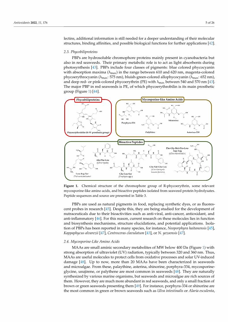
PBPs are hydrosoluble chromophore proteins mainly present in cyanobacteria butalso in red seaweeds. Their primary metabolic role is to act as light absorbents duringphotosynthesis [43]. PBPs include four classes of pigments: blue colored phycocyaninwith absorption maxima (λmax) in the range between 610 and 620 nm, magenta-coloredphycoerythrocyanin (λmax: 575 nm), bluish-green-colored allophycocyanin (λmax: 652 nm),and deep red- or pink-colored phycoerythrin (PE) with λmax between 540 and 570 nm [43].The major PBP in red seaweeds is PE, of which phycoerythrobilin is its main prostheticgroup (Figure 1) [44].
Antioxidants 2022, 11, x FOR PEER REVIEW 5 of 26
The major PBP in red seaweeds is PE, of which phycoerythrobilin is its main prosthetic
group (Figure 1) [44].
Figure 1. Chemical structure of the chromophore group of R-phycoerythrin, some relevant myco-
sporine-like amino acids, and bioactive peptides isolated from seaweed protein hydrolysates. Pep-
tide sequences and source are presented in Table 3.
PBPs are used as natural pigments in food, replacing synthetic dyes, or as fluorescent
probes in research [45]. Despite this, they are being studied for the development of
nutraceuticals due to their bioactivities such as anti-viral, anti-cancer, antioxidant, and
anti-inflammatory [46]. For this reason, current research on these molecules lies in func-
tion and biosynthesis mechanisms, structure elucidations, and potential applications. Iso-
lation of PBPs has been reported in many species, for instance, Neoporphyra haitanensis [45],
Kappaphycus alvarezii [47], Centroceras clavulatum [43], or N. yezoensis [47].
2.4. Mycosporine-Like Amino Acids
MAAs are small aminic secondary metabolites of MW below 400 Da (Figure 1) with
strong absorption of ultraviolet (UV) radiation, typically between 320 and 360 nm. Thus,
MAAs are useful molecules to protect cells from oxidative processes and solar UV-in-
duced damage [48]. Up to now, more than 20 MAAs have been characterized in seaweeds
and microalgae. From these, palaythine, asterina, shinorine, porphyra-334, mycosporine-
glycine, usujirene, or palythene are most common in seaweeds [48]. They are naturally
synthesized by various marine organisms, but seaweeds and microalgae are rich sources
of them. However, they are much more abundant in red seaweeds, and only a small frac-
tion of brown or green seaweeds presenting them [49]. For instance, porphyra-334 or shi-
norine are the most common in green or brown seaweeds such as Ulva intestinalis or Alaria
esculenta, but red species like Pyropia columbina or C. crispus may additionally contain
palythene much higher levels of mycosporine-glycine [49].
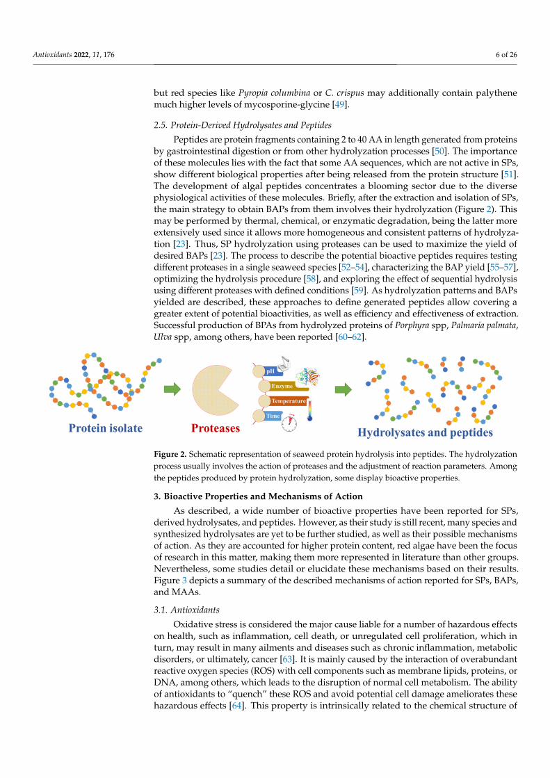
2.5. Protein-Derived Hydrolysates and Peptides
Peptides are protein fragments containing 2 to 40 AA in length generated from pro-
teins by gastrointestinal digestion or from other hydrolyzation processes [50]. The im-
portance of these molecules lies with the fact that some AA sequences, which are not ac-
tive in SPs, show different biological properties after being released from the protein
Figure 1. Chemical structure of the chromophore group of R-phycoerythrin, some relevantmycosporine-like amino acids, and bioactive peptides isolated from seaweed protein hydrolysates.Peptide sequences and source are presented in Table 3.
PBPs are used as natural pigments in food, replacing synthetic dyes, or as fluores-cent probes in research [45]. Despite this, they are being studied for the development ofnutraceuticals due to their bioactivities such as anti-viral, anti-cancer, antioxidant, andanti-inflammatory [46]. For this reason, current research on these molecules lies in functionand biosynthesis mechanisms, structure elucidations, and potential applications. Isola-tion of PBPs has been reported in many species, for instance, Neoporphyra haitanensis [45],Kappaphycus alvarezii [47], Centroceras clavulatum [43], or N. yezoensis [47].
2.4. Mycosporine-Like Amino Acids
MAAs are small aminic secondary metabolites of MW below 400 Da (Figure 1) withstrong absorption of ultraviolet (UV) radiation, typically between 320 and 360 nm. Thus,MAAs are useful molecules to protect cells from oxidative processes and solar UV-induceddamage [48]. Up to now, more than 20 MAAs have been characterized in seaweedsand microalgae. From these, palaythine, asterina, shinorine, porphyra-334, mycosporine-glycine, usujirene, or palythene are most common in seaweeds [48]. They are naturallysynthesized by various marine organisms, but seaweeds and microalgae are rich sources ofthem. However, they are much more abundant in red seaweeds, and only a small fraction ofbrown or green seaweeds presenting them [49]. For instance, porphyra-334 or shinorine arethe most common in green or brown seaweeds such as Ulva intestinalis or Alaria esculenta,
Antioxidants 2022, 11, 176 6 of 26
but red species like Pyropia columbina or C. crispus may additionally contain palythenemuch higher levels of mycosporine-glycine [49].
2.5. Protein-Derived Hydrolysates and Peptides
Peptides are protein fragments containing 2 to 40 AA in length generated from proteinsby gastrointestinal digestion or from other hydrolyzation processes [50]. The importanceof these molecules lies with the fact that some AA sequences, which are not active in SPs,show different biological properties after being released from the protein structure [51].The development of algal peptides concentrates a blooming sector due to the diversephysiological activities of these molecules. Briefly, after the extraction and isolation of SPs,the main strategy to obtain BAPs from them involves their hydrolyzation (Figure 2). Thismay be performed by thermal, chemical, or enzymatic degradation, being the latter moreextensively used since it allows more homogeneous and consistent patterns of hydrolyza-tion [23]. Thus, SP hydrolyzation using proteases can be used to maximize the yield ofdesired BAPs [23]. The process to describe the potential bioactive peptides requires testingdifferent proteases in a single seaweed species [52–54], characterizing the BAP yield [55–57],optimizing the hydrolysis procedure [58], and exploring the effect of sequential hydrolysisusing different proteases with defined conditions [59]. As hydrolyzation patterns and BAPsyielded are described, these approaches to define generated peptides allow covering agreater extent of potential bioactivities, as well as efficiency and effectiveness of extraction.Successful production of BPAs from hydrolyzed proteins of Porphyra spp, Palmaria palmata,Ulva spp, among others, have been reported [60–62].
Antioxidants 2022, 11, x FOR PEER REVIEW 6 of 26
structure [51]. The development of algal peptides concentrates a blooming sector due to
the diverse physiological activities of these molecules. Briefly, after the extraction and iso-
lation of SPs, the main strategy to obtain BAPs from them involves their hydrolyzation
(Figure 2). This may be performed by thermal, chemical, or enzymatic degradation, being
the latter more extensively used since it allows more homogeneous and consistent pat-
terns of hydrolyzation [23]. Thus, SP hydrolyzation using proteases can be used to max-
imize the yield of desired BAPs [23]. The process to describe the potential bioactive pep-
tides requires testing different proteases in a single seaweed species [52–54], characteriz-
ing the BAP yield [55–57], optimizing the hydrolysis procedure [58], and exploring the
effect of sequential hydrolysis using different proteases with defined conditions [59]. As
hydrolyzation patterns and BAPs yielded are described, these approaches to define gen-
erated peptides allow covering a greater extent of potential bioactivities, as well as effi-
ciency and effectiveness of extraction. Successful production of BPAs from hydrolyzed
proteins of Porphyra spp, Palmaria palmata, Ulva spp, among others, have been reported
[60–62].
Figure 2. Schematic representation of seaweed protein hydrolysis into peptides. The hydrolyzation
process usually involves the action of proteases and the adjustment of reaction parameters. Among
the peptides produced by protein hydrolyzation, some display bioactive properties.
3. Bioactive Properties and Mechanisms of Action
As described, a wide number of bioactive properties have been reported for SPs, de-
rived hydrolysates, and peptides. However, as their study is still recent, many species and
synthesized hydrolysates are yet to be further studied, as well as their possible mecha-
nisms of action. As they are accounted for higher protein content, red algae have been the
focus of research in this matter, making them more represented in literature than other
groups. Nevertheless, some studies detail or elucidate these mechanisms based on their
results. Figure 3 depicts a summary of the described mechanisms of action reported for
SPs, BAPs, and MAAs.
Figure 2. Schematic representation of seaweed protein hydrolysis into peptides. The hydrolyzationprocess usually involves the action of proteases and the adjustment of reaction parameters. Amongthe peptides produced by protein hydrolyzation, some display bioactive properties.
3. Bioactive Properties and Mechanisms of Action
As described, a wide number of bioactive properties have been reported for SPs,derived hydrolysates, and peptides. However, as their study is still recent, many species andsynthesized hydrolysates are yet to be further studied, as well as their possible mechanismsof action. As they are accounted for higher protein content, red algae have been the focusof research in this matter, making them more represented in literature than other groups.Nevertheless, some studies detail or elucidate these mechanisms based on their results.Figure 3 depicts a summary of the described mechanisms of action reported for SPs, BAPs,and MAAs.
3.1. Antioxidants
Oxidative stress is considered the major cause liable for a number of hazardous effectson health, such as inflammation, cell death, or unregulated cell proliferation, which inturn, may result in many ailments and diseases such as chronic inflammation, metabolicdisorders, or ultimately, cancer [63]. It is mainly caused by the interaction of overabundantreactive oxygen species (ROS) with cell components such as membrane lipids, proteins, orDNA, among others, which leads to the disruption of normal cell metabolism. The abilityof antioxidants to “quench” these ROS and avoid potential cell damage ameliorates thesehazardous effects [64]. This property is intrinsically related to the chemical structure of

Antioxidants 2022, 11, 176 7 of 26
antioxidants. In the case of SP and BAPs, their antioxidant activity may be related to theiramino acid composition and conformation [40].
Antioxidants 2022, 11, x FOR PEER REVIEW 7 of 26
Figure 3. Summary of reported mechanisms of action of several SPs and BAPs. Their location with
respect to the bilayer membrane represents that these effects are inducted inside, outside the cell, or
on the cell surface. Abbreviations: PBPs: phycobiliproteins; MAAs: mycosporine-like amino acids;
BAPs: bioactive peptides; ROS: reactive oxygen species; IL-1β: interleukin-1β; TNF-α: tumor necro-
sis factor- α; COX-2: cyclooxygenase-2; iNOS: inducible nitric oxide synthase; NF-κB: nuclear factor-
κB; DPP-IV: dipeptidyl peptidase-IV; GLP-1: glucagon-like peptide-1; ACE: angiotensin I-converter
enzyme.
3.1. Antioxidants
Oxidative stress is considered the major cause liable for a number of hazardous ef-
fects on health, such as inflammation, cell death, or unregulated cell proliferation, which
in turn, may result in many ailments and diseases such as chronic inflammation, metabolic
disorders, or ultimately, cancer [63]. It is mainly caused by the interaction of overabun-
dant reactive oxygen species (ROS) with cell components such as membrane lipids, pro-
teins, or DNA, among others, which leads to the disruption of normal cell metabolism.
The ability of antioxidants to “quench” these ROS and avoid potential cell damage ame-
liorates these hazardous effects [64]. This property is intrinsically related to the chemical
structure of antioxidants. In the case of SP and BAPs, their antioxidant activity may be
related to their amino acid composition and conformation [40].
Several studies have corroborated the antioxidant properties of SPs. For example, the
antioxidant activity of a purified GP from C. decorticatum was assessed based on the ability
to scavenge 2,2-diphenyl-1-picrylhydrazyl (DPPH), hydroxyl, superoxide, and nitric ox-
ide radicals. According to the results, the GP showed positive results in a dose-dependent
manner, which was attributed to the presence of aromatic amino acid present in its chem-
ical structure [35,65]. A thyroglobulin-binding lectin from Sargassum fusiforme inhibited
up to 80% of DPPH radicals and 60% of 2,2′-azino-bis(3-ethylbenzothiazoline-6-sulfonic
acid) (ABTS) radicals. Authors considered that these results were due to its glycan and
aminoacidic composition, rich in G, T, and A [40]. Regarding MAAs, these compounds
have been long-known to be effective antioxidants, especially against UV-light induced
oxidation [66]. A recent and extensive study evaluated the antioxidant activity of a wide
number of MAAs isolated from G. domingensis, such as asterina-330, shinorine, palythine,
or palythinol [24]. Among the isolated MAAs, asterina-330 showed by far the most
Figure 3. Summary of reported mechanisms of action of several SPs and BAPs. Their location withrespect to the bilayer membrane represents that these effects are inducted inside, outside the cell,or on the cell surface. Abbreviations: PBPs: phycobiliproteins; MAAs: mycosporine-like aminoacids; BAPs: bioactive peptides; ROS: reactive oxygen species; IL-1β: interleukin-1β; TNF-α: tumornecrosis factor- α; COX-2: cyclooxygenase-2; iNOS: inducible nitric oxide synthase; NF-κB: nuclearfactor- κB; DPP-IV: dipeptidyl peptidase-IV; GLP-1: glucagon-like peptide-1; ACE: angiotensinI-converter enzyme.
Several studies have corroborated the antioxidant properties of SPs. For example,the antioxidant activity of a purified GP from C. decorticatum was assessed based on theability to scavenge 2,2-diphenyl-1-picrylhydrazyl (DPPH), hydroxyl, superoxide, andnitric oxide radicals. According to the results, the GP showed positive results in a dose-dependent manner, which was attributed to the presence of aromatic amino acid presentin its chemical structure [35,65]. A thyroglobulin-binding lectin from Sargassum fusiformeinhibited up to 80% of DPPH radicals and 60% of 2,2′-azino-bis(3-ethylbenzothiazoline-6-sulfonic acid) (ABTS) radicals. Authors considered that these results were due to its glycanand aminoacidic composition, rich in G, T, and A [40]. Regarding MAAs, these compoundshave been long-known to be effective antioxidants, especially against UV-light inducedoxidation [66]. A recent and extensive study evaluated the antioxidant activity of a widenumber of MAAs isolated from G. domingensis, such as asterina-330, shinorine, palythine, orpalythinol [24]. Among the isolated MAAs, asterina-330 showed by far the most significantantioxidant activity with 10.03 µmol Trolox equivalents/mol of the compound, in contrastwith the 2953 µmol Trolox equivalents/mol of the compound obtained for gallic acid after1 h of ABTS assay.
Numerous studies have successfully produced BAPs with antioxidant properties fordifferent seaweed species. According to data, it has been suggested that BAPs displayhigher antioxidant activity than SPs or SP hydrolysates of higher MW. This could be dueto the enhanced cell and chemical accessibility of released amino acids from proteins. Forexample, fractionation of P. palmata chymotrypsin-produced BAPs based on their MWshowed that those fractions with lower ones displayed higher antioxidant activities. Thefraction containing peptides <10 kDa displayed the most significant antioxidant properties
Antioxidants 2022, 11, 176 8 of 26
following a DPPH assay [55]. In another study conducted with the same species, theauthors obtained a peptide SDIAPGGNM, which showed an oxygen radical absorbancecapacity (ORAC) of 152 nmol TE/µmol peptide and a ferric reducing antioxidant power(FRAP) of 21 nmol TE/µmol peptide [67]. Other seaweeds have also been reported to bea source of BAPs. For example, proteins from P. haitanensis were digested using pepsin,and their antioxidant properties were evaluated in vitro by 2,2-diphenyl-1-picrylhydrazyl(DPPH) assay and ferric reducing antioxidant power (FRAP), showing positive results [68].Pepsin and pepsin-Corolase® hydrolyzed biomass from P. dioica resulted in a diverse BAPpattern, of which the pepsin fraction displayed significantly higher antioxidant activityon various antioxidant assays. Yet both treatments resulted in an increase in antioxidantactivity, which was related to the release of >10 kDa peptides [69]. In another study by thesame authors, P. dioica biomass and its protein isolate were hydrolyzed using the proteasesProlyve® and Flavourzyme®. This digestion achieved a 5-fold increase in antioxidantactivity. The authors noted that phenolic compounds were also released after enzymaticdigestion, which paired with a positive correlation with protein degradation, suggesting asynergistic dynamic between these elements [70]. Finally, a recent study characterized theoptimum hydrolysis conditions of G. lemaneiformis proteins with chymotrypsin [58]. Thevariables considered in this experiment were enzyme/substrate ratio (1–3%), temperature(41–19 ◦C), and pH (8.5–9.5), whereas substrate concentration and reaction time were keptconstant (10 mg/mL and 2.0 h, respectively). According to the authors, the optimumconditions to improve the extraction of antioxidant peptides were enzyme/substrate ratio(1:10), 46.4 ◦C, and pH 9.2. After separation and purification steps, GLWKTF was identifiedas the principal antioxidant, whose properties were attributed to its small molecular sizeand the presence of hydrophobic and/or aromatic amino acids [58].
3.2. Antimicrobials and Antivirals
Among SPs, lectins have been reported in recent years to be potent and effectiveantibacterial and antiviral agents due to their binding with monosaccharide residues of cellwalls or membrane of yeasts, bacteria, and viruses [25,36]. Specifically, different mannose-binding lectins have been isolated from seaweeds, which have potent antiviral properties,as many viruses present mannose residues in their capsid or envelope [36]. For example,two recently isolated lectins from the green alga C. isthmocladum with galactose-bindingactivity were found to inhibit biofilm formation of Staphylococcus aureus and S. epidermidisup to 60%, compared to the control samples (Table 2). However, they did not display anysignificant antibacterial or antibiofilm activity against Escherichia coli [71]. The mannose-binding lectin griffithsin, derived from the red macroalga genus Griffithsia sp., has becomea main focus of research since it was isolated in 2004 [72]. Griffithsin has been thoroughlystudied for its potential applications against human and cattle viral infections, showingpromising results against human immunodeficiency virus (HIV), human herpes virus(HSV), hepatitis C or severe acute respiratory syndrome-coronavirus (SARS-CoV), amongothers, both in vitro and in vivo [73,74]. Indeed, recent work has demonstrated that thesynergistic cutaneous application of carrageenan and griffithsin inhibited simian-HIV inrhesus macaques, as well as HSV and HPV in mice [75]. The broad action range of griffithsinagainst viruses with diverse structural features has made it to be proposed as an antiviralagent against SARS-CoV-2, as it can effectively bind to the Spike surface GPs of SARS-CoV,inhibiting cell-to-cell infection [76]. It has also been shown to have a prolonged half-life,both by oral and parenteral administration, which supports its potential application inantiviral treatment [77]. Grifonin-1, a griffithsin-derived peptide, has also shown significantinhibition of HIV infection in in vitro cultures, along with a very low cytotoxicity index [78].In addition, a mannose-binding lectin recently isolated from Grateulopia chiangii showedsignificant inhibition against various strains of influenza, like H1N1, and towards HSV1and HSV2 (Table 2) [79]. Nevertheless, it did not show antiviral activity against HIV,indicating that although broad, the action range of these lectins is dependent on theirbinding site affinities.
Antioxidants 2022, 11, 176 9 of 26
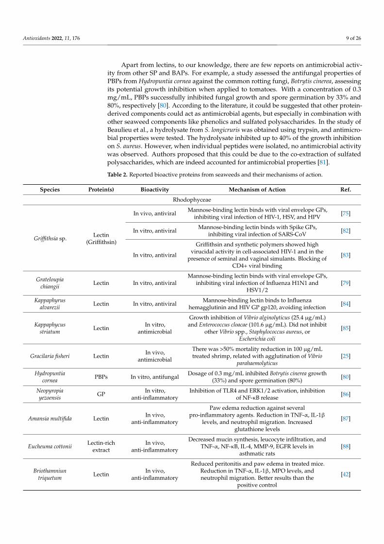
Apart from lectins, to our knowledge, there are few reports on antimicrobial activ-ity from other SP and BAPs. For example, a study assessed the antifungal properties ofPBPs from Hydropuntia cornea against the common rotting fungi, Botrytis cinerea, assessingits potential growth inhibition when applied to tomatoes. With a concentration of 0.3mg/mL, PBPs successfully inhibited fungal growth and spore germination by 33% and80%, respectively [80]. According to the literature, it could be suggested that other protein-derived components could act as antimicrobial agents, but especially in combination withother seaweed components like phenolics and sulfated polysaccharides. In the study ofBeaulieu et al., a hydrolysate from S. longicruris was obtained using trypsin, and antimicro-bial properties were tested. The hydrolysate inhibited up to 40% of the growth inhibitionon S. aureus. However, when individual peptides were isolated, no antimicrobial activitywas observed. Authors proposed that this could be due to the co-extraction of sulfatedpolysaccharides, which are indeed accounted for antimicrobial properties [81].
Table 2. Reported bioactive proteins from seaweeds and their mechanisms of action.
Species Protein(s) Bioactivity Mechanism of Action Ref.
Rhodophyceae
Griffithsia sp. Lectin(Griffithsin)
In vivo, antiviral Mannose-binding lectin binds with viral envelope GPs,inhibiting viral infection of HIV-1, HSV, and HPV [75]
In vitro, antiviral Mannose-binding lectin binds with Spike GPs,inhibiting viral infection of SARS-CoV [82]
In vitro, antiviral
Griffithsin and synthetic polymers showed highvirucidal activity in cell-associated HIV-1 and in the
presence of seminal and vaginal simulants. Blocking ofCD4+ viral binding
[83]
Grateloupiachiangii Lectin In vitro, antiviral
Mannose-binding lectin binds with viral envelope GPs,inhibiting viral infection of Influenza H1N1 and
HSV1/2[79]
Kappaphyrusalvarezii Lectin In vitro, antiviral Mannose-binding lectin binds to Influenza
hemagglutinin and HIV GP gp120, avoiding infection [84]
Kappaphycusstriatum Lectin In vitro,
antimicrobial
Growth inhibition of Vibrio alginolyticus (25.4 µg/mL)and Enterococcus cloacae (101.6 µg/mL). Did not inhibit
other Vibrio spp., Staphylococcus aureus, orEscherichia coli
[85]
Gracilaria fisheri Lectin In vivo,antimicrobial
There was >50% mortality reduction in 100 µg/mLtreated shrimp, related with agglutination of Vibrio
parahaemolyticus[25]
Hydropuntiacornea PBPs In vitro, antifungal Dosage of 0.3 mg/mL inhibited Botrytis cinerea growth
(33%) and spore germination (80%) [80]
Neopyropiayezoensis GP In vitro,
anti-inflammatoryInhibition of TLR4 and ERK1/2 activation, inhibition
of NF-κB release [86]
Amansia multifida Lectin In vivo,anti-inflammatory
Paw edema reduction against severalpro-inflammatory agents. Reduction in TNF-α, IL-1β
levels, and neutrophil migration. Increasedglutathione levels
[87]
Eucheuma cottonii Lectin-richextract
In vivo,anti-inflammatory
Decreased mucin synthesis, leucocyte infiltration, andTNF-α, NF-κB, IL-4, MMP-9, EGFR levels in
asthmatic rats[88]
Briothamniuntriquetum Lectin In vivo,
anti-inflammatory
Reduced peritonitis and paw edema in treated mice.Reduction in TNF-α, IL-1β, MPO levels, andneutrophil migration. Better results than the
positive control
[42]
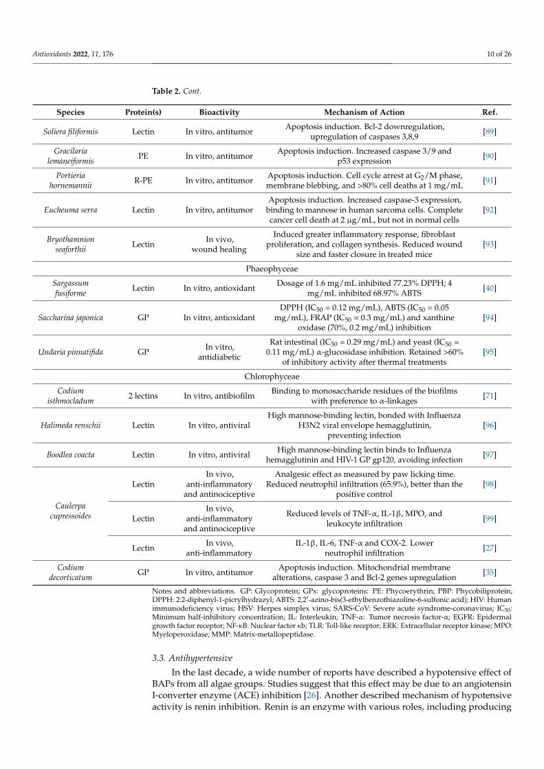
Antioxidants 2022, 11, 176 10 of 26
Table 2. Cont.
Species Protein(s) Bioactivity Mechanism of Action Ref.
Soliera filiformis Lectin In vitro, antitumor Apoptosis induction. Bcl-2 downregulation,upregulation of caspases 3,8,9 [89]
Gracilarialemaneiformis PE In vitro, antitumor Apoptosis induction. Increased caspase 3/9 and
p53 expression [90]
Portieriahornemannii R-PE In vitro, antitumor Apoptosis induction. Cell cycle arrest at G2/M phase,
membrane blebbing, and >80% cell deaths at 1 mg/mL [91]
Eucheuma serra Lectin In vitro, antitumorApoptosis induction. Increased caspase-3 expression,
binding to mannose in human sarcoma cells. Completecancer cell death at 2 µg/mL, but not in normal cells
[92]
Bryothamnionseaforthii Lectin In vivo,
wound healing
Induced greater inflammatory response, fibroblastproliferation, and collagen synthesis. Reduced wound
size and faster closure in treated mice[93]
Phaeophyceae
Sargassumfusiforme Lectin In vitro, antioxidant Dosage of 1.6 mg/mL inhibited 77.23% DPPH; 4
mg/mL inhibited 68.97% ABTS [40]
Saccharina japonica GP In vitro, antioxidantDPPH (IC50 = 0.12 mg/mL), ABTS (IC50 = 0.05
mg/mL), FRAP (IC50 = 0.3 mg/mL) and xanthineoxidase (70%, 0.2 mg/mL) inhibition
[94]
Undaria pinnatifida GP In vitro,antidiabetic
Rat intestinal (IC50 = 0.29 mg/mL) and yeast (IC50 =0.11 mg/mL) α-glucosidase inhibition. Retained >60%
of inhibitory activity after thermal treatments[95]
Chlorophyceae
Codiumisthmocladum 2 lectins In vitro, antibiofilm Binding to monosaccharide residues of the biofilms
with preference to α-linkages [71]
Halimeda renschii Lectin In vitro, antiviralHigh mannose-binding lectin, bonded with Influenza
H3N2 viral envelope hemagglutinin,preventing infection
[96]
Boodlea coacta Lectin In vitro, antiviral High mannose-binding lectin binds to Influenzahemagglutinin and HIV-1 GP gp120, avoiding infection [97]
Caulerpacupressoides
LectinIn vivo,
anti-inflammatoryand antinociceptive
Analgesic effect as measured by paw licking time.Reduced neutrophil infiltration (65.9%), better than the
positive control[98]
LectinIn vivo,
anti-inflammatoryand antinociceptive
Reduced levels of TNF-α, IL-1β, MPO, andleukocyte infiltration [99]
Lectin In vivo,anti-inflammatory
IL-1β, IL-6, TNF-α and COX-2. Lowerneutrophil infiltration [27]
Codiumdecorticatum GP In vitro, antitumor Apoptosis induction. Mitochondrial membrane
alterations, caspase 3 and Bcl-2 genes upregulation [35]
Notes and abbreviations. GP: Glycoprotein; GPs: glycoproteins: PE: Phycoerythrin; PBP: Phycobiliprotein;DPPH: 2:2-diphenyl-1-picrylhydrazyl; ABTS: 2,2′-azino-bis(3-ethylbenzothiazoline-6-sulfonic acid); HIV: Humanimmunodeficiency virus; HSV: Herpes simplex virus; SARS-CoV: Severe acute syndrome-coronavirus; IC50:Minimum half-inhibitory concentration; IL: Interleukin; TNF-α: Tumor necrosis factor-α; EGFR: Epidermalgrowth factor receptor; NF-κB: Nuclear factor κb; TLR: Toll-like receptor; ERK: Extracellular receptor kinase; MPO:Myeloperoxidase; MMP: Matrix-metallopeptidase.
3.3. Antihypertensive
In the last decade, a wide number of reports have described a hypotensive effect ofBAPs from all algae groups. Studies suggest that this effect may be due to an angiotensinI-converter enzyme (ACE) inhibition [26]. Another described mechanism of hypotensiveactivity is renin inhibition. Renin is an enzyme with various roles, including producing
Antioxidants 2022, 11, 176 11 of 26
angiotensin I (ACE substrate) and stimulating renal activity [31]. Together, they constitutethe known renin-ACE-aldosterone axis, which is the main pathway involved in cardiacpressure regulation [100]. Several studies confirming the anti-hypertensive properties ofseaweed BAPs will be mentioned below and have been compiled in Table 3.
A study evaluated the effect of different proteases to obtain a G. lemaneiformis proteinhydrolysate [52]. The results showed that trypsin produced the extract with the highest ACEinhibitory activity, up to 78%. Further characterization of peptide sequence indicated thatQVEY was the major BAP. A similar experiment was carried out with proteins obtained fromU. intestinalis using Alcalase, α-chymotrypsin, papain, pepsin, and trypsin. Again, trypsinproduced the extract with the highest ACE inhibitory activity, and two peptides wereindicated as the most active (FGMPLDR and MELVLR) [53]. Another related experimentregarding the use of different proteases to obtain BAPs from C. lentillifera indicated thatthermolysin was the most appropriate choice in comparison to α-chymotrypsin, pepsin,and trypsin. In this case, the peptides with the highest ACE inhibitory activity were FDGIPand AIDPVRA [101]. Among hydrolytic enzymes, pepsin is one of the most used in thelast years. For instance, the peptide sequence NMEKGSSSVVSSRM (with anticoagulantactivity) was obtained from the hydrolysis of N. yezoensis proteins using this enzyme [56]. Arelated experiment using U. rigida proteins indicated that the peptides with ACE inhibitoryactivity (IP and AFL) were obtained from the hydrolysis with pepsin [57]. In anotherexperiment with this enzyme, peptides with ACE inhibitory activity (ALLAGDPSVLEDRand VVGGTGPVDEWGIAGAR) were also obtained from Bangia fusco-purpurea [102].
Another strategy to obtain hydrolysates from seaweeds is the use of sequential hy-drolysis with different enzymes. This strategy was used to obtain peptides with ACEinhibitory activity from P. palmata [59]. In this case, the order of hydrolysis was thermolysin,pepsin, trypsin, and chymotrypsin. According to the authors, additional hydrolysis afterthermolysin use did not increase the release of peptides with biological potential. Addi-tionally, a reduction in total ACE inhibitory activity in the hydrolysate was obtained afterconsecutive hydrolysis with thermolysin, pepsin, and trypsin. The peptide sequence LRYwas indicated as the most active from the thermolysin hydrolysate. In this sense, it seemsreasonable to indicate that the selection of protease and the hydrolysis conditions have agreat impact on the release of peptides with biological activity. Nonetheless, additionalexperiments are required to clarify the role of sequential hydrolysis in the release of bioac-tive peptides to characterize their protein hydrolyzation patterns and how this affects theproperties of yielded BAPs.
The antihypertensive activity of BAPs has also been corroborated in vivo in severalstudies. For instance, Fitzgerald et al. obtained a renin-inhibitory peptide by hydrolyzingP. palmata protein isolates with papain, which sequence was IRLIIVLMPILMA [103]. Thepeptide showed ACE inhibition at very low concentrations (IC50 = 0.32 mg/mL). The sameteam carried out an in vivo study with this same peptide on spontaneously hypertensiverats but obtained it by chemical synthesis. The effect of the peptide was compared withunpurified P. palmata hydrolysate, and the results showed that their effect was very similar,while a significant reduction in systolic blood pressure was achieved [104]. Another in vivostudy reported that U. pinnatifida BAPs were able to significantly reduce blood pressure inspontaneously hypertensive rats. These BAPs were dipeptides mainly based on Tyr or Trpthat showed this hypotensive effect with a single oral dose of 1 mg/kg body weight [105]. Toour knowledge, the only clinical trial reporting a reduction in blood pressure was publishedin 2002 by Shirako Co. Ltd. researchers, which served as a scientific basis for developingand patenting this product. They reported that a 1.8 g/day intake of nori (Porphyra sp.)hydrolysate achieved a significant reduction in systolic blood pressure in hypertensivesubjects (from 157 to 142 mmHg) but did not in normotensive ones (≤120 mmHg) [106].

Antioxidants 2022, 11, 176 12 of 26
Table 3. Bioactive peptides and hydrolysates obtained from seaweed proteins.
Species Hydrolysis Sequence/s Bioactivity Results Ref.
Rhodophyceae
Gracilariopsislemaneiformis
Trypsin; E:S (1:25),37 ◦C, pH 8, 8 h QVEY In vitro,
antihypertensiveACE inhibitory activity.
IC50= 0.25 mg/mL [52]
α-Chymotrypsin; E:S(1:25), 37 ◦C, pH 8, 2 h ELWKTF In vitro,
antioxidantDPPH radical scavenging.
EC50 = 1.51 mg/mL [58]
Porphyra spp. Pepsin; E:S (1:100),pH 2, 45 ◦C, 4 h GGSK, ELS In vitro,
antidiabetic
α-Amylase inhibition.IC50(GGSK) = 0.8 mg/mL,
IC50(ELS)= 0.9 mg/mL[54]
Porphyra dioica
Alcalase® andFlavourzyme®; E:S
(1:100), 50 ◦C, pH 7, 4 h
DYYLR, AGFY, YLVA,AFIT, MKTPITE,
TYIA, LDLW
In vitro,antioxidant
Most antioxidant onORAC assay:
IC50(AFIT) = 0.4 µg/mL,IC50(MKTPITE) =
0.007 mg/mL
[60]
DYYLR, AGFY,YLVA, TYIA
In vitro,antihypertensive
Most inhibiting BAPs:IC50(TYIA) = 0.04 mg/mL,IC50(TYIA) = 0.07 mg/mL
[60]
YLVA In vitro,antidiabetic
DPP-IV inhibition,IC50 = 0.2 mg/mL [60]
Prolyve®; E:S (1:100),50 ◦C, pH 8, 2 h
n.a., increasedproduction of <1 kDa
peptides
In vitro,antioxidant
ORAC (IC50 = 2.7 mmol TE/g),DPPH (IC50 =
0.2 mmol TE/g), FRAP(IC50 = 0.4 mmol TE/g)
[107]
Neopyropiayezoensis
Chemical synthesis IY, MKY, AKTSY, LRY Clinical trial,antihypertensive
Blood pression reductionfrom 157/95 to
142/86 mmHg with1.8 g/day for 35 days
[106]
Pepsin; E:S (1:40), pH 2,45 ◦C, 2 h NMEKGSSSVVSSRM Ex vivo,
anticoagulantBlood clotting retardation;
IC50 = 4.49 µg/mL [56]
Chemical synthesis “PPY” peptide In vitro, antitumor
Doses ≥125 ng/mLinduced autophagy andapoptosis in MCF-7 cellsvia the mTOR pathway
[108]
Chemical synthesis “PPY” peptide In vitro,anti-inflammatory
Doses ≥250 ng/mLinhibited expression of
inflammatory cytokines inmurine macrophages
[29]
Pyropiacolumbina
Alkaline protease; E:S(1:40), 55 ◦C, pH 9.5, 2 h
n.a., ~2.4 kDapeptides
In vitro,antihypertensive
ACE inhibitory activity.IC50 = 1.2 mg/mL [109]
In vitro,anticoagulant
Antiplatelet aggregation.2.8 mg/mL achieved
18.7% inhibition[109]
Trypsin; E:S (1:20),50 ◦C, pH 8, 4 h n.a., >400 Da peptides
In vitro,antioxidant and
anti-inflammatory
DPPH (IC50 = 2.8 mg/mL),ABTS (IC50 = 2.4 mg/mL);
upregulation ofIL10 at 0.1 mg/mL
[50]
Fungal protease; E:S(1:20), 55 ◦C, pH 4.3, 3 hand Flavourzyme®; E:S(1:50), 55 ◦C, pH 7, 4 h
n.a. In vitro,anti-inflammatory
Upregulation of IL-10 inmurine spenocytes,macrophages and
lymphocytes at≥0.01 mg/mL
[110]
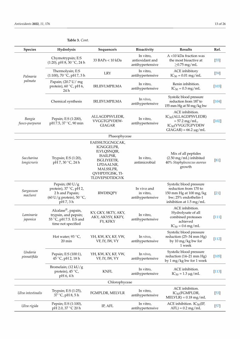
Antioxidants 2022, 11, 176 13 of 26
Table 3. Cont.
Species Hydrolysis Sequence/s Bioactivity Results Ref.
Palmariapalmata
Chymotrypsin; E:S(1:20), pH 8, 30 ◦C, 24 h 33 BAPs < 10 kDa
In vitro,antioxidant andantihypertensive
A <10 kDa fraction wasthe most bioactive at≥0.75 mg/mL
[55]
Thermolysin; E:S(1:100), 70 ◦C, pH 7, 3 h LRY In vitro,
antihypertensiveACE inhibitory
IC50 = 0.01 mg/mL [59]
Papain; (20.7 U/ mgprotein), 60 ◦C, pH 6,
24 hIRLIIVLMPILMA In vitro,
antihypertensiveRenin inhibition.
IC50 = 0.3 mg/mL [103]
Chemical synthesis IRLIIVLMPILMA In vivo,antihypertensive
Systolic blood pressurereduction from 187 to
155 mm Hg at 50 mg/kg bw[104]
Bangiafusco-purpurea
Pepsin; E:S (1:200),pH 7.5, 37 ◦C, 90 min
ALLAGDPSVLEDR,VVGGTGPVDEW-
GIAGAR
In vitro,antihypertensive
ACE inhibition.IC50(ALLAGDPSVLEDR)
= 57.2 mg/mL,IC50(VVGGTGPVDEW
GIAGAR) = 66.2 µg/mL
[102]
Phaeophyceae
Saccharinalongicruris
Trypsin; E:S (1:20),pH 7, 30 ◦C, 24 h
EAESSLTGGNGCAK,IGNGGELPR,ILVLQSNQIR,
ISAILPSR,ISGLIYEETR,LPDAALNR,MALSSLPR,
QVHPDTGISK, TI-TLDVEPSDTIDGVK
In vitro,antimicrobial
Mix of all peptides(2.50 mg/mL) inhibited
40% Staphylococcus aureusgrowth
[81]
Sargassummaclurei
Pepsin; (80 U/gprotein), 37 ◦C, pH 2,
2 h and Papain;(60 U/g protein), 50 ◦C,
pH 7, 3 h
RWDISQPYIn vivo and
in vitro,antihypertensive
Systolic blood pressurereduction from 170 to
150 mm Hg at 100 mg/kgbw; 25% endothelin-1
inhibition at 1.5 mg/mL
[21]
Laminariajaponica
Alcalase®, papain,trypsin, and pepsin;
55 ◦C, pH 7.5. E:S andtime not specified
KY, GKY, SKTY, AKY,AKY, AKYSY, KKFY,
FY, KFKY
In vitro,antihypertensive
ACE inhibition.Hydrolysate of all
combined proteasesachieved
IC50 = 0.6 mg/mL
[111]
Undariapinnatifida
Hot water; 93 ◦C,20 min
YH, KW, KY, KF, VW,VF, IY, IW, VY
In vivo,antihypertensive
Systolic blood pressurereduction (25–34 mm Hg)
by 10 mg/kg bw for1 week
[112]
Pepsin; E:S (100:1),45 ◦C, pH 2, 18 h
YH, KW, KY, KF, VW,VF, IY, IW, VY
In vivo,antihypertensive
Systolic blood pressurereduction (14–21 mm Hg)by 1 mg/kg bw for 1 week
[105]
Bromelain; (12 kU/gprotein), 45 ◦C,
pH 6, 4 hKNFL In vitro,
antihypertensiveACE inhibition.
IC50 = 1.3 µg/mL [113]
Chlorophyceae
Ulva intestinalis Trypsin; E:S (1:25),37 ◦C, pH 8, 5 h FGMPLDR, MELVLR In vitro,
antihypertensive
ACE inhibition.IC50(FGMPLDR,
MELVLR) = 0.18 mg/mL[53]
Ulva rigida Pepsin; E:S (1:100),pH 2.0, 37 ◦C 20 h IP, AFL In vitro,
antihypertensiveACE inhibition. IC50(IP,
AFL) = 0.2 mg/mL [57]
Antioxidants 2022, 11, 176 14 of 26
Table 3. Cont.
Species Hydrolysis Sequence/s Bioactivity Results Ref.
Ulva lactuca Papain; E:S (1:100),60 ◦C, pH 6, 24 h
55 non-allergenicBAPs identified
In vitro,antihypertensive
ACE inhibition (93%) in>1 kDa hydrolysate fraction [114]
Enteromorphaclathrata
Alcalase®; 2.9 kU/gprotein, T not stated,
pH 7.6, 90 minPAFG In vitro,
antihypertensiveACE inhibition.
IC50 = 0.014 mg/mL [115]
Caulerpalentillifera
Thermolysin; E:S (1:50),60 ◦C, pH 8.5, 16 h FDGIP, AIDPVRA In vitro,
antihypertensive
ACE inhibition. IC50(FDGIP)= 0.03 mg/mL,
IC50 (AIDPVRA)= 0.04 mg/mL[101]
Abbreviations: E:S: Enzyme:substrate (w/w); IC50: Half-inhibitory concentration; TE: Trolox equivalents: BAPs:bioactive peptides; ACE: angiotensin I-converter enzyme; DPPH: 2,2-diphenyl-1-picrylhydrazyl; ABTS: (2,2′-azino-bis(3-ethylbenzothiazoline-6-sulfonic acid)); IL: interleukin; ORAC: oxygen radical absorbance capacity;FRAP: ferric reducing ability of plasma; mTOR: mammalian target of rapamycin.
3.4. Anti-Inflammatory
Inflammation is a complex response to cell and/or tissue damage, which can betriggered by several factors, such as oxidative damage, infections, or cancer [116]. Theseprocesses and cellular interactions involve pro- and anti-inflammatory mediators, whichup- or downregulate the inflammatory response. Therefore, the mechanisms by whichSP and BAPs have been described to induce anti-inflammatory effects, both in vitro andin vivo, are related to an increased expression of anti-inflammatory mediators and/ordownregulation of the expression of pro-inflammatory ones. The main pro-inflammatorymediators are cytokines like interleukins (IL) (e.g., IL-1β, 2, 6, and 8) and also tumornecrosis factor-α (TNF-α). Other compounds like the prostaglandin cyclooxygenase-2(COX-2) or the chemokine inducible nitric oxide synthase (iNOS) also play a vital rolein inducing an inflammatory response, especially producing ROS and nitric oxide (NO),which mainly activate macrophages. These cells, when activated, e.g., via toll-like receptors(TLR), lead these innate immune cells to release IL-6, TNF-α, and also transforming growthfactor-β, which promotes cell proliferation [117]. The nuclear factor κB (NF-κB) andmitogen-activated protein kinase (MAPK) are also key pro-inflammatory intermediariesthat are produced after TLR activation [118].
Regarding SP with anti-inflammatory activity, most of the studies point to lectinsas prime examples. In fact, several in vivo experiments show that these proteins exertanti-inflammatory properties through various pathways. For example, various workshave assessed the in vivo anti-inflammatory activity of lectins isolated from the green algaC. cupressoides (Table 2). Mice submitted to high concentrations of carrageenan to induceinflammatory response were treated with C. cupressoides lectins administered intravenously.The lectin treatment showed similar results to that of dexamethasone, significantly re-ducing inflammation signals, such as neutrophil levels and paw edema. Additionally,the lectins appeared to reduce nociception while not displaying any significant variationin organs weight or transaminase levels [98]. Very similar results were obtained in ratswith temporomandibular induced arthritis. In this study, after the same treatment, thetissue showed a significant decrease in TNF-α, IL-1β, and myeloperoxidase (MPO) levels,as well as a significant reduction in leukocyte concentration [99]. Additionally, it wasfound that lectin treatment was able to reverse the tissue damage and exert an analgesiceffect via a non-opioid pathway. In another work, the authors described the molecularpathway through which the lectin from C. cupressoides exerts anti-inflammatory effects,using several pro-inflammatory agents injected in rat paws [27]. A combination of en-zymatic and immunohistochemistry staining methods unveiled that this lectin elicitedan anti-inflammatory effect by inhibiting the primary cytokines IL-1β, IL-6, TNF-α, andCOX-2, as well as lowering neutrophil infiltration, as demonstrated by reduced MPOlevels. However, no inhibition of hemeoxygenase-2 (HO-2) was observed. Additionally, theauthors confirmed that the anti-inflammatory effect was due to the carbohydrate-binding

Antioxidants 2022, 11, 176 15 of 26
site of the lectin since when co-administered with mucin, its anti-inflammatory effect wasinhibited. This was confirmed by testing the anti-inflammatory effect of the lectin not onlyagainst carrageenan but also against dextran and histamine, with successful reductions inpaw edemas. However, when directly injected into rat paws instead of intravenously, thelectin induced a strong inflammatory response, which is related to the absent interactionwith pro-inflammatory agents but also to the administration route [27]. A 30 kDa lectinpurified from Amansia multifida displayed similar effects, modulating paw edema andperitonitis formation in mice [87]. This lectin consistently ameliorated paw edema againstcarrageenan, histamine, prostaglandin E2, and compound 48/80 (an inducer of histaminerelease). Pre-treatment with this lectin also greatly reduced neutrophil infiltration followingcarrageenan-induced peritonitis, as well as significantly lower TNF-α IL-1β expression inthe affected tissue (Table 2). Moreover, its administration showed lower MPO activity, withvalues even lower than those observed for indomethacin. Its anti-inflammatory effect wasalso confirmed by the increased levels of glutathione, which were greater than in the controlgroup, thus indicating that its anti-inflammatory property is also related to preventingfurther oxidative damage [87].
Other studies have reported the anti-inflammatory properties of other SP, althoughfew examples can be found. For instance, a 3.5 kDa GP from N. yezoensis was tested in vitrolipopolysaccharide (LPS)-activated murine RAW 264.7 macrophages [86]. This GP reducedIL-1 receptor-associated kinase 4 binding to the macrophages’ TLR4, which inhibited NF-κBand resulted in lower production of TNF-α, IL-1β, COX-2, and iNOS. Additionally, phos-phorylation of Jun N-terminal kinase (JNK) and extracellular signal-related kinase (ERK)was lowered, which are also relevant mediators to the MAPK inflammatory pathway [86].
BAPs have also been reported to ameliorate inflammatory responses, but only in vitro.For instance, P. columbina hydrolysates managed to induce anti-inflammatoryresponses [110]. Using macrophages, T lymphocytes, and splenocytes isolated from ratspleen, cell toxicity, proliferation, and cytokine production were assessed. The authorsfound that the hydrolysate showed no toxicity, and, in combination with concanavalin A(a lymphocyte mitogen), it exerted a synergistic effect on lymphocyte proliferation. Thehydrolysate also decreased TNF-α and IL-6 while increasing IL-10 release. The observedanti-inflammatory effect was, therefore, due to an increased IL-10 release, as this interleukinacts as an anti-inflammatory mediator in macrophages and lymphocytes while suppressingthe MAPK and NF-κB dependent pathways. The same team tested the anti-inflammatorypotential of an SP hydrolysate obtained from the simultaneous hydrolyzation of variousUlva species [62]. Employing the same experimental model as in the previous work, theanti-inflammatory pathways of this hydrolysate were tested. These hydrolysates showedan anti-inflammatory but also an immunomodulatory effect [62]. Similarly, in vitro anti-inflammatory potential of the BAP PPY1 from N. yezoensis was tested. Using LPS-stimulatedRAW 264.7 macrophages, the authors found reduced expression of iNOS, COX-2, IL-1β,and NF-κB. This downregulation of pro-inflammatory mediators was linked to lowerexpression of p38 mitogen-activated protein kinases and JNK [29].
3.5. Antitumoral
Cancer is a complex disease characterized by uncontrolled cell proliferation. Inflam-mation, oxidative stress, and immunomodulation have interconnected roles on cancer asinducer factors but also as outcomes of it [119]. Among several cancer treatment options, cy-tostatic agents or specific antitumor molecules are the focus of novel anticancer drugs [120].Although there are several pathways resulting in cell apoptosis, the major mechanisms areregulated by caspase proteins, the mammalian target of rapamycin (mTOR), and others,like up- or downregulation of Bcl-2 family genes or inhibition of protein-53. In this sense,several SP and BAPs have been described to exert antitumoral effects on various in vitromodels. However, in vivo studies are still necessary to assess their potential.
Lectins play a role in immunology by agglutinating the carbohydrate domain ofseveral membrane GPs. This allows seaweed mannose-specific lectins to bind to specific

Antioxidants 2022, 11, 176 16 of 26
GPs from certain tumoral cells and block their proliferation. Although reports on specificanticancer studies testing seaweed lectins are scarce, a recent study reported anticancerproperties from two S. filiformis lectins [89]. Testing on human breast (MCF-7) cancercells, it was found that these lectins induced apoptosis, relating to downregulation ofantiapoptotic genes like Bcl-2 and upregulation of proapoptotic genes like Bax and caspases3, 8, and 9. The authors determined that this anticancer activity could also be due to themannose oligosaccharides present in the MCF-7 cells surface, for which these lectins werespecific [89]. Another study reported that PE from G. lemaneiformis induced apoptosis inhuman colon (SW480) cancer cells. Following a thorough proteomic and morphologicalanalysis, exposition to PE resulted in a loss of cell adherence, arresting the cell cycle at Sand G2/M phases, and inducing apoptosis. Apoptosis was evidenced by flow cytometrybut also by significant expression of the apoptotic genes for caspase-9 and 3 and protein-53.The loss in cell adherence, besides observed cells, was demonstrated by a lower expressionof annexin A2, which resulted in lower cell adherence and is also related to an upregulationof caspase-3 or protein-53 [90]. Similarly, R-PE from Portiera hornemannii was tested againsthepatic (HepG2) and lung (A549) carcinoma cells. Their results indicated that this R-PEinduced apoptosis in these cancer cell lines by arresting the cell cycle at the G2/M phase,reducing cell proliferation, and leading to DNA fragmentation. This was demonstratedby fluorescence microscopy, which evidenced that this response was dose-dependent [91].MAAs have also been described to possess antitumoral properties to a certain degree,possibly because of their potent antioxidant activity. Various MAA-rich extracts fromC. chrispus, M. estellatus, and P. palmata were tested for their anticancer activity againstcervical (HeLa) cancer and lymphoma (U937) cells [121]. The study determined that theseextracts increased the activity of caspases 3 and 7, which was related to changes in cells toapoptotic morphology.
Regarding BAPs, few studies have corroborated their antitumoral properties. Forexample, the peptide PPY from N. yezoensis was reported to induce apoptosis in MCF-7cancer cells. The study focused on the PPY potential to inhibit the insulin-like growth factor-I receptor (IGF-IR), as its overexpression is related to tumor proliferation and survival.After fluorescent staining, a reduction in cell proliferation and increased apoptosis wasobserved. The underlying molecular mechanism was related to a decreased activation ofp85, which is a subunit of phosphatidylinositol 3-kinases (PI3K) and is a critical regulatorof cell proliferation and differentiation. Additionally, IGF-R expression was significantlyreduced, as well as ERK, which decreased inflammation and induced apoptosis. Cellcycle arrest was evidenced by cyclins down-expression, which was correlated with thecyclin inhibitors p21 and p27 [122]. The same team later described that these anticancereffects of PPY were also related to an upregulation of mTOR, among other key apoptosisinducers. Additionally, these MCF-7 cells also showed downregulation of p70S6 kinase,the expression of which is related to cancer metastasis [108].
3.6. Anti-Diabetic and Anti-Obesity
Type 2 diabetes mellitus and obesity are metabolic syndromes developed by hormonaland/or diet imbalances that may derive from several related diseases. To prevent orameliorate these diseases, there are several known target molecules, such as dipeptidylpeptidase-IV (DPP-IV) and the digestive enzymes α-glucosidase and α-amylase. DPP-IV is a transmembrane exopeptidase enzyme that degrades proteinic hormones, such asthe glucagon-like peptide-1 (GLP-1). Both α-glucosidase and α-glucosidase metabolizeglucose and starch, respectively, in the small intestine [123]. GLP-1 is an incretin hormonethat induces the secretion of glucose-dependent insulin, as well as producing a satietysensation [124]. Therefore, inhibiting the action of these mentioned enzymes and promotingsecretion of both GLP-1 and insulin would result in improved management of total glucoseplasma levels and metabolic function.
According to the literature, most of the studies reporting anti-diabetic and anti-obesity properties of seaweed compounds are related to BAPs. For instance, BAPs from

Antioxidants 2022, 11, 176 17 of 26
N. haitanensis, obtained by ultrasound extraction and simulated gastric digestion, showedα-glucosidase inhibitory effects. The peptides were separated into several fractions by gelchromatography, and those with higher peptide content and lower MW displayed moreantioxidant and inhibitory activities [68]. Similarly, BAPs from a <3 kDa Porphyra spp.hydrolysate fraction displayed very high α-amylase-inhibitory activity. The inhibitoryactivity was confirmed by artificial synthesis of the peptides, achieving inhibition ofIC50 = 2.58 mM. Further isolation of peptides revealed that GGSK and ELS were themost relevant BAPs [54]. As noted by the authors, this inhibition mechanism was notonly related to the lower MW of the peptides but also that it was non-competitive withthe substrate. Several studies have also shown the inhibitory activity against DPP-IVof P. palmata SP hydrolysates and BAPs. An SP hydrolysate from this alga significantlyinhibited DPP-IV, with an IC50 of 1.17 mg/mL, which was a quarter of the concentrationneeded for the untreated protein isolate to achieve the same inhibition rate [125]. Similarresults were reported for other hydrolysates obtained with different proteases. Hydrolysateproduced with Corolase® displayed the most significant inhibition (IC50 = 1.65 mg/mL).Interestingly, the hydrolysate obtained from the aqueous-soluble fraction was more active(IC50 = 3.16 mg/mL) than that of the alkaline-soluble one, and the combination of bothfractions showed an intermediate activity (IC50 = 2.26 mg/mL) [126]. Several BAPs from P.palmata hydrolysate with DDP-IV, ACE inhibition, and antioxidant activities were identified.The authors cross-checked these isolated peptide sequences and found that many of thesewere part of PBPs and the RuBisCo large subunit, which indicates that these proteins maycontain the most bioactive peptide sequences [60]. Finally, it has been reported that aBAP from N. yezoensis stimulated the proliferation and differentiation of murine smallintestine cells [127]. This was achieved at concentrations from 1 µg/mL by triggering theIGF-IR signaling pathway, which induces diverse changes in key protein expression inthe cell nucleus and leads to cell differentiation. The authors analyzed that this occurredvia upregulation of this receptor’s substrates such as IGF-IR, insulin receptor substrate-I(IRS-I), sarcoma homology collagen, and phosphotyrosine. This upregulation inducing anincreased response of IGF-IR leads to the activation of other related pathways that resultin tissue development and angiogenesis without increasing an inflammatory response.This effect of N. yezoensis BAP may be accounted for a functional role in small intestinalepithelial tissue development [127].
Anti-diabetic and anti-obesity effects of seaweed BAPs have also been corroborated byin vivo studies. A study reported that oral treatment for 18 days with P. palmata hydrolysateto diabetic mice resulted in a significant reduction in glycemia, increased insulin secretion,and reduced glycated hemoglobin levels. Moreover, this protein hydrolysate also reducedenergy intake and increased total plasma GLP-1 levels. The most relevant highlight of thestudy was that these responses were not statistically different from those of the positivecontrol (metformin) under the same treatment conditions [128]. The same team assessedseveral insulinotropic and toxicity effects of a P. palmata hydrolysate digested with differentproteases and simulated gastric digestion. Using various suitable pancreatic, adipose andenteroendocrine cell cultures, they found that not only the hydrolysates did inhibit DPP-IV,but also increased insulin and GLP-1 secretion. The authors inferred that this was due toan actual response and not the release of these molecules following cell death. An increasein calcium transmembrane mobilization, as well as an upregulation of cyclic adenosinemonophosphate (cAMP) was found liable for the overall incretin effect. When testing theseeffects in vivo with mice (100 mg/kg body weight), it was found an improved glucosetolerance, as well as satiety response after 90 min of oral administration, which wouldindicate a positive increase in GLP-1 and other incretin upregulation [129].
4. Potential Applications4.1. Seaweed Proteins
Considering the described biological properties of SPs, several potential applicationshave been proposed. One of the most promising SP groups is lectins due to their broad
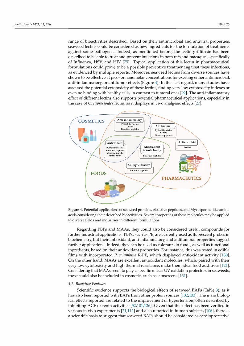
Antioxidants 2022, 11, 176 18 of 26
range of bioactivities described. Based on their antimicrobial and antiviral properties,seaweed lectins could be considered as new ingredients for the formulation of treatmentsagainst some pathogens. Indeed, as mentioned before, the lectin griffithsin has beendescribed to be able to treat and prevent infections in both rats and macaques, specificallyof Influenza, HSV, and HIV [75]. Topical application of this lectin in pharmaceuticalformulations could prove to be a possible preventive treatment against these infections,as evidenced by multiple reports. Moreover, seaweed lectins from diverse sources haveshown to be effective at pico- or nanomolar concentrations for exerting either antimicrobial,anti-inflammatory, or antitumor effects (Figure 4). In this last regard, many studies haveassessed the potential cytotoxicity of these lectins, finding very low cytotoxicity indexes oreven no binding with healthy cells, in contrast to tumoral ones [92]. The anti-inflammatoryeffect of different lectins also supports potential pharmaceutical applications, especially inthe case of C. cupressoides lectin, as it displays in vivo analgesic effects [27].
Antioxidants 2022, 11, x FOR PEER REVIEW 18 of 26
Figure 4. Potential applications of seaweed proteins, bioactive peptides, and Mycosporine-like
amino acids considering their described bioactivities. Several properties of these molecules may be
applied to diverse fields and industries in different formulations.
Regarding PBPs and MAAs, they could also be considered useful compounds for
further industrial applications. PBPs, such as PE, are currently used as fluorescent probes
in biochemistry, but their antioxidant, anti-inflammatory, and antitumoral properties sug-
gest further applications. Indeed, they can be used as colorants in foods, as well as func-
tional ingredients, based on their antioxidant properties. For instance, this was tested in
edible films with incorporated P. columbina R-PE, which displayed antioxidant activity
[130]. On the other hand, MAAs are excellent antioxidant molecules, which, paired with
their very low cytotoxicity and high thermal resistance, make them ideal food additives
[121]. Considering that MAAs seem to play a specific role as UV oxidation protectors in
seaweeds, these could also be included in cosmetics such as sunscreens [131].
4.2. Bioactive Peptides
Scientific evidence supports the biological effects of seaweed BAPs (Table 3), as it has
also been reported with BAPs from other protein sources [132,133]. The main biological
effects reported are related to the improvement of hypertension, often described by inhib-
iting ACE or renin activities [52,101,126]. Given that this effect has been verified in various
in vivo experiments [21,112] and also reported in human subjects [106], there is a scientific
basis to suggest that seaweed BAPs should be considered as cardioprotective agents. Ad-
ditionally, some BAPs from N. yezoensis were reported to exert anticoagulant activity,
which would contribute to cardiac function in some cases [56].
Seaweed BAPs have also been described to be potent antioxidant molecules at low
concentrations and in various antioxidant assays [107]. This suggests that their use as a
functional ingredient in foods or cosmetics could be multivalent, acting as preservers
and/or dietary antioxidant compounds.
Furthermore, BAPs, such as described from P. palmata, are reported to exert antidia-
betic and anti-obesity effects [128]. Foremost, these effects are related to upregulation of
GLP-1 and/or inhibition of DPP-IV, which results in an improved metabolic function and
control of glucose levels [60]. Seaweed BAPs may also act as α-glucosidase inhibitors, re-
ducing gut glucose absorption and subsequent glycemic levels [54]. Given that these ef-
fects have been described in animal models, there is a great potential for these peptides as
dietary supplements for diabetes and/or obesity management [128,129]. The anti-inflam-
matory or antitumoral properties of seaweed BAPs have also been highlighted. Anti-
Figure 4. Potential applications of seaweed proteins, bioactive peptides, and Mycosporine-like aminoacids considering their described bioactivities. Several properties of these molecules may be appliedto diverse fields and industries in different formulations.
Regarding PBPs and MAAs, they could also be considered useful compounds forfurther industrial applications. PBPs, such as PE, are currently used as fluorescent probes inbiochemistry, but their antioxidant, anti-inflammatory, and antitumoral properties suggestfurther applications. Indeed, they can be used as colorants in foods, as well as functionalingredients, based on their antioxidant properties. For instance, this was tested in ediblefilms with incorporated P. columbina R-PE, which displayed antioxidant activity [130].On the other hand, MAAs are excellent antioxidant molecules, which, paired with theirvery low cytotoxicity and high thermal resistance, make them ideal food additives [121].Considering that MAAs seem to play a specific role as UV oxidation protectors in seaweeds,these could also be included in cosmetics such as sunscreens [131].
4.2. Bioactive Peptides
Scientific evidence supports the biological effects of seaweed BAPs (Table 3), as ithas also been reported with BAPs from other protein sources [132,133]. The main biolog-ical effects reported are related to the improvement of hypertension, often described byinhibiting ACE or renin activities [52,101,126]. Given that this effect has been verified invarious in vivo experiments [21,112] and also reported in human subjects [106], there isa scientific basis to suggest that seaweed BAPs should be considered as cardioprotective
Antioxidants 2022, 11, 176 19 of 26
agents. Additionally, some BAPs from N. yezoensis were reported to exert anticoagulantactivity, which would contribute to cardiac function in some cases [56].
Seaweed BAPs have also been described to be potent antioxidant molecules at lowconcentrations and in various antioxidant assays [107]. This suggests that their use asa functional ingredient in foods or cosmetics could be multivalent, acting as preserversand/or dietary antioxidant compounds.
Furthermore, BAPs, such as described from P. palmata, are reported to exert antidi-abetic and anti-obesity effects [128]. Foremost, these effects are related to upregulationof GLP-1 and/or inhibition of DPP-IV, which results in an improved metabolic functionand control of glucose levels [60]. Seaweed BAPs may also act as α-glucosidase inhibitors,reducing gut glucose absorption and subsequent glycemic levels [54]. Given that theseeffects have been described in animal models, there is a great potential for these pep-tides as dietary supplements for diabetes and/or obesity management [128,129]. Theanti-inflammatory or antitumoral properties of seaweed BAPs have also been highlighted.Anti-inflammatory effects of seaweed BAPs are mostly related to the upregulation of anti-inflammatory cytokines such as IL10, ameliorating inflammatory reactions [110], but alsothrough immunomodulation [62]. Regarding antitumoral effects, while available reportshave only been performed in vitro, several works describe these peptides to induce apop-tosis while showing almost null cytotoxicity [108,134]. However, while there is abundantevidence of in vitro describing molecular effects of these BAPs, more animal studies arerequired to validate and corroborate these properties to devise effective applications. No-tably, the great majority of seaweed BAPs are of short length, which has proven to makethem resistant to gastrointestinal digestions and retain their biological properties, such asantihypertensive or antioxidant [104,105]. This fact further supports their suitability asfood additives or dietary supplements.
Altogether, even though accumulating reports support the bioactive properties ofseaweed BAPs, additional studies are necessary to improve the knowledge about the useof seaweed protein hydrolysates and bioactive peptides in different systems and fields ofresearch. Further research should focus on deeper studies about effects in vivo and directapplications in (i.e., pharmacological, food science, and cosmetics) to explore the widevariety of activities of seaweed peptides.
5. Conclusions
Seaweeds are being increasingly exploited on a global scale due to their dietary andnutritional benefits, of which their protein and amino acid composition pose a valuablecomponent. Considering their protein content and bioactivities, seaweeds can be feasible touse as a source of these compounds with functional properties. SP such as red algae’s PBPs,as well as lectins, have been extensively studied in recent years as potential new functionalingredients or pharmacological agents since significant evidence supports their use asanti-inflammatory, antitumoral, or antiviral agents. Moreover, SP hydrolysates containseveral BAPs, which have been isolated and demonstrated to possess effective biologicalproperties such as the ones mentioned above but also to be anti-diabetic or anti-obesityagents, as well as important hypotensive molecules. Several works have analyzed thepotential mechanisms of action of these proteins and derived peptides, highlighting theireffective potential for various applications. Altogether, despite the growing evidence on SPand their derived BAPs, much further work is needed to evaluate the extent of their possiblemechanisms of action. Likewise, further works would need to assess potential bioactive SPin many species, especially from green or brown algae, as these are less studied groups.
Author Contributions: Conceptualization, J.E. and M.A.P.; methodology, J.E. and P.G.-O.; software,J.E., P.E.S.M., M.P. and P.G.-O.; validation, J.M.L., J.S.-G. and M.A.P.; formal analysis, J.E., P.O. andM.A.P.; investigation, J.E., P.O., P.E.S.M. and M.P.; data curation, J.E., P.E.S.M. and M.P.; writing—original draft preparation, J.E., P.O., P.E.S.M. and M.P.; writing—review and editing, P.G.-O., J.E.,M.A.P. and J.M.L.; visualization, P.O., J.S.-G., M.A.P. and J.E.; supervision, M.A.P., J.S.-G. and J.M.L.All authors have read and agreed to the published version of the manuscript.

Antioxidants 2022, 11, 176 20 of 26
Funding: The research leading to these results was supported by MICINN supporting the Ramóny Cajal grant for M.A. Prieto (RYC-2017-22891), to Xunta de Galicia for the pre-doctoral grant ofP. Garcia-Oliveira (ED481A-2019/295), the program Grupos de Referencia Competitiva (GRUPOAA1-GRC 2018) that supports the work of J. Echave, to the Bio Based Industries Joint Undertaking(JU) under grant agreement No 888003 UP4HEALTH Project (H2020-BBI-JTI-2019) that supports thework of P. Otero. The JU receives support from the European Union’s Horizon 2020 research andinnovation program and the Bio Based Industries Consortium. P.E.S. Munekata, M. Pateriro, andJ.M. Lorenzo acknowledge GAIN (Axencia Galega de Innovación) for supporting this study (grantnumber IN607A2019/01).
Conflicts of Interest: The authors declare no conflict of interest.
Abbreviations
SP Seaweed proteinsAA Amino acidsEAA Essential amino acidsNEAA Non-essential amino acidsBAPs Bioactive peptidesGP GlycoproteinPBP PhycobiliproteinPE PhycoerythrinMAAs Mycosporine-like amino acidsλmax Absorption maximaORAC Oxygen radical absorption capacityFRAP Ferric reducing antioxidant powerDPPH 2,2-diphenyl-1-picrylhydrazylABTS 2,2′-azino-bis(3-ethylbenzothiazoline-6-sulfonic acid)UV UltravioletACE Angiotensin-I converter enzymeROS Reactive oxygen speciesHIV Human immunodeficiency virusHSV Herpes simplex virusHPV Human papilloma virusSARS-CoV Severe acute respiratory syndrome-coronavirusIL InterleukinTNF-α Tumor necrosis factor-αCOX-2 Cyclooxygenase-2NO Nitric oxide synthaseiNOS Nitric oxideTLR Toll-like receptorsTGF-β Transforming growth factor-βNF-κB Nuclear factor κbMAPK Mitogen-activated protein kinaseLPS LipopolysaccharidesMPO MyeloperoxidaseHO-2 Hemeoxygenase-2JNK Jun N-terminal kinaseERK Extracellular signal-related kinasemTOR Mammalian target of rapamycinIGF-IR Insulin-like growth factor-I receptorPI3K Phosphatidylinositol 3-kinaseDPP-IV Dipeptidyl peptidase-IVGLP-1 Glucagon-like peptide-1IRS-I Insulin receptor substrate-I
Antioxidants 2022, 11, 176 21 of 26
References1. Dawczynski, C.; Schubert, R.; Jahreis, G. Amino acids, fatty acids, and dietary fibre in edible seaweed products. Food Chem. 2007,
103, 891–899. [CrossRef]2. Gómez-Ordóñez, E.; Jiménez-Escrig, A.; Rupérez, P. Dietary fibre and physicochemical properties of several edible seaweeds
from the northwestern Spanish coast. Food Res. Int. 2010, 43, 2289–2294. [CrossRef]3. Machado, M.; Machado, S.; Pimentel, F.B.; Freitas, V.; Alves, R.C.; Oliveira, M.B.P.P. Amino acid profile and protein quality
assessment of macroalgae produced in an integrated multi-trophic aquaculture system. Foods 2020, 9, 1382. [CrossRef]4. Cermeño, M.; Kleekayai, T.; Amigo-Benavent, M.; Harnedy, P.A.; FitzGerald, R.J. Current knowledge on the extraction, pu-
rification, identification, and validation of bioactive peptides from seaweed. Electrophoresis 2020, 41, 1694–1717. [CrossRef][PubMed]
5. Cherry, P.; O’Hara, C.; Magee, P.J.; McSorley, E.M.; Allsopp, P.J. Risks and benefits of consuming edible seaweeds. Nutr. Rev. 2019,77, 307–329. [CrossRef]
6. Kazir, M.; Abuhassira, Y.; Robin, A.; Nahor, O.; Luo, J.; Israel, A.; Golberg, A.; Livney, Y.D. Extraction of proteins from two marinemacroalgae, Ulva sp. and Gracilaria sp., for food application, and evaluating digestibility, amino acid composition and antioxidantproperties of the protein concentrates. Food Hydrocoll. 2019, 87, 194–203. [CrossRef]
7. Paiva, L.; Lima, E.; Patarra, R.F.; Neto, A.I.; Baptista, J. Edible Azorean macroalgae as source of rich nutrients with impact onhuman health. Food Chem. 2014, 164, 128–135. [CrossRef]
8. Fleurence, J. Seaweed proteins. Trends Food Sci. Technol. 1999, 10, 25–28. [CrossRef]9. Shuuluka, D.; Bolton, J.J.; Anderson, R.J. Protein content, amino acid composition and nitrogen-to-protein conversion factors
of Ulva rigida and Ulva capensis from natural populations and Ulva lactuca from an aquaculture system, in South Africa. J. Appl.Phycol. 2013, 25, 677–685. [CrossRef]
10. Cian, R.E.; Fajardo, M.A.; Alaiz, M.; Vioque, J.; González, R.J.; Drago, S.R. Chemical composition, nutritional and antioxidantproperties of the red edible seaweed Porphyra columbina. Int. J. Food Sci. Nutr. 2014, 65, 299–305. [CrossRef]
11. Poojary, M.M.; Orlien, V.; Olsen, K. Conventional and enzyme-assisted green extraction of umami free amino acids from Nordicseaweeds. J. Appl. Phycol. 2019, 31, 3925–3939. [CrossRef]
12. Bocanegra, A.; Macho-González, A.; Garcimartín, A.; Benedí, J.; Sánchez-Muniz, F.J. Whole Alga, Algal Extracts, and Compoundsas Ingredients of Functional Foods: Composition and Action Mechanism Relationships in the Prevention and Treatment of Type-2Diabetes Mellitus. Int. J. Mol. Sci. 2021, 22, 3816. [CrossRef]
13. Harnedy, P.A.; Fitzgerald, R.J. Bioactive proteins, peptides, and amino acids from macroalgae. J. Phycol. 2011, 47, 218–232.[CrossRef]
14. O’Connor, J.; Meaney, S.; Williams, G.A.; Hayes, M. Extraction of protein from four different seaweeds using three differentphysical pre-treatment strategies. Molecules 2020, 25, 2005. [CrossRef]
15. Biancarosa, I.; Espe, M.; Bruckner, C.G.; Heesch, S.; Liland, N.; Waagbø, R.; Torstensen, B.; Lock, E.J. Amino acid composition,protein content, and nitrogen-to-protein conversion factors of 21 seaweed species from Norwegian waters. J. Appl. Phycol. 2017,29, 1001–1009. [CrossRef]
16. Taboada, C.; Millan, R.; Miguez, I. Evaluation of marine algae Undaria pinnatifida and Porphyra purpurea as a food supplement:Composition, nutritional value and effect of intake on intestinal, hepatic and renal enzyme activities in rats. J. Sci. Food Agric.2013, 93, 1863–1868. [CrossRef] [PubMed]
17. Vieira, E.F.; Soares, C.; Machado, S.; Correia, M.; Ramalhosa, M.J.; Oliva-teles, M.T.; Paula Carvalho, A.; Domingues, V.F.; Antunes,F.; Oliveira, T.A.C.; et al. Seaweeds from the Portuguese coast as a source of proteinaceous material: Total and free amino acidcomposition profile. Food Chem. 2018, 269, 264–275. [CrossRef]
18. Ortiz, J.; Uquiche, E.; Robert, P.; Romero, N.; Quitral, V.; Llantén, C. Functional and nutritional value of the Chilean seaweedsCodium fragile, Gracilaria chilensis and Macrocystis pyrifera. Eur. J. Lipid Sci. Technol. 2009, 111, 320–327. [CrossRef]
19. Ferreira, M.; Teixeira, C.; Abreu, H.; Silva, J.; Costas, B.; Kiron, V.; Valente, L.M.P. Nutritional value, antimicrobial and antioxidantactivities of micro- and macroalgae, single or blended, unravel their potential use for aquafeeds. J. Appl. Phycol. 2021, 33,3507–3518. [CrossRef]
20. Trigueros, E.; Sanz, M.T.; Alonso-Riaño, P.; Beltrán, S.; Ramos, C.; Melgosa, R. Recovery of the protein fraction with highantioxidant activity from red seaweed industrial solid residue after agar extraction by subcritical water treatment. J. Appl. Phycol.2021, 33, 1181–1194. [CrossRef]
21. Zheng, Y.; Zhang, Y.; San, S. Efficacy of a novel ACE-inhibitory peptide from Sargassum maclurei in hypertension and reduction ofintracellular endothelin-1. Nutrients 2020, 12, 653. [CrossRef] [PubMed]
22. Lorenzo, J.M.; Agregán, R.; Munekata, P.; Franco, D.; Carballo, J.; Sahin, S.; Lacomba, R.; Barba, F. Proximate Composition andNutritional Value of Three Macroalgae: Ascophyllum nodosum, Fucus vesiculosus and Bifurcaria bifurcata. Mar. Drugs 2017, 15, 360.[CrossRef]
23. Echave, J.; Fraga-Corral, M.; Garcia-Perez, P.; Popovic-Djordjevic, J.; Avdovic, E.H.; Radulovic, M.; Xiao, J.; Prieto, M.A.; Simal-Gandara, J. Seaweed Protein Hydrolysates and Bioactive Peptides: Extraction, Purification and Applications. Mar. Drugs 2021, 19,500. [CrossRef] [PubMed]
24. Torres, P.; Santos, J.P.; Chow, F.; Pena Ferreira, M.J.; dos Santos, D.Y.A.C. Comparative analysis of in vitro antioxidant capacitiesof mycosporine-like amino acids (MAAs). Algal Res. 2018, 34, 57–67. [CrossRef]
Antioxidants 2022, 11, 176 22 of 26
25. Boonsri, N.; Rudtanatip, T.; Withyachumnarnkul, B.; Wongprasert, K. Protein extract from red seaweed Gracilaria fisheri preventsacute hepatopancreatic necrosis disease (AHPND) infection in shrimp. J. Appl. Phycol. 2017, 29, 1597–1608. [CrossRef]
26. Seca, A.; Pinto, D. Overview on the Antihypertensive and Anti-Obesity Effects of Secondary Metabolites from Seaweeds. Mar.Drugs 2018, 16, 237. [CrossRef]
27. de Queiroz, I.N.L.; Quinderé, A.L.G.; Rodrigues, J.A.G.; de Sousa Oliveira Vanderlei, E.; Ribeiro, N.A.; da Conceição Rivanor,R.L.; Ribeiro, K.A.; Coura, C.O.; Pereira, K.M.A.; Chaves, H.V.; et al. Dual effects of a lectin from the green seaweed Caulerpacupressoides var. lycopodium on inflammatory mediators in classical models of inflammation. Inflamm. Res. 2015, 64, 971–982.[CrossRef]
28. Lafarga, T.; Acién-Fernández, F.G.; Garcia-Vaquero, M. Bioactive peptides and carbohydrates from seaweed for food applications:Natural occurrence, isolation, purification, and identification. Algal Res. 2020, 48, 101909. [CrossRef]
29. Lee, H.A.; Kim, I.H.; Nam, T.J. Bioactive peptide from Pyropia yezoensis and its anti-inflammatory activities. Int. J. Mol. Med. 2015,36, 1701–1706. [CrossRef]
30. Conde, E.; Balboa, E.M.; Parada, M.; Falqué, E. Algal proteins, peptides and amino acids. In Functional Ingredients from Algaefor Foods and Nutraceuticals; Dominguez, H., Ed.; Woodhead Publishing Elsevier: Cambridge, UK, 2013; pp. 135–180. ISBN9780857095121.
31. Hayes, M.; Tiwari, B.K. Bioactive carbohydrates and peptides in foods: An overview of sources, downstream processing stepsand associated bioactivities. Int. J. Mol. Sci. 2015, 16, 22485–22508. [CrossRef] [PubMed]
32. Charoensiddhi, S.; Conlon, M.A.; Franco, C.M.M.; Zhang, W. The development of seaweed-derived bioactive compounds for useas prebiotics and nutraceuticals using enzyme technologies. Trends Food Sci. Technol. 2017, 70, 20–33. [CrossRef]
33. Yoshiie, T.; Maeda, M.; Kimura, M.; Hama, Y.; Uchida, M.; Kimura, Y. Structural features of N-glycans of seaweed glycoproteins:Predominant occurrence of high-mannose type N-glycans in marine plants. Biosci. Biotechnol. Biochem. 2012, 76, 1996–1998.[CrossRef] [PubMed]
34. Wijesekara, I.; Lang, M.; Marty, C.; Gemin, M.-P.; Boulho, R.; Douzenel, P.; Wickramasinghe, I.; Bedoux, G.; Bourgougnon, N.Different extraction procedures and analysis of protein from Ulva sp. in Brittany, France. J. Appl. Phycol. 2017, 29, 2503–2511.[CrossRef]
35. Thangam, R.; Senthilkumar, D.; Suresh, V.; Sathuvan, M.; Sivasubramanian, S.; Pazhanichamy, K.; Gorlagunta, P.K.; Kannan, S.;Gunasekaran, P.; Rengasamy, R.; et al. Induction of ROS-Dependent Mitochondria-Mediated Intrinsic Apoptosis in MDA-MB-231Cells by Glycoprotein from Codium decorticatum. J. Agric. Food Chem. 2014, 62, 3410–3421. [CrossRef]
36. Barre, A.; Simplicien, M.; Benoist, H.; Van Damme, E.J.M.; Rougé, P. Mannose-Specific Lectins from Marine Algae: DiverseStructural Scaffolds Associated to Common Virucidal and Anti-Cancer Properties. Mar. Drugs 2019, 17, 440. [CrossRef]
37. Frenkel, J.; Vyverman, W.; Pohnert, G. Pheromone signaling during sexual reproduction in algae. Plant J. 2014, 79, 632–644.[CrossRef]
38. Damme, E.J.M.; Van Peumans, W.J.; Barre, A.; Rougé, P. Plant Lectins: A Composite of Several Distinct Families of Structurallyand Evolutionary Related Proteins with Diverse Biological Roles. CRC Crit. Rev. Plant Sci. 1998, 17, 575–692. [CrossRef]
39. Ambrosio, A.L.; Sanz, L.; Sánchez, E.I.; Wolfenstein-Todel, C.; Calvete, J.J. Isolation of two novel mannan- and L-fucose-bindinglectins from the green alga Enteromorpha prolifera: Biochemical characterization of EPL-2. Arch. Biochem. Biophys. 2003, 415,245–250. [CrossRef]
40. Wu, M.; Tong, C.; Wu, Y.; Liu, S.; Li, W. A novel thyroglobulin-binding lectin from the brown alga Hizikia fusiformis and itsantioxidant activities. Food Chem. 2016, 201, 7–13. [CrossRef] [PubMed]
41. Günaydın, G.; Edfeldt, G.; Garber, D.A.; Asghar, M.; No
Antioxidants 2022, 11, x FOR PEER REVIEW 22 of 26
29. Lee, H.A.; Kim, I.H.; Nam, T.J. Bioactive peptide from Pyropia yezoensis and its anti-inflammatory activities. Int. J. Mol. Med.
2015, 36, 1701–1706. https://doi.org/10.3892/ijmm.2015.2386.
30. Conde, E.; Balboa, E.M.; Parada, M.; Falqué, E. Algal proteins, peptides and amino acids. In Functional Ingredients from Algae for
Foods and Nutraceuticals; Dominguez, H., Ed.; Woodhead Publishing Elsevier: Cambridge, UK, 2013; pp. 135–180, ISBN
9780857095121.
31. Hayes, M.; Tiwari, B.K. Bioactive carbohydrates and peptides in foods: An overview of sources, downstream processing steps
and associated bioactivities. Int. J. Mol. Sci. 2015, 16, 22485–22508. https://doi.org/10.3390/ijms160922485.
32. Charoensiddhi, S.; Conlon, M.A.; Franco, C.M.M.; Zhang, W. The development of seaweed-derived bioactive compounds for
use as prebiotics and nutraceuticals using enzyme technologies. Trends Food Sci. Technol. 2017, 70, 20–33.
https://doi.org/10.1016/j.tifs.2017.10.002.
33. Yoshiie, T.; Maeda, M.; Kimura, M.; Hama, Y.; Uchida, M.; Kimura, Y. Structural features of N-glycans of seaweed glycoproteins:
Predominant occurrence of high-mannose type N-glycans in marine plants. Biosci. Biotechnol. Biochem. 2012, 76, 1996–1998.
https://doi.org/10.1271/bbb.120463.
34. Wijesekara, I.; Lang, M.; Marty, C.; Gemin, M.-P.; Boulho, R.; Douzenel, P.; Wickramasinghe, I.; Bedoux, G.; Bourgougnon, N.
Different extraction procedures and analysis of protein from Ulva sp. in Brittany, France. J. Appl. Phycol. 2017, 29, 2503–2511.
https://doi.org/10.1007/s10811-017-1239-7.
35. Thangam, R.; Senthilkumar, D.; Suresh, V.; Sathuvan, M.; Sivasubramanian, S.; Pazhanichamy, K.; Gorlagunta, P.K.; Kannan,
S.; Gunasekaran, P.; Rengasamy, R.; et al. Induction of ROS-Dependent Mitochondria-Mediated Intrinsic Apoptosis in MDA-
MB-231 Cells by Glycoprotein from Codium decorticatum. J. Agric. Food Chem. 2014, 62, 3410–3421.
https://doi.org/10.1021/jf405329e.
36. Barre, A.; Simplicien, M.; Benoist, H.; Van Damme, E.J.M.; Rougé, P. Mannose-Specific Lectins from Marine Algae: Diverse
Structural Scaffolds Associated to Common Virucidal and Anti-Cancer Properties. Mar. Drugs 2019, 17, 440.
https://doi.org/10.3390/md17080440.
37. Frenkel, J.; Vyverman, W.; Pohnert, G. Pheromone signaling during sexual reproduction in algae. Plant J. 2014, 79, 632–644.
https://doi.org/10.1111/tpj.12496.
38. Damme, E.J.M.; Van; Peumans, W.J.; Barre, A.; Rougé, P. Plant Lectins: A Composite of Several Distinct Families of Structurally
and Evolutionary Related Proteins with Diverse Biological Roles. CRC Crit. Rev. Plant Sci. 1998, 17, 575–692.
https://doi.org/10.1080/07352689891304276.
39. Ambrosio, A.L.; Sanz, L.; Sánchez, E.I.; Wolfenstein-Todel, C.; Calvete, J.J. Isolation of two novel mannan- and L-fucose-binding
lectins from the green alga Enteromorpha prolifera: Biochemical characterization of EPL-2. Arch. Biochem. Biophys. 2003, 415, 245–
250. https://doi.org/10.1016/S0003-9861(03)00232-7.
40. Wu, M.; Tong, C.; Wu, Y.; Liu, S.; Li, W. A novel thyroglobulin-binding lectin from the brown alga Hizikia fusiformis and its
antioxidant activities. Food Chem. 2016, 201, 7–13. https://doi.org/10.1016/j.foodchem.2016.01.061.
41. Günaydın, G.; Edfeldt, G.; Garber, D.A.; Asghar, M.; Noȅl-Romas, L.; Burgener, A.; Wählby, C.; Wang, L.; Rohan, L.C.; Guenth-
ner, P.; et al. Impact of Q-Griffithsin anti-HIV microbicide gel in non-human primates: In situ analyses of epithelial and immune
cell markers in rectal mucosa. Sci. Rep. 2019, 9, 18120. https://doi.org/10.1038/s41598-019-54493-4.
42. Fontenelle, T.P.C.; Lima, G.C.; Mesquita, J.X.; Lopes, J.L.; de Brito, T.V.; Vieira-Júnior, F.C.; Sales, A.B.; Aragão, K.S.; de Souza,
M.H.L.P.; Barbosa, A.L.R.; et al. Lectin obtained from the red seaweed Bryothamnion triquetrum: Secondary structure and anti-
inflammatory activity in mice. Int. J. Biol. Macromol. 2018, 112, 1122–1130. https://doi.org/10.1016/j.ijbiomac.2018.02.058.
43. Nair, D.; Krishna, J.G.; Panikkar, M.V.N.; Nair, B.G.; Pai, J.G.; Nair, S.S. Identification, purification, biochemical and mass spec-
trometric characterization of novel phycobiliproteins from a marine red alga, Centroceras clavulatum. Int. J. Biol. Macromol. 2018,
114, 679–691. https://doi.org/10.1016/j.ijbiomac.2018.03.153.
44. Yabuta, Y.; Fujimura, H.; Kwak, C.S.; Enomoto, T.; Watanabe, F. Antioxidant activity of the phycoerythrobilin compound
formed from a dried Korean purple laver (Porphyra sp.) during in vitro digestion. Food Sci. Technol. Res. 2010, 16, 347–351.
https://doi.org/10.3136/fstr.16.347.
45. Chen, X.; Wu, M.; Yang, Q.; Wang, S. Preparation, characterization of food grade phycobiliproteins from Porphyra haitanensis
and the application in liposome-meat system. LWT Food Sci. Technol. 2017, 77, 468–474. https://doi.org/10.1016/j.lwt.2016.12.005.
46. Li, W.; Su, H.; Pu, Y.; Chen, J.; Liu, L.; Liu, Q.; Qin, S. Phycobiliproteins : Molecular structure, production, applications, and
prospects. Biotechnol. Adv. 2019, 37, 340–353. https://doi.org/10.1016/j.biotechadv.2019.01.008.
47. Guan, X.; Wang, J.; Zhu, J.; Yao, C.; Liu, J.; Qin, S.; Jiang, P. Photosystem II Photochemistry and Phycobiliprotein of the Red
Algae Kappaphycus alvarezii and Their Implications for Light Adaptation. Biomed Res. Int. 2013, 2013, 1–9.
https://doi.org/10.1155/2013/256549.
48. Gacesa, R.; Lawrence, K.P.; Georgakopoulos, N.D.; Yabe, K.; Dunlap, W.C.; Barlow, D.J.; Wells, G.; Young, A.R.; Long, P.F. The
mycosporine-like amino acids porphyra-334 and shinorine are antioxidants and direct antagonists of Keap1-Nrf2 binding. Bio-
chimie 2018, 154, 35–44. https://doi.org/10.1016/j.biochi.2018.07.020.
49. Sun, Y.; Zhang, N.; Zhou, J.; Dong, S.; Zhang, X.; Guo, L.; Guo, G. Distribution, Contents, and Types of Mycosporine-Like Amino
Acids (MAAs) in Marine Macroalgae and a Database for MAAs Based on These Characteristics. Mar. Drugs 2020, 18, 43.
https://doi.org/10.3390/md18010043.
50. Cian, R.E.; Martínez-Augustin, O.; Drago, S.R. Bioactive properties of peptides obtained by enzymatic hydrolysis from protein
byproducts of Porphyra columbina. Food Res. Int. 2012, 49, 364–372. https://doi.org/10.1016/j.foodres.2012.07.003.
l-Romas, L.; Burgener, A.; Wählby, C.; Wang, L.; Rohan, L.C.; Guenthner,P.; et al. Impact of Q-Griffithsin anti-HIV microbicide gel in non-human primates: In situ analyses of epithelial and immune cellmarkers in rectal mucosa. Sci. Rep. 2019, 9, 18120. [CrossRef]
42. Fontenelle, T.P.C.; Lima, G.C.; Mesquita, J.X.; Lopes, J.L.; de Brito, T.V.; Vieira-Júnior, F.C.; Sales, A.B.; Aragão, K.S.; de Souza,M.H.L.P.; Barbosa, A.L.R.; et al. Lectin obtained from the red seaweed Bryothamnion triquetrum: Secondary structure andanti-inflammatory activity in mice. Int. J. Biol. Macromol. 2018, 112, 1122–1130. [CrossRef]
43. Nair, D.; Krishna, J.G.; Panikkar, M.V.N.; Nair, B.G.; Pai, J.G.; Nair, S.S. Identification, purification, biochemical and massspectrometric characterization of novel phycobiliproteins from a marine red alga, Centroceras clavulatum. Int. J. Biol. Macromol.2018, 114, 679–691. [CrossRef] [PubMed]
44. Yabuta, Y.; Fujimura, H.; Kwak, C.S.; Enomoto, T.; Watanabe, F. Antioxidant activity of the phycoerythrobilin compound formedfrom a dried Korean purple laver (Porphyra sp.) during in vitro digestion. Food Sci. Technol. Res. 2010, 16, 347–351. [CrossRef]
45. Chen, X.; Wu, M.; Yang, Q.; Wang, S. Preparation, characterization of food grade phycobiliproteins from Porphyra haitanensis andthe application in liposome-meat system. LWT Food Sci. Technol. 2017, 77, 468–474. [CrossRef]
46. Li, W.; Su, H.; Pu, Y.; Chen, J.; Liu, L.; Liu, Q.; Qin, S. Phycobiliproteins: Molecular structure, production, applications, andprospects. Biotechnol. Adv. 2019, 37, 340–353. [CrossRef]
47. Guan, X.; Wang, J.; Zhu, J.; Yao, C.; Liu, J.; Qin, S.; Jiang, P. Photosystem II Photochemistry and Phycobiliprotein of the Red AlgaeKappaphycus alvarezii and Their Implications for Light Adaptation. BioMed Res. Int. 2013, 2013, 256549. [CrossRef] [PubMed]
48. Gacesa, R.; Lawrence, K.P.; Georgakopoulos, N.D.; Yabe, K.; Dunlap, W.C.; Barlow, D.J.; Wells, G.; Young, A.R.; Long, P.F.The mycosporine-like amino acids porphyra-334 and shinorine are antioxidants and direct antagonists of Keap1-Nrf2 binding.Biochimie 2018, 154, 35–44. [CrossRef]
Antioxidants 2022, 11, 176 23 of 26
49. Sun, Y.; Zhang, N.; Zhou, J.; Dong, S.; Zhang, X.; Guo, L.; Guo, G. Distribution, Contents, and Types of Mycosporine-LikeAmino Acids (MAAs) in Marine Macroalgae and a Database for MAAs Based on These Characteristics. Mar. Drugs 2020, 18, 43.[CrossRef] [PubMed]
50. Cian, R.E.; Martínez-Augustin, O.; Drago, S.R. Bioactive properties of peptides obtained by enzymatic hydrolysis from proteinbyproducts of Porphyra columbina. Food Res. Int. 2012, 49, 364–372. [CrossRef]
51. Lorenzo, J.M.; Munekata, P.E.S.; Gómez, B.; Barba, F.J.; Mora, L.; Pérez-Santaescolástica, C.; Toldrá, F. Bioactive peptides as naturalantioxidants in food products—A review. Trends Food Sci. Technol. 2018, 79, 136–147. [CrossRef]
52. Cao, D.; Lv, X.; Xu, X.; Yu, H.; Sun, X.; Xu, N. Purification and identification of a novel ACE inhibitory peptide from marine algaGracilariopsis lemaneiformis protein hydrolysate. Eur. Food Res. Technol. 2017, 243, 1829–1837. [CrossRef]
53. Sun, S.; Xu, X.; Sun, X.; Zhang, X.; Chen, X.; Xu, N. Preparation and identification of ACE inhibitory peptides from the marinemacroalga Ulva intestinalis. Mar. Drugs 2019, 17, 179. [CrossRef]
54. Admassu, H.; Gasmalla, M.A.A.; Yang, R.; Zhao, W. Identification of Bioactive Peptides with α-Amylase Inhibitory Potential fromEnzymatic Protein Hydrolysates of Red Seaweed (Porphyra spp). J. Agric. Food Chem. 2018, 66, 4872–4882. [CrossRef] [PubMed]
55. Beaulieu, L.; Sirois, M.; Tamigneaux, É. Evaluation of the in vitro biological activity of protein hydrolysates of the edible red alga,Palmaria palmata (dulse) harvested from the Gaspe coast and cultivated in tanks. J. Appl. Phycol. 2016, 28, 3101–3115. [CrossRef]
56. Indumathi, P.; Mehta, A. A novel anticoagulant peptide from the Nori hydrolysate. J. Funct. Foods 2016, 20, 606–617. [CrossRef]57. Paiva, L.; Lima, E.; Neto, A.I.I.; Baptista, J. Isolation and characterization of angiotensin I-converting enzyme (ACE) inhibitory
peptides from Ulva rigida C. Agardh protein hydrolysate. J. Funct. Foods 2016, 26, 65–76. [CrossRef]58. Zhang, X.; Cao, D.; Sun, X.; Sun, S.; Xu, N. Preparation and identification of antioxidant peptides from protein hydrolysate of
marine alga Gracilariopsis lemaneiformis. J. Appl. Phycol. 2019, 31, 2585–2596. [CrossRef]59. Furuta, T.; Miyabe, Y.; Yasui, H.; Kinoshita, Y.; Kishimura, H. Angiotensin I Converting Enzyme Inhibitory Peptides Derived from
Phycobiliproteins of Dulse Palmaria palmata. Mar. Drugs 2016, 14, 32. [CrossRef]60. Cermeño, M.; Stack, J.; Tobin, P.R.; O’Keeffe, M.B.; Harnedy, P.A.; Stengel, D.B.; FitzGerald, R.J. Peptide identification from a
Porphyra dioica protein hydrolysate with antioxidant, angiotensin converting enzyme and dipeptidyl peptidase IV inhibitoryactivities. Food Funct. 2019, 10, 3421–3429. [CrossRef]
61. Harnedy, P.A.; O’Keeffe, M.B.; FitzGerald, R.J. Purification and identification of dipeptidyl peptidase (DPP) IV inhibitory peptidesfrom the macroalga Palmaria palmata. Food Chem. 2015, 172, 400–406. [CrossRef]
62. Cian, R.E.; Hernández-Chirlaque, C.; Gámez-Belmonte, R.; Drago, S.R.; Sánchez de Medina, F.; Martínez-Augustin, O. Greenalga Ulva spp. hydrolysates and their peptide fractions regulate cytokine production in splenic macrophages and lymphocytesinvolving the TLR4-NFκB/MAPK pathways. Mar. Drugs 2018, 16, 235. [CrossRef] [PubMed]
63. Reuter, S.; Gupta, S.C.; Chaturvedi, M.M.; Aggarwal, B.B. Oxidative stress, inflammation, and cancer: How are they linked? FreeRadic. Biol. Med. 2010, 49, 1603–1616. [CrossRef] [PubMed]
64. Yu, Z.; Yin, Y.; Zhao, W.; Wang, F.; Yu, Y.; Liu, B.; Liu, J.; Chen, F. Characterization of ACE-Inhibitory Peptide Associated withAntioxidant and Anticoagulation Properties. J. Food Sci. 2011, 76, C1149–C1155. [CrossRef]
65. Senthilkumar, D.; Jayanthi, S. Antioxidant activities of purified glycoprotein extracted from Codium decorticatum. J. Appl. Pharm.Sci. 2015, 5, 101–104. [CrossRef]
66. Suh, H.-J.; Lee, H.-W.; Jung, J. Mycosporine Glycine Protects Biological Systems Against Photodynamic Damage by QuenchingSinglet Oxygen with a High Efficiency. Photochem. Photobiol. 2003, 78, 109. [CrossRef]
67. Harnedy, P.A.; O’Keeffe, M.B.; FitzGerald, R.J. Fractionation and identification of antioxidant peptides from an enzymaticallyhydrolysed Palmaria palmata protein isolate. Food Res. Int. 2017, 100, 416–422. [CrossRef]
68. Wen, L.; Tan, S.; Zeng, L.; Wang, Z.; Ke, X.; Zhang, Z.; Tang, H.; Guo, H.; Xia, E. Ultrasound-assisted extraction and in vitrosimulated digestion of Porphyra haitanensis proteins exhibiting antioxidative and α-glucosidase inhibitory activity. J. Food Meas.Charact. 2020, 14, 3291–3298. [CrossRef]
69. Pimentel, F.B.; Cermeño, M.; Kleekayai, T.; Machado, S.; Rego, A.; Fernandes, E.; Alves, R.C.; Oliveira, M.B.P.P.; FitzGerald, R.J.Contribution of in vitro simulated gastrointestinal digestion to the antioxidant activity of Porphyra dioica conchocelis. Algal Res.2020, 51, 102085. [CrossRef]
70. Pimentel, F.B.; Cermeño, M.; Kleekayai, T.; Harnedy, P.A.; Fernandes, E.; Alves, R.C.; Oliveira, M.B.P.P.; FitzGerald, R.J. EnzymaticModification of Porphyra dioica-Derived Proteins to Improve their Antioxidant Potential. Molecules 2020, 25, 2838. [CrossRef]
71. Carneiro, R.F.; Duarte, P.L.; Chaves, R.P.; da Silva, S.R.; Feitosa, R.R.; de Sousa, B.L.; Alves, A.W.S.; de Vasconcelos, M.A.; daRocha, B.A.M.; Teixeira, E.H.; et al. New lectins from Codium isthmocladum Vickers show unique amino acid sequence andantibiofilm effect on pathogenic bacteria. J. Appl. Phycol. 2020, 32, 4263–4276. [CrossRef]
72. Mori, T.; O’Keefe, B.R.; Sowder, R.C.; Bringans, S.; Gardella, R.; Berg, S.; Cochran, P.; Turpin, J.A.; Buckheit, R.W.; McMahon,J.B.; et al. Isolation and characterization of Griffithsin, a novel HIV-inactivating protein, from the red alga Griffithsia sp. J. Biol.Chem. 2005, 280, 9345–9353. [CrossRef] [PubMed]
73. Meuleman, P.; Albecka, A.; Belouzard, S.; Vercauteren, K.; Verhoye, L.; Wychowski, C.; Leroux-Roels, G.; Palmer, K.E.; Dubuisson,J. Griffithsin has antiviral activity against hepatitis C virus. Antimicrob. Agents Chemother. 2011, 55, 5159–5167. [CrossRef][PubMed]
74. Lusvarghi, S.; Bewley, C.A. Griffithsin: An antiviral lectin with outstanding therapeutic potential. Viruses 2016, 8, 296. [CrossRef][PubMed]
Antioxidants 2022, 11, 176 24 of 26
75. Derby, N.; Lal, M.; Aravantinou, M.; Kizima, L.; Barnable, P.; Rodriguez, A.; Lai, M.; Wesenberg, A.; Ugaonkar, S.; Levendosky,K.; et al. Griffithsin carrageenan fast dissolving inserts prevent SHIV HSV-2 and HPV infections in vivo. Nat. Commun. 2018, 9,1–9. [CrossRef]
76. Barre, A.; Damme, E.J.M.V.; Van Simplicien, M.; Benoist, H.; Rougé, P. Man-Specific, GalNAc/T/Tn-Specific and Neu5Ac-SpecificSeaweed Lectins as Glycan Probes for the SARS-CoV-2 (COVID-19) Coronavirus. Mar. Drugs 2020, 18, 543. [CrossRef] [PubMed]
77. Barton, C.; Kouokam, J.C.; Hurst, H.; Palmer, K.E. Pharmacokinetics of the antiviral lectin griffithsin administered by differentroutes indicates multiple potential uses. Viruses 2016, 8, 331. [CrossRef] [PubMed]
78. Micewicz, E.D.; Cole, A.L.; Jung, C.L.; Luong, H.; Phillips, M.L.; Pratikhya, P.; Sharma, S.; Waring, A.J.; Cole, A.M.; Ruchala, P.Grifonin-1: A small HIV-1 entry inhibitor derived from the algal lectin, griffithsin. PLoS ONE 2010, 5, e14360. [CrossRef]
79. Hwang, H.-J.; Han, J.-W.; Jeon, H.; Cho, K.; Kim, J.; Lee, D.-S.; Han, J.W. Characterization of a Novel Mannose-Binding Lectinwith Antiviral Activities from Red Alga, Grateloupia chiangii. Biomolecules 2020, 10, 333. [CrossRef]
80. Righini, H.; Francioso, O.; Di Foggia, M.; Quintana, A.M.; Roberti, R. Preliminary Study on the Activity of Phycobiliproteinsagainst Botrytis cinerea. Mar. Drugs 2020, 18, 600. [CrossRef]
81. Beaulieu, L.; Bondu, S.; Doiron, K.; Rioux, L.E.E.; Turgeon, S.L.L. Characterization of antibacterial activity from protein hy-drolysates of the macroalga Saccharina longicruris and identification of peptides implied in bioactivity. J. Funct. Foods 2015, 17,685–697. [CrossRef]
82. O’Keefe, B.R.; Giomarelli, B.; Barnard, D.L.; Shenoy, S.R.; Chan, P.K.S.; McMahon, J.B.; Palmer, K.E.; Barnett, B.W.; Meyerholz,D.K.; Wohlford-Lenane, C.L.; et al. Broad-Spectrum In Vitro Activity and In Vivo Efficacy of the Antiviral Protein Griffithsinagainst Emerging Viruses of the Family Coronaviridae. J. Virol. 2010, 84, 2511–2521. [CrossRef]
83. Alexandre, K.; Malatji, K.; Mulaudzi, T. Comparison of the antiviral activity of the microbicide candidate griffithsin and itstandemers derivatives against different modes of HIV-1 transmission. Virology 2020, 544, 12–20. [CrossRef]
84. Hirayama, M.; Shibata, H.; Imamura, K.; Sakaguchi, T.; Hori, K. High-Mannose Specific Lectin and Its Recombinants froma Carrageenophyta Kappaphycus alvarezii Represent a Potent Anti-HIV Activity Through High-Affinity Binding to the ViralEnvelope Glycoprotein gp120. Mar. Biotechnol. 2016, 18, 144–160. [CrossRef]
85. Hung, L.D.; Hirayama, M.; Ly, B.M.; Hori, K. Biological activity, cDNA cloning and primary structure of lectin KSA-2 from thecultivated red alga Kappaphycus striatum (Schmitz) Doty ex Silva. Phytochem. Lett. 2015, 14, 99–105. [CrossRef]
86. Shin, E.S.; Hwang, H.J.; Kim, I.H.; Nam, T.J. A glycoprotein from Porphyra yezoensis produces anti-inflammatory effects inliposaccharide-stimulated macrophages via the TLR4 signaling pathway. Int. J. Mol. Med. 2011, 28, 809–815. [CrossRef]
87. Mesquita, J.X.; de Brito, T.V.; Fontenelle, T.P.C.; Damasceno, R.O.S.; de Souza, M.H.L.P.; Lopes, J.L.S.; Beltramini, L.M.; Barbosa,A.L.R.; Freitas, A.L.P. Lectin from red algae Amansia multifida Lamouroux: Extraction, characterization and anti-inflammatoryactivity. Int. J. Biol. Macromol. 2021, 170, 532–539. [CrossRef] [PubMed]
88. Abu Bakar, N.; Anyanji, V.U.; Mustapha, N.M.; Lim, S.L.; Mohamed, S. Seaweed (Eucheuma cottonii) reduced inflammation, mucinsynthesis, eosinophil infiltration and MMP-9 expressions in asthma-induced rats compared to Loratadine. J. Funct. Foods 2015, 19,710–722. [CrossRef]
89. Chaves, R.P.; da Silva, S.R.; do Nascimento-Neto, L.G.; Carneiro, R.F.; da Silva, A.L.C.; Sampaio, A.H.; de Sousa, B.L.; Cabral,M.G.; Videira, P.A.; Teixeira, E.H.; et al. Structural characterization of two isolectins from the marine red alga Solieria filiformis(Kützing) P.W. Gabrielson and their anticancer effect on MCF-7 breast cancer cells. Int. J. Biol. Macromol. 2018, 107, 1320–1329.[CrossRef] [PubMed]
90. Li, P.; Ying, J.; Chang, Q.; Zhu, W.; Yang, G.; Xu, T.; Yi, H.; Pan, R.; Zhang, E.; Zeng, X.; et al. Effects of phycoerythrin fromGracilaria lemaneiformis in proliferation and apoptosis of SW480 cells. Oncol. Rep. 2016, 36, 3536–3544. [CrossRef] [PubMed]
91. Senthilkumar, N.; Kurinjimalar, C.; Thangam, R.; Suresh, V.; Kavitha, G.; Gunasekaran, P.; Rengasamy, R. Further studies andbiological activities of macromolecular protein R-Phycoerythrin from Portieria hornemannii. Int. J. Biol. Macromol. 2013, 62, 107–116.[CrossRef]
92. Hayashi, K.; Walde, P.; Miyazaki, T.; Sakayama, K.; Nakamura, A.; Kameda, K.; Masuda, S.; Umakoshi, H.; Kato, K. ActiveTargeting to Osteosarcoma Cells and Apoptotic Cell Death Induction by the Novel Lectin Eucheuma serra Agglutinin Isolatedfrom a Marine Red Alga. J. Drug Deliv. 2012, 2012, 842785. [CrossRef]
93. do Nascimento-Neto, L.G.; Carneiro, R.F.; da Silva, S.R.; da Silva, B.R.; Arruda, F.V.S.; Carneiro, V.A.; do Nascimento, K.S.;Saker-Sampaio, S.; da Silva, V.A.; Porto, A.L.F.; et al. Characterization of Isoforms of the Lectin Isolated from the Red AlgaeBryothamnion seaforthii and Its Pro-Healing Effect. Mar. Drugs 2012, 10, 1936–1954. [CrossRef] [PubMed]
94. Kim, E.-Y.; Kim, Y.-R.; Nam, T.-J.; Kong, I.-S. Antioxidant and DNA protection activities of a glycoprotein isolated from a seaweed,Saccharina japonica. Int. J. Food Sci. Technol. 2012, 47, 1020–1027. [CrossRef]
95. Rafiquzzaman, S.M.; Min Lee, J.; Ahmed, R.; Lee, J.-H.; Kim, J.-M.; Kong, I.-S. Characterisation of the hypoglycaemic activity ofglycoprotein purified from the edible brown seaweed, Undaria pinnatifida. Int. J. Food Sci. Technol. 2015, 50, 143–150. [CrossRef]
96. Mu, J.; Hirayama, M.; Sato, Y.; Morimoto, K.; Hori, K. A novel high-mannose specific lectin from the green alga Halimeda renschiiexhibits a potent anti-influenza virus activity through high-affinity binding to the viral hemagglutinin. Mar. Drugs 2017, 15, 255.[CrossRef]
97. Sato, Y.; Hirayama, M.; Morimoto, K.; Yamamoto, N.; Okuyama, S.; Hori, K. High mannose-binding lectin with preference for thecluster of α1-2-mannose from the green alga Boodlea coacta is a potent entry inhibitor of HIV-1 and influenza viruses. J. Biol. Chem.2011, 286, 19446–19458. [CrossRef]

Antioxidants 2022, 11, 176 25 of 26
98. Vanderlei, E.S.O.; Patoilo, K.K.N.R.; Lima, N.A.; Lima, A.P.S.; Rodrigues, J.A.G.; Silva, L.M.C.M.; Lima, M.E.P.; Lima, V.;Benevides, N.M.B. Antinociceptive and anti-inflammatory activities of lectin from the marine green alga Caulerpa cupressoides. Int.Immunopharmacol. 2010, 10, 1113–1118. [CrossRef] [PubMed]
99. Rivanor, R.L.; Chaves, H.V.; Do Val, D.R.; De Freitas, A.R.; Lemos, J.C.; Rodrigues, J.A.G.; Pereira, K.M.A.; De Araújo, I.W.F.;Bezerra, M.M.; Benevides, N.M.B. A lectin from the green seaweed Caulerpa cupressoides reduces mechanical hyper-nociceptionand inflammation in the rat temporomandibular joint during zymosan-induced arthritis. Int. Immunopharmacol. 2014, 21, 34–43.[CrossRef]
100. Ocaranza, M.P.; Riquelme, J.A.; García, L.; Jalil, J.E.; Chiong, M.; Santos, R.A.S.; Lavandero, S. Counter-regulatory renin–angiotensin system in cardiovascular disease. Nat. Rev. Cardiol. 2020, 17, 116–129. [CrossRef]
101. Joel, C.; Sutopo, C.; Prajitno, A.; Su, J.-H.; Hsu, J.-L. Screening of Angiotensin-I Converting Enzyme Inhibitory Peptides Derivedfrom Caulerpa lentillifera. Molecules 2018, 23, 3005. [CrossRef]
102. Wu, Q.; Cai, Q.-F.; Yoshida, A.; Sun, L.-C.; Liu, Y.-X.; Liu, G.-M.; Su, W.-J.; Cao, M.-J. Purification and characterization of twonovel angiotensin I-converting enzyme inhibitory peptides derived from R-phycoerythrin of red algae (Bangia fusco-purpurea).Eur. Food Res. Technol. 2017, 243, 779–789. [CrossRef]
103. Fitzgerald, C.; Mora-Soler, L.; Gallagher, E.; O’Connor, P.; Prieto, J.; Soler-Vila, A.; Hayes, M. Isolation and Characterization ofBioactive Pro-Peptides with in Vitro Renin Inhibitory Activities from the Macroalga Palmaria palmata. J. Agric. Food Chem. 2012,60, 7421–7427. [CrossRef]
104. Fitzgerald, C.; Aluko, R.E.; Hossain, M.; Rai, D.K.; Hayes, M. Potential of a Renin Inhibitory Peptide from the Red SeaweedPalmaria palmata as a Functional Food Ingredient Following Confirmation and Characterization of a Hypotensive Effect inSpontaneously Hypertensive Rats. J. Agric. Food Chem. 2014, 62, 8352–8356. [CrossRef] [PubMed]
105. Sato, M.; Hosokawa, T.; Yamaguchi, T.; Nakano, T.; Muramoto, K.; Kahara, T.; Funayama, K.; Kobayashi, A.; Nakano, T.Angiotensin I-Converting Enzyme Inhibitory Peptides Derived from Wakame (Undaria pinnatifida) and Their AntihypertensiveEffect in Spontaneously Hypertensive Rats. J. Agric. Food Chem. 2002, 50, 6245–6252. [CrossRef]
106. Saito, M.; Hosokawa, T.; Yamaguchi, T.; Nakano, T.; Muramoto, K.; Kahara, T.; Funayama, K. Antihypertensive effect ofNori-peptides derived from red alga Porphyra yezoensis in hypertensive patients. Am. J. Hypertens. 2002, 15, A210. [CrossRef]
107. Pimentel, F.B.; Machado, M.; Cermeño, M.; Kleekayai, T.; Machado, S.; Rego, A.M.; Abreu, M.H.; Alves, R.C.; Oliveira, M.B.P.P.;FitzGerald, R.J. Enzyme-Assisted Release of Antioxidant Peptides from Porphyra dioica Conchocelis. Antioxidants 2021, 10, 249.[CrossRef] [PubMed]
108. Park, S.J.; Ryu, J.; Kim, I.H.; Choi, Y.H.; Nam, T.J. Activation of the mTOR signaling pathway in breast cancer MCF-7 cells by apeptide derived from Porphyra yezoensis. Oncol. Rep. 2015, 33, 19–24. [CrossRef]
109. Cian, R.E.; Garzón, A.G.; Ancona, D.B.; Guerrero, L.C.; Drago, S.R. Hydrolyzates from Pyropia columbina seaweed have antiplateletaggregation, antioxidant and ACE I inhibitory peptides which maintain bioactivity after simulated gastrointestinal digestion.LWT Food Sci. Technol. 2015, 64, 881–888. [CrossRef]
110. Cian, R.E.; López-Posadas, R.; Drago, S.R.; Sánchez de Medina, F.; Martínez-Augustin, O. A Porphyra columbina hydrolysateupregulates IL-10 production in rat macrophages and lymphocytes through an NF-κB, and p38 and JNK dependent mechanism.Food Chem. 2012, 134, 1982–1990. [CrossRef]
111. Chen, J.-C.C.; Wang, J.; Zheng, B.-D.D.; Pang, J.; Chen, L.-J.J.; Lin, H.T.; Guo, X. Simultaneous Determination of 8 SmallAntihypertensive Peptides with Tyrosine at the C-Terminal in Laminaria japonica Hydrolysates by RP-HPLC Method. J. FoodProcess. Preserv. 2016, 40, 492–501. [CrossRef]
112. Suetsuna, K.; Maekawa, K.; Chen, J.-R. Antihypertensive effects of Undaria pinnatifida (wakame) peptide on blood pressure inspontaneously hypertensive rats. J. Nutr. Biochem. 2004, 15, 267–272. [CrossRef]
113. Feng, X.; Liao, D.; Sun, L.; Wu, S.; Lan, P.; Wang, Z.; Li, C.; Zhou, Q.; Lu, Y.; Lan, X. Affinity Purification of Angiotensin ConvertingEnzyme Inhibitory Peptides from Wakame (Undaria Pinnatifida) Using Immobilized ACE on Magnetic Metal Organic Frameworks.Mar. Drugs 2021, 19, 177. [CrossRef]
114. Garcia-Vaquero, M.; Mora, L.; Hayes, M. In Vitro and In Silico Approaches to Generating and Identifying Angiotensin-ConvertingEnzyme I Inhibitory Peptides from Green Macroalga Ulva lactuca. Mar. Drugs 2019, 17, 204. [CrossRef]
115. Pan, S.; Wang, S.; Jing, L.; Yao, D. Purification and characterisation of a novel angiotensin-I converting enzyme (ACE)-inhibitorypeptide derived from the enzymatic hydrolysate of Enteromorpha clathrata protein. Food Chem. 2016, 211, 423–430. [CrossRef]
116. Crusz, S.M.; Balkwill, F.R. Inflammation and cancer: Advances and new agents. Nat. Rev. Clin. Oncol. 2015, 12, 584–596.[CrossRef] [PubMed]
117. Grivennikov, S.I.; Greten, F.R.; Karin, M. Immunity, Inflammation, and Cancer. Cell 2010, 140, 883–899. [CrossRef] [PubMed]118. Tornatore, L.; Thotakura, A.K.; Bennett, J.; Moretti, M.; Franzoso, G. The nuclear factor kappa B signaling pathway: Integrating
metabolism with inflammation. Trends Cell Biol. 2012, 22, 557–566. [CrossRef] [PubMed]119. Chen, C.Y.; Kao, C.L.; Liu, C.M. The cancer prevention, anti-inflammatory and anti-oxidation of bioactive phytochemicals
targeting the TLR4 signaling pathway. Int. J. Mol. Sci. 2018, 19, 2729. [CrossRef]120. Garcia-Oliveira, P.; Otero, P.; Pereira, A.G.; Chamorro, F.; Carpena, M.; Echave, J.; Fraga-Corral, M.; Simal-Gandara, J.; Prieto,
M.A. Status and Challenges of Plant-Anticancer Compounds in Cancer Treatment. Pharmaceuticals 2021, 14, 157. [CrossRef]121. Athukorala, Y.; Trang, S.; Kwok, C.; Yuan, Y. Antiproliferative and Antioxidant Activities and Mycosporine-Like Amino Acid
Profiles of Wild-Harvested and Cultivated Edible Canadian Marine Red Macroalgae. Molecules 2016, 21, 119. [CrossRef]
Antioxidants 2022, 11, 176 26 of 26
122. Park, S.J.; Ryu, J.; Kim, I.H.; Choi, Y.H.; Nam, T.J. Induction of apoptosis by a peptide from Porphyra yezoensis: Regulation of theinsulin-like growth factor I receptor signaling pathway in MCF-7 cells. Int. J. Oncol. 2014, 45, 1011–1016. [CrossRef]
123. Mennella, I.; Fogliano, V.; Vitaglione, P. Salivary lipase and α-amylase activities are higher in overweight than in normal weightsubjects: Influences on dietary behavior. Food Res. Int. 2014, 66, 463–468. [CrossRef]
124. Opinto, G.; Natalicchio, A.; Marchetti, P. Physiology of incretins and loss of incretin effect in type 2 diabetes and obesity. Arch.Physiol. Biochem. 2013, 119, 170–178. [CrossRef]
125. Stack, J.; Tobin, P.R.; Gietl, A.; Harnedy, P.A.; Stengel, D.B.; FitzGerald, R.J. Seasonal variation in nitrogenous components andbioactivity of protein hydrolysates from Porphyra dioica. J. Appl. Phycol. 2017, 29, 2439–2450. [CrossRef]
126. Harnedy, P.A.; FitzGerald, R.J. In vitro assessment of the cardioprotective, anti-diabetic and antioxidant potential of Palmariapalmata protein hydrolysates. J. Appl. Phycol. 2013, 25, 1793–1803. [CrossRef]
127. Lee, M.K.; Kim, I.H.; Choi, Y.H.; Choi, J.W.; Kim, Y.M.; Nam, T.J. The proliferative effects of Pyropia yezoensis peptide on IEC-6cells are mediated through the epidermal growth factor receptor signaling pathway. Int. J. Mol. Med. 2015, 35, 909–914. [CrossRef][PubMed]
128. McLaughlin, C.M.; Sharkey, S.J.; Harnedy, P.A.; Parthsarathy, V.; Allsopp, P.J.; McSorley, E.M.; FitzGerald, R.J.; O’Harte, F.P.M.Twice daily oral administration of Palmaria palmata protein hydrolysate reduces food intake in streptozotocin induced diabeticmice, improving glycaemic control and lipid profiles. J. Funct. Foods 2020, 73, 104101. [CrossRef]
129. McLaughlin, C.M.; Harnedy, P.A.; Lafferty, R.A.; Sharkey, S.; Parthsarathy, V.; Allsopp, P.J.; McSorley, E.M.; FitzGerald, R.J.;O’Harte, F.P.M. Macroalgal protein hydrolysates from Palmaria palmata influence the ‘incretin effect’ in vitro via DPP-4 inhibitionand upregulation of insulin, GLP-1 and GIP secretion. Eur. J. Nutr. 2021, 60, 4439–4452. [CrossRef]
130. Cian, R.E.; Salgado, P.R.; Drago, S.R.; González, R.J.; Mauri, A.N. Development of naturally activated edible films with antioxidantproperties prepared from red seaweed Porphyra columbina biopolymers. Food Chem. 2014, 146, 6–14. [CrossRef]
131. Ryu, J.; Park, S.J.; Kim, I.H.; Choi, Y.H.; Nam, T.J. Protective effect of porphyra-334 on UVA-induced photoaging in human skinfibroblasts. Int. J. Mol. Med. 2014, 34, 796–803. [CrossRef]
132. Corrochano, A.R.; Ferraretto, A.; Arranz, E.; Stuknyte, M.; Bottani, M.; O’Connor, P.M.; Kelly, P.M.; De Noni, I.; Buckin, V.; Giblin,L. Bovine whey peptides transit the intestinal barrier to reduce oxidative stress in muscle cells. Food Chem. 2019, 288, 306–314.[CrossRef] [PubMed]
133. Sawada, Y.; Sakamoto, Y.; Toh, M.; Ohara, N.; Hatanaka, Y.; Naka, A.; Kishimoto, Y.; Kondo, K.; Iida, K. Milk-derived peptideVal-Pro-Pro (VPP) inhibits obesity-induced adipose inflammation via an angiotensin-converting enzyme (ACE) dependentcascade. Mol. Nutr. Food Res. 2015, 59, 2502–2510. [CrossRef] [PubMed]
134. Lee, M.K.; Kim, I.H.; Choi, Y.H.; Nam, T.J. A peptide from Porphyra yezoensis stimulates the proliferation of IEC 6 cells by activatingthe Insulin-like growth factor I receptor signaling pathway. Int. J. Mol. Med. 2015, 35, 533–538. [CrossRef] [PubMed]