Tumor-Penetrating Peptides
8
REVIEW ARTICLE published: 27 August 2013 doi: 10.3389/fonc.2013.00216 Tumor-penetrating peptides TambetTeesalu 1,2 , Kazuki N. Sugahara 1,3 and Erkki Ruoslahti 1,4 * 1 Cancer Research Center, Sanford-Burnham Medical Research Institute, La Jolla, CA, USA 2 Laboratory of Cancer Biology, Centre of Excellence forTranslational Medicine, Institute of Biomedicine andTranslational Medicine, University ofTartu,Tartu, Estonia 3 Department of Surgery, College of Physicians and Surgeons, Columbia University, NewYork, NY, USA 4 Department of Cell, Molecular and Developmental Biology, University of California Santa Barbara, Santa Barbara, CA, USA Edited by: Angelo Corti, San Raffaele Scientific Institute, Italy Reviewed by: Angelo Corti, San Raffaele Scientific Institute, Italy Fabrizio Marcucci, Istituto Superiore di Sanità, Italy *Correspondence: Erkki Ruoslahti, Cancer Research Center, Sanford-Burnham Medical Research Institute, 10901 NorthTorrey Pines Road, La Jolla, CA 92037, USA e-mail: [email protected] Tumor-homing peptides can be used to deliver drugs into tumors. Phage library screen- ing in live mice has recently identified homing peptides that specifically recognize the endothelium of tumor vessels, extravasate, and penetrate deep into the extravascular tumor tissue. The prototypic peptide of this class, iRGD (CRGDKGPDC), contains the integrin-binding RGD motif. RGD mediates tumor-homing through binding to αv integrins, which are selectively expressed on various cells in tumors, including tumor endothelial cells.The tumor-penetrating properties of iRGD are mediated by a second sequence motif, R/KXXR/K. This C-end Rule (or CendR) motif is active only when the second basic residue is exposed at the C-terminus of the peptide. Proteolytic processing of iRGD in tumors activates the cryptic CendR motif, which then binds to neuropilin-1 activating an endocytic bulk transport pathway through tumor tissue. Phage screening has also yielded tumor- penetrating peptides that function like iRGD in activating the CendR pathway, but bind to a different primary receptor. Moreover, novel tumor-homing peptides can be constructed from tumor-homing motifs, CendR elements and protease cleavage sites. Pathologies other than tumors can be targeted with tissue-penetrating peptides, and the primary receptor can also be a vascular “zip code” of a normal tissue. The CendR technology provides a solution to a major problem in tumor therapy, poor penetration of drugs into tumors.The tumor-penetrating peptides are capable of taking a payload deep into tumor tissue in mice, and they also penetrate into human tumors ex vivo. Targeting with these peptides specifi- cally increases the accumulation in tumors of a variety of drugs and contrast agents, such as doxorubicin, antibodies, and nanoparticle-based compounds. Remarkably the drug to be targeted does not have to be coupled to the peptide; the bulk transport system activated by the peptide sweeps along any compound that is present in the blood. Keywords: synaphic targeting, homing peptide, tumor-penetrating peptide, neuropilin-1, αv integrins, C-end Rule INTRODUCTION A major problem in systemic therapy is that only a small propor- tion of administered drug reaches its intended target site(s). Selec- tive delivery of the drug to the target tissue can alleviate this prob- lem. Affinity-based physical targeting (synaphic, pathotrophic, or active targeting) makes use of molecular markers that are specif- ically expressed at the target, and not elsewhere in the body, to accomplish selective targeting of systemically administered drugs (1). The desired outcome of the synaphic targeting is similar to topical application: increased local accumulation and lower systemic concentration of the therapeutic payload. Synaphic targeting efforts have led to improved cancer drug delivery, but this approach only partially solves the selective delivery problem. Delivering a payload to a molecule specif- ically expressed on the surface of vascular cells in the target tissue can be effective because the vasculature is readily avail- able for blood-borne probes. Thus, anti-angiogenic and vascular disrupting compounds can benefit from this approach. In fact, many of these compounds inherently target the vascular endothe- lium. An obvious example is antibodies that block the vascular endothelial growth factor receptors [VEGF-Rs, (2)]. These recep- tors are generally expressed at elevated levels in tumor vasculature. Hence the antibody (or other VEGFR ligand) has more binding sites in tumor vessels than elsewhere and could selectively carry a payload there. Less well known is that many of the natural and designed anti-angiogenic proteins highjack integrin-binding plasma proteins (fibronectin, vitronectin, fibrinogen) to selec- tively target the angiogenic tumor vessels. The anti-angiogenic proteins for which this has been shown include angiostatin, endo- statin, anginex, and anastellin (3). However, besides tumor ves- sels, it is desirable to also target the tumor cells (and stromal cells) within the tumor. While delivering a drug to tumor ves- sels can improve the efficacy of the drug, the drug still has to extravasate and penetrate into the extravascular tumor tissue to reach the tumor cells. The technology we review in this article provides a solution to the tumor penetration problem. It can also help to deal with another, less appreciated problem of synaphic delivery: that the number of available receptors in a tumor is likely to be too low for the delivery of sufficient quantities of a payload drug. www.frontiersin.org August 2013 |Volume 3 | Article 216 | 1
-
Upload
independent -
Category
Documents
-
view
1 -
download
0
Transcript of Tumor-Penetrating Peptides
REVIEW ARTICLEpublished: 27 August 2013
doi: 10.3389/fonc.2013.00216
Tumor-penetrating peptidesTambetTeesalu1,2, Kazuki N. Sugahara1,3 and Erkki Ruoslahti 1,4*1 Cancer Research Center, Sanford-Burnham Medical Research Institute, La Jolla, CA, USA2 Laboratory of Cancer Biology, Centre of Excellence for Translational Medicine, Institute of Biomedicine and Translational Medicine, University of Tartu, Tartu, Estonia3 Department of Surgery, College of Physicians and Surgeons, Columbia University, New York, NY, USA4 Department of Cell, Molecular and Developmental Biology, University of California Santa Barbara, Santa Barbara, CA, USA
Edited by:Angelo Corti, San Raffaele ScientificInstitute, Italy
Reviewed by:Angelo Corti, San Raffaele ScientificInstitute, ItalyFabrizio Marcucci, Istituto Superioredi Sanità, Italy
*Correspondence:Erkki Ruoslahti , Cancer ResearchCenter, Sanford-Burnham MedicalResearch Institute, 10901 North TorreyPines Road, La Jolla, CA 92037, USAe-mail: [email protected]
Tumor-homing peptides can be used to deliver drugs into tumors. Phage library screen-ing in live mice has recently identified homing peptides that specifically recognize theendothelium of tumor vessels, extravasate, and penetrate deep into the extravasculartumor tissue. The prototypic peptide of this class, iRGD (CRGDKGPDC), contains theintegrin-binding RGD motif. RGD mediates tumor-homing through binding to αv integrins,which are selectively expressed on various cells in tumors, including tumor endothelialcells.The tumor-penetrating properties of iRGD are mediated by a second sequence motif,R/KXXR/K. This C-end Rule (or CendR) motif is active only when the second basic residueis exposed at the C-terminus of the peptide. Proteolytic processing of iRGD in tumorsactivates the cryptic CendR motif, which then binds to neuropilin-1 activating an endocyticbulk transport pathway through tumor tissue. Phage screening has also yielded tumor-penetrating peptides that function like iRGD in activating the CendR pathway, but bind toa different primary receptor. Moreover, novel tumor-homing peptides can be constructedfrom tumor-homing motifs, CendR elements and protease cleavage sites. Pathologies otherthan tumors can be targeted with tissue-penetrating peptides, and the primary receptorcan also be a vascular “zip code” of a normal tissue. The CendR technology provides asolution to a major problem in tumor therapy, poor penetration of drugs into tumors. Thetumor-penetrating peptides are capable of taking a payload deep into tumor tissue in mice,and they also penetrate into human tumors ex vivo. Targeting with these peptides specifi-cally increases the accumulation in tumors of a variety of drugs and contrast agents, suchas doxorubicin, antibodies, and nanoparticle-based compounds. Remarkably the drug to betargeted does not have to be coupled to the peptide; the bulk transport system activatedby the peptide sweeps along any compound that is present in the blood.
Keywords: synaphic targeting, homing peptide, tumor-penetrating peptide, neuropilin-1, αv integrins, C-end Rule
INTRODUCTIONA major problem in systemic therapy is that only a small propor-tion of administered drug reaches its intended target site(s). Selec-tive delivery of the drug to the target tissue can alleviate this prob-lem. Affinity-based physical targeting (synaphic, pathotrophic, oractive targeting) makes use of molecular markers that are specif-ically expressed at the target, and not elsewhere in the body, toaccomplish selective targeting of systemically administered drugs(1). The desired outcome of the synaphic targeting is similarto topical application: increased local accumulation and lowersystemic concentration of the therapeutic payload.
Synaphic targeting efforts have led to improved cancer drugdelivery, but this approach only partially solves the selectivedelivery problem. Delivering a payload to a molecule specif-ically expressed on the surface of vascular cells in the targettissue can be effective because the vasculature is readily avail-able for blood-borne probes. Thus, anti-angiogenic and vasculardisrupting compounds can benefit from this approach. In fact,many of these compounds inherently target the vascular endothe-lium. An obvious example is antibodies that block the vascular
endothelial growth factor receptors [VEGF-Rs, (2)]. These recep-tors are generally expressed at elevated levels in tumor vasculature.Hence the antibody (or other VEGFR ligand) has more bindingsites in tumor vessels than elsewhere and could selectively carrya payload there. Less well known is that many of the naturaland designed anti-angiogenic proteins highjack integrin-bindingplasma proteins (fibronectin, vitronectin, fibrinogen) to selec-tively target the angiogenic tumor vessels. The anti-angiogenicproteins for which this has been shown include angiostatin, endo-statin, anginex, and anastellin (3). However, besides tumor ves-sels, it is desirable to also target the tumor cells (and stromalcells) within the tumor. While delivering a drug to tumor ves-sels can improve the efficacy of the drug, the drug still has toextravasate and penetrate into the extravascular tumor tissue toreach the tumor cells. The technology we review in this articleprovides a solution to the tumor penetration problem. It can alsohelp to deal with another, less appreciated problem of synaphicdelivery: that the number of available receptors in a tumor islikely to be too low for the delivery of sufficient quantities of apayload drug.
www.frontiersin.org August 2013 | Volume 3 | Article 216 | 1
Teesalu et al. Tumor-penetrating peptides
VASCULAR ZIP CODES IN DRUG DELIVERYThe endothelia of vessels in different organs, even when mor-phologically indistinguishable, are molecularly (and as a result,likely functionally) different [“vascular zip codes,” (4)]. Moreover,specific response patterns are activated in vascular cells duringprocesses such as tumor growth, inflammation, tissue repair, andatherosclerosis. Many of the zip codes elicited by these processesare secondary to angiogenesis, the sprouting of new blood vesselsfrom existing vessels. A common denominator is endothelial cell(and pericyte) activation, but each condition can also put an indi-vidual signature of the vasculature. One set of activation-relatedcell surface molecules, comprised of P-selectin, E-selectin, ICAM-1, and VCAM-l, is turned on by inflammation in venular endothe-lial cells and mediates leukocyte rolling and adhesion/emigrationin response to inflammation (5, 6). Another signature set of cellsurface molecules, comprising certain integrins, growth factorreceptors, extracellular proteases, and extracellular matrix pro-teins, is expressed during angiogenesis, which is the main factormaking tumor vasculature distinguishable from normal vascula-ture in the adult organism. Lymphangiogenesis and macrophageinfiltration also contribute to tumor-related marker molecules (7).
In vivo phage display has been instrumental in establishingthe extent of the molecular specialization in the vasculature andhas contributed a number of new markers of tumor vasculature(4, 8). Bacteriophage can be genetically modified to incorporaterandom peptide sequences as fusions with the coat proteins at adiversity of about one billion variants per library, which is close tothe total number of possible permutations of a random 7-aminoacid sequence (1.28E9). For in vivo selection, a library of phagedisplaying random peptides is injected systemically into the ani-mals, followed by removal of target organs, amplification of thebound phage, and subjecting the amplified pool to another roundof selection. In vivo peptide phage screening combines subtrac-tive elements (removal of phage displaying pan-specific peptides)with positive selection at the target tissue (9). This technologyhas yielded peptides with unique tumor-penetrating properties asdiscussed below.
TUMOR-PENETRATING PEPTIDESMODULAR STRUCTURE OF TUMOR-PENETRATING PEPTIDESAbout 10 years ago, our laboratory identified a peptide, LyP-1(CGNKRTRGC), with the ability to take the phage expressingit to the lymphatic vessels and hypoxic areas in tumors (10,11). Surprisingly, the LyP-1 phage reached its targets in tumorswithin minutes of intravenous injection. Given that the phage isa nanoparticle and consequently diffuses slowly, diffusion did notseem to account for the rapid spreading within the tumor. It tookthe discovery of the CendR system, and the realization that it wasresponsible for the spreading within tumors of a more recentlyidentified tumor-homing peptide, iRGD, to understand how thesepeptides penetrate into tumors (12, 13).
Tumor-penetrating peptides like iRGD and LyP-1 contain threeindependent modules: a vascular homing motif, an R/KXXR/Ktissue penetration motif, and a protease recognition site. Thesemodules cooperate to ensure a multistep, highly specific processof tumor-homing and penetration. The sequence of the prototypictumor-penetrating peptide, iRGD, is CRGDR/KGPDC. We mostly
use the K-variant, CRGDKGPDC, because it appears to providestronger tumor-homing than the R-variant. Following systemicadministration, the iRGD peptide is first recruited through itsRGD motif to αv integrins, which are overexpressed on tumorendothelial cells. After the initial binding, proteolytic process-ing exposes the internal R/KXXR/K motif at the C-terminus ofthe truncated peptide. We have termed the R/KXXR/K motifthe C-end Rule or CendR motif (pronounced sender) becauseof the requirement of C-terminal exposure for activity. The C-terminal CendR motif interacts with neuropilin-1 (NRP-1), andthe NRP-1 interaction triggers the activation of a transport path-way (CendR pathway) through the vascular wall and throughextravascular tumor tissue (12, 13). These peptides can take alongboth conjugated and co-administered payloads into the tumorparenchyma.
We came across the CendR phenomenon while screening phagelibraries for peptides that would bind to and internalize into cellsisolated from tumors grown in mice. We were initially disap-pointed to find that, independent of the starting library con-figuration (we used cysteine-flanked cyclic and linear randomheptapeptide libraries), the selected peptides all looked similar;they all had a C-terminal arginine or lysine residues with anotherbasic amino acid at the −3 position. However, we soon realizedthat the consensus motif, R/KXXR/K, had to be some kind of amaster cell internalization signal and set out to study it. It is worthnoting that, while our laboratory used the filamentous phage dis-play system introduced by Smith (14, 15) in our early studies (8,16), we later switched to the T7 phage. The important distinction isthat in T7, the exogenous peptide is expressed at the C-terminus ofthe phage coat protein, whereas it is at the N-terminal end in thefilamentous phage. Thus, the C-terminal truncations producingthe CendR motif could only be selected for in the T7 system.
The binding and internalization of R/KXXR/K-displayingphage or synthetic nanoparticles required the presence of freeC-terminal arginine or lysine residues as addition of additionalamino acid residues to the motif or amidation of the carboxyl ter-minus resulted in loss of activity (12). In addition to the prostatecancer cell lines, the active CendR motif triggered binding, andinternalization in many cultured tumor cell lines and in cells insuspensions prepared from normal mouse tissues. Studies on theprototypic active CendR peptide, RPARPAR,showed that the bind-ing only takes place for the peptide made of l-amino acids and thatthe binding can be inhibited by excess of free peptide, suggest-ing the existence of a saturable receptor with a chiral recognitionspecificity. In contrast, cell-penetrating peptides, widely used forintracellular delivery of payloads in vitro are independent of posi-tion and chirality, and no specific receptors for them have beenidentified.
Affinity chromatography with RPARPAR identified NRP-1 asthe main binding molecule for RPARPAR. NRP-1 is a trans-membrane receptor with major roles in cell migration andendothelial cell sprouting in blood vessels, while NRP-2 with asimilar, but not identical binding specificity is abundant and playsan important role in lymphatic vessels (17, 18). NRP-1 is bestknown for its role as a co-receptor for certain members of the vas-cular endothelial growth factor (VEGF) and semaphorin families(19, 20). The NRP-1-binding VEGFs and semaphorins, and TGFβ,
Frontiers in Oncology | Pharmacology of Anti-Cancer Drugs August 2013 | Volume 3 | Article 216 | 2
Teesalu et al. Tumor-penetrating peptides
all have C-terminal CendR motifs. Tuftsin is an immonomodu-latory peptide that has been shown to bind to NRP-1 [it has aC-terminal arginine residue, but lacks the complete CendR motif;(21)]. It induces vascular permeability (22), but no evidence ontissue penetration has been presented.
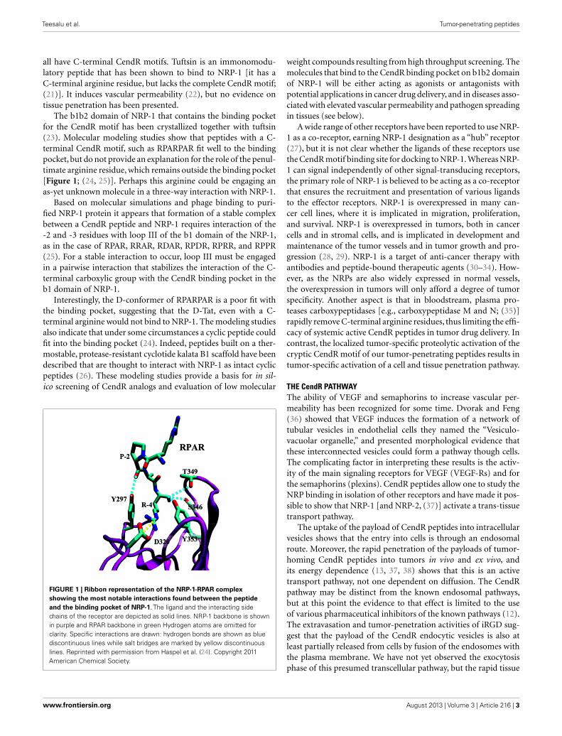
The b1b2 domain of NRP-1 that contains the binding pocketfor the CendR motif has been crystallized together with tuftsin(23). Molecular modeling studies show that peptides with a C-terminal CendR motif, such as RPARPAR fit well to the bindingpocket, but do not provide an explanation for the role of the penul-timate arginine residue, which remains outside the binding pocket[Figure 1; (24, 25)]. Perhaps this arginine could be engaging anas-yet unknown molecule in a three-way interaction with NRP-1.
Based on molecular simulations and phage binding to puri-fied NRP-1 protein it appears that formation of a stable complexbetween a CendR peptide and NRP-1 requires interaction of the-2 and -3 residues with loop III of the b1 domain of the NRP-1,as in the case of RPAR, RRAR, RDAR, RPDR, RPRR, and RPPR(25). For a stable interaction to occur, loop III must be engagedin a pairwise interaction that stabilizes the interaction of the C-terminal carboxylic group with the CendR binding pocket in theb1 domain of NRP-1.
Interestingly, the D-conformer of RPARPAR is a poor fit withthe binding pocket, suggesting that the D-Tat, even with a C-terminal arginine would not bind to NRP-1. The modeling studiesalso indicate that under some circumstances a cyclic peptide couldfit into the binding pocket (24). Indeed, peptides built on a ther-mostable, protease-resistant cyclotide kalata B1 scaffold have beendescribed that are thought to interact with NRP-1 as intact cyclicpeptides (26). These modeling studies provide a basis for in sil-ico screening of CendR analogs and evaluation of low molecular
FIGURE 1 | Ribbon representation of the NRP-1-RPAR complexshowing the most notable interactions found between the peptideand the binding pocket of NRP-1. The ligand and the interacting sidechains of the receptor are depicted as solid lines. NRP-1 backbone is shownin purple and RPAR backbone in green Hydrogen atoms are omitted forclarity. Specific interactions are drawn: hydrogen bonds are shown as bluediscontinuous lines while salt bridges are marked by yellow discontinuouslines. Reprinted with permission from Haspel et al. (24). Copyright 2011American Chemical Society.
weight compounds resulting from high throughput screening. Themolecules that bind to the CendR binding pocket on b1b2 domainof NRP-1 will be either acting as agonists or antagonists withpotential applications in cancer drug delivery, and in diseases asso-ciated with elevated vascular permeability and pathogen spreadingin tissues (see below).
A wide range of other receptors have been reported to use NRP-1 as a co-receptor, earning NRP-1 designation as a “hub” receptor(27), but it is not clear whether the ligands of these receptors usethe CendR motif binding site for docking to NRP-1. Whereas NRP-1 can signal independently of other signal-transducing receptors,the primary role of NRP-1 is believed to be acting as a co-receptorthat ensures the recruitment and presentation of various ligandsto the effector receptors. NRP-1 is overexpressed in many can-cer cell lines, where it is implicated in migration, proliferation,and survival. NRP-1 is overexpressed in tumors, both in cancercells and in stromal cells, and is implicated in development andmaintenance of the tumor vessels and in tumor growth and pro-gression (28, 29). NRP-1 is a target of anti-cancer therapy withantibodies and peptide-bound therapeutic agents (30–34). How-ever, as the NRPs are also widely expressed in normal vessels,the overexpression in tumors will only afford a degree of tumorspecificity. Another aspect is that in bloodstream, plasma pro-teases carboxypeptidases [e.g., carboxypeptidase M and N; (35)]rapidly remove C-terminal arginine residues, thus limiting the effi-cacy of systemic active CendR peptides in tumor drug delivery. Incontrast, the localized tumor-specific proteolytic activation of thecryptic CendR motif of our tumor-penetrating peptides results intumor-specific activation of a cell and tissue penetration pathway.
THE CendR PATHWAYThe ability of VEGF and semaphorins to increase vascular per-meability has been recognized for some time. Dvorak and Feng(36) showed that VEGF induces the formation of a network oftubular vesicles in endothelial cells they named the “Vesiculo-vacuolar organelle,” and presented morphological evidence thatthese interconnected vesicles could form a pathway though cells.The complicating factor in interpreting these results is the activ-ity of the main signaling receptors for VEGF (VEGF-Rs) and forthe semaphorins (plexins). CendR peptides allow one to study theNRP binding in isolation of other receptors and have made it pos-sible to show that NRP-1 [and NRP-2, (37)] activate a trans-tissuetransport pathway.
The uptake of the payload of CendR peptides into intracellularvesicles shows that the entry into cells is through an endosomalroute. Moreover, the rapid penetration of the payloads of tumor-homing CendR peptides into tumors in vivo and ex vivo, andits energy dependence (13, 37, 38) shows that this is an activetransport pathway, not one dependent on diffusion. The CendRpathway may be distinct from the known endosomal pathways,but at this point the evidence to that effect is limited to the useof various pharmaceutical inhibitors of the known pathways (12).The extravasation and tumor-penetration activities of iRGD sug-gest that the payload of the CendR endocytic vesicles is also atleast partially released from cells by fusion of the endosomes withthe plasma membrane. We have not yet observed the exocytosisphase of this presumed transcellular pathway, but the rapid tissue
www.frontiersin.org August 2013 | Volume 3 | Article 216 | 3
Teesalu et al. Tumor-penetrating peptides
penetration of the CendR payloads support of this possibility.However, we cannot exclude that an alternative pathway such aspropelling cell surface-bound payload forward by the cell mem-brane or membrane projections. Genetic and proteomics studiesare underway to elucidate the cellular molecular basis of the CendRpathway.
Our discovery of the CendR tissue transport pathway raises thefascinating question of the physiological function of this path-way. While the focus so far has been on how this pathway mightbe used in drug delivery, it obviously does not exist for this pur-pose. One possibility is that it facilitates the transfer of nutrientsto cells that are far from blood vessels or otherwise under duress.The overexpression of NRP-1 in tumors suggests that supplyingnutrient deficient/hypoxic areas in tumors may be yet another waytumors make use of a physiological pathway to foster their owngrowth. The CendR pathway may have been hijacked by virusesand microbial toxins for cell entry and tissue spreading. Cleavageof a viral surface proteins and pro-toxins by host proteases (mostcommonly furins and related enzymes) at sites that create an activeCendR motif is a recurrent theme seen in many pathogens. Exam-ples include the Human T-lymphotropic virus-2, Crimean–Congohemorrhagic fever virus, tick-born encephalitis virus, and Ebolaviruses, as well as anthrax toxin (39–43). CendR sequences arealso present in snake and bee venoms (e.g., mellitin), and maycontribute to the spreading of these toxins in tissues.
Vascular edema is associated with many diseases (hemorrhagicvirus infections, sepsis, and vascular leak syndromes). Severalproinflammatory vasoactive (poly)peptides capable of increas-ing vascular permeability display a functionally important argi-nine residue at their C-terminus. Examples include complementC3a and C5a anaphylatoxins (C-terminal sequences ASHLGLARand KDMQLGR, respectively) as well as kinins (bradykinin andkallidin, which have an identical C-terminal sequence, RPPGF-SPFR). Intriguingly, we have observed that phage that displaypeptides corresponding to the C-terminal amino acids of C5/3aand bradykinin bind to the recombinant b1b2 domain of NRP(in preparation) and that the binding is reversed by an excess ofthe free peptide. It remains to be seen whether the NRP/CendRaxis plays a role in the activity of C3/5a, bradykinin, and/or othervasoactive peptides.
DESIGNER PEPTIDES FOR CendR PATHWAY ACTIVATIONHaving worked out the two-motif requirement for a tumor-homing peptide to have CendR activity, we tested the universalityof the concept by designing a new peptide with such activities.We used as the starting point the NGR tumor-homing motif pre-viously identified by our laboratory (44, 45), which recognizes aform of aminopeptidase N in angiogenic tumor vessels (46, 47).We added a second arginine to the NGR motif to convert it into theCendR motif, RNGR and embedded that motif in the iRGD frame-work by replacing RGDK with RNGR. The resulting peptide, iNGR(CRNGRGPDC) has all the properties of iRGD, except that itstumor recruitment is not mediated by integrin but another recep-tor, presumably aminopeptidase N (48). We have also designedtumor-homing CendR peptides by arranging in tandem a CendRmotif, a proteolytic cleavage site for a tumor-associated proteasethat cleaves after a basic residue, and a tumor-homing motif(Teesalu et al., in preparation). These peptides also home to and
penetrate into tumors. A construct created to deliver a non-specificcell-penetrating peptide, appears to serendipitously follow thisdesign (49). Whether these tandem tumor-penetrating peptidesare as effective as the peptides in which the homing motif andCendR motif overlap remains to be seen. The iRGD and LyP-1peptides lose their affinity for the primary receptor [αv integrinsfor iRGD and a mitochondrial/cell surface protein p32 for LyP-1(7) after the proteolytic cleavage that activates the CendR motif hastaken place (13, 37)]. The resulting release of the peptide from theprimary receptor may facilitate subsequent binding to NRP-1 andmake the primary receptor available for binding of another intactpeptide. Peptides with tandem motifs would lack this latter feature.Another possible design for CendR activation would be blockingthe C-terminus with a chemical group other than an amino acid orpeptide. One can envision peptides, the CendR activity of whichis triggered by a phosphatase, demethylase, sulfatase, etc. To theextent such an enzyme is specific for the target tissue, new usefulprobes could be created.
DRUG DELIVERY WITH TUMOR-PENETRATING PEPTIDESTHE DRUG PENETRATION PROBLEMTo reach tumor cells and tumor-associated parenchymal cells (e.g.,tumor-associated fibroblasts, macrophages), drugs must cross thevascular barrier and penetrate into the extravascular stroma. Can-cerous tissue is heterogeneous, with striking regional differencesin tumor structure (leaky vasculature and defective lymphatics,which causes buildup of interstitial fluid pressure in the tumor),and physiology (e.g., inflammation, fibrosis, hypoxia, acidity).These features translate into steep drug gradients and variabil-ity in the uptake and distribution of anti-cancer drugs withintumor parenchyma (50). For example, evaluation of doxorubicindistribution in tumors after systemic administration showed thatthe concentration of this drug decreases exponentially with dis-tance from tumor blood vessels, reaching half of its perivascularconcentration at a distance of about 40 µm (51). The distribu-tion of trastuzumab (Herceptin) in breast tumor xenografts isalso highly heterogeneous with many tumor cells exposed to nodetectable drug (52). To some extent, the tumor drug delivery chal-lenges are alleviated by the Enhanced Permeability and Retention(EPR) effect – accumulation of compounds (typically liposomes,nanoparticles, and macromolecular drugs) in tumor tissue morethan they do in normal tissues. The underlying causes of the EPReffect are abnormal structure and function of tumor vessels: poorlyaligned endothelial cells with fenestrations, deficient pericyte cov-erage, and lack of lymphatic drainage. However, EPR is highlyvariable as it is influenced by differences between tumor types andheterogeneity within individual tumor. Tumor interstitial pressure(IFP) depends on integrity of blood and lymphatic vessels, tumorcell proliferation, deposition of matrix molecules, and interac-tion of cells with the matrix molecules. The difference betweentumor microvascular fluid pressure and IFP determines intratu-moral convection fluxes that have a major influence on the vascularexit of the compounds over 10 kDa. Intratumoral fluid pressuregradients can be in some cases favorably influenced by vasodilatorycompounds such as bradykinin, endothelin, and calcium channelantagonists, to allow better tumor perfusion and increased drugdelivery (53). Other approaches include dissolving extracellularmatrix with enzymes such as collagenase or hyaluronidase (54),
Frontiers in Oncology | Pharmacology of Anti-Cancer Drugs August 2013 | Volume 3 | Article 216 | 4
Teesalu et al. Tumor-penetrating peptides
or killing or inhibiting the activity of tumor-associated fibroblasts(55). Obviously, the delivery of enzymes and drugs aimed at low-ering the IFP to the tumor parenchyma faces the similar tumorpenetration challenges seen for the cancer drugs.
CendR-ENHANCED DRUG DELIVERYThe tumor-homing CendR peptides provide a solution to the drugpenetration problem. A probe or drug attached to iRGD or LyP-1 is delivered to extracellular tumor tissue more effectively thanthe drug alone. We have extensively demonstrated the tumor pen-etration with fluorescein (FAM)-labeled peptides. Intravenouslyinjected FAM-iRGD, LyP-1, and iNGR are found dispersed intumor parenchyma minutes after administered, whereas FAM-labeled inactive control peptides do not appear in the tumors atall. FAM-labeled homing peptides that lack a CendR motif bind tothe blood vessels, but do not penetrate into the rest of the tumor(10, 11, 13, 48). Remarkably, iRGD and LyP-1 have quite differentdistributions within tumors, presumably reflecting the expressionof their primary receptors in different tumor compartments (7,10, 13). The effect of the cryptic CendR motif is vividly illus-trated by the differences between iRGD and conventional RGDpeptides, such as CRGDC and cycloRGDfK. While iRGD pay-load, even a poorly diffusing nanoparticle, readily enters tumorparenchyma, the conventional RGD peptides only take their pay-load to the tumor vessels (13, 38). LyP-1 and CGKRK, a peptidewe have recently shown to also use p32 as its receptor but lack theCendR activity (56) show a similar difference (11, 57).
The observations with the fluorescent probe described aboveprompted us to study the ability of iRGD and the other CendRpeptides to enhance the delivery of actual anti-cancer drugs totumors. We have shown that therapeutics as diverse as a smallmolecular weight drug (doxorubicin), trastuzumab (anti-Her2antibody), and the nanoparticle drugs Abraxane and Doxil can
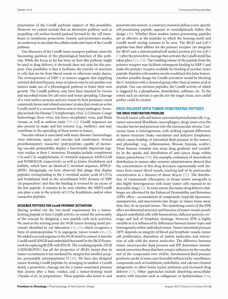
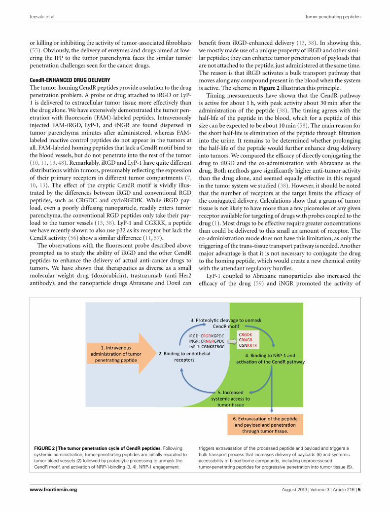
benefit from iRGD-enhanced delivery (13, 38). In showing this,we mostly made use of a unique property of iRGD and other simi-lar peptides; they can enhance tumor penetration of payloads thatare not attached to the peptide, just administered at the same time.The reason is that iRGD activates a bulk transport pathway thatmoves along any compound present in the blood when the systemis active. The scheme in Figure 2 illustrates this principle.
Timing measurements have shown that the CendR pathwayis active for about 1 h, with peak activity about 30 min after theadministration of the peptide (38). The timing agrees with thehalf-life of the peptide in the blood, which for a peptide of thissize can be expected to be about 10 min (58). The main reason forthe short half-life is elimination of the peptide through filtrationinto the urine. It remains to be determined whether prolongingthe half-life of the peptide would further enhance drug deliveryinto tumors. We compared the efficacy of directly conjugating thedrug to iRGD and the co-administration with Abraxane as thedrug. Both methods gave significantly higher anti-tumor activitythan the drug alone, and seemed equally effective in this regardin the tumor system we studied (38). However, it should be notedthat the number of receptors at the target limits the efficacy ofthe conjugated delivery. Calculations show that a gram of tumortissue is not likely to have more than a few picomoles of any givenreceptor available for targeting of drugs with probes coupled to thedrug (1). Most drugs to be effective require greater concentrationsthan could be delivered to this small an amount of receptor. Theco-administration mode does not have this limitation, as only thetriggering of the trans-tissue transport pathway is needed. Anothermajor advantage is that it is not necessary to conjugate the drugto the homing peptide, which would create a new chemical entitywith the attendant regulatory hurdles.
LyP-1 coupled to Abraxane nanoparticles also increased theefficacy of the drug (59) and iNGR promoted the activity of
FIGURE 2 |The tumor penetration cycle of CendR peptides. Followingsystemic administration, tumor-penetrating peptides are initially recruited totumor blood vessels (2) followed by proteolytic processing to unmask theCendR motif, and activation of NRP-1-binding (3, 4). NRP-1 engagement
triggers extravasation of the processed peptide and payload and triggers abulk transport process that increases delivery of payloads (6) and systemicaccessibility of blood-borne compounds, including unprocessesedtumor-penetrating peptides for progressive penetration into tumor tissue (5).
www.frontiersin.org August 2013 | Volume 3 | Article 216 | 5
Teesalu et al. Tumor-penetrating peptides
doxorubicin in a mouse tumor model in a way similar to iRGD(48), by a factor of about 3. Importantly, the iRGD work with dox-orubicin showed that there was no change in the main side effect ofthis drug, cardiotoxicity. This side effect was nearly eliminated by athreefold reduction of the drug dose. Thus, the tumor-penetratingpeptides can be used both to enhance the activity of anti-cancerdrugs, or lowering the side effect with the same anti-cancer activity,or some of both.
The tumor-penetrating peptides can also enhance tumor imag-ing, as demonstrated by coating iron oxide nanoparticles withiRGD for MRI imaging. iRGD gave stronger images than a conven-tional RGD peptide, CRGDC; the main difference was that iRGDspread into the whole tumor, whereas only highlighted the tumorvessels (13). LyP-1 has been used in optical imaging of tumors (11,61) and atherosclerotic plaques (60), as well as in MRI and PETimaging of plaques (61). LyP-1 homes to and penetrates into acti-vated macrophages in tumors and atherosclerotic plaques (60, 61)revealing a similarity between the macrophages in tumors and theplaques (61). LyP-1 has also been shown to selectively accumulatein tumor-draining lymph nodes prior to the arrival of tumor cells,defining a premalignant niche in tumors (62).
CONCLUSION AND FUTURE PROSPECTSThe discovery of tumor-penetrating peptides has led to the iden-tification of a new trans-tissue transport pathway, the C-end Ruleor CendR pathway. The physiological function of the CendR path-way and its molecular workings are obviously important questions
to be answered in future studies. Activating the pathway in atumor-specific manner, which is accomplished with peptides theCendR motif of which is activated in tumors, provides a way ofincreasing the activity of anti-cancer drugs and enhancing tumorimaging. Thus, the tumor-penetrating CendR peptides representa potentially significant advance in cancer treatment.
ACKNOWLEDGMENTSThe authors’ original work reviewed in this article is supportedby Cancer Center Support Grant CA30199 to SBMRI, Innova-tor Awards W81XWH-08-1-0727, W81XWH-09-0698 from theDepartment of Defense, and grant CA CA152327 from theNational Cancer Institute. Tambet Teesalu is supported by Euro-pean Research Council starting grant (GliomaDDS) and theWellcome Trust Award 095077/Z/10/Z.
NIH AUTHOR DISCLAIMERThe views and opinions of authors expressed on OER websitesdo not necessarily state or reflect those of the U.S. Government,and they may not be used for advertising or product endorsementpurposes.
DOD AUTHOR DISCLAIMERThe opinions expressed herein are those of the author(s) and arenot necessarily representative of those of the Uniformed ServicesUniversity of the Health Sciences (USUHS), the Department ofDefense (DOD); or, the United States Army, Navy, or Air Force.
REFERENCES1. Ruoslahti E, Bhatia SN, Sailor MJ.
Targeting of drugs and nanopar-ticles to tumors. J Cell Biol(2010) 188:759–68. doi:10.1083/jcb.200910104
2. Ferrara N, Alitalo K. Clinicalapplications of angiogenic growthfactors and their inhibitors.Nat Med (1999) 5:1359–64.doi:10.1038/9467
3. Akerman ME, Pilch J, Peters D,Ruoslahti E. Angiostatic peptidesuse plasma fibronectin to home toangiogenic vasculature. Proc NatlAcad Sci U S A (2005) 102:2040–5.doi:10.1073/pnas.0409844102
4. Ruoslahti E. Peptides as target-ing elements and tissue penetra-tion devices for nanoparticles. AdvMater (2012) 24:3747–56. doi:10.1002/adma.201200454
5. Girard JP, Springer TA. Highendothelial venules (HEVs),specialized endothelium for lym-phocyte migration. Immunol Today(1995) 16:449–57. doi:10.1016/0167-5699(95)80023-9
6. Libby P, DiCarli M, WeisslederR. The vascular biology of ath-erosclerosis and imaging targets.J Nucl Med (2010) 51(Suppl 1):33S–7. doi:10.2967/jnumed.109.069633
7. Fogal V, Zhang L, Kra-jewski S, Ruoslahti E.Mitochondrial/cell-surface proteinp32/gC1qR as a molecular targetin tumor cells and tumor stroma.Cancer Res (2008) 68:7210–8. doi:10.1158/0008-5472.CAN-07-6752
8. Pasqualini R, Ruoslahti E. Tis-sue targeting with phage peptidelibraries. Mol Psychiatry (1996)1:423.
9. Teesalu T, Sugahara KN, RuoslahtiE. Mapping of vascular ZIP codesby phage display. Methods Enzy-mol (2012) 503:35–56. doi:10.1016/B978-0-12-396962-0.00002-1
10. Laakkonen P, Porkka K, HoffmanJA, Ruoslahti E. A tumor-homingpeptide with a targeting specificityrelated to lymphatic vessels. NatMed (2002) 8:751–5.
11. Laakkonen P, Akerman ME, BiliranH, Yang M, Ferrer F, KarpanenT, et al. Antitumor activity of ahoming peptide that targets tumorlymphatics and tumor cells. ProcNatl Acad Sci U S A (2004) 101:9381–6. doi:10.1073/pnas.0403317101
12. Teesalu T, Sugahara KN, Kotam-raju VR, Ruoslahti E. C-endrule peptides mediate neuropilin-1-dependent cell, vascular, and tissuepenetration. Proc Natl Acad Sci U S A
(2009) 106:16157–62. doi:10.1073/pnas.0908201106
13. Sugahara KN, Teesalu T, KarmaliPP, Kotamraju VR, Agemy L, GirardOM, et al. Tissue-penetrating deliv-ery of compounds and nanopar-ticles into tumors. Cancer Cell(2009) 16:510–20. doi:10.1016/j.ccr.2009.10.013
14. Scott JK, Smith GP. Searching forpeptide ligands with an epitopelibrary. Science (1990) 249:386–90.doi:10.1126/science.1696028
15. Smith GP. Filamentous fusionphage, novel expression vec-tors that display cloned anti-gens on the virion surface.Science (1985) 228:1315–7.doi:10.1126/science.4001944
16. Koivunen E, Gay DA, Ruoslahti E.Selection of peptides binding to thealpha 5 beta 1 integrin from phagedisplay library. J Biol Chem (1993)268:20205–10.
17. Yuan L, Moyon D, Pardanaud L,Breant C, Karkkainen MJ, AlitaloK, et al. Abnormal lymphaticvessel development in neuropilin 2mutant mice. Development (2002)129:4797–806.
18. Xu Y, Yuan L, Mak J, Par-danaud L, Caunt M, KasmanI, et al. Neuropilin-2 mediatesVEGF-C-induced lymphatic
sprouting together with VEGFR3.J Cell Biol (2010) 188:115–30.doi:10.1083/jcb.200903137
19. He Z, Tessier-Lavigne M. Neu-ropilin is a receptor for theaxonal chemorepellent semaphorinIII. Cell (1997) 90:739–51. doi:10.1016/S0092-8674(00)80534-6
20. Soker S, Takashima S, MiaoHQ, Neufeld G, Klagsbrun M.Neuropilin-1 is expressed byendothelial and tumor cells asan isoform-specific receptor forvascular endothelial growth factor.Cell (1998) 92:735–45. doi:10.1016/S0092-8674(00)81402-6
21. von Wronski MA, Raju N, Pil-lai R, Bogdan NJ, Marinelli ER,Nanjappan P, et al. Tuftsin bindsneuropilin-1 through a sequencesimilar to that encoded by exon8 of vascular endothelial growthfactor. J Biol Chem (2006) 281:5702–10. doi:10.1074/jbc.M511941200
22. Gerdin B, Lindeberg G, Ragnars-son U, Saldeen T, Wallin R. Struc-tural requirements for microvas-cular permeability-increasing abil-ity of peptides. Studies on ana-logues of a fibrinogen pentapep-tide fragment. Biochim Biophys Acta(1983) 757:366–70. doi:10.1016/0304-4165(83)90063-6
Frontiers in Oncology | Pharmacology of Anti-Cancer Drugs August 2013 | Volume 3 | Article 216 | 6
Teesalu et al. Tumor-penetrating peptides
23. Vander Kooi CW, Jusino MA, Per-man B, Neau DB, Bellamy HD,Leahy DJ. Structural basis for ligandand heparin binding to neuropilinB domains. Proc Natl Acad Sci U SA (2007) 104:6152–7. doi:10.1073/pnas.0700043104
24. Haspel N, Zanuy D, Nussinov R,Teesalu T, Ruoslahti E, Aleman C.Binding of a C-end rule peptide tothe neuropilin-1 receptor, a molec-ular modeling approach. Biochem-istry (2011) 50:1755–62. doi:10.1021/bi101662j
25. Zanuy D, Kotla R, Nussinov R,Teesalu T, Sugahara KN, AlemanC, et al. Sequence dependence ofC-end rule peptides in bindingand activation of neuropilin-1receptor. J Struct Biol (2013) 182:78–86. doi:10.1016/j.jsb.2013.02.006
26. Getz JA, Cheneval O, CraikDJ, Daugherty PS. Designof a Cyclotide antagonist ofneuropilin-1 and -2 that potentlyinhibits endothelial cell migration.ACS Chem Biol (2013) 8:1147–54.doi:10.1021/cb4000585
27. Uniewicz KA, Fernig DG. Neu-ropilins, a versatile partner ofextracellular molecules that regu-late development and disease. FrontBiosci (2008) 13:4339–60. doi:10.2741/3008
28. Bagri A, Tessier-Lavigne M, WattsRJ. Neuropilins in tumor biology.Clin Cancer Res (2009) 15:1860–4. doi:10.1158/1078-0432.CCR-08-0563
29. Pellet-Many C, Frankel P, JiaH, Zachary I. Neuropilins, struc-ture, function and role in disease.Biochem J (2008) 411:211–26. doi:10.1042/BJ20071639
30. Jia H, Bagherzadeh A, HartzoulakisB, Jarvis A, Lohr M, Shaikh S, etal. Characterization of a bicyclicpeptide neuropilin-1 (NP-1) antag-onist (EG3287) reveals importanceof vascular endothelial growthfactor exon 8 for NP-1 bind-ing and role of NP-1 in KDRsignaling. J Biol Chem (2006)281:13493–502. doi:10.1074/jbc.M512121200
31. Hong TM, Chen YL, Wu YY,Yuan A, Chao YC, Chung YC, etal. Targeting neuropilin 1 as anantitumor strategy in lung cancer.Clin Cancer Res (2007) 13:4759–68.doi:10.1158/1078-0432.CCR-07-0001
32. Liang WC, Dennis MS, StawickiS, Chanthery Y, Pan Q, ChenY, et al. Function blocking anti-bodies to neuropilin-1 generatedfrom a designed human synthetic
antibody phage library. J Mol Biol(2007) 366:815–29. doi:10.1016/j.jmb.2006.11.021
33. Karjalainen K, Jaalouk DE, Bueso-Ramos CE, Zurita AJ, KuniyasuA, Eckhardt BL, et al. Targetingneuropilin-1 in human leukemiaand lymphoma. Blood (2011)117:920–7. doi:10.1182/blood-2010-05-282921
34. Nasarre C, Roth M, Jacob L,Roth L, Koncina E, Thien A,et al. Peptide-based interferenceof the transmembrane domain ofneuropilin-1 inhibits glioma growthin vivo. Oncogene (2010) 29:2381–92. doi:10.1038/onc.2010.9
35. Skidgel RA. Bradykinin-degradingenzymes, structure, function,distribution, and potential rolesin cardiovascular pharmacology.J Cardiovasc Pharmacol (1992)20(Suppl 9):S4–9. doi:10.1097/00005344-199206209-00002
36. Dvorak AM, Feng D. The vesiculo-vacuolar organelle (VVO). Anew endothelial cell perme-ability organelle. J HistochemCytochem (2001) 49:419–32.doi:10.1177/002215540104900401
37. Roth L, Agemy L, Kotamraju VR,Braun G, Teesalu T, SugaharaKN, et al. Transtumoral target-ing enabled by a novel neuropilin-binding peptide. Oncogene (2012)31:3754–63. doi:10.1038/onc.2011.537
38. Sugahara KN, Teesalu T, KarmaliPP, Kotamraju VR, Agemy L,Greenwald DR, et al. Coadmin-istration of a tumor-penetratingpeptide enhances the efficacyof cancer drugs. Science (2010)328:1031–5. doi:10.1126/science.1183057
39. Chambers TJ, Weir RC, GrakouiA, McCourt DW, Bazan JF, Fletter-ick RJ, et al. Evidence that the N-terminal domain of nonstructuralprotein NS3 from yellow fever virusis a serine protease responsible forsite-specific cleavages in the viralpolyprotein. Proc Natl Acad Sci U SA (1990) 87:8898–902. doi:10.1073/pnas.87.22.8898
40. Wool-Lewis RJ, Bates P. Endoprote-olytic processing of the ebola virusenvelope glycoprotein, cleavage isnot required for function. J Virol(1999) 73:1419–26.
41. Sjöberg M, Wallin M, LindqvistB, Garoff H. Furin cleavagepotentiates the membrane fusion-controlling intersubunit disul-fide bond isomerization activityof leukemia virus Env. J Virol(2006) 80:5540–51. doi:10.1128/JVI.01851-05
42. Sanchez AJ, Vincent MJ, Erick-son BR, Nichol ST. Crimean-congo hemorrhagic fever virusglycoprotein precursor is cleavedby furin-like and SKI-1 proteasesto generate a novel 38-kilodaltonglycoprotein. J Virol (2006) 80:514–25. doi:10.1128/JVI.80.1.514-525.2006
43. Molloy SS, Bresnahan PA, LepplaSH, Klimpel KR, Thomas G. Humanfurin is a calcium-dependent ser-ine endoprotease that recognizesthe sequence arg-X-X-arg andefficiently cleaves anthrax toxin pro-tective antigen. J Biol Chem (1992)267:16396–402.
44. Arap W, Pasqualini R, RuoslahtiE. Cancer treatment by targeteddrug delivery to tumor vascula-ture in a mouse model. Science(1998) 279:377–80. doi:10.1126/science.279.5349.377
45. Koivunen E, Wang B, RuoslahtiE. Phage libraries displaying cyclicpeptides with different ring sizes,ligand specificities of the RGD-directed integrins. Biotechnology (NY) (1995) 13:265–70. doi:10.1038/nbt0395-265
46. Pasqualini R, Koivunen E, KainR, Lahdenranta J, Sakamoto M,Stryhn A, et al. AminopeptidaseN is a receptor for tumor-homingpeptides and a target for inhibit-ing angiogenesis. Cancer Res (2000)60:722–7.
47. Corti A, Curnis F, Arap W,Pasqualini R. The neovascula-ture homing motif NGR, morethan meets the eye. Blood (2008)112:2628–35. doi:10.1182/blood-2008-04-150862
48. Alberici L, Roth L, SugaharaKN, Agemy L, Kotamraju VR,Teesalu T, et al. De novo designof a tumor-penetrating peptide.Cancer Res (2013) 73:804–12.doi:10.1158/0008-5472.CAN-12-1668
49. Myrberg H, Zhang L, Mae M, Lan-gel U. Design of a tumor-homingcell-penetrating peptide. BioconjugChem (2008) 19(1):70–5. doi:10.1021/bc0701139
50. Tredan O, Galmarini CM, Patel K,Tannock IF. Drug resistance and thesolid tumor microenvironment. JNatl Cancer Inst (2007) 99:1441–54.doi:10.1093/jnci/djm135
51. Primeau AJ, Rendon A, Hed-ley D, Lilge L, Tannock IF. Thedistribution of the anticancerdrug doxorubicin in relation toblood vessels in solid tumors.Clin Cancer Res (2005) 11:8782–8.doi:10.1158/1078-0432.CCR-05-1664
52. Baker JH, Lindquist KE, HuxhamLA, Kyle AH, Sy JT, MinchintonAI. Direct visualization of hetero-geneous extravascular distributionof trastuzumab in human epidermalgrowth factor receptor type 2 over-expressing xenografts. Clin CancerRes (2008) 14:2171–9. doi:10.1158/1078-0432.CCR-07-4465
53. Feron O. Targeting the tumor vas-cular compartment to improve con-ventional cancer therapy. TrendsPharmacol Sci (2004) 25:536–42.doi:10.1016/j.tips.2004.08.008
54. Tufto I, Hansen R, Byberg D,Nygaard KHH, Tufto J, DaviesCDL. The effect of collagenaseand hyaluronidase on transientperfusion in human osteosarcomaxenografts grown orthotopicallyand in dorsal skinfold chambers.Anticancer Res (2007) 27:1475–81.
55. Xing F, Saidou J, Watabe K. Can-cer associated fibroblasts (CAFs)in tumor microenvironment. FrontBiosci (2010) 15:166–79. doi:10.2741/3613
56. Agemy L, Kotamraju VR,Friedmann-Morvinski D, SharmaS, Sugahara KN, Ruoslahti E.Proapoptotic peptide-mediatedcancer therapy targeted to cell sur-face p32. Mol Ther (Forthcoming2013).
57. Agemy L, Friedmann-Morvinski D,Kotamraju VR, Roth L, SugaharaKN, Girard OM, et al. Targetednanoparticle enhanced proapop-totic peptide as potential therapy forglioblastoma. Proc Natl Acad Sci U SA (2011) 108:17450–5. doi:10.1073/pnas.1114518108
58. Werle M, Bernkop-Schnurch A.Strategies to improve plasma halflife time of peptide and proteindrugs. Amino Acids (2006) 30:351–67. doi:10.1007/s00726-005-0289-3
59. Karmali PP, Kotamraju VR, Kastan-tin M, Black M, Missirlis D, Tir-rell M, et al. Targeting of albumin-embedded paclitaxel nanoparticlesto tumors. Nanomedicine (2009)5:73–82. doi:10.1016/j.nano.2008.07.007
60. Uchida M, Kosuge H, TerashimaM, Willits DA, Liepold LO, YoungMJ, et al. Protein cage nanoparti-cles bearing the LyP-1 peptide forenhanced imaging of macrophage-rich vascular lesions. ACS Nano(2011) 5:2493–502. doi:10.1021/nn102863y
61. Hamzah J, Kotamraju VR, SeoJW, Agemy L, Fogal V, MahakianLM, et al. Specific penetration andaccumulation of a homing pep-tide within atherosclerotic plaques
www.frontiersin.org August 2013 | Volume 3 | Article 216 | 7
Teesalu et al. Tumor-penetrating peptides
of apolipoprotein E-deficient mice.Proc Natl Acad Sci U S A(2011) 108:7154–9. doi:10.1073/pnas.1104540108
62. Zhang F, Niu G, Lin X, Jacob-son O, Ma Y, Eden HS, et al.Imaging tumor-induced sentinellymph node lymphangiogenesiswith LyP-1 peptide. Amino Acids(2012) 42:2343–51. doi:10.1007/s00726-011-0976-1
Conflict of Interest Statement: TambetTeesalu, Kazuki N. Sugahara, and ErkkiRuoslahti are shareholders in CendRTherapeutics Inc., and Erkki Ruoslahtiis a shareholder in EnduRx Pharma-ceuticals. The companies have rights tosome of the technology described in thepaper.
Received: 18 June 2013; paper pendingpublished: 27 June 2013; accepted: 06
August 2013; published online: 27 August2013.Citation: Teesalu T, Sugahara KN andRuoslahti E (2013) Tumor-penetratingpeptides. Front. Oncol. 3:216. doi:10.3389/fonc.2013.00216This article was submitted to Pharmacol-ogy of Anti-Cancer Drugs, a section of thejournal Frontiers in Oncology.Copyright © 2013 Teesalu, Sugaharaand Ruoslahti. This is an open-access
article distributed under the terms of theCreative Commons Attribution License(CC BY). The use, distribution orreproduction in other forums is permitted,provided the original author(s) or licensorare credited and that the original publica-tion in this journal is cited, in accordancewith accepted academic practice. No use,distribution or reproduction is permit-ted which does not comply with theseterms.
Frontiers in Oncology | Pharmacology of Anti-Cancer Drugs August 2013 | Volume 3 | Article 216 | 8