Experimental rat lung tumor model with intrabronchial tumor cell implantation
9
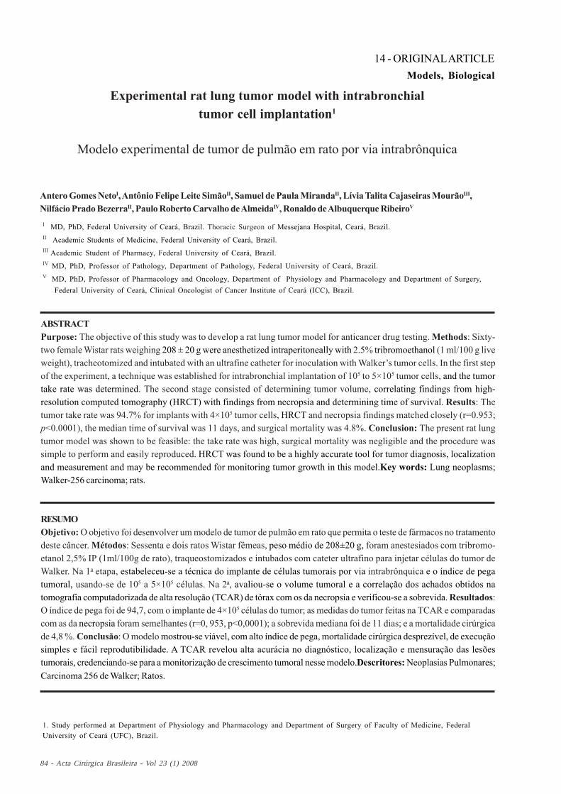
84 - Acta Cirúrgica Brasileira - Vol 23 (1) 2008 14 - ORIGINAL ARTICLE Models, Biological Experimental rat lung tumor model with intrabronchial tumor cell implantation 1 Modelo experimental de tumor de pulmão em rato por via intrabrônquica Antero Gomes Neto I , Antônio Felipe Leite Simão II , Samuel de Paula Miranda II , Lívia Talita Cajaseiras Mourão III , Nilfácio Prado Bezerra II , Paulo Roberto Carvalho de Almeida IV , Ronaldo de Albuquerque Ribeiro V 1. Study performed at Department of Physiology and Pharmacology and Department of Surgery of Faculty of Medicine, Federal University of Ceará (UFC), Brazil. I MD, PhD, Federal University of Ceará, Brazil. Thoracic Surgeon of Messejana Hospital, Ceará, Brazil. II Academic Students of Medicine, Federal University of Ceará, Brazil. III Academic Student of Pharmacy, Federal University of Ceará, Brazil. IV MD, PhD, Professor of Pathology, Department of Pathology, Federal University of Ceará, Brazil. V MD, PhD, Professor of Pharmacology and Oncology, Department of Physiology and Pharmacology and Department of Surgery, Federal University of Ceará, Clinical Oncologist of Cancer Institute of Ceará (ICC), Brazil. ABSTRACT Purpose: The objective of this study was to develop a rat lung tumor model for anticancer drug testing. Methods: Sixty- two female Wistar rats weighing 208 ± 20 g were anesthetized intraperitoneally with 2.5% tribromoethanol (1 ml/100 g live weight), tracheotomized and intubated with an ultrafine catheter for inoculation with Walker’s tumor cells. In the first step of the experiment, a technique was established for intrabronchial implantation of 10 5 to 5×10 5 tumor cells, and the tumor take rate was determined. The second stage consisted of determining tumor volume, correlating findings from high- resolution computed tomography (HRCT) with findings from necropsia and determining time of survival. Results: The tumor take rate was 94.7% for implants with 4×10 5 tumor cells, HRCT and necropsia findings matched closely (r=0.953; p<0.0001), the median time of survival was 11 days, and surgical mortality was 4.8%. Conclusion: The present rat lung tumor model was shown to be feasible: the take rate was high, surgical mortality was negligible and the procedure was simple to perform and easily reproduced. HRCT was found to be a highly accurate tool for tumor diagnosis, localization and measurement and may be recommended for monitoring tumor growth in this model.Key words: Lung neoplasms; Walker-256 carcinoma; rats. RESUMO Objetivo: O objetivo foi desenvolver um modelo de tumor de pulmão em rato que permita o teste de fármacos no tratamento deste câncer. Métodos: Sessenta e dois ratos Wistar fêmeas, peso médio de 208±20 g, foram anestesiados com tribromo- etanol 2,5% IP (1ml/100g de rato), traqueostomizados e intubados com cateter ultrafino para injetar células do tumor de Walker. Na 1 a etapa, estabeleceu-se a técnica do implante de células tumorais por via intrabrônquica e o índice de pega tumoral, usando-se de 10 5 a 5×10 5 células. Na 2 a , avaliou-se o volume tumoral e a correlação dos achados obtidos na tomografia computadorizada de alta resolução (TCAR) de tórax com os da necropsia e verificou-se a sobrevida. Resultados: O índice de pega foi de 94,7, com o implante de 4×10 5 células do tumor; as medidas do tumor feitas na TCAR e comparadas com as da necropsia foram semelhantes (r=0, 953, p<0,0001); a sobrevida mediana foi de 11 dias; e a mortalidade cirúrgica de 4,8 %. Conclusão: O modelo mostrou-se viável, com alto índice de pega, mortalidade cirúrgica desprezível, de execução simples e fácil reprodutibilidade. A TCAR revelou alta acurácia no diagnóstico, localização e mensuração das lesões tumorais, credenciando-se para a monitorização de crescimento tumoral nesse modelo.Descritores: Neoplasias Pulmonares; Carcinoma 256 de Walker; Ratos.
-
Upload
independent -
Category
Documents
-
view
0 -
download
0
Transcript of Experimental rat lung tumor model with intrabronchial tumor cell implantation
Neto AG et al
84 - Acta Cirúrgica Brasileira - Vol 23 (1) 2008
14 - ORIGINAL ARTICLEModels, Biological
Experimental rat lung tumor model with intrabronchialtumor cell implantation1
Modelo experimental de tumor de pulmão em rato por via intrabrônquica
Antero Gomes NetoI, Antônio Felipe Leite SimãoII, Samuel de Paula MirandaII, Lívia Talita Cajaseiras MourãoIII,Nilfácio Prado BezerraII, Paulo Roberto Carvalho de AlmeidaIV, Ronaldo de Albuquerque RibeiroV
1. Study performed at Department of Physiology and Pharmacology and Department of Surgery of Faculty of Medicine, FederalUniversity of Ceará (UFC), Brazil.
I MD, PhD, Federal University of Ceará, Brazil. Thoracic Surgeon of Messejana Hospital, Ceará, Brazil.II Academic Students of Medicine, Federal University of Ceará, Brazil.III Academic Student of Pharmacy, Federal University of Ceará, Brazil.IV MD, PhD, Professor of Pathology, Department of Pathology, Federal University of Ceará, Brazil.V MD, PhD, Professor of Pharmacology and Oncology, Department of Physiology and Pharmacology and Department of Surgery, Federal University of Ceará, Clinical Oncologist of Cancer Institute of Ceará (ICC), Brazil.
ABSTRACTPurpose: The objective of this study was to develop a rat lung tumor model for anticancer drug testing. Methods: Sixty-two female Wistar rats weighing 208 ± 20 g were anesthetized intraperitoneally with 2.5% tribromoethanol (1 ml/100 g liveweight), tracheotomized and intubated with an ultrafine catheter for inoculation with Walker’s tumor cells. In the first stepof the experiment, a technique was established for intrabronchial implantation of 105 to 5×10⁵ tumor cells, and the tumortake rate was determined. The second stage consisted of determining tumor volume, correlating findings from high-resolution computed tomography (HRCT) with findings from necropsia and determining time of survival. Results: Thetumor take rate was 94.7% for implants with 4×10⁵ tumor cells, HRCT and necropsia findings matched closely (r=0.953;p<0.0001), the median time of survival was 11 days, and surgical mortality was 4.8%. Conclusion: The present rat lungtumor model was shown to be feasible: the take rate was high, surgical mortality was negligible and the procedure wassimple to perform and easily reproduced. HRCT was found to be a highly accurate tool for tumor diagnosis, localizationand measurement and may be recommended for monitoring tumor growth in this model.Key words: Lung neoplasms;Walker-256 carcinoma; rats.
RESUMOObjetivo: O objetivo foi desenvolver um modelo de tumor de pulmão em rato que permita o teste de fármacos no tratamentodeste câncer. Métodos: Sessenta e dois ratos Wistar fêmeas, peso médio de 208±20 g, foram anestesiados com tribromo-etanol 2,5% IP (1ml/100g de rato), traqueostomizados e intubados com cateter ultrafino para injetar células do tumor deWalker. Na 1a etapa, estabeleceu-se a técnica do implante de células tumorais por via intrabrônquica e o índice de pegatumoral, usando-se de 105 a 5×10⁵ células. Na 2a, avaliou-se o volume tumoral e a correlação dos achados obtidos natomografia computadorizada de alta resolução (TCAR) de tórax com os da necropsia e verificou-se a sobrevida. Resultados:O índice de pega foi de 94,7, com o implante de 4×10⁵ células do tumor; as medidas do tumor feitas na TCAR e comparadascom as da necropsia foram semelhantes (r=0, 953, p<0,0001); a sobrevida mediana foi de 11 dias; e a mortalidade cirúrgicade 4,8 %. Conclusão: O modelo mostrou-se viável, com alto índice de pega, mortalidade cirúrgica desprezível, de execuçãosimples e fácil reprodutibilidade. A TCAR revelou alta acurácia no diagnóstico, localização e mensuração das lesõestumorais, credenciando-se para a monitorização de crescimento tumoral nesse modelo.Descritores: Neoplasias Pulmonares;Carcinoma 256 de Walker; Ratos.
Acta Cirúrgica Brasileira - Vol 23 (1) 2008 - 85
Experimental rat lung tumor model with intrabronchial tumor cell implantation
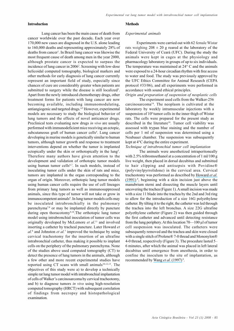
Introduction
Lung cancer has been the main cause of death fromcancer worldwide over the past decade. Each year over170,000 new cases are diagnosed in the U.S. alone leadingto 160,000 deaths and representing approximately 28% ofdeaths from cancer1. In Brazil lung cancer was likewise themost frequent cause of death among men in the year 2000,although prostate cancer is expected to surpass theincidence of lung cancer in 20062. Screening with low-dosehelicoidal computed tomography, biological markers andother methods for early diagnosis of lung cancer currentlyrepresent an important field of study, especially sincechances of cure are considerably greater when patients aresubmitted to surgery while the disease is still localized3.Apart from the newly introduced chemotherapy drugs, othertreatment forms for patients with lung cancer are nowbecoming available, including immunomodulating,antiangiogenic and targeted drugs.4,5 However, experimentalmodels are necessary to study the biological behavior oflung tumors and the effects of novel anticancer drugs.Preclinical tests evaluating new drugs in vivo are usuallyperformed with immunodeficient mice receiving an ectopic,subcutaneous graft of human cancer cells4. Lung cancerdeveloping in murine models is genetically similar to humantumors, although tumor growth and response to treatmentinterventions depend on whether the tumor is implantedectopically under the skin or orthotopically in the lung5.Therefore many authors have given attention to thedevelopment and validation of orthotopic tumor modelsusing human tumor cells6,7. In such models, instead ofinoculating tumor cells under the skin of rats and mice,tumors are implanted in the organ corresponding to theorgan of origin. Moreover, orthotopic lung tumor modelsusing human cancer cells require the use of cell lineagesfrom primary lung tumors as well as immunosuppressedanimals, since this type of tumor will not take or grow inimmunocompetent animals8. In lung tumor models cells maybe inoculated intrabronchially in the pulmonaryparenchyma7,8 or may be implanted directly by punctureduring open thoracotomy9,10.The orthotopic lung tumormodel using intrabronchial inoculation of tumor cells wasoriginally developed by McLemore et al.11 and involvedinserting a catheter by tracheal puncture. Later Howard etal.8 and Johnston et al.7 improved the technique by usingcervical tracheotomy for the insertion of an ultrafineintrabronchial catheter, thus making it possible to implantcells on the periphery of the pulmonary parenchyma. Noneof the studies above used computed tomography (CT) todetect the presence of lung tumors in the animals, althougha few other and more recent experimental studies havereported using CT scans with small animals10,12,13. Theobjectives of this study were a) to develop a technicallysimple rat lung tumor model with intrabronchial implantationof cells of Walker’s carcinosarcoma by cervical tracheotomy,and b) to diagnose tumors in vivo using high-resolutioncomputed tomography (HRCT) with subsequent correlationof findings from necropsy and histopathologicalexamination.
Methods
Experimental animals
Experiments were carried out with 62 female Wistarrats weighing 208 ± 20 g reared at the laboratory of theFederal University of Ceará (UFC). During the study theanimals were kept in cages at the physiology andpharmacology laboratory in groups of up to six individuals.The temperature was maintained at 24° C and the animalswere exposed to a 24-hour circadian rhythm with free accessto water and food. The study was previously approved bythe UFC Ethics Committee for Animal Research (CEPAprotocol #33/06), and all experiments were performed inaccordance with sound ethical principles.Origin and preparation of suspension of neoplastic cells
The experiment used cells from the Walker-256carcinosarcoma14. The neoplasm is cultivated at thelaboratory by weekly intramuscular injections with asuspension of 106 tumor cells in the inner thigh of Wistarrats. The cells were prepared for the present study asdescribed in the literature10. Tumor cell viability wasassessed with trypan blue staining and the number ofcells per 1 ml of suspension was determined using aNeubauer chamber. The suspension was subsequentlykept at 4°C during the entire experiment.Technique of intrabronchial tumor cell implantation
The animals were anesthetized intraperitoneallywith 2.5% tribromoethanol at a concentration of 1 ml/100 glive weight, then placed in dorsal decubitus and submittedto hair clipping and antisepsis with Povidine®(polyvinylpyrrolidone) in the cervical area. Cervicaltracheotomy was performed as described by Howard et al.(1991) 8, beginning with a skin incision just above themanubrium sterni and dissecting the muscle layers untiluncovering the trachea (Figure 1). A small incision was madewith a size 11 blade into the trachea by the 2nd and 3rd ringto allow for the introduction of a size 16G polyethylenecatheter. By tilting it to the right, the catheter was led throughthe trachea into the left bronchus. A size 22G ultrafinepolyethylene catheter (Figure 2) was then guided throughthe first catheter and advanced until detecting resistancefrom the lung periphery. At this location 70—100 µl of tumorcell suspension was inoculated. The catheters weresubsequently removed and the trachea and skin were closedwith a single stitch of Prolene® 7-0 thread and Mononylon®4-0 thread, respectively (Figure 3). The procedure lasted 5–6 minutes, after which the animal was placed in left lateraldecubitus until emergence from anesthesia, in order toconfine the inoculum to the site of implantation, asrecommended by Wang et al. (1997) 9.

Neto AG et al
86 - Acta Cirúrgica Brasileira - Vol 23 (1) 2008
FIGURE 1 – A) animal in dorsal decubitus; B) surgical site;C) cross-sectional cervicotomy above themanubrium sterni and D) exposed trachea.
FIGURE 3 - A) trachea intubated with size 16G catheter; B)catheter in position of intrabronchialinoculation with tumor cells; C) stitchedtrachea; D) skin suture.
FIGURE 2 - A) Size 16G polyethylene catheter and size 22G ultrafine polyethylene catheter; B) ultrafinecatheter emerging from inside 16G catheterat the site of inoculation.
High-resolution computed tomography (HRCT)
The animals were anesthetized intraperitoneallywith 10% chloral hydrate at a dosage of 0.1 ml/30 g liveweight in order to maintain hypnosis during HRCT. Duringthe scan the animals were kept in ventral decubitus with theaid of a cloth, avoiding the use of adhesive tape. Followingscanning with HRCT the animals were placed in cotton-wadded boxes to keep them warm until emergence fromanesthesia and transference to cages. Scanning wasperformed with a Siemens tomograph (SOMATON AR.TX:130 KV, 50 mA, average FOV 5 cm, high-resolution filter for2-mm sections, scanning time 3 seconds per section [150mAS]). On the average, six 2-mm sections separated by 2-mm intervals were made of the lower half of the chest wherethe tumor was located. Images were captured in widewindow mode for the lung study and in narrow windowmode for the study of the mediastinum. Tumors weremeasured in two dimensions (axial and perpendicular) inwide window mode (Figure 4).
Experimental designThe experimental model was developed in two steps
with the animals assigned to experimental groups at random.Animals that died during follow-up of non-tumor-relatedcauses or presented no tumors upon necropsia andhistopathological examination were excluded from theanalysis.
Step 1 (n=32): Establishment of intrabronchial tumorcell implantation technique and subsequent take rate.
FIGURE 4 - Measurement of tumor (arrows) on high-resolution computed tomography of the chestperformed in wide window mode (lung study)on the 5th day of tumor implantation.
Acta Cirúrgica Brasileira - Vol 23 (1) 2008 - 87
Experimental rat lung tumor model with intrabronchial tumor cell implantation
The animals were randomly assigned to fourgroups and, using the technique described above,inoculated intrabronchially with different concentrationsof Walker’s tumor cells in order to establish the number ofcells required for tumor take: Group 1 (n=8), 105 cells; Group2 (n=8), 2 x 10⁵ cells; Group 3 (n=10), 4 x 10⁵ cells; and Group4 (n=6), 5 x 10⁵ cells. On the sixth day the animals wereeuthanized with chloral hydrate and submitted to necropsisthrough median sternotomy and laparotomy for the jointexcision of trachea, lungs and heart in order to verify thepresence of tumors in the chest and abdomen (liver andadrenal gland tumors). Lung sections were fixed in bufferedisotonic formaldehyde (100 mL of 37% formaldehydesolution, 900 mL distilled water, 4 g monobasic sodiumphosphate and 6.5 g dibasic sodium phosphate). Twenty-four hours later samples were immersed in 70% alcohol,stained with hematoxylin-eosin and examinedhistopathologically by a blinded pathologist.
Step 2 (n=30): Assessment of tumor volume,correlation of HRCT findings with findings from necropsia,and determination of time of survival.
The animals were randomly assigned to one of twoprotocols, A and B, both of which with intrabronchialinoculation of 4 x 10⁵ tumor cells. Although the highest takerate in Step 1 was observed after inoculation with 5 x 10⁵
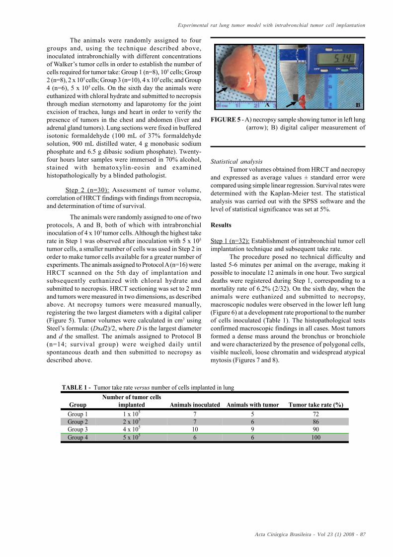
tumor cells, a smaller number of cells was used in Step 2 inorder to make tumor cells available for a greater number ofexperiments. The animals assigned to Protocol A (n=16) wereHRCT scanned on the 5th day of implantation andsubsequently euthanized with chloral hydrate andsubmitted to necropsis. HRCT sectioning was set to 2 mmand tumors were measured in two dimensions, as describedabove. At necropsy tumors were measured manually,registering the two largest diameters with a digital caliper(Figure 5). Tumor volumes were calculated in cm3 usingSteel’s formula: (Dxd2)/2, where D is the largest diameterand d the smallest. The animals assigned to Protocol B(n=14; survival group) were weighed daily untilspontaneous death and then submitted to necropsy asdescribed above.
Statistical analysisTumor volumes obtained from HRCT and necropsy
and expressed as average values ± standard error werecompared using simple linear regression. Survival rates weredetermined with the Kaplan-Meier test. The statisticalanalysis was carried out with the SPSS software and thelevel of statistical significance was set at 5%.
Results
Step 1 (n=32): Establishment of intrabronchial tumor cellimplantation technique and subsequent take rate.
The procedure posed no technical difficulty andlasted 5-6 minutes per animal on the average, making itpossible to inoculate 12 animals in one hour. Two surgicaldeaths were registered during Step 1, corresponding to amortality rate of 6.2% (2/32). On the sixth day, when theanimals were euthanized and submitted to necropsy,macroscopic nodules were observed in the lower left lung(Figure 6) at a development rate proportional to the numberof cells inoculated (Table 1). The histopathological testsconfirmed macroscopic findings in all cases. Most tumorsformed a dense mass around the bronchus or bronchioleand were characterized by the presence of polygonal cells,visible nucleoli, loose chromatin and widespread atypicalmytosis (Figures 7 and 8).
TABLE 1 - Tumor take rate versus number of cells implanted in lung
GroupNumber of tumor cells
implanted Animals inoculated Animals with tumor Tumor take rate (%)Group 1 1 x 10⁵ 7 5 72Group 2 2 x 10⁵ 7 6 86Group 3 4 x 10⁵ 10 9 90Group 4 5 x 10⁵ 6 6 100
FIGURE 5 - A) necropsy sample showing tumor in left lung(arrow); B) digital caliper measurement of

Neto AG et al
88 - Acta Cirúrgica Brasileira - Vol 23 (1) 2008
FIGURE 6 - Fifth day following intrabronchial implantationwith Walker’s tumor cells. Rat A with tumor(arrows) located in the lateral segment of theleft lower lobe, showing the lateral face (A)and medial face (B). Rat B with tumor (arrows)located in the posterior segment of the leftlower lobe, showing the posterior face (C) andmedial face (D).
FIGURE 7 - Histopathological test with hematoxylin-eosinstaining. A) Tumor on the sixth day ofimplantation forming a peribronchiolar mass(thick arrow) with adjacent lung showing signsof swelling and congestion (thin arrow).Magnified 40X. B) Tumor infiltratingbronchiolar wall (arrow). Magnified 100X.
FIGURE 8 - Histopathological test with hematoxylin-eosin staining (400X),showing tumor on the sixth day of implantation, characterizedby polygonal cells with large nuclei, visible nucleoli, loosechromatin and widespread atypical mytosis (arrows) affectingmost of the lung parenchyma in this area.
Step 2, Protocol A (n=16): Assessment of tumor volumeand correlation of HRCT findings with findings fromnecropsy.
On the fifth day following implantation animalsinoculated with 4 x 10⁵ tumor cells and submitted to HRCTand necropsy presented a tumor take rate of 100%. Onesurgical death was registered. The correlation between
findings of tumor volume (cm3) obtained with HRCT andnecropsy was positive (r=0.953; p<0.0001) (Figures 9 and10). The necropsy confirmed HRCT findings with regard totumor volume and location (lateral or posterior segment ofthe left lower lobe). The microscopic examination yieldedfindings similar to those obtained in Step 1.

Acta Cirúrgica Brasileira - Vol 23 (1) 2008 - 89
Experimental rat lung tumor model with intrabronchial tumor cell implantation
FIGURE 9 - Simple linear regression test showing positivecorrelation between HRCT and necropsyfindings for tumor volume (cm3) (R2= 0.908;p=0.0001).
FIGURE 10 - Rat lung on the fifth day following implantationwith 4 x 10⁵ cells of Walker’s carcinosarcoma.Rat A with tumor (arrows) located in theposterior segment of the left lower lobe. Rat Bwith tumor (arrows) located in the lateralsegment of the left lower lobe. Surgicalsamples (A and B); HRCT wide window mode(lung study) (C and D).
Step 2, Protocol B (n=14): Time of survival for ratsinoculated with 4 x 10⁵ cells of Walker’s carcinosarcoma.
An animal which died of an unknown cause onthe third day following tumor cell implantation wasexcluded from the analysis. Of the remaining 13 animalssubmitted to necropsy after spontaneous death inconsequence of the neoplasm, only one presented no tumor.Tumors confirmed by necropsy and histopathologicalexamination displayed the same level of differentiation as
tumors examined during Step 1. In almost all cases, the tumoroccupied most of the lung with large areas of necrosis andhemorrhagic foci (Figure 11). Eight animals (66%) presentedloco-regional tumor dissemination either towards themediastinum (n=6; 49.5 %) or the pleura (n=2; 16.5%). Nodistant metastases were detected in the lung, liver or adrenalglands, as opposed to what is commonly observed withlung cancer in humans. All animals died within two weeks;the median survival time was 11.00±0.38 days (Figure 12).
FIGURE 11 - Animals of the control group with tumor massoccupying most of the left lung and invadingthe mediastinum. The right lung is preserved.
FIGURE 12 - Average survival time of rats in thecontrol group: 10.92±0.29 days (CI 95%;range 10.35—11.48). Median survivaltime: 11.00±0.38 days (CI 95%; range10.27—11.74).
Neto AG et al
90 - Acta Cirúrgica Brasileira - Vol 23 (1) 2008
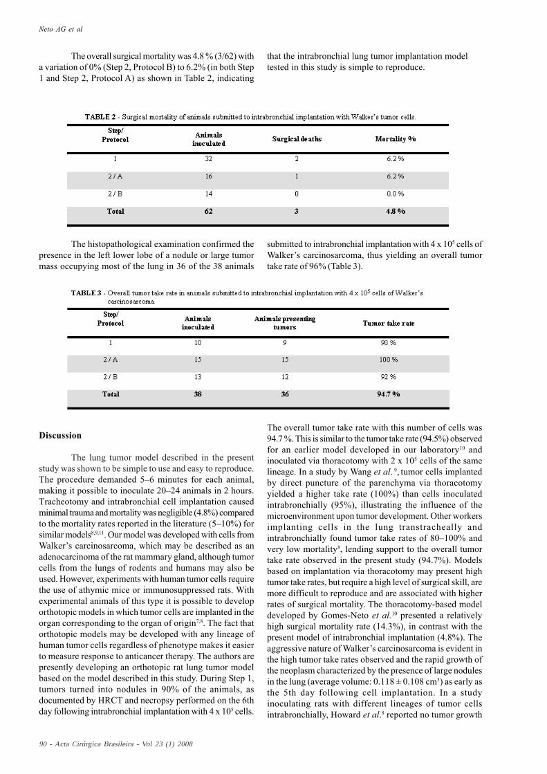
The overall surgical mortality was 4.8 % (3/62) witha variation of 0% (Step 2, Protocol B) to 6.2% (in both Step1 and Step 2, Protocol A) as shown in Table 2, indicating
that the intrabronchial lung tumor implantation modeltested in this study is simple to reproduce.
The histopathological examination confirmed thepresence in the left lower lobe of a nodule or large tumormass occupying most of the lung in 36 of the 38 animals
submitted to intrabronchial implantation with 4 x 10⁵ cells ofWalker’s carcinosarcoma, thus yielding an overall tumortake rate of 96% (Table 3).
Discussion
The lung tumor model described in the presentstudy was shown to be simple to use and easy to reproduce.The procedure demanded 5–6 minutes for each animal,making it possible to inoculate 20–24 animals in 2 hours.Tracheotomy and intrabronchial cell implantation causedminimal trauma and mortality was negligible (4.8%) comparedto the mortality rates reported in the literature (5–10%) forsimilar models8,9,11. Our model was developed with cells fromWalker’s carcinosarcoma, which may be described as anadenocarcinoma of the rat mammary gland, although tumorcells from the lungs of rodents and humans may also beused. However, experiments with human tumor cells requirethe use of athymic mice or immunosuppressed rats. Withexperimental animals of this type it is possible to developorthotopic models in which tumor cells are implanted in theorgan corresponding to the organ of origin7,8. The fact thatorthotopic models may be developed with any lineage ofhuman tumor cells regardless of phenotype makes it easierto measure response to anticancer therapy. The authors arepresently developing an orthotopic rat lung tumor modelbased on the model described in this study. During Step 1,tumors turned into nodules in 90% of the animals, asdocumented by HRCT and necropsy performed on the 6thday following intrabronchial implantation with 4 x 10⁵ cells.
The overall tumor take rate with this number of cells was94.7 %. This is similar to the tumor take rate (94.5%) observedfor an earlier model developed in our laboratory10 andinoculated via thoracotomy with 2 x 10⁵ cells of the samelineage. In a study by Wang et al. 9, tumor cells implantedby direct puncture of the parenchyma via thoracotomyyielded a higher take rate (100%) than cells inoculatedintrabronchially (95%), illustrating the influence of themicroenvironment upon tumor development. Other workersimplanting cells in the lung transtracheally andintrabronchially found tumor take rates of 80–100% andvery low mortality8, lending support to the overall tumortake rate observed in the present study (94.7%). Modelsbased on implantation via thoracotomy may present hightumor take rates, but require a high level of surgical skill, aremore difficult to reproduce and are associated with higherrates of surgical mortality. The thoracotomy-based modeldeveloped by Gomes-Neto et al.10 presented a relativelyhigh surgical mortality rate (14.3%), in contrast with thepresent model of intrabronchial implantation (4.8%). Theaggressive nature of Walker’s carcinosarcoma is evident inthe high tumor take rates observed and the rapid growth ofthe neoplasm characterized by the presence of large nodulesin the lung (average volume: 0.118 ± 0.108 cm3) as early asthe 5th day following cell implantation. In a studyinoculating rats with different lineages of tumor cellsintrabronchially, Howard et al.8 reported no tumor growth
Acta Cirúrgica Brasileira - Vol 23 (1) 2008 - 91
Experimental rat lung tumor model with intrabronchial tumor cell implantation
until the third week following implantation. Other workersimplanting human cancer cell lineages intrabronchially innude rat lung tumor models reported observing the firstsmall lung nodules (<1–3 mm diameter) during the 5th weekfollowing inoculation with 20 x 106 cells.15 Using Walker’scarcinosarcoma, our model presented lung nodules on the5th day following inoculation, and in less than two weeksall animals in the control group had died. Howard et al.16
developed an orthotopic nude rat lung tumor model withendobronchial implantation of human lineages of lungcarcinoma (NCI-H460) and found distant metastases inseveral organs, the earliest of which in the mediastinal lymphnodes. The most severely affected areas were thecontralateral lung, kidneys, brain, bones and (lessfrequently) the adrenal glands. However, no metastaseswere observed until the 14th day following implantation.Dissemination to the mediastinal lymph nodes was detectedon the 21st day and distant metastases only by the 28thday. In a study using the same lineages in nude rats and thesame model, Johnston et al.7 found metastases in themediastinal lymph nodes in 100% of the animals in thecontrol group, as well as systemic metastases in the bones(95%), kidneys (83%), brain (48%) and contralateral lung(82%). The authors found the lung cancer model useful, inspite of the aggressiveness of the tumor causing the animalsto die by the 5th week of implantation by way of local andsystemic dissemination. In our model tumors developedquickly and disseminated to the mediastinum, but no distantor systemic metastases were found, perhaps because ofthe early death of the animals (median survival time: 11days) in consequence of the aggressive nature of the celllineage inoculated. Not even the contralateral lungdisplayed metastases, proving that no dissemination orendobronchial leakage of tumor cells occurred at the momentof implantation, a possibility discussed by some authors7,16.The massive mediastinal dissemination observed in thepresent study resembles findings from human patients withsmall-cell carcinoma. In fact, small-cell carcinoma behavesvery aggressively in humans, usually with earlydissemination to the mediastinum, although distantmetastases are also frequently observed17. Presentlyoncological research tends to employ mice inoculatedsubcutaneously when testing new anticancer drugs18.However, the validity of the results obtained with thesemodels is questionable due to differences in thepharmacodynamic aspects of subcutaneous tumor graftsand tumors in their organ of origin. Becausechemosensitivity and response to therapy with anticancerdrugs depend on the microenvironmental interaction ofstroma and tumor, the choice of anatomical site ofimplantation is of considerable importance6. Thus, thedevelopment of orthotopic animal models is likely toincrease our ability to anticipate human response to therapywith anticancer drugs19. The results obtained in the presentstudy demonstrate the feasibility of our rat lung tumor modelbased on intrabronchial inoculation of tumor cells in thelower left lung and may contribute to the development ofan orthotopic lung tumor model using immunosuppressedrats inoculated with human tumor cells. The HRCT techniqueemployed to diagnose lung tumors in rats inoculated with
Walker’s tumor cells was shown to be time-saving, non-invasive and capable of early tumor detection. HRCTfindings regarding tumor size were validated byhistopathological examination and correlated closely withnecropsy findings (r=0.953; p<0.0001), suggesting thatHRCT scanning may be used in the future to evaluate tumorgrowth and volume without euthanasia, thus avoiding timeand resource-consuming procedures. Other recentlypublished studies using experimental models10,12,13 foundthe HRCT scanning technique to be an efficient tool fordiagnosing lung nodules and to be less costly than magneticresonance imaging20. The efficacy of HRCT scanning in thedetection and measurement of tumors, as demonstrated byour study, makes it a suitable and non-invasive techniquefor diagnosing lung tumors, monitoring tumor growth andevaluating response to anticancer drugs in vivo in animalssubmitted to implantation with tumor cells.
Conclusion
1. Model of intrabronchial tumor implantationproved feasible: the take rate was high, surgical mortalitywas negligible and the procedure was simple to performand easy to reproduce.
2. High-resolution computed tomography wasfound to be a highly accurate tool for tumor diagnosis,localization and measurement and may be recommendedfor monitoring tumor growth in this model.
References
1. Jemal A, Tiwari RC, Murray T, Ghafoor A, Samuels A,Ward E, Feuer EJ, Thun MJ. Cancer statistics. CA CancerJ. Clin. 2004; 54:8-29.
2. Instituto Nacional do Câncer. Estimativa 2006: Incidênciade câncer no Brasil. Rio de Janeiro, INCA/MS; 2005.
3 Libby DM, Wu N, Lee IJ, Farooqi A, Smith JP, PasmantierMW, Mccauley D, Yankelevitz DF, Henschke CI. CTscreening for lung cancer: the value of short-term CTfollow-up. Chest. 2006; 129(4):1039-42.
4. Ciardiello F, Caputo R, Bianco R, Damiano V, Pomatico G,De Placido S, Bianco AR, Tortora G. Antitumor effect andpotentiation of cytotoxic drugs activity in human cancercells by ZD-1839 (Iressa), an epidermal growth factorreceptor-selective tyrosine kinase inhibitor. Clin. CancerRes. 2000;6:2053-63.
5. Masferrer JL, Leahy KM, Koki AT, Zweifel BS, Settle SL,Woerner BM, Edwards DA, Flickinger AG, Moore RJ,Seibert K. Antiangiogenic and antitumor activities ofcyclooxygenase-2 inhibitors. Cancer Res.2000;60(1):1306–11.
6.Onn A, Isobe T, Itasaka S, Wu W, O’reilly MS, Ki HW,Fidler IJ, Herbst RS. Development of an orthotopic model
Neto AG et al
92 - Acta Cirúrgica Brasileira - Vol 23 (1) 2008
to study the biology and therapy of primary human lungcancer in nude mice. Clin. Cancer Res. 2003;9:5532-39.
7.Johnston MR, Mullen JBM, Pagura ME, Howard RB.Validation of an orthotopic model of human lung cancerwith regional and systemic metastases. Ann. Thorac. Surg.2001;71:1120-25.
8.Howard RB, Chu H, Zeligman BE, Marcell T, Bunn PA,Mclemore TL, Mulvin DW, Cowen ME, Johnston MR.Irradiated nude rat model for orthotopic human lungcancers. Cancer Res. 1991;51: 3274-80.
9.Wang HY, Ross HM, Ng B, Burt ME. Establishment of anexperimental intrapulmonary tumor nodule model. Ann.Thorac. Surg. 1997;64:216-19.
10.Gomes-Neto, A.; Pessoa BBGP, Aguiar AS, Furtado BM,Moraes MO, Ribeiro RA. Modelo de tumor de pulmão emrato com o carcinossarcoma de Walker. Acta Cir. Bras.2002;17(1):12-22.
11.Mclemore TL, Liu MC, Blacker PC, Gregg M, Alley MC,Abbott BJ, Shoemaker RH, Bohlman ME, Litterst CC,Hubbard WC. Novel intrapulmonary model for orthotopicpropagation of human lung cancers in athymic nude mice.Cancer Res. 1987;47:5132-40.
12.De Clerck NM, Meurrens K, Weiler H, Van Dyck D,Vanhoutte G, Terpsra P, Postnov AA. High-Resolution X-ray Microtomography for the Detection of Lung Tumorsin Living Mice. Neoplasia. 2004;6:374-9.
13.Greschus S, Kiessling F, Lichy MP, Moll J, Mueller MM,Savai R, Rose F, Ruppert C, Gunther A, Luecke M, Fusenig
NE, Semmler W, Traupe H. Potential applications of flat- panel volumetric CT in morphologic and functional small
animal imaging. Neoplasia. 2005;7(8):730-40.
14.Earle WR. A study of the Walker rat mammary carcinoma256, in vivo and in vitro. Am. J. Cancer. 1935;24:566-612.
15. March TH, Marron-Terada PG, Belinsky SA. Refinementof an orthotopic lung cancer model in the nude rat. Vet.Pathol. 2001;38:483-90.
16.Howard RB, Mullen JBM, Pagura ME, Johnston MR.Characterization of a highly metastatic, orthotopic lungcancer model in the nude rat. Clinical & ExperimentalMetastasis. 1999;17:157–62.
17.Capelozzi VL. Anatomia patológica do câncer do pulmão.In: Zamboni M, Carvalho WR. Câncer do pulmão. 1ed.São Paulo: Atheneu, 2005. p.47-68.
18.Ciardiello F, Caputo R, Bianco R, Damiano V, Fontanini G,Cuccato S, De Placido S, Bianco AR, Tortora G. Inhibitionof growth factor production and angiogenesis in humancancer cells by ZD1839 (Iressa), a selective epidermalgrowth factor receptor tyrosine kinase inhibitor. Clin.Cancer Res. 2001;7:1459–65.
19. Killion, JJ, Radinsky R, Fidler IJ. Orthotopic models arenecessary to predict therapy of transplantable tumors inmice. Cancer Metast. Rev. 1998;17:279-84.
20.Kennell SJ, Davis IA, Branning J, Pan HP, Kabalka GW,Paulus MJ. High resolution computer tomography andMRI for lung tumor growth in mice undergoingradioimmunotherapy: correlation with histology. Med.Phys. 2000;27(5):1101-07.
*Color figures available from www.scielo.br/acb
Conflict of interest: noneFinancial source: CNPq
Received: August 20, 2007Review: October 25, 2007
Accepted: November 26, 2007
Correspondence:Antero Gomes NetoHospital de MessejanaFrei Cirilo Avenue, 348060864-190 Fortaleza - Ceará, Brazile-mail: [email protected]
How to cite this article
Gomes Neto A, Simão AFL, Miranda SP, Mourão LTC, Bezerra NP, Almeida PRC, Ribeiro RA. Experimental rat lung tumormodel with intrabronchial tumor cell implantation. Acta Cir Bras. [serial on the Internet] 2008 Jan-Feb;23(1). Available fromURL: http://www.scielo.br/acb