Experimental rat lung tumor model with intrabronchial tumor cell implantation
Upload
khangminh22Category
view
0download
0
(https://www.aetna.com/)
-->
Tumor Markers
Clinical Policy Bulletins Medical Clinical Policy Bulletins
Number: 0352
Policy
I. Aetna considers any of the following serum tumor markers for the stated indication
medically necessary:
A. Prostate-specific antigen (PSA) for prostate cancer screening (see
CPB 0521 - Prostate Cancer Screening (../500_599/0521.html)), staging, monitoring
response to therapy, and detecting disease recurrence
B. Carcinoembryonic antigen (CEA) for any off the following:
1. As a preoperative prognostic indicator in members with known colorectal carcinoma or
mucinous appendiceal carcinoma when it will assist in staging and surgical treatment
planning; or
2. Pancreatic cyst fluid CEA for distinguishing mucinous from non-mucinous malignant
pancreatic cysts; or
3. To detect asymptomatic recurrence of colorectal cancer after surgical and/or medical
treatment for the diagnosis of colorectal cancer (not as a screening test for colorectal
cancer); or
4. To monitor response to treatment for metastatic colorectal cancer; or
5. For cholangiocarcinoma, gallbladder cancer, lung cancer, medullary thyroid
cancer, metastatic breast cancer, mucinous ovarian cancer, and occult primary; or
6. For evaluation of jaundice, abnormal liver function tests (LFTs) or for
obstruction/abnormality on liver imaging.
C. 1p19q codeletion molecular cytogenetic analysis for astrocytomas and gliomas
D. 5-hydroxyindoleacetic acid (5-HIAA) for neuroendocrine tumors
E. ALK gene fusion as a molecular biomarker in non-small cell lung cancer
Policy History
12/17/2019
Effective: 09/13/1999
Next
Review: 03/27/2020
AdditionalInformation
Last Review
Review
History
Definitions
Clinical Policy
Bulletin
Notes

F. ALK gene rearrangement for diffuse large B cell lymphoma, peripheral T-cell lymphoma,
and post-transplant lymphoproliferative disorder
G. ALK translocations for selecting candidates for crizotinib (Xalkori) in inflammatory
myofibroblastic tumor
H. APC for familial adenomatous polyposis when criteria are met in
CPB 0140 - Genetic Testing (../100_199/0140.html); and for desmoid fibromatosis;
experimental for other indications
I. Afirma Thyroid FNA analysis for assessing fine needle aspiration samples from thyroid
nodules that are indeterminate; experimental for other indications. Repeat testing is
considered experimental and investigational
J. Alpha fetoprotein (AFP) for the following indications: hepatocellular carcinoma;
mediastinal mass; ovarian cancer; pelvic mass; testicular cancer; testicular mass; thymic
carcinoma; and thymoma
K. Alfa fetoprotein (AFP) for testing for hepatocellular carcinoma in hepatitis B carriers, or
for persons with cirrhosis and one or more of the following risk factors: alcohol use;
alpha-1 antitrypsin deficiency; Asian female at least 50 years of age; Asian male at least 40
years of age; family history of HCC; genetic hemochromatosis; hepatitis C; nonalcoholic
steatohepatitis; and stage 4 primary biliary cirrhosis
L. Alpha fetoprotein (AFP): serial measurements to diagnose germ cell tumors in members
with adenocarcinoma, or carcinoma not otherwise specified, involving mediastinal nodes;
or the diagnosis and monitoring of hepatocellular carcinoma (e.g., before considering
liver transplantation)
M. BCL2 and BCL6 for the diagnosis of non-Hodgkin’s lymphoma and Castleman's disease
N. BCR/ABL fluorescent in situ hybridization (FISH) for lymphoblastic lymphoma, acute
myeloid leukemia, acute lymphocytic leukemia, chronic myelogenous leukemia, and
suspected myeloproliferative neoplasm; experimental for other indications
O. Beta-2 microglobulin (B2M) for multiple myeloma, non-Hodgkin's lymphoma and
Waldenström's macroglobulinemia/ lymphoplasmacytic lymphoma
P. BRAF V600 mutation for indeterminate thyroid nodules, hairy cell leukemia;
gastrointestinal stromal tumors; Lynch syndrome; non-small cell lung cancer; anaplastic
thyroid carcinoma; and melanoma (see
CPB 0715 - Pharmacogenomic and Pharmacodynamic Testing) (../700_799/0715.html); or
Lynch syndrome for persons meeting criteria in
CPB 0140 - Genetic Testing (../100_199/0140.html); and colorectal cancer if KRAS
nonmutated; experimental for other indications
Q. Breast Cancer Index to assess necessity of adjuvant chemotherapy in females or males
with recently diagnosed breast tumors, where all of the following criteria are met:
1. Breast cancer is nonmetastatic (node negative); and
2. Breast tumor is estrogen receptor positive; and
3. Breast tumor is HER2 receptor negative; and
4. Adjuvant chemotherapy is not precluded due to any other factor (e.g., advanced age and/or
significant co-morbidities); and
2

5. Member and physician (prior to testing) have discussed the potential results of the test
and agree to use the results to guide therapy.
R. Cancer antigen 125 (CA 125) levels for any of the following:
1. As a preoperative diagnostic aid in women with ovarian masses that are suspected to be
malignant, such that arrangements can be made for intraoperative availability of a
gynecological oncologist if the CA 125 is increased; or
2. As a screening test for ovarian cancer when there is a family history of hereditary ovarian
cancer syndrome (a pattern of clusters of ovarian cancer within two or more generations),
where testing is performed concurrently with transvaginal ultrasound and prophylactic
salpingo-oophorectomy has not been performed. For this indication, screening is
considered medically necessary every six months beginning at 30 years of age or 10 years
before the earliest age of the first diagnosis of ovarian cancer in the family; or
3. Diagnosis of ovarian cancer in women with new symptoms (bloating, pelvic or abdominal
pain, difficulty eating or feeling full quickly, or urinary frequency and urgency) that have
persisted for three or more weeks, where the clinician has performed a pelvic and rectal
examination and suspects ovarian cancer; or
4. In members with adenocarcinoma of unknown primary, to rule out ovarian cancer; or
5. In members with known ovarian cancer, as an aid in the monitoring of disease,
response to treatment, detection of recurrent disease, or assessing value of
performing second-look surgery
S. CA 15-3: Serial measurements of CA 15-3 (also known as CA 27-29 or Truquant RIA) in
following the course of treatment in women diagnosed with breast cancer, especially advanced
metastatic breast cancer (an increasing CA 15-3 level may suggest treatment failure).
T. CA 19-9 for the following indications:
1. to monitor the clinical response to therapy or detect early recurrence of disease in
members with known gastric cancer, pancreatic cancer, gallbladder cancer,
cholangiocarcinoma or adenocarcinoma of the ampulla of Vater; or
2. to rule out cholangiocarcinoma in persons with primary sclerosing cholangitis undergoing
liver transplantation; or
3. For evaluation of jaundice, abnormal liver function tests (LFTs) or obstruction/abnormality
on imaging; or
4. As a tumor marker for mucinous appendiceal carcinoma
U. CALCA (calcitonin) expression for medullary thyroid cancer or for adenocarcinoma or
anaplastic/undifferentiated tumors of the head and neck.
V. CALB2 (calretinin) expression for lung cancer and occult primary
W. CD 20, for determining eligibility for anti-CD20 treatment (rituximab) -- see
CPB 0314 - Rituximab (Rituxan) (0314.html)
X. CD 25, for determining eligibility for denileukin diftitox (Ontak) treatment

Y. CD 31 immunostaining, for diagnosis of angiosarcoma.
Z. CD 33, for determining eligibility for anti-CD33 (gemtuzumab, Mylotarg) treatment
AA. CD 52, for determining eligibility for anti-CD52 (alemtuzumab, Campath) treatment
AB. CD117 (c-kit), for determining eligibility for treatment with imatinib mesylate (Gleevec)
AC. CHGA (Chromogranin A) expression for neuroendocrine tumors, non-small cell lung
cancer, Merkel cell carcinoma and occult primary
AD. Cyclin D1, for diagnosis and predicting disease recurrence of mantle cell lymphoma
AE. DecisionDx-UM (Castle Biosciences, Phoenix, AZ) for risk stratification of persons with
localized uveal melanoma
AF. EndoPredict (also known as 12-gene score) to assess necessity of adjuvant chemotherapy
in females or males with recently diagnosed breast tumors, where all of the following
criteria are met:
1. Breast cancer is nonmetastatic (node negative); and
2. Breast tumor is estrogen receptor positive; and
3. Breast tumor is HER2 receptor negative; and
4. Adjuvant chemotherapy is not precluded due to any other factor (e.g., advanced age and/or
significant co-morbidities); and
5. Member and physician (prior to testing) have discussed the potential results of the test
and agree to use the results to guide therapy
AG. Epidermal growth factor receptor (EGFR) mutation testing for predicting response to EGFR-
targeting tyrosine kinase inhibitors (erlotinib (Tarceva), gefitinib (Iressa), afatinib (Gilotrif)) in
non-small cell lung cancer
AH. FLT3 gene mutation testing for acute myeloid leukemia (AML)
AI. Human chorionic gonadotropin (HCG), serial measurement to diagnose germ cell tumors in
members with adenocarcinoma, or carcinoma not otherwise specified, involving mediastinal
nodes, or to monitor treatment in members with known trophoblastic tumors (invasive
hydatidiform moles and choriocarcinomas) and germinal cell tumors (teratocarcinoma and
embryonal cell carcinoma) of the ovaries or testes, or to monitor for relapse after
remission is achieved
AJ. Beta human chorionic gonadotropin (beta-HCG) for mediastinal mass; ovarian cancer;
pelvic mass; testicular mass; testicular cancer; thymoma; or thymic carcinoma
AK. Human epidermal growth factor receptor 2 (HER2) evaluation in breast, gastric and
esophageal cancer - see
CPB 0313 - Trastuzumab (Herceptin), Ado-Trastuzumab (Kadcyla) and Pertuzumab
(Perjeta) (0313.html)
AL. IGH@ (Immunoglobulin heavy chain locus), gene rearrangement analysis to detect abnormal
clonal population(s) in non-Hodgkin’s lymphomas,hairy cell leukemia, and post-transplant
lymphoproliferative disorder
AM. IGK@ (Immunoglobulin kappa light chain locus), gene rearrangement analysis, evaluation to
detect abnormal clonal population(s) for non-Hodgkin’s lymphoma, systemic light chain
amyloidosis
2

AN. Isocitrate dehydrogenase 1 and 2 (IDH1 and IDH2) gene mutation for AML,
chondrosarcomas, or gliomas and glioblastomas
AO. INHA (inhibin) expression for ovarian cancer or pelvic mass
AP. Lactate dehydrogenase (LDH) for acute lymphoblastic leukemia (ALL), bone cancer, kidney
cancer, kidney mass, lung cancer, multiple myeloma, non-Hodgkin's lymphoma, pelvic
mass, ovarian cancer, testicular cancer, or testicular mass
AQ. K-ras (KRAS) mutation analysis, with BRAF reflex testing, to predict non-response to
cetuximab (Erbitux) and panitumumab (Vectibix) in the treatment of anal
adenocarcinoma, metastatic colorectal cancer and small bowel adenocarcinoma; K-ras
(KRAS) mutation analysis to predict non-response to erlotinib (Tarceva) in the treatment of
non-small cell lung cancer; experimental for all other indications
AR. KRAS, NRAS and BRAF for metastatic colon cancer
AS. Mammaprint to assess necessity of adjuvant chemotherapy in females or males with
recently diagnosed breast tumors, where all of the following criteria are met:
1. Breast cancer is nonmetastatic (node negative ) or with 1-3 involved ipsilateral axillary
lymph nodes; and
2. Breast tumor is estrogen receptor positive or progesterone receptor positive; and
3. Breast tumor is HER2 receptor negative (Rationale: adjuvant chemotherapy with
trastuzumab (Herceptin) is considered to be medically necessary regardless of
Mammaprint score for HER2 receptor positive lesions); and
4. Member is determined to be at "high clinical risk" of recurrence using Adjuvant! Online
(http://www.adjuvantonline.com) (see page 20 of MINDACT study
(http://www.nejm.org/doi/suppl/10.1056/NEJMoa1602253/suppl_file/nejmoa1602253_
appendix.pdf) supplement for definitions of high clinical risk); and
5. Adjuvant chemotherapy is not precluded due to any other factor (e.g., advanced age and/or
significant co-morbidities); and
6. Member and physician (prior to testing) have discussed the potential results of the test and
agree to use the results to guide therapy
AT. Measurement of estrogen and progesterone receptors on primary breast cancers, and on
metastatic lesions if the results would influence treatment planning
AU. Mismatch repair (MSI/dMMR) (MLH1, MSH2, MSH6) tumor testing (somatic mutations) for
colorectal cancer, esophageal cancer, esophagogastric junction cancer, gastric
cancer, pancreatic cancer, cervical cancer, uterine cancer, prostate cancer, testicular
cancer, and penile cancer; for medical necessity of screening of germline mutations for
HNPCC/Lynch Syndrome with MLH1, MSH2, MSH6, see
CPB 0140 - Genetic Testing (../100_199/0140.html);
AV. MLH1, MSH2 and PMS2 for ovarian cancer
AW. Mycosis fungoides, diagnosis: polymerase chain reaction (PCR) for T-cell receptor gamma
chain gene rearrangement as an adjunct to the histopathologic diagnosis of mycosis
fungoides
2
1

AX. MYD88 (myeloid differentiation primary response 88) to differentiate Waldenstrom's
macroglobinemia (WM) versus marginal zone lymphoma (MZL) if plasmacytic
differentiation present for gastric MALT lymphoma, nodal marginal zone lymphoma,
nongastric MALT lymphoma, and splenic marginal zone lymphoma
AY. Next generation sequencing of tumor DNA (e.g., ClonoSeq) to detect or quantify minimal
residual disease in persons with multiple myeloma or acute lymphocytic leukemia
AZ. Myeloperoxidase (MPO) immunostaining, FLT3-ITD, CEBPA mutation, NPM1 mutation, and
KIT mutation for diagnosis of acute myeloid leukemia
BA. NPM1 in acute myeloid leukemia (AML); experimental for other indications
BB. PAM50 Risk of Recurrence (ROR) Score (also known as Prosigna Breast Cancer Prognostic
Gene Signature Assay) to assess necessity of adjuvant chemotherapy in females or males
with recently diagnosed breast tumors, where all of the following criteria are met:
1. Breast cancer is nonmetastatic (node negative); and
2. Breast tumor is estrogen receptor positive; and
3. Breast tumor is HER2 receptor negative; and
4. Adjuvant chemotherapy is not precluded due to any other factor (e.g., advanced age and/or
significant co-morbidities); and
5. Member and physician (prior to testing) have discussed the potential results of the test
and agree to use the results to guide therapy.
BC. PDGFRA for gastrointestinal stromal tumors (GIST)
BD. PDGFRB testing for myelodysplastic syndromes (MDS) and dermatofibrosarcoma
protuberans
BE. PML/RARA for acute promyelocytic leukemia; experimental for all other indications
BF. PTEN for persons meeting Cowden syndrome testing criteria in
CPB 0140 - Genetic Testing (../100_199/0140.html); experimental for all other indications
BG. Placental alkaline phosphatase (PLAP), to diagnose germ cell seminoma and non-
seminoma germ cell tumors in unknown primary cancers.
BH. Quest Diagnostics Thyroid Cancer Mutation Panel for assessing fine needle aspiration
samples from thyroid nodules that are indeterminate; experimental for other indications.
Repeat testing is considered experimental and investigational
BI. ROS-1 to predict response to crizotinib (Xalkori) for the treatment of non-small cell lung
cancer (NSCLC)
BJ. RUNX1 for myelodysplastic syndrome
BK. Steroid hormone receptor status in both pre-menopausal and post-menopausal
members to identify individuals most likely to benefit from endocrine forms of adjuvant
therapy and therapy for recurrent or metastatic breast cancer
BL. Targeted hematologic genomic sequencing panel (5-50 genes) for myelodysplastic
syndromes (MDS) and myeloproliferative neoplasms (MPN)
BM. Targeted solid organ genomic sequencing panel (5-50 genes) for non-small cell lung
cancer (including Oncomine Dx Target Test (Thermo Fisher Scientific, Carlsbad, CA))
2

BN. T-cell receptor gene rearrangements (TRA@, TRB@, TRD@, TRG@) for T-cell
prolymphocytic leukemia, T-cell large granular lymphocytic leukemia, nasal type
extranodal NK/T-cell lymphoma, peripheral T-cell lymphoma, primary cutaneous CD30+ T-
cell lymphoproliferative disorders, Castleman's disease, mycosis fungoides/Sezary
syndrome
BO. ThyGenX (formerly Mirinform Thyroid) for assessing fine needle aspiration samples from
thyroid nodules that are indeterminate; experimental for other indications.Repeat testing
is considered experimental and investigational
BP. ThyraMIR as a reflex test following ThyGenX for assessing fine needle aspiration samples
from thyroid nodules that are indeterminate; experimental for other indications. Repeat
testing is considered experimental and investigational
BQ. Thymidine kinase for chronic lymphocytic leukemia (CLL)/small lymphocytic lymphoma
(SLL)
BR. Thyroglobulin antibodies for thyroid cancer
BS. Thyroglobulin (TG) expression for thyroid cancer, occult primary, and adenocarcinoma or
anaplastic/undifferentiated tumors of the head and neck
BT. Thyroid transcription factor-1 (TTF-1) for lung cancer or neuroendocrine tumors
BU. Thyroseq for assessing fine needle aspiration samples from thyroid nodules that are
indeterminate; experimental for other indications. Repeat testing is considered
experimental and investigational
BV. TP53 for chronic lymphocytic leukemia/small lymphocytic lymphoma, mantle cell
lymphoma, splenic marginal zone lymphoma, acute myeloid leukemia, occult primary,
and myelodysplastic syndromes
BW. Oncotype Dx (also known as 21 gene RT-PCR test) to assess necessity of adjuvant
chemotherapy in females or males with recently diagnosed breast tumors, where all of
the following criteria are met:
1. Breast cancer is nonmetastatic (node negative ) or with 1-3 involved ipsilateral axillary
lymph nodes; and
2. Breast tumor is estrogen receptor positive; and
3. Breast tumor is HER2 receptor negative or breast tumor is HER2 receptor positive and less
than 1 cm in diameter. (Rationale: adjuvant chemotherapy with trastuzumab (Herceptin) is
considered to be medically necessary regardless of an Oncotype Dx score for HER2
receptor positive lesions 1 cm or more in diameter); and
4. Adjuvant chemotherapy is not precluded due to any other factor (e.g., advanced age and/or
significant co-morbidities); and
5. Member and physician (prior to testing) have discussed the potential results of the test
and agree to use the results to guide therapy (i.e., member will forgo adjuvant
chemotherapy if Oncotype Dx score is low).
BX. Urokinase plasminogen activator (uPA) and plasminogen activator inhibitor 1 (PAI-1) to
assess necessity of adjuvant chemotherapy in females or males with recently diagnosed
1
2

breast tumors, where all of the following criteria are met:
1. Breast cancer is nonmetastatic (node negative); and
2. Breast tumor is estrogen receptor positive; and
3. Breast tumor is HER2 receptor negative; and
4. Adjuvant chemotherapy is not precluded due to any other factor (e.g., advanced age and/or
significant co-morbidities); and
5. Member and physician (prior to testing) have discussed the potential results of the test
and agree to use the results to guide therapy.
In addition, urokinase plasminogen activator (uPA) and plasminogen activator inhibitor 1
(PAI-1) is considered medically necessary for the determination of prognosis in patients
with newly diagnosed, node negative breast cancer
BY. Veristrat proteomic testing for patients with advanced NSCLC, whose tumors were
without EGFR and anaplastic lymphoma kinase (ALK) mutations, who had progressed
after at least one chemotherapy regimen), and for whom erlotinib was considered an
appropriate treatment.
BZ. WT-1 gene expression for non-small cell lung cancer and occult primary
CA. ZAP-70, for assessing prognosis and need for aggressive therapy in persons with chronic
lymphocytic leukemia (CLL)/small lymphocytic lymphoma (SLL).
Either standard node dissection negative by hematoxylin and eosin (H&E) staining or
sentinel node negative by H&E staining (if sentinel node is negative by H&E, but
immunoassay is positive, then still considered node negative for this purpose). In addition,
women with isolated tumor cells in lymph nodes (micrometastases) are considered node
negative.
More than one Oncotype Dx test may be medically necessary for persons with breast
cancer who have two or more histologically distinct tumors that meet medical necessity
criteria. Repeat Oncotype Dx testing or testing of multiple tumor sites in the same person has
no proven value for other indications. Oncotype Dx is considered experimental and
investigational for ductal carcinoma in situ (OncotypeDx DCIS), colon cancer (OncotypeDx
Colon), prostate cancer (OncotypeDx Prostate) and all other indications.
Aetna considers use of more than one type of test to determine necessity of adjuvant
therapy in breast cancer (Oncotype Dx Breast, Breast Cancer Index, EndoPredict,
PAM50, Mammaprint, or uPA and PAI-1) experimental and investigational.
II. Aetna considers urinary biomarkers (e.g., bladder tumor antigen (BTA) (e.g., BTA Stat and BTA
TRAK), nuclear matrix protein (NMP22) test, the fibrin/fibrinogen degradation products (Aura-
Tek FDfP) test, or fluorescence in situ hybridization (FISH) (e.g., Pathnostics Bladder FISH test,
UroVysion Bladder Cancer test medically necessary in any of the following conditions:
1
2
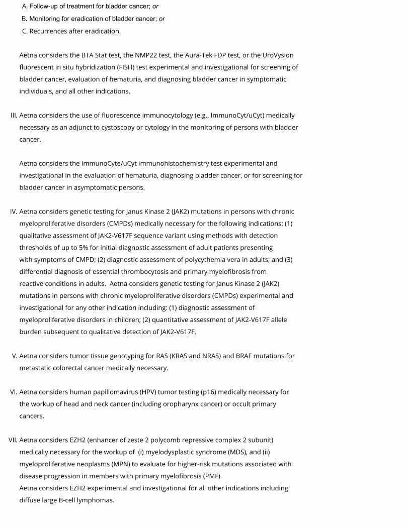
A. Follow-up of treatment for bladder cancer; or
B. Monitoring for eradication of bladder cancer; or
C. Recurrences after eradication.
Aetna considers the BTA Stat test, the NMP22 test, the Aura-Tek FDP test, or the UroVysion
fluorescent in situ hybridization (FISH) test experimental and investigational for screening of
bladder cancer, evaluation of hematuria, and diagnosing bladder cancer in symptomatic
individuals, and all other indications.
III. Aetna considers the use of fluorescence immunocytology (e.g., ImmunoCyt/uCyt) medically
necessary as an adjunct to cystoscopy or cytology in the monitoring of persons with bladder
cancer.
Aetna considers the ImmunoCyte/uCyt immunohistochemistry test experimental and
investigational in the evaluation of hematuria, diagnosing bladder cancer, or for screening for
bladder cancer in asymptomatic persons.
IV. Aetna considers genetic testing for Janus Kinase 2 (JAK2) mutations in persons with chronic
myeloproliferative disorders (CMPDs) medically necessary for the following indications: (1)
qualitative assessment of JAK2-V617F sequence variant using methods with detection
thresholds of up to 5% for initial diagnostic assessment of adult patients presenting
with symptoms of CMPD; (2) diagnostic assessment of polycythemia vera in adults; and (3)
differential diagnosis of essential thrombocytosis and primary myelofibrosis from
reactive conditions in adults. Aetna considers genetic testing for Janus Kinase 2 (JAK2)
mutations in persons with chronic myeloproliferative disorders (CMPDs) experimental and
investigational for any other indication including: (1) diagnostic assessment of
myeloproliferative disorders in children; (2) quantitative assessment of JAK2-V617F allele
burden subsequent to qualitative detection of JAK2-V617F.
V. Aetna considers tumor tissue genotyping for RAS (KRAS and NRAS) and BRAF mutations for
metastatic colorectal cancer medically necessary.
VI. Aetna considers human papillomavirus (HPV) tumor testing (p16) medically necessary for
the workup of head and neck cancer (including oropharynx cancer) or occult primary
cancers.
VII. Aetna considers EZH2 (enhancer of zeste 2 polycomb repressive complex 2 subunit)
medically necessary for the workup of (i) myelodysplastic syndrome (MDS), and (ii)
myeloproliferative neoplasms (MPN) to evaluate for higher-risk mutations associated with
disease progression in members with primary myelofibrosis (PMF).
Aetna considers EZH2 experimental and investigational for all other indications including
diffuse large B-cell lymphomas.
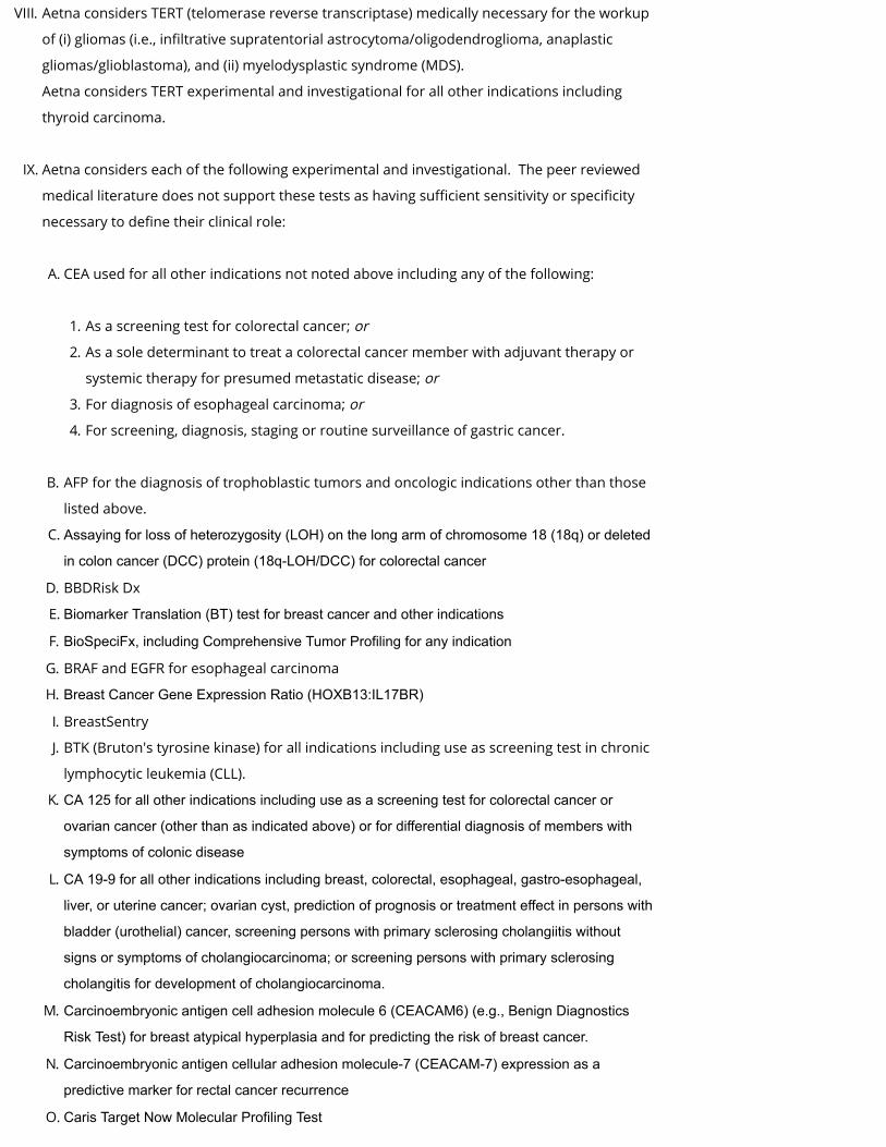
VIII. Aetna considers TERT (telomerase reverse transcriptase) medically necessary for the workup
of (i) gliomas (i.e., infiltrative supratentorial astrocytoma/oligodendroglioma, anaplastic
gliomas/glioblastoma), and (ii) myelodysplastic syndrome (MDS).
Aetna considers TERT experimental and investigational for all other indications including
thyroid carcinoma.
IX. Aetna considers each of the following experimental and investigational. The peer reviewed
medical literature does not support these tests as having sufficient sensitivity or specificity
necessary to define their clinical role:
A. CEA used for all other indications not noted above including any of the following:
1. As a screening test for colorectal cancer; or
2. As a sole determinant to treat a colorectal cancer member with adjuvant therapy or
systemic therapy for presumed metastatic disease; or
3. For diagnosis of esophageal carcinoma; or
4. For screening, diagnosis, staging or routine surveillance of gastric cancer.
B. AFP for the diagnosis of trophoblastic tumors and oncologic indications other than those
listed above.
C. Assaying for loss of heterozygosity (LOH) on the long arm of chromosome 18 (18q) or deleted
in colon cancer (DCC) protein (18q-LOH/DCC) for colorectal cancer
D. BBDRisk Dx
E. Biomarker Translation (BT) test for breast cancer and other indications
F. BioSpeciFx, including Comprehensive Tumor Profiling for any indication
G. BRAF and EGFR for esophageal carcinoma
H. Breast Cancer Gene Expression Ratio (HOXB13:IL17BR)
I. BreastSentry
J. BTK (Bruton's tyrosine kinase) for all indications including use as screening test in chronic
lymphocytic leukemia (CLL).
K. CA 125 for all other indications including use as a screening test for colorectal cancer or
ovarian cancer (other than as indicated above) or for differential diagnosis of members with
symptoms of colonic disease
L. CA 19-9 for all other indications including breast, colorectal, esophageal, gastro-esophageal,
liver, or uterine cancer; ovarian cyst, prediction of prognosis or treatment effect in persons with
bladder (urothelial) cancer, screening persons with primary sclerosing cholangiitis without
signs or symptoms of cholangiocarcinoma; or screening persons with primary sclerosing
cholangitis for development of cholangiocarcinoma.
M. Carcinoembryonic antigen cell adhesion molecule 6 (CEACAM6) (e.g., Benign Diagnostics
Risk Test) for breast atypical hyperplasia and for predicting the risk of breast cancer.
N. Carcinoembryonic antigen cellular adhesion molecule-7 (CEACAM-7) expression as a
predictive marker for rectal cancer recurrence
O. Caris Target Now Molecular Profiling Test

P. CDH1 and TP53 for ovarian cancer
Q. CDX2 as a prognostic biomarker for colon cancer
R. CEA, Cyfra21-1 (a cytokeratin 19 fragment), p53, squamous cell carcinoma antigen (SCC-Ag)
and vascular endothelial growth factor C (VEGF-C) for diagnosis of esophageal carcinoma
S. Circulating cell-free nucleic acids in colorectal cancer
T. Circulating tumor cell (CTC) assays (e.g., CellSearch assay) for all indications, including, but
not limited to metastatic breast, colorectal, melanoma, and prostate cancers
U. CK5, CK14, p63, and Racemase P504S testing for prostate cancer
V. c-Met expression for predicting prognosis in persons with advanced NSCLC and colorectal
cancer, and other indications
W. Cofilin (CFL1) as a prognostic and drug resistance marker in non-small cell lung cancer
X. ColonSentry test for screening of colorectal cancer
Y. ColoPrint, CIMP, LINE-1 hypomethylation, and Immune cells for colon cancer
Z. Colorectal Cancer DSA (Almac Diagnostics, Craigavon, UK)
AA. ConfirmMDx for prostate cancer
AB. CxBladder test for bladder cancer
AC. Cyclin D1 and FADD (Fas-associated protein with death domain) for head and neck squamous
cell carcinoma
AD. Decipher test (a RNA biomarkers assay) for prostate cancer
AE. Decision DX-Melanoma (Castle Biosciences, Phoenix, AZ)
AF. DCIS Recurrence Score
AG. Des-gamma-carboxy prothrombin (DCP) (also known as "prothrombin produced by vitamin K
absence or antagonism II" [PIVKA II]) for diagnosing and monitoring hepatocellular carcinoma
(HCC) and other indications
AH. EarlyCDT-Lung test
AI. EGFR gene expression analysis for transitional (urothelial) cell cancer
AJ. EGFRVIII for glioblastoma multiforme
AK. EML4-ALK as a diagnostic tool for stage IV non-small-cell lung cancer
AL. Envisia Genomic Classifier
AM. Estrogen and progesterone receptors when used alone to assign a member with breast cancer
to prognostic groupings since they are relatively weak predictors of long-term relapse and
breast cancer related mortality rates
AN. Excision repair cross-complementation group 1 protein (ERCC1) for persons with NSCLC,
colon or with gastric cancer who are being considered for treatment with platinum-based
chemotherapy, and other indications
AO. ExoDx Prostate/ExosomeDx Prostate (IntelliScore)
AP. Fibrin/fibrinogen degradation products (FDP) test (e.g., DR-70 or Onko-Sure) for colorectal
cancer
AQ. FoundationOne, FoundationOne CDx and FoundationOne Heme (except where
FoundationOne CDx is used as a companion diagnostic test for somatic/tumor BRCA testing,
see

CPB 0227 - BRCA Testing, Prophylactic Mastectomy, and Prophylactic Oophorectomy
(../200_299/0227.html)
and CPB 0715 - Pharmacogenetic and Pharmacodynamic Testing (../700_799/0715.html))
AR. Galectin-3 for breast cancer, ovarian cancer, pancreatic cancer, and prostate cancer
AS. Gene hypermethylation for prostate cancer
AT. GeneKey (GeneKey Corp., Boston, MA)
AU. GeneSearch Breast Lymph Node (BLN) assay
AV. Glutathione-S-transferase P1 (GSTP1) for screening, detection and management of prostate
cancer
AW. Guanylyl cyclase c (GCC or GUCY2C) (e.g., Previstage GCC Colorectal Cancer State Test) for
colorectal cancer
AX. HeproDx
AY. HER2 testing of appendiceal cancer
AZ. HERmark testing for breast cancer and other indications
BA. HMGB1 and RAGE in cutaneous malignancy (e.g., basal cell carcinoma, melanoma, and
squamous cell carcinoma)
BB. Human epididymis protein 4 (HE4) (e.g., Elecsys HE4 assay) for endometrial cancer, ovarian
cancer, or evaluation of pelvic mass, including to assist in the determination of referral for
surgery to a gynecologic oncologist or general surgery, and for other indications
BC. IHC4 (e.g., NexCourse IHC4 by AQUA Technology) for breast cancer
BD. Immunoassay using magnetic nanosensor for diagnosis of lung cancer
BE. Insight DX Breast Cancer Profile
BF. Ki67 for breast cancer
BG. Ki-67 in upper tract urinary carcinoma
BH. 4Kscore
BI. Lectin-reactive alpha-fetoprotein (AFP-L3) for liver cancer
BJ. Liquid biopsy (e.g., CancerIntercept, Colvera, GeneStrat, FoundationACT, FoundationOne
Liquid, Guardant360, Neolab Prostate) for any indication, including, but not limited
to, breast cancer, colorectal cancer, lung cancer, melanoma, or ovarian cancer (For
EGFR liquid biopsy for non-small cell lung cancer (e.g., cobas EGFR Mutation Test v2) and
PIK CA testing (therascreen PIK CA RGQ PCR Kit) for breast cancer, see
CPB 715 - Pharmacogenetic and Pharmacodynamic Testing (../700_799/0715.html))
BK. Long non-coding RNA in gallbladder cancer
BL. Lymph3Cx Lymphoma Molecular Classification Assay to distinguish between primary
mediastinal B-cell lymphoma (PMBCL) and diffuse large B-cell lymphoma (DLBCL)
BM. Mammostrat
BN. Mass spectrometry-based proteomic profiling for indeterminate pulmonary nodules
BO. MatePair targeted rearrangements (whole genome next-generation sequencing) for
hematolymphoid neoplasia and solid organ neoplasia
BP. Measurement of circulating tumor cells (e.g., CellMax Life and FirstSightCRC) for screening
of colorectal cancer
BQ. Merkel SmT Oncoprotein Antibody Titer
BR. Merkel Virus VP1 Capsid Antibody
3 3
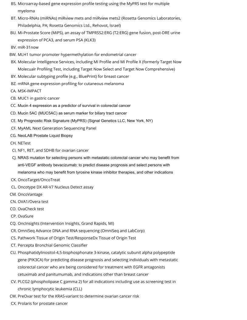
BS. Microarray-based gene expression profile testing using the MyPRS test for multiple
myeloma
BT. Micro-RNAs (miRNAs) miRview mets and miRview mets2 (Rosetta Genomics Laboratories,
Philadelphia, PA; Rosetta Genomics Ltd., Rehovot, Israel)
BU. Mi-Prostate Score (MiPS), an assay of TMPRSS2:ERG (T2:ERG) gene fusion, post-DRE urine
expression of PCA3, and serum PSA (KLK3)
BV. miR-31now
BW. MLH1 tumor promoter hypermethylation for endometrial cancer
BX. Molecular Intelligence Services, including MI Profile and MI Profile X (formerly Target Now
Molecualr Profiling Test, including Target Now Select and Target Now Comprehensive)
BY. Molecular subtyping profile (e.g., BluePrint) for breast cancer
BZ. mRNA gene expression profiling for cutaneous melanoma
CA. MSK-IMPACT
CB. MUC1 in gastric cancer
CC. Mucin 4 expression as a predictor of survival in colorectal cancer
CD. Mucin 5AC (MUC5AC) as serum marker for biliary tract cancer
CE. My Prognostic Risk Signature (MyPRS) (Signal Genetics LLC, New York, NY)
CF. MyAML Next Generation Sequencing Panel
CG. NeoLAB Prostate Liquid Biopsy
CH. NETest
CI. NF1, RET, and SDHB for ovarian cancer
CJ. NRAS mutation for selecting persons with metastatic colorectal cancer who may benefit from
anti-VEGF antibody bevacizumab; to predict disease prognosis and select persons with
melanoma who may benefit from tyrosine kinase inhibitor therapies, and other indications
CK. OncoTarget/OncoTreat
CL. Oncotype DX AR-V7 Nucleus Detect assay
CM. OncoVantage
CN. OVA1/Overa test
CO. OvaCheck test
CP. OvaSure
CQ. OncInsights (Intervention Insights, Grand Rapids, MI)
CR. OmniSeq Advance DNA and RNA sequencing (OmniSeq and LabCorp)
CS. Pathwork Tissue of Origin Test/ResponseDx Tissue of Origin Test
CT. Percepta Bronchial Genomic Classifier
CU. Phosphatidylinositol-4,5-bisphosphonate 3-kinase, catalytic subunit alpha polypeptide
gene (PIK3CA) for predicting disease prognosis and selecting individuals with metastatic
colorectal cancer who are being considered for treatment with EGFR antagonists
cetuximab and panitumumab, and indications other than breast cancer
CV. PLCG2 (phospholipase C gamma 2) for all indications including use as screening test in
chronic lymphocytic leukemia (CLL)
CW. PreOvar test for the KRAS-variant to determine ovarian cancer risk
CX. Prolaris for prostate cancer

CY. ProOnc TumorSourceDx test (Prometheus Laboratories, San Diego, CA) to identify tissue
or origin for metastatic tumor
CZ. Prostate core mitotic test
DA. Prostate Px and Post-Op Px for predicting recurence of prostate cancer
DB. Prostate Cancer Risk Panel (FISH analysis by Mayo Clinic)
DC. Proveri prostate cancer assay (PPCA)
DD. PSA for screening women with breast cancer or for differentiating benign from malignant
breast masses
DE. PTEN gene expression for non-small cell lung cancer
DF. Ras oncogenes (except KRAS, NRAS and BRAF)
DG. ResponseDx Colon
DH. Ribonucleotide reductase subunit M1 (RRM1) for persons with NSCLC who are being
considered for treatment with gemcitabine-based chemotherapy, and other indications
DI. ROMA (Risk of Ovarian Malignancy Algorithm) for ovarian cancer
DJ. Rotterdam Signature 76-gene panel
DK. SelectMDx for prostate cancer
DL. Serum amyloid A as a biomarker for endometrial endometrioid carcinoma to monitor disease
recurrence and rtargetesponse to therapy
DM. TargetPrint gene expression test for evaluation of estrogen receptor, progesterone receptor,
and HER2receptor status in breast cancer
DN. The 41-gene signature assay
DO. Theros CancerType ID (bioTheranostics Inc., San Diego, CA)
DP. Thymidylate synthase
DQ. TMPRSS fusion genes for prostate cancer
DR. Topographic genotyping (Pancragen (formerly PathFinderTG))
DS. Total (whole) gene sequencing for cancer
DT. TP53 mutation analysis for ovarian cancer
DU. UroCor cytology panels (DD23 and P53) for bladder cancer
DV. Vascular Endothelial Growth Factor (VEGF)
DW. Vascular endothelial growth factor receptor 2 (VEGFR2) expression for identifying persons
with colorectal cancer that is likely to respond to VEGF inhibition, and other indications
DX. Whole exome sequencing (somatic mutations) (e.g., EXaCT-1 Whole Exome Testing) for
cancer
DY. Any of the following circulating tumor markers also is considered experimental and
investigational for screening asymptomatic subjects for cancer, diagnosis, staging, routine
surveillance of cancer and monitoring the response to treatment:
a2-PAG CA-SCC MAM-6 TAG12
AMACR Cathepsin-D, Cathepsin-L Motility-related protein
(MRP)
TAG72
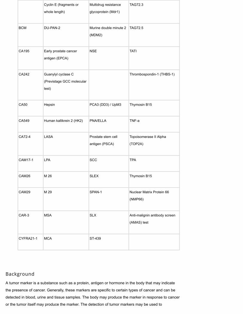
Cyclin E (fragments or
whole length)
Multidrug resistance
glycoprotein (Mdr1)
TAG72.3
BCM DU-PAN-2 Murine double minute 2
(MDM2)
TAG72.5
CA195 Early prostate cancer
antigen (EPCA)
NSE TATI
CA242 Guanylyl cyclase C
(Previstage GCC molecular
test)
Thrombospondin-1 (THBS-1)
CA50 Hepsin PCA3 (DD3) / UpM3 Thymosin B15
CA549 Human kallikrein 2 (HK2) PNA/ELLA TNF-a
CA72-4 LASA Prostate stem cell
antigen (PSCA)
Topoisomerase II Alpha
(TOP2A)
CAM17-1 LPA SCC TPA
CAM26 M 26 SLEX Thymosin B15
CAM29 M 29 SPAN-1 Nuclear Matrix Protein 66
(NMP66)
CAR-3 MSA SLX Anti-malignin antibody screen
(AMAS) test
CYFRA21-1 MCA ST-439
Background
A tumor marker is a substance such as a protein, antigen or hormone in the body that may indicate
the presence of cancer. Generally, these markers are specific to certain types of cancer and can be
detected in blood, urine and tissue samples. The body may produce the marker in response to cancer
or the tumor itself may produce the marker. The detection of tumor markers may be used to

determine a diagnosis or as an indicator of disease (cancer) progression. It can also be used to
document clinical response to treatment. Tumor markers include, but may not be limited to, alpha-
fetoprotein (AFP), CA 15-3/CA 27.29, CA 19-9, CA-125, carcinoembryonic antigen (CEA) and
prostate-specific antigen (PSA).
Tumor markers are normally produced in low quantities by cells in the body. Detection of a higher-
than-normal serum level by radioimmunoassay or immunohistochemical techniques usually indicates
the presence of a certain type of cancer. Currently, the main use of tumor markers is to assess a
cancer's response to treatment and to check for recurrence. In some types of cancer, tumor marker
levels may reflect the extent or stage of the disease and can be useful in predicting how well the
disease will respond to treatment. A decrease or return to normal in the level of a tumor marker may
indicate that the cancer has responded favorably to therapy. If the tumor marker level rises, it may
indicate that the cancer is spreading. Finally, measurements of tumor marker levels may be used
after treatment has ended as a part of follow-up care to check for recurrence.
However, in many cases the literature states that measurements of tumor marker levels alone are
insufficient to diagnose cancer for the following reasons: (1) tumor marker levels can be elevated in
people with benign conditions; (2) tumor marker levels are not elevated in every person with
cancer, especially in the early stages of the disease; and (3) many tumor markers are not specific
to a particular type of cancer; and (4) the level of a tumor marker can be elevated by more than
one type of cancer.
Examples of Tumor Markers Include
5-Hydroxyindoleacetic acid (5-HIAA) -- the main metabolite of serotonin, used as a marker in
the evaluation of carcinoid tumors;
Beta-2-Microglobulin (B2M) – A protein found on the surface of many cells. High levels of
B2M are an indicator of certain kinds of cancer, including chronic lymphocytic leukemia, non-
Hodgkin's lymphoma and multiple myeloma or kidney disease;
Beta Human Chorionic Gonadotropin (beta HCG) – A type of tumor marker that may be
found in higher than normal amounts in individuals with some types of cancer;
Calcitonin – Hormone secreted by the thyroid that lowers blood calcium;
Calretinin – A calcium-binding protein that is used as a marker in the evaluation of lung
cancer and other diseases.
Chromogranin A – A protein found inside neuroendocrine cells, which releases chromogranin
A and other hormones into the blood. Chromogranin A may be found in higher than normal
amounts in individuals with certain neuroendocrine tumors, small cell lung cancer, prostate
cancer and other conditions
Guanylyl cyclase c (GCC) – An enzyme that may be expressed only in the cells that line the
intestine from the duodenum to the rectum.
Inhibin – One of two hormones (designated inhibin-A and inhibin-B) secreted by the gonads
(by Sertoli cells in the male and the granulosa cells in the female) and inhibits the production
of follicle-stimulating hormone (FSH) by the pituitary gland;
Lactate Dehydrogenase (LDH) – Marker used to monitor treatment of testicular cancer;

Mucin-1 (MUC-1) – Carbohydrate antigen elevated in individuals with tumors of the breast,
ovary, lung and prostate as well as other disorders;
Napsin A – Protein used as a marker in the evaluation of lung cancer;
Prealbumin – Marker of nutritional status and a sensitive indicator of protein synthesis. Also
referred to as transthyretin;
Prostate Specific Antigen (PSA) – Substance produced by the prostate gland. Levels of PSA in
the blood often increase in men with prostate cancer.
Thyroglobulin – Protein found in the thyroid gland. Some thyroglobulin can be found in the
blood and this amount may be measured after thyroid surgery to determine whether thyroid
cancer has recurred;
Thyroid Transcription Factor-1 (TTF-1) – A protein that is used as a tumor marker in the
evaluation of lung cancer;
Transferrin – A protein in blood plasma that carries iron derived from food intake to the liver,
spleen and bone marrow.
Tumors may be evaluated with histology, which involves examination of the structure, especially the
microscopic structure, of organic tissues. Methods of detecting tumor markers include, but are not
limited to: Fluorescence in Situ Hybridization (FISH) – Laboratory technique used to detect small
deletions or rearrangements in chromosomes. Immunohistochemical (IHC) Analysis – Laboratory
process of detecting an organism in tissues with antibodies.
Gene mutation testing can purportedly be used to find somatic mutations in cancerous cells that are
not inherited. Some examples of genes that may have somatic mutations include: IDH1 and IDH2
genes (associated with acute myeloid leukemia [AML], gliomas and chondrosarcomas); NPM1 and
FLT3 genes (associated with AML).
Individualized molecular tumor profiling is a laboratory method of testing a panel of tumor markers,
which may include genetic as well as biochemical markers, to establish a personalized molecular
profile of a tumor to recommend treatment options.
Mass spectrometry based proteomic profiling (eg, Veristrat, Xpresys Lung) is a multivariate serum
protein test that uses mass spectrometry and proprietary algorithms to analyze proteins in an
individual’s serum. The Xpresys is no longer on the market.
Next-generation sequence (NGS) tests use select genes to purportedly identify molecular growth
drivers for improved risk stratification and targeted therapies. Examples include: FoundationOne and
OncoVantage for solid tumor cancers; FoundationOne Heme for hematological cancers and
sarcomas; and ThyGenX for indeterminate thyroid nodules.
Liquid biopsy refers to serum testing for DNA fragments that are shed by cancer cells and released
into the bloodstream. This method is purportedly used for screening, diagnosis and/or monitoring of
cancer cells that may otherwise require a tissue sample.

Multianalyte assays with algorithmic analyses (MAAAs) are laboratory measurements that use a
mathematical formula to analyze multiple markers that may be associated with a particular disease
state and are designed to evaluate disease activity or an individual’s risk for disease. The laboratory
performs an algorithmic analysis using the results of the assays and sometimes other information,
such as sex and age and converts the information into a numeric score, which is conveyed on a
laboratory report. Generally, MAAAs are exclusive to a single laboratory which owns the algorithm.
MAAAs have been proposed for the evaluation of pelvic masses, including assisting in the
determination of referral for surgery to a gynecologic oncologist or to a general surgeon.
Topographic genotyping (eg, PathFinderTG) is a test that examines a panel of 15 to 20 genetic
markers in tissue biopsy or other tissue specimens to purportedly aid in the determination of
indeterminate or equivocal cancer diagnoses.
PSA
Prostate Specific Antigen (PSA) is a substance produced by the prostate gland. Levels of PSA in the
blood often increase in men with prostate cancer. Elevated levels of Prostate-Specific Antigen (PSA)
may also be found in the blood of men with benign prostate conditions, such as prostatitis and benign
prostatic hyperplasia (BPH). While PSA does not allow distinction between benign prostate conditions
and cancer, an elevated PSA level may indicate that other tests are necessary to determine whether
cancer is present. PSA levels have been shown to be useful in monitoring the effectiveness of
prostate cancer treatment, and in checking for recurrence after treatment has ended. Use of PSA for
screening remains very controversial. Although researchers are in the process of studying the value
of PSA along with digital rectal exams for routine screening of men ages 55 to 74 for prostate cancer;
and the literature does not show at this time whether using PSA to screen for prostate cancer actually
does reduce the number of deaths caused by this cancer. The American Cancer Society
recommends clinicians and patients consider screening with PSA and digital rectal exam for African
American men and men with familial tendency age 40 or older and all men age 50 or older.
Cancer Care Ontario guidelines on active surveillance of prostate cancer (Morash, et al., 2015) state
that the active surveillance protocol should include the following tests: PSA test every 3 to 6 months;
digital rectal examination every year, and a 12- to 14-core confirmatory transrectal ultrasound (TRUS)
biopsy (including anterior directed cores) within 6 to 12 months, then serial biopsy a minimum of
every 3 to 5 years thereafter. The guidelines state that "[c]urrent evidence shows that PSA kinetics
does not reliably predict disease stability or reclassification to higher risk state. There was conflicting
evidence whether PSA is a good predictor of disease progression or reclassification. Differences
were also found in the ability of different measures of PSA, such as PSA velocity, PSA density, and
PSA doubling time for predicting progression or reclassification. PSA monitoring is considered a
necessary component of an AS protocol, but a rising PSA may be best viewed as a trigger for
reappraisal (e.g., MRI, repeat biopsy) rather than a trigger for intervention."
PCA3

Prostate cancer antigen 3 (PCA3, also known as DD3) is a gene that has been found to be highly
overexpressed in prostate cancer. This gene has been investigated as a potential diagnostic marker
for prostate cancer. However, there are no published clinical outcome studies of the effectiveness of
the PCA3 gene in screening, diagnosis or management of prostate cancer.
Prostate cancer antigen 3 (PCA3) (Progensa, Gene-Probe, Inc.) encodes a prostate-specific mRNA.
It is one of the most prostate cancer-specific genes identified, with over-expression in about 95% of
cancers tested. The PCA3 urine assay is an amplified nucleic acid assay, which uses transcription-
mediated amplification (TMA) to quantify PCA3 and PSA mRNA in prostate cells found in urine
samples. The PCA3 score is calculated as the ratio between PCA3 and PSA mRNA. The main target
population of this non-invasive test is men with raised PSA but a negative prostate biopsy. Other
target groups include men with a slightly raised PSA, as well as men with signs and symptoms
suggestive of prostate cancer.
van Gils and colleagues (2007) stated that PCA3 is a promising prostate cancer marker. These
investigators performed a multi-center study to validate the diagnostic performance of the PCA3 urine
test established in an earlier single-institution study. The first voided urine after digital rectal
examination (DRE) was collected from a total of 583 men with serum PSA levels between 3 and 15
ng/ml who were to undergo prostate biopsies. These researchers determined the PCA3 score in
these samples and correlated the results with the results of the prostate biopsies. A total of 534 men
(92 %) had an informative sample. The area under the receiver-operating characteristic curve, a
measure of the diagnostic accuracy of a test, was 0.66 for the PCA3 urine test and 0.57 for serum
PSA. The sensitivity for the PCA3 urine test was 65 %, the specificity was 66 % (versus 47 % for
serum PSA), and the negative predictive value was 80 %. The authors concluded that the findings of
this multi-center study validated the diagnostic performance of the PCA3 urine test in the largest
group studied thus far using a PCA3 gene-based test.
Marks and associates (2007) examined the potential utility of the investigational PCA3 urine assay to
predict the repeat biopsy outcome. Urine was collected after DRE (3 strokes per lobe) from 233 men
with serum PSA levels persistently 2.5 ng/ml or greater and at least one previous negative biopsy.
The PCA3 scores were determined using a highly sensitive quantitative assay with TMA. The ability
of the PCA3 score to predict the biopsy outcome was assessed and compared with the serum PSA
levels. The RNA yield was adequate for analysis in the urine samples from 226 of 233 men (i.e., the
informative specimen rate was 97 %). Repeat biopsy revealed prostate cancer in 60 (27 %) of the
226 remaining subjects. Receiver operating characteristic curve analysis yielded an area under the
curve of 0.68 for the PCA3 score. In contrast, the area under the curve for serum PSA was 0.52.
Using a PCA3 score cutoff of 35, the assay sensitivity was 58 % and specificity 72 %, with an odds
ratio of 3.6. At PCA3 scores of less than 5, only 12 % of men had prostate cancer on repeat biopsy;
at PCA3 scores of greater than 100, the risk of positive biopsy was 50 %. The authors concluded that
in men undergoing repeat prostate biopsy to rule out cancer, the urinary PCA3 score was superior to
serum PSA determination for predicting the biopsy outcome. The high specificity and informative rate
suggest that the PCA3 assay could have an important role in prostate cancer diagnosis.

Groskopf et al (2007) reported that the PCA3 score is independent of prostate volume and was highly
correlated with the risk of positive biopsy. The PCA3 test was performed on 529 men scheduled for
prostate biopsy. Overall, the PCA3 score had a sensitivity of 54% and a specificity of 74%. A PCA3
score of less than 5 was associated with a 14% risk of positive biopsy, while a PCA3 score of greater
than 100 was associated with a 69% risk of positive biopsy.
Haese et al (2007) presented preliminary results from a European multicenter study of PCA3.
Enrolled patients had a PSA level of less than or equal to 2.5 ng/mL, had 1 or 2 previous negative
biopsies, and were scheduled for repeat biopsy. The specificity of the PCA3 score (cutoff 35) was
found to be 78%, and the sensitivity was 67%. Patients with a PCA3 score of greater than or equal to
35 had a 33% probability of a positive repeat biopsy, compared to a 6% probability for those with a
PCA3 score of less than 35.
In a review on biomarkers for prostate cancer detection, Parekh, et al. (2007) stated that prostate
stem cell antigen, alpha-methyl coenzyme-A racemase, PCA3, early prostate cancer antigen, hepsin
and human kallikrein 2 are promising markers that are currently undergoing validation.
An assessment by the BlueCross BlueShield Association Technology Evaluation Center (BCBSA,
2008) found that, in general, PCA3 assay results to date are preliminary; interpretation of results has
not been standardized and clinical utility studies of decision-making for initial biopsy, repeat biopsy or
treatment have not been reported.
Tosoian et al (2010) evaluated the relationship between PCA3 and prostate biopsy results in men in a
surveillance program. Urine specimens were obtained from 294 men with prostate cancer enrolled in
the Johns Hopkins surveillance program. The follow-up protocol included semi-annual free and
total PSA measurements, digital rectal examination and annual surveillance prostate biopsy. Cox
proportional hazards regression was used to evaluate the association between PCA3 results and
progression on surveillance biopsy (defined as Gleason pattern 4 or 5, more than 2 positive biopsy
cores or more than 50% involvement of any core with cancer). Patients with progression on biopsy
(12.9%) had a mean PCA3 score similar to that of those without progression (60.0 versus 50.8, p =
0.131). Receiver operating characteristics analysis suggested that PCA3 alone could not be used to
identify men with progression on biopsy (area under the curve = 0.589, 95% CI 0.496 to 0.683, p =
0.076). After adjustment for age and date of diagnosis PCA3 was not significantly associated with
progression on biopsy (p = 0.15). The authors concluded that in men with low risk prostate cancer
who were carefully selected for surveillance the PCA3 score was not significantly associated with
short-term biopsy progression. They stated that further analysis is necessary to assess the
usefulness of PCA3 in combination with other biomarkers or in selected subsets of patients
undergoing surveillance.
While there are studies examining the positive and negative predictive values of the PCA3 urine
assay, there is currently a lack of evidence of the effect of this test on management of individuals with
or suspected of prostate cancer. The PCA3 urine assay shows promise as a prostate cancer
diagnostic tool, however, more research is needed to ascertain the clinical value of this assay for
screening and diagnostic purposes.

An assessment of PCA3 prepared for the Agency for Healthcare Research and Quality (2013)
concluded: "For diagnostic accuracy, there was a low strength of evidence that PCA3 had better
diagnostic accuracy for positive biopsy results than tPSA elevations, but insufficient evidence that this
led to improved intermediate or long-term health outcomes. For all other settings, comparators, and
outcomes, there was insufficient evidence."
The Evaluation of Genomic Applications in Practice and Prevention (EGAPP) Working Group
(2013) found insufficient evidence to recommend prostate cancer antigen 3 (PCA3) testing to inform
decisions for when to re-biopsy previously biopsy-negative patients for prostate cancer or to inform
decisions to conduct initial biopsies for prostate cancer in at-risk men (e.g., previous elevated
prostate-specific antigen test or suspicious digital rectal examination). The EGAPP Working Group
found insufficient evidence to recommend PCA3 testing in men with cancer-positive biopsies to
determine if the disease is indolent or aggressive in order to develop an optimal treatment plan. The
EGAPP Working Group concluded that, based on the available evidence, the overall certainty of
clinical validity to predict the diagnosis of prostate cancer using PCA3 is deemed “low.” The EGAPP
Working Group discouraged clinical use for diagnosis unless further evidence supports improved
clinical validity. The EGAPP Working Group also found that, based on the available evidence, the
overall certainty of net health benefit is deemed “low.” The EGAPP Working Group discourages
clinical use unless further evidence supports improved clinical outcomes.
Guidelines from the European Association of Urology (2015) state that "[b]iological markers, include
urine markers such as PCA3, the TMPRSS2: ERG fusion gene or PSA isoforms such as the Phi
index, appear promising as does genomics on the tissue sample itself. However, further study data
will be needed before such markers can be used in standard clinical practice."
A Cancer Care Ontario Guideline on prostate cancer surveillance (Morash, et al., 2015), which has
been endorsed by the American Society for Clinical Oncology (2016), did not include PCA3 level in
their recommendation because evidence of PCA3 to predict disease reclassification in prostate
cancer was lacking.
National Institute for Health and Care Excellence (NICE)’s clinical practice guideline on “Diagnosing
prostate cancer: PROGENSA PCA3 assay and Prostate Health Index” (2015) stated that “ The
PROGENSA PCA3 assay and the Prostate Health Index are not recommended for use in people
having investigations for suspected prostate cancer, who have had a negative or inconclusive
transrectal ultrasound prostate biopsy”. The assessment cited studies finding that adding the PCA3
score to clinical assessment and MRI had very little effect on the size of the reported area under the
curve, with minimal change in derived sensitivity and specificity for clinical assessment with MRI
compared with clinical assessment using MRI and the PCA3 assay.
In a Lancet review of prostate cancer, Attard, et al. (2016) stated that "[s]everal studies have so far
proven inconclusive as to whether PCA3 is useful to selectively detect aggressive prostate cancers."
B15

Hutchinson et al (2005) stated that in tissue-based assays, thymosin beta15 (B15) has been shown to
correlate with prostate cancer and with recurrence of malignancy. To be clinically effective, it must be
shown that thymosin B15 is released by the tumor into body fluids in detectable concentrations.
These researchers developed a quantitative assay that can measure clinically relevant levels of
thymosin B15 in human urine. Sixteen antibodies were raised against recombinant thymosin B15
and/or peptide conjugates. One antibody, having stable characteristics over the wide range of pH
and salt concentrations found in urine and minimal cross-reactivity with other beta thymosins, was
used to develop a competitive enzyme-linked immunosorbent assay (ELISA). Urinary thymosin B15
concentration was determined for control groups; normal (n = 52), prostate intraepithelial neoplasia
(PIN, n = 36), and patients with prostate cancer; untreated (n = 7) with subsequent biochemical
failure, radiation therapy (n = 17) at risk of biochemical recurrence. The operating range of the
competition ELISA fell between 2.5 and 625 ng/ml. Recoveries exceeded 75%, and the intra- and
inter-assay coefficients of variability were 3.3% and 12.9%, respectively. No cross-reactivity with
other urine proteins was observed. A stable thymosin B15 signal was recovered from urine
specimens stored at -20 degrees C for up to 1 year. At a threshold of 40 (ng/dl)/microg protein/mg
creatinine), the assay had a sensitivity of 58% and a specificity of 94%. Relative to the control
groups, thymosin B15 levels were greater than this threshold in a significant fraction of patients with
prostate cancer (p < 0.001), including 5 of the 7 patients who later experienced PSA recurrence. The
authors concluded that an ELISA that is able to detect thymosin B15 at clinically relevant
concentrations in urine from patients with prostate cancer has been established. They noted that the
assay will provide a tool for future clinical studies to validate urinary thymosin B15 as a predictive
marker for recurrent prostate cancer.
CEA
Carcinoembryonic antigen (CEA) is a normal cell product that is over-expressed by
adenocarcinomas, primarily of the colon, rectum, breast, and lung. It is normally found in small
amounts in the blood of most healthy people, but may become elevated in people who have cancer or
some benign conditions.
CEA is an oncofetal glycoprotein present in the gastrointestinal tract and body fluids of the embryo
and fetus (Chin, et al., 2006). It is also present in certain adult gastrointestinal cells, including the
mucosal cells of the colorectum, and small amounts are present in blood. Blood levels are often
elevated in patients with disseminated cancers and in some patients with nonmalignant disease.
According to the available literature, the primary use of CEA is in monitoring colorectal cancer,
especially when the disease has metastasized. CEA is also used after treatment to check for
recurrence of colorectal cancer. However, the literature indicates a wide variety of other cancers can
produce elevated levels of this tumor marker, including melanoma; lymphoma; and cancers of the
breast, lung, pancreas, stomach, cervix, bladder, kidney, thyroid, liver, and ovary. Elevated CEA
levels can also occur in patients with non-cancerous conditions, including inflammatory bowel
disease, pancreatitis, and liver disease.

The American Society of Clinical Oncology (ASCO)'s update of recommendations for the use of
tumor markers in gastrointestinal cancer (Gershon, et al., 2006) stated that post-operative CEA levels
should be performed every 3 months for stage II and III disease for at least 3 years if the patient is a
potential candidate for surgery or chemotherapy of metastatic disease.
CA-125
Cancer antigen 125 (CA-125) is a test that evaluates ovarian cancer treatment. CA-125 is a protein
that is found more in ovarian cancer cells than in other cells. CA-125 is expressed by >80 percent of
non-mucinous ovarian epithelial neoplasms (Chin et al, 2006). Approximately half of women with
metastatic ovarian cancer have an elevated CA-125 level.
The Gynecologic Cancer Foundation, the Society of Gynecologic Oncologists, and the American
Cancer Society have issued a consensus statement to promote early detection of ovarian cancer,
which recommends that women who have symptoms, including bloating, pelvic or abdominal pain,
difficulty eating or feeling full quickly, and urinary frequency and urgency, are urged to see a
gynecologist if symptoms are new and persist for more than three weeks (ACS, 2007; SGO, 2007).
Ovarian cancer is among the deadliest types of cancer because diagnosis usually comes very late,
after the cancer has spread. If the cancer is found and surgically removed before it spreads outside
the ovary, the five year survival rate is 93%. However, only 19% of cases are detected early enough
for that kind of successful intervention. It is estimated that 22,430 new cases and 15,280 deaths will
be reported in 2007 (ACS, 2007). The consensus statement recommendations are based on studies
that show the above symptoms appeared in women with ovarian cancer more than in other women
(Goff, et al., 2004; Daly & Ozols, 2004). The recommendations acknowledge that there is not
consensus on what physicians should do when patients present with these symptoms. According to a
consensus statement issued by the Gynecologic Cancer Foundation, pelvic and rectal examination in
women with the symptoms is one first step. If there is a suspicion of cancer, the next step may be a
transvaginal ultrasound to check the ovaries for abnormal growths, enlargement, or telltale pockets of
fluid that can indicate cancer. Testing for CA-125 levels should also be considered.
There is no evidence available that measurement of CA-125 can be effectively used for widespread
screening to reduce mortality from ovarian cancer, nor that the use of this test would result in
decreased rather than increased morbidity and mortality. According to the available literature, not all
women with elevated CA 125 levels have ovarian cancer. CA 125 levels may also be elevated by
cancers of the uterus, cervix, pancreas, liver, colon, breast, lung, and digestive tract. Non-cancerous
conditions that can cause elevated CA 125 levels include endometriosis, pelvic inflammatory disease,
peritonitis, pancreatitis, liver disease, and any condition that inflames the pleura. Menstruation and
pregnancy can also cause an increase in CA 125. However, according to the available literature,
changes in CA 125 levels can be effectively used in the management of treatment for ovarian cancer.
In women with ovarian cancer being treated with chemotherapy, the literature suggests a falling CA
125 level generally indicates that the cancer is responding to treatment and increased survival is
expected. Increasing CA 125 levels during or after treatment, on the other hand, may suggest that the
cancer is not responding to therapy or that residual cancer remains. According to the available
literature, failure of the CA 125 level to return to normal after three cycles of chemotherapy indicates

residual tumor, early treatment failure and decreased survival. Under accepted guidelines, CA 125
levels can also be used to monitor patients for recurrence of ovarian cancer. Although an elevated
CA 125 level is highly correlated with the presence of ovarian cancer, the literature suggests a normal
value does not exclude residual or recurrent disease.
Aetna's preventive services guidelines are based on the recommendations of leading primary care
medical professional organizations and federal public health agencies. None of these organizations
recommend routine screening of average-risk, asymptomatic women with serum CA-125 levels for
ovarian cancer. These organizations have concluded that serum CA-125 levels are not sufficiently
sensitive or specific for use as a screening test for ovarian cancer, and that the harms of such
screening outweigh the benefits.
The American College of Obstetricians and Gynecologists (2002) has stated that "[u]nfortunately,
there is no screening test for ovarian cancer that has proved effective in screening low-risk
asymptomatic women. Measurement of CA 125 levels and completion of pelvic ultrasonography (both
abdominal and transvaginal) have been the two tests most thoroughly evaluated.... Data suggest that
currently available tests do not appear to be beneficial for screening low-risk, asymptomatic women
because their sensitivity, specificity, positive predictive value, and negative predictive value have all
been modest at best. Because of the low incidence of disease, reported to be approximately one
case per 2,500 women per year, it has been estimated that a test with even 100% sensitivity and 99%
specificity would have a positive predictive value of only 4.8%, meaning 20 of 21 women undergoing
surgery would not have primary ovarian cancer. Unfortunately, no test available approaches this level
of sensitivity or specificity."
The National Cancer Institute (2004) has stated: "There is insufficient evidence to establish that
screening for ovarian cancer with serum markers such as CA 125 levels, transvaginal ultrasound, or
pelvic examinations would result in a decrease in mortality from ovarian cancer. A serious potential
harm is the false-positive test result, which may lead to anxiety and invasive diagnostic procedures.
There is good evidence that screening for ovarian cancer with the tests above would result in more
diagnostic laparoscopies and laparotomies than new ovarian cancers found. Unnecessary
oophorectomies may also result."
The U.S. Preventive Services Task Force (2004) recommends against routine screening with serum
CA-125 level for ovarian cancer. The Task Force concluded that the potential harms of such
screening outweigh the potential benefits.
HE4
Human Epididymis Protein 4 (HE4) is a secreted glycoprotein that is being studied as a potential
marker for ovarian cancer.
A variety of other tumor markers have been investigated for early detection of ovarian cancer as well
as different combinations of tumor markers complementary to CA 125 that could potentially offer
greater sensitivity and specificity than CA 125 alone. Preliminary studies on HE4 (human epididymis

protein 4), a marker for ovarian cancer, reported similar sensitivity to CA 125 when comparing ovarian
cancer cases to healthy controls, and a higher sensitivity when comparing ovarian cancer cases to
benign gynecologic disease (Hellstrom, et al., 2003 & 2008; Moore, et al., 2008;) However, an
assessment on genomic tests for ovarian cancer prepared by Duke University for the Agency for
Healthcare Research and Quality (AHRQ, 2006) stated, "Although research remains promising,
adaptation of genomic tests into clinical practice must await appropriately designed and powered
studies in relevant clinical settings." Further studies are needed to determine if HE4 significantly
adds to the sensitivity of CA 125 while maintaining a high specificity.
National Comprehensive Cancer Network (NCCN) guidelines (2016) state that data show that HE4
and several other markers do not increase early enough to be useful in detecting early-stage ovarian
cancer.
CA 15-3
Cancer antigen 15-3 (CA 15-3) is a serum cancer antigen that has been used in the management of
patients with breast cancer. According to the available literature, its low detection rate in early stage
disease indicates that CA 15-3 cannot be used to screen or diagnose patients with breast cancer. It
has been widely used to monitor the effectiveness of treatment for metastatic cancer. Elevated serum
CA 15-3 concentrations are found in 5 percent of stage I, 29 percent of stage II, 32 percent of stage
III and 95 percent of stage IV carcinoma of the breast (Chin, et al, 2006). Most (96 percent) patients
with a CA 15-3 increase of greater than 25 percent have disease progression. Most (nearly 100
percent) patients with a CA 15-3 decrease of greater than 50 percent are responding to treatment.
Cancers of the ovary, lung, and prostate may also raise CA 15-3 levels. The literature indicates
elevated levels of CA 15-3 may be associated with non-cancerous conditions, such as benign breast
or ovarian disease, endometriosis, pelvic inflammatory disease, and hepatitis.
Similar to the CA 15-3 antigen, CA 27-29 is found in the blood of most breast cancer patients. The
literature indicates CA 27-29 levels may be used in conjunction with other procedures (such as
mammograms and measurements of other tumor marker levels) to check for recurrence in women
previously treated for stage II and stage III breast cancer. CA 27-29 levels can also be elevated by
cancers of the colon, stomach, kidney, lung, ovary, pancreas, uterus, and liver. First trimester
pregnancy, endometriosis, ovarian cysts, benign breast disease, kidney disease, and liver disease
are non-cancerous conditions that can also elevate CA 27-29 levels.
Elevated CA 27.29 levels are primarily associated with metastatic breast cancer, where it can be used
to monitor the course of disease, response to treatment, and detect disease recurrence (Chin, et al.,
2006). Elevated serum CA 27.29 concentrations are found in 95 percent of stage IV breast cancer. In
addition, CA 27.29 has been found to be elevated in lung (43 percent), pancreas (47 percent),
ovarian (56 percent), and liver (55 percent) cancer.
CA 19-9

Cancer antigen 19-9 (CA 19-9) is a mucin-glycoprotein first identified from a human colorectal
carcinoma cell line and is present in epithelial tissue of the stomach, gall bladder, pancreas and
prostate (Chin, et al., 2006). Concentrations are increased in patients with pancreatic, gastric, and
colon cancer as well as in some nonmalignant conditions. Increasing levels generally indicate disease
progression, whereas decreasing levels suggest therapeutic response.
Initially found in colorectal cancer patients, CA 19-9 has also been identified in patients with
pancreatic, stomach, hepatocellular cancer, and bile duct cancer. In those who have pancreatic
cancer, the literature indicates higher levels of CA 19-9 tend to be associated with more advanced
disease. Although the sensitivity of the CA 19-9 level in patients with pancreatic cancer is relatively
high, the specificity is lowered by elevations that occur in patients with benign pancreatic or liver
disease. Non-cancerous conditions that may elevate CA 19-9 levels include gallstones, pancreatitis,
cirrhosis of the liver, and cholecystitis. Although excellent correlations have been reported between
CA 19-9 measurements and relapse in patients with pancreatic cancer who are followed after surgical
resection, no patient has been salvaged by the earlier diagnosis of relapse, a fact that reflects the
lack of effective therapy.
Guidelines from the National Comprehensive Cancer Network (NCCN, 2010) state that measurement
of CA 19-9 should be considered in evaluating patients with intrahepatic or extrahepatic
cholangiocarcinoma and gallbladder cancer. The guidelines note that CA 19-9 is often elevated in
persons with cholangiocarcinoma or gallbladder cancer, although this marker is not specific for these
cancers. Nehls, et al. (2004) considered CA19-9 as one of the several new potential tumor markers
for the diagnosis of cholangiocarcinoma. Levy, et al. (2005) aimed to characterize the test properties
of CA 19-9 and of a change in CA 19-9 over time in predicting cholangiocarcinoma in patients with
primary sclerosing cholangitis. Charts of 208 patients were reviewed. Fourteen patients had
cholangiocarcinoma. Median CA 19-9 was higher with cholangiocarcinoma (15 versus 290 U/ml, p <
0.0001). A cutoff of 129 U/ml provided: sensitivity 78.6%, specificity 98.5%, adjusted positive
predictive value 56.6% and negative predictive value 99.4%. The median change over time was 664
U/ml in cholangiocarcinoma compared to 6.7 U/ml in primary sclerosing cholangitis alone (p <
0.0001). A cutoff of 63.2 U/ml for change in CA 19-9 provided: sensitivity 90%, specificity 98% and
positive predictive value 42%.
CA 19-9 is produced by adenocarcinomas of the pancreas, stomach, gall-bladder, colon, ovary, and
lung, and it is shed into the circulation. Although numerous studies have addressed the potential
utility of CA 19-9 in adenocarcinoma of the colon and rectum, the sensitivity of CA 19-9 was always
less than that of the CEA test for all stages of disease. Its use for screening asymptomatic
populations has been hampered by a false-positive rate of 15% to 30% in patients with non-
neoplastic diseases of the pancreas, liver, and biliary tract. Only a few studies have addressed the
use of CA 19-9 in monitoring patients' post-primary therapy. Significant postsurgical decreases are
observed for CA 19-9, but these decreases have not been correlated with survival or disease-free
interval. In monitoring response to treatment, decreases in CEA have been found to more accurately
reflect response to therapy than did decreases of CA 19-9. Progressive increases of the marker may
signal disease progression in 25% of the patients who express the CA 19-9 marker, but this
monitoring provides only a minimal lead time of 1 to 3 months. Monitoring with CA 19-9 has not been
shown to improve the management of patients with colorectal cancer. The serum CA 19-9 level does
not add significant information to that provided by CEA, which is currently regarded as the marker of
choice for this neoplasm.
Sinakos and colleagues (2011) evaluated the long-term outcomes in Mayo Clinic patients presenting
with primary sclerosing cholangitis (PSC) between 2000 and 2010 (n= 73) for incidence of
cholangiocarcinoma (CCA). The results showed initial levels of CA 19-9 in patients without CCA
were significantly lower than those from patients with CCA (p < 0.0001). No factors known to affect
CA 19-9 levels were identified in 33% of the patients without CCA; endoscopic treatment and
recurrent bacterial cholangitis were associated with levels of CA 19-9 in 26% and 22% of these
patients, respectively.
Juntermanns (2011) prospectively analyzed a bile duct tumor database and retrieved records of 238
patients who underwent surgery between 1999 and 2008. Their findings included that pre-operative
CA19-9 serum levels did not show a statically reliable differentiation between benign or malignant
dignity. The authors concluded that current diagnostics cannot differentiate malignant from benign
tumor masses in the hepatic hilum with required reliability. The authors further concluded that
administration of CIK cells, thymus factor, IL-2 and IFN-alpha after AHSCT could improve the
immunologic function of patients, and TH1/TH2 ratio may virtually reflect the immune status of
patients, but that more information is required to make prognostic assessments of immune
reconstruction and the long-term survival rate.
Sarbia et al (1993) investigated 69 adenocarcinomas of the esophagogastric junction and found high
rates of antigen expression were found for the “intestinal” markers CA 19-9 (between 55.5% and
100%) and BW 494 (between 42.9 and 86.7%). The authors concluded that these data, in
combination with CK-20 expression, PGII, and 2B5 indicate that the distribution of adenocarcinomas
with gastric and.or intestinal differentiation at the esophagogastric junction forms a continuum with out
clear-cut borders. This study has not been replicated and NCCN guidelines for Esophageal and
Esophagogastric Junction Cancers does not include recommendations for CA 19-9 testing for these
indications (NCCN, 2011).
The American Society of Clinical Oncology (ASCO)'s update of recommendations for the use of
tumor markers in gastrointestinal cancer (Gershon, et al., 2006) stated that for pancreatic cancer, CA
19-9 can be measured every 1 to 3 months for patients with locally advanced or metastatic disease
receiving active therapy.
Mucinous carcinoma of the appendix is a rare entity most commonly associated with primary tumors
of the appendix and colon, and for which spread is generally confined to the abdominal cavity
(Andreopoulou et al, 2007). Imaging assessment of these mucinous lesions is difficult, and recent
studies have explored the use of tumor markers as clinical tools in evaluation of mucinous carcinoma
of the appendix.
Carmignani et al (2004) evaluated patients with synchronous systemic and intraperitoneal
dissemination of appendix cancer treated with cytoreductive surgery and perioperative regional
chemotherapy with a mean follow up time of 42.6 months. Results of this study indicated that
patients with elevated CEA and CA 19-9 levels had a shorter median survival time (p=0.0083 and p =
0.0193, respectively). In a subsequent study, Carmingnani et al (2004) prospectively recorded tumor
markers CEA and CA19-9 within 1 week prior to definitive treatment. The investigators found CEA
elevated in 56% of 532 patients and CA19-9 elevated in 67.1% of those patients. They reported that
“although the absolute level of tumor marker did not correlate with prognosis, a normal value
indicated an improved survival.” Their findings included an elevated CEA in 35.2% of 110 patients
determined to have recurrent disease and an elevated CA 19-9 in 62.9%, while 68.2% of patients had
at least one of the tumor markers elevated.
Current guidelines indicate that for liver transplantation for primary sclerosing cholangitis, stringent
efforts should be made to detect superadded cholangiocarcinoma, including measurement of CA 19-9
(Devlin & O'Grady, 1999).
Carmignani et al (2004a) conducted a study to report the role of combined treatments, including
cytoreductive surgery and perioperative regional chemotherapy, in patients with synchronous
systemic and intraperitoneal dissemination of appendix cancer. Study subjects were treated with
cytoreductive surgery and perioperative regional chemotherapy and statistical analysis of variables
utilized survival as an end point and included demographic characteristics, prior surgical score (PSS),
tumor marker levels, peritoneal cancer index (PCI), and completeness of cytoreduction (CC). With a
mean follow-up of 42.6 months, median survival time (MST) for 15 patients was 28 months and 5-
year survival rate was 29.4 %. Female patients had a longer MST than male patients (p = 0.0199)
and survival was better in patients with PSS 0 and 1 (p = 0.0277). Patients with elevated CEA and CA
19-9 levels had a shorter MST (p = 0.0083 and p = 0.0193, respectively) while PCI and CC
comparisons did not show significant differences. The morbidity rate (n = 2) was 13.3 % and the
mortality (n = 2) rate was also 13.3 %. The authors concluded that “acceptable morbidity and
mortality and a 29.4 % 5-year survival rate allows cytoreductive surgery and regional chemotherapy
to be considered as a treatment option for selected patients with synchronous systemic and
intraperitoneal dissemination of appendix cancer.”
Carmignani et al (2004b) in a further publication regarding gastrointestinal cancer, stated that
carcinoembryonic antigen (CEA) and carbohydrate antigen 19-9 (CA 19-9) tumor markers have found
selected clinical application. The authors remarked that the use of these tumor markers in mucinous
epithelial tumors of the appendix has not been previously determined. Thus, the authors conducted a
study in which, in patients with peritoneal dissemination of a mucinous epithelial malignancy of the
appendix, tumor markers CEA and CA 19-9 were prospectively recorded preoperatively within 1 week
prior to definitive treatment and if the appendiceal tumor recurred, the tumor marker was determined.
The primary endpoint was the accuracy of these two tumor markers in the management of this
disease for these two specific clinical situations. CEA was elevated in 56 % of 532 patients and CA
19-9 was elevated in 67.1 % of these patients. Although the absolute level of tumor marker did not
correlate with prognosis, a normal value indicated an improved survival. CEA was elevated in 35.2 %
of 110 patients determined to have recurrent disease and CA 19-9 was elevated in 62.9 %. At least

one of the tumor markers was elevated in 68.2 % of patients. An elevated CEA tumor marker at the
time of recurrence indicated a reduced prognosis and both CEA and CA 19-9 tumor markers were
elevated in a majority of these patients. This should be a valuable diagnostic tool previously
underutilized in this group of patients. These tumor markers were also of benefit in the assessment of
prognosis in that a normal level indicated an improved prognosis. At the time of a reoperative
procedure, CEA and CA 19-9 tumor markers gave information regarding the progression of disease
and have practical value in the management of epithelial appendiceal malignancy with peritoneal
dissemination.
Andreopoulou et al (2007) stated that mucinous carcinoma of the appendix is a rare entity with a
distinct natural history that poses diagnostic and therapeutic challenges and that mucinous peritoneal
carcinomatosis is most commonly associated with primary tumors of the appendix and colon. The
authors stated that usually the spread remains confined to the abdominal cavity and that imaging
assessment of these mucinous lesions is difficult, while tumor markers (CEA and CA19.9) may be
surrogates for extent of disease.
Recruitment for large scale studies given the rare nature of mucinous appendiceal carcinoma would
be challenging. However, available evidence does illustrate a benefit to use of CA 19-9 in patients
with mucinous appendiceal carcinoma.
Cathepsins
This enzyme plays a critical role in protein catabolism and tissue remodeling (Chin, et al., 2006).
Over-expression is associated with non-ductal carcinoma and metastasis at the time of breast cancer
diagnosis. High levels may have clinical significance in predicting decreased metastasis-free survival
and decreased overall survival in women with node-negative breast cancer.
Svatek et al (2008) examined the role of urinary cathepsin B and L in the detection of bladder
urothelial cell carcinoma. These investigators concluded that urinary cathepsin L is an independent
predictor of bladder cancer presence and invasiveness in patients with a history of urothelial
carcinoma of the bladder. They stated that further evaluation of this marker is necessary before its
use as an adjunct to cystoscopy for urothelial carcinoma of the bladder.
CD 20
CD 20 is used to determine eligibility for rituximab (Rituxan; anti-CD20) treatment in patients with B-
cell non-Hodgkin's lymphomas (NHL) (Chin, et al., 2006). Rituximab is a genetically engineered,
chimeric murine/human monoclonal antibody directed against the CD20 antigen found on the surface
of normal and malignant B-cell lymphocytes. Since non-Hodgkin's Lymphoma (NHL) subtypes may
differ in their response to rituximab, determination of drug sensitivity is important for choosing therapy.
CD 25

CD 25 is used to determine eligibility for denileukin diftitox treatment in patients with persistent or
recurrent CTCL (Chin, et al., 2006). Denileukin diftitox (Ontak) is a cutaneous T-cell lymphoma
(CTCL) therapy that targets the high-affinity interleukin-2 (IL-2) receptor. The IL-2 receptor may exist
in a low-affinity form (CD25), an intermediate-affinity form (CD122/CD132), and a high-affinity form
(CD25/CD122/CD132). Patients whose malignant cells express the CD25 component of the IL-2
receptor may respond to Ontak therapy.
CD 33
CD 33 is used to determine eligibility for gemtuzumab (Mylotarg, anti-CD33) treatment in patients with
acute myeloid leukemia (Chen, et al., 2006). Gemtuzumab consists of a recombinant, humanized IgG
kappa antibody conjugated to a cytotoxic anti-tumor antibiotic, calicheamicin, which binds specifically
to the CD33 antigen. This antigen is found on the surface of leukemic blasts and immature normal
cells of myelomonocytic lineage, but not in normal hematopoietic stem cells.
CD 52
CD 52 is used to determine eligibility for alemtuzumab (Campath, anti-CD52) treatment in patients
with chronic lymphocytic leukemia (Chen, et al., 2006). CD52 is an antigen that can be expressed at
high density on the surface of malignant CLL cells. Alemtuzumab is a humanized antibody targeted
against CD52 and its binding is necessary for cell death and therapeutic response.
CD 117, c-kit
CD 117 is used to determine eligibility for treatment with imatinib mesylate in patients with c-kit-
positive gastrointestinal stromal tumors (GISTs) (Chen, et al., 2006). The glycoprotein c-kit (CD117) is
a member of the receptor tyrosine kinase subclass III family and has been implicated in a number of
malignancies. Imatinib mesylate, a tyrosine kinase inhibitor, is effective in treating GISTs and other
tumors that express c-kit.
HCG
Human chorionic gonadotropin (HCG) is normally produced in increasing quantities by the placenta
during pregnancy. Accepted guidelines provide that HCG levels can be used to screen for
choriocarcinoma in women who are at high risk for the disease, and to monitor the treatment of
trophoblastic disease. The literature states that elevated HCG levels may also indicate the presence
of cancers of the testis, ovary, liver, stomach, pancreas, and lung.
Accepted guidelines provide that alpha fetoprotein (AFP) and b-HCG measurements are valuable for
determining prognosis and monitoring therapy in patients with non-seminomatous germ cell cancer.
Because of the low incidence of elevated AFP and b-HCG levels in early-stage cancer, the literature
suggests these markers have no value in screening for testicular cancer. However, the specificity of
these markers is such that when determined simultaneously, at least one marker will be positive in
85% of patients with active cancer. The value of AFP and b-HCG as markers is enhanced by a low
frequency of false-positive results and by the chemoresponsiveness of testicular cancer. The
literature states that only rarely do patients with other types of cancer have elevated levels of AFP.
Non-cancerous conditions that can cause elevated AFP levels include benign liver conditions, such
as cirrhosis or hepatitis, ataxia telangiectasia, Wiscott-Aldrich syndrome, and pregnancy.
AFP
Alpha-fetoprotein (AFP) is a protein that is normally elevated in pregnant women since it is produced
by the fetus; however, AFP is not usually found in the blood of adults. In men and in women who are
not pregnant, an elevated level of AFP may indicate liver, ovarian or testicular cancer.
Alpha-fetoprotein is normally produced by a developing fetus. Alpha fetoprotein levels begin to
decrease soon after birth and are usually undetectable in the blood of healthy adults, except during
pregnancy. According to accepted guidelines, an elevated level of AFP strongly suggests the
presence of either primary liver cancer or germ cell cancer of the ovary or testicle. As AFP is an
established test for the diagnosis and monitoring of hepatoma, it is used as a screening tool to rule
out the presence of a liver neoplasm before considering liver transplantation. This is especially
pertinent in cases (e.g., cirrhosis) where there is an increased risk of developing a primary liver tumor.
Elevated serum AFP levels are most closely associated with nonseminomatous testicular cancer and
hepatocellular cancer (Chin, 2006). The rate of clearance from serum after treatment is an indicator of
the effectiveness of therapy. Conversely, the growth rate of progressive disease can be monitored by
serially measuring serum AFP concentrations over time.
ER, PR
Estrogen receptor (ER) and progesterone receptor (PR) predicts response to hormone therapy for
women with advanced breast cancer and those receiving adjuvant treatment, and prognosticates the
aggressiveness of a tumor (Chin, 2006).
The estrogen receptor and progesterone receptor are intracellular receptors that are measured
directly in tumor tissue. These receptors are polypeptides that bind their respective hormones,
translocate to the nucleus, and induce specific gene expression. Breast cancers are dependent upon
estrogen and/or progesterone for growth and this effect is mediated through ERs and progesterone
receptors (ER/PR) (Chin, et al., 2006). Both receptors may be over-expressed in malignant breast
tissue. Most oncologists have used the estrogen receptor and also the progesterone receptor not only
to predict the probability of response to hormonal therapy at the time of metastatic disease, but also
to predict the likelihood of recurrent disease, and to predict the need for adjuvant hormonal therapy or
chemotherapy. Although these latter uses for estrogen and progesterone receptors are commonly
accepted by most oncologists, the data on which these conclusions are based are controversial.
NSE

Neuron-specific enolase (NSE) has been detected in patients with neuroblastoma, small cell lung
cancer, Wilms' tumor, melanoma, and cancers of the thyroid, kidney, testicle, and pancreas. However,
studies of NSE as a tumor marker have concentrated primarily on patients with neuroblastoma and
small cell lung cancer. According to the available literature, measurement of NSE level in patients
with these diseases cannot be correlated to the extent of the disease, the patient's prognosis, or the
patient's response to treatment because of the poor sensitivity of this marker.
LASA
LASA is a complex marker that measures the amount of sialic acid in serum and can be elevated in
serum from patients with any number of different neoplasms. Elevations in blood LASA levels have
been reported in patients with mammary (63 percent), gastroenteric (65 percent), pulmonary (79
percent), and ovarian (94 percent) neoplasms as well as those with leukemia (86 percent), lymphoma
(87 percent), melanoma (84 percent), sarcoma (97 percent), and Hodgkin disease (91 percent). As a
result, this assay may not have high specificity or sensitivity necessary for cancer detection (Chen, et
al., 2006). This serum cancer marker has not been widely accepted for use in the detection or
prognosis of colorectal carcinoma. There is no practical information concerning outcome and the use
of LASA in the medical literature. Although several articles describe the use of LASA in the diagnosis
of colorectal cancer and its association with tumor-node-metastasis (TNM) stage, it has been shown
that patients with colorectal polyps and colorectal carcinoma both have elevated LASA levels, and
that the levels returned to baseline after removal of either polyps or carcinomas.
p53
p53 is a tumor suppressor gene on the short arm of chromosome 17 that encodes a protein that is
important in the regulation of cell division. Although the full role of p53 in the normal and neoplastic
cell is unknown, there is evidence that the gene product is important in preventing the division of cells
containing damaged DNA. p53 gene deletion or mutation is a frequent event along with other
molecular abnormalities in colorectal carcinogenesis. The literature on p53 abnormality and prognosis
in colorectal cancer suffers from a paucity of reported data and the use of a variety of techniques in
assay and statistical analysis in the small numbers of cases analyzed. For these reasons, the
literature generally does not recommend p53 analysis as a routine approach to assisting in the
management of patients with colorectal cancer.
Guidelines from the American Society for Clinical Oncology (2016) recommend against the use of
p53 to guide adjuvant chemotherapy in breast cancer. This is a moderate-strength recommendation
based upon intermediate-quality evidence.
Zap-70
Zeta-chain-associated protein kinase 70, which is used as a prognostic marker in (CLL).

Zap-70 is indicated to assess prognosis and need for aggressive therapy in patients with chronic
lymphocytic leukemia (CLL) (Chin, et al., 2006). ZAP-70 is a 70-kD member of the Syk family of
protein tyrosine kinases. It is expressed primarily in T-cells and natural killer (NK) cells and is critical
for signal transduction following T-cell receptor engagement. In CLL B-cells, elevated ZAP-70
expression appears to predict the need for therapy as effectively as IgVH mutation status. Although
ZAP-70 expression is strongly correlated with IgVH mutation status, the combination of the two
markers may provide greater prognostic value than either marker alone. Positive ZAP-70 results
predict an aggressive disease course.
uPA
The serine protease urokinase-type plasminogen activator (uPA) and its primary inhibitor,
plasminogen activator inhibitor-1 (PAI-1), have shown promise for risk assessment and prediction of
therapeutic response in primary breast cancer (Chin, et al., 2006). High levels of uPA or PAI-1 in
primary tumor tissue are associated with an aggressive disease course and poor prognosis in both
node-positive and node-negative breast cancer.
A report by the Belgian Healthcare Knowledge Centre (KCE) (San Miguel, et al., 2015) found no
studies reporting on the impact of uPA/PAI-1 on clinical management (clinical utility).
Guidelines from the American Society for Clinical Oncology (2016) state: "If a patient has ER/PgR-
positive, HER2-negative (node-negative) breast cancer, the clinician may use urokinase plasminogen
activator and plasminogen activator inhibitor type 1 to guide decisions on adjuvant systemic therapy."
This is a weak recommendation based upon high-quality evidence. The ASCO guidelines
recommend the use of urokinase plasminogen activator and plasminogen activator inhibitor type 1 to
guide decisions on adjuvant systemic therapy in patients with HER2-positive breast cancer or TN
breast cancer.
IgVh Mutation Status
Chronic lymphocytic leukemia (CLL) patients can be divided into two basic groups on the basis of the
mutational status of the immunoglobulin heavy-chain variable-region (IgVH) gene in leukemic cells
(Chin, 2006). Patients with IgVH mutations have longer survival than those without IgVH mutation.
Thus, mutation analysis may be useful for planning management strategies.
Kappa / Lambda Light Chain
Elevated serum levels of monoclonal free light chains are associated with malignant plasma cell
proliferation (e.g., multiple myeloma), primary amyloidosis, and light chain deposition disease (Chen
et al, 2006). The appearance of higher levels of free light chains in the urine may be indicative of
kidney disease or malignant lymphoproliferative disease such as multiple myeloma. These tests have
been used for the detection of multiple myeloma.
KRAS

The ras proto-oncogenes are normal cellular components, which are thought to be important for
transduction of signals required for proliferation and differentiation. The ras oncogene family has 3
members: H-ras, K-ras, and N-ras. Ras gene mutations can be found in a variety of tumor types,
although the incidence varies greatly. The highest incidences are found in adenocarcinomas of the
pancreas (90 %), colon (50 %), and lung (30 %); thyroid tumors (50 %), and myeloid leukemia (30 %).
Investigators have established an association between some genotypes of K-ras (KRAS) oncogenes
and response to treatment with cetuximab or panitumumab (Lievre et al, 2006 and 2008; Di Fiore et
al, 2007; Gonçalves et al, 2008; De Roock et al, 2008). Patients whose tumors express specific
forms of the KRAS gene exhibit considerably decreased responses to cetuximab and panitumumab.
It has been theorized that cetuximab and panitumumab do not target epidermal growth factor receptor
(EGFR) associated with these specific KRAS mutations and thus are unable to block their activation.
It has been suggested that KRAS genotype be considered as a selection factor for cancer patients
who are candidates for treatment with cetuximab or panitumumab.
Karapetis and colleagues (2008) stated that treatment with cetuximab improves overall survival (OS)
and progression-free survival (PFS) and preserves the quality of life in patients with colorectal cancer
that has not responded to chemotherapy. The mutation status of the K-ras gene in the tumor may
affect the response to cetuximab and have treatment-independent prognostic value. These
investigators analyzed tumor samples, obtained from 394 of 572 patients (68.9 %) with colorectal
cancer who were randomly assigned to receive cetuximab plus best supportive care or best
supportive care alone, to look for activating mutations in exon 2 of the K-ras gene. They evaluated if
the mutation status of the K-ras gene was associated with survival in the cetuximab and supportive-
care groups. Of the tumors evaluated for K-ras mutations, 42.3 % had at least one mutation in exon
2 of the gene. The effectiveness of cetuximab was significantly associated with K-ras mutation status
(p = 0.01 and p < 0.001 for the interaction of K-ras mutation status with OS and PFS, respectively).
In patients with wild-type K-ras tumors, treatment with cetuximab as compared with supportive care
alone significantly improved OS (median of 9.5 versus 4.8 months; hazard ratio for death, 0.55; 95 %
confidence interval [CI], 0.41 to 0.74; p < 0.001) and PFS (median of 3.7 months versus 1.9 months;
hazard ratio for progression or death, 0.40; 95 % CI, 0.30 to 0.54; p < 0.001). Among patients with
mutated K-ras tumors, there was no significant difference between those who were treated with
cetuximab and those who received supportive care alone with respect to OS (hazard ratio, 0.98; p =
0.89) or PFS (hazard ratio, 0.99; p = 0.96). In the group of patients receiving best supportive care
alone, the mutation status of the K-ras gene was not significantly associated with OS (hazard ratio for
death, 1.01; p = 0.97). The authors concluded that patients with a colorectal tumor bearing mutated
K-ras did not benefit from cetuximab, whereas patients with a tumor bearing wild-type K-ras did
benefit from cetuximab. The mutation status of the K-ras gene had no influence on survival among
patients treated with best supportive care alone.
The ASCO's provisional clinical opinion on testing for KRAS gene mutations in patients with
metastatic colorectal carcinoma to predict response to anti-EGFR monoclonal antibody therapy
(Allegra et al, 2009) stated that based on systematic reviews of the relevant literature, all patients with
metastatic colorectal carcinoma who are candidates for anti-EGFR antibody therapy should have their

tumor tested for KRAS mutations in a CLIA-accredited laboratory. If KRAS mutation in codon 12 or
13 is detected, then patients with metastatic colorectal carcinoma should not receive anti-EGFR
antibody therapy as part of their treatment.
The KRAS oncogene mutation tests are intended to aid in the formulation of treatment decisions for
patients who may be candidates for treatment of metastatic epithelial cancers with anti-EGFR
therapies such as cetuximab or panitumumab. Several tests for KRAS mutation are currently
available in the United States; however, at this time, no KRAS genotype test kits have been approved
by the FDA.
At the 2008 Annual Meeting of the American Society of Clinical Oncology (ASCO), data on 540
patients with metastatic colorectal cancer in the randomized, phase III CRYSTAL trial were
presented. Among 192 patients with KRAS mutations, there was no improvement in overall
responses or PFS from the addition of cetuximab to standard chemotherapy. In the patients with
normal KRAS, the 1-year PFS rate was 43 % for patients receiving cetuximab versus 25 % for those
receiving only standard chemotherapy, and the overall response rate was 59 % versus 43 %,
respectively (van Cutsem, 2008). Also at the 2008 ASCO meeting, data from 233 metastatic
colorectal cancer patients were presented that confirmed the correlation of KRAS status with patient
response to anti-EGFR therapy. No benefit was found after addition of cetuximab to standard
chemotherapy with FOLFOX (the combination of fluorouracil, leucovorin, and oxaliplatin) in patients
with a mutated KRAS; however, addition of cetuximab to FOLFOX increased both response rate and
PFS in patients with a wild-type (i.e., un-mutated) KRAS gene (Bokemeyer, 2008). Response to
panitumumab was correlated to KRAS status in a published phase III trial. A total of 427 patients with
metastatic colorectal cancer received either panitumumab or best supportive care. Panitumumab
exhibited a 17% response rate among patients with normal KRAS, but 0% response among patients
with KRAS mutations (Amado, 2008).
A meta-analysis of results from 8 studies involving 817 patients with colorectal cancer found that the
presence of KRAS mutation predicted lack of response to treatment with anti-EGFR monoclonal
antibodies (e.g., panitumumab or cetuximab), whether as stand-alone therapy or in combination with
chemotherapy (Linardou et al, 2008). This analysis also provided empirical evidence that k-RAS
mutations are highly specific negative predictors of response (de-novo resistance) to single-agent
EGFR tyrosine-kinase inhibitors in advanced non-small cell lung cancer; and similarly to anti-EGFR
monoclonal antibodies alone or in combination with chemotherapy in patients with metastatic
colorectal cancer.
The Blue Cross and Blue Shield Association (BCBSA, 2008) Technology Evaluation Center Medical
Advisory Panel concluded that use of KRAS mutation analysis meets TEC criteria to predict non-
response to anti-EGFR monoclonal antibodies cetuximab and panitumumab to treat metastatic
colorectal cancer. The TEC assessment found that the evidence is sufficient to conclude that patients
with mutated KRAS tumors in the setting of metastatic colorectal cancer do not respond to anti-EGFR
monoclonal antibody therapy. The assessment explained that the data show that the clinical benefit of
using EGFR inhibitors in treating metastatic colorectal cancer, either as monotherapy or in
combination with other treatment regimens, is not seen in patients with KRAS-mutated tumors. The

assessment found: "This data supports knowing a patient's tumor mutation status before
consideration of use of an EGFR inhibitor in the treatment regimen. Identifying patients whose
tumors express mutated KRAS will avoid exposing patients to ineffective drugs, avoid exposure to
unnecessary drug toxicities, and expedite the use of the best available alternative therapy."
Colorectal cancer guidelines from the National Comprehensive Cancer Network (NCCN, 2010)
recommend consideration of reflex BRAF testing in patients with wild type KRAS. The NCCN
guidelines explain that several small studies suggest that patients with wild-type KRAS and a BRAF
mutation are unlikely to respond to anti-EGFR therapies such as cetuximab and panitumumab. The
guidelines explain that patients with a known BRAF mutation are unlikely to respond to anti-EGFR
antibodies, although the data are somewhat inconsistent. Studies demonstrate that in patients with
metastatic colorectal cancer, about 8 percent have mutations in the BRAF gene. Testing for the BRAF
V600E mutation is performed by PCR amplification and direct DNA sequence analysis.
Ratner et al (2010) stated that ovarian cancer (OC) is the single most deadly form of women's cancer,
typically presenting as an advanced disease at diagnosis in part due to a lack of known risk factors or
genetic markers of risk. The KRAS oncogene and altered levels of the microRNA (miRNA) let-7 are
associated with an increased risk of developing solid tumors. In this study, these researchers
investigated a hypothesized association between an increased risk of OC and a variant allele of
KRAS at rs61764370, referred to as the KRAS-variant, which disrupts a let-7 miRNA binding site in
this oncogene. Specimens obtained were tested for the presence of the KRAS-variant from non-
selected OC patients in 3 independent cohorts, 2 independent ovarian case-control studies, and OC
patients with hereditary breast and ovarian cancer syndrome (HBOC) as well as their family
members. The results indicated that the KRAS-variant is associated with more than 25 % of non-
selected OC cases. Furthermore, these researchers found that it is a marker for a significant
increased risk of developing OC, as confirmed by 2 independent case-control analyses. Lastly, they
determined that the KRAS-variant was present in 61 % of HBOC patients without BRCA1 or BRCA2
mutations, previously considered uninformative, as well as in their family members with cancer.
These findings supported the hypothesis that the KRAS-variant is a genetic marker for increased risk
of developing OC, and they suggested that the KRAS-variant may be a new genetic marker of cancer
risk for HBOC families without other known genetic abnormalities.
Hollestelle et al (2011) noted that recently, a variant allele in the 3'UTR of the KRAS gene
(rs61764370 T>G) was shown to be associated with an increased risk for developing non-small cell
lung cancer, as well as OC, and was most enriched in OC patients from HBOC families. This
functional variant has been shown to disrupt a let-7 miRNA binding site leading to increased
expression of KRAS in vitro. In the current study, these investigators genotyped this KRAS-variant in
breast cancer index cases from 268 BRCA1 families, 89 BRCA2 families, 685 non-BRCA1/BRCA2
families, and 797 geographically matched controls. The allele frequency of the KRAS-variant was
found to be increased among patients with breast cancer from BRCA1, but not BRCA2 or non-
BRCA1/BRCA2 families as compared to controls. As BRCA1 carriers mostly develop ER-negative
breast cancers, these researchers also examined the variant allele frequency among indexes from
non-BRCA1/BRCA2 families with ER-negative breast cancer. The prevalence of the KRAS-variant
was, however, not significantly increased as compared to controls, suggesting that the variant allele

not just simply associates with ER-negative breast cancer. Subsequent expansion of the number of
BRCA1 carriers with breast cancer by including other family members in addition to the index cases
resulted in loss of significance for the association between the variant allele and mutant BRCA1
breast cancer. In this same cohort, the KRAS-variant did not appear to modify breast cancer risk for
BRCA1 carriers. More importantly, results from the current study suggested that KRAS-variant
frequencies might be increased among BRCA1 carriers, but solid proof requires confirmation in a
larger cohort of BRCA1 carriers.
Therascreen KRAS RGQ PCR Kit (Qiagen) is intended to detect 7 mutations in codons 12 and 13 of
the KRAS gene (Raman, et al., 2013). The kit utilizes two technologies — ARMS and Scorpions —
for detection of mutations in real-time PCR. The therascreen KRAS RGQ PCR kit is being developed
as a companion diagnostic to aid clinicians, through detection of KRAS mutations, in the identification
of patients with metastatic colorectal cancer (mCRC) who are more likely to benefit from cetuximab.
PreOvar™ tests (Mira Dx) for the KRAS-variant, and will help identify ovarian cancer patients whose
female relatives should also be evaluated for the KRAS-variant (Raman, et al., 2013). PreOvar™ may
also help assess the relative risk of developing ovarian cancer for women who have a family history
of ovarian cancer without a living proband (ancestor with the disease). The KRAS-Variant is present
in 6-10% of the general population and 25% of non-selected women with epithelial ovarian cancer.
Additionally, the KRAS-variant was identified in over 60% of Hereditary Breast and Ovarian Cancer
(HBOC) patients that were previously classified as “uninformative,” or negative for other known
genetic markers of ovarian cancer risk. The test determines if KRAS-variant may put someone at
increased risk for developing ovarian cancer.
The Evaluation of Genomic Applications in Practice and Prevention (EGAPP) Working Group (EWG)
(2013) found that, for patients with metastatic colorectal cancer (mCRC) who are being considered for
treatment with cetuximab or panitumumab, there is convincing evidence to recommend clinical use of
KRAS mutation analysis to determine which patients are KRAS mutation positive and therefore
unlikely to benefit from these agents before initiation of therapy. The level of certainty of the evidence
was deemed high, and the magnitude of net health benefit from avoiding potentially ineffective and
harmful treatment, along with promoting more immediate access to what could be the next most
effective treatment, is at least moderate.
The EWG found insufficient evidence to recommend for or against BRAF V600E testing for the same
clinical scenario (EGAPP, 2013). The level of certainty for BRAF V600E testing to guide antiepidermal
growth factor receptor (EGFR) therapy was deemed low. The EWG encourages further studies of the
potential value of testing in patients with mCRC who were found to have tumors that are wild type
(mutation negative) for KRAS to predict responsiveness to therapy.
Bladder Cancer: BTA-stat, NMP22, Urovysion, ImmunoCyt
In the United States, bladder malignancy is the 4th commonest cancer in men and the 8th
commonest in women. Patients usually present with urinary tract symptoms (e.g., gross or
microscopic hematuria or irritative voiding symptoms such as frequency, dysuria, and urgency).

Evaluations of these patients usually entail voided-urine cytology, cystoscopy, and upper urinary tract
imaging such as intravenous pyelography, renal sonography, or retrograde pyelography. Most newly
diagnosed bladder cancers are superficial (i.e., not invading beyond the lamina propria on histological
examination), and are known as transitional cell carcinoma (TCC). These superficial bladder cancers
are usually managed by transurethral resection. However, the literature shows that approximately
50 to 75 % of treated TCC recur. Furthermore, 10 to 15 % of TCC progress to muscle-invasive
bladder cancer. According to the literature, the prevalence of recurrence after initial treatment as well
as the natural history of TCC necessitates long-term follow-up. Following treatment, accepted
guidelines provide that patients who have previously been diagnosed with TCC should usually
undergo urine cytology/cystoscopy every 3 months in the 1st year, every 6 months in the 2nd year,
and once-yearly afterwards.
Currently, urine cytology with confirmatory cystoscopy represents the cornerstone for the identification
of bladder tumors. However, the subjectivity and low sensitivity of cytology led to the development of
several urine-based tests as adjuncts to cytology/cystoscopy for the diagnosis and follow-up of
patients with TCC. These tests include the BTA Stat test (Bard Diagnostic, Redmond, WA), the
NMP22 test (Matritech, Newton, MA), the Aura-Tek FDP test (PerImmune, Rockville, MD), and the
Vysis UroVysion FISH Test (Vysis, Inc., Downers Grove, IL). They are usually objective, qualitative
(BTA Stat and Aura-Tek FDP), or quantitative (NMP22, UroVysion), and have higher sensitivity than
cytology, but some have lower specificity. So far, no single bladder tumor marker has emerged as the
generally accepted test of choice, and none has been established as a screening tool for bladder
malignancy.
Urine-based markers, such as proteins with increased cancer cell expression or chromosomal
abnormalities in the urine, may be detected using a variety of laboratory methods to aid in the
management of bladder cancer. The following markers/tests are currently available:
Bladder tumor antigen (BTA) (eg, BTA stat and BTA TRAK)
Fluorescence immunocytology (eg, ImmunoCyt/uCyt+)
Fluorescence in situ hybridization (FISH) (eg, UroVysion)
mRNA quantification by RT-qPRC testing (eg, Cxbladder)
Nuclear matrix protein 22 (NMP22) (eg, NMP22 BladderChek and Matritech NMP22 Test)
Urine-based markers have a role in the detection of bladder cancer recurrence in individuals with a
history of bladder cancer and are used adjunctively with urinary cytology and cystoscopy. These tests
have also been proposed for bladder cancer screening, diagnosis of bladder cancer in individuals
symptomatic of bladder cancer and for the evaluation of hematuria.
The UroVysion Bladder Cancer Kit (UroVysion Kit) (Baycare Laboratories) is designed to detect
aneuploidy for chromosomes 3, 7, 17, and loss of the 9p21 locus via fluorescence in situ hybridization
(FISH) in urine specimens from persons with hematuria suspected of having bladder cancer (Raman,
et al., 2013). FISH analysis is used in conjunction with cystoscopy to monitor for recurrence among
those with previously diagnosed bladder cancer. FISH analysis is a surveillance tool in established
primary and secondary bladder adenocarcinoma.

The ImmunoCyt is an immunocytochemistry assay for the detection of tumor cells shed in the urine of
patients previously diagnosed with bladder cancer (Chen, et al., 2006). This test is intended to
augment the sensitivity of cytology for the detection of tumor cells in the urine of individuals previously
diagnosed with bladder cancer. The test has been used for detection of tumor cells in the urine of
individuals previously diagnosed with bladder cancer, and for use in conjunction with cytoscopy as an
aid in the management of bladder cancer.
Although urine cytology has been shown to be less accurate than urinary biomarker tests, familiarity
with the method as well as ease of performance justify the continued routine use of the former by
primary care physicians, especially in patients who have no history of bladder malignancy. The urine-
based biomarker tests have been shown to be accurate in detecting low-grade bladder tumors. In
particular, these tests may be of help in deciding the need for further diagnostic assessment of
patients with a history of bladder cancer and negative results on urine cytology. For example,
elevated levels of urinary bladder tumor markers in patients with a history of TCC may warrant earlier,
rather than delayed, cystoscopic examination. On the other hand, consideration may be given to
lengthening the intervals between cystoscopic investigations when values of these tumor markers are
normal.
An assessment by the Adelaide Health Technology Assessment (Mundy & Hiller, 2009) concluded
that the NMP BladderCheck and UroVysion FISH assay, designed for the detection of bladder cancer
in high risk patients, have poor sensitivity and poor positive predictive values. The
assessment recommended that these assays not be used in asymptomatic patients. The
assessment suggested, however, that these tests may be useful in the monitoring of patients with
transitional cell carcinoma between cytoscopies. The AHTA recommended that this technology not be
assessed further.
An assessment prepared for the Agency for Healthcare Research and Quality (Meleth, et al., 2014)
found: "Although UroVysion is marketed as a diagnostic rather than a prognostic test, limited
evidence from two small studies (total N=168) rated as low or medium risk of bias supported
associations between test result and prognosis for risk of recurrence. We found no studies that
directly assessed the impact of a test of interest on both physician decision-making and downstream
health outcomes to establish clinical utility. We attempted to construct an indirect chain of evidence to
answer the overarching question, but we were unable to do so. Even in the cases where the tests
seemed to add value in determining prognosis (i.e., evidence of clinical validity), we found no
evidence that using the test was related to improved outcomes for patients."
The American Urologic Association’s guideline on “Diagnosis, evaluation and follow-up of
asymptomatic microhematuria (AMH) in adults” (Davis et al, 2012) stated that “The use of urine
cytology and urine markers (Nuclear Matrix Protein 22 [NMP22], bladder tumor antigen [BTA]-stat,
and UroVysion fluorescence in situ hybridization assay [FISH]) is not recommended as a part of the
routine evaluation of the asymptomatic microhematuria patient”.

Chou et al (2015) systematically reviewed the evidence on the accuracy of urinary biomarkers for
diagnosis of bladder cancer in adults who have signs or symptoms of the disease or are undergoing
surveillance for recurrent disease. Data sources included Ovid MEDLINE (January 1990 through
June 2015), Cochrane Central Register of Controlled Trials, Cochrane Database of Systematic
Reviews, and reference lists. A total of 57 studies that evaluated the diagnostic accuracy of
quantitative or qualitative nuclear matrix protein 22 (NMP22), qualitative or quantitative bladder tumor
antigen (BTA), FISH, fluorescent immunohistochemistry (ImmunoCyt [Scimedx]), and Cxbladder
(Pacific Edge Diagnostics USA) using cystoscopy and histopathology as the reference standard met
inclusion criteria; case-control studies were excluded. Dual extraction and quality assessment of
individual studies were carried out; overall strength of evidence (SOE) was also assessed. Across
biomarkers, sensitivities ranged from 0.57 to 0.82 and specificities ranged from 0.74 to 0.88. Positive
likelihood ratios ranged from 2.52 to 5.53, and negative likelihood ratios ranged from 0.21 to 0.48
(moderate SOE for quantitative NMP22, qualitative BTA, FISH, and ImmunoCyt; low SOE for others).
For some biomarkers, sensitivity was higher for initial diagnosis of bladder cancer than for diagnosis
of recurrence. Sensitivity increased with higher tumor stage or grade. Studies that directly compared
the accuracy of quantitative NMP22 and qualitative BTA found no differences in diagnostic accuracy
(moderate SOE); head-to-head studies of other biomarkers were limited. Urinary biomarkers plus
cytologic evaluation were more sensitive than biomarkers alone but missed about 10 % of bladder
cancer cases. The authors concluded that urinary biomarkers miss a substantial proportion of
patients with bladder cancer and are subject to false-positive results in others; accuracy is poor for
low-stage and low-grade tumors. They stated that research is needed to understand how the use of
these biomarkers with other diagnostic tests affect the use of cystoscopy and clinical outcomes.
In an editorial that accompanied the afore-mentioned study, Abbosh and Plimack (2015) stated that
“Until urinary biomarkers become available that are sufficiently accurate to supplant the current
recommended detection algorithms in biomarker-negative patients, they will not be a cost-effective
addition to strategies to detect bladder cancer”.
In summary, urine-based bladder tumor marker tests have been shown to be useful as an adjunct to
urine cytology and cystoscopy in monitoring for recurrences of bladder cancer, but according to the
available literature should not be used as a screening tool for bladder malignancy. The U.S.
Preventive Services Task Force (USPSTF, 2004) has concluded that the potential harms of screening
for bladder cancer using available tests, such as microscopic urinalysis, urine dipstick, urine cytology,
or such new tests as bladder tumor antigen (BTA) or nuclear matrix protein (NMP22) immunoassay,
outweigh any potential benefits.
Cxbladder
O'Sullivan and colleagues (2012) examined if the RNA assay uRNA® and its derivative Cxbladder®
have greater sensitivity for the detection of bladder cancer than cytology, NMP22™ BladderChek™
and NMP22™ ELISA, and whether they are useful in risk stratification. A total of 485 patients
presenting with gross hematuria but without a history of urothelial cancer were recruited prospectively
from 11 urology clinics in Australasia. Voided urine samples were obtained before cystoscopy. The
sensitivity and specificity of the RNA tests were compared to cytology and the NMP22 assays using

cystoscopy as the reference. The ability of Cxbladder to distinguish between low grade, stage Ta
urothelial carcinoma and more advanced urothelial carcinoma was also determined. uRNA detected
41 of 66 urothelial carcinoma cases (62.1 % sensitivity, 95 % confidence interval [CI]: 49.3 to 73.8)
compared with NMP22 ELISA (50.0 %, 95 % CI: 37.4 to 62.6), BladderChek (37.9 %, 95 % CI: 26.2
to 50.7) and cytology (56.1 %, 95 % CI: 43.8 to 68.3). Cxbladder, which was developed on the study
data, detected 82 %, including 97 % of the high grade tumors and 100 % of tumors stage 1 or
greater. The cut-offs for uRNA and Cxbladder were pre-specified to give a specificity of 85 %. The
specificity of cytology was 94.5 % (95 % CI: 91.9 to 96.5), NMP22 ELISA 88.0 %, (95 % CI: 84.6 to
91.0) and BladderChek 96.4 % (95 % CI: 94.2 to 98.0). Cxbladder distinguished between low-grade
Ta tumors and other detected urothelial carcinoma with a sensitivity of 91 % and a specificity of 90
%. The authors concluded that uRNA and Cxbladder showed improved sensitivity for the detection of
urothelial carcinoma compared to the NMP22 assays. Stratification with Cxbladder provides a
potential method to prioritize patients for the management of waiting lists.
An UpToDate review on “Clinical presentation, diagnosis, and staging of bladder cancer” (Lotan and
Choueiri, 2013) does not mention the use of mRNA biomarkers/PCR testing as a management tool
for bladder cancer. Furthermore, NCCN’s clinical practice guideline on “Bladder cancer” (Version
1.2014) does not mention the use of mRNA biomarkers/PCR testing as a management tool for
bladder cancer.
An assessment of urinary biomarkers for diagnosis of bladder cancer prepared for the Agency for
Healthcare Research and Quality (Chou, et al., 2016) identified only one study of Cxbladder meeting
inclusion criteria, graded as moderate quality, with an overall strength of evidence of "low,"
OncotypeDx Breast
Oncotype Dx (Genomic Health, Inc., Redwood City, CA) is a diagnostic laboratory-developed assay
that quantifies the likelihood of breast cancer recurrence in women with newly diagnosed, stage I or
II, node negative, estrogen receptor positive breast cancer, who will be treated with tamoxifen. The
assay analyzes the expression of a panel of 21 genes, and is intended for use in conjunction with
other conventional methods of breast cancer analysis. Together with staging, grading, and other
tumor marker analyses, Oncotype Dx is intended to provide greater insight into the likelihood of
systemic disease recurrence. Clinical studies have evaluated the prognostic significance of the
Oncotype Dx multigene assay in breast cancer (Paik et al, 2004; Esteva et al, 2003).
Oncotype Dx analyses the patterns of 21 genes is being applied as a quantification tool for likelihood
of breast cancer recurrence within 10 years of newly diagnosed, stage I or II, lymph node-negative,
hormone receptor-positive breast cancer in women who will be treated with tamoxifen (Raman, et a.,
2013). Oncotype is being applied as a quantification tool for likelihood of breast cancer recurrence in
10 years in women with newly diagnosed breast cancer. It is also intended to assist in making
decisions regarding adjuvant chemotherapy based on recurrence likelihood.

There currently is a lack of evidence from prospective clinical studies of the impact of this test on the
management of women with breast cancer demonstrating improvements in clinical outcomes (Lopez,
et al., 2010; Romeo, et al., 2010; Tiwana, et al., 2013; IETS, 2013), Bast and Hortobagyi (2004)
commented that “[b]efore use of the recurrence score [from the Oncotype Dx multigene assay] is
applied to general patient care, however, additional studies are needed.” The National Cancer
Institute is sponsoring a prospective, randomized controlled clinical study, the TAILORx study, using
the Oncotype Dx assay to help identify a group of patients with a mid-range risk of recurrence to
determine whether treating patients with hormonal therapy only is equivalent to treating them with
hormonal therapy in combination with adjuvant chemotherapy.
However, there is indirect evidence of the clinical utility of the Oncotype Dx. Paik et al (2006) used
banked tumor samples from previous clinical studies of tamoxifen and adjuvant chemotherapy in
early breast cancer to assess the performance of the Oncotype Dx multigene assay in predicting
response to adjuvant chemotherapy. These investigators examined tumor samples from subjects
enrolled in the National Surgical Adjuvant Breast and Bowel Project (NSABP) B20 trial to determine
whether there is a correlation between the recurrence score (RS) determined by Oncotype Dx in
tumor samples and subsequent response to adjuvant chemotherapy. A total of 651 patients were
assessable (227 randomly assigned to tamoxifen and 424 randomly assigned to tamoxifen plus
chemotherapy). The test for interaction between chemotherapy treatment and RS was statistically
significant (p = 0.038). Patients with high-RS (RS greater than or equal to 31) tumors (ie, high risk of
recurrence) had a large benefit from chemotherapy (relative risk, 0.26; 95% confidence interval 0.13
to 0.53; absolute decrease in 10-year distant recurrence rate: mean, 27.6%; standard error, 8.0%).
Patients with low-RS (less than 18) tumors derived minimal, if any, benefit from chemotherapy
treatment (relative risk, 1.31; 95% confidence interval, 0.46 to 3.78; absolute decrease in distant
recurrence rate at 10 years: mean, -1.1%; standard error, 2.2%). The investigators found that patients
with intermediate-RS tumors did not appear to have a large benefit, but the investigators concluded
that the uncertainty in the estimate cannot exclude a clinically important benefit.
One limitation of the study by Paik et al (2006) is that the NASBP B20 trial was conducted before the
advent of important advances in breast cancer chemotherapy, including the introduction of
trastuzumab (Herceptin), which has been demonstrated to improve overall and disease-free survival
in breast cancer patients with HER2 positive tumors. Current guidelines recommend the use of
trastuzumab adjuvant chemotherapy in women with metastatic HER2 positive breast cancer, and
women with HER2 positive nonmetastatic breast cancers 1 cm or more in diameter. Thus, the
Oncotype Dx score would not influence the decision to use adjuvant trastuzumab in women with
HER2 positive tumors 1 cm or more in diameter.
Commenting on an early report of this study by Paik et al, of the Oncotype Dx presented in abstract
form, the BlueCross BlueShield Association Technology Evaluation Center assessment stated that
“additional studies in different populations are needed to confirm whether risk prediction is sufficiently
accurate for physicians and patients to choose with confidence whether to withhold adjuvant
chemotherapy.”

An international consensus group (Azim, et al., 2013) found the available evidence on the analytical
and clinical validity of Oncotype Dx Breast to be convincing. However, neither the Oncotype Dx or
none of the other genomic tests the evaluated demonstrated robust evidence of clinical utility: they
stated that it was not clear from the current evidence that modifying treatment decisions based on the
results of a given genomic test could result in improving clinical outcome.
The selection criteria for the TailorRx prospective trial of OncotypeDx state that candidates should
have negative axillary nodes as determined by a sentinel lymph node biopsy and/or axillary dissection
as defined by the American Joint Committee on Cancer 6th Edition Staging System (NCI, 2009). The
AJCC 6th Edition criteria redefined isolated tumor cells as node negative (the prior version of the
criteria, AJCC 5th Edition, classified isolated tumor cells as node positive). "Isolated tumor cells
(single cells or cell deposits) will now be defined as tumor cell deposits no larger than 0.2 mm in
diameter that may or may not (but usually do not) show histologic evidence of malignant activity.
Pending further information, isolated tumor cells will be classified as node-negative, because it is
believed that the unknown benefits of providing treatment for these small lesions would not outweigh
the morbidity caused by the treatment itself." (Singletary, et al., 2002). However, the banked tumor
samples used in the study by Paik, et al. (2006) to validate the OncotypeDx were classified based on
AJCC 5th Ed. criteria. In addition, there is new evidence demonstrating that women with isolated
tumor cells are at a significantly increased risk of breast cancer. Investigators from
the Netherlands found an association between isolated tumor cells and micrometastases in regional
lymph nodes and clinical outcome of breast cancer (de Boer, et al., 2009). These investigators
identified all patients in the Netherlands who underwent a sentinel-node biopsy for breast cancer
before 2006 and had breast cancer with favorable primary-tumor characteristics and isolated tumor
cells or micrometastases in the regional lymph nodes. Patients with node-negative disease were
randomly selected from the years 2000 and 2001. The primary end point was disease-free
survival. The investigators identified 856 patients with node-negative disease who had not received
systemic adjuvant therapy (the node-negative, no-adjuvant-therapy cohort), 856 patients with isolated
tumor cells or micrometastases who had not received systemic adjuvant therapy (the node-positive,
no-adjuvant-therapy cohort), and 995 patients with isolated tumor cells or micrometastases who had
received such treatment (the node-positive, adjuvant-therapy cohort). The median follow-up was 5.1
years. The adjusted hazard ratio for disease events among patients with isolated tumor cells who did
not receive systemic therapy, as compared with women with node-negative disease, was 1.50 (95%
confidence interval [CI], 1.15 to 1.94); among patients with micrometastases, the adjusted hazard
ratio was 1.56 (95% CI, 1.15 to 2.13). Among patients with isolated tumor cells or micrometastases,
the adjusted hazard ratio was 0.57 (95% CI, 0.45 to 0.73) in the node-positive, adjuvant-therapy
cohort, as compared with the node-positive, no-adjuvant-therapy cohort. The investigators concluded
that isolated tumor cells or micrometastases in regional lymph nodes were associated with a reduced
5-year rate of disease-free survival among women with favorable early-stage breast cancer who did
not receive adjuvant therapy. In patients with isolated tumor cells or micrometastases who received
adjuvant therapy, disease-free survival was improved.
The Medical Advisory Panel of the BlueCross BlueShield Association Technology Evaluation Center
(BCBCA, 2014) concluded that use of Oncotype DX to determine recurrence risk for deciding whether
to undergo adjuvant chemotherapy in women with unilateral, nonfixed, hormone receptor‒positive,

lymph node‒negative breast cancer who will receive hormonal therapy meets the Blue Cross and
Blue Shield Association Technology Evaluation Center (TEC) criteria. A technology assessment by
the BlueCross BlueShield Association (2014) stated: "Technical performance of the assay is well
documented and is unlikely to be a major source of variability; rather, tissue sampling is likely the
greatest source of variability. Retrospective epidemiologic analyses indicated strong, independent
associations between Oncotype DX recurrence score (RS) result and distant disease recurrence or
death from breast cancer. The evidence identified a subset of conventionally classified, high-risk
patients who are at sufficiently low risk of recurrence by Oncotype DX that they might reasonably
decide that the harms (toxicity) of chemotherapy outweigh the very small absolute benefit. Two
studies of the original validation data, in which conventionally classified patients were reclassified by
Oncotype DX result, indicated that the test provides significant recurrence risk information in addition
to conventional criteria for individual patient risk classification. Additional evidence indicated that
Oncotype DX results are significantly associated with breast cancer death in a community-based
patient population, and that RS high-risk patients benefit from chemotherapy, whereas benefits for
other RS categories were not statistically significant. Thus, the evidence was judged sufficient to
permit conclusions regarding probable health outcomes."
The Oncotype Dx has also been promoted for use in women with node-positive, ER-positive breast
cancer. An assessment by the BlueCross BlueShield Association (2010) concluded that it has not yet
been demonstrated whether use of the Oncotype Dx for selecting adjuvant chemotherapy in patients
with lymph-node-positive breast cancer improves health outcomes. The report explained that
the evidence for not selecting chemotherapy for women with low RS values is based on low event
rates and wide confidence intervals that include the possibility of benefit from chemotherapy.
Because the data allow for a possible benefit of chemotherapy in patients with low RS results, it is
unknown if health outcomes would be improved, the same, or worse, if chemotherapy was withheld in
these women. The report stated that, due to the lack of clear and sufficient information, there is a
need for a second, confirmatory study. The report stated that the Fred Hutchinson Cancer Research
Center will conduct a nationwide, NCI-sponsored, Phase III clinical trial to determine the predictive
ability of the Oncotype Dx to identify which patients with lymph-node-positive breast cancer will
benefit from chemotherapy.
The clinical evidence base for OncotypeDX is considered to be the most robust. There was some
evidence on the impact of the test on decision-making and to support the case that OncotypeDX
predicts chemotherapy benefit; however, few studies were UK based and limitations in relation to
study design were identified. OncotypeDX has a more robust evidence base, but further evidence on
its impact on decision-making in the UK and the predictive ability of the test in an estrogen receptor
positive (ER+), lymph node negative (LN-), human epidermal growth factor receptor 2 negative
(HER2-) population receiving current drug regimens is needed.
Guidelines from the National Comprehensive Cancer Network (NCCN, 2015) state that "the 21-gene
RT-PCR assay recurrence score can be considered in select patients with 1-3 involved ipsilateral
axillary lymph nodes to guide the addition of combination chemotherapy to standard hormone
therapy. A retrospective analysis of a prospective randomized trial suggests that the test is predictive
in this group similar to its performance in node-negative disease.” The NCCN guidelines (2015)

explained: “Unplanned, retrospective subset analysis from a single randomized clinical trial in post-
menopausal, ALN-positive, ER-positive breast cancer found that the 21-gene RT-PCR assay may
provide predictive information for chemotherapy in addition to tamoxifen [citing Albain, et al., 2010].
Patients with a high score in the study benefited from chemotherapy, whereas patients with a low
score did not appear to benefit from the addition of chemotherapy regardless of the number of
positive lymph nodes. Patient selection for assay use remains controversial.” “The RxPONDER trial
will confirm the SWOG-8814 trial data for women with ER-positive, node-positive disease treated with
endocrine therapy with or without chemotherapy based on risk scores.”
Guidance from the National Institute for Health and Care Excellence (2013) stated: "Oncotype DX is
recommended as an option for guiding adjuvant chemotherapy decisions for people with estrogen
ER+, LN- and HER2- early breast cancer if: The person is assessed as being at intermediate risk;
and information on the biological features of the cancer provided by Oncotype DX is likely to help in
predicting the course of the disease and would therefore help when making the decision about
prescribing chemotherapy; and the manufacturer provides Oncotype DX to National Health Service
(NHS) organisations according to the confidential arrangement agreed with the National Institute for
Health and Care Excellence (NICE). NICE encourages further data collection on the use of Oncotype
DX in the NHS."
An assessment by the Belgian Healthcare Knowledge Center (KCE) (San Miguel, et al., 2015)
concluded that "the evidence for Oncotype DX is more robust than the evidence for other tests."
The KCE Report noted, however, that important evidence gaps are still present. The KCE review
mostly identified studies supporting the prognostic ability (clinical validity) of the test. The KCE judged
these studies to be of moderate to high quality. The KCE found no prospective studies reporting on
the impact of Oncotype DX on long-term outcomes such as overall survival, while four studies
indicated that Oncotype DX leads to changes in decision making. The KCE identified two studies on
the predictive benefit of the test, one for lymph node patients. The KCE reported also noted that the
first evidence relating to improvements in quality of life and reductions in patient anxiety as a result of
using the test has been reported, but this is based on small patient numbers and further evidence is
required.
Guidelines from the American Society for Clinical Oncology (2016) state: "If a patient has ER/PgR-
positive, HER2-negative (node-negative) breast cancer, the clinician may use the 21-gene recurrence
score (RS; Oncotype DX; Genomic Health, Redwood City, CA) to guide decisions on adjuvant
systemic chemotherapy." This is a strong recommendation based upon high quality evidence. The
ASCO guidelines recommend against OncotypeDx Breast to guide decisions on adjuvant systemic
chemotherapy for patients with ER/PgR-positive, HER2-negative (node-positive) breast cancer. The
guidelines also recommend against the use of OncotypeDx Breast in women with HER2-positive
breast cancer or TN breast cancer. The guidelines recommended against the use of OncotypeDx
Breast to guide decisions on extended endocrine therapy for patients with ER/PgR-positive, HER-2
negative (node-negative) breast cancer who have had 5 years of endocrine therapy without evidence
of recurrence.

Acceptance of 21-gene recurrence score assays as tools for clinical decision making in women or
men with early stage breast cancer is controversial due to the lack of prospective validation studies,
nevertheless, 2007 guidelines from an expert panel convened by ASCO on tumor markers in breast
cancer concluded that multiparameter gene expression analysis (i.e., Oncotype Dx assay) can be
used to predict the risk of recurrence in women with newly diagnosed, node-negative, ER-positive
breast cancer. Although it is reasonable to consider the use of a 21-gene recurrence score assay in
males, none of the data generated to date have been in men with breast cancer (Gradishar, 2010).
A 2009 abstract that looked at cases of male breast cancer (BC) with Oncotype Dx, concluded, “This
large genomic study of male BC reveals a heterogeneous biology as measured by the standardized
quantitative oncotype Dx breast cancer assay, similar to that observed in female BC. Some
differences, which may reflect the differences in hormone biology between males and females, were
noted and deserve further study.” (Shak et al, 2009).
MammaPrint
MammaPrint a 70-gene profile that classifies breast cancer into Low Risk or High Risk of recurrence,
by measuring genes representative of all the pathways of cancer metastases which were selected for
their predictive relationship to 10-year recurrence probability (Raman, et al., 2013). MammaPrint is
indicated for women who have stage I or II breast cancer, are lymph node positive or negative, are
ER-positive or negative and tumor size of less than five centimeters. MammaPrint determines if the
patient is a candidate for chemotherapy.
In February 2007, the Food and Drug Administration (FDA) approved Mammaprint (Agendia,
Amsterdam), a DNA microarray-based test used to predict whether women with early breast cancer
might face the disease again. The test measures the activity of 70 genes, providing information about
the likelihood that cancer will recur. It measures each of these genes in a sample of a woman's
breast-cancer tumor and then uses a specific formula to produce a score that determines if the
patient is deemed low-risk or high-risk for metastasis. In clinical trials, 1 in 4 women found to be at
high risk by Mammaprint had recurrence of their cancer within 5 years. However, there are questions
regarding the accuracy of this test. The positive predictive values at 5 and 10 years were 23 % and
29 %, respectively, while the corresponding negative predictive values were 95 % and 90 %,
respectively.
Mammaprint was tested on 307 patients under the age of 61 years who underwent surgery for stage I
or stage II breast cancer, and who have tumor size equal to or less than 5 cm, and lymph node-
negative. The study found that Mammaprint more than doubled physicians' ability to predict breast
cancer recurrence.
Cardoso et al (2016) conducted a study to evaluate the clinical utility of the 70-gene signature test
(MammaPrint). The study was exerpted from a phase III randomized trial. In this study, of 6693
enrolled women with early stage breast cancer, women with low clinical and genomic risk did not
receive chemotherapy whereas those at high risk did receive chemotherapy. All study subjects had
their genomic risk evaluated using MammaPrint. The authors noted that “the primary goal was to

assess whether, among patients with high-risk clinical features and a low-risk-gene-expression profile
who did not receive chemotherapy, the lower boundary of the 95% confidence interval for the rate of
5-year survival without distant metastasis would be 92% (i.e. the noninferiority boundary, or higher).
The number of women found to be at high clinical risk and low genomic risk was 1550. In this group,
the 5 year survival rate without distant metastases was 94.7% among those not receiving
chemotherapy. The authors concluded that among women with early-stage-breast cancer who were
at high clinical risk and low genomic risk for recurrence, the receipt of chemotherapy on the basis of
the 60 gene signature led to a 5-year survival rate without distant metastasis that was 1.5 percentage
points lower than the rate with chemotherapy.
A comment by Hudis and Dickler (2016) stated that it can be challenging to convince practitioners that
chemotherapy is not need in an otherwise healthy younger population. They further noted that the
primary aim of the study on one study of a 70-gene signature test was to “declare non-inferiority
against a predefined benchmark of a 5 year metastasis-free survival rate in just one cohort: patients
with a high clinical risk for whom a discordant low genomic risk led to the omission of otherwise
standard chemotherapy.” They concluded that although for select patients providers may wish to use
the MammaPrint, the actions they will take as a result of this testing will be variable and may over
time change as a result of further study.
The study by Cardoso et al (2016) was a 5-year median follow-up results of the MINDACT trial, which
is to follow subjects for 10 years. The authors noted that follow-up is ongoing to determine whether
their findings remain valid for longer-term outcome. These investigators noted that “In the critical
group of patients at high clinical risk and low genomic risk, the use of adjuvant chemotherapy led to a
trend toward a higher rate of the 5-year outcome than that with no chemotherapy, which included a
rate of survival without distant metastasis that was 1.5 percentage points higher, a rate of disease-
free survival that was 2.8 percentage points higher, and a rate of overall survival that was 1.4
percentage points higher with chemotherapy than with no chemotherapy in the intention-to-treat
population and a rate of survival without distant metastasis that was 1.9 percentage points higher, a
rate of disease-free survival that was 3 percentage points higher, and a rate of overall survival that
was 1.5 percentage points higher with chemotherapy than with no chemotherapy in the per-protocol
population. The study was not powered to assess the statistical significance of these differences.
Some 50 % of the study patients were defined as being at low clinical risk. In this group, we did not
find any meaningful difference in the 5-year rate of survival without distant metastasis between
patients at high genomic risk who received chemotherapy and those who did not receive
chemotherapy. On the basis of these data, the results for the 70-gene signature do not provide
evidence for making recommendations regarding chemotherapy for patients at low clinical risk”.
In an editorial that accompanied the afore-mentioned study, Hudis and Dickler (2016) stated that “a
difference of 1.5 percentage points, if real, might mean more to one patient than to another. Thus,
the stated difference does not precisely exclude a benefit that clinicians and patients might find
meaningful. An adequately powered randomization or a higher threshold for 5-year metastasis-free
survival might have provided a more convincing result but would have raised other major challenges
for the investigators”.

A focused update by the American Society for Clinical Oncology (ASCO) (Kopp, et al., 2017) states
that If a patient has hormone receptor–positive, human epidermal growth factor receptor 2 (HER2)–
negative, node-negative breast cancer, the MammaPrint assay may be used in those with high
clinical risk to inform decisions on withholding adjuvant systemic chemotherapy due to its ability to
identify a good-prognosis population with potentially limited chemotherapy benefit. The guidelines
state that, if a patient has hormone receptor–positive, HER2-negative, node-positive breast cancer,
the MammaPrint assay may be used in patients with one to three positive nodes and a high clinical
risk to inform decisions on withholding adjuvant systemic chemotherapy. However, such patients
should be informed that a benefit from chemotherapy cannot be excluded, particularly in patients with
greater than one involved lymph node. The guideline update was based upon an assessment of data
on clinical utility from the MINDACT trial plus other published literature.
BluePrint
Molecular subtyping profile or BluePrint is proposed for the evaluation of an individual’s prognosis
when diagnosed with breast cancer. The multigene profile classifies breast cancer into basal type,
luminal type and ERBB type (HER2/neu positive) molecular subclasses to stratify an individual’s risk
to purportedly assist with treatment decisions.
Agendia BluePrint has an 80-gene profile that classifies breast cancer into molecular subtypes
(Raman, et al., 2013). The profile separates tumors into Basal-type, Luminal-type and ERBB2-type
subgroups by measuring the functionality of downstream genes for each of these molecular pathways
to inform the physician of the potential effect of adjuvant therapy.
Krijgsman et al (2012) noted that classification of breast cancer into molecular subtypes maybe
important for the proper selection of therapy, as tumors with seemingly similar histopathological
features can have strikingly different clinical outcomes. Herein, these researchers reported the
development of a molecular subtyping profile (BluePrint), which enables rationalization in patient
selection for either chemotherapy or endocrine therapy prescription. An 80-Gene Molecular
Subtyping Profile (BluePrint) was developed using 200 breast cancer patient specimens and
confirmed on 4 independent validation cohorts (n = 784). Additionally, the profile was tested as a
predictor of chemotherapy response in 133 breast cancer patients, treated with T/FAC neoadjuvant
chemotherapy. BluePrint classification of a patient cohort that was treated with neoadjuvant
chemotherapy (n = 133) shows improved distribution of pathological Complete Response (pCR),
among molecular subgroups compared with local pathology: 56 % of the patients had a pCR in the
Basal-type subgroup, 3 % in the MammaPrint low-risk, luminal-type subgroup, 11 % in the
MammaPrint high-risk, luminal-type subgroup, and 50 % in the HER2-type subgroup. The group of
genes identifying luminal-type breast cancer is highly enriched for genes having an Estrogen
Receptor binding site proximal to the promoter-region, suggesting that these genes are direct targets
of the Estrogen Receptor. Implementation of this profile may improve the clinical management of
breast cancer patients, by enabling the selection of patients who are most likely to benefit from either
chemotherapy or from endocrine therapy.

An assessment by the National Institute for Health Research (Ward, et al., 2013) found the evidence
for Blueprint was limited. Because of the limited available data identified for this test, the NIHR was
unable to draw firm conclusions about its analytical validity, clinical validity (prognostic ability) and
clinical utility. The report stated that further evidence on the prognostic and predictive ability of this
test was required.
A report by the Belgian Healthcare Knowledge Centre (KCE) (San Miguel, et al., 2015) found
that limited evidence for the prognostic ability (clinical validity) of BluePrint. The KCE
found insufficient evidence on the impact of BluePrint on clinical management (clinical utility).
Furthermore, there is no information regarding BluePrint/molecular subtyping from NCCN’s clinical
practice guideline on “Breast cancer” (Version 2.2013).
TargetPrint
TargetPrint®, ER/PR/HER2 Expression Assay (Agendia) is a microarray-based gene expression test
which offers a quantitative assessment of the patient’s level of estrogen receptor (ER), progesterone
receptor (PR) and HER2/neu overexpression within her breast cancer (Raman, et al., 2013).
TargetPrint is offered in conjunction with MammaPrint to provide the physician an even more
complete basis for treatment decisions. TargetPrint delivers an added benefit to the diagnostic
process. Immunohistochemistry provides a semi-quantitative positive or negative result, whereas the
gene expression result provided by TargetPrint allows physicians to integrate the absolute level of
ER, PR and HER2 gene expression into treatment planning. TargetPrint determines if the patient is a
candidate for hormonal therapy.
TargetPrint is a microarray-based gene expression test which offers a quantitative assessment of the
patient’s level of estrogen receptor (ER), progesterone receptor (PR) and HER2/neu overexpression
in breast cancer. The manufacturer states that TargetPrint is offered in conjunction with MammaPrint
gene expression profiling to provide the physician an even more complete basis for treatment
decisions. The manufacturer states that, as compared to Immunohistochemistry (IHC), TargetPrint
provides additional information. Whereas IHC provides a semi-quantitative positive or negative result,
the gene expression result provided by TargetPrint provides data on the absolute level of ER, PR and
HER2 gene expression. Published information on the TargetPrint is limited to studies examining its
correlation with measurements of ER, PR, and HER2 receptors (Gunven et al, 2011; Gevensleben et
al, 2010; Roepman et al, 2009). There is a lack of evidence from published prospective clinical
studies that demonstrates that quantification of ER, PR, and HER2 gene expression by TargetPrint
alters management such that clinical outcomes are improved.
Symphony
Symphony (Agendia) provides complete tumor profiling and is used to support therapeutic choices for
breast cancer (Raman, 2013). SYMPHONY includes four assays to support breast cancer treatment
decisions: MammaPrint® determines the risk of recurrence. BluePrint™ determines molecular
subtypes and TargetPrint® determines estrogen receptor (ER), progesterone receptor (PR), and

HER2 status. TheraPrint™ identifies alternative types of therapy for metastatic disease. SYMPHONY
provides genomic information assisting with therapeutic decisions even for cases that have been
otherwise classified as indeterminate, such as grade 2, small tumors, HER2 and/or lymph node
positive. MammaPrint® determines if the patient is a candidate for chemotherapy. TargetPrint®
determines if the patient is a candidate for hormonal therapy. BluePrint® provides information on the
sub-classification of the tumor which guides the choice of therapies and combinations of therapies.
TheraPrint® identifies alternative types of therapy for metastatic disease.
Rotterdam Signature 76-Gene Panel
The Rotterdam Signature test (Veridex) is a 76-gene expression assay (Raman, 2013). Sixty genes
are intended to evaluate estrogen-receptor positive samples and 16 genes to evaluate estrogen-
receptor negative samples. In a validation study that tested the signature on samples from 148
women, 50 fell into the low-risk group and 98 into the high-risk group. The test had 88% specificity
and 39% sensitivity for the low-risk group, with a hazard ratio for distant relapse within 5 years of 5.74
comparing the high-risk group to the low-risk group. The Rotterdam Signature identifies women at
high and low risk of disease recurrence.
The Rotterdam Signature 76-gene panel (Veridex, LLC) is a multivariate index assay that is intended
to assist in assessing a patient’s risk of systemic recurrence of cancer following successful initial
treatment of localized node-negative breast cancer with surgery and tamoxifen alone. This multigene
assay is intended for use in lymph-node negative breast cancer patients. The Rotterdam Signature
panel uses microarray processing to measure cellular concentrations of mRNA in fresh tissue
samples. The Rotterdam Signature panel uses the Human Genome U133a GeneChip (Affymetrix,
Inc.) to identify patients that have gene expression signatures associated with either a low or high risk
of developing metastatic disease. A multicenter study investigated the ability of the Rotterdam 76-
gene signature to identify patients at risk of distant metastases within 5 and 10 years of first
diagnosis, using frozen tissue samples from 180 patients with node-negative breast cancer who had
not received systemic chemotherapy (Foekens, et al., 2006). The Rotterdam 76-gene signature
correctly identified 27 out of 30 cases of relapse within 5 years (90% sensitivity) and 75 out of 150
patients who did not relapse (50% specificity). An earlier summary of the same study (Foekens, et al.,
2005) reported a hazard ratio for distant metastasis-free survival comparing favorable versus
unfavorable signature = 7.41 (95% confidence interval 2.63-20.9); p = 8.5 x 10⁻⁶). The hazard ratio of
overall survival comparing favorable versus unfavorable signature = 5.45 (95% confidence interval
1.62-18.3); p = .002. There are no published studies that have assessed the clinical utility of the
Rotterdam 76-gene signature by monitoring the long-term outcomes of the patients selected and not
selected for chemotherapy on the basis of assay results.
Breast Cancer Gene Expression Ratio / Breast Cancer Index
The Breast Cancer Gene Expression Ratio (HOXB13:IL17BR, also known as H/I) (AviaraDx, Inc.,
Carlsbad, CA) is intended to predict the risk of disease recurrence in women with estrogen receptor
(ER)-positive, lymph node-negative breast cancer. The Breast Cancer Gene Expression Ratio is

based on the ratio of the expression of two genes: the homeobox gene-B13 (HOXB13) and the
interleukin- 17B receptor gene (IL17BR). In breast cancers that are more likely to recur, the HOXB13
gene tends to be over-expressed, while the IL-17BR gene tends to be under-expressed.
Ma, et al. (2004) reported on the early validation of the HOXB13:IL17BR gene expression ratio. The
investigators generated gene expression profiles of hormone receptor-positive primary breast cancers
in a set of 60 patients treated with adjuvant tamoxifen monotherapy. An expression signature
predictive of disease-free survival was reduced to a two-gene ratio, HOXB13 versus IL17BR, which
outperformed existing biomarkers. The investigators concluded that ectopic expression of HOXB13 in
MCF10A breast epithelial cells enhances motility and invasion in vitro, and its expression is increased
in both preinvasive and invasive primary breast cancer. The investigators suggested
that HOXB13:IL17BR expression ratio may be useful for identifying patients appropriate for
alternative therapeutic regimens in early-stage breast cancer.
In an 852-patient retrospective study, Ma, et al (2006) found that the HOXB13:IL17BR ratio (H:I
expression ratio) independently predicted breast cancer recurrence in patients with ER-positive,
lymph-node negative cancer. The H:I expression ratio was found to be predictive in patients who
received tamoxifen therapy as well as in those who did not. Expression of HOXB13, IL17BR, CHDH,
estrogen receptor (ER) and progesterone receptor (PR) were quantified by real-time polymerase
chain reaction (PCR) in 852 formalin-fixed, paraffin-embedded primary breast cancers from 566
untreated and 286 tamoxifen-treated breast cancer patients. Gene expression and clinical variables
were analyzed for association with relapse-free survival (RFS) by Cox proportional hazards
regression models. The investigators reported that, in the entire cohort, expression of HOXB13 was
associated with shorter RFS (p = .008), and expression of IL17BR and CHDH was associated with
longer RFS (p < 0.0001 for IL17BR and p = 0.0002 for CHDH). In ER-positive patients, the
HOXB13:IL17BR index predicted clinical outcome independently of treatment, but more strongly in
node-negative patients. In multivariate analysis of the ER-positive node-negative subgroup including
age, PR status, tumor size, S phase fraction, and tamoxifen treatment, the two-gene index remained
a significant predictor of RFS (hazard ratio [HR] = 3.9; 95 % CI:1.5 to 10.3; p = .007).
The value of the Breast Cancer Gene Expression Ratio was also evaluated in a study by Goetz et al
(2006). That study found that a high H:I expression ratio is associated with an increased rate of
relapse and mortality in ER-positive, lymph node-negative cancer patients treated with surgery and
tamoxifen. Goetz et al (2006) examined the association between the ratio of the HOXB13 to IL17BR
expression and the clinical outcomes of relapse and survival in women with ER-positive breast cancer
enrolled onto a North Central Cancer Treatment Group adjuvant tamoxifen trial (NCCTG 89-30-52).
Tumor blocks were obtained from 211 of 256 eligible patients, and quantitative reverse transcription-
PCR profiles for HOXB13 and IL-17BR were obtained from 206 patients. In the node-positive cohort
(n = 86), the HOXB13/IL-17BR ratio was not associated with relapse or survival. In contrast, in the
node-negative cohort (n = 130), a high HOXB13/IL-17BR ratio was associated with significantly worse
RFS [HR, 1.98; p = 0.031], disease-free survival (DFS) (HR, 2.03; p = 0.015), and OS (HR, 2.4; p =
0.014), independent of standard prognostic markers.

The Blue Cross and Blue Shield Association Technology Evaluation Center (BCBSA, 2007)
announced that its Medical Advisory Panel (MAP) concluded that the use of the Breast Cancer Gene
Expression Ratio gene expression profiling does not meet the TEC criteria.
The Evaluation of Genomic Applications in Practice and Prevention (EGAPP) Working Group (2009)
found insufficient evidence to make a recommendation for or against the use of the H:I ratio test to
improve outcomes in defined populations of women with breast cancer. EGAPP concluded that
the evidence is insufficient to assess the balance of benefits and harms of the proposed uses of this
test. The EWG encouraged further development and evaluation of these technologies.
In a systematic review on gene expression profiling assays in early-stage breast cancer, Marchionni,
et al. (2008) summarized evidence on the validity and utility of 3 gene expression-based prognostic
breast cancer tests: Oncotype Dx, MammaPrint, and H/I. The authors concluded that gene
expression technologies show great promise to improve predictions of prognosis and treatment
benefit for women with early-stage breast cancer. However, more information is needed on the extent
of improvement in prediction, characteristics of women in whom the tests should be used, and how
best to incorporate test results into decision making about breast cancer treatment.
Guidelines from the American Society for Clinical Oncology (Harris, et al., 2007) found that, in newly
diagnosed patients with node-negative, estrogen-receptor positive breast cancer, the Oncotype Dx
assay can be used to predict the risk of recurrence in patients treated with tamoxifen. The ASCO
guidelines concluded that Oncotype Dx may be used to identify patients who are predicted to obtain
the most therapeutic benefit from adjuvant tamoxifen and may not require adjuvant chemotherapy.
The ASCO guidelines found, in addition, that patients with high recurrence scores appear to achieve
relatively more benefit from adjuvant chemotherapy than from tamoxifen. ASCO found that there are
insufficient data at present to comment on whether these conclusions generalize to hormonal
therapies other than tamoxifen, or whether this assay applies to other chemotherapy regimens.
Guidelines from the American Society for Clinical Oncology (Harris, et al., 2007) concluded that the
precise clinical utility and appropriate application for other multiparameter assays, such as the
MammaPrint assay, the Rotterdam Signature, and the Breast Cancer Gene Expression Ratio are
under investigation. ASCO also found insufficient data to recommend use of proteomic patterns for
management of patients with breast cancer.
Sgori, et al. (2013) found that, in the absence of extended letrozole therapy, high H/I identifies a
subgroup of ER-positive patients disease-free after 5 years of tamoxifen who are at risk for late
recurrence. The investigators also found that, when extended endocrine therapy with letrozole is
prescribed, high H/I predicts benefit from therapy and a decreased probability of late disease
recurrence. Sgori, et al. conducted a prospective-retrospective, nested case-control design of 83
recurrences matched to 166 nonrecurrences from letrozole- and placebo-treated patients within
MA.17 trial. Expression of H/I within primary tumors was determined by reverse-transcription
polymerase chain reaction with a prespecified cutpoint. The investigators determined the predictive
ability of H/I for ascertaining benefit from letrozole using multivariable conditional logistic regression
including standard clinicopathological factors as covariates. All statistical tests were two-sided. The
investigators reported that high H/I was statistically significantly associated with a decrease in late

recurrence in patients receiving extended letrozole therapy (odds ratio [OR] = 0.35; 95% confidence
interval [CI] = 0.16 to 0.75; P = .007). In an adjusted model with standard clinicopathological factors,
high H/I remained statistically significantly associated with patient benefit from letrozole (OR = 0.33;
95% CI = 0.15 to 0.73; P = .006). Reduction in the absolute risk of recurrence at 5 years was 16.5%
for patients with high H/I (P = .007). The interaction between H/I and letrozole treatment was
statistically significant (P = .03).
BioTheranostics Breast Cancer Index (BCI) is a prognostic biomarker that provides quantitative
assessment of the likelihood of distant recurrence in patients diagnosed with estrogen receptor-
positive, lymph node-negative breast cancer (Raman, et al., 2013). In development and validation
studies, BCI stratified about 50% of tamoxifen treated ER+, node-negative breast cancer patients into
a low risk group for 10-year distant recurrence. BCI is a molecular assay developed from the
combination of two indices: HOXB13:IL17BR and five cell cycle-associate gene index (BUB1B,
CENPA, NEK2, RACGAP1, RRM2) that assesses tumor grade. The test is performed on a formalin-
fixed, paraffin-embedded (FFPE) tissue block.
Ma, et al. (2008) reported on the development and early validation of a five-gene reverse transcription
PCR assay for molecular grade index (MGI) that has subsequently been incorporated into BCI and is
suitable for analyzing routine formalin-fixed paraffin-embedded clinical samples. The investigators
found that the combination of MGI and HOXB13:IL17BR outperformed either alone and identifies a
subgroup (approximately 30%) of early stage estrogen receptor-positive breast cancer patients with
very poor outcome despite endocrine therapy. From their previously published list of genes whose
expression correlates with both tumor grade and tumor stage progression, the investigators selected
five cell cycle-related genes to build MGI and evaluated MGI in two publicly available microarray data
sets totaling 410 patients. Using two additional cohorts (n =323), the investigators developed a real-
time reverse transcription PCR assay for MGI, validated its prognostic utility, and examined its
interaction with HOXB13:IL17BR. The investigators reported that MGI performed consistently as a
strong prognostic factor and was comparable with a more complex 97-gene genomic grade index in
multiple data sets. In patients treated with endocrine therapy, MGI and HOXB13:IL17BR modified
each other's prognostic performance. High MGI was associated with significantly worse outcome only
in combination with high HOXB13:IL17BR, and likewise, high HOXB13:IL17BR was significantly
associated with poor outcome only in combination with high MGI.
Jerevall, et al. (2011) reported on the development of the Breast Cancer Index, a dichotomous index
combining two gene expression assays, HOXB13:IL17BR (H:I) and molecular grade index (MGI), to
assess risk of recurrence in breast cancer patients. The study objective was to demonstrate the
prognostic utility of the combined index in early-stage breast cancer. In a blinded retrospective
analysis of 588 ER-positive tamoxifen-treated and untreated breast cancer patients from the
randomized prospective Stockholm trial which was conducted during 1976 to 1990, H:I and MGI were
measured using real-time RT-PCR. Association with patient outcome was evaluated by Kaplan-Meier
analysis and Cox proportional hazard regression. A continuous risk index was developed using Cox
modelling. The investigators found that the dichotomous H:I+MGI was significantly associated with
distant recurrence and breast cancer death. The greater than 50% of tamoxifen-treated patients
categorized as low-risk had less than 3% 10-year distant recurrence risk. A continuous risk model

(Breast Cancer Index (BCI)) was developed with the tamoxifen-treated group and the prognostic
performance tested in the untreated group was 53% of patients categorized as low risk with an 8.3%
10-year distant recurrence risk.
Jankowitz, et al. (2011) reported on a study to validate the prognostic performance of BCI in
estrogen-receptor positive, lymph node negative breast cancer patients. The investigators found that,
in this characteristically low-risk cohort, BCI classified high versus low-risk groups with about a five-
fold difference in 10-year risk of distant recurrence and breast cancer-specific death.
The investigators identified tumor samples from 265 estrogen-receptor positive, lymph-node
negative tamoxifen-treated patients from a single academic institution's cancer research registry.
They performed the BCI assay and assigned scores based on a predetermined risk model. The
investigators assessed risk by BCI and Adjuvant Online! (AO) and correlated these to clinical
outcomes in the patient cohort. The investigators found that BCI was a significant predictor of
outcome in this cohort of estrogen-receptor positive, lymph-node negative patients (median age: 56-y;
median follow-up: 10.3-y), treated with adjuvant tamoxifen alone or tamoxifen with chemotherapy
(32%). BCI categorized 55%, 21%, and 24% of patients as low, intermediate and high-risk,
respectively. The 10-year rates of distant recurrence were 6.6%, 12.1% and 31.9% and of breast
cancer-specific mortality were 3.8%, 3.6% and 22.1% in low, intermediate, and high-risk groups,
respectively. In a multivariate analysis including clinicopathological factors, BCI was a significant
predictor of distant recurrence (HR for 5-unit increase = 5.32 [CI 2.18-13.01; P = 0.0002]) and breast
cancer-specific mortality (HR for a 5-unit increase = 9.60 [CI 3.20-28.80; P < 0.0001]). AO was
significantly associated with risk of recurrence. In a separate multivariate analysis, both BCI and AO
were significantly predictive of outcome. In a time-dependent (10-year) ROC curve accuracy analysis
of recurrence risk, the addition of BCI and AO increased predictive accuracy in all patients from 66%
(AO only) to 76% (AO+BCI) and in tamoxifen-only treated patients from 65% to 81%. The authors
concluded that BCI and AO are independent predictors with BCI having additive utility beyond
standard of care parameters that are encompassed in AO. The authors acknowledge that this study is
limited by the fact that it was a retrospective, single-institution study and that results may have been
biased on the basis of specimen availability and patterns of referral to the tertiary academic center.
Mathieu, et al. (2012) assessed the performance of BCI to predict chemosensitivity based on
pathological complete response (pCR) and breast conservation surgery (BCS). The authors
performed the BCI assay on tumor samples from 150 breast cancer patients from a single
institution treated with neoadjuvant chemotherapy. The authors used logistical regression and c-index
to assess predictive strength and additive accuracy of BCI beyond clinicopathologic factors. BCI
classified 42% of patients as low, 35% as intermediate and 23% as high risk. Low BCI risk group had
98.4% negative predictive value (NPV) for pCR and 86% NPV for BCS. High versus low BCI group
had a 34 and 5.8 greater likelihood of achieving pCR and BCS, respectively (P=0.0055; P=0.0022).
BCI increased c-index for pCR (0.875-0.924; p=0.017) and BCS prediction (0.788-0.843; p=0.027)
beyond clinicopathologic factors. The authors concluded that BCI significantly predicted pCR and
BCS beyond clinicopathologic factors. High NPVs indicate that BCI could be a useful tool to identify
breast cancer patients who are not eligible for neoadjuvant chemotherapy. The authors concluded
that "these results suggest that BCI could be used to assess both chemosensitivity and eligibility for
BCS." The authors stated that an important limitation of this study is that, in this retrospective

analysis, patients were not selected based on ER or HER2 expression for the indications of
neoadjuvant chemotherapy. The authors explained that this could have increased the predictive
strength of BCI given that this biomarker was initially developed and validated in ER + node-negative
patients
Zhang, et al. (2013) examined the prognostic performance of BCI for prediction of early (0-5 years)
and late (more than 5 years) risk of distant recurrence in patients with estrogen receptor-positive
(ER(+)), lymph node-negative (LN(-)) tumors. The BCI model was validated by retrospective analyses
of tumor samples from tamoxifen-treated patients from a randomized prospective trial (Stockholm
TAM, n = 317) and a multi-institutional cohort (n = 358). Within the Stockholm TAM cohort, BCI risk
groups stratified the majority (approximately 65%) of patients as low risk with less than 3% distant
recurrence rate for 0 to 5 years and 5 to 10 years. In the multi-institutional cohort, which had larger
tumors, 55% of patients were classified as BCI low risk with less than 5% distant recurrence rate for 0
to 5 years and 5 to 10 years. Zhang and colleagues found that, for both cohorts, continuous BCI was
the most significant prognostic factor beyond standard clinicopathologic factors for 0 to 5 years and
more than five years. The authors concluded that the prognostic sustainability of BCI to assess early-
and late-distant recurrence risk at diagnosis has clinical use for decisions of chemotherapy at
diagnosis and for decisions for extended adjuvant endocrine therapy beyond five years.
Sgori, et al. (2013) compared the prognostic ability of the BCI assay, the Oncotype DX Breast, and
IHC4 for both early and late recurrence in patients with estrogen-receptor-positive, node-negative
(N0) disease who took part in the Arimidex, Tamoxifen, Alone or in Combination (ATAC) clinical trial.
In this prospective comparison study, Sgori and colleagues obtained archival tumor blocks from the
TransATAC tissue bank from all postmenopausal patients with estrogen-receptor-positive breast
cancer from whom the Oncotype DX and IHC4 values had already been derived. The
investigators did BCI analysis in matched samples with sufficient residual RNA using two BCI models
-- cubic (BCI-C) and linear (BCI-L)-using previously validated cutoffs. The prospectively-defined
primary study objective was to evaluate overall (0–10y) prognostic performance of the BCI-C model
for DR in ER+ N0 patients. Secondary objectives were: 1) assessment of the prognostic performance
of the BCI-L model and its components, H/I and MGI, for overall (0–10y), early (0–5y) and late (5–
10y) DR; 2) comparative performance of BCI-L versus the Oncotype DX RS and IHC4. To assess the
ability of the biomarkers to predict recurrence beyond standard clinicopathological variables, the
investigators calculated the change in the likelihood-ratio from Cox proportional hazards
models. Suitable tissue was available from 665 patients with estrogen-receptor-positive, N0 breast
cancer for BCI analysis. The primary analysis showed significant differences in risk of distant
recurrence over 10 years in the categorical BCI-C risk groups (p<0·0001) with 6·8% (95% CI 4·4-
10·0) of patients in the low-risk group, 17·3% (12·0-24·7) in the intermediate group, and 22·2% (15·3-
31·5) in the high-risk group having distant recurrence. BCI-C analyzed as a continuous variable was
not significantly associated with overall (0–10y) risk of DR when adjusted for CTS (inter-quartile
HR=1·39; 95% CI, 0·99 to 3·70; LR-Δχ2=3·70; P=0·054). Comparison of the prognostic performance
of BCI-L to BCI-C indicated that unlike BCI-C, BCI-L was a significant predictor of risk of recurrence
as a continuous variable, and the HR after adjustment with CTS was 2·19 versus 4.86 between high-
and low-risk groups for BCI-C and BCI-L, respectively. Thus, all subsequent analyses were performed
utilizing BCI-L. The secondary analysis showed that BCI-L was a much stronger predictor for overall

(0-10 year) distant recurrence compared with BCI-C (interquartile HR 2·30 [95% CI 1·62-3·27];
likelihood ratio (LR)-Δχ(2)=22·69; p<0·0001). When compared with BCI-L, the Oncotype Dx breast
score was less predictive (HR 1·48 [95% CI 1·22-1·78]; LR-Δχ(2)=13·68; p=0·0002) and IHC4 was
similar (HR 1·69 [95% CI 1·51-2·56]; LR-Δχ(2)=22·83; p<0·0001). All further analyses were done with
the BCI-L model. In a multivariable analysis, all assays had significant prognostic ability for early
distant recurrence (BCI-L HR 2·77 [95% CI 1·63-4·70], LR-Δχ(2)=15·42, p<0·0001; Oncotype Dx
Breast score HR 1·80 [1·42-2·29], LR-Δχ(2)=18·48, p<0·0001; IHC4 HR 2·90 [2·01-4·18], LR-
Δχ(2)=29·14, p<0·0001); however, only BCI-L was significant for late distant recurrence (BCI-L HR
1·95 [95% CI 1·22-3·14], LR-Δχ(2)=7·97, p=0·0048; 21-gene recurrence score HR 1·13 [0·82-1·56],
LR-Δχ(2)=0·48, p=0·47; IHC4 HR 1·30 [0·88-1·94], LR-Δχ(2)=1·59, p=0·20). The authors concluded
that BCI-L was the only significant prognostic test for risk of both early and late distant recurrence and
identified two risk populations for each timeframe. BCI-L could help to identify patients at high risk for
late distant recurrence who might benefit from extended endocrine or other therapy. An important
limitation is that the evaluation of BCI-L was a secondary objective of this study; the primary objective
was evaluation of BCI-C.
An editorial (Ignatiadis, 2013) accompanying the study by Sgroi, et al. stated that the BCI test is
“ready for prime time” in treatment decision making for post-menopausal, estrogen-receptor positive
women who have undergone 5 years of hormonal therapy. The editorial noted that there are other
molecular diagnostic assays that also have been shown to predict late recurrence. For support, the
editorial cited a study by Sestak, et al. (2013), which found that, in the last follow-up phase, Clinical
Treatment Score (CTS) added most prognostic information for distant recurrence in years 5 to 10
for breast cancer patients in the ATAC trial. Sestak, et al. reported that, in a multivariate model that
incorporated CTS, PAM50 provided the strongest additional prognostic factor in the 5 to 10
year followup phase, followed by BCI, and with IHC4 and RS adding the least prognostic information.
A manufacturer funded study (Gustavsen, et al., 2014) reported on a model that found BCI to be cost
saving from a third-party payer perspective, based upon assumptions about the impact of BCI on
adjuvant chemotherapy use, extended endocrine therapy use, and endocrine therapy
compliance. The authors developed two economic models to project the cost-effectiveness of BCI in
a hypothetical population of patients with estrogen-receptor positive, lymph-node negative breast
cancer compared with standard clinicopathologic diagnostic modalities. The authors modeled costs
associated with adjuvant chemotherapy, toxicity, followup, endocrine therapy, and recurrence over 10
years. The models examined cost utility compared with standard practice when used at diagnosis and
in patients disease-free at 5 years post diagnosis. The authors reported that use of BCI was projected
to be cost saving in both models. In the newly diagnosed population, net cost savings were $3803 per
patient tested. In the 5 years post diagnosis population, BCI was projected to yield a net cost savings
of $1803 per patient tested. The authors reported that sensitivity analyses demonstrated that BCI was
cost saving across a wide range of clinically relevant input assumptions.
Preliminary data suggest that molecular approaches including gene expression platforms such as BCI
may add to classical clinical parameters including tumor size and node status at diagnosis, but further
research is needed (Smith, et al., 2014; Bianchini & Gianni, 2013; Ignatiadis and Sotiriou, 2013). The
clinical utility of BCI and other molecular diagnostics in predicting late recurrence has yet to be

established (Foukakis and Bergh, 2015). It also remains to be established which of several molecular
diagnostic tests in development are the most appropriate for detecting late recurrence (Sestak &
Kuzick, 2015).
An assessment by the National Institute for Health Research (Ward, et al., 2013) found that, based on
the limited available data, no firm conclusions can be drawn about the analytical validity, clinical
validity (prognostic ability) and clinical utility of the Breast Cancer Index. The assessment stated that
further evidence on the prognostic and predictive ability of this test is required. An assessment by
IETS (2013) and a consensus statement (Azim, et al., 2013) reached similar conclusions.
An assessment by the BlueCross BlueShield Association (2015) concluded that the evidence is
insufficient to permit conclusions about the Breast Cancer Index on health outcomes. Although
evidence supports the association of risk classes defined by the Breast Cancer Index and recurrence
and survival outcomes, it remains to be shown whether the Breast Cancer Index adds incremental
prognostic information to standard clinical risk classifiers.
An assessment by the Belgian Healthcare Knowledge Centre (KCE) (San Miguel, et al.,2015) found
that the evidence for the H/I ratio assay is limited to studies supporting the prognostic ability (clinical
validity) of the test. They found insufficient evidence for the impact of the H/I ratio assay on clinical
management (clinical utility).
A review published in the ASCO Educational Book (Smith, et al., 2014) reviewed the BCI and other
currently available molecular diagnostics for selecting and determining the optimal duration of
endocrine adjuvant therapy in women with early stage estrogen receptor positive breast
cancer: "Further research into applying molecular features and gene expression scores to standard
clinico-pathologic criteria for tailoring extended endocrine therapy is now a high priority.... An
important research challenge is now to identify which patients are likely to benefit from this type of
long-term therapy. Preliminary data suggest that molecular approaches including gene expression
platforms such as ROR may add to classical clinical parameters including tumor size and node status
at diagnosis."
A Palmetto Medicare Local Coverage Determination (LCD) allows coverage of the Breast Cancer
Index in certain post-menopausal women with estrogen-receptor positive breast cancer, reasoning
that the data defined benefit of the BCI test appears to be when a woman is having significant side
effects or has other concerns regarding adjuvant tamoxifen therapy and is opposed to taking more
than 5 years of tamoxifen or starting on an AI (letrazole) after tamoxifen (CMS, 2014). The LCD
noted, however, that, there is an increase in recurrence risk with increasing BCI score such that, "at
the 95% confidence interval (CI), the risk in some individuals categorized in the BCI-low group could
be as high as 20%. Due to the data complexity, there is a significant possibility that a physician might
consider all BCI-L patients at negligible risk, and thus not consider extended hormone therapy and
consequently lead women from the NCCN recommended interventions. Given the low toxicity and low
cost of extended therapy, the false sense of security could deny many women from lifesaving
therapy."

There is a lack of consensus among guidelines regarding the value of molecular assays in
determining whether longer durations of adjuvant endocrine therapy beyond 5 years are clinically
indicated. Guidelines from the American Society for Clinical Oncology (Burstein, et al., 2014) on
adjuvant endocrine therapy for hormone-receptor positive breast cancer state: "Well-established
clinical factors including tumor size; nodal status; ER, PgR, and HER2 biomarkers; and molecular
diagnostic assays serve as prognostic factors for breast cancer recurrence. However, there are no
robust specific clinical or biomarker measures that selectively predict early versus late recurrence, nor
predict whether tamoxifen or AI therapy would be appropriate treatment, nor determine whether
longer durations of adjuvant endocrine therapy are clinically indicated." The National Comprehensive
Cancer Network guidelines for breast cancer version 2, 2015 states: "Multiple other multi-gene mor
multi-gene expression assay systems have been developed. These systems are generally based
upon small, retrospective studies, and the Panel believes that none are currently sufficiently validated
to warrant inclusion in the guideline." The St. Gallen guideline panel (Coates, et al., 2015) found that
Oncotype DX, MammaPrint, PAM-50 ROR score, EndoPredict and the Breast Cancer Index were all
considered usefully prognostic for years 1-5, but only the Oncotype Dx commanded a majority in
favor of its value in predicting the usefulness of chemotherapy. The Panel agreed that the PAM50
ROR score was clearly prognostic beyond five years, and that the Mammaprint was not prognostic
beyond 5 years. The Panel was divided about the prognostic value of the Breast Cancer Index, the
Oncotype DX, and EndoPredict in this time period. ESMO guidelines (Senkus, et al., 2013)
state: "Molecular signatures for ER-positive breast cancer such as OncotypeDx, EndoPredict, Breast
Cancer Index or for all types of breast cancer (pNO-1) such as MammaPrint and Genomic Grade
Index are commercially available, but none of them have proven robust clinical utility so far. In some
cases of difficult decision, such as grade 2 ER-positive HER-2 negative and node-negative breast
cancer, MammaPrint and Oncotype DX may be used in conjunction with all clinicopathological
factors, to help in treatment decision-making."
Guidelines from the American Society for Clinical Oncology (2016) state: "If a patient has ER/PgR-
positive, HER2-negative (node-negative) breast cancer, the clinician may use the Breast Cancer
Index to guide decisions on adjuvant systemic therapy." This is a moderate strength recommendation
based upon intermediate quality evidence. ASCO guidelines recommend use of the Breast Cancer
Index to guide decisions on adjuvant systemic therapy in patients with ER/PgR=positive, HER2-
negative (node-positive) breast cancer. The guidelines also recommend against the use of the Breast
Cancer Index in HER2-positive breast cancer or TN breast cancer. The guidelines also recommended
against the use of The Breast Cancer Index to guide decisions on extended endocrine therapy
for patients with ER/PgR-positive, HER-2 negative (node-negative) breast cancer who have had 5
years of endocrine therapy without evidence of recurrence.
Mammostrat
Mammostrat (Clarient) is a novel test for estimating the risk for recurrence in hormone-receptor
positive, early stage breast cancer that is independent of proliferation and grade (Raman, et al.,
2013). Five biomarkers are combined with a defined mathematical algorithm resulting in a risk index.
Mammostrat is clinically validated and has been studied on more than 4,500 total patients in
numerous independent cohorts that include the NSABP B14 and B20 trials. Clinicians and patients

are faced with difficult choices as to whether to add toxic adjuvant chemotherapy in addition to
standard endocrine treatment. Mammostrat may help clinicians understand the inherent
aggressiveness of the tumor and the likelihood of tumor recurrence.
The Mammostrat is a prognostic immunohistochemistry (IHC) test that measures the risk of breast
cancer recurrence in post-menopausal, node-negative, estrogen receptor-expressing breast cancer
patients who will receive hormonal therapy and are considering adjuvant chemotherapy. The test
analyzes five monoclonal antibody biomarkers and applies a diagnostic algorithm to assess whether
patients have a high, moderate, or low risk of recurrence after they have had their breast cancer
tumor surgically removed and have been treated with tamoxifen.
Bartlett et al (2010) tested the efficacy of the Mammostrat in a mixed population of cases treated in a
single center with breast-conserving surgery and long-term follow-up. Tissue microarrays from a
consecutive series of 1,812 women managed by wide local excision and post-operative radiotherapy
were collected. Of 1,390 cases stained, 197 received no adjuvant hormonal or chemotherapy, 1,044
received tamoxifen only, and 149 received a combination of hormonal therapy and chemotherapy.
Median age at diagnosis was 57 years, 71% were post-menopausal, 23.9% were node-positive and
median tumor size was 1.5 cm. Samples were stained using triplicate 0.6 mm2 tissue microarray
cores, and positivity for p53, HTF9C, CEACAM5, NDRG1 and SLC7A5 was assessed. Each case
was assigned a Mammostrat risk score, and distant recurrence-free survival (DRFS), relapse-free
survival (RFS) and overall survival (OS) were analyzed by marker positivity and risk score. Increased
Mammostrat scores were significantly associated with reduced DRFS, RFS and OS in ER-positive
breast cancer (p < 0.00001). In multivariate analyses the risk score was independent of conventional
risk factors for DRFS, RFS and OS (p < 0.05). In node-negative, tamoxifen-treated patients, 10-year
recurrence rates were 7.6 +/- 1.5% in the low-risk group versus 20.0 +/- 4.4% in the high-risk group.
Further, exploratory analyses revealed associations with outcome in both ER-negative and un-treated
patients. The authors concluded that the Mammostrat can act as an independent prognostic tool for
ER-positive, tamoxifen-treated breast cancer and the results of the study revealed a possible
association with outcome regardless of node status and ER-negative tumors.
There is insufficient evidence to determine whether the Mammostrat test is better than conventional
risk assessment tools in predicting the recurrence of breast cancer. Furthermore, neither NCCN or
ASCO have incorporated the test into their guidelines as a management tool. Guidance from the
National Institute for Health and Clinical Excellence (NICE, 2013) states that the Mammostrat is "only
recommended for use in research in people with ER+, LN− and HER2− early breast cancer, to collect
evidence about potentially important clinical outcomes and to determine the ability of the tests to
predict the benefit of chemotherapy ... The tests are not recommended for general use in these
people because of uncertainty about their overall clinical benefit and consequently their cost
effectiveness."
An assessment by the Belgian Healthcare Knowledge Centre (KCE) (San Miguel, et al., 2015) found
that the evidence for Mammostrat is mainly limited to studies supporting the prognostic ability (clinical
validity) of the test. The KCE stated that these studies include a large sample size and appear to be

of reasonable quality. The KCE cited one study reporting on clinical utility in terms of the predictive
ability of the test by risk group. "However, further evidence is required."
Guidelines from the American Society for Clinical Oncology (2016) state: "If a patient has ER/PgR-
positive, HER2-negative (node-positive or node-negative) breast cancer, the clinician should not use
the five-protein assay (Mammostrat; Clarient, a GE Healthcare company, Aliso Viejo, CA) to guide
decisions on adjuvant systemic therapy." This is a moderate strength recommendation based upon
intermediate-quality evidence. The ASCO guidelines recommend against the use of Mammostrat
to guide decisions on adjuvant systemic therapy for patients with HER2-positive or TN breast cancer.
OvaChek
The OvaCheck™ (Correlogic Systems, Inc.) is a proteomic analysis of blood for the early detection of
ovarian cancer. A similar test, which involved a different molecular pattern, was the subject of a 2002
study of 216 women with ovarian cancer. That study showed that the proteomic test had a specificity
of 100% and a sensitivity of 95%, with a positive predictive value of 94% (Petricoin, et al., 2002).
While this study showed that a proteomic test detected ovarian cancers even where CA-125 levels
were normal, this study included only women who had been detected with ovarian cancer by other
means. There is inadequate evidence that this test will be effective for screening women with
undetected ovarian cancer.
In addition, there is concern, given the low prevalence of ovarian cancer, that this test is not
sufficiently specific for use in screening. The National Cancer Institute explains that even an ovarian
cancer test with a specificity of 99% means that 1% of those who did not have cancer would test
positive, which is “far too high a rate for commercial use” (NCI, 2004). For a rare disease such as
ovarian cancer, which has an approximate prevalence of 1 in 2,500 in the general population, a 99%
specificity and 100% sensitivity translates into 25 women falsely identified for every one true cancer
found.
The OvaCheck™ test employs electrospray ionization (ESI) type of mass spectrometry using highly
diluted denatured blood samples. This method differs from a matrix-assisted laser desorption
ionization (MALDI) analysis of undiluted native sera samples that was used in the Lancet study and is
currently under investigation by the National Cancer Institute and Food and Drug Administration (NCI,
2004). The NCI notes that “[t]he class of molecules analyzed by these two approaches, and thus the
molecules that constitute the diagnostic patterns, would be expected to be entirely different.” Neither
the NCI nor FDA has been involved in the design or validation of OvaCheck™ methodology.
As the Ovacheck test is performed as a “home-brewed” test by two national laboratories instead of as
a commercially available kit, FDA approval of the OvaCheck test may not be required. The Society
for Gynecologic Oncologists (SGO, 2004) has reviewed the literature regarding OvaCheck and
concluded that “more research is needed to validate the test’s effectiveness before offering it to the
public.” Similarly, the American College of Obstetricians and Gynecologists (2004) has stated that
"more research is needed to validate the test's effectiveness before recommending it to the public."

An assessment of the Ovacheck test and other genomic tests for ovarian cancer prepared for the
Agency for Healthcare Research and Quality by the Duke Evidence-Based Practice Center (Myers, et
al., 2006) reached the following conclusions: "Genomic test sensitivity/specificity estimates are
limited by small sample sizes, spectrum bias, and unrealistically large prevalence of ovarian cancer;
in particular, estimates of positive predictive values derived from most of the studies are substantially
higher than would be expected in most screening or diagnostic settings. We found no evidence
relevant to the question of the impact of genomic tests on health outcomes in asymptomatic women.
Although there is a relatively large literature on the association of test results and various clinical
outcomes, the clinical utility of changing management based on these results has not been
evaluated." Specifically regarding Ovacheck and other proteomic tests for ovarian cancer, the
assessment found that, "[a] lthough all studies reported good discrimination for the particular protein
profile studied, there were several recurrent issues that limit the ability to draw inferences about
potential clinical applicability," in particular technical issues with the assays themselves, variations in
analytic methods used among studies, and an unrealistically high prevalence of ovarian cancer in the
datasets compared to what would be expected in a normal screening population.
OvaSure
OvaSure is an ovarian cancer screening test that entails the use of 6 biomarkers (leptin, prolactin,
osteopontin, insulin-like growth factor II, macrophage inhibitory factor and CA-125) to assess the
presence of early stage ovarian cancer in high-risk women. Visintin et al (2008) characterized and
validated the OvaSure for discriminating between disease-free and ovarian cancer patients. These
researchers analyzed 362 healthy controls and 156 newly diagnosed ovarian cancer patients.
Concentrations of leptin, prolactin, osteopontin, insulin-like growth factor II, macrophage inhibitory
factor, and CA-125 were determined using a multiplex, bead-based, immunoassay system. All 6
markers were evaluated in a training set (181 samples from the control group and 113 samples from
ovarian cancer patients) and a test set (181 sample control group and 43 ovarian cancer). Multiplex
and ELISA exhibited the same pattern of expression for all the biomarkers. None of the biomarkers
by themselves was good enough to differentiate healthy versus cancer cells. However, the
combination of the 6 markers provided a better differentiation than CA-125. Four models with less
than 2% classification error in training sets all had significant improvement (sensitivity 84 % to 98% at
specificity 95%) over CA-125 (sensitivity 72% at specificity 95%) in the test set. The chosen model
correctly classified 221 out of 224 specimens in the test set, with a classification accuracy of 98.7%.
The authors noted that the OvaSure is the first blood biomarker test with a sensitivity of 95.3% and a
specificity of 99.4% for the detection of ovarian cancer. Six markers provided a significant
improvement over CA-125 alone for ovarian cancer detection. Validation was performed with a
blinded cohort. They stated that this novel multiplex platform has the potential for efficient screening
in patients who are at high risk for ovarian cancer.
However, the Society of Gynecologic Oncologists (SGO, 2008) released an opinion regarding
OvaSure, which stated that additional research is needed before the test should be offered to women
outside the context of a research study. Moreover, SGO stated that it will "await the results of further
clinical validation of OvaSure with great interest".

Furthermore, according to the FDA’s web site, the FDA sent the Laboratory Corporation of America a
warning letter stating that it is illegally marketing OvaSure to detect ovarian cancer. According to the
FDA warning letter, their review indicates that this product is a device under section 201(h) of the
Food, Drug, and Cosmetic Act (FDCA or Act), 21 U.S.C. 321(h), because it is intended for use in the
diagnosis of disease or other conditions, or in the cure, treatment, prevention, or mitigation of
disease. The Act requires that manufacturers of devices that are not exempt obtain marketing
approval or clearance for their products from the FDA before they may offer them for sale. This helps
protect the public health by ensuring that new devices are shown to be both safe and effective or
substantially equivalent to other devices already legally marketed in this country for which approval is
not required. According to the FDA warning letter, no such determination has been made for
OvaSure.
NCCN Guidelines Panel Members (NCCN, 2016) believe that the OvaSure screening test should not
be used to detect ovarian cancer. The NCCN guidelines explain that the OvaSure test uses 6
biomarkers, including leptin, prolactin, osteopontin, insulin-like growth factor II, macrophae inhibitory
factor, and CA-125.
Thrombospondin-1
Thrombospondin-1 (THBS-1), an angiogenesis inhibitor, has been identified as a potential monitoring
marker in gynecologic malignancies. In a randomized phase III study on the co-expression of
angiogenic markers and their associations with prognosis in advanced epithelial ovarian cancer,
Secord, et al. (2007) reported that high THBS-1 may be an independent predictor of worse
progression-free and overall survival in women with advanced-stage EOC. However, the authors
stated, "A larger prospective study is warranted for validation of these findings."
Previstage GCC
Guanylyl Cyclase C (GCC or GUCY2C) (Diagnocure) a gene coding for a protein found in cells, lining
the intestine from the duodenum to the rectum (Raman, et al., 2013). It is involved in water transport,
crypt morphology and suppression of tumorigenesis. It is not normally found in tissue in other parts of
the body, and therefore, GCC detected outside of the intestine, indicates presence of colorectal
cancer metastases. Early studies have indicated that the presence of GCC in the blood may be an
early indicator of micrometastases that would otherwise escape detection by the current standard
methods of monitoring. Earlier detection provides an opportunity for more immediate treatment or
surgical intervention to potentially improve patient outcomes and survival rates. This is a diagnostic
test for recurrence by identification of micrometastasis in the blood.
Guanyl cyclase C (GCC) is a receptor protein normally expressed in high concentrations on the
luminal surface of the gastrointestinal epithelium. Expression of GCC persists on mucosal cells that
have undergone malignant transformation. Thus, GCC has potential use as a marker to determine
spread of colorectal cancer to lymph nodes. A retrospective study of 21 patients post surgical
resection of colorectal cancer found that all 11 of 21 patients who were free of cancer for 5 years or
more were negative for GCC in lymph nodes, whereas all 10 of 21 patients whose cancer returned

within 3 years of surgery were positive for GCC. However, the value of the GCC marker test in the
management of colorectal cancer needs to be evaluated in prospective clinical outcome studies. A
large prospective study is currently being conducted to compare standard histological examination of
lymph nodes to the GCC marker test.
Previstage™ Guanylyl Cyclase C (GCC or GUCY2C) (Diagnocure) is a gene coding for a protein
found in cells, lining the intestine from the duodenum to the rectum (Raman, et al., 2013). It is
involved in water transport, crypt morphology and suppression of tumorigenesis. It is not normally
found in tissue in other parts of the body, and therefore, GCC detected outside of the intestine,
indicates presence of colorectal cancer metastases. GCC mRNA has shown to be highly accurate in
detecting the spread and recurrence of colorectal cancer, respectively in lymph nodes and blood,
thereby representing a significant improvement over traditional detection methods. Previstage is a
predictive test for risk stratification of recurrence and prognostic marker for recurrence.
Thymidylate Synthase
Thymidylate synthase is a DNA synthesis related gene. According to Compton (2008), the prognostic
value of this promising and potentially clinically applicable molecular marker has been studied in
colorectal cancer. Compton found that the independent influence of this marker on prognosis
remains unproven. Compton explained that "[v]ariability in assay methodology, conflicting results
from various studies examining the same factor, and the prevalence of multiple small studies that lack
statistically robust, multivariate analyses all contribute to the lack of conclusive data." Compton
concluded that before this marker can be incorporated into clinically meaningful prognostic
stratification systems, more studies are required using multivariate analysis, well-characterized
patient populations, reproducible and current methodology, and standardized reagents.
In a special report on pharmacogenomics of cancer, the BlueCross and BlueShield Association's
Technology Evaluation Center (TEC) (2007) described the results of a meta-analysis on thymidylate
synthase protein expression and survival in colorectal cancer that stated low thymidylate synthase
expression was significantly associated with better survival, but heterogeneity and possible bias
prevented firm conclusions.
Guidelines from the American Society for Colon and Rectal Surgeons (2004) stated: "In the future,
DNA analysis and the intratumoral expression of specific chemical substances", including thymidylate
synthase, "may be used routinely to further assess prognosis or response to therapy." In addition,
Shankaran et al (2008) stated in a review on the role of molecular markers in predicting response to
therapy in patients with colorectal cancer, "Although to date no molecular characteristics have
emerged as consistent predictors of response to therapy, retrospective studies have investigated the
role of a variety of biomarkers, including microsatellite instability, loss of heterozygosity of 18q, type II
transforming growth factor beta receptor, thymidylate synthase, epidermal growth factor receptor, and
Kirsten-ras (KRAS)."
VEGF

Tumour angiogenesis is associated with invasiveness and the metastatic potential of various
cancers. Vascular endothelial growth factor (VEGF), the most potent and specific angiogenic factor
identified to date, regulates normal and pathologic angiogenesis. An evidence report from Cancer
Care Ontario (Welch et al, 2008) on the use of the VEGF inhibitor bevacizumab in colorectal cancer
explained that the increased expression of VEGF has been correlated with metastasis, recurrence,
and poor prognosis in many cancers, including colorectal cancer. Guidelines from the National
Institute for Health and Clinical Excellence (NICE, 2007) explained that bevacizumab (Avastin) is a
recombinant humanised monoclonal IgG1 antibody that acts as an angiogenesis inhibitor. It targets
the biological activity of VEGF, which stimulates new blood vessel formation in the tumour. However,
neither the FDA approved labeling of bevacizumab or evidence-based guidelines recommend
measurement of VEGF to diagnose colorectal cancer or to select patients for treatment. In a special
report on pharmacogenomics of cancer, the BlueCross and BlueShield Association's Technology
Evaluation Center (TEC) (2007) stated that pre-treatment VEGF levels do not appear to be predictive
of response to anti-angiogenic therapy.
Shin and colleagues (2013) evaluated inhibitory effects of bevacizumab on VEGF signaling and tumor
growth in-vitro and in-vivo, and assessed phosphorylation of VEGF receptor 2 (VEGFR2) and
downstream signaling in endothelial cells as pharmacodynamic markers using phospho-flow
cytometry. These researchers also validated markers in patients with mCRC treated with
bevacizumab-based chemotherapy. In in-vitro studies, bevacizumab inhibited proliferation of human
umbilical vein endothelial cells in association with reduced VEGF signaling. Notably, bevacizumab
inhibited VEGF-induced phosphorylation of VEGFR-2, Akt, and extra-cellular signal-regulated kinase
(ERK). In-vivo, treatment with bevacizumab inhibited growth of xenografted tumors and attenuated
VEGF-induced phosphorylation of Akt and ERK. The median percentages of VEGFR2 + pAkt + and
VEGFR2 + pERK + cells, determined by phospho-flow cytometry, were approximately 3-fold higher in
mCRC patients than in healthy controls. Bevacizumab treatment decreased VEGFR2 + pAkt + cells in
18 of 24 patients on day 3. The authors concluded that bevacizumab combined with chemotherapy
decreased the number of VEGFR2 + pAkt + cells, reflecting impaired VEGFR2 signaling. Together,
these data suggested that changes in the proportion of circulating VEGFR2 + pAkt + cells may be a
potential pharmacodynamic marker of the effectiveness of anti-angiogenic agents, and could prove
valuable in determining drug dosage and administration schedule.
ProstatePx
Donovan et al (2008) from Aureon, the manufacturer of Prostate Px, reported on the development
and validation of their systems pathology model for predicting prostate cancer recurrence after
prostatectomy. The clinical utility of defining high risk for failure after radical prostatectomy is to
decide whether patients require closer follow-up than average or whether adjuvant radiotherapy,
hormone therapy, or chemotherapy would be of benefit. In this analysis, the concordance index for
the systems pathology approach used by Aureon was 0.83, but was only slightly better than a 10-
variable model that used only the usual clinical parameters, with a concordance index of 0.80. The
corresponding hazard ratios for clinical failure were 6.37 for the 10-variable clinical model, and 9.11
for the systems pathology approach. In an accompanying editorial, Klein, et al. (2009) questioned the
clinical significance of these differences. They noted that "[a]lthough the difference in concordance
indices was statistically significant, the question is whether there is sufficient clinical relevance to
justify the extra effort, expense, and clinical expertise needed for the systems approach ... In
contemporary clinical practice, a patient with a hazard ratio of 6.37 generated by the model using
easily derived, routinely reported clinical and pathological parameters is just as likely to be a
candidate for closer monitoring or adjuvant therapy than one with a hazard ratio of 9.11 generated by
the systems approach".
Sutcliffe et al (2009) provided an evidence-based perspective on the prognostic value of novel
markers in localized prostate cancer and identified the best prognostic model including the 3 classical
markers and investigated if models incorporating novel markers are better. Eight electronic
bibliographic databases were searched. The reference lists of relevant articles were checked and
various health services research-related resources consulted via the internet. The search was
restricted to publications from 1970 onwards in the English language. Selected studies were
assessed, data extracted using a standard template, and quality assessed using an adaptation of
published criteria. Because of the heterogeneity regarding populations, outcomes and study type,
meta-analyses were not undertaken and the results are presented in tabulated format with a narrative
synthesis of the results. A total of 30 papers met the inclusion criteria, of which 28 reported on
prognostic novel markers and 5 on prognostic models. A total of 21 novel markers were identified
from the 28 novel marker studies. There was considerable variability in the results reported, the
quality of the studies was generally poor and there was a shortage of studies in some categories. The
marker with the strongest evidence for its prognostic significance was PSA velocity (or doubling time).
There was a particularly strong association between PSA velocity and prostate cancer death in both
clinical and pathological models. In the clinical model the hazard ratio for death from prostate cancer
was 9.8 (95 % CI 2.8 to 34.3, p < 0.001) in men with an annual PSA velocity of more than 2 ng/ml
versus an annual PSA velocity of 2 ng/ml or less; similarly, the hazard ratio was 12.8 (95 % CI 3.7 to
43.7, p < 0.001) in the pathological model. The quality of the prognostic model studies was adequate
and overall better than the quality of the prognostic marker studies. Two issues were poorly dealt with
in most or all of the prognostic model studies: (i) inclusion of established markers, and (ii)
consideration of the possible biases from study attrition. Given the heterogeneity of the models,
they can not be considered comparable. Only 2 models did not include a novel marker, and 1 of
these included several demographical and co-morbidity variables to predict all-cause mortality. Only 2
models reported a measure of model performance, the C-statistic, and for neither was it calculated in
an external data set. It was not possible to assess whether the models that included novel markers
performed better than those without. This review highlighted the poor quality and heterogeneity of
studies, which render much of the results inconclusive. It also pinpointed the small proportion of
models reported in the literature that are based on patient cohorts with a mean or median follow-up of
at least 5 years, thus making long-term predictions unreliable. Prostate-specific antigen velocity,
however, stood out in terms of the strength of the evidence supporting its prognostic value and the
relatively high hazard ratios. There is great interest in PSA velocity as a monitoring tool for active
surveillance but there is as yet no consensus on how it should be used and, in particular, what
threshold should indicate the need for radical treatment.

In an editorial on clinically relevant prognostic markers for prostate cancer, Gelmann and Henshall
(2009) stated that "[u]ntil we have sufficiently discriminating markers to inform treatment decisions,
the problem of whom to treat will continue to grow exponentially as the number of cases of screening-
detected low-risk cancer increases".
Circulating Tumor Cells (e.g., CellSearch)
Circulating tumor cell (CTC) test, CellSearch, is a blood test that has been proposed as a method to
determine prognosis, evaluate progression and assess treatment response in individuals with
metastatic breast, colorectal and prostate cancers. CTC assays were developed to detect cells that
break away from tumors and enter the blood stream.
The CellSearch™ Epithelial Cell Kit, along with the CellSpotter™ Analyzer (Veridex, LLC, Warren,
NJ) is a device designed to automate the detection and enumeration of circulating tumor cells (CTCs)
of epithelial origin (CD45-, EpCAM+, and cytokeratins 8, 18+ and/or 19+) in whole blood in patients
with advanced breast cancer (Ellery, et al., 2010; Raman, et al., 2011). It is intended for use in
adjunctively monitoring and predicting cancer disease progression and response to therapy.
The CellSearch Epithelial Cell Kit received FDA 510(k) clearance on January 21, 2004. The FDA
concluded that the device is substantially equivalent to immunomagnetic circulating cancer cell
selection and enumeration systems. These devices consist of biological probes, fluorochromes and
other reagents, preservation and preparation devices and semi-automated analytical instruments to
select and count circulating cancer cells in a prepared sample of whole blood.
The CellSearch Epithelial Cell Kit quantifies CTCs by marking cancerous cells with tiny, protein-
coated magnetic balls in whole blood. These cells are stained with fluorescent markers for
identification and then dispensed into a cartridge for analysis where a strong magnetic field is applied
to the mixture causing the magnetically marked cells to move to the cartridge surface. The cartridge
is then analyzed by the CellSpotter Analyzer. A medical professional rechecks the CTCs and the
CellSpotter Analyzer tallies the final CTC count.
In a prospective, multicenter study, Cristofanilli et al (2004) used the CellSearch System on 177
patients with measurable metastatic breast cancer for levels of CTCs both before the patients started
a new line of therapy and at follow-up. The progression of the disease or the response to treatment
was determined with the use of standard imaging studies at the participating centers every nine to
twelve weeks. Outcomes were assessed according to levels of CTCs at baseline, before the patients
started a new therapy. In the first test, patients with 5 or more CTCs per 7.5 ml of blood compared to
a group with fewer than 5 CTCs had a shorter median progression-free survival (2.7 months vs. 7.0
months) and shorter overall survival (10.1 months vs. greater than 18 months). At the follow-up visit,
approximately three to four weeks after the initiation of therapy, the percentage of patients with more
than 5 CTC was reduced from 49 percent to 30 percent, suggesting a benefit from therapy. The
difference in progression-free survival between the two groups remained consistent (2.1 months for
women with 5 or more CTCs vs. 7 months for women with less than 5 CTCs). Overall, survival in the
women with more than 5 CTCs was 8.2 months compared to greater than 18 months in the cohort

with less than 5 CTCs. Cristofanilli concluded that the number of CTCs before treatment was an
independent predictor of progression-free survival and overall survival in patients with metastatic
breast cancer. However, Cristofanilli also concluded that the results may not be valid for patients who
do not have measurable disease or for those starting a new regimen of hormone therapy,
immunotherapy, or both. He states, “The prognostic implications of an elevated level of circulating
tumor cells for patients with metastatic disease who are starting a new treatment may be an
opportunity to stratify these patients in investigational studies”. Furthermore, the study did not
address whether patients with an elevated number of circulating tumor cells might benefit from other
therapies. Thus, this minimally invasive assay requires further evaluation as a prognostic marker of
disease progression and response to therapy.
The clinical application of quantifying CTCs in the peripheral blood of breast cancer patients remains
unclear. Published data in the peer-reviewed medical literature are needed to determine how such
measurements would guide treatment decisions and whether these decisions would result in
beneficial patient outcomes (Kahn, et al., 2004; Abeloff, et al., 2004). An assessment of CellSearch
by AETSA (2006) concluded "In the current stage of development of this technology, there is no
evidence that it provides any advantage over existing technology for CTC identification or indeed any
additional clinical use." Guidelines from the American Society for Clinical Oncology (Harris, et al.,
2007) found: "The measurement of circulating tumor cells (CTCs) should not be used to make the
diagnosis of breast cancer or to influence any treatment decisions in patients with breast cancer.
Similarly, the use of the recently U.S. Food and Drug Administration (FDA)-cleared test for CTC
(CellSearch Assay) in patients with metastatic breast cancer cannot be recommended until further
validation confirms the clinical value of this test."
An assessment by the Canadian Agency for Drugs and Technologies in Health (CADTH, 2012) found
that studies indicate that measurement of CTCs using the CellSearch system could be used as
prognostic factors for progression of the disease and the potential treatment of patients with ovarian
cancer. No economic studies were identified, therefore the cost-effectiveness of the CellSearch
system could not be summarized.
Although studies relate circulating tumor cells to prognostic indicators (see, e.g., Cohen, et al., 2008;
De Giorgi, et al., 2009), there are a lack of published prospective clinical studies demonstrating that
measurement of CTCs alters management such that clinical outcomes are improved. Such clinical
outcome studies are currently ongoing. Current guidelines from the National Comprehensive Cancer
Network (NCCN) make no recommendations for use of circulating tumor cells.
Guidelines from the American Society for Clinical Oncology (2016) state: "The clinician should not
use circulating tumor cells to guide decisions on adjuvant systemic therapy." This is a strong
recommendation based upon intermediate-quality evidence.
Scher et al (2015) noted that clinical trials in castration-resistant prostate cancer (CRPC) need new
clinical end-points that are valid surrogates for survival. These researchers evaluated circulating
tumor cell (CTC) enumeration as a surrogate outcome measure. Examining CTCs alone and in
combination with other biomarkers as a surrogate for OS was a secondary objective of COU-AA-301,

a multi-national, randomized, double-blind phase III trial of abiraterone acetate plus prednisone
versus prednisone alone in patients with metastatic CRPC previously treated with docetaxel. The
biomarkers were measured at baseline and 4, 8, and 12 weeks, with 12 weeks being the primary
measure of interest. The Prentice criteria were applied to test candidate biomarkers as surrogates for
OS at the individual-patient level. A biomarker panel using CTC count and lactate dehydrogenase
(LDH) level was shown to satisfy the 4 Prentice criteria for individual-level surrogacy; 12-week
surrogate biomarker data were available for 711 patients. The abiraterone acetate plus prednisone
and prednisone-alone groups demonstrated a significant survival difference (p = 0.034); surrogate
distribution at 12 weeks differed by treatment (p < 0.001); the discriminatory power of the surrogate to
predict mortality was high (weighted c-index, 0.81); and adding the surrogate to the model eliminated
the treatment effect on survival. Overall, 2-year survival of patients with CTCs less than 5 (low risk)
versus patients with CTCs greater than or equal to 5 cells/7.5 ml of blood and LDH greater than 250
U/L (high risk) at 12 weeks was 46 % and 2 %, respectively. The authors concluded that a biomarker
panel containing CTC number and LDH level was shown to be a surrogate for survival at the
individual-patient level in this trial of abiraterone acetate plus prednisone versus prednisone alone for
patients with metastatic CRPC. They stated that independent phase III clinical trials are needed to
validate these findings.
An assessment from the Institut National d’Excellence en Santé et Services Sociaux (INESSS)
(Arsenault & Le Blanc, 2016) concluded: "Based on the scientific literature identified, the use of
CellSearch tests as a predictive and prognostic biomarker in patients with early-stage breast cancer
is not justified. The evidence is insufficient for establishing a concrete association between the
presence of CTCs pre- and posttreatment and patient survival. In the case of patients with metastatic
breast cancer, the examination of the scientific literature suggests that CTC enumeration prior to
treatment could be a prognostic biomarker for patient survival. Despite the prognostic value of CTC
enumeration, based on studies, its clinical utility has yet to be confirmed. For now, CellSearch tests
should not be used outside the context of a clinical study. Further studies are needed to determine if
the CellSearch test could play a clinically significant role in managing breast cancer patients."
Her-2/neu
Estrogen and progestin receptors are important prognostic markers in breast cancer, and the higher
the percentage of overall cells positive as well as the greater the intensity, the better the prognosis.
Estrogen and progesterone receptor positivity in breast cancer cells is an indication the patient may
be a good candidate for hormone therapy. HER-2/neu is an oncogene. Its gene product, a protein, is
over-expressed in approximately 20 to 30% of breast cancers. The over-expressed protein is present
in unusually high concentration on the surface of some malignant breast cancer cells, causing these
cells to rapidly proliferate. It is important because these tumors are susceptible to treatment with
Herceptin (trastuzumab), which specifically binds to this over-expressed protein. Herceptin blocks
these protein receptors, inhibiting continued replication and tumor growth. HER2/neu may also be
expressed in ovarian, gastric, colorectal, endometrial, lung, bladder, prostate, and salivary gland
(Chen, et al., 2006).

HER-2/neu is an oncogene encoding a growth factor receptor related to epidermal growth factor
receptor (EGFR) and is amplified in approximately 25-30 percent of node-positive breast cancers
(Chin, et al. 2006). Overexpression of HER-2/neu is associated with decreased disease-free and
overall survival. Over-expression of HER-2/neu may be used to identify patients who may be may
benefit from trastuzumab (Herceptin™ ) and/or high dose chemotherapy. Trastuzumab is a
humanized monoclonal antibody targeting the HER 2/neu (c-erbB-2) oncogene.
Her-2 has been used to: assess prognosis of stage II, node positive breast cancer patients; predict
disease-free and overall survival in patients with stage II, node positive breast cancer treated with
adjuvant cyclophosphamide, doxorubicin, 5-fluorouracil chemotherapy; and determine patient
eligibility for Herceptin treatment (Chen, et al., 2006). The College of American Pathologists (CAP)
recommends FISH as an optimal method for HER2/neu testing; therefore, positive IHC results are
usually confirmed by FISH testing.
There are additional tests that may be used in breast cancer cases, such as DNA ploidy, Ki-67 or
other proliferation markers. However, most authorities believe that HER-2/neu, estrogen and
progesterone receptor status are the most important to evaluate first. The other tests do not have
therapeutic implications and, when compared with grade and stage of the disease, are not
independently significant with respect to prognosis.
Harris et al (2007) updated ASCO's recommendations for the use of tumor marker tests in the
prevention, screening, treatment, and surveillance of breast cancer. Thirteen categories of breast
tumor markers were considered, 6 of which were new for the guideline. The following categories
showed evidence of clinical utility and were recommended for use in practice: CA 15-3, CA 27.29,
CEA, estrogen receptor, progesterone receptor, human epidermal growth factor receptor 2, urokinase
plasminogen activator, plasminogen activator inhibitor 1, and certain multi-parameter gene expression
assays. Not all applications for these markers were supported, however. The following categories
demonstrated insufficient evidence to support routine use in clinical practice: DNA/ploidy by flow
cytometry, p53, cathepsin D, cyclin E (fragments or whole length), proteomics, certain multi-
parameter assays, detection of bone marrow micrometastases, and circulating tumor cells (e.g.,
CellSearch assay). These guidelines found present data insufficient to recommend measurement of
Ki67, cyclin D, cyclin E, p27, p21, thymidine kinase, topoisomerase II, or other markers of proliferation
to assign patients to prognostic groups. The guidelines also found insufficient data to recommend
assessment of bone marrow micrometastases for management of patients with breast cancer.
Guidelines from the American Society for Clinical Oncology (2016) recommend against the use of
soluble HER2 levels to guide selection of type of adjuvant therapy in breast cancer. This is a
moderate-strength recommendation based upon low-quality evidence. The guidelines also
recommend against the use of HER2 gene coamplification to guide adjuvant chemotherapy selection
in breast cancer.
PancraGen (formerly PathFinderTG - Pancreas)

PathFinderTG (RedPath Integrated Pathology, Pittsburgh, PA), also known as topographic
genotyping, is described by the manufacturer as a quantitative genetic mutational analysis platform
for resolving “indeterminate, atypical, suspicious, equivocal and non-diagnostic specimen” diagnoses
from pathology specimens (RedPath, 2007). The manufacturer states that PathFinder TG "focuses on
acquired mutational damage rather than inherited genetic predisposition for certain diseases,
although there are certain NIH recommended inherited conditions for which we do test." The
manufacturer states that the temporal sequence of acquired mutational damage revealed by the
PathFinderTG test is an earlier demonstration of tumor biological aggressiveness than current staging
systems that rely on the depth of invasion already achieved by the tumor. Most available published
evidence for topographic genotyping focuses on retrospective analyses of pathology specimens
examining correlations of test results with tumor characteristics (e.g., Saad et al, 2008; Lin et al,
2008; Finkelstein et al, 2003; Pollack et al, 2001; Riberio et al, 1998; Kounelis et al, 1998; Finkelstein
et al, 1998; Holst et al, 1998; Jones et al, 1997; Holst et al, 1997; Pricolo et al, 1997; Przygodzki et al,
1997; Finkelstein et al, 1996; Kanbour-shakir et al, 1996; Ribeiro et al, 1996; Pryzgodzki et al, 1996;
Safatle-Ribeiro et al, 1996; Papadaki et al, 1996; Przygodzki et al, 1996; Pricolo et al, 1996;
Finkelstein et al, 1994). There are no prospective clinical outcome studies on the use of topographic
genotyping in guiding patient management. Current evidence-based guidelines from leading medical
professional organizations and public health agencies do not include recommendations for
topographic genotyping. In a review on molecular analysis of pancreatic cyst fluid, Shen and
colleagues (2009) stated that a large study with validation of PathFinderTG molecular testing of
pancreatic fluid will be needed before a firm conclusion can be drawn.
A systematic evidence review of the PathFinderTG prepared for the Agency for Healthcare Research
and Quality (Trikalinos, et al., 2010) reviewed evidence available at that time, and found that most
studies on loss-of-heterozygosity based topographic genotyping with PathfinderTG were excluded
because they only described the molecular profile of different tumors, without assessing the ability of
the method to help make diagnosis, prognosis or treatment guidance. The review found no studies
that directly measured whether using loss-of-heterozygosity based topographic genotyping with
PathfinderTG improves patient-relevant clinical outcomes. The review reported that eligible studies on
the diagnostic and prognostic ability of loss-of-heterozygosity based topographic genotyping with
PathfinderTG were small in sample sizes and had overt methodological limitations. The review
reported that important characteristics of their designs were not clearly reported. The report noted
that loss-of-heterozygosity based topographic genotyping with PathfinderTG is claimed to be
particularly useful in cases where conventional pathology is unable to provide a conclusive diagnosis.
However, the included studies were not designed to address this question. Therefore, it is unclear if
the findings of the reviewed studies are directly applicable to patients with the same cancers but with
inconclusive diagnosis.
A subsequent study by Panarelli et al (2012) comparing PathFinderTG to cytological examination,
finding concordance in 35 percent of cases. The authors concluded that the PathfinderTG panel may
aid the classification of pancreatic lesions, but is often inaccurate and should not replace cytologic
evaluation of these lesions.
The manufacturer has announced that the PathginderTG - Pancreas has been rebranded Pancragen.

Al Haddad et al (2015) reported on a multicenter retrospective chart review study to determine the
diagnostic accuracy of integrated molecular pathology (Pancragen) for pancreatic adenocarcinoma,
and the utility of IMP testing under current guideline recommendations for managing pancreatic
cysts. The authors found that Pancragen more accurately determined the malignant potential of
pancreatic cysts than a Sendai 2012 guideline management criteria model. Patients who had
undergone previous Pancragen testing as prescribed by their physician and for whom clinical
outcomes were available from retrospective record review were included (n = 492). Performance was
determined by correlation between clinical outcome and previous Pancragen diagnosis
("benign"/"statistically indolent" vs. "statistically higher risk [SHR]"/"aggressive") or an International
Consensus Guideline (Sendai 2012) criteria model for "surveillance" vs. "surgery." The Cox
proportional hazards model determined hazard ratios for malignancy. Benign and statistically
indolent Pancragen diagnoses had a 97 % probability of benign follow-up for up to 7 years and 8
months from initial Pancragen testing. SHR and aggressive diagnoses had relative hazard ratios for
malignancy of 30.8 and 76.3, respectively (both P < 0.0001). Sendai surveillance criteria had a 97 %
probability of benign follow-up for up to 7 years and 8 months, but for surgical criteria the hazard ratio
was only 9.0 (P < 0.0001). In patients who met Sendai surgical criteria, benign and statistically
indolent Pancragen diagnoses had a > 93 % probability of benign follow-up, with relative hazard ratios
for SHR and aggressive IMP diagnoses of 16.1 and 50.2, respectively (both P < 0.0001). The authors
concluded that Pancragen may improve patient management by justifying more relaxed observation
in patients meeting Sendai surveillance criteria.
Loren et al (2016) used registry data to determine if initial adjunctive Pancragen testing influenced
future real-world pancreatic cyst management decisions for intervention or surveillance relative to
2012 International Consensus Guideline (ICG) recommendations, and if this benefitted patient
outcomes. Using data from the National Pancreatic Cyst Registry, the investigators
evaluated associations between real-world decisions (intervention vs. surveillance), ICG model
recommendations (surgery vs. surveillance) and Pancragen diagnoses (high-risk vs. low-risk) using 2
× 2 tables. The investigators used Kaplan Meier and hazard ratio analyses to assess time to
malignancy. Odds ratios (OR) for surgery decision were determined using logistic regression. Of 491
patients, 206 received clinical intervention at follow-up (183 surgery, 4 chemotherapy, 19 presumed
by malignant cytology). Overall, 13 % (66/491) of patients had a malignant outcome and 87 %
(425/491) had a benign outcome at 2.9 years' follow-up. When ICG and Pancragen were concordant
for surveillance/surgery recommendations, 83 % and 88 % actually underwent surveillance or
surgery, respectively. However, when discordant, Pancragen diagnoses were predictive of real-world
decisions, with 88 % of patients having an intervention when ICG recommended surveillance
but Pancragen indicated high risk, and 55 % undergoing surveillance when ICG recommended
surgery but Pancragen indicated low risk. These Pancragen-associated management decisions
benefitted patient outcomes in these subgroups, as 57 % had malignant and 99 % had benign
outcomes at a median 2.9 years' follow-up. Pancragen was also more predictive of real-world
decisions than ICG by multivariate analysis: OR 11.4 (95 % CI 6.0 - 23.7) versus 3.7 (2.4 - 5.8),
respectively.

Kowalski et al (2016) examined the utility of integrated molecular pathology (IMP) in managing
surveillance of pancreatic cysts based on outcomes and analysis of false negatives (FNs) from a
previously published cohort (n=492). In endoscopic ultrasound with fine-needle aspiration (EUS-FNA)
of cyst fluid lacking malignant cytology, IMP demonstrated better risk stratification for malignancy at
approximately 3 years’ follow-up than International Consensus Guideline (Fukuoka) 2012
management recommendations in such cases. The investigators reviewed patient outcomes and
clinical features of Fukuoka and IMP FN cases. Practical guidance for appropriate surveillance
intervals and surgery decisions using IMP were derived from follow-up data, considering EUS-FNA
sampling limitations and high-risk clinical circumstances observed. Surveillance intervals for patients
based on IMP predictive value were compared with those of Fukuoka. Outcomes at follow-up for IMP
low-risk diagnoses supported surveillance every 2 to 3 years, independent of cyst size, when EUS-
FNA sampling limitations or high-risk clinical circumstances were absent. In 10 of 11 patients with FN
IMP diagnoses (2% of cohort), EUS-FNA sampling limitations existed; Fukuoka identified high risk in
9 of 11 cases. In 4 of 6 FN cases by Fukuoka (1% of cohort), IMP identified high risk. Overall, 55% of
cases had possible sampling limitations and 37% had high-risk clinical circumstances. Outcomes
support more cautious management in such cases when using IMP.
An American Gastroenterological Association Technical Review (Scheiman et al, 2014) stated:
"Testing for molecular alterations in pancreatic cyst fluid is currently available and reimbursed by
Medicare under certain circumstances. Case series have confirmed malignant cysts have greater
number and quality of molecular alterations, but no study has been properly designed to identify how
the test performs in predicting outcome with regard need to surgery, surveillance or predicts
interventions leading to improved survival. This adjunct to fine-needle aspiration (FNA) may provide
value in distinct clinical circumstances, such as confirmation of a serous lesion due to a lack of KRAS
or GNAS mutation in a macrocystic serous cystadenoma, but its routine use is not supported at the
present time."
A guideline from the American Society for Gastrointestinal Endoscopy (Muthusamy, et al., 2016)
stated: "A more recent study demonstrated that integrated molecular analysis of cyst fluid (ie,
combining molecular analysis with results of imaging and clinical features) was able to better
characterize the malignant potential of pancreatic cysts compared to consensus guidelines for the
management of mucinous cysts [citing Al Haddad, et al., 2015]. ... Molecular analysis (which requires
only 200 mL of fluid) may be most useful in small cysts with nondiagnostic cytology, equivocal cyst
fluid CEA results, or when insufficient fluid is present for CEA testing [citing Al Haddad, et al., 2014].
However, additional research is needed to determine the precise role molecular analysis of cyst fluid
will play in evaluating pancreatic cystic lesions."
Guidelines published in April 2015 by the American Gastroenterological Association (Vege, et al.,
2015) have no recommendations for use of topographic genotyping for evaluating pancreatic cysts.
Other guidelines (NCCN, 2015; Vege, et al., 2015; Del Chiaro, et al., 2013; Sahani, et al., 2013;
Tanaka, et al., 2012) have no firm recommendations for topographic genotyping for assessing
indeterminate pancreatic cysts.

The International consensus guidelines for “The management intraductal papillary mucinous
neoplasm (IPMN) and mucinous cystic neoplasm (MCN) of the pancreas” (Tanaka et al, 2012) stated
that endoscopic ultrasound (EUS) is recommended for all cysts with worrisome features or for cysts
greater than 3 cm without these features. Endoscopic US confirmation of a mural nodule, any
features of main duct involvement (intraductal mucin or thickened main duct wall), or suspicious or
positive cytology for malignancy is an indication for surgical resection. Cysts with high-risk stigmata
should be resected in patients medically fit for surgery, although EUS is optional. Endoscopic US can
be considered in smaller cysts without worrisome features but is not required. Endoscopic US
analysis should include at least cytology, amylase level, and CEA. The guidelines stated that
elevated CEA is a marker that distinguishes mucinous from non-mucinous cysts, but not benign from
malignant cysts.
Khalid et al (2004) noted that brush cytology of biliary strictures to diagnose pancreaticobiliary
malignancy suffers from poor sensitivity. These researchers attempted to improve the diagnostic
yield of pancreaticobiliary brush cytology through analysis of tumor suppressor gene linked
microsatellite marker loss of heterozygosity (LOH) and k-ras codon 12 mutation detection. A total of
26 patients with biliary strictures underwent endoscopic retrograde cholangiography with brush
cytology. A panel of 12 polymorphic microsatellite markers linked to 6 tumor suppressor genes was
developed. Genomic DNA from cell clusters acquired from brush cytology specimens and micro-
dissected surgical malignant and normal tissue underwent polymerase chain amplification reaction
(PCR); PCR products were compared for LOH and k-ras codon 12 mutations. A total of 17 patients
were confirmed to have pancreaticobiliary adenocarcinoma; 9 patients had benign strictures (8
proven surgically, 1 by follow-up). Cytomorphological interpretation was positive for malignancy (n =
8), indeterminate (n = 10), and negative for malignancy (n = 8). Selected malignant appearing
cytological cell clusters and micro-dissected histological samples from cancer showed abundant LOH
characteristic of malignancy while brushings from 9 cases without cancer carried no LOH (p < 0.001);
LOH and k-ras mutations profile of the cytological specimens was almost always concordant with the
tissue samples. Presence of k-ras mutation predicted malignancy of pancreatic origin (p < 0.001).
The authors concluded that LOH and k-ras codon 12 mutation analysis of PCR amplified DNA from
biliary brush cytology discriminated reactive from malignant cells, with 100 % sensitivity, specificity,
and accuracy. Minor variations in LOH in brushings and in different sites within the same tumor likely
reflect intra-tumoral mutational heterogeneity during clonal expansion of pre- and neoplastic lineages.
Nodit et al (2006) noted that the clinical course of pancreatic endocrine tumor (PET) varies depending
on tumor aggressiveness, disease extent, and possibly accumulated molecular alterations.
Endoscopic ultrasound-guided fine-needle aspiration (EUS-FNA) results, although accurate in
diagnosing PET, correlated poorly with PET outcome. The role of detecting key molecular
abnormalities in predicting PET behavior and clinical outcome from EUS-FNA material remains
unknown. In this study, patients with confirmed PET who underwent EUS-FNA during a 32-month
period were included. Patient demographics and clinical data were recorded and follow-up
information was obtained by contacting their physician to evaluate disease progression.
Representative tumor cells were micro-dissected from the FNA material. DNA was harvested and
amplified, targeting a panel of 17 polymorphic microsatellite markers on chromosomes 1p, 3p, 5q, 9p,
10q, 11q, 17p, 17q, 21q, and 22q. The polymerase chain reaction (PCR) products were subjected to

fluorescent capillary gel electrophoresis to detect microsatellite loss. The fractional allelic loss (FAL)
was calculated. A total of 25 patients were studied; 13 were classified histologically as benign PET
limited to the pancreas and 12 as malignant PET (invasive or metastatic). The mean FAL in the
benign and malignant PET was 0.03 and 0.37 (p < 0.0001), respectively. In addition, the mean FAL
was significantly greater in those with disease progression as compared with patients with stable
disease (0.45 versus 0.09, respectively, p < 0.0001). The authors concluded that micro-satellite loss
analysis of EUS-FNA material from PET can be performed reliably and an FAL value of more than 0.2
is associated with disease progression . These researchers stated that this technique may have value
in the pre-operative assessment and risk stratification of patients with PET.
The authors stated that the small sample size and limited follow-up period were drawbacks of this
study, which needed replication in larger prospective studies with longer follow-up periods. The
impact of individual micro-satellite markers on the PET clinical course also required further study. In
this study certain microsatellites (3p26, 5q23, 17q23, and 21q23) were lost only in malignant PETs,
but with the small number of specimens studied the significance of this was unclear. Interestingly,
both malignant PETs with a single allelic loss (5q23 and 17q23) each involved micro-satellites not lost
in the benign PET.
Finkelstein et al (2012) aimed to supplement microscopic examination of biliary cytobrush specimens
to improve sensitivity by mutational profiling (MP) of: (i) selected cells micro-dissected from cytology
slides; and (ii) corresponding cell-free DNA (cfDNA) in residual supernatant fluid. From 43
patients with brushings of bile or pancreatic duct strictures, DNA was extracted from micro-dissected
cells and 1 to 2 ml of cytocentrifugation supernatant fluid. Mutational analysis targeted 17 genomic
sites associated with pancreaticobiliary cancer, including sequencing for KRAS point mutation and
LOH analysis of micro-satellites located at 1p, 3p, 5q, 9p, 10q, 17p, 17q, 21q, and 22q. Mutations
were found in 25/28 patients with malignancy, and no mutations were found in 5/5 patients with
benign surgical results. The cell-free supernatant fluid generally contained higher levels and quality
of DNA, resulting in increased detection of mutations in most patients. KRAS mutations only
occurred in patients with pancreatic cancer; MP of supernatant fluid specimens resulted in high
sensitivity and specificity for malignancy, improving the detection of malignancy over cytology alone.
The authors concluded that In this study they had shown that neoplastic free DNA was present in the
extracellular compartment even when a particular cytology sample lacked sufficient cellularity to
afford a definitive diagnosis. Most importantly, the cell-free supernatant, available as a residual
specimen after cytocentrifugation, should be regarded as a potentially valuable source of information
due to its content of adequate amounts of free DNA for robust mutational analysis with the capacity to
address issues related to sampling variation and to detect neoplasia at an early stage of
development.
The authors stated that this study had several limitations. First, the total number of test samples was
relatively small and the results shown here require confirmation with additional specimens. In
particular, the addition of more confirmed negative specimens would strengthen the findings around
sensitivity. Second, this study was restricted to the use of Saccomanno’s fixation which, though
commonly used, was not the only fixative used in cytology practice. While each fixative merits

individual testing with respect to its capacity to deliver adequate levels of representative, intact
supernatant DNA for MP, it was reasonable to expect favorable results with other methods of sample
preparation as most cytology fixatives were alcohol-based and not expected to induce significant
DNA degradation. Indeed, additional unpublished work in the authors’ laboratory involving testing of
other supernatant fluids indicated that most common cytology fixatives yielded amplifiable DNA (with
the notable exception of CytoRich Red). This was consistent with their prior experience in genotyping
micro-dissected cytology slides, in which most slides yielded amplifiable DNA regardless of the
cytology fixative used. While none of the supernatant specimens evaluated in this study failed to
provide adequate DNA for MP, it was expected that a small proportion of markedly hypocellular
specimens, likely from non-neoplastic states, would fail to meet the lower limit of DNA quantity for
analysis. It should be noted that in this study, when micro-dissection alone was used, 2/18 cases
proved to be false negative for mutation detection (1 cholangiocarcinoma and 1 pancreatic
adenocarcinoma). While no false negative malignant stricture cases were seen in cohort 2A where
both micro-dissection and supernatant fluid analysis were utilized, in 2 patients, the supernatant fluid
manifested a lesser extent of mutational change than that present in the corresponding micro-
dissected stained cytology cells. These findings emphasized that the sampling variation and other
limitations may nevertheless be present in individual cases limiting or preventing the detection of
cancer. It remained essential to integrate all of the information including clinical and imaging findings
to optimize individual patient diagnosis.
Finkelstein et al (2014) noted that diagnosis of fine-needle aspirations of pancreatic solid masses is
complicated by many factors that keep its false-negative rate high. These researchers’ novel
approach analyzes cell-free cytocentrifugation supernatant, currently a discarded portion of the
specimen. Supernatant and cytology slides were collected from 25 patients: 11 cases with confirmed
outcome [5 positive (adenocarcinoma) and 6 negative (inflammatory states)], plus 14 without
confirmed outcomes. Slides were micro-dissected, DNA was extracted from micro-dissections and
corresponding supernatants, and all were analyzed for KRAS point mutation and loss of
heterozygosity. Notably, higher levels of free DNA were found in supernatants than in corresponding
micro-dissected cells. Supernatants contained sufficient DNA for mutational profiling even when
samples contained few to no cells. Mutations were present in 5/5 malignancies and no mutations
were present in inflammatory states. The authors concluded that these findings supported using
supernatant for mutational genotyping when diagnostic confirmation is needed for pancreatic solid
masses. These researchers stated that the data presented suggested that supernatant fluid should
be regarded as a valuable source of information that may address many diagnostic issues and may
serve as a useful, complimentary tool for pathologists when microscopic examination is suboptimal.
The authors stated that several limitations of this molecular analysis of cytocentrifugation supernatant
were recognized. The total number of test samples was not large, and the promising results shown
here need to be evaluated with a greater number of specimens. In addition, this study was restricted
to the use of Saccomanno’s fixation. Ideally, each commonly used fixative should be individually
tested for its capacity to deliver adequate levels of representative supernatant DNA for mutational
profiling. It is reasonable, however, to expect favorable results with other methods of sample

preparation since most cytology fixatives are alcohol based and are not expected to induce significant
DNA degradation. Consistently, prior work has shown that cytology specimens based on micro-
dissected stained cytology cells, are especially suitable for mutational analysis.
Deftereos et al (2014) stated that FNA of pancreatic solid masses can be significantly impacted by
sampling variation. Molecular analysis of tumor DNA can be an aid for more definitive diagnosis.
These investigators evaluated how molecular analysis of the cell-free cytocentrifugation supernatant
DNA can help reduce sampling variability and increase diagnostic yield. Atotal of 23-FNA smears
from pancreatic solid masses were performed. Remaining aspirates were rinsed for preparation of
cytocentrifuged slides or cell blocks. DNA was extracted from supernatant fluid and assessed for
DNA quantity spectrophotometrically and for amplifiability by quantitative PCR (qPCR). Supernatants
with adequate DNA were analyzed for mutations using PCR/capillary electrophoresis for a broad
panel of markers (KRAS point mutation by sequencing, micro-satellite fragment analysis for loss of
heterozygosity (LOH) of 16 markers at 1p, 3p, 5q, 9p, 10q, 17p, 17q, 21q, and 22q). In selected
cases, micro-dissection of stained cytology smears and/or cytocentrifugation cellular slides were
analyzed and compared. In all, 5/23 samples cytologically confirmed as adenocarcinoma showed
detectable mutations both in the micro-dissected slide-based cytology cells and in the
cytocentrifugation supernatant. While most mutations detected were present in both micro-dissected
slides and supernatant fluid specimens, the latter showed additional mutations supporting greater
sensitivity for detecting relevant DNA damage. Clonality for individual marker mutations was higher in
the supernatant fluid than in micro-dissected cells. Cytocentrifugation supernatant fluid contains
levels of amplifiable DNA suitable for mutation detection and characterization. The finding of
additional detectable mutations at higher clonality indicated that supernatant fluid may be enriched
with tumor DNA. The authors concluded that the findings of this study suggested how the
supernatant fluid can be utilized as a source of molecular information and could become a powerful
addition to standard cytology evaluation. Mutational profiling of DNA in normally discarded
supernatant fluid may help resolve occasional diagnostic challenges and may serve as a useful,
complementary tool for cytopathologists when microscopic examination yielded no conclusive
diagnosis or when a specimen is suboptimal.
Malhotra et al (2014) aimed to better understand the supporting role that MP of DNA from micro-
dissected cytology slides and supernatant specimens may play in the diagnosis of malignancy in FNA
and biliary brushing specimens from patients with pancreaticobiliary masses. Cytology results were
examined in a total of 30 patients with associated surgical (n = 10) or clinical (n = 20) outcomes; MP
of DNA from micro-dissected cytology slides and from discarded supernatant fluid was analyzed in 26
patients with atypical, negative or indeterminate cytology. Cytology correctly diagnosed aggressive
disease in 4 patients. Cytological diagnoses for the remaining 26 were as follows: 16 negative (9
false negative), 9 atypical, 1 indeterminate. MP correctly determined aggressive disease in 1 false
negative cytology case and confirmed a negative cytology diagnosis in 7 of 7 cases of non-
aggressive disease. Of the 9 atypical cytology cases, MP correctly diagnosed 7 as positive and 1 as
negative for aggressive disease. One specimen that was indeterminate by cytology was correctly
diagnosed as non-aggressive by MP. When first-line malignant (positive) cytology results were
combined with positive 2nd-line MP results, 12/21 cases of aggressive disease were identified,
compared to 4/21 cases identified by positive cytology alone. The authors concluded that when 1st-

line cytology results were uncertain (atypical), questionable (negative), or not possible (non-
diagnostic/indeterminate), MP provided additional information regarding the presence of aggressive
disease. When used in conjunction with 1st-line cytology, MP increased detection of aggressive
disease without compromising specificity in patients that were difficult to diagnose by cytology alone.
The authors stated that this study had several drawbacks including a small sample size that limited
their ability to calculate the diagnostic performance of MP in pancreatic masses and associated biliary
strictures. Although MP allowed these researchers to detect additional cases of aggressive disease,
even when cytology and MP results were combined into one overall diagnosis, 9 cases of malignancy
were missed. These falsely negative results were likely due to a combination of the less than perfect
sensitivity of both tests as well as to sampling limitations related to FNA and brushing techniques.
These investigators noted that despite such limitations, these promising findings do provide support
for future larger scale studies, with the addition of supernatant analysis providing an opportunity to
overcome some of these limitations.
Gonda et al (2017) stated that it is a challenge to detect malignancies in biliary strictures. Various
sampling methods are available to increase diagnostic yield, but these require additional procedure
time and expertise. These investigators evaluated the combined accuracy of fluorescence in situ
hybridization (FISH) and PCR-based DNA MP of specimens collected using standard brush
techniques. These researchers performed a prospective study of 107 consecutive patients treated for
biliary strictures by endoscopic retrograde cholangiopancreatography from June 2012 through June
2014. They carried out routine cytology and FISH analyses on cells collected by standard brush
techniques, and analyzed supernatants for point mutations in KRAS and LOH mutations in tumor-
suppressor genes at 10 loci (MP analysis was performed at Interpace Diagnostics). Strictures were
determined to be non-malignant based on repeat image analysis or laboratory test results 12 months
after the procedure. Malignant strictures were identified based on subsequent biopsy or cytology
analyses, pathology analyses of samples collected during surgery, or death from biliary malignancy.
These researchers determined the sensitivity and specificity with which FISH and MP analyses
detected malignancies using the exact binomial test. The final analysis included 100 patients; 41 %
had biliary malignancies. Cytology analysis identified patients with malignancies with 32 % sensitivity
and 100 % specificity. Addition of FISH or MP results to cytology results increased the sensitivity of
detection to 51 % (p < 0.01) without reducing specificity. The combination of cytology, MP, and FISH
analyses detected malignancies with 73 % sensitivity (p < 0.001); FISH identified an additional 9 of
the 28 malignancies not detected by cytology analysis, and MP identified an additional 8
malignancies; FISH and MP together identified 17 of the 28 malignancies not detected by cytology
analysis. The authors concluded that these findings supported the use of both FISH testing and
PCR-based MP of tumor-suppressor gene LOH and KRAS in evaluation of cytology-negative or
indeterminate biliary strictures; MP allowed for increased diagnostic yield from each individual brush,
given that normally discarded, cell-free supernatant material that contains DNA can be analyzed.
Based on these findings, these researchers suggested using either FISH or MP as a 2nd-line
diagnostic modality to 1st-line cytology. They stated that MP may be best prioritized to scenarios of
low cellularity. Any case that is negative or indeterminate by 2 testing modalities should undergo a
3rd to increase the probability of detecting possible malignancy. To do so, normally discarded

supernatant fluid should be retained for MP testing during the standard cytology cytocentrifugation
preparation of cells for cytology. These researchers stated that additional studies may help to better
understand the reflex order of sequential testing and the impact of this reflex on health economics.
The authors stated that this study had several drawbacks that may have impacted generalized
conclusions. A somewhat higher benign stricture rate was noted in their cases than in other prior
series. There also were relatively few primary sclerosing cholangitis (PSC) patients included in this
study. Prior studies have shown that there is a significant aneuploidy rate associated with pre-
malignant lesions seen in PSC. Because of this, specificity of FISH for malignancy was expected to
be lower in a cohort of PSC patients than the authors reported in their cohort. Less was known about
detection of KRAS mutations in the progression of PSC to cholangiocarcinoma. However, based on
this study cohort and prior studies, these findings likely were not generalizable to the PSC population.
Khosravi et al (2018) stated that indeterminate cytology occurs in a significant number of patients with
solid pancreaticobiliary lesion that undergo EUS-FNA or endoscopic retrograde
cholangiopancreatography (ERCP) and can incur further expensive testing and inappropriate surgical
intervention. Mutation profiling improves diagnostic accuracy and yield but the impact on clinical
management is uncertain. These researchers determined the performance of MP in clinical practice
and its impact on management in solid pancreaticobiliary patients with indeterminate cytology. Solid
pancreaticobiliary patients with non-diagnostic, benign, atypical or suspicious cytology who had past
MP testing were included. Mutation profiling examined KRAS mutation and a tumor suppressor gene
associated loss of heterozygosity mutation panel covering 10 genomic loci. Two endo-sonographers
made management recommendations without and then with MP results, indicating their level of
confidence. Mutation profiling improved diagnostic accuracy in 232 patients with indeterminate
cytology. Among patients with non-diagnostic cytology, low-risk MP provided high specificity and
negative predictive value (NPV) for the absence of malignancy while high-risk MP identified
malignancies otherwise undetected. Mutation profiling increased clinician confidence in management
recommendations and resulted in more conservative management in 10 % of patients. Mutation
profiling increased the rate of benign disease in patients recommended for conservative management
(84 % to 92 %, p < 0.05) and the rate of malignant disease in patients recommended for aggressive
treatment (53 % to 71 %, p < 0.05). The authors concluded that MP improved diagnostic accuracy
and significantly impacted management decisions. Low-risk MP results increased recommendations
for conservative management and increased the rate of benign outcomes those patients, helping to
avoid unnecessary aggressive interventions and improve patient outcomes. These researchers
stated that their study was limited by its retrospective nature. Moreover, they noted that although
high-risk MP results were able to help confirm the presence of malignancy in cases in which cytology
indicated a high suspicion of malignancy, low-risk results could not effectively exclude the possibility
of malignancy in such cases.
Kushnir et al (2018) noted that routine cytology of biliary stricture brushings obtained during ERCP
has suboptimal sensitivity for malignancy. These investigators compared the individual and combined
ability of cytology, FISH analysis and PCR-based MP to detect malignancy in standard biliary
brushings. They performed a prospective study of patients undergoing ERCP using histology or 1
year follow-up to determine patient outcomes; MP was performed on free-DNA from biliary brushing

specimens using normally discarded supernatant fluid. MP examined KRAS point mutations and
tumor suppressor gene associated LOH mutations at 10 genomic loci; FISH examined chromosome
specific gains or losses. A total of 101 patients were included in final analysis and 69 % had
malignancy. Cytology had 26 % sensitivity and 100 % specificity for malignancy. Using either FISH
or MP in combination with cytology increased sensitivity to 44 % and 56 %, respectively. The
combination of all 3 tests (cytology, FISH, and MP) had the highest sensitivity for malignancy (66 %).
There was no difference in the specificity of cytology, FISH or MP testing when examined alone or in
combination; MP improved diagnostic yield of each procedure from 22 % to 100 %; FISH improved
yield to 90 %; MP detected 21 malignancies beyond that identified by cytology; FISH detected an
additional 13. The combination of FISH and MP testing detected an additional 28 malignancies. The
authors concluded that both MP and FISH are complimentary molecular tests that could significantly
increase detection of biliary malignancies when used in combination with routine cytology of standard
biliary brush specimens.
Gene Expression Profiling for Cancer of Unknown Primary
Carcinoma of unknown primary (CUP) is a biopsy-proven metastatic solid tumor with no primary
tumor identified and represents approximately 2% to 4% of all cancer cases. The diagnosis of CUP is
made following inconclusive results from standard tests (e.g., biopsy, immunochemistry and other
blood work, chest x-rays, and occult blood stool test). The absence of a known primary tumor
presents challenges to the selection of appropriate treatment strategies. As a result, patients have a
poor prognosis, and fewer than 25% survive 1 year from the time of diagnosis. A variety of tissue-
biopsy testing techniques currently are used to determine the origin of the CUP, including
immunochemistry; histological examination of specimens stained with eosin and hematoxylin, and
electron microscopy. These techniques definitively identify the type of carcinoma in less than 20% to
30% of CUP.
Gene expression profiling is a technique used to identify the genetic makeup of a tissue sample by
characterizing the patterns of mRNA transcribed, or "expressed", by its DNA. Specific patterns of
gene expression, reflected in unique configurations of mRNA, are associated with different tumor
types. By comparing the gene expression profile (GEP) of an unknown tumor to the profiles of known
primary cancers ("referent" profiles), it may be possible to determine the type of tumor from which the
CUP originated.
In July 2008, the FDA cleared for marketing the Pathwork Tissue of Origin test (Pathwork
Diagnostics, Sunnyvale, CA), a gene expression profiling test that uses microarray processing to
determine the type of cancer cells present in a tumor of unknown origin. The test uses the PathChip
(Affymetrix Inc., Santa Clara, CA), a custom-designed gene expression array, to measure the
expression from 1,668 probe sets to quantify the similarity of tumor specimens to 15 common
malignant tumor types, including: bladder, breast, colorectal, gastric, germ cell, hepatocellular, kidney,
non-small cell lung, non-Hodgkin's lymphoma, melanoma, ovarian, pancreatic, prostate, soft tissue
sarcoma, and thyroid. The degree of correspondence between the tissue sample's GEP and a
referent profile is quantified and expressed as a probability-based score.

A multi-center, clinical validation study reported on comparisons of diagnoses based on GEP from
477 banked tissue samples of undifferentiated and poorly differentiated metastases versus standard
of care pathology based diagnoses. Comparison of the GEP based diagnoses versus pathology
based diagnoses yielded an 89 % agreement and the concurrence was greater than 92 % for 8 out of
15 types of primary tumors. The overall accuracy of the test was approximately 95 % and 98 % for
positive and negative determinations, respectively (Monzon et al, 2007).
Gene expression profiling is a promising technology in the management of CUP; however, there is
insufficient evidence of its clinical utility compared to that achieved by expert pathologists using
current standards of practice. A draft clinical guideline on metastatic malignant disease of unknown
origin by the National Institute for Clinical Excellence (NICE, 2010) recommends against using gene
expression profiling (e.g., Pathwork TOT, CupPrint, Theros CancerTypeID, miRview Mets) to identify
primary tumors in patients with CUP. The guideline explained that currently there is no evidence that
gene-expression based profiling improves the management or changes the outcomes for patients
with CUP. Guidelines on occult primary from the National Comprehensive Cancer Network (NCCN,
2010) state that, while gene expression profiling looks promising, "prospective clinical trials are
necessary to confirm whether this approach can be used in choosing treatment options which would
improve the prognosis of patients with occult primary cancers."
An assessment by the Andalusian Agency for Health Technology Assessment (AETSA, 2012) of
microRNAs as a diagnostic tool for lung cancer found only two studies assessing the analytical
validity of miRview in patients with non-small cell lung cancer. The sensitivity of the miRNA for the
detection of carcinoma was between 96% and 100% and the specificity was between 90% and 100%.
The area under the ROC curve was close to unity and the positive and negative probability ratios
showed a high diagnostic accuracy (9.6 and 0.04, respectively). The assessment stated that,
although the quality of the studies was moderate to high, the sensitivity of the diagnostic test may be
overestimated as it is a case-control design.
A technology assessment prepared for the Agency for Healthcare Research and Quality (Meleth et al,
2013) found that the clinical accuracy of the PathworkDx, miRview, and CancerTypeID are similar,
ranging from 85 percent to 88 percent, and that the evidence that the tests contribute to identifying a
tumor of unknown origin was moderate. The assessment concluded that we do not have sufficient
evidence to assess the effect of the tests on treatment decisions and outcomes. The assessment
noted that most studies of these tests were funded wholly or partially by the manufacturers of these
tests, and that the most urgent need in the literature is to have the clinical utility of the tests evaluated
by research groups that have no evidence conflict of interest.
Monzon et al (2009) stated that malignancies found in unexpected locations or with poorly
differentiated morphologies can pose a significant challenge for tissue of origin determination.
Current histologic and imaging techniques fail to yield definitive identification of the tissue of origin in
a significant number of cases. The aim of this study was to validate a predefined 1,550-gene
expression profile for this purpose. Four institutions processed 547 frozen specimens representing
15 tissues of origin using oligonucleotide microarrays were used in this study. Half of the specimens
were metastatic tumors, with the remainder being poorly differentiated and undifferentiated primary

cancers chosen to resemble those that present as a clinical challenge. In this blinded multi-center
validation study the 1,550-gene expression profile was highly informative in tissue determination. The
study found overall sensitivity (positive percent agreement with reference diagnosis) of 87.8 % (95 %
CI: 84.7 % to 90.4 %) and overall specificity (negative percent agreement with reference diagnosis) of
99.4 % (95 % CI: 98.3 % to 99.9 %). Performance within the subgroup of metastatic tumors (n = 258)
was found to be slightly lower than that of the poorly differentiated and undifferentiated primary tumor
subgroup, 84.5 % and 90.7 %, respectively (p = 0.04). Differences between individual laboratories
were not statistically significant. The authors concluded that this study represents the first adequately
sized, multi-center validation of a gene-expression profile for tissue of origin determination restricted
to poorly differentiated and undifferentiated primary cancers and metastatic tumors. These results
indicate that this profile should be a valuable addition or alternative to currently available diagnostic
methods for the evaluation of uncertain primary cancers.
Monzon and Koen (2010) stated that tumors of uncertain or unknown origin are estimated to
constitute 3 % to 5 % of all metastatic cancer cases. Patients with these types of tumors show worse
outcomes when compared to patients in which a primary tumor is identified. New molecular tests that
identify molecular signatures of a tissue of origin have become available. The authors reviewed the
literature on existing molecular approaches to the diagnosis of metastatic tumors of uncertain origin
and discuss the current status and future developments in this area. Published peer-reviewed
literature, available information from medical organizations (NCCN), and other publicly available
information from tissue-of-origin test providers and/or manufacturers were used in this review. The
authors concluded that molecular tests for tissue-of-origin determination in metastatic tumors are
available and have the potential to significantly impact patient management. However, available
validation data indicate that not all tests have shown adequate performance characteristics for clinical
use. Pathologists and oncologists should carefully evaluate claims for accuracy and clinical utility for
tissue-of-origin tests before using test results in patient management. The personalized medicine
revolution includes the use of molecular tools for identification/confirmation of the site of origin for
metastatic tumors, and in the future, this strategy might also be used to determine specific therapeutic
approaches.
Anderson and Weiss (2010) noted that pathologists use various panels of IHC stains to identify the
site of tissue of origin for metastatic tumors, particularly poorly or undifferentiated cancers of unknown
or uncertain origin. Although clinicians believe that immunostains contribute greatly to determining
the probable primary site among 3 or more possibilities, objective evidence has not been convincingly
presented. This meta-analysis reviews the objective evidence supporting this practice and
summarizes the performance reported in 5 studies published between 1993 and 2007. A literature
search was conducted to identify IHC performance studies published since 1990 that were masked,
included more than 3 tissues types, and used more than 50 specimens. The 5 studies found in this
search were separated into 2 subgroups for analysis: those, which included only metastatic tumors (n
= 368 specimens) and the blended studies, which combined primary tumors and metastases (n = 289
specimens). The meta-analysis found that IHCs provided the correct tissue identification for 82.3 %
(95 % CI: 77.4 % to 86.3 %) of the blended primary and metastatic samples and 65.6 % (95 % CI:
60.1 % to 70.7 %) of metastatic cancers. This difference is both clinically and statistically significant.
The authors concluded that this literature review confirms that there is still an unmet medical need in

identification of the primary site of metastatic tumors. It establishes minimum performance
requirements for any new diagnostic test intended to aid the pathologist and oncologist in tissue of
origin determination.
GeneSearch BLN
The presence of breast tumor cells in axillary lymph nodes is a key prognostic indicator in breast
cancer. During surgery to remove breast tumors, patients often undergo biopsy of the sentinel (i.e.,
first) node(s) that receive lymphatic fluid from the breast. Excised sentinel lymph nodes are currently
evaluated post-operatively by formalin-fixed paraffin-embedded Hematoxylin and Eosin (H&E)
histology and IHC. GeneSearch™ Breast Lymph Node (BLN) assay (Veridex, LLC, Warren, NJ) is a
novel method to examine the extracted sentinel lymph nodes for metastases and can provide
information during surgery within 30 to 40 minutes from the time the sentinel node is removed,
potentially avoiding a second operation for some patients. The GeneSearch BLN assay received
FDA pre-market approval on July 16, 2007 as a qualitative in vitro diagnostic test for the rapid
detection of metastases larger than 0.2 mm in nodal tissue removed from sentinel lymph node
biopsies of breast cancer patients. GeneSearch BLN assay uses real time reverse transcriptase
polymerase chain reaction (RT-PCR) to detect the gene expression markers, mammaglobin (MG) and
cytokeratin 10 (CI19), which are abundant in breast tissue but scarce in lymph node cells. In the
clinical trial conducted by Veridex, which was submitted to the FDA, the sensitivity of the GeneSearch
BLN Assay was reported to be 87.6 % and the specificity was 94.2 % (Julian et al, 2008). According
to the product labeling, "The GeneSearch™ Breast Lymph Node (BLN) assay may be used in
conjunction with sentinel lymph node biopsy for a patient who has been counseled on use of this test
and has been informed of its performance. False positive results may be associated with increased
morbidity. False negative and inconclusive test results may be associated with delayed axillary node
dissection. Clinical studies so far are inconclusive about a benefit from treatment based on findings
of breast cancer micro-metastases in sentinel lymph nodes."
Blumencranz et al (2007) compared the GeneSearch BLN assay with results from conventional
histologic evaluation from 416 patients at 11 clinical sites and reported that the GeneSearch BLN
assay detected 98 % of metastases greater than 2 mm in size and 57 % of metastasis less than 0.2
mm. False positives were reported in 4 % of the cases. However, there were several limitations of
this study, including the lack of a description of patient recruitment, inadequate descriptions of several
analyses performed, substantial variations in test performance across sites, and ad hoc comparison
of the assay to other intra-operative techniques.
Viale et al (2008) analyzed 293 lymph nodes from 293 patients utilizing the GeneSearch BLN assay.
Using histopathology as the reference standard, the authors reported that the BLN assay correctly
identified 51 of 52 macro-metastatic and 5 of 20 micro-metastatic sentinel lymph nodes (SLNs), with a
sensitivity of 98.1 % to detect metastases larger than 2 mm, 94.7 % for metastases larger than 1 mm,
and 77.8 % for metastases larger than 0.2 mm. The overall concordance with histopathology was
90.8 %, with a specificity of 95.0 %, a positive predictive value of 83.6 %, and a negative predictive
value of 92.9 %. When the results were evaluated according to the occurrence of additional
metastases to non-SLN in patients with histologically positive SLNs, the assay was positive in 33

(91.7 %) of the 36 patients with additional metastases and in 22 (66.6 %) of the 33 patients without
further echelon involvement. The authors concluded that the sensitivity of the GeneSearch BLN
assay is comparable to that of the histopathologic examination of the entire SLN by serial sectioning
at 1.5 to 2 mm.
Although treatment for metastases larger than 2.0 mm is widely accepted as beneficial, clinical
studies have not yet provided data for a consensus on benefit from treatment based on very small
breast cancer metastases (between 0.2 mm and 2.0 mm) in SLNs. False positive results may be
associated with increased morbidity, usually due to effects of axillary node dissection surgery.
Patients who undergo axillary lymph node dissection (ALND) have significantly higher rates of
increased swelling in the upper arm and forearm (lymphedema), pain, numbness, and motion
restriction about the shoulder when compared with patients who undergo only sentinel lymph node
dissection (SLND). False negative and inconclusive test results may be associated with delayed
axillary node dissection. Clinical studies so far are inconclusive regarding a benefit from treatment
based on findings of breast cancer micro-metastases in SLNs. Preliminary data suggest that the
GeneSearch BLN assay has high specificity and moderate sensitivity when only macro-metastases
are included in the analysis. The clinical significance of micro-metastases is still being debated in the
literature, thus, the failure of the GeneSearch BLN assay to perform adequately in the detection of
micro-metastases is of unknown significance.
A systematic evidence review by the BlueCross BlueShield Association Technology Evaluation
Center (BCBSA, 2007) determined that the use of the GeneSearch BLN assay to detect sentinel
node metastases in early stage breast cancer does not meet the TEC criteria. The assessment
stated, "There are several operational issues that add difficulty to the use of the GeneSearch assay,
including the need for fresh specimens (rather than putting them in formalin for permanent fixation),
the learning curve involved in reducing both the percentage of invalid results (from about 15% initially
to 4 - 8% for more experienced technicians) and the time to perform the test compared to alternative
intra-operative techniques (which take less than 15 minutes)." Furthermore, the assessment stated
"The GeneSearch assay also provides less information for staging than other intra-operative
procedures, since it cannot distinguish between micro- and macro-metases. Nor can it indicate the
location of the metastasis (inside or outside the node). Post-operative histology is therefore required
in all cases. It is less crucial when frozen section histology is performed, since pathologists can judge
the size of the metastasis and its location from this test, although distortion is possible. To
summarize, the data available is inadequate to assess the clinical utility of the GeneSearch assay
compared to either post-operative histology alone or to the alternative intra-operative tests such as
imprint cytology and frozen section histology. In addition, the balance of benefits versus harms may
require higher specificity to avoid unnecessary ALNDs and their sequelae, whereas the GeneSearch
design emphasizes sensitivity."
A report by Adelaide Health Technology Assessment stated that, if the GeneSearch BLN Assay is to
play a role in reducing the mortality of breast cancer patients, it will be through more accurate
diagnosis of breast cancer metastasis during SNB (Ellery, et al., 2010). The report noted, however,

that, as yet there are no data to indicate whether SNB itself lowers the mortality rate among breast
cancer patients. Hence, it is unclear whether the GeneSearch BLN Assay would have any indirect
effect on breast cancer mortality until further investigation into SNB concludes.
Thus, there is insufficient evidence to make a conclusion about the effectiveness of the GeneSearch
BLN assay. The FDA is requiring the manufacturer to conduct two post-approval studies. The
primary objective of the first study is to estimate the positive predictive concordance between the
GeneSearch BLN assay and histology as routinely practiced and the objectives of the second clinical
study are (i) determine the assay turn-around-time from the time of node removal to the report
of the assay result to the surgeon and (ii) determine whether the assay result was or was not
received in time to make an intra-operative decision and (iii) collect data in relation to other
surgical procedures during the sentinel lymph node dissection/breast surgery to determine if
the assay turn-around-time resulted in longer surgery time.
BT Test
Provista Life Sciences (Phoenix, AZ) has developed a laboratory test called the Biomarker
Translation Test, or the BT Test, which is a test score based on a multi-protein biomarker analysis
(i.e., IL-2, -6,-8,-12, TNFa, EGF, FGF, HGF, VEGF) and medical profile of an individual's risk factors
for breast cancer. It is intended to be used as an adjunctive test along with other breast cancer
detection modalities, however, there are no published studies of the effectiveness of this test.
Bcl-2
Bcl-2 (B-cell CLL/lymphoma 2; BCL2) is a proto-oncogene whose protein product, bcl-2, suppresses
programmed cell death (apoptosis), resulting in prolonged cellular survival without increasing cellular
proliferation. Dysregulation of programmed cell death mechanisms plays a role in the pathogenesis
and progression of cancer as well as in the responses of tumors to therapeutic interventions. Many
members of the Bcl-2 family of apoptosis-related genes have been found to be differentially
expressed in various malignancies (Reed, 1997).
Salgia (2008) reviewed the evidence for detection of Bcl-2 in lung cancer. The author observed that
Bcl-2 over-expression has been reported in 22 to 56% of lung cancers with a higher expression in
squamous cell carcinoma as compared to adenocarcinoma histology. The author concluded,
however, that the association of Bcl-2 expression and prognosis in non-small cell lung cancer is
unclear. Multiple reports have demonstrated that Bcl-2-positive lung cancers are associated with a
superior prognosis compared to those that are Bcl-2 negative. However, other studies have failed to
demonstrate any survival impact with bcl-2 positivity, while over-expression has also been associated
with a poorer outcome. A meta-analysis that included 28 studies examining the prognostic influence
of Bcl-2 in non-small cell lung cancer concluded that over-expression of Bcl-2 was associated with a
significantly better prognosis in surgically resected (hazard ratio 0.5, 95% CI 0.39-0.65).

Compton (2008) recently reviewed the evidence on the Bcl-2 oncogene and other tumor markers in
colon cancer. Compton explained that Bcl-2 is a gene related to apoptosis/cell suicide. Bcl-2 over-
expression leading to inhibition of cell death signaling has been observed as a relatively early event in
colorectal cancer development. The author concluded that the independent influence of the Bcl-2
oncogene on prognosis remains unproven, and explained that the variability in assay methodology,
conflicting results from various studies examining the same factor, and the prevalence of multiple
small studies that lack statistically robust, multivariate analyses all contribute to the lack of conclusive
data. Compton concluded that before the Bcl-2 oncogene and certain other tumor markers can be
incorporated into clinically meaningful prognostic stratification systems, "more studies are required
using multivariate analysis, well-characterized patient populations, reproducible and current
methodology, and standardized reagents."
CD31
Compton (2008) reviewed the evidence for intratumor microvessel density (MVD) and antibodies
against CD31 in colorectal cancer. The author explained that intratumoral MVD is a reflection of
tumor-induced angiogenesis. Microvessel density has been independently associated with shorter
survival in some, but not all studies. A meta-analysis of all studies relating MVD expression to
prognosis concluded that at least some of the variability could be explained by the different methods
of MVD assessment. The author noted that there was a significant inverse correlation between
immunohistochemical expression and survival when MVD was assessed using antibodies against
CD31 or CD34, but not factor VIII. The author concluded, however, that there is a need for evaluation
of MVD in large studies of prognostic factors using multivariate analysis; however, standard
guidelines for staining, evaluation, and interpretation of MVD are lacking.
In a review, Hayes (2008) reviewed the evidence for assessing angiogenesis factors in breast
cancer. The author noted that, in an early report, MVD count (as indicated by IHC staining for
endothelial cells, such as factor VIII-related antigen or CD31) was a statistically significant
independent predictor of both disease-free and overall survival in women with both node-negative
and node-positive breast cancer. The author noted, however, that subsequent data are conflicting,
with some studies confirming and others refuting the initial findings. The author stated that, "As with
many of the other tumor marker studies, evaluation of angiogenesis is complicated by technical
variation, reader inconsistency, and potential interaction with therapy."
Burgdorf (2006) reviewed the use of CD31 in acquired progressive lymphangioma. The author stated
that special staining techniques reveal that the cells are variably positive for CD31, but that the
staining patterns are too variable to be of diagnostic importance.
Some authorities have stated that CD31 staining may be useful for diagnosing angiosarcomas
(Schwartz, 2008; Carsi and Sim, 2008; Fernandez and Schwartz, 2007; McMains and Gourin, 2007).
CD31 immunostaining can help confirm that the tumor originates from blood vessels.
TOP2A

Topoisonmerase II alpha is a protein encoded by the TOP2A gene and is proposed as a predictive
and prognostic marker for breast cancer. It is also proposed as an aid in predicting response to
anthracycline therapy in breast cancer. Two types of tests are available for topoisonmerase II alpha:
topoisomerase II alpha protein expression testing by immunohistochemistry (IHC); and TOP2A gene
amplification testing by FISH (eg, TOP2A FISH pharmDx Assay).
The topoisomerase II alpha gene (TOP2A) is located adjacent to the HER-2 oncogene at the
chromosome location 17q12-q21 and is either amplified or deleted (with equal frequency) in a great
majority of HER-2 amplified primary breast tumors and also in tumors without HER-2 amplification.
Recent experimental as well as numerous, large, multi-center trials suggest that amplification (and/or
deletion) of TOP2A may account for both sensitivity or resistance to commonly used cytotoxic drugs
(e.g., anthracyclines) depending on the specific genetic defect at the TOP2A locus. An analysis of
TOP2A aberrations in the Danish Breast Cancer Cooperative Group trial 89D (Nielsen, et al., 2008)
suggested a differential benefit of adjuvant chemotherapy in patients with primary breast cancer,
favoring treatment with epirubicin in patients with TOP2A amplifications, and perhaps deletions;
however, the authors concluded that, "Additional studies are needed to clarify the exact importance of
TOP2A deletions on outcome, but deletions have proven to be associated with a very poor
prognosis."
The National Comprehensive Cancer Network (NCCN, 2008) guideline on breast cancer does not
address the use of TOP2A testing. Guidelines from the American Society for Clinical Oncology (2016)
state: "The clinician should not use TOP2A gene amplification or TOP2A protein expression by IHC to
guide adjuvant chemotherapy selection.: This is a moderate-strength recommendation based upon
high quality evidence. The guidelines also recommend against the use of TOP2A gene
coamplification to guide adjuvant chemotherapy selection.
TSP-1
Ghoneim et al (2008) explained that thrombospondin-1 (TSP-1) is a member of a family of five
structurally related extracellular glycoproteins that plays a major role in cell-matrix and cell to cell
interactions. Due to its multifunctional nature and its ability to bind to a variety of cell surface
receptors and matrix proteins, TSP-1 has been identified as a potential regulator of angiogenesis and
tumor progression. Data collected by Secord, et al. (2007) suggested that high THBS-1 levels may
be an independent predictor of worse progression-free and overall survival in women with advanced-
stage epithelial ovarian cancer. However, a phase II clinical trail (Garcia, et al., 2008) of bevacizumab
and low-dose metronomic oral cyclophosphamide in recurrent ovarian cancer reported that levels of
TSP-1 were not associated with clinical outcome.
mdr1
In a review on multidrug resistance in acute leukemia, List and Spier (1992) explained that the mdr1
gene or its glycoprotein product, P-glycoprotein, is detected with high frequency in secondary acute
myeloid leukemia (AML) and poor-risk subsets of acute lymphoblastic leukemia. Investigations of
mdr1 regulation in normal hematopoietic elements have shown a pattern that corresponds to its

regulation in acute leukemia, explaining the linkage of mdr1 to specific cellular phenotypes.
Therapeutic trials are now in progress to test the ability of various MDR-reversal agents to restore
chemotherapy sensitivity in high-risk acute leukemias.
In a phase III multi-center randomized study to determine whether quinine would improve the survival
of adult patients with de novo AML, Soary et al (2003) reported that neither mdr1 gene or P-
glycoprotein expression influenced clinical outcome.
A phase I/II study of the MDR modulator Valspodar (PSC 833, Novartis Pharma) combined with
daunorubicin and cytarabine in patients with relapsed and primary refractory acute myeloid leukemia
(Gruber et al, 2003) reported that P-glycoprotein did not give an obvious improvement to the
treatment results.
MRP-1
Motility-related protein (MRP-1) is a glycoprotein with a sequence identical to that of CD9, a white
blood cell differentiation antigen. The level of MRP-1/CD9 expression has been found in
investigational studies to inhibit cell motility and low MRP-1/CD9 expression may be associated with
the metastatic potential of breast cancer (Miyake et al, 1995). CD9 immuno-expression is also being
investigated as a potential new predictor of tumor behavior in patients with squamous cell carcinoma
of the head and neck (Mhawech et al, 2004) as well as other tumors (e.g., urothelial bladder
carcinoma, colon cancer, lung cancer); however, prospective studies are needed to determine the
clinical role of MRP-1/CD9 expression in tumors.
PLAP
The National Comprehensive Cancer Network's guideline on occult primary tumors includes placental
alkaline phosphatase (PLAP) as a useful marker to assist in identifying germ cell seminoma and non-
seminoma germ cell tumors in unknown primary cancer (NCCN, 2009).
MPO
Myeloperoxidase (MPO), a blood protein, is a major component of azurophilic granules of
neutrophils. Myeloperoxidase analysis has been used to distinguish between the immature cells in
acute myeloblastic leukemia (cells stain positive) and those in acute lymphoblastic leukemia (cells
stain negative). The National Comprehensive Cancer Network guidelines on acute myeloid leukemia
(AML) include MPO analysis in the classification of AML (NCCN, 2008).
Matsuo et al (2003) examined the prognostic factor of the percentage of MPO-positive blast cells for
AML. Cytochemical analysis of MPO was performed in 491 patients who were registered to the
Japan Adult Leukemia Study Group (AML92 study). Patients were divided into two groups using the
percentage of MPO-positive blast (high [ > or = 50%] and low [< 50%]). Complete remission rates
were 85.4% in the former and 64.1% in the latter (p = 0.001). The OS and DFS were significantly
better in the high MPO group (48.3 versus 18.7% for OS, and 36.3 versus 20.1% for DFS, p < 0.001,

respectively). Multi-variate analysis showed that both karyotype and the percentage of MPO-positive
blast cells were equally important prognostic factors. The high MPO group still showed a better
survival even when restricted to the intermediate chromosomal risk group or the patients with normal
karyotype (p < 0.001). The OS of patients with normal karyotype in the high MPO group was almost
equal to that of the favorable chromosomal risk group. The authors concluded that the percentage of
MPO-positive blast cells is a simple and highly significant prognostic factor for AML patients, and
especially useful to stratify patients with normal karyotype.
DCP
The most commonly used marker for hepatocellular carcinoma (HCC) is the AFP level. Des-gamma-
carboxy prothrombin (DCP) (also known as "prothrombin produced by vitamin K absence or
antagonism II" [PIVKA II]) has also shown promise in the diagnosis of HCC (Toyoda et al, 2006;
Ikoma et al, 2002; Nomura et al, 1996; Liebman et al, 1984). In one series of 76 patients with HCC,
this marker was elevated in 69 patients with a mean serum concentration of 900 mcg/L. Much lower
mean values were seen in patients with chronic active hepatitis, metastatic disease to the liver, and
normal subjects (10 and 42 mcg/L and undetectable, respectively) (Liebman et al, 1984). Elevations
in serum levels of DCP are less frequent in tumors less than 3 cm in size (Nakamura et al, 2006;
Weitz and Liebman, 1993;). Aoyagi et al (1996) as well as Weitz and Liebman (1993) reported that
abnormal prothrombin levels do not correlate well with serum AFP.
Toyoda at al (2006) measured AFP, lens culinaris agglutinin A-reactive fraction of AFP (AFP-L3), and
DCP for the evaluation of tumor progression and prognosis of patients with HCC (n = 685) at the time
of initial diagnosis. Positivity for AFP > 20 ng/dL, AFP-L3 > 10% of total AFP, and/or DCP > 40
mAU/mL was determined. In addition, tumor markers were measured after treatment of HCC. Of the
685 patients, 337 (55.8%) were positive for AFP, 206 (34.1%) were positive for AFP-L3, and 371
(54.2%) were positive for DCP. In a comparison of patients positive for only 1 tumor marker, patients
positive for AFP-L3 alone had a greater number of tumors, whereas patients positive for DCP alone
had larger tumors and a higher prevalence of portal vein invasion. When patients were compared
according to the number of tumor markers present, the number of markers present clearly reflected
the extent of HCC and patient outcomes. The number of markers present significantly decreased
after treatment. The authors concluded that tumor markers AFP-L3 and DCP appeared to represent
different features of tumor progression in patients with HCC and that the number of tumor markers
present could be useful for the evaluation of tumor progression, prediction of patient outcome, and
treatment efficacy.
The National Comprehensive Cancer Network's guideline on HCC (NCCN, 2008) does not include
measurement of DCP among the surveillance test options for HCC. According to NCCN guidelines,
proposed surveillance for the early detection of HCC among high-risk populations (e.g., chronic
hepatitis C virus-infected patients) includes liver ultrasonography every 3 to 6 months and evaluation
of alkaline phosphatase, albumin, and AFP. The guidelines stated, "It is not yet clear if early detection
of hepatocellular cancer with routine screening improves the percentage of patients detected with
disease at a potentially curative stage, but high-risk chronic hepatitis C virus - infected patients should
be considered for ongoing recurrent screening until these issues have been resolved. The level of

des-gamma-carboxy-prothrombin protein induced by vitamin K absence (PIVKA-II) is also increased
in many patients with hepatocellular carcinoma. However, as is true with AFP, PIVKA-II may be
elevated in patients with chronic hepatitis." Furthermore, according to Sherman (2008), DCP has not
been adequately studied as a screening test for HCC and cannot be recommended at this time.
NMP66
Researchers at Matritech (Newton, MA) have detected the presence of nuclear matrix protein (NMP)
in the blood of women at the early stage of breast cancer, which is absent in the blood of healthy
women, as well as those with fibroadenoma. NMP66 has been selected as a marker for further
development and clinical trials of a test for use in the detection and monitoring of women with, or at
risk for, breast cancer have been initiated (Wright and McGechan, 2003). However, there are no
published studies on the effectiveness of NMP66 testing at this time.
HERmark
HERmark Breast Cancer Assay ( biosciences monogram) is used to help determine prognosis and
therapeutic choices for metastatic breast cancer (Raman, et al., 2013). Clinical practice guidelines
recommend determining HER2 status in patients with all invasive breast cancer, but caution that
current HER2 testing methods such as central immunohistochemistry and Fluorescence in situ
Hybridization test may be inaccurate in approximately 20% of cases. According to the HERmark Web
site, their method precisely quantifies HER2 total protein and HER2 homodimer levels in formalin-
fixed, paraffin-embedded tissue sections and outperformed Fluorescence in situ Hybridization at
determining patient outcomes in patients with metastatic breast cancer.
HERmark testing has been proposed for a number of indications, including use to predict response to
trastuzumab in the treatment of metastatic breast cancer. Monogram, the manufacturer of
the HERmark test, claims that the test can provide a more precise and quantitative measurement of
the HER2 gene than IHC and fluorescent in-situ hybridization (FISH) tests. The HERmark provides a
quantitative measurement of HER2 total protein and HER2 homodimer levels, while conventional
methods are an indirect measure of the HER2 gene, the manufacturer claims. The HERmark test will
be offered as a CLIA-validated assay through Monogram's CAP-certified clinical laboratory. Other
proposed indications for HERmark include determining the prognosis for breast cancer, and
predicting treatment results in cancers other than breast cancer (e.g., ovarian prostate, head and
neck, etc.). There are no current recommendations from leading medical professional organizations
for the use of HERmark testing for breast cancer.
Yardley, et al. (2015) compared quantitative HER2 expression by the HERmark Breast Cancer Assay
(HERmark) with routine HER2 testing by immunohistochemistry (IHC) and fluorescence in situ
hybridization (FISH), and correlated HER2 results with overall survival (OS) of breast cancer patients
in a multicenter Collaborative Biomarker Study (CBS). Two hundred and thirty-two formalin-fixed,
paraffin-embedded breast cancer tissues and local laboratory HER2 testing results were provided by
11 CBS sites. HERmark assay and central laboratory HER2 IHC retesting were retrospectively
performed in a blinded fashion. HER2 results by all testing methods were obtained in 192 cases.

HERmark yielded a continuum of total HER2 expression (H2T) ranging from 0.3 to 403 RF/mm2
(approximately 3 logs). The distribution of H2T levels correlated significantly (P<0.0001) with all
routine HER2 testing results. The concordance of positive and negative values (equivocal cases
excluded) between HERmark and routine HER2 testing was 84% for local IHC, 96% for central IHC,
85% for local FISH, and 84% for local HER2 status. OS analysis revealed a significant correlation of
shorter OS with HER2 positivity by local IHC (HR=2.6, P=0.016), central IHC (HR=3.2, P=0.015), and
HERmark (HR=5.1, P<0.0001) in this cohort of patients most of whom received no HER2-targeted
therapy. The OS curve of discordant low (HER2 positive but H2T low, 10% of all cases) was aligned
with concordant negative (HER2 negative and H2T low, HR=1.9, P=0.444), but showed a significantly
longer OS than concordant positive (HER2 positive and H2T high, HR=0.31, P=0.024). Conversely,
the OS curve of discordant high (HER2 negative but H2T high, 9% of all cases) was aligned with
concordant positive (HR=0.41, P=0.105), but showed a significantly shorter OS than concordant
negative (HR=41, P<0.0001).
MDM2
Noon et al (2010) stated that renal cell carcinoma (RCC) is the most common type of kidney cancer
and follows an unpredictable disease course. These researchers reviewed 2 critical genes
associated with disease progression -- p53 and murine double minute 2 (MDM2) -- and provided a
comprehensive summary and critical analysis of the literature regarding these genes in RCC.
Information was compiled by searching the PubMed database for articles that were published or e-
published up to April 1, 2009. Search terms included renal cancer, renal cell carcinoma, p53, and
MDM2. Full articles and any supplementary data were examined; and, when appropriate, references
were checked for additional material. All studies that described assessment of p53 and/or MDM2 in
renal cancer were included. The authors concluded that increased p53 expression, but not p53
mutation, is associated with reduced overall survival/more rapid disease progression in RCC. There
also was evidence that MDM2 up-regulation is associated with decreased disease-specific survival.
Two features of RCC stood out as unusual and will require further investigation: (i) increased p53
expression is tightly linked with increased MDM2 expression; and (ii) patients who have tumors
that display increased p53 and MDM2 expression may have the poorest overall survival.
Because there was no evidence to support the conclusion that p53 mutation is associated with poorer
survival, it seemed clear that increased p53 expression in RCC occurs independent of mutation. The
authors stated that further investigation of the mechanisms leading to increased p53/MDM2
expression in RCC may lead to improved prognostication and to the identification of novel therapeutic
interventions.
OVA1
OVA1 is a blood test used to aid in the evaluation of pelvic masses for the likelihood of malignancy
before surgery. OVA1 measures five biomarkers: apolipoprotein A1 (Apo A-1), beta-2 microglobulin
(B2M), CA-125 prealbumin and transferrin. The results of these measurements are applied to an
algorithm, resulting in a numerical score.

The OVA1 Test (Vermillion Inc. and Quest Diagnostics) is a serum test that is intended to help
physicians determine if a woman is at risk for a malignant pelvic mass prior to biopsy or exploratory
surgery, when the physician’s independent clinical and radiological evaluation does not indicate
malignancy (Mundy, et al., 2010). The OVA1 Test employs an in vitro diagnostic multivariate index
(IVDMIA) that combines the results of five immunoassays to produce a numerical score indicating a
women's likelihood of malignancy. The OVA1 Test is intended to help physicians assess if a pelvic
mass is benign or malignant in order to help determine whether to refer a woman to a gynecologic
oncologist for surgery. The OVA1 Test was cleared by the FDA for use in women who meet the
following criteria: over age 18, ovarian adnexal mass present for which surgery is planned, and not
yet referred to an oncologist. The intended use of the OVA1 Test is an aid to further assess the
likelihood that malignancy is present when the physician’s independent clinical and radiological
evaluation does not indicate malignancy. According to the product labeling, the OVA1 Test is not
intended as a screening or stand-alone diagnostic assay. There is a lack of evidence in the peer-
reviewed published medical literature on the OVA1 Test.
Ueland et al (2011) sought to compare the effectiveness of physician assessment with the OVA1
multivariate index assay in identifying high-risk ovarian tumors. The multivariate index assay was
evaluated in women scheduled for surgery for an ovarian tumor in a prospective, multi-institutional
trial involving 27 primary- care and specialty sites throughout the United States. Preoperative serum
was collected, and results for the multivariate index assay, physician assessment, and CA 125 were
correlated with surgical pathology. Physician assessment was documented by each physician before
surgery. CA 125 cutoffs were chosen in accordance with the referral guidelines of the American
College of Obstetricians and Gynecologists. The study enrolled 590 women, with 524 evaluable for
the multivariate index assay and CA 125, and 516 for physician assessment. Fifty-three percent were
enrolled by nongynecologic oncologists. There were 161 malignancies and 363 benign ovarian
tumors. Physician assessment plus the multivariate index assay correctly identified malignancies
missed by physician assessment in 70% of nongynecologic oncologists, and 95% of gynecologic
oncologists. The multivariate index assay also detected 76% of malignancies missed by CA 125.
Physician assessment plus the multivariate index assay identified 86% of malignancies missed by CA
125, including all advanced cancers. The investigators stated that the performance of the multivariate
index assay was consistent in early- and late-stage cancers.
Ware Miller et al (2011) sought to estimate the performance of the ACOG referral guidelines for pelvic
mass with the OVA1 multivariate index assay. A prospective, multi-institutional trial included 27
primary care and specialty sites throughout the United States. The College guidelines were evaluated
in women scheduled for surgery for an ovarian mass. Clinical criteria and blood for biomarkers were
collected before surgery. A standard CA 125-II assay was used and the value applied to the
multivariate index assay algorithm and the CA 125 analysis. Study results were correlated with
surgical pathology. Of the 590 women enrolled with ovarian mass on pelvic imaging, 516 were
evaluable. There were 161 malignancies (45 premenopausal and 116 postmenopausal). The College
referral criteria had a modest sensitivity in detecting malignancy. Replacing CA 125 with the
multivariate index assay increased the sensitivity (77-94%) and negative predictive value (87-93%)
while decreasing specificity (68-35%) and positive predictive value (52-40%). Similar trends were
noted for premenopausal women and early-stage disease.

Bristow et al (2013) sought to validate the effectiveness of a multivariate index assay in identifying
ovarian malignancy compared to clinical assessment and CA125-II, among women undergoing
surgery for an adnexal mass after enrollment by non-gynecologic oncology providers. A prospective,
multi-institutional trial enrolled female patients scheduled to undergo surgery for an adnexal mass
from 27 non-gynecologic oncology practices. Pre-operative serum samples and physician
assessment of ovarian cancer risk were correlated with final surgical pathology. A total of 494
subjects were evaluable for multivariate index assay, CA125-II, and clinical impression. Overall, 92
patients (18.6%) had a pelvic malignancy. Primary ovarian cancer was diagnosed in 65 patients
(13.2%), with 43.1% having FIGO stage I disease. For all ovarian malignancies, the sensitivity of the
multivariate index assay was 95.7% (95%CI=89.3-98.3) when combined with clinical impression. The
multivariate index assay correctly predicted ovarian malignancy in 91.4% (95%CI=77.6-97.0) of cases
of early-stage disease, compared to 65.7% (95%CI=49.2-79.2) for CA125-II. The multivariate index
assay correctly identified 83.3% malignancies missed by clinical impression and 70.8% cases missed
by CA125-II. Multivariate index assay was superior in predicting the absence of an ovarian
malignancy, with a negative predictive value of 98.1% (95%CI=95.2-99.2). Both clinical impression
and CA125-II were more accurate at identifying benign disease. The multivariate index assay
correctly predicted benign pathology in 204 patients (50.7%, 95%CI=45.9-55.6) when combined with
clinical impression.
Longoria et al (2014) sought to analyze the effectiveness of the OVA1 multivariate index assay (MIA)
in identifying early-stage ovarian malignancy compared to clinical assessment, CA 125-II, and
modified American Congress of Obstetricians and Gynecologists (ACOG) guidelines among women
undergoing surgery for an adnexal mass. Patients were recruited in 2 related prospective, multi-
institutional trials involving 44 sites. All women had preoperative imaging and biomarker analysis.
Preoperative biomarker values, physician assessment of ovarian cancer risk, and modified ACOG
guideline risk stratification were correlated with surgical pathology. A total of 1016 patients were
evaluable for MIA, CA 125-II, and clinical assessment. Overall, 86 patients (8.5%) had primary-stage
I/II primary ovarian malignancy, with 70.9% having stage I disease and 29.1% having stage II
disease. For all early-stage ovarian malignancies, MIA combined with clinical assessment had
significantly higher sensitivity (95.3%; 95% confidence interval [CI], 88.6-98.2) compared to clinical
assessment alone (68.6%; 95% CI, 58.2-77.4), CA 125-II (62.8%; 95% CI, 52.2-72.3), and modified
ACOG guidelines (76.7%; 95% CI, 66.8-84.4) (P < .0001). Among the 515 premenopausal patients,
the sensitivity for early-stage ovarian cancer was 89.3% (95% CI, 72.8-96.3) for MIA combined with
clinical assessment, 60.7% (95% CI, 42.4-76.4) for clinical assessment alone, 35.7% (95% CI, 20.7-
54.2) for CA 125-II, and 78.6% (95% CI, 60.5-89.8) for modified ACOG guidelines. Early-stage
ovarian cancer in postmenopausal patients was correctly detected in 98.3% (95% CI, 90.9-99.7) of
cases by MIA combined with clinical assessment, compared to 72.4% (95% CI, 59.8-82.2) for clinical
assessment alone, 75.9% (95% CI, 63.5-85.0) for CA 125-II, and 75.9% (95% CI, 63.5-85.0) for
modified ACOG guidelines.
Bristow et al (2013) assessed the impact on referral patterns of using the OVA1 Multivariate Index
Assay, CA125, modified-American College of Obstetricians and Gynecologists referral guidelines,
and clinical assessment among patients undergoing surgery for an adnexal mass after initial
evaluation by nongynecologic oncologists. Overall, 770 patients were enrolled by nongynecologic

oncologists from 2 related, multiinstitutional, prospective trials and analyzed retrospectively. All
patients had preoperative imaging and biomarker analysis. The subset of patients enrolled by
nongynecologic oncologists was analyzed to determine the projected referral patterns and sensitivity
for malignancy based on multivariate index assay (MIA), CA125, modified-American College of
Obstetricians and Gynecologists (ACOG) guidelines, and clinical assessment compared with actual
practice. The prevalence of malignancy was 21.3% (n = 164). In clinical practice, 462/770 patients
(60.0%) were referred to a gynecologic oncologist for surgery. Triage based on CA125 predicted
referral of 157/770 patients (20.4%) with sensitivity of 68.3% (95% confidence interval [CI], 60.8-
74.9). Triage based on modified-ACOG guidelines would have resulted in referral of 256/770 patients
(33.2%) with a sensitivity of 79.3% (95% CI, 72.4-84.8). Clinical assessment predicted referral of
184/763 patients (24.1%) with a sensitivity of 73.2% (95% CI, 65.9-79.4). Risk stratification using
multivariate index assay would have resulted in referral of 429/770 (55.7%) patients, with sensitivity of
90.2% (95% CI, 84.7-93.9). MIA demonstrated statistically significant higher sensitivity (P < .0001)
and lower specificity (P < .0001) for detecting malignancy compared with clinical assessment, CA125,
and modified-ACOG guidelines.
Goodrich et al (2014) investigated the relationship between imaging and the multivariate index assay
(MIA) in the prediction of the likelihood of ovarian malignancy before surgery. Subjects were recruited
in 2 related prospective, multiinstitutional trials that involved 44 sites across the United States.
Women had ovarian imaging, biomarker analysis, and surgery for an adnexal mass. Ovarian tumors
were classified as high risk for solid or papillary morphologic condition on imaging study. Biomarker
and imaging results were correlated with surgical findings. Of the 1110 women who were enrolled with
an adnexal mass on imaging, 1024 cases were evaluable. There were 255 malignant and 769 benign
tumors. High-risk findings were present in 46% of 1232 imaging tests and 61% of 1024 MIA tests.
The risk of malignancy increased with rising MIA scores; similarly, the likelihood of malignancy was
higher for high-risk, compared with low-risk, imaging. Sensitivity and specificity for the prediction of
malignancy were 98% (95% CI, 92-99) and 31% (95% CI, 27-34) for ultrasound or MIA; 68% (95%
CI, 58-77) and 75% (95% CI, 72-78) for ultrasound and MIA, respectively. For computed tomography
scan or MIA, sensitivity was 97% (95% CI, 92-99) and specificity was 22% (95% CI, 16-28); the
sensitivity and specificity for computed tomography scan and MIA were 71% (95% CI, 62-79) and
70% (95% CI, 63-76). Only 1.6% of ovarian tumors were malignant when both tests indicated low
risk.
An assessment by the BlueCross BlueShield Association Technology Evaluation Center (BCBSA,
2013) stated: "The evidence regarding the effect of OVA1 ...on health outcomes is indirect and based
on studies of diagnostic performance of the tests in patients undergoing surgery for adnexal masses.
Although the studies show improvements in sensitivity and worsening of specificity with the use of the
tests in conjunction with clinical assessment, there are problems in concluding that this results in
improved health outcomes. The clinical assessment performed in the studies is not well
characterized. Although OVA1 improves sensitivity, specificity declines so much that most patients
test positive."

An technology assessment by the ECRI Institute (2015) concluded that the evidence on OVA1
consists of cross-sectional diagnostic accuracy studies. This evidence as reported in article abstracts
is unclear as to whether use of OVA1 improves patient-oriented outcomes because none of the
studies reported the direct impact of these tests on survival or quality of life. The primary rationale for
using these tests is to select the type of surgeon to perform the primary surgery.
Stewart et al (2016) reported on a survey of primary care physicians on how often they refer patients
diagnosed with ovarian cancer to gynecological oncologists, finding that a total of 84% of primary
care physicians (87% of family/general practitioners, 81% of internists and obstetrician/gynecologists)
said they always referred patients to gynecologic oncologists for treatment. Common reasons for not
always referring were patient preference or lack of gynecologic oncologists in the practice area. A
total of 23% of primary care physicians had heard of the OVA1 test, which helps to determine whether
gynecologic oncologist referral is needed. The authors noted that, although referral rates reported
here are high, it is not clear whether ovarian cancer patients are actually seeing gynecologic
oncologists for care.
Eskander et al (2016) conducted a retrospective chart review of patients who received the
OVA1. Twenty-two obstetricians/gynecologists were recruited from a variety of practices and hospitals
throughout the United States. A total of 136 patients with elevated-risk assay results were assessed,
of whom 122 underwent surgery to remove an adnexal mass. Prior to surgery, 98 (80%) of the
patients were referred to a gynecologic oncologist with an additional 11 (9%) having a gynecologic
oncologist available if required by intra-operative findings. Primary ovarian cancer was found in 65
(53%) patients, and gynecologic oncologists performed 61 (94%) of the initial surgeries these
patients. Similar results were found in premenopausal and postmenopausal patients.
Forde et al (2016) conducted an economic analysis model to evaluate the clinical and cost
implications of adopting OVA1 in clinical practice versus the modified ACOG referral guidelines and
CA-125 alone, over a lifetime horizon, from the perspective of the public payer. Clinical parameters
used to characterize patients' disease status, quality of life, and treatment decisions were estimated
using the results of published studies; costs were approximated using reimbursement rates from CMS
fee schedules. Model endpoints included overall survival (OS), costs, quality-adjusted life years
(QALYs), and incremental cost-effectiveness ratio (ICER). The cost-effectiveness threshold was set to
$50,000 per QALY. One-way sensitivity analysis was performed to assess uncertainty of individual
parameters included in the analysis. All costs were reported in 2014 US dollars. Use of OVA1 was
cost-effective, resulting in fewer re-operations and pre-treatment CT scans. Overall OVA1 resulted in
an ICER of $35,094/QALY gained. OVA1 was also cost-saving and QALY-increasing compared to use
of CA-125 alone with an ICER of $12,189/QALY gained. One-way sensitivity analysis showed the
ICER was most affected by the following parameters: (1) sensitivity of OVA1; (2) sensitivity of
mACOG; and (3) percentage of patients, not referred to a gynecologic oncologist, who were
correctly diagnosed with advanced epithelial ovarian cancer (EOC). The authors concluded that
OVA1 is a more cost-effective triage strategy than mACOG or CA-125. It is expected to increase the
percentage of women with ovarian cancer that are referred to gynecologic oncologists, which is

shown to improve clinical outcomes. Limitations include the use of assumptions when published data
was unavailable, and the use of multiple sources for survival data.
Urban et al (2017) reported that the addition of a patient-reported symptom index (SI), which captures
subjective symptoms in an objective manner, improved the sensitivity of the OVA1 multivariate index
assay (MIA). The investigators conducted a prospective study of patients seen at a tertiary care
medical center. Following consent, patients completed an SI and preoperative serum was collected
for an OVA1 multivariate index assay. Results for the SI and OVA1 were correlated with operative
findings and surgical pathology. Of 218 patients enrolled, 124 (56.9%) had benign disease and 94
(43.1%) had borderline tumors or carcinomas. Sixty-six patients had a primary ovarian or fallopian
tube cancer. The median age of patients enrolled in this study was 54 years (interquartile range, 44-
63 years), of whom 148 (67.9%) were postmenopausal. More than a third (36.3%) of patients with
benign masses was accurately identified as low risk by MIA and SI. The sensitivity, specificity,
positive predictive value (PPV) and negative predictive value (NPV) of the SI relative to primary
ovarian cancer was 87.9% (95% CI, 77.9%-93.7%), 70.2% (95% CI, 61.6%-77.5%), 61.1% (95% CI
51.0-70.2%) and 91.6% (95% CI, 84.3%-95.7%), respectively. The sensitivity, specificity, PPV and
NPV of CA125 was 75.4% (95% CI, 63.7%-84.2%), 85.7% (95% CI 78.3%-90.9%), 74.2%
(62.6%-82.3%) and 86.4% (95% CI, 79.1%-91.5%), respectively. The sensitivity, specificity, PPV and
NPV of the MIA were 93.9% (95% CI, 85.4%-97.6%), 55.6 (95% CI 46.9%-64.1%), 53.0% (95% CI
44.0%-61.8% and 94.5% (95% CI, 94.5%-100%), respectively. The overall sensitivity for the
combination of MIA plus SI was 100% (66/66; 95% CI, 94.5%-100%), and specificity was 36.3%
(45/124; 95% CI, 28.4%-45.0%), with a PPV of 45.5% (37.6% to 53.6%) and a NPV of 100% (95%
CI, 92.1%-100%). Limitations of this study noted by the authors include the small sample size and
the high prevalence of ovarian malignancies in this population that was largely from a tertiary care
center. It should also be noted that the sensitivity and negative predictive value of SI plus CA 125 was
96.9% (95% CI 89.5%-99.2%) and 97.3% (95% CI 90.5%-99.2%), which exceeded that of MIA alone
but was somewhat less than MIA plus SI.
Ovarian cancer guidelines from the National Comprehensive Cancer Network (2016) note that the
Society of Gynecologic Oncology (SGO), the FDA, and the Mayo Clinic have stated that the OVA1
test should not be used as a screening tool to detect ovarian cancer. The NCCN explains that the
OVA1 attempts to preoperatively classify adenixal masses as benign or malignant and suggests that
patients can be assessed for who should undergo surgery by an experienced gynecologic oncologist
and who can have surgery in the community. "Based upon data documenting an increased survival,
NCCN guidelines panel members recommended that all patients should undergo surgery by an
experienced gynecologic oncologist (Category 1 recommendation)."
Guidelines on management of adnexal masses from the American College of Obstetricians and
Gynecologists (ACOG, 2016) state that the OVA1 multivariate index assay has demonstrated higher
sensitivity and negative predictive value compared with clinical impression and CA 125 alone. The
guidelines state that serum biomarker panels [OVA1 and ROMA] may be used as an alternative to CA
125 alone in determining the need for referral to or consultation with a gynecological oncologist when

an adenexal mass requires surgery. The guidelines state that trials that have evaluated the predictive
value of these panels show potential for improved specificity; "[h]owever, comparative research has
not yet defined the best testing approach."
ACOG guidelines (2016) state that, primarily based upon consensus and expert opinion (Level C), "
[s]erum biomarker panels may be used as an alternative to CA 125 level alone in determining the
need for referral to or consultation with a gynecological oncologist when an adnexal mass requires
surgery." The guidelines state that, based upon "limited or inconsistent" evidence (Level
B), consultation or referral to a gynecological oncologist is recommended for women with an adnexal
mass that meet one or more of the following criteria; (1) postmenopausal with elevated CA 125
level, ultrasound findings suggestive of malignancy, ascites, a nodular or fixed pelvic mass, or
evidence of abdominal or distant metastases; (2) premenosausal with very elevated CA 125 level,
ultrasound findings suggestive of malignancy, ascites, a nodular or fixed pelvic mass, or
evidence of abdominal or distant metastases; (3) premenopausal or postmenopausal with an
elevated score on a formal risk assessment test such as the multivariate index assay, risk of
malignancy index, or the Risk of Ovarian Malignancy algorithm or one of the ultrasound-based
scoring systems from the International Ovarian Tumor Analysis group.
The UK National Institute for Health Research Health Technology Assessment Programme has
commissioned an assessment (Westwood, et al., 2016) comparing the Risk of Malignancy
Index (RMI) to alternative risk scores for ovarian cancer, including Overa/OVA2 (Vermillion), as well
as the ROMA score, simple rules ultrasound classification system (IOTA), Assessment of Different
NEoplasias in the adnexa (ADNEX) model (IOTA group). The assessment is scheduled to be
completed in 2017.
ColonSentry
The ColonSentry test (GeneNews, Toronto, Canada) measures the expression of seven genes, which
serve as biomarkers to detect colorectal cancer. Interpretation of the status of these seven
biomarkers is intended to assist physicians in identifying patients who have an increased current risk.
According to the manufacturer, individuals assessed as having an increased current risk of colorectal
cancer should consider having a colonoscopy. Individuals assessed as having a decreased current
risk of colorectal cancer should discuss with their doctor further screening, including repeating
ColonSentry at regular intervals. There is a lack of evidence in the peer-reviewed published medical
literature on the effectiveness of colorectal cancer screening with ColonSentry. No current evidence-
based guidelines from medical professional organizations or public health agencies recommend
ColonSentry for colorectal cancer screening.
Prostate Px
Prostate Px (Aureon) uses a prostate cancer patient's biopsy tissue to provide an assessment
of disease severity and disease recurrence. Clinical data is integrated with an analysis of each
patient’s cancer using tissue histology and molecular biomarkers, such as androgen receptor,

associated with disease progression. Although the manufacturer states that the results of the Prostate
Px can be used in decision-making, there is a lack of evidence of the clinical utility of this test in
altering the management of patients such that clinical outcomes are improved.
Post-Op Px
Post-Op Px is a prognostic test that utilizes a patented systems pathology approach to analyze
prostatectomy tissue by combining cellular, molecular and clinical information to provide a thorough
and more accurate picture of each patient's individual risk of prostate cancer recurrence. (Aureon,
2010). Donovan et al (2011) evaluated the performance of a systems-based risk assessment tool
with standard defined risk groups and the 10 year postoperative normogram for predicting disease
progression. The systems model was found to be more accurate than standard risk groups both to
predict significant disease progression (p < 0.001) and for predicting prostate-specific antigen
recurrence (p < 0.001). However, this study has not been replicated in the peer-reviewed literature.
EGFR
An assessment by the BlueCross BlueShield Association Technology Evaluation Center (2010)
concluded that tumor-cell epidermal growth factor receptor (EGFR) mutation analysis to predict
response to erlotinib (Tarceva) in patients with advanced non-small cell lung cancer (NSCLC) meets
the Blue Cross and Blue Shield Association Technology Evaluation Center (TEC) criteria.
Furthermore, guidelines from the National Comprehensive Cancer Network (NCCN, 2010)
recommend EGFR testing for the following histologic subtypes of NSCLC: (i) adenocarcinoma, (ii)
large cell, and (iii) NSCLC not otherwise specified. Epidermal growth factor receptor testing is not
recommended for squamous cell carcinoma.
The Alberta Provincial Thoracic Tumour Team’s clinical practice guideline on “Non-small cell lung
cancer stage IV” (2011) stated that “First-line monotherapy with the epidermal growth factor receptor
(EGFR) tyrosine kinase inhibitor gefitinib is recommended for patients with EGFR mutation-positive
NSCLC. Testing for EGFR mutations should take place for all eligible patients with advanced NSCLC
and adenocarcinoma histology who are being considered for first-line therapy with gefitinib,
irrespective of their gender, ethnicity, and smoking status”.
NCCN non-small cell lung cancer guidelines (2015) state that EGFR and ALK testing should be
conducted as part of a multiplex/next generation sequencing. The NCCN NSCLC Guidelines Panel
"strongly endorses broader molecular profiling with the goal of identifying rare mutations for which
effective drugs may already be available, or to appropriately counsel patients regarding the
availability of clinical trials. Broad molecular profiling is a key component of the improvement of care
of patients with NSCLC.”
Gao et al (2012) stated that gefiinib and erlotinib are 2 similar small molecules of selective and
reversible epidermal growth factor receptor-tyrosine kinase inhibitors (EGFR-TKIs), which have been
approved for second-line or third-line indication in previously treated advanced NSCLC patients. The
results of comparing the EGFR-TKI with standard platinum-based doublet chemotherapy as the first-

line treatment in advanced NSCLC patients with activated EGFR mutation were still controversial. A
meta-analysis was performed to derive a more precise estimation of these regimens. Finally, 6
eligible trials involved 1,021 patients were identified. The patients receiving EGFR-TKI as front-line
therapy had a significantly longer PFS than patients treated with chemotherapy [median PFS was 9.5
versus 5.9 months; HR = 0.37; 95 % CI: 0.27 to 0.52; p < 0.001]. The overall response rate (ORR) of
EGFR-TKI was 66.60 %, whereas the ORR of chemotherapy regimen was 30.62 %, which was also a
statistically significant favor for EGFR-TKI [relative risk (RR) = 5.68; 95 % CI: 3.17 to 10.18; p <
0.001]. The OS was numerically longer in the patients received EGFR-TKI than patients treated by
chemotherapy, although the difference did not reach a statistical significance (median OS was 30.5
versus 23.6 months; HR = 0.94; 95 % CI: 0.77 to 1.15; p = 0.57). Comparing with first-line
chemotherapy, treatment of EGFR-TKI achieved a statistical significantly longer PFS, higher ORR
and numerically longer OS in the advanced NSCLC patients harboring activated EGFR mutations,
thus, it should be the first choice in the previously untreated NSCLC patients with activated EGFR
mutation.
Guidelines from the American Society for Clinical Oncology (2016) state: "The clinician should not
use HER1/epidermal growth factor receptor expression by IHC to guide adjuvant chemotherapy
selection" in breast cancer.
CEACAM-7
Messick et al (2010) evaluated carcinoembryonic antigen cellular adhesion molecule-7 (CEACAM-7)
expression in rectal cancer as a predictive recurrence factor. A single-institution colorectal cancer
database and a frozen tissue biobank were queried for rectal cancer patients. CEACAM-7
messenger RNA (mRNA) expression from normal rectal mucosa and rectal cancers was analyzed
using quantitative real-time polymerase chain reaction (PCR). Expression-level differences among
normal tissue, disease-free survivors, and those that developed recurrence were analyzed. A total of
84 patients were included in the study, which consisted of 37 patients with non-recurrent disease
(median follow-up of 170 months), 29 patients with recurrent disease, and 18 patients with stage IV
disease. CEACAM-7 expression was decreased 21-fold in rectal cancers compared with normal
mucosa (p = 0.002). The expression levels of CEACAM-7 were relatively decreased in tumors that
developed recurrence compared with non-recurrence, significantly for stage II patients (14-fold
relative decrease, p = 0.002). For stages I-III, disease-free survival segregates were based on
relative CEACAM-7 expression values (p = 0.036), specifically for stage II (p = 0.018). The authors
concluded that CEACAM-7 expression is significantly decreased in rectal cancer. Expression
differences between long-term survivors and those with recurrent disease introduce a potential tumor
marker to define a subset of patients who benefit most from adjuvant therapy. Moreover, they stated
that additional study and validation are needed before CEACAM-7 can be applied in clinical settings.
CFL1
Castro et al (2010) assessed the potential value of cofilin (CFL1) gene (main member of the
invasion/metastasis pathway) as a prognostic and predictive NSCLC biomarker. Meta-analysis of
tumor tissue microarray was applied to examine expression of CFL1 in archival lung cancer samples

from 111 patients, and its clinicopathologic significance was investigated. The robustness of the
finding was validated using another independent data set. Finally, the authors assayed in vitro the
role of CFL1 levels in tumor invasiveness and drug resistance using 6 human NSCLC cell lines with
different basal degrees of CFL1 gene expression. Cofilin levels in biopsies discriminated between
good and bad prognosis at early tumor stages (IA, IB, and IIA/B), where high CFL1 levels are
correlated with lower overall survival rate (p < 0.0001). Biomarker performance was further analyzed
by IHC, hazard ratio (p < 0.001), and receiver-operating characteristic curve (area = 0.787; p <
0.001). High CFL1 mRNA levels and protein content are positively correlated with cellular
invasiveness (determined by Matrigel Invasion Chamber System) and resistance (2-fold increase in
drug 50 % growth inhibition dose) against a list of 22 alkylating agents. Hierarchical clustering
analysis of the CFL1 gene network had the same robustness for stratified NSCLC patients. The
authors concluded that these findings indicated that the CFL1 gene and its functional gene network
can be used as prognostic biomarkers for NSCLC and could also guide chemotherapeutic
interventions. Moreover, prospective, large-scale, randomized clinical trials are needed to establish
the role of CFL1 as a prognostic and drug resistance marker for NSCLC.
EarlyCDT-Lung
The EarlyCDT-Lung (Oncimmune, De Soto, KS) test measures antibodies to 6 tumor-associated
antigens: p53, NY-ESO-1, CAGE, GBU4-5, Annexin 1, and SOX2. Elevation of any one of the panel
of immuno-biomarkers above a predetermined cut-off value suggests that a tumor might be present.
The test is designed to be used in conjunction with diagnostic imaging. High-risk individuals with a
positive EarlyCDT-Lung would have additional testing such as a CT scan or the test would be used as
a follow-up test for indeterminate lung nodules identified by CT.
Boyle et al (2011) reported the sensitivity and specificity of an autoantibody panel of 6 tumor-related
antigens (p53, NY-ESO-1, CAGE, GBU4-5, Annexin 1 and SOX2) in patients with lung cancer. Three
cohorts of patients with newly diagnosed lung cancer were identified: group 1 (n = 145), group 2 (n =
241) and group 3 (n = 269). Patients were individually matched by gender, age and smoking history
to a control individual with no history of malignant disease. Serum samples were obtained after
diagnosis but before any anticancer treatment. Autoantibody levels were measured against the panel
of 6 tumor-related antigens (p53, NY-ESO-1, CAGE, GBU4-5, Annexin 1 and SOX2). Assay
sensitivity was tested in relation to demographic variables and cancer type/stage. The autoantibody
panel demonstrated a sensitivity/specificity of 36 %/91 %, 39 %/89 % and 37 %/90 % in groups 1, 2
and 3, respectively, with good reproducibility. There was no significant difference between different
lung cancer stages, indicating that the antigens included covered the different types of lung cancer
well. The authors concluded that the assay confirms the value of an autoantibody panel as a
diagnostic tool and offers a potential system for monitoring patients at high-risk of lung cancer.
There is insufficient evidence of the effectiveness of the EarlyCDT-Lung as a screening test for the
early detection of lung cancer. Systematic screening for lung cancer is not unequivocally
recommended by any major professional organization. The USPSTF (2004) concluded that current
evidence was insufficient to recommend for, or against, screening for lung cancer. Whether earlier
detection of lung cancer will translate to a mortality benefit remains unclear.

E-cad
Deeb et al (2004) stated that E-cadherin (E-cad) and epidermal growth factor receptor (EGFR) are
important cell adhesion and signaling pathway mediators. Theyr reported the results of a study which
aimed to assess their expression in lung adenocarcinoma (AdC) and squamous cell carcinoma (SCC)
and their association with clinicopathologic variables. Two to three cores from 130 resectable lung
cancers (stages I-IIIA) were arrayed into three blocks using a Beecher system. Markers expression
and coexpression were analyzed against clinicopathologic variables (age, gender, smoking status,
performance status, weight loss, histology, grade, stage, and lymph node involvement) and patient
survival. For E-cad, 65 cases (55%) were positive (+), 53 (45%) were negative (-); and for EGRF, 43
cases (34%) were (+), and 83 (66%) were (-). There was no significant association between E-cad or
EGFR, and any of the clinicopathologic variables except for an association between EGFR(+) and
SCC histologic type. Both negative and cytoplasmic staining of E-cad correlated with shorter patient
survival with P=0.008 and 0.002, respectively. EGFR expression did not correlate with patient
survival, but, patients with E-cad(-)/EGFR(+) phenotype had poorer survival than those with E-
cad(+)/EGFR(-) (P=0.026). The authors concluded that lung AdC and SCC may be stratified based
on expression of E-cad and EGFR with the E-cad(-)/EGFR(+) expression having a worse disease
outcome.
EML4-ALK
Yoshida et al (2011) report that a subset of lung cancers harbors an EML4-ALK (echinoderm
microtubule-associated protein-like 4-anaplastic lymphoma kinase) gene fusion, and they examined
15 lung adenocarcinomas with reverse-transcriptase polymerase chain reaction-proven EML4-ALK
fusion transcripts and 30 ALK-negative cases. Positive rearrangement signals (splits or isolated 3'
signals) were identified in 13 to 78% (mean ± SD, 41% ± 19%) of tumor cells in the ALK-positive
cohort and in 0 to 15% (mean ± SD, 6% ± 4%) of cells in the ALK-negative cohort. Sensitivity was at
93% and specificity at 100%. The only false-negative tumor having only 13% CISH-positive cells
displayed predominantly (76%) isolated 5' signals unaccompanied by 3' signals. FISH showed largely
similar signal profiles, and the results were completely concordant with CISH.The authors stated that
they have successfully introduced CISH for diagnosing EML4-ALK-positive lung adenocarcinoma.
This method allows simultaneous visualization of genetics and tumor cytomorphology and facilitates
the molecular evaluation and could be applicable in clinical practice to detect lung cancer that may be
responsive to ALK inhibitors.
Ellis et al (2011) conducted a systematic review and a consensus meeting of Canadian lung cancer
oncologists and pathologists to make recommendations on the use of biomarkers in NSCLC. The
articles were reviewed by pairs of oncologists and pathologists to determine eligibility for inclusion.
Ten oncologists and pathologists reviewed and summarized the literature at a meeting attended by 37
individuals. The findings included that there is some evidence that histology is prognostic for survival
as well as evidence from multiple randomized clinical trials to recommend the following: histologic
subtype is predictive of treatment efficacy and for some agents toxicity. Immunohistochemistry testing
should be performed on NSCLC specimens that cannot be classified accurately with conventional
H&E staining. As EGFR mutations are predictive of benefit from tyrosine kinase inhibitors, diagnostic

NSCLC samples should be routinely tested for EGFR-activating mutations. Clinical data on K-RAS
mutations are inconsistent, therefore testing is not recommended. There is insufficient evidence to
recommend other biomarker testing. No biomarkers to date reliably predict improved efficacy for anti-
VEGF therapy. The authors concluded that outine assessment for EML4/ALK mutations is not
recommended at present, although emerging data suggest that it may become valuable in the near
future.
MUC4
Shanmugan and co-workers (2010) stated that mucin 4 (MUC4) is aberrantly expressed in colorectal
adenocarcinomas (CRCs) but its prognostic value is unknown. Archival tissue specimens collected
from 132 CRC patients who underwent surgical resection without pre-surgery or pos-tsurgery therapy
were evaluated for expression of MUC4 by using a mouse monoclonal antibody and horseradish
peroxidase. MUC4 expression levels were correlated with clinicopathologic features and patient
survival. Survival was estimated by both uni-variate Kaplan-Meier and multi-variate Cox regression
methods. In both normal colonic epithelium and CRCs, MUC4 staining was localized primarily in the
cytoplasm. The optimal immunostaining cut-off value (greater than or equal to 75 % positive cells
and an immunostaining score greater than or equal to 2.0), which was derived by using the bootstrap
method, was used to categorize CRCs into groups of high expression (33 of 132 patients; 25 %) or
low expression (99 of 132 patients; 75 %). Patients who had early stage tumors (stages I and II) with
high MUC4 expression had a shorter disease-specific survival (log-rank; p = 0.007) than patients who
had with low expression. Patients who had advanced-stage CRCs (stages III and IV) did not
demonstrate such a difference (log-rank; p = 0.108). Multi-variate regression models that were
generated separately for patients with early stage and advanced-stage CRC confirmed that increased
expression of MUC4 was an independent indicator of a poor prognosis only for patients who had
early stage CRCs (HR 3.77; 95 % CI: 1.46 to 9.73). The authors stated that aftr validating these
findings in larger retrospective and prospective studies, a stage-based anayses could establish the
utility of MUC4 as a prognostic molecular marker of early stage CRC.
ProOnc TumorSourceDx
ProOnc TumorSourceDx test is designed to identify tissue or origin for metastastic tumor. It identifies
25 possible classes of tissue origin corresponding to 17 distinct tissues and organs. It requires only
48 microRNAs to identify tissue of origin based on microRNA expression levels. However, there is
insufficient evidence regarding its clinical value as tumor markers.
SAA
Cocco and associates (2010) examined the expression of serum amyloid A (SAA) in endometrial
endometrioid carcinoma and evaluated its potential as a serum biomarker. SAA gene and protein
expression levels were evaluated in endometrial endometrioid carcinoma and normal endometrial
tissues, by real-time PCR, IHC, and flow cytometry. SAA concentration in 194 serum samples from
50 healthy women, 42 women with benign diseases, and 102 patients including 49 grade 1, 38 grade
2, and 15 grade 3 endometrial endometrioid carcinoma was also studied by a sensitive bead-based

immunoassay. SAA gene expression levels were significantly higher in endometrial endometrioid
carcinoma when compared with normal endometrial tissues (mean copy number by real-time PCR =
182 versus 1.9; p = 0.001). IHC revealed diffuse cytoplasmic SAA protein staining in poorly
differentiated endometrial endometrioid carcinoma tissues. High intra-cellular levels of SAA were
identified in primary endometrial endometrioid carcinoma cell lines evaluated by flow cytometry, and
SAA was found to be actively secreted in vitro. SAA concentrations (microg/ml) had medians of 6.0
in normal healthy women and 6.0 in patients with benign disease (p = 0.92). In contrast, SAA values
in the serum of endometrial endometrioid carcinoma patients had a median of 23.7, significantly
higher than those of the healthy group (p = 0.001) and benign group (p = 0.001). Patients harboring
G3 endometrial endometrioid carcinoma were found to have SAA concentrations significantly higher
than those of G1/G2 patients. The authors concluded that SAA is not only a liver-secreted protein,
but is also an endometrial endometrioid carcinoma cell product. SAA is expressed and actively
secreted by G3 endometrial endometrioid carcinoma, and it is present in high concentration in the
serum of endometrial endometrioid carcinoma patients. SAA may represent a novel biomarker for
endometrial endometrioid carcinoma to monitor disease recurrence and response to therapy. They
stated that additional studies are needed to validate these findings.
Caris Target Now / Caris Molecular Profiling Service
Molecular Intelligence Services (formerly Target Now Molecular Profiling Test) uses a multi-platform
profiling approach including gene sequencing (NGS and Sanger), protein expression analysis
(immunohistochemistry) and gene copy number analysis (chromogenic or fluorescence in situ
hybridization [FISH]). The test has been used to examine tumor samples for underlying molecular
alterations that may yield insights into potentially overlapping and different therapeutic options for
individuals with these tumor types.
According to the manufacturer, the Caris Life Sciences molecular profiling test, Caris Target Now,
examines the genetic and molecular changes unique to a patient's tumor so that treatment options
may be matched to the tumor's molecular profile. The manufacturer states that the Caris Target Now
test is performed after a cancer diagnosis has been established and the patient has exhausted
standard of care therapies or if questions in therapeutic management exist. Using tumor samples
obtained from a biopsy, the tumor is examined to identify biomarkers that may have an influence on
therapy. Using this information, Caris Target Now is intended to provide information on the drugs that
will be more likely to produce a positive response. The manufacturer states that Caris Target Now can
be used with any solid cancer such as lung cancer, breast cancer, and prostate cancer.
There is insufficient evidence to support the use of Caris Target Now molecular profiling. A study (Von
Hoff et al, 2010) compared the progression-free survival (PFS) of patients with refractory metastatic
cancers using a treatment regimen selected by Caris Target Now molecular profiling of the patient's
tumor with the PFS for the most recent regimen on which the patient had experienced
progression. The investigators prespecified that a molecular profiling approach would be deemed of
clinical benefit for the individual patient who had a PFS ratio (defined as a ratio of PFS on molecular
profiling-selected therapy to PFS on prior therapy) of greater than or equal to 1.3. In 86 patients who
had molecular profiling attempted, there was a molecular target detected in 84 (98 %). Sixty-six of the
84 patients were treated according to molecular profiling results. Eighteen (27 %) of 66 patients had
a PFS ratio of greater than or equal 1.3 (95 % CI:17 % to 38 %). Therefore, the null hypothesis
(that less than or equal to 15 % of this patient population would have a PFS ratio of greater than or
equal to 1.3) was rejected. The authors concluded that, in 27 % of patients, the molecular
profiling approach resulted in a longer PFS on an molecular profiling-suggested regimen than on the
regimen on which the patient had just experienced progression. An accompanying editorial
(Doroshow, 2010) noted that the trial had a number of significant limitations, including uncertainty
surrounding the achievement of time to progression (the study’s primary endpoint), and a lack of a
randomized design for this trial.
A report by the National Horizon Scanning Centre (2013) stated that the company stated that the
tumor profiling service provided by Caris Life Sciences has been extensively altered with the addition
of several new technologies. The new service is named Caris Life Sciences Molecular Intelligence
Services. The NHSC stated that randomized controlled trials comparing clinical outcomes for patients
using Caris molecular profiling to those receiving standard specialist care are needed to determine
whether this testing service is effective and cost-effective.
CoA racemase (P504S) and HMWCK (34betaE12)
Kumaresan et al (2010) reviewed 1034 cases of morphologically difficult prostate cancer, which were
divided into benign (585), malignant (399) and suspicious (50) and evaluated using CoA racemase
(P504S) and HMWCK (34betaE12). Forty nine suspicious cases were resolved by using both
markers whereas 1 case was resolved by further support with CD68. The original diagnosis was
changed in 15 of 50 suspicious cases from benign to malignant, one case from benign to high grade
PIN, and in one case from malignant to benign. The authors concluded that a combination of
HMWCK and AMACR is of value in combating morphologically suspicious cases and that although
the sensitivity and specificity of HMWCK and AMACR in this study were high, “it should be used with
caution, keeping in mind all their pitfalls and limitations.”
P504S
Murray et al (2010) studied P504S expressing circulating prostate cells as a marker for prostate
cancer. The authors stated that PSA is the only biomarker routinely used in screening. This study
aimed to develop a system to test the presence of circulating prostate cells in men without a
diagnosis of prostate cancer in realation with age, serum PSA levels and prostate biopsy by
determining the co-expression of several markers such as CD82, HER-2 and matrix
metalloproteinase 2 MMP-20. The results indicated that among 409 men screened for prostate
cancer 16.6% were positive for circulating prostate cells. The authors concluded that the study of
circulating prostate cells with various markers could be a useful complementary screening test for
prostate cancer in men with increased PSA level.
FLT3
FLT3 has been used to predict prognosis in acute myelogenous leukemia (Chin, et al, 2006).
Mutations in FLT3 are common in AML and have been associated with poorer survival in children and
in younger adults with normal cytogenetics receiving intensive chemotherapy.
The NCCN Task Force issued a report in November of 2011 which updated their position regarding
molecular markers for diagnosis, prognosis, prediction, and companion diagnostic markers (Febbo et.
al., 2011). As a result of these recommendations, use of MGMT, IDH mutation and
1p/19q codeletion are now established for glioma. Also, use of ALK gene fusion has been
established for non-small cell lung cancer. The updated NCCN guidelines have not yet established
the efficacy of ColoPrint, CIMP, LINE-1 hypomethylation, or Immune cells for colon cancer. Similarly,
the efficacy of FLT3-TKD mutation, WT1 mutation, RUNX1 mutation, MLL-PTD, IDH1 mutation, IDH2
R172, and IDH2 codon 140 mutation has not been established for use in acute myeloid leukemia.
ColoPrint
ColoPrint (Agendia) is an 18-gene profile that classifies colon cancer into Low Risk or High Risk of
relapse, by measuring genes representative of the metastatic pathways of colon cancer metastases
which were selected for their predictive relationship to 5-year distant metastases probability (Raman,
et al., 2013). ColoPrint is indicated for stage II colon cancer, and provides relapse risk stratification
independent of clinical and pathologic factors such as T4-stage and MSI status. ColoPrint determines
if the patient is a candidate for chemotherapy. An NCCN Task Force report (NCCN, 2011) concluded
that the efficacy of ColoPrint has not been established.
DecisionDx
The DecisionDx test is a gene expression profile that determines the molecular signature of a
patient's melanoma. The results of the test provide knowledge regarding the risk of near term
metastasis (5 years). Tumors with a Class 1 signature are associated with a good prognosis and a
low potential to spread (or metastasize), while tumors with a Class 2 signature have a high potential
to spread. There is a lack of adequate published evidence on the clinical utility of this test. Current
guidelines on melanoma from the National Comprehensive Cancer Network make no
recommendation for this test.
Aaberg, et al. (2014) conducted a chart review and cross-sectional survey of ophthalmologists who
treat uveal melanoma to assess current clinical practices for uveal melanoma (UM) and the impact of
molecular prognostic testing on treatment decisions. This study involved a chart review of all
Medicare beneficiaries tested by UM gene expression profile in 2012, conducted under an
institutional review board-approved protocol. In addition, 109 ophthalmologists specializing in the
treatment of UM were invited to participate in 24-question survey in 2012; 72 were invited to
participate in a 23-question survey in 2014. The review of Medicare medical records included 191
evaluable patients, 88 (46%) with documented medical treatment actions or institutional policies
related to surveillance plans. Of these 88, all gene expression profiling (GEP) Class 1 UM patients
were treated with low-intensity surveillance. All GEP Class 2 UM patients were treated with high-
intensity surveillance (P<0.0001 versus Class 1). There were 36 (19%) with information concerning

referrals after initial diagnosis. Of these 36, all 23 Class 2 patients were referred to medical oncology;
however, none of the 13 Class 1 patients were referred (P<0.0001 versus Class 1). Only Class 2
patients were recommended for adjunctive treatment regimens. 2012 survey: 50 respondents with an
annual median of 35 new UM patients. The majority of respondents (82%) performed molecular
analysis of UM tumors after fine needle biopsy (FNAB); median: 15 FNAB per year; 2014 survey: 35
respondents with an annual median of 30 new UM patients. The majority offered molecular analyses
of UM tumor samples to most patients. Patients with lowmetastatic risk (disomy 3 or GEP Class 1)
were generally assigned to less frequent (every 6 or 12 months) and less intensive clinical visits.
Patients with high metastatic risk (monosomy 3 or GEP Class 2) were assigned to more frequent
surveillance with hepatic imaging and liver function testing every 3-6 months. High-risk patients were
considered more suitable for adjuvant treatment protocols.
Chappell, et al. (2012) reported on a retrospective case series of uveal melanoma patients gene
expression profiles to characterize the clinical spectrum of class 1 and class 2 uveal melanomas and
their relationship with intraocular proton radiation response. A total of 197 uveal melanoma patients
from a single institution were analyzed for pathology, clinical characteristics, and response to
radiation therapy. A total of 126 patients (64%) had class 1 tumors and 71 (36%) had class 2 tumors.
Patients with class 2 tumors had more advanced age (mean: 64 years vs 57 years; P = .001), had
thicker initial mean ultrasound measurements (7.4 mm vs 5.9 mm; P = .0007), and were more likely
to have epithelioid or mixed cells on cytopathology (66% vs 38%; P = .0004). Although mean
pretreatment and posttreatment ultrasound thicknesses were significantly different between class 1
and class 2 tumors, there was no difference in the mean change in thickness 24 months after
radiation therapy (mean difference: class 1 = -1.64 mm, class 2 = -1.47; P = .47) or in the overall rate
of thickness change (slope: P = .64). Class 2 tumors were more likely to metastasize and cause
death than class 1 tumors (disease-specific surviva [DSS]: P < .0001).
Worley, et al. (2007) compared a gene expression-based classifier versus the standard genetic
prognostic marker, monosomy 3, for predicting metastasis in uveal melanoma. Gene expression
profiling, fluorescence in situ hybridization (FISH), and array comparative genomic hybridization
(aCGH) were done on 67 primary uveal melanomas. Clinical and pathologic prognostic factors were
also assessed. The investigators found that the gene expression-based molecular classifier assigned
27 tumors to class 1 (low risk) and 25 tumors to class 2 (high risk). By Cox univariate proportional
hazards, class 2 signature (P = 0.0001), advanced patient age (P =0.01), and scleral invasion (P =
0.007) were the only variables significantly associated with metastasis. Only the class 2 signature
was needed to optimize predictive accuracy in a Cox multivariate model. A less significant association
with metastasis was observed for monosomy 3 detected by aCGH (P = 0.076) and FISH (P = 0.127).
The sensitivity and specificity for the molecular classifier (84.6% and 92.9%, respectively) were
superior to monosomy 3 detected by aCGH (58.3% and 85.7%, respectively) and FISH (50.0% and
72.7%, respectively). Positive and negative predictive values (91.7% and 86.7%, respectively) and
positive and negative likelihood ratios (11.9 and 0.2, respectively) for the molecular classifier were
also superior to those for monosomy 3.

In a prospective case series study, Corrêa and Augsburger (2016) sought to determine whether any
conventional clinical prognostic factors for metastasis from uveal melanoma retain prognostic
significance in multivariate models incorporating gene expression profile (GEP) class of the tumor
cells. The investigators conducted a single-institution study of GEP testing and other conventional
prognostic factors for metastasis and metastatic death in 299 patients with posterior uveal melanoma
evaluated by fine-needle aspiration biopsy (FNAB) at the time of or shortly prior to initial treatment.
Univariate prognostic significance of all evaluated potential prognostic variables (patient age, largest
linear basal diameter of tumor [LBD], tumor thickness, intraocular location of tumor, melanoma
cytomorphologic subtype, and GEP class) was performed by comparison of Kaplan-Meier event rate
curves and univariate Cox proportional hazards modeling. Multivariate prognostic significance of
combinations of significant prognostic factors identified by univariate analysis was performed using
step-up and step-down Cox proportional hazards modeling. GEP class was the strongest prognostic
factor for metastatic death in this series. However, tumor LBD, tumor thickness, and intraocular tumor
location also proved to be significant individual prognostic factors in this study. On multivariate
analysis, a 2-term model that incorporated GEP class and largest basal diameter was associated with
strong independent significance of each of the factors.
Corrêa and Augsburger (2014) sought to determine the relative sufficiency of paired aspirates of
posterior uveal melanomas obtained by FNAB for cytopathology and GEP, and their prognostic
significance for predicting death from metastasis The investigators conducted a prospective non-
randomized single-center study of 159 patients with posterior uveal melanoma sampled by FNAB in
at least two tumor sites between September 2007 and December 2010. Cases were analyzed with
regard to sufficiency of the obtained aspirates for cytopathologic classification and GEP classification.
Statistical strength of associations between variables and GEP class was computed using Chi-square
test. Cumulative actuarial survival curves of subgroups of these patients based on their
cytopathologic versus GEP-assigned categories were computed by the Kaplan-Meier method. The
endpoint for this survival analysis was death from metastatic uveal melanoma. FNAB aspirates were
insufficient for cytopathologic classification in 34 of 159 cases (21.9 %). In contrast, FNAB aspirates
were insufficient for GEP classification in only one of 159 cases (0.6 %). This difference is statistically
significant (P < 0.001). Six of 34 tumors (17.6 %) that yielded an insufficient aspirate for
cytopathologic diagnosis were categorized as GEP class 2, while 43 of 125 tumors (34.7 %) that
yielded a sufficient aspirate for cytopathologic diagnosis were categorized as GEP class 2. To date,
14 of the 49 patients with a GEP class 2 tumor (28.6 %) but only five of the 109 patients with a GEP
class 1 tumor (5.6 %) have developed metastasis. Fifteen of 125 patients (12 %) whose tumors
yielded sufficient aspirates for cytopathologic classification but only four of 34 patients (11.8 %)
whose tumors yielded insufficient aspirates for cytopathologic classification developed metastasis.
The median post-biopsy follow-up time for surviving patients in this series was 32.5 months.
Cumulative actuarial 5-year probability of death from metastasis 14.1 % for those with an insufficient
aspirate for cytopathologic classification versus 22.4 % for those with a sufficient aspirate for
cytopathologic classification (log rank P = 0.68). In contrast, the cumulative actuarial 5-year probability
of metastatic death was 8.0 % for those with an insufficient/unsatisfactory aspirate for GEP
classification or GEP class 1 tumor, versus 45.0 % for those with a GEP class 2 tumor (log rank P =
0.005).

In a prospective study, Oniken, et al. (2012) evaluated the prognostic performance of the
DecisionDx 15 gene expression profiling (GEP) assay that assigns primary posterior uveal
melanomas to prognostic subgroups: class 1 (low metastatic risk) and class 2 (high metastatic risk). A
total of 459 patients with posterior uveal melanoma were enrolled from 12 independent centers.
Tumors were classified by GEP as class 1 or class 2. The first 260 samples were also analyzed for
chromosome 3 status using a single nucleotide polymorphism assay. Net reclassification
improvement analysis was performed to compare the prognostic accuracy of GEP with the 7th edition
clinical Tumor-Node-Metastasis (TNM) classification and chromosome 3 status. The investigators
found that the GEP assay successfully classified 446 of 459 cases (97.2%). The GEP was class 1 in
276 cases (61.9%) and class 2 in 170 cases (38.1%). Median follow-up was 17.4 months (mean, 18.0
months). Metastasis was detected in 3 class 1 cases (1.1%) and 44 class 2 cases (25.9%) (log-rank
test, P<10(-14)). Although there was an association between GEP class 2 and monosomy 3 (Fisher
exact test, P<0.0001), 54 of 260 tumors (20.8%) were discordant for GEP and chromosome 3 status,
among which GEP demonstrated superior prognostic accuracy (log-rank test, P = 0.0001). By using
multivariate Cox modeling, GEP class had a stronger independent association with metastasis than
any other prognostic factor (P<0.0001). Chromosome 3 status did not contribute additional prognostic
information that was independent of GEP (P = 0.2). At 3 years follow-up, the net reclassification
improvement of GEP over TNM classification was 0.43 (P = 0.001) and 0.38 (P = 0.004) over
chromosome 3 status.
Klufas et al (2015) reported their experience with uveal melanoma (UM)-specific GEP testing on a
series of choroidal metastatic tumors confirmed by cytopathology so that clinicians may be aware that
receiving a class 1 or class 2 test result in non-melanoma is possible. These investigators performed
a retrospective review of all cytopathology and DecisionDx-UM GEP reports between January 2012
to December 2014 from intra-operative FNA biopsy of choroidal tumors undergoing brachytherapy. A
total of 4 patients were identified to have cytopathology consistent with a non-melanoma primary. All
4 patients presented with a unilateral, single choroidal tumor, which was treated with iodine-125
brachytherapy and underwent intra-operative FNA biopsy for cytopathology and UM-specific GEP
testing for molecular prognostication. Gene expression profile testing of the choroidal tumor in each
patient revealed class 1A in 3 patients and class 2 in 1 patient. The authors concluded that
DecisionDx-UM GEP may be a helpful test for molecular prognostication in patients with UM;
however, class 1 and class 2 test results are indeed possible in the setting of a non-melanoma
malignancy. They recommended that cytopathology and/or other melanoma-specific testing be
performed in all cases of suspected choroidal melanoma because GEP with this assay is unable to
rule out the diagnosis of a choroidal melanoma.
Plasseuard, et al. (2016) sought to evaluate the clinical validity and utility of DecisionDx-
UM. Beginning March 2010, 70 patients were enrolled in a prospective,multicenter, IRB-approved
study to document patient management differences and clinical outcomes associated with low-risk
Class 1 and high-risk Class 2 results indicated by DecisionDx-UM testing. Thirty-seven patients in the
prospective study were Class 1 and 33 were Class 2. Class 1 patients had 100% 3-year metastasis-
free survival compared to 63% for Class 2 (log rank test 𝑝 = 0.003) with 27.3 median follow-up
months in this interim analysis. Class 2 patients received significantly higher-intensity monitoring and
more oncology/clinical trial referrals compared to Class 1 patients (Fisher’s exact test 𝑝 = 2.1 × 10

−13 and 𝑝 = 0.04, respectively.). The investigators concluded that the results of this study provide
additional, prospective evidence in an independent cohort of patients that Class 1 and Class 2
patients are managed according to the differential metastatic risk indicated by DecisionDx-UM.
In a review of the management of ocular melanoma, Blum, et al. (2016) commented on the potential
use of DecisionDx, noting that, although there is no clear survival benefit from earlier detection of
metastatic disease, patients could benefit from clinical trial eligibility and palliative therapy with earlier
detection.
The United Kingdom’s national guidelines on “Uveal melanoma” (2015) recommended these
molecular diagnostic tests be performed as part of a research protocol.
Gerami, et al. (2015) reported on the DecisionDx predictive genetic signature for classifying tumors
as class 1 (low risk) or class 2 (high risk) for metastasis. Using earlier studies, they compared
differences in the levels of 28 genes, including some control genes, using RT-PCR. The 5-year
disease-free survival (DFS) rate for the 164 sample training set was 91% for class 1 and 25% for
class 2 (P< 0.0001), while the 5-year DFS rate for the 104 sample validation set (stage I–IV) was
97% for class 1 and 31% for class 2 (P< 0.0001). The signature was used to classify stage I and
stage IIA tumors, accurately predicting 120 of 134 tumors without metastases as class 1 (90%) and
24 of 30 tumors with metastases as class 2 (80%).
Molecular Diagnostics for Thyroid Cancer
Molecular markers associated with thyroid cancer have been proposed to assist in determining
malignancy and to guide surgery decisions for individuals with indeterminate fine needle aspiration
(FNA) thyroid nodule cytopathology.
Thyroid nodules are abnormal growths or lumps that develop in the thyroid gland. While most are
benign (not cancerous), a small percent are malignant (cancerous).9 To determine malignancy, fine
needle aspiration (FNA) is used to obtain a specimen (aspirate) from the nodule which is evaluated
by cytopathology and classified based on the results. Most are classified as benign (70 to 75%) and a
small percentage as malignant (5% to 10%). Approximately 25% are classified as indeterminate
(unable to determine a diagnosis) and warrant further evaluation, which often includes thyroid
surgery and histopathologic evaluation of thyroid tissue. However, nearly 80% of indeterminate
nodules are benign based upon histopathology results.
Thyroid gene expression classifier tests and thyroid cancer mutation analysis of fine needle aspirates
in thyroid nodules differ from genetic testing. Genetic testing, also known as germline mutation
testing, analyzes an individual’s DNA and can identify genetic mutations to determine inherited risk of
disease. An individual’s germline DNA is constant and identical in all body tissue types. RNA activity
is measured by gene expression analysis. It is dynamic and responds to cellular environmental
signals. Mutation analysis of fine needle aspirates or tumor tissue determines DNA mutations that
have been acquired over an individual’s lifetime. These DNA changes are present only in the tissue

sampled, are not usually representative of an individual’s germline DNA and are not inheritable. For
information regarding gene testing (also known as germline mutation testing) for thyroid cancer (eg,
multiple endocrine neoplasia [MEN]), see .
Guidelines on thyroid carcinoma from the National Comprehensive Cancer Network (NCCN, 2014)
state: "Molecular diagnostic testing to detect individual mutations (e.g., BRAF, RET/PTC, RAS,
PAX8/PPAR [peroxisome proliferator-activated receptors] gamma) or pattern recognition approaches
using molecular classifiers may be useful in the evaluation of FNA samples that are indeterminate to
assist in management decisions. The choice of the precise molecular test depends on the cytology
and the clinical question being asked." Indeterminate groups include: 1) follicular or Hurthle cell
neoplasms; and 2) AUS/FLUS. The NCCN Panel recommends (category 2B) molecular diagnostic
testing for evaluating FNA results that are suspicious for: 1) follicular or Hurthle cell neoplasms; or 2)
AUS/FLUS (see Nodule Evaluation in the NCCN Guidelines for Thyroid Carcinoma). For the 2014
update, the NCCN Panel revised the recommendation for molecular diagnostic testing from category
2A to category 2B for indeterminate FNA results based on a series of panel votes. The panel noted
that the molecular testing (both the Gene Expression Classifier and the individual mutation analysis)
was available in the majority of NCCN Member Institutions (>75%). About 70% of the panelists would
recommend using a gene expression classifier in the evaluation of follicular lesions. The gene
expression classifier measures the expression of at least 140 genes. BRAF mutation analysis was
recommended by 50% of the panelists in the evaluation of thyroid nodules (not restricted to the
follicular lesions). Furthermore, about 60% of the panelists would recommend BRAF testing in the
evaluation of follicular lesions. A minority of panelists expressed concern regarding observation of
follicular lesions because they were perceived as potentially pre-malignant lesions with a very low, but
unknown, malignant potential if not surgically resected (leading to a recommendation for either
observation or definitive surgical resection in lesions classified as benign by molecular testing).
Rather than proceeding to immediate surgical resection to obtain a definitive diagnosis for these
intermediate FNA cytology groups (follicular lesions), patients can be followed with observation if the
application of a specific molecular diagnostic tests results in a predicted risk of malignancy that is
comparable to the rate seen in cytologically benign thyroid FNAs (approximately < 5%). NCCN
guidelines state that it is important to note that the predictive value of molecular diagnostics may be
significantly influenced by the pre-test probability of disease associated with the various FNA cytology
groups. Furthermore, in the cytologically indeterminate groups, the risk of malignancy for FNA can
vary widely between institutions. Because the published studies have focused primarily on adult
patients with thyroid nodules, the diagnostic utility of molecular diagnostics in pediatric patients
remains to be defined. Therefore, proper implementation of molecular diagnostics into clinical care
requires an understanding of both the performance characteristics of the specific molecular test and
its clinical meaning across a range of pre-test disease probabilities.
For support for use of a gene classifier, the NCCN guidelines reference validation studies of the
Afirma Thyroid FNA Analysis (Alexander et al, 2012; Chudova et al, 2010; Kloos, et al., 2013; McIver,
et al., 2014) and Thyroseq (Nikiforov, et al., 2009; Ohori, et al., 2010; Nikiforov, et al., 2011). These
CPB 319 - RET Proto-oncogen Testing (0319.html)
studies demonstrate that this molecular diagnostic meets NCCN threshold of predicting malignancy of
5 % or less (i.e., a negative predictive value of 95 %), allowing physicians to observe an
indeterminate thyroid nodule in lieu of surgery.
Guidelines from the American Thyroid Association (2015) state that, "if molecular testing is being
considered, patients should be counseled regarding the potential benefits and limitations of testing
and about the possible uncertainties in the therapeutic and long-term clinical implications of results.
This is a strong recommendation, based upon low quality evidence. The guidelines state that
the largest studies of preoperative molecular markers in patients with indeterminate FNA cytology
have respectively evaluated a seven-gene panel of genetic mutations and rearrangements (BRAF,
RAS, RET/PTC, PAX8/PPARc), a gene expression classifier (167 GEC; mRNA expression of 167
genes), and galectin-3 immunohistochemistry (cell blocks). The guidelines note that these respective
studies have been subject to various degrees of blinding of outcome assessment. The guidelines
state that "there is currently no single optimal molecular test that can definitively rule in or rule out
malignancy in all cases of indeterminate cytology, and long-term outcome data proving clinical utility
are needed."
Guidelines from the American Association of Clinical Endocrinologists (Gharib, et al., 2016) state
that molecular testing should be considered to complement not replace cytologic evaluation, where
the results are expected to influence clinical management. As a general rule, not recommended in
nodules with established benign or malignant cytologic characteristics. The guidelines recommend
considering the detection of BRAF and RET/PTC and, possibly, PAX8/PPARG and RAS mutations if
such detection is available. The guidelines state that, because of the insufficient evidence and the
limited follow-up, they do not recommend either in favor of or against the use of gene expression
classifiers (GECs) for cytologically indeterminate nodules.
Thyroid gene expression classifier (GEC) (eg, Afirma Gene Expression Classifier) is a messenger
ribonucleic acid (mRNA) gene expression assay that analyzes thyroid FNA specimens to classify
indeterminate nodules using a proprietary algorithm. The thyroid GEC analyzes the mRNA
expression of 167 genets. The thyroid GEC is described as a "rule-out" test because a negative (ie,
benign) result rules out the presence of cancer.
Thyroid malignancy classifier tests (eg, Afirma MTC, Afirma BRAF) purport to analyze thyroid nodules
that have been classified as "malignant" or "suspicious for malignancy" through cytopathology or as
"suspicious for malignancy" on GEC. Afirma MTC was developed to identify the presence of
medullary thyroid cancer (MTC) while Afirma BRAF was designed to determine the presence of
BRAF V600E mutation. Both are automatically initiated in the lab, if ordered by a physician, after a
thyroid nodule has been classified as having abnormal findings. This process is referred to as reflex
testing.
Thyroid cancer targeted mutational analysis of thyroid FNA samples has also been proposed to
detect individual gene mutations associated with thyroid cancer and include BRAF V600E, RAS
(HRAS, KRAS, NRAS), RET/PTC, PAX8/PPARgamma, PIK3CA.

On May 4, 2018, the FDA approved Tafinlar (dabrafenib) and Mekinist (trametinib), administered
together, for the treatment of anaplastic thyroid cancer (ATC) that cannot be removed by surgery or
has spread to other parts of the body (metastatic), and has a type of abnormal gene, BRAF V600E
(BRAF V600E mutation-positive).
Thyroid cancer mutational panel using next generation sequencing (NGS) (eg, ThyroSeq) analyzes
deoxyribonucleic acid (DNA) and RNA in FNA thyroid samples to determine the presence of gene
mutations associated with thyroid cancer.
Quest Diagnostics offers a molecular test panel designed to help physicians determine if a thyroid
gland is cancerous and requires surgical removal. The test includes the seven gene panel of
mutations and rearrangements addressed by the American Thyroid Association for the clinical
management of indeterminate thyroid biopsies. According to Quest Diagnostics, the Quest
Diagnostics Thyroid Cancer Mutation Panel aids in detecting cancer in thyroid biopsies which are
found to be indeterminate for cancer by current cytology test methods. Approximately 15% to 20% of
these biopsies, which are collected by fine needle aspiration (FNA), produce indeterminate results.
An unclear result may increase the risk that a physician, in an abundance of caution, will biopsy
additional tissue using a larger needle or surgically remove part or all of a thyroid suspected of having
cancer that is later diagnosed as healthy. About 300,000 thyroid FNA biopsy procedures are
performed annually in the United States. The panel identifies mutations of the molecular markers
BRAF V600E, RAS, RET/PTC, and PAX8/PPAR gamma, which are associated with papillary and
follicular thyroid cancer, two common forms of the disease. The manufacturer states that practice
guidelines from the American Thyroid Association recommend that physicians consider these
markers as aids in clinical management of patients with indeterminate biopsy test results. Results of a
Quest Diagnostics study found that 90 of 149 FNA specimens, or about 60%, had mutations of one or
more of the four markers tested by the new panel (Reitz, et al., 2014). The authors of the study stated
that the presence of the four markers was generally mutually exclusive, suggesting potential value in
a hierarchical screening strategy for samples with limited tissue. According to the American Cancer
Society, about two tests in every 10 may need to be repeated because the sample does not contain
enough cells for testing.
Thyroid cancer mutational panel (eg, ThyGenX [formerly miRInform], Thyroid Cancer Mutational
Panel) evaluates thyroid FNA samples to detect gene mutations associated with thyroid cancer (eg,
BRAF V600E, RAS [HRAS, KRAS, NRAS], RET/PTC, PAX8/ PPARgamma, PIK3CA). Mutational
panels are described as "rule-in" tests because a positive result indicates that a nodule is at high risk
for malignancy; therefore, identifies or, rules in, cancer. However, because these mutations occur
infrequently overall in thyroid cancer, a negative result does not rule out cancer.
Thyroid microRNA (miRNA) GEC (eg, RosettaGX Reveal, ThyraMIR) measures the expression levels
of microRNAs to supposedly classify thyroid nodules with indeterminate FNA cytopathology. miRNA
GEC may be offered alone or in combination with a thyroid cancer mutational panel (ie, ThyraMIR
and ThyGenX ) to purportedly enhance specificity and sensitivity testing results.

ThyraMIR thyroid miRNA classifier is a PCR-based microRNA (miRNA) gene expression classifier
that examines the expression levels of 10 miRNA genes within FNA biopsy: miR-29-1-5p, miR-31-5p,
miR-138-1-3p, miR-139-5p, miR-146b-5p, miR-155. miR-204-5p, miR-222-3p, miR-375, and miR-
551b-3p. It is performed following a negative ThyGenX result for all mutations or when mutations
detected are not fully indicative of malignancy (i.e., ThyGenX results which favor a benign nodule but
cancer could still be present). The test is used on the same FNA cytology sample. The ThyraMIR
test reports a qualitative positive or negative result based on the gene expression levels.
A study combining seven-gene mutational testing (ThyGenX) with expression of a set of 10 miRNA
genes (ThyraMIR) on preoperative FNA sampling from 109 patients with indeterminate cytology,
showed 89% sensitivity, 85% specificity, with a 73% PPV and 94% NPV on this group with a 32%
prevalence of malignancy (Labourier, et al., 2015). Labourier, et al. (2015) reported that testing with
ThyGenX and ThyraMIR for DNA, mRNA, and miRNA can accurately classify benign and malignant
thyroid nodules, increase the diagnostic yield, and futher improve the preoperative risk-based
management of benign thyroid nodules with indeterminate cytology. Labourier, et al. (2015) tested
surgical specimens and preoperative FNAs (n = 638) for 17 validated gene alterations using the
miRInform Thyroid test (ThyGenX) and with a 10-miRNA gene expression classifier (ThyraMIR)
generating positive (malignant) or negative (benign) results. Cross-sectional sampling of thyroid
nodules with atypia of undetermined significance/follicular lesion of undetermined significance
(AUS/FLUS) or follicular neoplasm/suspicious for a follicular neoplasm (FN/SFN) cytology (n = 109)
was conducted at 12 endocrinology centers across the United States. Qualitative molecular results
were compared with surgical histopathology to determine diagnostic performance and model clinical
effect. Mutations were detected in 69% of nodules with malignant outcome. Among mutation-
negative specimens, miRNA testing correctly identified 64% of malignant cases and 98% of benign
cases. The diagnostic sensitivity and specificity of the combined algorithm was 89% (95% confidence
interval [CI], 73-97%) and 85% (95% CI, 75-92%), respectively. At 32% cancer prevalence, 61% of
the molecular results were benign with a negative predictive value of 94% (95% CI, 85-98%).
Independently of variations in cancer prevalence, the test increased the yield of true benign results by
65% relative to mRNA-based gene expression classification and decreased the rate of avoidable
diagnostic surgeries by 69%.
In a news article on tests for indeterminate thyroid nodules, Tucker (2015) reviewed the data from
Labourier, et al. (20150 plus additional abstracts on ThyGenX/ThyraMIR presented at the American
Association of Clinical Endocrinologists' 2015 Annual Scientific and Clinical Congress, The article
quoted AACE immediate past president R Mack Harrell, MD, as cautioning that "more validation is
needed in real-world settings with larger numbers for this new platform, as well as for other 'next-
generation' molecular tests such as the ThyroSeq …. So much of the predictive value of these tests
depends on what you start with. If you start with a highly selected tertiary-care-referral cancer
community, the efficacy of the test is completely different from starting with a practice that's receiving
every thyroid nodule in town. So, it needs to be tested in a true community-practice-type setting with
lots of patients before you can be sure exactly how it's going to perform”.

Lithwick-Yanai, et al. (2017) sought to develop an assay, the RosettaGX Reveal, that could classify
indeterminate thyroid nodules as benign or suspicious, using routinely prepared fine needle aspirate
(FNA) cytology smears. A training set of 375 FNA smears was used to develop the microRNA-based
assay, which was validated using a blinded, multicenter, retrospective cohort of 201 smears. Final
diagnosis of the validation samples was determined based on corresponding surgical specimens,
reviewed by the contributing institute pathologist and two independent pathologists. Validation
samples were from adult patients (≥18 years) with nodule size >0.5 cm, and a final diagnosis
confirmed by at least one of the two blinded, independent pathologists. The developed assay
differentiates benign from malignant thyroid nodules, using quantitative RT-PCR. Test performance on
the 189 samples that passed quality control: negative predictive value: 91% (95% CI 84% to 96%);
sensitivity: 85% (CI 74% to 93%); specificity: 72% (CI 63% to 79%). Performance for cases in which
all three reviewing pathologists were in agreement regarding the final diagnosis (n=150): negative
predictive value: 99% (CI 94% to 100%); sensitivity: 98% (CI 87% to 100%); specificity: 78% (CI 69%
to 85%). The auhors concluded that this assay utilizing microRNA expression in cytology
smears distinguishes benign from malignant thyroid nodules using a single FNA stained smear, and
does not require fresh tissue or special collection and shipment conditions. The authors stated that
this assay offers a valuable tool for the preoperative classification of thyroid samples with
indeterminate cytology. Limitations of this study include its small size and large number of post hoc
exclusions to create a set in which all three pathologists were in agreement.
Benjamin, et al. (2016) reported on the analytical validation of the RosettaGX Reveal assay. More
than 800 FNA slides were tested, including slides stained with Romanowsky-type and Papanicolaou
stains. The assay was examined for the following features: intranodule concordance, effect of stain
type, minimal acceptable RNA amounts, performance on low numbers of thyroid cells, effect of time
since sampling, and analytical sensitivity, specificity, and reproducibility. The authors reported that the
assay can be run on FNA slides for which as little as 1% of the cells are thyroid epithelial cells or from
which only 5 ng of RNA have been extracted. Samples composed entirely of blood failed quality
control and were not classified. Stain type did not affect performance. All slides were stored at room
temperature. However, the length of time between FNA sampling and processing did not affect assay
performance. There was a high level of concordance between laboratories (96%), and the
concordance for slides created from the same FNA pass was 93%. The authors concluded that the
microRNA-based assay was robust to various physical processing conditions and to differing sample
characteristics. The authors concluded that given the assay's performance, robustness, and use of
routinely prepared FNA slides, it has the potential to provide valuable aid for physicians in the
diagnosis of thyroid nodules.
Bhatia et al (2015) noted that FNA cytology, being the mainstay to diagnose thyroid nodules, does not
provide definitive results in a subset of patients. The use of molecular markers testing has been
described as a useful aid in differentiation of thyroid nodules that present with an indeterminate
cytodiagnosis. Molecular tests, such as the Afirma gene classifier, mutational assay and
immunohistochemical markers have been increasingly used to further increase the accuracy and
defer unnecessary surgeries for benign thyroid nodules. However, in light of the current literature,
their emerging roles in clinical practice are limited due to financial and technical limitations.
Nevertheless, their synergistic implementation can predict the risk of malignancy and yield an

accurate diagnosis. This review discussed the clinical utility of various molecular tests done on FNA
indeterminate nodules to avoid diagnostic thyroidectomies and warrant the need of future multi-
Institutional studies.
ROMA
Risk of Ovarian Malignancy Algorithm (ROMA) is a blood test cleared by the FDA to aid in the
evaluation of pelvic masses for the likelihood of malignancy before surgery. ROMA measures human
epididymis protein 4 (HE4) and CA-125. These measurements are applied to an algorithm, combined
with menopausal status, to calculate a numerical score.
The BCBS TEC’s assessment on “Multi-Analyte Testing for the Evaluation of Adnexal Masses” (2013)
concluded that ROMA does not meet TEC criteria. It noted that “evidence regarding the effect of ...
ROMA and effects on health outcomes is indirect, and based on studies of diagnostic performance of
the tests in patients undergoing surgery for adnexal masses. Although the studies show
improvements in sensitivity and worsening of specificity with the use of the tests in conjunction with
clinical assessment, there are problems in concluding that this results in improved health
outcomes. The clinical assessment performed in the studies is not well characterized... ROMA does
not improve the sensitivity of testing to a great extent. Underlying these issues is some uncertainty
regarding the benefit of initial treatment by a gynecologic oncologist beyond the need for reoperation
is some cases”.
Guidelines on ovarian cancer from the National Comprehensive Cancer Network (NCCN, 2016) state
that "it has been suggested taht specific biomarkers (serum HE4 and CA-125) along with an
algorithm (Risk of Ovarian Malignancy Algorithm [ROMA]) may be useful for determining whether a
pelvic mass is malignant or benign. The FDA has approved the use of HE4 and CA-125 for estimating
the risk for ovarian cancer in women with a pelvic mass. Currently, the NCCN Panel does not
recommend the use of these biomarkers for determining the status of an undiagnosed pelvic mass."
Guidelines on management of adnexal masses from the American College of Obstetricians and
Gynecologists (ACOG, 2017) state that ROMA includes HE4, which has been found to be more
sensitive and specific than CA 125 for the evaluation of adnexal masses. The guidelines state that
serum biomarker panels [OVA1 and ROMA] may be used as an alternative to CA 125 alone in
determining the need for referral to or consultation with a gynecological oncologist when an adenexal
mass requires surgery. The guidelines state that trials that have evaluated the predictive value of
these panels show potential for improved specificity; "[h]owever, comparative research has not yet
defined the best testing approach."
The UK National Institute for Health Research Health Technology Assessment Programme has
commissioned an assessment (Westwood, et al., 2016) comparing the Risk of Malignancy
Index (RMI) to alternative risk scores for ovarian cancer, including the ROMA score, as well as
Overa/OVA2 (Vermillion), simple rules ultrasound classification system (IOTA), Assessment of
Different NEoplasias in the adnexa (ADNEX) model (IOTA group). The assessment is scheduled to
be completed in 2017.

ERCC1
Yu and colleagues (2012) stated that the excision repair cross-complementation group 1 (ERCC1)
plays an essential role in DNA repair and has been linked to resistance to platinum-based anticancer
drugs among advanced NSCLC patients. These investigators examined if ERCC1 Asn118Asn and
C8092A genetic variants are associated with treatment response of platinum chemotherapy. They
performed a meta-analysis using 10 eligible cohort studies (including 11 datasets) with a total of
1,252 NSCLC patients to summarize the existing data on the association between the ERCC1
Asn118Asn and C8092A polymorphisms and response to platinum regiments. Odds ratio or hazard
ratio with 95 % CI were calculated to estimate the correlation. These researchers found that neither
ERCC1 C8092A polymorphism nor Asn118Asn variant is associated with different response of
platinum-based treatment among advanced NSCLC patients. Additionally, these 2 genetic variants
are not related to treatment response in either Caucasian patients or Asian patients. The authors
concluded that the findings of this meta-analysis indicated that the ERCC1 Asn118Asn and C8092A
polymorphisms may not be good prognostic biomarkers for platinum-based chemotherapy in patients
with stage III-IV NSCLC.
Wang et al (2012) performed a meta-analysis by using 20 eligible studies to examine polymorphisms
of ERCC1, GSTs, TS and MTHFR in predicting clinical outcomes (response rate, OS and toxicity) of
gastric cancer (GC) patients treated with platinum/5-Fu-based chemotherapy. The association was
measured using random/fixed effect odds ratios (ORs) or hazard ratios (HRs) combined with their 95
% CIs according to the studies' heterogeneity. Statistical analysis was performed with the software
STATA 9.0 package. No significant association was found between response rate and genetic
polymorphism in TS, MTHFR, ERCC1, GSTM1 and GSTP1. However, response rate was higher in
GSTT1 (+) genotype compared with GSTT1 (-) genotype (T-/T+: OR = 0.67, 95 % CI: 0.47 to 0.97).
With regard to long-term outcomes, these researchers observed a significant longer OS in TS 3R/3R
[(2R2R+2R3R)/3R3R: HR = 1.29, 95 % CI: 1.02 to 1.64] and GSTP1 GG/GA [(GG+AG)/AA: HR =
0.51, 95 % CI: 0.39 to 0.67] genotypes. In addition, significant association was demonstrated
between toxicity and genetic polymorphism in TS, MTHFR and GSTP1 in included studies. The
authors concluded that polymorphisms of ERCC1, GSTs, TS and MTHFR were closely associated
with clinical outcomes of GC patients treated with platinum/5-Fu-based chemotherapy. Moreover,
they state that studies with large sample size using the method of multi-variant analyses may help us
to give more persuasive data on the putative association in future.
In a meta-analysis, Gong and colleagues (2012) examined if RRM1 expression is associated with the
clinical outcome of gemcitabine-containing regimen in advanced NSCLC. An electronic search was
conducted using the databases PubMed, Medline, EMBASE, Cochrane library and CNKI, from
inception to May, 2011. A systemic review of the studies on the association between RRM1
expression in advanced NSCLC and clinical outcome of gemcitabine-containing regimen was
performed. Pooled odds ratios (OR) for the response rate, weighted median survival and time to
progression were calculated using the software Revman 5.0. The search strategy identified 18
eligible studies (n = 1,243). Response rate to gemcitabine-containing regimen was significantly
higher in patients with low/negative RRM1 (OR = 0.31, 95 % CI: 0.21 to 0.45, p < 0.00001). Non-
small cell lung cancer SCLC patients with low/negative RRM1 who were treated with gemicitabine-

containing regimen survived 3.94 months longer (95 % CI: 2.15 to 5.73, p < 0.0001) and had longer
time to progression for 2.64 months (95 % CI: 0.39 to 4.89, p = 0.02) than those with high/positive
RRM1. The authors concluded that low/negative RRM1 expression in advanced NSCLC was
associated with higher response rate to gemcitabine-containing regimen and better prognosis.
Moreover, they stated that large phase III randomized trials are needed to identify whether RRM1
detection is clinically valuable for predicting the prognosis and sensitivity to gemcitabine-containing
regimen in advanced NSCLC.
Friboulet et al (2013) stated that the ERCC1 protein is a potential prognostic biomarker of the
effectiveness of cisplatin-based chemotherapy in NSCLC. Although several ongoing trials are
evaluating the level of expression of ERCC1, no consensus has been reached regarding a method for
evaluation.
Besse et al (2013) noted that somatic ERCC1 and ribonucleotide reductase M1 (RRM1) expression
levels have been extensively explored as markers of DNA repair capacity in tumor cells. Although
low ERCC1 and/or RRM1 expression is generally associated with sensitivity to platinum, the results
published in retrospective and prospective studies are not always consistent. These researchers
examined the function of these 2 biomarkers as well as the tools available for their assessment and
the associated technical issues. Their prognostic and predictive values were summarized and
considered in terms of customizing systemic therapy according to biomarker (ERCC1 and RRM1)
expression levels. The authors discussed why the use of both markers should at this point be
restricted to clinical research.
GSTP1
Prostate cancer is the most common cancer in men and the second leading cause of cancer-related
deaths in the US. Due to the reportedly high false-negative rate of initial biopsy results after elevated
PSA level, new approaches for improved detection in prostate cancer are needed. Several studies
have shown that hypermethylation of the promoter regions of the GST-P1 and APC genes occurs at a
significantly higher frequency in prostate cancer samples than in benign conditions of the prostate
gland. Hypermethylation of the promoter regions of the GST-P1 and APC genes can aid in
prognosticating for prostate cancer (Raman, et al., 2013).
Trock et al (2012) noted that hyper-methylation of genes such as glutathione-S-transferase P1
(GSTP1) and adenomatous polyposis coli (APC) occurs with high frequency in prostate tumor tissue
but is much less common in the benign prostate; however, the potential value of gene methylation
biomarkers as an adjunct to biopsy histopathology has had little study. When measured in
histologically benign prostate biopsy tissue, APC gene hyper-methylation was found to have high
negative-predictive value (NPV) and high sensitivity. GSTP1 hype-rmethylation was found to have
lower performance than APC. These investigators evaluated the performance of DNA methylation
biomarkers in the setting of repeat biopsy in men with an initially negative prostate biopsy but a high
index of suspicion for missed prostate cancer. They prospectively evaluated 86 men with an initial
histologically negative prostate biopsy and high-risk features. All men underwent repeat 12-core
ultrasonography-guided biopsy. DNA methylation of GSTP1 and APC was determined using tissue

from the initially negative biopsy and compared with histology of the repeat biopsy. The primary
outcome was the relative NPV of APC compared with GSTP1, and its 95 % CI. On repeat biopsy,
21/86 (24 %) men had prostate cancer. APC and GSTP1 methylation ratios below the threshold
(predicting no cancer) produced a NPV of 0.96 and 0.80, respectively. The relative NPV was 1.2 (95
% CI: 1.06 to 1.36), indicating APC has significantly higher NPV. Methylation ratios above the
threshold yielded a sensitivity of 0.95 for APC and 0.43 for GSTP1. Combining both methylation
markers produced a performance similar to that of APC alone. APC methylation patterns were
consistent with a possible field effect or occurrence early in carcinogenesis. The authors concluded
that APC methylation provided a very high NPV with a low percentage of false-negatives, in the first
prospective study to evaluate performance of DNA methylation markers in a clinical cohort of men
undergoing repeat biopsy. They stated that the potential of APC methylation to reduce unnecessary
repeat biopsies warrants validation in a larger prospective cohort.
In a systematic review and meta-analysis, Yu and colleagues (2013) examined the association
between GSTP1 Ile105Val polymorphism and prostate cancer (PCa) in different inheritance models.
A total of 13 eligible studies were pooled into this meta-analysis. There was significant association
between the GSTP1 Ile158Val variant genotypes and PCa for Ile/Ile versus Val/Val comparison [odds
ratio (OR) = 0.705; I 2 = 63.7 %; 95 % CI: 0.508 to 0.977], Ile/Val versus Val/Val comparison (OR =
0.736; I 2 = 8.0 %; 95 % CI: 0.613 to 0.883), and dominant model (OR = 0.712; I 2 = 45.5 %; 95 % CI:
0.555 to 0.913). However, no associations were detected for other genetic models. In the stratified
analysis by ethnicity, significant associations between GSTP1 Ile105Val polymorphism and PCa risk
were also found among Caucasians (Ile/Ile versus Val/Val comparison OR = 0.818, I 2 = 0.0 %, 95 %
CI: 0.681 to 0.982; Ile/Val versus Val/Val comparison OR = 0.779, I 2 = 0.0 %, 95 % CI: 0.651 to 0.933;
and dominant model OR = 0.794, I 2 = 0.0 %, 95 % CI: 0.670 to 0.941), while there were no
associations found for other genetic models. However, no associations were found in Asians and
African-Americans for all genetic models when stratified by ethnicity. The authors concluded that the
findings of this meta-analysis indicated that GSTP1 Ile105Val polymorphisms contributed to the PCa
susceptibility. However, they stated that a study with the larger sample size is needed to further
evaluate gene-environment interaction on GSTP1 Ile105Val polymorphisms and PCa risk.
An assessment by the Swedish Office of Heath Technology Assessment (SBU, 2011) concluded
that the scientific evidence is insufficient to determine the diagnostic accuracy of the me-GSTP1 urine
test.
CEACAM6
An UpToDate review on "Screening for breast cancer" (Fletcher, 2013) does NOT mention the use of
carcinoembryonic antigen cell adhesion molecule 6 (CEACAM6). Also, the NCCN's clinical practice
guideline on "Breast Cancer" (Version 2.2013) does NOT mention the use of carcinoembryonic
antigen cell adhesion molecule 6 (CEACAM6).
DCIS Recurrence Score

An UpToDate review on “Ductal carcinoma in situ: Treatment and prognosis” (Collins et al, 2013)
states that “A DCIS Recurrence Score utilizing a multigene assay has been developed and a
prospective evaluation of this assay was performed using tumors from 327 patients who participated
in the aforementioned E5194 trial. In a preliminary analysis, patients were stratified by recurrence
score into three groups that were associated the following risks of an ipsilateral breast event (DCIS or
invasive breast cancer) or invasive breast cancer:
Low (less than 39) -- 12 and 5 %, respectively
Intermediate (39 to 54) -- 25 and 9 %, respectively
High (greater than or equal to 55) -- 27 and 19 %, respectively
These results suggest that the DCIS score may help select patients who should undergo adjuvant
radiation. However, further validation of these results is required before the multigene assay can
become a part of clinical practice. It is also worth noting that a 12 percent risk of an ipsilateral breast
event at 10 years in the lowest risk category may not be low enough to justify the routine omission of
post-excision RT”.
MyPRS
MyPRS Plus (Signal Genetics) analyzes all of the nearly 25,000 genes in a patient’s genome to
determine the gene expression profile that is associated with their condition (Raman, et al., 2013). In
the case of myeloma, the gene expression profile is made up of the 70 most relevant genes which aid
in the prediction of the patient’s outcome. MyPRS helps patients and physicians determine the best
treatment for patients with Myeloma.
The NCCN’s clinical practice guideline on multiple myeloma (MM) (Version 2.2013) stated that
“Further understanding of the molecular subtypes of MM is emerging from the application of high-
throughput genomic tools such as gene expression profiling (GEP). With the currently available novel
treatment approaches, a majority of patients with MM can now anticipate long-term disease control.
However, patients with cytogenetically and molecularly defined high-risk disease do not receive the
same benefit from current approaches as low-risk patients. GEP is a powerful and fast tool with the
potential to provide additional prognostic value to further define risk-stratification, help therapeutic
decisions, and inform novel drug design and development. At the present time, standardized testing
for GEP is not available and there is inadequate data to determine how this prognostic information
should be used to direct patient management”. The NCCN guideline does not include a specific
recommendation for the use of the MyPRS test in risk-stratification or determining prognosis in the
clinical management of patients with MM.
Oxnard et al (2013) stated that the identification of oncogenic driver mutations underlying sensitivity
to EGFR and anaplastic lymphoma kinase tyrosine kinase inhibitors has led to a surge of interest in
identifying additional targetable oncogenes in NSCLC. A number of new potentially oncogenic gene
alterations have been characterized in recent years, including BRAF mutations, HER2 insertions,
phosphatidylinositol-4,5-bisphosphonate 3-kinase, catalytic subunit alpha polypeptide gene (PIK3CA)
mutations, fibroblast growth factor receptor 1 (FGFR1) amplifications, discoidin domain receptor 2

(DDR2) mutations, ROS1 re-arrangements, and RET re-arrangements. These investigators
discussed the techniques used to discover each of these candidate oncogenes, the prevalence of
each in NSCLC, the pre-clinical data supporting their role in lung cancer, and data on small molecular
inhibitors in development.
NRAS
Janku et al (2013) noted that despite development of new therapies, metastatic colorectal cancer
(mCRC) largely remains an incurable disease. Approximately 2 to 6 % of colorectal cancers harbor
NRAS mutations. The anti-VEGF antibody bevacizumab is a backbone of most therapeutic
regimens; however, biomarkers predicting its activity are not known. These investigators reported 2
cases of mCRC with a Q61K NRAS mutation that had a favorable response to bevacizumab and the
histone deacetylase inhibitor valproic acid. In contrast, none of 10 patients with wild-type NRAS or
unknown NRAS status and mutated KRAS (NRAS and KRAS mutations are mutually exclusive)
responded to the same regimen. The authors concluded that these results suggested that NRAS
mutation merits further investigation as a potential biomarker predicting the efficacy of bevacizumab-
based treatment.
The EGAPP EWG (2013) found insufficient evidence to recommend for or against testing for
mutations in NRAS, and/or loss of expression of PTEN or AKT proteins. The level of certainty for this
evidence was low. In the absence of supporting evidence, and with consideration of other contextual
issues, the EWG discourages the use of these tests in guiding decisions on initiating anti-EGFR
therapy with cetuximab or panitumumab unless further evidence supports improved clinical
outcomes.
Phosphatidylinositol-4,5-bisphosphonate 3-kinase, catalytic subunit alpha polypeptide gene(PIK3CA)
The EGAPP EWG (2013) found insufficient evidence to recommend for or against testing for
mutations in PIK3CA. The level of certainty for this evidence was low. In the absence of supporting
evidence, and with consideration of other contextual issues, the EWG discourages the use of these
tests in guiding decisions on initiating anti-EGFR therapy with cetuximab or panitumumab unless
further evidence supports improved clinical outcomes.
Guidelines from the American Society for Clinical Oncology (Sepulveda, et al., 2017) stated: "There is
insufficient evidence to recommend PIK3CA mutational analysis of colorectal carcinoma tissue for
therapy selection outside of a clinical trial (Type: no recommendation; Strength of Evidence:
insufficient, benefits/harms balance unknown; Quality of Evidence: insufficient)."
Cyclin D1 and FADD
Cyclin D1 is used to diagnose of mantle cell lymphoma and predict recurrence of disease (Chin, et
al., 2006). D-type cyclins are predominantly expressed in the G1 phase of the cell cycle. The
expression pattern of cyclin D1 has been extensively studied in certain cancer types including
lymphoma and non-small cell lung cancer. Approximately 30 percent of breast carcinomas are Cyclin

D1 positive. Over expression of Cyclin D1 is now a well established criterion for the diagnosis of
Mantle Cell Lymphoma, a malignant, non-Hodgkin's lymphoma which is characterized by a unique
chromosomal translocation t(11;14).
Rasamny et al (2012) stated that cyclin D1 and FADD (Fas-associated protein with death domain)
regulate the cell cycle and apoptosis, respectively, and are located on chromosome 11q13, which is
frequently amplified in head and neck squamous cell carcinoma (HNSCC). This study evaluated
these proteins as predictors of clinical outcomes for HNSCC. A total of 222 patients with upper aero-
digestive HNSCC were included in this study. Patients with tumors that were strongly positive for
cyclin D1 and FADD had reduced OS (p = 0.003 and p < 0.001), disease-specific survival (DSS; p =
0.039 and p < 0.001), and DFS (p = 0.026 and p < 0.001) survival, respectively. Together, the 2
markers effectively stratified OS (p < 0.001), DSS (p < 0.001), and DFS (p = 0.002). Strong FADD
staining correlated with greater alcohol consumption and varied significantly with primary tumor site:
56 % of hypopharynx tumors expressed high levels of FADD but only 7 % of glottis tumors. Using
Cox regression analysis, FADD and N stage were significant independent predictors of DSS and
DFS, whereas cyclin D1, FADD, and N stage were independently significant for OS. The authors
concluded that cyclin D1 and FADD may have utility as predictors of long-term outcomes for patients
with HNSCC. Moreover, they stated that further study is needed to determine if these proteins predict
response to different treatment approaches or assist in selecting patients for multi-modality therapy.
Prolaris
Prolaris (Myriad Genetics, Salt Lake City, UT) uses archived tumor specimens as the mRNA source,
reverse transcriptase polymerase chain reaction amplification, and a low density RTPCR array
platform. Prolaris is used to quantify expression levels of 31 cell cycle progression (CCP) genes and
15 housekeeper genes to generate a CCP score. An assessment by the BlueCross BlueShield
Association Technology Evaluation Center (TEC) concluded that direct evidence is insufficient to
establish the analytic validity, clinical validity, or clinical utility of the Prolaris test. The BlueCross
BlueShield Association assessment (BCBSA, 2015) stated: "Published evidence is sparse and
insufficient to draw conclusions on the analytic validity, clinical validity, or clinical utility of Prolaris ... in
patients under active surveillance program."
An assessment by the Adelaide Health Technology Assessment (Ellery, et al., 2014) found that there
is currently uncertainty around the clinical utility of Prolaris. Citing a study by Shore, et al. (2014)
showning that only a small percent of urologist would definitely change treatment based on the test
results, "it would appear that there is hesitancy about the use of the technology in clinical practice,
and it appears that changes to clinical management based on the prognostic information provided by
these genetic tests are unlikely to occur. Therefore HealthPACT recommends that no further research
be conducted on their behalf at this point in time."
NCCN prostate cancer guidelines (2015) state: "The Prolaris assay produces a cell cycle progression
(CCP) score from RNA expression levels of 31 genes involved in CCP. . . .For example, Prolaris has
been successful in 93% of radical prostatectomy specimens, and 70% of diagnostic prostate biopsy
specimens. The Prolaris CCP score has been demonstrated predictive when applied in prospective-

retrospective designs for biochemical recurrence or metastasis after radical prostatectomy, for
survival when men were observed after diagnosis on transurethral resection of prostate or diagnostic
needle biopsy, and for biochemical recurrence and survival after external beam radiation therapy. ..
Prolaris has changed treatment recommendations in 32% to 65% of cases and may enhance
adherence to the treatment recommended. .. Both [Prolaris and Oncotype DX Prostate] molecular
biomarker tests have been developed with extensive industry support, guidance, and involvement,
and have been marketed under the less rigorous FDA regulatory pathway for biomarkers. Their
clinical utility awaits evaluation by prospective, randomized clinical trials, which are unlikely to be
done. The marketplace and comparative effectiveness research may be the only means for these
tests and others like them to gain their proper place for better risk stratification for men with clinically
localized prostate cancer.”
A guideline from the American Society for Clinical Oncology on active surveillance of prostate cancer
(Chen, et al., 2016) stated that "[u]se of ancillary tests beyond DRE, PSA, and biopsy to improve
patient selection or as part of monitoring in an AS regimen remains investigational. Although there is
a potential for genomic tests that use biopsy tissue to predict patients who are more rather than less
likely to have disease progression and cancer-specific mortality and for multiparametric magnetic
resonance imaging (mpMRI) to guide biopsies to find more clinically aggressive disease, prospective
validation of these tests is needed to assess their impact on patient outcomes such as survival.
Selective use of these ancillary tests in patients with discordant clinical and/or pathologic findings
may be appropriate."
An assessment by the National Institute for Health and Care Excellence (NICE, 2016) noted that most
of the relevant evidence for Prolaris is on clinical validity, and evidence for the prognostic value of
Prolaris is based only on the retrospective analyses of archived material. No studies examined the
prospective use of Prolaris on patient outcomes.
Guidelines on localized prostate cancer from the American Urologic Association (Sanda, et al., 2017)
state based upon expert opinion that, among most low-risk localized prostate cancer patients, Prolaris
and other tissue based genomic biomarkers have not shown a clear role in the selection of
candidates for active surveillance. The guidelines also indicate that tissue based genomic biomarkers
are not necessary for followup. The guidelines state that the Prolaris test has not been validated as
providing substantial benefit in the active surveillance population.
Oncotype Dx Prostate
The Oncotype DX test for prostate cancer (Genomic Health) is a genomic test that determines the
risk of the cancer before treatment begins (Raman, et al., 2013). The test predicts how likely it is that
the cancer is low risk and contained within the prostate, or higher risk and more likely to grow and
spread. With this information, the patient and their doctor can choose the most appropriate treatment
option. For example, a lower risk prostate cancer with more favorable pathology, one that may not
need invasive treatment and can be safely managed through close and careful monitoring – a

treatment approach called active surveillance. This genomic test measures biology through the
expression of 17 genes across multiple key biological pathways in prostate cancer which can predict
the aggressiveness of prostate cancer providing an individualized risk assessment.
Oncotype Dx Prostate Cancer Assay (Genomic Health, Redwood City, CA) is used to quantify
expression levels of 12 cancer-related and 5 reference genes to generate a Genomic Prostate Score
(GPS). In the final analysis, the cell cycle progression (CCP) score (median 1.03, interquartile range
0.41 to 1.74) and GPS (range of 0 to 100) are combined in proprietary algorithms with clinical risk
criteria (PSA, Gleason grade, tumor stage) to generate new risk categories (i.e., reclassification)
intended to reflect biological indolence or aggressiveness of individual lesions, and thus inform
management decisions.
Klein, et al. (2014) sought to identify and validate a biopsy-based gene expression signature that
predicts clinical recurrence, prostate cancer (PCa) death, and adverse pathology. Gene expression
was quantified by reverse transcription-polymerase chain reaction for three studies-a discovery
prostatectomy study (n=441), a biopsy study (n=167), and a prospectively designed, independent
clinical validation study (n=395)-testing retrospectively collected needle biopsies from contemporary
(1997-2011) patients with low to intermediate clinical risk who were candidates for active surveillance
(AS). The main outcome measures defining aggressive PCa were clinical recurrence, PCa death,
and adverse pathology at prostatectomy. Cox proportional hazards regression models were used to
evaluate the association between gene expression and time to event end points. Results from the
prostatectomy and biopsy studies were used to develop and lock a multigene-expression-based
signature, called the Genomic Prostate Score (GPS); in the validation study, logistic regression was
used to test the association between the GPS and pathologic stage and grade at prostatectomy.
Decision-curve analysis and risk profiles were used together with clinical and pathologic
characteristics to evaluate clinical utility. Of the 732 candidate genes analyzed, 288 (39%) were found
to predict clinical recurrence despite heterogeneity and multifocality, and 198 (27%) were predictive of
aggressive disease after adjustment for prostate-specific antigen, Gleason score, and clinical stage.
Further analysis identified 17 genes representing multiple biological pathways that were combined
into the GPS algorithm. In the validation study, GPS predicted high-grade (odds ratio [OR] per 20
GPS units: 2.3; 95% confidence interval [CI], 1.5-3.7; p<0.001) and high-stage (OR per 20 GPS units:
1.9; 95% CI, 1.3-3.0; p=0.003) at surgical pathology. GPS predicted high-grade and/or high-stage
disease after controlling for established clinical factors (p<0.005) such as an OR of 2.1 (95% CI, 1.4-
3.2) when adjusting for Cancer of the Prostate Risk Assessment score. A limitation of the validation
study was the inclusion of men with low-volume intermediate-risk PCa (Gleason score 3+4), for whom
some providers would not consider AS.
Cullen, et al. (2015) used a racially diverse cohort of men (20% African American [AA]) to evaluate
the association of the clinically validated 17-gene Genomic Prostate Score (GPS) with recurrence
after radical prostatectomy and adverse pathology (AP) at surgery. Biopsies from 431 men treated for
National Comprehensive Cancer Network (NCCN) very low-, low-, or intermediate-risk PCa between
1990 and 2011 at two US military medical centers were tested to validate the association between
GPS and biochemical recurrence (BCR) and to confirm the association with AP. Metastatic
recurrence (MR) was also evaluated. Cox proportional hazards models were used for BCR and MR,
and logistic regression was used for AP. Central pathology review was performed by one
uropathologist. AP was defined as primary Gleason pattern 4 or any pattern 5 and/or pT3 disease.
GPS results (scale: 0-100) were obtained in 402 cases (93%); 62 men (15%) experienced BCR, 5
developed metastases, and 163 had AP. Median follow-up was 5.2 yr. GPS predicted time to BCR in
univariable analysis (hazard ratio per 20 GPS units [HR/20 units]: 2.9; p<0.001) and after adjusting
for NCCN risk group (HR/20 units: 2.7; p<0.001). GPS also predicted time to metastases (HR/20
units: 3.8; p=0.032), although the event rate was low (n=5). GPS was strongly associated with AP
(odds ratio per 20 GPS units: 3.3; p<0.001), adjusted for NCCN risk group. In AA and Caucasian
men, the median GPS was 30.3 for both, the distributions of GPS results were similar, and GPS was
similarly predictive of outcome.
Dall'Era, et al. (2015) performed a retrospective chart review to assess the impact of incorporating the
Oncotype DX Genomic Prostate Score on treatment recommendations and decisions for men with
newly diagnosed low risk prostate cancer in community urology practices. A total of 24 urologists who
ordered the Oncotype DX prostate cancer assay soon after launch (May 2013) were invited to
participate in the study. Clinicopathological data, Genomic Prostate Score results and treatment
related information were retrieved from medical records. Data also were collected for a pre-Genomic
Prostate Score baseline group diagnosed from May 2012 to April 2013. Descriptive analyses were
performed to evaluate the proportion of men for whom active surveillance was recommended and
used before and after the availability of Genomic Prostate Score. Overall 15 physicians contributing
211 patients (Genomic Prostate Score group 124, baseline group 87) participated in the chart review.
Patients in the Genomic Prostate Score and baseline groups had comparable risk based on
traditional clinical pathological features, with 82% with NCCN® very low or low risk disease. With
Genomic Prostate Score the relative increase in active surveillance recommended was 22% (baseline
50% and Genomic Prostate Score 61%, absolute increase of 11%) and the relative increase in use of
active surveillance was 56% (baseline 43% and Genomic Prostate Score 67%, absolute increase of
24%). Treatment recommendations for active surveillance were directionally consistent with assay
reported risk.
Badani, et al. (2015) performed a prospective study to assess the impact of incorporating Oncotype
Dx GPS into treatment recommendations in 3 high volume urology practices. Men with newly
diagnosed prostate cancer meeting specific NCCN criteria were prospectively enrolled in the trial.
Biopsy tissue was analyzed. Urologists indicated treatment recommendations on questionnaires
administered before and after GPS. The primary study objectives were to assess all changes in
treatment modality and/or treatment intensity after GPS. A total of 158 men were included in analysis,
including 35, 71 and 52 at NCCN very low, low and low-intermediate risk. Biological risk predicted by
GPS differed from NCCN clinical risk alone in 61 men (39%). Overall 18% of recommendations
between active surveillance and immediate treatment changed after GPS. The relative increase in
recommendations for active surveillance was 24% (absolute change 41% to 51%). In 41 of 158 men
(26%) modality and/or intensity recommendations changed after GPS, including 25, 14 and 2 in
whom recommendation intensity decreased, increased and were equivocal, respectively. All changes
were directionally consistent with GPS. The NCCN low risk group showed the greatest absolute
recommendation change after GPS (37%). In 17 of 57 men (30%) the initial recommendation of

radical prostatectomy was changed to active surveillance after GPS. Urologists indicated greater
confidence and found that incorporating GPS was useful in 85% and 79% of cases, respectively,
including when biological risk confirmed the clinical risk category.
Brand, et al. (2016) performed a patient-specific meta-analysis (MA) of two independent clinical
validation studies of a 17-gene biopsy-based genomic assay (Oncotype Dx Prostate) as a predictor of
favorable pathology at radical prostatectomy. Patient-specific MA was performed on data from 2
studies (732 patients) using the Genomic Prostate Score (GPS; scale 0-100) together with Cancer of
the Prostate Risk Assessment (CAPRA) score or National Comprehensive Cancer Network (NCCN)
risk group as predictors of the likelihood of favorable pathology (LFP). Risk profile curves associating
GPS with LFP by CAPRA score and NCCN risk group were generated. Decision curves and receiver
operating characteristic curves were calculated using patient-specific MA risk estimates. Patient-
specific MA-generated risk profiles ensure more precise estimates of LFP with narrower confidence
intervals than either study alone. The investigators stated that GPS added significant predictive value
to each clinical classifier. A model utilizing GPS and CAPRA provided the most risk discrimination. In
decision-curve analysis, greater net benefit was shown when combining GPS with each clinical
classifier compared with the classifier alone. The area under the receiver operating characteristic
curve improved from 0.68 to 0.73 by adding GPS to CAPRA, and 0.64 to 0.70 by adding GPS to
NCCN risk group. The proportion of patients with LFP >80% increased from 11% using NCCN risk
group alone to 23% using GPS with NCCN. Using GPS with CAPRA identified the highest proportion-
31%-of patients with LFP > 80%.
In a prospective study, Albala, et al. (2016) evaluated the clinical utility and economic impact of the
Oncotype DX Prostate GPS in patients with low-risk prostate cancer. The study was conducted at a
single large urology group practice and enrolled patients with a single insurance carrier. The
insurance carrier calculated cost data from the first 180 days after diagnosis (including the cost of the
diagnostic biopsy) and provided the average treatment cost per patient from their analysis,
and compared management patterns and costs from a baseline, untested population to a similar
prospective, GPS-tested population. The primary endpoint of the study was the net percentage
difference in prospective treatment decisions with use of Oncotype DX Prostate GPS as compared
with the baseline treatment patterns without use of GPS. Of the 71 men in the baseline group who
were NCCN very low risk and low risk, 27 (38%) were managed with AS, 25 (35%) had RP, 18 (25%)
were managed with IMRT and 1 (1%) had whole-gland cryoablation. In the 51 GPS-tested NCCN
very-low-risk and low-risk patients, 30 (59%) were managed with AS, 13 (25%) had an RP, 6 (12%)
were managed with IMRT, 1 (2%) was managed with multimodal therapy (IMRT and brachytherapy),
and 1 (2%) chose focal cryoablation. AS utilization was 21% higher in the prospective GPS-tested
cohort of very-low-risk and low-risk men compared with the baseline cohort of risk-group-matched
men. The rate of RP was 10% lower and the rate of IMRT was 14% lower in the prospective cohort of
very-low-risk and low-risk men when GPS was incorporated into treatment decisions compared with
the baseline cohort of risk group-matched men In the 29 GPS untested NCCN intermediate-risk
patients, 5 (17.2%) were managed by AS, 12 (41.4%) by RP, 11 (37.9%) by IMRT, and 1 (3.4%) by
CyberKnife radiosurgery. In the 29 GPS-tested NCCN favorable intermediate-risk patients, no patient
chose AS, 14 (48%) chose RP, 11 (38%) chose RT, 1 (3%) chose brachytherapy, and 3 (10%) chose
multimodal treatment. AS utilization decreased and RP slightly increased in NCCN favorable

intermediate-risk group patients after using GPS. IMRT usage remained unchanged between the
baseline and prospective groups. Comparing payer costs in the first 180 days after diagnosis for the
entire NCCN risk population (n = 80), there was an average cost addition of $1023 per patient,
including the cost of the GPS at $4520 (total net addition of $81,855 for the entire GPS-tested
population). Comparing payer costs in the first 180 days after diagnosis for th baseline and
prospective NCCN very-low-risk and low-risk populations, there was an average savings per patient
of $2286, including the cost of the GPS.
An assessment by the BlueCross BlueShield Technology Evaluation Center (TEC, 2014) concluded
that direct evidence is insufficient to establish the analytic validity, clinical validity, or clinical utility
of the Oncotype Dx Prostate.
The BlueCross BlueShield Technology Evaluation Center’s assessment on “Gene Expression
Analysis for Prostate Cancer Management” (BCBSA, 2015) concluded that “Evidence is insufficient to
determine whether . . . Oncotype Dx Prostate testing improves health outcomes in the investigational
setting. Based on the above, neither the Prolaris nor Oncotype Dx Prostate array-based gene
expression test meets the TEC criteria”. The assessment stated: Published evidence is sparse and
insufficient to draw conclusions on the . . . clinical validity or utility of Oncotype Dx Prostate in patients
under active surveillance program."
An assessment by Adelaide Health Technology Assessment (Ellery, et al., 2014) concluded that
"there is uncertainty about the clinical utility" of the Oncotype Dx Prostate and the Prolaris tests,
"even when taking into account the highest level of evidence available" The assessment stated that it
remains to be verified whether genetic expression of the unique gene panels involved are robust to
heterogeneous sampling of prostate tissue at the time of biopsy. Also, the need for tissue which has
previously been fixed for histological analysis is of some concern. The assessment observed that this
is the most obvious reason for the relatively high number of patients for whom a valid test results
could not be obtained.
European Association of Urology (2015) prostate cancer guidelines state that genomics on the tissue
samping appear "promising," but "further study data will be needed before such markers can be used
in standard clinical practice."
NCCN guidelines on prostate cancer (NCCN, 2016) state: "These molecular tests listed have been
developed with extensive industry support, guidance, and involvement and have been marketed
under the less rigorous FDA regulatory pathway for biomarkers. Although full assessment of their
clinical utility requires prospective, randomized clinical trials, which are unlikely to be done, the panel
believes that men with clinically localized disease may consider the use of tumor-based molecular
assays at this time. Future comparative effectiveness research may allow these tests and others like
them to gain additional evidence regarding their utility for better risk stratification of men with prostate
cancer.”

More recently, NCCN guidelines on prostate cancer (NCCN, 2019) state: "These molecular biomarker
tests have been developed with extensive industry support, guidance, and involvement, and have
been marketed under the less rigorous FDA regulatory pathway for biomarkers. Although full
assessment of their clinical utility requires prospective, randomized clinical trials, which are unlikely to
be done, the panel believe that men with low or favorable intermediate risk disease may consider the
use of Decipher, Oncotype Dx Prostate, Prolaris, or ProMark during initial risk stratification. . . . Future
comparative effectiveness research may allow these tests and others like them to gain additional
evidence regarding their utility for better risk stratification of men with prostate cancer."
linical practice guidelines from the American Urological Association (AUA)/American Society for
Radiation Oncology (ASTRO)/Society of Urologic Oncology (SUO) and the American Society of
Clinical Oncology (ASCO) state that among men with low-risk prostate cancer, tissue-based genomic
biomarkers have not shown a clear role in the selection of candidates for active surveillance
(Bekelman, et al., 2018; Sanda, et al., 2018a; Sanda, et al., 2018b).
Guidelines on prostate cancer from the European Association of Urology (EAU), the European
Society for Radiotherapy and Oncology (ESTRO), the European Society of Urogenital Radiology
(ESUR), and the International Society of Geriatric Oncology (SIOG) (Mottet, et al., 2018) state that
the Oncotype Dx Prostate has been shown in prospective studies to provide prognostic information in
men with clinically localized prostate cancer, additional to conventional clinico-pathological
parameters, including pathologic grade and PSA level. "The results of prospective multicentre studies
are awaited before a recommendation can be made regarding their routine application."
A guideline from the American Society for Clinical Oncology on active surveillance of prostate cancer
(Chen, et al., 2016) stated that "[u]se of ancillary tests beyond DRE, PSA, and biopsy to improve
patient selection or as part of monitoring in an AS regimen remains investigational. Although there is
a potential for genomic tests that use biopsy tissue to predict patients who are more rather than less
likely to have disease progression and cancer-specific mortality and for multiparametric magnetic
resonance imaging (mpMRI) to guide biopsies to find more clinically aggressive disease, prospective
validation of these tests is needed to assess their impact on patient outcomes such as survival.
Selective use of these ancillary tests in patients with discordant clinical and/or pathologic findings
may be appropriate."
Guidelines on localized prostate cancer from the American Urologic Association (Sanda, et al., 2017)
state based upon expert opinion that, among most low-risk localized prostate cancer patients, the
Oncotype Dx Prostate and other tissue based genomic biomarkers have not shown a clear role in the
selection of candidates for active surveillance. The guidelines also indicate that tissue based genomic
biomarkers are not necessary for followup. The guidelines state that the Oncotype Dx Prostate test
has not been validated as providing substantial benefit in the active surveillance population.
In a prospective, multi-institutional study, Eure et al (2017) studied the impact of genomic testing in
shared decision-making for men with clinically low-risk PCa. Patients with clinically low-risk PCa
were enrolled in this study of a validated 17-gene tissue-based reverse transcription polymerase
chain reaction assay (Genomic Prostate Score [GPS]). These investigators reported on outcomes in

the first 297 patients enrolled in the study with valid 17-gene assay results and decision-change data.
The primary end-points were shared decision on initial management and persistence on active
surveillance (AS) at 1 year post-diagnosis; AS utilization and persistence were compared with similar
end-points in a group of patients who did not have genomic testing (baseline cohort). Secondary
end-points included perceived utility of the assay and patient decisional conflict before and after
testing; 1-year results were available on 258 patients. Shift between initial recommendation and
shared decision occurred in 23 % of patients. Utilization of AS was higher in the GPS-tested cohort
than in the untested baseline cohort (62 % versus 40 %). The proportion of men who selected and
persisted on AS at 1 year was 55 % and 34 % in the GPS and baseline cohorts, respectively.
Physicians reported that GPS was useful in 90 % of cases. Mean decisional conflict scores declined
in patients after GPS testing. The authors concluded that patients who received GPS testing were
more likely to select and persist on AS for initial management compared with a matched baseline
group. They stated that these data indicated that GPS help guide shared decisions in clinically low-
risk PCa.
The authors stated that this study had several drawbacks. This study was based on an interim
analysis of the first 297 patients enrolled in a large (n = 1,200), multi-center prospective trial, and
should thus be considered preliminary. There were marked differences between participating
practices in terms of baseline utilization of AS; this may impact change rates in a meaningful fashion.
Definitive surgical pathology was not available in patients who chose AS. The high rates of initial
decision for AS in the GPS-tested cohort could be partly due to a Hawthorne effect; providers may
have over-stated their utilization of AS at baseline in this study that was focused on a test for AS
decision-making. The actual rate of AS in routine practice could be lower than what had been
collected on the study. They stated that despite these limitations, this analysis highlighted the
potential benefits of incorporating genomic testing into individualized risk estimation and shared
decision-making for PCa patients in a real-world setting.
Steven and colleagues (2017) stated that many men with low-risk prostate cancer (PCa) receive
definitive treatment despite recommendations that have been informed by 2 large, randomized trials
encouraging active surveillance (AS). These researchers conducted a retrospective cohort study
using the Optum Research Database (Eden Prairie, MN) of electronic health records (EHR)and
administrative claims data to assess AS use for patients tested with a 17-gene Genomic Prostate
Score (GPS; Genomic Health, Redwood City, CA) assay and/or prostate magnetic resonance
imaging (MRI). De-identified records were extracted on health plan members enrolled from June
2013 to June 2016 who had greater than or equal to 1 record of PCa (n = 291,876). Inclusion criteria
included age greater than or equal to 18 years, new diagnosis, American Urological Association low-
risk PCa (stage T1 to T2a, PSA less than or equal to 10 ng/ml, Gleason score = 6), and clinical
activity for at least 12 months before and after diagnosis. Data included baseline characteristics, use
of GPS testing and/or MRI, and definitive procedures. GPS or MRI testing was performed in 17 % of
men (GPS, n = 375, 4 %; MRI, n = 1,174, 13 %). AS use varied from a low of 43 % for men who only
underwent MRI to 89 % for GPS tested men who did not undergo MRI (p < 0.001). At 6-month follow-
up, AS use was 31.0 % higher (95 % CI: 27.6 % to 34.5 %; p < 0.001) for men receiving the GPS test
only versus men who did not undergo GPS testing or MRI; the difference was 30.5 % at 12-month

follow-up. The authors concluded that in a large US payer system, the GPS assay was associated
with significantly higher AS use at 6 and 12 months compared with men who had MRI only, or no
GPS or MRI testing.
The authors stated that “Several limitations were inherent to a study design with an EHR and claims
database. First, some patients followed to 6 months had no follow-up data at 12 months, which may
be due to patients seeking care elsewhere, an insurance change, or a small risk of mortality between
This definition likely captured the majority of AS patients, but may have permitted some
misclassification (some selected patients may not have truly undergone guideline-based AS). Finally,
physicians who ordered the GPS test may have had different, unmeasurable, perspectives toward
adoption of emerging medical technologies compared with those who did not order the test and thus
may not be representative of the field at large”.
Van Den Eeden et al (2018) stated that a 17-gene biopsy-based reverse transcription polymerase
chain reaction assay, which provides a Genomic Prostate Score (GPS-scale 0 to 100), has been
validated as an independent predictor of adverse pathology and biochemical recurrence after radical
prostatectomy (RP) in men with low- and intermediate-risk PCa. In a retrospective study, these
researchers evaluated GPS as a predictor of PCa metastasis and PCa-specific death (PCD) in a
large cohort of men with localized PCa and long-term follow-up. A stratified cohort sampling design
was performed in a cohort of men treated with RP within Kaiser Permanente Northern California;
RNA from archival diagnostic biopsies was assayed to generate GPS results. The se researchers
assessed the association between GPS and time to metastasis and PCD in pre-specified uni- and
multi-variable statistical analyses, based on Cox proportional hazard models accounting for sampling
weights. The final study population consisted of 279 men with low-, intermediate-, and high-risk PCa
between 1995 and 2010 (median follow-up of 9.8 years), and included 64 PCD and 79 metastases.
Valid GPS results were obtained for 259 (93 %). In uni-variable analysis, GPS was strongly
associated with time to PCD, hazard ratio (HR)/20 GPS units = 3.23 (95 % CI: 1.84 to 5.65; p <
0.001), and time to metastasis, HR/20 units = 2.75 (95 % CI: 1.63 to 4.63; p < 0.001). The
association between GPS and both end-points remained significant after adjusting for NCCN, AUA,
and Cancer of the Prostate Risk Assessment (CAPRA) risks (p < 0.001). No patient with low- or
intermediate-risk disease and a GPS of less than 20 developed metastases or PCD (n = 31). In
receiver operating characteristic analysis of PCD at 10 years, GPS improved the c-statistic from 0.78
(CAPRA alone) to 0.84 (GPS + CAPRA; p < 0.001). The authors concluded that GPS is a strong
independent predictor of long-term outcomes in clinically localized PCa in men treated with RP and
may improve risk stratification for men with newly diagnosed disease.
The authors stated that it is important to note that all patients in the cohort had undergone definitive
therapy with RP, and thus a limitation of the study was that it did not assess outcomes in patients
managed with AS or radiation therapy (RT). Another drawback of the study was that patients were
treated during an era when definitive treatment was standard of care with little adoption of active
surveillance.
Canfield et al (2018) stated that many men with low-risk PCa receive definitive treatment despite
recommendations that have been informed by 2 large, randomized trials encouraging AS. These
researchers conducted a retrospective cohort study using the Optum Research Database (Eden
Prairie, MN) of electronic health records and administrative claims data to assess AS use for patients
tested with a 17-gene GPS (Genomic Health, Redwood City, CA) assay and/or prostate magnetic
resonance imaging (MRI). De-identified records were extracted on health plan members enrolled
from June 2013 to June 2016 who had greater than or equal to 1 record of PCa (n = 291,876).
Inclusion criteria included age greater than or equal to 18 years, new diagnosis, AUA low-risk PCa
(stage T1 to T2a, prostate-specific antigen less than or equal to 10 ng/ml, Gleason score 5 6), and
clinical activity for at least 12 months before and after diagnosis. Data included baseline
characteristics, use of GPS testing and/or MRI, and definitive procedures. GPS or MRI testing was
performed in 17 % of men (GPS, n = 375, 4 %; MRI, n = 1,174, 13 %). AS use varied from a low of
43 % for men who only underwent MRI to 89 % for GPS tested men who did not undergo MRI (P <
0.001). At 6-month follow-up, AS use was 31.0 % higher (95 % CI: 27.6 % to 34.5 %; p < 0.001) for
men receiving the GPS test only versus men who did not undergo GPS testing or MRI; the difference
was 30.5 % at 12-month follow-up. The authors concluded that in a large US payer system, the GPS
assay was associated with significantly higher AS use at 6 and 12 months compared with men who
had MRI only, or no GPS or MRI testing.
The authors noted that several limitations were inherent to a study design with an EHR and claims
database. First, some patients followed to 6 months had no follow-up data at 12 months, which may
be due to patients seeking care elsewhere, an insurance change, or a small risk of mortality between
6 and 12 months. Second, because no unique CPT code for AS exists, these researchers defined it
as no definitive therapy during the observation period. This definition likely captured the majority of
AS patients, but may have allowed some misclassification (some selected patients may not have truly
undergone guideline-based AS). Finally, physicians who ordered the GPS test may have had
different, unmeasurable, perspectives toward adoption of emerging medical technologies compared
with those who did not order the test, and thus may not be representative of the field at large.
Lynch et al (2018) compared management strategies for PCa at 6 Veterans Affairs' medical centers
(VAMCs) before and after introduction of the Oncotype DX Genomic Prostate Score (GPS) assay.
These investigators reviewed records of patients diagnosed with PCa between 2013 and 2014 to
identify management patterns in an untested cohort. From 2015 to 2016, these patients received
GPS testing in a prospective study. Charts from 6 months post-biopsy were reviewed for both
cohorts to compare management received in the untested and tested cohorts. Men who just received
their diagnosis and had NCCN very low-, low-, and select cases of intermediate-risk PCa were
eligible for this study. Patient characteristics were generally similar in the untested and tested
cohorts. Active surveillance (AS) utilization was 12 % higher in the tested cohort compared with the
untested cohort. In men younger than 60 years, utilization of AS in tested men was 33 % higher than
in untested men; AS in tested men was higher across all NCCN risk groups and races, particular in
low-risk men (72 % versus 90 % for untested vs tested, respectively). Tested veterans exposed to
Agent Orange (AO) received less AS than untested veterans. Tested non-exposed veterans received
19 % more AS than untested veterans. Median GPS results did not significantly differ as a factor of
race or AO exposure. The authors concluded that men who received GPS testing are more likely to

utilize AS within the year post-diagnosis, regardless of age, race, and NCCN risk group. Median GPS
was similar across racial groups and AO exposure groups, suggesting similar biology across these
groups. They stated that GPS assay may be a useful tool to refine risk assessment of PCa and
increase rates of AS among clinically and biologically low-risk patients, which is in line with guideline-
based care. Moreover, they stated that future studies showing the persistence on AS and longer-term
outcomes should be considered to further support the utility of the GPS assay.
This study has several drawbacks. The untested cohort included a significantly larger proportion of
intermediate-risk patients. Although this may be construed to imply that the greater utilization of AS
in the tested cohort was driven by lower baseline risk, within group changes indicated that there was
higher AS utilization in tested patients regardless of baseline risk group; this change was most
pronounced for the NCCN low-risk category; but was present in NCCN very-low and intermediate-
risk patients. The authors considered any patients who did not receive a definitive treatment within 6
months of biopsy as undergoing AS. Hence, some patients classified as receiving AS may have
been simply delaying planned definitive management. The untested patients were seen in the VA in
the 2 years prior to enrollment of the tested patients. Practice patterns are evolving in the direction of
higher AS rates across all practice settings, and some of this shift may be related to temporal tends.
However, these researchers believed it unlikely that the differences between groups in this study
would have been achieved in a 1- to 2-year period, particularly since the institutions and providers
were the same and the baseline rate of AS was already very high.
Hu et al (2018) noted that tissue-based GECs may assist with management decisions in patients with
newly diagnosed PCa. These investigators evaluated the current use of GEC tests and determined
how the test results were associated with primary disease management. In this observational study,
patients diagnosed with localized PCa were tracked through the Michigan Urological Surgery
Improvement Collaborative registry. The utilization and results of 3 GECs (Decipher Prostate Biopsy,
Oncotype DX Prostate, and Prolaris) were prospectively collected. Practice patterns, predictors of
GEC use, and effect of GEC results on disease management were examined. Of 3,966 newly
diagnosed patients, 747 (18.8 %) underwent GEC testing. The rate of GEC use in individual
practices ranged from 0 % to 93 %, and patients undergoing GEC testing were more likely to have a
lower PSA level, lower Gleason score, lower clinical T stage, and fewer positive cores (all p < 0.05).
Among patients with clinical favorable risk of cancer, the rate of AS differed significantly among
patients with a GEC result above the threshold (46.2 %), those with a GEC result below the threshold
(75.9 %), and those who did not undergo GEC (57.9 %; p < 0.001 for comparison of the 3 groups).
This resulted in an estimate that, for every 9 men with favorable risk of cancer who undergo GEC
testing, 1 additional patient may have their disease initially managed with AS. On multi-variable
analysis, patients with favorable-risk PCa who were classified as GEC low risk were more likely to be
managed on AS than those without testing (OR, 1.84; p = 0.006). The authors concluded that there
is large variability in practice-level use and GEC tests ordered in patients with newly diagnosed,
localized PCa. In patients with clinical favorable risk of cancer, GEC testing significantly increased
the use of AS. These researchers stated that additional follow-up will help examine if incorporation of
GEC testing into initial patient care favorably affects clinical outcomes.

Kornberg et al (2019) stated that the OncotypeDx GPS (Genomic Prostate Score) is a 17-gene RNA
expression assay intended to help guide treatment decisions in men diagnosed with PCa. The PI-
RADS (Prostate Imaging Reporting and Data System) version 2 was developed to standardize the
risk stratification of lesions identified on multi-parametric prostate MRI. These researchers examined
if these tests are associated with an increased risk of biopsy up-grading in men on AS. They
identified all patients on AS at the University of California-San Francisco who had low /intermediate
risk PCa (PSA of 20 ng/ml or less and clinical stage T1/T2) and Gleason score 6 disease who
underwent multiple biopsies and had a GPS available and/or had undergone multi-parametric
prostate MRI with an available PI-RADS version 2 score. The primary study outcome was biopsy up-
grading, defined as an increase in the Gleason score from 3 + 3 to 3 + 4 or greater, which was
analyzed by Cox proportional hazards regression. Of the men 140 had only GPS test findings, 169
had only a PI-RADS version 2 score and 131 had both data. Each 5-unit increase in the GPS was
associated with an increased risk of biopsy up-grading (HR 1.28, 95 % CI: 1.19 to 1.39, p < 0.01). PI-
RADS scores of 5 versus 1-2 (HR 4.38, 95 % CI: 2.36 to 8.16, p < 0.01) and 4 versus 1-2 (HR 2.62,
95 % CI 1.45 to 4.76, p < 0.01) were also associated with an increased risk of a biopsy up-grade. On
sub-analysis of patients with GPS and PI-RADS version 2 scores the GPS was associated with
biopsy up-grading, adding value to the clinical co-variates (partial likelihood ratio p = 0.01). The
authors concluded that a higher GPS or a PI-RADS version 2 score of 4 or 5 was associated with an
increased risk of biopsy up-grading.
Eggener et al (2019) validated the 17-gene Oncotype DX Genomic Prostate Score (GPS) biopsy-
based gene expression assay as a predictor of adverse pathology (AP, Gleason score [pGS] greater
than or equal to 4+3 and/or greater than or equal to pT3) in a prospectively enrolled cohort. Between
July 2014 and September 2015, a total of 1,200 men with very low-, low-, and favorable intermediate-
risk prostate cancer (PCa) enrolled in a multi-institutional prospective study of the GPS assay. The
subset who proceeded to immediate radical prostatectomy (RP) after GPS testing was included in a
pre-specified sub-analysis of GPS on biopsy and its association with surgical AP on RP using logistic
regression and receiver operating characteristic curves. The effect of GPS testing on physicians' and
patients' attitudes about decision-making was assessed with the Decisional Conflict Scale. A total of
114 patients (treated by 59 physicians from 19 sites) elected RP and 40 (35 %) had AP; GPS result
was a significant predictor of AP (odds ratio per 20 GPS units [OR/20 units]: 2.2; 95 % CI: 1.2 to 4.1;
p = 0.008) in uni-variable analysis and remained significant after adjustment for biopsy Gleason score,
clinical T-stage, and logPSA (OR/20 units: 1.9; 95 % CI: 1.0 to 3.8; p = 0.04), or NCCN risk group
(OR/20 units: 2.0; 95 % CI: 1.1 to 3.7; p = 0.02). Mean pre-GPS Decisional Conflict Scale score was
27 (95 % CI: 24 to 31), which improved significantly after GPS testing to 14 (95 % CI: 11 to 17) (p <
0.001). The authors concluded that in this real-world multi-institutional study, the GPS assay was
prospectively confirmed as an independent predictor of AP at surgery; GPS testing was associated
with reduced patient decisional conflict.
The authors stated that the data presented were not without limitations; RP was performed by 59
surgeons from 19 centers and providers were not coached on how to address PCa decision-making
in a uniform fashion. In addition, biopsy and prostatectomy specimens were evaluated locally without
central review. While the lack of standardization across sites introduced the potential for site-specific
variation and inter-observer variability in grading/staging, the fact that these results were derived from

diverse practices may increase the external validity and relevance of these findings for routine use of
GPS testing in practice. Although there were only 40 AP events, there was a strong and significant
association between the GPS result and AP and it was unlikely additional data points would have
substantively modified the result. This study did not include patients managed with initial AS or
radiation therapy; assessments of the GPS assay in men managed with initial AS or radiation therapy
are ongoing. These researchers stated that despite these limitations, this prospective analysis
highlighted the added value of GPS testing for prediction of AP in a contemporary, AS-eligible PCa
cohort. The added potential for genomic testing to inform management decisions may help to
increase the pool of men who are eligible and appropriate for AS, while identifying men with more
aggressive disease who may consider definitive treatment.
An accompanying editorial asked "how do we know that similar improvements in decisional conflict
could not have been achieved through the use of free, publically-available decision aids?" (Singhal, et
al., 2019). The editorialists noted that the authors acknowledged that this study did not include men
who elected active surveillance. "While men electing treatment could be expected to experience
some level of relief after simply making this decision, the more pertinent question may be how GPS
impacts patients pursuing a less definitive, more anxiety-associated approach such as surveillance."
The editorialists noted that the study reported on the odds ratio and the overall range of GPS scores,
but noted that it is difficult to capture the clinical significance of the odds ratio without a clear
illustration of the GPS distribution. Odds ratios considered strong in the research setting are not
adequate for discriminating between subjects who do and do not experience the outcome at an
individual level. The editorialists also observed that once challenge to tests such as Oncotype Dx
Prostate that report on a continuum of risk is the lack of a clear, singular threshold that can rule in or
rule out the projected outcome. The editorialist suggested that future studies report threshold values
with very high specificity and sensitivity observed in the study population. "Clinical utility in this setting
will continue to be challenged by a need to identify such thresholds for reliable, individual-level
decision making." The editorialists concluded: "Additional studies will help to clarify the optimal clinical
scenarios for implementing this test and others in this rapidly-evolving arena."
Oncotype DX AR-V7 Nucleus Detect Assay for Men with Metastatic Castrate ResistantProstate Cancer
The Oncotype DX nuclear-localized androgen receptor splice variant 7 (AR-V7) Nuclear Detect test is
a circulating tumor cell-based, liquid biopsy test, which detects patients with CTCs who have nuclear
expression of the AR-V7 truncated protein and is intended to provide information that can help guide
treatment selection in patients with metastatic castration resistant prostate cancer (mCRPC). It has
been hypothesized that the that detection of androgen-receptor splice variant 7 messenger RNA (AR-
V7) in CTCs from men with advanced prostate cancer is associated with resistance to enzalutamide
and abiraterone.
Antonarakis, et al. (2014) used a quantitative reverse-transcriptase-polymerase-chain-reaction assay
to evaluate AR-V7 in circulating tumor cells from prospectively enrolled patients with metastatic
castration-resistant prostate cancer who were initiating treatment with either enzalutamide or

abiraterone. The investigators examined associations between AR-V7 status (positive vs. negative)
and prostate-specific antigen (PSA) response rates (the primary end point), freedom from PSA
progression (PSA progression-free survival), clinical or radiographic progression-free survival, and
overall survival. A total of 31 enzalutamide-treated patients and 31 abiraterone-treated patients were
enrolled, of whom 39% and 19%, respectively, had detectable AR-V7 in circulating tumor cells.
Among men receiving enzalutamide, AR-V7-positive patients had lower PSA response rates than AR-
V7-negative patients (0% vs. 53%, P=0.004) and shorter PSA progression-free survival (median, 1.4
months vs. 6.0 months; P<0.001), clinical or radiographic progression-free survival (median, 2.1
months vs. 6.1 months; P<0.001), and overall survival (median, 5.5 months vs. not reached;
P=0.002). Similarly, among men receiving abiraterone, AR-V7-positive patients had lower PSA
response rates than AR-V7-negative patients (0% vs. 68%, P=0.004) and shorter PSA progression-
free survival (median, 1.3 months vs. not reached; P<0.001), clinical or radiographic progression-free
survival (median, 2.3 months vs. not reached; P<0.001), and overall survival (median, 10.6 months
vs. not reached, P=0.006). The association between AR-V7 detection and therapeutic resistance was
maintained after adjustment for expression of full-length androgen receptor messenger RNA. The
investigators concluded that detection of AR-V7 in circulating tumor cells from patients with
castration-resistant prostate cancer may be associated with resistance to enzalutamide and
abiraterone. They stated that these findings require large-scale prospective validation. An
accompanying editoral (Nelson, et al., 2014) agreed that "the small number of patients in the study by
Antonarakis and colleagues mandates validation. The proprietary combination of antibodies used to
capture circulating tumor cells is a potential limitation. Whether other methods of isolating prostate-
cancer cells would yield similar results should be determined."
Antonarakis, et al. (2015) investigated whether AR-V7-positive patients would retain sensitivity to
taxane chemotherapy and whether AR-V7 status would have a differential impact on taxane-treated
men compared with enzalutamide- or abiraterone-treated men. The investigators examined CTCs for
AR-V7 mRNA using a reverse-transcription polymerase chain reaction assay. From January 2013 to
July 2014, the investigators prospectively enrolled patients with metastatic CRPC initiating taxane
chemotherapy (docetaxel or cabazitaxel) at a single academic institution. Their prespecified statistical
plan required a sample size of 36 taxane-treated men. The investigators evaluated associations
between AR-V7 status and prostate-specific antigen (PSA) response rates, PSA progression-free
survival (PSA PFS), and clinical and/or radiographic progression-free survival (PFS). After
incorporating updated data from the above study of 62 patients treated with enzalutamide or
abiraterone, they also investigated the interaction between AR-V7 status (positive or negative) and
treatment type (taxane vs enzalutamide or abiraterone). Of 37 taxane-treated patients enrolled, 17
(46%) had detectable AR-V7 in CTCs. Prostate-specific antigen responses were achieved in both
AR-V7-positive and AR-V7-negative men (41% vs 65%; P = .19). Similarly, PSA PFS (hazard ratio
[HR], 1.7, 95% CI, 0.6-5.0; P = .32) and PFS (HR, 2.7, 95% CI, 0.8-8.8; P = .11) were comparable in
AR-V7-positive and AR-V7-negative patients. A significant interaction was observed between AR-V7
status and treatment type (P < .001). Clinical outcomes were superior with taxanes compared with
enzalutamide or abiraterone therapy in AR-V7-positive men, whereas outcomes did not differ by
treatment type in AR-V7-negative men. In AR-V7-positive patients, PSA responses were higher in
taxane-treated vs enzalutamide- or abiraterone-treated men (41% vs 0%; P < .001), and PSA PFS
and PFS were significantly longer in taxane-treated men (HR, 0.19 [95% CI, 0.07-0.52] for PSA PFS,

P = .001; HR, 0.21 [95% CI, 0.07-0.59] for PFS, P = .003). The investigators concluded that detection
of AR-V7 in CTCs from men with metastatic CRPC was not associated with primary resistance to
taxane chemotherapy. In AR-V7-positive men, taxanes appeared to be more efficacious than
enzalutamide or abiraterone therapy, whereas in AR-V7-negative men, taxanes and enzalutamide or
abiraterone may have comparable efficacy. The investigators suggested that circulating tumor cell-
based AR-V7 detection may serve as a treatment selection biomarker in CRPC. An accompanying
editorial (Taplin and Balk, 2015) noted that "An obvious implication of these AR-V7 studies is that
men with AR-V7–positive CRPC, particularly if they have also been treated previously with
abiraterone or enzalutamide, are more likely to respond to taxanes than to an alternative AR-targeted
therapy, but this needs to be confirmed in further studies using validated assays." They noted that,
although the difference in response to taxane chemotherapy was not statistically significant,
confidence intervals were wide, and there was a 24% difference (AR-V7–positive men less likely to
respond) and a larger trial would be needed to accurately quantitate any difference.
Luo (2016) stated that prostate cancer cells demonstrate a remarkable "addiction" to androgen
receptor (AR) signaling in all stages of disease progression. As such, suppression of AR signaling
remains the therapeutic goal in systemic treatment of prostate cancer. A number of molecular
alterations arise in patients treated with AR-directed therapies. These molecular alterations may
indicate the emergence of treatment resistance and may be targeted for the development of novel
agents for prostate cancer. The presence of functional androgen receptor splice variants may
represent a potential explanation for resistance to abiraterone and enzalutamide, newer AR-directed
agents developed to treat mCRPC. In the last 8 years, many androgen receptor splice variants have
been identified and characterized. Among these, androgen receptor splice variant-7 (AR-V7) has
been investigated extensively. In AR-V7, the entire COOH-terminal ligand-binding domain of the
canonical AR is truncated and replaced with a variant-specific peptide of 16 amino acids.
Functionally, AR-V7 is capable of mediating constitutive nuclear localization and androgen receptor
signaling in the absence of androgens, or in the presence of enzalutamide. Methods have been
developed to detect AR-V7 in clinical mCRPC specimens. The author concluded that AR-V7 can be
reliably measured in both tissue and CTCs derived from mCRPC patients, making it possible to
conduct both cross-sectional and longitudinal clinical correlative studies; current evidence derived
from studies focusing on detection of AR-V7 in mCRPC support its potential clinical utility as a
treatment selection marker.
Ciccarese and co-workers (2016) noted that AR plays a key role in progression to mCRPC. Despite
the recent progress in targeting persistent AR activity with the next-generation hormonal therapies
(abiraterone acetate and enzalutamide), resistance to these agents limits therapeutic efficacy for
many patients. Several explanations for response and/or resistance to abiraterone acetate and
enzalutamide are emerging, but growing interest is focusing on importance of AR splice variants (AR-
Vs) and in particular of AR-V7. Increasing evidences highlighted the concept that variant expression
could be used as a potential predictive biomarker and a therapeutic target in advanced prostate
cancer. The authors concluded that understanding the mechanisms of treatment resistance or
sensitivity can help to achieve a more effective management of mCRPC, increasing clinical outcomes
and representing a promising and engaging area of prostate cancer research.

In a cross-sectional, cohort study, Scher and colleagues (2016) determined if pre-therapy nuclear AR-
V7 protein expression and localization on CTCs is a treatment-specific marker for response and
outcomes between androgen receptor signaling (ARS) inhibitors and taxanes. A total of 265 men
with progressive mCRPC undergoing a change in treatment were considered; 86 were excluded
because they were not initiating ARS or taxane therapy; and 18 were excluded for processing time
constraints, leaving 161 patients for analysis. Between December 2012 and March 2015, blood was
collected and processed from patients with progressive mCRPC immediately prior to new line of
systemic therapy. Patients were followed-up to 3 years. Main outcome measures were (PSA
response, time receiving therapy, radiographic PFS (rPFS), and OS. Overall, of 193 prospectively
collected blood samples from 161 men with mCRPC, 191 were evaluable (128 pre-ARS inhibitor and
63 pre-taxane). AR-V7-positive CTCs were found in 34 samples (18 %), including 3 % of 1st-line, 18
% of 2nd-line, and 31 % of 3rd- or greater line samples. Patients whose samples had AR-V7-positive
CTCs before ARS inhibition had resistant post-therapy PSA changes (PTPC), shorter rPFS, shorter
time on therapy, and shorter OS than those without AR-V7-positive CTCs. Overall, resistant PTPC
were seen in 65 of 112 samples (58 %) without detectable AR-V7-positive CTCs prior to ARS
inhibition. There were statistically significant differences in OS but not in PTPC, time on therapy, or
rPFS for patients with or without pre-therapy AR-V7-positive CTCs treated with a taxane. A multi-
variable model adjusting for baseline factors associated with survival showed superior OS with
taxanes relative to ARS inhibitors when AR-V7-positive CTCs were detected pre-therapy (HR, 0.24;
95 % CI: 0.10 to 0.57; p = 0.035). The authors concluded that these findings validated CTC nuclear
expression of AR-V7 protein in men with mCRPC as a treatment-specific biomarker that is associated
with superior survival on taxane therapy over ARS-directed therapy in a clinical practice setting.
Moreover, they stated that continued examination of this biomarker in prospective studies will further
aid clinical utility. Moreover, they stated that given the magnitude of sub-stratification and outcome
specificity of the nuclear specific AR-V7 protein test in CTCs, a diagnostic-grade test that informs the
selection of ARS inhibitors or taxanes has the potential to significantly improve outcomes, by enabling
patients to receive treatments to which they are most likely to respond while avoiding the toxic effects
and costs associated with an ineffective treatment. These investigators stated that prospective trials
to validate these findings and further elucidate clinical utility are currently in development. An
accompanying editorial (Montgomery and Plymate, 2016) noted that the assay used in this study may
be less sensitive and specific than the assay used in the previously described studies by Antonorakis,
et al. The editorialist observed that there was a lower proportion of patients with AR-V7–positive
CTCs at each line of therapy using the protein-based assay used by Sher, et al. compared with the
mRNA-based assay used by Antonorakis, et al. Although the patient groups were not entirely
identical, this difference in detection may have reflected a greater sensitivity provided by polymerase
chain reaction (PCR) used by Antonorakis versus antibody detection systems used by Sher, et al.
The editorialist also noted that issues of specificity of antibodies versus PCR are clearly different. The
editorialist cited studies showing that AR-V7 antibodies can react with nonprostate tissue and cell
lines in which no AR-V7 can be identified.
In a cross-sectional, cohort study, Scher and colleagues (2017) evaluated if expanding the positivity
criteria to include both nuclear and cytoplasmic AR-V7 localization ("nuclear-agnostic") identifies
more patients who would benefit from a taxane over an androgen receptor signaling inhibitor (ARSi)
such as abiraterone acetate, apalutamide, and enzalutamide. Between December 2012 and March

2015,a total of 193 pre-therapy blood samples, 191 of which were evaluable, were collected and
processed from 161 unique mCRPC patients before starting a new line of systemic therapy for
disease progression at the Memorial Sloan Kettering Cancer Center. The association between two
AR-V7 scoring criteria, post-therapy PTPC and OS following ARSi or taxane treatment, was explored.
One criterion required nuclear-specific AR-V7 localization, and the other required an AR-V7 signal
but was agnostic to protein localization in CTCs. Correlation of AR-V7 status to PTPC and OS was
investigated. Relationships with survival were analyzed using multi-variable Cox regression and log-
rank analyses. A total of 34 (18 %) samples were AR-V7-positive using nuclear-specific criteria, and
56 (29 %) were AR-V7-positive using nuclear-agnostic criteria. Following ARSi treatment, none of the
16 nuclear-specific AR-V7-positive samples and 6 of the 32 (19 %) nuclear-agnostic AR-V7-positive
samples had greater than or equal to 50 % PTPC at 12 weeks. The strongest baseline factor
influencing OS was the interaction between the presence of nuclear-specific AR-V7-positive CTCs
and treatment with a taxane (HR 0.24, 95 % CI: 0.078 to 0.79; p = 0.019). This interaction was not
significant when nuclear-agnostic criteria were used. The authors stated that the results highlighted
an important limitation of mRNA-based approaches in CTCs: the inability to determine whether the
AR-V7 message has been translated into protein and, if so, whether the protein is present in the
nucleus, where it is known to function as an oncogenic driver of tumor growth. A prospective clinical
trial testing the predictive capacity of pooled CTC AR-V7 mRNA and nuclear-specific AR-V7 protein in
matched samples is ongoing. They concluded that to reliably inform treatment selection using an AR-
V7 protein biomarker in CTCs, nuclear-specific localization is needed. In an accompanying editorial,
Lamb, et al. (2017) noted that a criticism of the study by Sher, et al. is that the ‘‘nuclear’’ or ‘‘agnostic’’
status could be assigned based on a single CTC. "We await further data to confirm whether a single
cell really can reflect the predominant disease burden of an individual. In addition, it will be important
to establish the biological explanation for these findings."
Bernemann, et al. (2017) found that a subgroup of patients can benefit from abiraterone and/or
enzalutamide despite detection of AR-V7 splice variants in their CTCs. The investigators assessed
the response in a cohort of 21 AR-V7 PCR positive castration-resistant prostate cancer patients who
had received therapy with abiraterone or enzalutamide. The investigators detected a subgroup of six
AR-V7 positive patients showing benefit from either abiraterone or enzalutamide. Their progression
free survival was 26 days (censored) to 188 days. Four patients displayed a prostate-specific
antigen decrease of >50%. When analyzing prior therapies, the investigators noticed that only one of
the six patients had received next-generation ADT prior to CTC collection. As a result, the
investigators concluded that AR-V7 status in CTC cannot entirely predict nonresponse to next
generation ADT, and AR-V7-positive patients should not be systematically denied abiraterone or
enzalutamide treatment, especially as effective alternative treatment options are still limited.
Scher et al (2018) examined if a validated assay for the AR-V7 protein in circulating tumor cells could
determine differential OS among patients with mCRPC treated with taxanes versus ARSi. This
blinded, correlative study conducted from December 31, 2012, to September 1, 2016, included 142
patients with histologically confirmed mCRPC and who were treated at Memorial Sloan Kettering
Cancer Center, the Royal Marsden, or the London Health Sciences Centre. Blood samples were
obtained prior to administration of ARS inhibitors or taxanes as a 2nd-line or greater systemic therapy
for progressing mCRPC. Main outcome measures were OS after treatment with an ARSi or taxane in

relation to pre-therapy AR-V7 status. Among the 142 patients in the study (mean [SD] age of 69.5
[9.6] years), 70 were designated as high-risk by conventional prognostic factors. In this high-risk
group, patients positive for AR-V7 who were treated with taxanes had superior OS relative to those
treated with ARSi (median OS, 14.3 versus 7.3 months; HR, 0.62; 95 % CI: 0.28 to 1.39; p = 0.25).
Patients negative for AR-V7 who were treated with ARSi had superior OS relative to those treated
with taxanes (median OS, 19.8 versus 12.8 months; HR, 1.67; 95 % CI: 1.00 to 2.81; p = 0.05). The
authors concluded that the findings of this study suggested that nuclear-localized AR-V7 protein in
circulating tumor cells could identify patients who may live longer with taxane chemotherapy versus
ARSi treatment. Moreover, these researchers stated that a limitation of this study was that patients
were not prospectively randomized to treatment based on the biomarker results, addressed in part
through the use of risk scores in the analysis to mitigate confounding between treatment and
underlying patient risk for which latent, unknown imbalances might not be captured by the included
features.
An editorial accompanying the study by Sher, et al. noted a number of challenges that remain. The
stated that intrapatient heterogeneity with contemporaneous AR-V7–positive and ARV7–negative
CTCs is an issue; follow up tissue studies need to explore intermetastatic and intrametastatic AR-V7
positivity as AR-V7–positive patients may still have a sensitive cell population subset. The editorialists
also stated that confidence in false-negative rates will require concurrent evaluation of CTC AR-V7
positivity in men with AR-V7–positive tumor biopsies. Such studies must take into account the fact
that many patients with CRPC, especially if they are chemotherapy naive, do not have CTCs in their
blood, which is a cause of false-negative results. The editorialists stated that, in addition, there is a
need to determine whether the AR-V7 assay is prognostic or predictive. Studies must correlate
response to treatment with assay positivity, and not just survival data, to ensure that the assay is not
simply a prognostic biomarker. The editorialists noted that AR-V7 positivity, in the study by Sher, et al,
was associated with higher LDH, alkaline phosphatase, and PSA levels, suggesting a higher disease
burden in the taxane arm. This finding indicates that AR-V7 positivity by this assay may be more
prognostic, associated with disease burden, than predictive. Finally, this assay used by Sher, etal. is
an antibody to AR-V7 cryptic exon 3 reported to be nonspecific, increasing the risk of false-positive
results. This concern could be reduced by comparing this CTC AR-V7 protein assay used by Sher et
al. with the CTC AR-V7 mRNA assay used in other studies.
NCCN's clinical practice guideline on prostate cancer (2019) stated that: "These clinical experiences
suggest that AR-V7 assays are promising predictors of aberaterone and and enzalutamine
resistance. Furthermore, the prevalance of AR-V7 positivity is only 3 % in patients prior to treatment
with enzalutamide, abiraterone, and taxanes, so the panel believes that AR-V7 detection would not
be useful to inform treatment decisions in the naive setting. On the other hand, the prevalence of AR-
V7 positivity is higher after progression on abiraterone or enzalutamide (19 % - 39%), but data have
already shown that abiraterone / enzalutamide cross-over therapy is rarely effective and taxanes are
more effective in this setting. The panel recommends the use of AR-V7 tests can be considered to
help guide selection of therapy in the post abiraterone / ensalutamide metastatic CRPC setting."

Armstrong et al (2019) noted that AR-V7 results in a truncated receptor, which leads to ligand-
independent constitutive activation that is not inhibited by anti-androgen therapies, including
abiraterone or enzalutamide. Given that previous reports suggested that circulating tumor cell (CTC)
AR-V7 detection is a poor prognostic indicator for the clinical efficacy of secondary hormone
therapies, these researchers conducted a prospective multi-center validation study. PROPHECY is a
multi-center, prospective-blinded study of men with high-risk metastatic castration-resistant prostate
cancer (mCRPC) starting abiraterone acetate or enzalutamide treatment. The primary objective was
to validate the prognostic significance of baseline CTC AR-V7 on the basis of radiographic or clinical
PFS by using the Johns Hopkins University modified-AdnaTest CTC AR-V7 mRNA assay and the
Epic Sciences CTC nuclear-specific AR-V7 protein assay; OS and PSA responses were secondary
end-points. These researchers enrolled 118 men with mCRPC who were starting abiraterone or
enzalutamide treatment; AR-V7 detection by both the Johns Hopkins and Epic AR-V7 assays was
independently associated with shorter PFS (HR, 1.9 [95 % CI: 1.1 to 3.3; p = 0.032] and 2.4 [95 % CI:
1.1 to 5.1; p = 0.020], respectively) and OS (HR, 4.2 [95 % CI: 2.1 to 8.5] and 3.5 [95 % CI: 1.6 to
8.1], respectively) after adjusting for CTC number and clinical prognostic factors. Men with AR-V7-
positive mCRPC had fewer confirmed PSA responses (0 % to 11 %) or soft tissue responses (0 % to
6 %). The observed percentage agreement between the 2 AR-V7 assays was 82 %. The authors
concluded that detection of AR-V7 in CTCs by 2 blood-based assays was independently associated
with shorter PFS and OS with abiraterone or enzalutamide, and such men with mCRPC should be
offered alternative treatments.
Boeriggter, et al. (2019) noted that the study by Armstrong, et al. has some important limitations.
First, the number of AR-v7 positive patients was relatively small and no alternative treatments were
tested. Second, there were discrepancies in AR-v7 positive results between both tests. Eleven
patients tested positive for AR-v7 with the Epic Sciences (antibody) test platform, while 28 patients
were AR-v7 positive tested using the AdnaTest (PCR test). "Taken together, AR-v7 detection is a
promising predictive biomarker. Detection of AR-v7 in CTCs might be predictive for response to
enzalutamide and abiraterone treatment, and it seems that for these patients taxane treatment should
be preferred. However, large prospective clinical trials are needed to validate the AdnaTest and Epic
Sciences platform to confirm their clinical utility for mCRPC patients."
Sciarra, et al. (2019) sought to critically analyze: frequency of the AR-V7 expression in metastatic
castration-resistant prostate cancer (mCRPC) cases-impact of AR-V7 expression on abiraterone,
enzalutamide, and taxane therapy. The authors searched in the Medline and Cochrane Library
database from the literature of the past 10 years. The authors critically evaluated the level of
evidence according to the European Association of Urology (EAU) guidelines. Twelve clinical trials
were selected. The authors said that the determination of AR-V7 in peripheral blood using circulating
tumor cells mRNA seems to be the preferred method. At baseline, the mean percentage of cases with
AR-V7 positivity was 18.3% (range 17.8%-28.8%). All data on mCRPC submitted to enzalutamide or
abiraterone reported a significantly (P <.05) lower clinical progression-free survival (CPFS) and overall
survival (OS) in AR-V7+ than AR-V7- cases (CPFS hazard ratio [HR]: 2.3; 95% CI 1.1-4.9; OS HR:
3.0; 95% CI 1.4-6.3). In mCRPC cases submitted to chemotherapies data are not homogeneous and
some studies showed no association between CPFS or OS and AR-V7 status (OS HR 1.6; 95% CI
0.6-4.4; P = .40). The authors concluded that the suggestion is that taxane therapy is more

efficacious than abiraterone or enzalutamide for men with AR-V7+ CRPC. The authors found, on the
contrary, that clinical outcomes did not seem to differ significantly on the basis of the type
of therapy used among AR-V7- cases.
Prostavysion
ProstaVysion (Bostwick Labs) is a prognostic genetic panel for prostate cancer (Raman, et al., 2013).
This test examines two major mechanisms of prostate carcinogenesis: ERG gene fusion/translocation
and the loss of the PTEN tumor suppressor gene. This test is a tissue-based panel. By examining
these two markers, ProstaVysion is able to provide a molecular analysis of prostate cancer
aggressiveness and long-term patient prognosis. ERG gene fusions are found in 40% of primary
prostate cancers and are associated with a more aggressive phenotype. Deletion of PTEN occurs in
both localized prostate cancers and 60% of metastases.
PAM50 and Prosigna
PAM50 Breast Cancer Intrinsic Classifier (University of Utah) examines 50 genes and sorts breast
cancer into four subtypes (Raman, et al., 2013). Each subtype responds differently to standard
therapies, and knowing the subtype allows doctors to tailor treatment for each patient. PAM50 assay
can aid profiling for both prognosis and prediction of benefit from adjuvant tamoxifen and has been
found superior to immunohistochemistry.
A National Institute for Health Research assessment (Ward, et al., 2013) found the evidence for
PAM50 to be limited. The report concluded that "the evidence base for PAM50 is still relatively
immature."
An international working group (Azim, et al, 2013) found insufficient evidence of the analytic and
clinical validity of the PAM50. They found insufficient evidence of the clinical utility of the PAM50 or
the other breast cancer genomic tests that they assessed.
A report by the Belgian Healthcare Knowledge Centre (KCE) (San MIguel, et al., 2015) found that the
evidence for PAM50 is limited to studies supporting the prognostic ability (clinical validity) of the test.
Most of the evidence is in node-positive patients. The KCE found insufficient evidence on the impact
of PAM50 on clinical management (clinical utility).
Prosigna is intended for use as a prognostic indicator in conjunction with other clinicopathologic
factors for distant recurrence‐free survival at 10 years in postmenopausal women with hormone
receptor (HR)–positive, lymph node‒negative/stage I or II, or lymph node‒positive (1‐3 positive
nodes)/stage II breast cancer to be treated with adjuvant endocrine therapy alone. The assay
measures the expression profiles of genes included in the PAM50 gene signature, as well as 8
housekeeping genes (for normalization), 6 positive controls and 8 negative controls.

The BlueCross BlueShield Association (2015) concluded that the use of Prosigna to determine
recurrence risk in women with early-stage breast cancer does not meet the TEC criteria. The
evidence is insufficient to permit conclusions regarding health outcomes. Assay performance
characteristics of the commercially available version of the test indicate high reproducibility.
A medical technology innovation briefing by the National Institute for Health and Clinical Excellence
(NICE, 2015) noted that none of the women analyzed in the clinical validation studies (citing Gnant, et
al. (2014), Sestak, et al. (2015) and Dowsett et al. (2013)) had chemotherapy as part of their initial
treatment. As a result, the prognostic value of the Prosigna ROR score in a chemotherapy-treated
population is unknown. The briefing also noted that the populations included in the patient cohorts
included in these clinical validation studies. Sestak, et al. (2015) combined data previously analysed
by Dowsett et al. (2013) and Gnant et al. (2014). Dowsett et al. (2013) and Sestak et al. (2015) used
the clinical treatment score as a comparator rather than the online tools Adjuvant! Online and
PREDICT, or the NPI, which are standard practice in the UK. Similarly, Gnant et al. (2014) used a
combination score of clinicopathologic parameters as the comparator for Prosigna. NICE stated that
such indices are always incomplete because they may not include all parameters used by clinicians in
other health systems to aid clinical decision-making. The NICE briefing also pointed out that all
included studies received financial support or disclosed competing interests from the manufacturer,
and this introduces the potential for bias in the reporting of outcomes.
Guidelines from the American Society for Clinical Oncology (2016) state: "If a patient has ER/PgR-
positive, HER2-negative (node-negative) breast cancer, the clinician may use the PAM50 risk of
recurrence (ROR) score (Prosigna Breast Cancer Prognostic Gene Signature Assay; NanoString
Technologies, Seattle,WA), in conjunction with other clinicopathologic variables, to guide decisions on
adjuvant systemic therapy." This is a strong recommendation based upon high quality evidence. The
guidelines recommend against the use of PAM50 to guide decisions on adjuvant systemic therapy in
patints with ER/PgR-positive, HER2-negative (node-positive) breast cancer. The guidelines also
recommend against the use of PAM 50 in HER2-positive breast cancer and TN breast cancer. The
guidelines recommended against the use of PAM50 to guide decisions on extended endocrine
therapy for patients with ER/PgR-positive, HER-2 negative (node-negative) breast cancer who have
had 5 years of endocrine therapy without evidence of recurrence.
Cancer Care Ontario Guidelines (Chang, et al., 2016) state: "Although no assay represents a gold
standard, Oncotype DX is supported by the widest range of evidence for prognosis and prediction of
chemotherapy benefit, while both Prosigna and EndoPredict have evidence-based validity in
providing some of the same or similar clinical information."
OncotypeDx Colon
The Oncotype Dx Colon has been promoted for use in colorectal cancer. However, there is a lack of
evidence establishing the clinical utility of this test in colorectal cancer.

The results of the Quick and Simple and Reliable Study (QUASAR) were published by Gray et al
(2011). The purpose of the QUASAR study was develop quantitative gene expression assays to
assess recurrence risk and benefits from chemotherapy in patients with stage II colon cancer.
Recurrence score (RS) and treatment score (TS) were calculated from gene expression levels of 13
cancer-related genes and from five reference genes. The results of the study showed risk of
recurrence to be significantly associated with RS (95 % confidence interval [CI]: 1.11 to 1.74; p =
0.004). Recurrence risks at 3 years were 12 %, 18 %, and 22 % for predefined low, intermediate, and
high recurrence risk groups, respectively. Continuous RS was associated with risk of recurrence (p =
0.006), but there was no trend for increased benefit from chemotherapy at higher TS (p = 0.95). The
continuous 12-gene RS has been validated in a prospective study for assessment of recurrence risk
in patients with stage II colon cancer after surgery. RS was also found to provide prognostic value
that complements T stage and mismatch repair.
Yamanaka, et al. (2016) evaluated the 12-gene Recurrence Score assay for stage II and III colon
cancer without chemotherapy to reveal the natural course of recurrence risk in stage III disease. A
cohort-sampling design was used. From 1,487 consecutive patients with stage II to III disease who
had surgery alone, 630 patients were sampled for inclusion with a 1:2 ratio of recurrence and
nonrecurrence. Sampling was stratified by stage (II v III). The assay was performed on formalin-fixed,
paraffin-embedded primary cancer tissue. Association of the Recurrence Score result with
recurrence-free interval (RFI) was assessed by using weighted Cox proportional hazards regression.
Overall, 597 of 630 patients were analyzable-247 patients had stage II, and 350 had stage III colon
cancer. The continuous Recurrence Score was significantly associated with RFI after adjustment for
disease stage (hazard ratio for a 25-unit increase in Recurrence Score, 2.05; 95% CI, 1.47 to 2.86; P
< .001). With respect to prespecified subgroups, as defined by low (< 30), intermediate (30 to 40),
and high (≥ 41) Recurrence Score risk groups, patients with stage II disease in the high-risk group
had a 5-year risk of recurrence similar to patients with stage IIIA to IIIB disease in the low-risk group
(19% v 20%), whereas patients with stage IIIA to IIIB disease in the high-risk group had a recurrence
risk similar to that of patients with stage IIIC disease in the low-risk group (approximately 38%).
The authors concluded that this study provides the first validation of the 12-gene Recurrence Score
assay in stage III colon cancer without chemotherapy and showed the heterogeneity of recurrence
risks in stage III as well as in stage II colon cancer.
The NCCN’s clinical practice guideline on “Colon cancer” (Version 2.2015) states that there are
insufficient data to recommend the use of multi-gene assays (e.g., the Oncotype DX colon cancer
assay) to determine adjuvant therapy.
An assessment prepared for the Agency for Healthcare Research and Quality (Meleth,, 2014) stated:
"For CRC, evidence did not adequately support added prognostic value for Oncotype DX Colon.
evidence either did not support added prognostic value or we found no studies with sufficiently low
RoB to support a conclusion about prognostic value."

The Institut national d’excellence en santé et services sociaux (INESSS) (Boily, et al., 2016) reviewed
the data on Oncotype Dx Colon, noting that the Oncotype Dx Colon has prognostic value in stage II
colon cancer, but is not currently reimbursed in Quebec.
Decipher
The Decipher test appears to be a RNA biomarkers “assay” for prostate cancer. Decipher does this
by measuring the expression levels of 22 RNA biomarkers involved in multiple biological pathways
across the genome that are associated with aggressive prostate cancer.
Studies of the Decipher genetic test have evaluated its correlation with tumor characteristics (Den, et
al., 2016; Klein, et al., 2016) and reported on the use of this gene panel to predict biochemical
recurrence, metastatic progression, and disease-specific survival after radical prostatectomy with or
without external beam radiotherapy (Ehro, et al., 2013; Den, et al., 2013; Cooperberg, et al., 2015;
Ross, et al., 2014; Klein, et al., 2015; Karnes, et al., 2013; Den, et al., 2014; Den, et al., 2015; Lee, et
al., 2016; Klein, et al., 2016; Glass, et al., 2016; Freedland, et al., 2016; Ross, et al., 2016). The
impact of Decipher was evaluated in a clinical utility study where 21 uro-oncologists were presented
24 patient cases (12 potential candidates for adjuvant and 12 for salvage external beam radiation
therapy) and were asked for treatment recommendations with and without information from the
genetic test (Badani, et al., 2013). The recommendation changed in 43% of the adjuvant cases and
53% in the salvage setting, suggesting a potentially significant impact on treatment decisions after
radical prostatectoy.
Michalopoulos et al (2014) reported that the Decipher genomic classifier was useful in the clinic when
used as a part of the risk stratification in recommending adjuvant radiation to patients with high-risk
pathologic features. In that study, 43% of patients shifted to observation based on information of
Decipher genomic classifier after radical prostatectomy. The authors stated that this study had
several drawbacks. First, treatment recommendations were submitted using an electronic DCI, rather
than tracking actual treatment administered per review of medical charts. Thus, treatment
recommendations may vary from actual practice patterns. Second, these researchers were unable to
assess the patient’s influence on the treatment decision-making process. Third, the association
between genomic classifier (GC) test results and treatment recommendations was determined using
“early adopters” of the test. Therefore, the treatment recommendations may not be indicative of
physician decision-making among other physicians in the field; for example, those who were hesitant
or reluctant to modify their practice patterns, or those who were not aware of the availability of the GC
test. It should be noted, however, that participants were community-based physicians rather than
those with appointments at academic/research centers, and treatment strategies may deviate from
standard practice. Last, the influence of practice management decisions on patient outcomes would
involve initially inferences based on “chain-of-evidence” and related decision-analytical principles
commonly used by the highly regarded USPSTF and recently endorsed in an editorial discussion
regarding the research agenda following a large clinical validity trial of a biomarker test for colon
cancer screening. Consensus is emerging that long-term, prospective studies in diverse settings will
optimize generalizable knowledge to inform best practices for such technologies. This approach and
other recommendations for evidence development was recently recommended by a diverse and

independent group of researchers, insurers, and policy-makers addressing the challenges of
translating the promise of tumor biology research into practice. Studies in other settings, funded by
diverse independent sources (e.g., NIH), would be appropriate and warranted.
Nguyen et al (2015) examined how the results of the Decipher test altered recommendations of
radiation oncologists and urologists for adjuvant treatment of 10 patients status post RP for prostate
cancer. Using clinical information alone, observation was recommended in 42% of decisions made by
urologists versus 23% by radiation oncologists (P < .0001). The GC test results altered 35% and 45%
of treatment recommendations made by radiation oncologists and urologists, respectively. Badani, et
al. (2015) reported on a study where 51 urologists provided treatment recommendations for patients
with high-risk prostate cancer with and without Decipher GC test results. Each urologist was asked to
provide treatment recommendations on 10 cases randomly drawn from a pool of 100 case histories.
Without Decipher GC test result knowledge, observation was recommended for 57% (n = 303),
adjuvant radiation therapy (ART) for 36% (n = 193) and other treatments for 7% (n = 34) of patients.
Overall, 31% (95% CI: 27-35%) of treatment recommendations changed with knowledge of the
Decipher GC test results. However, the long-term impact of these changes in management is
unknown (Bostrom et al, 2015).
Spratt et al (2017) performed an individual patient-level metaanalysis of the performance of the
Decipher genomc classifier in high-risk men after prostatectomy to predict the development of
metastatic disease. MEDLINE, EMBASE, and the Decipher genomic resource information database
were searched for published reports between 2011 and 2016 of men treated by prostatectomy that
assessed the benefit of the Decipher test. Multivariable Cox proportional hazards models fit to
individual patient data were performed; meta-analyses were conducted by pooling the study-specific
hazard ratios (HRs) using random-effects modeling. Extent of heterogeneity between studies was
determined with the I(2) test. Five studies (975 total patients, and 855 patients with individual patient-
level data) were eligible for analysis, with a median follow-up of 8 years. Of the total cohort, 60.9%,
22.6%, and 16.5% of patients were classified by Decipher as low, intermediate, and high risk,
respectively. The 10-year cumulative incidence metastases rates were 5.5%, 15.0%, and 26.7% ( P <
.001), respectively, for the three risk classifications. Pooling the study-specific Decipher HRs across
the five studies resulted in an HR of 1.52 (95% CI, 1.39 to 1.67; I(2) = 0%) per 0.1 unit. In
multivariable analysis of individual patient data, adjusting for clinicopathologic variables, Decipher
remained a statistically significant predictor of metastasis (HR, 1.30; 95% CI, 1.14 to 1.47; P < .001)
per 0.1 unit. The C-index for 10-year distant metastasis of the clinical model alone was 0.76; this
increased to 0.81 with inclusion of Decipher. The authors concluded that the Decipher test can
improve prognostication of patients postprostatectomy. The authors stated that future study of how to
best incorporate genomic testing in clinical decision-making and subsequent treatment
recommendations is warranted.
Dalela et al (2017) aimed to develop and internally validate a risk-stratification tool incorporating the
Decipher score, along with routinely available clinicopathologic features, to identify patients who
would benefit the most from postoperative adjuvant radiation therapy. Patient and Methods Our
cohort included 512 patients with prostate cancer treated with radical prostatectomy at one of four US
academic centers between 1990 and 2010. All patients had ≥ pT3a disease, positive surgical

margins, and/or pathologic lymph node invasion. Multivariable Cox regression analysis tested the
relationship between available predictors (including Decipher score) and clinical recurrence (CR),
which were then used to develop a novel risk-stratification tool. Overall, 21.9% of patients received
adjuvant radiotherapy. Median follow-up in censored patients was 8.3 years. The 10-year CR rate
was 4.9% vs. 17.4% in patients treated with adjuvant radiotherapy versus initial observation ( P <
.001). Pathologic T3b/T4 stage, Gleason score 8-10, lymph node invasion, and Decipher score > 0.6
were independent predictors of CR (all P < .01). The cumulative number of risk factors was 0, 1, 2,
and 3 to 4 in 46.5%, 28.9%, 17.2%, and 7.4% of patients, respectively. Adjuvant radiotherapy was
associated with decreased CR rate in patients with two or more risk factors (10-year CR rate 10.1% in
aRT v 42.1% in initial observation; P = .012), but not in those with fewer than two risk factors (P =
0.18). The investigators concluded that, using the new model to indicate adjuvant radiotherapy might
reduce overtreatment, decrease unnecessary adverse effects, and reduce risk of CR in the subset of
patients (approximately 25% of all patients with aggressive pathologic disease in our cohort) who
benefit from this therapy.
In a review of genomic predictors of outcome in prostate cancer, Bostrom et al (2015) noted that the
Decipher test, like other gene panels (Prolaris, Oncotype DX Genomic Prostate Score) have been
evaluated in terms of potential prognostic value after RP. The future will tell if this additional
information is considered sufficient by the urologic and prostate cancer patients to change practice
(Bostrom, et al., 2015; Nguyen, et al., 2015). Bostrom, et al. (2015) commented: “Although clinical
studies have suggested potential benefits with these tests, real clinical use and long-term data are
needed to judge the added value.”
Guidelines on localized prostate cancer from the American Urologic Association (Sanda, et al., 2017)
state based upon expert opinion that, among most low-risk localized prostate cancer patients,
Decipher and other tissue based genomic biomarkers have not shown a clear role in the selection of
candidates for active surveillance. The guidelines also indicate that tissue based genomic biomarkers
are not necessary for followup. The guidelines state that the Decipher test has not been validated as
providing substantial benefit in the active surveillance population.
Furthermore, NCCN’s clinical practice guideline on “Prostate cancer” (Version 4.2018) states that
“These molecular biomarker tests have been developed with extensive industry support, guidance,
and involvement, and have been marketed under the less rigorous FDA regulatory pathway for
biomarkers. Although full assessment of their clinical utility requires prospective randomized clinical
trials, which are unlikely to be done, the panel believes that men with low or favorable intermediate
disease may consider the use of Decipher, Oncotype DX Prostate, Polaris, or ProMark during initial
risk stratification. In addition, Decipher may be considered during workup for radical prostatectomy
PSA persistence or recurrence (category 2B). Future comparative effectiveness research may allow
these tests and others like them to gain additional evidence regarding their utility for better
stratification of men with prostate cancer”.
Klein et al (2016) evaluated the ability of the Decipher genomic classifier in predicting metastasis from
analysis of prostate needle biopsy diagnostic tumor tissue specimens. A total of 57 patients with
available biopsy specimens were identified from a cohort of 169 men treated with radical

prostatectomy (RP) in a previously reported Decipher validation study at Cleveland Clinic. A Cox
multivariable proportional hazards model and survival C-index were used to evaluate the performance
of Decipher. With a median follow-up of 8 years, 8 patients metastasized and 3 died of prostate
cancer. The Decipher plus NCCN model had an improved C-index of 0.88 (95 % CI: 0.77 to 0.96)
compared to NCCN alone (C-index 0.75, 95 % CI: 0.64 to 0.87). On multi-variable analysis (MVA),
Decipher was the only significant predictor of metastasis when adjusting for age, pre-operative PSA
and biopsy Gleason score (Decipher HR per 10 % increase 1.72, 95 % CI: 1.07 to 2.81, p = 0.02).
The authors concluded that biopsy Decipher predicted the risk of metastasis at 10 years post-radical
prostatectomy. These researchers stated that while further validation is needed on larger cohorts,
pre-operative knowledge of Decipher risk derived from biopsy could indicate the need for multi-
modality therapy and help set patient expectations of therapeutic burden.
Nguyen et al (2017a) examined the ability of a biopsy-based 22-marker genomic classifier (GC) to
predict for distant metastases after radiation and a median of 6 months of androgen deprivation
therapy (ADT). These researchers studied 100 patients with intermediate-risk (55 %) and high-risk
(45 %) prostate cancer who received definitive radiation plus a median of 6 months of ADT (range of
3 to 39 months) from 2001 to 2013 at a single center and had available biopsy tissue. Six to ten 4-
micron sections of the needle biopsy core with the highest Gleason score and percentage of tumor
involvement were macro-dissected for RNA extraction; GC scores (range of 0.04 to 0.92) were
determined. The primary end-point of the study was time to distant metastasis. Median follow-up
was 5.1 years. There were 18 metastases during the study period. On uni-variable analysis (UVA),
each 0.1 unit increase in GC score was significantly associated with time to distant metastasis (HR:
1.40 (1.10 to 1.84), p = 0.006) and remained significant after adjusting for clinical variables on MVA
(adjusted HR: 1.36 (1.04 to 1.83), p = 0.024). The c-index for 5-year distant metastasis was 0.45 (95
% CI: 0.27 to 0.64) for Cancer of the Prostate Risk Assessment score, 0.63 (0.40 to 0.78) for NCCN
risk groups, and 0.76 (0.57 to 0.89) for the GC score. Using pre-specified GC risk categories, the
cumulative incidence of metastasis for GC>0.6 reached 20 % at 5 years after radiation (p = 0.02).
The authors believed this was the first demonstration of the ability of the biopsy-based GC score to
predict for distant metastases after definitive radiation and ADT for intermediate- and high-risk
prostate cancer. Patients with the highest GC risk (GC greater than 0.6) had high rates of metastasis
despite multi-modal therapy suggesting that they could potentially be candidates for treatment
intensification and/or enrollment in clinical trials of novel therapy.
The authors stated that a limitation of this study was the size of the data-set and length of follow-up,
and thus further studies are needed to validate these findings in larger data sets with longer follow-up.
In addition, the hypothesis that patients with very low GC score of less than or equal to 0.2 may be
able to omit ADT requires further testing in previously treated cohorts as well as in prospective
studies, which are currently being planned. Finally, while the GC score was prognostic for distant
metastasis, it did not have a significant association with biochemical recurrence. This may reflect the
fact that the test was originally developed to specifically predict for distant metastasis and generally
only a minority of biochemical recurrences will lead to distant metastasis. Another consideration was
that none of the patients in this study received a multi-parametric MRI, and it has previously been
shown that MRI could add some prognostic information to clinical variables through up-staging or
through detecting potentially higher grade lesions. These researchers stated that future studies

should evaluate how GC adds to prognostic value when multiparametric MRI has also been
performed, although studies with the cell-cycle progression score suggested that the MRI and the
genomic information are capturing different types of prognostic information.
Nguyen et al (2017b) evaluated the ability of biopsy Decipher to predict metastasis and prostate
cancer-specific mortality (PCSM) in primarily intermediate- to high-risk patients treated with RP or
radiation therapy (RT). A total of 235 patients treated with either RP (n = 105) or RT ± androgen
deprivation therapy (n = 130) with available genomic expression profiles generated from diagnostic
biopsy specimens from 7 tertiary referral centers were included in this study. The highest-grade core
was sampled and Decipher was calculated based on a locked random forest model. Metastasis and
PCSM were the primary and secondary outcomes of the study, respectively. Cox analysis and c-
index were used to evaluate the performance of Decipher. With a median follow-up of 6 years among
censored patients, 34 patients developed metastases and 11 died of prostate cancer. On MVA,
biopsy Decipher remained a significant predictor of metastasis (HR: 1.37 per 10 % increase in score,
95 % CI]: 1.06 to 1.78, p = 0.018) after adjusting for clinical variables. For predicting metastasis 5-
year post-biopsy, Cancer of the Prostate Risk Assessment score had a c-index of 0.60 (95 % CI: 0.50
to 0.69), while Cancer of the Prostate Risk Assessment plus biopsy Decipher had a c-index of 0.71
(95 % CI: 0.60 to 0.82); NCCN risk group had a c-index of 0.66 (95 % CI: 0.53 to 0.77), while NCCN
plus biopsy Decipher had a c-index of 0.74 (95 % CI: 0.66 to 0.82). Biopsy Decipher was a significant
predictor of PCSM (HR: 1.57 per 10 % increase in score, 95 % CI: 1.03 to 2.48, p = 0.037), with a 5-
year PCSM rate of 0 %, 0 %, and 9.4 % for Decipher low, intermediate, and high, respectively. The
authors concluded that these findings suggested that patients classified as Decipher high risk have a
very high 5-year risk of distant metastasis (21 %) and PCSM (9.4 %), and may therefore be rationally
subjected to multi-modal therapy or enrolled into clinical trials targeting men with highest-risk disease.
These researchers stated that the cohort size of this study was limited by access to biopsy tissue
from community and referral health centers; 93 % of the unavailable cohort were either unavailable or
had inadequate tissue and 7.4 % failed RNA extraction. Of the 909 patients eligible for this study,
only 235 had biopsy tissue available from the institution in which the RPs or RT ± ADT were
performed. A larger cohort size with longer follow-up would have strengthened this study and might
have given more PCSM events to allow for a MVA of predictors of PCSM rather than only univariable
analysis. Running multi-variable models on a relatively small number of events could also lead to
issues with validity, and that was why these investigators fit the Cox models using an adaptation of
Firth's penalized approach that was designed to minimize bias in this scenario. Another limitation of
this study was that the majority of patients were NCCN intermediate- or high-risk and the authors
were unable to draw conclusions regarding Decipher among low-risk patients, who only represented
10 % of the patients in the study. Such information would be useful to guide decisions about
treatment versus active surveillance. Finally, research is ongoing to determine the concordance
between Decipher scores derived from biopsy versus prostatectomy samples, which has been
reported in prior small studies to be 64 %, 75 %, and 86 %.
Gore et al (2017) stated that patients with PCa and their providers face uncertainty as they consider
ART or salvage radiotherapy (SRT) after undergoing radical prostatectomy. These researchers
prospectively evaluated the impact of the Decipher test, which predicts metastasis risk after radical

prostatectomy, on decision-making for ART and SRT. A total of 150 patients who were considering
ART and 115 who were considering SRT were enrolled. Providers submitted a management
recommendation before processing the Decipher test and again at the time of receipt of the test
results. Patients completed validated surveys on PCa-specific decisional effectiveness and PCa-
related anxiety. Before the Decipher test, observation was recommended for 89 % of patients
considering ART and 58 % of patients considering SRT. After Decipher testing, 18 % (95 % CI: 12 %
to 25 %) of treatment recommendations changed in the ART arm, including 31 % among high-risk
patients; and 32 % (95 % CI: 24 % to 42 %) of management recommendations changed in the
salvage arm, including 56 % among high-risk patients. Decisional Conflict Scale (DCS) scores were
better after viewing Decipher test results (ART arm: median DCS before Decipher, 25 and after
Decipher, 19 [p < 0.001]; SRT arm: median DCS before Decipher, 27 and after Decipher, 23 [p <
0.001]). PCa-specific anxiety changed after Decipher testing; fear of PCa disease recurrence in the
ART arm (p = 0.02) and PCa-specific anxiety in the SRT arm (p = 0.05) decreased significantly
among low-risk patients. Decipher results reported per 5 % increase in 5-year metastasis probability
were associated with the decision to pursue ART (OR, 1.48; 95 % CI: 1.19 to 1.85) and SRT (OR,
1.41; 95 % CI: 1.09 to 1.81) in multi-variable logistic regression analysis. The authors concluded that
knowledge of Decipher test results was associated with treatment decision-making and improved
decisional effectiveness among men with PCa who were considering ART and SRT. These
researchers stated that the Decipher test has the potential to be an important adjunct to clinical
decision-making in men with adverse pathology or a rising PSA after undergoing radical
prostatectomy for PCa.
The authors stated that this study had several drawbacks. First, they were presenting interim data
regarding treatment recommendations, which may not correlate with the actual treatment received.
Final analysis of the current study will identify treatments received within 12 months of Decipher
testing. Second, patients were their own controls; these researchers did not include a group
unexposed to Decipher testing. Patients who have additional time to consider their clinical and
pathological characteristics may have decisional effectiveness changes parallel with the current study
findings. Third, patients in the SRT arm had heterogeneous time since prostatectomy, which may
influence treatment recommendations independent of Decipher testing results. Last, to the best of
their knowledge, no genomic test to-date has been validated against a control to demonstrate that
use of the test improved PCa-specific outcomes.
Spratt et al (2018) noted that it is clinically challenging to integrate genomic-classifier results that
report a numeric risk of recurrence into treatment recommendations for localized prostate cancer,
which are founded in the framework of risk groups. These investigators developed a novel clinical-
genomic risk grouping system that can readily be incorporated into treatment guidelines for localized
prostate cancer. Two multi-center cohorts (n = 991) were used for training and validation of the
clinical-genomic risk groups, and 2 additional cohorts (n = 5,937) were used for re-classification
analyses. Competing risks analysis was used to estimate the risk of distant metastasis. Time-
dependent c-indices were constructed to compare clinicopathologic risk models with the clinical-
genomic risk groups. With a median follow-up of 8 years for patients in the training cohort, 10-year
distant metastasis rates for NCCN low, favorable-intermediate, unfavorable-intermediate, and high-
risk were 7.3 %, 9.2 %, 38.0 %, and 39.5 %, respectively. In contrast, the 3-tier clinical-genomic risk

groups had 10-year distant metastasis rates of 3.5 %, 29.4 %, and 54.6 %, for low-, intermediate-,
and high-risk, respectively, which were consistent in the validation cohort (0 %, 25.9 %, and 55.2 %,
respectively). C-indices for the clinical-genomic risk grouping system (0.84; 95 % CI: 0.61 to 0.93)
were improved over NCCN (0.73; 95 % CI: 0.60 to 0.86) and Cancer of the Prostate Risk Assessment
(0.74; 95 % CI: 0.65 to 0.84), and 30 % of patients using NCCN low/intermediate/high would be re-
classified by the new 3-tier system and 67 % of patients would be re-classified from NCCN 6-tier
(very-low- to very-high-risk) by the new 6-tier system. The authors concluded that a commercially
available genomic classifier in combination with standard clinicopathologic variables could generate a
simple-to-use clinical-genomic risk grouping that more accurately identifies patients at low-,
intermediate, and high-risk for metastasis and can be easily incorporated into current guidelines to
better risk-stratify patients.
The authors noted that they did not include a separate NCCN very-high-risk category in their model
for several reasons. First, although these men have poor oncologic outcomes, there is a lack of
consensus for the definition of very-high-risk disease and thus, it was not included in American
Urological Association/American Society for Radiation Oncology/Society of Urologic Oncology 2017
guidelines. Second, only 1.5 % of their training cohort was NCCN very-high-risk. In contrast, 25.7 %
of their training cohort was clinical-genomic high-risk, which has significantly worse outcomes than
the NCCN high-risk group, and thus, these researchers have identified a much larger group of
patients with very poor outcomes. Lastly, a potential source of bias that was present in this
retrospective cohort was that the samples analyzed were typically older than 10 years. Thus, it was
possible that samples with larger tumor burden were more likely to be analyzed successfully. This
may explain why the event rates were generally higher than comparable clinical trial series. This was
in contrast to normal clinical use tissue, which has a high pass rate, even for patients with NCCN at
very-low risk. Given constant stage and grade migration, it was challenging to simultaneously have
modern patients who also have long-term outcomes. For example, 12-year outcomes were recently
reported from Radiation Therapy Oncology Group (RTOG) 9601, a trial that started over 20 years
ago. These investigators stated that despite these drawbacks, it will be important for continued
validation of their clinical-genomic risk system.
Berlin et al (2019) stated that NCCN has recently endorsed the stratification of intermediate-risk
prostate cancer (IR-PCa) into favorable and unfavorable subgroups and recommended the addition of
ADT to RT for unfavorable IR-PCa. Recently, more accurate prognostication was demonstrated by
integrating a 22-feature GC to the NCCN stratification system. These researchers tested the utility of
the GC to better identify patients with IR-PCa who are sufficiently treated by RT alone. They
identified a novel cohort comprising 121 patients with IR-PCa treated with dose-escalated image
guided RT (78 Gy in 39 fractions) without ADT. GC scores were derived from tumors sampled in
diagnostic biopsies; MVAs, including both NCCN sub-classification and GC scores, were performed
for biochemical failure (PSA nadir + 2 ng/ml) and metastasis occurrence. By NCCN sub-
classification, 33 (27.3 %) and 87 (71.9 %) of men were classified as having favorable and
unfavorable IR-PCa, respectively (1 case unclassifiable). GC scores were high in 3 favorable IR-PCa
and low in 60 unfavorable IR-PCa. Higher GC scores, but not NCCN risk subgroups, were
associated with biochemical relapse (HR, 1.36; 95 % CI: 1.09 to 1.71] per 10 % increase; p = 0.007)
and metastasis (HR, 2.05; 95 % CI: 1.24 to 4.24; p = 0.004). GC predicted biochemical failure at 5

years (area under the curve [AUC], 0.78; 95 % CI: 0.59 to 0.91), and the combinatorial NCCN + GC
model significantly out-performed the NCCN alone model for predicting early-onset metastasis (AUC
for 5-year metastasis of 0.89 versus 0.86 [GC alone] versus 0.54 [NCCN alone]). The authors
demonstrated the accuracy of the GC for predicting disease recurrence in IR-PCa treated with dose-
escalated image guided RT alone. The authors concluded that these findings high-lighted the need
to evaluate this GC in a prospective clinical trial examining the role of ADT-RT in clinicogenomic-
defined IR-PCa subgroups.
The authors stated that this study had several drawbacks. First, it was arguable that this study was
under-powered given the modest sample size and consequently few metastatic events. Nonetheless,
this patient cohort was identified from a prospective registry with stringent inclusion criteria of
adequate diagnostic biopsy tissue for central pathology review and sampling, omission of concurrent
ADT with RT, and contemporary RT dose intensity and technique of 78 Gy in 39 fractions delivered
using IGRT. This reflected real life clinical practice, and the fact that the GC was robust for
prognosticating these patients was compelling evidence for its routine clinical implementation in men
with IR-PCa treated with RT. Next, although these researchers acknowledged that the addition of
ADT to RT for unfavorable IR-PCa disease was considered standard practice by several institutions,
it remains debatable whether the reported benefits of the combinatorial approach are maintained in
the context of RT dose-escalation. Presently, the European Organization for Research and
Treatment of Cancer 22991 phase-III clinical trial provides the main supportive evidence specific to
this clinical conundrum. However, it must be noted that the trial's cohort also consisted of 25 %
NCCN-defined high-risk patients, treatment schedules with minor dose escalation (i.e., 70 Gy or 74
Gy in more than 75 % of cases), and treatment delivered without image guidance; all elements could
in part explain the poor outcomes in the EBRT-alone control arm. Thus, at the time of the present
study, the authors’ practice for clinical management of IR-PCa remained largely unchanged, and the
low rates of biochemical relapse and metastatic events observed supported this approach. For
example, the 5-year biochemical relapse-free rates in this series were 94 % and 88 % for the
favorable and unfavorable subgroups, respectively, mirroring the 87 % reported in the DE-RT arm
from the Radiation Therapy Oncology Group 0126 trial in predominantly favorable IR-PCa.
Nevertheless, the authors could not completely exclude the presence of selection bias within this
cohort; in fact, during the last few years their practice has increasingly embraced the combination of
DE-IGRT and short-term ADT, especially in IR-PCa harboring unfavorable indices and/or other
aggressive features such as intra-ductal and cribiform subpathologies. Finally, although these
researchers have shown the potential utility of the GC test for identifying an unfavorable subgroup of
men who likely require treatment intensification beyond DE-IGRT, this study was not positioned to
determine the efficacy of combined ADT and DE-IGRT to overcome the adverse prognosis of patients
with a GC high risk score.
Kim et al (2019) stated that many men diagnosed with prostate cancer are active surveillance (AS)
candidates. However, AS may be associated with increased risk of disease progression and
metastasis due to delayed therapy. Genomic classifiers, e.g., Decipher, may allow better risk-stratify
newly diagnosed prostate cancers for AS. Decipher was initially assessed in a prospective cohort of
prostatectomies to explore the correlation with clinically meaningful biologic characteristics and then
assessed in diagnostic biopsies from a retrospective multi-center cohort of 266 men with NCCN very

low/low and favorable-intermediate risk prostate cancer. Decipher and Cancer of the Prostate Risk
Assessment (CAPRA) were compared as predictors of adverse pathology (AP) for which there is
universal agreement that patients with long life-expectancy are not suitable candidates for AS
(primary pattern 4 or 5, advanced local stage [pT3b or greater] or lymph node involvement). Decipher
from prostatectomies was significantly associated with adverse pathologic features (p < 0.001).
Decipher from the 266 diagnostic biopsies (64.7 % NCCN-very-low/low and 35.3 % favorable-
intermediate) was an independent predictor of AP (OR 1.29 per 10 % increase, 95 % CI: 1.03 to 1.61,
p-value 0.025) when adjusting for CAPRA. CAPRA AUC was 0.57, (95 % CI: 0.47 to 0.68). Adding
Decipher to CAPRA increased the AUC to 0.65 (95 % CI: 0.58 to 0.70). Negative predictive value
(NPV), which determines the degree of confidence in the absence of AP for patients, was 91 % (95 %
CI: 87 to 94 %) and 96 % (95 % CI: 90 to 99 %) for Decipher thresholds of 0.45 and 0.2, respectively.
Using a threshold of 0.2, Decipher was a significant predictor of AP when adjusting for CAPRA (p-
value 0.016). The authors concluded that the Decipher test could be applied to prostate biopsies
from NCCN-very-low/low and favorable-intermediate risk patients to predict absence of adverse
pathologic features. These patients are predicted to be good candidates for AS.
The authors stated that this study did not have long-term follow-up to consider survival outcomes and
the sample size and low number of events did not allow Decipher to be assessed in individual NCCN
risk (e.g., favorable intermediate only) risk groups. These researchers stated that an ongoing multi-
institutional study of favorable-intermediate risk patients aims to address this limitation.
National Comprehensive Cancer Network’s Biomarkers Compendium (2019) states that "Men with
low or favorable intermediate risk disease may consider the use of the following tumor-based
molecular assays: Decipher, Oncotype DX Prostate, Prolaris, Promark".
miRNAs for Prostate Cancer
Maugeri-Sacca et al (2013) stated that prostate cancer is one of the most common causes of cancer-
related death. The management of prostate cancer patients has become increasingly complex,
consequently calling on the need for identifying and validating prognostic and predictive biomarkers.
Growing evidence indicates that microRNAs play a crucial role in the pathobiology of neoplastic
diseases. The deregulation of the cellular "miRNome" in prostate cancer has been connected with
multiple tumor-promoting activities such as aberrant activation of growth signals, anti-apoptotic
effects, pro-metastatic mechanisms, alteration of the androgen receptor pathway, and regulation of
the cancer stem cell phenotype. With the elucidation of molecular mechanisms controlled by
microRNAs, investigations have been conducted in an attempt to exploit these molecules in the
clinical setting. Moreover, the multi-faceted biological activity of microRNAs makes them an attractive
candidate as anti-cancer agents. This review summarized the current knowledge on microRNA
deregulation in prostate cancer, and the rationale underlying their exploitation as cancer biomarkers
and therapeutics.
Yu and Xia (2013) discussed the novel biomarkers of microRNAs in prostate cancer. The literatures
about microRNAs and prostate cancer cited in this review were obtained mainly from PubMed
published in English from 2004 to 2012. Original articles regarding the novel role of microRNAs in

prostate cancer were selected. MicroRNAs play an important role in prostate cancer such as cell
differentiation, proliferation, apoptosis, and invasion. Especially microRNAs correlate with prostate
cancer cell epithelial-mesenchymal transition (EMT), cancer stem cells (CSCs), drug sensitivity,
cancer microenvironment, energy metabolism, androgen independence transformation, and diagnosis
prediction. The authors concluded that microRNAs are involved in various aspects of prostate cancer
biology. Moreover, they state that the role of microRNA in the initiation and development of prostate
cancer deserves further study.
Chiam et al (2014) noted that epigenome alterations are characteristic of nearly all human
malignancies and include changes in DNA methylation, histone modifications and microRNAs
(miRNAs). However, what induces these epigenetic alterations in cancer is largely unknown and their
mechanistic role in prostate tumorigenesis is just beginning to be evaluated. Identification of the
epigenetic modifications involved in the development and progression of prostate cancer will not only
identify novel therapeutic targets but also prognostic and diagnostic markers. This review focused on
the use of epigenetic modifications as biomarkers for prostate cancer.
Furthermore, the National Comprehensive Cancer Network’s clinical practice guideline on “Prostate
cancer” (Version 1.2014) does not mention the use of RNA/microRNA biomarker as a management
tool.
Galectin-3
There has been emerging evidence for galectin-3 in the pathogenesis and progression of prostate
cancer. However, there is insufficient evidence for its impact in screening, diagnosis or
management. National Comprehensive Cancer Network’s clinical practice guideline on “Prostate
cancer” (Version 1.2015) as well as its Biomarkers Compendium has no recommendation for galectin-
3 in prostate cancer.
MLH1 Promotor Methylation
Metcalf et al (2014) stated that colorectal cancer (CRC) that displays high microsatellite instability
(MSI-H) can be caused by either germline mutations in mismatch repair (MMR) genes, or non-
inherited transcriptional silencing of the MLH1 promoter. A correlation between MLH1 promoter
methylation, specifically the 'C' region, and BRAF V600E status has been reported in CRC studies.
Germline MMR mutations also greatly increase risk of endometrial cancer (EC), but no systematic
review has been undertaken to determine if these tumor markers may be useful predictors of MMR
mutation status in EC patients. Endometrial cancer cohorts meeting review inclusion criteria
encompassed 2,675 tumors from 20 studies for BRAF V600E, and 447 tumors from 11 studies for
MLH1 methylation testing. BRAF V600E mutations were reported in 4/2,675 (0.1 %) endometrial
tumors of unknown MMR mutation status, and there were 7/823 (0.9 %) total sequence variants in
exon 11 and 27/1012 (2.7 %) in exon 15. Promoter MLH1 methylation was not observed in tumors
from 32 MLH1 mutation carriers, or for 13 MSH2 or MSH6 mutation carriers. MMR mutation-negative
individuals with tumor MLH1 and PMS2 IHC loss displayed MLH1 methylation in 48/51 (94 %) of
tumors. These researchers had also detailed specific examples that showed the importance of MLH1

promoter region, assay design, and quantification of methylation. The authors concluded that this
review showed that BRAF mutations occurs so infrequently in endometrial tumors they can be
discounted as a useful marker for predicting MMR-negative mutation status, and further studies of
endometrial cohorts with known MMR mutation status are needed to quantify the utility of tumor
MLH1 promoter methylation as a marker of negative germline MMR mutation status in EC patients.
Furthermore, UpToDate reviews on “Endometrial carcinoma: Clinical features and diagnosis” (Chen
ad Berek, 2015) and “Overview of endometrial carcinoma” (Plaxe and Mundt, 2015) as well as
NCCN’s clinical practice guideline on “Uterine neoplasms” (Version 2.2015) do not mention testing of
MLH1 promoter methylation.
p16
p16 is a tumor suppressor gene that regulates cellular proliferation and growth by acting as a cyclin-
dependent kinase 4 (CDK4) inhibitor (Chen, et al. 2006). This test determines if a patient has a p16
gene mutation, indicating a predisposition for melanoma and pancreatic cancer.
Chung et al (2014) noted that although p16 protein expression, a surrogate marker of oncogenic
human papillomavirus (HPV) infection, is recognized as a prognostic marker in oropharyngeal
squamous cell carcinoma (OPSCC), its prevalence and significance have not been well-established
in cancer of the oral cavity, hypopharynx, or larynx, collectively referred as non-OPSCC, where HPV
infection is less common than in the oropharynx. p16 expression and high-risk HPV status in non-
OPSCCs from RTOG 0129, 0234, and 0522 studies were determined by immunohistochemistry (IHC)
and in-situ hybridization (ISH). Hazard ratios from Cox models were expressed as positive or
negative, stratified by trial, and adjusted for clinical characteristics. p16 expression was positive in
14.1 % (12 of 85), 24.2 % (23 of 95), and 19.0 % (27 of 142) and HPV ISH was positive in 6.5 % (6 of
93), 14.6 % (15 of 103), and 6.9 % (7 of 101) of non-OPSCCs from RTOG 0129, 0234, and 0522
studies, respectively. Hazard ratios for p16 expression were 0.63 (95 % CI: 0.42 to 0.95; p = 0.03)
and 0.56 (95 % CI: 0.35 to 0.89; p = 0.01) for PFS and OS, respectively. Comparing OPSCC and
non-OPSCC, patients with p16-positive OPSCC have better PFS and OS than patients with p16-
positive non-OPSCC, but patients with p16-negative OPSCC and non-OPSCC have similar
outcomes. The authors concluded that similar to results in patients with OPSCC, patients with p16-
negative non-OPSCC have worse outcomes than patients with p16-positive non-OPSCC, and HPV
may also have a role in outcome in a subset of non-OPSCC. However, these investigators stated
that further development of a p16 IHC scoring system in non-OPSCC and improvement of HPV
detection methods are needed before broad application in the clinical setting; they noted that
additional research using multi-modality testing in non-OPSCC and development of more accurate
HPV testing are indicated.
MUC5AC
Ruzzenente et al (2014) stated that mucin 5AC (MUC5AC) is a glycoprotein found in different
epithelial cancers, including biliary tract cancer (BTC). These researchers examined the role of
MUC5AC as serum marker for BTC and its prognostic value after operation with curative intent. From

January 2007 to July 2012, a quantitative assessment of serum MUC5AC was performed with
enzyme-linked immunoassay in a total of 88 subjects. Clinical and biochemical data (including CEA
and Ca 19-9) of 49 patients with BTC were compared with a control population that included 23
patients with benign biliary disease (BBD) and 16 healthy control subjects (HCS). Serum MUC5AC
was greater in BTC patients (mean 17.93 ± 10.39 ng/ml) compared with BBD (mean 5.95 ± 5.39
ng/ml; p < 0.01) and HCS (mean 2.74 ± 1.35 ng/ml) (p < 0.01). Multi-variate analysis showed that
MUC5AC was related with the presence of BTC compared with Ca 19-9 and CEA: p < 0.01, p =
0.080, and p = 0.463, respectively. In the BTC group, serum MUC5AC greater than or equal to 14
ng/ml was associated with lymph-node metastasis (p = 0.050) and American Joint Committee on
Cancer and International Union for Cancer Control stage IVb disease (p = 0.047). Moreover, in
patients who underwent operation with curative intent, serum MUC5AC greater than or equal to 14
ng/ml was related to a worse prognosis compared with patients with lesser levels, with 3-year survival
rates of 21.5 % and 59.3 %, respectively (p = .039). The authors concluded that MUC5AC could be
proposed as new serum marker for BTC. Moreover, the quantitative assessment of serum MUC5AC
could be related to tumor stage and long-term survival in patients with BTC undergoing operation with
curative intent.
The authors stated that “Limitations of this study include the lack of data on serum levels of MUC5AC
in patients with obstructive jaundice and with premalignant biliary lesions such as hepatolithiasis,
sclerosing cholangitis, and choledochal cysts …. Therefore, further studies should be addressed to
clarify the diagnostic value of serum MUC5AC also in patients with obstructive jaundice and with
premalignant lesions …. Our data should be confirmed by well-designed large-scale prospective
studies”.
Furthermore, NCCN’s clinical practice guideline on “Hepatobiliary cancers” (Version 2.2015) does not
mention mucin 5AC (MUC5AC) as a management tool.
Tp53
In a pilot study, Erickson et al (2014) examined if tumor cells could be detected in the vagina of
women with serous ovarian cancer through TP53 analysis of DNA samples collected by placement of
a vaginal tampon. Women undergoing surgery for a pelvic mass were identified in the gynecologic
oncology clinic. They placed a vaginal tampon before surgery, which was removed in the operating
room. Cells were isolated and DNA was extracted from both the cells trapped within the tampon and
the primary tumor. In patients with serous carcinoma, the DNA was interrogated for the presence of
TP53 mutations using a method capable of detecting rare mutant alleles in a mixture of mutant and
wild-type DNA. A total of 33 patients were enrolled; 8 patients with advanced serous ovarian cancer
were included for analysis; and 3 had a prior tubal ligation. TP53 mutations were identified in all 8
tumor samples. Analysis of the DNA from the tampons revealed mutations in 3 of the 5 patients with
intact tubes (sensitivity 60 %) and in none of the 3 patients with tubal ligation. In all 3 participants
with mutation detected in the tampon specimen, the tumor and the vaginal DNA harbored the exact
same TP53 mutation. The fraction of DNA derived from exfoliated tumor cells ranged from 0.01 % to
0.07 %. The authors concluded that in this pilot study, DNA derived from tumor was detected in the
vaginas of 60 % of patients with ovarian cancer with intact fallopian tubes. They stated that with

further development, this approach may hold promise for the early detection of this deadly disease.
They stated that for this method to ultimately be clinically useful, several factors should be considered
-- this approach will have to be shown to be able to adequately detect early states of disease to
provide sufficient lead time for an effective intervention. In this regard, one of the drawbacks of this
study was that all samples were obtained from patients with late-stage cancer. Another limitation was
that these researchers did not sequence the DNA from tampons from patients with benign disease.
Thus, specificity could not be calculated. These investigators stated that larger studies are needed to
further validate this method and identify a more precise detection rate.
In an editorial that accompanied the afore-mentioned study, Mulch (2014) stated that “In terms of
clinical utility, the sensitivity of this test may be around 60 % in patients with intact tubes and with
clinically obvious cancer, but we do not know what it will be in patients with less advanced disease ….
However, the barrier to ovarian cancer screening is the fact that the prevalence of the disease is so
low in the general population that any screening test must have an unrealistic sensitivity and
specificity …. This technology shows great promise …. The technology represented here has the
potential to do what other screening tests have not …. We must be careful not to endorse it until its
usefulness is fully validated”.
Furthermore, NCCN’s clinical practice guideline on “Ovarian cancers” (Version 3.2014) does not
mention TP53 mutation analysis as a management tool.
Zhang et al (2015) summarized the potential diagnostic value of 5 serum tumor markers in
esophageal cancer. These researchers systematically searched PubMed, Embase, Chinese National
Knowledge Infrastructure (CNKI) and Chinese Biomedical Database (CBM), through February 28,
2013, without language restriction. Studies were assessed for quality using QUADAS (quality
assessment of studies of diagnostic accuracy). The positive likelihood ratio (PLR) and negative
likelihood ratio (NLR) were pooled separately and compared with overall accuracy measures using
diagnostic odds ratios (DORs) and symmetric summary receiver operating characteristic (SROC)
curves. Of 4,391 studies initially identified, 44 eligible studies including 5 tumor markers met the
inclusion criteria for the meta-analysis, while meta-analysis could not be conducted for 12 other tumor
markers. Approximately 79.55 % (35/44) of the included studies were of relatively high quality
(QUADAS score greater than or equal to 7). The summary estimates of PLR, NLR and DOR for
diagnosing EC were as follows: CEA, 5.94/0.76/9.26; Cyfra21-1 (a cytokeratin 19 fragment),
12.110.59/22.27; p53 antibody, 6.71/0.75/9.60; squamous cell carcinoma antigen (SCC-Ag),
7.66/0.68/12.41; and vascular endothelial growth factor C (VEGF-C), 0.74/0.37/8.12. The estimated
SROC curves showed that the performance of all 5 tumor markers was reasonable. The authors
concluded that the current evidence suggested that CEA, Cyfra21-1, p53, SCC-Ag and VEGF-C have
a potential diagnostic value for esophageal carcinoma.
Ki67
There is a strong correlation between proliferation rate and clinical outcome in a variety of tumor
types and measurement of cell proliferative activity is an important prognostic marker (Chen, et al.,
2006). This marker correlates with flow cytometric S-phase.

There is insufficient evidence for Ki67. NCCN guidelines on breast cancer (2015) state: "The
measurement of nuclear antigen, Ki-67 by IHC, gives an estimate of the tumor cells in the proliferative
phase (G1, G2 and M phases) of the cell cycle. Studies have demonstrated the prognostic value of
Ki-67 as a biomarker and its usefulness in predicting response and clinical outcome. One small study
suggests that measurement of Ki-67 after short-term exposure to endocrine treatment may be useful
to select patients resistant to endocrine therapy and those who may benefit from additional
interventions. However, these data require larger analytic and clinical validation. In addition,
standardization of tissue handling and processing is required to improve the reliability and value of Ki-
67 testing. At this time, there is no conclusive evidence that Ki-67 alone, especially baseline Ki-67 as
an individual biomarker, helps to select the type of endocrine therapy for an individual patient.
Therefore, the NCCN Breast Cancer Panel does not currently recommend assessment of Ki-67.”
The p16/KI-67 Dual Stain test (CINtec PLUS) claims to detect virally induced oncogenic molecular
changes in the cell through the immune cytochemical double staining of the tumor suppressor gene
p16 and the proliferation marker Ki-67 and thereby to improve the triage of women with equivocal
cytological results (Kisser, et al., 2014). The Ludwig Boltzmann Institut conducted a systematic review
of studies assessing utlity of the p16/Ki-67 Dual Stain test in the triage of equivocal or mild to
moderate dysplasia results in cervical cancer screening. The authors of the assessment stated that
they could not identify any studies assessing clinical outcomes such as mortality or morbidity and only
one high quality study assessing diagnostic accuracy of the test: the evaluation of the clinical utility of
the test was therefore not possible (Kisser, et al., 2014). Consequently the test was not
recommended for inclusion in the benefits catalogue of public health insurances.
Guidelines from the American Society for Clinical Oncology (2016) state: "Protein encoded by the
MKI67 gene labeling index by IHC should not be used to guide choice on adjuvant chemotherapy."
This is a moderate-strength recommendation based upon intermediate-quality evidence.
Veristrat
Mass spectrometry based proteomic profiling (such as Veristrat) is a multivariate serum protein test
that uses mass spectrometry and proprietary algorithms to analyze proteins in an individual’s serum.
NCCN guidelines on non-small cell lung cancer (NCCN, 2015) recommend proteomic testing for
patients with NSCLC and wild-type EGFR or with unknown EGFR status. The guidelines state that a
patient with a “poor” classification should not be offered erlotinib in the second-line setting. For
support, NCCN guidelines reference a study by Gregorc, et al. (2014), who reported that serum
protein test status (Veristrat) is predictive of differential benefit in overall survival for erlotinib versus
chemotherapy in the second-line setting, and that patients classified as likely to have a poor outcome
have better outcomes on chemotherapy than on erlotinib. From Feb 26, 2008, to April 11, 2012,
patients (aged ≥18 years) with histologically or cytologically confirmed, second-line, stage IIIB or IV
non-small-cell lung cancer were enrolled in 14 centrers in Italy. Patients were stratified according to a
minimization algorithm by Eastern Cooperative Oncology Group performance status, smoking history,
center, and masked pretreatment serum protein test classification, and randomly assigned centrally in
a 1:1 ratio to receive erlotinib (150 mg/day, orally) or chemotherapy (pemetrexed 500 mg/m2,
INK4a

intravenously, every 21 days, or docetaxel 75 mg/m2, intravenously, every 21 days). The proteomic
test classification was masked for patients and investigators who gave treatments, and treatment
allocation was masked for investigators who generated the proteomic classification. The primary
endpoint was overall survival and the primary hypothesis was the existence of a significant interaction
between the serum protein test classification and treatment. Analyses were done on the per-protocol
population. Investigators randomly assigned 142 patients to chemotherapy and 143 to erlotinib, and
129 (91%) and 134 (94%), respectively, were included in the per-protocol analysis. 88 (68%) patients
in the chemotherapy group and 96 (72%) in the erlotinib group had a proteomic test classification of
good. Median overall survival was 9·0 months (95% CI 6·8–10·9) in the chemotherapy group and 7·7
months (5·9–10·4) in the erlotinib group. The investigators noted a significant interaction between
treatment and proteomic classification (pinteraction = 0·017 when adjusted for stratification factors;
pinteraction=0·031 when unadjusted for stratification factors). The investigators found that patients
with a proteomic test classification of poor had worse survival on erlotinib than on chemotherapy
(hazard ratio 1·72 [95% CI 1·08–2·74], p=0·022). There was no significant difference in overall
survival between treatments for patients with a proteomic test classification of good (adjusted HR
1·06 [0·77–1·46], p=0·714). In the group of patients who received chemotherapy, the most common
grade 3 or 4 toxic effect was neutropenia (19 [15%] vs one [<1%] in the erlotinib group), whereas skin
toxicity (one [<1%] vs 22 [16%]) was the most frequent in the erlotinib group.
Multiplex Testing for Myeloid Hematopathologic Disorders
Multiplex testing/next generation sequencing can assist in the diagnosis of various myeloid
hematopathologic disorders, particularly myelodysplastic syndrome (MDS). The International
Consensus Working Group (ICWG) (Valent, et al., 2007) recommends that minimal diagnostic criteria
for MDS include: A) Prerequisite criteria, including stable cytopenia in one or more cell line, and
exclusion of other potential disorders as a primary reason for dysplasia and/or cytopenia; B) MDS-
related (decisive) criteria, including significant dysplasia, a blast count of 5-19%, and/or specific MDS
cytogenetic abnormalities; and co-criteria for patients fulfilling A) but not B), including clear signs of a
monoclonal population utilizing molecular markers (such as DNA mutations) or flow cytometry, or
markedly reduced colony formation. In addition, many of the genes have independent prognostic
value in various myeloid malignancies including ASXL1, RUNX1, ETV6, EZH2, TP53 in multivariable
analysis in MDS. Other critical disease genes such as DNMT3A, CBL, IDH2, IDH1, SRSF2, ZRSR2,
NRAS, U2AF1, and SF3B1 have also been shown to be independent predictors of survival in MDS as
well as ASXL1, SRSF2, CBL, and IDH2 in chronic myelomonocytic leukemia (CMML), IDH1/2, EZH2,
SRSF2, ASXL1 in primary myelofibrosis (PMF), and SETBP1 in atypical chronic myeloid leukemia
(aCML).
ResponseDx
ResponseDX: Colon® (Response Genetics) panel utilizes testing of multiple genes including KRAS
mutation, BRAF mutation, ERCC1 expression, MSI, c-Met expression, EGFR expression, VEGFR2
expression, NRAS mutation, PIK3CA mutation, and Thymidylate synthetase (Raman, et al., 2013).
The test predicts disease prognosis and selects patients who might benefit from alternative therapies
and aids in selection of metastatic colorectal cancer patients that might benefit from EGFR-targeted
monoclonal antibody therapies.
ResponseDX:Lung® panel (Response Genetics) utilizes testing of multiple genes including ROS1
rearrangements, EGFR mutation, EML4-ALK rearrangement, ALK, ERCC1 expression, RRM1
expression, c-MET expression, TS expression, KRAS mutation, and PIK3CA mutation (Raman, et al.,
2013) The test is used in patients with non-small cell lung cancer who are being considered for
treatment with the tyrosine kinase inhibitor (TKI) Crizotinib.
ResponseDX:Melanoma® panel (Response Genetics) utilizes testing of multiple genes including
BRAF mutation, and NRAS mutation (Raman, et al., 2013). The test is performed on formalin-fixed,
paraffin embedded (FFPE) biopsy specimen, using fluorescence in situ hybridization (FISH) and
polymerase chain reaction (PCR). The test is intended for patients with melanoma who are being
considered for treatment with the tyrosine kinase inhibitor (TKI) and EGFR antagonists cetuximab and
panitumumab.
ResponseDX: Gastric® panel (Response Genetics) utilizes testing of multiple genes including HER2
gene amplification, ERCC1 expression, and Thymidilate Synthetase expression (Raman, et al.,
2013). This is a PCR-based test performed on formalin-fixed, paraffin-embedded biopsy specimens.
Amplification of the HER2 gene is associated with increased disease recurrence and a worse
prognosis. ERCC1 expression predicts the best therapeutic combination of agents including platinum
and select patients who might benefit from platinum-based therapies. Thymidylate synthetase (TS)
expression predicts the best therapeutic combination of agents including pemetrexed or 5-FU and
select patients who might benefit from pemetrexed-based therapies.
4K Score
4Kscore Test measures the blood plasma levels of four different prostate-derived kallikrein proteins
[Total PSA, Free PSA, Intact PSA and Human Kallikrein2 (hK2)] and combines results in an algorithm
with age, DRE (nodules, no nodules) and prior biopsy results. The result is purportedly an individual’s
specific probability for finding a high-grade, Gleason score 7 or higher prostate cancer upon biopsy.
Parekh, et al. (2015) performed the first prospective evaluation of the 4Kscore in predicting Gleason
≥7 PCa in the USA. The investigators prospectively enrolled 1012 men scheduled for prostate biopsy,
regardless of prostate-specific antigen level or clinical findings, from 26 US urology centers between
October 2013 and April 2014. The primary outcome was Gleason ≥7 PCa on prostate biopsy. The
area under the receiver operating characteristic curve, risk calibration, and decision curve analysis
(DCA) were determined, along with comparisons of probability cutoffs for reducing the number of
biopsies and their impact on delaying diagnosis. Gleason ≥7 PCa was found in 231 (23%) of the 1012
patients. The investigators stated that the 4Kscore showed excellent calibration and demonstrated
higher discrimination (area under the curve [AUC] 0.82) and net benefit compared to a modified
Prostate Cancer Prevention Trial Risk Calculator 2.0 model and standard of care (biopsy for all men)
according to DCA. A possible reduction of 30-58% in the number biopsies was identified with delayed

diagnosis in only 1.3-4.7% of Gleason ≥7 PCa cases, depending on the threshold used for biopsy.
Pathological assessment was performed according to the standard of care at each site without
centralized review.
Stattin, et al. (2015) conducted a case-control study nested within a population-based cohort. PSA
and three additional kallikreins (4KScore) were measured in cryopreserved blood from a population-
based cohort in Västerbotten, Sweden. Of 40,379 men providing blood at ages 40, 50, and 60 years
from 1986 to 2009, 12,542 men were followed for >15 yr. From this cohort, the Swedish Cancer
Registry identified 1423 incident PCa cases, 235 with distant metastasis. Most metastatic cases
occurred in men with PSA in the top quartile at age 50 yr (69%) or 60 yr (74%), whereas 20-yr risk of
metastasis for men with PSA below median was low (≤0.6%). The investigators reported that, among
men with PSA >2 ng/ml, a prespecified model based on four kallikrein markers significantly enhanced
the prediction of metastasis compared with PSA alone. About half of all men with PSA >2 ng/ml were
defined as low risk by this model and had a ≤1% 15-yr risk of metastasis. The authors concluded that,
for men in their fifties, screening should focus on those in the top 10% to 25% of PSA values because
the majority of subsequent cases of distant metastasis are found among these men. Testing of four
kallikrein markers in men with an elevated PSA could aid biopsy decision making.
Voigt, et al. (2014) conducted a systematic review and meta-analysis to examine the aggregated
results from published studies of the Kallikrein Panel. The results of the meta-analysis were used to
model the Kallikrein Panel's effect on healthcare costs. The authors reported that meta-analysis
demonstrates a statistically significant improvement of 8-10% in predictive accuracy. The authors
estimated that 48% to 56% of current prostate biopsies could be avoided and that use of the
Kallikrein Panel could result in annual US savings approaching $1 billion.
Konety, et al. (2015) conducted a clinical utility study to assess the influence of the 4Kscore Test on
the decision to perform prostate biopsies in men referred to urologists for abnormal PSA and/or DRE
results. The study population included 611 patients seen by 35 academic and community urologists in
the United States. Urologists ordered the 4Kscore Test as part of their assessment of men referred for
abnormal PSA and/or DRE test results. Results for the patients were stratified into low risk (< 7.5%),
intermediate risk (7.5%-19.9%), and high risk (≥ 20%) for aggressive prostate cancer. The
investigators reported that the 4Kscore Test results influenced biopsy decisions in 88.7% of the men.
Performing the 4Kscore Test resulted in a 64.6% reduction in prostate biopsies in patients; the actual
percentage of cases not proceeding to biopsy were 94.0%, 52.9%, and 19.0% for men who had low-,
intermediate-, and high-risk 4Kscore Test results, respectively. A higher 4Kscore Test was associated
with greater likelihood of having a prostate biopsy (P < 0.001). The investigators reported that, among
the 171 patients who had a biopsy, the 4Kscore risk category is strongly associated with biopsy
pathology.
Lin, et al. (2016) sought to evaluate the utility of the 4Kscore in predicting the presence of high-grade
cancer in men on active surveillance. Plasma collected before the first and subsequent surveillance
biopsies was assessed for 718 men prospectively enrolled in the multi-institutional Canary PASS trial.
Biopsy data were split 2:1 into training and test sets. The investigators developed statistical models
that included clinical information and either the 4Kpanel or serum prostate-specific antigen (PSA).

The endpoint was reclassification to Gleason ≥7. The investigators used receiver operating
characteristic (ROC) curve analyses and area under the curve (AUC) to assess discriminatory
capacity, and decision curve analysis (DCA) to report clinical net benefit. Significant predictors for
reclassification were 4Kpanel (odds ratio [OR] 1.54, 95% confidence interval [CI] 1.31-1.81) or PSA
(OR 2.11, 95% CI 1.53-2.91), ≥20% cores positive (OR 2.10, 95% CI 1.33-3.32), two or more prior
negative biopsies (OR 0.19, 95% CI 0.04-0.85), prostate volume (OR 0.47, 95% CI 0.31-0.70), and
body mass index (OR 1.09, 95% CI 1.04-1.14). ROC curve analysis comparing 4K and base models
indicated that the 4Kpanel improved accuracy for predicting reclassification (AUC 0.78 vs 0.74) at the
first surveillance biopsy. Both models performed comparably for prediction of reclassification at
subsequent biopsies (AUC 0.75 vs 0.76). In DCA, both models showed higher net benefit compared
to biopsy-all and biopsy-none strategies. Limitations include the single cohort nature of the study and
the small numbers; results should be validated in another cohort before clinical use.
Guidelines from the National Comprehensive Cancer Network (NCCN, 2016) lists
the 4Kscore nonpreferentially among a number of tests (i.e., the percent free PSA and the Prostate
Health Index (PHI)) that can be considered for patients prior to biopsy and among several tests (i.e.,
percent free PSA, PHI, PCA3 and ConfirmMDx) for those with prior negative biopsy for men
thought to be at higher risk for clinically significant prostate cancer. The NCCN guidelines state that
the 4Kscore cannot be recommended over other tests (i.e., the percent free PSA, the Prostate Health
Index (PHI), The NCCN guidelines explain that head-to-head comparisons have been performed in
Europe for some of these tests, performed individually or in combinations in the intial or repeat biopsy
settings, but sample sizes were small and results varied. The NCCN guidelines stated that the
optimal order of biomarker tests and imaging is unknown, and that it remains unclear how to interpret
multiple tests in individual patients, especially when results are contradictory. The panel states that it
is important for patients and their urologists to understand, however, that no cutoff threshold
has been established for the 4KScore.
Recommendations from Memorial Sloan Kettering (Vickers, et al., 2016) state that in biopsy-
naive men with PSA ≥3 ng/mL, prostate MRI is the strongest independent predictor of clinically
significant prostate cancer, but "[a]s evidence continues to build, we believe that prostate MRI may
emerge as a valuable tool to reduce overdiagnosis of PCa, most likely in concert with newer
biomarkers, such as the Prostate Health Index, the 4Kscore, and single nucleotide polymorphism
panels.
A 2016 MolDx assessment of the 4KScore concluded that "the intended use population has been
inadequately validated; the 4Kscore model has continuously changed; the model has been
recurrently tested on potentially inappropriate patients (PSA > 10) and patients with inadequate
biopsy sampling; it is unclear how much the hK2 and possibly intact PSA contribute to the model; the
value of the 4Kscore model/algorithm is fraught with statistical hypothesis and not prospective
outcomes or concordance in a defined patient population likely to be considered for biopsy (eg: PSA
3-10 ng/mL); assumptions are made that no harm will come to following young men with unknown low
grade prostate cancer (not on AS); there is significant difficulty equating the model used in the
Swedish study to the presently proposed formula; and the incidence of clinically diagnosable prostate
cancer in patients with low risk by the model/algorithm at 10 years is very concerning."

ConfirmMDx
ConfirmMDx (MDxHealth, Irvine, CA) is an epigenetic assay using multiplex polymerase chain
reaction (PCR) to measure DNA methylation of gene regions that are associated with cancer. It is
designed to distinguish patients with prostate cancer who have a true-negative biopsy from those who
may have occult cancer. The test supposedly helps urologists rule-out prostate cancer-free men from
undergoing unnecessary repeat biopsies and, helps rule-in high-risk patients who may require repeat
biopsies and potential treatment. However, there is inadequate evidence to support the clinical value
of ConfirmMDx in patients with prostate cancer.
Stewart et al (2013) reported that ConfirmMDx, a multiplex quantitative methylation specific
polymerase chain reaction assay determining the methylation status of GSTP1, APC and RASSF1,
was strongly associated with repeat biopsy outcome up to 30 months after initial negative biopsy in
men with suspicion of prostate cancer. The investigators blindly tested archived prostate biopsy
needle core tissue samples of 498 subjects from the United Kingdom and Belgium with
histopathologically negative prostate biopsies, followed by positive (cases) or negative (controls)
repeat biopsy within 30 months. Clinical performance of the epigenetic marker panel, emphasizing
negative predictive value, was assessed and cross-validated. Multivariate logistic regression was
used to evaluate all risk factors. The epigenetic assay performed on the first negative biopsies of this
retrospective review cohort resulted in a negative predictive value of 90 % (95 % CI: 87 to 93). In a
multivariate model correcting for patient age, prostate specific antigen, digital rectal examination and
first biopsy histopathological characteristics the epigenetic assay was a significant independent
predictor of patient outcome (OR 3.17, 95 % CI: 1.81 to 5.53). The investigators stated that adding
this epigenetic assay could improve the prostate cancer diagnostic process and decrease
unnecessary repeat biopsies.
Partin et al (2014) reported that the ConfirmMDx epigenetic assay was a significant, independent
predictor of prostate cancer detection in a repeat biopsy collected an average of 13 months after an
initial negative result. The investigators evaluated the archived, cancer negative prostate biopsy core
tissue samples of 350 subjects from a total of 5 urological centers in the United States. All subjects
underwent repeat biopsy within 24 months with a negative (controls) or positive (cases)
histopathological result. Centralized blinded pathology evaluation of the 2 biopsy series was
performed in all available subjects from each site. Biopsies were epigenetically profiled for GSTP1,
APC and RASSF1 relative to the ACTB reference gene using quantitative methylation specific
polymerase chain reaction. Pre-determined analytical marker cutoffs were used to determine assay
performance. Multivariate logistic regression was used to evaluate all risk factors. The epigenetic
assay resulted in a negative predictive value of 88 % (95 % CI: 85 to 91). In multivariate models
correcting for age, prostate specific antigen, digital rectal examination, first biopsy histopathological
characteristics and race the test proved to be the most significant independent predictor of patient
outcome (OR 2.69, 95 % CI: 1.60 to 4.51). The investigators stated that adding this epigenetic assay
to other known risk factors may help decrease unnecessary repeat prostate biopsies.
Wu and colleagues (2011) noted that PSA screening has low specificity. Assessment of methylation
status in body fluids may complement PSA screening if the test has high specificity. The purpose of

this study was to conduct a meta-analysis of the sensitivity and specificity for prostate cancer
detection of glutathione-s-transferase-π (GSTP1) methylation in body fluids (plasma, serum, whole
blood, urine, ejaculate, and prostatic secretions). These researchers conducted a comprehensive
literature search on Medline (PubMed). They included studies if they met all 4 of the following
criteria: (i) measurement of DNA methylation in body fluids; (ii) a case-control or case-only
design; (iii) publication in an English journal; and (iv) adult subjects. Reviewers conducted data
extraction independently using a standardized protocol. A total of 22 studies were finally included in
this paper. Primer sequences and methylation method in each study were summarized and
evaluated using meta-analyses. This paper represented a unique cross-disciplinary approach to
molecular epidemiology. The pooled specificity of GSTP1 promoter methylation measured in plasma,
serum, and urine samples from negative-biopsy controls was 0.89 (95 % CI: 0.80 to 0.95). Stratified
analyses consistently showed a high specificity across different sample types and methylation
methods (include both primer sequences and location). The pooled sensitivity was 0.52 (95 % CI:
0.40 to 0.64). The authors concluded that the pooled specificity of GSTP1 promoter methylation
measures in plasma, serum, and urine was excellent and much higher than the specificity of PSA.
The sensitivity of GSTP1 was modest, no higher than that of PSA. They stated that these findings
suggested that measurement of GSTP1 promoter methylation in plasma, serum, or urine samples
may complement PSA screening for prostate cancer diagnosis.
Van Neste et al (2012a) from the MDxHealth stated that PSA-directed prostate cancer screening
leads to a high rate of false-positive identifications and an unnecessary biopsy burden. Epigenetic
biomarkers have proven useful, exhibiting frequent and abundant inactivation of tumor suppressor
genes through such mechanisms. An epigenetic, multiplex PCR test for prostate cancer diagnosis
could provide physicians with better tools to help their patients. Biomarkers like GSTP1, APC and
RASSF1 have demonstrated involvement with prostate cancer, with the latter 2 genes playing
prominent roles in the field effect. The epigenetic states of these genes can be used to assess the
likelihood of cancer presence or absence. An initial test cohort of 30 prostate cancer-positive
samples and 12 cancer-negative samples was used as basis for the development and optimization of
an epigenetic multiplex assay based on the GSTP1, APC and RASSF1 genes, using methylation
specific PCR (MSP). The effect of prostate needle core biopsy sample volume and age of formalin-
fixed paraffin-embedded (FFPE) samples was evaluated on an independent follow-up cohort of 51
cancer-positive patients. Multiplexing affects copy number calculations in a consistent way per
assay. Methylation ratios are therefore altered compared to the respective singleplex assays, but the
correlation with patient outcome remains equivalent. In addition, tissue-biopsy samples as small as
20 μm can be used to detect methylation in a reliable manner. The age of FFPE-samples does have
a negative impact on DNA quality and quantity. The authors concluded that the developed multiplex
assay appears functionally similar to individual singleplex assays, with the benefit of lower tissue
requirements, lower cost and decreased signal variation. This assay can be applied to small biopsy
specimens, down to 20 microns, widening clinical applicability. Increasing the sample volume can
compensate the loss of DNA quality and quantity in older samples.
Van Neste et al (2012b) noted that prostate cancer is the most common cancer diagnosis in men and
a leading cause of death. Improvements in disease management would have a significant impact
and could be facilitated by the development of biomarkers, whether for diagnostic, prognostic, or
predictive purposes. The blood-based prostate biomarker PSA has been part of clinical practice for
over 2 decades, although it is surrounded by controversy. While debates of usefulness are ongoing,
alternatives should be explored. Particularly with recent recommendations against routine PSA-
testing, the time is ripe to explore promising biomarkers to yield a more efficient and accurate
screening for detection and management of prostate cancer. Epigenetic changes, more specifically
DNA methylation, are among the most common alterations in human cancer. These changes are
associated with transcriptional silencing of genes, leading to an altered cellular biology. One gene in
particular, GSTP1, has been widely studied in prostate cancer. Thus, a meta-analysis has been
conducted to examine the role of this and other genes and the potential contribution to prostate
cancer management and screening refinement. More than 30 independent, peer-reviewed studies
have reported a consistently high sensitivity and specificity of GSTP1 hyper-methylation in
prostatectomy or biopsy tissue. The meta-analysis combined and compared these results. The
authors concluded that GSTP1 methylation detection can serve an important role in prostate cancer
management. The meta-analysis clearly confirmed a link between tissue DNA hyper-methylation of
this and other genes and prostate cancer. They stated that detection of DNA methylation in genes,
including GSTP1, could serve an important role in clinical practice.
Andres et al (2013) synthesized the principal advances in the field of the study of epigenetics and
specifically DNA methylation regarding the diagnosis of urological neoplasms. Review of the
literature (PubMed, MEDLINE y COCHRANE) on the study of DNA methylation in urological
neoplasms (prostate cancer, bladder cancer, renal cancer and testicular cancer), considering all the
studies published up to January 2013 was carried out. It was possible to determine the state of
methylation of many genes in tumor samples. When these were compared with healthy tissue
samples, it was possible to define the specific aberrant methylation patterns for each type of tumor.
The study and definition of specific abnormal methylation patterns of each type of tumor is a tool
having potential utility for diagnosis, evaluation, prediction of prognosis and treatment of the different
forms of genito-urinary cancer. The analysis of gene methylation in urine after micturition or post-
prostatic massage urine, semen, in the wash plasma or fluid from prostatic biopsies may allow early
detection of bladder, prostate, renal and testicular cancer. In each of the neoplasms, an epigenetic
signature that may be detected in the DNA has been identified, obtained from very scarce or not at all
invasive specimens, with potential in the diagnosis and evaluation of prognosis. Validation of these
studies will confirm the accuracy, effectiveness, and reproducibility of the results available up to now.
Criteria have still not been developed that determine if a gene panel provides sufficient information in
the health care practice to guide an unequivocal diagnosis or therapeutic conduct. More studies are
needed to compare sensitivity, specificity, positive- and negative-predictive values of the test in each
case. Multi-center studies analyzing the real reproducibility of these results in a clinical setting also
do not exist. The authors concluded that the study of aberrant DNA methylation in biological
specimens of patients has an enormous potential for the early diagnosis and screening of
genitourinary neoplasms. They stated that more studies are needed to define the series of genes
that would mean unequivocal signatures of malignancy. This methodology also has potential when
defining prognostic groups and potential of response to different therapies

Lin et al (2013) prostate cancer is the second leading cause of cancer death among men worldwide,
and not all men diagnosed with prostate cancer will die from the disease. A critical challenge,
therefore, is to distinguish indolent prostate cancer from more advanced forms to guide appropriate
treatment decisions. These investigators used Enhanced Reduced Representation Bisulfite
Sequencing, a genome-wide high-coverage single-base resolution DNA methylation method to profile
seven localized prostate cancer samples, 7 matched benign prostate tissues, and 6 aggressive
castration-resistant prostate cancer (CRPC) samples. They integrated these data with RNA-seq and
whole-genome DNA-seq data to comprehensively characterize the prostate cancer methylome,
detect changes associated with disease progression, and identify novel candidate prognostic
biomarkers. The analyses revealed the correlation of cytosine guanine dinucleotide island (CGI)-
specific hyper-methylation with disease severity and association of certain breakpoints (deletion,
tandem duplications, and inter-chromosomal translocations) with DNA methylation. Furthermore,
integrative analysis of methylation and single-nucleotide polymorphisms (SNPs) uncovered
widespread allele-specific methylation (ASM) for the first time in prostate cancer. These researchers
found that most DNA methylation changes occurred in the context of ASM, suggesting that variations
in tumor epigenetic landscape of individuals are partly mediated by genetic differences, which may
affect prostate cancer disease progression. They further selected a panel of 13 CGIs demonstrating
increased DNA methylation with disease progression and validated this panel in an independent
cohort of 20 benign prostate tissues, 16 prostate cancer a, and 8 aggressive CRPCs. The authors
concluded that these results warrant clinical evaluation in larger cohorts to help distinguish indolent
prostate cancer from advanced disease.
Wojno and colleagues (2014) reported on an observational study that suggests that the ConfirmMDx
may reduce biopsy rates in persons suspected of having prostate cancer. The investigators noted
that the diagnosis of prostate cancer (PCa) is dependent on histologic confirmation in biopsy core
tissues. The biopsy procedure is invasive, puts the patient at risk for complications, and is subject to
significant sampling errors. The authors stated that an epigenetic test that uses methylation-specific
PCR to determine the epigenetic status of the PCa-associated genes GSTP1, APC, and RASSF1 has
been clinically validated and is used in clinical practice to increase the negative predictive value
(NPV) in men with no history of PCa compared with standard histopathology. The investigators
stated that such information can help to avoid unnecessary repeat biopsies. The investigators
posited that the repeat biopsy rate may provide preliminary clinical utility evidence in relation to this
assay's potential impact on the number of unnecessary repeat prostate biopsies performed in U.S.
urology practices. The investigators stated that the purpose of this preliminary study was to quantify
the number of repeat prostate biopsy procedures to demonstrate a low repeat biopsy rate for men
with a history of negative histopathology who received a negative epigenetic assay result on testing of
the residual prostate tissue. In this field observation study, practicing urologists used the ConfirmMDx
for PCa to evaluate cancer-negative men considered at risk for PCa. The authors stated that his test
has been previously validated in 2 blinded multi-center studies that showed the superior NPV of the
epigenetic test over standard histopathology for cancer detection in prostate biopsies. A total of 5
clinical urology practices that had ordered a minimum of 40 commercial epigenetic test requisitions
for patients with previous, cancer-negative biopsies over the course of the previous 18 months were
contacted to assess their interest to participate in the study. Select demographic and prostate-
screening parameter information, as well as the incidence of repeat biopsy, specifically for patients

with a negative test result, was collected and merged into 1 collective database. All men from each of
the 5 sites who had negative assay results were included in the analysis. A total of 138 patients were
identified in these urology practices and were included in the analysis. The median age of the men
was 63 years, and the current median serum PSA level was 4.7 ng/ml. Repeat biopsies had been
performed in 6 of the 138 (4.3 %) men with a negative epigenetic assay result, in whom no evidence
of cancer was found on histopathology. The authors concluded that in this study, a low rate of repeat
prostatic biopsies was observed in the group of men with previous histopathologically negative
biopsies who were considered to be at risk for harboring cancer. The data suggested that patients
managed using the ConfirmMDx for PCa negative results had a low rate of repeat prostate biopsies.
Moreover, they stated that these results warrant a large, controlled, prospective study to further
evaluate the clinical utility of the epigenetic test to lower the unnecessary repeat biopsy rate.
The PASCUAL Study is a 600 patient randomized, controlled prospective study to track the clinical
utility of the ConfirmMDx assay in U.S. urologic practices for the management of patients with a
previous histopathologically cancer-negative biopsy, but clinical risk factors suggesting the need for a
repeat biopsy (MDxHealth, 2016). The study, initiated in 2014, will compare the rate of repeat
biopsies under the standard of care to the rate of repeat biopsies in patients managed with
ConfirmMDx test results. The study is expected to be completed in 2017.
A review by Bostrom et al (2015) stated that commercially available epigenetic ConfirmMDx may be
of value when repeat biopsies are considered after negative initial prostate biopsies. The review
concluded: "Many new genetic-based tests are newly available or in late stages of clinical
development, with potential applications in PCa decisions ranging from the need for repeat biopsy to
initial treatment selection, decisions about secondary therapy, and selection of treatment for
advanced disease. Greater understanding of the potential long-term benefits and limitations of these
tests is important, and how exactly they should be used in clinical practice to optimize decision
making must be the subject of future prospective studies".
A HealthPACT assessment of ConfirmMDX (Foerster et al, 2013) concluded: "From the evidence
base available, prostate cancer detection using DNA methylation assays appears to offer some
benefit over existing methods of diagnosis. In particular, the high NPVs and the low rates of false‐
negatives observed indicate that DNA methylation assays may provide a means of reducing the
number of healthy men incorrectly diagnosed and subjected to unnecessary biopsy; although, high‐
quality comparative studies are needed before this can be truly determined. Further studies
investigating the effects of prostate cancer detection using DNA methylation compared with
conventional techniques on overall patient survival are also required".
Nguyen and colleagues (2015) stated that over the past several years, multiple biomarkers designed
to improve prostate cancer risk stratification have become commercially available, while others are
still being developed. In this review, these researchers focused on the evidence supporting recently
reported biomarkers, with a focus on gene expression signatures. Many recently developed
biomarkers are able to improve upon traditional risk assessment at nearly all stages of disease.
ConfirmMDx uses gene methylation patterns to improve detection of clinically significant cancer
following negative biopsy. Both the Prolaris and Oncotype DX Genomic Prostate Score tests can

improve risk stratification following biopsy, especially among men who are eligible for active
surveillance. Prolaris and the Decipher genomic classifier have been associated with risk of adverse
outcome following prostatectomy, while Oncotype DX is being studied in this setting. Finally, recent
reports of the association of androgen receptor-V7 in circulating tumor cells with resistance to
enzalutamide and abiraterone raised the possibility of extending the use of genetic biomarkers to
advanced disease. The authors concluded that with the development of multiple genetic expression
panels in prostate cancer, careful study and validation of these tests and integration into clinical
practice will be critical to realizing the potential of these tools.
Guidelines from 2015 from the National Comprehensive Cancer Network on prostate cancer
screening (NCCN, 2015) concluded, regarding ConfirmMDx: "Despite the good NPV of this test, the
panel noted that the study population included only patients who were ultimately selected for repeat
biopsy. Furthermore, the panel noted that the NPV of first negative biopsy alone is already in the
range of 75% to 80% and questioned the true value added by the test results. Therefore, until
prospective data for this test or data comparing this test to other tests are available, the panel does
not recommend its use".
Guidelines from the National Comprehensive Cancer Network (NCCN, 2016) recommend
ConfirmMDx nonpreferentially among several markers (percent free PSA, PHI, 4KScore, PCA3 and
ConfirmMDx) for men contemplating repeat biopsy because the assay may identify individuals at
higher risk of prostate cancer diagnosis on repeat biopsy. The guidelines note that direct comparisons
have been performed for some of these tests, used independently or in combinations, in the initial or
repat biopsy settings, but sample sizes were small and results varied. Therefore, the NCCN panel
concluded that no biomarker test can be recommended over any other at this time. The NCCN panel
noted the optimal order of biomarker tests and imaging is unknown; and it remains unclear how to
interpret results of multiple tests in individual patients, especially when results are contradictory.
A Joint Consensus Statement from the American Urological Association and the Society of
Abdominal Radiology (Rosenkrantz, et al., 2016) state: "Non-imaging markers (i.e., PSA-based
measures as well as PCA3) are likely useful in further selecting patients with a negative or low-
suspicion MRI (PI-RADS score of 1 or 2, respectively) that may deserve a systematic biopsy despite
the MRI results. However, targeted biopsy remains warranted for intermediate or high suspicion MRI
lesions despite results from these ancillary markers given the consistently observed strong
independent effect of the MRI suspicion score on cancer detection in multivariate models. Further
investigation is warranted to identify which of these markers best complements MRI findings in the
repeat biopsy setting."
Zhuang and Johnson (2016) noted that progress has been made in applying genetic information to
disease management in the postgenomic era, and precision medicine is emerging in prostate cancer
management. The prostate health index, the 4-kallikrein (4K) score, and the PCA3, TMPRSS2- ERG,
and Prostarix tests have potential for refining prostate cancer screening in conjunction with traditional
prostate-specific antigen (PSA) testing. The Confirm MDx and PCA3 tests have shown promise in
identifying men who need be re-biopsied after a primary negative biopsy. Oncotype DX, Prolaris, the
biopsy-based Decipher prostate cancer test, and ProMark may improve predictive risk stratification in

addition to the traditional Gleason score and tumor stage. Decipher and Prolaris may predict
biochemical recurrence and metastasis after radical prostatectomy and possibly help identify patients
who need adjuvant therapy. Androgen receptor splice variant 7 appears effective in guiding the
selection of 2nd hormonal manipulation with abiraterone or enzalutamide versus chemotherapy when
treating metastatic castration-resistant prostate cancer.
Van Neste et al (2016) noted that prostate cancer (PCa) diagnosis is challenging because efforts for
effective, timely treatment of men with significant cancer typically result in over-diagnosis and repeat
biopsies. The presence or absence of epigenetic aberrations, more specifically DNA-methylation of
GSTP1, RASSF1, and APC in histopathologically negative prostate core biopsies has resulted in an
increased negative predictive value (NPV) of approximately 90 % and thus could lead to a reduction
of unnecessary repeat biopsies. These researchers examined if in methylation-positive men, DNA-
methylation intensities could help to identify those men harboring high-grade (Gleason score [GS] of
greater than or equal to 7) PCa, resulting in an improved PPV. Two cohorts, consisting of men with
histopathologically negative index biopsies, followed by a positive or negative repeat biopsy, were
combined. EpiScore, a methylation intensity algorithm was developed in methylation-positive men,
using area under the curve (AUC) of the receiver operating characteristic (ROC) as metric for
performance. Next, a risk score was developed combining EpiScore with traditional clinical risk
factors to further improve the identification of high-grade (GS of greater than or equal to 7) cancer.
Compared to other risk factors, detection of DNA-methylation in histopathologically negative biopsies
was the most significant and important predictor of high-grade cancer, resulting in a NPV of 96 %. In
methylation-positive men, EpiScore was significantly higher for those with high-grade cancer detected
upon repeat biopsy, compared to those with either no or low-grade cancer. The risk score resulted in
further improvement of patient risk stratification and was a significantly better predictor compared to
currently used metrics as PSA and the prostate cancer prevention trial (PCPT) risk calculator (RC). A
decision curve analysis indicated strong clinical utility for the risk score as decision-making tool for
repeat biopsy. The authors concluded that low DNA-methylation levels in PCa-negative biopsies led
to a NPV of 96 % for high-grade cancer. The risk score, comprising DNA-methylation intensity and
traditional clinical risk factors, improved the identification of men with high-grade cancer, with a
maximum avoidance of unnecessary repeat biopsies. This risk score resulted in better patient risk
stratification and significantly out-performed current risk prediction models such as PCPTRC and
PSA. The risk score could help to identify patients with histopathologically negative biopsies
harboring high-grade PCa.
Van Neste et al (2017) noted that early detection of aggressive PCa remains crucial for effective
treatment of patients. However, PCa screening remains controversial due to a high rate of over-
diagnosis and over-treatment. To better reconcile both objectives, more effective methods for
assessing disease severity at the time of diagnosis are needed. The relationship between DNA-
methylation and high-grade PCa was examined in a cohort of 102 prospectively enrolled men who
received standard 12-core prostate biopsies. EpiScore, an algorithm that quantifies the relative DNA
methylation intensities of GSTP1, RASSF1, and APC in prostate biopsy tissue, was evaluated as a
method to compensate for biopsy under-sampling and improve risk stratification at the time of
diagnosis. DNA-methylation intensities of GSTP1, RASSF1, and APC were higher in biopsy cores
from men diagnosed with GS of greater than or equal to 7 cancer compared to men with diagnosed

GS of 6 disease. This was confirmed by EpiScore, which was significantly higher for subjects with
high-grade biopsies and higher NCCN risk categories (both p < 0.001). In patients diagnosed with
GS of greater than or equal to 7, increased levels of DNA-methylation were present, not only in the
high-grade biopsy cores, but also in other cores with no or low-grade disease (p < 0.001). By
combining EpiScore with traditional clinical risk factors into a logistic regression model, the prediction
of high GS reached an AUC of 0.82 (95 % confidence interval [CI]: 0.73 to 0.91) with EpiScore, digital
rectal examination (DRE), and atypical histological findings as most important contributors. The
authors concluded that in men diagnosed with PCa, DNA-methylation profiling could detect under-
sampled high-risk PCa in prostate biopsy specimens through a field effect. Predictive accuracy
increased when EpiScore was combined with other clinical risk factors. They stated that these
findings that EpiScore could aid in the detection of occult high-grade disease at the time of diagnosis,
thereby improving the selection of candidates for active surveillance.
Kretschmer and colleagues (2017) stated that in the era of personalized medicine and precision
oncology, innovative genetic biomarkers are of emerging interest to close the diagnostic and
prognostic gap that is left by current clinicopathologic risk classifiers. These investigators
summarized evidence regarding prognostic and predictive genetic biomarkers that are currently in
widespread clinical use at initial diagnosis as well as following definitive treatment of prostate cancer.
They gave a brief summary about basic principles of biomarker research studies and present current
data for the Progensa PC3 test, TMPRSS2:ERG gene fusion, ConfirmMDx, Prolaris gene panel,
OncotypeDX Genomic Prostate score, and Decipher classifier. The authors concluded that evidence
regarding those genetic biomarkers has heavily increased recently; however, there is still a lack of
large, multi-centric and prospective clinical validation studies. Furthermore, they stated that
comparative studies that investigate the prognostic value of various genetic biomarkers are needed.
BioSpeciFx
BioSpeciFx is an individualized molecular tumor profiling of a panel of tumor markers to establish a
personalized molecular profile of a tumor to recommend treatment options. It is often ordered in
combination with an invitro chemosensitivity/chemoresistance assay or ChemoFx. See
and CPB
. The combination of molecular
profiling and invitro drug response marker testing is sometimes referred to as comprehensive tumor
profiling.
HeproDx-TM
mRNA expression testing for hepatocellular carcinoma (HCC) (eg, HeproDx-TM) purportedly
incorporates levels of 161 genes, fresh hepatocellular carcinoma tumor tissue, AFP level and an
algorithm to report a risk classifier related to HCC recurrence and metastasis.
NETest
CPB 245 - Tumor Chemosensitivity Assays (../200_299/0245.html)
758 - Tumor Chemoresistance Assays (../700_799/0758.html)

NETest is a multianalyte algorithm PCR-based gene blood test that measures 51 neuroendocrine
tumor specific gene transcripts in combination with molecular biomarkers which purportedly allows
monitoring of neuroendocrine tumor gene activity levels.
Pęczkowska, et al. (2017) evaluated whether NETEST has clinical utility as a diagnostic and
prognostic marker. The investigators conducted a prospective cohort study. Subjects included well-
differentiated aragangliomas and pheochromocytomas (PPGLs) (n = 32), metastatic (n = 4); SDHx
mutation (n = 25); 12 biochemically active, lanreotide treated (n = 4). Age- and gender-matched
controls and GEP-NETs were compared. PPGL were NETest positive (100%). All exhibited higher
scores than controls (55 ± 5% vs 8 ± 1%, P = 0.0001), similar to GEP-NETs (47 ± 5%). ROC analysis
area under curve was 0.98 for differentiating PPGLs/controls (cut-off for normal: 26.7%). Mutation
status was not directly linked to NETest. Genetic and molecular clustering was associated (P < 0.04)
with NETest scores. Metastatic (80 ± 9%) and multicentric (64 ± 9%) disease had significantly (P <
0.04) higher scores than localized disease (43 ± 7%). Progressive disease (PD) had the highest
scores (86 ± 2%) vs stable (SD, 41 ± 2%) (P < 0.0001). The area under the curve for PD from SD was
0.93 (cut-off for PD: 53%). Proliferation, epigenetic and somatostatin receptor gene expression was
elevated (P < 0.03) in PD. Metabolic gene expression was decreased in SDHx mutations. Repeat
NETest measurements defined clinical status in the 9 patients (6 SD and 3 PD). Amine measurement
was non-informative. Multivariate analysis identified NETest >53% as an independent prognostic
factor.
Pavel, et al. (2016) assessed the NETest as a predictive and prognostic marker of progression of
gastroenteropancreatic neuroendocrine tumors. GEP-NETs (n = 34) followed for a median 4 years
(2.2-5.4) were evaluated. WHO tumor grade/stage grade 1: n = 17, grade 2: n = 14, grade 3: n = 1
(for 2, no grade was available); 31 (91%) were stage IV. Baseline and longitudinal imaging and blood
biomarkers were available in all, and progression was defined per standard clinical protocols
(RECIST 1.0). The NETest was measured by quantitative PCR of blood and multianalyte algorithmic
analysis (disease activity scaled 0-100% with low <40% and high activity risk cutoffs >80%);
chromogranin A (CgA) was measured by radioimmunoassay (normal <150 µg/l); progression-free
survival (PFS) was analyzed by Cox proportional-hazard regression and Kaplan-Meier analysis. At
baseline, 100% were NETest positive, and CgA was elevated in 50%. The only baseline variable (Cox
modeling) associated with PFS was NETest (hazard ratio = 1.022, 95% confidence interval = 1.005-
1.04; p < 0.012). Using Kaplan-Meier analyses, the baseline NETest (>80%) was significantly
associated (p = 0.01) with disease progression (median PFS 0.68 vs. 2.78 years with <40% levels).
The NETest was more informative (96%) than CgA changes (<under>></under>25%) in consistently
predicting disease alterations (40%, p < 2 × 10⁻⁵, χ2 = 18). The NETest had an earlier time point
change than imaging (1.02 ± 0.15 years). Baseline NETest levels >40% in stable disease were 100%
prognostic of disease progression versus CgA (χ2 = 5, p < 0.03). Baseline NETest values <40%
accurately (100%) predicted stability over 5 years (p = 0.05, χ2 = 3.8 vs. CgA).
Bodei, et al. (2016) assessed the accuracy of circulating NET transcripts as a measure of PRRT
efficacy, and moreover to identify prognostic gene clusters in pretreatment blood that could be
interpolated with relevant clinical features in order to define a biological index for the tumor and a
predictive quotient for PRRT efficacy. NET patients (n = 54), M: F 37:17, median age 66, bronchial: n

= 13, GEP-NET: n = 35, CUP: n = 6 were treated with (177)Lu-based-PRRT (cumulative activity: 6.5-
27.8 GBq, median 18.5). At baseline: 47/54 low-grade (G1/G2; bronchial typical/atypical), 31/49
(18)FDG positive and 39/54 progressive. Disease status was assessed by RECIST1.1. Transcripts
were measured by real-time quantitative reverse transcription PCR (qRT-PCR) and multianalyte
algorithmic analysis (NETest); CgA by enzyme-linked immunosorbent assay (ELISA). Gene cluster
(GC) derivations: regulatory network, protein:protein interactome analyses. The disease control rate
was 72 %. Median PFS was not achieved (follow-up: 1-33 months, median: 16). Only grading was
associated with response (p < 0.01). At baseline, 94 % of patients were NETest-positive, while CgA
was elevated in 59 %. NETest accurately (89 %, χ(2) = 27.4; p = 1.2 × 10(-7)) correlated with treatment
response, while CgA was 24 % accurate. Gene cluster expression (growth-factor signalome and
metabolome) had an AUC of 0.74 ± 0.08 (z-statistic = 2.92, p < 0.004) for predicting response (76 %
accuracy). Combination with grading reached an AUC: 0.90 ± 0.07, irrespective of tumor origin.
Circulating transcripts correlated accurately (94 %) with PRRT responders (SD+PR+CR; 97 %) vs.
non-responders (91 %).
Modlin, et al. (2016) examined whether a blood-based multianalyte neuroendocrine gene transcript
assay (NETest) would define tumor cytoreduction and therapeutic efficacy. A total of 35 GEP-NETs in
2 groups were evaluated. I: after surgery (R0, n = 15; residual, n = 12); II: nonsurgery (n = 8:
embolization with gel-foam alone [bland: n = 3]), transarterial chemoembolization (n = 2), and
radiofrequency embolization (n = 3). Measurement (quantitative real-time-polymerase chain reaction)
and chromogranin A (CgA; enzyme-linked immunosorbent assay) were undertaken preoperatively
and 1 month after treatment. NETest score was increased in 35 (100%) preoperatively; 14 (40%) had
increased CgA (χ(2) = 30, P < 2 × 10(-8)). Resection reduced NETest from 80 ± 5% to 29% ± 5, (P <
.0001). CgA decrease was insignificant (14.3 ± 1.6 U/L to 12.2 ± 1.7 U/L). NETest decreases
correlated with diminished tumor volume (R(2) = 0.29, P = .03). Cytoreduction significantly reduced
NETest from 82 ± 3% to 41% ± 6, P < .0001). CgA was not decreased (21.4 ± 5.5 U/L to 18.4 ± 10.1
U/L). Four (36%) of 11 R0s with increased NETest at 1 month developed positive imaging (sensitivity
100%, specificity 20%). One hundred percent (ablated group) were transcript- and image-positive.
Modlin, et al. (2015) reported the sensitivity and selectivity of the NETest to detect tumors with
reference to other benign and malignant gastrointestinal diseases. A total of 179 cases
(gastrointestinal tumors: n=81; pancreatic disease: n=98) were prospectively collected and assessed
using the NETest or chromogranin A (CgA) to determine metrics for detecting small intestinal and
pancreatic NETs. For intestinal carcinoids, the accuracy of the NETest was 93% (all NETs positive
and 3 (12%) colorectal tumors were positive). CgA was positive in 80%, but 29% (n=7) of colorectal
cancers were CgA positive. For pancreatic disease, the NETest accuracy was 94% (96% NETs
positive, 2 (6%) of intraductal papillary ucinous neoplasms (IPMNs) were positive). The accuracy of
CgA was 56% (29% of pancreatic NETs were CgA positive). Overall, the NETest was significantly
more sensitive than CgA for the detection of small intestinal (area under the curve 0.98 vs. 0.75
P<0.0001) and pancreatic NETs (0.94 vs. 0.52, P<0.0001). NETest scores were elevated (P<0.05) in
extensive disease and were more accurate (76-80%) than CgA levels (20-32%). The metrics of the
multianalyte NETest met the performance criteria proposed by the National Institutes of Health for
biomarkers, whereas CgA measurement did not.

Modlin, et al. (2014) evaluated a PCR-based 51 transcript signature (NETest) and compared it to
chromogranin A (CgA), pancreastatin (PST) and neurokinin A (NKA). The multigene signature was
evaluated in two groups: i) a validation set of 40 NETs and controls and ii) a prospectively collected
group of NETs (n=41, 61% small intestinal, 50% metastatic, 44% currently treated and 41 age-sex
matched controls). Samples were analyzed by a two-step PCR (51 marker genes) protocol and
ELISAs for CgA, PST and NKA. Sensitivity comparisons included χ(2), non-parametric
measurements, ROC curves and predictive feature importance (PFAI) analyses. NETest identified 38
of 41 NETs. Performance metrics were: sensitivity 92.8%, specificity 92.8%, positive predictive value
92.8% and negative predictive value 92.8%. Single analyte ELISA metrics were: CgA 76, 59, 65, and
71%; PST 63, 56, 59, and 61% and NKA 39, 93, 84, and 60%. The AUCs (ROC analysis) were:
NETest: 0.96±0.025, CgA: 0.67±0.06, PST 0.56±0.06, NKA: 0.66±0.06. NETest significantly
outperformed single analyte tests (area differences: 0.284-0.403, Z-statistic 4.85-5.9, P<0.0001).
PFAI analysis determined NETest had most value (69%) in diagnosis (CgA (13%), PST (9%), and
NKA (9%)). Test data were consistent with the validation set (NETest >95% sensitivity and specificity,
AUC =0.98 vs single analytes: 59-67% sensitivity, AUCs: 0.58-0.63).
Chen, et al. (2017) commented that NETEST and other novel biomarkers are promising biomarkers in
gastroenteropancreatic neuroendocrine tumors with potential clinical benefit, but further research is
needed before their clinical application.
Pulmotype
A multiantibody immunohistochemistry (IHC) assay (eg, Pulmotype) purportedly aids in the
differentiation of squamous and adenocarcinoma histology for NSCLC. The assay uses tissue from a
lung cancer biopsy to measure five biomarkers: cytokeratin 5/6 (CK5/6), mucin-1 (MUC-1), tripartite
motif containing protein 29 (TRIM 29), carcinoembryonic antigen-related cell adhesion molecule
(CEACAM) and SLC7A5. The results of these measurements are applied to an algorithm, resulting in
a class assignment.
NexCourse IHC4
NexCourse IHC4 by AQUA Technology is a test proposed to determine the risk of breast cancer
recurrence by analyzing protein expression of estrogen receptor (ER)/progesterone receptor (PR),
HER2 and Ki67. Determination of ER/PR is performed routinely on all individuals with invasive breast
cancer using immunohistochemistry (IHC) to select those individuals who are most likely to respond
to hormone therapy.
EndoPredict
EndoPredict is a multi-gene assay that predicts the likelihood of women with estrogen receptor (ER)-
positive and human epidermal growth factor receptor 2 (HER2)-negative breast cancer developing
metastases within 10 years of the initial diagnosis (NICE, 2015). The test combines measurements of

gene expression (the EndoPredict [EP] score) with nodal status and tumor size to generate a
comprehensive risk score (the EPclin score) which is used to identify tumor types that will not benefit
from chemotherapy.
Guidelines from the American Society for Clinical Oncology state that, "[i]f a patient has ER/PgR-
positive, HER2-negative (node-negative) breast cancer, the clinician may use the 12-gene risk score
(EndoPredict; Sividon Diagnostics, K¨oln, Germany) to guide decisions on adjuvant systemic
chemotherapy." This is amoderate strength recommendation based upon intermediate-quality
evidence. The guidelines recommend against the use of the 12-gene scoreto guide decisions
onadjuvant systemic chemotherapy in ER/PgR-positive, HER2-negative (node-positive breast cancer.
The guidelines also recommend against the use of Endopredict in patients with HER2-positive breast
cancer or TN breast cancer. The guidelines recommended against the use of EndoPredict to guide
decisions on extended endocrine therapy for patients with ER/PgR-positive, HER-2 negative (node-
negative) breast cancer who have had 5 years of endocrine therapy without evidence of recurrence.
An assessment by the National Institute for Health and Care Excellence (NICE, 2015) found that the
published clinical evidence comes from 3 analytical validation and 5 clinical validation studies in
which the test was generally shown to be reproducible and to have prognostic power. In 1 impact
evaluation study, EndoPredict results were reported to change treatment decisions. A cost-
effectiveness analysis found that using EndoPredict in combination with non-UK clinical guidelines
was less costly and more effective than clinical guideline risk stratification alone.
Cancer Care Ontario Guidelines (Chang, et al., 2016) state: "Although no assay represents a gold
standard, Oncotype DX is supported by the widest range of evidence for prognosis and prediction of
chemotherapy benefit, while both Prosigna and EndoPredict have evidence-based validity in
providing some of the same or similar clinical information."
Immunohistochemistry 4 (IHC4)
IHC4 measures the levels of 4 key proteins (ER, PR, HER2 and Ki-67) in addition to classical clinical
and pathological variables (for example, age, nodal status, tumour size and grade) and calculates a
risk score for distant recurrence using an algorithm (NICE, 2013). Quantitative assessments of ER,
PR, and Ki-67 are needed for the IHC4 test. An online calculator for IHC4 is in development. The test
uses formalin-fixed paraffin-embedded samples.
Guidelines from the American Society for Clinical Oncology (2016) state: "If a patient has ER/PgR-
positive, HER2-negative (node-positive or node-negative) breast cancer, the clinician should not use
immunohistochemistry 4 (IHC4) to guide decisions on adjuvant systemic chemotherapy." This is a
moderate-strength recommendation based upon intermediate-quality evidence. The ASCO guidelines
recommend the use of IHC4 to guide decisions on adjuvant systemic therapy for patien with HER2-
positive breast cancer or TN breast cancer. The guidelines also recommended against the use of
IHC4 to guide decisions on extended endocrine therapy for patients with ER/PgR-positive, HER-2
negative (node-negative) breast cancer who have had 5 years of endocrine therapy without evidence
of recurrence.

Tumor Infiltrating Lymphocytes
Guidelines from the American Society for Clinical Oncology (2016) state: "If a patient has ER/PgR-
positive, HER2-negative (node-positive or node-negative) breast cancer, the clinician should not use
tumor-infiltrating lymphocytes to guide decisions on adjuvant systemic therapy." This is a strong
recommendation based upon insufficient evidence. The guidelines recommend against the use of
tumor-infiltrating lymphocytes to guide decisions on adjuvant systemic therapy in patients with HER2-
positive breast cancer or TN breast cancer.
Other Markers to Guide Adjuvant Therapy in Breast Cancer
Oncology (2016) recommends against CEP17 duplication, TIMP-1, FOXP3 and microtubule-
associated protein Tau mRNA expression or mRNA expression by IHC to guide adjuvant
chemotherapy selection. The ASCO guidelines also recommend against CYP2D6 polymorphisms
and p27 to guide endocrine therapy selection.
Active Surveillance of Prostate Cancers in Men with "Favorable" Intermediate-Risk Disease
Mulcahy (2016) stated that the NCCN is the first major organization in the U.S. that has broadened
the scope of prostate cancers that qualify for active surveillance to include men with "favorable"
intermediate-risk disease. Active surveillance includes ongoing disease monitoring (with PSA testing,
biopsies, and imaging), but does not include definitive treatment, such as surgery or radiation, or
related harms, such as erectile dysfunction and incontinence. Previously, the NCCN and other
organizations (e.g., the American Urological Association [AUA]) have recommended active
surveillance only for very-low-risk and low-risk prostate cancers. However, the NCCN is now
recommending active surveillance for intermediate-risk prostate cancer with a Gleason score of 7
(3+4), which is considered favorable (the grade 3 prostate cancer is predominant accounting for at
least 50 % of the biopsied tissue, and the grade 4 is secondary accounting for at least 5 % but less
than 50 %). Favorable intermediate risk also requires that less than 50 % of a patient's biopsy cores
are positive, and that he has no more than 1 NCCN intermediate-risk factor. Those risk factors
include a tumor stage of T2b to T2c and a PSA value of 10 to 20 ng/ml. Moreover, the new NCCN
recommendation does not include active surveillance for "unfavorable" intermediate-risk cancer, such
as that with a Gleason score of 7 (4+3).
Dr. James Mohler of the NCCN Prostate Cancer Panel noted that the use of active surveillance in
select men with intermediate-risk prostate cancer has been underway for years at major academic
centers. However, the new NCCN recommendation is a "little more inclusive than what many
urologists have used for selecting these [favorable intermediate-risk] patients. Specifically, it is "more
liberal" in terms of the percentage of positive biopsies it allows and, thus, will allow an even higher
percentage of intermediate-risk patients to be monitored with active surveillance”. The
recommendation is based on a radiation oncology study in which 1,024 patients with intermediate-risk
prostate cancer underwent radiotherapy with or without androgen-deprivation therapy (Zumsteg et al,
2013). In that study, the authors analyzed recurrence-free survival and distant metastases outcomes,
and concluded that intermediate-risk disease is "heterogenous" with "favorable and unfavorable

subsets" -- groupings that the NCCN is now using. The idea that active surveillance should be used
in favorable intermediate-risk disease was endorsed by Dr. Ann Raldow and her colleagues of the
Harvard Radiation Oncology Program. In their observational study, Raldow et al (2015) compared
3,972 men with low-risk prostate cancer with 1,608 men with favorable intermediate-risk prostate
cancer, all of whom were treated with brachytherapy from 1997 to 2013. These investigators found
that rates of prostate-cancer-specific mortality and all-cause mortality were similar in the 2 groups,
and concluded that these findings provided "evidence to support active surveillance as an initial
approach for men with favorable intermediate-risk prostate cancer".
Dr. Mohler stated that the NCCN Prostate Cancer Panel has been advocating active-surveillance for
some time. In 2010, the NCCN was the first organization to recommend active surveillance as the
sole initial therapy for many men. The use of active surveillance has increased in recent years in the
U.S.; estimates range from a high of 50 % of men with low-risk prostate cancer in Michigan, where an
insurance-industry-funded awareness program is underway, to a low of 8 % nationally. Medscape
Medical News reported that another estimate of the prevalence of the practice came from CaPSURE,
a prostate cancer registry that has been collecting data on men managed at 47 clinical, primarily
community-based, sites. An analysis of the data collected from 2008 to 2013 showed that the primary
treatment of 38.4 % of men with low-risk tumors was watchful waiting or active surveillance.
Furthermore, Dr. Stacy Loeb of the New York University stated that the U.S. has lagged behind
certain European countries in the adoption of active surveillance. As of 2013, the use of active
surveillance in Sweden was 78 % for men with very-low-risk disease and 59 % for men with low-risk
disease.
CDX2 as a Prognostic Biomarker in Colon Cancer
Dalerbra and colleagues (2016) stated that the identification of high-risk stage II colon cancers is key
to the selection of patients who require adjuvant treatment after surgery. Microarray-based multigene-
expression signatures derived from stem cells and progenitor cells hold promise, but they are difficult
to use in clinical practice. These investigators used a new bioinformatics approach to search for
biomarkers of colon epithelial differentiation across gene-expression arrays and then ranked
candidate genes according to the availability of clinical-grade diagnostic assays. With the use of
subgroup analysis involving independent and retrospective cohorts of patients with stage II or stage
III colon cancer, the top candidate gene was tested for its association with DFS and a benefit from
adjuvant chemotherapy. The transcription factor CDX2 ranked first in the screening test. A group of
87 of 2,115 tumor samples (4.1 %) lacked CDX2 expression. In the discovery data set, which
included 466 patients, the rate of 5-year DFS was lower among the 32 patients (6.9 %) with CDX2-
negative colon cancers than among the 434 (93.1 %) with CDX2-positive colon cancers (HR for
disease recurrence, 3.44; 95 % CI: 1.60 to 7.38; p = 0.002). In the validation data set, which included
314 patients, the rate of 5-year DFS was lower among the 38 patients (12.1 %) with CDX2 protein-
negative colon cancers than among the 276 (87.9 %) with CDX2 protein-positive colon cancers (HR,
2.42; 95 % CI: 1.36 to 4.29; p = 0.003). In both these groups, these findings were independent of the
patient's age, sex, and tumor stage and grade. Among patients with stage II cancer, the difference in
5-year DFS was significant both in the discovery data set (49 % among 15 patients with CDX2-
negative tumors versus 87 % among 191 patients with CDX2-positive tumors, p = 0.003) and in the

validation data set (51 % among 15 patients with CDX2-negative tumors versus 80 % among 106
patients with CDX2-positive tumors, p = 0.004). In a pooled database of all patient cohorts, the rate
of 5-year DFS was higher among 23 patients with stage II CDX2-negative tumors who were treated
with adjuvant chemotherapy than among 25 who were not treated with adjuvant chemotherapy (91 %
versus 56 %, p = 0.006). the authors concluded that lack of CDX2 expression identified a subgroup
of patients with high-risk stage II colon cancer who appeared to benefit from adjuvant chemotherapy.
They stated that given the exploratory and retrospective design of the study, these findings need to
be validated; they advocated these results to be confirmed within the framework of randomized,
clinical trials, in conjunction with genomic DNA sequencing studies.
PDGFRB Testing
The National Comprehensive Cancer Network’s Biomarkers Compendium (2016) recommends the
following for PDGFRB testing:
Myelodysplastic Syndromes (MDS): Helpful in some clinical situations: Evaluate CMML patients for
5q31-33 translocations and/or PDGFR beta gene rearrangements. (Category of Evidence: 2A).
Non-Melanoma Skin Cancers - Dermatofibrosarcoma Protuberans (DFSP): Tumors lacking the
t(17;22) translocation may not respond to imatinib. Molecular analysis of a tumor using cytogenetics
may be useful prior to the institution of imatinib therapy. (Category of Evidence: 2A).
Guardant360
The Guardant360 panel analyzes cell-free circulating tumor DNA (liquid biopsy) for 73 genes
associated with a wide variety of solid tumors.
Noting that data on the influence of hybrid capture (HC)-based NGS on treatment are
limited, Rozenblum, et al. (2017) investigated its impact on treatment decisions and clinical outcomes
in a series of patients at a cancer center. This retrospective study included patients with advanced
lung cancer on whom HC-based NGS with broad gene panels was performed between November
2011 and October 2015. HC-based NGS was performed upon the recommendation of the treating
physician, mostly on the basis of young age and smoking history. The results of standard molecular
testing for EGFR mutations and ALK rearrangements were negative before HC-based NGS in 80.2%
(81 of 101) and 70.3% (71 of 101) of the patients, respectively. Upfront HC-based NGS was
performed on 15 patients because of very little biopsy material. HC-based NGS was performed off-
site on tumor samples with FoundationOne (Foundation Medicine, Inc., Cambridge, MA) (n = 82) or
on blood samples using a liquid biopsy approach with Guardant360 if the tissue sample had been
exhausted (n= 18). The study focused on gene analyses (GAs) with potential clinical relevance. Initial
analysis (level 1) included GAs associated with U.S. Food and Drug Administration–approved
anticancer therapies (including off-label drugs) for all cancer types. A subsequent analysis (level 2)
included GAs with appropriate evidence-based targeted agents with antidriver activity in lung cancer,
as recommended by the National Comprehensive Cancer Network (NCCN) guidelines for NSCLC.
GAs associated with investigational treatments were not included in the current analysis.

Demographic and clinicopathologic characteristics, treatments, and outcome data were collected. A
total of 101 patients were included (median age 63 years [53% females, 45% never-smokers, and
85% with adenocarcinoma]). HC-based NGS was performed upfront and after EGFR/ALK testing
yielded negative or inconclusive results in 15% and 85% of patients, respectively. In 51.5% of
patients, HC-based NGS was performed before first-line therapy, and in 48.5%, it was performed after
treatment failure. HC-based NGS identified clinically actionable genomic alterations in 50% of
patients, most frequently in EGFR (18%), Ret proto-oncogene (RET) (9%), ALK (8%), Mesenchymal-
epithelial transition factor (MET) receptor tyrosine kinase gene (6%), and erb-b2 receptor tyrosine
kinase 2 gene (ERBB2) (5%). In 15 patients, it identified EGFR/ALK aberrations after negative results
of prior standard testing. Treatment strategy was changed for 43 patients (42.6%). The overall
response rate in these patients was 65% (complete response 14.7%, partial response 50%). Median
survival was not reached. Immunotherapy was administered in 33 patients, mostly without an
actionable driver, with a presenting disease control rate of 32%, and with an association with tumor
mutation burden. The authors noted a number of limitations of this study, including its retrospective
nature, its small sample size, and its being a single-center study. In addition, the high percentage of
never-smokers, the preponderance of female patients, and the relatively young median age of the
patient group represented a selection bias with a high pretest probability for the existence of driver
mutation. The authors noted that the results of large prospective trials such as the UK National Lung
Matrix Trial and the National Cancer Institute's Molecular Analysis for Therapy Choice Program are
thus eagerly anticipated.
Kim, et al. (2017) reported on an interim analysis of an open-label prospective, clinical trial of ctDNA
in patients with metastatic NSCLC, gastric cancer (GC), and other cancers. The investigators
reported that somatic alterations were detected in 59 patients with GC (78%), and 25 patients (33%)
had targetable alterations (ERBB2, n = 11;MET, n = 5; FGFR2, n = 3; PIK3CA, n = 6). In NSCLC, 62
patients (85%) had somatic alterations, and 34 (47%) had targetable alterations (EGFR, n = 29; ALK,
n = 2; RET, n = 1; ERBB2, n = 2). A subgroup of subjects (10 with GC and 17 with NSCLC) who had
tissue for confirmation of ctDNA findings were treated with targeted therapy. The investigators
reported that response rate and disease control rate were 67% and 100%, respectively, in GC and
87% and 100%, respectively, in NSCLC. The authors noted that this is the first prospective study to
examine the clinical utility of comprehensive ctDNA genomic testing to guide matched therapy
selection. The authors stated that, because this study was not randomized, its primary limitation is the
potential for selection bias to enroll patients more likely to benefit. In addition, the cohort is
heterogeneous, including patients at varying lines of therapy and with various concomitant
treatments, which limits conclusions in this interim analysis. Not all patients with targetable alterations
could receive matched therapy because of the various requirements of the multiple parallel matched
therapy substudy protocols, performance status, or loss to follow-up. The authors stated that the final
analysis will help to address the modest sample size of this interim analysis as well as report on
progression-free survival. The authors stated that future studies should examine ctDNAguided
matched therapy outcomes in more racially diverse cohorts.
Noting that there is a paucity of data on the concordance between plasma cell-free circulating tumor
DNA (ctDNA) and tissue-based genomic testing, Villaflor, et al. (2016) reported on a descriptive study
of subjects with NSCLC undergoing analysis of ctDNA using Guardant360 next-generation

sequencing assay at a single institution. The authors stated that this study is the first clinic-based
series of NSCLC patients assessing outcomes of targeted therapies using a commercially available
ctDNA assay. Of the 90 patients submitted for ctDNA analysis as part of clinical care, 68 had provided
informed consent for inclusion in this study. Thirty-eight samples from the 68 subjects were tested
using the 54-gene ctDNA panel while the remaining 31 samples were analyzed on the 68-gene
ctDNA panel. Of note, the 54-gene panel did not include ALK, RET or ROS1 fusions. Tissue-based
testing was performed on 44 subjects using 9 different testing platforms. Demographic,
clinicopathologic information and results from tissue and plasma-based genomic testing were
reviewed for each subject. The majority of patients had a diagnosis of lung adenocarcinoma (n = 55,
81%), with the remainder lung squamous cell carcinoma (n = 12, 17.7%) and other lung cancers (n =
1, 1.3%). Over 80% of patients had detectable ctDNA. Thirty-one patients had matched tissue and
blood samples; the reason for lack of tissue results for the remaining 37 patients was not routinely
documented. In cases with detectable ctDNA and completed tissue analysis, an EGFR activating was
found in both tissue and blood in 5 paired samples, and in tissue only in 2 samples (71%
concordance). The time between biopsy and blood draw ranged from 0 days to 7 years, with an
average of 8.8 months and median of 1.4 years between biopsy and blood draw. The investigators
found no correlation between concordance and timing of blood draw versus tissue biopsy. A total of 9
subjects with paired tissue and blood samples had an EGFR driver mutation identified in plasma and
tissue (n = 5), plasma only (n = 1) or tissue only (n = 3). Eight of these individuals were treated with
erlotinib or afatinib at first or second line. Two patients were still responding to therapy at the time of
data analysis. Of the 6 remaining patients, the median progression-free survival was 11.5 months
(range 7.5 months–29 months; 95% CI–5.7–28.7). The investigators stated that these data suggest
that biopsy-free ctDNA analysis is a viable first choice when the diagnostic tissue biopsy is insufficient
for genotyping or at the time of progression when a repeated invasive tissue biopsy is not
possible/preferred. The authors noted however, that the numbers in this series are modest and further
research in larger prospective cohorts is needed.
Thompson, et al. (2016) evaluated the feasibility of using cell-free circulating tumor DNA (ctDNA)
NGS as a complement or alternative to tissue NGS in a single-center observational study. A total of
112 plasma samples obtained from a consecutive study of 102 prospectively enrolled patients with
advanced NSCLC were subjected to ultra-deep sequencing of 68 or 70 genes and matched with
tissue samples, when possible. The investigators detected 275 alterations in 45 genes, and at least
one alteration in the ctDNA for 86 of 102 patients (84%), with EGFR variants being most common.
ctDNA NGS detected 50 driver and 12 resistance mutations, and mutations in 22 additional genes for
which experimental therapies, including clinical trials, are available. Although ctDNA NGS was
completed for 102 consecutive patients, tissue sequencing was only successful for 50 patients (49%).
The overall concordance for all variants covered and detected by both platforms was 60%. When
wild-type calls, that is, genes for which no variants were detected, are considered, the overall
concordance was 97.5%. Actionable EGFR mutations were detected in 24 tissue and 19 ctDNA
samples, yielding concordance of 79%, with a shorter time interval between tissue and blood
collection associated with increased concordance (P = 0.038). ctDNA sequencing identified eight
patients harboring a resistance mutation who developed progressive disease while on targeted
therapy, and for whom tissue sequencing was not possible.

Schwaederle, et al. (2016) extracted plasma from 171 patients with a variety of cancers and analyzed
the plasma for ctDNA (54 genes and copy number variants (CNVs) in three genes (EGFR, ERBB2
and MET)). The most represented cancers were lung (23%), breast (23%), and glioblastoma (19%).
Ninety-nine patients (58%) had at least one detectable alteration, where actionability was defined as
an alteration that was either the direct target or a pathway component that could be targeted by at
least one FDA approved or investigational drug in a clinical trial. The most frequent alterations were
TP53 (29.8%), followed by EGFR (17.5%), MET (10.5%), PIK3CA (7%), and NOTCH1 (5.8%). In
contrast, of 222 healthy volunteers, only one had an aberration (TP53). Ninety patients with non-brain
tumors had a discernible aberration (65% of 138 patients; in 70% of non-brain tumor patients with an
alteration, the anomaly was potentially actionable). Nine of 33 patients (27%) with glioblastoma had
an alteration (6/33 (18%) potentially actionable). Overall, sixty-nine patients had potentially actionable
alterations (40% of total; 69.7% of patients (69/99) with alterations); 68 patients (40% of total; 69% of
patients with alterations), by an FDA-approved drug. In summary, 65% of diverse cancers (as well as
27% of glioblastomas) had detectable ctDNA aberration(s), with the majority theoretically actionable
by an approved agent. The authors noted a number of study limitations. First, this study included a
limited number of patients in each histology. Second, clinical annotation was not available since the
database was de-identified. Third, the definition of "actionable" and the level of evidence needed for
such a determination is a matter of debate and in constant evolution. Fourth, the use of tissue-based
next generation sequencing as a comparison to establish clinical utility was not accessible for this
group of de-identified patients. Finally, whether or not the patients would have responded to these
drugs could not be addressed in this study, and will require further investigation.
Liang et al (2016) performed a retrospective chart review of 100 patients with stage 4 or high-risk
stage 3 breast cancer. Of the 100 patients included in this study, 29 had a tissue analysis done during
the course of treatment. Only the specific genomic alterations tested in both the cell-free DNA
(cfDNA) and tissue DNA were included in this analysis. Of the 29 patients with tissue analysis, 6 had
no evidence of disease at the time of cfDNA analysis and were excluded from the comparative
analysis of genomic alterations found between cfDNA and tissue DNA. A total of 55 single nucleotide
variants (SNVs) and 4 copy number variants (CNVs) were evaluated for both cfDNA and tissue DNA
from the 23 remaining patients. The degree of agreement between genomic alterations found in
tumor DNA (tDNA) and cfDNA was determined by Cohen's Kappa. Clinical disease progression was
compared to mutant allele frequency using a 2-sided Fisher's exact test. The presence of mutations
and mutant allele frequency was correlated with PFS using a Cox proportional hazards model and a
log-rank test. The most commonly found genomic alterations were mutations in TP53 and PIK3CA,
and amplification of EGFR and ERBB2. PIK3CA mutation and ERBB2 amplification demonstrated
robust agreement between tDNA and cfDNA (Cohen's kappa = 0.64 and 0.77, respectively). TP53
mutation and EGFR amplification demonstrated poor agreement between tDNA and cfDNA (Cohen's
kappa = 0.18 and 0.33, respectively). The directional changes of TP53 and PIK3CA mutant allele
frequency were closely associated with response to therapy (p = 0.002). The investigators stated that
the presence of TP53 mutation (p = 0.0004) and PIK3CA mutant allele frequency [p = 0.01, HR 1.074
(95 % CI: 1.018 to 1.134)] was excellent predictors of PFS. The authors concluded that identification
of selected cancer-specific genomic alterations from cfDNA may be a non-invasive way to monitor

disease progression, predict PFS, and offer targeted therapy. They noted that this study was limited
by its small sample size and the inherent nature of retrospective data collection of existing genomic
information.
Aggarwal et al (2018) noted that the clinical implications of adding plasma-based circulating tumor
DNA next-generation sequencing (NGS) to tissue NGS for targetable mutation detection in non-small
cell lung cancer (NSCLC) have not been formally assessed. In a prospective, cohort study, these
researchers examined if plasma NGS testing was associated with improved mutation detection and
enhanced delivery of personalized therapy in a real-world clinical setting. This trial enrolled 323
patients with metastatic NSCLC who had plasma testing ordered as part of routine clinical
management. Plasma NGS was performed using a 73-gene commercial platform. Patients were
enrolled at the Hospital of the University of Pennsylvania from April 1, 2016, through January 2,
2018. The database was locked for follow-up and analyses on January 2, 2018, with a median
follow-up of 7 months (range of 1 to 21 months). The number of patients with targetable alterations
detected with plasma and tissue NGS; the association between the allele fractions (AFs) of mutations
detected in tissue and plasma; and the association of response rate with the plasma AF of the
targeted mutations. Among the 323 patients with NSCLC (60.1 % women; median age of 65 years
[range of 33 to 93]), therapeutically targetable mutations were detected in EGFR, ALK, MET, BRCA1,
ROS1, RET, ERBB2, or BRAF for 113 (35.0 %) overall; 94 patients (29.1 %) had plasma testing only
at the discretion of the treating physician or patient preference. Among the 94 patients with plasma
testing alone, 31 (33.0 %) had a therapeutically targetable mutation detected, thus obviating the need
for an invasive biopsy. Among the remaining 229 patients who had concurrent plasma and tissue
NGS or were unable to have tissue NGS, a therapeutically targetable mutation was detected in tissue
alone for 47 patients (20.5 %), whereas the addition of plasma testing increased this number to 82
(35.8 %); 36 of 42 patients (85.7 %) who received a targeted therapy based on the plasma result
achieved a complete response (CR) or a partial response (PR) or stable disease (SD). The plasma-
based targeted mutation AF had no correlation with depth of Response Evaluation Criteria in Solid
Tumors (RECIST) response (r = -0.121; p = 0.45). The authors concluded that given the ease of
obtaining plasma-based genotyping and the success observed with such a non-invasive approach,
these findings argued for incorporation of plasma-based genotyping into routine clinical management
of patients with NSCLC.
The authors stated that this study had several drawbacks. This single-center study was conducted
among physicians who were comfortable ordering and interpreting plasma NGS tests. This user bias
probably enriched for patients who had plasma NGS only and were likely to have targetable
mutations. A sizeable proportion of patients underwent testing after progression to detect resistance
mutations, which likely increased the frequency of patients with EGFR T790M. Moreover, this study
only considered plasma NGS testing at a single point. The clinical utility of longitudinal plasma NGS-
based monitoring is an area of active study in this group.
In an editorial that accompanied the afore-mentioned study by Aggarwal et al (2018), Gyawali and
West (2018) noted that “Putting aside the question of whether and when NGS is appropriate, what
does the study by Aggarwal and colleagues demonstrate for the role of plasma vs tissue NGS? We
cannot conclude from this work that plasma testing should obviate the need for tissue NGS in most

patients, since 29 % of the patients with a therapeutically targetable mutation and who had
undergone NGS testing from both plasma and tissue had the mutation detected in tissue only. But
the study compellingly demonstrates that plasma NGS can obviate the need for tissue NGS in
patients in whom plasma testing demonstrates a mutation, given the response and disease control
rate among patients who had therapeutically targetable mutations identified from plasma. The
relatively high rate of molecular marker detection from plasma also offers a strong option for patients
for whom tissue is not available and challenging to obtain. These results, combined with the patient
satisfaction with the relative ease of providing blood rather than a solid tissue sample, suggest a
clinical strategy of pursuing plasma NGS first, then tissue NGS if plasma NGS cannot detect relevant
mutations. Another driver of plasma NGS is the cost-effectiveness of liquid biopsy over tissue biopsy,
as suggested in the Statement Paper by the International Association for the Study of Lung Cancer5;
however, data to support this claim are still lacking. In the context of initial workup of advanced non-
squamous NSCLC, for which many therapeutically targetable mutations are potentially present,
broader clinical use of NGS from one source or another seems reasonable, based on cost and time
and tissue efficiency. However, this may not hold true in other contexts in which the relevant targets
are very limited, such asT790Min acquired resistance on an earlier-generation EGFR TKI, or are
extremely infrequent and/or are not clinically relevant, as in squamous NSCLC, acquired resistance to
various other driver mutations, and many other cancer settings. In summary, mounting data now
support a role for plasma NGS as a helpful tool to supplement or even obviate the need for often
scarce and difficult-to-obtain tissue for NGS testing, but this should not circumvent the central
question of whether NGS testing will improve clinical outcomes and thus whether it should be
performed at all. Next-generation sequencing should not be presumed to be the right tool for every
job … A shotgun approach may be appropriate if there is a sufficient chance of hitting a target
suspected to be there, but we do not know exactly where; however, there are more accurate and
precise weapons if we have a better idea where the true target is. If not, and if there is little reason to
expect the existence of a real target, merely having a readily available shotgun should not lead us to
shoot blindly in the dark without acknowledging that we may do unexpected damage”.
Odegaard et al (2018) stated that liquid biopsies are powerful tools that enable non-invasive
genotyping of advanced solid tumors; however, comprehensive, structured validation studies
employing validated orthogonal comparator methods are lacking. These researchers analytically and
clinically validated a circulating cell-free tumor DNA sequencing test for comprehensive tumor
genotyping and demonstrated its clinical feasibility. Analytical validation was conducted according to
established principles and guidelines. Blood-to-blood clinical validation comprised blinded external
comparison to clinical digital droplet PCR across 222 consecutive biomarker-positive clinical
samples. Blood-to-tissue clinical validation comprised comparison of Digital Sequencing calls to
those documented in the medical record of 543 consecutive lung cancer patients. Clinical experience
was reported from 10,593 consecutive clinical samples. Digital sequencing technology enabled
variant detection down to 0.02 % to 0.04 % allelic fraction/2.12 copies with less than or equal to 0.3
%/2.24 to 2.76 copies 95 % limits of detection while maintaining high specificity (prevalence-adjusted
positive predictive value (PPV) greater than 98 %). Clinical validation using orthogonal plasma- and
tissue-based clinical genotyping across more than 750 patients demonstrated high accuracy and
specificity (positive percent agreement (PPAs) and negative percent agreement (NPAs) greater than
99 % and PPVs 92 to 100 %). Clinical use in 10,593 advanced adult solid tumor patients

demonstrated high feasibility (greater than 99.6 % technical success rate) and clinical sensitivity (85.9
%), with high potential actionability (16.7 % with FDA-approved on-label treatment options; 72.0 %
with treatment or trial recommendations), particularly in non-small cell lung cancer where 34.5 % of
patient samples comprised a directly targetable standard-of-care biomarker. The authors concluded
that high concordance with orthogonal clinical plasma- and tissue-based genotyping methods
supported the clinical accuracy of digital sequencing across all 4 types of targetable genomic
alterations. Digital sequencing’s clinical applicability is further supported by high rates of technical
success and biomarker target discovery.
McCoach et al (2018) stated that patients with advanced NSCLC whose tumors harbor anaplastic
lymphoma kinase (ALK) gene fusions benefit from treatment with ALK inhibitors (ALKi). Analysis of
cell-free circulating tumor DNA (cfDNA) may provide a non-invasive way to identify ALK fusions and
actionable resistance mechanisms without an invasive biopsy. The Guardant360 (G360; Guardant
Health) de-identified database of NSCLC cases was queried to identify 88 consecutive patients with
96 plasma-detected ALK fusions. G360 is a clinical cfDNA NGS test that detects point mutations,
select copy number gains, fusions, insertions, and deletions in plasma. Identified fusion partners
included EML4 (85.4 %), STRN (6 %), and KCNQ, KLC1, KIF5B, PPM1B, and TGF (totaling 8.3 %);
42 ALK-positive patients had no history of targeted therapy (cohort 1), with tissue ALK molecular
testing attempted in 21 (5 negative, 5 positive, and 11 tissue insufficient). Follow-up of 3 of the 5
tissue-negative patients showed responses to ALKi; 31 patients were tested at known or presumed
ALKi progression (cohort 2); 16 samples (53% ) contained 1 to 3 ALK resistance mutations. In 13
patients, clinical status was unknown (cohort 3), and no resistance mutations or bypass pathways
were identified. In 6 patients with known EGFR-activating mutations, an ALK fusion was identified on
progression (cohort 4; 4 STRN, 1 EML4; 1 both STRN and EML4); 5 harbored EGFR T790M. The
authors concluded that in this cohort of cfDNA-detected ALK fusions, these researchers
demonstrated that comprehensive cfDNA NGS provided a non-invasive means of detecting
targetable alterations and characterizing resistance mechanisms on progression.
The authors stated that this study had several drawbacks. First, this was a retrospective analysis
reliant on clinical information provided on sample submission. Thus, complete treatment history and
clinical follow-up was not available (and cannot be verified) for all patients. This included patient
demographic information, type and length of prior therapies, local tissue testing modality, and prior
molecular testing results both at diagnosis and progression re-biopsy. Further, there were limitations
to the cfDNA platform including the identification of multiple sub-clonal populations, which may not be
clinically relevant to resistance. Additionally, given G360 is a clinical cfDNA assay, only ALK fusion
events that occurred with partners with known biologic significance were reported. Finally, in this
study these researchers identified 6 patients in cohort 2 whose ALK fusion were not identified by
cfDNA, instead they were identified by the presence of the ALK resistance mutation. This reflected
the complexity of fusion proteins and the fact that ALK has numerous fusion variants that may hinder
identification by small fragment cfDNA analyses. Additionally, these investigators were unable to
estimate the true false negative rate of cfDNA in detecting ALK fusions given the database search
parameters.

Laufer-Geva et al (2018) stated that NGS of cfDNA enables non-invasive genomic analysis of NSCLC
patients. Although plasma-detected genomic alterations (GAs) have been shown to predict targeted
therapy response, evidence of durability of response is lacking or limited to small cohorts as is the
impact of cfDNA NGS results on clinical decisions. This retrospective study of stage IIIB/IV NSCLC
patients between the years 2014 and 2017 in Israel used cfDNA NGS (Guardant360) to identify
targetable GAs. These researchers consecutively tested 116 NSCLC patients, 41.4 % before 1st-line
therapy (group A), 34.5 % upon progression on chemotherapy or immunotherapy (group B1), and
24.1 % upon progression on EGFR tyrosine kinase inhibitors (group B2). Targetable GAs were found
in 31 % of group A (15 of 48 patients), 32.5 % in group B1 (13 of 40 patients) and 71 % in group B2
(20 of 28 patients). Treatment decision was changed to targeted therapy in 23 % (11 of 48 patients),
25 % (10 of 40 patients) and 32 % (9 of 28 patients), respectively (total cohort 26 %; 30/116).
Objective response rate (Response Evaluation Criteria in Solid Tumors [RECIST]) was 43 % (12 of
28 patients) including 1 CR, PR in 39 % (11 of 28 patients), SD in 32 % (9 of 28 patients), and
progressive disease in 25 % (7 of 28 patients). Disease control rate was 75 % for 5 months median
treatment duration. The authors concluded that comprehensive cfDNA testing impacted clinical
decisions in 1/4 to 1/3 of initial and subsequent lines of treatment in advanced NSCLC patients. This
retrospective study extended previous reports by showing that responses based on cfDNA were
durable and change treatment decisions at initial presentation and at progression.
The authors stated that limitations of this study included its retrospective nature, although the
response and disease control rate (DCR) were consistent with 2 previous prospective studies. Also,
more than 50 % of the at-progression patients were in the 3rd-line of treatment or higher, where
response rates would be expected to be lower than published studies of 2nd-line targeted therapies.
Response rate, survival, and duration of treatment in this study population, which was enriched for
plasma-positive patients with limited or failed tumor tissue genotyping, may introduce theoretical
selection bias as cfDNA may be more likely to be positive in patients with aggressively growing
metastases whereas single lesion biopsy-based genotyping is indifferent to whether disease is
indolent or aggressive. If true, however, then the plasma-based treatment results become more,
rather than less, compelling. Nonetheless, the clinical outcomes reported in this study reflected the
real-life impact. Because patients were not randomized, and cfDNA testing ordered on those with a
higher pre-test probability of mutation (female, non-smoking, etc.) the prevalence of GAs here may be
higher than a cohort enrolled by randomization.
Lam and associates (2019) stated that major guidelines do not recommend routine molecular profiling
of lung squamous-cell carcinoma (LUSC) because the prevalence of actionable alterations is thought
to be low. Increased utilization of next-generation sequencing (NGS), particularly with cfDNA,
facilitates re-evaluation of this premise. These investigators retrospectively evaluated the prevalence
of actionable alterations in 2 distinct LUSC cohorts totaling 492 patients. A total of 410 consecutive
patients with stage 3B or 4 LUSC were tested with a targeted cfDNA NGS assay, and 82 patients with
LUSC of any stage were tested with a tissue NGS cancer panel. In the overall cohort, 467 patients
(94.9 %) had a diagnosis of LUSC, and 25 patients (5.1 %) had mixed histology with a squamous
component. A total of 10.5 % of the LUSC subgroup had somatic alterations with therapeutic
relevance, including in EGFR (2.8 %), ALK/ROS1 (1.3 %), BRAF (1.5 %), and MET amplification or
exon 14 skipping (5.1 %); 16 % of patients with mixed histology had an actionable alteration. In the

LUSC subgroup, 3 evaluable patients were treated with targeted therapy for an actionable alteration;
all of them experienced partial response. The authors concluded that in this large, real-world LUSC
cohort, they observed a clinically significant prevalence of actionable alterations. These researchers
stated that further evaluation of the genomic landscape in this setting is needed to potentially identify
under-appreciated treatment options.
Leighl and colleagues (2019) stated that complete and timely tissue genotyping is challenging,
leading to significant numbers of patients with newly diagnosed metastatic NSCLC (mNSCLC) being
under-genotyped for all 8 genomic biomarkers recommended by professional guidelines. These
researchers attempted to demonstrate non-inferiority of comprehensive cfDNA relative to physician
discretion SOC tissue genotyping to identify guideline-recommended biomarkers in patients with
mNSCLC. Prospectively enrolled patients with previously untreated mNSCLC undergoing physician
discretion SOC tissue genotyping submitted a pre-treatment blood sample for comprehensive cfDNA
analysis (Guardant360). Among 282 patients, physician discretion SOC tissue genotyping identified
a guideline-recommended biomarker in 60 patients versus 77 cfDNA identified patients (21.3 %
versus 27.3 %; p < 0.0001 for non-inferiority). In tissue-positive patients, the biomarker was identified
alone (12/60) or concordant with cfDNA (48/60), an 80 % cfDNA clinical sensitivity for any guideline-
recommended biomarker. For FDA-approved targets (EGFR, ALK, ROS1, BRAF) concordance was
greater than 98.2 % with 100 % PPV for cfDNA versus tissue (34/34 EGFR, ALK, or BRAF positive
patients). Utilizing cfDNA in addition to tissue increased detection by 48 %, from 60 to 89 patients,
including those with negative, not assessed, or insufficient tissue results. cfDNA median turnaround
time was significantly faster than tissue (9 versus 15 days; p < 0.0001). Guideline-complete
genotyping was significantly more likely (268 versus 51; p < 0.0001). The authors concluded that a
comprehensive, sensitive, and specific cfDNA test used in patients with newly diagnosed mNSCLC
successfully identified guideline recommended biomarkers at a rate at least as high as SOC tissue
testing and returned these results significantly faster and for a significantly higher proportion of the
population. Moreover, cfDNA-detected guideline recommended biomarkers were invariably present
in tissue, when tissue was successfully tested, reinforcing that cfDNA genotyping results may be used
in clinical management in the same way tissue genotyping results are currently used. Lastly, when
modeled together, these results suggested that initial biomarker assessment using cfDNA rather than
tissue (“blood first”), reserving tissue for PD-L1 IHC and reflex testing when cfDNA is negative for any
known oncogenic driver mutations, improved biomarker discovery rate, turn-around time, and
increased the number of patients with newly diagnosed mNSCLC who receive guideline complete
biomarker testing.
The authors stated that 1 main drawback of this study was that, while cfDNA testing utilized a single
platform, tissue genomic assessment was not standardized but was instead left to physician’s
discretion SOC, which included a variety of methodologies, including PCR, FISH, IHC, and/or NGS.
As only 18 % of patients successfully underwent comprehensive tissue genomic profiling, many
alterations that were identified in cfDNA alone were in fact a result of incomplete tissue genotyping
due to methodology choice and/or tissue testing failure as opposed to analytical discordance between
the tests. As part of the study design, providers were specifically instructed to not make any changes
to their SOC tissue genotyping practices, however, these investigators could not rule out the
possibility that the receipt of a cfDNA clinical result may have influenced the decision to pursue

further tissue genotyping in instances of sequential testing. Moreover, these findings may not apply
to other cfDNA tests that are less sensitive or less comprehensive. They stated that while this limited
certain comparisons, this design was critical to the fundamental question addressed by this study,
whether a well-validated cfDNA test can match or even improve upon SOC tissue methods.
An accompanying commentary (Meador and Oxnard, 2019) noted that the sensitivity of cfDNA
genotyping can be low in patients with lower metastatic burden, likely due to reduced shed of tumor
DNA into the plasma. The commentators stated that this insensitivity of cfDNA sequencing must be
acknowledged as a significant barrier to its application. "Ultimately, negative cfDNA sequencing may
be better thanno genotyping at all, but it is not sufficient to rule out the presence of targetable driver
mutations given the impaired sensitivity of these assays and unknown rate of tumor shed in any given
patient."
Willis, et al. (2019) sought to analytically validate microsatellite instability (MSI) testing using
Guardant360 according to established guidelines and clinically validate it using 1,145 cfDNA samples
for which tissue MSI status based on standard-of-care tissue testing was available. The landscape of
cfDNA-based MSI across solid tumor types was investigated in a cohort of 28,459 clinical plasma
samples. Clinical outcomes for 16 patients with cfDNA MSI-H gastric cancer treated with
immunotherapy were evaluated. In evaluable patients, cfDNA testing accurately detected 87%
(71/82) of tissue MSI-H and 99.5% of tissue microsatellite stable (863/867) for an overall accuracy of
98.4% (934/949) and a positive predictive value of 95% (71/75). Concordance of cfDNA MSI with
tissue PCR and next-generation sequencing was significantly higher than IHC. Prevalence of cfDNA
MSI for major cancer types was consistent with those reported for tissue. Finally, robust clinical
activity of immunotherapy treatment was seen in patients with advanced gastric cancer positive for
MSI by cfDNA, with 63% (10/16) of patients achieving complete or partial remission with sustained
clinical benefit. Limitations included the small number of subjects for which clinical outcomes were
evaluated.
National Comprehensive Cancer Network’s clinical practice guideline on “Non-small cell lung cancer”
(Version 3.2019) states that “The panel feels that cell-free/circulating tumor DNA testing should not be
used in lieu of tissue diagnosis. Standard and guidelines for cell-free DNA (cfDNA)/circulating tumor
DNA testing for genetic alterations have not been established, there is up to a 30 % false-negative
rate, and alterations can be detected that are not related to the tumor (e.g., clonal hematopoiesis of
indeterminate potential [CHIP] … However, cfDNA testing can be used in specific circumstances if the
patient is not medically fit for invasive tissue sampling, or there is insufficient tissue for molecular
analysis and follow-up tissue-based analysis will be done if an oncogenic driver is not identified.
Given the previous caveats, careful consideration is required to determine whether cfDNA findings
reflect a true driver mutation or an unrelated finding”. Since the Guardant360 includes a panel of 68
genes, and only about 5 of which are actionable. The clinical value of the of the entire gene panel of
Guardant360 has not been established.
CancerIntercept

CancerIntercept (Pathway Genomics) is a liquid biopsy intended for use as a non-invasive screening
test designed for early cancer detection and monitoring. Cell-free DNA (cfDNA) in the blood is tested
for the presence of circulating tumor DNA (ctDNA) by screening for specific cancer-associated
mutations using polymerase chain reaction (PCR) to amplify both the mutant and wild type DNA,
followed by a “specific enrichment of the mutant and simultaneous removal of the wild type DNA by
using a proprietary technology,” after which the “mutant DNA is sequenced on Illumina’s next-
generation sequencing platform.” The tests analyze the presence of 96 frequently occurring DNA
mutation hot spots in nine cancer driver genes (BRAF, CTNNB1, EGFR, FOXL2, GNAS, KRAS,
NRAS, PIK3CA and TP53) that, when mutated, can cause cancer or contribute to cancer
progression. These mutations are commonly associated with lung, breast, ovarian, colorectal cancers
and melanoma, and may occur less frequently in other cancer types (such as pancreatic, head and
neck, thyroid, gastric and prostate cancers). Clinical trials are ongoing to assess the correlation of
liquid biopsy results with the actual presence or absence of these mutations in the tumor itself.
The test is offered for two general indications: CancerIntercept Detect is a liqud biopsy designed to
detect tumor DNA in high-risk individuals; CancerIntercept Monitor is intended to monitor patients with
active or previously diagnosed cancer.
There is a lack of adequate clinical validation to justify CancerIntercept Detect's recommended use in
screening high-risk patients for cancer. There is a lack of clinical trial evidence showing that
CancerIntercept Detect results in earlier diagnosis or decreases mortality from cancer.
Clinical trials are also examining the prognostic value of various mutations screened for by the
CancerIntercept Monitor test in terms of recurrence, survival, and response to treatment.
Circulating Cell-Free Nucleic Acids in Colorectal Cancer
Toth and colleagues (2016) stated that screening methods for the most frequent diagnosed malignant
tumor, CRC, have limitations. Total circulating cell-free DNA (cfDNA) analysis came into focus as a
potential screening test for CRC. Detection of epigenetic and genetic alterations of cfDNA as DNA
methylation or DNA mutations and related ribonucleic acids may improve cancer detection based on
unique, CRC-specific patterns. These investigators summarized the CRC-specific nucleic acid
biomarkers measured in peripheral blood and their potential as screening markers. Detection of DNA
mutation has inadequate sensitivity; however, methylated DNA can be established with higher
sensitivity from CRC plasma samples. The ribonucleic acid based miRNA studies represented higher
sensitivity for CRC as compared with mRNA studies. Recently, isolation of cfDNA has become
automated, highly reproducible and a high throughput method. The authors concluded that with
automated possible diagnostic tools, a new approach may be available for CRC screening as liquid
biopsy.
Spindler (2017) noted that circulating DNA can be used to measure cfDNA and for detection and
quantification of tumor-specific genetic alterations in the peripheral blood, and the broad clinical
potential of circulating DNA has attracted increasing focus over the past decade. Concentrations of
circulating DNA are high in metastatic CRC, and the total levels of cfDNA have been reported to hold
strong prognostic value. Colorectal tumors are characterized by a high frequency of well known,
clinically relevant genetic alteration, which is readily detected in the cfDNA and holds potential for
tailoring of palliative therapy and for monitoring during treatment. These investigators reviewed the
current literature that has specifically reported data on the potential utility of cfDNA and on tumor-
specific mutations in metastatic CRC (mCRC). Methodological, biological and clinical aspects were
discussed based on the most recent development in this specific setting, and eligible studies were
identified by systematic literature searched from PubMed and Embase in addition to conference
papers and communications. The literature regarding cfDNA in CRC is broad and heterogeneous
concerning aims, nomenclature, methods, cohorts and clinical end-points and consequently difficult to
include in a single systematic search. However, the available data underline a strong clinical value of
measuring both total cfDNA levels and tumor-specific mutations in the plasma of patients with mCRC,
pre- and during systemic therapy. The authors concluded that this paper had gathered the most
recent literature on several aspects of cfDNA in mCRC, including methodological, biological and
clinical aspects, and discussed the large clinical potential in this specific setting, which needs to be
validated in carefully designed prospective studies in statistically relevant cohorts.
HMGB1 and RAGE in Cutaneous Malignancy
Tesarova and associates (2016) noted that activation of the receptor for advanced glycation end-
products (RAGE) due to its increased expression in cancer cells or its stimulation by multiple ligands
(AGEs, high-mobility group box-1 [HMGB1], S100 proteins, etc.) may contribute to the proliferation,
invasiveness of tumor cells and formation of distant metastases and also to the resistance of cancer
to treatment. RAGE ligands could thus become both useful markers of disease severity and its
outcome and, a potential therapeutic target. The authors concluded that better understanding of the
role of RAGE activation in different types of cancer may help to define the role of ligand/RAGE
antagonists as promising cancer treatment.
Nguyen and colleagues (2017) stated that inflammation and the immune system play a role in the
development and progression of melanoma, basal cell carcinoma (BCC), and SCC. The pro-
inflammatory and tumor-promoting effects of HMGB1 protein and RAGE have been investigated in
these cutaneous malignancies. The clinical implication of these molecules is not fully described. The
National Library of Medicine database was searched for articles addressing the clinical relevance of
HMGB1 and RAGE in melanoma, BCC, and SCC. This systematic review included 9 articles, with 6
summarizing RAGE in cutaneous malignancies and 3 involving HMGB1. RAGE has been found to be
up-regulated in SCC lesions, as well as melanoma. Levels of RAGE were highest in stage IV
melanomas. Lower levels of soluble RAGE have been associated with poor OS in melanoma.
Sporadic extracellular expression of HMGB1 was evident in BCC and SCC lesions, which could be
released by necrotic tumor cells. HMGB1 was found to be a prognostic marker in melanoma, and
HMGB1 levels were elevated in patients who were non-responders to ipilimumab treatment. The
authors concluded that HMGB1 and RAGE could serve as potential prognostic markers or therapeutic
targets in treating melanoma, BCC, and SCC; however, further research regarding the clinical utility of
the HMGB1-RAGE axis in cutaneous malignancies is needed.
Ki-67 in Upper Tract Urinary Carcinoma

Ley and associates (2015) noted that upper urinary-tract urothelial carcinomas (UTUC) constitute 5 %
of urothelial malignancies. Prognostic biomarkers would allow lower risk surgical approaches for less
aggressive UTUCs. One biomarker, Ki-67/mindbomb E3 ubiquitin protein ligase 1 (Ki-67/MIB-1),
showed promise in UTUC, but there have been conflicting findings regarding its prognostic role. This
systematic review and meta-analysis examined the prognostic value of Ki-67/MIB-1 in UTUC in terms
of UTUC-specific mortality rate, 5-year DFS, and 5-year OS (including DSS). A systematic review of
the current literature produced 654 records. A total of 13 studies consisting of 1,030 patients were
finally included in the meta-analysis; HRs with 95 % CI were extracted or estimated. The individual
HR estimates were combined into a pooled HR using a fixed-effects model that summed
homogeneity of the individual true HRs. Patients with Ki-67/MIB-1 over-expression displayed
significantly higher UTUC-specific mortality rate (pooled HR: 2.14, 95 % CI: 1.73 to 2.64; p <
0.00001), significantly reduced 5-year DFS (pooled HR: 2.27, 95 % CI: 1.79 to 2.92; p < 0.00001),
and significantly reduced 5-year OS (pooled HR = 1.77; 95 % CI: 1.39 to 2.23 p < 0.00001). There
was significant heterogeneity detected in the UTUC-specific mortality rate meta-analysis (I(2) = 63 %)
and the 5-year DFS meta-analysis (I(2) = 65 %), but there was no significant heterogeneity detected
in the 5-year OS meta-analysis (I(2) = 0%). Egger's testing showed that none of the outcomes was
influenced by publication bias (p > 0.05). The authors concluded that Ki-67/MIB-1 over-expression
showed promise as a prognostic biomarker for UTUC patients and required further investigation.
Fan and colleagues (2016) stated that UTUC is a relatively uncommon but aggressive disease. The
Ki-67 antigen is a classic marker of cellular proliferation, but there is still controversy regarding the
significance and importance of Ki-67 in tumor progression. In this study, these researchers first
detected Ki-67 expression in UTUC patients by immunohistochemistry. Subsequently, they
quantitatively combined the results with those from the published literature in a meta-analysis after
searching several databases. Immunohistochemistry results demonstrated that patients with muscle-
invasive tumors (T2-T4) had higher Ki-67 expression than those with non-muscle-invasive tumors
(Tis-T1), suggesting that high Ki-67 expression may be associated with the aggressive form of
UTUC. Kaplan-Meier curves showed that patients with high Ki-67 expression had significantly poorer
cancer-specific survival (CSS) and DFS. Furthermore, multi-variate analysis suggested that Ki-67
expression was an independent prognostic factor for CSS (HR = 3.196) and DFS (HR = 3.517) in
UTUC patients. Then, a meta-analysis of the published literature investigating Ki-67 expression and
its effects on UTUC prognosis was conducted. After searching the PubMed, Medline, Embase,
Cochrane Library and Scopus databases, a total of 12 articles met the eligibility criteria for this
analysis. The eligible studies included a total of 1,740 patients with a mean number of 82 patients
per study (range of 38 to 475). The combined results showed that increased Ki-67 levels were
associated with poor survival and disease progression, with a pooled HR estimate of 2.081 and
2.791, respectively. In subgroup analysis, the pooled HR was statistically significant for CSS (HR =
2.276), metastasis-free survival (HR = 3.008) and DFS (HR = 6.336). The authors concluded that
high Ki-67 expression was associated with poor survival in patients with UTUC, as well as a high risk
of disease progression, although these findings need to be interpreted with caution. They stated that
large-scale, adequately designed, prospective trials are needed to further confirm the value of Ki-67
in prognosis of UTUC patients.
Long Non-Coding RNA in Gallbladder Cancer and Non-Small Cell Lung Cancer

Ricciuti and associates (2016) stated that recent advances in tiling array and high throughput
analyses revealed that at least 87.3 % of the human genome is actively transcribed, though less than
3 % of the human genome encodes proteins. This unexpected truth suggests that most of the
transcriptome is constituted by non-coding RNA. Among them, high-resolution microarray and
massively parallel sequencing analyses identified long non-coding RNAs (lncRNAs) as non-protein-
coding transcripts. lncRNAs are largely poly-adenylated and greater than 200 nucleotides in length
transcripts, involved in gene expression through epigenetic and transcriptional regulation, splicing,
imprinting and subcellular transport. Although lncRNAs functions are largely uncharacterized,
accumulating data indicate that they are involved in fundamental biological functions. Conversely,
their dysregulation has increasingly been recognized to contribute to the development and
progression of several human malignancies, especially lung cancer, which represents the leading
cause of cancer-related deaths worldwide. These researchers conducted a comprehensive review of
the published literature focusing on lncRNAs function and disruption in non-small cell lung cancer
(NSCLC) biology, also highlighting their value as biomarkers and potential therapeutic targets.
lncRNAs are involved in NSCLC pathogenesis, modulating fundamental cellular processes such as
proliferation, cell growth, apoptosis, migration, stem cell maintenance and epithelial to mesenchymal
transition, also serving as signaling transducers, molecular decoys and scaffolds. Furthermore,
lncRNAs represent very promising biomarkers in early-stage NSCLC patients and may become
particularly useful in non-invasive screening protocols. lncRNAs may be used as predictive
biomarkers for chemotherapy and targeted therapies sensitivity. In addition, selectively targeting
oncogenic lncRNAs could provide a new therapeutic tool in treating NSCLC patients. The authors
concluded that lncRNAs disruption plays a pivotal role in NSCLC development and progression.
They stated that these molecules also serve as diagnostic, prognostic and predictive biomarkers;
characterization of lncRNA genes and their mechanisms of action will foster development of a more
comprehensive clinical approach, with the final goal to benefit patients.
Xu and colleagues (2016) noted that lung cancer ranks as the first most common cancer and the first
leading cause of cancer-related death in China and worldwide. Due to the difficulty in early diagnosis
and the onset of cancer metastasis, the 5-year survival rate of lung cancer remains extremely low.
Long noncoding RNAs, which lacking protein-coding ability, have recently emerged as pivotal
participants in biological processes, often dysregulated in a range of cancers, including lung cancer.
These investigators highlighted the recent findings of lncRNAs in lung cancer pathogenesis. The
authors concluded that while the understanding of lncRNAs in the onset and progression of lung
cancer is still in its infancy, there is no doubt that understanding the activities of lncRNAs will certainly
secure strong biomarkers and improve treatment options for lung cancer patients.
Furthermore, National Comprehensive Cancer Network’s clinical practice guidelines on “Non-small
cell lung cancer” (Version 4.2017) does not mention detection of long non-coding RNAs as a
management tool.
Khandelwal and colleagues (2017) noted that gallbladder cancer (GBC) is the most common and
aggressive form of biliary tract carcinoma with an alarmingly low 5-year survival rate. Despite its high
mortality rate, the underlying mechanisms of GBC pathogenesis are not completely understood.
Recently, from a growing volume of literature, long non-coding RNAs (lncRNAs) have emerged as

key regulators of gene expression and appear to play vital roles in many human cancers. To-date, a
number of lncRNAs have been implicated in GBC, but their potential roles in GBC have not been
systematically examined. These investigators discussed the emerging roles of lncRNAs in GBC, and
the pathways involved. Specifically, they noted that some lncRNAs show greater expression in T1
and T2 tumor stages compared to T3 and T4 tumor stages and that their dysregulation leads to
alterations in cell cycle progression and can cause an increase in GBC cell proliferation or apoptosis.
Furthermore, some lncRNAs control the epithelial-mesenchymal transition process, while others take
part in the regulation of ERK/MAPK and Ras cancer-associated signaling pathways. These
researchers also presented their potential utility in diagnosis, prognosis, and/or treatment of GBC.
The authors concluded that the overall goal of this review was to stimulate interest in the role of
lncRNAs in GBC, which may open new avenues in the determination of GBC pathogenesis and may
lead to the development of new preventive and therapeutic strategies for GBC.
MUC1 in Gastric Cancer
Wang and colleagues (2016) stated that MUC1, a member of the mucin family, is expressed in tumors
of various human organs and may function as an anti-adhesion molecule that inhibits cell-to-cell
adhesion, inducing tumor metastasis, and served as a potential biomarker of tumor progression in
early gastric cancer. However, its prognostic significance in gastric cancer is still in dispute. These
researchers performed a meta-analysis to evaluate the relationship between MUC1 expression and
prognosis of gastric cancer. A total of 10 eligible studies with 834 cases and 548 controls were
included. MUC1 positive cases were highly positive in intestinal-type carcinomas (OR = 1.76, 95 %
CI: 1.27 to 2.44, p = 0.0008 fixed-effect), higher rate of vascular invasion (OR = 1.64, 95 % CI: 1.13 to
2.39, p = 0.009 fixed-effect), and lymph node metastasis (OR = 2.10, 95 % CI: 1.20 to 3.67, p = 0.01
random-effect), as well as lower 5-year survival rate (HR = 0.27, 95 % CI: 0.11 to 0.66, p = 0.004
random-effect). However, the presence of MUC1 was not associated with gender, tumor size,
histologic differentiation, and clinical stage. The authors concluded that MUC1 is a prognostic factor
in gastric cancer, which acts as a marker of poor outcome in patients with gastric cancer; further
clinical studies are needed to confirm the role of MUC1 in clinical practice.
Furthermore, National Comprehensive Cancer Network’s clinical practice guidelines on “Gastric
cancer” (Version 3.2016) did not mention the use of MUC1 as a biomarker.
Percepta Bronchial Genomic Classifier (Veracyte)
Silvestri et al (2015) stated that bronchoscopy is frequently non-diagnostic in patients with pulmonary
lesions suspected to be lung cancer. This often results in additional invasive testing, although many
lesions are benign. These researchers sought to validate a bronchial-airway gene-expression
classifier that could improve the diagnostic performance of bronchoscopy. Current or former smokers
undergoing bronchoscopy for suspected lung cancer were enrolled at 28 centers in 2 multi-center
prospective studies (AEGIS-1 and AEGIS-2). A gene-expression classifier was measured in
epithelial cells collected from the normal-appearing main-stem bronchus to assess the probability of
lung cancer. A total of 639 patients in AEGIS-1 (298 patients) and AEGIS-2 (341 patients) met the
criteria for inclusion. A total of 43 % of bronchoscopic examinations were non-diagnostic for lung

cancer, and invasive procedures were performed after bronchoscopy in 35 % of patients with benign
lesions. In AEGIS-1, the classifier had an area under the receiver-operating-characteristic curve
(AUC) of 0.78 (95 % confidence interval [CI]: 0.73 to 0.83), a sensitivity of 88 % (95 % CI: 83 to 92),
and a specificity of 47 % (95 % CI: 37 to 58). In AEGIS-2, the classifier had an AUC of 0.74 (95 % CI:
0.68 to 0.80), a sensitivity of 89 % (95 % CI: 84 to 92), and a specificity of 47 % (95 % CI: 36 to 59).
The combination of the classifier plus bronchoscopy had a sensitivity of 96 % (95 % CI: 93 to 98) in
AEGIS-1 and 98 % (95 % CI: 96 to 99) in AEGIS-2, independent of lesion size and location. In 101
patients with an intermediate pre-test probability of cancer, the negative predictive value of the
classifier was 91 % (95 % CI: 75 to 98) among patients with a non-diagnostic bronchoscopic
examination. The authors concluded that the gene-expression classifier improved the diagnostic
performance of bronchoscopy for the detection of lung cancer. In intermediate-risk patients with a
non-diagnostic bronchoscopic examination, a negative classifier score provided support for a more
conservative diagnostic approach.
The authors noted that there are several important limitations to this study: (i) specimens from 155
patients (11 %) yielded insufficient or poor-quality RNA, precluding measurement of the
classifier. However, similar rates of insufficient RNA quality or quantity have been observed with
other gene expression tests that have been integrated into clinical practice, and it may be
possible to improve sample quality by decreasing the time between sample collection and RNA
isolation. Patients who were not included in the study for this reason do not appear to differ in
terms of cancer prevalence or other clinical features in comparison with the overall study
population; however, it cannot be determined whether the classifier has similar performance in
this group; (ii) 9 % of patients were lost to follow-up, and 5 % did not have a definitive diagnosis
established at 12 months after bronchoscopic examination. This rate of loss to follow-up is not
unexpected in an observational trial in which the subsequent evaluation after bronchoscopic
examination was not mandated to occur at the study center. Although the follow-up period was
limited to 12 months, it is unlikely that they missed a substantial number of cancers that would
have been found with an additional year of follow-up. Although guidelines suggest 24 months of
surveillance, these recommendations are based on older studies regarding solitary pulmonary
nodules discovered on chest radiography (not computed tomography [CT]). The high sensitivity
of CT makes it unlikely that solid nodules that are stable in the first year will have subsequent
growth; this is supported by studies of lung cancer screening in which nodules that were stable
for 1 year had a conversion rate to cancer of only 1 per 1,000 during year 2; (iii) the exclusion
criteria in this study limit the generalizability of these findings among life life-time non-smokers
and smokers with a history of lung cancer. It is unclear whether a similar field of injury exists in
people who have never smoked or in very light smokers who have lung cancer and whether the
field of injury persists after tumor resection; further studies are needed to evaluate these
questions; (iv) these investigators considered bronchoscopy to be “diagnostic” only when the
procedure yielded a lung-cancer diagnosis. There were 49 bronchoscopic examinations in which
a specific benign cause was identified, but 31 of the patients received further invasive testing,
including 4 patients who ultimately had lung cancer diagnosed on subsequent lung biopsy; this
suggests that the concern for cancer remained elevated despite the initial benign finding on

bronchoscopic examination; and (v) these researchers did not assess the accuracy of a model
incorporating the classifier in combination with clinical variables. Although risk-prediction
models have been developed for solitary pulmonary nodules, there are no validated models for
patients undergoing diagnostic bronchoscopic examination, which includes patients with a
broad range of findings, including larger lesions (i.e., greater than 3 cm), infiltrates, or
lymphadenopathy. Thus, most patients are selected for bronchoscopy on the basis of the
physician’s qualitative assessment of the probability of lung cancer. The authors showed that their
classifier performed well in patients with an intermediate probability of cancer as assessed by a
physician in a process that incorporated the available clinical risk factors.
Whitney et al (2015) stated that the gene expression profile of cytologically-normal bronchial airway
epithelial cells has previously been shown to be altered in patients with lung cancer. Although
bronchoscopy is often used for the diagnosis of lung cancer, its sensitivity is imperfect, especially for
small and peripheral suspicious lesions. In this study, these researchers derived a gene expression
classifier from bronchoscopically-obtained airway epithelial cells that detects the presence of cancer
in current and former smokers undergoing bronchoscopy for suspect lung cancer and evaluated its
sensitivity to detect lung cancer among patients from an independent cohort. They collected
bronchial epithelial cells (BEC) from the main-stem bronchus of 299 current or former smokers (223
cancer-positive and 76 cancer-free subjects) undergoing bronchoscopy for suspected lung cancer in
a prospective, multi-center study. RNA from these samples was run on gene expression microarrays
for training a gene-expression classifier. A logistic regression model was built to predict cancer
status, and the finalized classifier was validated in an independent cohort from a previous study.
These researchers found 232 genes whose expression levels in the bronchial airway were associated
with lung cancer. They then built a classifier based on the combination of 17 cancer genes, gene
expression predictors of smoking status, smoking history, and gender, plus patient age. This
classifier had a ROC curve AUC of 0.78 (95 % CI: 0.70 to 0.86) in patients whose bronchoscopy did
not lead to a diagnosis of lung cancer (n = 134). In the validation cohort, the classifier had a similar
AUC of 0.81 (95 % CI: 0.73 to 0.88) in this same subgroup (n = 118). The classifier performed
similarly across a range of mass sizes, cancer histologies and stages. The negative predictive value
was 94 % (95 % CI: 83 to 99 %) in subjects without bronchoscopy-detected lung cancer. The authors
concluded that they developed a gene expression classifier measured in bronchial airway epithelial
cells that is able to accurately identify lung cancer in current and former smokers who have
undergone bronchoscopy for suspicion of lung cancer. They stated that due to the high NPV of the
classifier, it could potentially inform clinical decisions regarding the need for further invasive testing in
patients whose bronchoscopy is non-diagnostic.
Ferguson et al (2016) noted that bronchoscopy is frequently used for the evaluation of suspicious
pulmonary lesions found on computed tomography, but its sensitivity for detecting lung cancer is
limited. Recently, a bronchial genomic classifier was validated to improve the sensitivity of
bronchoscopy for lung cancer detection, demonstrating a high sensitivity and negative predictive
value (NPV) among patients at intermediate risk (10 to 60 %) for lung cancer with an inconclusive
bronchoscopy. These researchers examined if a negative genomic classifier result that down-
classifies a patient from intermediate risk to low risk (less than 10 %) for lung cancer would reduce

the rate that physicians recommend more invasive testing among patients with an inconclusive
bronchoscopy. These researchers conducted a randomized, prospective, decision impact survey
study assessing pulmonologist recommendations in patients undergoing work-up for lung cancer who
had an inconclusive bronchoscopy. Cases with an intermediate pretest risk for lung cancer were
selected from the AEGIS trials and presented in a randomized fashion to pulmonologists either with
or without the patient's bronchial genomic classifier result to determine how the classifier results
impacted physician decisions. A total of 202 physicians provided 1,523 case evaluations on 36
patients. Invasive procedure recommendations were reduced from 57 % without the classifier result
to 18 % with a negative (low risk) classifier result (p < 0.001). Invasive procedure recommendations
increased from 50 to 65 % with a positive (intermediate risk) classifier result (p < 0.001). When
stratifying by ultimate disease diagnosis, there was an overall reduction in invasive procedure
recommendations in patients with benign disease when classifier results were reported (54 to 41 %,
p < 0.001). For patients ultimately diagnosed with malignant disease, there was an overall increase in
invasive procedure recommendations when the classifier results were reported (50 to 64 %, p =
0.003). The authors concluded that these findings suggested that a negative (low risk) bronchial
genomic classifier result reduces invasive procedure recommendations following an inconclusive
bronchoscopy and that the classifier overall reduced invasive procedure recommendations among
patients ultimately diagnosed with benign disease. They stated that these results support the
potential clinical utility of the classifier to improve management of patients undergoing bronchoscopy
for suspect lung cancer by reducing additional invasive procedures in the setting of benign disease.
Vachani et al (2016) stated that bronchoscopy is often the initial diagnostic procedure performed in
patients with pulmonary lesions suggestive of lung cancer. A bronchial genomic classifier was
previously validated to identify patients at low risk for lung cancer after an inconclusive
bronchoscopy. In this study, these investigators evaluated the potential of the classifier to reduce
invasive procedure utilization in patients with suspected lung cancer. In 2 multi-center trials of
patients undergoing bronchoscopy for suspected lung cancer, the classifier was measured in normal-
appearing bronchial epithelial cells from a main-stem bronchus. Among patients with low and
intermediate pretest probability of cancer (n = 222), subsequent invasive procedures after an
inconclusive bronchoscopy were identified. Estimates of the ability of the classifier to reduce
unnecessary procedures were calculated. Of the 222 patients, 188 (85 %) had an inconclusive
bronchoscopy and follow-up procedure data available for analysis; 77 (41 %) patients underwent an
additional 99 invasive procedures, which included surgical lung biopsy in 40 (52 %) patients. Benign
and malignant diseases were ultimately diagnosed in 62 (81 %) and 15 (19 %) patients, respectively.
Among those undergoing surgical biopsy, 20 (50 %) were performed in patients with benign disease.
If the classifier had been used to guide decision making, procedures could have been avoided in 50
% (21 of 42) of patients undergoing further invasive testing. Furthermore, among 35 patients with an
inconclusive index bronchoscopy who were diagnosed with lung cancer, the sensitivity of the
classifier was 89 %, with 4 (11 %) patients having a false-negative classifier result. The authors
concluded that invasive procedures after an inconclusive bronchoscopy occur frequently, and most
are performed in patients ultimately diagnosed with benign disease. They stated that using the
genomic classifier as an adjunct to bronchoscopy may reduce the frequency and associated morbidity
of these invasive procedures.

UpToDate reviews on “Overview of the initial evaluation, diagnosis, and staging of patients with
suspected lung cancer” (Thomas and Gould, 2017a), “Selection of modality for diagnosis and staging
of patients with suspected non-small cell lung cancer” (Thomas and Gould, 2017b), and “Overview of
the initial evaluation, treatment and prognosis of lung cancer” (Midthun, 2017) do not mention the use
of genomic testing/classifier.
Also, an UpToDate review on ““Procedures for tissue biopsy in patients with suspected non-small cell
lung cancer” (Thomas and Gould, 2017c) states that “Although obtaining samples of lavage fluid or
tissue for genomic analysis has been studied as a potential diagnostic tool designed to enhance the
sensitivity of bronchoscopy for the diagnosis of lung cancer, further study is required before it can be
recommended for routine use”.
Furthermore, National Comprehensive Cancer Network’s clinical practice guidelines on “Non-small
cell lung cancer” (Version 4.2017) and “Small cell lung cancer” (Version 2.2017) do not mention the
use of genomic testing/classifier.
SelectMDx
SelectMDx is a reverse transcription PCR (RT-PCR) assay performed on post-DRE, first-void urine
specimens from patients with clinical risk factors for PCa, who are being considered for biopsy. The
test measures the mRNA levels of the DLX1 and HOXC6 biomarkers, using KLK3 expression as
internal reference, to aid in patient selection for prostate biopsy. Higher expression levels of DLX1
and HOXC6 mRNA are associated with an increased probability for high-grade (Gleason score (GS)
greater than or equal to 7) prostate cancer. SelectMDx provides the likelihood of detecting PCa upon
biopsy, and the probability for high-grade versus low-grade disease, with an AUC of 0.89 (95 % CI:
0.86 to 0.92).
Carlsson and Roobol (2017) provided an overview of the current state of the evidence and highlight
recent advances in the evaluation and diagnosis of clinically significant PCa, focusing on biomarkers,
risk calculators and multi-parametric MRI (mpMRI). In 2017 there are numerous options to improve
early detection as compared to a purely PSA-based approach. All have strengths and drawbacks. In
addition to repeating the PSA and performing clinical work-up (DRE and estimation of prostate
volume), additional tests investigated in the initial biopsy setting are: % free PSA, PHI, 4Kscore,
SelectMDx, and Michigan Prostate Score and in the repeat setting: % free PSA, PHI, 4Kscore,
Prostate Cancer Antigen 3, and ConfirmMDx. Risk calculators are available for both biopsy settings
and incorporate clinical data with, or without, biomarkers; mpMRI is an important diagnostic adjunct.
The authors concluded that there are numerous tests available that can help increase the specificity
of PSA, in the initial and repeat biopsy setting; they all coincide with a small decrease in sensitivity of
detecting high-grade cancer. They noted that cost-effectiveness is crucial; and the way forward is a
multi-variable risk assessment on the basis of readily available clinical data, potentially with the
addition of PSA sub-forms, preferably at low cost; MRI in the pre-diagnostic setting is promising, but
is not ready for “prime time”.
Hendriks and colleagues (2017) noted that the diagnosis of PCa is currently based on serum PSA
testing and/or abnormal DRE and histopathologic evaluation of prostate biopsies. The main
drawback of PSA testing is the lack of specificity for PCa. To improve early detection of PCa more
specific biomarkers are needed. In the past few years, many new promising biomarkers have been
identified; however, to-date, only a few have reached clinical practice. These researchers discussed
new blood-based and urinary biomarker models that are promising for usage in PCa detection, follow-
up and treatment decision-making. These include PHI, PCA3, 4-kallikrein panel (4K),
transmembrane protease serine 2-ERG (TMPRSS2-ERG), ExoDx Prostate Intelliscore, SelectMDx
and the Mi-Prostate score. Only few head-to-head studies are available for these new fluid-based
biomarkers and/or models. The blood-based PHI and urinary PCA3 are 2 FDA-approved biomarkers
for diagnosis of PCa. These investigators also provided an overview of published studies comparing
these 2 available biomarkers for prediction of (i) PCa upon prostate biopsy, (ii) pathological features
in radical prostatectomy specimen, and (iii) significant PCa in patients eligible for active surveillance.
Studies showed opposing results in comparison of PHI with PCA3 for prediction of PCa upon initial
and repeat prostate biopsy; PHI and PCA3 are able to predict pathological findings on radical
prostatectomy specimen, such as tumor volume and Gleason score. Only PHI could predict seminal
vesicle invasion and only PCA3 could predict multi-focality. There is some evidence that PHI
outperformed PCA3 in predicting significant PCa in an active surveillance population. The authors
concluded that future research should focus on independent validation of promising fluid-based
biomarkers/models, and prospective comparison of biomarkers with each other.
Dijkstra and associates (2017) examined the cost-effectiveness of a new urinary biomarker-based
risk score (SelectMDx; MDxHealth, Inc., Irvine, CA) to identify patients for TRUS-guided biopsy and
to compare this with the current standard of care (SOC), using only PSA to select for TRUS-guided
biopsy. A decision-tree and Markov model were developed to evaluate the cost-effectiveness of
SelectMDx as a reflex test versus SOC in men with a PSA level of greater than 3 ng/ml. Transition
probabilities, utilities and costs were derived from the literature and expert opinion. Cost-
effectiveness was expressed in QALYs and healthcare costs of both diagnostic strategies, simulating
the course of patients over a time horizon representing 18 years. Deterministic sensitivity analyses
were performed to address uncertainty in assumptions. A diagnostic strategy including SelectMDx
with a cut-off chosen at a sensitivity of 95.7 % for high-grade PCa resulted in savings of €128 and a
gain of 0.025 QALY per patient compared to the SOC strategy. The sensitivity analyses showed that
the disutility assigned to active surveillance had a high impact on the QALYs gained and the disutility
attributed to TRUS-guided biopsy only slightly influenced the outcome of the model. The authors
concluded that based on the currently available evidence, the reduction of over-diagnosis and over-
treatment due to the use of the SelectMDx test in men with PSA levels of greater than 3 ng/ml may
lead to a reduction in total costs per patient and a gain in QALYs.
An UpToDate review on “Prostate biopsy” (Benway and Andriole, 2017) does not mention SelectMDx.
Furthermore, NCCN’s clinical practice guideline on “Prostate cancer” (Version 2.2017) does not
mention SelectMDx as a management tool.
ExoDx Prostate (IntelliScore)

ExoDx Prostate (IntelliScore) is a non-invasive urine-based liquid biopsy for PCa. It is used to identify
high-grade prostate cancer (HGPCA) both at the time of biopsy and at surgery. ExoDx Prostate is an
exosomal RNA (exoRNA)-based assay that can be used prior to initial biopsy as well as for
sequential monitoring of disease progression in patients enrolled in active surveillance.
Di Meo and associates (2017) noted that there is a growing trend towards exploring the use of a
minimally invasive "liquid biopsy" to identify biomarkers in a number of cancers, including urologic
malignancies. Multiple aspects can be assessed in circulating cell-free DNA, including cell-free DNA
levels, integrity, methylation and mutations. Other prospective liquid biopsy markers include
circulating tumor cells, circulating RNAs (microRNA [miRNA], long non-coding RNAs [lncRNAs] and
messenger RNA [mRNA]), cell-free proteins, peptides and exosomes have also emerged as non-
invasive cancer biomarkers. These circulating molecules can be detected in various biological fluids,
including blood, urine, saliva and seminal plasma. Liquid biopsies hold great promise for
personalized medicine due to their ability to provide multiple non-invasive global snapshots of the
primary and metastatic tumors. The authors noted that although a promising source of cancer
biomarkers, few exosomal biomarkers have been implemented into clinical practice. This is partly
due to the lack of accurate isolation and detection methods. They speculate that the development of
sensitive capture platforms is likely to trigger the introduction of novel exosomal biomarkers into the
clinic in the near future.
Panigrahi and Deep (2017) noted that African American men in the US have higher incidence and
mortality rates due to PCa compared to other races. In 2016 alone, nearly 30,000 cases of PCa in
African American men were diagnosed and 4,450 men died from PCa. The underlying reasons for
this health disparity in PCa are complex and include social, economic, and biologic determinants. To
reduce or eliminate this health disparity, one must better understand the biology of the disease in
African Americans and then develop novel diagnostic and prognostic biomarkers useful for timely and
effective treatment decisions. Recently, there has been remarkable progress in understanding the
role of exosomes (vesicles of 30 to 150 nm diameter) in cancer development and progression.
Exosomes are loaded with unique cargo, including proteins, nucleic acids, lipids, and metabolites,
that could predict the cells of their origin. Thus, circulating exosomes in cancer patients are being
used as a type of biopsy to identify novel biomarkers for early diagnosis, prognosis, and
therapeutics. The authors discussed the promising use of exosomes to identify race-related unique
biological features of PCa, and discover novel biomarkers for better diagnosis and prognosis of PCa,
with the goal of reducing cancer health disparities.
Foj and colleagues (2017) stated that miRNAs are non-coding small RNAs, involved in post-
transcriptional regulation of many target genes. In this study, 5 miRNAs that have been consistently
found deregulated in PCa (miR-21, miR-141, miR-214, miR-375, and let-7c) were analyzed in urinary
pellets from 60 PCa patients and 10 healthy subjects by qRT-PCR. Besides, urinary exosomes were
isolated by differential centrifugation and analyzed for those miRNAs. Significant up-regulation of
miR-21, miR-141, and miR-375 was found comparing PCa patients with healthy subjects in urinary
pellets, while miR-214 was found significantly down-regulated. Regarding urinary exosomes, miR-21
and miR-375 were also significantly up-regulated in PCa but no differences were found for miR-141.
Significant differences were found for let-7c in PCa in urinary exosomes while no differences were

observed in urinary pellets. A panel combining miR-21 and miR-375 is suggested as the best
combination to distinguish PCa patients and healthy subjects, with an AUC of 0.872. Furthermore,
the association of miRNAs with clinicopathological characteristics was investigated. MiR-141 resulted
significantly correlated with Gleason score in urinary pellets and let-7c with clinical stage in urinary
exosomes. Additionally, miR-21, miR-141, and miR-214 were found significantly deregulated in
intermediate/high-risk PCa versus low-risk/healthy subjects in urinary pellets. Significant differences
between both groups were found in urinary exosomes for miR-21, miR-375, and let-7c. The authors
concluded that these findings suggested that the analysis of miRNAs-especially miRNA-21 and miR-
375- in urine could be useful as biomarkers in PCa.
Yang and co-workers (2017) stated that exosomes are membrane-bound extracellular vesicles
involved in intercellular communication and tumor cell metastasis. In this study, flow field-flow
fractionation (FlFFF) was utilized to separate urinary exosomes by size, demonstrating a significant
difference in exosome sizes between healthy controls and patients with PCa. Exosome fractions of
different sizes were collected for microscopic analysis during an FlFFF run and evaluated with
exosome marker proteins using Western blot analysis. The results indicated that exosomes of
different sizes originated from different types of cells. Collected exosome fractions were further
examined using nanoflow ultrahigh performance liquid chromatography-electrospray ionization-
tandem mass spectrometry (nUPLC-ESI-MS/MS) for lipidomic analysis. A total of 162 lipids (from
286 identified) were quantified using a selected reaction monitoring (SRM) method. The overall
amount of lipids increased by 1.5- to 2-fold in patients with PCa and degree of increase was more
significant in the smaller fractions (diameter less than 150 nm) than in the larger ones (diameter
greater than 150 nm) some classes of lipids. In addition, neutral lipids like diacylglycerol (DAG) and
triacylglycerol (TAG) decreased in all exosomes without size dependency. Moreover, a dramatic
increase in 22:6/22:6-phosphatidylglycerol (PG) was observed and significant decrease in (16:0,
16:0)- and (16:1, 18:1)-DAG species (nearly 5-fold) and high abundant TAG species (greater than
2.5-fold) was observed in patients with PCa. The authors concluded that the findings of this study
indicated that FlFFF can be employed for the high-speed screening of urinary exosome sizes in
patients with PCa and lipidomic analysis of the fractionated exosomes has potential for developing
and distinguishing biomarkers of PCa.
Pan and colleagues (2017) stated that exosomes are small vesicular bodies released by a variety of
cells. Exosomes contain miRNAs, mRNAs and proteins with the potential to regulate signaling
pathways in recipient cells. Exosomes deliver nucleic acids and proteins to mediate the
communication between cancer cells and stroma cells. These investigators summarized recent
progress in the understanding of the role of exosomes in PCa. The tumorigenesis, metastasis and
drug resistance of PCa are associated with the cargos of exosomes such as miRNA, lncRNAs and
proteins. In addition, PCa cells modulate surrounding stromal cells via the exosomes. Affected
stromal cells employ the exosomes to modulate microenvironment and promote tumor growth and
metastasis. Exosomes derived from PCa cells contribute to cancer chemo-resistance. The lipid
bilayer membrane of the exosomes makes them promising carriers of drugs and other therapeutic
molecules targeting PCa. Furthermore, exosomes can be detected and isolated from various body
fluids for the diagnosis of PCa. The authors concluded that accumulating evidences confirm that
exosomes are implicated in the progression and metastasis of PCa. Many biological molecules are

encapsulated in the exosomes from PCa such as miRNAs, lncRNAs and proteins, and their
expression levels differ from those of normal prostate cells. The easy isolation of exosomes from
body fluid enables them as potential biomarkers of PCa. Furthermore, the lipid bilayer membrane of
exosomes makes them promising carriers of drugs and other therapeutic molecules to target PCa. In
the near future, it is expected that the power of this nano-sized vesicles would be realized to promote
the clinical application of exosomes in PCa diagnosis and therapy.
Next-Generation Sequencing and Solid Cancers
Forouzanfar and associates (2017) noted that esophageal squamous cell carcinoma is one of the
deadliest of all the cancers. Its metastatic properties portend poor prognosis and high rate of
recurrence. A more advanced method to identify new molecular biomarkers predicting disease
prognosis can be whole exome sequencing (WES). These researchers reported the most effective
genetic variants of the Notch signaling pathway in esophageal SCC susceptibility by WES. These
investigators analyzed 9 probands in unrelated familial esophageal SCC pedigrees to identify
candidate genes. Genomic DNA was extracted and WES performed to generate information about
genetic variants in the coding regions. Bioinformatics software applications were utilized to exploit
statistical algorithms to demonstrate protein structure and variants conservation. Polymorphic
regions were excluded by false-positive investigations. Gene-gene interactions were analyzed for
Notch signaling pathway candidates. These researchers identified novel and damaging variants of
the Notch signaling pathway through extensive pathway-oriented filtering and functional predictions,
which led to the study of 27 candidate novel mutations in all 9 patients. Detection of the tri-nucleotide
repeat containing 6B gene mutation (a slice site alteration) in 5 of the 9 probands, but not in any of
the healthy samples, suggested that it may be a susceptibility factor for familial esophageal SCC.
Noticeably, 8 of 27 novel candidate gene mutations (e.g., epidermal growth factor, signal transducer
and activator of transcription 3, MET) act in a cascade leading to cell survival and proliferation. The
authors concluded that these findings suggested that the tri-nucleotide repeat containing 6B mutation
may be a candidate predisposing gene in esophageal SCC. In addition, some of the Notch signaling
pathway genetic mutations may act as key contributors to esophageal SCC.
Kyrochristos and colleagues (2017) stated that hepatobiliary and pancreatic (HBP) cancers are
associated with high cancer-related death rates. Surgery aiming for complete tumor resection (R0)
remains the cornerstone of the treatment for HBP cancers. The current progress in the adjuvant
treatment is quite slow, with gemcitabine chemotherapy available only for pancreatic ductal
adenocarcinoma (PDA). In the advanced and metastatic setting, only 2 targeted drugs have been
approved by the FDA, which are sorafenib for hepatocellular carcinoma and erlotinib for PDA. It is a
pity that multiple phase III randomized control trials (RCTs) examining the effectiveness of targeted
agents have negative results. Failure in the development of effective drugs probably reflects the poor
understanding of genome-wide alterations and molecular mechanisms orchestrating therapeutic
resistance and recurrence. In the post-ENCODE (Encyclopedia of DNA Elements) era, cancer is
referred to as a highly heterogeneous and systemic disease of the genome. The unprecedented
potential of NGS technologies to accurately identify genetic and genomic variations has attracted
major research and clinical interest. The applications of NGS include targeted NGS with potential
clinical implications, while WES and whole-genome sequencing (WGS) focus on the discovery of both

novel cancer driver genes and therapeutic targets. These advances dictate new designs for clinical
trials to validate biomarkers and drugs. The authors discussed the findings of available NGS studies
on HBP cancers and the limitations of genome sequencing analysis to translate genome-based
biomarkers and drugs into patient care in the clinic. They concluded that the validity of NGS
technologies to identify tumor heterogeneity-associated therapeutic resistance and relapse gives rise
to high expectations for translating these advances into patient-centric trials and clinical benefit. In
the medium-term, targeted NGS enables the conduction of umbrella and basket clinical trials. The
identification of mutated or amplified gene-based patient subgroups and the subsequent tumor-
guided treatment with targeted drugs from the list of available FDA-approved agents, matching these
specific genetic alterations, could improve personalized patient care. By contrast, the discovery of
novel therapeutic targets by WES and WGS studies raises much higher expectations to substantially
broaden the targeted drugs catalogue with a long-term perspective. However, this concept requires
evaluation and confirmation by appropriately designed large-scale clinical trials. These researchers
stated that targeted NGS, WES, and WGS could enable the development of robust biomarkers for
tailored treatment; and translational NGS research represents a top prospect for faster progress than
any other available technology to achieve precision oncology.
Lianos and co-workers (2017) noted that by identifying cancer driver genes involved in tumorigenesis,
WES analyses enable the development of robust biomarkers and novel therapeutic targets to reach
precision oncology. In this study, WES analyses were performed in matched gastric cancer-normal
gastric tissues from 2 patients. These researchers compared genes highlighted with those of a
database and recent WES/WGS studies. They identified 32 highlighted gastric cancer genes, 2 of
these (DEFB118 and RNF43) may provide future potential clinical implications. The authors
concluded that definitive evidence on extensive genetic heterogeneity suggested the need for large-
scale NGS studies to validate gastric cancer driver genes catalog. This list represents the foundation
for developing genome-based biomarkers to guide precision gastric cancer treatment.
Oncomine Dx Target Test
The Oncomine Dx Target Test (Thermo Fisher Scientific, Inc, Carlsbad, CA) is a qualitative in vitro
diagnostic test that uses targeted high-throughput, parallel-sequencing technology to detect
sequence variations in 23 genes in DNA and RNA isolated from formalin-fixed, paraffin-embedded
tumor (FFPE) tissue samples from patients with non-small cell lung cancer (NSCLC) using the Ion
PGM Dx System. In June 2017, the FDA granted premarket approval for Oncomine Dx, the first next-
generation sequencing (NGS) –based test, that simultaneously screens tumor samples for
biomarkers within four days, and are associated with three genes that are associated with FDA-
approved therapies for non-small cell lung cancer (NSCLC). Three of the markers provide an aid in
selecting patients for approved targeted therapies, while others are currently being investigated in
clinical trials and are potentially actionable in the future. Results from analysis of these three genes
can be used to identify patients who may be eligible for treatment with one of the following: the
combined therapy of Tafinlar® and Mekinist®, XALKORI®, or IRESSA®. The test became available in
the United States in July 2017 (Thermo Fisher, 2017).
RAS (KRAS and NRAS) and BRAF Mutations Testing for Colon Cancer

NCCN’s clinical practice guidelines on “Colon cancer” (Version 1.2018) states that “All patients with
metastatic colorectal cancer should have tumor tissue genotyped for RAS (KRAS and NRAS) and
BRAF mutations”.
MSK-IMPACT
MSK-IMPACT is a 468-gene oncopanel intended to detect gene mutations and other critical genetic
aberrations in both rare and common cancers created by the Department of Pathology at Memorial
Sloan Kettering Cancer Center (MSK). The MSK-IMPACT assay is a single-site, qualitative in vitro
diagnostic test that uses targeted next-generation sequencing of formalin-fixed, paraffin-embedded
tumor tissue matched with normal specimens from patients with solid malignant neoplasms to detect
tumor gene alterations in a broad multigene panel. The test is intended to provide information on
somatic mutations (point mutations and small insertions and deletions) and microsatellite instability
(MSI) for use by qualified healthcare professionals in accordance with professional guidelines. It is
not conclusive or prescriptive for labeled use of any specific therapeutic product. The FDA reviewed
data for MSK-IMPACT through the de novo premarket review pathway, a regulatory pathway for
novel, low- to moderate-risk devices that are not substantially equivalent to an already legally
marketed device. Following the de novo authorization, the FDA intends to allow future, similar tests to
come to the U.S. market as substantial equivalents.
FDA authorization was based upon a large-scale, prospective clinical sequencing initiative which
evaluated the clinical impact of real-time large-scale tumor sequencing in patients with metastatic
cancer. In this trial, Zehir et al (2017) stated tumor molecular profiling is a fundamental component of
precision oncology, enabling the identification of genomic alterations in genes and pathways that can
be targeted therapeutically. The existence of recurrent targetable alterations across distinct
histologically defined tumor types, coupled with an expanding portfolio of molecularly targeted
therapies, demands flexible and comprehensive approaches to profile clinically relevant genes across
the full spectrum of cancers. The authors established a large-scale, prospective clinical sequencing
initiative using a comprehensive assay, MSK-IMPACT, through which we have compiled tumor and
matched normal sequence data from a unique cohort of more than 10,000 patients with advanced
cancer and available pathological and clinical annotations. Using these data, the authors identified
clinically relevant somatic mutations, novel noncoding alterations, and mutational signatures that
were shared by common and rare tumor types. The authors reported that nearly 37 percent of
patients who had their tumors sequenced through MSK-IMPACT had at least one actionable
mutation, meaning that drugs to target these mutations were available either in a clinical trial or as
part of the standard of care. In addition, this assay allows for the detection of microsatellite instability
(MSI) providing a means to select patients who may benefit from immunotherapy. Of 5,009 patients
tested by MSK-IMPACT prior to July 2015, 1,894 (38%) were enrolled on any clinical trial, 811 (16%)
were enrolled on a clinical trial with a targeted agent, and 527 (11%) harbored genomic alterations
matching the drug target. Of all matches, 72% occurred after the MSK-IMPACT reports were issued,
and the remaining matches were based on the results of prior molecular testing. The authors
concluded that while this study represents a first step towards evaluating the clinical impact of large-
scale prospective tumor sequencing, more systematic studies are needed to assess the long-term
effects of clinical cancer genomics on patient outcomes. These studies will require detailed,

longitudinal follow-up. Additionally, data sharing across laboratories and institutions engaged in tumor
sequencing is paramount in order to realize the full discovery potential of the resulting datasets. To
this end, the authors have deposited their full dataset into the cBioPortal for Cancer Genomics.
Merkel SmT Oncoprotein Antibody Titer
This is a quantitative test specific to detection of antibodies to the Merkel cell polyoma virus
oncoprotein.
Paulson et al (2017) stated Merkel cell carcinoma (MCC) is an aggressive skin cancer with a
recurrence rate of >40%. Of the 2000 MCC cases per year in the United States, most are caused by
the Merkel cell polyomavirus (MCPyV). Antibodies to MCPyV oncoprotein (T-antigens) have been
correlated with MCC tumor burden. The present study assesses the clinical utility of MCPyV-
oncoprotein antibody titers for MCC prognostication and surveillance. MCPyV-oncoprotein antibody
detection was optimized in a clinical laboratory. A cohort of 219 patients with newly diagnosed MCC
were followed prospectively (median follow-up, 1.9 years). Among the seropositive patients, antibody
titer and disease status were serially tracked. Antibodies to MCPyV oncoproteins were rare among
healthy individuals (1%) but were present in most patients with MCC (114 of 219 patients [52%]; P
< .01). Seropositivity at diagnosis independently predicted decreased recurrence risk (hazard ratio,
0.58; P = .04) in multivariate analyses adjusted for age, sex, stage, and immunosuppression. After
initial treatment, seropositive patients whose disease did not recur had rapidly falling titers that
became negative by a median of 8.4 months. Among seropositive patients who underwent serial
evaluation (71 patients; 282 time points), an increasing oncoprotein titer had a positive predictive
value of 66% for clinically evident recurrence, whereas a decreasing titer had a negative predictive
value of 97%. The authors concluded determination of oncoprotein antibody titer assists in the clinical
management of patients with newly diagnosed MCC by stratifying them into a higher risk
seronegative cohort, in which radiologic imaging may play a more prominent role, and into a lower
risk seropositive cohort, in which disease status can be tracked in part by oncoprotein antibody titer.
Paulson et al (2010) stated Merkel cell polyomavirus (MCPyV) is a common infectious agent that is
likely involved in the etiology of most Merkel cell carcinomas (MCC). Serum antibodies recognizing
the MCPyV capsid protein VP1 are detectable at high titer in nearly all MCC patients and remain
stable over time. Although antibodies to the viral capsid indicate prior MCPyV infection, they provide
limited clinical insight into MCC because they are also detected in more than half of the general
population. The authors investigated whether antibodies recognizing MCPyV large and small tumor-
associated antigens (T-Ag) would be more specifically associated with MCC. Among 530 population
control subjects, these antibodies were present in only 0.9% and were of low titer. In contrast, among
205 MCC cases, 40.5% had serum IgG antibodies that recognize a portion of T-Ag shared between
small and large T-Ags. Among cases, titers of T-Ag antibodies fell rapidly (∼8-fold per year) in
patients whose cancer did not recur, whereas they rose rapidly in those with progressive disease.
Importantly, in several patients who developed metastases, the rise in T-Ag titer preceded clinical
detection of disease spread. These results suggest that antibodies recognizing T-Ag are relatively
specifically associated with MCC, do not effectively protect against disease progression, and may
serve as a clinically useful indicator of disease status.

Merkel Virus VP1 Capsid Antibody
This test is similar to the Merkel SmT Oncoprotein Antibody Titer test, but this test detects antibodies
to the Merkel cell polyoma virus capsid protein (VP1) and the result is reported as positive or
negative.
Samimi et al (2016) stated Merkel cell polyomavirus (MCPyV) is the main aetiological agent of Merkel
cell carcinoma (MCC). Serum antibodies against the major MCPyV capsid protein (VP1) are detected
in the general population, whereas antibodies against MCPyV oncoproteins (T antigens) have been
reported specifically in patients with MCC. The primary aim was to assess whether detection of serum
antibodies against MCPyV proteins at baseline was associated with disease outcome in patients with
MCC. The secondary aim was to establish whether evolution of these antibodies during follow-up was
associated with the course of the disease. Serum T-antigen and VP1 antibodies were assessed by
enzyme-linked immunosorbent assay using recombinant proteins in a cohort of 143 patients with
MCC, including 84 patients with serum samples available at baseline. Low titres of VP1 antibodies at
baseline (< 10 000) were significantly and independently associated with increased risk of recurrence
[hazard ratio (HR) 2·71, 95% confidence interval (CI) 1·13-6·53, P = 0·026] and death (HR 3·74, 95%
CI 1·53-9·18, P = 0·004), whereas T-antigen antibodies were not found to be associated with
outcome. VP1 antibodies did not differ between patients in remission and those with recurrence or
progression during follow-up. However, T-antigen antibodies were more frequently detected in
patients with recurrence or progression at 12 months (P = 0·020) and 24 months (P = 0·016) after
diagnosis. VP1 antibodies constitute a prognostic marker at baseline, whereas T-antigen antibodies
constitute a marker of disease recurrence or progression if detected > 12 months after diagnosis.
MyAML Next Generation Sequencing Panel
MyAML is a 194 targeted next generation sequencing (NGS) gene panel. Coding regions and
potential genomic breakpoints within known somatic gene fusions are sequenced with 300bp paired
end reads on an Illumina MiSeq instrument to an average depth of coverage >1000x. Using
Invivoscribe's proprietary MyInformatics annotation and bioinformatics database, testing idenifies
single nucleotide variants (SNVs), indels, inversions and translocations. In addition, allelic
frequencies can also be used to investigate potential aneuploidy and clonality.
OmniSeq Advance DNA and RNA sequencing (OmniSeq and LabCorp)
OmniSeq Advance is an assay that combines targeted therapy and immune-oncology biomarkers into
a single clinical test. The OmniSeq Advance Assay uses two different testing methodologies: IHC and
DNA-sequencing, RNA-sequencing and MSI performed by NGS. The components of this assay
include PD-L1, CD8 by IHC, microsatellite instability (MSI), tumor mutational burden (TMB), RNA-
sequencing of over 50 critical immune markers to characterize the tumor micro-environment (TME),
and somatic genomic profiling of 144 genes. The assay combines these tests from fixed-formalin
paraffin embedded patient samples. The assay is expected to provide benefits for the treatment of
patients with solid tumor cancers and the development of new precision therapies. Utilizing data from

patients tested with both OmniSeq Comprehensive and OmniSeq Immune Report Card, the company
projects that the OmniSeq Advance test will report clinically actionable results for 99% of tested
patients.
Prostate Cancer Risk Panel (FISH analysis by Mayo Clinic)
This laboratory developed fluorescence in situ hybridization test(FISH) by Mayo Clinic is aimed at
assisting with determining the probability of a higher prostate tumor grade. The genes analyzed are
ASAP1, HDAC9, CHD1, and PTEN.
Karnes et al stated in an effort to lessen overtreatment of prostate cancer, further risk stratification on
needle biopsy specimens can be critical for patient management. Men with low-risk disease are
candidates for active surveillance. Currently, the most important feature for risk stratification is
Gleason score; however, sampling error in the needle biopsy procedure results in a significant
underestimation of risk in harboring a Gleason grade 4 (Group 2 or 3). The objective of this study was
to use genomic features associated with significant prostate cancer previously identified by massively
parallel mate-pair next generation sequencing (NGS), and create a model now using fluorescence in
situ hybridization (FISH) that could be applied to needle biopsies to improve risk stratification. FISH
probes for six genomic alterations associated with significant prostate cancer were applied to 150
contemporary consecutive needle biopsy specimens of men who underwent radical prostatectomy
(RP), and a model was constructed that predicted for men with Gleason score (GS) 6 (Group 1) on
needle biopsy, the probability of GS 7 and higher (Group >2) in the RP specimen. The final outcome
measured was the predicted probability of harboring a significant cancer or GS > 7 (Group >2) in the
prostate gland based on a derived formula from FISH analysis of single core needle biopsies.
Concordance measures were created using an elastic net model. The application of these probes to
needle biopsy specimens confirmed that a model composed of PTEN, CHD1, ASAP1 and HDAC9
was predictive of upgrading (AUC 0.788) from GS 6 on needle biopsy to GS > 7 in RP specimens.
The AUC on the biopsies was less than that on earlier discovery and validation sets likely related to
inter-tumoral heterogeneity and sampling bias from biopsies. The authors concluded that use of this
model could be clinically useful in risk stratification for patients considering active surveillance for
prostate cancer by separating those GS 6 (Group 1) on biopsies into “lower” or “higher” risk.
Tumor Human Papillomavirus (HPV) Testing
The National Comprehensive Cancer Network (NCCN) Biomarkers Compendium (2018) include a 2A
recommendation for human papillomavirus (HPV) tumor testing in the workup of occult primary
cancers and cancer of the oropharynx (e.g., base of tongue, tonsil, posterior pharyngeal wall, soft
palate). Tumor HPV association by testing p16 immunohistochemistry (IHC) for the workup of cancer
of the oropharynx is preferred. Per NCCN guidelines on "Cancer of the Oropharynx" (2018), "p16
expression is highly correlated with HPV status and prognosis. Expression of p16 as detected by IHC,
is a widely available surrogate biomarker that has very good agreement with HPV status as
determined by the gold standard of HPV E6/E7 mRNA expression. Other tests include HPV detection
through PCR and in situ hybridization (ISH).
The NCCN guidelines on "Head and Neck Cancers" (version 2.2018) states that there is emerging
evidence that shows HPV infection may also be associated with increased risk of SCC of the larynx,
and the overall incidence of HPV- positive head and neck cancers is increasing in the U.S.
Though there "is a small proportion of non-oropharyngeal tumor sites that are related to HPV, there is
lack of consistent evidence in support of prognostic significance, routine HPV testing or p16 testing of
non-oropharyngeal cancers (e.g., paransal sinus, oral cavity, larynx).
The NCCN Biomarkers Compendium (2018) for HPV infection include a 2A recommendation for HPV
testing in the workup of head and neck cancers that are considered occult primary (e.g., sqaumous
cell carcinoma, adenocarcinoma, and anaplastic/undifferentiated epithelial tumors.
BreastSentry
BreastSentry is a test that measures the serum levels of 2 biomarkers, pro-neurotensin (pro-NT) and
pro-enkephalin (pro-ENK), which are supposedly predictive of a woman’s risk for developing breast
cancer. Available evidence to support the use of these markers were observational studies
examining the correlation of these markers with incidence of breast cancer. However, there is no
evidence on the use of these markers in a screening program, much less data on what actions would
be taken based upon the results of this testing.
Melander and colleagues (2014) noted that high fasting plasma pro-NT concentration was associated
with the development of breast cancer in the Malmo Diet and Cancer Study (MDCS). These
investigators aimed at replicating the initial finding in an independent second cohort. The Malmo
Preventive Project (MPP) is a population study and comprised 18,240 subjects when examined in
2002 to 2006. Of women without history of breast cancer at examination, these researchers included
all who developed breast cancer during follow-up (n = 130) until December 31, 2010, and a random
sample of women without breast cancer until the end of follow-up (n = 1,439) for baseline plasma pro-
NT assessment (mean age of 70.0 ± 4.4 years); pro-NT was measured in fasting plasma samples
and was related to the risk of later breast cancer development using multi-variate logistic regression.
Pro-neurotensin [OR per standard deviation (SD) increment of LN-transformed pro-NT] was
significantly related to incident breast cancer [OR, 2.09; 95 % CI: 1.79 to 2.44; p < 0.001; adjusted for
age, body mass index (BMI), smoking, and hormone replacement therapy]. The effect estimate in the
MPP was larger than in the discovery cohort (MDCS), with the main difference between the 2 cohorts
being that women of the MPP study were on the average about 10 years older and follow-up time
was shorter than that of the MDCS. The authors concluded that as initially found in the MDCS,
fasting plasma pro-NT was significantly associated with the development of breast cancer in the MPP
study as well. They stated that the replication suggested that pro-NT may be of value for the
identification of women at high risk of breast cancer development.
Melander and associates (2015) stated that in experimental studies, enkephalins (ENKs) and related
opioids have been implicated as negative regulators of breast cancer development by enhancing
immune-mediated tumoral defense as well as directly inhibiting cancer cells. These researchers
hypothesized that plasma levels of ENKs are predictive of the long-term breast cancer risk. They
measured pro-ENK A, a surrogate for mature ENK, and examined its predictive value for the

development of breast cancer in a large population of middle-aged women and an independent study
population. These investigators related pro-ENK in fasting plasma samples from 1,929 healthy
women (mean age of 57.6 ± 5.9 years) of the MDCS to breast cancer incidence (n = 123) during a
median follow-up of 14.7 years. For replication, pro-ENK was related to risk of breast cancer (n =
130) in an older independent sample from the MPP consisting of 1,569 women (mean age of 70.0 ±
4.4 years). In the MDCS, pro-ENK was inversely related to the risk of incident breast cancer, with a
hazard ratio (HR) per each SD increment of logarithm-transformed pro-ENK of 0.72 (95 % CI: 0.62 to
0.85; p < 0.001). The linear elevation of risk over pro-ENK quartiles 3, 2, and 1, with the 4rth quartile
as a reference, was 1.38 (95 % CI: 0.73 to 2.64), 2.29 (95 % CI: 1.26 to 4.15), and 3.16 (95 % CI:
1.78 to 5.60; for the trend, p < 0.001), respectively. These results were replicated in the MPP, where
the continuous OR for incident breast cancer was 0.63 (95 % CI: 0.52 to 0.76; p < 0.001) and the risk
over pro-ENK quartiles 3, 2, and 1, where the 4th quartile was the reference, was 2.48 (95 % CI: 1.25
to 4.94), 2.94 (95 % CI: 1.50 to 5.77), and 4.81 (95 % CI: 2.52 to 9.18; for the trend, p < 0.001),
respectively. The authors concluded that low fasting plasma concentration of the opioid precursor
peptide pro-ENK was associated with an increased risk of future breast cancer in middle-aged and
post-menopausal women. Moreover, they stated that these findings supported the results of several
previous experimental studies and should encourage further research regarding the ENK system as a
potential therapeutic target in the prevention and treatment of breast cancer.
The authors stated that this study had 2 main drawbacks. Mammographic screening is performed
every 2nd year in Swedish women aged 40 to 74 years to identify individuals with sub-clinical but
existing breast cancer, whereas pro-ENK testing may identify higher-risk individuals several years
before they develop detectable breast cancer. Unfortunately, these investigators did not have data
regarding the family history of breast cancer and the presence of benign breast disease. Thus, they
could not determine whether the association between low levels of pro-ENK and breast cancer risk
may be mediated through, or confounded by, these factors. These researchers did not have access
to individual data from mammographic examinations actually performed, and could not exclude a
confounding relationship between pro-ENK and mammographic screening density. However, the
Kaplan-Meier curves of 1st breast cancer events versus pro-ENK data continued to separate
throughout follow-up, and most evidently from 10 to 15 years after pro-ENK measurement. This
observation was of interest when the potential biologic explanations of these findings were
considered.
Furthermore, National Comprehensive Cancer Network’s clinical practice guideline on “Breast
cancer” (Version 2.2018) does not mention pro-neurotensin (pro-NT) and pro-enkephalin (pro-ENK).
miR-31now
miR-31now, a proprietary miRNA-based molecular test by GoPath Laboratories, which predicts the
potential clinical benefits associated with anti-EGFR therapy for patients with RAS wild type (WT)
metastatic colorectal cancer. The mir-31now test quantifies the expression of the miR-31-3p in
formalin-fixed, paraffin-embedded (FFPE) tumor samples using standard quantitative reverse
transcription PCR (RT-qPCR) [1]. miR-31now is the first positive theranostic test for patients with
metastatic colorectal cancer (mCRC).

Laurent-Puig et al (2019) stated MiR-31-3p expression has been shown to be associated with
response to anti-EGFR therapy. The authors investigated the predictive role of this biomarker in the
FIRE-3 study population, including its ability to differentiate outcomes between patients receiving anti-
EGFR and anti-VEGF therapy. MiR-31-3p expression was measured in primary tumors obtained from
340 RAS WT mCRC patients enrolled in the FIRE-3 Trial. This included 164 patients randomized to
receive FOLFIRI plus cetuximab (FOLFIRI+Cetux) and 176 to FOLFIRI plus bevacizumab
(FOLFIRI+Beva). Patients were divided into subgroups defined by low or high miR-31-3p expression
using a pre-specified cut-off and by treatment arm. Analyses were performed to assess treatment
efficacy by subgroup. Overall Survival (OS) and Progression Free Survival (PFS) were analyzed
using Kaplan-Meier curves and Cox regression models. Investigator-assessed objective response
(iOR), early tumor shrinkage at 6 weeks (ETS), and centrally-reviewed objective response (cOR)
were analyzed using logistic regression models. The predictive value of miR-31-3p expression level
was assessed through a treatment interaction test using multivariate models adjusted for potential
confounding factors. Low miR-31-3p expressers benefited from cetuximab compared to bevacizumab
for PFS (HR=0.74 [0.55;1.00];P=0.05), OS (HR=0.61 [0.41;0.88];P<0.01), iOR (OR=4.0
[1.9;8.2];P<0.01), ETS (OR=4.0 [2.1;7.7];P<0.01) and cOR (OR=4.9 [2.3;10.5]:P<0.01) in multivariate
analyses. There was no difference in outcomes for high expressers between treatment arms. MiR-31-
3p expression level was predictive of treatment effect for PFS (P=0.03), OS (P=0.05), iOR (P=0.02),
ETS (P=0.04) and cOR (P<0.01). The authors concluded that miR-31-3p expression level was
validated as a predictive biomarker of cetuximab therapy efficacy for RAS WT mCRC patients.
National Comprehensive Cancer Network’s clinical practice guideline on “Colon Cancer” (Version
3.2018) does not mention miR-31-3p.
BBDRisk Dx
BBDRisk Dx by Silbiotech, Inc. is a genomic risk test for women diagnosed with precancerous breast
tumors that have one or more of the following: Atypical Ductal Hyperplasias (ADH), Atypical Lobular
Hyperplasias (ALH), Usual Ductal Hyperplasia (UDH), Papilloma, or Sclerosing Adenosis The test
assays a set of cancer biomarker proteins in the tumor tissue and is intended to predict the likelihood
of developing breast cancer in the next five years after diagnosis. Based on the cancer marker levels
in the tumor tissue, a ‘Cancer Risk Score’ in the range of 0-10 will be provided. The higher the risk
score indicates a higher risk of developing breast cancer.
National Comprehensive Cancer Network’s clinical practice guideline on “Breast Cancer” (Version
1.2018) does not mention BBDRisk Dx.
DecisionDx-Melanoma
Sidiropoulos et al (2014) noted that primary dermal melanoma (PDM) is a subtype of melanoma
confined to the dermis that may be morphologically impossible to distinguish from cutaneous
metastatic melanoma (CMM). These researchers sought to better characterize PDM by describing
the clinical, histologic, and molecular features of 49 cases of PDM and examine if a gene expression-
profiling (GEP) test could help distinguish PDM from CMM. They described 49 cases of PDM and

examined if any clinical or histopathologic features had a statistically significant relationship with
outcome. In addition, these investigators performed a melanoma GEP test on a subset of the PDM
and CMM cases. Overall recurrence was infrequent and seen in 9 of 49 cases; 6 patients had loco-
regional recurrences and 3 patients had distant metastasis. None of the clinical or histologic
parameters showed a statistically significant relationship with recurrence. There was a statistically
significant association of a class I signature by DecisionDx-Melanoma assay (Castle Biosciences Inc.,
Friendswood, TX) for PDM whereas CMM were more frequently class II (p = 0.023). The authors
concluded that most conventional staging parameters used for prognosis in cutaneous melanoma
have limited applicability to PDM. They stated that the melanoma prognostic assay may be a useful
tool for distinguishing PDM from CMM.
Berger et al (2016) sought to ascertain clinical management changes determined by DecisionDx-
Melanoma outcome, which classifies cutaneous melanoma (CM) patients being at low (Class 1) or
high (Class 2) risk for recurrence. Medical charts were reviewed from 156 CM patients from six
institutions (three dermatology and three surgical oncology practices) who were consecutively tested
between May 2013 and December 2015. Clinical management data that were compiled and
compared before and after receipt of the 31-gene expression test result included frequency of
physical exams, frequency and modality of imaging, and referrals to surgical and medical oncologists.
Forty-two percent of patients were Stage I, 47% were Stage II and 8% were Stage III. Overall, 95
patients (61%) were Class 1 and 61 (39%) were Class 2. Documented changes in management were
observed in 82 (53%) patients, with the majority of Class 2 patients (77%) undergoing management
changes compared to 37% of Class 1 patients (p < 0.0001 by Fisher's exact test). The majority
(77/82, 94%) of these changes were concordant with the risk indicated by the test result (p < 0.0001
by Fisher's exact test), with increased management intensity for Class 2 patients and reduced
management intensity for Class 1 patients. The authors concluded that molecular risk classification
by gene expression profiling has clinical impact and influences physicians to direct clinical
management of CM patients. The vast majority of the changes implemented after the receipt of test
results were reflective of the low or high recurrence risk associated with the patient's molecular
classification. Because follow-up data was not collected for this patient cohort, the study is limited for
the assessment of the impact of gene expression profile based management changes on healthcare
resource utilization and patient outcome.
Furthermore, National Comprehensive Cancer Network’s clinical practice guideline on “Melanoma”
(Version 2.2018) provides no recommendation on the use of gene expression profiling tests.Ferris et
al (2017) noted that a significant proportion of patients with American Joint Committee on Cancer
(AJCC)-defined early-stage cutaneous melanoma have disease recurrence and die. A 31-gene
expression profile (GEP; DecisionDx-Melanoma) that accurately assesses metastatic risk associated
with primary cutaneous melanomas has been described. These researchers compared accuracy of
the GEP in combination with risk determined using the web-based AJCC Individualized Melanoma
Patient Outcome Prediction Tool. GEP results from 205 stage I/II cutaneous melanomas with
sufficient clinical data for prognostication using the AJCC tool were classified as low (class 1) or high
(class 2) risk. Two 5-year overall survival (OS) cut-offs (AJCC 79 % and 68 %), reflecting survival for
patients with stage IIA or IIB disease, respectively, were assigned for binary AJCC risk. Cox
univariate analysis revealed significant risk classification of distant metastasis-free survival (DMFS)

and OS (hazard ratio [HR] range of 3.2 to 9.4, p < 0.001) for both tools. In all, 43 (21 %) cases had
discordant GEP and AJCC classification (using 79 % cut-off); 11 of 13 (85 %) deaths in that group
were predicted as high-risk by GEP but low risk by AJCC. The authors concluded that GEP provided
valuable prognostic information and improved identification of high-risk melanomas when used
together with the AJCC online prediction tool. These investigators stated that combining GEP-based
classification with AJCC staging may help identify the majority of patients who would benefit from
increased clinical and imaging-based surveillance, to identify metastatic disease earlier and ultimately
administer the most effective therapies developed to-date to improve patient outcomes. Moreover, the
authors stated that the drawbacks of this study included specimens reflected tertiary care center
referrals; more effective therapies have been approved for clinical use after accrual.
Farberg et al (2017) stated that current guidelines for cutaneous malignant melanoma (CMM) provide
general recommendations regarding surveillance while indicating that management should be tailored
to patients' individual probability of recurrence. A 31-GEP (31-GEP) test to predict metastatic risk has
been previously validated, and classified patients as either Class 1 (low risk) or Class 2 (high risk).
These researchers determined the impact of the 31-GEP test's result on clinical decision-making.
Dermatology residents who attended a national educational conference were presented with clinical
validity evidence for the 31-GEP. Respondents were given 6 CMM patient vignettes with descriptions
of clinical features and answered questions about their willingness to recommend sentinel lymph
node biopsy (SLNBx) or imaging based on each scenario. Additionally, respondents were asked to
provide the Breslow thickness (BT), ranging from 0.7 to 1.5mm in 0.1-mm increments, at which they
would recommend SLNBx, imaging, or oncology referral. The number of respondents who would
recommend each management modality based upon 3 outcomes (no result, Class 1, or Class 2) was
quantified. Differences between response groups were assessed using Fisher's exact test. The
majority of respondents (62 %, 57 %, and 55 %, respectively) indicated a 1.0-mm BT as the guiding
modality, reflecting adherence to current guidelines. After inclusion of a Class 2 result, the BT used
to guide SLNBx, oncology referral, and imaging was changed in 47 %, 50 % and 47 % of the
responses, respectively, with 95 %, 84 % and 97 % of the cases, respectively, changed in a risk-
appropriate direction (decreased BT). The authors concluded that based on a 31-GEP Class 1 or
Class 2 result, risk appropriate recommendations were more likely to be made for each management
modality tested in 5 of the 6 patient vignettes (p < 0.05). The authors concluded that the 31-GEP test
had a significant and appropriate impact on management while remaining within the context of
established guidelines.
Hsueh et al (2017) carried out a prospective evaluation of the GEP performance in patients enrolled
in 2 clinical registries. A total of 222 cutaneous melanoma (CM) patients enrolled in the EXPAND
(NCT02355587) and INTEGRATE (NCT02355574) registries met the criteria of age greater than or
equal to 16 years, successful GEP result and greater than or equal to 1 follow-up visit for inclusion in
this interim analysis. Primary end-points were recurrence-free survival (RFS), DMFS, and OS.
Median follow-up was 1.5 years for event-free patients. Median age for subjects was 58 years (range
of 18 to 87) and median Breslow thickness was 1.2 mm (range of 0.2 to 12.0); 88 % (282/322) of
cases had stage I/II disease and 74 % (237/322) had a SLN biopsy; 77 % (248/322) had class 1
molecular profiles; 1.5-year RFS, DMFS, and OS rates were 97 versus 77 %, 99 versus 89 %, and
99 versus 92 % for class 1 versus class 2, respectively (p < 0.0001 for each). Multi-variate Cox

regression showed Breslow thickness, mitotic rate, and GEP class to significantly predict recurrence
(p < 0.01), while tumor thickness was the only significant predictor of distant metastasis and overall
survival in this interim analysis. The authors concluded that interim analysis of patient outcomes from
a combined prospective cohort supports the 31-gene GEP's ability to stratify early-stage CM patients
into 2 groups with significantly different metastatic risk; RFS outcomes in this real-world cohort were
consistent with previously published analyses with retrospective specimens. They stated that GEP
testing complemented current clinic-pathologic features and increased identification of high-risk
patients. Moreover, these investigators stated that “While direct evidence of a benefit from
surveillance has not been published, considering the rapid time to event observed in this interim
analysis, and the accuracy of risk prediction by the GEP test, increased surveillance with imaging for
class 2 patients might be useful, especially in those patients who would not be offered surveillance
options based on stage”. It should be noted that this was an interim analysis.
Cook et al (2018) noted that the DecisionDx-Melanoma test provides prognostic information for
patients with CM. Using formalin-fixed paraffin-embedded primary tumor tissue, the RT-PCR-based
test classifies patients into a low- (Class 1) or high-risk (Class 2) category for recurrence based on
expression of 31 genes. The current study was designed to assess the analytical validity of this test.
Inter-assay, inter-instrument, and inter-operator studies were performed to evaluate reliability of the
31-GEP test results, sample stability and reagent stability. From March 2013 through June 2016, the
GEP test was performed on 8,244 CM tumors. De-identified data from pathology reports were used
to assess technical success. Robust sample and reagent stability was observed. Inter-assay
concordance on 168 specimens run on 2 consecutive days was 99 % and matched probability scores
were significantly correlated (R2 = 0.96). Inter-instrument concordance was 9 5%, and probability
scores had a correlation R2 of 0.99 (p < 0.001). From 8,244 CM specimens submitted since 2013, 85
% (7023) fulfilled pre-specified tumor content parameters. In these samples with sufficient tumor
requirements, the technical success of the test was 98 %. The authors concluded that DecisionDx-
Melanoma is a robust GEP test that demonstrated strong reproducibility between experiments and
had high technical reliability on clinical samples.
Dillon et al (2018) stated that a 31- GEP test that has been clinically validated identifies melanoma
patients with low (Class 1) or high (Class 2) risk of metastasis based on primary tumor biology.
These researchers prospectively evaluated the test impact on clinical management of melanoma
patients. Physicians at 16 dermatology, surgical or medical oncology centers examined patients to
assess clinical features of the primary melanoma. Recommendations for clinical follow-up and
surveillance were collected. Following consent of the patient and performance of the GEP test,
recommendations for management were again collected, and pre- and post-test recommendations
were assessed to determine changes in management resulting from the addition of GEP testing to
traditional clinic-pathologic risk factors. Post-test management plans changed for 49 % (122 of 247)
of cases in the study when compared to pre-test plans; 36 % (66 of 181) of Class 1 cases had a
management change, compared to 85 % (56 of 66) of Class 2 cases. GEP class was a significant
factor for change in care during the study (p < 0.001), with Class 1 accounting for 91 % (39 of 43) of
cases with decreased management intensity, and Class 2 accounting for 72 % (49 of 68) of cases
with increases. The authors concluded that the reported study showed that the 31-gene GEP test

improved net health outcomes in the management of cutaneous melanoma. Physicians used test
results to guide risk-appropriate changes that match the biological risk of the tumor, including
directing more frequent and intense surveillance to high-risk, Class 2 patients.
Zager et al (2018) stated that the heterogeneous behavior of patients with melanoma makes
prognostication challenging. To address this, a GEP test to predict metastatic risk was previously
developed. This study evaluated the GEP's prognostic accuracy in an independent cohort of
cutaneous melanoma patients. This multi-center study analyzed primary melanoma tumors from 523
patients, using the GEP to classify patients as Class 1 (low risk) and Class 2 (high risk). Molecular
classification was correlated to clinical outcome and assessed along with AJCC v7 staging criteria.
Primary end-points were RFS and DMFS. The 5-year RFS rates for Class 1 and Class 2 were 88 %
and 52 %, respectively, and DMFS rates were 93 % versus 60 %, respectively (P < 0.001). The GEP
was a significant predictor of RFS and DMFS in univariate analysis (HR = 5.4 and 6.6, respectively, p
< 0.001 for each), along with Breslow thickness, ulceration, mitotic rate, and SLN status (p < 0.001 for
each). GEP, tumor thickness and SLN status were significant predictors of RFS and DMFS in a multi-
variate model that also included ulceration and mitotic rate (RFS HR = 2.1, 1.2, and 2.5, respectively,
p < 0.001 for each; and DMFS HR = 2.7, 1.3 and 3.0, respectively, p < 0.01 for each). The authors
concluded that the GEP test is an objective predictor of metastatic risk and provided additional
independent prognostic information to traditional staging to help estimate an individual's risk for
recurrence. The assay identified 70 % of stage I and II patients who ultimately developed distant
metastasis. Moreover, they stated that its role in consideration of patients for adjuvant therapy should
be examined prospectively.
The authors stated that one of the drawbacks of this study was the inclusion of samples in the cohort
that were diagnosed prior to widespread standardization of reporting for pathological variables such
as Breslow thickness, ulceration and mitosis and therefore some pathology reports did not specify all
features. However, the Cox regression models assessing the association between GEP and those
factors accounted for this limitation and only patients with all factors specified were included in this
analysis. Another drawback was the retrospective nature of the study and thus did not take into
account recent advances in management of patients with advanced melanoma in the adjuvant and
metastatic settings. However, recently published results of an interim analysis of the GEP test in a
prospective cohort showed consistency of results with this another retrospective cohorts.
Greenhaw et al (2018) stated that cutaneous melanomas (CMs) with similar clinical and
histopathologic features can harbor differing capacities for metastasis. A validated gene expression
profile (GEP) test offers prognostic information by classifying CMs as low-risk (Class 1A/1B) or high-
risk (Class 2A/2B) for metastasis. These researchers performed an independent study of the
predictive accuracy of the GEP test, to determine what clinical and histopathologic features predict
high-risk classification, and to evaluate how intermediate classes (1B and 2A) performed clinically.
Using their institution's prospectively collected melanoma registry, the authors identified patients who
had been treated for CM within the last 5 years and undergone GEP testing. Clinical, histopathologic,
and outcomes data were analyzed. A sub-cohort of patients with known metastatic disease were
identified and tested. The GEP test accurately identified 77 % of metastatic CMs as high risk (Class
2). The GEP had a negative predictive value (NPV) of 99 % for Class 1 CMs. Class 2 CMs were 22

times more likely to metastasize. The authors concluded that GEP test's performance in their
independent cohort corresponded with previous industry-sponsored studies and proved to be a
helpful clinical prognostic tool with the potential to direct patient care protocols.
Gastman et al (2019) examined risk prediction by the 31-GEP test within 3 low-risk (according to the
American Joint Committee on Cancer) populations of patients with CM: those who are SLN-negative,
those with stage I to IIA tumors, and those with thin (less than or equal to 1 mm [T1]) tumors. A total
of 3 previous validation studies provided a non-overlapping cohort of 690 patients with 31-GEP
results, staging information, and survival outcomes. Kaplan-Meier and Cox regression analysis were
performed. The results included the identification of 70 % of SLN-negative patients who experienced
metastasis as Class 2, the discovery of reduced recurrence-free survival for patients with thin tumors
and Class 2B biology compared with that of those with Class 1A biology (p < 0.0001); and
determination of the 31-GEP test as an independent predictor of risk compared with traditional
staging factors in patients with stage I to IIA tumors. The authors concluded that the findings of this
study confirmed that the 31-GEP test is an independent prognostic factor, and the strongest
prognostic indicator, compared to current staging factors. Managing these patients according to their
individual risk (e.g., by more frequent clinical follow-up and increased surveillance/imaging for early
identification of metastatic disease) is consistent with current national guidelines. Furthermore, it is
likely that there will be interest in evaluating contemporary adjuvant therapies in stage II patients. To
do this, identifying patient groups with high rates of metastatic events will be necessary for any
clinical trial that includes this population.
The authors stated that this study was limited by incomplete pathologic staging data owing to
variation in contemporaneous reporting standards between 1998 and 2014 and the lack of centralized
pathology review. However, the study cohort reflected the current clinical situation wherein
histopathologic assessment of CM may be prone to subjectivity, thus supporting a need for additional
methods of risk assessment that are not subject to inter-observer variability. As Cox regression
analyses were performed by using only those cases in which all variables were identified, the number
of cases included in each of the analyses, as indicated in each table, was less than the total number
of cases. To address this limitation, Cox regression analysis was also performed including only the
co-variates of SLNB and 31-GEP subclass. Both SLN positivity and 31-GEP Class 2B remained
independent predictors of recurrence in patients with T1 tumors (p ≤ 0.005 for both [data not shown]).
A second limitation was the retrospective nature of the sample collection. Another possible caveat
was the proportion of stage III cases within the cohort. Although the overall cohort exhibited a higher
rate of SLN positivity than that typically observed in clinical practice, the MSS outcomes for each
stage aligned with the AJCC 8th Edition staging outcomes, which indicated that from the stand-point
of stage, the population was representative of contemporary patients with melanoma.
Vetto et al (2019) determined if GEP could be used to identify patients with T1-T2 melanoma at low-
risk for SLN positivity? Bioinformatics modeling determined a population in which a 31-GEP test
predicted less than 5 % SLN positivity. Multi-center, prospectively-tested (n = 1,421) and
retrospective (n = 690) cohorts were used for validation and outcomes, respectively. Patients 55 to
64 years and greater than or equal to 65 years with a class 1A (low-risk) profile had SLN positivity
rates of 4.9 % and 1.6 %. Class 2B (high-risk) patients had SLN positivity rates of 30.8 % and 11.9

%. Melanoma-specific survival was 99.3 % for patients greater than or equal to 55 years with class
1A, T1-T2 tumors and 55.0 % for class 2B, SLN-positive, T1-T2 tumors. The authors concluded that
incorporation of molecular signatures to guide biopsy recommendations is now routine for patients
with thyroid, prostate and lung cancers. These findings showed that a gene expression signature
could be applied in melanoma to identify a patient population with less than 5 % predicted probability
of a positive SLN with demonstrated high survival rates and therefore has potential utility in guiding
SLNB decisions. Additional multi-center retrospective and prospective studies to confirm and expand
these results are ongoing or planned. If used in this way, the 31-GEP test could potentially reduce a
substantial proportion of SLNBs while still maintaining a robust survival rate in those patients with
low-risk tumor biology. These patients could benefit from avoidance of risks associated with surgery
and anesthesia; SLN guidance using the GEP test is not meant to deter patients from surgical
consultation, as it is always important to discuss all risks/benefits in an individual clinical situation.
Rather, this test may serve as an additional decision-making tool for adhering to national
recommendations of personalized care.
The authors stated that drawbacks of this study included the fact that long-term follow-up was not
available for patients in the prospective cohorts, however outcomes were modeled in the
retrospective cohorts which have long-term outcomes. Another potential drawback was that the study
did not include a significant number of T1b-T2 patients who might have been considered for SLNB,
but who either decided not to undergo the procedure or the procedure was not performed due to
medical contra-indications. As with all innovative approaches, replication in additional patient cohorts
is recommended, to confirm the validity of this approach. Thus, a 2nd multi-center validation study is
ongoing to evaluate patients with T1-T2 melanoma who were clinically tested with the 31-GEP test
and their SLNB results. Additionally, prospective, multi-center studies to track and evaluate clinical
outcomes of patients for whom the 31-GEP test is used to guide SLNB decisions under IRB-approved
protocols are planned. Based on results of the MSLT-I trial it is expected that class 1A patients with
T1-T2 tumors who forgo the SLNB procedure will not demonstrate a significant difference in
melanoma-specific survival rates compared with those who are managed with SLNB.
The American Academy of Dermatology (AAD)’s guidelines on “Care for the management of primary
cutaneous melanoma” (Swetter et al, 2019) state that although ancillary diagnostic molecular
techniques (e.g., CGH, FISH, GEP) may be used for equivocal melanocytic neoplasms [Strength of
evidence = C; level of evidence = III], routine molecular testing, including GEP, for prognostication is
discouraged until better use criteria are defined. The application of molecular information for clinical
management (e.g., sentinel lymph node eligibility, follow-up, and/or therapeutic choice) is not
recommended outside of a clinical study or trial” [Strength of evidence = C, level of evidence = III].
The guidelines explain: “Diagnostic molecular techniques are still largely investigative and may be
appropriate as ancillary tests in equivocal melanocytic neoplasms, but they are not recommended for
routine diagnostic use in CM. These include comparative genomic hybridization, fluorescence in-situ
hybridization, gene expression profiling (GEP), and (potentially) next-generation sequencing. These
tests may help to differentiate benign nevi from CM, including atypical Spitz tumors. In the opinion of
the WG, there is also insufficient evidence of benefit to recommend routine use of currently available
prognostic molecular tests, including GEP, to provide more accurate prognosis beyond currently
known clinicopathologic factors”.

Furthermore, National Comprehensive Cancer Network’s clinical practice guideline on “Cutaneous
melanoma” (Version 1.2019) states “While there is interest in newer molecular prognostic techniques
such as gene expression profiling to differentiate melanomas at low versus high risk for metastasis,
routine (baseline) prognostic genetic testing of primary cutaneous melanomas (before or following
sentinel lymph node biopsy (SLNB)” is not recommended outside of a clinical trial (study)”.
Marks and associates (2019) stated that treatment plans for cutaneous melanoma are based upon
individual risk of recurrence. Decisions made post-diagnosis include recommendation for a SLNBx,
followed by management decisions such as surveillance, frequency of follow-up, and inter-disciplinary
consultations including possible adjuvant therapy use. These have traditionally been guided by
clinicopathologic factors, but discordance exists, as a substantial number of melanoma deaths occur
in patients diagnosed with disease considered to be early stage by such factors, including a negative
SLNBx. Molecular testing can be used to apply an objective approach that optimizes individualized
patient care. The 31-gene expression profile (31-GEP) test has been validated in nearly 1,600
patients as an independent predictor of risk of recurrence, distant metastasis and death in Stage I to
III melanoma and can guide SLNBx decisions in patient subgroups, as demonstrated in 1,421
patients. While clinical use of the 31-GEP test has been adopted into routine practice, an evidence-
based analysis of a decision point for use in thin, T1 tumors would be clinically useful. To help define
an appropriate population for 31-GEP testing, these investigators evaluated changes in patient
management, cumulative differential risk across Breslow thicknesses based on a large data-set, and
31-GEP subclass distribution in a clinically tested cohort. The authors concluded that these findings
suggested a Breslow thickness threshold of 0.3 mm for using the 31-GEP test for guiding
management decisions dependent on individual risk of recurrence.
Dubin and colleagues (2019) reviewed the current literature and establish the level of evidence for a
cutaneous melanoma 31-GEP test. These researchers carried out a review of 7 development and
validation studies for the 31-GEP test. The respective strengths and weaknesses of each study were
applied to the level of evidence criteria from major organizations that publish guidelines for melanoma
management: AJCC, NCCN, and AAD. Evaluating each study led to classifying the 31-GEP test as
level I/II, I-IIIB, and IIA according to AJCC, NCCN, and AAD criteria, respectively. This stands in
contrast to the official unrated status conferred by the AJCC and NCCN, and the II/IIIC rating
designated by the AAD. The authors concluded that differences between the authors' findings and
official published ratings may be attributed to chronological issues, as many of the studies were not
yet published when the afore-mentioned organizations conducted their reviews. There was also
difficulty in applying the NCCN criteria to this prognostic test, as their guidelines were intended for
evaluation of predictive markers. Nevertheless, based upon the most current data available,
integration of the 31-GEP test into clinical practice may be warranted in certain clinical situations.
These researchers found the 31-GEP test to be particularly useful for patients with invasive
melanoma or older patients with T1/T2 melanomas. For patients with invasive melanoma, the results
of the molecular test may help guide the frequency of skin examinations and utilization of SLNBx or
imaging following diagnosis. Patients aged older than 65 years diagnosed with T1/T2 melanomas
may also benefit from molecular testing, especially in the evaluation of the risks and benefits of a
SLNBx.

Furthermore, NCCN’s clinical practice guideline on “Cutaneous melanoma” (Version 2.2019) states
that “Commercially available GEP tests are marketed as being able to classify cutaneous melanoma
into separate categories based on risk of metastasis. However, it remains unclear whether these
tests provide clinically actionable prognostic information when used in addition to or in comparison
with known clinicopathologic factors or multi-variable nomograms that incorporate patient sex, age,
tumor location and thickness, ulceration, mitotic rate, lympho-vascular invasion, microsatellites, and
SLNB status. Furthermore, the impact of these tests on treatment outcomes or follow-up schedules
has not been established.
FoundationOne CDx Assay
Kato et al (2018) stated that patients with rare tumors may lack approved treatments and clinical trial
access. Although each rare tumor is uncommon, cumulatively they account for approximately 25 %
of cancers. These researchers recently initiated a Rare Tumor Clinic that emphasized a precision
medicine strategy. They examined the first 40 patients presenting at the Rare Tumor Clinic. Next-
generation sequencing (NGS) of tissue and plasma-derived, circulating-tumor DNA (ctDNA), and
protein markers were assessed. Median age was 58 years (range of 31 to 78 years); 70 % (28/40)
were women; median number of previous systemic therapies was 2 (range of 0 to 7). The most
common diagnoses were sarcoma (n = 7) for solid tumors and Erdheim-Chester disease (n = 5) for
hematologic malignancies; 20 distinct diagnoses were seen. Examples of ultra-rare tumors included
ameloblastoma, yolk sac liver tumor, ampullary cancer, and Castleman's disease. Altogether, 32 of
33 patients (97 %) with tissue NGS and 15 of 33 (45 %) with ctDNA sequencing harbored greater
than or equal to 1 alteration. Overall, 92.5 % of patients (37/40) had greater than or equal to 1
actionable target based on either genomic (n = 32) or protein (n = 27) markers. In total, 52.5 %
(21/40) received matched therapy; 52.4 % (11/21) achieved stable disease (SD) of greater than or
equal to 6 months (n = 3), partial remission (PR; n = 6), or complete remission (CR; n = 2). Matched
therapy resulted in significantly longer progression-free survival (PFS) compared with last prior
unmatched therapy (hazard ratio [HR] 0.26, 95 % confidence interval [CI]: 0.10 to 0.71, p = 0.008).
The authors concluded that identifying genomic and protein markers in patients with rare/ultra-rare
tumors was feasible. When therapies were matched, greater than 50 % of patients attained SD of
greater than or equal to 6 months, PR, or CR. Moreover, they stated that further precision medicine
clinical investigations focusing on rare and ultra-rare tumors are urgently needed. (This study did not
mention FoundationOne)
Pishvaian et al (2018) noted that to broaden access to and implementation of precision medicine in
the care of patients with pancreatic cancer, the Know Your Tumor (KYT) program was initiated using
a turn-key precision medicine system. Patients undergo commercially available multi-omic profiling to
determine molecularly rationalized clinical trials and off-label therapies. Tumor samples were
obtained for 640 patients from 287 academic and community practices covering 44 states. College of
American Pathologists/Clinical Laboratory Improvement Amendments-accredited laboratories were
used for genomic, proteomic, and phosphoprotein-based molecular profiling. Tumor samples were
adequate for NGS in 96 % and IHC in 91 % of patients. A tumor board reviewed the results for every
patient and found actionable genomic alterations in 50 % of patients (with 27 % highly actionable) and
actionable proteomic alterations (excluding chemo-predictive markers) in 5 %. Actionable alterations

commonly found were in DNA repair genes (BRCA1/2 or ATM mutations, 8.4 %) and cell-cycle genes
(CCND1/2/3 or CDK4/6 alterations, 8.1 %). A subset of samples was assessed for actionable
phosphoprotein markers. Among patients with highly actionable biomarkers, those who received
matched therapy (n = 17) had a significantly longer median progression-free survival (PFS) than
those who received unmatched therapy [n = 18; PFS = 4.1 versus 1.9 months; HR, 0.47; 95 % CI:
0.24 to 0.94; p (adj) = 0.03]. The authors concluded that a comprehensive precision medicine system
can be implemented in community and academic settings, with highly actionable findings observed in
over 25 % of pancreatic cancers. Patients whose tumors have highly actionable alterations and
receive matched therapy demonstrated significantly increased PFS. These investigators stated that
these findings support further prospective evaluation of precision oncology in pancreatic cancer. The
authors noted that this study was non-randomized with heterogeneously treated patients and clinical
history, which may introduce bias, particularly due to selection of patients with an interest in clinical
research and the potential for patients with poor performance status being ineligible for trials of
targeted therapies. These investigators stated that as a real-world evidence type study, they believed
that programs such as theirs play a critical and important role in precision medicine as a “signal
finding” effort that could high-light important therapeutic targets-drug combinations that warrant
further exploration in more rigorous prospective studies.
An assessment of FoundationOne CDx by the Ludwig Boltzmann Institute for Health Technology
Assessment (Wild and Grössmann, 2019) concluded: "Currently, there is no scientific evidence that
diagnostics with multi-gene panels for the development of therapy recommendations lead to better
clinical outcomes. Few biomarkers are validated and recommended by the EMA, as well as by the
FDA. Many more are at a research stage, although many expectations and hopes are being raised for
multi-gene panels. It can be predicted that multi-gene panels will have the potential to stimulate a
broad off-label use of drugs without having to test them for clinical relevance in clinical trials. These
consequences should be given attention, as many approved oncology medications only have a
marginal benefit (0-2 according to ESMO Magnitude of Clinical Benefit Scale) and may represent a
potential therapeutic option, but have little actual clinical relevance."
BRAF and EGFR for Esophageal Carcinoma
National Comprehensive Cancer network (NCCN)’s Biomarkers Compendium (2019) does not list
esophageal and esophagogastric junction cancers to be associated with EGFR or BRAF.
CA 19-9 for Ovarian Cyst
An UpToDate review on “Serum biomarkers for evaluation of an adnexal mass for epithelial
carcinoma of the ovary, fallopian tube, or peritoneum” (Ueland and Li, 2019) states that “Cancer
antigen 19-9 -- Cancer antigen 19-9 (CA 19-9) is a mucin protein that may be elevated in ovarian
cancer, but is used sparingly in ovarian cancer management. CA 19-9 is used primarily to monitor
disease response to therapy or detect cancer recurrence in patients with a documented gastric
cancer, pancreatic cancer, gallbladder cancer, cholangiocarcinoma, or adenocarcinoma of the
ampulla of Vater”. It does not mention the use of CA 19-9 for ovarian cyst.

Furthermore, National Comprehensive Cancer Network’s Biomarkers Compendium (2019) does not
list ovarian cyst as a cancer/disease associated with CA19-9 expression.
CA 19-9 for Prediction of Prognosis or Treatment Effect in Bladder (Urothelial) Cancer
Yaegashi and colleagues (2019) examined if baseline serum CA 19-9 predicts prognosis or treatment
effect in patients with advanced urothelial carcinoma (UC). These researchers retrospectively
analyzed data of patients diagnosed with locally advanced or metastatic or recurrent UC between
April 2008 and November 2014; CA19-9 was determined using enzyme-linked immunosorbent assay
(ELISA) and the relationship between CA19-9 and prognosis were analyzed. Of 40 patients, 7 with
CA19-9 less than or equal to 2 U/ml who were suspected of having Lewis A-negative blood type and
2 patients with advanced metastatic liver disease were excluded. UC-specific survival in metastatic
disease significantly correlated with prognosis (p = 0.018); OS in patients with high serum CA19-9
demonstrated a significantly better prognosis than in those with low concentrations (log-rank test, p =
0.032). The authors concluded that high baseline serum CA19-9 may predict a good prognosis in
patients with advanced UC.
Furthermore, National Comprehensive Cancer Network’s clinical practice guideline on “Bladder
cancer” (Version 1.2019) does not mention serum CA19-9 as a management tool.
Colvera (Circulating Tumor DNA / ctDNA)
National Comprehensive Cancer Network’s clinical practice guidelines on “Colon cancer” (Version
4.2018) and “Rectal cancer” (Version 3.2018) do not mention circulating tumor DNA (ctDNA) as a
management tool.
Galectin-3 for Breast Cancer
Zhang et al (2014) stated that galectin-3 has a relatively high level of expression in triple-negative
breast cancers and is a potential marker for this disease. However, the clinical and prognostic
implications of galectin-3 expression in breast cancer remain unclear. These researchers examined
mastectomy specimens from 1,086 breast cancer cases and matching, adjacent non-cancerous
tissues using immunohistochemistry. Overall, triple-negative breast cancers expressed galectin-3
more strongly than did other breast cancers types (63.59 % versus 21.36 %, p = 0.001). Galectin-3
expression was not found to be an independent prognostic factor for breast cancer by Cox regression
analysis, but was associated with chemotherapeutic resistance. Apoptosis was only weakly induced
by arsenic trioxide (ATO) treatment in galectin-3-positive breast cancer cells (MDA-MB-231 and MCF-
7), although ATO treatment up-regulated galectin-3 expression. Knock-down of galectin-3 in MDA-
MB-231 cells sensitized them to killing by ATO. The authors concluded that these findings supported
a possible role for galectin-3 as a marker for triple-negative breast cancer progression and as a
therapeutic target in combination with ATO treatment, although the mechanisms that underlie this
synergy require further investigation.

White et al (2015) noted that to metastasize, tumor cells often need to migrate through a layer of
collagen-containing scar tissue which encapsulates the tumor. A key component of scar tissue and
fibrosing diseases is the monocyte-derived fibrocyte, a collagen-secreting pro-fibrotic cell. To test the
hypothesis that invasive tumor cells may block the formation of the fibrous sheath, these researchers
examined if tumor cells secrete factors that inhibit monocyte-derived fibrocyte differentiation. They
found that the human metastatic breast cancer cell line MDA-MB-231 secretes activity that inhibits
human monocyte-derived fibrocyte differentiation, whereas less aggressive breast cancer cell lines
secrete less of this activity. Purification indicated that galectin-3 binding protein (LGALS3BP) is the
active factor. Recombinant LGALS3BP inhibits monocyte-derived fibrocyte differentiation, and
immuno-depletion of LGALS3BP from MDA-MB 231 conditioned media removes the monocyte-
derived fibrocyte differentiation-inhibiting activity. LGALS3BP inhibits the differentiation of monocyte-
derived fibrocytes from wild-type mouse spleen cells, but not from SIGN-R1(-/-) mouse spleen cells,
suggesting that CD209/SIGN-R1 is required for the LGALS3BP effect. These investigators found that
galectin-3 and galectin-1, binding partners of LGALS3BP, potentiate monocyte-derived fibrocyte
differentiation. In breast cancer biopsies, increased levels of tumor cell-associated LGALS3BP were
observed in regions of the tumor that were invading the surrounding stroma. The authors concluded
that these findings suggested LGALS3BP and galectin-3 as new targets to treat metastatic cancer
and fibrosing diseases.
Bulten et al (2015) stated that it remains challenging to identify patients at risk of anthracycline-
induced cardiotoxicity. To better understand the different risk-stratifying approaches, these
researchers evaluated (123)I-metaiodobenzylguanidine ((123)I-mIBG) scintigraphy and its
interrelationship with conventional echocardiography, 2D strain imaging and several biomarkers.
They performed (123)I-mIBG scintigraphy, conventional and strain echocardiography and biomarker
(NT-proBNP, TNF-α, galectin-3, IL-6, troponin I, ST-2 and sFlt-1) assessment in 59 breast cancer
survivors 1 year after anthracycline treatment. Inter-observer and inter-method variability was
calculated on planar and SPECT (123)I-mIBG scintigraphy, using the heart/mediastinum (H/M) ratio
and wash-out (WO). Pearson's r and multi-variate analyses were performed to identify correlations
and independent predictors of (123)I-mIBG scintigraphy results. Delayed planar anterior whole-heart
ROI (WH) H/M ratios and WO were the most robust (123)I-mIBG parameters. Significant correlations
were observed between (123)I-mIBG parameters and several conventional echo parameters, global
longitudinal and radial strain (GLS and GRS) and galectin-3. The highest Pearson's r was observed
between delayed H/M ratio and GRS (Pearson's r 0.36, p = 0.01). Multi-variate analysis showed that
GRS was the only independent predictor of the delayed WH H/M ratio (p = 0.023). The authors
concluded that the delayed planar H/M ratio was the most robust (123)I-mIBG parameter. It
correlated with several conventional echocardiographic parameters, GLS, GRS and galectin-3. Of
these, only GRS predicts the H/M ratio.
Ilmer et al (2016) noted that galectin-3 (Gal3) plays diverse roles in cancer initiation, progression, and
drug resistance depending on tumor type characteristics that are also associated with cancer stem
cells (CSCs). Recurrence of breast carcinomas may be attributed to the presence of breast CSCs
(BCSCs). BCSCs exist in mesenchymal-like or epithelial-like states and the transition between these
states endows BCSCs with the capacity for tumor progression. The discovery of a feedback loop with
galectins during epithelial-to-mesenchymal transition (EMT) prompted these researchers to examine

its role in breast cancer stemness. To elucidate the role of Gal3 in BCSCs, these investigators
performed various in-vitro and in-vivo studies such as sphere-formation assays, Western blotting, flow
cytometric apoptosis assays, and limited dilution xenotransplant models. Histological staining for
Gal3 in tissue microarrays of breast cancer patients was performed to analyze the relationship of
clinical outcome and Gal3 expression. These researchers showed in a cohort of 87 node-positive
breast cancer patients treated with doxorubicin-based chemotherapy that low Gal3 was associated
with increased lympho-vascular invasion and reduced overall survival (OS). Analysis of in-vitro
BCSC models demonstrated that Gal3 knock-down by small hairpin RNA (shRNA) interference in
epithelial-like mammary spheres led to EMT, increased sphere-formation ability, drug-resistance, and
heightened aldefluor activity. Furthermore, Gal3-negative BCSCs were associated with enhanced
tumorigenicity in orthotopic mouse models. The authors concluded that in at least some breast
cancers, loss of Gal3 might be associated with EMT and cancer stemness-associated traits, predicts
poor response to chemotherapy, and poor prognosis. Moreover, they stated that “It will be important
to evaluate the expression of Gal3 in circulating tumor cells, and its role as a predictive biomarker in
determining response to therapy and DSS (disease-specific survival) and OS in prospective clinical
trials in a variety of breast cancer patients”
De Iuliis et al (2016) stated that it is important to identify novel plasmatic biomarkers that can
contribute to assessing the prognosis and outcome of breast cancer patients. Neuregulin-1 (NRG1)
and Gal3 are proteins that are involved in breast cancer development and patient survival; therefore,
these researchers examined if the serum concentration of these 2 proteins can be correlated to
breast cancer progression. Plasmatic NRG1 and Gal3 were evaluated in 25 healthy controls and 50
breast cancer patients at baseline and at 3 and 6 months after treatment with anthracyclines and
taxanes, with or without trastuzumab. NRG1 and Gal3 were significantly more elevated in cancer
patients than in healthy controls; furthermore, NRG1 and Gal3 were significantly increased after
chemotherapy and were predictive of mortality at 1 year. The authors concluded that circulating
NRG1 and Gal3 can be additional biomarkers indicative of prognosis and outcomes for breast cancer
patients. This was a relatively small study (n = 500 with only 1-year follow-up. These findings need
to be validated by well-designed studies.
Furthermore, National Comprehensive Cancer Network’s clinical practice guideline on “Breast
cancer” (Version 4.2018) does not mention galectin-3 as a biomarker.
Galectin-3 for Ovarian Cancer
National Comprehensive Cancer Network’s clinical practice guideline on “Ovarian cancer” (Version
2.2018) does not mention galectin-3 as a diagnostic tool.
Galectin-3 for Pancreatic Cancer
Shimamura et al (2002) stated that galectin-3, a member of the beta-galactoside-binding lectin family,
has multiple biological functions including cell-cell interactions and cell-extracellular matrix adhesion,
cellular proliferation, cellular differentiation, and apoptosis. These researchers determined the
relationship of galectin-3 expression to clinicopathological findings and patient prognosis in ductal

adenocarcinoma of the pancreas. They examined galectin-3 expression in 104 surgically resected
pancreatic ductal adenocarcinoma cases with stages I through IV using immunohistochemistry and
investigated the relationship of it to overall survival (OS). Patients were divided into 2 groups: a low
expression group, where less than 60 % of tumor cells were positive; and a high expression group,
where greater than or equal to 60 % of tumor cells were positive. Cases in the low expression group
had a significant tendency to be at later stages, to have distant metastasis, and to have less
differentiated tumors, compared with cases in the high expression group (p = 0.001 for stage, p =
0.001 for metastasis, and p = 0.006 for differentiation). Post-operative OS was worse in the low
galectin-3 expression group than in the high galectin-3 expression group (p = 0.004). Multi-variate
analysis showed that the risk ratio of prognosis was 2.06 among patients in the low galectin-3
expression group compared with the high galectin-3 expression group (p = 0.006). The authors
concluded that decreased expression of galectin-3 was associated with advanced stage, tumor de-
differentiation, and metastasis in ductal adenocarcinoma of the pancreas; galectin-3 expression might
be a useful prognostic marker for survival in ductal adenocarcinoma of the pancreas.
Gaida et al (2012) noted that galectin-3 influences neoangiogenesis, tumor cell adhesion, and tumor-
immune-escape mechanisms. Hence, the expression of galectin-3 in pancreatic ductal
adenocarcinoma (PDAC) was evaluated. Galectin-3 expression in PDAC cell lines was proven by the
presence of intracellular protein and by release into the supernatant. Furthermore, galectin-3 was
found in the majority of human tissue samples. Serum concentrations of galectin-3 in PDAC patients
did not differ significantly from healthy donors and did not correlate with established tumor markers.
The authors concluded that galectin-3 was expressed in PDAC tissues suggesting a role in tumor
development; however, no relationship between expression and clinical findings could be established.
Jiang et al (2014) analyzed galectin-3 immunohistochemical expression in fine-needle aspiration
(FNA) cell blocks of PDAC, pancreatic neuroendocrine neoplasms (PNEN), gastro-intestinal stromal
tumors (GIST) and non-tumor pancreatic tissue. In parallel, galectin-3 and PTEN levels were
evaluated in a tumor tissue microarray (TMA); 44 of 46 PDAC FNA and 32 of 33 PDAC TMA
demonstrated tumor-specific galectin-3 positivity. In contrast, galectin-3 was not detected in PNEN
and GIST. Total loss of PTEN was displayed by 26 of 33 PDAC, while non-neoplastic tissues all
retained PTEN expression. The authors concluded that galectin-3 could be a valuable marker to help
diagnose PDAC and rule out PNEN and GIST. In addition, PTEN positivity strongly argued against a
diagnosis of PDAC. These data also advocated their potential diagnostic roles in the work up of
challenging cytologic cases requiring ancillary test confirmation.
Coppin et al (2016) stated that CA 19-9 is the gold standard biomarker of pancreatic adenocarcinoma
despite several weaknesses in particular a high rate of false positives or negatives. CA-125
corresponding to MUC16 and galectin-3, a lectin able to interact with mucin-associated
carbohydrates, are tumor-associated proteins. These researchers examined if combined
measurement of CA 19-9, galectin-3 and CA-125 may help to better discriminate pancreatic
adenocarcinoma versus non-malignant pancreatic diseases. They evaluated by
immunohistochemistry the expression of MUC4, MUC16 (CA-125) and galectin-3 in 31 pancreatic
adenocarcinomas. These investigators measured CA 19-9, CA-125 and Gal-3 in the serum from
patients with pancreatic benign diseases (n = 58) or adenocarcinoma (n = 44). Clinical performance

of the 3 biomarkers for cancer diagnosis and prognosis was analyzed. By immunohistochemistry,
MUC16 and Gal-3 were expressed in 74 % and 84 % of adenocarcinomas versus 0 % and 3.2 % in
peri-tumoral regions, respectively. At the serum level, CA 19-9 and CA125 were significantly higher in
patients with pancreatic adenocarcinoma whereas Gal-3 levels did not differ. The performance of CA
19-9 for cancer detection was higher than those of CA-125 or Gal-3 by ROC analysis. However, CA-
125 offered the highest specificity for malignancy (81 %) because of an absence of false-positives
among type 2 diabetic patients. Cancer deaths assessed 6 or 12 months after diagnosis varied
according to the initial CA-125 level (p < 0.006). The authors concluded that Gal-3 is not an
interesting biomarker for pancreatic adenocarcinoma detection. CA 19-9 alone exhibits the best
performance but measuring CA-125 provides complementary information in terms of diagnosis and
prognosis.
Nigjeh et al (2017) noted that PDAC is a lethal disease characterized by its late diagnosis, poor
prognosis and rapid development of drug resistance. Using the data-independent acquisition (DIA)
technique, the authors applied a spectral library-based proteomic approach to analyze N-glycosylated
peptides in human plasma, in the context of pancreatic cancer study. The authors extended the
application of DIA to the quantification of N-glycosylated peptides enriched from plasma specimens
from a clinically well-defined cohort that consists of patients with early stage PDAC, chronic
pancreatitis and healthy subjects. The analytical platform was evaluated in light of its robustness for
quantitative analysis of large-scale clinical specimens. The analysis indicated that the level of N-
glycosylated peptides derived from galectin-3 binding proteins (LGALS3BP) were frequently elevated
in plasma from PDAC patients, concurrent with the altered N-glycosylation of LGALS3BP observed in
the tumor tissue. The authors concluded that the glycosylation form of LGALS3BP influenced its
function in the galectin network, which is profoundly involved in cancer progression, immune
response and drug resistance. As one of the major binding ligands of galectin network, discovery of
site specific N-glycosylation changes of LGALS3BP in association of PDAC may provide useful clues
to facilitate cancer detection or phenotype stratification.
Furthermore, National Comprehensive Cancer Network’s clinical practice guideline on “Pancreatic
adenocarcinoma” (Version 1.2019) does not mention galectin-3.
KRAS, NRAS and BRAF for Metastatic Colon Cancer
National Comprehensive Cancer Network’s clinical practice guideline on “Colon cancer” (Version
4.2018) recommends RAS and BRAF (individually or part of next-generation sequencing [NGS]
panel) for patients with metastatic colon cancer.
NF1, RET, and SDHB for Ovarian Cancer
Norris and (2018) stated that neurofibromatosis type 1 (NF1) is caused by mutations in the NF1 gene
encoding neurofibromin, which negatively regulates Ras signaling. NF1 patients have an increased
risk of developing early onset breast cancer, however, the association between NF1 and high grade
serous ovarian cancer (HGSOC) is unclear. Since most NF1-related tumors exhibit early bi-allelic
inactivation of NF1, the authors evaluated the evolution of genetic alterations in HGSOC in an NF1

patient. Somatic variation analysis of WES of tumor samples from both ovaries and a peritoneal
metastasis showed a clonal lineage originating from an ancestral clone within the left adnexa, which
exhibited copy number (CN) loss of heterozygosity (LOH) in the region of chromosome 17 containing
TP53, NF1, and BRCA1 and mutation of the other TP53 allele. This event led to bi-allelic inactivation
of NF1 and TP53 and LOH for the BRCA1 germline mutation. Subsequent CN alterations were found
in the dominant tumor clone in the left ovary and nearly 100 % of tumor at other sites. Neurofibromin
modeling studies suggested that the germline NF1 mutation could potentially alter protein function.
The authors concluded that these findings demonstrated early, bi-allelic inactivation of neurofibromin
in HGSOC and highlighted the potential of targeting RAS signaling in NF1 patient.
The NCCN Biomarkers Compendium (2019) does not list ovarian cancer to be associated with RET
and SDHB. While the Compendium lists “Genetic/Familial High-Risk Assessment: Breast and
Ovarian” as disease associated with NF1 mutation, it only notes “Increased risk of breast cancer”
under the “NCCN Recommendation: Clinical Decision“.
BTK (Brution's Typrosine Kinase) and PLCG2 (Phospholipase C Gamma 2)
The National Comprehensive Cancer Network (NCCN) guidelines for "Chronic
lymphocytic/smalllymphocytic lymphoma" (v.2.2019) states that testing for BTK and PLCG2 mutations
may be useful in patients receiving ibrutinib and suspected of having progression; however, BTK and
PLCG2 mutation status alone is not an indication to change treatment. Testing for mutations as
screening for resistance is not currently recommended.
Lampson and Brown (2018) state that BTK and PLCG2 mutations are found in approximately 80% of
CLL patients with acquired resistance to ibrutinib; however, it remains unclear if these mutations are
solely associated with disease relapse or is the direct cause. The authors reviewed the properties of
both CLL and ibrutinib that complicate attempts to definitively conclude whether BTK/PLCG2
mutations are passengers or drivers of ibrutinib-resistant disease. The authors concluded that while
BTK/PLCG2 mutations have characteristics suggesting that these mutations can drive ibrutinib
resistance, a definitive answer remains formally unproven until specific inhibition of such mutations is
shown to cause regression of ibrutinib-resistant CLL. Furthermore, data suggest that alternative
mechanisms of resistance do exist in some patients. The authors further conclude that multiple
unanswered questions remain regarding resistance to ibrutinib in CLL, requiring a need for further
exploration. Testing the efficacy of drugs that can inhibit the BTK C481S mutation in patients with
ibrutinib-resistant disease is warranted.
EZH2 (Ehancer of Zeste 2 Polycomb Repressive Complex 2 Subunit)
The National Comprehensive Cancer Center (NCCN) Biomarkers Compendium (2019) for “EZH2”
includes the following category 2A recommendations:
Myelodysplastic syndromes (MDS) for somatic mutation of EZH2 for cytopenia(s), suspect
myelodysplasia. For initial evaluation, consider genetic testing for somatic mutations (i.e.,
acquired mutations) in genes associated with MDS.

Myeloproliferative neoplasms (MPN) - additional molecular testing using multi-gene NGS
panel should be considered to evaluate for higher-risk mutations associated with disease
progression in patients with primary myelofibrosis (PMF). Next-generation sequencing (NGS)
remains a research tool in many situations. However, it may be useful to establish clonality in
selected circumstances (e.g., "triple negative" non-mutated JAK2, MPL, and CALR.
Identification of "higher-risk" mutations may be helpful in the decision-making regarding
allogeneic HCT for patients with PMF.
The NCCN guidelines on "B-cell lymphomas" (v.1.2019) does not provide a recommendation for
EZH2 testing. Thus, NCCN does not provide a recommendation for diffuse large B-cell lymphoma
(DLBC).
Intlekofer et al (2018) state that there is an unmet need to develop genomic biomarker-driven
therapeutics to improve outcomes for patients with diffuse large B-cell lymphoma (DLBCL), which
currently has a relapse rate of over 30%. The authors sought to define the genomic landscape of
DLBCL by using formalin-fixed paraffin-embedded (FFPE) biopsy specimens in order to help
underline genomic alterations that characterize DLBCL. Archived FFPE biopsy specimens from 1989
to 2012 were reviewed on 198 patients with DLBCL. Samples were sequenced using the
FoundationOne-Heme platform that uses DNA sequencing to interrogate the entire coding sequence
of 406 genes, selected introns of 31 genes involved in rearrangements, and utilizes RNA sequencing
to interrogate 265 genes known to be somatically altered in human hematologic malignancies. Of 219
FFPE DLBCL samples attempted, 214 were successfully sequenced. The median number of genomic
alterations (Gas) per case was 6, with 97% of patients harboring at least one alteration. The most
commonly identified single nucleotide variants (SNVs) were in KMT2D (MLL2; 31%, n = 62), TP53
(24%, n = 48), MYD88 (18%, n = 36), CREBBP (18%, n = 35), and B2M (Beta-2-microglobulin; 17%; n
= 33). A cluster of BCL2trans and KMT2Dmut corresponded with a GCB subtype and with high rates
of TP53mut, EZH2mut, and TNFRSF14mut (p = 0.002). Of note, the largest cluster of 80 patients
(40%) did not have a distinct genomic signature. The authors further observed an enrichment in
MYD88mut, ETV6mut, and PRDM1mut among non-GCB and EZH2mut among GCB tumors;
however, these did not remain significant after correction for FDR. In 41% (n = 81) there was a GA
targeted by a non-FDA-approved drug with compelling clinical evidence either in DLBCL (level 3A;
33%, n = 66; mostly histone deacetylase and EZH2 inhibitors in CREBBPmut, EP300mut, and
EZH2mut) or in another indication (level 3B; 8%, n = 15). The authors note that prior studies reported
EZH2 mutations frequencies as high as 24%, whereas they found EZH2mut in 11% of their cohort, a
difference that would have major implications for designing a trial with sequencing-based selection of
patients for treatment with EZH2 inhibitors. The authors concluded that despite an accumulating body
of research into the genomic landscape of DLBCL, very few GAs have been found to be associated
with treatment refractoriness or disease relapse. The authors report that their study confirms prior
associations between TP53mut and survival. Though marginally significant, CDKN2A/Bdel and
B2Mmut were also found to be associated with shorter OS. As larger sequencing cohorts are
assembled, future studies will continue to refine the association between GAs and treatment
outcomes.
MYD88 (Myeloid Differentiation Primary Response 88)

NCCN Biomarkers Compendium (2019) for “MYD88” includes the following category 2A
recommendations:
Gastric MALT lymphoma - Useful under certain circumstances, such as molecular analysis to
detect antigen receptor gene rearrangements; MYD88 mutation status to differentiate
Waldenstrom’s macroglobinemia (WM) versus marginal zone lymphomas (MZL) if plasmacytic
differentiation present
Nodal marginal zone lymphoma and nongastric MALT lymphoma - Molecular analysis to
detect antigen receptor gene rearrangements; MYD88 mutation status to differentiate WM
versus MZL if plasmacytic differentiation present; PCR for t(11;18)
Splenic marginal zone lymphoma - Useful under certain circumstances, such as molecular
analysis to detect antigen receptor gene rearrangements; MYD88 mutation status to
differentiate WM versus MZL if plasmacytic differentiation present; BRAF mutation status to
differentiate MZL from HCL by IHC or sequencing; PCR for t(11;18).
TERT (Telomerase Reverse Transcriptase)
The National Comprehensive Cancer Network (NCCN) Biomarkers Compendium (2019) for “TERT”
includes the following category 2A recommendations:
Central nervous system cancers – adult low-grad (WHO Grade II) infiltrative supratentorial,
astrocystoma/oligodendroglioma, and anaplastic gliomas/glioblastoma for which TERT
mutation testing is recommended but not required for gliomas in the following clinical
setting: MRI compatible with infiltrative glioma; pre-adjuvant therapy, surgery or biopsy; or
recurrent or progressive low-grade disease, surgery or biopsy.
Myelodysplastic syndromes (MDS) – cultured skin fibroblasts specimen type for cytopenia(s)
and suspect myelodysplasia. For initial evaluation, consider additional molecular and genetic
testing for hereditary hematologic malignancy predisposition in a subset of patients,
particularly in younger patients. Shortened telomere length has been associated with
diseases of bone marrow failure, including inherited disorders such as DC, particularly in the
presence of mutations in the DKC1, TERT, or TERC genes that encode for components of the
telomere complex.
NCCN guidelines on “Central nervous system cancers” (version 2.2018) states that the diagnostic
value of TERT mutations are almost invariably present in 1p/19q codeleted oligodendroglioma, and
are found in most glioblastomas. TERT mutation, in combination with IDH mutation and 1p/19q
codeletion, is characteristic of oligodendroglioma. Absence of TERT mutation, coupled with IDH
mutation, designates astrocytoma. For prognostic value, in the absence of an IDH mutation, TERT
mutations in diffusely infiltrative gliomas are associated with reduced overall survival compared to
gliomas lacking TERT mutations.
The NCCN guidelines on “Thyroid carcinoma” (v.3.2018) state that some studies have linked the
BRAF V600E mutation to poor prognosis, especially when occurring with TERT promoter mutation.
The NCCN panel does not make a formal recommendation for TERT mutation testing for thyroid

carcinoma.
Ibrahimpasis et al. (2019) state that poorly differentiated thyroid cancer (PDTC) is rare and accounts
for most fatalities from non-anaplastic follicular cell-derived thyroid cancer. The authors note that
there are limited studies on PDTC due to its rarity; however, in light of the evolution of ultra-deep
next-generation sequencing (NGS) technologies and through correlation of clinico-pathological and
genomic characteristics of PDTC, an improved understanding of the biology of PDTC has been
facilitated. Thus, the authors reviewed the diagnostic criteria, clinico-pathological characteristics,
management and outcomes in PDTC, as well as genomic drivers in PDTC reported in recent NGS
studies, as well as future prospects in improving outcomes in PDTC patients. The authors reviewed
various gene mutations drivers in PDTC; however, specifically regarding TERT, the authors state that
TERT promoter mutations represent the most common alteration in PDTC (40%). The authors
conclude that “new insights into the clinico-pathological and molecular characteristics of PDTC,
together with further advancement in ultra deep sequencing technologies, will be conducive in
narrowing the focus in order to develop novel targeted therapies and improve the outcomes in PDTC
patients.”
Bournaud et al. (2019) state that TERT promoter mutations are associated with adverse clinico-
pathological characteristics in thyroid carcinomas and considered as a major indicator of poor
outcomes. Nevertheless, most studies have pooled heterogeneous types of thyroid carcinomas and
have been conducted retrospectively. Thus, the authors conducted a prospective, observational study
to evaluate the association between TERT promoter mutations and recurrence in a series of 173
intermediate- to-high-risk patients with thyroid carcinoma. Patients were tested for TERT promoter,
BRAF, and RAS mutations of their primary tumor. The prevalence of TERT promoter mutations was
20.2% (35/173) in the total population. The authors found that it was significantly higher in tumors
harboring aggressive histological features (poorly differentiated carcinoma, tall cell variant of papillary
cancer or widely invasive follicular cancer) than in non-aggressive tumors: 32.7% (16/49) versus
15.3% (19/124; p = 0.020). TERT promoter mutations were also strongly associated with age ≥45
years (p = 0.005), pT4 stage (p = 0.015), metastatic disease (p = 0.014), and extra-thyroidal
extension (p = 0.002). TERT promoter mutations were associated with poor outcomes in the total
population (p < 0.001) but not in the subgroup of non-metastatic patients (p = 0.051). However, they
were associated with a worse outcome in patients both free of metastases and devoid of aggressive
histological features. Neither BRAF nor RAS mutations were associated with event-free survival in
non-metastatic patients. The authors concluded that TERT promoter mutations may help to better
define the prognosis of localized thyroid cancer patients without aggressive histology.
Liquid Biopsy for Lung Cancer
Lindeman and colleagues (2018) noted that in 2013, an evidence-based guideline was published by
the College of American Pathologists (CAP), the International Association for the Study of Lung
Cancer (IASLC), and the Association for Molecular Pathology (AMP) to set standards for the
molecular analysis of lung cancers to guide treatment decisions with targeted inhibitors. The authors
stated that the 2013 guideline was largely re-affirmed with updated recommendations to allow testing
of cytology samples, require improved assay sensitivity, and recommend against the use of

immunohistochemistry for EGFR testing. Principal new recommendations included ROS1 testing for
all adenocarcinoma patients; the inclusion of additional genes (ERBB2, MET, BRAF, KRAS, and RET)
for laboratories that perform NGS panels; immunohistochemistry as an alternative FISH for ALK
and/or ROS1 testing; use of 5 % sensitivity assays for EGFR T790M mutations in patients with
secondary resistance to EGFR inhibitors; and the use of cell-free DNA to "rule in" targetable
mutations when tissue is limited or hard to obtain. In particular, the authors stated that there is
currently insufficient evidence to support the use of circulating plasma cfDNA molecular methods for
establishing a primary diagnosis of lung adenocarcinoma. However, in some clinical settings in which
tissue is limited and/or insufficient for molecular testing, physicians may use a cfDNA assay to identify
EGFR mutations. Thus, physicians may use plasma cfDNA methods to identify EGFR T790M
mutations in lung adenocarcinoma patients with progression or secondary clinical resistance to
EGFR-targeted TKIs; testing of the tumor sample is recommended if the plasma result is negative.
Kalemkerian and associates (2018) stated that the CAP, the IASLC, and the AMP recently updated
their recommendations for molecular testing for the selection of patients with lung cancer for
treatment with targeted TKIs. ASCO has a policy and set of procedures for endorsing clinical practice
guidelines that have been developed by other professional organizations. The molecular testing
guideline was reviewed for developmental rigor by methodologists. Then an ASCO Expert Panel
reviewed the content and the recommendations. The ASCO Expert Panel determined that the
recommendations from the CAP/IASLC/AMP molecular testing guideline are clear, thorough, and
based upon the most relevant scientific evidence. ASCO endorsed the guideline with minor
modifications. This update clarified that any sample with adequate cellularity and preservation may
be tested and that analytical methods must be able to detect mutation in a sample with as little as 20
% cancer cells. It strongly recommended against evaluating EGFR expression by
immunohistochemistry for selection of patients for EGFR-targeted therapy. New for 2018 were
recommendations for stand-alone ROS1 testing with additional confirmation testing in all patients with
advanced lung adenocarcinoma, and RET, ERBB2 (HER2), KRAS, and MET testing as part of larger
panels. ASCO also recommended stand-alone BRAF testing in patients with advanced lung
adenocarcinoma. Recommendations were also provided for testing methods for lung cancers that
have a non-adenocarcinoma non-small-cell component, for patients with targetable mutations who
have relapsed on targeted therapy, and for testing the presence of circulating cell-free DNA.
An ASCO and College of American Pathologists (CAP) joint review of circulating tumor DNA analysis
in patients with cancer (Merker, et al., 2018) concluded: "Some ctDNA assays have demonstrated
clinical validity and utility with certain types of advanced cancer; however, there is insufficient
evidence of clinical validity and utility for the majority of ctDNA assays in advanced cancer. Evidence
shows discordance between the results of ctDNA assays and genotyping tumor specimens and
supports tumor tissue genotyping to confirm undetected results from ctDNA tests. There is no
evidence of clinical utility and little evidence of clinical validity of ctDNA assays in early-stage cancer,
treatment monitoring, or residual disease detection. There is no evidence of clinical validity and
clinical utility to suggest that ctDNA assays are useful for cancer screening, outside of a clinical trial."
BCR/ABL fluorescent in situ hybridization (FISH)

The National Comprehensive Cancer network (NCCN) Myeloproliferative Neoplasms guidelines
(v.2.2019), state that FISH or a multiplex reverse transcriptase polymerase chain reaction (RT-PCR)
on peripheral blood to detect BCR-ABL1 transcripts and exclude the diagnosis of CML is especially
recommended for patients with left-shifted leukocytosis and/or thrombocytosis with basophilia.
Immunoassay Using Magnetic Nanosensor for Diagnosis of Lung Cancer
Gao and colleagues (2017) developed a real-time assay for highly sensitive, label-free, multi-plexed
electrical detection of lung cancer biomarkers by using silicon nanowire field-effect (SiNW-FET)
devices. Highly responsive SiNW arrays were fabricated using a CMOS-compatible anisotropic self-
stop etching technique with mass reproducibility and low-cost character. The SiNW nanosensor was
integrated with PDMS microfluidic device, which allows rapid analyte delivery, makes the analysis to
be conducted using exceedingly small samples and enables potential multi-plexed detection. The
nanowire arrays allowed highly selective and sensitive multi-plexed detection of microRNA
(miRNA)-126 and CEA. Due to high surface-to-volume ratio that the nanowire dimensions confer, the
detection floor of single molecule was achieved. The potential utility in identifying clinical samples for
early diagnosis of cancer was demonstrated by analyzing biomarkers in clinical related samples. The
developed nanosensor with capability for multi-plexed real-time monitoring of biomarkers with high
sensitivity and selectivity in clinically relevant samples is highly attractive for diagnosis and treatment
of cancer and other diseases.
Wang and associates (2017) noted that DNA methylation is an important epigenetic modification in
human genomes. These researchers developed a single quantum dot (QD)-based nanosensor for
sensitive detection of DNA methylation at both CpG and non-CpG sites using tricyclic ligation chain
reaction (LCR)-mediated QD-based fluorescence resonance energy transfer (FRET). They designed
2 sets of DNA probes (X and Y, X' and Y') for methylated DNA assay. In the presence of
thermostable DNA ligase, probes X and Y may adjacently hybridize with the methylated DNA to
obtain the ligated XY products that may function as the templates for probes X' and Y' to generate the
X'Y' products. The resultant X'Y' products may in turn act as the templates to ligate probes X and Y
for the generation of XY products, consequently inducing tricyclic LCR amplification under thermal
denaturation conditions to generate a large number of XY products. The subsequent hybridization of
XY products with the capture and reporter probes results in the formation of sandwich hybrids that
may assemble on the 605QD surface to obtain 605QD-oligonucleotide-Cy5 nanostructures, inducing
efficient FRET from the 605QD to Cy5 and the emission of Cy5. This nanosensor could detect DNA
methylation at single 5-methylcytosine (5-mC) resolution with a detection limit of as low as 1.0 aM
and a large dynamic range of 7 orders of magnitude. The authors concluded that this nanosensor
could distinguish as low as a 0.01 % methylation level, and it could detect DNA methylation in human
lung cancer cells as well, holding great potential for accurate epigenetic evaluation and early cancer
diagnosis.
Hao and co-workers (2019) presented an electrolyte-gated graphene field effect transistor (GFET)
nanosensor using aptamer for rapid, highly sensitive and specific detection of a lung cancer
biomarker interleukin-6 (IL-6) with enhanced stability. The negatively charged aptamer folded into a
compact secondary conformation upon binding with IL-6, thus altering the carrier concentration of

graphene and yielding a detectable change in the drain-source current Ids. Aptamer has smaller size
than other receptors (e.g., antibodies), making it possible to bring the charged IL-6 more closely to
the graphene surface upon affinity binding, thereby enhancing the sensitivity of the detection. Thanks
to the higher stability of aptamer over antibodies, which degraded easily with increasing storage time,
consistent sensing performance was obtained by the nanosensor over extended-time (greater than
24 hours) storage at 25 °C. Furthermore, due to the GFET-enabled rapid transduction of the affinity
recognition to IL-6, detection of IL-6 could be achieved in several minutes (less than 10 minutes).
The authors concluded that experimental results indicated that this nanosensor could rapidly and
specifically respond to the change in IL-6 levels with high consistency after extended-time storage
and a detection limit (DL) down to 139 fM; thus, the nanosensor holds great potential for lung cancer
diagnosis at its early stage.
UpToDate reviews on “Overview of the initial evaluation, diagnosis, and staging of patients with
suspected lung cancer” (Thomas and Gould, 2019a) and “Selection of modality for diagnosis and
staging of patients with suspected non-small cell lung cancer” (Thomas and Gould, 2019b) do not
mention the use of magnetic nanosensor of as a diagnostic tool.
Furthermore, National Comprehensive Cancer Network’s clinical practice guidelines on “Non-small
cell lung cancer” (Version 5.2019) and “Small cell lung cancer” (Version 1.2019) do not mention the
use of magnetic nanosensor of as a diagnostic tool.
Measurement of Circulating Tumor Cells (e.g., CellMax Life and FirstSightCRC) for Screeningof Colorectal Cancer
Yang and colleagues (2017) noted that CTCs have been accepted as a prognostic marker in patients
with mCRC (International Union for Cancer Control [UICC] stage IV). However, the prognostic value
of CTCs in patients with non-mCRC (UICC stage I to III) still remains in dispute. These researchers
carried out a meta-analysis to examine the prognostic significance of CTCs detected by the RT-PCR
method in patients diagnosed with non-mCRC patients. A comprehensive literature search for
relevant articles was performed in the Embase, PubMed, Ovid, Web of Science, Cochrane library and
Google Scholar databases. The studies were selected according to pre-determined
inclusion/exclusion criteria. Using the random-effects model of Stata software, version12.0 (2011)
(Stata Corp, College Station, TX), to conduct the meta-analysis, and the HR, RR and their 95 % CIs
were regarded as the effect measures. Subgroup analyses and meta-regression were also
conducted to clarify the heterogeneity. A total of 12 eligible studies, containing 2,363 patients with
non-mCRC, were suitable for final analyses. The results showed that the OS (HR = 3.07, 95 % CI:
2.05 to 4.624, p < 0.001; I2 = 55.7 %, p = 0.008) and DFS (HR = 2.58, 95 % CI: 2.00 to 3.32, p < 0.001;
I2 = 34.0 %, p = 0.085) were poorer in patients with CTC-positive, regardless of the sampling time,
adjuvant therapy and TNM stage. CTC-positive was also significantly associated with regional lymph
nodes (RLNs) metastasis (RR = 1.62, 95 % CI: 1.17 to 2.23, p = 0.003; I2 = 74.6 %, p < 0.001), depth
of infiltration (RR = 1.41, 95 % CI: 1.03 to 1.92, p = 0.03; I2 = 38.3 %, p = 0.136), vascular invasion
(RR = 1.66, 95 % CI: 1.17 to 2.36, p = 0.004; I2 = 46.0 %, p = 0.135), tumor grade (RR = 1.19, 95 %
CI: 1.02 to 1.40, p = 0.029; I2 = 0 %, p = 0.821) and TNM stage (I, II versus III) (RR = 0.76, 95 % CI:
0.71 to 0.81, p < 0.001; I2 = 0 %, p = 0.717). However, there was no significant relationship between

CTC-positive and tumor size (RR = 1.08, 95 % CI: 0.94 to 1.24, p = 0.30; I2 = 0 %, p = 0.528). The
authors concluded that detection of CTCs by RT-PCR method had prognostic value for non-mCRC
patients, and CTC-positive was associated with poor prognosis and poor clinicopathological
prognostic factors. However, the prognostic value of CTCs supported the use of CTCs as an
indicator of metastatic disease prior to the current classification of mCRC meaning it is detectable by
CT/MRI. This study did not address the use of CTC for screening of CRC.
Lopresti and associates (2019) stated that CTCs represent an easy, repeatable and representative
access to information regarding solid tumors. However, their detection remains difficult because of
their paucity, their short half-life, and the lack of reliable surface biomarkers. Flow cytometry (FC) is a
fast, sensitive and affordable technique, ideal for rare cells detection. Adapted to CTCs detection
(i.e., extremely rare cells), most FC-based techniques require a time-consuming pre-enrichment step,
followed by a 2-hours staining procedure, impeding on the efficiency of CTCs detection. These
researchers overcame these caveats and reduced the procedure to less than 1 hour, with minimal
manipulation. First, cells were simultaneously fixed, permeabilized, then stained. Second, using low-
speed FC acquisition conditions and 2 discriminators (cell size and pan-cytokeratin expression),
these investigators suppressed the pre-enrichment step. Applied to blood from donors with or without
known malignant diseases, this protocol ensured a high recovery of the cells of interest independently
of their epithelial-mesenchymal plasticity and could predict which samples were derived from cancer
donors. The authors concluded that this proof-of-concept study laid the bases of a sensitive tool to
detect CTCs from a small amount of blood upstream of in-depth analyses (colorectal cancer was one
of the key words in this study).
Baek and co-workers (2019) noted that CTCs in the blood have been used as diagnostic markers in
patients with CRC. In a prospective study, these researchers evaluated a CTC detection system
based on cell size to examine CTCs and their potential as early diagnostic and prognostic biomarkers
for CRC. From 2014 to 2015, a total of 88 patients with newly diagnosed CRC, who were scheduled
for surgery, and 31 healthy volunteers were enrolled and followed-up in Pusan National University
Hospital; CTCs were enriched using a centrifugal microfluidic system with a new fluid-assisted
separation technique (FAST) and detected by cytomorphological evaluation using fluorescence
microscopy. Two or more CTCs were detected using FAST in 74 patients and 3 healthy volunteers.
The number of CTCs in the CRC group was significantly higher than that in the healthy volunteers (p
< 0.001). When a ROC curve was created to differentiate patients with CRC from healthy volunteers,
the sensitivity and specificity were almost optimized when the critical CTC value was 5/7.5 ml of
blood. When this value was used, the sensitivity and specificity in differentiating patients with CRC
from the healthy controls were 75 % and 100 %, respectively. In patients with CRC with greater than
or equal to 5 CTCs, vascular invasion was frequently identified (p = 0.035). All patients with stage IV
were positive for CTCs. Patients with greater than or equal to 5 CTCs showed a trend toward poor
OS and PFS. The authors concluded that this study demonstrated promising results with the use of
FAST-based CTC detection for the early diagnosis and prognosis of CRC. This study did not address
the use of CTC for screening of CRC.

UpToDate reviews on “Screening for colorectal cancer: Strategies in patients at average risk”
(Doubeni, 2019) and “Screening for colorectal cancer in patients with a family history of colorectal
cancer or advanced polyp” (Ramsey and Grady, 2019) do not mention measurement of circulating
tumor cells as a screening tool.
Furthermore, National Comprehensive Cancer Network’s clinical practice guidelines on “Colon
cancer” (Version 2.2019) and “Rectal cancer” (Version 2.2019) do not mention measurement of
circulating tumor cells as a screening tool.
mRNA Gene Expression Profiling for Cutaneous Melanoma
Wang and colleagues (2019) stated that metastatic melanoma of the skin has an aggressive course
with high morbidity and mortality. Thus, an increased understanding of the pathogenesis of
metastatic melanoma has gained increasing attention, including the role of epigenetic modification
and competing endogenous RNA (ceRNA). These researchers used bioinformatics data to undertake
an integrative analysis of long non-coding RNA (lncRNA), microRNA (miRNA) and messenger RNA
(mRNA) expression to construct a ceRNA network in metastatic melanoma. Data from the Cancer
Genome Atlas (TCGA), the Gene Ontology (GO) database, and Kyoto Encyclopedia of Genes and
Genomes (KEGG) pathway were analyzed. There were 471 cases that included 103 primary solid
tumors and 368 cases of metastatic melanoma that included transcriptome sequencing data
(including lncRNA and mRNA); 452 cases had miRNA sequencing data. Analysis of chip data
identified 85 6 mRNAs, 67 miRNAs, and 250 lncRNAs that were differentially expressed in cases of
metastatic melanoma, of which 25 miRNAs, 18 lncRNAs, and 18 mRNAs participated in the formation
of ceRNAs. Survival analysis identified 7 differentially expressed mRNAs, 5 differentially expressed
miRNAs (miRNA-29c, miRNA-100, miR-142-3p, miR-150, miR-516a-2), and 6 differentially expressed
lncRNAs (AC068594.1, C7orf71, FAM41C, GPC5-AS1, MUC19, LINC00402) that were correlated
with survival time in patients with metastatic melanoma. Bioinformatics data and integrative analysis
identified lncRNA, miRNA, and mRNA expression to construct a ceRNA and patient survival network
in metastatic melanoma. The authors concluded that these findings supported the need for further
studies on the mechanisms involved in the regulation of metastatic melanoma by ceRNAs; and the
use of public genomic data might identify potential therapeutic targets and provide new ways to
examine the functions and mechanisms of ceRNAs in melanoma and other malignant tumors.
Sun and associates (2019) demonstrated altered expression of lncRNA and mRNA in melanoma
using data from the TCGA database; GO, KEGG enrichment, and protein-protein interaction (PPI)
analyses were conducted. These investigators also constructed a functional lncRNA-mRNA
regulatory network and Kaplan-Meier analysis. They identified 246 differentially expressed (DE)
lncRNAs and 856 DEmRNAs. A total of 184 DElncRNAs and 428 DEmRNAs were up-regulated in
metastatic melanoma, while all others were down-regulated. Also, these investigators examined the
co-expression pattern of 363 genes, among which 26 up-regulated lncRNAs, 9 down- regulated
lncRNAs, 49 up-regulated mRNAs and 151 down-regulated mRNAs were identified as being co-
expressed with others. Survival analysis suggested high levels of 14 lncRNAs and 10 mRNAs may
significantly increase or decrease OS. These differentially expressed genes are also potentially

prognostic in melanoma. The authors concluded that these findings suggested potential roles for
lncRNAs and mRNAs during melanoma progression and provide candidate biomarkers for further
studies.
Roca and co-workers (2019) noted that recent studies have demonstrated the existence of multiple
copies of E2F1 gene in melanoma specimens which could explain the deregulated E2F1 activity in
this type of cancer. This finding suggested a key role for this transcription factor in the malignant
transformation of melanocytes. Thus, E2F1 has been considered as a potential therapeutic target for
this form of skin cancer. Since germline copy number variations (CNVs) have been associated with
increased susceptibility to different types of cancer, these researchers examined germline E2F1 CNV
in melanoma patients. However, CNVs not necessarily lead to gene dosage imbalance, hence,
further factors, in association with CNVs, could contribute to clinical manifestations. Considering that
heat stress has been hypothesized as a contributing factor to skin cancer, these investigators also
examined the effect of heat stress on E2F1 expression. E2F1 CNV was measured in genomic DNA
isolated from blood of 552 patients diagnosed with melanoma and 520 healthy subjects using
TaqMan Copy Number Assays. E2F1 mRNA expression was also evaluated by RT-qPCR in the
melanoma cell line, SK MEL 267, before and after exposure to heat stress. They found that patients
diagnosed with melanoma (1.6 %, 9/552) harbored frequently altered germline E2F1 copies
compared to healthy subjects (0 %, 0/520). Moreover, the difference among the 2 groups was
statistically significant (p = 0.004). Furthermore, they found that heat exposure alone could
significantly induce E2F1 expression. The authors concluded that this was the 1st study that showed
a relation between germline E2F1 CNV and melanoma, suggesting that altered copies of this gene
might be a predisposing factor to skin cancer. These findings also suggested that environmental
insults, such as heat stress, could contribute to an aberrant E2F1 activity by inducing E2F1 mRNA
expression. Therefore, subjects with multiple constitutive copies of E2F1 are at greater risk of
developing melanoma when exposed to heat. Altogether these findings corroborated with the
hypothesis that susceptibility to melanoma depends on both the environment and genetic factors.
Liu and colleagues (2019) stated that paired box 3 (PAX3) is a transcription factor and critical
regulator of pigment cell development during embryonic development. However, while there have
been several studies on PAX3, its expression patterns and precise role remain to be clarified. These
researchers described an in-depth computational study of tumor-associated gene information, with
specific emphasis on the expression of PAX3 in melanoma, using Oncomine along with an
investigation of corresponding expression profiles in an array of cancer cell lines through Cancer Cell
Line Encyclopedia analysis. Based on Kaplan-Meier analysis, the prognostic value of high PAX3
expression in tissues from patients with melanoma compared with normal tissues was assessed.
PAX3 was more highly expressed in male patients with melanoma compared with female patients
with melanoma. Using Oncomine and Coexpedia analysis, it was demonstrated that PAX3
expression was clearly associated with SRY-box 10 expression. The survival analysis results
revealed that high PAX3 mRNA expression was associated with worse survival rates in patients with
melanoma. The authors concluded that these findings suggested that PAX3 may be a biomarker and
essential prognostic factor for melanoma, and provided an important theoretical basis for the
development of melanoma treatments.

Prabhakar and associates (2019) noted that Wnt/β-catenin signaling plays an important role in
melanocyte biology, especially in the early stages of melanocyte transformation and
melanomagenesis. β-catenin, encoded by the gene CTNNB1, is an intra-cellular signal transducer of
Wnt signaling and activates transcription of genes important for cell proliferation and survival. Wnt/β-
catenin signaling is frequently activated in melanoma through oncogenic mutations of β-catenin and
elevated β-catenin levels are positively correlated with melanoma aggressiveness. Molecular
mechanisms that regulate β-catenin expression in melanoma are not fully understood. MicroRNA-
214 is known to function as a tumor suppressor by targeting β-catenin in several types of cancer
cells. These investigators examined the regulation of β-catenin by miR-214 and its role in
melanoma. They showed that β-catenin mRNA levels were negatively correlated with miR-214 in
melanoma. However, over-expression of miR-214 paradoxically increased β-catenin protein levels
and promoted malignant properties of melanoma cells including resistance to mitogen-activated
protein kinase inhibitors (MAPKi). RNA-seq analysis revealed that melanoma cells predominantly
express a β-catenin mRNA isoform lacking miR-214 target site. Using matched miRNA and mRNA-
seq and bioinformatics analysis, these researchers identified novel miR-214 targets, ankyrin repeat
domain 6 (ANKRD6) and C-terminal binding protein 1 (CTBP1), that were involved in negative
regulation of Wnt signaling. Over-expression of miR-214 or knockdown of the novel miR-214 targets,
ANKRD6 or CTBP1, increased melanoma cell proliferation, migration, and decreased sensitivity to
MAPKi. The authors concluded that these findings suggested that in melanoma cells, β-catenin was
not regulated by miR-214; and the functions of miR-214 in melanoma were mediated partly by
regulating proteins involved in attenuation of Wnt/β-catenin signaling.
Furthermore, National Comprehensive Cancer Network’s clinical practice guideline on “Cutaneous
melanoma” (Version 2.2019) does not mention mRNA gene expression profiling as a management
tool.
Mi-Prostate Score (MiPS)
The MiPS assay is a multiplex analysis of TMPRSS2:ERG (T2:ERG) gene fusion, post-DRE urine
expression of PCA3, and serum PSA (KLK3). The MiPS assay tests for the presence of two prostate
cancer biomarkers: a piece of RNA made from the PCA3 gene, found to be overactive in 95 percent
of all prostate cancers, and another RNA marker that is found only when TMPRSS2 and ERG
abnormally fuse. TMPRSS2:ERG, or T2-ERG, is a strong indicator of prostate cancer. The MiPS test
is not meant to be used alone as a prostate cancer screening tool, nor is it intended to replace PSA.
The Mi-Prostate Score test is designed to provide additional information for patients who have
undergone PSA testing. The performance in men who have not undergone PSA testing is unknown.
The NCCN Prostate Cancer Early Detection Guidelines (v.2.2019) states that rearrangements of the
ERG gene are found in approximately half of prostate cancers and early studies suggested that the
combination of TMPRSS2:ERG (T2:ERG) gene fusion and PCA3 improved the prediction of prostate
cancer on biopsy (Tomlinson 2016; Sanda 2017). Based on these early results, the NCCN panel
considers MiPS to be investigational at present time, but will review additional information as it
becomes available.

Newcomb et al (2019) stated for men on active surveillance for prostate cancer, biomarkers may
improve prediction of reclassification to higher grade or volume cancer. This study examined the
association of urinary PCA3 and TMPRSS2:ERG (T2:ERG) with biopsy-based reclassification. Urine
was collected at baseline, 6, 12, and 24 months in the multi-institutional Canary Prostate Active
Surveillance Study (PASS), and PCA3 and T2:ERG levels were quantitated. Reclassification was an
increase in Gleason score or ratio of biopsy cores with cancer to ≥34%. The association of biomarker
scores, adjusted for common clinical variables, with short- and long-term reclassification was
evaluated. Discriminatory capacity of models with clinical variables alone or with biomarkers was
assessed using receiver operating characteristic (ROC) curves and decision curve analysis (DCA).
Seven hundred and eighty-two men contributed 2069 urine specimens. After adjusting for PSA,
prostate size, and ratio of biopsy cores with cancer, PCA3 but not T2:ERG was associated with short-
term reclassification at the first surveillance biopsy (OR = 1.3; 95% CI 1.0-1.7, p = 0.02). The addition
of PCA3 to a model with clinical variables improved area under the curve from 0.743 to 0.753 and
increased net benefit minimally. After adjusting for clinical variables, neither marker nor marker
kinetics was associated with time to reclassification in subsequent biopsies. The authors stated that
studies evaluating the use of PCA3 in active surveillance have been limited and sample sizes have
been small. In the current study, which is the largest study to date of PCA3 in men using active
surveillance, after adjustment for clinical variables available after cancer diagnosis, the authors found
a significant association of PCA3 with reclassification at the sBx1 (adjusted OR = 1.3, p = 0.02), but
not for subsequent biopsies (adjusted OR = 1.01, p = 0.96). Although they found no association
between T2:ERG and biopsy reclassification, some studies have suggested improved performance
when PCA3 and T2:ERG are used in combination or combined into a MiPS score for the initial
diagnosis of PCa. Thus, the authors combined PCA3 and T2:ERG into a MIPS score, but found little
or no improvement over PCA3 alone. The authors concluded that PCA3 but not T2:ERG was
associated with cancer reclassification in the first surveillance biopsy but has negligible improvement
over clinical variables alone in ROC or DCA analyses. Neither marker was associated with
reclassification in subsequent biopsies.
Sanda et al (2017) stated potential survival benefits from treating aggressive (Gleason score, ≥7)
early-stage prostate cancer are undermined by harms from unnecessary prostate biopsy and
overdiagnosis of indolent disease. The objective of their study was to evaluate the a priori primary
hypothesis that combined measurement of PCA3 and TMPRSS2:ERG (T2:ERG) RNA in the urine
after digital rectal examination would improve specificity over measurement of prostate-specific
antigen alone for detecting cancer with Gleason score of 7 or higher. As a secondary objective, to
evaluate the potential effect of such urine RNA testing on health care costs. They conducted a
prospective, multicenter diagnostic evaluation and validation in academic and community-based
ambulatory urology clinics. Participants were a referred sample of men presenting for first-time
prostate biopsy without preexisting prostate cancer: 516 eligible participants from among 748
prospective cohort participants in the developmental cohort and 561 eligible participants from 928 in
the validation cohort. The interventions included: Urinary PCA3 and T2:ERG RNA measurement
before prostate biopsy. The main outcome measure was the presence of prostate cancer having
Gleason score of 7 or higher on prostate biopsy. Pathology testing was blinded to urine assay results.
In the developmental cohort, a multiplex decision algorithm was constructed using urine RNA assays
to optimize specificity while maintaining 95% sensitivity for predicting aggressive prostate cancer at

initial biopsy. Findings were validated in a separate multicenter cohort via prespecified analysis,
blinded per prospective-specimen-collection, retrospective-blinded-evaluation (PRoBE) criteria. Cost
effects of the urinary testing strategy were evaluated by modeling observed biopsy results and
previously reported treatment outcomes. Among the 516 men in the developmental cohort (mean
age, 62 years; range, 33-85 years) combining testing of urinary T2:ERG and PCA3 at thresholds that
preserved 95% sensitivity for detecting aggressive prostate cancer improved specificity from 18% to
39%. Among the 561 men in the validation cohort (mean age, 62 years; range, 27-86 years), analysis
confirmed improvement in specificity (from 17% to 33%; lower bound of 1-sided 95% CI, 0.73%;
prespecified 1-sided P = .04), while high sensitivity (93%) was preserved for aggressive prostate
cancer detection. Forty-two percent of unnecessary prostate biopsies would have been averted by
using the urine assay results to select men for biopsy. Cost analysis suggested that this urinary
testing algorithm to restrict prostate biopsy has greater potential cost-benefit in younger men. The
authors concluded that combined urinary testing for T2:ERG and PCA3 can avert unnecessary biopsy
while retaining robust sensitivity for detecting aggressive prostate cancer with consequent potential
health care cost savings.
Tomlins et al (2016) state TMPRSS2:ERG (T2:ERG) and prostate cancer antigen 3 (PCA3) are the
most advanced urine-based prostate cancer (PCa) early detection biomarkers. The authors aimed to
validate logistic regression models, termed Mi-Prostate Score (MiPS), that incorporate serum
prostate-specific antigen (PSA; or the multivariate Prostate Cancer Prevention Trial risk calculator
version 1.0 [PCPTrc]) and urine T2:ERG and PCA3 scores for predicting PCa and high-grade PCa on
biopsy. T2:ERG and PCA3 scores were generated using clinical-grade transcription-mediated
amplification assays. Pretrained MiPS models were applied to a validation cohort of whole urine
samples prospectively collected after digital rectal examination from 1244 men presenting for biopsy.
Area under the curve (AUC) was used to compare the performance of serum PSA (or the PCPTrc)
alone and MiPS models. Decision curve analysis (DCA) was used to assess clinical benefit. Among
informative validation cohort samples (n=1225 [98%], 80% from patients presenting for initial biopsy),
models incorporating T2:ERG had significantly greater AUC than PSA (or PCPTrc) for predicting PCa
(PSA: 0.693 vs 0.585; PCPTrc: 0.718 vs 0.639; both p<0.001) or high-grade (Gleason score >6) PCa
on biopsy (PSA: 0.729 vs 0.651, p<0.001; PCPTrc: 0.754 vs 0.707, p=0.006). MiPS models
incorporating T2:ERG score had significantly greater AUC (all p<0.001) than models incorporating
only PCA3 plus PSA (or PCPTrc or high-grade cancer PCPTrc [PCPThg]). DCA demonstrated net
benefit of the MiPS_PCPTrc (or MiPS_PCPThg) model compared with the PCPTrc (or PCPThg)
across relevant threshold probabilities. The authors concluded that incorporating urine
TMPRSS2:ERG (T2:ERG) and PCA3 scores improves the performance of serum PSA (or PCPTrc)
for predicting PCa and high-grade PCa on biopsy. The authors noted that limitations of this study,
which included included the use of PCPTrc_v1 and PCPThg_v1, rather than updated version 2 (v2)
risk calculators, because the MiPS models were locked for subsequent validation studies prior to
PCPT_v2 risk calculator development. Of note, PCPTrc_v2 and PCPThg_v2 were poorly calibrated in
the validation cohort, with no significant difference in AUCs compared with version 1 (PCPThg_v1
showed significantly increased AUC compared to PCPThg_v2). In addition, the authors observed
greater improvement for predicting all cancers, compared with high-grade cancer only, when
incorporating T2:ERG plus PCA3 scores. Although overdiagnosis of low-grade cancer drives
overtreatment, whether these models show utility in identifying the subset of patients with low-grade

cancer who harbor undiagnosed higher grade cancer ( approximately 20–40%) or can be combined
with novel imaging or tissue-based prognostic tests should be investigated. Of note, tissue and urine
assessment of PCA3 and/or T2:ERG have been variably associated with significant disease and
progression, supporting the need for additional investigation in these settings. Last, the validation
cohort consisted of men without cancer undergoing biopsy based on current standard of care (i.e.,
elevated serum PSA), so conclusions regarding performance in men on active surveillance or
screening populations is unknown.
Salami et al (2013) sought to develop a clinical algorithm combining serum PSA with detection of
TMPRSS2:ERG fusion and PCA3 in urine collected after digital rectal exam (post-DRE urine) to
predict prostate cancer on subsequent biopsy. Post-DRE urine was collected in 48 consecutive
patients before prostate biopsy at 2 centers; quantitative reverse transcription-polymerase chain
reaction (qRT-PCR) was used to detect PCA3 and TMPRSS2:ERG fusion transcript expression.
Serum PSA was measured by clinical assay. The performance of TMPRSS2:ERG fusion, PCA3, and
serum PSA as biomarkers predicting prostate cancer at biopsy was measured; a clinically practical
algorithm combining serum PSA with TMPRSS2:ERG and PCA3 in post-DRE urine to predict
prostate cancer was developed. Post-DRE urine sediment provided informative RNA in 45 patients;
prostate cancer was present on subsequent biopsy in 15. TMPRSS2:ERG in post-DRE urine was
associated with prostate cancer (OR = 12.02; P < 0.001). PCA3 had the highest sensitivity in
predicting prostate cancer diagnosis (93%), whereas TMPRSS2:ERG had the highest specificity
(87%). TMPRSS2:ERG had the greatest discriminatory value in predicting prostate cancer (AUC =
0.77 compared with 0.65 for PCA3 and 0.72 for serum PSA alone). Combining serum PSA, PCA3,
and TMPRSS2:ERG in a multivariable algorithm optimized for clinical utility improved cancer
prediction (AUC = 0.88; specificity = 90% at 80% sensitivity). The authors concluded that a clinical
algorithm specifying biopsy for all patients with PSA ≥ 10 ng/ml, while restricting biopsy among those
with PSA <10 ng/ml to only those with detectable PCA3 or TMPRSS2:ERG in post-DRE urine,
performed better than the individual biomarkers alone in predicting prostate cancer.
Lymph3Cx Lymphoma Molecular Classification Assay
Lymph3Cx is a 58‐gene expression assay by fluorescent probe hybridization and applicable to
formalin‐fixed paraffin‐embedded tissue intended to distinguish between primary mediastinal B-cell
lymphoma (PMBCL) and diffuse large B-cell lymphoma (DLBCL). Mottok et al (2018) stated primary
mediastinal large B-cell lymphoma (PMBCL) is recognized as a distinct entity in the World Health
Organization classification. Currently, the diagnosis relies on consensus of histopathology, clinical
variables, and presentation, giving rise to diagnostic inaccuracy in routine practice. Previous studies
have demonstrated that PMBCL can be distinguished from subtypes of diffuse large B-cell lymphoma
(DLBCL) based on gene expression signatures. However, requirement of fresh-frozen biopsy material
has precluded the transfer of gene expression-based assays to the clinic. Here, the authors
developed a robust and accurate molecular classification assay (Lymph3Cx) for the distinction of
PMBCL from DLBCL subtypes based on gene expression measurements in formalin-fixed, paraffin-
embedded tissue. A probabilistic model accounting for classification error, comprising 58 gene
features, was trained on 68 cases of PMBCL and DLBCL. Performance of the model was
subsequently evaluated in an independent validation cohort of 158 cases and showed high
agreement of the Lymph3Cx molecular classification with the clinicopathological diagnosis of an
expert panel (frank misclassification rate, 3.8%). Furthermore, the authors state they demonstrate
reproducibility of the assay with 100% concordance of subtype assignments at 2 independent
laboratories. The authors conclude that future studies will determine Lymph3Cx's utility for routine
diagnostic purposes and therapeutic decision making.
FoundationACT
Chung and co-workers (2017) stated that genomic changes that occur in breast cancer during the
course of disease have been informed by sequencing of primary and metastatic tumor tissue. For
patients with relapsed and metastatic disease, evolution of the breast cancer genome highlights the
importance of using a recent sample for genomic profiling to guide clinical decision-making.
Obtaining a metastatic tissue biopsy can be challenging, and analysis of circulating tumor DNA
(ctDNA) from blood may provide a minimally invasive alternative. In this study, hybrid capture-based
genomic profiling was carried out on ctDNA from 254 female patients with ER+ breast cancer.
Peripheral blood samples were submitted by clinicians in the course of routine clinical care between
May 2016 and March 2017. Sequencing of 62 genes was carried out to a median unique coverage
depth of 7503×. Genomic alterations (GAs) in ctDNA were evaluated and compared with matched
tissue samples and genomic datasets of tissue from breast cancer. At least 1 GA was reported in 78
% of samples. Frequently altered genes were TP53 (38 %), ESR1 (31 %) and PIK3CA (31 %).
Temporally matched ctDNA and tissue samples were available for 14 patients; 89 % of mutations
detected in tissue were also detected in ctDNA. Diverse ESR1 GAs including mutation, re-
arrangement and amplification, were observed. Multiple concurrent ESR1 GAs were observed in 40
% of ESR1-altered cases, suggesting polyclonal origin; ESR1 compound mutations were also
observed in 2 cases. ESR1-altered cases harbored co-occurring GAs in PIK3CA (35 %), FGFR1 (16
%), ERBB2 (8 %), BRCA1/2 (5 %), and AKT1 (4 %). The authors concluded that GAs relevant to
relapsed/metastatic breast cancer management were identified, including diverse ESR1 GAs.
Genomic profiling of ctDNA demonstrated sensitive detection of mutations found in tissue. Detection
of amplifications was associated with ctDNA fraction. These researchers stated that genomic
profiling of ctDNA may provide a complementary and possibly alternative approach to tissue-based
genomic testing for patients with ER+ metastatic breast cancer.
In a retrospective study, Zhou et al (2018) examined if the plasma ctDNA level and tumor biological
features in patients with advanced solid tumors affected the detection of GAs by a plasma ctDNA
assay. Cell-free DNA (cfDNA) extracted from frozen plasma (n = 35) or fresh whole blood (n = 90)
samples were subjected to a 62-gene hybrid capture-based NGS assay FoundationACT.
Concordance was analyzed for 51 matched FoundationACT and FoundationOne (tissue) cases. The
maximum somatic allele frequency (MSAF) was used to estimate the amount of tumor fraction of
cfDNA in each sample. The detection of GAs was correlated with the amount of cfDNA, MSAF, total
tumor anatomic burden (dimensional sum), and total tumor metabolic burden (SUVmax sum) of the
largest 10 tumor lesions on PET/CT scans. FoundationACT detected GAs in 69 of 81 (85 %) cases
with MSAF greater than 0; 42 of 51 (82 %) cases had greater than or equal to 1 concordance GAs
matched with FoundationOne, and 22 (52 %) matched to the NCCN-recommended molecular targets.

FoundationACT also detected 8 unique molecular targets, which changed the therapy in 7 (88 %)
patients who did not have tumor re-biopsy or sufficient tumor DNA for genomic profiling assay. In all
samples (n = 81), GAs were detected in plasma cfDNA from cancer patients with high MSAF quantity
(p = 0.0006) or high tumor metabolic burden (p = 0.0006) regardless of cfDNA quantity (p = 0.2362).
The authors concluded that the findings of this study supported the utility of using plasma-based
genomic assays in cancer patients with high plasma MSAF level or high tumor metabolic burden.
Moreover, these researchers stated that further studies are needed to optimize the clinical application
of plasma ctDNA NGS assays in cancer types other than NSCLC.
The authors stated that this study had several drawbacks. First, these investigators included cancer
patients with multiple cancer types at initial diagnosis and tumor progression who subsequently
underwent different treatment regimens. The majority (73 % ) of cases in this study was NSCLC.
Further study should consider the impact of different molecular subtypes and cancer types on tumor
metabolic activity that might affect the production of plasma ctDNA. Second, current clinical reports
only use SUVmax as the PET biomarker and do not routinely measure SUVmax for all metabolically
active tumor lesions, especially in patients with extensive metastatic disease, which can significantly
alter the value of the metabolic tumor sum. Conversely, the quantification of tumor metabolic burden
was labor-intensive and subjected to observer variations. Further investigation should examine
automated imaging tools to allow fast and objective quantification of the SUVmax sum and define the
detection threshold in each cancer type for each assay for broad clinical application. Third, FDG PET
could not distinguish increased metabolism caused by tumor cells from that caused by infectious or
non-infectious inflammation such as pneumonitis. Development of tumor-specific molecular imaging
might improve the distinction of these clinical entities.
Clark and colleagues (2018) stated that genomic profiling of circulating tumor DNA derived from
cfDNA in blood can provide a non-invasive method for detecting genomic biomarkers to guide clinical
decision-making for cancer patients. These researchers developed a hybrid capture-based NGS
assay for genomic profiling of circulating tumor DNA from blood (FoundationACT). High-sequencing
coverage and molecular barcode-based error detection enabled accurate detection of GAs, including
short variants (base substitutions, short insertions/deletions) and genomic re-arrangements at low
allele frequencies (AFs), and copy number amplifications. Analytical validation was performed on
2,666 reference alterations. The assay achieved greater than 99 % overall sensitivity (95 % CI: 99.1
% to 99.4 %) for short variants at AF greater than 0.5 %, greater than 95 % sensitivity (95 % CI: 94.2
% to 95.7 %) for AF 0.25 % to 0.5 %, and 70 % sensitivity (95 % CI: 68.2 % to 71.5 %) for AF 0.125
% to 0.25 %. No false positives were detected in 62 samples from healthy volunteers. Genomic
alterations detected by FoundationACT demonstrated high concordance with orthogonal assays run
on the same clinical cfDNA samples. In 860 routine clinical FoundationACT cases, genomic
alterations were detected in cfDNA at comparable frequencies to tissue; for the subset of cases with
temporally matched tissue and blood samples, 75 % of genomic alterations and 83 % of short variant
mutations detected in tissue were also detected in cfDNA. The authors concluded that on the basis
of analytical validation results, FoundationACT has been approved for use in their Clinical Laboratory
Improvement Amendments-certified/College of American Pathologists-accredited/New York State-
approved laboratory. These researchers stated that the development of this blood-based ctDNA
assay may provide an alternative or complementary approach to tissue-based genomic testing for

patients with cancer.
The authors stated that this clinical series included a broad range of cancer types. Further tumor
type-specific studies are needed to understand the performance of genomic profiling of ctDNA in the
context of each cancer type. For example, a recent study demonstrated robust performance of the
FoundationACT assay for estrogen receptor-positive breast cancer (Chung et al, 2017); blood-based
genomic profiling may be challenging for disease types, such as glioma, that often do not release
sufficient ctDNA into the blood. Additional studies of clinical validity and clinical utility are needed for
ctDNA assays, and the comparison of ctDNA assays to approved tissue-based predictive biomarker
tests will inform the relative roles of ctDNA versus tissue-based genomic profiling in the management
of patients with cancer.
Schrock and associates (2018) noted that genomic profiling of tumor biopsies from advanced GI and
anal cancers is increasingly used to inform treatment. In some cases, tissue biopsy can be
prohibitive, and these investigators examined if analysis of blood-derived ctDNA may provide a
minimally invasive alternative. Hybrid capture-based genomic profiling of 62 genes was performed on
blood-based ctDNA from 417 patients with GI carcinomas to examine the presence of GA and
compare with matched tissue samples. Evidence of ctDNA was detected in 344 of 417 samples (82
%), and of these, greater than or equal to 1 reportable GA was detected in 89 % (306/344) of
samples. Frequently altered genes were TP53 (72 %), KRAS (35 %), PIK3CA (14 %), BRAF (8 %),
and EGFR (7 %). In temporally matched ctDNA and tissue samples available from 25 patients, 86 %
of alterations detected in tissue were also detected in ctDNA, including 95 % of short variants, but
only 50 % of amplifications. Conversely, 63 % of alterations detected in ctDNA were also detected in
matched tissue. The authors noted that the limited accessibility of tumor tissue in advanced cancer
patients represents a significant clinical challenge. Blood-derived ctDNA may provide an alternative
approach for genomic profiling in cases where tissue biopsy is prohibitive, and ctDNA testing may
have the additional advantage of identifying heterogenous alterations not present in a single tumor
site; however, the clinical implications of detection of such alterations requires further investigation.
ctDNA testing is currently limited relative to tissue testing in the detection of gene amplification, and
currently available ctDNA assays are typically less comprehensive than available tissue-based
assays. They concluded that genomic profiling of ctDNA detected potentially clinically relevant GAs
in a significant subset of patients with GI carcinomas. In these tumor types, most alterations detected
in matched tissue were also detected in ctDNA, and with the exception of amplifications, ctDNA
sequencing routinely detected additional alterations not found in matched tissue, consistent with
tumor heterogeneity. These researchers stated that these findings suggested feasibility and utility of
ctDNA testing in advanced GI cancers as a complementary approach to tissue testing, and further
investigation is needed.
Schrock and colleagues (2019) noted that genomic profiling informs selection of matched targeted
therapies as part of routine clinical care in NSCLC. Tissue biopsy is the criterion standard; however,
genomic profiling of blood-derived ctDNA has emerged as a minimally invasive alternative. These
researchers evaluated hybrid capture-based genomic profiling of 62 genes on blood-based ctDNA
from 1,552 patients with NSCLC. Evidence of ctDNA was detected in 80 % of samples, and in 86 %
of these cases, at least 1 reportable GA was detected. Frequently altered genes were tumor protein

p53 gene (TP53) (59 %), EGFR (25 %), and KRAS (17 %). Comparative analysis with a tissue
genomic database (n = 21,500) showed similar frequencies of GAs per gene, although KRAS
mutation and EGFR T790M were more frequent in tissue and ctDNA, respectively (both p < 0.0001),
likely reflecting the use of liquid versus tissue biopsy after relapse during targeted therapy. In
temporally matched ctDNA and tissue samples from 33 patients with evidence of ctDNA in their
blood, 64 % of GAs detected in tissue were also detected in ctDNA, including 78 % of short variants
(58 of 74) and 100 % of re-arrangements (4 of 4), but only 16 % of amplifications (4 of 25). The
authors concluded that genomic profiling of ctDNA detected clinically relevant GAs in a significant
subset of NSCLC cases. Most alterations detected in matched tissue were also detected in ctDNA.
These results suggested the utility of ctDNA testing in advanced NSCLC as a complementary
approach to tissue testing. These researchers stated that blood-based ctDNA testing may be
especially useful at the time of progression during targeted therapy.
Commenting on the afore-mentioned study by Schrock et al (2019), Lissa and Robles (2019) stated
that a growing number of NGS-based liquid biopsy panels indicated to guide personalized therapies
are entering the market, and they are rapidly gaining clinical adoption. Assay sensitivity can be
variable in the detection of certain genomic alterations, and the potential for biased assessment and
support for different therapy decisions based on the ctDNA platform used is worrisome. Therefore,
robust analytical standards will need to be established and independent comparison of NGS panels
will be necessary to clarify the accuracy of this approach to inform patient treatment decisions. The
increased sensitivity of NGS-based liquid biopsy panels also comes with a potential drawback.
Unexpected GAs have been found at low allele frequencies in ctDNA that are not likely derived from
the tumor but instead related to clonal hematopoiesis, an aging related gain of somatic mutations in
blood cells that can cause false-positive results if not carefully screened. The authors concluded that
comprehensive genomic profiling of ctDNA has the potential to improve the clinical management of
patients with advanced NSCLC. However, detailed and carefully conducted retrospective analyses of
tissues for which the genomic profiling has proven clinical utility, or prospective clinical trials, will be
needed to evaluate whether a test actually improves patient outcomes.
Envisia Genomic Classifier
Raghu et al (2019) stated that in the appropriate clinical setting, the diagnosis of idiopathic pulmonary
fibrosis (IPF) requires a pattern of usual interstitial pneumonia to be present on high-resolution chest
CT (HRCT) or surgical lung biopsy. A molecular usual interstitial pneumonia signature can be
identified by a machine learning algorithm in less-invasive transbronchial lung biopsy samples.
These investigators reported prospective findings for the clinical validity and utility of this molecular
test. They prospectively recruited 237 patients for this study from those enrolled in the Bronchial
Sample Collection for a Novel Genomic Test (BRAVE) study in 29 U.S. and European sites. Patients
were undergoing evaluation for interstitial lung disease and had had samples obtained by clinically
indicated surgical or transbronchial biopsy or cryobiopsy for pathology. Histopathological diagnoses
were made by experienced pathologists. Available HRCT scans were reviewed centrally; 3 to 5
transbronchial lung biopsy samples were collected from all patients specifically for this study, pooled
by patient, and extracted for transcriptomic sequencing. After exclusions, diagnostic histopathology

and RNA sequence data from 90 patients were used to train a machine learning algorithm (Envisia
Genomic Classifier, Veracyte, San Francisco, CA) to identify a usual interstitial pneumonia pattern.
The primary study end-point was validation of the classifier in 49 patients by comparison with
diagnostic histopathology. To assess clinical utility, these researchers compared the agreement and
confidence level of diagnosis made by central multi-disciplinary teams based on anonymized clinical
information and radiology results plus either molecular classifier or histopathology results. The
classifier identified usual interstitial pneumonia in transbronchial lung biopsy samples from 49 patients
with 88 % specificity (95 % confidence interval [CI]: 70 to 98) and 70 % sensitivity (47 to 87). Among
42 of these patients who had possible or inconsistent usual interstitial pneumonia on HRCT, the
classifier showed 81 % positive predictive value (PPV; 95 % CI: 54 to 96) for underlying biopsy-
proven usual interstitial pneumonia. In the clinical utility analysis, these investigators found 86 %
agreement (95 % CI: 78 to 92) between clinical diagnoses using classifier results and those using
histopathology data. Diagnostic confidence was improved by the molecular classifier results
compared with histopathology results in 18 with IPF diagnoses (proportion of diagnoses that were
confident or provisional with high confidence 89 % versus 56 %, p = 0·0339) and in all 48 patients
with non-diagnostic pathology or non-classifiable fibrosis histopathology (63 % versus 42 %, p =
0·0412). The authors concluded that the molecular test provided an objective method to aid clinicians
and multi-disciplinary teams in ascertaining a diagnosis of IPF, particularly for patients without a clear
radiological diagnosis, in samples that can be obtained by a less invasive method. Moreover, these
researchers stated that further prospective clinical validation and utility studies are planned.
Furthermore, an UpToDate review on “Clinical manifestations and diagnosis of idiopathic pulmonary
fibrosis” (King, 2019) states that “Future Directions -- An investigational technology has been
developed that analyzes RNA sequence data of 190 genes from transbronchial biopsy samples to
classify patients as having a molecular pattern of usual interstitial pneumonia (UIP) or not. In a
validation cohort of 49 patients with new onset interstitial lung disease, 3 to 5 transbronchial biopsies
were obtained from each participant for RNA molecular analysis. The analysis demonstrated 88 %
sensitivity (95 % CI 70-98) and 70 % specificity (95 % CI 47-87) for differentiating UIP from non UIP.
Among patients with possible or inconsistent UIP on high resolution computed tomography (HRCT),
the RNA molecular test showed a positive predictive value of 81 % (95 % CI 54-96) for biopsy-proven
UIP. Further study is needed to clarify whether the test can reduce the need for surgical biopsies in
patients without a pattern of UIP on HRCT or aid in diagnosis when histopathology is not definitive. It
is important to remember that UIP can be found in multiple settings (e.g., hypersensitivity
pneumonitis, rheumatoid arthritis, drug-induced lung toxicity), and this test does not distinguish
among causes of UIP”.
Appendix
Glossary of Terms
a2-PAG Pregnancy-associated alpha2 glycoprotein
BCM Breast cancer mucin

BTA Bladder tumor antigen
CA19-9 Cancer antigen 19-9
CA50 Cancer antigen 50
CA72-4 Cancer antigen 72-4
CA195 Cancer antigen 195
CA242 Cancer antigen 242
CA549 Cancer antigen 549
CA-SCC Squamous cell carcinoma
CAM17-1 Monoclonal antimucin antibody 17-1
CAM26 Monoclonal antimucin antibody 26
CAM29 Monoclonal antimucin antibody 29
CAR3 Antigenic determinant recognized by monoclonal antibody AR3
DU-PAN-2 Sialylated carbohydrate antigen DU-PAN-2
FDP Fibrin/fibrinogen degradation products
GCC Guanylyl cyclase C
MCA Mucin-like carcinoma-associated antigen
NMP22 Nuclear matrix protein22
NSE Neuron-specific enolase
PLAP Placental alkaline phosphatase
PNA-ELLA Peanut lectin-bonding assay
SLEX Sialylated Lewis X-antigen
SLX Sialylated SSEA-1 antigen
SPAN-1 Sialylated carbonated antigen SPAN-1
ST-439 Sialylated carbonated antigen ST-439
TAG12 Tumor-associated glycoprotein 12
TAG72 Tumor-associated glycoprotein 72
TAG72.3 Tumor-associated glycoprotein 72.3
TATI Tumor-associated trypsin inhibitor
TNF-a Tumor necrosis factor alpha
TPA Tissue polypeptide antigen
CPT Codes / HCPCS Codes / ICD-10 Codes
Information in the [brackets] below has been added for clarification purposes. Codes requiring a7th character are represented by "+":

Code Code DescriptionCode Code Description
Prostate-specific antigen (PSA):
CPT codes covered if selection criteria are met:
84152 Prostate specific antigen (PSA);complexed (direct measurement)
84153 total
84154 free
CPT codes not covered for indications listed in the CPB:
81313 PCA3/KLK3 (prostate cancer antigen 3 [non-protein coding]/kallikrein-related peptidase
3 [prostate specific antigen]) ratio (eg, prostate cancer)
HCPCS codes covered if selection criteria are met:
G0103 Prostate cancer screening; prostate specific antigen test (PSA)
ICD-10 codes covered if selection criteria are met:
C61 Malignant neoplasm of prostate
D07.5 Carcinoma in situ of prostate
D40.0 Neoplasm of uncertain behavior of prostate
R97.20 - R97.21 Elevated prostate specific antigen [PSA]
Z12.5 Encounter for screening for malignant neoplasm of prostate
Z85.46 Personal history of malignant neoplasm of prostate
ICD-10 codes not covered for indications listed in the CPB:
C50.011 - C50.929 Malignant neoplasm of breast
D05.00 - D05.92 Carcinoma in situ of breast
D24.1 - D24.9 Benign neoplasm of breast
D48.60 - D48.62 Neoplasm of uncertain behavior of breast
D49.3 Neoplasm of unspecified behavior of breast
Z12.39 Encounter for other screening for malignant neoplasm of breast
Carcinoembryonic antigen (CEA):
CPT codes covered if selection criteria are met:
82378 Carcinoembryonic antigen (CEA)
ICD-10 codes covered if selection criteria are met:
C18.0 - C20 Malignant neoplasm of colon, rectosigmoid junction and rectum
C22.1 Intrahepatic bile duct carcinoma [cholangiocarcinoma]
C23 - C24.9 Malignant neoplasm of gallbladder and other and unspecified parts of biliary tract
C25.0 - C25.9 Malignant neoplasm of pancreas
C34.00 - C34.92 Malignant neoplasm of bronchus and lung
C50.011 - C50.929 Malignant neoplasm of breast
C56.1 - C56.9 Malignant neoplasm of ovary
C73 Malignant neoplasm of thyroid gland [medullary thyroid cancer]

Code Code Description
C80.0 - C80.1 Disseminated and other malignant neoplasm, unspecified
D01.0 Carcinoma in situ of colon
D01.5 Carcinoma in situ of liver, gallbladder and bile ducts
D02.20 - D02.22 Carcinoma in situ of bronchus and lung
D05.00 - D05.92 Carcinoma in situ of breast
D07.39 Carcinoma in situ of other female genital organs [ovary]
D09.3 Carcinoma in situ of thyroid and other endocrine glands
D13.4 Benign neoplasm of liver [intrahepatic bile ducts]
D13.6 Benign neoplasm of pancreas
D13.7 Benign neoplasm of endocrine pancreas [Benign neoplasm of islets of Langerhans]
D24.1 0 D24.9 Benign neoplasm of breast
D27.0 - D27.9 Benign neoplasm of ovary
D34 Benign neoplasm of thyroid gland
K86.2 - K86.3 Cyst and pseudocyst of pancreas
R17 Carcinoma in situ of other female genital organs [ovary]
R93.2 Abnormal findings on diagnostic imaging of liver and biliary tract
R94.5 Abnormal results of liver function studies
Z85.030 - Z85.048 Personal history of malignant neoplasm of large intestine, rectum, rectosigmoid junction,
and anus
ICD-10 codes not covered for indications listed in the CPB:
C15.3 - C15.9 Malignant neoplasm of esophagus
D48.60 - D48.62 Neoplasm of uncertain behavior of breast
D49.3 Neoplasm of unspecified behavior of breast
Z12.2 Encounter for screening for malignant neoplasm of respiratory organs
Z12.11 - Z12.12 Encounter for screening for malignant neoplasm of colon and rectum
Z12.39 Encounter for other screening for malignant neoplasm of breast
CDH1 and TP53:
CPT codes not covered for indications listed in the CPB:
CDH1 and TP53 - no specific code:
ICD-10 codes not covered for indications listed in the CPB:
C56.1 - C56.9 Malignant neoplasm of ovary
Adenomatous polyposis coli (APC):
CPT codes covered if selection criteria are met:
81201 - 81203 APC (adenomatous polyposis coli) (eg, familial adenomatosis polyposis [FAP],
attenuated FAP) gene analysis
ICD-10 covered if selection criteria are met:

Code Code Description
D12.0 - D12.9 Benign neoplasm of colon
D48.1 Neoplasm of uncertain behavior of connective and other soft tissue [desmoid
fibromatosis]
Z83.71 Family history of colonic polyps
Afirma Thyroid FNA analysis:
CPT codes covered if selection criteria are met:
81545 Oncology (thyroid), gene expression analysis of 142 genes, utilizing fine needle aspirate,
algorithm reported as a categorical result (eg, benign or suspicious)
ICD-10 codes covered if selection criteria are met:
D44.0 Neoplasm of uncertain behavior of thyroid gland [indeterminate thyroid nodules] [not
covered for repeat testing of indeterminate thyroid nodules]
E04. 0 - E04.9 Other nontoxic goiter
BCL2 and BCL6 for the diagnosis of non-Hodgkin's lymphoma and Castleman disease - no specific code:
ICD-10 codes covered if selection criteria are met:
C82.00 - C88.9 Follicular lymphoma, Non-follicular lymphoma, Mature T/NK-cell lymphomas, Other
specified and unspecified types of non-follicular lymphoma, Other specified types of
T/NK-cell lymphoma, and Malignant immunoproliferative diseases and certain other
B-cell lymphomas [non-Hodgkin’s lymphomas]
D47.Z2 Castleman disease
Z12.89 Encounter for screening for malignant neoplasm of other sites [for diagnosis of
non-Hodgkin's lymphoma and Castleman disease]
FISH assay of the BCR/ABL gene:
CPT codes covered if selection criteria are met:
0040U BCR/ABL1 (t(9;22)) (eg, chronic myelogenous leukemia) translocation analysis, major
breakpoint, quantitative
81206 - 81208 BCR/ABL1 (t(9;22)) (eg, chronic myelogenous leukemia) translocation analysis
88271 Molecular cytogenetics; DNA probe, each (eg, FISH)
88275 interphase in situ hybridization, analyze 100-300 cells
ICD-10 codes covered if selection criteria are met:
C83.50 - C83.59 Lymphoblastic (diffuse) lymphoma
C91.00 - C91.02 Acute myeloblastic leukemia
C91.10 - C91.12 Chronic lymphocytic leukemia of B-cell type
C92.00 - C92.12 Myeloid leukemia
C92.20 - C92.62 Atypical chronic myeloid leukemia BCR/ABL – negative, myeloid sarcoma, acute
promyelocytic leukemia, acute myelomonocytic leukemia, and acute myeloid leukemia
with 11q23-abnormality
C92.A0 - C92.A2 Acute myeloid leukemia with multilineage dysplasia
C92.Z0 - C92.Z2 Other myeloid leukemia

Code Code Description
C94.40 - C94.42 Acute panmyelosis with myelofibrosis
D45 Polycythemia vera
D47.1 Chronic myeloproliferative disease
D47.4 Osteomyelofibrosis
D69.3 Immune thrombocytopenic purpura
D75.81 Myelofibrosis
Cancer antigen 125 (CA 125):
CPT codes covered if selection criteria are met:
86304 Immunoassay for tumor antigen, quantitative; CA 125
ICD-10 codes covered if selection criteria are met:
C56.1 - C56.9 Malignant neoplasm of ovary
D39.10 - D39.12 Neoplasm of uncertain behavior of ovary
Z12.73 Encounter for screening for malignant neoplasm of ovary
Z80.41 Family history of malignant neoplasm of ovary
ICD-10 codes not covered for indications listed in the CPB:
Z12.11 - Z12.12 Encounter for screening for malignant neoplasm of colon and rectum
Z85.43 Personal history of malignant neoplasm of ovary
Serial measurements of CA 15-3 (also known as CA 27-29 or Truquant RIA):
CPT codes covered if selection criteria are met:
86300 Immunoassay for tumor antigen, quantitative; CA 15-3 (27.29)
ICD-10 codes covered if selection criteria are met:
C50.011 - C50.019
C50.111 - C50.119
C50.211 - C50.219
C50.311 - C50.319
C50.411 - C50.419
C50.511 - C50.519
C50.611 - C50.619
C50.811 - C50.819
C50.911 - C50.919
Malignant neoplasm of the female breast
D05.00 - D05.92 Carcinoma in situ of breast
Z85.3 Personal history of malignant neoplasm of breast
ICD-10 codes not covered for indications listed in the CPB:
Z12.31 - Z12.39 Encounter for screening for malignant neoplasm of breast
CA 19-9:
CPT codes covered if selection criteria are met:
86301 Immunoassay for tumor antigen, quantitative; CA 19-9
ICD-10 codes covered if selection criteria are met:

Code Code Description
C16.0 - C16.9 Malignant neoplasm of stomach
C18.1 Malignant neoplasm of appendix [mucinous appendiceal carcinoma]
C22.1 Intrahepatic bile duct carcinoma [cholangiocarcinoma]
C23 - C24.9 Malignant neoplasm of gallbladder and other and unspecified parts of biliary tract
C25.0 - C25.9 Malignant neoplasm of pancreas
D00.2 Carcinoma in situ of stomach
D01.5 Carcinoma in situ of liver, gallbladder and bile ducts [covered for gallbladder and bile
duct]
D01.7 - D01.9 Carcinoma in situ of other and unspecified digestive organs
R17 Unspecified jaundice
R93.2 Abnormal findings on diagnostic imaging of liver and biliary tract
R94.5 Abnormal results of liver function studies
Z76.82 Awaiting organ transplant status
Z85.028 Personal history of other malignant neoplasm of stomach
Z85.07 - Z85.09 Personal history of malignant neoplasm of pancreas and other digestive organs
ICD-10 codes not covered for indications listed in the CPB (not all-inclusive):
C15.3 - C15.9 Malignant neoplasm of esophagus
C18.0 - C20 Malignant neoplasm of colon, rectosigmoid junction and rectum
C22.0, C22.2 - C22.9 Malignant neoplasm of liver
C50.011 - C50.929 Malignant neoplasm of the breast
C53.0 - C55, C58 Malignant neoplasm of uterus
C67.0 - C67.9 Malignant neoplasm of bladder
D01.0 Carcinoma in situ of colon
D01.5 Carcinoma in situ of liver, gallbladder and bile ducts [not covered for liver]
D05.00 - D05.92 Carcinoma in situ of breast
N83.00 - N83.299 Ovarian cysts
Cardioembryonic antigen cellular adhesion molecule-7 (CEACAM-7) - No specific code:
ICD-10 codes not covered for indications listed in the CPB:
C19 - C21.8 Malignant neoplasm of rectum, rectosigmoid junction and anus
D01.1 - D01.2 Carcinoma in situ of rectosigmoid junction and rectum
Z85.048 Personal history of other malignant neoplasm of rectum, rectosigmoid junction, and anus
Molecular Intelligence Services, including MI Profile and MI Profile PLUS (formerly Target Now Molecular ProfilingTest, including Target Now Select and Target Now Comprehensive) - No specific code:
Cyfra21-1 (a cytokeratin 19 fragment,) p53, & Squamous cell carcinoma antigen (SCC-Ag) - No specific code:
ICD-10 codes not covered for indications listed in the CPB:
C15.3 - C15.9 Malignant neoplasm of esophagus

Code Code Description
Vascular endothelial growth factor C (VEGF-C) - No specific code:
ICD-10 codes not covered for indications listed in the CPB:
C15.3 - C15.9 Malignant neoplasm of esophagus
Human epidermal growth factor receptor 2 (HER2) evaluation:
CPT codes covered if selection criteria are met:
83950 Oncoprotein; Her-2/neu
ICD-10 codes covered if selection criteria are met:
C15.3 - C15.9 Malignant neoplasm of esophagus
C16.0 - C16.9 Malignant neoplasm of stomach
C50.011 - C50.929 Malignant neoplasm of breast [see criteria]
ICD-10 codes not covered for indications listed in the CPB (not all-inclusive):
C18.1 Malignant neoplasm of appendix
IGH@ (Immunoglobulin heavy chain locus):
CPT codes covered if selection criteria are met:
81261 IGH@ (Immunoglobulin heavy chain locus) (eg, leukemias and lymphomas, B-cell), gene
rearrangement analysis to detect abnormal clonal population(s); amplified methodology
(eg, polymerase chain reaction)
ICD-10 codes covered if selection criteria are met:
C85.10 - C85.99 Other specified and unspecified types of non-Hodgkin lymphoma
C91.40 - C91.42 Hairy cell leukemia
D47.z1 Post-transplant lymphoproliferative disorder (PTLD)
E85.9 Amyloidosis, unspecified [systemic light chain]
IGK@ (Immunoglobulin kappa light chain locus):
CPT codes covered if selection criteria are met:
81264 IGK@ (Immunoglobulin kappa light chain locus) (eg, leukemia and lymphoma, B-cell),
gene rearrangement analysis, evaluation to detect abnormal clonal population(s)
ICD-10 codes covered if selection criteria are met:
C85.10 - C85.99 Other specified and unspecified types of non-Hodgkin lymphoma
C91.40 - C91.42 Hairy cell leukemia
E85.9 Amyloidosis, unspecified [systemic light chain]
Serial measurements of human chorionic gonadotropin (HCG):
CPT codes covered if selection criteria are met:
84702 Gonadotropin, chorionic (hCG); quantitative
ICD-10 codes covered if selection criteria are met:
C56.1 - C56.9 Malignant neoplasm of ovary
C58 Malignant neoplasm of placenta (e.g., choriocarcinoma)
C62.00 - C62.92 Malignant neoplasm of testis
C77.1 Secondary malignant neoplasm of intrathoracic lymph nodes [mediastinal nodes]

Code Code Description
D07.30 - D07.39 Carcinoma in situ of other and unspecified female genital organs [germinal cell tumors
(teratocarcinoma and embryonal cell carcinoma) of the ovaries]tumors (teratocarcinoma
and embryonal cell carcinoma) of the ovaries]
D07.60 - D07.69 Carcinoma in situ of other and unspecified male genital organs
D39.2 Neoplasm of uncertain behavior of placenta
O01.9 Hydatidiform mole, unspecified
Z85.43 Personal history of malignant neoplasm of ovary
Z85.47 Personal history of malignant neoplasm of testis
Serial measurements of AFP to diagnose germ cell tumors or the diagnosis and monitoring of hepatocellularcarcinoma:
CPT codes covered if selection criteria are met:
82105 Alpha-fetoprotein (AFP); serum
ICD-10 codes covered if selection criteria are met:
B17.10 - B17.11 Acute hepatitis C without or with hepatic coma
B18.2 Chronic viral hepatitis C
B19.20 - B19.21 Unspecified viral hepatitis C without or with hepatic coma
C22.0 - C22.9 Malignant neoplasm of the liver and intrahepatic bile ducts
C37 Malignant neoplasm of thymus
C56.1 - C56.9 Malignant neoplasm of ovary
C62.00 - C62.92 Malignant neoplasm of testes
C77.1 Secondary malignant neoplasm of intrathoracic lymph nodes [mediastinal nodes]
D01.5 Carcinoma in situ of liver, gallbladder and bile ducts
D07.30 - D07.39 Carcinoma in situ of other and unspecified female genital organs [germ cell tumors]
D07.60 - D07.69 Carcinoma in situ of other and unspecified male genital organs
D15.0 Benign neoplasm of thymus
E83.110 Hereditary hemochromatosis
E88.01 Alpha-1-antitrypsin deficiency
F10.10 - F10.99 Alcohol related disorders
K70.30 - K70.31 Alcoholic cirrhosis of liver without or with ascites
K74.3 Primary biliary cirrhosis brackets [stage 4 primary biliary cirrhosis]
K74.60 - K74.69 Unspecified or other cirrhosis of liver
K75.81 Nonalcoholic steatohepatitis (NASH)
N50.8 Other specified disorders of male genital organs [testicular mass]
R19.00 Intra-abdominal and pelvic swelling, mass, lump, unspecified site
R19.07 - R19.09 Generalized and other intra-abdominal and pelvic swelling, mass and lump

Code Code Description
R22.2 Localized swelling, mass and lump, trunk
Z12.89 Encounter for screening for malignant neoplasm of other sites
Z22.51 Carrier of viral Hepatitis B
Z80.0 Family history of malignant neoplasm of digestive organs [family history of hepatocellular
carcinoma]
Z85.43 Personal history of malignant neoplasm of ovary
Z85.47 Personal history of malignant neoplasm of testis
ICD-10 codes not covered for indications listed in the CPB (not all-inclusive):
C58 Malignant neoplasm of placenta (e.g., choriocarcinoma)
D39.2 Neoplasm of uncertain behavior of placenta
O01.9 Hydatidiform mole, unspecified
Serial measurements of AFP and HCG together to diagnose and monitor testicular cancer:
CPT codes covered if selection criteria are met:
82105 Alpha-fetoprotein (AFP); serum
84702 Gonadotropin, chorionic (hCG); quantitative
ICD-10 codes covered if selection criteria are met:
C62.00 - C62.92 Malignant neoplasm of testes
D07.60 - D07.69 Carcinoma in situ of other and unspecified male genital organs
Z12.71 Encounter for screening for malignant neoplasm of testis
Measurement of estrogen and progesterone receptors and steroid receptor status:
CPT codes covered if selection criteria are met:
84233 Receptor assay; estrogen
84234 progesterone
ICD-10 codes covered if selection criteria are met:
C50.011 - C50.929 Malignant neoplasm of breast
D05.00 - D05.92 Carcinoma in situ of breast
MLH1, MSH2 and PMS2 genetic testing for ovarian cancer:
CPT codes covered if selection criteria are met:
81292 MLH1 (mutL homolog 1, colon cancer, nonpolyposis type 2) (eg, hereditary
non-polyposis colorectal cancer, Lynch syndrome) gene analysis; full sequence analysis
81294 MLH1 (mutL homolog 1, colon cancer, nonpolyposis type 2) (eg, hereditary
non-polyposis colorectal cancer, Lynch syndrome) gene analysis; duplication/deletion
variants
81295 MSH2 (mutS homolog 2, colon cancer, nonpolyposis type 1) (eg, hereditary
non-polyposis colorectal cancer, Lynch syndrome) gene analysis; full sequence analysis

Code Code Description
81297 MSH2 (mutS homolog 2, colon cancer, nonpolyposis type 1) (eg, hereditary
non-polyposis colorectal cancer, Lynch syndrome) gene analysis; duplication/deletion
variants
81317 PMS2 (postmeiotic segregation increased 2 [S. cerevisiae]) (eg, hereditary
non-polyposis colorectal cancer, Lynch syndrome) gene analysis; full sequence analysis
81319 PMS2 (postmeiotic segregation increased 2 [S. cerevisiae]) (eg, hereditary
non-polyposis colorectal cancer, Lynch syndrome) gene analysis; duplication/deletion
variants
81403 Molecular pathology procedure, Level 4 (eg, analysis of single exon by DNA sequence
analysis, analysis of >10 amplicons using multiplex PCR in 2 or more independent
reactions, mutation scanning or duplication/deletion variants of 2-5 exons) (partial
description – too long)
ICD-10 codes covered if selection criteria are met:
C56.1 - C56.9 Malignant neoplasm of ovary
Targeted hematologic genomic sequencing panel (5-50 genes) for myelodysplastic syndromes:
CPT codes covered if selection criteria are met:
81450 Targeted genomic sequence analysis panel, hematolymphoid neoplasm or disorder,
DNA analysis, and RNA analysis when performed, 5-50 genes (eg, BRAF, CEBPA,
DNMT3A, EZH2, FLT3, IDH1, IDH2, JAK2, KRAS, KIT, MLL, NRAS, NPM1, NOTCH1),
interrogation for sequence variants, and copy number variants or rearrangements, or
isoform expression or mRNA expression levels, if performed
ICD-10 codes covered if selection criteria are met:
D45 Polycythemia vera
D46.0 - D46.9 Myelodysplastic syndromes
D47.1 Chronic myeloproliferative disease
D47.3 Essential (hemorrhagic) thrombocythemia
Targeted hematologic genomic sequencing panel (5-50 genes) for non-small cell lung cancer:
CPT codes covered if selection criteria are met:
81445 Targeted genomic sequence analysis panel, solid organ neoplasm, DNA analysis, and
RNA analysis when performed, 5-50 genes (eg, ALK, BRAF, CDKN2A, EGFR, ERBB2,
KIT, KRAS, NRAS, MET, PDGFRA, PDGFRB, PGR, PIK3CA, PTEN, RET), interrogation
for sequence variants and copy number variants or rearrangements, if performed
ICD-10 codes covered if selection criteria are met:
C34.00 - C34.92 Malignant neoplasm of bronchus and lung [non-small cell]
Oncomine Dx Target Test:
CPT codes covered if selection criteria are met:
0022U Targeted genomic sequence analysis panel, non-small cell lung neoplasia, DNA and
RNA analysis, 23 genes, interrogation for sequence variants and rearrangements,
reported as presence/absence of variants and associated therapy(ies) to consider
[Oncomine Dx Target Test]
™
™

Code Code Description
ICD-10 codes covered if selection criteria are met:
C34.00 - C34.92 Malignant neoplasm of bronchus and lung [non-small cell]
T-cell receptor gene rearrangements:
CPT codes covered if selection criteria are met:
81340 TRB@ (T cell antigen receptor, beta) (eg, leukemia and lymphoma), gene
rearrangement analysis to detect abnormal clonal population(s); using amplification
methodology (eg, polymerase chain reaction)
81341 using direct probe methodology (eg, Southern blot)
81342 TRG@ (T cell antigen receptor, gamma) (eg, leukemia and lymphoma), gene
rearrangement analysis, evaluation to detect abnormal clonal population(s)
ICD-10 codes covered if selection criteria are met:
C84.00 - C84.09 Mycosis fungoides
C84.10 - C84.19 Sezary disease
C84.40 - C84.49 Peripheral T-cell lymphoma, not classified
C86.0 Extranodal NK/T-cell lymphoma, nasal type
C86.6 Primary cutaneous CD30-positive T-cell lymphoproliferations
C91.60 - C91.62 Prolymphocytic leukemia of T-cell type
C91.Z0 - C91.Z2 Other lymphoid leukemia with bracketed info [T-cell large granular lymphocytic]
D47.Z2 Castleman's disease
ThyGenX (formerly MirInform Thyroid) - no specific code:
Thyroseq:
CPT codes covered if selection criteria are met:
0018U Oncology (thyroid), microRNA profiling by RT-PCR of 10 microRNA sequences, utilizing
fine needle aspirate, algorithm reported as a positive or negative result for moderate to
high risk of malignancy [ThyraMIR]
0026U Oncology (thyroid), DNA and mRNA of 112 genes, next-generation sequencing, fine
needle aspirate of thyroid nodule, algorithmic analysis reported as a categorical result
("Positive, high probability of malignancy" or "Negative, low probability of malignancy")
ICD-10 codes covered if selection criteria are met:
D44.0 Neoplasm of uncertain behavior of thyroid gland [indeterminate thyroid nodules]
E04.0 - E04.9 Other nontoxic goiter [thyroid nodules] [not covered for repeat testing of indeterminate
thyroid nodules]
TP53 for chronic lymphocytic leukemia/small lymphocytic lymphoma, mantle cell lymphoma, splenic marginalzone lymphoma, acute myeloid leukemia, occult primary, and myelodysplastic syndromes - no specific code:
ICD-10 codes covered if selection criteria are met:
C80.1 Malignant (primary) neoplasm, unspecified [occult primary]
C83.00 - C83.09 Small cell B cell lymphoma [splenic marginal zone lymphoma]
C83.10 - C83.19 Mantle cell lymphoma

Code Code Description
C91.10 - C91.12 Chronic lymphocytic leukemia of B-cell type
C92.60 - C92.62 Acute myeloid leukemia with 11q23 abnormality
C92.A0 - C92.A2 Acute myeloid leukemia with multilineage dysplasia
C94.00 - C94.02 Acute erythroid leukemia [acute myeloid leukemia]
C94.20 - C94.22 Acute megakaryoblastic leukemia [acute myeloid leukemia]
D46.0 - D46.9 Myelodysplastic syndromes
K-ras (KRAS) with BRAF reflex testing:
CPT codes covered if selection criteria are met :
81210 BRAF (v-raf murine sarcoma viral oncogene homolog B1) (eg, colon cancer), gene
analysis, V600E variant
81275 KRAS (Kirsten rat sarcoma viral oncogene homolog) (eg, carcinoma) gene analysis;
variants in exon 2 (eg, codons 12 and 13)
81276 KRAS (Kirsten rat sarcoma viral oncogene homolog) (eg, carcinoma) gene analysis;
additional variant(s) (eg, codon 61, codon 146)
Other CPT codes related to the CPB:
88363 Examination and selection of retrieved archival (ie, previously diagnosed) tissue(s) for
molecular analysis (eg, KRAS mutational analysis)
Other HCPCS codes related to the CPB:
J9055 Injection, cetuximab, 10 mg [to predict non-response to cetuximab (Erbitux) and
panitumumab (Vectibix) in the treatment of anal adenocarcinoma]
J9303 Injection, panitumumab, 10 mg [to predict non-response to cetuximab (Erbitux) and
panitumumab (Vectibix) in the treatment of anal adenocarcinoma]
ICD-10 codes covered if selection criteria are met:
C17.0 - C17.9 Malignant neoplasm of small intestine [small bowel adenocarcinoma]
C18.0 - C20 Malignant neoplasm of colon, rectosigmoid junction and rectum [metastatic colorectal
cancer]
C21.0 - C21.1 Malignant neoplasm of anal canal and anus [anal adenocarcinoma]
C34.00 - C34.92 Malignant neoplasm of bronchus and lung
D01.1 - D01.2 Carcinoma in situ of rectum [if KRAS nonmutated] [Lynch syndrome (HNPCC)]
D12.7 - D12.9 Benign neoplasm of rectum and anal canal [if KRAS nonmutated] [Lynch syndrome
(HNPCC)]
D44.0 Neoplasm of uncertain behavior of thyroid gland [indeterminate thyroid nodules]
E04.0 - E04.9 Other nontoxic goiter [thyroid nodules] [not covered for repeat testing of indeterminate
thyroid nodules]
MSI/dMMR, MLH1, MSH2, MSH6:
CPT codes covered if selection criteria are met:
81292 - 81294 MLH1 gene analysis

Code Code Description
81295 - 81297 MSH2 gene analysis
81298 - 81300 MSH6 gene analysis
81301 Microsatellite instability analysis (eg, hereditary non-polyposis colorectal cancer, Lynch
syndrome) of markers for mismatch repair deficiency (eg, BAT25, BAT26), includes
comparison of neoplastic and normal tissue, if performed
ICD-10 codes covered if selection criteria are met:
C15.3 - C15.9 Malignant neoplasm of esophagus
C16.0 - C16.9 Malignant neoplasm of stomach
C18.0 - C18.9 Malignant neoplasm of colon [Lynch syndrome (HNPCC)] [all persons with Stage 2 colon
cancer]
C19 - C21.8 Malignant neoplasm of rectum, rectosigmoid junction, and anus [Lynch syndrome
(HNPCC)] [all persons with Stage 2 colon cancer] [under age 50]
C25.0 - C25.9 Malignant neoplasm of pancreas
C53.0 - C53.9 Malignant neoplasm of cervix uteri
C54.0 - C54.9 Malignant neoplasm of corpus uteri
C60.0 - C60.9 Malignant neoplasm of penis
C61 Malignant neoplasm of prostate
C62.0 - C62.92 Malignant neoplasm of testis
D12.7 - D12.9 Benign neoplasm of rectum and anal canal [under age 50]
D01.1 - D01.2 Carcinoma in situ of rectum [under age 50]
MYD88:
CPT codes covered if selection criteria are met:
81305 MYD88 (myeloid differentiation primary response 88) (eg, Waldenstrom's
macroglobulinemia, lymphoplasmacytic leukemia) gene analysis, p.Leu265Pro (L265P)
variant
ICD-10 codes covered if selection criteria are met:
C83.00 - C83.09 Small cell B-cell lymphoma
C88.0 Waldenstrom macroglobulinemia
C88.4 Extranodal marginal zone B-cell lymphoma of mucosa-associated lymphoid tissue
[MALT-lymphoma]
Next generation sequencing of tumor DNA (e.g., ClonoSeq):
CPT codes covered if selection criteria are met:
81450 Targeted genomic sequence analysis panel, hematolymphoid neoplasm or disorder,
DNA analysis, and RNA analysis when performed, 5-50 genes (eg, BRAF, CEBPA,
DNMT3A, EZH2, FLT3, IDH1, IDH2, JAK2, KRAS, KIT, MLL, NRAS, NPM1, NOTCH1),
interrogation for sequence variants, and copy number variants or rearrangements, or
isoform expression or mRNA expression levels, if performed
ICD-10 codes covered if selection criteria are met:
Code Code Description
C90.00 - C90.02 Multiple myeloma
C91.00 - C91.02 Acute lymphoblastic leukemia [ALL]
MSK-IMPACT:
CPT codes not covered for indications listed in the CPB:
0048U Oncology (solid organ neoplasia), DNA, targeted sequencing of protein-coding exons of
468 cancer-associated genes, including interrogation for somatic mutations and
microsatellite instability, matched with normal specimens, utilizing formalin-fixed
paraffin-embedded tumor tissue, report of clinically significant mutation(s)
MUC1 - no specific code:
ICD-10 codes not covered for indications listed in the CPB:
C16.0 - C16.9 Malignant neoplasm of stomach
ALK Gene Fusion:
No specific code
ICD-10 codes covered if selection criteria are met:
C34.00 - C34.92 Malignant neoplasm of bronchus and lung [non-small-cell cancer]
ALK Gene Rearrangement - No specific code:
ICD-10 codes covered if selection criteria are met:
C83.30 - C83.39 Diffuse large B-cell lymphoma
C84.40 - C84.49 Peripheral T-cell lymphoma, not classified
D47.z1 Post-transplant lymphoproliferative disorder (PTLD)
ALK Translocations - no specific code :
Other CPT codes related to CPB:
81401 Molecular pathology procedure, Level 2 (eg, 2-10 SNPs, 1 methylated variant, or 1
somatic variant [typically using nonsequencing target variant analysis], or detection of a
dynamic mutation disorder/triplet repeat)
ICD-10 codes covered if selection criteria are met:
C34.00 - C34.92 Malignant of neoplasm of bronchus and lung [non-small-cell lung cancer]
Urokinase plasminogen activator (uPA) and plasminogen activator inhibitor 1 (PAI-1):
CPT codes covered if selection criteria are met:
85415 Fibrinolytic factors and inhibitors; plasminogen activator
ICD-10 codes covered if selection criteria are met:
C50.011 - C50.929 Malignant neoplasm of breast [node negative]
D05.00 - D05.92 Carcinoma in situ of breast
Veristrat - No specific code :
CPT codes not covered for indications listed in the CPB:
81538 Oncology (lung), mass spectrometric 8-protein signature, including amyloid A, utilizing
serum, prognostic and predictive algorithm reported as good versus poor overall survival
ICD-10 codes covered if selection criteria are met:

Code Code Description
C34.00 - C34.92 Malignant neoplasm of bronchus and lung [for persons with advanced NSCLC, whose
tumors are without EGFR and ALK mutations, who have progressed after at least one
chemotherapy regimen, and for whom erlotinib is considered an appropriate treatment]
CD 117 (c-kit):
CPT codes covered if selection criteria are met:
81272 KIT (v-kit Hardy-Zuckerman 4 feline sarcoma viral oncogene homolog) (eg,
gastrointestinal stromal tumor [GIST], acute myeloid leukemia, melanoma), gene
analysis, targeted sequence analysis (eg, exons 8, 11, 13, 17, 18)
81273 KIT (v-kit Hardy-Zuckerman 4 feline sarcoma viral oncogene homolog) (eg,
mastocytosis), gene analysis, D816 variant(s)
88184 Flow cytometry, cell surface, cytoplasmic, or nuclear marker, technical component only;
first marker
+ 88185 each additional marker (List separately in addition to code for first marker)
ICD-10 codes covered if selection criteria are met:
[for determining eligibility for treatment with Gleevac]
C15.3 - C15.9 Malignant neoplasm of esophagus
C92.10 - C92.12 Chronic myeloid leukemia, BCR/ABL-positive
CD 20:
CPT codes covered if selection criteria are met:
88184 Flow cytometry, cell surface, cytoplasmic, or nuclear marker, technical component only;
first marker
+ 88185 each additional marker (List separately in addition to code for first marker)
ICD-10 codes covered if selection criteria are met:
[for determining eligibility for treatment with Rituxan]
C81.00 - C86.6
C88.4
C91.10 - C91.12
C91.40 - C91.42
C96.0 - C96.4
C96.a - C96.9
Malignant neoplasms of lymphoid, hematopoietic and related tissue
CD 25:
CPT codes covered if selection criteria are met:
88184 Flow cytometry, cell surface, cytoplasmic, or nuclear marker, technical component only;
first marker
+ 88185 each additional marker (List separately in addition to code for first marker)
ICD-10 codes covered if selection criteria are met:
[for determining eligibility for treatment with Ontak]
C84.00 - C84.49 Mycosis fungoides, Sezary disease and peripheral T-cell lymphoma, not classified
CD 31 - No specific code:

Code Code Description
Other CPT codes related to the CPB:
88341 - 88344 Immunohistochemistry or immunocytochemistry, per specimen
ICD-10 codes covered if selection criteria are met:
C49.0 - C49.9 Malignant neoplasm of other connective and soft tissue [angiosarcoma]
CD 33:
CPT codes covered if selection criteria are met:
88184 Flow cytometry, cell surface, cytoplasmic, or nuclear marker, technical component only;
first marker
+ 88185 each additional marker (List separately in addition to code for first marker)
ICD-10 codes covered if selection criteria are met:
[for determining eligibility for treatment with Mylotarg]
C91.00 - C91.02 Acute lymphoblastic leukemia [ALL]
C92.00 - C92.02
C92.40 - C92.a2
Acute myeloid leukemia
C93.00 - C93.02 Acute monoblastic/monocytic leukemia
C94.00 - C94.02 Acute erythroid leukemia
C95.00 - C95.02 Acute leukemia of unspecified cell type
CD 52:
CPT codes covered if selection criteria are met:
88184 Flow cytometry, cell surface, cytoplasmic, or nuclear marker, technical component only;
first marker
+ 88185 each additional marker (List separately in addition to code for first marker)
ICD-10 codes covered if selection criteria are met:
[for determining eligibility for treatment with Campath]
C82.00 - C82.99
C83.10 - C83.89
C84.00 - C84.49
C84.a0 - C84.99
C85.10 - C86.6
C91.10 - C91.12
C91.40 - C91.42
Malignant neoplasms of lymphoid, hematopoietic and related tissue
Cyclin D1:
CPT codes covered if selection criteria are met:
81401 Molecular pathology procedure, Level 2 (eg, 2-10 SNPs, 1 methylated variant, or 1
somatic variant [typically using nonsequencing target variant analysis], or detection of a
dynamic mutation disorder/triplet repeat) (EML4/ALK inv(2)) (eg, non-small-cell lung
cancer), translocation or inversion analysis
ICD-10 codes covered if selection criteria are met:
C83.10 - C83.19 Mantle cell lymphoma [diagnosing and predicting disease recurrence]

Code Code Description
ICD-10 codes not covered for indications listed in the CPB:
C44.02, C44.121 -
C44.129, C44.221 -
C44.229, C44.320 -
C44.329, C44.42
Squamous cell carcinoma of lip, eyelid, ear and external canal, face, scalp and neck
DecisionDx-UM :
CPT code covered if selection criteria are met:
0081U Oncology (uveal melanoma), mRNA, gene-expression profiling by real-time RT-PCR of
15 genes (12 content and 3 housekeeping genes), utilizing fine needle aspirate or
formalin-fixed paraffin-embedded tissue, algorithm reported as risk of metastasis
ICD-10 codes covered if selection criteria are met:
C69.30 - C69.42 Malignant neoplasm of choroid and ciliary body [localized uveal melanoma]
Endopredict (12-gene score) :
HCPCS codes covered if selection criteria are met:
S3854 Gene expression profiling panel for use in the management of breast cancer treatment
[EndoPredict]
ICD-10 codes covered if selection criteria are met:
C50.011 - C50.929 Malignant neoplasm of breast
Fas-Associated Protein with Death Domain FADD - No specific code:
ICD-10 codes not covered for indications listed in the CPB:
C44.02, C44.121 -
C44.129, C44.221 -
C44.229, C44.320 -
C44.329, C44.42
Squamous cell carcinoma of lip, eyelid, ear and external canal, face, scalp and neck
Prostate PX, Post-op PX:
Other CPT codes related to the CPB:
88305 Level IV - Surgical pathology, gross and micropscopic examination
88313 Special stain including interpretation and report; Group II, al other (eg, iron trichrome),
except stain for microorganisms, stains for enzyme constituents, or
immunocytochemistry and immunohistochemistry
88323 Consultation and report on referred material requiring preparation of slides
88341 - 88344 Immunohistochemistry or immunocytochemistry, per specimen
88350 Immunofluorescence, per specimen; each additional single antibody stain procedure
(List separately in addition to code for primary procedure)
ICD-10 codes not covered for indications listed in the CPB:
C61 Malignant neoplasm of prostate
NRAS mutation:
CPT codes covered if selection criteria are met:
81311 NRAS (neuroblastoma RAS viral [v-ras] oncogene homolog) (eg, colorectal carcinoma),
gene analysis, variants in exon 2 (eg, codons 12 and 13) and exon 3 (eg, codon 61)

Code Code Description
Other CPT codes related to the CPB:
81404 Molecular pathology procedure, Level 5 (eg, analysis of 2-5 exons by DNA sequence
analysis, mutation scanning or duplication/deletion variants of 6-10 exons, or
characterization of a dynamic mutation disorder/triplet repeat by Southern blot analysis)
Ras oncogenes (except KRAS and BRAF) - No specific code:
Epidermal growth factor receptor (EGFR) Testing:
CPT codes covered if selection criteria are met:
81235 EGFR (epidermal growth factor receptor) (eg, non-small cell lung cancer) gene analysis,
common variants (eg, exon 19 LREA deletion, L858R, T790M, G719A, G719S, L861Q)
Other CPT codes related to the CPB:
88341 - 88344 Immunohistochemistry or immunocytochemistry, per specimen
88381 Microdissection (ie, sample preparation of microscopically identified target); manual
ICD-10 codes covered if selection criteria are met:
C34.00 - C34.92 Malignant neoplasm of bronchus and lung [non small cell lung cancer]
ICD-10 codes not covered for indications listed in the CPB :
C71.0 - C71.9 Malignant neoplasm of brain [Glioblastoma multiforme]
D09.0 Carcinoma in situ of bladder [urothelial carcinoma]
D09.10 - D09.19 Carcinoma in situ of other and unspecified urinary organs (ureter, renal pelvis) [urothelial
carcinoma]
ROS-1 - No specific code:
ICD-10 codes covered if selection criteria are met:
C34.00 - C34.92 Malignant neoplasm of bronchus and lung [non small cell lung cancer]
ZAP-70:
CPT codes covered if selection criteria are met:
88184 Flow cytometry, cell surface, cytoplasmic, or nuclear marker, technical component only;
first marker
+ 88185 each additional marker (List separately in addition to code for first marker)
ICD-10 codes covered if selection criteria are met :
C91.10 - C91.12 Chronic lymphocytic leukemia of B-cell type [assessing prognosis and need for
aggressive therapy]
Oncotype Dx:
CPT codes covered if selection criteria are met:
81514 Oncology (breast), mRNA, gene expression profiling by real-time RT-PCR of 11 genes (7
content and 4 housekeeping), utilizing formalin-fixed paraffin- embedded tissue,
algorithms reported as percentage risk for metastatic recurrence and likelihood of benefit
from extended endocrine therapy
81519 Oncology (breast), mRNA, gene expression profiling by real-time RT-PCR of 21 genes,
utilizing formalin-fixed paraffin embedded tissue, algorithm reported as recurrence score
CPT codes not covered for indications listed in the CPB:

Code Code Description
0047U Oncology (prostate), mRNA, gene expression profiling by real-time RT-PCR of 17 genes
(12 content and 5 housekeeping), utilizing formalin-fixed paraffin-embedded tissue,
algorithm reported as a risk score
81525 Oncology (colon), mRNA, gene expression profiling by real-time RT-PCR of 12 genes (7
content and 5 housekeeping), utilizing formalin-fixed paraffin-embedded tissue, algorithm
reported as a recurrence score
Other CPT codes related to the CPB:
88360 Morphometric analysis, tumor immunohistochemistry (eg, Her-2/neu, estrogen
receptor/progesterone receptor), quantitative or semiquantitative, per specimen, each
single antibody stain procedure; manual
88361 using computer-assisted technology
88367 - 88377 Morphometric analysis, in situ hybridization, (quantitative or semi-quantitative)
ICD-10 codes covered if selection criteria are met:
C50.011 - C50.019
C50.111 - C50.119
C50.211 - C50.219
C50.311 - C50.319
C50.411 - C50.419
C50.511 - C50.519
C50.611 - C50.619
C50.811 - C50.819
C50.911 - C50.919
Malignant neoplasm of female breast [except node positive] [HER2-negative,
estrogen-receptor positive, node-negative breast cancer]
C50.021 - C50.029
C50.121 - C50.129
C50.221 - C50.229
C50.321 - C50.329
C50.421 - C50.429
C50.521 - C50.529
C50.621 - C50.629
C50.821 - C50.829
C50.921 - C50.929
Malignant neoplasm of male breast
C77.3 Secondary and unspecified malignant neoplasm of axilla and upper limb lymph nodes
[1-3 involved ipsilateral axillary lymph nodes]
ICD-10 codes not covered for indications listed in the CPB:
C18.0 - C20 Malignant neoplasm of colon, rectosigmoid junction and rectum
C61 Malignant neoplasm of prostate
D01.0 Carcinoma in situ of colon
D05.10 - D05.12 Intraductal carcinoma in situ of breast
D07.5 Carcinoma in situ of prostate

Code Code Description
Z85.030 - Z85.048 Personal history of malignant neoplasm of large intestine, rectum, rectosigmoid junction,
and anus
Myeloperoxidase (MPO) immunostaining FLT3-ITD, CEBPA mutation, NPM1 mutation and KIT mutation:
CPT codes covered if selection criteria are met:
0046U FLT3 (fms-related tyrosine kinase 3) (eg, acute myeloid leukemia) internal tandem
duplication (ITD) variants, quantitative
0049U NPM1 (nucleophosmin) (eg, acute myeloid leukemia) gene analysis, quantitative
81245 - 81246 FLT3 (fms-related tyrosine kinase 3) (eg, acute myeloid leukemia), gene analysis
83876 Myeloperoxidase (MPO)
ICD-10 codes covered if selection criteria are met:
C92.00 - C92.02
C92.40 - C92.a2
Acute myeloid leukemia
PDGFRG:
CPT codes covered if selection criteria are met:
81314 PDGFRA (platelet-derived growth factor receptor, alpha polypeptide) (eg, gastrointestinal
stromal tumor [GIST]), gene analysis, targeted sequence analysis (eg, exons 12, 18)
ICD-10 codes covered if selection criteria are met:
C49.4 Malignant neoplasm of connective and soft tissue of abdomen
PML/RARA:
CPT codes covered if selection criteria are met:
81315 - 81316 PML/RARalpha, (t(15;17)), (promyelocytic leukemia/retinoic acid receptor alpha) (eg,
promyelocytic leukemia) translocation analysis
ICD-10 codes covered if selection criteria are met:
C92.00 - C92.02 Acute myeloblastic leukemia
Placental alkaline phosphatase (PLAP):
CPT codes covered if selection criteria are met:
84080 Phosphatase, alkaline; isoenzymes
ICD-10 codes covered if selection criteria are met:
C56.1 - C56.9 Malignant neoplasm of ovary
C62.00 - C62.92 Malignant neoplasm of testes
D07.30 - D07.39 Carcinoma in situ of other and unspecified female genital organs [germ cell tumors]
D07.60 - D07.69 Carcinoma in situ of other and unspecified male genital organs
Z85.43 Personal history of malignant neoplasm of ovary
Z85.47 Personal history of malignant neoplasm of testis
Bladder tumor antigen (BTA) Stat Test, the nuclear matrix protein (NMP22) test, the fibrin/fibrinogen degradationproducts (Aura-Tek FDP) test, Pathnostics Bladder FISH test or the UroVysion fluorescent in situ hybridization(FISH) test, BTA TRAK:
CPT codes covered if selection criteria are met:
85362 - 85380 Fibrin degradation products

Code Code Description
86294 Immunoassay for tumor antigen, qualitative or semiquantitative (e.g., bladder tumor
antigen)
86386 Nuclear Matrix Protein 22 (NMP22) qualitative
88120 Cytopathology, in situ hybridization (eg, FISH), urinary tract specimen with morphometric
analysis, 3-5 molecular probes, each specimen; manual
88121 using computer-assisted technology
88364 - 88366 In situ hybridization (eg, FISH), each probe
ICD-10 codes covered if selection criteria are met:
C67.0 - C67.9 Malignant neoplasm of bladder
D09.0 Carcinoma in situ of bladder
Z85.51 Personal history of malignant neoplasm of bladder
ICD-10 codes not covered for indications listed in the CPB (not all-inclusive):
R31.0 - R31.9 Hematuria
Z12.6 Encounter for screening for malignant neoplasm of bladder
ImmunoCyte (uCyt) - No specific code:
ICD-10 codes covered if selection criteria are met:
C67.0 - C67.9 Malignant neoplasm of bladder
ICD-10 codes not covered for indications listed in the CPB:
R31.0 - R31.9 Hematuria
Z12.6 Encounter for screening for malignant neoplasm of bladder [diagnosis or screening in
asymptomatic persons]
Janus Kinase 2 (JAK2) mutations:
CPT codes covered if selection criteria are met:
0027U JAK2 (Janus kinase 2) (eg, myeloproliferative disorder) gene analysis, targeted
sequence analysis exons 12-15
81270 JAK2 (Janus kinase 2) (eg, myeloproliferative disorder) gene analysis, p.Val617Phe
(V617F) variant [not covered for diagnostic assessment of myeloproliferative disorders in
children; and quantitative assessment of JAK2-V617F allele burden subsequent to
qualitative detection of JAK2-V617F]
ICD-10 codes covered if selection criteria are met:
C92.10 - C92.12 Chronic myeloid leukemia, BCR/ABL-positive
D45 Polycythemia vera
D47.1 Chronic myeloproliferative disease
D47.3 Essential (hemorrhagic) thrombocythemia
D47.4 Osteomyelofibrosis
D75.81 Myelofibrosis
RAS, KRAS, NRAS and BRAF:
CPT codes covered if selection criteria are met:

Code Code Description
81210 BRAF (v-raf murine sarcoma viral oncogene homolog B1) (eg, colon cancer), gene
analysis, V600E variant
81275 KRAS (Kirsten rat sarcoma viral oncogene homolog) (eg, carcinoma) gene analysis;
variants in exon 2 (eg, codons 12 and 13)
81276 KRAS (Kirsten rat sarcoma viral oncogene homolog) (eg, carcinoma) gene analysis;
additional variant(s) (eg, codon 61, codon 146)
81311 NRAS (neuroblastoma RAS viral [v-ras] oncogene homolog) (eg, colorectal carcinoma),
gene analysis, variants in exon 2 (eg, codons 12 and 13) and exon 3 (eg, codon 61)
ICD-10 codes covered if selection criteria are met:
C18.0 - C20 Malignant neoplasm of colon, rectosigmoid junction and rectum
BRAF, V600 mutation analysis:
CPT codes covered if selection criteria are met:
81210 BRAF (v-raf murine sarcoma viral oncogene homolog B1) (eg, colon cancer), gene
analysis, V600E variant
ICD-10 codes covered if selection criteria are met:
C18.0 - C21.8 Malignant neoplasm of colon, rectosigmoid junction, rectum, anus and anal canal
C34.00 - C34.92 Malignant neoplasm of bronchus and lung
C43.0 - C43.9 Melanoma of skin [for consideration of Vemurafenib, Dabrafenib and Trametinib]
C49.4 Malignant neoplasm of connective and soft tissue of abdomen [gastrointestinal stromal
tumors ]
C73 Malignant neoplasm of thyroid gland
C91.40 - C91.42 Hairy cell leukemia
D44.0 Neoplasm of uncertain behavior of thyroid gland [indeterminate thyroid nodules]
Assaying for loss of heterozygosity (LOH) on the long arm of chromosome 18 (18q) or deleted in colon cancer(DCC) protein (18q-LOH/DCC) for colorectal cancer:
No specific code
ICD-10 codes not covered for indications listed in the CPB:
C18.0 - C20 Malignant neoplasm of colon, rectum, and rectosigmoid junction
OvaCheck test:
No specific code
ICD-10 codes not covered for indications listed in the CPB:
C56.1 - C56.9 Malignant neoplasm of ovary
Z12.73 Encounter for screening for malignant neoplasm of ovary
Ovasure - No specific code:
Other CPT codes related to the CPB:
82985 Glycated protein
83520 Immunoassay, analyte quantitative; not otherwise specified
84146 Prolactin

Code Code Description
84305 Somatomedin
86304 Immunoassay for tumor antigen, quantitative; CA 125
Circulating cell-free nucleic acids - No specific code:
ICD-10 codes not covered for indications listed in the CPB:
C18.0 - C20 Malignant neoplasm of colon, rectosigmoid junction, and rectum
Circulating tumor cell (CTC) (e.g., CellSearch assay) & Circulating cell-free nucleic acids:
CPT codes not covered for indications listed in the CPB:
86152 Cell enumeration using immunologic selection and identification in fluid specimen (eg,
circulating tumor cells in blood)
86153 physician interpretation and report, when required
88346 Immunofluorescence, per specimen; initial single antibody stain procedure
88361 Morphometric analysis, tumor immunohistochemistry (e.g., Her-2/neu, estrogen
receptor/progesterone receptor), quantitative or semiquantitative, each antibody; using
computer-assisted technology
ICD-10 codes not covered for indications listed in the CPB (not all-inclusive):
C18.0 - C20 Malignant neoplasm of colon, rectosigmoid junction, and rectum
C43.0 - C43.9 Malignant melanoma of skin
C50.011 - C50.929 Malignant neoplasm of breast
C61 Malignant neoplasm of prostate
C79.81 Secondary malignant neoplasm of breast
Cofilin (CFL1) - No specific code:
ICD-10 codes not covered for indications listed in the CPB:
C34.00 - C34.92 Malignant neoplasm of bronchus and lung [non-small-cell lung cancer]
ColonSentry - No specific code:
ICD-10 codes not covered for indications listed in the CPB:
Z12.11 - Z12.12 Encounter for screening for malignant neoplasm of colon and rectum
Decipher test (a RNA biomarkers assay) - No specific code:
ICD-10 codes not covered for indications listed in the CPB:
C61 Malignant neoplasm of prostate
Oncotype DX® Breast DCIS Score Test:
CPT codes not covered for indictions listed in the CPB:
0045U Oncology (breast ductal carcinoma in situ), mRNA, gene expression profiling by
real-time RT-PCR of 12 genes (7 content and 5 housekeeping), utilizing formalin-fixed
paraffin-embedded tissue, algorithm reported as recurrence score
Early CDT-Lung Test:
CPT codes not covered for indications listed in the CPB:
83520 Immunoassay for analyte other than infectious agent antibody or infectious agent
antigen; quantitative, not otherwise specified [as a screening for the early detection of
lung cancer]
™

Code Code Description
Galectin-3:
CPT codes not covered for indications listed in the CPB:
82777 Galectin-3
ICD-10 codes not covered for indications listed in the CPB (not all-inclusive):
C25.0 - C25.9 Malignant neoplasm of pancreas
C50.011 - C50.929 Malignant neoplasm of breast
C56.1 - C56.9 Malignant neoplasm of ovary
C61 Malignant neoplasm of prostate
Ki67 :
CPT codes not covered for indications listed in the CPB:
88360 Morphometric analysis, tumor immunohistochemistry (eg, Her-2/neu, estrogen
receptor/progesterone receptor), quantitative or semiquantitative, per specimen, each
single antibody stain procedure; manual
88361 using computer-assisted technology
ICD-10 codes not covered for indications listed in the CPB:
C50.011 - C50.929 Malignant neoplasm of breast
C64.1 0 C66.9 Malignant neoplasm of kidney, renal pelvis, and ureter
Breast cancer index:
CPT codes covered if selection criteria are met:
81518 Oncology (breast), mRNA, gene expression profiling by real-time RT-PCR of 11 genes (7
content and 4 housekeeping), utilizing formalin-fixed paraffin-embedded tissue,
algorithms reported as percentage risk for metastatic recurrence and likelihood of benefit
from extended endocrine therapy
ICD-10 codes covered if selection criteria are met :
C50.011 - C50.929 Malignant neoplasm of breast
Mammaprint:
CPT codes covered if selection criteria are met:
81521 Oncology (breast), mRNA, microarray gene expression profiling of 70 content genes and
465 housekeeping genes, utilizing fresh frozen or formalin-fixed paraffin-embedded
tissue, algorithm reported as index related to risk of distant metastasis
HCPCS codes covered if selection criteria are met :
S3854 Gene expression profiling panel for use in the management of breast cancer treatment
ICD-10 codes covered if selection criteria are met :
C50.011 - C50.929 Malignant neoplasm of breast
Z17.0 Estrogen receptor positive status [ER+]
Z17.1 Estrogen receptor negative status [ER-]
Mammostrat - No specific code:
MLH1 tumor promoter hypermethylation:
CPT codes not covered for indications listed in the CPB:

Code Code Description
81288 MLH1 (mutl homolog 1, colon cancer, nonpolyposis type 2) (eg, hereditary non-polyposis
colorectal cancer, Lynch syndrome) gene analysis; promoter methylation analysis
ICD-10 codes not covered for indications listed in the CPB:
C54.1 Malignant neoplasm of endometrium
Mucin 4 expression:
CPT codes not covered for indications listed in the CPB:
88313 Group II, all other (eg, iron, trichrome), except immunocytochemistry and
immunoperoxidase stains, including interpretation and report, each
ICD-10 codes not covered for indications listed in the CPB:
C18.0 - C20 Malignant neoplasm of colon, rectum and rectosigmoid junction
Mucin 5AC (MUC5AC) - No specific code:
ICD-10 codes not covered for indications listed in the CPB:
C22.1 Intrahepatic bile duct carcinoma
C24.0 - C24.9 Malignant neoplasm of other and unspecified parts of biliary tract
NF1, RET, and SDHB:
CPT codes not covered for indications listed in the CPB:
NF1, RET, and SDHB - no specific code:
ICD-10 codes not covered for indications listed in the CPB:
C56.1 - C56.9 Malignant neoplasm of ovary
Microarray-based gene expression profile testing:
Other CPT codes related to the CPB:
81406 Molecular pathology procedure, Level 7 (eg, analysis of 11-25 exons by DNA sequence
analysis, mutation scanning or duplication/deletion variants of 26-50 exons, cytogenomic
array analysis for neoplasia)
OVA1:
CPT codes not covered for indications listed in the CPB:
0003U Oncology (ovarian) biochemical assays of five proteins (apolipoprotein A-1, CA 125 II,
follicle stimulating hormone, human epididymis protein 4, transferrin), utilizing serum,
algorithm reported as a likelihood score
81503 Oncology (ovarian), biochemical assays of five proteins (CA-125, apolipoprotein A1,
beta-2 microglobulin, transferrin and pre-albumin), utilizing serum, algorithm reported as
a risk score
p16 protein expression - No specific code:
ICD-10 codes not covered for indications listed in the CPB:
C00.0 - C14.8 Malignant neoplasms of lip, oral cavity and pharynx [non-oropharyngeal squamous cell
carcinoma]
Pathwork Tissue of Origin Test:
CPT codes not covered for indications listed in the CPB:
81504 Oncology (tissue of origin), microarray gene expression profiling of > 2000 genes,
utilizing formalin-fixed paraffin-embedded tissue, algorithm reported as tissue similarity
scores

Code Code Description
PreOvar Test for the KRAS-variant [to determine ovarian cancer risk]:
ICD-10 codes not covered for indications listed in the CPB:
C56.1 - C56.9 Malignant neoplasm of ovary
Z85.43 Personal history of malignant neoplasm of ovary
Prolaris:
CPT codes not covered for indications listed in the CPB:
81541 Oncology (prostate), mRNA gene expression profiling by real-time RT-PCR of 46 genes
(31 content and 15 housekeeping), utilizing formalin-fixed paraffin-embedded tissue,
algorithm reported as a disease-specific mortality risk score
ICD-10 codes not covered for indications listed in the CPB:
C61 Malignant neoplasm of prostate
ProOnc Tumor Source Dx Test - No specific code:
ROMA:
CPT codes not covered for indications listed in the CPB:
81500 Oncology (ovarian), biochemical assays of two proteins (CA-125 and HE4), utilizing
serum, with menopausal status, algorithm reported as a risk score
86304 Immunoassay for tumor antigen, quantitative; CA 125
86305 Human epididymis protein 4 (HE4)
ICD-10 codes not covered for indications listed in the CPB (not all-inclusive):
C56.1 - C56.9 Malignant neoplasm of ovary
Rotterdam Signature 76-gene Panel:
HCPCS codes not covered for indications listed in the CPB:
S3854 Gene expression profiling panel for use in the management of breast cancer treatment
Serum amyloid A:
CPT codes not covered for indications listed in the CPB:
88342 Immunohistochemistry or immunocytochemistry, each separately identifiable antibody
per block, cytologic preparation, or hematologic smear; first separately identifiable
antibody per slide
Other CPT codes related to the CPB :
88341 - 88344 Immunohistochemistry or immunocytochemistry, per specimen
ICD-10 codes not covered for indications listed in the CPB:
C54.0 - C54.8 Malignant neoplasm of corpus uteri, isthmus and uterus
Z85.42 Personal history of malignant neoplasm of uterus
Breast Cancer Gene Expression Ratio (HOXB13:IL17BR):
HCPCS codes not covered for indications listed in the CPB:
S3854 Gene expression profiling panel for use in the management of breast cancer treatment
PAM50 ROR (Prosigna Breast Cancer Prognostic Gene Signature Assay) :
CPT codes covered if selection criteria are met:

Code Code Description
0008M Oncology (breast), MRNA analysis of 58 genes using hybrid capture, on formalin-fixed
paraffin-embedded (FFPE) tissue, prognostic algorithm reported as a risk score
[Prosigna]
81520 Oncology (breast), mRNA gene expression profiling by hybrid capture of 58 genes (50
content and 8 housekeeping), utilizing formalin-fixed paraffin-embedded tissue, algorithm
reported as a recurrence risk score
CPT codes not covered for indications listed in the CPB:
81406 Molecular pathology procedure, Level 7 (eg, analysis of 11-25 exons by DNA sequence
analysis, mutation scanning or duplication/deletion variants of 26-50 exons, cytogenomic
array analysis for neoplasia) [when specified as the following]: PALB2 (partner and
localizer of BRCA2) (eg, breast and pancreatic cancer), full gene sequence
ICD-10 codes covered if selection criteria are met:
C50.011 - C50.929 Malignant neoplasm of breast
ICD-10 codes not covered for indications listed in the CPB:
C61 Malignant neoplasm of prostate
PTEN gene expression:
CPT codes covered if selection criteria are met:
81321 - 81323 PTEN (phosphatase and tensin homolog) (eg, Cowden syndrome, PTEN hamartoma
tumor syndrome) gene analysis
ICD-10 codes covered if selection criteria are met :
Q85.8 Other phakomatoses, not elsewhere classified [Cowden syndrome]
ICD-10 codes not covered for indications listed in the CPB:
C34.00 - C34.92 Malignant neoplasm of bronchus and lung [non-small cell lung cancer]
GeneSearch Breast Lymph Node (BLN) assay - No specific code:
Thymidylate synthase - No specific code:
No specific code
Other CPT codes related to the CPB:
88341 - 88344 Immunohistochemistry or immunocytochemistry, per specimen
88360 Morphometric analysis, tumor immunohistochemistry (eg, Her-2/neu, estrogen
receptor/progesterone receptor), quantitative or semiquantitative, per specimen, each
single antibody stain procedure; manual
88361 using computer-assisted technology
Topographic genotyping (PathfinderTG) - No specific code:
Biomarker Translation (BT) - No specific code:
ICD-10 codes not covered for indications listed in the CPB:
C50.011 - C50.929 Malignant neoplasm of breast
BRAF and EGFR:
CPT codes not covered for indications listed in the CPB:
81210 BRAF (B-Raf proto-oncogene, serine/threonine kinase) (eg, colon cancer, melanoma),
gene analysis, V600 variant(s)

Code Code Description
81235 EGFR (epidermal growth factor receptor) (eg, non-small cell lung cancer) gene analysis,
common variants (eg, exon 19 LREA deletion, L858R, T790M, G719A, G719S, L861Q)
ICD-10 codes not covered for indications listed in the CPB:
C15.3 - C15.9 Malignant neoplasm of esophagus
BTK and PLCG2:
CPT codes not covered for indications listed in the CPB:
81320 PLCG2 (phospholipase C gamma 2) (eg, chronic lymphocytic leukemia) gene analysis,
common variants (eg, R665W, S707F, L845F)
81233 BTK (Bruton's tyrosine kinase) (eg, chronic lymphocytic leukemia) gene analysis,
common variants (eg, C481S, C481R, C481F)
ICD-10 codes not covered for indications listed in the CPB:
C91.10 - C91.12 Chronic lymphocytic leukemia of B-cell type
HE4:
CPT codes not covered for indications listed in the CPB:
81500 Oncology (ovarian), biochemical assays of two proteins (CA-125 and HE4), utilizing
serum, with menopausal status, algorithm reported as a risk score
86305 Human epididymis protein 4 (HE4)
Other CPT codes related to the CPB:
86316 Immunoassay for tumor antigen; other antigen, quantitative (e.g., CA 50, 72-4, 549),
each
ICD-10 codes not covered for indications listed in the CPB:
C54.1 Malignant neoplasm of endometrium
C56.1 - C56.9 Malignant neoplasm of ovary
R19.00 Intra-abdominal and pelvic swelling, mass, lump, unspecified site [not covered for
evaluation of pelvic mass, including assistance in the determination of referral for
surgery to a gynecologic oncologist or general surgery]
R19.07 - R19.09 Generalized and other intra-abdominal and pelvic swelling, mass and lump [not covered
for evaluation of pelvic mass, including assistance in the determination of referral for
surgery to a gynecologic oncologist or general surgery]
HERmark - No specific code:
ICD-10 codes not covered for indications listed in the CPB:
C50.011 - C50.929 Malignant neoplasm of breast
D05.00 - D05.92 Carcinoma in situ of breast
TargetPrint Gene Expression:
Other CPT codes related to the CPB:
88360 Morphometric analysis, tumor immunohistochemistry (eg, Her-2/neu, estrogen
receptor/progesterone receptor), quantitative or semiquantitative, per specimen, each
single antibody stain procedure; manual
88361 using computer-assisted technology
Code Code Description
88367 - 88377 Morphometric analysis,in situ hybridization (quantitative or semiquantitative)
HCPCS codes not covered for indications listed in the CPB:
S3854 Gene expression profiling panel for use in the management of breast cancer treatment
ICD-10 codes not covered for indications listed in the CPB:
C50.011 - C50.929 Malignant neoplasm of breast
TP53 - No specific code:
ICD-10 codes not covered for indications listed in the CPB:
C56.1 - C56.9 Malignant neoplasm of ovary
CK5, CK14, p63 and Racemase P504S:
Other CPT codes related to the CPB:
88341 - 88344 Immunohistochemistry or immunocytochemistry, per specimen
ICD-10 codes not covered for indications listed in the CPB:
C61 Malignant neoplasm of prostate
EML4-ALK:
Other CPT codes related to the CPB:
88381 Microdissection (ie, sample preparation of microscopically identified target); manual
ICD-10 codes not covered for indications listed in the CPB:
C34.0 - C34.92 Malignant neoplasm of bronchus and lung [non-small-cell lung cancer]
Coloprint, CIMP, Line-1 hypomethylation and immune cells - No specific code:
ICD-10 codes not covered for indications listed in the CPB:
C18.0 - C20 Malignant neoplasm of colon, rectosigmoid junction and rectum
ConfirmMDx:
CPT codes not covered for indications listed in the CPB:
81551 Oncology (prostate), promoter methylation profiling by real-time PCR of 3 genes
(GSTP1, APC, RASSF1), utilizing formalin-fixed paraffin-embedded tissue, algorithm
reported as a likelihood of prostate cancer detection on repeat biopsy
ICD-10 codes not covered for indications listed in the CPB:
C61 Malignant neoplasm of prostate
Des-gamma-carboxyl prothrombin (DCP):
CPT codes not covered for indications listed in the CPB:
83951 Oncoprotein; des-gamma-carboxy-prothrombin (DCP)
ICD-10 codes not covered for indications listed in the CPB (not all-inclusive):
C22.0 Liver cell carcinoma
D01.5 Carcinoma in situ of liver and biliary system
5-hydroxyindoleacetic acid (5-HIAA):
CPT codes covered if selection criteria are met :
83497 Hydroxyindolacetic acid, 5-(HIAA)
ICD-10 codes covered if selection criteria are met:
C7A.00 - C7A.8 Malignant neuroendocrine tumors
D3A.00 - D3A.8 Benign neuroendocrine tumors

Code Code Description
Beta-2 microglobulin (B2M):
CPT codes covered if selection criteria are met :
82232 Beta-2 microglobulin
ICD-10 codes covered if selection criteria are met:
C85.10 - C85.99 Other specified and unspecified types of non-Hodgkin lymphoma
C88.0 Waldenstrom macroglobulinemia
C90.00 - C90.02 Multiple myeloma
CALCA (Calcitonin) expression:
CPT codes covered if selection criteria are met :
82308 Calcitonin
ICD-10 codes covered if selection criteria are met:
C73 Malignant neoplasm of thyroid
C76.0 Malignant neoplasm of head, face and neck
CALB2 (Calretinin) expression:
CPT codes coveredif selection criteria are met:
88342 Immunohistochemistry or immunocytochemistry, each separately identifiable antibody
per block, cytologic preparation, or hematologic smear; first separately identifiable
antibody per slide
88341 each additional single antibody stain procedure (List separately in addition to code for
primary procedure)
ICD-10 codes covered if selection criteria are met:
C34.00 - C34.92 Malignant neoplasm of bronchus and lung
C80.0 - C80.1 Disseminated and other malignant neoplasm, unspecified
CHGA (Chromogranin A) expression:
CPT codes covered if selection criteria are met :
86316 Immunoassay for tumor antigen; other antigen, quantitative (e.g., CA 50, 72-4, 549),
each
ICD-10 codes covered if selection criteria are met:
C34.00 - C34.92 Malignant neoplasm of bronchus and lung [non-small cell lung cancer]
C4A.0 - C4A.9 Merkel cell carcinoma
C7A.00 - C7A.8 Malignant neuroendocrine tumors
C80.0 - C80.1 Disseminated and other malignant neoplasm, unspecified
D3A.00 - D3A.8 Benign neuroendocrine tumors
Beta human chorionic Gonadotropin (beta-hCG):
CPT codes covered if selection criteria are met :
84704 Gonadotropin, chorionic (hCG); free beta chain
ICD-10 codes covered if selection criteria are met:
C37 Malignant neoplasm of thymus

Code Code Description
C56.1 - C56.9 Malignant neoplasm of ovary
C62.00 - C62.92 Malignant neoplasm of testis
D07.39 Carcinoma in situ of other female genital organs
D07.69 Carcinoma in situ of other male genital organs [testis]
D15.0 Benign neoplasm of thymus
D27.0 - D27.9 Benign neoplasm of ovary
D29.20 - D29.22 Benign neoplasm of testis
N50.8 Other specified disorders of male genital organs [testicular mass]
R19.00 Intra-abdominal and pelvic swelling, mass, lump, unspecified site
R19.07 - R19.09 Generalized and other intra-abdominal and pelvic swelling, mass and lump
R22.2 Localized swelling, mass and lump, trunk
Isocitrate dehydrogenase 1 and 2 (IDH1, IDH2):
CPT codes covered if selection criteria are met :
81120 IDH1 (isocitrate dehydrogenase 1 [NADP+], soluble) (eg, glioma), common variants (eg,
R132H, R132C)
81121 IDH2 (isocitrate dehydrogenase 2 [NADP+], mitochondrial) (eg, glioma), common
variants (eg, R140W, R172M)
83570 Isocitric dehydrogenase (IDH)
ICD-10 codes covered if selection criteria are met:
C40.00 - C41.9 Malignant neoplasm of bone and articular cartilage [chondrosarcoma]
C71.0 - C71.9 Malignant neoplasm of brain, spinal cord, cranial nerves and other parts of central
nervous system [glioma] [glioblastoma]
C92.00 - C92.02,
C92.40 - C92.a2
Acute myeloid leukemia (AML)
INHA (Inhibin) expression:
CPT codes covered if selection criteria are met :
86336 Inhibin A
ICD-10 codes covered if selection criteria are met:
C56.1 - C56.9 Malignant neoplasm of ovary
D07.39 Carcinoma in situ of other female genital organs
D27.0 - D27.9 Benign neoplasm of ovary
R19.00 Intra-abdominal and pelvic swelling, mass, lump, unspecified site
R19.07 - R19.09 Generalized and other intra-abdominal and pelvic swelling, mass and lump
Lactate dehydrogenase (LDH):
CPT codes covered if selection criteria are met :
83615 Lactate dehydrogenase (LD), (LDH)

Code Code Description
83625 isoenzymes, separation and quantitation
ICD-10 codes covered if selection criteria are met:
C34.00 - C34.92 Malignant neoplasm of bronchus and lung
C40.00 - C41.9 Malignant neoplasm of bone and articular cartilage
C56.1 - C56.9 Malignant neoplasm of ovary
C62.00 - C62.92 Malignant neoplasm of testis
C64.1 - C65.9 Malignant neoplasm of kidney and renal pelvis
C85.10 - C85.99 Non-hodgkin's lymphoma
C90.00 - C90.02 Multiple myeloma
C91.00 - C91.02 Acute lymphoblastic leukemia (ALL)
D02.20 - D02.22 Carcinoma in situ of bronchus and lung
D07.39 Carcinoma in situ of other female genital organs
D07.69 Carcinoma in situ of other male genital organs [testis]
D14.30 - D14.32 Benign neoplasm of bronchus and lung
D16.0 - D16.9 Benign neoplasm of bone and articular cartilage
D27.0 - D27.9 Benign neoplasm of ovary
D29.20 - D29.22 Benign neoplasm of testes
D30.00 - D30.12 Benign neoplasm of kidney and renal pelvis
N28.89 Other specified disorders of kidney and ureter [kidney mass]
N50.8 Other specified disorders of male genital organs [testicular mass]
R19.00 Intra-abdominal and pelvic swelling, mass, lump, unspecified site
R19.07 - R19.09 Generalized and other intra-abdominal and pelvic swelling, mass and lump
PDGFRB testing - No specific code:
ICD-10 codes covered if selection criteria are met:
D46.0 - D46.9 Myelodysplastic syndromes (MDS)
D48.5 Neoplasm of uncertain behavior of skin [dermatofibrosarcoma]
Quest Diagnostic Thyroid Cancer Mutation Panel:
CPT codes covered if selection criteria are met :
81445 Targeted genomic sequence analysis panel, solid organ neoplasm, DNA analysis, and
RNA analysis when performed, 5-50 genes (eg, ALK, BRAF, CDKN2A, EGFR, ERBB2,
KIT, KRAS, NRAS, MET, PDGFRA, PDGFRB, PGR, PIK3CA, PTEN, RET), interrogation
for sequence variants and copy number variants or rearrangements, if performed
ICD-10 codes covered if selection criteria are met:
D44.0 Neoplasm of uncertain behavior of thyroid gland
E04.0 - E04.9 Other nontoxic goiter [thyroid nodules] [not covered for repeat testing of indeterminate
thyroid nodules]

Code Code Description
RUNX1 mutation:
CPT codes covered if selection criteria are met :
81334 RUNX1 (runt related transcription factor 1) (eg, acute myeloid leukemia, familial platelet
disorder with associated myeloid malignancy), gene analysis, targeted sequence
analysis (eg, exons 3-8)
81401 Molecular pathology procedure, Level 2 (eg, 2-10 SNPs, 1 methylated variant, or 1
somatic variant [typically using nonsequencing target variant analysis], or detection of a
dynamic mutation disorder/triplet repeat)
ICD-10 codes covered if selection criteria are met:
D46.0 - D46.9 Myelodysplastic syndromes (MDS)
Thymidine kinase:
CPT codes covered if selection criteria are met :
81405 Molecular pathology procedure, Level 6 (eg, analysis of 6-10 exons by DNA sequence
analysis, mutation scanning or duplication/deletion variants of 11-25 exons, regionally
targeted cytogenomic array analysis)
ICD-10 codes covered if selection criteria are met:
C91.10 - C91.12,
C91.90 - C91.91
Chronic lymphocytic leukemia (CLL)/small lymphocytic lymphoma (SLL)
Thyroglobulin antibody:
CPT codes covered if selection criteria are met :
86800 Thyroglobulin antibody
ICD-10 codes covered if selection criteria are met:
C73 Malignant neoplasm of thyroid gland
D09.3 Carcinoma in situ of thyroid and other endocrine glands
D34 Benign neoplasm of thyroid gland
Thyroglobulin (TG) expression:
CPT codes covered if selection criteria are met :
84432 Thyroglobulin
ICD-10 codes covered if selection criteria are met:
C73 Malignant neoplasm of thyroid gland
C76 Malignant neoplasm of head, face and neck
C80.0 - C80.1 Disseminated and other malignant neoplasm, unspecified
D09.3 Carcinoma in situ of thyroid and other endocrine glands
D34 Benign neoplasm of thyroid gland
Thyroid transcription factor-1 (TTF-1):
CPT codes covered if selection criteria are met:
88342 Immunohistochemistry or immunocytochemistry, each separately identifiable antibody
per block, cytologic preparation, or hematologic smear; first separately identifiable
antibody per slide

Code Code Description
88341 each additional single antibody stain procedure (List separately in addition to code for
primary procedure)
ICD-10 codes covered if selection criteria are met:
C34.00 - C34.92 Malignant neoplasm of bronchus and lung
C7A.00 - C7A.8 Malignant neuroendocrine tumors
D02.20 - D02.22 Carcinoma in situ of bronchus and lung
D14.30 - D14.32 Benign neoplasm of bronchus and lung
D3A.00 - D3A.8 Benign neuroendocrine tumors
WT-1 gene expression - No specific code:
ICD-10 codes covered if selection criteria are met:
C34.00 - C34.92 Malignant neoplasm of bronchus and lung [non-small-cell lung cancer]
C80.0 - C80.1 Disseminated and other malignant neoplasm, unspecified
HPV testing tumor testing (p16):
CPT codes covered if selection criteria are met:
87624 Infectious agent detection by nucleic acid (DNA or RNA); Human Papillomavirus (HPV),
high-risk types (eg, 16, 18, 31, 33, 35, 39, 45, 51, 52, 56, 58, 59, 68)
ICD-10 codes covered if selection criteria are met:
C00.0 - C14.8 Malignant neoplasm of lip, oral cavity, and pharynx
C76.0 Malignant neoplasm of head, face and neck
C80.1 Malignant (primary) neoplasm, unspecified
EZH2:
CPT codes covered if selection criteria are met:
81236 EZH2 (enhancer of zeste 2 polycomb repressive complex 2 subunit) (eg,
myelodysplastic syndrome, myeloproliferative neoplasms) gene analysis, full gene
sequence
81237 EZH2 (enhancer of zeste 2 polycomb repressive complex 2 subunit) (eg, diffuse large
B-cell lymphoma) gene analysis, common variant(s) (eg, codon 646)
ICD-10 codes covered if selection criteria are met:
D46.20 - D46.9 Myelodysplastic syndrome
D45 Polycythemia vera
D69.3 Immune thrombocytopenic purpura
C94.40 - C94.42 Acute panmyelosis with myelofibrosis
D47.1 Chronic myeloproliferative disease
D47.4 Osteomyelofibrosis
D75.81 Myelofibrosis
C92.10 - C92.12 Chronic myeloid leukemia, BCR/ABL-positive
ICD-10 codes not covered for indications listed in the CPB (not all-inclusive):

Code Code Description
C83.30 - C83.39 Diffuse large B-cell lymphoma
TERT (telomerase reverse transcriptase):
CPT codes covered if selection criteria are met:
81345 TERT (telomerase reverse transcriptase) (eg, thyroid carcinoma, glioblastoma
multiforme) gene analysis, targeted sequence analysis (eg, promoter region)
ICD-10 codes covered if selection criteria are met:
C71.0 - C71.9 Malignant neoplasm of brain
D46.20 - D46.9 Myelodysplastic syndrome
ICD-10 codes not covered for indications listed in the CPB (not all-inclusive):
C73 Malignant neoplasm of thyroid gland
Carcinoembryonic antigen cell adhesion molecule 6 (CEACAM6) (e.g., Benign Diagnostics Risk Test) - No specificcode:
ICD-10 codes not covered for indications listed in the CPB (not all-inclusive):
N62 Hypertrophy of breast [breast atypical hyperplasia]
CDX2:
ICD-10 codes not covered for indications listed in the CPB (not all-inclusive):
C18.0 - C18.9 Malignant neoplasm of colon
D01.0 Carcinoma in situ of colon
D12.0 - D12.9 Benign neoplasm of colon
CxBladder test :
CPT codes not covered for indications listed in the CPB:
0012M Oncology (urothelial), mRNA, gene expression profiling by real-time quantitative PCR of
five genes (MDK, HOXA13, CDC2 [CDK1], IGFBP5, and XCR2), utilizing urine,
algorithm reported as a risk score for having urothelial carcinoma
0013M Oncology (urothelial), mRNA, gene expression profiling by real-time quantitative PCR of
five genes (MDK, HOXA13, CDC2 [CDK1], IGFBP5, and CXCR2), utilizing urine,
algorithm reported as a risk score for having recurrent urothelial carcinoma
ICD-10 codes not covered for indications listed in the CPB (not all-inclusive):
C67.0 - C67.9 Malignant neoplasm of bladder
4Kscore:
CPT codes not covered for indications listed in the CPB:
81539 Oncology (high-grade prostate cancer), biochemical assay of four proteins (Total PSA,
Free PSA, Intact PSA, and human kallikrein-2 [hK2]), utilizing plasma or serum,
prognostic algorithm reported as a probability score
Fibrinogen degradation products (FDP) test (e.g., DR-70 or Onko-Sure) - No specific code:
ICD-10 codes not covered for indications listed in the CPB (not all-inclusive):
C18.0 - C20 Malignant neoplasm of colon, rectosigmoid junction, and rectum
HMGB1 and RAGE - No specific code:
ICD-10 codes not covered for indications listed in the CPB:
C43.0 - C44.99 Melanoma and other malignant neoplasms of skin

Code Code Description
IHC4 (e.g., NexCourse IHC4) - No specific code:
ICD-10 codes not covered for indications listed in the CPB (not all-inclusive):
C50.011 - C50.929 Malignant neoplasm of breast
Lectin-reactive alpha-fetoprotein (AFP-L3):
CPT codes not covered for indications listed in the CPB:
82107 Alpha-fetoprotein (AFP); AFP-L3 fraction isoform and total AFP (including ratio)
ICD-10 codes not covered for indications listed in the CPB (not all-inclusive):
C22.0, C22.2 - C22.9 Malignant neoplasm of liver
Liquid biopsy (e.g., CancerIntercept, GeneStrat, Colvera, Neolab Prostate)::
CPT codes not covered for indications listed in the CPB:
0011M Oncology, prostate cancer, mRNA expression assay of 12 genes (10 content and 2
housekeeping), RT-PCR test utilizing blood plasma and/or urine, algorithms to predict
high-grade prostate cancer risk
86152 Cell enumeration using immunologic selection and identification in fluid specimen (eg,
circulating tumor cells in blood)
86153 physician interpretation and report, when required
ICD-10 codes not covered for indications listed in the CPB (not all-inclusive):
C18.0 - C20 Malignant neoplasm of colon, rectosigmoid junction, and rectum
C34.00 - C34.92 Malignant neoplasm of bronchus and lung
C43.0 - C43.9 Malignant melanoma of skin
C50.011 - C50.929 Malignant neoplasm of breast
C56.1 - C56.9 Malignant neoplasm of ovary
C61 Malignant neoplasm of prostate
Long non-coding RNA - No specific code:
ICD-10 codes not covered for indications listed in the CPB:
C23 Malignant neoplasm of gallbladder
Mass spectrometry-based proteomic profiling (e.g., Xpresys Lung):
CPT codes not covered for indications listed in the CPB:
81538 Oncology (lung), mass spectrometric 8-protein signature, including amyloid A, utilizing
serum, prognostic and predictive algorithm reported as good versus poor overall survival
ICD-10 codes not covered for indications listed in the CPB (not all-inclusive):
R91.8 Other nonspecific abnormal finding of lung field [indeterminate pulmonary nodules]
OncoVantage:
CPT codes not covered for indications listed in the CPB:
81445 Targeted genomic sequence analysis panel, solid organ neoplasm, DNA analysis, and
RNA analysis when performed, 5-50 genes (eg, ALK, BRAF, CDKN2A, EGFR, ERBB2,
KIT, KRAS, NRAS, MET, PDGFRA, PDGFRB, PGR, PIK3CA, PTEN, RET), interrogation
for sequence variants and copy number variants or rearrangements, if performed
Select MDX - No specific code:
Code Code Description
ICD-10 codes not covered for indications listed in the CPB:
C61 Malignant neoplasm of prostate
Exo Dx :
CPT codes not covered for indications listed in the CPB:
0005U Oncology (prostate) gene expression profile by real-time RT-PCR of 3 genes (ERG,
PCA3, and SPDEF), urine, algorithm reported as risk score
ICD-10 codes not covered for indications listed in the CPB:
C81.00 - C96.9 Hematolymphoid neoplasia
C00.0 - C43.9,
C44.00 0 C80.2
Solid organ neoplasia
Matepair targeted rearrangements (whole genome next-generation sequencing):
CPT codes not covered for indications listed in the CPB:
0013U Oncology (solid organ neoplasia), gene rearrangement detection by whole genome
next-generation sequencing, DNA, fresh or frozen tissue or cells, report of specific gene
rearrangement(s)
0014U Hematology (hematolymphoid neoplasia), gene rearrangement detection by whole
genome next-generation sequencing, DNA, whole blood or bone marrow, report of
specific gene rearrangement(s)
0056U Hematology (acute myelogenous leukemia), DNA, whole genome next-generation
sequencing to detect gene rearrangement(s), blood or bone marrow, report of specific
gene rearrangement(s)
ICD-10 codes not covered for indications listed in the CPB:
C81.00 - C96.9 Hematolymphoid neoplasia
C00.0 - C43.9,
C44.00 - C80.2
Solid organ neoplasia
Experimental and investigational tumor markers:
CPT codes not covered for indications listed in the CPB:
0006M Oncology (hepatic), MRNA expression levels of 161 genes, utilizing fresh hepatocellular
carcinoma tumor tissue, with alpha-fetoprotein level, algorithm reported as a risk
classifier [Heprodx]
0007M Oncology (gastrointestinal neuroendocrine tumors), real-time PCR expression analysis
of 51 genes, utilizing whole peripheral blood, algorithm reported as a nomogram of tumor
disease index [Netest]
0013U Oncology (solid organ neoplasia), gene rearrangement detection by whole genome
next-generation sequencing, DNA, fresh or frozen tissue or cells, report of specific gene
rearrangement(s)
0019U Oncology, RNA, gene expression by whole transcriptome sequencing, formalin-fixed
paraffin embedded tissue or fresh frozen tissue, predictive algorithm reported as
potential targets for therapeutic agents

Code Code Description
0037U Targeted genomic sequence analysis, solid organ neoplasm, DNA analysis of 324
genes, interrogation for sequence variants, gene copy number amplifications, gene
rearrangements, microsatellite instability and tumor mutational burden
0050U Targeted genomic sequence analysis panel, acute myelogenous leukemia, DNA
analysis, 194 genes, interrogation for sequence variants, copy number variants or
rearrangements
0053U Oncology (prostate cancer), FISH analysis of 4 genes (ASAP1, HDAC9, CHD1 and
PTEN), needle biopsy specimen, algorithm reported as probability of higher tumor grade
0057U Oncology (solid organ neoplasia), mRNA, gene expression profiling by massively parallel
sequencing for analysis of 51 genes, utilizing formalin-fixed paraffin-embedded tissue,
algorithm reported as a normalized percentile rank
0058U Oncology (Merkel cell carcinoma), detection of antibodies to the Merkel cell polyoma
virus oncoprotein (small T antigen), serum, quantitative
0059U Oncology (Merkel cell carcinoma), detection of antibodies to the Merkel cell polyoma
virus capsid protein (VP1), serum, reported as positive or negative
0067U Oncology (breast), immunohistochemistry, protein expression profiling of 4 biomarkers
(matrix metalloproteinase-1 [MMP-1], carcinoembryonic antigen-related cell adhesion
molecule 6 [CEACAM6], hyaluronoglucosaminidase [HYAL1], highly expressed in cancer
protein [HEC1]), formalin-fixed paraffin-embedded precancerous breast tissue, algorithm
reported as carcinoma risk score
0069U Oncology (colorectal), microRNA, RT-PCR expression profiling of miR-31-3p,
formalin-fixed paraffin-embedded tissue, algorithm reported as an expression score
0090U Oncology (cutaneous melanoma), mRNA gene expression profiling by RT-PCR of 23
genes (14 content and 9 housekeeping), utilizing formalin-fixed paraffin-embedded
tissue, algorithm reported as a categorical result (ie, benign, indeterminate, malignant)
0091U Oncology (colorectal) screening, cell enumeration of circulating tumor cells, utilizing
whole blood, algorithm, for the presence of adenoma or cancer, reported as a positive or
negative result
0092U Oncology (lung), three protein biomarkers, immunoassay using magnetic nanosensor
technology, plasma, algorithm reported as risk score for likelihood of malignancy
0120U Oncology (B-cell lymphoma classification), mRNA, gene expression profiling by
fluorescent probe hybridization of 58 genes (45 content and 13 housekeeping genes),
formalin-fixed paraffin-embedded tissue, algorithm reported as likelihood for primary
mediastinal B-cell lymphoma (PMBCL) and diffuse large B-cell lymphoma (DLBCL) with
cell of origin subtyping in the latter
81218 CEBPA (CCAAT/enhancer binding protein [C/EBP], alpha) (eg, acute myeloid leukemia),
gene analysis, full gene sequence

Code Code Description
81455 Targeted genomic sequence analysis panel, solid organ or hematolymphoid neoplasm,
DNA analysis, and RNA analysis when performed, 51 or greater genes (eg, ALK, BRAF,
CDKN2A, CEBPA, DNMT3A, EGFR, ERBB2, EZH2, FLT3, IDH1, IDH2, JAK2, KIT,
KRAS, MLL, NPM1, NRAS, MET, NOTCH1, PDGFRA, PDGFRB, PGR, PIK3CA, PTEN,
RET), interrogation for sequence variants and copy number variants or rearrangements,
if performed
81540 Oncology (tumor of unknown origin), mRNA, gene expression profiling by real-time
RT-PCR of 92 genes (87 content and 5 housekeeping) to classify tumor into main cancer
type and subtype, utilizing formalin-fixed paraffin-embedded tissue, algorithm reported
as a probability of a predicted main cancer type and subtype
82387 Cathepsin-D
84275 Sialic acid
86316 Immunoassay for tumor antigen; other antigen, quantitative (e.g., CA 50, 72-4, 549),
each
88342 Immunohistochemistry or immunocytochemistry, each separately identifiable antibody
per block, cytologic preparation, or hematologic smear; first separately identifiable
antibody per slide [Cyclin E (fragments or whole length)]
There are no specific codes for the tumor markers listed below:
anti-VEGF antibody bevacizumab; BluePrint molecular subtyping profile for breast cancer; BreastSentry;
Catalytic subunit alpha polypeptide gene (PIK3CA); C-Met expression; Glutathione-S-transferase P1 (GSTP1);
Percepta Bronchial Genomic Classifier; Phosphatidylinositol-4,5-bisphosphonate 3-kinase; Proveri prostate
cancer assay (PPCA); Ribonucleotide reductase subunit M1 (RRM1); ROS1 re-arrangements; Previstage
GCC; Prostate core mitotic test; UroCor cytology assay (DD23 and P53); BioSpeciFx; Guardant 360, Oncotype
DX AR-V7 Nucleus Detect Assay; FoundationOne Liquid; Envisia Genomic Classifier
The above policy is based on the following references:
1. Lein M, Stephan C, Jung K, et al. Relation of free PSA/total PSA in serum for differentiating
between patients with prostatic cancer and benign hyperplasia of the prostate: Which
cutoff should be used? Cancer Invest. 1998;16(1):45-49.
2. Littrup PJ. Future benefits and cost-effectiveness of prostate carcinoma screening.
American Cancer Society. Cancer. 1997;80(9):1864-1870.
3. Gao X, Porter AT, Grignon DJ, et al. Diagnostic and prognostic markers for human prostate
cancer. Prostate. 1997;31(4):264-281.
4. Duffy MJ. PSA as a marker for prostate cancer: A critical review. Ann Clin Biochem.
1996;33(Pt 6):511-519.
5. No authors listed. Ovarian cancer: Screening, treatment, and followup. NIH Consens
Statement. 1994;12(3):1-30.

6. Watine J, Charet JC. Are the ATS (American Thoracic Society) and the ERS (European
Respiratory Society) correct in not recommending routine tfumor marker assays for
screening, staging, or evaluation of non-small cell lung cancer? Rev Mal Respir.
1999;16(2):139-149.
7. Seifert JK Morris DI. Indicators of recurrence following cryotherapy for hepatic metastases
from colorectal cancer. Br J Surg. 1999;86(2):234-240.
8. Lagautriere F, Valvano L, Chaazl M, et al. Prognostic factors in colorectal adenocarcinoma.
Ann Ital Chir. 1998;69(4):491-497.
9. Hamm CM, Cripps C. Carcinoembryonic antigen in metastatic colorectal cancer. Clin Invest
Med. 1998;21(4-5):186-191.
10. Jessup JM, Loda M. Prognostic markers in rectal carcinoma. Semin Surg Oncol.
1998;15(2):131-140.
11. Mazurek A. Niklinski J. Laudanski T. et al. Clinical tumour markers in ovarian cancer. Eur J
Cancer Prev. 1998;7(1):23-35.
12. American Society of Clinical Oncology. Clinical practice guidelines for the use of tumor
markers in breast and colorectal cancer. Adopted on May 17, 1996 by the American Society
of Clinical Oncology. J Clin Oncol. 1996;14(10):2843-2877.
13. American Society of Clinical Oncology. 1997 update of recommendations for the use of
tumor markers in breast and colorectal cancer. Adopted on November 7, 1997 by the
American Society of Clinical Oncology. J Clin Oncol. 1998;16(2):793-795.
14. Harrison LE, Guillem JG, Paty P, et al. Preoperative carcinoembryonic antigen predicts
outcomes in node-negative colon cancer patients: A multivariate analysis of 572 patients. J
Am Coll Surg. 1997;185(1):55-59.
15. Lamerz R, Stieber P, Fateh-Moghadam A. Serum marker combinations in human breast
cancer. In Vivo. 1993;7(6B):607-613.
16. American College of Physicians. Screening for ovarian cancer: Recommendations and
rationale. Ann Intern Med. 1994 J;121(2):141-142.
17. Carlson KJ. Screening for ovarian cancer. Ann Intern Med. 1994;121(2):124-132
18. Mogenson O. Prognostic value of CA 125 in advanced ovarian cancer. Gynecol Oncol.
1992;44:207-212.
19. Patsner B, Mann WJ, Vissicchio M, Loesch M. Comparison of serum CA-125 and lipid-
associated sialic acid (LASA-P) in monitoring patients with invasive ovarian adenocarcinoma.
Gynecol Oncol. 1988;30(1):98-103.
20. Makar AP, Kristensen GB, Bormer OP, Trope CG. CA 125 measured before a second-look
laparotomy us an independent prognostic factor for survival in patients with epithelial
ovarian cancer. Gynecol Oncol. 1992;45(3):323-328.
21. Werner M, Faser C, Silverberg M. Clinical utility and validation of emerging biochemical
markers for mammary adenocarcinoma. Clin Chem. 1993;39(11 Pt 2):2386-2396.
22. Bates SE. Clinical Applications of serum tumor markers. Ann Intern Med. 1991;115(8):623-
638.
23. Steinberg W. The clinical utility of the CA19-9 tumor associated antigen. Am J Gastroenterol.
1990;85:350-355.
24. Devlin J, O'Grady J. Indications for referral and assessment in adult liver transplantation: A
clinical guideline. British Society of Gastroenterology. Gut. 1999;45 Suppl 6:VI1-VI22.

25. Jorgensen LG,, Osterlind K, Hansen HH, Cooper EH. Serum neuron specific enolase (NSE) is
a determinant of response duration in small cell lung cancer. Br J Cancer. 1992;66(3):594-
598.
26. Johnson PW, Joel SP, Love S, et al. Tumour markers for prediction of survival and monitoring
of remission in small cell lung cancer. Br J Cancer. 1993;67(4):760-766.
27. Wobbes T, Thomas CM, Segers MF, Nagengast FM. Evaluation of seven tumor markers in
the treatment sera of patients with gastric carcinoma. Cancer. 1992;69(8):2036-2041.
28. Hayes DF. Serum tumor markers for breast cancer. Anticancer Drugs. 1995;6(Suppl 2):26-
27.
29. Coveney EC, Geraghty JG, Sherry F, et al. The clinical value of CEA and CA 15-3 in breast
cancer management. Int J Biol Markers. 1995;10(1):35-41.
30. Sharma S, Zippe CD, Pandrangi L, et al. Exclusion criteria enhance the specificity and
positive predictive value of NMP22 and BTA Stat. J Urol. 1999;162(1):53-57.
31. Pirtskalaishvili G, Konety BR, Getzenberg RH. Update on urine-based markers for bladder
cancer. How sensitive and specific are the new noninvasive tests? Postgrad Med.
1999;106(6):85-86, 91-94.
32. Sawczuk IS, Lee B. The mechanism and clinical applications of the NMP22 tumor marker
immunoassay: A review. Am Clin Lab. 1999;18(3):24-26.
33. Burchardt M, Burchardt T, Shabsigh A, et al. Current concepts in biomarker technology for
bladder cancer. Clin Chem. 2000;46(5):595-605.
34. Grocela JA, McDougal WS. Utility of nuclear matrix protein (NMP22) in the detection of
recurrent bladder cancer. Urol Clin North Am. 2000;27(1):47-51.
35. Brown FM. Urine cytology. Is it still the gold standard for screening? Urol Clin North Am.
2000;27(1):25-37.
36. Bast RC Jr, Ravdin P, Hayes DF, et al. 2000 update of recommendations for the use of tumor
markers in breast and colorectal cancer: Clinical practice guidelines of the American Society
of Clinical Oncology. J Clin Oncol. 2001;19(6):1865-1878.
37. Smith RA, Cokkinides V, von Eschenbach AC, et al. American Cancer Society guidelines for
the early detection of cancer. CA Cancer J Clin. 2002;52(1):8-22.
38. Morabito A, Magnani E, Gion M, et al. Prognostic and predictive indicators in operable
breast cancer. Clin Breast Cancer. 2003;3(6):381-390.
39. Duffy MJ, van Dalen A, Haglund C, et al. Clinical utility of biochemical markers in colorectal
cancer: European Group on Tumour Markers (EGTM) guidelines. Eur J Cancer.
2003;39(6):718-727.
40. Bubendorf L, Grilli B, Sauter G, et al. Multiprobe FISH for enhanced detection of bladder
cancer in voided urine specimens and bladder washings. Am J Clin Pathol. 2001;116(1):79-
86.
41. Sarosdy MF, Schellhammer P, Bokinsky G, et al. Clinical evaluation of a multi-target
fluorescent in situ hybridization assay for detection of bladder cancer. J Urol.
2002;168(5):1950-1954.
42. Kruger S, Mess F, Bohle A, Feller AC. Numerical aberrations of chromosome 17 and the
9p21 locus are independent predictors of tumor recurrence in non-invasive transitional cell
carcinoma of the urinary bladder. Int J Oncol. 2003;23(1):41-48.
43. Society of Gynecologic Oncologists (SGO). Society of Gynecologic Oncologists statement
regarding OvaCheck™. Position Statements. Chicago, IL: SGO; February 7, 2004. Available at:
http://www.sgo.org/policy/position_statement.cfm. Accessed June 4, 2004.
44. National Cancer Institute (NCI). Questions and answers: OvaCheck ™ and NCI/FDA Ovarian
Cancer Clinical Trials using proteomics technology. NCI News. Bethesda, MD: NCI; March 22,
2004.Available at: http://www.cancer.gov/newscenter/pressreleases/ProteomicsOvarian.
Accessed June 4, 2004.
45. Permanente Medical Group. Ovacheck. Permanente Medical Group Physician Home Pages.
San Francisco, CA: Kaiser Permanente; 2004. Available at:
http://www.permanente.net/kaiser/pages/f21815.html. Accessed June 4, 2004.
46. Sullivan MG. Validity testing indefinitely delays Ovacheck release. FDA raises regulatory
issues. Ob.Gyn.News Online. 2004;39(7):5.
47. U.S. Food and Drug Administration (FDA), Center for Devices and Radiologic Health (CDRH),
Office of In Vitro Diagnostic Device Evaluation and Safety (OVID). Letter to Laboratory
Corporation of America. Re: Ovacheck - Ovarian Cancer Screen. Rockville, MD: FDA; March
2, 2004.
48. U.S. Food and Drug Administration (FDA), Center for Devices and Radiologic Health (CDRH),
Office of In Vitro Diagnostic Device Evaluation and Safety (OVID). Letter to Quest
Diagnostics. Re: Ovacheck - Ovarian Cancer Screen. Rockville, MD: FDA; March 2, 2004.
49. Petricoin EF, Ardekani AM, Hitt BA, et al. Use of proteomic patterns in serum to identify
ovarian cancer. Lancet. 2002;359(9306):572-577.
50. U.S. Preventive Services Task Force (USPSTF). Screening for bladder cancer in adults. In:
Guide to Clinical Preventive Services. Report of the U.S. Preventive Services Task Force. 3rd
ed. Rockville, MD: Agency for Healthcare Research and Quality (AHRQ); June 2004.
51. American College of Obstetricians and Gynecologists (ACOG). The role of the generalist
obstetrician-gynecologist in the early detection of ovarian cancer. ACOG Committee
Opinion No. 280. Washington, DC: ACOG; December 2002.
52. National Cancer Institute (NCI). Ovarian Cancer (PDQ): Screening. Health Professional
Version. Bethesda, MD: NCI; updated February 20, 2004.
53. U.S. Preventive Services Task Force (USPSTF). Screening for ovarian cancer. In: Guide to
Clinical Preventive Services. Report of the U.S. Preventive Services Task Force. 3rd ed.
Rockville, MD: Agency for Healthcare Research and Quality (AHRQ); May 2004.
54. American College of Obstetricians and Gynecologists (ACOG). Position of the American
College of Obstetricians and Gynecologists Committee on Gynecologic Practice Regarding
OvaCheck. Washington, DC: ACOG; February 25, 2004.
55. Andrews E. Ovacheck: Breakthrough or also ran? Nat Rev Med. 2004;1(5).
56. Check E. Proteomics and cancer: Running before we can walk? Nature. 2004;429(6991):496-
497.
57. Diamandis EP. OvaCheck: Doubts voiced soon after publication. Nature.
2004;430(7000):611.
58. Gandini O, Luci L, Stigliano A, et al. Is DD3 a new prostate-specific gene? Anticancer Res.
2003;23:305-308.
59. Schalken JA, Hessels D, Verhaegh G. New targetes for therapy in prostate cancer:
Differential display code 3 (DD3/PCA3), a highly prostate cancer specific gene. Urology.

2003;62(suppl 5A):34-43.
60. Bussemakers MJG, van Bokhoven A, Verheagh GW, et al. DD3: A new prostate-specific gene,
highly overexpressed in prostate cancer. Cancer Res. 1999;59:5975-5979.
61. de Kok JB, Verhaegh GW, Roelofs RW et al. DDS (PCA3), a very sensitive and specific marker
to detect prostate tumors. Cancer Res. 2002;62:2695-2698.
62. Hessels D, Klein Gunnewiek JM, van Oort I, et al. DD3 (PCA3) based molecular urine analysis
for the diagnosis of prostate cancer. Eur Urol. 2003;44:8-16.
63. Abeloff MD, Armitage JO, Niederhuber JE, et al. Clinical Oncology. 3rd ed. Philadelphia, PA.
Churchill Livingstone; 2004: 2369.
64. U.S. Food and Drug Administration (FDA), Center for Devices and Radiologic Health (CDRH).
CellSearch™ Epithelial Cell Kit / CellSpotter™ Analyzer. 510(k) no. K031588. Rockville, MD:
FDA; January 21, 2004.
65. Cristofanilli M, Budd GT, Ellis MJ, et al. Circulating tumor cells, disease progression, and
survival in metastatic breast cancer. N Engl J Med. 2004;351(8):781-791.
66. Kahn HJ, Presta A, Yang LY, et al. Enumeration of circulating tumor cells in the blood of
breast cancer patients after filtration enrichment: Correlation with disease stage. Breast
Cancer Res Treat. 2004;86(3):237-247.
67. Allard WJ, Matera J, Miller MC, et al. Tumor cells circulate in the peripheral blood of all major
carcinomas but not in healthy subjects or patients with nonmalignant diseases. Clin Cancer
Res. 2004;10(20):6897-6904.
68. Veridex, LLC. CellSearch™ [website]. Warren, NJ: Veridex; 2005. Available at:
http://www.veridex.com. Accessed December 17, 2004.
69. Muller V, Pantel K. Bone marrow micrometastases and circulating tumor cells: Current
aspects and future perspectives. Breast Cancer Res. 2004;6(6):258-261.
70. Gilbey AM, Burnett D, Coleman RE, Holen I. The detection of circulating breast cancer cells
in blood. J Clin Pathol. 2004;57(9):903-911.
71. Ring A, Smith IE, Dowsett M. Circulating tumour cells in breast cancer. Lancet Oncol. 2004
Feb;5(2):79-88.
72. Pantel K, Muller V, Auer M, et al. Detection and clinical implications of early systemic tumor
cell dissemination in breast cancer. Clin Cancer Res. 2003;9(17):6326-6334.
73. Baker M, Gillanders WE, Mikhitarian K, et al. The molecular detection of micrometastatic
breast cancer. Am J Surg. 2003;186(4):351-358.
74. Weigelt B, Bosma AJ, Hart AA, et al. Marker genes for circulating tumour cells predict
survival in metastasized breast cancer patients. Br J Cancer. 2003;88(7):1091-1094.
75. Glas A S, Roos D, Deutekom M, et al. Tumor markers in the diagnosis of primary bladder
cancer: A systematic review. J Urol. 2003;169(6):1975-1982.
76. Paik S, Shak S, Tang G, et al. A multigene assay to predict recurrence of tamoxifen-treated,
node-negative breast cancer. N Engl J Med. 2004;351(27):2817-2826.
77. Esteva FJ, Sahin AA, Coombes K, et al. Multi-gene RT-PCR assay for predicting recurrence in
node negative breast cancer patients -- M.D. Anderson Clinical Validation Study [abstract].
Breast Cancer Res Treat. 2003;82 (Suppl 1):S11.
78. Bast RC Jr, Hortobagyi GN. Individualized care for patients with cancer - a work in progress.
N Engl J Med. 2004;351(27):2865-2867.

79. Kamisawa T, Fukayama M, Koike M, et al. Carcinoma of the ampulla of Vater: Expression of
cancer-associated antigens inversely correlated with prognosis. Am J Gastroenterol.
1988;83(10):1118-1123.
80. Matorras R, Rodriguez-Escuderoi FJ, Diez J, et al. Monitoring endometrial adenocarcinoma
with a four tumor marker combination. CA 125, squamous cell carcinoma antigen, CA 19.9
and CA 15.3. Acta Obstet Gynecol Scand. 1992;71(6):458-464.
81. Dorandeu A, Raoul JL, Siriser F, et al. Carcinoma of the ampulla of Vater: prognostic factors
after curative surgery: A series of 45 cases. Gut. 1997;40:350-355.
82. Abrams RA, Grochow LB, Chakravarthy A, et al. Intensified adjuvant therapy for pancreatic
and periampullary adenocarcinoma: Survival results and observations regarding patterns
of failure, radiotherapy dose and CA19-9 levels. Int J Radiat Oncol Biol Phys.
1999;44(5):1039-1046.
83. Kau SY, Shyr YM, Su CH, et al. Diagnostic and prognostic values of CA 19-9 and CEA in
periampullary cancers. J Am Coll Surg. 1999;188(4):415-420.
84. Cherchi PL, Dessole S, Ruiu GA, et al. The value of serum CA 125 and association CA 125/CA
19-9 in endometrial carcinoma. Eur J Gynaecol Oncol. 1999;20(4):315-317.
85. Rojas MP, Telaro E, Russo A, et al. Follow-up strategies for women treated for early breast
cancer. Cochrane Database Syst Rev. 2000;(4):CD001768.
86. Grossfeld GD, Litwin MS, Wolf JS Jr, et al. Evaluation of asymptomatic microscopic hematuria
in adults: The American Urological Association best practice policy--part II: Patient
evaluation, cytology, voided markers, imaging, cystoscopy, nephrology evaluation, and
follow-up. Urology. 2001;57(4):604-610.
87. Segal R, Lukka H, Klotz LH, et al.; Cancer Care Ontario Practice Guidelines Initiative
Genitourinary Cancer Disease Site Group. Surveillance programs for early stage non-
seminomatous testicular cancer: A practice guideline. Can J Urol. 2001;8(1):1184-1192.
88. Levine M, Moutquin JM, Walton R, Feightner J. Chemoprevention of breast cancer. A joint
guideline from the Canadian Task Force on Preventive Health Care and the Canadian Breast
Cancer Initiative's Steering Committee on Clinical Practice Guidelines for the Care and
Treatment of Breast Cancer. CMAJ. 2001;164(12):1681-1690.
89. U.S. Preventive Services Task Force. Chemoprevention of breast cancer: Recommendations
and rationale. Ann Intern Med. 2002;137(1):56-58.
90. Jeffery GM, Hickey BE, Hider P. Follow-up strategies for patients treated for non-metastatic
colorectal cancer. Cochrane Database Syst Rev. 2007;(1):CD002200.
91. Garcia-Barcina M, Martin Bueno AE, de Bujanda Fernandez Pierola L, et al. Tumour markers
in certain cancer locations. D-02-03. Vitoria-Gasteiz, Spain: Basque Office for Health
Technology Assessment, Health Department Basque Government (OSTEBA); 2002.
92. Gupta S, Bent S, Kohlwes J. Test characteristics of alpha-fetoprotein for detecting
hepatocellular carcinoma in patients with hepatitis C. Ann Intern Med. 2003;139(1):46-50.
93. Scottish Intercollegiate Guidelines Network (SIGN). Epithelial ovarian cancer. A national
clinical guideline. SIGN Pub. No. 75. Edinburgh, Scotland: Scottish Intercollegiate Guidelines
Network (SIGN); October 2003.
94. Baron TH, Mallery JS, Hirota WK, et al. The role of endoscopy in the evaluation and
treatment of patients with pancreaticobiliary malignancy. Gastrointest Endosc.
2003;58(5):643-649.

95. Riley RD, Burchill SA, Abrams KR, et al. A systematic review and evaluation of the use of
tumour markers in paediatric oncology: Ewing's sarcoma and neuroblastoma. Health
Technol Assess. 2003;7(5):1-162
96. Society for Surgery of the Alimentary Tract (SSAT). Operative treatment for chronic
pancreatitis. Manchester, MA: SSAT; 2004.
97. Institute for Clinical Systems Improvement (ICSI). Breast cancer treatment. ICSI Healthcare
Guidelines. Bloomington, MN: ICSI; September 2004.
98. Fradet Y, Saad F, Aprikian A, et al. uPM3, a new molecular urine test for the detection of
prostate cancer. Urology. 2004;64(2):311-315; discussion 315-316.
99. Tinzl M, Marberger M, Horvath S, Chypre C. DD3PCA3 RNA analysis in urine--a new
perspective for detecting prostate cancer. Eur Urol. 2004;46(2):182-186; discussion 187.
100. Pfister DG, Johnson DH, Azzoli CG, et al. American Society of Clinical Oncology treatment of
unresectable non-small-cell lung cancer guideline: Update 2003. J Clin Oncol.
2004;22(2):330-353.
101. Otchy D, Hyman NH, Simmang C, et al. Practice parameters for colon cancer. Dis Colon
Rectum. 2004;47(8):1269-1284.
102. Saad F. UPM3: Review of a new molecular diagnostic urine test for prostate cancer. Can J
Urol. 2005;12 Suppl 1:40-43; discussion 99-100.
103. Bast RC Jr, Badgwell D, Lu Z, et al. New tumor markers: CA125 and beyond. Int J Gynecol
Cancer. 2005;15 Suppl 3:274-281.
104. Garner EI. Advances in the early detection of ovarian carcinoma. J Reprod Med.
2005;50(6):447-453.
105. Fields MM, Chevlen E. Ovarian cancer screening: A look at the evidence. Clin J Oncol Nurs.
2006;10(1):77-81.
106. Paik et al. Gene Expression and Benefit of Chemotherapy in Women With Node-Negative,
Estrogen Receptor-Positive Breast Cancer J. Clin. Oncol. 2006 0 (2006), p.
JCO.2005.04.7985v1
107. Habel LA, Shak S, Jacobs MK, et al. A population-based study of tumor gene expression and
risk of breast cancer death among lymph node-negative patients. Breast Cancer Res. 2006;
8:R25 (doi:10.1186/bcr1412).
108. Burstein HJ, Paik S, Ravdin PM, Albain KS. Adjuvant chemotherapy for patients with
estrogen receptor-postitive breast cancer. 2006 ASCO Educational Sessions. 2006:49-55.
109. Aguado J. Identification of circulating tumour cells in metastatic breast cancer with the
CellSearch (TM) system [summary]. Technological Report. Seville, Spain: Agencia de
Evaluacion de Tecnologias Sanitarias de Andalucia (AETSA); 2006.
110. Hultcrantz R, Olsson R, Danielsson A, et al. A 3-year prospective study on serum tumor
markers used for detecting cholangiocarcinoma in patients with primary sclerosing
cholangitis. J Hepatol. 1999;30(4):669-673.
111. Chakravatri A, Zehr EM, Zietman AL, et al. Thymosin beta-15 predicts for distant failure in
patients with clinically localized prostate cancer-results from a pilot study. Urology.
2000;55(5):635-638.
112. van de Vijver, He YD, van’t Veer LJ, et al. A gene-expression signature as a predictor of
survival in breast cancer. N Engl J Med. 2002;347(25):1999-2009.

113. Nehls O, Gregor M, Klump B. Serum and bile markers for cholangiocarcinoma. Semin Liver
Dis. 2004;24(2):139-154.
114. Levy C, Lymp J, Angulo P, et al. The value of serum CA 19-9 in predicting
cholangiocarcinomas in patients with primary sclerosing cholangitis. Dig Dis Sci.
2005;50(9):1734-1740.
115. Hutchinson LM, Chang EL, Becker CM, et al. Development of a sensitive and specific
enzyme-linked immunosorbent assay for thymosin beta15, a urinary biomarker of human
prostate cancer. Clin Biochem. 2005;38(6):558-571.
116. Locker GY, Hamilton S, Harris J, et al; ASCO. ASCO 2006 update of recommendations for the
use of tumor markers in gastrointestinal cancer. J Clin Oncol. 2006;24(33):5313-5327.
117. Khatcheressian JL, Wolff AC, Smith TJ, et al; American Society of Clinical Oncology. American
Society of Clinical Oncology 2006 update of the breast cancer follow-up and management
guidelines in the adjuvant setting. J Clin Oncol. 2006;24(31):5091-5097.
118. National Horizon Scanning Centre (NHSC). NMP22 BladderChek proteomic assay for the
detection of bladder cancer - horizon scanning technology note. Birmingham, UK: NHSC;
2006.
119. Parrella A, Mundy L, Merlin T, Hiller J. NMP22 BladderChek(TM) point-of-care diagnostic test
for bladder cancer. Horizon Scanning Prioritising Summary - Volume 9. Adelaide, SA:
Adelaide Health Technology Assessment (AHTA) on behalf of National Horizon Scanning
Unit (HealthPACT and MSAC); 2005.
120. California Technology Assessment Forum (CTAF). Gene expression profiling as a guide for
the management of early stage breast cancer. Technology Assessment. San Francisco, CA:
CTAF; 2007. Available at: http://ctaf.org/content/general/detail/621. Accessed. April 13,
2007.
121. Myers ER, Havrilesky LJ, Kulasingam SL, et al. Genomic tests for ovarian cancer detection
and management. Evidence Report/Technology Assessment 145. Prepared by the Duke
Evidence-Based Practice Center for the Agency for Healthcare Research and Quality (AHRQ).
AHRQ Publication No. 07-E0001. Rockville, MD: AHRQ; October 2006.
122. Finkelstein SD, Marsh W, Demetris AJ, et al. Microdissection-based allelotyping discriminates
de novo tumor from intrahepatic spread in hepatocellular carcinoma. Hepatology.
2003;37(4):871-879.
123. Finkelstein SD, Przygodzki R, Pricolo VE, et al. Prediction of biologic aggressiveness in
colorectal cancer by p53/K-ras-2 topographic genotyping. Mol Diagn. 1996;1(1):5-28.
124. Kanbour-shakir A, Kounelis S, Papadaki H, et al. Relationship of p53 genotype to second-
look recurrence and survival in ovarian epithelial malignancy. Mol Diagn. 1996;1(2):121-129.
125. Ribeiro U, Safatle-Ribeiro AV, Posner MC, et al. Comparative p53 mutational analysis of
multiple primary cancers of the upper aerodigestive tract. Surgery. 1996;120(1):45-53.
126. Przygodzki RM, Koss MN, Moran CA, et al. Pleomorphic (giant and spindle cell) carcinoma is
genetically distinct from adenocarcinoma and squamous cell carcinoma by K-ras-2 and p53
analysis. Am J Clin Pathol. 1996;106(4):487-492.
127. Safatle-Ribeiro AV, Ribeiro Junior U, Reynolds JC, et al. Morphologic, histologic, and
molecular similarities between adenocarcinomas arising in the gastric stump and the intact
stomach. Cancer. 1996;78(11):2288-2299.

128. Przygodzki RM, Finkelstein SD, Keohavong P, et al. Sporadic and thorotrast-induced
angiosarcomas of the liver manifest frequent and multiple point mutations in K-ras-2. Lab
Invest. 1997;76(1):153-159.
129. Pricolo VE, Finkelstein SD, Bland KI. Topographic genotyping of colorectal carcinoma: From
a molecular carcinogenesis model to clinical relevance. Ann Surg Oncol. 1997;4(3):269-278.
130. Holst VA, Finkelstein S, Colby TV, et al. p53 and K-ras mutational genotyping in pulmonary
carcinosarcoma, spindle cell carcinoma, and pulmonary blastoma: Implications for
histogenesis. Am J Surg Pathol. 1997;21(7):801-811.
131. Jones MW, Kounelis S, Papadaki H, et al. The origin and molecular characterization of
adenoid basal carcinoma of the uterine cervix. Int J Gynecol Pathol. 1997;16(4):301-306.
132. Jones MW, Kounelis S, Hsu C, et al. Prognostic value of p53 and K-ras-2 topographic
genotyping in endometrial carcinoma: A clinicopathologic and molecular comparison. Int J
Gynecol Pathol. 1997;16(4):354-360.
133. Kounelis S, Jones MW, Papadaki H, et al. Carcinosarcomas (malignant mixed mullerian
tumors) of the female genital tract: Comparative molecular analysis of epithelial and
mesenchymal components. Hum Pathol. 1998;29(1):82-87.
134. Ribeiro U Jr, Finkelstein SD, Safatle-Ribeiro AV, et al. p53 sequence analysis predicts
treatment response and outcome of patients with esophageal carcinoma. Cancer.
1998;83(1):7-18.
135. Finkelstein SD, Tiffee JC, Bakker A, et al. Malignant transformation in sinonasal papillomas is
closely associated with aberrant p53 expression. Mol Diagn. 1998;3(1):37-41.
136. Holst VA, Finkelstein S, Yousem SA. Bronchioloalveolar adenocarcinoma of lung:
Monoclonal origin for multifocal disease. Am J Surg Pathol. 1998;22(11):1343-1350.
137. Pollack IF, Finkelstein SD, Burnham J, et al.; Children's Cancer Group. Age and TP53
mutation frequency in childhood malignant gliomas: Results in a multi-institutional cohort.
Cancer Res. 2001;61(20):7404-7407.
138. Papadaki H, Kounelis S, Kapadia SB, et al. Relationship of p53 gene alterations with tumor
progression and recurrence in olfactory neuroblastoma. Am J Surg Pathol. 1996;20(6):715-
721.
139. Przygodzki RM, Finkelstein SD, Langer JC, et al. Analysis of p53, K-ras-2, and C-raf-1 in
pulmonary neuroendocrine tumors. Correlation with histological subtype and clinical
outcome. Am J Pathol. 1996;148(5):1531-1541.
140. Pricolo VE, Finkelstein SD, Wu TT, et al. Prognostic value of TP53 and K-ras-2 mutational
analysis in stage III carcinoma of the colon. Am J Surg. 1996;171(1):41-46.
141. Finkelstein SD, Przygodzki R, Pricolo VE, et al. K-ras-2 topographic genotyping of pancreatic
adenocarcinoma. Arch Surg. 1994;129(4):367-73.
142. Lin X, Finkelstein SD, Zhu B, Silverman JF. Molecular analysis of multifocal papillary thyroid
carcinoma. J Mol Endocrinol. 2008;41(4):195-203.
143. Saad RS, Denning KL, Finkelstein SD, et al. Diagnostic and prognostic utility of molecular
markers in synchronous bilateral breast carcinoma. Mod Pathol. 2008;21(10):1200-1207.
144. Krishnamurti U, Sasatomi E, Swalsky PA, et al. Analysis of loss of heterozygosity in atypical
and negative bile duct brushing cytology specimens with malignant outcome: Are 'negative'
cytologic findings a representation of morphologically subtle molecular alterations? Arch
Pathol Lab Med. 2007;131(1):74-80.

145. Khalid A, Nodit L, Zahid M, et al. Endoscopic ultrasound fine needle aspirate DNA analysis to
differentiate malignant and benign pancreatic masses. Am J Gastroenterol.
2006;101(11):2493-2500.
146. Maheshwari V, Tsung A, Lin Y, et al. Analysis of loss of heterozygosity for tumor-suppressor
genes can accurately classify and predict the clinical behavior of mucinous tumors arising
from the appendix. Ann Surg Oncol. 2006;13(12):1610-1616.
147. Lapkus O, Gologan O, Liu Y, et al. Determination of sequential mutation accumulation in
pancreas and bile duct brushing cytology. Mod Pathol. 2006;19(7):907-913.
148. Krishnamurti U, Sasatomi E, Swalsky PA, et al. Microdissection-based mutational genotyping
of serous borderline tumors of the ovary. Int J Gynecol Pathol. 2005;24(1):56-61.
149. American Cancer Society (ACS). Ovarian cancer has early symptoms. First national
consensus on common warning signs. ACS News Center. Atlanta, GA: ACS; June 14, 2007.
150. Berchuck A; Society for Gynecologic Oncologists. Ovarian cancer symptoms consensus
statement. Chicago, IL: Society for Gynecologic Oncologists; 2007. Available at:
http://www.sgo.org/publications/OvarianCancerSymptoms.pdf. Accessed July 6, 2007.
151. Goff BA, Mandel LS, Melancon CH, Muntz HG. Frequency of symptoms of ovarian cancer in
women presenting to primary care clinics. JAMA. 2004;291(22):2705-2712.
152. Daly MB, Ozols RF. Symptoms of ovarian cancer--where to set the bar? JAMA.
2004;291(22):2755-2756.
153. Groskopf J, Aubin SM, Deras IL, et al. APTIMA PCA3 molecular urine test: Development of a
method to aid in the diagnosis of prostate cancer. Clin Chem. 2006;52(6):1089-1095.
154. Groskopf J, Nakanishi H, Deras IL, et al. The PCA3 score correlates with tumor volume but
not prostate size, and can synergize with other patient information for predicting biopsy
outcome. Abstrac 1709. Annual Meeting of the American Urological Association. Anaheim,
CA; May 19-24, 2007.
155. van Gils MP, Hessels D, van Hooij O, et al The time-resolved fluorescence-based PCA3 test
on urinary sediments after digital rectal examination; A Dutch multicenter validation of the
diagnostic performance. Clin Cancer Res. 2007;13(3):939-943.
156. Marks LS, Fradet Y, Deras IL, et al. PCA3 molecular urine assay for prostate cancer in men
undergoing repeat biopsy. Urology. 2007;69(3):532-535.
157. Gene-Probe, Inc. Gen-Probe PCA3 assay. Package Insert. San Diego, CA: Gen-Probe; April
2007.
158. Haese A, Van Poppel H, Marberger M, et al. The value of the PCA3 assay in guiding decision
which men with a negative prostate biopsy need immediate repeat biopsy: Preliminary
European data. Abstract. 22nd Annual EAU Congress. Berlin, Germany; March 21-24, 2007.
159. National Horizon Scanning Center (NHSC). Prostate cancer gene 3 (Progensa PCA3) assay in
the diagnosis of prostate cancer. Horizon Scanning Technology Briefing. Birmingham, UK:
NHSC; December 2006.
160. George B, Datar RH, Wu L, et al. p53 gene and protein status: The role of p53 alterations in
predicting outcome in patients with bladder cancer. J Clin Oncol. 2007;25(34):5352-5358.
161. Real FX. p53: It has it all, but will it make it to the clinic as a marker in bladder cancer? J Clin
Oncol. 2007;25(34):5341-5344.
162. Parekh DJ, Ankerst DP, Troyer D, et al. Biomarkers for prostate cancer detection. J Urol.
2007;178(6):2252-2259.

163. Harris L, Fritsche H, Mennel R, et al; American Society of Clinical Oncology. American Society
of Clinical Oncology 2007 update of recommendations for the use of tumor markers in
breast cancer. J Clin Oncol. 2007;25(33):5287-5312.
164. Svatek RS, Karam J, Karakiewicz PI, et al. Role of urinary cathepsin B and L in the detection
of bladder urothelial cell carcinoma. J Urol. 2008;179(2):478-484; discussion 484.
165. Marchionni L, Wilson RF, Wolff AC, et al. Systematic review: Gene expression profiling
assays in early-stage breast cancer. Ann Intern Med. 2008;148(5):358-369.
166. Wolf I, Ben-Baruch N, Shapira-Frommer R, et al. Association between standard clinical and
pathologic characteristics and the 21-gene recurrence score in breast cancer patients: A
population-based study. Cancer. 2008;112(4):731-736.
167. Australia and New Zealand Horizon Scanning Network (ANZHSN). Genesearch™ breast
lymph node (BLN) assay. Horizon Scanning Technology Prioritising Summaries. Canberra,
ACT: Australian Government, Department of Health and Ageing; May 2008.
168. Blumencranz P, Whitworth PW, Deck K, et al. Scientific Impact Recognition Award. Sentinel
node staging for breast cancer: intraoperative molecular pathology overcomes
conventional histologic sampling errors. Am J Surg. 2007; 194(4):426-432.
169. Blue Cross Blue Shield Association (BCBSA), Technology Evaluation Center (TEC). Use of
genesearch breast lymph node assay to detect sentinel node metastases in early stage
breast cancer. TEC Assessment Program. Chicago, IL:BCBSA; 2007; 22(8).
170. U. S. Food and Drug Administration. GeneSearch BLN Testkit. P060017. Rockville, MD: FDA.
July 16, 2007.
171. U.S. Food and Drug Administration CDRH Consumer Information. Genesearch BLN Test Kit -
P060017. Rockville, MD: FDA. August 3, 2007.
172. Viale G, Bosari S, Maxxarol G, et al. Intra-operative examination of axillary sentinel lymph
nodes in breast carcinoma patients. Cancer 1999; 85(11):2433-2438.
173. Smeets A, Christiaens M. Implications of the sentinel lymph node procedure for local and
systemic adjuvant treatment. Current Opinion in Oncology 2005; 17:539-544.
174. Viale G, Dell'Orto P, Biasi MO, et al. Comparative evaluation of an extensive histopathologic
examination and a real-time reverse-transcription-polymerase chain reaction assay for
mammaglobin and cytokeratin 19 on axillary sentinel lymph nodes of breast carcinoma
patients. Ann Surg. 2008;247(1):136-142.
175. Julian TB, Blumencranz P, Deck K, et al. Novel intraoperative molecular test for sentinel
lymph node metastases in patients with early-stage breast cancer. J Clin Oncol.
2008;26(20):3338-3345.
176. Visintin I, Feng Z, Longton G, et al. Diagnostic markers for early detection of ovarian cancer.
Clin Cancer Res. 2008;14(4):1065-1072.
177. Society of Gynecologic Oncologists (SGO).. Statement regarding OvaSure. Chicago, IL: SGO;
July 2, 2008. Available at: http://blog.targethealth.com/?p=1492. Accessed September 2,
2008.
178. U.S. Food and Drug Administration (FDA), Center for Devices and Radiologic Health (CDRH),
Office of In Vitro Diagnostic Device Evaluation and Safety (OVID). Warning Letter to
Laboratory Corporation of America. Rockville, MD: FDA; September 29, 2008.
179. Coradini D, Daidone MG. Biomolecular prognostic factors in breast cancer. Curr Opin
Obstet Gynecol. 2004;16(1):49-55.

180. Provista Life Sciences. The BT test and early breast cancer detection [website]. Phoenix, AZ:
Provista Life Sciences; 2008. Available at: http://www.provistals.com/BTTestOverview.aspx.
Accessed September 8, 2008.
181. Urban N, McIntosh MW, Andersen M, Karlan BY. Ovarian cancer screening. Hematol Oncol
Clin North Am. 2003;17(4):989-1005.
182. Lu KH, Patterson AP, Wang L, et al. Selection of potential markers for epithelial ovarian
cancer with gene expression arrays and recursive descent partition analysis. Clin Cancer
Res. 2004;10(10):3291-3300.
183. Drapkin R, von Horsten HH, Lin Y, et al. Human epididymis protein 4 (HE4) is a secreted
glycoprotein that is overexpressed by serous and endometrioid ovarian carcinomas. Cancer
Res. 2005;65(6):2162-2169.
184. Rosenthal AN, Menon U, Jacobs IJ. Screening for ovarian cancer. Clin Obstet Gynecol.
2006;49(3):433-447.
185. Scholler N, Crawford M, Sato A, et al. Bead-based ELISA for validation of ovarian cancer
early detection markers. Clin Cancer Res. 2006;12(7 Pt 1):2117-2124.
186. Havrilesky LJ, Whitehead CM, Rubatt JM, et al. Evaluation of biomarker panels for early stage
ovarian cancer detection and monitoring for disease recurrence. Gynecol Oncol.
2008;110(3):374-382.
187. Lowe KA, Shah C, Wallace E, et al. Effects of personal characteristics on serum CA125,
mesothelin, and HE4 levels in healthy postmenopausal women at high-risk for ovarian
cancer. Cancer Epidemiol Biomarkers Prev. 2008;17(9):2480-2487.
188. Compton CC. Pathology and prognostic determinants of colorectal cancer. UpToDate
[online serial]. Waltham, MA: UpToDate; 2008.
189. BlueCross BlueShield Association (BCBSA), Technology Evaluation Center (TEC). Special
Report: Pharmacogenomics of cancer-candidate genes.TEC Assessment Program. Chicago,
IL: BCBSA; November 2007;22(5).
190. Duffy MJ, van Dalen A, Haglund C, et al. Tumour markers in colorectal cancer: European
Group on Tumour Markers (EGTM) guidelines for clinical use. Eur J Cancer. 2007
Jun;43(9):1348-1360.
191. Shankaran V, Wisinski KB, Mulcahy MF, et al. The role of molecular markers in predicting
response to therapy in patients with colorectal cancer. Mol Diagn Ther. 2008;12(2):87-98.
192. Welch S, Kocha RB, Rumble K, et al., Gastrointestinal Cancer Disease Site Group. Cancer
Care Ontario. The role of bevacizumab (avastin) combined with chemotherapy in the
treatment of patients with advanced colorectal cancer: Guideline recommendations.
Evidence-based Series #2-25: Section 1. Toronto, ON: Cancer Care Ontario; May 2008.
193. National Institute for Health and Clinical Excellence (NICE). Bevacizumab and cetuximab for
the treatment of metastatic colorectal cancer. Technology Appraisal Guidance 118. London,
UK: NICE; January 2007.
194. Secord AA, Darcy KM, Hutson A, et al; Gynecologic Oncology Group study. Co-expression of
angiogenic markers and associations with prognosis in advanced epithelial ovarian cancer:
a Gynecologic Oncology Group study. Gynecol Oncol. 2007;106(1):221-232.
195. Hellstrom I, Raycraft J, Hayden-Ledbetter M, et al. The HE4 (WFDC2) protein is a biomarker
for ovarian carcinoma. Cancer Res. 2003;63:3695.

196. Hellstrom I, Hellstrom KE. SMRP and HE4 as biomarkers for ovarian carcinoma when used
alone and in combination with CA125 and/or each other. Adv Exp Med Biol. 2008;622:15-21
197. Moore RG, Brown AK, Miller MC, et al. The use of multiple novel tumor biomarkers for the
detection of ovarian carcinoma in patients with a pelvic mass. Gynecol Oncol.
2008;108(2):402-408.
198. Bast RC Jr, Brewer M, Zou C, et al. Prevention and early detection of ovarian cancer: Mission
impossible? Recent Results Cancer Res. 2007;174:91-100.
199. Adams JM, Cory S. The Bcl-2 apoptotic switch in cancer development and therapy.
Oncogene. 2007;26:1324-1337.
200. Salgia R. Molecular markers in non-small cell lung cancer. UpToDate [online serial].
Waltham, MA; UpToDate; 2008.
201. Reed JC. Bcl-2-family proteins and hematologic malignancies: history and future prospects.
Blood. 2008;111(7):3322-3330.
202. Bos JL. Ras oncogenes in human cancer: A review. Cancer Res. 1989;49(17):4682-4689.
203. Lièvre A, Bachet JB, Le Corre D, et al. KRAS mutation status is predictive of response to
cetuximab therapy in colorectal cancer. Cancer Res. 2006;66(8):3992-3995.
204. Di Fiore F, Blanchard F, Charbonnier F, et al. Clinical relevance of KRAS mutation detection
in metastatic colorectal cancer treated by Cetuximab plus chemotherapy. Br J Cancer.
2007;96(8):1166-1169.
205. De Roock W, Piessevaux H, De Schutter J, et al. KRAS wild-type state predicts survival and is
associated to early radiological response in metastatic colorectal cancer treated with
cetuximab. Ann Oncol. 2008;19(3):508-515.
206. Lièvre A, Bachet JB, Boige V, et al. KRAS mutations as an independent prognostic factor in
patients with advanced colorectal cancer treated with cetuximab. J Clin Oncol.
2008;26(3):374-379.
207. Moroni M, Veronese S, Benvenuti S, et al. Gene copy number for epidermal growth factor
receptor (EGFR) and clinical response to antiEGFR treatment in colorectal cancer: a cohort
study. Lancet Oncol. 2005;6(5):279-286.
208. Gonçalves A, Esteyries S, Taylor-Smedra B, et al. A polymorphism of EGFR extracellular
domain is associated with progression free-survival in metastatic colorectal cancer patients
receiving cetuximab-based treatment. BMC Cancer. 2008;8(169).
209. Van Cutsem E, Lang I., D'haens G., et al. KRAS status and efficacy in the first-line treatment
of patients with metastatic colorectal cancer (mCRC) treated with FOLFIRI with or without
cetuximab: The CRYSTAL experience. J Clin Oncol. 2008;26: Abstr. 2.
210. Linardou H, Dahabreh IJ, Kanaloupiti D, et al. Assessment of somatic k-RAS mutations as a
mechanism associated with resistance to EGFR-targeted agents: A systematic review and
meta-analysis of studies in advanced non-small-cell lung cancer and metastatic colorectal
cancer. Lancet Oncol. 2008;9(10):962-972.
211. Bokemeyer C, Bondarenko I, Hartmann J, et al. KRAS status and efficacy of first-line
treatment of patients with metastatic colorectal cancer (mCRC) with FOLFOX with or
without cetuximab: The OPUS experience. J Clin Oncol. 2008; 26: (May 20 Suppl; Abstr.
4000).
212. Amado RG, Wolf M, Peeters M, et al. Wild-type KRAS is required for panitumumab efficacy
in patients with metastatic colorectal cancer. J Clin Oncol. 2008;26(10):1626-1634.

213. Blue Cross Blue Shield Association (BCBSA), Technology Evaluation Center (TEC). KRAS
mutations and epidermal growth factor receptor inhibitor therapy in metastatic colorectal
cancer. TEC Assessment Program. Chicago, IL: BCBSA; January 2009;25(6).
214. BlueCross BlueShield Association (BCBSA), Technology Evaluation Center (TEC). Special
report: Recent developments in prostate cancer genetics and genetic testing. TEC
Assessments in Press. Chicago, IL: BCBSA; September 2008.
215. Nielsen KV, Ejlertsen B, Møller S, et al. The value of TOP2A gene copy number variation as a
biomarker in breast cancer: Update of DBCG trial 89D. Acta Oncol. 2008;47(4):725-734.
216. Järvinen TA, Liu ET. Simultaneous amplification of HER-2 (ERBB2) and topoisomerase IIalpha
(TOP2A) genes--molecular basis for combination chemotherapy in cancer. Curr Cancer Drug
Targets. 2006;6(7):579-602.
217. Pritchard KI, Messersmith H, Elavathil L, et al. HER-2 and topoisomerase II as predictors of
response to chemotherapy. J Clin Oncol. 2008;26(5):736-744.
218. Everhard S, Kaloshi G, Crinière E, et al. MGMT methylation: A marker of response to
temozolomide in low-grade gliomas. Ann Neurol. 2006;60(6):740-743.
219. Palanichamy K, Erkkinen M, Chakravarti A. Predictive and prognostic markers in human
glioblastomas. Curr Treat Options Oncol. 2006;7(6):490-504.
220. van den Bent MJ, Kros JM. Predictive and prognostic markers in neuro-oncology. J
Neuropathol Exp Neurol. 2007;66(12):1074-1081.
221. Stupp R, Hegi ME, Gilbert MR, et al. Chemoradiotherapy in malignant glioma: standard of
care and future directions. J Clin Oncol. 2007;25(26):4127-4136.
222. Murat A, Migliavacca E, Gorlia T, et al. Stem cell-related "self-renewal" signature and high
epidermal growth factor receptor expression associated with resistance to concomitant
chemoradiotherapy in glioblastoma. J Clin Oncol. 2008;26(18):3015-3024.
223. Secord AA, Darcy KM, Hutson A, et al. Co-expression of angiogenic markers and
associations with prognosis in advanced epithelial ovarian cancer: A Gynecologic Oncology
Group study. Gynecol Oncol. 2007;106(1):221-232.
224. Ghoneim C, Soula-Rothhut M, Rothhut B. Thrombospondin-1 in differentiated thyroid
cancer: Dr. Jekyll and Mr. Hyde. Connect Tissue Res. 2008;49(3):257-260.
225. Brostjan C, Gebhardt K, Gruenberger B, et al. Neoadjuvant treatment of colorectal cancer
with bevacizumab: The perioperative angiogenic balance is sensitive to systemic
thrombospondin-1 levels. Clin Cancer Res. 2008;14(7):2065-2074.
226. Garcia AA, Hirte H, Fleming G, et al. Phase II clinical trial of bevacizumab and low-dose
metronomic oral cyclophosphamide in recurrent ovarian cancer: A trial of the California,
Chicago, and Princess Margaret Hospital phase II consortia. J Clin Oncol. 2008;26(1):76-82.
227. List AF, Spier CM. Multidrug resistance in acute leukemia: A conserved physiologic function.
Leuk Lymphoma. 1992;8(1-2):9-14.
228. Yuen AR, Sikic BI. Multidrug resistance in lymphomas. J Clin Oncol. 1994;12(11):2453-2459.
229. Solary E, Drenou B, Campos L, et al; Groupe Ouest Est Leucémies Aiguës Myéloblastiques.
Quinine as a multidrug resistance inhibitor: A phase 3 multicentric randomized study in
adult de novo acute myelogenous leukemia. Blood. 2003;15;102(4):1202-1210.
230. Gruber A, Björkholm M, Brinch L, et al. A phase I/II study of the MDR modulator Valspodar
(PSC 833) combined with daunorubicin and cytarabine in patients with relapsed and
primary refractory acute myeloid leukemia. Leuk Res. 2003;27(4):323-328.

231. Takara K, Sakaeda T, Okumura K. An update on overcoming MDR1-mediated multidrug
resistance in cancer chemotherapy. Curr Pharm Des. 2006;12(3):273-286.
232. Miyake M, Nakano K, Ieki Y, et al. Motility related protein 1 (MRP-1/CD9) expression: inverse
correlation with metastases in breast cancer. Cancer Res. 1995;55(18):4127-4131.
233. Erovic BM, Pammer J, Hollemann D, et al. Motility-related protein-1/CD9 expression in head
and neck squamous cell carcinoma Head Neck. 2003;25(10):848-857.
234. Mimori K, Kataoka A, Yoshinaga K, et al. Identification of molecular markers for metastasis-
related genes in primary breast cancer cells. Clin Exp Metastasis. 2005;22(1):59-67.
235. Mhawech P, Dulguerov P, Tschanz E, et al. Motility-related protein-1 (MRP-1/CD9)
expression can predict disease-free survival in patients with squamous cell carcinoma of
the head and neck. Br J Cancer. 2004;90(2):471-475.
236. Mhawech P, Herrmann F, Coassin M, et al. Motility-related protein 1 (MRP-1/CD9)
expression in urothelial bladder carcinoma and its relation to tumor recurrence and
progression. Cancer. 2003;98(8):1649-1657.
237. National Cancer Institute. Carcinoma of unknown primary treatment (PDQ®). Bethesda,
MD: NCI; 2008.
238. U.S. Food and Drug Administration (FDA). FDA clears test that helps identify type of cancer
in tumor sample. FDA News. Rockville, MD: FDA; July 31, 2008.
239. Dumur CI, Lyons-Weiler M, Sciulli C, et al. Interlaboratory performance of a microarray-
based gene expression test to determine tissue of origin in poorly differentiated and
undifferentiated cancers. J Mol Diagn. 2008;10(1):67-77.
240. Pathwork Diagnostics [website]. Announcing the pathwork tissue of origin test [website];
Sunnyvale, CA. Available at: http://www.pathworkdx.com/index.html. Accessed October 13,
2008.
241. Monzon FA, Dumur CI, Lyons-Weiler M, et al. Validation of a gene expression-based tissue
of origin test applied to poorly differentiated and undifferentiated cancers. Abstract
presented at the 13th Annual Meeting of the Association for Molecular Pathology, Los
Angeles, CA, November 7-10, 2007.
242. Flynn K. Bladder cancer surveillance. Brief Overview. Final Report. Boston, MA: Veterans
Health Administration, Office of Patient Care Services, Technology Assessment Program;
November 2007.
243. Nauseef WM, Olsson I, Arnljots K. Biosynthesis and processing of myeloperoxidase--a
marker for myeloid cell differentiation. Eur J Haematol. 1988;40(2):97-110.
244. Storr J, Dolan G, Coustan-Smith E, et al. Value of monoclonal anti-myeloperoxidase (MPO7)
for diagnosing acute leukaemia. J Clin Pathol. 1990;43(10):847-849.
245. Dunphy CH, Polski JM, Evans HL, Gardner LJ. Evaluation of bone marrow specimens with
acute myelogenous leukemia for CD34,CD15, CD117, and myeloperoxidase. Arch Pathol
Lab Med. 2001;125(8):1063-1069.
246. Vardiman JW, Harris NL, Brunning RD. The World Health Organization (WHO) classification
of the myeloid neoplasms. Blood. 2002;100(7):2292-2302.
247. Matsuo T, Kuriyama K, Miyazaki Y, et al. The percentage of myeloperoxidase-positive blast
cells is a strong independent prognostic factor in acute myeloid leukemia, even in the
patients with normal karyotype. Leukemia. 2003;17:1538-1543.

248. British Committee for Standards in Haematology, Milligan DW, Grimwade D, Cullis JO, et al.
Guidelines on the management of acute myeloid leukaemia in adults. Br J Haematol.
2006;135(4):450-474.
249. National Comprehensive Cancer Network (NCCN). Acute myeloid leukemia. NCCN Clinical
Practice Guidelines in Oncology v.1.2009. Fort Washington, PA: NCCN; September 2008.
250. Liebman HA, Furie BC, Tong MJ, et al. Des-gamma-carboxy (abnormal) prothrombin as a
serum marker of primary hepatocellular carcinoma. NEJM. 1984;310(22):1427-1431.
251. Fujiyama S, Izuno K, Gohshi K, et al. Clinical usefulness of des-gamma-carboxy prothrombin
assay in early diagnosis of hepatocellular carcinoma. Dig Dis Sci. 1991;36(12):1787-1792.
252. Weitz IC; Liebman HA. Des-gamma-carboxy (abnormal) prothrombin and hepatocellular
carcinoma: A critical review. Hepatology. 1993;18(4):990-997.
253. Nomura F, Ishijima M, Horikoshi A, et al. Determination of serum des-gamma-carboxy
prothrombin levels in patients with small-sized hepatocellular carcinoma: Comparison of
the conventional enzyme immunoassay and two modified methods. Am J Gastroenterol.
1996;91(7):1380-1383.
254. Aoyagi Y, Oguro M, Yanagi M, et al. Clinical significance of simultaneous determinations of
alpha-fetoprotein and des-gamma-carboxy prothrombin in monitoring recurrence in
patients with hepatocellular carcinoma. Cancer. 1996;77(9):1781-1786.
255. Hamamura K, Shiratori Y, Shiina S, et al. Unique clinical characteristics of patients with
hepatocellular carcinoma who present with high plasma des-gamma-carboxy prothrombin
and low serum alpha-fetoprotein. Cancer. 2000;88(7):1557-1564.
256. Ikoma J, Kaito M, Ishihara T, et al. Early diagnosis of hepatocellular carcinoma using a
sensitive assay for serum des-gamma-carboxy prothrombin: A prospective study.
Hepatogastroenterology. 2002;49(43):235-238.
257. Nagaoka S, Yatsuhashi H, Hamada H, et al. The des-gamma-carboxy prothrombin index is a
new prognostic indicator for hepatocellular carcinoma. Cancer. 2003;98(12):2671-2677.
258. Yuan LW, Tang W, Kokudo N, et al. Measurement of des-gamma-carboxy prothrombin
levels in cancer and non-cancer tissue in patients with hepatocellular carcinoma. Oncol Rep.
2004;12(2):269-273.
259. Yuen MF, Lai CL. Serological markers of liver cancer. Best Pract Res Clin Gastroenterol.
2005;19(1):91-99.
260. Nakamura S, Nouso K, Sakaguchi K, et al. Sensitivity and specificity of des-gamma-carboxy
prothrombin for diagnosis of patients with hepatocellular carcinomas varies according to
tumor size. Am J Gastroenterol. 2006;101(9):2038-2043.
261. Toyoda H, Kumada T, Kiriyama S, et al. Prognostic significance of simultaneous
measurement of three tumor markers in patients with hepatocellular carcinoma. Clin
Gastroenterol Hepatol. 2006;4(1):111-117.
262. Shirabe K, Itoh S, Yoshizumi T, et al. The predictors of microvascular invasion in candidates
for liver transplantation with hepatocellular carcinoma-with special reference to the serum
levels of des-gamma-carboxy prothrombin. J Surg Oncol. 2007;95(3):235-340.
263. Schwartz JM, Carithers RL. Clinical features and diagnosis of primary hepatocellular
carcinoma. UpToDate [online serial]. Waltham, MA: UpToDate; 2008.
264. Sherman M. Surveillance for hepatocellular carcinoma in adults with chronic liver disease.
UpToDate [online serial]. Waltham, MA: UpToDate; 2008.

265. Taylor CR. Cirrhosis. eMedicine Medicine Topic 175. Omaha, NE: eMedicine.com. January 7,
2009. Available at: http://emedicine.medscape.com/article/366426-overview. Accessed
January 13, 2009.
266. National Comprehensive Cancer Network (NCCN). Hepatobiliary cancers. NCCN Clinical
Practice Guidelines in Oncology v.1.2010. Fort Washington, PA: NCCN; 2010.
267. Lüftner D, Possinger K. Nuclear matrix proteins as biomarkers for breast cancer. Expert Rev
Mol Diagn. 2002;2(1):23-31.
268. Wright T, McGechan A. Breast cancer: New technologies for risk assessment and diagnosis.
Mol Diagn. 2003;7(1):49-55.
269. Matritech, Inc. [website]. Newton, MA; Matritech; 2009. Available at:
http://www.matritech.com/. Accessed on January 27, 2009.
270. National Comprehensive Cancer Network (NCCN). Occult primary. NCCN Clinical Practice
Guidelines in Oncology v.1.2009. Fort Washington, PA: NCCN; July 2008.
271. Singletary SE, Allred C, Ashley P, et al. Revision of the American Joint Committee on Cancer
staging system for breast cancer. J Clin Oncol. 2002;20(17):3628-3636.
272. National Cancer Institute. Hormone therapy with or without combination chemotherapy in
treating women who have undergone surgery for node-negative breast cancer (the TAILORx
Trial). ClinicalTrials.gov ID. NCT00310180. Bethesda, MD: National Library of Medicine;
updated August 11, 2009.
273. Karapetis CS, Khambata-Ford S, Jonker DJ, et al. K-ras mutations and benefit from
cetuximab in advanced colorectal cancer. N Engl J Med. 2008;359(17):1757-1765.
274. Allegra CJ, Jessup JM, Somerfield MR, et al. American Society of Clinical Oncology provisional
clinical opinion: Testing for KRAS gene mutations in patients with metastatic colorectal
carcinoma to predict response to anti-epidermal growth factor receptor monoclonal
antibody therapy. J Clin Oncol. 2009;27(12):2091-2096.
275. Marchionni L, Wilson RF, Marinopoulos SS, et al. Impact of gene expression profiling on
breast cancer outcomes. Evidence Report/Technology Assessment No. 160. Prepared by the
Johns Hopkins University Evidence-based Practice Center for the Agency for Healthcare
Research and Quality (AHRQ). AHRQ Publication No. 08-E002. Rockville, MD: AHRQ; January
2008.
276. Evaluation of Genomic Applications in Practice and Prevention (EGAPP) Working Group.
Recommendations from the EGAPP Working Group: Can tumor gene expression profiling
improve outcomes in patients with breast cancer? Genet Med. 2009;11(1):66-73.
277. de Boer M, van Deurzen CH, van Dijck JA, et al. Micrometastases or isolated tumor cells and
the outcome of breast cancer. N Engl J Med. 2009;361(7):653-663.
278. Goodman S, Dickerson K, Wilson R. Gene expression profile tests for early-stage breast
cancer. Effectiveness Guidance Document: Methodological Guidance for the Design of
Comparative Effectiveness Studies. Version 1.0. Baltimore, MD: Center for Medical
Technology Policy (CMTP); June 2009.
279. Kawai M, Uchiyama K, Tani M, et al. Clinicopathological features of malignant intraductal
papillary mucinous tumors of the pancreas: The differential diagnosis from benign entities.
Arch Surg. 2004;139(2):188-192.
280. Donovan MJ, Hamann S, Clayton M, et al. Systems pathology approach for the prediction of
prostate cancer progression after radical prostatectomy. J Clin Oncol. 2008;26(24):3923-

3929.
281. Klein EA, Stephenson AJ, Raghavan D, Dreicer R. Systems pathology and predicting outcome
after radical prostatectomy. J Clin Oncol. 2008;26(24):3916-3917.
282. Shen J, Brugge WR, Dimaio CJ, Pitman MB. Molecular analysis of pancreatic cyst fluid: A
comparative analysis with current practice of diagnosis. Cancer Cytopathol.
2009;117(3):217-227.
283. Leung KK, Ross WA, Evans D, et al. Pancreatic cystic neoplasm: The role of cyst morphology,
cyst fluid analysis, and expectant management. Ann Surg Oncol. 2009;16(10):2818-2824.
284. Sutcliffe P, Hummel S, Simpson E, et al. Use of classical and novel biomarkers as prognostic
risk factors for localised prostate cancer: A systematic review. Health Technol Assess.
2009;13(5):iii, xi-xiii 1-219.
285. Gelmann EP, Henshall SM. Clinically relevant prognostic markers for prostate cancer: The
search goes on. Ann Intern Med. 2009;150(9):647-649.
286. Tosoian JJ, Loeb S, Kettermann A, et al. Accuracy of PCA3 measurement in predicting short-
term biopsy progression in an active surveillance program. J Urol. 2010;183(2):534-538.
287. Noon AP, Vlatković N, Polański R, et al. p53 and MDM2 in renal cell carcinoma: Biomarkers
for disease progression and future therapeutic targets? Cancer. 2010;116(4):780-790.
288. Knauer M, Mook S, Rutgers EJ, et al. The predictive value of the 70-gene signature for
adjuvant chemotherapy in early breast cancer. Breast Cancer Res Treat. 2010;120(3):655-
661.
289. Fung ET. A recipe for proteomics diagnostic test development: the OVA1 test, from
biomarker discovery to FDA clearance. Clin Chem. 2010;56(2):327-329.
290. Trikalinos TA, Terasawa T, Raman G, et al. A systematic review of loss-of-heterozygosity
based topographic genotyping with PathfinderTG. Technology Assessment Report. Project
ID: GEND0308. Prepared by the Tufts Evidence-based Practice Center for the Agency for
Healthcare Research and Quality (AHRQ) under Contract No. HHSA 290 2007 10055 I.
Rockville, MD: AHRQ; March 1, 2010.
291. National Institute for Health and Clinical Excellence (NICE). Diagnosis and management of
metastatic malignant disease of unknown primary origin. Clinical Guideline. Draft for
Consultation. London, UK: NICE; December 2009.
292. National Comprehensive Cancer Network (NCCN). Occult primary (cancer of unknown
primary [CUP]). NCCN Clinical Practice Guidelines in Oncology v.1.2010. Fort Washington,
PA: NCCN; 2010.
293. Ring BZ, Seitz RS, Beck R, et al. Novel prognostic immunohistochemical biomarker panel for
estrogen receptor-positive breast cancer. J Clin Oncol. 2006; 24(19):3039-3047.
294. Ross DT, Kim CY, Tang G, et al. Chemosensitivity and stratification by a five monoclonal
antibody immunohistochemistry test in the NSABP B14 and B20 trials. Clin Cancer Res.
2008;14(20):6602-6609.
295. Cohen SJ, Punt CJ, Iannotti N, et al. Relationship of circulating tumor cells to tumor
response, progression-free survival, and overall survival in patients with metastatic
colorectal cancer. J Clin Oncol. 2008;26(19):3213-3221.
296. De Giorgi U, Valero V, Rohren E, et al. Circulating tumor cells and [18F]fluorodeoxyglucose
positron emission tomography/computed tomography for outcome prediction in
metastatic breast cancer. J Clin Oncol. 2009;27(20):3303-3311.

297. Carlson MR. Previstage GCC colorectal cancer staging test: A new molecular test to identify
lymph node metastases and provide more accurate information about the stage of patients
with colorectal cancer. Mol Diagn Ther. 2009;13(1):11-14.
298. Mejia A, Waldmana SA. Previstage GCC test for staging patients with colorectal cancer.
Expert Rev Mol Diagn. 2008;8(5):571-578.
299. Mejia A, Schulz S, Hyslop T, et al. GUCY2C reverse transcriptase PCR to stage pN0 colorectal
cancer patients. Expert Rev Mol Diagn. 2009;9(8):777-785.
300. Pénzváltó Z, Mihály Z, Gyorffy B. Gene expression based multigene prognostic and
predictive tests in breast cancer. Magy Onkol. 2009;53(4):351-359.
301. Bartlett JM, Thomas J, Ross DT, et al. Mammostrat as a tool to stratify breast cancer patients
at risk of recurrence during endocrine therapy. Breast Cancer Res. 2010;12(4):R47.
302. Boyle P, Chapman CJ, Holdenrieder S, et al. Clinical validation of an autoantibody test for
lung cancer. Ann Oncol. 2011;22(2):383-389.
303. Murray A, Chapman CJ, Healey G, et al. Technical validation of an autoantibody test for lung
cancer. Ann Oncol. 2010;21(8):1687-1693.
304. U.S. Preventive Services Task Force. Lung cancer screening: Recommendation statement.
Ann Intern Med. 2004;140(9):738-739.
305. Humphrey LL, Teutsch S, Johnson M; U.S. Preventive Services Task Force. Lung cancer
screening with sputum cytologic examination, chest radiography, and computed
tomography: An update for the U.S. Preventive Services Task Force. Ann Intern Med.
2004;140(9):740-753.
306. Gradishar WJ. Male breast cancer. UpToDate [online serial]. Waltham, MA: UpToDate; 2010.
307. Shak S. Palmer G, Baehner FL, et al; Genomic Health, Redwood City, CA; Indiana University
School of Medicine, Indianapolis, IN. Molecular characterization of male breast cancer by
standardized quantitative RT-PCR analysis: First large genomic study of 347 male breast
cancers compared to 82,434 female breast cancers. J Clin Oncol. 2009;27:15s (suppl; abstr
549).
308. Monzon FA, Lyons-Weiler M, Buturovic LJ, et al. Multicenter validation of a 1,550-gene
expression profile for identification of tumor tissue of origin. J Clin Oncol. 2009;27(15):2503-
2508.
309. Yurkovetsky Z, Skates S, Lomakin A, et al. Development of a multimarker assay for early
detection of ovarian cancer. J Clin Oncol. 2010;28(13):2159-2166.
310. Muller CY. Doctor, should I get this new ovarian cancer test-OVA1? Obstet Gynecol.
2010;116(2 Pt 1):246-247.
311. Monzon FA, Koen TJ. Diagnosis of metastatic neoplasms: molecular approaches for
identification of tissue of origin. Arch Pathol Lab Med. 2010;134(2):216-224.
312. Anderson GG, Weiss LM. Determining tissue of origin for metastatic cancers: Meta-analysis
and literature review of immunohistochemistry performance. Appl Immunohistochem Mol
Morphol. 2010;18(1):3-8.
313. Messick CA, Sanchez J, Dejulius KL, et al. CEACAM-7: A predictive marker for rectal cancer
recurrence. Surgery. 2010;147(5):713-719.
314. Castro MA, Dal-Pizzol F, Zdanov S, et al. CFL1 expression levels as a prognostic and drug
resistance marker in nonsmall cell lung cancer. Cancer. 2010;116(15):3645-3655.

315. Shanmugam C, Jhala NC, Katkoori VR, et al. Prognostic value of mucin 4 expression in
colorectal adenocarcinomas. Cancer. 2010;116(15):3577-3586.
316. Ratner E, Lu L, Boeke M, et al. A KRAS-variant in ovarian cancer acts as a genetic marker of
cancer risk. Cancer Res. 2010;70(16):6509-6515.
317. Hollestelle A, Pelletier C, Hooning M, et al. Prevalence of the variant allele rs61764370 T>G
in the 3'UTR of KRAS among Dutch BRCA1, BRCA2 and non-BRCA1/BRCA2 breast cancer
families. Breast Cancer Res Treat. 2011;128(1):79-84.
318. Cocco E, Bellone S, El-Sahwi K, et al. Serum amyloid A: A novel biomarker for endometrial
cancer. Cancer. 2010;116(4):843-851.
319. Park CM, Lee WY, Chun HK, et al. Relationship of polymorphism of the tandem repeat
sequence in the thymidylate synthase gene and the survival of stage III colorectal cancer
patients receiving adjuvant 5-flurouracil-based chemotherapy. J Surg Oncol. 2010;101(1):22-
27.
320. Sumi T, Katsumata K, Tsuchida A, et al. Correlations of clinicopathological factors with
protein expression levels of thymidylate synthase, dihydropyrimidine dehydrogenase and
orotate phosphoribosyltransferase in colorectal cancer. Chemotherapy. 2010;56(2):120-126.
321. BlueCross BlueShield Association (BCBSA), Technology Evaluation Center (TEC). Gene
expression profiling for women with lymph-node-positive breast cancer to select adjuvant
chemotherapy treatment. TEC Assessment Program. Chicago, IL: BCBSA; November
2010;25(1).
322. Albain KS, Barlow WE, Shak S, et al; Breast Cancer Intergroup of North America. Prognostic
and predictive value of the 21-gene recurrence score assay in postmenopausal women with
node-positive, oestrogen-receptor-positive breast cancer on chemotherapy: A retrospective
analysis of a randomised trial. Lancet Oncol. 2010;11(1):55-65.
323. No authors listed. Committee opinion no. 477: The role of the obstetrician-gynecologist in
the early detection of epithelial ovarian cancer. Obstet Gynecol. 2011;117(3):742-746.
324. Oratz R, Kim B, Chao C, et al. Physician survey of the effect of the 21-gene recurrence score
assay results on treatment recommendations for patients with lymph node-positive,
estrogen receptor-positive breast cancer. J Oncol Practice. 2011;7(2):94-99.
325. Gunven P, Randén M, Elmberger G, et al. Gene expression profiling guiding diagnosis and
therapy of rare mammary-like anogenital gland carcinomas. Med Oncol. 2012;29(1):127-
132.
326. Gevensleben H, Gohring UJ, Buttner R, et al. Comparison of MammaPrint and TargetPrint
results with clinical parameters in German patients with early stage breast cancer. Int J Mol
Med. 2010;26(6):837-843.
327. Roepman P, Horlings HM, Krijgsman O, et al. Microarray-based determination of estrogen
receptor, progesterone receptor, and HER2 receptor status in breast cancer. Clin Cancer
Res. 2009;15(22):7003-7011.
328. Von Hoff DD, Stephenson JJ Jr, Rosen P, et al. Pilot study using molecular profiling of
patients' tumors to find potential targets and select treatments for their refractory cancers.
J Clin Oncol. 2010;28(33):4877-4883.
329. Doroshow JH. Selecting systemic cancer therapy one patient at a time: Is there a role for
molecular profiling of individual patients with advanced solid tumors? J Clin Oncol.
2010;28(33):4869-4876.

330. Dutch College of Health Insurances (CvZ). Genexpressietest Mammaprint bij de
behandeling van het mammacarcinoom. Amsterdam, The Netherlands: CvZ; October 29,
2010.
331. Borg A, Bergh J, Kirstoffersson U. Genprofiltest vid bröstcancer. [Gene expression profiling
for breast cancer.] SBU Kommenterar. Publicerad 2010-10-26. Stockholm, Sweden: Swedish
Office of Health Technology Assessment (SBU); 2010.
332. Romeo MJA, Méndez AL. Gene expression test for breast cancer. Oncotype®. Executive
abstract. Informes de Evaluación de Tecnologías Sanitarias AETSA 2007/2-20. Sevilla, Spain:
Agencia de Evaluación de Tecnologías Sanitarias de Andalucía (AETSA); 2010.
333. Smartt P. A comparison of gene expression profiling tests for breast cancer. A systematic
review update. HSAC Report. Christchurch, New Zealand; Health Services Assessment
Collaboration (HSAC); November 2009;3 (16).
334. Mundy L, Hiller J. NMP22 BladderChek Diagnostic test for bladder cancer. Horizon Scanning
Prioritising Summary Update. Canberra, ACT: Department of Health and Ageing; November
2009.
335. Tice JA. The 70-gene signature (MammaPrint) as a guide for the management of early stage
breast cancer. Technology Assessment. San Francisco, CA: California Technology
Assessment Forum (CTAF); June 2, 2010.
336. Sarbia M, Borchard F, Hengels KJ. Histogenetical investigations on adenocarcinomas of the
esophagogastric junction. An immunohistochemical study. Pathol Res Pract.
1993;189(5):530-535.
337. Deeb G, Wang J, Ramnath N, et al. Altered E-cadherin and epidermal growth factor receptor
expressions are associated with patient survival in lung cancer: A study utilizing high-
density tissue microarray and immunohistochemistry. Mod Pathol. 2004;17(4):430-439.
338. Murray NP, Calaf GM, Badinez L, et al. P504S expressing circulating prostate cells as a
marker for prostate cancer. Oncol Rep. 2010;24(3):687-692.
339. Aureon Laboratories, Inc. Aureon Laboratories announces name change for existing post-
surgical prostate cancer recurrence test commercially available since February 2006, Post-
Op Px(TM) is the first diagnostic test to predict prostate cancer recurrence. March 5, 2010,
GlobeNewswire. Nasdaz Omx. Available at:
http://www.globenewswire.com/newsroom/news.html?d=185946. Accessed December 9,
2011.
340. Ellis PM, Blais N, Soulieres D, et al. A systematic review and Canadian consensus
recommendations on the use of biomarkers in the treatment of non-small cell lung cancer.
J Thorac Oncol. 2011;6(8):1379-1391.
341. Gray RG, Quirke P, Handley K, et al. Validation study of a quantitative multigene reverse
transcriptase-polynmerase chain reaction assay for assessment of recurrence risk in
patients with stage II colon cancer. J Clin Oncol. 2011;29:1-13.
342. Juntermanns B, Kaiser GM, Reis H, et al. Klatskin-mimicking lesions: Still a diagnostical and
therapeutical dilemma? Hepatogastroenterology. 2011;58(106):265-269.
343. Kumaresan K, Kakkar N, Verma A, et al. Diagnostic utility of α-methylacyl CoA racemase
(P504S) & HMWCK in morphologically difficult prostate cancer. Diagn Pathol. 2010;22(5):83.
344. Lopez SE, Velasco DC, Amaro JAB., A systematic review on efficacy and economic impact of
genetic tests in breast cancer and depression treatments. Madrid, Spain: Plan de Calidad

para el SNS del MSPSI, Unidad de Evaluación de Tecnologías Sanitarias, Agencia Laín
Entralgo; 2010.
345. National Comprehensive Cancer Network (NCCN). Esophageal and esophagogastric
junction cancers. NCCN Clinical Practice Guidelines in Oncology v.2.2011. Fort Washington,
PA: NCCN; May, 2011.
346. Sinakos E, Saenger AK, Keach J, et al. Many patients with primary sclerosing cholangitis and
increased serum levels of carbohydrate antigen 19-9 do not have cholangiocarcinoma. Clin
Gastroenterol Hepatol. 2011;9(5):434-439.
347. Yoshida A, Tsuta K, Nitta H, et al. Bright-field dual-color chromogenic in situ hybridization
for diagnosing echinoderm microtubule-associated protein-like 4-anaplastic lymphoma
kinase-positive lung adenocarcinomas. Am J Surg Pathol. 2011;35(8):1226-1234.
348. Ueland, FR, Desimone, CP, Seamon LG, et al. Effectiveness of a multivariate index assay in
the preoperative assessment of ovarian tumors. Obstet Gynecol. 2011;117(6):1289-1297.
349. Ware, MR, Smith, A, DeSimone CP, et al. Performance of the American College of
Obstetricians and Gynecologists' ovarian tumor referral guidelines with a multivariate index
assay. Obstet Gynecol. 2011;117(6):1298-1306.
350. Febbo, PG, Ladanyi, M, Aldape, KD, et al. NCCN Task Force Report: Evaluating the Clinical
Utility of Tumor Markers in Oncology. J Natl Compre Canc Netw. 2011;9:S1-S31.
351. National Comprehensive Cancer Network (NCCN). Breast cancer. NCCN Clinical Practice
Guidelines in Oncology. Version 2.2011. Fort Washington, PA: NCCN; 2011.
352. Adelstein BA, Dobbins TA, Harris CA, et al. A systematic review and meta-analysis of KRAS
status as the determinant of response to anti-EGFR antibodies and the impact of partner
chemotherapy in metastatic colorectal cancer. Eur J Cancer. 2011;47(9):1343-1354.
353. Nair VS, Maeda LS, Ioannidis JP. Clinical outcome prediction by microRNAs in human
cancer: A systematic review. J Natl Cancer Inst. 2012;104(7):528-540.
354. Raman G, Wallace B, Patel K, et al. Update on horizon scans of genetic tests currently
available for clinical use in cancers. Technology Assessment Report. Final Report. Prepared
by the Tufts Evidence-based Practice Center for the Agency for Healthcare Research and
Quality (AHRQ) under contract no. HHSA 290-2007-10055-I. Project ID: GEND0508. Rockville,
MD: AHRQ; April 15, 2011.
355. Sanz-Pamplona R, Berenguer A, Cordero D, et al. Clinical value of prognosis gene
expression signatures in colorectal cancer: A systematic review. PLoS One.
2012;7(11):e48877.
356. Siddiqui AA, Kowalski TE, Kedika R, et al. EUS-guided pancreatic fluid aspiration for DNA
analysis of KRAS and GNAS mutations for the evaluation of pancreatic cystic neoplasia: A
pilot study. Gastrointest Endosc. 2013;77(4):669-670.
357. Finkelstein SD, Bibbo M, Loren DE, et al. Molecular analysis of centrifugation supernatant
fluid from pancreaticobiliary duct samples can improve cancer detection. Acta Cytol.
2012;56(4):439-447.
358. Lin X, Zhu B, Finkelstein SD, et al. Significance of loss of heterozygosity in predicting axillary
lymph node metastasis of invasive ductal carcinoma of the breast. Appl Immunohistochem
Mol Morphol. 2012;20(2):116-123.
359. Ellsworth EM, Palma JF, Spence WC, et al. Mutational profiling of sporadic versus toxin-
associated brain cancer formation: Initial findings using loss of heterozygosity profiling. Int J

Hyg Environ
Health. 2012;215(3):427-433.
360. Hainsworth JD, Rubin MS, Spigel DR, et al. Molecular gene expression profiling to predict the
tissue of origin and direct site-specific therapy in patients With carcinoma of unknown
primary site: A prospective trial of the Sarah Cannon Research Institute. J Clin Oncol.
2013;31(2):217-223.
361. Drukker CA, Bueno-de-Mesquita JM, Retèl VP, et al. A prospective evaluation of a breast
cancer prognosis signature in the observational RASTER study. Int J Cancer.
2013;133(4):929-936.
362. Bradley LA, Palomaki G, Gutman S, et al. PCA3 testing for the diagnosis and management of
prostate cancer. Comparative Effectiveness Review No. 98. Prepared by the Blue Cross and
Blue Shield Technology Evaluation Center Evidence-based Practice Center under Contract No.
290-2007-10058-I). AHRQ Publication No. 13-EHC030-EF. Rockville, MD: Agency for
Healthcare Research and Quality (AHRQ); April 2013.
363. Little J, Wilson B, Carter R, et al. Multigene panels in prostate cancer risk assessment. Evidence
Report/Technology Assessment No. 209. Prepared by the McMaster University Evidence-based
Practice Center (EPC) under contract to the Agency for Healthcare Research and Quality
(AHRQ), Contract No. 290-2007-10060-1. AHRQ Publication No. 12-E020-EF. Rockville, MD:
Agency for Healthcare Research and Quality (AHRQ); 2012.
364. Alberts SR, Yu T, Behrens RJ, et al. Real-world comparative economics of a 12-gene assay for
prognosis in stage II colon cancer [abstract]. J Clin Oncol. 2012;30(suppl 34):Abstract 391.
365. Srivastava G, Renfro LA, Behrens RJ, et al. Prospective evaluation of a 12-gene assay on
treatment recommendations in patients with stage II colon cancer [abstract]. J Clin Oncol.
2012;30(suppl 34):Abstract 453.
366. Venook AP, Niedzwiecki D, Lopatin M, et al. Biologic determinants of tumor recurrence in stage
II colon cancer: Validation study of the 12-gene recurrence score in cancer and leukemia group
B (CALGB) 9581. J Clin Oncol. 2013;31(14):1775-1781.
367. National Horizon Scanning Centre (NHSC). UPDATED: RealTime mS9 Colorectal Cancer (CRC)
Assay for the early detection of colorectal cancer. Horizon Scanning Review. Birmingham, UK:
National Horizon Scanning Centre (NHSC); 2012.
368. National Collaborating Centre for Cancer. Diagnosis and management of metastatic
malignant disease of unknown primary origin. Developed for NICE. Cardiff, UK: National
Collaborating Centre for Cancer; July 2010.
369. Alberta Provincial Thoracic Tumour Team. Non-small cell lung cancer stage IV. Clinical
Practice Guideline No. LU-004. Edmonton, AB: Alberta Health Services, Cancer Care; June
2011.
370. Gao G, Ren S, Li A, et al. Epidermal growth factor receptor-tyrosine kinase inhibitor therapy
is effective as first-line treatment of advanced non-small-cell lung cancer with mutated
EGFR: A meta-analysis from six phase III randomized controlled trials. Int J Cancer.
2012;131(5):E822-E829.
371. Krijgsman O, Roepman P, Zwart W, et al. A diagnostic gene profile for molecular subtyping
of breast cancer associated with treatment response. Breast Cancer Res Treat.
2012;133(1):37-47.

372. Trock BJ, Brotzman MJ, Mangold LA, et al. Evaluation of GSTP1 and APC methylation as
indicators for repeat biopsy in a high-risk cohort of men with negative initial prostate
biopsies. BJU Int. 2012;110(1):56-62.
373. BlueCross BluShield Association (BCBSA). Technology Evaluation Center (TEC). Multi-analyte
testing for the evaluation of adnexal masses. TEC Assessment Program. Chicago, IL:
BCBSA; 2013.
374. Yu D, Shi J, Sun T, et al. Pharmacogenetic role of ERCC1 genetic variants in treatment
response of platinum-based chemotherapy among advanced non-small cell lung cancer
patients. Tumour Biol. 2012;33(3):877-884.
375. Wang Z, Chen JQ, Liu JL, et al. Polymorphisms in ERCC1, GSTs, TS and MTHFR predict clinical
outcomes of gastric cancer patients treated with platinum/5-Fu-based chemotherapy: A
systematic review. BMC Gastroenterol. 2012;12:137.
376. Gong W, Zhang X, Wu J, et al. RRM1 expression and clinical outcome of gemcitabine-
containing chemotherapy for advanced non-small-cell lung cancer: A meta-analysis. Lung
Cancer. 2012;75(3):374-380.
377. Friboulet L, Olaussen KA, Pignon JP, et al. ERCC1 isoform expression and DNA repair in non-
small-cell lung cancer. N Engl J Med. 2013;368(12):1101-1110.
378. Besse B, Olaussen KA, Soria JC. ERCC1 and RRM1: Ready for prime time? J Clin Oncol.
2013;31(8):1050-1060.
379. Fletcher SW. Screening for breast cancer. UpToDate [online serial]. Waltham, MA:
UpToDate; reviewed February 2013.
380. Collins LC, Laronga C, Wong JS. Ductal carcinoma in situ: Treatment and prognosis.
UpToDate [online serial]. Waltham, MA: UpToDate; reviewed February 2013.
381. National Comprehensive Cancer Network (NCCN). Breast cancer. NCCN Clinical Practice
Guidelines in Oncology, version 2.2013. Fort Washington, PA: NCCN; 2013.
382. National Comprehensive Cancer Network (NCCN). Multiple myeloma. NCCN Clinical Practice
Guidelines in Oncology, Version 2.2013. Fort Washington: PA: NCCN; 2013.
383. Oxnard GR, Binder A, Jänne PA. New targetable oncogenes in non-small-cell lung cancer. J
Clin Oncol. 2013;31(8):1097-1104.
384. Shin SJ, Hwang JW, Ahn JB, et al. Circulating vascular endothelial growth factor receptor
2/pAkt-positive cells as a functional pharmacodynamic marker in metastatic colorectal
cancers treated with antiangiogenic agent. Invest New Drugs. 2013;31(1):1-13.
385. Yu Z, Li Z, Cai B, et al. Association between the GSTP1 Ile105Val polymorphism and prostate
cancer risk: A systematic review and meta-analysis. Tumour Biol. 2013 34(3):1855-1863.
386. Janku F, Wheler JJ, Hong DS, Kurzrock R. Bevacizumab-based treatment in colorectal cancer
with a NRAS Q61K mutation. Target Oncol. 2013;8(3):183-188.
387. Khatcheressian JL, Hurley P, Bantug E, et al.; American Society of Clinical Oncology. Breast
cancer follow-up and management after primary treatment: American Society of Clinical
Oncology clinical practice guideline update. J Clin Oncol. 2013;31(7):961-965.
388. BlueCross BlueShield Association (BCBSA), Technology Evaluation Center (TEC). Special
report: Multiple molecular testing of cancers to identify targeted therapies. TEC
Assessments in Press. Chicago, IL: BCBSA; February 2013.
389. Meleth S, Whitehead N, Swinson T, Lux L. Technology assessment on genetic testing or
molecular pathology testing of cancers with unknown primary site to determine origin.

Technology Assessment Report. Final. Prepared by RTI International-University of North
Carolina at Chapel Hill Evidence-based Practice Center for the Agency for Healthcare
Research and Quality (AHRQ) under contract no. HHSA290200710057I #8. Project ID:
CANU5011. Rockville, MD: AHRQ; February 20, 2013.
390. Deverka P, Messner D, Dutta T. Evaluation of clinical validity and clinical utility of actionable
molecular diagnostic tests in adult oncology. Effectiveness Guidance Document. Baltimore,
MD: Center for Medical Technology Policy (CMTP); 2013.
391. Andreopoulou E, Yee H, Warycha MA, et al. Mucinous cancer of the appendix: challenges in
diagnosis and treatment. J Chemother. 2007;19(4):451-454.
392. Carmignani CP, Sugarbaker PH. Synchronous extraperitoneal and intraperitoneal
dissemination of appendix cancer. Eur J Surg Oncol. 2004a;30(8):864-868.
393. Carmignani CP, Hampton R, Sugarbaker CE, et al. Utility of CEA and CA 19-9 tumor markers
in diagnosis and prognostic assessment of mucinous epithelial cancers of the appendix. J
Surg Oncol. 2004b;87(4):162-166.
394. Rasamny JJ, Allak A, Krook KA, et al. Cyclin D1 and FADD as biomarkers in head and neck
squamous cell carcinoma. Otolaryngol Head Neck Surg. 2012;146(6):923-31.
395. O'Sullivan P, Sharples K, Dalphin M, et al. A multigene urine test for the detection and
stratification of bladder cancer in patients presenting with hematuria. J Urol.
2012;188(3):741-747.
396. NIHR HSC. Caris Target Now™ molecular profiling service for solid malignant tumours. Horizon
Scanning Review. Birmingham, UK: NIHR Horizon Scanning Centre (NIHR HSC); 2013
397. National Institute for Health and Care Excellence (NICE). Gene expression profiling and
expanded immunohistochemistry tests for guiding adjuvant chemotherapy decisions in
early breast cancer management: MammaPrint, Oncotype DX, IHC4 and Mammostrat.
Diagnostics Guidance (DG) 10. London, UK: NICE; September 2013.
398. Lotan Y, Choueiri TK. Clinical presentation, diagnosis, and staging of bladder cancer.
UpToDate [online serial]. Waltham, MA: UpToDate; reviewed December 2013.
399. National Comprehensive Cancer Network (NCCN). Bladder cancer. NCCN Clinical Practice
Guidelines in Oncology, Version 1.2014. Fort Washington, PA: NCCN; 2014.
400. Delahaye LJM, Wehkamp D, Floore AN, et al. Performance characteristics of the
MammaPrint breast cancer diagnostic gene signature. Personalized Medicine.
2013;10(8):801.
401. Sapino A, Roepman P, Linn SC, et al. MammaPrint molecular diagnostics on formalin-fixed,
paraffin-embedded tissue. J Mol Diagn. 2014;16(2):190-197.
402. BlueCross BlueShield Association (BCBSA), Technology Evaluation Center (TEC). Actions
Taken by the BlueCross BlueShield Association Medical Advisory Panel (MAP). Chicago, IL:
BCBSA; February 19, 2014.
403. Cheng S, Qu K, Abdool A, et al. A molecular diagnostic panel for thyroid cancer disease
management. J Clin Oncol 30, 2012 (suppl; abstr 5510). Available
at: http://meetinglibrary.asco.org/content/94089-114. Accessed April 1, 2014.
404. BlueCross BlueShield Association (BCBSA), Technology Evaluation Center (TEC). Gene
expression analysis for prostate cancer management. TEC Assessment Program. Chicago,
IL: BCBSA; April 2014;28(11).
405. Gerami P, Cook RW, Wilkinson J, et al. Development of a prognostic genetic signature to
predict the metastatic risk associated with cutaneous melanoma. Clin Cancer Res.
2015;21(1):175-183.
406. Canadian Agency for Drugs and Technologies in Health (CADTH). The “CellSearch” System
for Detecting Circulating Tumour Cells in Advanced Ovarian Cancer: Clinical Benefit and
Cost-Effectiveness. Rapid Response Report. Ottawa, ON: CADTH; November 16, 2012.
407. National Horizon Scanning Centre (NHSC). Update: OVA1™ test for the assessment of
suspected ovarian cancer. Horizon Scanning Report. Birmingham, UK: National Horizon
Scanning Centre, University of Birmingham; 2012.
408. Bristow RE, Smith A, Zhang Z, et al. Ovarian malignancy risk stratification of the adnexal
mass using a multivariate index assay. Gynecol Oncol. 2013;128(2):252-259.
409. Longoria TC, Ueland FR, Zhang Z, et al. Clinical performance of a multivariate index assay
for detecting early-stage ovarian cancer. Am J Obstet Gynecol. 2014;210(1):78.e1-e9.
410. Bristow RE, Hodeib M, Smith A, et al. Impact of a multivariate index assay on referral
patterns for surgical management of an adnexal mass. Am J Obstet Gynecol.
2013;209(6):581.e1-e8.
411. Goodrich ST, Bristow RE, Santoso JT, et al. The effect of ovarian imaging on the clinical
interpretation of a multivariate index assay. Am J Obstet Gynecol. 2014;211(1):65.e1-65.e11.
412. Eskander RN, Carpenter BA, Wu HG, Wolf JK. The clinical utility of an elevated-risk
multivariate index assay score in ovarian cancer patients. Curr Med Res Opin.
2016;32(6):1161-1165.
413. Stewart SL, Townsend JS, Puckett MC, Rim SH. Adherence of Primary Care Physicians to
Evidence-Based Recommendations to Reduce Ovarian Cancer Mortality. J Womens Health
(Larchmt). 2016;25(3):235-241.
414. ECRI Institute. OVA1 and ROMA Multivariate Biomarker Tests for Determining Malignancy
Risk of a Pelvic Mass. Technology Assessment. Plymouth Meeting, PA: ECRI; April 3, 2015.
415. Ellery B, Hiller J. CellSearch®: Detection of circulating tumour cells for the prognosis and
improved management of cancer patients. Horizon Scanning Technology Prioritising
Summary. Canberra, ACT: Department of Health and Ageing; November 2010.
416. Mundy L, Merlin T, Hiller J. Diagnostic tests for ovarian cancer. Horizon Scanning
Technology Prioritising Summary. Canberra, ACT: Department of Health and Ageing; 2010.
417. Davis R, Jones JS, Barocas DA, et al. Diagnosis, evaluation and follow-up of asymptomatic
microhematuria (AMH) in adults: AUA guideline. Linthicum, MD: American Urological
Association Education and Research, Inc. (AUA); May 2012.
418. Tanaka M, Fernandez-del Castillo C, Adsay V, et al; International Association of
Pancreatology. International consensus guidelines 2012 for the management of IPMN and
MCN of the pancreas. Pancreatology. 2012;12(3):183-197.
419. Ministerio de Salud y Protección Social, Departamento Administrativo de Ciencia Tecnología
e Innovación en Salud (Colciencias), Instituto Nacional de Cancerología Empresa Social del
Estado-Fedesalud; Instituto de Evaluación Technológica en Salud (IETS), Guía de Práctica
Clínica para la detección temprana, tratamiento integral, seguimiento y rehabilitación del
cáncer de mama. Guía No. GPC-2013-19. Bogotá, Colombia: Instituto Nacional de
Cancerología ESE; 2013.
420. Evaluation of Genomic Applications in Practice and Prevention (EGAPP) Working Group.
Recommendations from the EGAPP Working Group: can testing of tumor tissue for
mutations in EGFR pathway downstream effector genes in patients with metastatic
colorectal cancer improve health outcomes by guiding decisions regarding anti-EGFR
therapy? Genet Med. 2013;15(7):517-527.
421. Maugeri-Sacca M, Coppola V, De Maria R, Bonci D. Functional role of microRNAs in prostate
cancer and therapeutic opportunities. Crit Rev Oncog. 2013;18(4):303-315.
422. Yu JJ, Xia SJ. Novel role of microRNAs in prostate cancer. Chin Med J (Engl).
2013;126(15):2960-2964.
423. Tiwana SK, Smith A, Leggett L, et al. Use of Oncotype DX for guiding adjuvant chemotherapy
decisions in early stage invasive breast cancer patients in Alberta. A Health Technology
Assessment. Calgary, AB: Health Technology Assessment Unit, Institute for Public Health,
University of Calgary; August 7, 2013.
424. Ward S, Scope A, Rafia R, et al. Gene expression profiling and expanded
immunohistochemistry tests to guide the use of adjuvant chemotherapy in breast cancer
management: A systematic review and cost-effectiveness analysis. Health Technol Assess.
2013;17(44):1-302.
425. Chiam K, Ricciardelli C, Bianco-Miotto T. Epigenetic biomarkers in prostate cancer: Current
and future uses. Cancer Lett. 2014;342(2):248-256.
426. Metcalf AM, Spurdle AB. Endometrial tumour BRAF mutations and MLH1 promoter
methylation as predictors of germline mismatch repair gene mutation status: A literature
review. Fam Cancer. 2014;13(1):1-12.
427. Den RB, Yousefi K, Trabulsi EJ, et al. Genomic classifier identifies men with adverse
pathology after radical prostatectomy who benefit from adjuvant radiation therapy. J Clin
Oncol. 2015;33(8):944-951.
428. Cooperberg MR, Davicioni E, Crisan A, et al. Combined value of validated clinical and
genomic risk stratification tools for predicting prostate cancer mortality in a high-risk
prostatectomy cohort. Eur Urol. 2015;67(2):326-333.
429. Klein EA, Cooperberg MR, Magi-Galluzzi C, et al. A 17-gene assay to predict prostate cancer
aggressiveness in the context of Gleason grade heterogeneity, tumor multifocality, and
biopsy undersampling. Eur Urol. 2014;66(3):550-560.
430. Klein EA, Yousefi K, Haddad Z, . A genomic classifier improves prediction of metastatic
disease within 5 years after surgery in node-negative high-risk prostate cancer patients
managed by radical prostatectomy without adjuvant therapy. Eur Urol. 2015;67(4):778-786.
431. Den RB, Feng FY, Showalter TN, et al. Genomic prostate cancer classifier predicts
biochemical failure and metastases in patients after postoperative radiation therapy. Int J
Radiat Oncol Biol Phys. 2014;89(5):1038-1046.
432. Ross AE, Feng FY, Ghadessi M, et al. A genomic classifier predicting metastatic disease
progression in men with biochemical recurrence after prostatectomy. Prostate Cancer
Prostatic Dis. 2014;17(1):64-69.
433. Karnes RJ, Bergstralh EJ, Davicioni E, et al. Validation of a genomic classifier that predicts
metastasis following radical prostatectomy in an at risk patient population. J Urol.
2013;190(6):2047-2053.

434. Lobo JM, Dicker AP, Buerki C, et al. Evaluating the clinical impact of a genomic classifier in
prostate cancer using individualized decision analysis. PLoS One. 2015;10(3):e0116866.
435. Badani K, Thompson DJ, Buerki C, et al. Impact of a genomic classifier of metastatic risk on
postoperative treatment recommendations for prostate cancer patients: A report from the
DECIDE study group. Oncotarget. 2013;4(4):600-609.
436. Michalopoulos SN, Kella N, Payne R, et al.; PRO-ACT Study Group. Influence of a genomic
classifier on post-operative treatment decisions in high-risk prostate cancer patients:
Results from the PRO-ACT study. Curr Med Res Opin. 2014;30(8):1547-1556.
437. Badani KK, Thompson DJ, Brown G, et al. Effect of a genomic classifier test on clinical
practice decisions for patients with high-risk prostate cancer after surgery. BJU Int.
2015;115(3):419-429.
438. Nguyen HG, Welty CJ, Cooperberg MR. Diagnostic associations of gene expression
signatures in prostate cancer tissue. Curr Opin Urol. 2015;25(1):65-70.
439. Boström PJ, Bjartell AS, Catto JW, et al. Genomic predictors of outcome in prostate cancer.
Eur Urol. 2015;68(6):1033-1044.
440. Chung CH, Zhang Q, Kong CS, et al. p16 protein expression and human papillomavirus
status as prognostic biomarkers of nonoropharyngeal head and neck squamous cell
carcinoma. J Clin Oncol. 2014;32(35):3930-3938.
441. Erickson BK, Kinde I, Dobbin ZC, et al. Detection of somatic TP53 mutations in tampons of
patients with high-grade serous ovarian cancer. Obstet Gynecol. 2014;124(5):881-885.
442. Mutch DG. Can molecular diagnostics usher in a new era for screening, diagnosis, and
treatment of ovarian cancer? Obstet Gynecol. 2014;124(5):870-872.
443. National Comprehensive Cancer Network (NCCN). Ovarian cancers. NCCN Clinical Practice
Guidelines in Oncology, Version 3.2014. Fort Washington, PA: NCCN; 2014.
444. Ruzzenente A, Iacono C, Conci S, et al. A novel serum marker for biliary tract cancer:
Diagnostic and prognostic values of quantitative evaluation of serum mucin 5AC (MUC5AC).
Surgery. 2014;155(4):633-639.
445. Chen L-M, Berek JS. Endometrial carcinoma: Clinical features and diagnosis. UpToDate
[online serial]. Waltham, MA: UpToDate; reviewed January 2015.
446. Plaxe SC, Mundt AJ. Overview of endometrial carcinoma. UpToDate [online serial]. Waltham,
MA: UpToDate; reviewed January 2015.
447. National Comprehensive Cancer Network (NCCN). Colon cancer. NCCN Clinical Practice
Guidelines in Oncology, Version 2.2015. Fort Washington, PA: NCCN; 2015.
448. National Comprehensive Cancer Network (NCCN). Uterine neoplasms. NCCN Clinical
Practice Guidelines in Oncology, Version 2.2015. Fort Washington, PA: NCCN; 2015.
449. National Comprehensive Cancer Network (NCCN). Prostate cancer. NCCN Clinical Practice
Guidelines in Oncology, Version 1.2015. Fort Washington, PA: NCCN; 2015.
450. National Comprehensive Cancer Network (NCCN). Hepatobiliary cancers. NCCN Clinical
Practice Guidelines in Oncology, Version 2.2015. Fort Washington, PA: NCCN; 2015.
451. Zhang J, Zhu Z, Liu Y, et al. Diagnostic value of multiple tumor markers for patients with
esophageal carcinoma. PLoS One. 2015;10(2):e0116951.
452. BlueCross BlueShield Association (BCBSA). Gene expression analysis for prostate cancer
management. TEC Assessment. Chicago, IL: BCBSA; January 2015; 29(9).

453. BlueCross BlueShield Association (BCBSA). Gene expression profiling in women with lymph
node-negative breast cancer to select adjuvant chemotherapy. TEC Assessment. Chicago, IL:
BCBSA; October 2014;29(3).
454. BlueCross BlueShield Association (BCBSA), Technology Evaluation Center (TEC). Mutianalyte
testing for evaluation of adnexal masses. TEC Assessment Program. Chicago, IL: BCBSA;
April 2013;27(8).
455. Chang M, Ismaila N, Kamel-Reid S, et al. Comparison of Oncotype DX with Multi-gene
Profiling Assays, (e.g., MammaPrint, PAM50) and Other Tests (e.g., Adjuvant! Online, Ki-67
and IHC4) in Early-stage Breast Cancer. Recommendation Report MOAC-2. Edmonton, ON:
Program in Evidence-Based Care, Cancer Care Ontario; November 20, 2013.
456. Cuadros Celorrio M, Llanos Méndez A. MicroRNAs como herramienta diagnóstica en el
cáncer de pulmón. [MicroRNAs as a diagnostic tool for lung cancer], Summary. AETSA
2010/2-3. Seville, Spain: Andalusian Agency for Health Technology Assessment (AETSA);
2012
457. Martínez-Férez IM, Márquez-Peláez S, Isabel-Gómez R, Beltrán-Calvo C.. Pruebas genómicas
para el pronóstico de pacientes con cáncer de mama. MammaPrint® y Oncotype DX®.
[Prognostic genomic tests in early breast cancer: MammaPrint® and Oncotype DX®].
Summary. AETSA 2013/3. Seville, Spain: Andalusian Agency for Health Technology
Assessment (AETSA). 2014.
458. Meleth S, Reeder-Hayes K, Ashok M, et al. Technology assessment of molecular pathology
testing for the estimation of prognosis for common cancers. Technology Assessment
Report. Prepared for the RTI International-University of North Carolina at Chapel Hill
Evidence Based Practice Center (RTI-UNC EPC) for the Agency for Healthcare Research and
Quality (AHRQ), Contract No. HHSA2902007100561.Rockville, MD: Agency for Healthcare
Research and Quality (AHRQ); 2014.
459. Van den Bulcke, M, San Miguel L, Salgado R,, et al. Next generation sequencing panels for
target therapy in oncology and haematology-oncology. KCE Report 240. Brussels, Belgium:
Belgian Healthcare Knowledge Centre (KCE); March 19, 2015.
460. Nikiforov YE, Ohori NP, Hodak SP, et al. Impact of mutational testing on the diagnosis and
management of patients with cytologically indeterminate thyroid nodules: A prospective
analysis of 1056 FNA samples. J Clin Endocrinol Metab. 2011;96(11):3390-3397.
461. Ohori NP, Nikiforova MN, Schoedel KE, et al. Contribution of molecular testing to thyroid
fine-needle aspiration cytology of "follicular lesion of undetermined significance/atypia of
undetermined significance". Cancer Cytopathol. 2010;118(1):17-23.
462. Nikiforov YE, Steward DL, Robinson-Smith TM, et al. Molecular testing for mutations in
improving the fine-needle aspiration diagnosis of thyroid nodules. J Clin Endocrinol Metab.
2009;94(6):2092-2098.
463. Scheiman JM, Hwang JH, Moayyedi P. American Gastroenterological Association technical
review on the diagnosis and management of asymptomatic neoplastic pancreatic cysts.
Gastroenterology. 2015;148(4):824-848.
464. Vege SS, Ziring B, Jain R, Moayyedi P; Clinical Guidelines Committee. American
Gastroenterological Association Institute guideline on the diagnosis and management of
asymptomatic neoplastic pancreatic cysts. Gastroenterology. 2015;148(4):819-822.

465. Gregorc V, Novello S, Lazzari C, et al. Predictive value of a proteomic signature in patients
with non-small-cell lung cancer treated with second-line erlotinib or chemotherapy
(PROSE): A biomarker-stratified, randomised phase 3 trial. Lancet Oncol. 2014;15(7):713-
721.
466. Butts CA. VeriStrat validated in patients with non-small-cell lung cancer. Lancet Oncol.
2014;15(7):671-672.
467. Valent P, Horny HP, Bennett JM, et al. Definitions and standards in the diagnosis and
treatment of the myelodysplastic syndromes: Consensus statements and report from a
working conference. Leuk Res. 2007 Jun;31(6):727-736.
468. San Miguel L, Vlayen J, De Laet C. Gene expression profiling and immunohistochemistry
tests for personalized management of adjuvant chemotherapy decisions in early breast
cancer – a Rapid Assessment. Health Technology Assessment (HTA). KCE Reports 237.
D/2015/10.273/13. Brussels, Belgium: Belgian Health Care Knowledge Centre (KCE); 2015.
469. Kisser A, Zechmeister-Koss I. A systematic review of p16/Ki-67 immuno-testing for triage of
low grade cervical cytology. BJOG 2014.
470. Ellery B, Parsons J, Merlin T. Molecular testing for prostate cancer prognosis. Technology
Brief. Herston, QLD: Department of Health, Queensland; November 2014.
471. Shore N, Concepcion R, Saltzstein D, et al. Clinical utility of a biopsy-based cell cycle gene
expression assay in localized prostate cancer. Curr Med Res Opin. 2014;30(4):547-553.
472. Sestak I, Cuzick J, et al. Prediction of late distant recurrence after 5 years of endocrine
treatment: A combined analysis of patients from the Austrian Breast and Colorectal Cancer
Study Group 8 and Arimidex, Tamoxifen Alone or in Combination Randomized Trials using
the PAM50 Risk of Recurrence Score. J Clin Oncol. 2015;33(8):916-922.
473. Gnant M, Filipits M, et al. Predicting distant recurrence in receptor-positive breast cancer
patients with limited clinicopathological risk: Using the PAM50 Risk of Recurrence score in
1478 postmenopausal patients of the ABCSG-8 trial treated with adjuvant endocrine
therapy alone. Ann Oncol. 2014;25(2):339-345.
474. Dowsett M, Sestak I, et al. Comparison of PAM50 risk of recurrence score with Oncotype DX
and IHC4 for predicting risk of distant recurrence after endocrine therapy. J Clin Oncol.
2014;31(22): 2783-2790.
475. Green Park Collaborative. Demonstrating the clinical utility of next generation sequencing
in clinical oncology. Meeting Summary. Baltimore, MD: Center for Medical Technology
Policy (CMTP); July 7, 2014.
476. National Institute for Health and Care Excellence (NICE). The Prosigna gene expression
profiling assay for assessing long-term risk of breast cancer recurrence. Medtech
Innovation Briefing 27. London, UK: NICE; March 2015.
477. Evaluation of Genomic Applications in Practice and Prevention (EGAPP) Working Group.
Recommendations from the EGAPP Working Group: Does PCA3 testing for the diagnosis
and management of prostate cancer improve patient health outcomes? Genet Med.
2014;16(4):338-346.
478. Mundy L. Gene expression profiling of breast cancer. Technology Brief. Health Policy
Advisory Committee on Technology (HealthPACT) Emerging Health Techology. Brisbane,
QLD; Queensland Health; May 2012.

479. Sgroi DC, Sestak I, Cuzick J, et al. Prediction of late distant recurrence in patients with
oestrogen-receptor-positive breast cancer: A prospective comparison of the breast-cancer
index (BCI) assay, 21-gene recurrence score, and IHC4 in the TransATAC study population.
Lancet Oncol. 2013;14(11):1067-1076.
480. Zhang Y, Schnabel CA, Schroeder BE, et al. Breast cancer index identifies early-stage
estrogen receptor-positive breast cancer patients at risk for early- and late-distant
recurrence. Clin Cancer Res. 2013;19(15):4196-4205.
481. Sgroi DC, Carney E, Zarrella E, et al. Prediction of late disease recurrence and extended
adjuvant letrozole benefit by the HOXB13/IL17BR biomarker. J Natl Cancer Inst.
2013;105(14):1036-1042.
482. Gustavsen G, Schroeder B, Kennedy P, et al. Health economic analysis of Breast Cancer
Index in patients with ER+, LN-breast cancer. Am J Manag Care. 2014;20(8):e302-e310.
483. Habel LA, Sakoda LC, Achacoso N, et al. HOXB13:IL17BR and molecular grade index and risk
of breast cancer death among patients with lymph node-negative invasive disease. Breast
Cancer Res. 201315(2):R24.
484. Mathieu MC, Mazouni C, Kesty NC, et al. Breast Cancer Index predicts pathological complete
response and eligibility for breast conserving surgery in breast cancer patients treated with
neoadjuvant chemotherapy. Ann Oncol. 2012;23(8):2046-2052.
485. Jankowitz RC, Cooper K, Erlander MG, et al. Prognostic utility of the breast cancer index and
comparison to Adjuvant! Online in a clinical case series of early breast cancer. Breast
Cancer Res. 2011;13(5):R98.
486. Jerevall PL, Ma XJ, Li H, Salunga R, et al. Prognostic utility of HOXB13:IL17BR and molecular
grade index in early-stage breast cancer patients from the Stockholm trial. Br J Cancer.
2011;104(11):1762-1769.
487. Ma XJ, Wang Z, Ryan PD, et al. A two-gene expression ratio predicts clinical outcome in
breast cancer patients treated with tamoxifen. Cancer Cell. 2004;5(6):607-616.
488. Ma XJ, Salunga R, Dahiya S, et al. A five-gene molecular grade index and HOXB13:IL17BR are
complementary prognostic factors in early stage breast cancer. Clin Cancer Res.
2008;14(9):2601-2608.
489. Azim HA Jr, Michiels S, Zagouri F, et al. Utility of prognostic genomic tests in breast cancer
practice: The IMPAKT 2012 Working Group Consensus Statement. Ann Oncol.
2013;24(3):647-654.
490. Smith IE, Yeo B, Schiavon, G. The optimal duration and selection of adjuvant endocrine
therapy for breast cancer: How long is enough? ASCO Educational Book 2014. ASCO
University. Alexandria, VA: American Society for Clinical Oncology (ASCO); 2014.
491. National Comprehensive Cancer Network (NCCN). Breast cancer. NCCN Clinical Practice
Guidelines in Oncology, version 2.2015. Fort Washington, PA: NCCN; 2015.
492. National Comprehensive Cancer Network (NCCN). Ovarian cancer. NCCN Clinical Practice
Guidelines in Oncology, version 2.2015. Fort Washington, PA: NCCN; 2015.
493. Coates AS, Winer EP, Goldhirsch A, et al.; Panel Members. Tailoring therapies-improving the
management of early breast cancer: St Gallen International Expert Consensus on the
Primary Therapy of Early Breast Cancer 2015. Ann Oncol. 2015;26(8):1533-1546.
494. Burstein HJ, Temin S, Anderson H, et al. Adjuvant endocrine therapy for women with
hormone receptor-positive breast cancer: American Society of Clinical Oncology clinical

practice guideline focused update. J Clin Oncol. 2014;32(21):2255-2269
495. Centers for Medicare & Medicare Services (CMS). Local Coverage Determination (LCD):
MolDX: Breast Cancer Index℠ Genetic Assay (L35294). Palmetto GBA – MAC Part B.
Medicare Coverage Database. Baltimore, MD: CMS; effective November 3, 2014.
496. Sestak I, Dowsett M, Sgroi D. Comparison of fibe different scores for the prediction of late
recurrence for oestrogen receptor-positive breast cancer. Ann Oncol. 2013; 24 (suppl 3):
iii29.
497. Sestak I, Cuzick J. Markers for the identification of late breast cancer recurrence. Breast
Cancer Res. 2015;17:10.
498. Bianchini G, Gianni L. An unmet need: Tailoring extended adjuvant endocrine therapy. Br J
Cancer. 2013;109(12):2951-2953.
499. Ignatiadis M. Multigene assays for late recurrence of breast cancer. Lancet Oncol.
2013;14(11):1029-1030.
500. Mehta A, Carpenter JT. How do I recommend extended adjuvant hormonal therapy? Curr
Treat Options Oncol. 2014;15(1):55-62.
501. Ignatiadis M, Sotiriou C. Luminal breast cancer: From biology to treatment. Nat Rev Clin
Oncol. 2013;10(9):494-506.
502. Foukakis T, Bergh J. Prognostic and predictive factors in early, non-metastatic breast cancer.
UpToDate [online serial]. Waltham, MA: UpToDate; June 2015.
503. Senkus E, Kyriakides S, Penault-Llorca F, et al.; ESMO Guidelines Working Group. Primary
breast cancer: ESMO Clinical Practice Guidelines for diagnosis, treatment and follow-up.
Ann Oncol. 2013;24 Suppl 6:vi7-23.
504. Harris LN, Ismaila N, McShane LM, et al. Use of biomarkers to guide decisions on adjuvant
systemic therapy for women with early-stage invasive breast cancer: American Society of
Clinical Oncology Clinical Practice Guideline. J Clin Oncol. 2016;34(10):1134-1150.
505. Zumsteg ZS, Spratt DE, Pei I, et al. A new risk classification system for therapeutic decision
making with intermediate-risk prostate cancer patients undergoing dose-escalated
external-beam radiation therapy. Eur Urol.. 2013;64(6):895-902.
506. Raldow AC, Zhang D, Chen M-H, et al. Risk group and death from prostate cancer.
Implications for active surveillance in men with favorable intermediate-risk prostate cancer.
JAMA Oncol. 2015;1(3):334-340.
507. Mulcahy N. Medscape Medical News: Oncology. 'Very big deal': NCCN okays watching more
prostate cancer. January 13, 2016. Available at:
http://www.medscape.com/viewarticle/857151. Accessed March 1, 2016.
508. Al-Haddad MA, Kowalski T, Siddiqui A, et al. Integrated molecular pathology accurately
determines the malignant potential of pancreatic cysts. Endoscopy. 2015;47(2):136-142.
509. Loren D, Kowalski T, Siddiqui A, et al. Influence of integrated molecular pathology test
results on real-world management decisions for patients with pancreatic cysts: Analysis of
data from a national registry cohort. Diagn Pathol. 2016;11(1):5.
510. Barresi L, Tarantino I, Ligresti D, et al. Pancreatic cystic lesions: The challenge of risk
stratification for malignancy. Endoscopy. 2015;47(6):562.
511. DiMaio CJ, Kim MK. Adjunctive molecular analysis of pancreatic cyst fluid to determine
malignant potential. Gastroenterology. 2015;149(1):249-251.

512. Aaberg TM Jr, Cook RW, Oelschlager K, et al. Current clinical practice: Differential
management of uveal melanoma in the era of molecular tumor analyses. Clin Ophthalmol.
2014;8:2449-2460.
513. Chappell MC, Char DH, Cole TB, et al. Uveal melanoma: Molecular pattern, clinical features,
and radiation response. Am J Ophthalmol. 2012;154(2):227-232.e2.
514. Worley LA, Onken MD, Person E, et al. Transcriptomic versus chromosomal prognostic
markers and clinical outcome in uveal melanoma. Clin Cancer Res. 2007;13(5):1466-1471.
515. Corrêa ZM, Augsburger JJ. Independent prognostic significance of gene expression profile
class and largest basal diameter of posterior uveal melanomas. Am J Ophthalmol.
2016;162:20-27.
516. Correa ZM, Augsburger JJ. Sufficiency of FNAB aspirates of posterior uveal melanoma for
cytologic versus GEP classification in 159 patients, and relative prognostic significance of
these classifications. Graefes Arch Clin Exp Ophthalmol. 2014;252(1):131-135.
517. Field MG, Harbour JW. Recent developments in prognostic and predictive testing in uveal
melanoma. Curr Opin Ophthalmol. 2014;25(3):234-239.
518. Onken MD, Worley LA, Char DH, et al. Collaborative Ocular Oncology Group report number
1: Prospective validation of a multi-gene prognostic assay in uveal melanoma.
Ophthalmology. 2012;119(8):1596-1603.
519. Onken MD, Worley LA, Tuscan MD, Harbour JW. An accurate, clinically feasible multi-gene
expression assay for predicting metastasis in uveal melanoma. J Mol Diagn. 2010;12(4):461-
468.
520. Blum ES, Yang J, Komatsubara KM, Carvajal RD. Clinical management of uveal and
conjunctival melanoma. Oncology (Williston Park). 2016;30(1):29-32, 34-43, 48.
521. Morash C, Tey R, Agbassi C, et al. Active surveillance for the management of localized
prostate cancer: Guideline recommendations. Can Urol Assoc J. 2015;9(5-6):171-178.
522. Mottet N, Bellmunt J, Briers E, et al. Guidelines on Prostate Cancer. Arnhem, The
Netherlands: European Association of Urology; 2015.
523. Chen RC, Rumble RB, Loblaw DA, et al. Active Surveillance for the Management of Localized
Prostate Cancer (Cancer Care Ontario Guideline): American Society of Clinical Oncology
Clinical Practice Guideline Endorsement. J Clin Oncol. 2016;34(18):2182-2190.
524. Muntener M, Epstein JI, Hernandez DJ, et al. Prognostic significance of Gleason score
discrepancies between needle biopsy and radical prostatectomy. Eur Urol. 2008;53(4):767-
775; discussion 775-776.
525. Sidiropoulos M, Obregon R, Cooper C, et al. Primary dermal melanoma: A unique subtype
of melanoma to be distinguished from cutaneous metastatic melanoma: A clinical,
histologic, and gene expression-profiling study. J Am Acad Dermatol. 2014;71(6):1083-1092.
526. Cullen J, Rosner IL, Brand TC, et al. A biopsy-based 17-gene Genomic Prostate Score predicts
recurrence after radical prostatectomy and adverse surgical pathology in a racially diverse
population of men with clinically low- and intermediate-risk prostate cancer. Eur Urol.
2014;68(1):123-131.
527. Badani KK, Kemeter MJ, Febbo PG, et al. The impact of a biopsy based 17-gene Genomic
Prostate Score on treatment recommendations in men with newly diagnosed clinically
prostate cancer who are candidates for active surveillance. Urology Practice. 2015;2(4):181-
189.

528. Dall’Era MA, Maddala T, Polychronopoulos L, et al.. Utility of the Oncotype DX® prostate
cancer assay in clinical practice for treatment selection in men newly diagnosed with
prostate cancer: A retrospective chart review analysis. Urology Practice. 2015;2:343-348.
529. Brand TC, Zhang N, Crager MR, et al. Patient-specific meta-analysis of 2 clinical validation
studies to predict pathologic outcomes in prostate cancer using the 17-gene genomic
prostate score. Urology. 2016;89:69-75.
530. Albala D, Kemeter MJ, Febbo PG, et al. Health economic impact and prospective clinical
utility of Oncotype DX® Genomic Prostate Score. Rev Urol. 2016;18(3):123-132.
531. Alvarado M, Carter DL, Guenther JM, et al. The impact of genomic testing on the
recommendation for radiation therapy in patients with ductal carcinoma in situ: A
prospective clinical utility assessment of the 12-gene DCIS score™ result. J Surg Oncol.
2015;111(8):935-940.
532. Scher HI, Heller G, Molina A, et al. Circulating tumor cell biomarker panel as an individual-
level surrogate for survival in metastatic castration-resistant prostate cancer. J Clin Oncol.
2015;33(12):1348-1355.
533. Klufas MA, Itty S, McCannel CA, et al. Variable results for uveal melanoma-specific gene
expression profile prognostic test in choroidal metastasis. JAMA Ophthalmol.
2015;133(9):1073-1076.
534. National Institute for Health and Care Excellence (NICE). Diagnosing prostate cancer:
PROGENSA PCA3 assay and Prostate Health Index. Diagnostics Guidance (DG) No. 17.
London, UK: National Institute for Health and Care Excellence (NICE); June 2, 2015.
535. Correa ZM, Augsburger JJ. Independent prognostic significance of gene expression profile
class and largest basal diameter of posterior uveal melanomas. Am J Ophthalmol.
2016;162:20-27.
536. Nathan P, Cohen V, Coupland S, et al.; United Kingdom Uveal Melanoma Guideline
Development Working Group. Uveal Melanoma UK National Guidelines. Eur J Cancer.
2015;51(16):2404-2412.
537. Gerami P, Cook RW, Russell MC, et al. Gene expression profiling for molecular staging of
cutaneous melanoma in patients undergoing sentinel lymph node biopsy. J Am Acad
Dermatol. 2015;72(5):780-785.
538. Bhatia P, Deniwar A, Friedlander P, et al. Diagnostic potential of ancillary molecular testing
in differentiation of benign and malignant thyroid nodules. Anticancer Res.
2015;35(3):1237-1241.
539. Tucker ME. New test distinguishes benign from malignant thyroid nodules. Medscape
Medical News. New York, NY: Medscape; March 17, 2015. Available at:
http://www.medscape.com/viewarticle/844818.
540. Chou R, Gore JL, Buckley D, et al. Urinary biomarkers for diagnosis of bladder cancer: A
systematic review and meta-analysis. Ann Intern Med. 2015;163(12):922-931.
541. Abbosh PH, Plimack ER. Measuring the eficacy and value of urothelial cancer urinary
biomarkers. Ann Intern Med. 2015;163(12):954-955.
542. Rakovitch E, Nofech-Mozes S, Hanna W, et al. A population-based validation study of the
DCIS Score predicting recurrence risk in individuals treated by breast-conserving surgery
alone. Breast Cancer Res Treat. 2015;152(2):389-398.

543. Dall'Era M, Michalopoulos SN, Denes BS, et al. Clinical benefits and costs of a 17-gene assay
to assess risk of adverse pathology after positive prostate biopsy. J Urol. 2015;193(4
Suppl):e2-e3.
544. Narod SA, Iqbal J, Giannakeas V, et al. Breast cancer mortality after a diagnosis of ductal
carcinoma in situ. JAMA Oncol. 2015;1(7):888-896.
545. Esserman L, Yau C. Rethinking the standard for ductal carcinoma in situ treatment. JAMA
Oncol. 2015;1(7):881-883.
546. LaFranchi S. Thyroid nodules and cancer in children. UpToDate [online serial]. Waltham,
MA: UpToDate; reviewed January 2016.
547. Ross DS. Diagnostic approach to and treatment of thyroid nodules. UpToDate [online
serial]. Waltham, MA: UpToDate; reviewed January 2016a.
548. Ross DS. Overview of thyroid nodule formation. UpToDate [online serial]. Waltham, MA:
UpToDate; reviewed January 2016b.
549. Dalerba P, Sahoo D, Paik S, et al. CDX2 as a prognostic biomarker in stage II and stage III
colon cancer. N Engl J Med. 2016;374(3):211-222.
550. Chen R, Rumble R, Loblaw D, et al. Active surveillance for the management of localized
prostate cancer (Cancer Care Ontario Guideline): American Society of Clinical Oncology
Clinical Practice Guideline Endorsement. J Clin Oncol. 2016;34(18):2182-2190.
551. National Comprehensive Cancer Network (NCCN). Melanoma. NCCN Clinical Practice
Guidelines in Oncology, Version 2.2016. Fort Washington, PA: NCCN; 2016.
552. National Comprehensive Cancer Network (NCCN). Prostate cancer. NCCN Clinical Practice
Guidelines in Oncology, Version 3.2016. Fort Washington, PA: NCCN; 2016.
553. Kowalski T, Siddiqui A, Loren D, et al. Management of patients with pancreatic cysts:
Analysis of possible false-negative cases of malignancy. J Clin Gastroenterol. 2016;50(8):649-
657.
554. National Institute for Health and Care Excellence (NICE). EndoPredict gene expression
profiling assay for assessing risk of breast cancer recurrence. Medtech Innovation Briefing
44. London, UK: NICE; November 2015.
555. Wu T, Giovannucci E, Welge J, et al. Measurement of GSTP1 promoter methylation in body
fluids may complement PSA screening: A meta-analysis. Br J Cancer. 2011;105(1):65-73.
556. Stewart GD, Van Neste L, Delvenne P, et al. Clinical utility of an epigenetic assay to detect
occult prostate cancer in histopathologically negative biopsies: Results of the MATLOC
study. J Urol. 2013;189(3):1110-1116.
557. Wojno KJ, Costa FJ, Cornell RJ, et al. Reduced rate of repeated prostate biopsies observed in
ConfirmMDx clinical utility field study. Am Health Drug Benefits. 2014;7(3):129-134.
558. Van Neste L, Bigley J, Toll A, et al. A tissue biopsy-based epigenetic multiplex PCR assay for
prostate cancer detection. BMC Urol. 2012a; 12:16.
559. Van Neste L, Herman JG, Otto G, et al. The epigenetic promise for prostate cancer diagnosis.
Prostate. 2012b;72(11):1248-1261.
560. Andres G, Ashour N, Sanchez-Chapado M, et al. The study of DNA methylation in urological
cancer: Present and future. Actas Urol Esp. 2013;37(6):368-375.
561. Lin PC, Giannopoulou EG, Park K, et al. Epigenomic alterations in localized and advanced
prostate cancer. Neoplasia. 2013;15(4):373-383.

562. National Comprehensive Cancer Network (NCCN). Prostate cancer early detection. NCCN
Clinical Practice Guidelines in Oncology, verson 2.2015. Fort Washington, PA: NCCN; 2015.
563. Parekh DJ, Punnen S, Sjoberg DD, et al. A multi-institutional prospective trial in the USA
confirms that the 4Kscore accurately identifies men with high-grade prostate cancer. Eur
Urol. 2015;68(3):464-470.
564. Bryant RJ, Sjoberg DD, Vickers AJ, et al. Predicting high-grade cancer at ten-core prostate
biopsy using four kallikrein markers measured in blood in the ProtecT study. J Natl Cancer
Inst. 2015;107(7).
565. Konety B, Zappala SM, Parekh DJ, et al. The 4Kscore® Test reduces prostate biopsy rates in
community and academic urology practices. Rev Urol. 2015;17(4):231-240.
566. Vickers AJ, Eastham JA, Scardino PT, Lilja H. The Memorial Sloan Kettering Cancer Center
Recommendations for Prostate Cancer Screening. Urology. 2016;91:12-18.
567. National Institute for Health and Care Excellence (NICE). Prolaris gene expression assay for
assessing long-term risk of prostate cancer progression. MedTech Innovation Briefing
[MIB65]. London, UK: NICE; May 2016.
568. Partin AW, Van Neste L, Klein EA, et al. Clinical validation of an epigenetic assay to predict
negative histopathological results in repeat prostate biopsies. J Urol. 2014;192(4):1081-
1087.
569. Foerster V, Edgar-Hughes M, Forel D, Hoggan B. An assay to identify biopsy-negative
prostate cancer. Technology Brief. Health Policy Advisory Committee on Technology
(HealthPACT). Australian Safety and Efficacy Register of New Interventional Procedures -
Surgical (ASERPNIP-S). Brisbane, QLD: Queensland Department of Health; February 2013.
570. Moschini M, Spahn M, Mattei A, et al. Incorporation of tissue-based genomic biomarkers
into localized prostate cancer clinics. BMC Med. 2016;14:67.
571. Chang MC, Souter LH, Kamel-Reid S, et al. Clinical utility of multigene profiling assays in
early-stage breast cancer. Program in Evidence-Based Care Recommendation Report No.:
MOAC-4. Toronto, ON: Cancer Care Ontario; June 8, 2016.
572. Yamanaka T, Oki E, Yamazaki K, et al. 12-gene recurrence score assay stratifies the
recurrence risk in stage II/III colon cancer with surgery alone: The SUNRISE Study. J Clin
Oncol. 2016;34(24):2906-2913.
573. Stattin P, Vickers AJ, Sjoberg DD, et al. Improving the specificity of screening for lethal
prostate cancer using prostate-specific antigen and a panel of kallikrein markers: A nested
case-control study. Eur Urol. 2015;68(2):207-213.
574. Voigt JD, Zappala SM, Vaughan ED, Wein AJ. The Kallikrein Panel for prostate cancer
screening: Its economic impact. Prostate. 2014;74(3):250-259.
575. Attard G, Parker C, Eeles RA, et al. Prostate cancer. Lancet. 2016;387(10013):70-82.
576. White NM, Zhao SG, Zhang J, et al. Multi-institutional Analysis Shows that Low PCAT-14
Expression Associates with Poor Outcomes in Prostate Cancer. Eur Urol. 2016 Jul 22.
577. Stoyanova R, Pollack A, Takhar M, et al. Association of multiparametric MRI quantitative
imaging features with prostate cancer gene expression in MRI-targeted prostate biopsies.
Oncotarget. 2016;7(33):53362-53376.
578. Den RB, Santiago-Jimenez M, Alter J, et al. Decipher correlation patterns post
prostatectomy: Initial experience from 2 342 prospective patients. Prostate Cancer Prostatic
Dis. 2016;19(4):374-379.

579. Klein EA, Santiago-Jiménez M, Yousefi K, et al. Molecular analysis of low grade prostate
cancer utilizing a genomic classifier of metastatic potential. J Urol. 2016;197(1):122-128.
580. Lee HJ, Yousefi K, Haddad Z, et al. Evaluation of a genomic classifier in radical
prostatectomy patients with lymph node metastasis. Res Rep Urol. 2016;8:77-84.
581. Klein EA, Haddad Z, Yousefi K, et al. Decipher Genomic Classifier measured on prostate
biopsy predicts metastasis risk. Urology. 2016;90:148-152.
582. Glass AG, Leo MC, Haddad Z, et al. Validation of a genomic classifier for predicting post-
prostatectomy recurrence in a community based health care setting. J Urol.
2016;195(6):1748-1753.
583. Freedland SJ, Choeurng V, Howard L, et al. Utilization of a genomic classifier for prediction
of metastasis following salvage radiation therapy after radical prostatectomy. Eur Urol.
2016;70(4):588-596.
584. Ross AE, Johnson MH, Yousefi K, et al. Tissue-based genomics augments post-
prostatectomy risk stratification in a natural history cohort of intermediate- and high-risk
men. Eur Urol. 2016;69(1):157-165.
585. Den RB, Feng FY, Showalter TN, et al. Genomic prostate cancer classifier predicts
biochemical failure and metastases in patients after postoperative radiation therapy. Int J
Radiat Oncol
586. Biol Phys. 2014;89(5):1038-1046.
587. Erho N, Crisan A, Vergara IA, et al. Discovery and validation of a prostate cancer genomic
classifier that predicts early metastasis following radical prostatectomy. PLoS One.
2013;8(6):e66855.
588. Nguyen PL, Shin H, Yousefi K, et al. Impact of a Genomic classifier of metastatic risk on
postprostatectomy treatment recommendations by radiation oncologists and urologists.
Urology. 2015;86(1):35-40.
589. Rosenkrantz AB, Verma S, Choyke P, et al. Prostate MRI and MRI-targeted biopsy in patients
with prior negative biopsy. Collaborative Initiative of the American Urological Association
and the Society of Abdominal Radiology’s Prostate Cancer Disease-Focused Panel. American
Urological Association (AUA) and Society of Abdominal Radiology (SAR) Joint Consensus
Statement. Approved by the AUA and SAR Board of Directors 2016. Houston, TX: SAR; 2016.
590. Ferraz C, Eszlinger M, Paschke R. Current state and future perspective of molecular
diagnosis of fine-needle aspiration biopsy of thyroid nodules. J Clin Endocrinol Metab.
2011;96(7):2016-2026.
591. American Thyroid Association (ATA) Guidelines Taskforce on Thyroid Nodules and
Differentiated Thyroid Cancer, Cooper DS, Doherty GM, Haugen BR, et al. Revised American
Thyroid Association management guidelines for patients with thyroid nodules and
differentiated thyroid cancer. Thyroid. 2009;19(11):1167-1214.
592. Haugen BR, Alexander EK, Bible KC, et al. 2015 American Thyroid Association Management
Guidelines for Adult Patients with Thyroid Nodules and Differentiated Thyroid Cancer: The
American Thyroid Association Guidelines Task Force on Thyroid Nodules and Differentiated
Thyroid Cancer. Thyroid. 2016;26(1):1-133.
593. Gharib H, Papini E, Garber JR, et al; AACE/ACE/AME Task Force on Thyroid Nodules.
American Association of Clinical Endocrinologists, American College of Endocrinology, and
Associazione Medici Endocrinologi Medical Guidelines for clinical practice for the diagnosis
and management of thyroid nodules - 2016 update. Endocr Pract. 2016;22(5):622-639.
594. Labourier E, Shifrin A, Busseniers AE, et al. Molecular testing for miRNA, mRNA, and DNA on
fine-needle aspiration improves the preoperative diagnosis of thyroid nodules with
indeterminate cytology. J Clin Endocrinol Metab. 2015;100(7):2743-2750.
595. Labourier E. Utility and cost-effectiveness of molecular testing in thyroid nodules with
indeterminate cytology. Clin Endocrinol (Oxf). 2016;85(4):624-631.
596. Labourier E. Efficiency versus effectiveness: impact of molecular testing on healthcare costs
and clinical outcome of patients with indeterminate thyroid nodules [letter]. Med Oncol.
2016;33(7):79.
597. Stark A, Donahue TR, Reber HA, Hines O. Pancreatic cyst disease: A review. JAMA.
2016;315(17):1882-1893.
598. ASGE Standards of Practice Committee, Muthusamy VR, Chandrasekhara V, Acosta RD, et al.
The role of endoscopy in the diagnosis and treatment of cystic pancreatic neoplasms.
Gastrointest Endosc. 2016;84(1):1-9.
599. Al-Haddad M, DeWitt J, Sherman S, et al. Performance characteristics of molecular (DNA)
analysis for the diagnosis of mucinous pancreatic cysts. Gastrointest Endosc. 2014;79(1):79-
87.
600. U.S. Food and Drug Administration (FDA). Ovarian cancer screening tests: Safety
communication - FDA recommends against use. Safety Alerts for Human Medical Products.
Silver Spring, MD: FDA; September 7, 2016.
601. Yardley DA, Kaufman PA, Huang W, et al. Quantitative measurement of HER2 expression in
breast cancers: Comparison with 'real-world' routine HER2 testing in a multicenter
Collaborative Biomarker Study and correlation with overall survival. Breast Cancer Res.
2015;17:41.
602. Boily G, Boulanger J, Goulet S, et al. Algorithmes d’investigation, de traitement et de suivi
Cancer du côlon (mise à jour). Quebec, QC: Institut national d’excellence en santé et en
services sociaux (INESSS) et Groupe d’étude en oncologie du Québec (GEOQ); May 2016.
603. American College of Obstetricians and Gynecologists (ACOG). Evaluation and management
of adnexal masses. ACOG Practice Bulletin No. 174. Washington, DC: ACOG; November
2016.
604. Lei Y, Li Z, Qi L, et al. The prognostic role of Ki-67/MIB-1 in upper urinary-tract urothelial
carcinomas: A systematic review and meta-analysis. J Endourol. 2015;29(11):1302-1308.
605. Silvestri GA, Vachani A, Whitney D, et al; for the AEGIS Study Team. A bronchial genomic
classifier for the diagnostic evaluation of lung cancer. N Engl J Med. 2015;373(3):243-251.
606. Whitney DH, Elashoff MR, Porta-Smith K, et al. Derivation of a bronchial genomic classifier
for lung cancer in a prospective study of patients undergoing diagnostic bronchoscopy.
BMC Med Genomics. 2015;8(1):18.
607. Martinez-Useros J, Garcia-Foncillas J. The role of BRCA2 mutation status as diagnostic,
predictive, and prognosis biomarker for pancreatic cancer. Biomed Res Int.
2016;2016:1869304.
608. National Comprehensive Cancer Network (NCCN). Pancreatic adenocarcinoma. NCCN
Clinical Practice Guidelines in Oncology, Version 2.2016. Fort Washington, PA: NCCN; 2016.

609. Toth K, Bartak BK, Tulassay Z, Molnar B. Circulating cell-free nucleic acids as biomarkers in
colorectal cancer screening and diagnosis. Expert Rev Mol Diagn. 2016;16(2):239-252.
610. Wang XT, Kong FB, Mai W, et al. MUC1 immunohistochemical expression as a prognostic
factor in gastric cancer: Meta-analysis. Dis Markers. 2016;2016:9421571.
611. National Comprehensive Cancer Network (NCCN). Gastric cancer. NCCN Clinical Practice
Guidelines in Oncology, Version 3.2016. Fort Washington, PA: NCCN; 2016.
612. Ferguson JS, Van Wert R, Choi Y, et al. Impact of a bronchial genomic classifier on clinical
decision making in patients undergoing diagnostic evaluation for lung cancer. BMC Pulm
Med. 2016;16(1):66.
613. Vachani A, Whitney DH, Parsons EC, et al. Clinical utility of a bronchial genomic classifier in
patients with suspected lung cancer. Chest. 2016;150(1):210-218.
614. Tesarova P, Kalousova M, Zima T, Tesar V. HMGB1, S100 proteins and other RAGE ligands in
cancer - markers, mediators and putative therapeutic targets. Biomed Pap Med Fac Univ
Palacky Olomouc Czech Repub. 2016;160(1):1-10.
615. Fan B, Zhang H, Jin H, et al. Is overexpression of Ki-67 a prognostic biomarker of upper tract
urinary carcinoma? A retrospective cohort study and meta-analysis. Cell Physiol Biochem.
2016;40(6):1613-1625.
616. Ricciuti B, Mencaroni C, Paglialunga L, et al. Long noncoding RNAs: New insights into non-
small cell lung cancer biology, diagnosis and therapy. Med Oncol. 2016;33(2):18.
617. Xu YJ, Du Y, Fan Y. Long noncoding RNAs in lung cancer: What we know in 2015. Clin Transl
Oncol. 2016;18(7):660-665.
618. Spindler KG. Methodological, biological and clinical aspects of circulating free DNA in
metastatic colorectal cancer. Acta Oncol. 2017;56(1):7-16.
619. National Comprehensive Cancer Network (NCCN). Non-small cell lung cancer. NCCN Clinical
Practice Guidelines in Oncology, Version 4.2017. Fort Washington, PA: NCCN; 2017.
620. Thomas KW, Gould MK. Overview of the initial evaluation, diagnosis, and staging of patients
with suspected lung cancer. UpToDate [online serial]. Waltham, MA: UpToDate; reviewed
January 2017a.
621. Thomas KW, Gould MK. Selection of modality for diagnosis and staging of patients with
suspected non-small cell lung cancer. UpToDate [online serial]. Waltham, MA:
UpToDate; reviewed January 2017b.
622. Thomas KW, Gould MK. Procedures for tissue biopsy in patients with suspected non-small
cell lung cancer. UpToDate [online serial]. Waltham, MA: UpToDate; reviewed January
2017c.
623. Midthun DE. Overview of the initial evaluation, treatment and prognosis of lung cancer.
UpToDate [online serial]. Waltham, MA: UpToDate; reviewed January 2017.
624. National Comprehensive Cancer Network (NCCN). Small cell lung cancer. NCCN Clinical
Practice Guideline in Oncology, Version 2.2017. Fort Washington, PA: NCCN; 2017.
625. Nguyen AH, Detty SQ, Agrawal DK. Clinical implications of high-mobility group box-1
(HMGB1) and the receptor for advanced glycation end-products (RAGE) in cutaneous
malignancy: A systematic review. Anticancer Res. 2017;37(1):1-7.
626. Khandelwal A, Malhotra A, Jain M, et al. The emerging role of long non-coding RNA in
gallbladder cancer pathogenesis. Biochimie. 2017;132:152-160.

627. Urban RR, Smith A, Agnew K, et al. Evaluation of a validated biomarker test in combination
with a symptom index to predict ovarian malignancy. Int J Gynecol Cancer. 2017;27(2):233-
238.
628. Sepulveda AR, Hamilton SR, Allegra CJ, et al, Molecular biomarkers for the evaluation of
colorectal cancer: Guideline from the American Society for Clinical Pathology, College of
American Pathologists, Association for Molecular Pathology, and American Society of
Clinical Oncology. J Mol Diagn. 2017;19(2):187-225.
629. Spratt DE, Yousefi K, Deheshi S, et al. Individual patient-level meta-analysis of the
performance of the Decipher genomic classifier in high-risk men after prostatectomy to
predict development of metastatic disease. J Clin Oncol. 2017;35(18):1991-1998.
630. Dalela D, Santiago-Jiménez M, Yousefi K, et al. Genomic classifier augments the role of
pathological fatures in identifying optimal candidates for adjuvant radiation therapy in
patients with prostate cancer: Development and internal validation of a multivariable
prognostic model. J Clin Oncol. 2017;35(18):1982-1990.
631. Plasseraud KM, Cook RW, Tsai T, et al. Clinical performance and management outcomes
with the DecisionDx-UM gene expression profile test in a prospective multicenter study. J
Oncol. 2016; 2016: 5325762.
632. Berger AC, Davidson RS, Poitras JK, et al. Clinical impact of a 31-gene expression profile test
for cutaneous melanoma in 156 prospectively and consecutively tested patients. Curr Med
Res Opin. 2016;32(9):1599-1604.
633. Cardoso F, van't Veer LJ, Bogaerts J, et al.; MINDACT Investigators.. 70-Gene Signature as an
Aid to Treatment Decisions in Early-Stage Breast Cancer. N Engl J Med. 2016;375(8):717-729.
634. Hudis CA, Dickler M. Increasing Precision in Adjuvant Therapy for Breast Cancer. N Engl J
Med. 2016;375(8):790-791.
635. Westwood M, Ramaekers B, Armstrong A, et al.. Tests in secondary care to identify people
at high risk of ovarian cancer. PROSPERO. 2016:CRD42016053326.
636. Lithwick-Yanai G, Dromi N, Shtabsky A, et. al. Multicentre validation of a microRNA-based
assay for diagnosing indeterminate thyroid nodules utilizing fine needle aspirate smears. J
Clin Pathol. 2017;70(6):500-507.
637. Benjamin H, Schnitzer-Perlman T, Shtabsky A, et al. Analytical validity of a microRNA-based
assay for diagnosing indeterminate thyroid FNA smears from routinely prepared cytology
slides. Cancer Cytopathol. 2016;124(10):711-721.
638. Rozenblum AB, Ilouze M, Dudnik E, et al. Clinical Impact of Hybrid Capture-Based Next-
Generation Sequencing on Changes in Treatment Decisions in Lung Cancer. J Thorac Oncol.
2017;12(2):258-268.
639. Villaflor V, Won B, Nagy R, et al. Biopsy-free circulating tumor DNA assay identifies
actionable mutations in lung cancer. Oncotarget. 2016;7(41):66880-66891.
640. Thompson JC, Yee SS, Troxel AB, et al. Detection of therapeutically targetable driver and
resistance mutations in lung cancer patients by next-generation sequencing of cell-free
circulating tumor DNA. Clin Cancer Res. 2016;22(23):5772-5782.
641. Schwaederle M, Husain H, Fanta PT, et al. Detection rate of actionable mutations in diverse
cancers using a biopsy-free (blood) circulating tumor cell DNA assay. Oncotarget.
2016;7(9):9707-9717.

642. Liang DH, Ensor JE, Liu ZB, et al. Cell-free DNA as a molecular tool for monitoring disease
progression and response to therapy in breast cancer patients. Breast Cancer Res Treat.
2016;155(1):139-149.
643. Chen L, Chen M, Chen J. [Advances of circulating biomarkers in gastroenteropancreatic
neuroendocrine neoplasms]. Zhonghua Wei Chang Wai Ke Za Zhi. 2017;20(3):357-360.
644. Pęczkowska M, Cwikla J, Kidd M, et al. The clinical utility of circulating neuroendocrine gene
transcript analysis in well-differentiated paragangliomas and pheochromocytomas. Eur J
Endocrinol. 2017;176(2):143-157.
645. Bodei L, Kidd M, Modlin IM, et al. Measurement of circulating transcripts and gene cluster
analysis predicts and defines therapeutic efficacy of peptide receptor radionuclide therapy
(PRRT) in neuroendocrine tumors. Eur J Nucl Med Mol Imaging. 2016;43(5):839-851.
646. Modlin IM, Frilling A, Salem RR, et al. Blood measurement of neuroendocrine gene
transcripts defines the effectiveness of operative resection and ablation strategies. Surgery.
2016;159(1):336-347.
647. Ćwikła JB, Bodei L, Kolasinska-Ćwikła A, et al. Circulating transcript analysis (NETest) in GEP-
NETs treated with somatostatin analogs defines therapy. J Clin Endocrinol Metab.
2015;100(11):E1437-E1445.
648. Modlin IM, Kidd M, Bodei L, et al. The clinical utility of a novel blood-based multi-
transcriptome assay for the diagnosis of neuroendocrine tumors of the gastrointestinal
tract. Am J Gastroenterol. 2015;110(8):1223-1232.
649. Modlin IM, Drozdov I, Alaimo D, et al. A multianalyte PCR blood test outperforms single
analyte ELISAs (chromogranin A, pancreastatin, neurokinin A) for neuroendocrine tumor
detection. Endocr Relat Cancer. 2014;21(4):615-628.
650. Sanda MG, Chen RC, Crispino T, et al. Clinically localized prostate cancer: AUA/ASTRO/SUO
guideline. American Urological Association (AUA) / American Society for Radiation Oncology
(ASTRO) / Society of Urologic Oncology (SUO). Approved by the AUA Board of Directors April
2017.
651. Arsenault D, Le Blanc R. Cellules tumorales circulantes dans les cas de cancer du sein:
Utilisation clinique du test CellSearch. Note Informative [summary]. Quebec, QC: Institut
national d’excellence en santé et services sociaux (INESSS); Septembre 2016.
652. Cardoso F, van’t Veer LJ, Bogaerts J, et al; MINDACT Investigators. 70-Gene signature as an
aid to treatment decisions in early-stage breast cancer. N Engl J Med 2016;375(8):717-729.
653. Carlsson SV, Roobol MJ. Improving the evaluation and diagnosis of clinically significant
prostate cancer in 2017. Curr Opin Urol. 2017;27(3):198-204.
654. Hendriks RJ, van Oort IM, Schalken JA. Blood-based and urinary prostate cancer biomarkers:
A review and comparison of novel biomarkers for detection and treatment decisions.
Prostate Cancer Prostatic Dis. 2017;20(1):12-19.
655. Benway BM, Andriole GL. Prostate biopsy. UpToDate [online serial]. Waltham, MA:
UpToDate; reviewed May 2017.
656. National Comprehensive Cancer Network (NCCN). Prostate cancer. NCCN Clincial Practice
Guidelines in Oncology, Version 2.2017. Fort Washington, PA: NCCN; 2017.
657. Dijkstra S, Govers TM, Hendriks RJ, et al. Cost-effectiveness of a new urinary biomarker-
based risk score compared to standard of care in prostate cancer diagnostics - a decision
analytical model. BJU Int. 2017;120(5):659-665.

658. Forouzanfar N, Baranova A, Milanizadeh S, et al. Novel candidate genes may be possible
predisposing factors revealed by whole exome sequencing in familial esophageal
squamous cell carcinoma. Tumour Biol. 2017;39(5):1010428317699115.
659. Kyrochristos ID, Glantzounis GK, Ziogas DE, et al. From clinical standards to translating next-
generation sequencing research into patient care improvement for hepatobiliary and
pancreatic cancers. Int J Mol Sci. 2017;18(1).
660. Lianos GD, Glantzounis GK, Bali CD, et al. Identification of novel genes by whole-exome
sequencing can improve gastric cancer precision oncology. Future Oncol. 2017;13(10):883-
892.
661. Di Meo A, Bartlett J, Cheng Y, et al. Liquid biopsy: A step forward towards precision medicine
in urologic malignancies. Mol Cancer. 2017;16(1):80.
662. Panigrahi GK, Deep G. Exosomes-based biomarker discovery for diagnosis and prognosis of
prostate cancer. Front Biosci (Landmark Ed). 2017;22:1682-1696.
663. Foj L, Ferrer F, Serra M, et al. Exosomal and non-exosomal urinary miRNAs in prostate
cancer detection and prognosis. Prostate. 2017;77(6):573-583.
664. Yang JS, Lee JC, Byeon SK, et al. Size dependent lipidomic analysis of urinary exosomes from
patients with prostate cancer by flow field-flow fractionation and nanoflow liquid
chromatography-tandem mass spectrometry. Anal Chem. 2017;89(4):2488-2496.
665. Pan J, Ding M, Xu K, et al. Exosomes in diagnosis and therapy of prostate cancer.
Oncotarget. 2017;8(57):97693-97700.
666. Krop I, Ismaila N, Andre F, et al. Use of biomarkers to guide decisions on adjuvant systemic
therapy for women with early-stage invasive breast cancer: American Society of Clinical
Oncology Clinical Practice Guideline Focused Update. J Clin Oncol. 2017:JCO2017740472.
667. Thermo Fisher Scientific, Inc. FDA approves first companion diagnostic test to
simultaneously screen for multiple non-small cell lung cancer therapies. Press Release.
Carlsbad, CA: Thermo Fisher Scientific; June 22, 2017.
668. Thermo Fisher Scientific, Inc. Oncomine Dx target test technical and validation information.
Oncology Diagnostics [website]. Carlsbad, CA: Thermo Fisher Scientific; 2017. Available at:
https://www.thermofisher.com/us/en/home/clinical/diagnostic-testing/condition-disease-
diagnostics/oncology-diagnostics/oncomine-dx-target-test/oncomine-dx-target-test-us-
only/oncomine-dx-target-test-technical-validation-information.html. Accessed October 2,
2017.
669. National Institute for Health and Care Excellence (NICE). Caris Molecular Intelligence for
guiding cancer treatment. Medtech Innovation Briefing [MIB120]. London, UK: NICE;
September 2017.
670. King V, Mosbaek C, Carson S, et al. Gene expression profile testing of cancer tissue. Final
Evidence Report. Prepared by the Center for Evidence-Based Policy, Oregon Health and
Science University, for the Health Technology Assessment Program (HTA). Olympia, WA:
Washington State Health Care Authority, Health Technology Assessment Program (HTA);
February 16, 2018.
671. Kim KH, Zsebik GN, Straughn JM, Landen CN. Management of complex pelvic masses using
a multivariate index assay: A decision analysis. Gynecol Oncol. 2012;126(3):364-368.
672. Zhang Z. An in vitro diagnostic multivariate index assay (IVDMIA) for ovarian cancer:
Harvesting the power of multiple biomarkers. Rev Obstet Gynecol. 2012;5(1):35-41.

673. Forde GK, Hornberger J, Michalopoulos S, Bristow RE. Cost-effectiveness analysis of a
multivariate index assay compared to modified American College of Obstetricians and
Gynecologists criteria and CA-125 in the triage of women with adnexal masses. Curr Med
Res Opin. 2016;32(2):321-329.
674. Zhuang L, Johnson MT. How precisely can prostate cancer be managed? Int Neurourol J.
2016;20(Suppl 2):S120-S130.
675. Eure G, Germany R, Given R, et al. Use of a 17-gene prognostic assay in contemporary
urologic practice: Results of an interim analysis in an observational cohort. Urology.
2017;107:67-75.
676. Steven C, Michael JK, John H, Phillip GF. Active surveillance use among a low-risk prostate
cancer population in a large US payer system: 17-gene genomic prostate score versus other
risk stratification methods. Rev Urol. 2017;19(4):203-212.
677. Kretschmer A, Tolkach Y, Ellinger J, Kristiansen G. Molecular biomarkers and prognostic
factors for prostate cancer. Urologe A. 2017;56(7):933-944.
678. National Comprehensive Cancer Network (NCCN). Colon cancer. NCCN Clinical Practice
Guidelines in Oncology, Version 1.2018. Fort Washington, PA: NCCN; 2018.
679. Van Den Eeden SK, Lu R, Zhang N,. et al., A biopsy-based 17-gene genomic prostate score as
a predictor of metastases and prostate cancer death in surgically treated men with clinically
localized disease. Eur Urol. 2018;73(1):129-138.
680. Canfield S, Kemeter MJ, Hornberger J, Febbo PG. Active surveillance use among a low-risk
prostate cancer population in a large US payer system: 17-gene genomic prostate score
versus other risk stratification methods. Rev Urol. 2018;19(4):203-212.
681. Lynch JA, Rothney MP, Salup RR, et al. Improving risk stratification among veterans
diagnosed with prostate cancer: Impact of the 17-gene prostate score assay. Am J Manag
Care. 2018;24(1 Suppl):S4-S10.
682. Luo J. Development of AR-V7 as a putative treatment selection marker for metastatic
castration-resistant prostate cancer. Asian J Androl. 2016;18(4):580-585.
683. Ciccarese C, Santoni M, Brunelli M, et al. AR-V7 and prostate cancer: The watershed for
treatment selection? Cancer Treat Rev. 2016;43:27-35.
684. Scher HI, Lu D, Schreiber NA, et al. Association of AR-V7 on circulating tumor cells as a
treatment-specific biomarker with outcomes and survival in castration-resistant prostate
cancer. JAMA Oncol. 2016;2(11):1441-1449.
685. Scher HI, Graf RP, Schreiber NA, et al. Nuclear-specific AR-V7 protein localization is
necessary to guide treatment selection in metastatic castration-resistant prostate cancer.
Eur Urol. 2017;71(6):874-882.
686. Brodsky BS, Owens GM, Scotti DJ, et al. Economic impact of increased utilization of
multivariate assay testing to guide the treatment of ovarian cancer: Implications for payers.
Am Health Drug Benefits. 2017;10(7):351-359.
687. National Comprehensive Cancer Network (NCCN). Prostate cancer. NCCN Clinical Practice
Guidelines in Oncology, Version 2.2018. Fort Washington, PA: NCCN; 2018.
688. Dillon LD, Gadzia JE, Davidson RS, et al. Prospective, multicenter clinical impact evaluation
of a 31-gene expression profile test for management of melanoma patients. Skin.
2018;2(2):111-121.

689. National Comprehensive Cancer Network (NCCN). Melanoma. NCCN Clincal Practice
Guidelines in Oncology, Version 2.2018. Fort Washington, PA: NCCN; 2018.
690. U.S. Food and Drug Administration (FDA). FDA unveils a streamlined path for the
authorization of tumor profiling tests alongside its latest product action. Press Release.
Silver Spring, MD: FDA; November 15, 2017.
691. Zehir A, Benayed R, Shah RH, et al. Mutational landscape of metastatic cancer revealed
from prospective clinical sequencing of 10,000 patients. Nat Med. 2017;23(6):703-713.
692. Punnen S, Freedland SJ, Polascik TJ, et al. A multi-institutional prospective trial confirms
noninvasive blood test maintains predictive value in African American men.
2018;199(6):1459-1463.
693. Paulson KG, Lewis CW, Redman MW, et al. Viral oncoprotein antibodies as a marker for
recurrence of Merkel cell carcinoma: A prospective validation study. Cancer.
2017;123(8):1464-1474.
694. Paulson KG, Carter JJ, Johnson LG, et al. Antibodies to merkel cell polyomavirus T antigen
oncoproteins reflect tumor burden in merkel cell carcinoma patients. Cancer Res.
2010;70(21):8388-97.
695. Samimi M, Molet L, Fleury M, et al. Prognostic value of antibodies to Merkel cell
polyomavirus T antigens and VP1 protein in patients with Merkel cell carcinoma. Br J
Dermatol. 2016;174(4):813-22.
696. Laboratory Corporation of America Holdings. OmniSeq AdvanceSM Assay. Integrated
Oncology [website]. Burlington, NC: Laboratory Corporation of America Holdings; 2018.
Available at: https://www.integratedoncology.com/test-menu/omniseq-advance-
assay/4e3e0db4-7a52-4c5e-a677-4bc58fc7606f. Accessed July 20, 2018.
697. OmniSeq. OmniSeq and LabCorp Launch OmniSeq Advance℠ Assay [website]. Buffalo, NY:
OmniSeq; 2018. Available at: https://globenewswire.com/news-
release/2018/06/01/1515775/0/en/OmniSeq-and-LabCorp-Launch-OmniSeq-Advance-
Assay.html. Accessed July 10, 2018.
698. Karnes JR, Wastatzis G, Kosari F, et al. Risk Stratification of Prostate Cancer in Needle Biopsy
Specimens: Utilization of a Multi-gene FISH Panel
Prostate Cancer Foundation (PCF). Abstract. Prostate Cancer Foundation Annual Scientific
Retreat 2017. Washington D.C; October 5-7, 2017.
699. Invivoscribe, Inc. MyAML™ 194 targeted NGS gene panel - CLIA [website]. San Diego, CA:
Invivoscribe; 2018. Available at: https://www.invivoscribe.com/clinical-services/myaml-194-
targeted-ngs-gene-panel-clia. Accessed July 10, 2018.
700. National Comprehensive Cancer Network (NCCN). Head and neck cancers. NCCN Clinical
Practice Guidelines in Oncology, version 2.2018. Fort Washington, PA:NCCN, 2018.
701. National Comprehensive Cancer Network (NCCN). HPV infection. NCCN Biomarkers
Compendium. Fort Washington, PA: NCCN. 2018.
702. National Comprehensive Cancer Network (NCCN). HPV. NCCN Biomarkers Compendium.
Fort Washington, PA: NCCN, 2018.
703. Melander O, Belting M, Manjer J, et al. Validation of plasma proneurotensin as a novel
biomarker for the prediction of incident breast cancer. Cancer Epidemiol Biomarkers Prev.
2014;23(8):1672-1676.

704. Melander O, Orho-Melander M, Manjer J, et al. Stable peptide of the endogenous opioid
enkephalin precursor and breast cancer risk. J Clin Oncol. 2015;33(24):2632-2638.
705. National Comprehensive Cancer Network (NCCN). Breast cancer. NCCN Clinical Practice
Guidelines in Oncology, Version 2.2018. Fort Washington, PA: NCCN; 2018.
706. U.S. Food and Drug Administration (FDA). FDA grants regular approval to dabrafenib and
trametinib combination for metastatic NSCLC with BRAF V600E mutation. Approved Drugs.
Silver Spring, MD: FDA; June 22, 2017.
707. U.S. Food and Drug Administration (FDA). FDA approves new uses for two drugs
administered together for the treatment of BRAF-positive anaplastic thyroid cancer. Press
Release. Silver Spring, MD: FDA; May 4, 2018.
708. U.S. Food and Drug Administration (FDA). FDA approves encorafenib and binimetinib in
combination for unresectable or metastatic melanoma with BRAF mutations. Press Release.
Silver Spring, MD: FDA; June 27, 2018.
709. National Comprehensive Cancer Network (NCCN). Melanoma. NCCN Drugs & Biologics
Compendium. Fort Washington, PA: NCCN; 2018.
710. National Comprehensive Cancer Network (NCCN). Mekinist. NCCN Drugs & Biologics
Compendium. Fort Washington, PA: NCCN; 2018.
711. Laurent-Puig P, Grisoni ML, Heinemann V, et al. Validation of miR-31-3p expression to
predict cetuximab efficacy when used as first-line treatment in RAS wild-type metastatic
colorectal cancer. Clin Cancer Res. 2019;25(1):134-141.
712. National Comprehensive Cancer Network (NCCN). Colon cancer. NCCN Clinical Practice
Guidelines in Oncology, Version 3.2018. Fort Washington, PA: NCCN; 2018.
713. National Comprehensive Cancer Network (NCCN). Breast Cancer. NCCN Clinical Practice
Guidelines in Oncology, Version 1.2018. Fort Washington, PA: NCCN; 2018.
714. Coleman RL, Herzog TJ, Chan DW, et al. Validation of a second-generation multivariate index
assay for malignancy risk of adnexal masses. Am J Obstet Gynecol. 2016;215(1):82.e1-
82.e11.
715. Shimamura T, Sakamoto M, Ino Y, et al. Clinicopathological significance of galectin-3
expression in ductal adenocarcinoma of the pancreas. Clin Cancer Res. 2002;8(8):2570-
2575.
716. Khalid A, Pal R, Sasatomi E, et al. Use of microsatellite marker loss of heterozygosity in
accurate diagnosis of pancreaticobiliary malignancy from brush cytology samples. Gut.
2004;53(12):1860-1865.
717. Nodit L, McGrath KM, Zahid M, et al. Endoscopic ultrasound-guided fine needle aspirate
microsatellite loss analysis and pancreatic endocrine tumor outcome. Clin Gastroenterol
Hepatol. 2006;4(12):1474-14788.
718. Troyer DA, Lucia MS, de Bruïne AP, et al. Prostate cancer detected by methylated gene
markers in histopathologically cancer-negative tissues from men with subsequent positive
biopsies. Cancer Epidemiol Biomarkers Prev. 2009;18(10):OF1-OF6.
719. Gaida MM, Bach ST, Günther F, et al. Expression of galectin-3 in pancreatic ductal
adenocarcinoma. Pathol Oncol Res. 2012;18(2):299-307.
720. Aubry W, Lieberthal R, Willis A, et al. Budget impact model: Epigenetic assay can help avoid
unnecessary repeated prostate biopsies and reduce healthcare spending. Am Health Drug
Benefits. 2013;6(1):15-24.

721. Jiang K, Lawson D, Cohen C, Siddiqui MT. Galectin-3 and PTEN expression in pancreatic
ductal adenocarcinoma, pancreatic neuroendocrine neoplasms and gastrointestinal tumors
on fine-needle aspiration cytology. Acta Cytol. 2014;58(3):281-287.
722. Zhang H, Luo M, Liang X, et al. Galectin-3 as a marker and potential therapeutic target in
breast cancer. PLoS One. 2014;9(9):e103482.
723. Finkelstein SD, Bibbo M, Kowalski TE, et al. Mutational analysis of cytocentrifugation
supernatant fluid from pancreatic solid mass lesions. Diagn Cytopathol. 2014;42(8):719-725.
724. Deftereos G, Finkelstein SD, Jackson SA, et al. The value of mutational profiling of the
cytocentrifugation supernatant fluid from fine-needle aspiration of pancreatic solid mass
lesions. Mod Pathol. 2014;27(4):594-601.
725. Malhotra N, Jackson SA, Freed LL, et al. The added value of using mutational profiling in
addition to cytology in diagnosing aggressive pancreaticobiliary disease: Review of clinical
cases at a single center. BMC Gastroenterol. 2014;14:135.
726. White MJ, Roife D, Gomer RH. Galectin-3 binding protein secreted by breast cancer cells
inhibits monocyte-derived fibrocyte differentiation. J Immunol. 2015;195(4):1858-1867.
727. Bulten BF, Verberne HJ, Bellersen L, et al. Relationship of promising methods in the
detection of anthracycline-induced cardiotoxicity in breast cancer patients. Cancer
Chemother Pharmacol. 2015;76(5):957-967.
728. Campesato LF, Barroso-Sousa R, Jimenez L, et al. Comprehensive cancer-gene panels can
be used to estimate mutational load and predict clinical benefit to PD-1 blockade in clinical
practice. Oncotarget. 2015;6(33):34221-34227.
729. Ilmer M, Mazurek N, Gilcrease MZ, et al. Low expression of galectin-3 is associated with
poor survival in node-positive breast cancers and mesenchymal phenotype in breast cancer
stem cells. Breast Cancer Res. 2016;18(1):97.
730. Coppin L, Benomar K, Corfiotti F, et al. CA-125, but not galectin-3, complements CA 19-9 for
discriminating ductal adenocarcinoma versus non-malignant pancreatic diseases.
Pancreatology. 2016;16(1):115-120.
731. Van Neste L, Partin AW, Stewart GD, et al. Risk score predicts high-grade prostate cancer in
DNA-methylation positive, histopathologically negative biopsies. Prostate.
2016;76(12):1078-1087.
732. Santos ES, Raez LE, Castillero LDC, et al.. Genomic tissue analysis and liquid biopsy profiles
from patients diagnosed with advanced adenocarcinoma of the lung. Clin Oncol. 2016;1:1-
4.
733. Sacher AG, Paweletz C, Dahlberg SE, et al. Prospective validation of rapid plasma
genotyping for the detection of EGFR and KRAS mutations in advanced lung cancer. JAMA
Oncol. 2016;2(8):1014-1022.
734. Piotrowska Z, Drapkin B, Engelman JA, et al. Plasma T790M result alters treatment options
in a previously T790 wild-type EGFR-mutant lung cancer. J Thorac Oncol. 2016;11(8):e95-
e97.
735. Peled N, Roisman LC, Miron B, et al. Subclonal therapy by two EGFR TKIs guided by
sequential plasma cellfree DNA in EGFR-mutated lung cancer. J Thorac Oncol.
2017;12(7):e81-e84.
736. Schwaederle MC, Patel SP, Husain H, et al. Utility of genomic assessment of blood-derived
circulating tumor DNA (ctDNA) in patients with advanced lung adenocarcinoma. Clin Cancer

Res. 2017;23(17):5101-5111.
737. Kim ST, Banks KC, Lee S-H, et al. Prospective feasibility study for using cell-free circulating
tumor DNA-guided therapy in refractory metastatic solid cancers: An interim analysis. JCO
Precis Oncol. Published online June 26, 2017.
738. Van Neste L, Groskopf J, Grizzle WE, et al. Epigenetic risk score improves prostate cancer
risk assessment. Prostate. 2017;77(12):1259-1264.
739. De Iuliis F, Salerno G, Taglieri L, et al. Circulating neuregulin-1 and galectin-3 can be
prognostic markers in breast cancer. Int J Biol Markers. 2017;32(3):e333-e336.
740. Nigjeh EN, Chen R, Allen-Tamura Y, et al. Spectral library-based glycopeptide analysis-
detection of circulating galectin-3 binding protein in pancreatic cancer. Proteomics Clin
Appl. 2017;11(9-10).
741. Ferris LK, Farberg AS, Middlebrook B, et al. Identification of high-risk cutaneous melanoma
tumors is improved when combining the online American Joint Committee on Cancer
Individualized Melanoma Patient Outcome Prediction Tool with a 31-gene expression
profile-based classification. J Am Acad Dermatol. 2017;76(5):818-825.
742. Farberg AS, Glazer AM, White R, Rigel DS. Impact of a 31-gene expression profiling test for
cutaneous melanoma on dermatologists' clinical management decisions. J Drugs Dermatol.
2017;16(5):428-431.
743. Hsueh EC, DeBloom JR, Lee J, et al. Interim analysis of survival in a prospective, multi-center
registry cohort of cutaneous melanoma tested with a prognostic 31-gene expression profile
test. J Hematol Oncol. 2017;10(1):152.
744. Chalmers ZR, Connelly CF, Fabrizio D, et al. Analysis of 100,000 human cancer genomes
reveals the landscape of tumor mutational burden. Genome Med. 2017;9(1):34.
745. Goodman AM, Kato S, Bazhenova L, et al. Tumor mutational burden as an independent
predictor of response to immunotherapy in diverse cancers. Mol Cancer Ther.
2017;16(11):2598-2608.
746. Gonda TA, Viterbo D, Gausman V, et al. Mutation profile and fluorescence in situ
hybridization analyses increase detection of malignancies in biliary strictures. Clin
Gastroenterol Hepatol. 2017;15(6):913-919.
747. Gao A, Yang X, Tong J, et al. Multiplexed detection of lung cancer biomarkers in patients
serum with CMOS-compatible silicon nanowire arrays. Biosens Bioelectron. 2017;91:482-
488.
748. Wang ZY, Wang LJ, Zhang Q, et al. Single quantum dot-based nanosensor for sensitive
detection of 5-methylcytosine at both CpG and non-CpG sites. Chem Sci. 2017;9(5):1330-
1338.
749. Yang C, Zou K, Zheng L, Xiong B. Prognostic and clinicopathological significance of
circulating tumor cells detected by RT-PCR in non-metastatic colorectal cancer: A meta-
analysis and systematic review. BMC Cancer. 2017;17(1):725.
750. Hu JC, Tosoian JT, Qi J, et al. Clinical utility of gene expression classifiers in men with newly
diagnosed prostate cancer. JCO Precision Oncology. 2018.
751. Khosravi F, Sachdev M, Alshati A, et al. Mutation profiling impacts clinical decision making
and outcomes of patients with solid pancreatic lesions indeterminate by cytology. J
Pancreas (Online) 2018;19(1):6-11.

752. Hellmann MD, Ciuleanu TE, Pluzanski A, et al. Nivolumab plus ipilimumab in lung cancer
with a high tumor mutational burden. N Engl J Med. 2018;378(22):2093-2104.
753. Fischer RN, Wolf J. Tumor mutation burden as emerging biomarker in non-small cell lung
cancer and beyond. Translational Cancer Research . 2018;7(Suppl 7):S770-S773.
754. Perrot A, Lauwers-Cances V, Corre J, et al. Minimal residual disease negativity using deep
sequencing is a major prognostic factor in multiple myeloma. Blood 2018;132(23): 2456-
2464.
755. Cook RW, Middlebrook B, Wilkinson J, et al. Analytic validity of DecisionDx-Melanoma, a
gene expression profile test for determining metastatic risk in melanoma patients. Diagn
Pathol. 2018;13(1):13.
756. Zager JS, Gastman BR, Leachman S, et al. Performance of a prognostic 31-gene expression
profile in an independent cohort of 523 cutaneous melanoma patients. BMC Cancer.
2018;18(1):130.
757. Kato S, Kurasaki K, Ikeda S, Kurzrock R. Rare tumor clinic: The University of California San
Diego Moores Cancer Center experience with a precision therapy approach. Oncologist.
2018;23(2):171-178.
758. Pishvaian MJ, Bender RJ, Halverson D, et al. Molecular profiling of patients with pancreatic
cancer: Initial results from the know your tumor initiative. Clin Cancer Res.
2018;24(20):5018-5027.
759. National Comprehensive Cancer Network (NCCN). Colon cancer. NCCN Clinical Practice
Guidelines in Oncology, Version 4.2018. Fort Washington, PA: NCCN; 2018.
760. Norris EJ, Jones WD, Surleac MD, et al. Clonal lineage of high grade serous ovarian cancer in
a patient with neurofibromatosis type 1. Gynecol Oncol Rep. 2018;23:41-44.
761. National Comprehensive Cancer Network (NCCN). Breast cancer. NCCN Clinical Practice
Guidelines in Oncology, Version 4.2018. Fort Washington, PA: NCCN; 2018.
762. National Comprehensive Cancer Network (NCCN). Ovarian cancer. NCCN Clinical Practice
Guidelines in Oncology, Version 2.2018. Fort Washington, PA: NCCN; 2018.
763. National Comprehensive Cancer Network (NCCN). Prostate cancer. NCCN Clinical Practice
Guidelines in Oncology, Version 4.2018. Fort Washington, PA: NCCN; 2018.
764. National Comprehensive Cancer Network (NCCN). Rectal cancer. NCCN Clinical Practice
Guidelines in Oncology, Version 3.2018. Fort Washington, PA: NCCN; 2018.
765. Gyawali B, West H. Plasma vs tissue next-generation sequencing in non-small cell lung
cancer -- Either, both, or neither? JAMA Oncol. 2018 Oct 11;E1-E2
766. Odegaard JI, Vincent JJ, Morti SA, et al. Validation of a plasma-based comprehensive cancer
genotyping assay utilizing orthogonal tissue- and plasma-based methodologies. Clin Cancer
Res. 2018;24(15):3539-3549.
767. McCoach CE, Blakely CM, Banks KC, et al. Clinical utility of cell-free DNA for the detection of
ALK fusions and genomic mechanisms of ALK inhibitor resistance in non-small cell lung
cancer. Clin Cancer Res. 2018;24(12):2758-2770.
768. Laufer-Geva S, Rozenblum AB, Twito T, et al. The clinical impact of comprehensive genomic
testing of circulating cell-free DNA in advanced lung cancer. J Thorac Oncol.
2018;13(11):1705-1716.
769. National Comprehensive Cancer Network (NCCN). Non-small cell lung cancer. NCCN Clinical
Practice Guidelines in Oncology, Version 3.2019. Fort Washington, PA: NCCN; 2019.
770. Yaegashi H, Izumi K, Kadomoto S, et al. High serum CA19-9 concentration indicates high
chemosensitivity and better survival in advanced urothelial carcinoma. Anticancer Res.
2019;39(1):375-380.
771. National Comprehensive Cancer Network (NCCN). Bladder cancer. NCCN Clinical Practice
Guidelines in Oncology, Version 1.2019. Fort Washington, PA: NCCN; 2019.
772. Ueland FR, Li AJ. Serum biomarkers for evaluation of an adnexal mass for epithelial
carcinoma of the ovary, fallopian tube, or peritoneum. UpToDate [online serial]. Waltham,
MA: NCCN; reviewed January 2019.
773. National Comprehensive Cancer Network (NCCN). Biomarkers Compendium. NCCN: Fort
Washington, PA: NCCN; 2019.
774. National Comprehensive Cancer Network (NCCN). Cutaneous melanoma. NCCN Clinical
Practice Guidelines in Oncology, version 1.2019. Fort Washington, PA: NCCN; 2018.
775. National Comprehensive Cancer Network (NCCN). Pancreatic adenocarcinoma. NCCN
Clinical Practice Guidelines in Oncology, version 1.2019. Fort Washington, PA: NCCN; 2018.
776. National Comprehensive Cancer Network (NCCN). Chronic lymphocytic leukemia/small
lymphocytic lymphoma. NCCN Clinical Practice Guidelines in Oncology, version 2.2019. Fort
Washington, PA: NCCN, 2018.
777. Lampson BL, Brown JR. Are BTK and PLCG2 mutations necessary and sufficient for ibrutinib
resistance in chronic lymphocytic leukemia?. Expert Rev Hematol. 2018;11(3):185-194.
778. Woyach JA, Ruppert AS, Guinn D, et al. BTKC481S-mediated resistance to ibrutinib in chronic
lymphocytic leukemia. J Clin Oncol. 2017;35(13):1437-1443.
779. National Comprehensive Cancer Network (NCCN). B-cell lymphomas. NCCN Clinical Practice
Guidelines in Oncology, version 1.2019. Fort Washington, PA: NCCN; 2018.
780. Intlekofer AM, Joffe E, Batlevi CL, et al. Integrated DNA/RNA targeted genomic profiling of
diffuse large B-cell lymphoma using a clinical assay. Blood Cancer J. 2018;8(6):60.
781. National Comprehensive Cancer Network (NCCN). Central nervous system cancers. NCCN
Clinical Practice Guidelines in Oncology, version 2.2018. Fort Washington, PA: NCCN; 2018.
782. National Comprehensive Cancer Network (NCCN). Thyroid carcinoma. NCCN Clinical
Practice Guidelines in Oncology, version 3.2018. Fort Washington, PA: NCCN; 2018.
783. Bournaud C, Descotes F, Decaussin-Petrucci M, et al. TERT promoter mutations identify a
high-risk group in metastasis-free advanced thyroid carcinoma. Eur J Cancer. 2019;108:41-
49.
784. Bartlett JM, Bayani J, Marshall A, et al; OPTIMA TMG. Comparing breast cancer
multiparameter tests in the OPTIMA prelim trial: No test is more equal than the others. J
Natl Cancer Inst. 2016;108(9).
785. Nitz U, Gluz O, Christgen M5, et al. Reducing chemotherapy use in clinically high-risk,
genomically low-risk pN0 and pN1 early breast cancer patients: Five-year data from the
prospective, randomised phase 3 West German Study Group (WSG) PlanB trial. Breast
Cancer Res Treat. 2017;165(3):573-583.
786. Roberts MC, Miller DP, Shak S, Petkov VI. Breast cancer-specific survival in patients with
lymph node-positive hormone receptor-positive invasive breast cancer and Oncotype DX
Recurrence Score results in the SEER database. Breast Cancer Res Treat. 2017;163(2):303-
310.

787. Stemmer SM, Steiner M, Rizel S, et al. Clinical outcomes in patients with node-negative
breast cancer treated based on the recurrence score results: Evidence from a large
prospectively designed registry. NPJ Breast Cancer. 2017;3:33.
788. Mamounas EP, Russell CA, Lau A, et al. Clinical relevance of the 21-gene Recurrence Score®
assay in treatment decisions for patients with node-positive breast cancer in the genomic
era. NPJ Breast Cancer. 2018;4:27.
789. Sparano JA, Gray RJ, Makower DF, et al. Adjuvant chemotherapy guided by a 21-gene
expression assay in breast cancer. N Engl J Med. 2018;379(2):111-121.
790. Canfield S, Kemeter MJ, Febbo PG, Hornberger J. Balancing confounding and generalizability
using observational, real-world data: 17-gene genomic prostate score assay effect on active
surveillance. Rev Urol. 2018;20(2):69-76.
791. Scher HI, Graf RP, Schreiber NA, et al. Assessment of the validity of nuclear-localized
androgen receptor splice variant 7 in circulating tumor cells as a predictive biomarker for
castration-resistant prostate cancer. JAMA Oncol. 2018;4(9):1179-1186.
792. Svoboda RM, Glazer AM, Farberg AS, Rigel DS. Factors affecting dermatologists' use of a 31-
gene expression profiling test as an adjunct for predicting metastatic risk in cutaneous
melanoma. J Drugs Dermatol. 2018;17(5):544-547.
793. Mirsky R, Prado G, Svoboda R, et al. Management decisions made by physician assistants
and nurse practitioners in cutaneous malignant melanoma patients: Impact of a 31-gene
expression profile test. J Drugs Dermatol. 2018;17(11):1220-1223.
794. Schuitevoerder D, Heath M, Cook RW, et al. Impact of gene expression profiling on decision-
making in clinically node negative melanoma patients after surgical staging. J Drugs
Dermatol. 2018;17(2):196-199.
795. Greenhaw BN, Zitelli JA, Brodland DG. Estimation of prognosis in invasive cutaneous
melanoma: An independent study of the accuracy of a gene expression profile test.
Dermatol Surg. 2018;44(12):1494-500.
796. Helman E, Nguyen M, Karlovich CA, et al. Cell-free DNA NGS prediction of response and
resistance to a 3rd-generation EGFR inhibitor. Clin Lung Cancer. 2018;19(6):518-530.\
797. Lindeman NI, Cagle PT, Aisner DL, et al. Updated molecular testing guideline for the
selection of lung cancer patients for treatment with targeted tyrosine kinase inhibitors:
Guideline from the College of American Pathologists, the International Association for the
Study of Lung Cancer, and the Association for Molecular Pathology. Arch Pathol Lab Med.
2018;142(3):321-346.
798. Kalemkerian GP, Narula N, Kennedy EB,et al. Molecular testing guideline for the selection of
patients with lung cancer for treatment with targeted tyrosine kinase inhibitors: American
Society of Clinical Oncology Endorsement of the College of American
Pathologists/International Association for the Study of Lung Cancer/Association for
Molecular Pathology Clinical Practice Guideline Update. J Clin Oncol. 2018;36(9):911-919.
799. Gastman BR, Gerami P, Kurley SJ, et al. Identification of patients at risk of metastasis using a
prognostic 31-gene expression profile in subpopulations of melanoma patients with
favorable outcomes by standard criteria. J Am Acad Dermatol. 2019;80(1):149-157.
800. Swetter SM, Tsao H, Bichakjian CK, et al. Guidelines of care for the management of primary
cutaneous melanoma. J Am Acad Dermatol. 2019;80(1):208-250.

801. Eggener S, Karsh LI, Richardson T, et al. A 17-gene panel for prediction of adverse prostate
cancer pathologic features: Prospective clinical validation and utility. Urology. 2019;126:76-
82.
802. Hochheiser L, Hornberger J, Turner M, Lyman GH. Multi-gene assays: Effect on
chemotherapy use, toxicity and cost in estrogen receptor-positive early stage breast cancer.
J Comp Eff Res. 2019;8(5):289-304.
803. Vetto JT, Hsueh EC, Gastman BR, et al. Guidance of sentinel lymph node biopsy decisions in
patients with t1-t2 melanoma using gene expression profiling. Future Oncol.
2019;15(11):1207-1217.
804. Ibrahimpasic T, Ghossein R, Shah JP, et al. Poorly differentiated carcinoma of the thyroid
gland: Current status and future prospects. Thyroid. 2019;29(3):311-321.
805. Lam VK, Tran HT, Banks KC, et al. Targeted tissue and cell-free tumor DNA sequencing of
advanced lung squamous-cell carcinoma reveals clinically significant prevalence of
actionable alterations. Clin Lung Cancer. 2019;20(1):30-36.
806. National Comprehensive Cancer Network (NCCN). Myeloproliferative Neoplasms. NCCN
Clinical Practice Guidelines in Oncology, version 2.2019. Fort Washington, PA: NCCN; 2019.
807. Klein EA, Haddad Z, Yousefi K, et al. Decipher genomic classifier measured on prostate
biopsy predicts metastasis risk. Urology. 2016;90:148-152. (Ref. 599).
808. Nguyen PL, Martin NE, Choeurng V, et al. Utilization of biopsy-based genomic classifier to
predict distant metastasis after definitive radiation and short-course ADT for intermediate
and high-risk prostate cancer. Prostate Cancer Prostatic Dis. 2017a;20(2):186-192.
809. Nguyen PL, Haddad Z, Ross AE, et al. Ability of a genomic classifier to predict metastasis and
prostate cancer-specific mortality after radiation or surgery based on needle biopsy
specimens. Eur Urol. 2017b;72(5):845-852.
810. Gore JL, du Plessis M, Santiago-Jiménez M, et al. Decipher test impacts decision making
among patients considering adjuvant and salvage treatment after radical prostatectomy:
Interim results from the multicenter prospective PRO-IMPACT study. Cancer.
2017;123(15):2850-2859.
811. Chung JH, Pavlick D, Hartmaier R, et al. Hybrid capture-based genomic profiling of
circulating tumor DNA from patients with estrogen receptor-positive metastatic breast
cancer. Ann Oncol. 2017;28(11):2866-2873.
812. Spratt DE, Zhang J, Santiago-Jimenez M, et al. Development and validation of a novel
integrated clinical-genomic risk group classification for localized prostate cancer. J Clin
Oncol. 2018;36(6):581-590.
813. Zhou C, Yuan Z, Ma W, et al. Clinical utility of tumor genomic profiling in patients with high
plasma circulating tumor DNA burden or metabolically active tumors. J Hematol Oncol.
2018;11(1):129.
814. Clark TA, Chung JH, Kennedy M, et al. Analytical validation of a hybrid capture-based next-
generation sequencing clinical assay for genomic profiling of cell-free circulating tumor
DNA. J Mol Diagn. 2018;20(5):686-702.
815. Schrock AB, Pavlick D, Klempner SJ, et al. Hybrid capture-based genomic profiling of
circulating tumor DNA from patients with advanced cancers of the gastrointestinal tract or
anus. Clin Cancer Res. 2018;24(8):1881-1890.

816. Schrock AB, Welsh A, Chung JH, et al. Hybrid capture-based genomic profiling of circulating
tumor DNA from patients with advanced non-small cell lung cancer. J Thorac Oncol.
2019;14(2):255-264.
817. Lissa D, Robles AI. Comprehensive genomic analysis of circulating tumor DNA for patients
with advanced non-small cell lung cancer. Ann Transl Med. 2019;7(5):80.
818. Kornberg Z, Cowan JE, Westphalen AC, et al. Genomic prostate score, PI-RADS™ version 2
and progression in men with prostate cancer on active surveillance. J Urol. 2019;201(2):300-
307.
819. Berlin A, Murgic J, Hosni A, et al. Genomic classifier for guiding treatment of intermediate-
risk prostate cancers to dose-escalated image guided radiation therapy without hormone
therapy. Int J Radiat Oncol Biol Phys. 2019;103(1):84-91.
820. Kim HL, Li P, Huang HC, et al. Validation of the Decipher test for predicting adverse
pathology in candidates for prostate cancer active surveillance. Prostate Cancer Prostatic
Dis. 2019;22(3):399-405
821. Armstrong AJ, Halabi S, Luo J, et al. Prospective multicenter validation of androgen receptor
splice variant 7 and hormone Therapy resistance in high-risk castration-resistant prostate
cancer: The PROPHECY study. J Clin Oncol. 2019;37(13):1120-1129.
822. Sciarra A, Gentilucci A, Silvestri I, et al. Androgen receptor variant 7 (AR-V7) in sequencing
therapeutic agents for castratrion resistant prostate cancer: A critical review. Medicine
(Baltimore). 2019;98(19):e15608.
823. Hao Z, Pan Y, Huang C, et al. Sensitive detection of lung cancer biomarkers using an
aptameric graphene-based nanosensor with enhanced stability. Biomed Microdevices.
2019;21(3):65.
824. Thomas KW, Gould MK. Overview of the initial evaluation, diagnosis, and staging of patients
with suspected lung cancer. UpToDate [online serial]. Waltham, MA: NCCN; reviewed June
2019a.
825. Thomas KW, Gould MK. Selection of modality for diagnosis and staging of patients with
suspected non-small cell lung cancer. UpToDate [online serial]. Waltham, MA. Last reviewed
June 2019b.
826. National Comprehensive Cancer Network (NCCN). Non-small cell lung cancer. NCCN Clinical
Practice Guidelines in Oncology, Version 5.2019. Fort Washington, PA: NCCN; 2019.
827. National Comprehensive Cancer Network (NCCN). Small cell lung cancer. NCCN Clinical
Practice Guidelines in Oncology, Version 1.2019. Fort Washington, PA: NCCN; 2019.
828. Lopresti A, Malergue F, Bertucci F, et al. Sensitive and easy screening for circulating tumor
cells by flow cytometry. JCI Insight. 2019;5.
829. Doubeni C. Screening for colorectal cancer: Strategies in patients at average risk. UpToDate
[online serial]. Waltham, MA: UpToDate; reviewed June 2019.
830. Ramsey SD, Grady WM. Screening for colorectal cancer in patients with a family history of
colorectal cancer or advanced polyp. UpToDate [online serial]. Waltham, MA: UpToDate;
reviewed June 2019.
831. National Comprehensive Cancer Network (NCCN). Colon cancer. NCCN Clinical Practice
Guidelines in Oncology, Version 2.2019. Fort Washington, PA: NCCN; 2019.
832. National Comprehensive Cancer Network (NCCN). Rectal cancer. NCCN Clinical Practice
Guidelines in Oncology, Version 2.2019. Fort Washington, PA: NCCN; 2019.

833. Wang LX, Wan C, Dong ZB, et al. Integrative analysis of long noncoding RNA (lncRNA),
microRNA (miRNA) and mRNA expression and construction of a competing endogenous
RNA (ceRNA) network in metastatic melanoma. Med Sci Monit. 2019;25:2896-2907.
834. Sun L, Guan Z, Wei S, et al. Identification of long non-coding and messenger RNAs
differentially expressed between primary and metastatic melanoma. Front Genet.
2019;10:292.
835. Rocca MS, Benna C, Mocellin S, et al. E2F1 germline copy number variations and melanoma
susceptibility. J Transl Med. 2019;17(1):181.
836. Liu Y, Cui S, Li W, et al. PAX3 is a biomarker and prognostic factor in melanoma: Database
mining. Oncol Lett. 2019;17(6):4985-4993.
837. National Comprehensive Cancer Network (NCCN). Cutaneous melanoma. NCCN Clinical
Practice Guidelines in Oncology, Version 2.2019. Fort Washington, PA: NCCN; 2019.
838. National Comprehensive Cancer Network (NCCN). Prostate cancer. NCCN Biomarkers
Compendium. Fort Washington, PA: NCCN; 2019.
839. Kushnir VM, Mullady DK, Das K, et al. The diagnostic yield of malignancy comparing
cytology, FISH, and molecular analysis of cell free cytology brush supernatant in patients
with biliary strictures undergoing endoscopic retrograde cholangiography (ERC): A
prospective study. J Clin Gastroenterol. 2018 Aug 13 [Epub ahead of print].
840. Aggarwal C, Thompson JC, Black TA, et al. Clinical implications of plasma-based genotyping
with the delivery of personalized therapy in metastatic non-small cell lung cancer. JAMA
Oncol. 2018 Oct 11 [Epub ahead of print]
841. Podlipnik S, Carrera C, Boada A, et al. Early outcome of a 31-gene expression profile test in
86 ajcc stage ib-ii melanoma patients. A prospective multicentre cohort study. J Eur Acad
Dermatol Venereol. 2019;33(5):857-862.
842. Leighl NB, Page RD, Raymond VM, et al. Clinical utility of comprehensive cell-free DNA
analysis to identify genomic biomarkers in patients with newly diagnosed metastatic non-
small cell lung cancer. Clin Cancer Res. 2019 Apr 15 [Epub ahead of print].
843. Meador CB, Oxnard GR. Effective Cancer Genotyping-Many Means to One End. Clin Cancer
Res. 2019;25(15):4583-4585.
844. Antonarakis ES, Armstrong AJ, Dehm SM, Luo J. Androgen receptor variant-driven prostate
cancer: Clinical implications and therapeutic targeting. Prostate Cancer Prostatic Dis.
2016;19(3):231-241.
845. Antonarakis ES, Lu C, Wang H, et al. AR-V7 and resistance to enzalutamide and abiraterone
in prostate cancer. N Engl J Med. 2014;371(11):1028-1038.
846. Nelson PS. Targeting the androgen receptor in prostate cancer--a resilient foe. N Engl J Med.
2014;371(11):1067-1069.
847. Antonarakis ES, Lu C, Luber B, et al. Androgen Receptor Splice Variant 7 and Efficacy of
Taxane Chemotherapy in Patients With Metastatic Castration-Resistant Prostate Cancer.
JAMA Oncol. 2015;1(5):582-591.
848. Taplin ME, Balk SP. Has the Time Arrived for Biomarker-Directed Therapy in Castration-
Resistant Prostate Cancer? JAMA Oncol. 2015;1(5):577-579.
849. Leibrand CR, Price DK, Figg WD. Androgen receptor splice variant 7 (AR-V7) and drug
efficacy in castration-resistant prostate cancer: Biomarker for treatment selection exclusion
or inclusion? Cancer Biol Ther. 2016;17(5):467-469.
850. Scher HI, Lu D, Schreiber NA, Louw J, et al. Association of AR-V7 on Circulating Tumor Cells
as a Treatment-Specific Biomarker With Outcomes and Survival in Castration-Resistant
Prostate Cancer. JAMA Oncol. 2016;2(11):1441-1449.
851. Montgomery RB, Plymate SR. AR-V7 Protein in Circulating Tumor Cells-The Decider for
Therapy? JAMA Oncol. 2016;2(11):1450-1451.
852. Scher HI, Graf RP, Schreiber NA, et al. Assessment of the Validity of Nuclear-Localized
Androgen Receptor Splice Variant 7 in Circulating Tumor Cells as a Predictive Biomarker for
Castration-Resistant Prostate Cancer. JAMA Oncol. 2018;4(9):1179-1186.
853. Plymate SR, Sharp A, de Bono JS. Nuclear Circulating Tumor Cell Androgen Receptor Variant
7 in Castration-Resistant Prostate Cancer: The Devil Is in the Detail. JAMA Oncol.
2018;4(9):1187-1188.
854. Bastos DA, Antonarakis ES. AR-V7 and treatment selection in advanced prostate cancer: are
we there yet? Precis Cancer Med. 2018 Sep;1. pii: 13.
855. Boerrigter E, Groen LN, van Erp NP, et al. Clinical utility of emerging biomarkers in prostate
cancer liquid biopsies. Expert Rev Mol Diagn. 2019 Oct 2. [Epub ahead of print]
856. Bernemann C, Schnoeller TJ, Luedeke M, et al. Expression of AR-V7 in Circulating Tumour
Cells Does Not Preclude Response to Next Generation Androgen Deprivation Therapy in
Patients with Castration Resistant Prostate Cancer. Eur Urol. 2017;71(1):1-3.
857. Lamb AD, Lawrence MG, Sandhu S. Practical Polling for Prostate Cancer: AR-V7-based
Treatment Selection. Eur Urol. 2017;71(6):883-885.
858. Baek DH, Kim GH, Song GA, et al. Clinical potential of circulating tumor cells in colorectal
cancer: A prospective study. Clin Transl Gastroenterol. 2019 Jun 25 [Epub ahead of print].
859. Prabhakar K, Rodrίguez CI, Jayanthy AS, et al. Role of miR-214 in regulation of β-catenin and
the malignant phenotype of melanoma. Mol Carcinog. 2019 Jul 24 [Epub ahead of print].
860. National Institutes of Health (NIH), Early Detection Research Network (EDRN). MiPS (Mi
Prostate Score Urine Test. Biomarkers. Bethesda, MD: NIH; 2019. Available at:
https://edrn.nci.nih.gov/biomarkers/mips-mi-prostate-score-urine-test. Accessed October 7,
2019.
861. National Comprehensive Cancer Network (NCCN). Prostate Cancer Early Detection. NCCN
Clinical Practice Guidelines in Oncology, version 2.2019. Fort Washington, PA: NCCN; 2019.
862. Newcomb LF, Zheng Y, Faino AV, et al. Performance of PCA3 and TMPRSS2:ERG urinary
biomarkers in prediction of biopsy outcome in the Canary Prostate Active Surveillance
Study (PASS). Prostate Cancer Prostatic Dis. 2019;22(3):438-445.
863. Sanda MG, Feng Z, Howard DH, et al; and the EDRN-PCA3 Study Group. Association
Between Combined TMPRSS2:ERG and PCA3 RNA Urinary Testing and Detection of
Aggressive Prostate Cancer. JAMA Oncol. 2017;3(8):1085-1093.
864. Tomlins SA, Day JR, Lonigro RJ, et al. Urine TMPRSS2:ERG Plus PCA3 for Individualized
Prostate Cancer Risk Assessment. Eur Urol. 2016;70(1):45-53.
865. Salami SS, Schmidt F, Laxman B, et al. Combining urinary detection of TMPRSS2:ERG and
PCA3 with serum PSA to predict diagnosis of prostate cancer. Urol Oncol. 2013;31(5):566-
71.
866. Mottok A, Wright G, Rosenwald A, et al. Molecular classification of primary mediastinal large
B-cell lymphoma using routinely available tissue specimens. Blood. 2018;132(22):2401-
2405.

867. National Comprehensive Cancer Network (NCCN). Cutaneous melanoma. NCCN Clinical
Practice Guidelines in Oncology, Version 2.2019. Fort Washington, PA: NCCN; 2019.
868. Marks E, Caruso HG, Kurley SJ, et al. Establishing an evidence-based decision point for
clinical use of the 31-gene expression profile test in cutaneous melanoma. Skin.
2019;3(4):239-249.
869. Dubin DP, Dinehart SM, Farberg AS. Level of evidence review for a gene expression profile
test for cutaneous melanoma. Am J Clin Dermatol. 2019 Jul 29 [Epub ahead of print].
870. Bekelman JE, Rumble RB, Chen RC, et al. Clinically localized prostate cancer: ASCO Clinical
Practice Guideline endorsement of an American UrologicalAssociation/American Society for
Radiation Oncology/Society of Urologic Oncology guideline. J Clin Oncol. 2018:. [Epub ahead
of print]
871. Sanda MG, Cadeddu JA, Kirkby E, et al. Clinically Localized Prostate Cancer: AUA/ASTRO/SUO
Guideline. Part I: Risk Stratification, Shared Decision Making, and Care Options. J Urol.
2018a;199(3):683-690.
872. Sanda MG, Cadeddu JA, Kirkby E, et al. Clinically Localized Prostate Cancer: AUA/ASTRO/SUO
Guideline. Part II: Recommended Approaches and Details of Specific Care Options. J Urol.
2018b;199(4):990-997.
873. Eggener S, Karsh LI, Richardson T, et al. A 17-gene panel for prediction of adverse prostate
cancer pathologic features: Prospective clinical validation and utility. Urology. 2019;126:76-
82.
874. Singhal U, Morgan TM, Tosoian JJ. Editorial comment. Urology. 2019;126:82.
875. Merker JD, Oxnard GR, Compton C, et al. Circulating Tumor DNA Analysis in Patients With
Cancer: American Society of Clinical Oncology and College of American Pathologists Joint
Review. J Clin Oncol. 2018;36(16):1631-1641.
876. Willis J, Lefterova MI, Artyomenko A, et al. Validation of Microsatellite Instability Detection
Using a Comprehensive Plasma-Based Genotyping Panel. Clin Cancer Res. 2019 Aug 4.
[Epub ahead of print].
877. Raghu G, Flaherty KR, Lederer DJ, et al. Use of a molecular classifier to identify usual
interstitial pneumonia in conventional transbronchial lung biopsy samples: A prospective
validation study. Lancet Respir Med. 2019;7(6):487-496.
878. King TE, Jr. Clinical manifestations and diagnosis of idiopathic pulmonary fibrosis. UpToDate
Inc., Waltham, MA. Last reviewed September 2019.
879. Wild C, Grössmann N. FoundationOne CDx: Genetic profiling of solid tumours. Rapid
Assessment 014. Vienna, Austria: Ludwig Boltzmann Institute for Health Technology
Assessment; 2019.

Copyright Aetna Inc. All rights reserved. Clinical Policy Bulletins are developed by Aetna to assist in administering plan benefits and constitute
neither offers of coverage nor medical advice. This Clinical Policy Bulletin contains only a partial, general description of plan or program benefits
and does not constitute a contract. Aetna does not provide health care services and, therefore, cannot guarantee any results or outcomes.
Participating providers are independent contractors in private practice and are neither employees nor agents of Aetna or its affiliates. Treating
providers are solely responsible for medical advice and treatment of members. This Clinical Policy Bulletin may be updated and therefore is
subject to change.
Copyright © 2001-2020 Aetna Inc.
Language services can be provided by calling the number on your member ID card. For additional language assistance: Español 中文
Tiếng Việt 한국어 Tagalog Pусский العربیة Kreyòl Français Polski Português Italiano Deutsch ⽇本語 فارسی Other
Languages… (http://www.aetna.com/individuals-families/contact-aetna/information-in-other-languages.html)
Copyright © 2022 FDOKUMEN