Calcifying epithelial odontogenic tumor (Pindborg tumor): Report of case
7
Calcifying epithelial odontogenic tumor (Pindborg tumor) Report of two cases Edward R. Mops&, B.S., D.M.D., M.S.,” and lramir A. Gabriel, B.Ch.D., MIA’.,*” Washington, D. C. GEORGETOWN UNIVERSITY SCHOOL OF DENTISTRY The calcifying epithelial odontogenic tumor is a rare lesion which occurs chiefly in middle-aged Caucasians, with a slight predilection for men, and is found more commonly in the mandible than in the maxilla. Of the two cases reported here, one followed the usual pattern with respect to sex, age group, and location. The other, however, occurred in the maxilla of a teen-aged Negro girl. P indborg,l in 1958, described a calcifying epithelial odontogenic tumor, which he considered to be a separate entity among the odontogenic tumors. This tumor had been described previously under a variety of names, includ- ing calcifying fibroadamantoblastoma,2 adenoid adamantoblastoma,3 and malig- nant odontome.4 BernieF classified the neoplasm as an atypical form of amelo- blastoma. The calcifying epithelial odontogenic tumor (CEOT) is a rare lesion, oc- curring chiefly in middle-aged Caucasians, with a slight predilection for men. It is found in the mandible more commonly than in the maxilla, and it is usually associated with an unerupted tooth. The tumor is usually intrabony ; however, two cases of cxtraosscous lesions were reported by Pindborg6 and Patterson and colleagucs.7 The two cases that follow are reported because the first presents the typical history of occurrence as regards age group, race, and location, while in the second case the tumor was found in the maxilla of a teen-aged Negro girl. CASE 1 A 52.year-old white man was admitted to the hospital on March 20, 1969, for removal of an impacted lower right third molar with an associated cystic lesion. Seven years prior *Assistant Professor, Department of Oral Surgery. **Research Fellow, Department of Oral Pathology. 15
-
Upload
independent -
Category
Documents
-
view
1 -
download
0
Transcript of Calcifying epithelial odontogenic tumor (Pindborg tumor): Report of case

Calcifying epithelial odontogenic tumor (Pindborg tumor) Report of two cases
Edward R. Mops&, B.S., D.M.D., M.S.,” and lramir A. Gabriel, B.Ch.D., MIA’.,*” Washington, D. C.
GEORGETOWN UNIVERSITY SCHOOL OF DENTISTRY
The calcifying epithelial odontogenic tumor is a rare lesion which occurs chiefly in middle-aged Caucasians, with a slight predilection for men, and is found more commonly in the mandible than in the maxilla. Of the two cases reported here, one followed the usual pattern with respect to sex, age group, and location. The other, however, occurred in the maxilla of a teen-aged Negro girl.
P indborg,l in 1958, described a calcifying epithelial odontogenic tumor, which he considered to be a separate entity among the odontogenic tumors.
This tumor had been described previously under a variety of names, includ- ing calcifying fibroadamantoblastoma,2 adenoid adamantoblastoma,3 and malig- nant odontome.4 BernieF classified the neoplasm as an atypical form of amelo- blastoma.
The calcifying epithelial odontogenic tumor (CEOT) is a rare lesion, oc- curring chiefly in middle-aged Caucasians, with a slight predilection for men. It is found in the mandible more commonly than in the maxilla, and it is usually associated with an unerupted tooth. The tumor is usually intrabony ; however, two cases of cxtraosscous lesions were reported by Pindborg6 and Patterson and colleagucs.7
The two cases that follow are reported because the first presents the typical history of occurrence as regards age group, race, and location, while in the second case the tumor was found in the maxilla of a teen-aged Negro girl.
CASE 1
A 52.year-old white man was admitted to the hospital on March 20, 1969, for removal of an impacted lower right third molar with an associated cystic lesion. Seven years prior
*Assistant Professor, Department of Oral Surgery. **Research Fellow, Department of Oral Pathology.
15

16 Mopsik cud G’trbrici
Fig. 1. Roentgenogram taken on March 19! 1962, showing impacted molar with radio- lucent area. Note minute calcifications snrroundmg cystic lesion.
Fig. 2’. A, Roentgenogrxm tnkcn on July 18, 1968, showing expansion of lesion. 6, Pre- operative lateral view showing extent of lesion.
to admission (March 19, 1962), radiographs revealed the presence of the impacted molar with an associated small coronal radiolucent area (Fig. 1). The patient was advised else- where not to have the tooth removed but to have periodic follow-up radiographs. Through- out the intervening 6 years, the lesion remained static until July 18, 1968, when radiographs revealed expansion (Fig. 2).
The impression at that time was that the patient had an impacted third molar with an associated follicular cyst, possibly with ameloblastic changes. The past medical history and physical examination findings were not contributory.

Volume 32 Number 1
Calcifying epithelial odontogenic tumor 17
E stron stainj fieati’ eosin
‘ig. La. ing on> str
& an Xl
lin.
a, General view of tumor showing sheets of epithelial cells in relative [ematoxylin and eosin stain. Magnification, x40.) B, Abundant amou lorphous material separates tumor cell cords. (Hematoxylin and eosin st 00.) C., Area of tumor showing multiple foci of calcification. (Hema Magnification, x100.)
?ly 5 nt c ;ain. .toxq
ice11 lular ‘f I )ink- Ma tgni-
rlin and

On March 21, 1969, with the patient, unrler general anesthesia, an incision was matlo along the right anterior borcler of the ramus through the mucoperiost,emn. A fibrocystic mass was encountered. By careful blunt dissection, the mass was removed in tot,o after it was separated from its soft-tissue attachments about the lateral and medial aspects of the ramus. The wall of the cyst varied considerably in thickness and was firm and white in appearance. The margins of the wound were inspected for remaining tissue, and the lower third molar was removed. The area was irrigated with sterile normal saline solution and the defect was packed with iodoform and petrolatum gauze. Blood loss was minimal. The lesion was submitted for histopatl~ologic analysis. The report stated that the lesion was composed of numerous cords and islands of polyhedral cells with round vesicular nuclei, the cytoplasm of individual cells being eosinophilic. The tumor cells were separated by either fibrous tissue or an abundant amount of pink-staining amorphous material. Occa- sional acinar structures contained spherules of varying size (Fig. 3). The diagnosis was calcifying epithelial odontogenic tumor. The packing was removed gradually over a period of several weeks, followed by regular irrigations with sterile normal saline solution, until the defect was closed. A radiograph taken on Sept. 30, 1970, showed normal healing of the defect.
CASE 2 A 13.year-old Negro girl was seen in the emergency room on March 27, 1969, with a
complaint of right facial swelling of 2 to 3 days’ duration, resulting from a blow received in a fight at school. There was no pain or discomfort associated with the swelling. Past medical history revealed that the patient had been seen at Georgetown University Hospital between the ages of 1 and 5 years for treatment of a congenital hemangioma of the nose and forehead. At that time radon seeds were implanted about the tip of the nose and alae. Since the age of 5 years there had been neither recurrence of the hemangioma nor span taneous epistaxis, but on several occasions the patient was hospitalized because of nose- bleeds following trauma. The remaining past medical history disclosed no other illness. Physical examination revealed a right infraorbital swelling extending from the lateral nasal border diffusely over the canine fossa. There was noticeable depression of the colu- mella of the nose with pigmentation of the nasal tip. The nasal septum with the covering mucosa was normal in appearance. There was no swelling of the floor of the nasal cavity. Intraorally, there was a retained right maxillary deciduous upper canine. A palpable mass extended from the root of the deciduous upper canine distal to the second premolar region just beneath the malar process of the maxilla. The mass was firm and nontender. There was no palatal swelling. OcclusaI films of the region (Fig. 4) revealed an impacted canine

Volume 32 Number 1
Ca~lcifying epithelial odontogenic tumor 19
Fig. 5. d, Lesion composed of sheets of large cells with vesicular eosinophilic cytoplasm. Numerous small blood vessels are found in tumor. (Hematoxylin and eosin stain. Magnifica- tion, x40.) B, Higher-power view showing large sheets of epithelial cells with little inter- cellular stroma. (Hematoxylin and eosin stain. Magnification, x100.)
associated with a large coronal radiolucency consistent with a follicular cyst. The results of routine laboratory studies were within normal limits.
On April 11, 1969, with the patient under general anesthesia, an incision was made from the distal aspect of the upper left lateral incisor across the marginal gingiva to the mesial aspect of the upper right first molar. By blunt dissection, a mucoperiosteal flap was reflected with some difficulty because of perforations of the labial cortex by a large cystic mass with resulting adhesion of the capsule to the periosteum. The area was well vascular- ized. The cyst was enucleated in toto and submitted for histopathologic analysis, along with the impacted canine and the deciduous predecessor. The cavity was packed with petrolatum gauze brought out through the extraction site. Loss of blood was minimal. The patient did well postoperatively and was discharged on April 13, 1969, with instructions to return for follow-up visits.
The histopathologic report described piemces of neoplastic tissue composed of large poly- gonal cells with eosinophilic cytoplasm (Fig. 5) and uniformly round nuclei. The cells were arranged in broad cords and sheets with small amounts of intercellular tissue. Occa- sional calcific bodies were seen within the stroma. The blood vessels within the connective tissue had hyalinized walls. The diagnosis was calcifying epithelial odontogenie tumor. TO
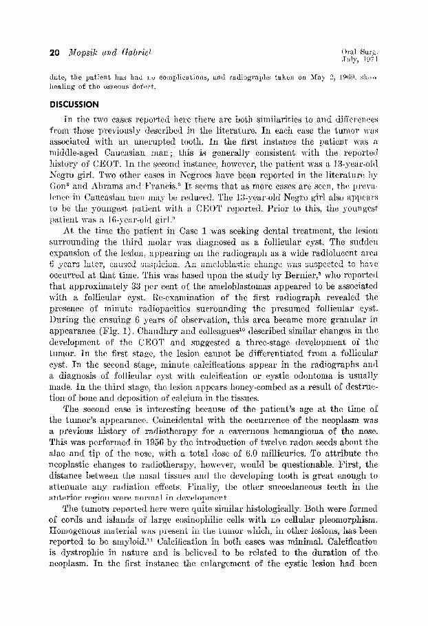
date, the patient has had no complications, and radiographs taken on May 2, lR@~. shrew healing of the osseous dcfwt.
DISCUSSION
In the t.wo cases rcportccl here there arc both similarities to and differences from those previously described in the literature. In each case the tumor was associated with an unerupted tooth. In the first instance the patient was a middle-aged Caucasian man ; this is generally consistent with the reported history of CEOT. In the second instance, however, the patient was a 13-year-ol(l Negro girl. Two other cases in Negroes have been reported in the literature by Gon* and Abrams and Francis.” It seems that as more cases are seen, the prevn- lt~ct: in Caucasian rntw may be reduced. The 13-year-old Negro girl also appears to be the youngest patient with a CEOT reported. Prior to this, the youngest patient was a 16-year-old girl .!I
At the time the patient in Case 1 was seeking dental treatment, the lesion surrounding the third molar was diagnosed as a follicular cyst. The sudden expansion of the lesion, appearing on the radiograph as a wide radiolucent area 6 years later, caused suspicion. An ameloblastic change was suspected to have occurred at that time. This was based upon the study by Bernier,5 who reported that approximately 33 per cent of the ameloblastomas appeared to be associated with a follicular cyst. Re-examination of the first radiograph revealed the presence of minute radiopacities surrounding the presumed follicular cyst. During the ensuing 6 years of observation, this area became more granular in appearance (Fig. 1). Chaudhry and colleagues1o described similar changes in the development of the CEOT and suggested a three-stage development of the tumor. In the first stage, the lesion cannot be differentiated from a follicular cyst. In the second stage, minute calcifications appear in the radiographs and a diagnosis of follicular cyst with calcification or cystic odontoma is usually made. In the third stage, the lesion appears honey-combed as a result of destruc- tion of bone and deposition of calcium in the tissues.
The second case is interesting because of the patient’s age at the time of the tumor’s appearance. Coincidental with the occurrence of the neoplasm was a previous history of radiotherapy for a cavernous hemangioma of the nose. This was performed in 1956 by the introduction of twelve radon seeds about the alae and tip of the nose, with a total dose of 6.0 millicuries. To attribute the neoplastic changes to radiotherapy, however, would be questionable. First, the distance between the nasal tissues and the developing tooth is great enough to attenuate any radiat,ion effects. Finally, the other succedaneous teeth in the anterior region were normal in development.
The tumors reported here were quite similar histologically. Both were formed of cords and islands of large eosinophilic cells with no cellular pleomorphism. Homogenous material was present in the tumor which, in other lesions, has been reported to be amyloid.” Calcification in both cases was minimal. Calcification is dystrophic in nature and is believed to be related to the duration of the neoplasm. In the first instance the enlargement of the cystic lesion had been

Vohme 32 Number 1
Calcifying epithelid odontogenic tumor 21
present for less than a year, and in the second case the lesion occurred at the time of the canine’s eruption.
In an effort to explain the pathogenesis of the neoplasm, Con,* after an exhaustive histochemical study of the tumor, postulated that the CEOT arises from an abnormal proliferation of the stratum intermedium. The reduced enamel epithelium normally fuses with the oral epithelium at the time of erup- tion. In some instances of retained teeth, the former fails to fuse with the latter, probably resulting in abnormal proliferation of the stratum inter-medium in an attempt to carry out its function.
SUMMARY AND CONCLUSION
The two tumors described here support the view that the calcifying epithelial odontogenic tumor is a neoplasm occurring within the epithelium surrounding retained teeth. The first case was followed over a 6-year period until the sudden enlargement of the surrounding follicle. This would tend to lend some support to those who suggest that all retained teeth be removed early in order to circum- vent possible neoplastic change.
We are indebted to Dr. Donald C. Reynolds for help in preparing this manuscript as well aa the material utilized in the first ease presentation.
REFERENCES
1. 2.
3.
4. 5.
6.
7.
8.
9.
10.
11.
Pindborg, J. J.: A Calcifying Epithelial Odontogenic Tumor, Cancer 11: 838-843, 1958. Field. H. J.. and Ackerman. A. A.: Calcifvina Fibroadamantoblastoma. Amer. J. Orthddont. 2s’: 543-545, 1942. ’
I ‘2
Thoma, K. H., and Goldman, H. M.: Odontogenic Tumors; Classification Based on Observation of Epithelial, Mesenehymal, and Mixed Varieties, Amer. J. Path. 22: 433. 471, 1946. Wunderer, 8.: Zur Frage maligner Odontome, Oest. Z. Stomat. 50: 567-571, 1953, Bernier, J. L.: Tumors of the Odontogenic Apparatus and Jaws (Atlas of Tumor Pathology, Sec. IV, Fast. lOa), Washington, 1960, Armed Forces Institute of Pathology. Pindborg, J. J.: The Calcifying Epithelial Odontogenic Tumor; Review of the Literature and Report of an Extraosseous Case, Acta Odont. Stand. 34: 419-430, 1966. Patterson, J. T., Martin, T. H., and DeJean, E. K.: Extraosseous Calcifying Odontogenic Epithelial Tumor; Report of a Case, ORAL SURG. 27: 363-367, 1969. Gon, F.: The Calcifying Epithelial Odontogenic Tumor; Review of a Case and a Study of Its Histogenesis, Brit. J. Cancer 19: 39-56, 1965. Abrams, A. M., and Francis, V. H.: Calcifying Epithelial Odontogenic Tumor; Report of Cases, J. Amer. Dent, Ass. 74: 1231-1240, 1967. Chaudhry, A. P., Holte, N. O., and Vickers, R. A.: Calcifying Epithelial Odontogenic Tumor; Report of a Case, ORAL SURG.~~: 843-848,1962. Gardner, D. G., Michaels, L., and Liepa, E.: Calcifying Epithelial Odontogenic Tumor; an Amyloid Producing Neoplasm, ORAL SURG. 26: 812-823, 1968.