The role of tryptophans on the cellular uptake and membrane interaction of arginine-rich cell...
10
The role of tryptophans on the cellular uptake and membrane interaction of arginine-rich cell penetrating peptides Marie-Lise Jobin a , Marine Blanchet a , Sarah Henry a , Stéphane Chaignepain a , Claude Manigand a , Sabine Castano a , Sophie Lecomte a , Fabienne Burlina b , Sandrine Sagan b , Isabel D. Alves a, ⁎ a CBMN-Univ Bordeaux, UMR 5248, Allée Geoffroy St Hilaire, 33600 Pessac, France b Sorbonne Universités — UPMC Univ Paris 06, École Normale Supérieure-PSL Research University, Département de Chimie, CNRS UMR 7203 LBM, 4 Place Jussieu, F-75005 Paris, France abstract article info Article history: Received 26 July 2014 Received in revised form 10 November 2014 Accepted 12 November 2014 Available online 20 November 2014 Keywords: Cell penetrating peptide Peptide/lipid interaction Lipid model systems Cell-penetrating peptides (CPP) are able to efficiently transport cargos across cell membranes without being cytotoxic to cells, thus present a great potential in drug delivery and diagnosis. While the role of cationic residues in CPPs has been well studied, that of Trp is still not clear. Herein 7 peptide analogs of RW9 (RRWWRRWRR, an efficient CPP) were synthesized in which Trp were systematically replaced by Phe residues. Quantification of cellular uptake reveals that substitution of Trp by Phe strongly reduces the internalization of all peptides despite the fact that they strongly accumulate in the cell membrane. Cellular internalization and biophysical studies show that not only the number of Trp residues but also their positioning in the helix and the size of the hydro- phobic face they form are important for their internalization efficacy, the highest uptake occurring for the analog with 3 Trp residues. Using CD and ATR-FTIR spectroscopy we observe that all peptides became structured in con- tact with lipids, mainly in α-helix. Intrinsic tryptophan fluorescence studies indicate that all peptides partition in the membrane in about the same manner (Kp ~ 10 5 ) and that they are located just below the lipid headgroups (~10 Å) with slightly different insertion depths for the different analogs. Plasmon Waveguide Resonance studies reveal a direct correlation between the number of Trp residues and the reversibility of the interaction following membrane washing. Thus a more interfacial location of the CPP renders the interaction with the membrane more adjustable and transitory enhancing its internalization ability. © 2014 Elsevier B.V. All rights reserved. 1. Introduction One of the major obstacles to the use of large therapeutic molecules or imaging agents having intracellular targets is their low permeability through biomembranes. One of the breakthroughs of the last 20 years is the discovery of cell-penetrating peptides (CPP) as molecules capable of internalizing into cells in a receptor- and energy-independent way and without being toxic to cells. Their great potential relies on the fact that they can transport a great variety of cargoes into cells both in terms of size and nature and some CPPs are even already used as drug delivery vector (for a review, see [1]). Green & Loewenstein and Frankel & Pabo discovered the first CPP almost simultaneously in 1988 [2,3]. They found that the Tat protein from HIV-1 was internalized into cells and Vives et al., found in 1997 the minimal sequence that was responsible for the protein internalization [4]. Following this finding, penetratin was discovered in 1994 by the group of Alain Prochiantz [5]. This is a peptide derived from the homeodomain of the Drosophila homeobox Antennapedia and was shown to possess a good internaliza- tion efficacy. Since then a large number of Structure/Activity Relation- ship studies have been performed both to study their membrane- translocating capabilities and to design novel sequences with greater efficacy and better selectivity. Since the Tat peptide possesses a large number of basic residues (6 Arg and 2 Lys on 13 residues) Wender and Rothbard discovered that a polyarginine comporting 9 residues is an efficient CPP [6]. Futaki's group then synthesized oligoarginines of different lengths (R n with 6 b n b 12) and studied their internalization efficiency [7]. They could determine that 8 Arg residues were sufficient to confer the polyarginine cell penetrating properties. Different deriva- tives of penetratin have also been synthetized and it was observed that the internalization was based neither on the chirality of the pep- tide, nor its amphiphilicity or its secondary structure [5,8]. Regarding the mechanisms implicated in their cellular internalization, it has been generally accepted that both endocytosis and direct translocation through the membrane are implicated in their uptake. The balance Biochimica et Biophysica Acta 1848 (2015) 593–602 Abbreviations: AMP, Anti-microbial Peptide; ATR-FTIR, Attenuated Total Reflectance-Fourier Transform Infrared; CPP, cell-penetrating peptide; CD, Circular dichroism; CHO, Chinese hamster ovary; DOPC, Dioleoylphosphatidylcholine; DOPG, Dioleoylphosphatidylglycerol; DLS, Dynamic light scattering; GAG, Glycosaminoglycan; HSPG, Heparan sulfate proteoglycans; LUV, Large unilamellar vesicle; MALDI, Matrix- Assisted Laser Desorption/Ionization; MLV, Multi lamellar vesicle; MS, Mass spectrome- try; PC, Phosphatidylcholine; PG, Phosphatidylglycerol; PWR, Plasmon Waveguide Resonance; SAR, Structure/Activity Relationship; SUV, Small Unilamellar Vesicles ⁎ Corresponding author. Tel.:+33 5 40 00 68 49. E-mail address: [email protected] (I.D. Alves). http://dx.doi.org/10.1016/j.bbamem.2014.11.013 0005-2736/© 2014 Elsevier B.V. All rights reserved. Contents lists available at ScienceDirect Biochimica et Biophysica Acta journal homepage: www.elsevier.com/locate/bbamem
-
Upload
laurentponson -
Category
Documents
-
view
4 -
download
0
Transcript of The role of tryptophans on the cellular uptake and membrane interaction of arginine-rich cell...

Biochimica et Biophysica Acta 1848 (2015) 593–602
Contents lists available at ScienceDirect
Biochimica et Biophysica Acta
j ourna l homepage: www.e lsev ie r .com/ locate /bbamem
The role of tryptophans on the cellular uptake andmembrane interactionof arginine-rich cell penetrating peptides
Marie-Lise Jobin a, Marine Blanchet a, Sarah Henry a, Stéphane Chaignepain a, Claude Manigand a,Sabine Castano a, Sophie Lecomte a, Fabienne Burlina b, Sandrine Sagan b, Isabel D. Alves a,⁎a CBMN-Univ Bordeaux, UMR 5248, Allée Geoffroy St Hilaire, 33600 Pessac, Franceb Sorbonne Universités — UPMC Univ Paris 06, École Normale Supérieure-PSL Research University, Département de Chimie, CNRS UMR 7203 LBM, 4 Place Jussieu, F-75005 Paris, France
Abbreviations: AMP, Anti-microbial Peptide; AReflectance-Fourier Transform Infrared; CPP, cell-pendichroism; CHO, Chinese hamster ovary; DOPC, DioleoDioleoylphosphatidylglycerol; DLS, Dynamic light scatteHSPG, Heparan sulfate proteoglycans; LUV, Large unilaAssisted Laser Desorption/Ionization; MLV, Multi lamellatry; PC, Phosphatidylcholine; PG, PhosphatidylglyceroResonance; SAR, Structure/Activity Relationship; SUV, Sm⁎ Corresponding author. Tel.:+33 5 40 00 68 49.
E-mail address: [email protected] (I.D. Alve
http://dx.doi.org/10.1016/j.bbamem.2014.11.0130005-2736/© 2014 Elsevier B.V. All rights reserved.
a b s t r a c t
a r t i c l e i n f oArticle history:Received 26 July 2014Received in revised form 10 November 2014Accepted 12 November 2014Available online 20 November 2014
Keywords:Cell penetrating peptidePeptide/lipid interactionLipid model systems
Cell-penetrating peptides (CPP) are able to efficiently transport cargos across cell membranes without beingcytotoxic to cells, thus present a great potential in drug delivery and diagnosis. While the role of cationic residuesin CPPs has been well studied, that of Trp is still not clear. Herein 7 peptide analogs of RW9 (RRWWRRWRR, anefficient CPP) were synthesized in which Trp were systematically replaced by Phe residues. Quantification ofcellular uptake reveals that substitution of Trp by Phe strongly reduces the internalization of all peptides despitethe fact that they strongly accumulate in the cell membrane. Cellular internalization and biophysical studiesshow that not only the number of Trp residues but also their positioning in the helix and the size of the hydro-phobic face they form are important for their internalization efficacy, the highest uptake occurring for the analogwith 3 Trp residues. Using CD and ATR-FTIR spectroscopywe observe that all peptides became structured in con-tact with lipids, mainly inα-helix. Intrinsic tryptophan fluorescence studies indicate that all peptides partition inthe membrane in about the same manner (Kp ~ 105) and that they are located just below the lipid headgroups(~10 Å) with slightly different insertion depths for the different analogs. PlasmonWaveguide Resonance studiesreveal a direct correlation between the number of Trp residues and the reversibility of the interaction followingmembranewashing. Thus amore interfacial location of the CPP renders the interactionwith themembranemoreadjustable and transitory enhancing its internalization ability.
© 2014 Elsevier B.V. All rights reserved.
1. Introduction
One of the major obstacles to the use of large therapeutic moleculesor imaging agents having intracellular targets is their low permeabilitythrough biomembranes. One of the breakthroughs of the last 20 yearsis the discovery of cell-penetrating peptides (CPP) asmolecules capableof internalizing into cells in a receptor- and energy-independent wayand without being toxic to cells. Their great potential relies on the factthat they can transport a great variety of cargoes into cells both interms of size and nature and some CPPs are even already used as drugdelivery vector (for a review, see [1]). Green & Loewenstein and Frankel& Pabo discovered the first CPP almost simultaneously in 1988 [2,3].
TR-FTIR, Attenuated Totaletrating peptide; CD, Circularylphosphatidylcholine; DOPG,ring; GAG, Glycosaminoglycan;mellar vesicle; MALDI, Matrix-r vesicle; MS, Mass spectrome-l; PWR, Plasmon Waveguideall Unilamellar Vesicles
s).
They found that the Tat protein from HIV-1 was internalized intocells and Vives et al., found in 1997 the minimal sequence that wasresponsible for the protein internalization [4]. Following this finding,penetratin was discovered in 1994 by the group of Alain Prochiantz[5]. This is a peptide derived from the homeodomain of the Drosophilahomeobox Antennapedia and was shown to possess a good internaliza-tion efficacy. Since then a large number of Structure/Activity Relation-ship studies have been performed both to study their membrane-translocating capabilities and to design novel sequences with greaterefficacy and better selectivity. Since the Tat peptide possesses a largenumber of basic residues (6 Arg and 2 Lys on 13 residues) Wenderand Rothbard discovered that a polyarginine comporting 9 residues isan efficient CPP [6]. Futaki's group then synthesized oligoarginines ofdifferent lengths (Rn with 6 b n b 12) and studied their internalizationefficiency [7]. They could determine that 8 Arg residues were sufficientto confer the polyarginine cell penetrating properties. Different deriva-tives of penetratin have also been synthetized and it was observedthat the internalization was based neither on the chirality of the pep-tide, nor its amphiphilicity or its secondary structure [5,8]. Regardingthe mechanisms implicated in their cellular internalization, it has beengenerally accepted that both endocytosis and direct translocationthrough the membrane are implicated in their uptake. The balance

594 M.-L. Jobin et al. / Biochimica et Biophysica Acta 1848 (2015) 593–602
between the two mechanisms depends on a great variety of aspectssuch as the nature and size of the CPP and its cargo, the nature of thelink between the two, the temperature at which internalization experi-ments are conducted, the cell lines used, among other parameters[9–12]. Electrostatic interactions between the positive charges in thepeptide and negative charges in the cell membrane surface have beenshown to be essential during thefirst stage of interactionwith themem-brane. The presence of basic amino acids in the sequence has been wellstudied and Arg residues have been reported to be especially importantfor cellular internalization. It was shown that the uptake efficiency is at-tributed to the type of bond formed between Arg and the lipidheadgroups rather than the charges presented to themembrane. IndeedArg can form bidentate hydrogen bonds that interact simultaneouslywith phosphate moieties on multiple lipid headgroups while Lys resi-dues can only form monodentate hydrogen bonds that interact withthe phosphate moiety on a single headgroup [13,14]. Guanidinium-rich peptides also establish strong electrostatic interactions with nega-tively charged heparan sulfate proteoglycans (HSPG) on the cell surface,important as a first recognition and their accumulation in the mem-brane [15,16]. The presence of hydrophobic residues for internalizationhas also been investigated. The substitution of the Trp48 and Trp56 byPhe abolished totally the internalization of penetratin [17]. From thesestudies, the Arg-rich CPP RW9 (RRWWRRWRR) was designed and de-termined to possess very high cellular uptake efficiency [18]. Biologicalstudies on RW9 have revealed an important decrease in internalizationin GAG-deficient cells evidencing that proteoglycans at the membranesurface are important for its cellular internalization [19]. Biophysicalstudies have shown its preferential interaction with anionic modelmembranes corroborating biological studies on the importance of elec-trostatic interactions between peptide and membranes. Previous stud-ies on RL9 (RRLLRRLRR), an analog of RW9 where Trp residues werereplaced by Leu residues have shown that this peptidewas not internal-ized into cells despite the fact that it accumulated in themembrane [19,20]. At the same time, oligoargininepeptide (R9) possessing no aromaticresidues internalizes very well in eukaryotic cells at approximately thesame level as RW9 [19]. The main question addressed here is to under-stand the role of the Trp in membrane translocation regarding the RW9sequence. Therefore, we synthesized 7 peptides inwhich Phe systemat-ically replaced Trp residues (RX9) (Table 1). The strategy was to keepthehydrophobicity but specially the aromaticity of thehydrophobic res-idues, because previous NMR studies on RW9 evidenced the existenceof π–cation interactions between certain Arg and Trp residues [19].
Quantification of the total amount of internalized peptides (all intra-cellular compartments included) shows that the replacement of all Trpresidues almost completely abolished the peptide internalization whilethe substitution of 1 or 2 Trp strongly decreases their internalizationefficiency. To understand these differences in cellular internalization wehave decided to investigate the interaction of these peptides with lipidmembranes and therefore shed some light into their membrane crossingand translocation. It should be noted that direct translocation through thecellmembrane is just one of themanymechanisms used by CPPs to inter-nalize, nonetheless a good understanding of CPP interaction with lipids isimportant. For that we have used lipid model systems and different bio-physical approaches in an attempt to correlate their cellular uptake and
Table 1Amino-acid sequences of the RX9 peptides used in this study.
Peptide sequence MW (Da) Charges (at pH 7)
RW9 Biotin(O2)-GGGG-RRWWRRWRR-NH2 1999 6RFFF9 Biotin(O2)-GGGG-RRFFRRFRR-NH2 1882 6RFFW9 Biotin(O2)-GGGG-RRFFRRWRR-NH2 1921 6RWFF9 Biotin(O2)-GGGG-RRWFRRFRR-NH2 1921 6RFWF9 Biotin(O2)-GGGG-RRFWRRFRR-NH2 1921 6RFWW9 Biotin(O2)-GGGG-RRFWRRWRR-NH2 1960 6RWWF9 Biotin(O2)-GGGG-RRWWRRFRR-NH2 1960 6RWFW9 Biotin(O2)-GGGG-RRWFRRWRR-NH2 1960 6
membrane direct translocationwith bilayer interaction. Since electrostat-ic interactions were found to be important for the membrane interactionof Arg-rich peptides with cellular membranes [see [21] for a review] wehave included anionic lipids in the model membranes used. Even thoughanionic lipids are veryweakly present in eukaryotic cell membrane, espe-cially in the outer leaflet, the few anionic lipids present (~2%) can havetheir potential enhancedby assembling into domains, a property reportedto be induced by certain CPPs [22]. Although, the outer leaflet of healthyeukaryotic membranes possesses almost no anionic lipids, importantelectrostatic interactions between the CPPs and GAG can be established.Often biophysicists have employed anionic lipids just tomimic the overallanionic character of the cell membrane surface, which is also ourapproach here. Additionally it should be noted that during certaincellular dysfunctions such as when cells become tumoral or enterapoptosis, the amount of anionic lipids in their outer leaflet (mostlyphosphatidylserine) increases up to 9%, rendering the cellular membranesignificantly more anionic [23–26]. CD and ATR-FTIR were used to inves-tigate the secondary structure of the peptides in contact with modelmembranes to define if there was a correlation between their tendencyto adopt a secondary structure in the presence of lipids and their internal-ization capacities. No direct correlation was found. Their cytotoxicity oncells and effect on model membranes were explored. The replacementof Trp by Phe induced no cytotoxicity or dye leakage although the pep-tides bind and slightly perturb the membrane. The affinity and insertiondepth of the peptides were studied by Plasmon Waveguide Resonanceand Trp fluorescence and a correlation with their internalization capaci-ties was established.
2. Materials & methods
2.1. Materials
All lipids were obtained from Avanti Polar Lipids (Alabaster, AL, USA).The calcein and acrylamide were obtained from Sigma Aldrich.Biotin(O2)-([1H]-G)4-RRFFRRFRR-NH2(RF9), Biotin(O2)-([1H]-G)4-RRFFRRWRR-NH2(RFFW9), Biotin(O2)-([1H]-G)4-RRWFRRFRR-NH2(RWFF9), Biotin(O2)-([1H]-G)4-RRFWRRFRR-NH2 (RFWF9),Biotin(O2)-([1H]-G)4-RRFWRRWRR-NH2 (RFWW9), Biotin(O2)-([1H]-G)4-RRWWRRFRR-NH2(RWWF9), Biotin(O2)-([1H]-G)44-RRWFRRWRR-NH2(RWFW9) and Biotin(O2)-([2H]-G)4-RRFFRRFRR-NH2,Biotin(O2)-([2H]-G)4-RRFFRRWRR-NH2, Biotin(O2)-([2H]-G)4-RRWFRRFRR-NH2, Biotin(O2)-([2H]-G)4-RRFWRRFRR-NH2, Biotin(O2)-([2H]-G)4-RRFWRRWRR-NH2, Biotin(O2)-([2H]-G)4-RRWWRRFRR-NH2, Biotin(O2)-([2H]-G)4-RRWFRRWRR-NH2 were synthesized usingthe Fmoc solid-phase strategy ([1H]-G and [2H]-G correspond to non-deuterated and bi-deuterated glycine, respectively). The oxidation pro-tocol of the biotin was as follows: 10 g of biotin was dissolved in 40 mLof H2O2 (30% in H2O) and 120 mL of AcOHwas added. The mixture wasstirred at room temperature for fewhours and a precipitatewas formed.The precipitatewasfiltered,washedwith Et2O anddriedunder vacuum.Oxidation efficiency of the biotin was checked by liquid-state NMR.Biotin sulfone was coupled to the peptide under the same conditionsused for the amino-acid coupling. Peptides were purified by HighPerformance Liquid Chromatography (HPLC), in a reverse phasecolumn (RP) C18 using H2O/CH3CN/TFA gradient. MALDI-TOF massspectrometry was used to characterize the peptides. To efficientlyremove the TFA counter-ion a simple method was used that consistsin lyophilizing the sample 3 times in the presence of 10mMHCl directlyreplacing TFA counter-ionswith chloride ions [27]. A low concentrationof HCl was used to prevent peptide degradation. The removal of TFAwas followed by 19F-NMR.
2.2. Cell culture
Wild type Chinese hamster ovary CHO-K1 (WT) cells were culturedin Dulbecco's modified Eagle's medium (DMEM) supplemented with
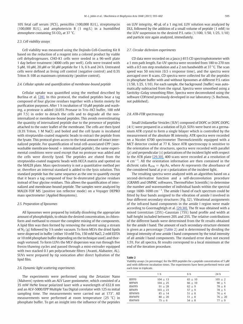
Table 2Viability assays (in percentage) for the RX9 peptides for a peptide concentration of 5 μMand with different incubation times. The experiments have been performed twice andeach time in triplicate.
1 h 6 h 24 h
RFFF9 104 ± 13 65 ± 10 105 ± 2RFFW9 104 ± 25 66 ± 10 90 ± 5RWFF9 91 ± 28 62 ± 9 78 ± 8RFWF9 103 ± 23 57 ± 9 78 ± 5RFWW9 85 ± 14 51 ± 8 76 ± 4RWWF9 80 ± 20 51 ± 8 76 ± 20RWFW9 75 ± 6 54 ± 8 77 ± 9
595M.-L. Jobin et al. / Biochimica et Biophysica Acta 1848 (2015) 593–602
10% fetal calf serum (FCS), penicillin (100,000 IU/L), streptomycin(100,000 IU/L), and amphotericin B (1 mg/L) in a humidifiedatmosphere containing 5% CO2 at 37 °C.
2.3. Cell viability assays
Cell viability was measured using the Dojindo Cell-Counting Kit 8based on the reduction of a reagent into a colored product by viablecell dehydrogenases. CHO-K1 cells were seeded in a 96-well plate1 day before treatment (4000 cells per well). Cells were treated with5 μM, 10 μM, 20 μM or 50 μM peptide for 1 h, 6 h and 24 h. Untreatedcells were defined as living cell control (negative control) and 0.3%Triton X-100 as maximum cytotoxicity (positive control).
2.4. Cellular uptake and quantification of membrane-bound peptide
Cellular uptake was quantified using the method described byBurlina et al. [28]. In this protocol, the studied peptides bear a tagcomposed of four glycine residues together with a biotin moiety forpurification purposes. After 1 h incubation of 10 μM peptide and wash-ing, a protease is added (0.05% Pronase in Tris–HCl buffer, 100 mMpH 7.5) in order to detach the cells and to degrade all the non-internalized or membrane-bound peptides. This avoids overestimatingthe quantity of internalized peptide due to the presence of peptidesattached to the outer leaflet of the membrane. The cells are then lysed(0.3% Triton, 1 M NaCl) and boiled and the cell lysate is incubatedwith streptavidin-coated magnetic beads to extract the peptide fromthe lysate. This protocol gives access to the total amount of intact inter-nalized peptide. For quantification of total cell-associated CPP (non-washable membrane-bound + internalized peptide), the same experi-mental conditions were used except that no protease was added andthe cells were directly lysed. The peptides are eluted from thestreptavidin-coated magnetic beads with HCCA matrix and spotted onthe MALDI plate. Mass spectrometry is not a quantitative method perse, therefore an internal standard is added to the lysis solution. Thisstandard peptide has the same sequence as the one to quantify exceptthat it bears a tag composed of four bi-deuterated glycine residuesinstead of four glycine residues. This allows the quantification of inter-nalized and membrane-bound peptide. The samples were analyzed byMALDI-TOF MS (positive ion reflector mode) on a Voyager DEPROmass spectrometer (Applied Biosystems).
2.5. Preparation of liposomes
All liposomes were prepared by initially dissolving the appropriateamount of phospholipids, to obtain the desired concentration, in chloro-form and methanol to ensure the complete mixing of the components.A lipid film was then formed by removing the solvent using a streamof N2 (g) followed by 3 h under vacuum. To formMLVs the dried lipidswere dispersed in buffer (either 10mMTris, 150mMNaCl, 2 mMEDTAor 10mMphosphate buffer depending on the techniqueused) and thor-ough vortexed. To form LUVs the MLV dispersion was run through fivefreeze/thawing cycles and passed through a mini-extruder equippedwith two stacked 0.1 μm polycarbonate filters (Avanti, Alabaster, AL).SUVs were prepared by tip sonication after direct hydration of thelipid film.
2.6. Dynamic light scattering experiments
The experiments were performed using the Zetasizer Nano(Malvern) system with an ALV Laser goniometer, which consisted of a35 mW HeNe linear polarized laser with a wavelength of 632.8 nmand an ALV-5000/EPP Multiple Tau Digital correlator with 125 ns initialsampling time. The measurements were carried out at 173°. Allmeasurements were performed at room temperature (25 °C) inphosphate buffer. To get an insight into the influence of the peptides
on LUV integrity, 40 μL of a 1 mg/mL LUV solution was analyzed byDLS, followed by the addition of a small volume of peptide (1 mM) tothe LUV suspension to the desired P/L ratio (1/100, 1/50, 1/25, 1/10)and particle size again analyzed, immediately.
2.7. Circular dichroism experiments
CD data were recorded on a Jasco J-815 CD spectrophotometer witha 1 mm path length. Far-UV spectra were recorded from 180 to 270 nmwith a 0.5 nm step resolution and a 2 nm bandwidth at 37 °C. The scanspeed was 50 nm/min (0.5 s response time), and the spectra wereaveraged over 8 scans. CD spectra were collected for all the peptidesin phosphate buffer with and without liposomes at different P/L ratios(1/50, 1/25, 1/10). For each sample, the background (buffer) was auto-matically subtracted from the signal. Spectra were smoothed using aSavitzky–Golay smoothing filter. Spectra were deconvoluted using thesoftware CDFriend previously developed in our laboratory (S. Buchoux,not published).
2.8. ATR-FTIR spectroscopy
Small Unilamellar Vesicles (SUV) composed of DOPC or DOPC:DOPG(4:1) were prepared in a solution of D2O. SUVs were burst on a germa-nium ATR crystal to form a single bilayer which is controlled by themeasurement of the absolute IR intensity. ATR spectra were recordedon a Nicolet 6700 spectrometer ThermoScientific equipped with aMCT detector cooled at 77 K. Since ATR spectroscopy is sensitive tothe orientation of the structures, spectra were recorded with parallel(p) and perpendicular (s) polarizations of the incident lightwith respectto the ATR plate [29,30]. 400 scans were recorded at a resolution of4 cm−1. All the orientation information are then contained in thedichroïc ratio RATR = Ap/As, where Ai represents the absorbance ofthe considered band at p or s polarization of the incident light.
The resulting spectra were analyzed with an algorithm based on asecond-derivative function and a self-deconvolution procedure(GRAMS and OMNIC softwares, ThermoFisher Scientific) to determinethe number and wavenumber of individual bands within the spectralrange 1600–1690 cm−1. The amide I band of each spectrum could befitted by four bands assigned to the vibration of amide I involved infour different secondary structures (Fig. S2). Vibrational assignmentsof the infrared band components in the amide I region were madeaccording to Goormaghtigh et al. [29,30]. The fit was obtained with amixed Lorentzian (25%)–Gaussian (75%) band profile and width athalf-height included between 20% and 25%. The relative contributionsof the different bands were determined from the fit results obtainedfor the amide I band. The amount of each secondary-structure elementis given as a percentage (Table 2) and is determined by dividing theintegral intensity of one amide I band component by the total intensityof all amide I band components. The standard error does not exceed1.5%. For all spectra, fit results correspond to a local minimum at theend of the iteration procedure.

596 M.-L. Jobin et al. / Biochimica et Biophysica Acta 1848 (2015) 593–602
2.9. Calcein leakage experiments
Calcein-containing LUVsweremade using the same protocol used tomake regular LUVs, except for the hydration step of the lipid films thatwas made with Tris buffer containing 70 mM calcein. The protocol toprepare calcein-loaded vesicles was the same as previously describedby Jobin et al. [31]. For the assay, the lipid concentration was set at1 μM and peptide concentration was allowed to vary from 10 nM to100 nM (P/L ratio of 1/100 to 1/10 respectively). All measurementswere performed on 96-well microplates using a Perkin Elmer LS55spectrometer (Buckinghamshire, UK). Data were collected every min-ute at room temperature using a λexc at 485 nm and λem at 515 nmwith an emission and excitation slit of 2.5 nm.
The fluorescence intensity at the equilibrium was measured after1.5 h. At the end of the assay, complete leakage of LUVs was achievedby adding 10 μL of 10% Triton X-100 solution dissolving the lipid mem-brane without interfering with the fluorescence signal. The percentageof calcein release was calculated according to the following equation:
% calcein leakage ¼ Ft−F0Ff−F0
� 100 ð1Þ
where the percent of calcein leakage is the fraction of dye released(normalizedmembrane leakage), Ft is themeasuredfluorescence inten-sity at time t, and F0 and Ff are respectively the fluorescence intensitiesat times t = 0, and after final addition of Triton X-100, respectively. Adilution correctionwas applied on the fluorescence intensity after injec-tion of the Triton X-100. Each experiment was repeated three times.
2.10. Fluorescence spectroscopy measurements
2.10.1. Nernst partition coefficient experimentsPeptide–phospholipid interactions were studied by monitoring the
changes in the Trp fluorescence emission spectra of the peptides uponaddition of LUVs. Intrinsic Trp fluorescence of the peptides wasmeasured before and after addition of different amounts of phospholipidvesicles to a 0.5 μM peptide solution. Trp fluorescence was measured atroom temperature on a Spex Fluoromax-4 (Horiba) spectrofluorometer.
Emission spectra were recorded between 300 and 500 nm with anexcitation wavelength of 280 nm, slit widths of 5 nm for excitationand 10 nm for emission. Peptide–lipid binding was determined fromthe quenching of the intrinsic Trp fluorescence intensity of the peptidesupon addition of LUVs. The maximum fluorescence intensity wasplotted vs. the added lipid concentration and fitted using the simplepartition model (2), the self-quenching model (3) and the three-statepartition model well described by Melo & Castanho [32]. The datawere analyzed using Origin.
IIw
¼ 1þ KpγL L½ �IL=Iw1þ KpγL L½ � ð2Þ
IIw
¼ 1þ KpγL L½ �IL=Iw1þ KpγL L½ � þ k2KpIL
þ 11þ KpγL L½ � ð3Þ
where [L] is the total phospholipid concentration available for thepeptide to interactwith, Kp is theNernst partition constant, k2 is propor-tional to the ratio between the bimolecular self-quenching rate and theradiative decay rate, γL is the phospholipids molar volume, which is0.763M−1 for the lipid used in this study. IL and IW are the fluorescenceintensities themixture would display if all the peptidewas in the lipidicor aqueous phase, respectively. IW can be obtained from the measure-ment of I in the absence of lipid; IL is a limit value of fluorescenceintensity as [L]→ ∞ and is determined as a system parameter togetherwith Kp.
2.10.2. Brominated lipid quenching experimentsDepth-dependent fluorescence quenching of tryptophan was
performed in LUVs composed of DOPC, DOPC:DOPG 4:1, and either(6,7)-, (9,10)-, or (11,12)-BrPC at molar ratios of 70:30 with DOPC:BrPC and 50:20:30 with DOPC:DOPG:BrPC. Fluorescence intensities inthe absence of quencher (F0) were measured in DOPC and DOPC:DOPG (4:1) vesicles. Spectra were recorded between 300 and 500 nmwith an increment of 1 nm and an integration time of 0.1 s, using anexcitation wavelength of 280 nm. The P/L molar ratio was 1:50 andthe peptide concentration was 0.5 μM (for RW9, RWFF9, RFWF9 andRWWF9) and 0.8 μM (for RFWW9). Data were corrected for vesiclebackground, and the total intensity was calculated as the sum of allmeasured points in each spectrum. Depth-dependent fluorescencequenching profiles (DFQPs) were fitted to data in Matlab using theDistribution Analysis (DA) method [33,34] as well as the parallaxmethod (PM) [35–38].
DA : lnF0F hð Þ
¼ Sσ
ffiffiffiffiffiffi2π
p exp − h− hmð Þ22σ2
" #ð4Þ
PM : lnF0F hð Þ
¼ πC R2c− h−hmð Þ2
h ið5Þ
In the equations above, F0 is thefluorescence intensity in the absenceof quencher, F(h) is the intensity in the presence of quencher at the dis-tance h (Å) from the bilayer center, and hm is the average insertiondepth of the tryptophan residues. In DA the DFQP data are fitted witha Gaussian function where σ denotes the dispersion, which is relatedto the in-depth distribution of the tryptophan chromophores, and S isthe area under the quenching profile, which is related to thequenchability of the tryptophan moiety. The Parallax Method fits datato a truncated parabola, and Rc is the radius of quenching. Averagebromine distances from the bilayer center (h) were taken to be 11.0,8.3, and 6.5 Å for (6,7)-BrPC, (9,10)-BrPCand (11,12)-BrPC, respectively[38].
2.10.3. Acrylamide quenching experimentsPeptide–lipid interactions are accompanied by changes in the acces-
sibility of the peptides to aqueous quenchers of Trp fluorescence. Acryl-amide quenching experiments were carried out on a 0.5 μM peptidesolution in the absence or presence of LUVs by addition of aliquots of4 M acrylamide solution. The peptide/lipid mixtures (P/L molar ratioof 1/50) were incubated for 15 min at room temperature prior to themeasurements. The excitation wavelength was set to 295 nm insteadof 280 nm to reduce absorbance by acrylamide. Fluorescence intensitieswere measured after addition of quencher at room temperature. Thequenching constants were obtained from the slope of the Stern–Volmerplots of F0/F vs. [quencher], with F0 and F as the fluorescence intensitiesin the absence and presence of quencher, respectively. The normalizedaccessibility factor (naf) was calculated in comparison with the Ksv foreach peptide in buffer.
2.11. Plasmon Waveguide Resonance measurements
Experiments were performed on a beta PWR instrument fromProterion Corp. (Piscataway, NJ) that had a spectral angular resolutionof 1 mdeg. The technique used to form supported bilayers was thesame that the one previously described by Salamon et al. [39–41].Data were fitted (GraphPad Prism) through a hyperbolic saturationcurve providing the dissociation constants. It should be noted thatsince concomitantly with the binding process other processes, such asmembrane reorganization and solvation occur, the dissociationconstants correspond to apparent dissociation constants. In order totest for binding reversibility following peptide binding at saturatingconcentrations, the PWR cell sample was washed twice, by flowing

Fig. 1. (A) MALDI-TOF MS quantification of RX9 and RW9 internalization in 1 millionCHO-K1 cells after peptide incubation for 1 h at 37 °C. (B) Membrane-bound peptidequantification on CHO-K1 cells (1 million) determined by mass spectrometry(MALDI-TOF) at 37 °C. Significance was tested using a Student t test: ns: p N 0.05;*0.01 b p b 0.05; **0.01 b p b 0.001; ***p b 0.005.
597M.-L. Jobin et al. / Biochimica et Biophysica Acta 1848 (2015) 593–602
buffer through the cell. Following that the spectra were acquired forboth polarizations. A comparison of the resonance angles before andafter washing gives information about the reversibility in the P/Linteraction.
3. Results & discussion
3.1. Effect of the peptides on the cell viability
The cell viability was tested with all RX9 peptides and was firststudied to investigate whether the replacement of Trp residues changestheir cytotoxicity (Table 2). The cell viability was measured on CHO-K1cells using the CCK8 test providing the rate of viable cells. Concentra-tions varied between 5 and 50 μM and peptides were incubated for 1,6 and 24 h at 37 °C. None of the peptides has shown any significantcytotoxicity at these concentrations at 1 h (which corresponds to theincubation time used to quantify cellular uptake). However we noticeda decrease of the cell viability for longer incubation time (6 h and 24 h).This demonstrates that the replacement of Trp by Phe residues in thesequence affects the cytotoxicity of the peptides.
3.2. Quantification of cellular uptake and membrane-bound peptide
Quantification of cellular uptake of each peptide was investigated bya protocol already published that uses MALDI-TOF mass spectrometryas the analytical technique [28]. The experiments were performedwith CHO-K1 cells at 37 °C, a temperature at which both endosomaland cytosolic peptides were quantified and at which peptides canenter through both endocytosis and direct translocation [42,43]. The re-sults show that all RX9 peptides are much less internalized (~1 pmol orless) than RW9 (~10 pmol), about a ten-fold decrease (Fig. 1A). Theleast internalized peptide (only ~0.2 pmol) is observed for the analogcontaining no Trp residues (RF9). The analogs containing 1 or 2 Trpresidues are slightly more internalized (~1 pmol) than RF9 but showno difference between them. The results indicate that the presence ofTrp residues in RW9 analogs is important for their cellular internaliza-tion. Since the first contact between the peptide and the cell is throughthe cell membrane, quantification of membrane-bound peptide wasperformed. Overall, the results show that RX9 peptides accumulate inthe membrane in the same extent as RW9 (~250 pmol) (Fig. 1B).Membrane accumulation at 4 °C provides similar results (data notshown). In terms of the two extreme behaviors, the peptide RWFF9accumulates themost in themembrane (~450 pmol), while the peptideRFWW9accumulates the least (~100 pmol). These results show that thepresence of Trp residues is important for RX9 cellular internalization. Nocorrelation exists between the amount of membrane-bound and that ofinternalized peptide in the RX9 series.
From the cellular uptake results we have selected for further studies5 peptides that appeared to be themost representative of the RX9 series,notablywith variable number of Trp residues and significant differencesin internalization capacities. We have therefore chosen the peptidewithout Trp residues (RF9), 2 peptides containing 1 Trp (RFWF9 andRWFF9) and 2 peptides with 2 Trp (RFWW9 and RWWF9). Biophysicsexperiments aimed at characterizing their interaction with lipidmembranes in an attempt to correlate that with the differences interms of cellular uptake efficacy were performed on the 5 selectedpeptides (described in the next sections).
3.3. Peptide secondary structure
As with RW9, the CD spectra of the RX9 peptides were stronglyaffected by the π–π stacking occurring between aromatic residues [19].In the case of RF9, RFWW9 and RWWF9 (3 Phe and 1 respectively), anegative band around 230 nmwas also observed that prevented properspectral decomposition (Fig. S1). This band is negative in the case of theRWWF9 and positive in the case of the RFWF9 as it depends on the
orientation of the aromatic residues. Peptides with 2 Phe (RFWF9 andRWFF9) gavemore interpretable results as no absorbance was observedat 230 nm allowing data decomposition and secondary structure deter-mination. In buffer and in the presence of zwitterionic lipids, thepeptides are in random coil conformationwith a smallα-helix contribu-tion (~10%; Fig. S1). In the presence of anionic lipids, 2 negative bands at208 and 222 nm are observed indicating α-helix structuration (~20%;Fig. S1). Helical structuration in the presence of anionic liposomes wasalso observed in the case of RW9 [19]. Moreover a preferential π–cationinteraction between residues Arg8 and Trp4 and Trp7 was evidenced bysolution NMR by Walrant et al. [19]. Since we replaced Trp4 and Trp7
by Phe in some cases, those interactions might have been affected.Indeed, the most internalized peptide for the RX9 series is RFWW9,which retains Trp4 and Trp7 and thus preserves these interactions.Therefore π–cation interactions seem to affect peptide internalization.This is to be expected since such interactions partially mask the chargeof the peptide allowing a less energetically unfavorable membranecrossing.
The results indicate that the replacement of some Trp by Phe resi-dues does not change the secondary structure of RX9 in interactionwith lipids and so the secondary structure upon lipid contact is notdirectly correlated to the internalization. The possibility of a correlationbetween the capacity of a CPP to adopt a particular secondary structureand to translocate is so far not clear in the literature. For example in thecase of penetratin it has been shown that α-helical structure is notnecessary for its internalization [8,44].
The secondary structure of the peptides in contact with lipids wasalso investigated by ATR-FTIR spectroscopy to avoid this issue causedby the aromatic residues (Table 3). We have found that the secondary

Table 3RX9 peptide secondary structure percent determined by ATR-FTIR spectroscopy.*RC: random coil.
Buffer DOPC DOPC:DOPG
Peptides RC* α-Helix β-Sheet β-Turn RC* α-Helix β-Sheet β-Turn RC* α-Helix β-Sheet β-Turn
RFFF9 19 35 32 14 24 37 11 28 20 41 27 12RWFF9 29 41 11 19 28 46 12 14 30 28 31 11RFWF9 22 33 19 26 19 34 20 27 32 33 28 7RFWW9 52 26 7 15 23 38 27 12 29 37 26 8RWWF9 30 31 22 17 25 38 22 15 31 40 18 11
598 M.-L. Jobin et al. / Biochimica et Biophysica Acta 1848 (2015) 593–602
structures of RX9 peptides were not well defined correspondingmostlyto a mixture of peptides adopting different secondary structures. Thuswe were unable to perform spectral decomposition and determinewith certainty the type and amount of each secondary structure.However we observed a marked increase in helical content in thepresence of membranes, especially those containing anionic lipids.
3.4. Peptide effect onmembrane supramolecular organization and integrity
Dynamic light scattering (DLS) experiments were performed inorder to investigate the influence of the peptides on liposome size anddistribution. Measurements were acquired in the absence and presenceof peptide and at different P/L ratios. Table S1 presents the variation insize distribution of liposomes composed of DOPC, DOPG and DOPC:DOPG (4:1 mol:mol) and for a P/L ratio of 1/50. In the case of DOPCthe size of the liposomes was not significantly modified by peptideaddition but the size of anionic liposomes (DOPG and DOPC:DOPG)increased importantly (up to 100 nm size increase) in the presence ofthe peptides. This shows that aggregation or fusion of anionic liposomesis induced by peptide addition. The same observation was reported byWalrant et al. on the effect of RW9on anionic lipids by following changesin turbidity [19]. No significant differences among the RX9 analogswereobserved indicating that lipid interactions are mostly driven by electro-static forces and therefore by the number of Arg residues which is thesame for all peptides.
Calcein leakage experimentswere performed to investigatewhetherRX9 peptides perturbed liposomemembrane integrity (e.g. pore forma-tion). In the case of anionic liposomes almost no leakage was observed(less than 1%) following 1 h peptide incubation at a P/L ratio of 1/50,meaning that the membrane integrity was not affected by the additionof the RX9 peptides. In the case of zwitterionic liposomes the leakagewas slightly higher (below 5%) indicating that RX9 peptides slightlyperturbed these liposomes (Table S2). We have observed that theleakage was similar (b5%) for all peptides and at all P/L ratios (1/100to 1/10; data not shown) so we assumed that this could result from asaturation of the bilayer surface by the peptide that induced a slightleakage. Overall, the results indicate that the peptides donot significantlyperturb the membrane integrity (no pore formation), which are inagreement with cell viability results.
Table 4Partition coefficients (Kp) of the RX9 peptides with increasing concentration of liposomes(from 2.5 μM to 200 μM) at constant peptide concentration (0.5 μM). The experimentshave been done in duplicate at room temperature.
Kp (.105)
DOPC DOPC/DOPG (4:1) DOPG
RW9 2.0 ± 0.8 3.8 ± 0.2 1.5 ± 0.6RWFF9 4.6 ± 0.5 3.1 ± 0.5 1.6 ± 0.2RFWF9 2.15 ± 0.05 1.765 ± 0.005 1.88 ± 0.07RFWW9 3.4 ± 0.7 1.77 ± 0.06 2.51 ± 0.07RWWF9 3.0 ± 0.5 0.9 ± 0.6 1.4 ± 0.2
3.5. Peptide interaction and partitioning with model membranes
The intrinsic Trp fluorescence of the peptides was used to character-ize the partitioning of the RX9 peptides with model membranes. TheNernst partition coefficient (Kp) was calculated at a constant peptideconcentration from the fluorescence intensity (I) vs. lipid concentration([L]) plots using the titration method already described [45,46]. Thecoefficient Kp is the ratio between membrane-bound and aqueousphase peptide concentrations. For all lipid systems the Kp obtainedwas in the range of 105 evidencing the importance of electrostaticrecognition in P/L interaction [47] (Table 4). This is consistent with thefact that RX9 peptides are positively charged at physiological pH (6+)(Table 1) and thus can establish important electrostatic interactions
Fig. 2. Partition curves of RW9 (A) and RFWW9 (B) peptides in the presence of liposomes.The peptide concentration used was 0.5 μMand the lipid stock concentration was 0.1 mMand 1 mM. The data were fitted with Eq. (2) for DOPC and DOPG curves in the case ofRFWF9, RFWW9 and RWWF9, Eq. (3) was used to fit the DOPC:DOPG (4:1 mol:mol)curves and the three-state model equations were used to fit DOPG curves of RW9 andRWFF9.

Fig. 3. Normalized accessibility factor (naf) of the RX9 peptides determined from theStern–Volmer constants normalized relatively to the KSV in buffer. The bars representthe naf for buffer (white), DOPC (black), DOPG (red) and DOPC:DOPG (4:1 mol:mol)(blue) liposomes. Experiments were done in triplicate at room temperature.
Fig. 4. Interaction of the peptides with the bilayer monitored by PWR. (A) Shifts in theresonance minimum upon interaction of increasing concentrations of peptide with thebilayer for p- and s-polarization (black curve and gray curve respectively).(B) Dissociation constants (KD) determined with PWR for peptide interaction withEggPC (black bars) and EggPC:POPG (4:1 mol:mol) (blue bars) supported bilayers. Theexperiments were done in triplicate at room temperature.
599M.-L. Jobin et al. / Biochimica et Biophysica Acta 1848 (2015) 593–602
with the negative phosphate groups in the lipid headgroups. Overall thepartition of the different peptides was comparable independently of thelipid composition.
Even though the Kp among theRX9 and RW9peptideswas the same,partition curves were different depending on liposome composition. Inthe case of DOPC fluorescence intensity of the peptides decreased uponlipid contact with a hyperbolic behavior but with no shift in themaximum wavelength fluorescence (Fig. 2).
This results from dynamic quenching of the Trp fluorescence follow-ing the interaction of the peptides with the lipid polar headgroups. Theabsence of shift in the maximum wavelength fluorescence upon lipidcontact suggests that the peptide might be adsorbed to the membranewith the same hydrophilic environment as when in solution and there-fore it is not deeply inserted in the hydrophobic core.
This behavior was slightly different in the presence of liposomescontaining DOPG (DOPC:DOPG 4:1). The same decrease in fluorescenceintensity was observed at low lipid concentrations and then at a higherlipid concentration a hyperbolic increase appeared along with a blueshift of ~10 nm in the maximum fluorescence intensity (Fig. 2). Thisbehavior has already been reported and results from peptide self-quenching upon bilayer insertion [32,48]. The Trp inserts more deeplyin the bilayer at the lipid acyl chain region changing its environmentto a more hydrophobic one. This different behavior in terms of Trpfluorescence in the presence of anionic lipids in themembranewas con-firmed with experiments on pure DOPG liposomes where a three-stepprocesswas observed (Fig. 2A). In thefirst phase, at low lipid concentra-tions, the maximum fluorescence intensity increases but no blue shiftappears indicating that the peptide interacts with the membranewithout inserting deeply in the hydrophobic core. This first superficialcontact might be the consequence of strong electrostatic interactionsbetween the Arg and the negative charges of DOPG. In the secondphase, at higher lipid concentrations, a large blue shift is observed(~20 nm) with a small decrease in intensity for certain peptides (RW9and RWFF9) followed by an increase until a plateau is reached(Fig. 2A). For the peptides RFWF9, RFWW9 and RWWF9, the curveprofile is only hyperbolic. This increase in fluorescence maximum isdue to solvent effects, which increase Trp quantum yield that is alsoevidenced by the blue-shift. This corroborates with a deep insertion ofthe peptide into the bilayer with the Trp residues oriented toward thehydrophobic core of the membrane. These two-stage membrane inter-action and insertion were already proposed by Walrant et al. for RW9,demonstrating that Arg residues act as anhinge bymeans of, in an initialstage, electrostatic interactions with the anionic lipids and then adeeper Trp insertion in a second stage [49].
The Kp observed was similar among the RX9 peptides and moresurprisingly for the different lipid systems. This indicates that the pro-portion of membrane-bound peptide, relative to the free one, is thesamewhatever themembrane composition but the kinetics and affinityof interaction might be different (this is further described below). Thenumber and position of Trp does not seem to affect the partitioning ofthe peptides in the membrane indicating that electrostatic interactionsplay a major role in this process.
Based on the blue shift observed with anionic liposomes we coulddetermine a “half-binding” corresponding to the concentration of lipidsnecessary to reach 50% of the blue-shift (Fig. S2). The half-binding wasmuch lower with DOPG than with DOPC:DOPG LUVs. This confirmsthat peptide interaction with the membrane is enhanced in the pres-ence of anionic lipids. To further investigate the insertion of thepeptidesinto liposomes of different compositions, we have calculated the Stern–Volmer constant (KSV) by doing acrylamide-quenching experiments(Fig. 3).We havenormalized the results to theKSV in the buffer to obtainthe normalized accessibility factor (naf). This allows us to compare theresults obtained among the different peptides. The results show thatTrp residues of all peptides upon contact with zwitterionic liposomesare almost totally exposed to the buffer (60 to 90% of accessibility).They are more buried in the presence of partially anionic liposomes
(20 to 30 % of accessibility) and almost completely with pure DOPGliposomes (10 to 20% of accessibility). No significant differences wereobserved among the RX9 and RW9 peptides with this technique.
Plasmon Waveguide Resonance (PWR) was used to study theaffinity of the RX9 for zwitterionic and anionic lipids and the effect inthe membrane organization. Upon peptide addition a decrease in theresonance angle was observed for both p- and s-polarization indicating

Table 5Average insertion depth of the peptides in DOPC:DOPG (4:1) LUVs determined by theDistribution Analysis (DA) and Parallax Methods (PM) together with the respectivemethod fitting parameters. The experiments have been done on in triplicate at roomtemperature.
DA PM
hm (Å) σ (Å) S hm (Å) Rc (Å)
RW9 10.4 ± 0.9 5.8 ± 1.2 1.3 ± 0.5 10.2 ± 0.7 8.2 ± 1.4RWFF9 9.7 ± 0.5 2.6 ± 0.4 0.6 ± 0.1 9.9 ± 0.8 4.8 ± 1.2RFWF9 9.9 ± 0.2 2.3 ± 0.4 0.3 ± 0.1 10.6 ± 0.9 5.1 ± 0.4RFWW9 11.2 ± 1 2.9 ± 1 0.7 ± 0.5 12.7 ± 0.1 7 ± 1RWWF9 11 ± 0.8 3.8 ± 1.2 0.9 ± 0.6 11.1 ± 0.5 6.5 ± 1.3
600 M.-L. Jobin et al. / Biochimica et Biophysica Acta 1848 (2015) 593–602
that a decrease in the mass of the system occurs upon peptide addition(Fig. 4). This might be due to a detergent effect of the peptide leading topartial removal of lipid from themembrane or to a reorganization of thelipids induced by the peptide leading to an overall higher surface areaoccupied by each lipid.
By following the resonance position shifts upon incrementedaddition of peptide and fitting with a hyperbolic function, KD valueswere obtained (see Materiel & methods for details). An affinity in thenanomolar range was observed for all peptide interactions and bothmembrane compositions; however the affinity was slightly higher inthe case of anionic lipids (KD ~ 1 nM for zwitterionic lipids and~0.2 nM for anionic lipids; Fig. 4B). This agrees well with the resultsobtained by fluorescence studies (KSV and “half-binding” constants)presented previously, indicating enhanced interaction and insertionwith anionic lipids. After reaching peptide saturation (plateau), thelipid membrane was washed by flowing buffer through the cell inorder to test whether peptide binding and effect on membranes werereversible. We observe a correlation between the number of Trpresidues and the reversibility of the interaction, the lower the numberof Trp residues, the less reversible being the interaction (Fig. 5). Weinterpret this observation as follows: Phe residues being more hydro-phobic than Trp allows the peptide to be more deeply inserted in themembrane and therefore in a less reversible and a more permanentmanner. Washing the membrane in the case of RW9 leads to an almostcomplete recovery of the membrane signal obtained before peptideaddition (almost completely reversible binding). The data allow us toconclude that the aromaticity of the amino-acids is important for thepeptide membrane translocation but a high hydrophobicity of suchresidues prevents the peptide translocation due to a deeper and/ormore irreversible lipid interaction.
3.6. Peptide insertion in the membrane
The studies of tryptophan fluorescence quenching by brominatedlipids provide information about the membrane insertion level of thepeptides. The Trp insertion depth from the bilayer center for the RX9analogs was obtained for DOPC/DOPG (4:1) LUVs. The average Trpinsertion level of the RX9 and RW9 is around 10–11 Å from the mem-brane center, according to both Distribution Analysis (DA) and ParallaxMethod (PM) analysis (Table 5; Fig. S3), thus Trp residues are locatedjust below the lipid polar headgroups. Overall the peptides with 2 Pheresidues are slightly more inserted in the bilayer (~9 Å) than the one
Fig. 5. Percentage of “washing-resistant” or irreversible membrane-bound peptide after awashing step of the PWR cell (2 mL) represented as a function of the number of Trp andPhe residues.
with 1 Phe (~11 Å). This is consistent with the reversibility of the bind-ing of the peptides observed with PWR.
The study shows that the higher hydrophobicity of the Phe plays animportant role in peptide insertion and depth in anionic membranes. Ifone assumes that the peptides are mostly structured in helical confor-mation their backbone should be located around 16 Å from the bilayercenter. Molecular dynamic simulation (MDS) data obtained on RW9show an insertion depth of the peptide backbone of 14 Å from the bilay-er center. The results are thus comparable, the small difference observedcan arise from the time range differences between the two experimentsthat are much longer in these studies than in MDS.We could not obtainresults with DOPC liposomes with the brominated lipid quenchingexperiments. This could be due to the fact that with DOPC the peptidesinteract more superficially and with slightly smaller affinity than withanionic liposomes.
The peptide effect on the membrane lipid orientation and organiza-tion was examined by polarized ATR-FTIR experiments (Table 6). Suchexperiments allow one to calculate the dichroic ratio (RATR = Ap/As)relative to the symmetric CH2 vibration mode (νs CH2) of the lipidchains (around 2853 cm−1) and how that was affected by the peptides.Such ratio provides information about the peptide effect on the lipidchain ordering and orientation. We have observed a slight increase inthe RATR of DOPC lipids in presence of most of the RX9 peptides (exceptRWFF9) corresponding to a slight disorganization of the lipid chains. Onthe contrary a slight decrease of the RATR was observed with anionicmembranes (DOPC:DOPG 4:1) indicating that the peptide furtherorganized these lipids. This probably results from the fact that peptideinteractionwith anionic lipids leads to a decrease of the repulsive forcesbetween the lipid headgroups and thus an increase in ordering andpacking of the membrane. This correlates well with calcein leakageexperiments in which no leakage was observed with anionic lipidsand a slight leakage was observed with zwitterionic lipids. Howeverno significant differenceswere evidenced among theRX9peptides lead-ing us to conclude thatmembrane perturbation induced by the peptidesis not a property that could explain the observed differences in cellularinternalization.
4. Conclusion
Herein we have investigated the role of Trp residues in the cellularinternalization and membrane interaction properties of the RW9 CPP.While arginine role has been thoroughly investigated and established
Table 6Dichroïc ratio (RATR) of symmetric carbonyl lipid stretching (νs CH2) around 2853 cm−1
for DOPC and DOPC:DOPG (4:1) in the absence and in the presence of 5 nmol of RX9peptides.
Peptides DOPC + peptide DOPC:DOPG + peptide
RFFF9 1.45 ± 0.1 1.62 ± 0.08 1.4 ± 0.09 1.42 ± 0.03RWFF9 1.53 ± 0.1 1.47 ± 0.1 1.5 ± 0.06 1.35 ± 0.1RFWF9 1.44 ± 0.1 1.5 ± 0.1 1.41 ± 0.05 1.33 ± 0.05RFWW9 1.39 ± 0.07 1.46 ± 0.01 1.39 ± 0.07 1.38 ± 0.05RWWF9 1.28 ± 0.09 1.4 ± 0.1 1.4 ± 0.04 1.37 ± 0.09
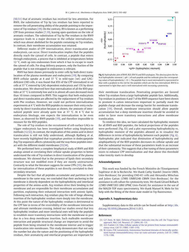
Fig. 6.Hydrophobic plot of RW9, RL9, RW16 and RX9 analogues. The abscissa gives the he-lical hydrophobicmoment (bμHN) of each peptide and the ordinate gives the correspond-ing values of hydrophobicity (bHN). Thepeptide that ismost internalized is represented inblue with a gradient to the red for the peptides which are the less internalized. RW16 isrepresented in light blue and is well internalized with increasing cytotoxicity.
601M.-L. Jobin et al. / Biochimica et Biophysica Acta 1848 (2015) 593–602
[50,51] that of aromatic residues has received far less attention. ForRW9, the substitution of Trp by Leu residues has been reported toremove the cell penetrating ability of the peptide pointing to the impor-tance of Trp residues [19]. On the contrary, R9 is known as a performingCPP from previous studies [7,19], leaving open questions on the role ofaromatic residues. The substitution of Trp by Phe residues in the RW9sequence leads to a major decrease in the cellular internalization,which was almost abolished for the peptide containing no Trp residues.In contrast, their membrane accumulation was retained.
Different modes of CPP internalization, direct translocation andendocytosis, can occur. Direct translocation implies that the peptidesdirectly reach the cytosol of cells. In contrast, a peptide that entersthrough endocytosis, a process that is inhibited at temperatures below12 °C, ends up into endosomes from which it has to escape to reachthe cytosol of cells. For drug delivery purposes, only the fraction of thepeptide that is not entrapped in endosomes is relevant.
RW9 has been shown previously to enter cells by both direct trans-location of the plasma membrane and endocytosis [19]. By comparingRW9 cellular uptake at 4 and 37 °C in wild-type (wt) and GAG-deficient CHO cells, it was found that 85% of the CPP internalized in wtcells at 37 °C entered by GAG-dependent endocytosis and 15% by directtranslocation.We observed here that internalization of all the RX9 pep-tides at 37 °C is extremely low and is in almost all cases decreasedmorethan 10 times compared to RW9. This suggests that both endocytosisand direct translocation are affected by the replacement of Trp residueswith Phe residues. However, we could not perform internalizationexperiments at 4 °Cwith the RX9peptides tomeasure their entry exclu-sively by direct translocation because as shown, the amounts of inter-nalized peptide were already quite low at 37 °C. At 4 °C due toendocytosis blockage, one expects the internalization to be evenlower, as observed for RW9 peptide [19], and therefore impossible tomeasure for the RX9 peptides.
The role of Trp residues in the recruitment of cell-surface GAGs totrigger endocytosis has been investigated before using biophysicalmethods [12,52]. In contrast, the implication of this amino acid in directtranslocation is still not fully understood, although Rydberg et al.have recently highlighted that tryptophan position in dodecapeptideArg/Trp sequences affects cytotoxicity and theway these peptides inter-act with the different model membranes [53,54].
We performed here a complete biophysical study of RW9 and RX9analogs aimed at correlating their cellular uptake properties to betterunderstand the role of Trp residues in direct translocation of the plasmamembrane. We showed that in the presence of lipids their secondarystructure was not modified even if they are mostly unstructured.Contrarily to what the literature suggests for some CPPs [55], the cellu-lar internalization of these peptides is not directly correlated to theirsecondary structure.
Despite the fact that all peptides accumulate and partition in themembrane in the same way, we concluded that their anchoring in themembrane is directly dependent on the hydrophobicity and interfacialproperties of the amino-acids. Arg residues drive their binding to themembrane and are responsible for their membrane accumulation andpartition, explaining their enhanced interaction with anionic lipids.Following such interaction, Arg residues play the role of hinge allowinginsertion of the hydrophobic residues more deeply into the membrane.At this point the nature of the hydrophobic residues is detrimental tothe CPP fate in terms of the reversibility of the membrane interactionand ultimate membrane crossing. Indeed our studies reveal that lesshydrophobic residues and more interfacial ones will help the peptidesto establish more transitory interactions with the membrane in partdue to a less deep membrane insertion. Such malleable membraneinteraction and peptide structural change are important to avoid thepeptide from being retained in the membrane interior and to triggertranslocation into membranes. This study demonstrates that not onlythe number but also the nature and the positioning of the hydrophobicresidues, their aromaticity and interfacial properties are important for
their membrane translocation. Penetrating properties are favoredwhen Trp residues form a large hydrophobic peptide face. Additionally,Trp residues in positions 4 and 7 of the RW9 sequence have been shownto promote π–cation interactions important to partially mask thepeptide charge and decrease the energy barrier for membrane translo-cation [19]. Overall, membrane interaction should allow peptideaccumulation but a deep membrane insertion should be avoided inorder to favor more transitory interactions and allow membranetranslocation.
To reinforce this idea, we have calculated the hydrophobic momentfor all RW9 and RX9 peptides, the helical projections of the peptideswere performed (Fig. S5) and a plot representing hydrophobicity vs.hydrophobic moment of the peptides allowed us to visualize thedifferences in terms of hydrophobicity between each peptide (Fig. 6).Hydrophobic plot indicated that diminution of hydrophobicity andamphipathicity of the RX9 peptides decreases its cellular uptake andthat the substantial increase of these parameters leads to an increaseof their cytotoxicity. This suggests that a fine tuning of these parametersexists to enhance CPP internalization and that above this thresholdvalue toxicity starts to develop.
Acknowledgments
This work was funded by the French Ministère de l'EnseignementSupérieur et de la Recherche. We thank Cathy Staedel (Inserm U869,Univ-Bordeaux) for providing CHO-K1 cells and Alexandra Milochauand Julien Gaitan (CNRS UMR5248 CBMN Univ-Bordeaux) for theassistance in cell culture for cytotoxicity tests. We thankGérard Bolbach(CNRS UMR7203 LBM UPMC Univ-Paris6) for assistance in the use ofthe MALDI-TOF mass spectrometry. We thank Manuel N. Melo for hishelp with the fitting of the three-state model in Trp fluorescence.
Appendix A. Supplementary data
Supplementary data to this article can be found online at http://dx.doi.org/10.1016/j.bbamem.2014.11.013.
References
[1] G.P. Dietz, M. Bahr, Delivery of bioactive molecules into the cell: the Trojan horseapproach, Mol. Cell. Neurosci. 27 (2004) 85–131.
[2] A.D. Frankel, C.O. Pabo, Cellular uptake of the tat protein from human immunodefi-ciency virus, Cell 55 (1988) 1189–1193.
[3] M. Green, P.M. Loewenstein, Autonomous functional domains of chemically synthe-sized human immunodeficiency virus tat trans-activator protein, Cell 55 (1988)1179–1188.

602 M.-L. Jobin et al. / Biochimica et Biophysica Acta 1848 (2015) 593–602
[4] E. Vives, P. Brodin, B. Lebleu, A truncated HIV-1 Tat protein basic domain rapidlytranslocates through the plasma membrane and accumulates in the cell nucleus, J.Biol. Chem. 272 (1997) 16010–16017.
[5] D. Derossi, A.H. Joliot, G. Chassaing, A. Prochiantz, The third helix of theAntennapedia homeodomain translocates through biological membranes, J. Biol.Chem. 269 (1994) 10444–10450.
[6] P.A. Wender, D.J. Mitchell, K. Pattabiraman, E.T. Pelkey, L. Steinman, J.B. Rothbard,The design, synthesis, and evaluation of molecules that enable or enhance cellularuptake: peptoid molecular transporters, Proc. Natl. Acad. Sci. U. S. A. 97 (2000)13003–13008.
[7] S. Futaki, T. Suzuki,W.Ohashi, T. Yagami, S. Tanaka, K. Ueda, Y. Sugiura, Arginine-richpeptides. An abundant source ofmembrane-permeable peptides having potential ascarriers for intracellular protein delivery, J. Biol. Chem. 276 (2001) 5836–5840.
[8] D. Derossi, S. Calvet, A. Trembleau, A. Brunissen, G. Chassaing, A. Prochiantz, Cellinternalization of the third helix of the Antennapedia homeodomain is receptor-independent, J. Biol. Chem. 271 (1996) 18188–18193.
[9] F. Duchardt, M. Fotin-Mleczek, H. Schwarz, R. Fischer, R. Brock, A comprehensivemodel for the cellular uptake of cationic cell-penetrating peptides, Traffic 8 (2007)848–866.
[10] S. El Andaloussi, P. Guterstam, U. Langel, Assessing the delivery efficacy andinternalization route of cell-penetrating peptides, Nat. Protoc. 2 (2007) 2043–2047.
[11] P. Guterstam, F. Madani, H. Hirose, T. Takeuchi, S. Futaki, S. El Andaloussi, A.Graslund, U. Langel, Elucidating cell-penetrating peptide mechanisms of action formembrane interaction, cellular uptake, and translocation utilizing the hydrophobiccounter-anion pyrenebutyrate, Biochim. Biophys. Acta 1788 (2009) 2509–2517.
[12] C.Y. Jiao, D. Delaroche, F. Burlina, I.D. Alves, G. Chassaing, S. Sagan, Translocation andendocytosis for cell-penetrating peptide internalization, J. Biol. Chem. 284 (2009)33957–33965.
[13] A. Mishra, V.D. Gordon, L. Yang, R. Coridan, G.C. Wong, HIV TAT forms pores inmembranes by inducing saddle-splay curvature: potential role of bidentatehydrogen bonding, Angew. Chem. Int. Ed. Engl. 47 (2008) 2986–2989.
[14] J.B. Rothbard, T.C. Jessop, P.A. Wender, Adaptive translocation: the role of hydrogenbonding and membrane potential in the uptake of guanidinium-rich transportersinto cells, Adv. Drug Deliv. Rev. 57 (2005) 495–504.
[15] A. Ziegler, J. Seelig, Binding and clustering of glycosaminoglycans: a commonproperty of mono- and multivalent cell-penetrating compounds, Biophys. J. 94(2008) 2142–2149.
[16] A. Ziegler, J. Seelig, Contributions of glycosaminoglycan binding and clustering to thebiological uptake of the nonamphipathic cell-penetrating peptideWR9, Biochemistry50 (2011) 4650–4664.
[17] B. Christiaens, S. Symoens, S. Verheyden, Y. Engelborghs, A. Joliot, A. Prochiantz, J.Vandekerckhove, M. Rosseneu, B. Vanloo, Tryptophan fluorescence study of theinteraction of penetratin peptides with model membranes, Eur. J. Biochem. 269(2002) 2918–2926.
[18] D. Delaroche, B. Aussedat, S. Aubry, G. Chassaing, F. Burlina, G. Clodic, G. Bolbach,S. Lavielle, S. Sagan, Tracking a new cell-penetrating (W/R) nonapeptide, through anenzyme-stable mass spectrometry reporter tag, Anal. Chem. 79 (2007) 1932–1938.
[19] A. Walrant, I. Correia, C.Y. Jiao, O. Lequin, E.H. Bent, N. Goasdoue, C. Lacombe,G. Chassaing, S. Sagan, I.D. Alves, Differentmembranebehaviour and cellular uptakeof three basic arginine-rich peptides, Biochim. Biophys. Acta 1808 (2011) 382–393.
[20] D. Derossi, G. Chassaing, A. Prochiantz, Trojan peptides: the penetratin system forintracellular delivery, Trends Cell Biol. 8 (1998) 84–87.
[21] I.D. Alves, Membrane-active peptides: mechanisms of action, Curr. Protein Pept. Sci.13 (2012) 601.
[22] P. Joanne, C. Galanth, N. Goasdoue, P. Nicolas, S. Sagan, S. Lavielle, G. Chassaing,C. El Amri, I.D. Alves, Lipid reorganization induced by membrane-active peptidesprobed using differential scanning calorimetry, Biochim. Biophys. Acta 1788(2009) 1772–1781.
[23] J. Connor, C. Bucana, I.J. Fidler, A.J. Schroit, Differentiation-dependent expression ofphosphatidylserine in mammalian plasma membranes: quantitative assessment ofouter-leaflet lipid by prothrombinase complex formation, Proc. Natl. Acad. Sci. U.S. A. 86 (1989) 3184–3188.
[24] B. Frey, U.S. Gaipl, The immune functions of phosphatidylserine in membranes ofdying cells and microvesicles, Semin. Immunopathol. 33 (2011) 497–516.
[25] S. Ran, A. Downes, P.E. Thorpe, Increased exposure of anionic phospholipids on thesurface of tumor blood vessels, Cancer Res. 62 (2002) 6132–6140.
[26] T. Utsugi, A.J. Schroit, J. Connor, C.D. Bucana, I.J. Fidler, Elevated expression ofphosphatidylserine in the outer membrane leaflet of human tumor cellsand recognition by activated human blood monocytes, Cancer Res. 51 (1991)3062–3066.
[27] V.V. Andrushchenko, H.J. Vogel, E.J. Prenner, Optimization of the hydrochloric acidconcentration used for trifluoroacetate removal from synthetic peptides, J. Pept.Sci. 13 (2007) 37–43.
[28] F. Burlina, S. Sagan, G. Bolbach, G. Chassaing, A direct approach to quantification ofthe cellular uptake of cell-penetrating peptides usingMALDI-TOFmass spectrometry,Nat. Protoc. 1 (2006) 200–205.
[29] E. Goormaghtigh, V. Cabiaux, J.M. Ruysschaert, Secondary structure and dosage ofsoluble and membrane proteins by attenuated total reflection Fourier-transforminfrared spectroscopy on hydrated films, Eur. J. Biochem. 193 (1990) 409–420.
[30] E. Goormaghtigh, V. Raussens, J.M. Ruysschaert, Attenuated total reflection infraredspectroscopy of proteins and lipids in biological membranes, Biochim. Biophys. Acta1422 (1999) 105–185.
[31] M.L. Jobin, P. Bonnafous, H. Temsamani, F. Dole, A. Grelard, E.J. Dufourc, I.D. Alves,The enhanced membrane interaction and perturbation of a cell penetrating peptidein the presence of anionic lipids: toward an understanding of its selectivity forcancer cells, Biochim. Biophys. Acta 1828 (2013) 1457–1470.
[32] M.N. Melo, M.A. Castanho, Omiganan interaction with bacterial membranes and cellwall models. Assigning a biological role to saturation, Biochim. Biophys. Acta 1768(2007) 1277–1290.
[33] A.S. Ladokhin, Distribution analysis of depth-dependent fluorescence quenching inmembranes: a practical guide, Methods Enzymol. 278 (1997) 462–473.
[34] A.S. Ladokhin, P.W. Holloway, E.G. Kostrzhevska, Distribution analysis of membranepenetration of proteins by depth-dependent fluorescence quenching, J. Fluoresc. 3(1993) 195–197.
[35] F.S. Abrams, E. London, Calibration of the parallax fluorescence quenching methodfor determination of membrane penetration depth: refinement and comparison ofquenching by spin-labeled and brominated lipids, Biochemistry 31 (1992)5312–5322.
[36] F.S. Abrams, E. London, Extension of the parallax analysis of membrane penetrationdepth to the polar region of model membranes: use of fluorescence quenching by aspin-label attached to the phospholipid polar headgroup, Biochemistry 32 (1993)10826–10831.
[37] A. Chattopadhyay, E. London, Parallax method for directmeasurement of membranepenetration depth utilizing fluorescence quenching by spin-labeled phospholipids,Biochemistry 26 (1987) 39–45.
[38] P.E. Thoren, D. Persson, E.K. Esbjorner, M. Goksor, P. Lincoln, B. Norden, Membranebinding and translocation of cell-penetrating peptides, Biochemistry 43 (2004)3471–3489.
[39] I.D. Alves, S.M. Cowell, Z. Salamon, S. Devanathan, G. Tollin, V.J. Hruby, Differentstructural states of the proteolipid membrane are produced by ligand binding tothe human delta-opioid receptor as shown by plasmon-waveguide resonancespectroscopy, Mol. Pharmacol. 65 (2004) 1248–1257.
[40] Z. Salamon, G. Tollin, Optical anisotropy in lipid bilayer membranes: coupledplasmon-waveguide resonance measurements of molecular orientation,polarizability, and shape, Biophys. J. 80 (2001) 1557–1567.
[41] Z. Salamon, G. Tollin, I. Alves, V. Hruby, Chapter 6. Plasmon resonance methods inmembrane protein biology applications to GPCR signaling, Methods Enzymol. 461(2009) 123–146.
[42] G. Drin, S. Cottin, E. Blanc, A.R. Rees, J. Temsamani, Studies on the internalizationmechanism of cationic cell-penetrating peptides, J. Biol. Chem. 278 (2003)31192–31201.
[43] H.L. Lee, E.A. Dubikovskaya, H. Hwang, A.N. Semyonov, H. Wang, L.R. Jones, R.J.Twieg, W.E. Moerner, P.A. Wender, Single-molecule motions of oligoargininetransporter conjugates on the plasma membrane of Chinese hamster ovary cells, J.Am. Chem. Soc. 130 (2008) 9364–9370.
[44] B. Christiaens, J. Grooten, M. Reusens, A. Joliot, M. Goethals, J. Vandekerckhove, A.Prochiantz, M. Rosseneu, Membrane interaction and cellular internalization ofpenetratin peptides, Eur. J. Biochem. 271 (2004) 1187–1197.
[45] S.T. Henriques, M.A. Castanho, Environmental factors that enhance the action of thecell penetrating peptide pep-1 A spectroscopic study using lipidic vesicles, Biochim.Biophys. Acta 1669 (2005) 75–86.
[46] N.C. Santos, M. Prieto, M.A. Castanho, Quantifying molecular partition into modelsystems of biomembranes: an emphasis on optical spectroscopic methods, Biochim.Biophys. Acta 1612 (2003) 123–135.
[47] P.M. Matos, H.G. Franquelim, M.A. Castanho, N.C. Santos, Quantitative assessment ofpeptide–lipid interactions. Ubiquitous fluorescence methodologies, Biochim.Biophys. Acta 1798 (2010) 1999–2012.
[48] M. Rodrigues, A. Santos, B.G. de la Torre, G. Radis-Baptista, D. Andreu, N.C. Santos,Molecular characterization of the interaction of crotamine-derived nucleolartargeting peptides with lipid membranes, Biochim. Biophys. Acta 1818 (2012)2707–2717.
[49] A.Walrant, A. Vogel, I. Correia, O. Lequin, B.E. Olausson, B. Desbat, S. Sagan, I.D. Alves,Membrane interactions of two arginine-rich peptides with different cell internaliza-tion capacities, Biochim. Biophys. Acta 1818 (2012) 1755–1763.
[50] C. Bechara, S. Sagan, Cell-penetrating peptides: 20 years later, where do we stand?FEBS Lett. 587 (2013) 1693–1702.
[51] M. Nishihara, F. Perret, T. Takeuchi, S. Futaki, A.N. Lazar, A.W. Coleman, N. Sakai, S.Matile, Arginine magic with new counterions up the sleeve, Org. Biomol. Chem. 3(2005) 1659–1669.
[52] C. Bechara,M. Pallerla, Y. Zaltsman, F. Burlina, I.D. Alves, O. Lequin, S. Sagan, Tryptophanwithin basic peptide sequences triggers glycosaminoglycan-dependent endocytosis,FASEB J. 27 (2013) 738–749.
[53] H.A. Rydberg, A. Kunze, N. Carlsson, N. Altgarde, S. Svedhem, B. Norden, Peptide-membrane interactions of arginine-tryptophan peptides probed using quartz crystalmicrobalance with dissipation monitoring, Eur. Biophys. J. 43 (2014) 12.
[54] H.A. Rydberg,M.Matson, H.L. Amand, E.K. Esbjorner, B. Norden, Effects of tryptophancontent and backbone spacing on the uptake efficiency of cell-penetrating peptides,Biochemistry 51 (2012) 5531–5539.
[55] S. Deshayes, T. Plenat, G. Aldrian-Herrada, G. Divita, C. Le Grimellec, F. Heitz, Primaryamphipathic cell-penetrating peptides: structural requirements and interactionswith model membranes, Biochemistry 43 (2004) 7698–7706.