Self-Assembly of a Designed Alternating Arginine/Phenylalanine Oligopeptide
39
Subscriber access provided by READING UNIV Langmuir is published by the American Chemical Society. 1155 Sixteenth Street N.W., Washington, DC 20036 Published by American Chemical Society. Copyright © American Chemical Society. However, no copyright claim is made to original U.S. Government works, or works produced by employees of any Commonwealth realm Crown government in the course of their duties. Article Self-Assembly of a Designed Alternating Arginine/Phenylalanine Oligopeptide Carla Cristina Decandio, Emerson Rodrigo da Silva, Ian W Hamley, Valeria Castelletto, Michelle da Silva Liberato, Vani X. Oliveira, Cristiano Luis Pinto Oliveira, and Wendel Andrade Alves Langmuir, Just Accepted Manuscript • DOI: 10.1021/acs.langmuir.5b00253 • Publication Date (Web): 30 Mar 2015 Downloaded from http://pubs.acs.org on April 3, 2015 Just Accepted “Just Accepted” manuscripts have been peer-reviewed and accepted for publication. They are posted online prior to technical editing, formatting for publication and author proofing. The American Chemical Society provides “Just Accepted” as a free service to the research community to expedite the dissemination of scientific material as soon as possible after acceptance. “Just Accepted” manuscripts appear in full in PDF format accompanied by an HTML abstract. “Just Accepted” manuscripts have been fully peer reviewed, but should not be considered the official version of record. They are accessible to all readers and citable by the Digital Object Identifier (DOI®). “Just Accepted” is an optional service offered to authors. Therefore, the “Just Accepted” Web site may not include all articles that will be published in the journal. After a manuscript is technically edited and formatted, it will be removed from the “Just Accepted” Web site and published as an ASAP article. Note that technical editing may introduce minor changes to the manuscript text and/or graphics which could affect content, and all legal disclaimers and ethical guidelines that apply to the journal pertain. ACS cannot be held responsible for errors or consequences arising from the use of information contained in these “Just Accepted” manuscripts.
-
Upload
independent -
Category
Documents
-
view
2 -
download
0
Transcript of Self-Assembly of a Designed Alternating Arginine/Phenylalanine Oligopeptide

Subscriber access provided by READING UNIV
Langmuir is published by the American Chemical Society. 1155 Sixteenth Street N.W.,Washington, DC 20036Published by American Chemical Society. Copyright © American Chemical Society.However, no copyright claim is made to original U.S. Government works, or worksproduced by employees of any Commonwealth realm Crown government in the courseof their duties.
Article
Self-Assembly of a Designed Alternating Arginine/Phenylalanine OligopeptideCarla Cristina Decandio, Emerson Rodrigo da Silva, Ian W Hamley, Valeria Castelletto, Michelle
da Silva Liberato, Vani X. Oliveira, Cristiano Luis Pinto Oliveira, and Wendel Andrade AlvesLangmuir, Just Accepted Manuscript • DOI: 10.1021/acs.langmuir.5b00253 • Publication Date (Web): 30 Mar 2015
Downloaded from http://pubs.acs.org on April 3, 2015
Just Accepted
“Just Accepted” manuscripts have been peer-reviewed and accepted for publication. They are postedonline prior to technical editing, formatting for publication and author proofing. The American ChemicalSociety provides “Just Accepted” as a free service to the research community to expedite thedissemination of scientific material as soon as possible after acceptance. “Just Accepted” manuscriptsappear in full in PDF format accompanied by an HTML abstract. “Just Accepted” manuscripts have beenfully peer reviewed, but should not be considered the official version of record. They are accessible to allreaders and citable by the Digital Object Identifier (DOI®). “Just Accepted” is an optional service offeredto authors. Therefore, the “Just Accepted” Web site may not include all articles that will be publishedin the journal. After a manuscript is technically edited and formatted, it will be removed from the “JustAccepted” Web site and published as an ASAP article. Note that technical editing may introduce minorchanges to the manuscript text and/or graphics which could affect content, and all legal disclaimersand ethical guidelines that apply to the journal pertain. ACS cannot be held responsible for errorsor consequences arising from the use of information contained in these “Just Accepted” manuscripts.

1
Self-Assembly of a Designed Alternating Arginine/Phenylalanine
Oligopeptide
Carla C. Decandioa, Emerson R. Silvaa,b,†, Ian W. Hamleyb, Valeria Castellettob,
Michelle S. Liberatoa, Vani X. Oliveira Jr.,a Cristiano L. P. Oliveirac and Wendel A.
Alvesa,†
aCentro de Ciências Naturais e Humanas, Universidade Federal do ABC, Santo André 09210-
580, Brazil bDepartment of Chemistry, University of Reading, Whiteknights, Reading RG6 6AD, United
Kingdom
cInstituto de Física, Universidade de São Paulo, São Paulo 05314970, Brazil
Abstract
A model octapeptide peptide consisting of an alternating sequence of arginine (Arg) and
phenylalanine (Phe) residues, namely [Arg-Phe]4 was prepared, and its self-assembly in solution
solution. The simple alternating [Arg-Phe]4 peptide sequence allows for unique insights into the
aggregation process and the structure of the self-assembled motifs. Fluorescence and UV-Vis
assays were used to determine critical aggregation concentrations, corresponding to the
formation of oligomeric species and β-sheet rich structures organized into both spheroidal
aggregates and highly-ordered fibrils. Electron and atomic force microscopy images show
globular aggregates and long unbranched fibres with diameters ranging from ~ 4 nm up to ~ 40
nm. Infrared and circular dichroism spectroscopy show the formation of β-sheet structures. X-
ray diffraction on oriented stalks show that the peptide fibres have an internal lamellar structure,
with an orthorhombic unit cell with parameters a ~ 27.6 Å, b ~ 9.7 Å and c ~ 9.6 Å. In situ
small-angle X-ray scattering (SAXS) shows the presence of low molecular weight oligomers in
equilibrium with mature fibres which are likely made up from 5 or 6 intertwined proto-
filaments. Finally, weak gel solutions are probed under gentle shear suggesting the ability of
these arginine-rich fibres to form networks.
Keywords: self-assembly, amyloid, fibrillization, oligopeptide, molecular Lego
Page 1 of 38
ACS Paragon Plus Environment
Langmuir
123456789101112131415161718192021222324252627282930313233343536373839404142434445464748495051525354555657585960

2
Introduction
The fibrillization of peptides has attracted a lot of interest due to its close association
with degenerative diseases such as Alzheimer’s, Parkinson’s, and diabetes type II.1-4 It is widely
recognized that such disorders arise from protein misfolding followed by self-assembly into
cytotoxic oligomers which form fibrillar structures, usually rich in β-strands, which are so-
called amyloid fibrils.5 Within this context, understanding the mechanisms involved in amyloid
formation is a significant challenge from the point-of-view of fundamental research or from the
perspective of control and treatment of these diseases.
Despite the fact that the formation of amyloid fibrils is typically observed for relatively
large proteins, it has been shown that fibrillization is possible for short sequences presenting
specific residues.6-8 For example, Tjernberg and co-workers studied fibril formation from short
segments derived from the Alzheimer’s peptide Αβ-406, 7, 9-11 containing from 3 to 10 amino
acids. The self-assembly into amyloid fibres was observed only for sequences containing more
than five residues, all the sequences containing phenylalanine residues. Since then,
phenylalanine and its strong ability for π-stacking interactions have been thought to play a
major role in amyloid formation.12 Thus, understanding the fibrillization of short peptides
containing aromatic residues, such as phenylalanine, provides a close insight into the formation
of amyloid-like structures. Unfortunately, a detailed framework to explain amyloid formation is
still lacking, although some general aspects are well-established and appear to be shared among
different amyloid fibres. For instance, morphological assays systematically show that amyloid
fibres are ~10 nm wide and a few microns long. The internal structure of amyloid fibres
consists of anti-parallel β-strands self-organised into β-sheets which run parallel to the long axis
of the fibrils. The final structures appear as a result of the stacking of proto-filaments into
mature fibres.5, 13, 14
Herein, we investigate the aggregation behaviour of a model octapeptide, containing L-
phenylalanine (Phe) and L-arginine (Arg) residues in the sequence [Arg-Phe]4, .15 The generic
sequence consisting of intercalated hydrophilic and hydrophobic amino acids, corresponds to a
Page 2 of 38
ACS Paragon Plus Environment
Langmuir
123456789101112131415161718192021222324252627282930313233343536373839404142434445464748495051525354555657585960

3
Type I peptide of the “molecular Lego” group, a family of peptides with recognized capabilities
to form fibrillar assemblies usually rich in β-sheet structures.16, 17 In addition, octapeptides have
been successfully used to form fibres and to prepare hydrogel matrices with potential
biotechnological applications.18-21 Our purpose here is to provide detailed information on the
aggregation and structure of [Arg-Phe]4, which is rich in arginine and does not rely on
complementary ionic pairing for the self-assembly.16 The choice of Arg and Phe is motivated by
the properties of their side-chains. Phe presents an aromatic side chain able to establish strong
π−π interactions and promote amyloid fibre formation.8, 11 Arg is endowed with a side-chain
guanidinium group, making it a strong H-bond donator.22 Arg is positively charged at
physiological pH, enabling cation-π interaction between the NH+ group and the phenyl ring in
the Phe side-chain.23 The self-assembly of [Arg-Phe]4 into fibres, using a solid-vapour
deposition strategy, was previously reported by us.15 Here, we report a detailed study of [Arg-
Phe]4 self-assembly in solution by using a wide range of experimental techniques encompassing
spectroscopy, microscopy and X-ray methods to determine the critical aggregation
concentration. We characterize in detail the secondary structure and give insights into the
pathway leading to the formation of fibres. We show that this simple alternating cationic
octapeptide exhibits many features of amyloid-fibrils and is a potential model for investigating
fibrillization.
Experimental Section
Peptide synthesis
All chemicals had analytical or HPLC grade. Protected amino acids, [(Boc-Phe-OH)]
and [(Boc-Arg(Tos)-OH)], 1,3-diisopropylcarbodiimide/N-hydroxybenzotriazole (DIC/HOBt),
trifluoroacetic acid (TFA), anisole, dichloromethane (DCM), dimethylformamide (DMF),
diisopropylethylamine (DIEA) and acetonitrile (ACN) were purchased from Sigma-Aldrich
(Saint Louis, MO, USA). Chloromethyl resin (Merrifield) was purchased from Advanced
Chemtech (Louisville, KY, USA), with substitution degree 0.90 mmol/g and starting amino acid
Page 3 of 38
ACS Paragon Plus Environment
Langmuir
123456789101112131415161718192021222324252627282930313233343536373839404142434445464748495051525354555657585960

4
coupled to the polymeric support. The synthesis of the octapeptides was performed as detailed
elsewhere15 and, in brief, the procedure was as follows: the protected group was removed by
reaction with 50% TFA and 2% anisole in DCM for 20 minutes. Coupling was carried out in 2.5
fold excess of DIC/HOBt in DCM/DMF (1:1, v:v). Reactions were monitored using the Kaiser
ninhydrin test and peptidyl resin was acetylated with 25% acetic anhydride in DMF with excess
DIEA for 20 minutes. Dry-protected resin was exposed to 70% TFA + 20% TFMSA and 10%
anisole during 12 hours, at 4°C. After free-drying, crude peptide was purified by reverse-phase
HPLC in aqueous solutions of ACN + 0.1% TFA (60:40, v:v) on a Waters Delta Prep 600
system, exhibiting purity > 96%. Mass spectrometry was conducted on liquid-chromatography
electrospray-ionization mass spectrometer, LC/ESI-MS yielding [Arg-Phe]4 (MM + H) = 1276
(calculated = 1275).
Sample Preparation
Aqueous solutions were prepared by dissolving peptides in Milli-Q water (resistivity >
18 MΩ.cm at 25 °C), at concentrations typically ranging from 1 × 10⁻⁵ wt% to 2 wt%.
Measurements with indicator paper showed pH between ~ 6.0 (at higher peptide concentrations)
and ~ 7.5 across the investigated range. For FTIR experiments, solutions were prepared in D2O
(Fluka). Critical aggregation concentration measurements were performed using solutions
containing thioflavin T (ThT) at 1 × 10⁻³ wt%, pyrene (Pyr) or 1-pyrenecarboxylic acid (1PCA)
at 1 × 10⁻⁴ wt%. Samples were submitted to ultrasound just after mixing (50 °C, ~15 minutes)
and then stored in the dark to equilibrate at room temperature for at least 24 hours before further
analyses.
Spectroscopy Methods
Fluorescence spectra were recorded on a Perkin Elmer spectrometer. Samples were
loaded into a 10 mm inner width quartz cuvette. Excitation wavelengths were λex.= 338 nm or
λex.= 440 nm for solutions containing Pyrene or ThT, respectively. Excitation and emission slits
Page 4 of 38
ACS Paragon Plus Environment
Langmuir
123456789101112131415161718192021222324252627282930313233343536373839404142434445464748495051525354555657585960

5
were kept at 5 nm and spectra were recorded in the (345-500) nm or (450-600) nm range for Pyr
and ThT fluorescence assays, respectively. Absorbance profiles were monitored on a Varian
Cary 50 spectrophotometer. Solutions were loaded into a 10 mm path length quartz cuvette. The
UV-Vis range was scanned from 220 nm to 370 nm and the data was treated for baseline
correction. Fourier transform infrared (FTIR) assays were carried out on a Varian 660-IR
spectrometer operating in ATR mode. Droplets from [Arg-Phe]4 solutions in D2O were
deposited onto a ZnSe crystal, left to dry and 80 accumulations were obtained in the interval
1500-1800 cm-1, with step resolution of 2 cm-1. After background subtraction, resonances were
deconvoluted and fitted to Gaussian profiles using the program Fityk24 to enable accurate
assignment. Circular dichroism (CD) measurements were performed using synchrotron
radiation at the CD beamline B23 (Diamond Light Source, UK), using the nitrogen-flushed
Module B end-station spectrophotometer. The samples were loaded in CaF2 cells with path
lengths varying between 0.05 and 10 mm according to concentration in order to optimize
absorbance. Data have been converted into mean residue ellipticity units (MRE) for comparison
between different concentrations and path lengths. The data, recorded with a 1 nm step and 1
second collection time per step, are presented as the averaged results from 4 scans. CD data was
corrected by background from the solvent, and correspond to absorbance A < 2 at any measured
point. All experiments were performed at 20 °C.
Microscopy Imaging
Atomic force (AFM) and high-resolution transmission electron microscopy (TEM) were
carried out at the Brazilian Nanotechnology National Laboratory (Campinas, Brazil). AFM was
performed using a Digital Instruments Nanoscope III operating in tapping mode. Droplets were
deposited onto freshly-cleaved mica substrates, left to rest for a couple of minutes, rinsed with
deionised water and dried overnight under a gentle nitrogen stream. Images were obtained by
scanning 1024 × 1024 pixels covering surfaces between 1 and 5 µm². Topology and phase data
were enhanced and analyzed using the Gwydion package.25 Electron microscopy was performed
Page 5 of 38
ACS Paragon Plus Environment
Langmuir
123456789101112131415161718192021222324252627282930313233343536373839404142434445464748495051525354555657585960

6
on a TEM-HR JEOL 3010 operating at 300 kV. Drops from a peptide solution were cast onto
Cu grids coated with carbon film, stained with a 2% uranyl acetate solution (Sigma) and let to
dry in a desiccator.
Fibre diffraction and Small-Angle X-ray Scattering
Fibre diffraction patterns were recorded from oriented stalks obtained by suspending
droplets of a 5 wt% peptide solution between the ends of wax-coated capillaries and letting
them dry in air at RT. We observed that solutions incubated for at least ten days at RT were
much better able to produce well-oriented samples. Stalks were vertically positioned onto a
RAXIS IV++X-ray diffractometer (Rigaku) equipped with a rotating anode generator and data
were collected using a Saturn 992 CCD camera, with sample-to-detector distance of 40 mm.
Small-Angle X-ray Scattering (SAXS) was carried out on the BioSAXS BM29 beamline at the
ESRF (Grenoble, France). Solutions were loaded into PCR tubes in an automated sample
changer kept at 20 °C. Around 50 µL from solutions were injected into a 1 mm quartz capillary
and successive frames of 0.2 s each were registered during flow to avoid radiation damage. Data
were recorded using a Pilatus 1M detector, positioned at sample-to-detector distance of 2,864
mm. The X-ray wavelength was λ = 0.99 Å and reduction to 1-D intensity curves was
performed by radial integration using the beamline software. This configuration provided high-
resolution data in the range 0.04 nm-1 ≤ q ≤ 4.9 nm-1 (where q = 4π/λ × sinθ, with 2θ the
scattering angle). Fit2D was used to process 2D images from both XRD and SAXS assays. The
program CLEARER26 was used for optimizing unit cell parameters from XRD assays and the
SASFit package27 was used to fit all scattering data.
Results and Discussion
Critical aggregation concentrations
Fluorescence experiments were used to determine the critical aggregation concentration,
c.a.c, of [Arg-Phe]4, The cac was measured in three separate experiments, by preparing a series
Page 6 of 38
ACS Paragon Plus Environment
Langmuir
123456789101112131415161718192021222324252627282930313233343536373839404142434445464748495051525354555657585960

7
of peptide solutions using Pyr, ThT or 1PCA as a probe. In these experiments, the concentration
of fluorophore (probe) was kept constant whereas the amount of peptide was varied across a
wide concentration range. ThT has been widely used to identify and quantify amyloid structures
due to the enhancement of its fluorescence emission upon association to β-sheet assemblies5, 28,
29 The exact mechanisms involved in this process is not fully understood. However, it has been
suggested that intercalation of the ThT probe into β-sheet grooves supresses torsions and twists
of the ThT, favouring higher fluorescence yields.28 Pyr species, in turn, are hydrophobic
compounds, highly sensitive to the polarity of the medium.30,31 During the self-assembly of
peptide monomers in a Pyr solution, the Pyr is sequestered within the self-assembled
hydrophobic clusters and the fluorescence of the probe is enhanced. The 1PCA chromophore is
very similar to Pyr, but it possesses a carboxyl group attached to the pyrenyl moiety (see inserts
in Figure 1). This polar group gives a slightly amphiphilic behaviour to the probe, modifying its
interaction with the assemblies in solution. Since our [Arg-Phe]4 peptide contains alternating
polar and nonpolar amino acids, 1PCA is able to interact either with hydrophilic or hydrophobic
sites in the arrays. Therefore we have monitored characteristic bands in the absorbance profile
to estimate the cac. The use of three different probes, i.e. ThT, Pyr and 1PCA, provides
measurements based on independent mechanisms allowing for estimations of cac values of
intermediate species on the self-assembly pathway.
In Figure 1A, we show the dependence of ThT fluorescence as a function of the peptide
concentration. Emission was measured at λem. = 490 nm (maximum) and data were normalized
to the intensity found in a peptide-free solution. The curve exhibits two distinct concentration
regimes. The more dilute domain is characterized by normalized fluorescence intensities below
unity, indicating a possible quenching of chromophore in solution. In the second domain, the
emission exhibits a plateau followed by a linear fluorescence increase which is assigned to
appearance of β-sheet-rich structures.5 To provide a quantitative estimation of the cac arising
from these two regimes, we have fitted the data at the low-concentration regime using a
sigmoidal function characterized by an inflection point at 0.0010 ± 0.0005 wt% peptide. This
Page 7 of 38
ACS Paragon Plus Environment
Langmuir
123456789101112131415161718192021222324252627282930313233343536373839404142434445464748495051525354555657585960

8
value is taken as an estimation of critical concentration and could be associated to formation of
low molecular weight oligomers without well-organized structures. The second domain has
been fitted with a linear function and its intercept with the plateau has been found at 0.035 ±
0.010 wt% peptide.
Figure 1B shows the intensity of the fluorescence emission of Pyr as a function of the
peptide concentration. Fluorescence was monitored at the emission maximum, λem = 393 nm.
The Pyr fluorescence emission in solutions containing peptide is much lower than in peptide-
free solutions, across the entire peptide concentration range. This finding results from the
unique design of [Arg-Phe]4, which contains an equal number of phenyl and guanidium moieties
alternating along the sequence. In this case, Pyr species interacting with phenyl groups in the
assemblies are also surrounded by strongly polar Arg side-chains hindering an appreciable
growth of fluorescence. Also, at neutral pH, Arg residues are positively-charged and this likely
implies very high ionic strength in medium. Despite the complex interplay between electrostatic
and aromatic stacking interactions, it is possible identify a clear inflexion point at 0.17 ± 0.03
wt% peptide (Figure 1B) which is attributed to the appearance of a higher level of organization,
maybe with polar and nonpolar galleries spatially separated within the assemblies. As discussed
below, this critical concentration could be attributed to formation of fibres in solution exhibiting
a well-defined crystalline ordering.
Figure 1C shows the UV-Visible spectra measured for peptide solutions containing
1PCA. The absorbance patterns in Figure 1C are quite complex, exhibiting resonances that arise
either from 1PCA or from the aromatic groups at the Phe side-chains. In addition, the profiles
depend on the concentration of peptide, with the appearance and disappearance of resonances
across the UV-Vis range. Again, two regimes are observed. In the more diluted one, spectra are
dominated by four strong peaks at 243 nm, 275 nm, 326 nm and 342 nm. These resonances are
characteristic of 1PCA and have been assigned to π-π* transitions in the molecule.32 As the
amount of [Arg-Phe]4 increases, a systematic decrease in intensity of these bands is observed
and the effect is even more remarkable in the near UV-region, where a doublet at 326 nm and
Page 8 of 38
ACS Paragon Plus Environment
Langmuir
123456789101112131415161718192021222324252627282930313233343536373839404142434445464748495051525354555657585960

9
342 nm is found (see insert in Figure 1C). Along with the disappearance of 1PCA bands, three
peaks characteristic of phenylalanine-containing compounds, emerge at 252 nm, 258 nm and
264 nm.33 To estimate the cac, we have normalized the 1PCA peak at 342 nm to the intensity
measured in a peptide-free solution and plotted the resulting values as a function of peptide
concentration (see Figure 1D). The curve has a sigmoid-like shape with an abrupt transition
between the domains reported above. To properly describe this sudden changeover, we have
used an empirical function which has been successfully employed in titration assays to describe
the behaviour of enthalpy changes close to the critical micelle concentration.34, 35
= ∙ [log ] + 1 + [log ] − ∆
+ ∙ [log ] +
In equation (1), Iabs is the normalized absorbance and c is the peptide concentration.
Parameters a1 and a4 are the slope while a2 and a5 are the intercept terms to account for the linear
regions of the profiles. Parameters a3 and ∆a3 are the parameters of an exponential profile used
to describe a transition between regimes and, correspond to the inflexion point of the curve (a3)
and the width (∆a3) of the transition. The cac was determined as the minimum of the first
derivative of the fitted curve in Figure 1D, while the corresponding FMWH was taken as the
uncertainty, providing cac ~ 0.048 ± 0.011 wt% peptide. Since this value is in good agreement
with the cac found from the ThT (Figure 1A), the transition point in Figure 1D was attributed to
the formation of β-sheet structures in solution.
Gathering the results exhibited above, we conclude that the aggregation behaviour is
complex and different levels of organization likely appear along the self-assembly pathway.
Briefly, the results in Figure 1 provide three critical concentrations : cac1 = 0.001 ± 0.005 wt%
peptide, cac2 = 0.042 ± 0.017 wt% peptide and cac3 = 0.17 ± 0.03 wt% peptide. Accurate
interpretations of these critical points are not trivial; however, on the basis of data presented in
the following, we can tentatively assign them, respectively, to oligomeric species (cac1),
Page 9 of 38
ACS Paragon Plus Environment
Langmuir
123456789101112131415161718192021222324252627282930313233343536373839404142434445464748495051525354555657585960

10
formation of globular aggregates (cac2) containing β-sheet cores and, finally, the appearance of
amyloid-fibres in solution (cac3).
Secondary structure
The secondary structure of the self-assembled peptide was investigated performing
FTIR and CD experiments. Figure 2A shows a representative FTIR spectrum corresponding to
0.25 wt% [Arg-Phe]4 in D2O, above cac3 and presumably containing fibres in solution. We
have focused our analyses on the amide I region, where vibrations from C=O stretching provide
information about the backbone conformation.36 Deconvolution using Gaussian peak fitting
reveals that the amide I region is dominated by a pronounced peak at 1624 cm-1, corresponding
to a classical signature of β-sheets formed via H-bonds between adjacent strands5. The observed
narrow and strong peak is consistent with highly delocalized vibrational modes oriented along
the axis perpendicular to strands and points to a high level of organization presumably into a
cross-β structure.37 A second strong peak is observed at 1673 cm-1 and it is assigned to the
presence of TFA counterions in solution.38 These vibrations appear flanked by two shallow
shoulders at 1589 cm-1 and 1722 cm-1. The resonance at 1589 cm-1 may be attributed to modes
within arginine side-chains 39, 40 and the mode at 1722 cm-1 is assigned to H-bonded carboxylic
acid stretch,41 indicating an acidic environment in agreement with the presence of TFA in
solution.
Investigating secondary structure at lower concentrations via FTIR methods is a non-
trivial task because of sensitivity limitations. In this case, CD analysis could be an alternative
tool to probe the secondary structure.42 A representative CD spectrum from [Arg-Phe]4 solutions
is exhibited in Figure 2B and supplementary data are found in the SI Fig.1. The extensive
presence of aromatic chromophores (50% of our peptide are made of Phe residues) leads to
heavily mixed spectra exhibiting electronic transitions from peptide backbones and/or benzene
side-chains.43,44 In this case, it is not possible to provide unequivocal assignments exclusively on
the basis of CD spectroscopy and only qualitative analysis has been made by comparing our
Page 10 of 38
ACS Paragon Plus Environment
Langmuir
123456789101112131415161718192021222324252627282930313233343536373839404142434445464748495051525354555657585960

11
data with fluorescence or FTIR information. As a rule, the spectra for the octapeptide
systematically exhibit a pronounced negative band at 188 nm, followed by a shallow positive
maximum at 198 nm. This signature has been previous assigned to β-turns and β-pleated sheet
conformations.45 At higher wavelength, a weak minimum is observed at 203 nm and a intense
positive maximum emerges at 219 nm. The shallow negative band could be related to a small
fraction of disordered secondary structures,43 whereas the strong positive peak arises from n-π*
transitions within the phenyl groups in phenylalanine.42-44 In samples with concentrations above
cac2, the growth in ThT emission observed in Figure 1A also suggests the presence of β-sheet
cores in these aggregates. For concentrations above cac3, FTIR spectra shown in Figure 2A and
XRD data discussed below unequivocally show β-sheet organization.
Microscopy Imaging
Figure 3 shows AFM images for peptide films dried from solutions 4 × 10⁻⁴ wt% (below
cac1) ; 0.056 wt%, (marginally above the cac2) ; and 0.5 wt% (well above cac3). Topographic
images reveal a clear relationship between morphology and concentration. In the most dilute
regime, we observe small aggregates with heights of ~3 nm and lateral sizes ranging from ~15
nm to ~ 100 nm (lateral values probably overestimated by ~ 5 nm due to the finite size of the
AFM tip46). The shape of these assemblies is irregular and elongated spheroids, possibly arising
from association of two or more rounded units, are also observed. At an intermediate
concentration, Figure 3B, the self-assembled nanostructure exhibits a dramatic change. A large
number of spheroidal aggregates are observed with heights around 8 nm and lateral diameters
around 20 nm. The formation of spheroidal aggregates has already been observed at the early
stages of amyloid-fibrillization when small globular or ellipsoidal aggregates associate to form
fibres.46 Sinuous semi-flexible fibrils are observed at high concentration (Figure 3C). A
complex network of entangled fibres appears with widths ranging from ~20 nm to ~45 nm and
heights ~ 10 nm. The results in Figure 3 clearly associate cac1, cac2, and cac3 to the self-
Page 11 of 38
ACS Paragon Plus Environment
Langmuir
123456789101112131415161718192021222324252627282930313233343536373839404142434445464748495051525354555657585960

12
assembly in solution of three different species, as previously from the results displayed in
Figures 1.
High resolution transmission electron microscopy (HRTEM) was performed to assess
the internal structure of the fibrils in Figure 3C in more detail. For this, samples were prepared
from a solution with [Arg-Phe]4 at 1.5 wt%. The corresponding images are displayed in Figure
3D-F and they clearly reveal the presence of sub-units forming the fibres. Specifically, we
observe structures with sizes between 15 nm and 20 nm, formed from intertwined filaments
with cross-sections ~7 nm. In Figure 3D and 3E, these sub filaments are indicated by white
arrows whereas fibres are highlighted by black arrows. Such a hierarchical process, where proto
fibrils entangle into larger filaments, apparently occurs down to quite small length-scales as
shown in Figure 3F. In this high-magnification image, we see thicker filaments formed by
proto-fibrils as small as 2.5 nm wide.
X-rays Measurements
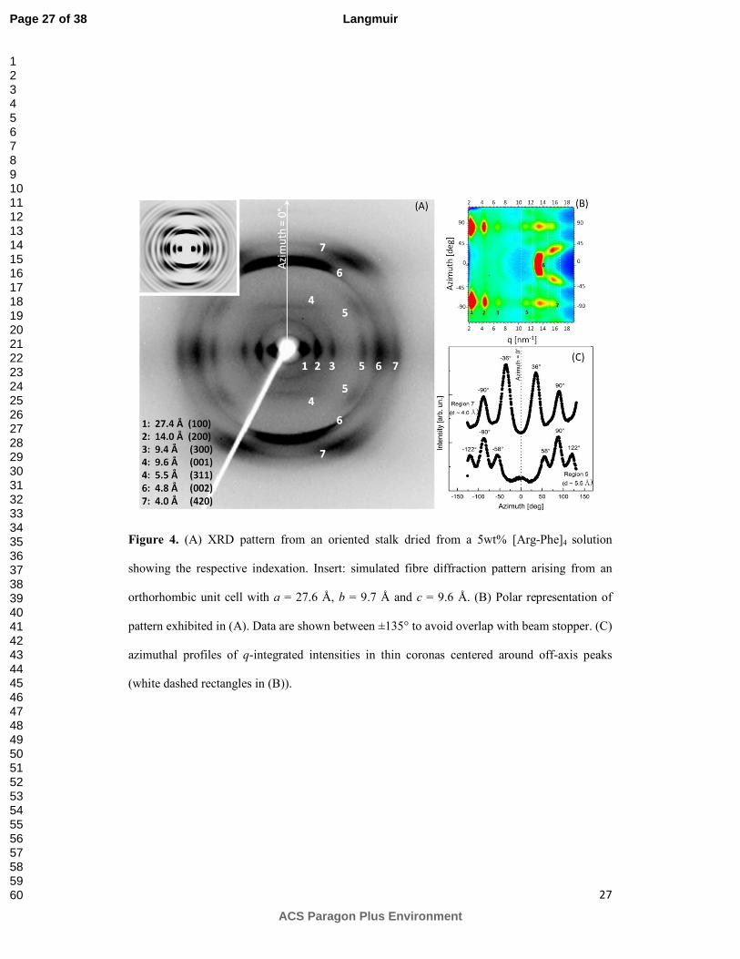
X-ray fibre diffraction was performed on stalks prepared from a concentrated sample
containing the octapeptide at 5 wt% to probe the ordering of β-strands in the assemblies. A
representative two-dimensional pattern from oriented fibres is exhibited in Figure 4A. This
highly oriented pattern, corresponding to a very well ordered cross-β pattern, enables a detailed
analysis of the fibril structure. Two meridional arcs are observed at d = 4.8 Å which are
associated to the separation between adjacent peptide backbones organized into β-strands
(position 6 in 4A).29 The pronounced localization of these reflections along the meridian
indicates perpendicular orientation of strands with respect to the stalk axis and their sharpness
points to a high degree of correlation between [Arg-Phe]4 chains. Also along the vertical axis,
we observe weak horizontal layer lines at d ~ 9.6 Å which corresponds to twice the β-strands
separation and could indicate anti-parallel organization of the chains.47 Therefore, this distance
is associated with the periodicity of strands with the same orientation along the structure. These
meridional diffractions appear accompanied by a group of strong equatorial peaks at d = 27.4 Å,
Page 12 of 38
ACS Paragon Plus Environment
Langmuir
123456789101112131415161718192021222324252627282930313233343536373839404142434445464748495051525354555657585960

13
14.0 Å and 9.4 Å, indicating the presence of a lamellar phase along the fibre cross-section in
agreement with previous molecular dynamic simulations.15 A second group of equatorial
reflections is found at 5.5 Å, 4.8 Å and 4.0 Å. In addition to horizontal spots, spacing d = 5.5 Å
and d = 4.0 Å are also found at off-axis positions. Analysis of these oriented reflections is more
complex, but they indicate higher order-degree in the fibres and very efficient packing of
peptide side-chains, possibly with helical twisting of β-sheets along the fibre axis.48 To provide
more detail on angular positions, a polar representation of the pattern is exhibited in Figure 4B,
where azimuthal angles appear as a function of scattering vector. In Figure 4C, q-averaged
profiles, integrated into thin slices around rings containing off-axis maxima, are shown. Peaks
corresponding to d = 4.0 Å are observed at χ ~ ±36° (maxima are also found at χ ~ ±155°;
however, this region was not included in the polar representation because of beam stopper
overlap). The peak at d = 5.5 Å is further from the azimuth and appear at χ ~ ±58° and χ ~
±122°.
The information in the XRD pattern allows to propose a structural model for [Arg-
Phe]4 fibres based on characteristic features of amyloid-like assemblies. We have indexed the
pattern according to an orthorhombic unit cell with lattice parameters a = 27.6 Å, b = 9.7 Å and
c = 9.6 Å. The outstanding equatorial reflections, marked by positions 1-3 in Figure 4A, have
been assigned to Miller planes (100), (200) and (300) whereas the shallow meridional line at 9.6
Å and the intense arc at 4.8 Å have been attributed to planes (001) and (002), respectively.
Finally, peaks at d = 5.5 Å and d = 4.0 Å have been indexed to planes (311) and (420) and
simulation performed using the Fibre Diffraction Simulation module of CLEARER,26 insert in
Figure 4A, have properly predicted their off-axis positions. The lattice parameters arising from
our structural model are fully compatible with major features typically observed in amyloid-
fibres.48 For example, parameter a = 27.6 Å is assigned to the length of an extended octapeptide
chain (~ 8 × 3.4 Å = 27.2 Å) and c = 9.6 Å is twice the separation between hydrogen bonded β-
strands, consistent with anti-parallel arrangement. The parameter b = 9.7 Å is compatible with
the spacing between stacked β-sheets running parallel to the fibre axis.49, 50
Page 13 of 38
ACS Paragon Plus Environment
Langmuir
123456789101112131415161718192021222324252627282930313233343536373839404142434445464748495051525354555657585960

14
Figure 5 displays a tentative model for the internal structure of [Arg-Phe]4 fibres, in
keeping with the information extracted from the results displayed in Figures 3C-F and 4. The
model in Figure 5 shows the formation of galleries hosting Phe or Arg layers, which are held
together by strong π-stacking interactions and H-bonds between peptide backbones. We propose
that the structure is stabilized due to the interplay of two complementary mechanisms. The first
one is ascribed to the presence of counter ions (namely residual TFA arising from the synthesis
procedure) screening electrostatic repulsions within arginine galleries. Similar phenomena have
been recently reported to play a major role in the self-assembly and stabilization of peptide
nanotubes built from cationic octapeptides.51 The second one could be associated to a possible
role of water molecules mediating H-bonding across the structure, in agreement with the strong
H-bond donating behaviour of arginine side-chains, consistent with previous molecular dynamic
simulations.15” This arrangement, with hydrophobic galleries flanked by hydrophilic layers, is
compatible with abundant π−π interactions identified in our UV-Vis data and is in good
agreement with Pyr fluorescence, which has been found to be relatively low due to the presence
of polar groups in the vicinity of chromophore species (Figure 1). Therefore, we propose that
the inflexion point in the Pyr fluorescence (Figure 1B) may be correlated to the appearance of
the hydrophobic layers able to host nonpolar species. At lower concentrations, when the
solution is populated by globular aggregates (Figure 3B), the system is less organized and
presumably strong segregation of hydrophobic and hydrophilic sites is not present, accounting
for the low value of the pyrene fluorescence emission.
SAXS provides convenient in situ methods to probe the internal structure of fibres.
Solutions were prepared at four peptide concentrations: 0.05 wt%, close to the estimated cac2;
0.1 wt%, 0.2 wt%, in the vicinities of cac3 and 1 wt%, well above cac3. In particular, a
viscosity growth after a few days at room temperature of the more concentrated sample,
suggested the formation of fibres. Samples studied by SAXS were investigated ~30 days after
mixing. Figure 6 shows the SAXS curves measured at 0.05 – 1 wt% peptide and the overall
results determined by model fitting analysis are summarized in Table 1. The shape of the
curves clearly depends on peptide concentration; however, a common feature among them is the
Page 14 of 38
ACS Paragon Plus Environment
Langmuir
123456789101112131415161718192021222324252627282930313233343536373839404142434445464748495051525354555657585960

15
presence of two levels of structural organizations observed at intermediate q-values. The SAXS
curves are characterized by a smooth descent and to describe this high-angle region we have
used a Porod cylinder form factor52 plus a flat background (see Eq. SI2 and SI3 in supporting
info file). Our results show that contributions from these small particles are well-described by
short cylinders with radii between R = 0.6 nm and R = 1.1 nm, and lengths between L = 2.6 nm
and 4.5 nm. We ascribe this component to the presence of small oligomers hosting just a few
[Arg-Phe]4 strands. The volume of these cylindrical aggregates ranges from ~3 nm³ to ~17 nm³
being able to accommodate from ~2 to ~11 monomers (octapeptide volume ~1.5 nm³). SAXS is
a powerful method providing structural information on amyloid oligomers, 53, 54 however,
accurate analysis is complex2 since they are present at low concentration, have low molecular
weight and many species may coexist in solution.
The most dilute sample, 0.05 wt%, exhibits a profile which contrasts with its more
concentrated counterparts. The low-q region of this sample is characterized by a remarkable
curvature and data from this domain have been properly fitted using a form factor for
polydisperse ellipsoids (see Eq. SI4).52 Our results have led to average prolate-shaped particles
with semi-principal axis of 43.4 ± 31.7 nm and radii R = 12.3 ± 8.9 nm, where uncertainty has
been obtained from the standard deviation of a Gaussian distribution. These results agree with
AFM images from samples at this same concentration, where aggregates of spheroidal particles
with diameters averaging ~ 20 nm have been found (see Figure 3B). The presence of similar
structures, both in size and shape, has been observed in the context of amyloid formation where
these aggregates are frequently described as “seeds” in the pathway toward the growth of
fibres.2, 47, 53 The high degree of dispersity in the radii in our solution data could be ascribed to
the proximity to a critical regime, where association and dissociation between aggregates
strongly affect the dynamic equilibrium and presumably generates size polydispersity.
Page 15 of 38
ACS Paragon Plus Environment
Langmuir
123456789101112131415161718192021222324252627282930313233343536373839404142434445464748495051525354555657585960

16
Table 1. Parameters obtained from model fitting of SAXS data.
Short cyl.
Form factor
Long cyl.
Form factor*
Ellipsoid
Form factor
Sample
(wt%)
Contribution R
(nm)
L
(nm)
R ± SD
(nm)
R± SD
(nm)
ννννR ± SD
(nm)
0.05
Oligomers 1.0 3.2 --- --- ---
Aggregates --- --- --- 12.3 ± 8.9 43.4± 31.7
0.1
Oligomers 0.7 3.3 --- --- ---
Fibres --- --- 7.0 ± 4.9 --- ---
0.2
Oligomers 1.1 4.5 --- --- ---
Fibres --- --- 8.0 ± 6.7 --- ---
1
Oligomers 0.6 2.6 --- --- ---
Fibres --- --- 6.5 ± 1.1 --- ---
*Cylinder length fixed at L = 500 nm and scattering length density fixed at η = 1.
For the more concentrated samples, the low-q regions in the SAXS profiles do not have
a curved shape being rather typified by a linear profile scaling with q~1. This characteristic
indicates that this domain is dominated by a cylinder form factor, in accordance with fibrillar
structures in solution. To describe these profiles, we have used Porod’s approximation for long
cylinders52 together with a Gaussian distribution to account for polydispersity in fibre radii. The
length of cylinders was fixed at L = 500 nm in agreement with long persistence lengths
indicated by TEM images and, since L >> R, this parameter simply behaved as a scaling factor.
The scattering length density was arbitrarily fixed at η = 1 and thus only four parameters
(radius, standard deviation, a scaling constant and a background constant) were left free to
adjust. Our fitting procedures yielded R = 7.0 ± 4.9 nm, R = 8.0 ± 6.7 nm and R = 6.5 ± 1.1 nm,
respectively, for samples with concentrations 0.1 wt%, 0.2 wt% and 1 wt%. It should be noted
that, although the concentration 0.1 wt% is marginally below cac3, data could not be properly
described by sphere form factors likely associated to globular aggregates. We suspect that this
Page 16 of 38
ACS Paragon Plus Environment
Langmuir
123456789101112131415161718192021222324252627282930313233343536373839404142434445464748495051525354555657585960

17
could be related to the fact of SAXS measurements had been performed ~ four weeks after
sample preparation which could imply some time-dependent evolution of the assemblies as
found in our aged sample discussed below. We observe that the in situ measurements agree with
images from AFM and TEM, however, an interesting feature arising from these assays is that
fibres exhibit high polydispersity in solution at lower concentrations. The fibres found here
show a wide range of sizes in the mixture, which may be due to their assembly from smaller
proto-filaments, suggested by the modelling of the XRD data discussed above. Considering
averages and standard deviations, fibres have minimum radius of ~1.3 nm and a maximum ~15
nm. The lowest values could be assigned to the size of a proto-filament formed by a pair of
twisted β-sheets, whereas the largest one is compatible with a pair of bundled fibres such those
ones observed in microscopy assays. The mature fibres have radii ~7.0 nm and are made up by
5 or 6 intertwined proto-filaments. In this case, the fibres investigated here exhibit unequivocal
features of amyloid-like structures across the mesoscale, from the supramolecular level, where
cross-β strands assembled into β-sheets, up to nanometer dimensions where intertwined proto-
filaments form the fibres.55
Upon increasing concentration, SAXS shows that the fibril radius polydispersity
decreases sharply. In this case, the mixture is considerably above cac and thus far from the
critical regime. In this scenario, equilibrium between fibres and oligomeric species is shifted
favouring fibrillization and this may explain the reduced polydispersity. In fact, concomitantly
with decrease on polydispersity of fibrillar component, we observe that the short-cylinder
contribution used to describe the high-angle region of the SAXS curves exhibits the lowest
volume among investigated samples corresponding approximately to dimers in solution.
We have also observed formation of weak gel solutions upon aging under refrigerated
conditions. In addition to samples discussed above, a 1 wt% formulation was examined ~ 120
days after preparation, when it exhibited higher viscosity and appreciable adhesion to walls of
the vial. To provide some insight on this state, we performed SAXS under flow using rate of Q
= 8.3 µl·s-1 across a 1 mm wide capillary. These conditions imply a Newtonian shear rate at the
Page 17 of 38
ACS Paragon Plus Environment
Langmuir
123456789101112131415161718192021222324252627282930313233343536373839404142434445464748495051525354555657585960

18
wall of γ = 32Q/πR³ = 85 s-1. A representative 2D pattern obtained under these conditions is
shown in Figure 7A and it clearly exhibits high anisotropy along the vertical axis which is
indicates alignment along flow direction (horizontal). In 7B, we show the polar representation
of the pattern, confirming that the intensity maxima are highly concentrated around the
meridional axes and the corresponding q-integrated profile, Figure 7C, shows peaks at -8° and -
185°. A quantitative estimation on orientation could be calculated through the orientational
order parameter given by: 56
= 3⟨cos $⟩ − 12 2
⟨'($⟩ = ) *, $'($ sin $.$/0) *, $ sin $.$/0
21
Where I(q,χ) is the q-integrated intensity at a given azimuthal angle χ. From Equations
(2), we obtained an order parameter F ~ 0.2 which denotes high orientation upon relatively
gentle shear. We suspect that this state of the mixture arises from a high correlation between
fibres, possibly due to entanglements formed upon aging. In fact, such anisotropy in the
scattering profile is not observed at early stages when fibres are uncorrelated and likely do not
form networks in solution. These findings indicate that these [Arg-Phe]4 fibrils could be used
for preparing self-sustained matrices and possibly designing hydrogel materials in the future.
This could be particularly attractive for providing an arginine-rich medium either to host
molecular cargoes57, 58 or to investigate specific reactions – e.g., nitric oxide catalysis59, 60 -
involving this amino acid in a biomimetic environment.
Page 18 of 38
ACS Paragon Plus Environment
Langmuir
123456789101112131415161718192021222324252627282930313233343536373839404142434445464748495051525354555657585960

19
Conclusions
We have found that an alternating arginine/phenylalanine octapeptide forms amyloid-
type β-sheet rich fibrils and other amyloid-like analogs along its self-assembly pathway. Critical
aggregation concentrations obtained from complementary fluorescence and absorbance
spectroscopy provided a detailed picture of the multi-step aggregation process. Particularly, our
data suggest the formation oligomeric species at concentrations as low as ~ 0.001 wt% and the
spontaneous appearance of spheroidal, β-sheet rich, aggregates at ~ 0.035 wt%. At higher
concentrations, above a critical value estimated at 0.17 wt%, long fibres appear in solution. The
secondary structure is predominantly β-sheets as revealed by FTIR and CD spectroscopy. TEM
shows that the fibrils are composed of intertwined proto-filaments. Fibre diffraction revealed
high level of crystalline symmetry with peptides organized into an orthorhombic unit cell with
lattice parameters a = 27.4 Å, b = 9.7 Å and c = 9.6 Å. The XRD pattern unequivocally
confirms a cross-β conformation with anti-parallel strands separated by 4.8 Å perpendicularly
arranged into β-sheets running parallel to the fibre long axis. In this configuration, our model
suggests the presence of galleries between stacked β-sheets comprising phenylalanine or
arginine side-chains which are kept together by strong π-stacking interactions and H-bonds.
Different numbers of interlaced fibrils lead to bundles of fibrils with polydispersity in diameter.
This conclusion was clearly supported by the SAXS data, which shows the presence of size
distributions accounting for different size populations in solutions. In addition to fibres, the
SAXS data indicate the formation of oligomeric aggregates in solution, even above the cac.
These assemblies were modelled using approximations to short cylindrical shapes and rough
estimations from their sizes and volumes allowed us estimate that there may be a significant
contribution from monomers close to the cac, with dimers/trimers and higher aggregates
forming at higher concentrations. Also, we found that upon aging the fibres form interconnected
networks able to align under gentle shearing. Our results show that the [Arg-Phe]4 octapeptide is
a useful model peptide, with a highly simplified sequence, incorporating dual functionality
Page 19 of 38
ACS Paragon Plus Environment
Langmuir
123456789101112131415161718192021222324252627282930313233343536373839404142434445464748495051525354555657585960

20
from aromatic and cationic residues, that exhibits all the characteristics associated with amyloid
fibres.
Acknowledgements
C.C.D. thanks UFABC for fellowships. E.R.S., M.S.L. and W.A.A. acknowledge FAPESP
(postdoc internship abroad 2014/03514-8, PhD fellowship 2012/15481-1 and regular support
2013/12997-0). I.W.H. was supported by a Royal Society-Wolfson Research Merit Award.
C.L.P.O. is supported by CNPq and INCT-FCx. We are grateful for the SRCD beamtime
awarded at the B23 beamline (Diamond Light Source, UK; Project Number SM10083-1) and to
R. Hussain for help during SRCD experiments. We would like to acknowledge B. Calisto and
A. Round for support during beamtimes at BM29 (ESRF, Grenoble, Project Numbers MX 1620
and MX1666). LNano staff (C. Costa, E.Lanzoni, R. Portugal and A. Cassago) are
acknowledged for helpful instructions during AFM and HR-TEM assays (proposals AFM2-
16867 and TEM-MSC 16618). B. Ferrante (UFABC) is acknowledged for aid during peptide
cleavage. T. Germano, D. Reis (at USP), A. Dehsorkhi and N. Spencer (at UoR) are recognized
for helping with exploratory lab SAXS assays and fibre diffraction experiments. We thank Prof.
Louise Serpell (Univ. Sussex) for providing a copy of the program CLEARER used for unit cell
optimizations in our XRD analyses. P. Oseliero (IFUSP) and T. Manieri (UFABC) are
acknowledged for helpful discussions on Equation 1 and support during lab-based CD assays.
Supporting Information Available
SAXS modelling and equations. This material is available free of charge via the Internet at
http://pubs.acs.org.
Author information
Corresponding authors
† E-mail: [email protected], tel.: +55 11 4996 0035; [email protected]. Tel.:
+55 11 4996 0193; Fax: +55 11 4996 3166.
Page 20 of 38
ACS Paragon Plus Environment
Langmuir
123456789101112131415161718192021222324252627282930313233343536373839404142434445464748495051525354555657585960

21
References
1. Hamley, I. W., Peptide Nanotubes. Angew. Chem., Int. Ed. 2014, 53, (27), 6866-6881.
2. Hamley, I. W., The Amyloid Beta Peptide: A Chemist's Perspective. Role in Alzheimer's
and Fibrillization. Chem. Rev. 2012, 112, (10), 5147-5192.
3. Selkoe, D. J., Alzheimer's Disease: Genes, Proteins, and Therapy. Physiol. Rev. 2001, 81,
(2), 741-766.
4. Chiti, F.; Dobson, C. M., Protein Misfolding, Functional Amyloid, and Human Disease.
Annu. Rev. Biochem. 2006, 75, 333-366.
5. Hamley, I. W., Peptide Fibrillization. Angew. Chem., Int. Ed. 2007, 46, (43), 8128-8147.
6. Tjernberg, L. O.; Callaway, D. J. E.; Tjernberg, A.; Hahne, S.; Lilliehook, C.; Terenius, L.;
Thyberg, J.; Nordstedt, C., A Molecular Model of Alzheimer Amyloid Beta-Peptide Fibril
Formation. J. Biol. Chem. 1999, 274, (18), 12619-12625.
7. Tjernberg, L. O.; Lilliehook, C.; Callaway, D. J. E.; Naslund, J.; Hahne, S.; Thyberg, J.;
Terenius, L.; Nordstedt, C., Controlling Amyloid Beta-Peptide Fibril Formation with Protease-
Stable Ligands. J. Biol. Chem. 1997, 272, (19), 12601-12605.
8. Krysmann, M. J.; Castelletto, V.; Kelarakis, A.; Hamley, I. W.; Hule, R. A.; Pochan, D. J.,
Self-Assembly and Hydrogelation of an Amyloid Peptide Fragment. Biochemistry 2008, 47, (16),
4597-605.
9. Tjernberg, L. O.; Pramanik, A.; Bjorling, S.; Thyberg, P.; Thyberg, J.; Nordstedt, C.;
Berndt, K. D.; Terenius, L.; Rigler, R., Amyloid Beta-Peptide Polymerization Studied Using
Fluorescence Correlation Spectroscopy. Chem. Biol. 1999, 6, (1), 53-62.
10. Tjernberg, L. O.; Naslund, J.; Lindqvist, F.; Johansson, J.; Karlstrom, A. R.; Thyberg, J.;
Terenius, L.; Nordstedt, C., Arrest of Beta-Amyloid Fibril Formation by a Pentapeptide Ligand. J.
Biol. Chem. 1996, 271, (15), 8545-8548.
11. Naslund, J.; Schierhorn, A.; Hellman, U.; Lannfelt, L.; Roses, A. D.; Tjernberg, L. O.;
Silberring, J.; Gandy, S. E.; Winblad, B.; Greengard, P.; Nordstedt, C.; Terenius, L., Relative
Abundance of Alzheimer A-Beta Amyloid Peptide Variants in Alzheimer-Disease and Normal
Aging. Proc. Natl. Acad. Sci. U. S. A.1994, 91, (18), 8378-8382.
12. Gazit, E., A Possible Role for Pi-Stacking in the Self-Assembly of Amyloid Fibrils. FASEB
J. 2002, 16, (1), 77-83.
13. Serpell, L., Amyloid Structure. Essays Biochem. 2014, 56, 1-10.
14. Jahn, T. R.; Makin, O. S.; Morris, K. L.; Marshall, K. E.; Tian, P.; Sikorski, P.; Serpell, L. C.,
The Common Architecture of Cross-beta Amyloid. J. Mol. Biol. 2010, 395, (4), 717-727.
15. Liberato, M. S.; Kogikoski, S.; da Silva, E. R.; Silva, R. H.; Oliveira, V. X.; Scott, L. P.; Ando,
R. A.; Alves, W. A.; Coutinho-Neto, M. D., Self-Assembly of Arg-Phe Nanostructures via the
Solid-Vapor Phase Method. J. Phys. Chem. B 2013, 117, 733-740.
16. Zhang, S. G.; Lockshin, C.; Cook, R.; Rich, A., Unusually Stable Beta-Sheet Formation in
an Ionic Self-Complementary Oligopeptide. Biopolymers 1994, 34, (5), 663-672.
17. Zhang, S., Emerging Biological Materials Through Molecular Self-Assembly. Biotechnol.
Adv. 2002, 20, (5–6), 321-339.
18. Maslovskis, A.; Guilbaud, J. B.; Grillo, I.; Hodson, N.; Miller, A. F.; Saiani, A., Self-
Assembling Peptide/Thermoresponsive Polymer Composite Hydrogels: Effect of Peptide-
Polymer Interactions on Hydrogel Properties. Langmuir 2014, 30, (34), 10471-10480.
19. Mujeeb, A.; Miller, A. F.; Saiani, A.; Gough, J. E., Self-Assembled Octapeptide Scaffolds
for in vitro Chondrocyte Culture. Acta Biomater. 2013, 9, (1), 4609-4617.
20. Boothroyd, S.; Saiani, A.; Miller, A. F., Formation of Mixed Ionic Complementary
Peptide Fibrils. Macromol. Symp. 2008, 273, 139-145.
21. Guilbaud, J. B.; Vey, E.; Boothroyd, S.; Smith, A. M.; Ulijn, R. V.; Saiani, A.; Miller, A. F.,
Enzymatic Catalyzed Synthesis and Triggered Gelation of Ionic Peptides. Langmuir 2010, 26,
(13), 11297-11303.
Page 21 of 38
ACS Paragon Plus Environment
Langmuir
123456789101112131415161718192021222324252627282930313233343536373839404142434445464748495051525354555657585960

22
22. Shimoni, L.; Glusker, J. P., Hydrogen-Bonding Motifs of Protein Side-Chains –
Descriptions of Binding of Arginine and Amide Groups. Protein Sci. 1995, 4, (1), 65-74.
23. Gallivan, J. P.; Dougherty, D. A., Cation-pi Interactions in Structural Biology. Proc. Natl.
Acad. Sci. U. S. A. 1999, 96, (17), 9459-64.
24. Wojdyr, M., Fityk: a general-purpose peak fitting program. J. Appl. Crystallogr. 2010,
43, 1126-1128.
25. Necas, D.; Klapetek, P., Gwyddion: an Open-Source Software for SPM Data Analysis.
Cent. Eur. J. Phys. 2012, 10, (1), 181-188.
26. Makin, O. S.; Sikorski, P.; Serpell, L. C., CLEARER: a New Tool for the Analysis of X-Ray
Fibre Diffraction Patterns and Diffraction Simulation from Atomic Structural Models. J. Appl.
Crystallogr. 2007, 40, 966-972.
27. Bressler, J. K. a. I. SASfit for Fitting Small-Angle Scattering Curves, 0.93.5; Paul Scherrer
Institute: Villigen, 2011.
28. Nilsson, M. R., Techniques to Study Amyloid Fibril Formation in vitro. Methods 2004,
34, (1), 151-160.
29. Castelletto, V.; Gouveia, R. M.; Connon, C. J.; Hamley, I. W., New RGD-Peptide
Amphiphile Mixtures Containing a Negatively Charged Diluent. Faraday Discuss. 2013, 166,
381-397.
30. Winnik, F. M., Photophysics of Preassociated Pyrenes in Aqueous Polymer-Solutions
and in Other Organized Media. Chem. Rev. 1993, 93, (2), 587-614.
31. Hamley, I. W.; Dehsorkhi, A.; Castelletto, V., Coassembly in Binary Mixtures of Peptide
Amphiphiles Containing Oppositely Charged Residues. Langmuir 2013, 29, (16), 5050-9.
32. Karabacak, M.; Cinar, M.; Kurt, M.; Babu, P. C.; Sundaraganesan, N., Experimental and
Theoretical FTIR and FT-Raman Spectroscopic Analysis of 1-Pyrenecarboxylic Acid.
Spectrochim. Acta A 2013, 114, 509-519.
33. Piao, L.; Liu, Q.; Li, Y.; Wang, C., Adsorption of L-Phenylalanine on Single-Walled Carbon
Nanotubes. J. Phys. Chem. C 2008, 112, (8), 2857-2863.
34. Shimizu, S.; Pires, P. A. R.; El Seoud, O. A., Thermodynamics of Micellization of
Benzyl(2-acylaminoethyl)Dimethylammonium Chloride Surfactants in Aqueous Solutions: A
Conductivity and Titration Calorimetry Study. Langmuir 2004, 20, (22), 9551-9559.
35. Kiraly, Z.; Dekany, I., A Thermometric Titration Study on the Micelle Formation of
Sodium Decyl Sulfate in Water. J. Colloid Interface Sci. 2001, 242, (1), 214-219.
36. Byler, D. M.; Susi, H., Examination of the Secondary Structure of Proteins by
Deconvoluted FTIR Spectra. Biopolymers 1986, 25, (3), 469-487.
37. Moran, S. D.; Zanni, M. T., How to Get Insight into Amyloid Structure and Formation
from Infrared Spectroscopy. J. Phys. Chem. Lett. 2014, 5, (11), 1984-1993.
38. Eker, F.; Griebenow, K.; Schweitzer-Stenner, R., A Beta(1-28) Fragment of the Amyloid
Peptide Predominantly Adopts a Polyproline II Conformation in an Acidic Solution.
Biochemistry 2004, 43, (22), 6893-6898.
39. Hong, J.; Wert, J.; Asher, S. A., UV Resonance Raman and DFT Studies of Arginine Side
Chains in Peptides: Insights into Arginine Hydration. J. Phys. Chem. B. 2013, 117, (24), 7145-56.
40. Barth, A., The Infrared Absorption of Amino Acid Side Chains. Prog. Biophys. Mol. Biol.
2000, 74, (3-5), 141-73.
41. Frey, B. L.; Corn, R. M., Covalent Attachment and Derivatization of Poly(L-Lysine)
Monolayers on Gold Surfaces as Characterized by Polarization-Modulation FT-IR Spectroscopy.
Anal. Chem. 1996, 68, (18), 3187-3193.
42. Nordén, B.; Rodger, A.; Dafforn, T.; Royal Society of Chemistry (Great Britain), Linear
dichroism and circular dichroism : a textbook on polarized-light spectroscopy. Royal Society of
Chemistry: Cambridge, 2010; p. 293.
43. Bulheller, B. M., Rodger, A., Hirst, J. D., Circular and Linear Dichroism of Proteins. Phys.
Chem. Chem. Phys. 2007, 9, 2020–35.
Page 22 of 38
ACS Paragon Plus Environment
Langmuir
123456789101112131415161718192021222324252627282930313233343536373839404142434445464748495051525354555657585960

23
44. Woody, R. W., Aromatic Side-Chain Contributions to the Far Ultraviolet Circular
Dichroism of Peptides and Proteins. Biopolymers 1978, 17, 1451-67.
45. Brahms, S., Brahms, J., Spach, G., Brack, A., Identification of β,β-Turns and Unordered
Conformations in Polypeptide Chains by Vacuum Ultraviolet. Proc. Natl. Acad. Sci. U.S.A. 1977,
74, (8), 3208-12.
46. Kowalewski, T.; Holtzman, D. M., In situ Atomic Force Microscopy Study of Alzheimer's
Beta-Amyloid Peptide on Different Substrates: New Insights into Mechanism of Beta-Sheet
Formation. Proc. Natl. Acad. Sci. U. S. A. 1999, 96, (7), 3688-93.
47. Serpell, L. C., Alzheimer's Amyloid Fibrils: Structure and Assembly. Biochimica Et
Biochim. Biophys. Acta, Mol. Basis Dis. 2000, 1502, (1), 16-30.
48. Morris, K. L.; Zibaee, S.; Chen, L.; Goedert, M.; Sikorski, P.; Serpell, L. C., The Structure
of Cross-beta Tapes and Tubes Formed by an Octapeptide, alpha S beta 1. Angew. Chem., Int.
Ed. 2013, 52, (8), 2279-2283.
49. Rosler, A.; Klok, H. A.; Hamley, I. W.; Castelletto, V.; Mykhaylyk, O. O., Nanoscale
Structure of Poly(ethylene glycol) Hybrid Block Copolymers Containing Amphiphilic Beta-Strand
Peptide Sequences. Biomacromol. 2003, 4, (4), 859-63.
50. Haris, P. I.; Chapman, D., The Conformational Analysis of Peptides Using Fourier
Transform IR Spectroscopy. Biopolymers 1995, 37, (4), 251-63.
51. Gobeaux, F., Fay, N., Tarabout, C., Meriadec, C., Meneau, F., Ligeti, M., Buisson, D-A.,
Cintrat, J-C., Nguyen, K. M. H., Perrin, L., Valery, C., Artzner, F. and Paternostre, M. Structural
Role of Counterions Adsorbed on Self-Assembled Peptide Nanotubes. J. Am. Chem. Soc. 2012,
134, 723-733.
52. Pedersen, J. S., Analysis of Small-Angle Scattering Data from Colloids and Polymer
Solutions: Modeling and Least-Squares Fitting. Adv. Colloid Interface Sci. 1997, 70, (0), 171-210.
53. Vestergaard, B.; Groenning, M.; Roessle, M.; Kastrup, J. S.; van de Weert, M.; Flink, J.
M.; Frokjaer, S.; Gajhede, M.; Svergun, D. I., A Helical Structural Nucleus is the Primary
Elongating Unit of Insulin Amyloid Fibrils. PLoS Biol 2007, 5, (5), e134.
54. Oliveira, C. L. P.; Behrens, M. A.; Pedersen, J. S.; Erlacher, K.; Otzen, D.; Pedersen, J. S.,
A SAXS Study of Glucagon Fibrillation. J. Mol. Biol. 2009, 387, (1), 147-161.
55. Malinchik, S. B.; Inouye, H.; Szumowski, K. E.; Kirschner, D. A., Structural Analysis of
Alzheimer's Beta(1-40) Amyloid: Protofilament Assembly of Tubular Fibrils. Biophys. J. 1998,
74, (1), 537-545.
56. Alexander, L. E., X-ray diffraction methods in polymer science. Wiley-Interscience: New
York,, 1969; P.582.
57. Zhang, Z.; Lai, Y.; Yu, L.; Ding, J., Effects of Immobilizing Sites of RGD Peptides in
Amphiphilic Block Copolymers on Efficacy of Cell Adhesion. Biomater. 2010, 31, (31), 7873-
7882.
58. Castelletto, V.; Gouveia, R. M.; Connon, C. J.; Hamley, I. W.; Seitsonen, J.; Nykanen, A.;
Ruokolainen, J., Alanine-Rich Amphiphilic Peptide Containing the RGD Cell Adhesion Motif: a
Coating Material for Human Fibroblast Attachment and Culture. Biomater. Sci. 2014, 2, (3),
362-369.
59. He, M. Y.; Potuck, A.; Zhang, Y.; Chu, C. C., Arginine-based polyester
amide/polysaccharide hydrogels and their biological response. Acta Biomater. 2014, 10, (6),
2482-2494.
60. Marletta, M. A., Nitric-Oxide Synthase - Aspects Concening Structure and Catalysis. Cell
1994, 78, (6), 927-930.
Page 23 of 38
ACS Paragon Plus Environment
Langmuir
123456789101112131415161718192021222324252627282930313233343536373839404142434445464748495051525354555657585960

24
Figure 1. Estimation on critical concentrations from spectroscopy assays. (A) behaviour of ThT
fluorescence as a function of log [concentration] in solution containing the probe at 1 × 10⁻³
wt%. Red line is a sigmoidal fit and black line is a linear fit. (B) Pyrene fluorescence from
solutions containing the probe at 1 × 10⁻⁵ wt%. (C) Absorbance profiles of 1PCA in solutions
containing [Arg-Phe]4 at different concentrations. Inset: 1-PCA resonances used to probe cac.
(D) Intensity of band @342 nm as a function of log [concentration]. Data were fitted by a
modified sigmoid function (red line) and cac obtained from the minimum of the first derivative
(gray line).
Page 24 of 38
ACS Paragon Plus Environment
Langmuir
123456789101112131415161718192021222324252627282930313233343536373839404142434445464748495051525354555657585960

25
Figure 2. Secondary structure assays. (A) FTIR data of a 0.25 wt% [Arg-Phe]4 solution into
D2O. Gray lines correspond to deconvoluted bands with peaks at 1589 cm-1, 1624 cm-1, 1673
cm-1 and 1722 cm-1. (B) CD spectrum from a sample at 0.05 wt% into H2O.
Page 25 of 38
ACS Paragon Plus Environment
Langmuir
123456789101112131415161718192021222324252627282930313233343536373839404142434445464748495051525354555657585960

26
Figure 3. Top row: Ex-situ AFM imaging from [Arg-Phe]4 solutions at different concentrations,
4 × 10⁻⁴ wt%, below cac1; 0.056 wt%, marginally above the cac2; and 0.5 wt%, well above the
cac3. Bottom row: High-resolution TEM images from a 24h-old sample at 1.5 wt%. (D) and (E)
White arrows point smaller fibrils which form wider intertwined fibres (indicated by black
arrows). In (F), the arrow points to the lowest fibril observed in our images, only ~2.0 nm wide.
Also, in the middle of the image, it is possible visualize twisted fibrils.
Page 26 of 38
ACS Paragon Plus Environment
Langmuir
123456789101112131415161718192021222324252627282930313233343536373839404142434445464748495051525354555657585960
27
Figure 4. (A) XRD pattern from an oriented stalk dried from a 5wt% [Arg-Phe]4 solution
showing the respective indexation. Insert: simulated fibre diffraction pattern arising from an
orthorhombic unit cell with a = 27.6 Å, b = 9.7 Å and c = 9.6 Å. (B) Polar representation of
pattern exhibited in (A). Data are shown between ±135° to avoid overlap with beam stopper. (C)
azimuthal profiles of q-integrated intensities in thin coronas centered around off-axis peaks
(white dashed rectangles in (B)).
Page 27 of 38
ACS Paragon Plus Environment
Langmuir
123456789101112131415161718192021222324252627282930313233343536373839404142434445464748495051525354555657585960

28
Figure 5. Scheme of structure based on XRD data. Perspective views highlight the stack of anti-
parallel cross-β chains formed along the fibre axis (A) and organization into β-sheets plates
linked by π-stacking interactions and hydrogen bonding between side-chains (B). (C)
Representation of the orthorhombic unit cell describing the peptide packing in the fibres.
Page 28 of 38
ACS Paragon Plus Environment
Langmuir
123456789101112131415161718192021222324252627282930313233343536373839404142434445464748495051525354555657585960

29
Figure 6. SAXS data from solutions containing [Arg-Phe]4 peptides. Red lines correspond to
fits performed using a combination of two form factors (see text for details). Solid gray line:
ellipsoid form factor contribution, dotted gray line: long-cylinder contribution and dashed-
dotted gray line: short-cylinder contribution.
Page 29 of 38
ACS Paragon Plus Environment
Langmuir
123456789101112131415161718192021222324252627282930313233343536373839404142434445464748495051525354555657585960

30
Figure 7. SAXS data from aged [Arg-Phe]4 solution under linear flow. (A) 2D pattern
exhibiting strong anisotropy along direction perpendicular to shear. (B) polar representation of
pattern exhibited in (A) and corresponding q-averaged azimuthal intensities.
Graphical abstract
Page 30 of 38
ACS Paragon Plus Environment
Langmuir
123456789101112131415161718192021222324252627282930313233343536373839404142434445464748495051525354555657585960

218x159mm (150 x 150 DPI)
Page 31 of 38
ACS Paragon Plus Environment
Langmuir
123456789101112131415161718192021222324252627282930313233343536373839404142434445464748495051525354555657585960

152x199mm (150 x 150 DPI)
Page 32 of 38
ACS Paragon Plus Environment
Langmuir
123456789101112131415161718192021222324252627282930313233343536373839404142434445464748495051525354555657585960

461x274mm (150 x 150 DPI)
Page 33 of 38
ACS Paragon Plus Environment
Langmuir
123456789101112131415161718192021222324252627282930313233343536373839404142434445464748495051525354555657585960

460x318mm (150 x 150 DPI)
Page 34 of 38
ACS Paragon Plus Environment
Langmuir
123456789101112131415161718192021222324252627282930313233343536373839404142434445464748495051525354555657585960

278x180mm (150 x 150 DPI)
Page 35 of 38
ACS Paragon Plus Environment
Langmuir
123456789101112131415161718192021222324252627282930313233343536373839404142434445464748495051525354555657585960

140x203mm (300 x 300 DPI)
Page 36 of 38
ACS Paragon Plus Environment
Langmuir
123456789101112131415161718192021222324252627282930313233343536373839404142434445464748495051525354555657585960

449x141mm (150 x 150 DPI)
Page 37 of 38
ACS Paragon Plus Environment
Langmuir
123456789101112131415161718192021222324252627282930313233343536373839404142434445464748495051525354555657585960

226x136mm (150 x 150 DPI)
Page 38 of 38
ACS Paragon Plus Environment
Langmuir
123456789101112131415161718192021222324252627282930313233343536373839404142434445464748495051525354555657585960