Transferability of Multipole Charge-Density Parameters: Application to Very High Resolution...
13
# 1998 International Union of Crystallography Acta Crystallographica Section D Printed in Great Britain – all rights reserved ISSN 0907-4449 # 1998 1306 Acta Cryst. (1998). D54, 1306–1318 Transferability of Multipole Charge-Density Parameters: Application to Very High Resolution Oligopeptide and Protein Structures Christian Jelsch,* Virginie Pichon-Pesme, Claude Lecomte and Andre ´ Aubry Laboratoire de Cristallographie et Mode ´lisation des Mate ´riaux Mine ´raux et Biologiques (LCM3B), Universite ´ Henri Poincare ´, Faculte ´ des Sciences, CNRS UPRESA-7036, BP 239, 54506 Vandoeuvre-le `s-Nancy CEDEX, France. E-mail: [email protected] (Received 16 December 1997; accepted 23 March 1998 ) Abstract Crystallography at sub-atomic resolution permits the observation and measurement of the non-spherical character of the electron density (parameterized as multipoles) and of the atomic charges. This fine description of the electron density can be extended to structures of lower resolution by applying the notion of transferability of the charge and multipole parameters. A database of such parameters has been built from charge-density analysis of several peptide crystals. The aim of this study is to assess for which X-ray structures the application of transferability is physically mean- ingful. The charge-density multipole parameters have been transferred and the X-ray structure of a 3 10 helix octapeptide Ac-Aib 2 -l-Lys(Bz)-Aib 2 -l-Lys(Bz)-Aib 2 - NHMe refined subsequently, for which diffraction data have been collected to a resolution of 0.82 A ˚ at a cryogenic temperature of 100 K. The multipoles transfer resulted in a significant improvement of the crystal- lographic residual factors wR and wR free. The accumulation of electrons in the covalent bonds and oxygen lone pairs is clearly visible in the deformation electron-density maps at its expected value. The refinement of the charges for nine different atom types led to an additional improvement of the R factor and the refined charges are in good agreement with those of the AMBER molecular modelling dictionary. The use of scattering factors calculated from average results of charge-density work gives a negligible shift of the atomic coordinates in the octapeptide but induces a significant change in the temperature factors (B ’ 0.4 A ˚ 2 ). Under the spherical atom approximation, the temperature factors are biased as they partly model the deformation electron density. The transfer of the multipoles thus improves the physical meaning of the thermal-displace- ment parameters. The contribution to the diffraction of the different components of the electron density has also been analyzed. This analysis indicates that the electron- density peaks are well defined in the dynamic deforma- tion maps when the thermal motion of the atoms is moderate (B typically lower than 4 A ˚ 2 ). In this case, a non-truncated Fourier synthesis of the deformation density requires that the diffraction data are available to a resolution better than 0.9 A ˚ . 1. Introduction In the usual least-squares treatment of X-ray diffraction data the continuous electron density is subdivided into atomic charge densities and the basic assumptions are that atoms are neutral and of spherical shape with a radial dependence equal to that of free atoms in the gas phase. At sub-atomic resolutions, typically 0.6 A ˚ (or sin= greater than 0.8 A ˚ 1 ), the atomic charge and the non-spherical character of the electron density is observable and can be quantified (Lecomte, 1995; Coppens, 1997). Owing to chemical bonding and atom– atom interactions, the atomic electron density is not spherical and the deformation density can be mapped by accurate low-temperature X-ray diffraction. Taking into account the asphericity of the electron density enables the deconvolution of the thermal-motion parameters U ij and the atomic deformation density. The multipole formalism has been developed in the program MOLLY (Hansen & Coppens, 1978) atom r core r P val 3 val r P l 0; l max 03 R l 0 r: P m 0;1 P lm y lm ; ’ : In this formalism, the total density is decomposed into core electrons and valence electrons. The term core represents the spherically symmetric Hartree–Fock core electron density and the term val the spherically aver- aged free-atom Hartree–Fock valence electron density. The second term gives an estimate of the atomic charge (N val P val ), where N val is the number of valence elec- trons. The C, N and O atoms have for instance two core electrons and 2, 3 and 4 valence electrons, respectively; H atoms have one valence electron. The third term describes the non-spherical part of the valence electron distribution as a multipole density. The y lm are spherical harmonic functions in real form. The R l are Slater type radial functions. The ; ’ coordinates are expressed in a local axis system centered on the atom.
-
Upload
independent -
Category
Documents
-
view
0 -
download
0
Transcript of Transferability of Multipole Charge-Density Parameters: Application to Very High Resolution...

# 1998 International Union of Crystallography Acta Crystallographica Section DPrinted in Great Britain ± all rights reserved ISSN 0907-4449 # 1998
1306
Acta Cryst. (1998). D54, 1306±1318
Transferability of Multipole Charge-Density Parameters: Application to Very High ResolutionOligopeptide and Protein Structures
Christian Jelsch,* Virginie Pichon-Pesme, Claude Lecomte and Andre Aubry
Laboratoire de Cristallographie et ModeÂlisation des MateÂriaux MineÂraux et Biologiques (LCM3B), Universite HenriPoincareÂ, Faculte des Sciences, CNRS UPRESA-7036, BP 239, 54506 Vandoeuvre-leÁs-Nancy CEDEX, France.
E-mail: [email protected]
(Received 16 December 1997; accepted 23 March 1998 )
Abstract
Crystallography at sub-atomic resolution permits theobservation and measurement of the non-sphericalcharacter of the electron density (parameterized asmultipoles) and of the atomic charges. This ®nedescription of the electron density can be extended tostructures of lower resolution by applying the notion oftransferability of the charge and multipole parameters.A database of such parameters has been built fromcharge-density analysis of several peptide crystals. Theaim of this study is to assess for which X-ray structuresthe application of transferability is physically mean-ingful. The charge-density multipole parameters havebeen transferred and the X-ray structure of a 310 helixoctapeptide Ac-Aib2-l-Lys(Bz)-Aib2-l-Lys(Bz)-Aib2-NHMe re®ned subsequently, for which diffraction datahave been collected to a resolution of 0.82 AÊ at acryogenic temperature of 100 K. The multipoles transferresulted in a signi®cant improvement of the crystal-lographic residual factors wR and wR free. Theaccumulation of electrons in the covalent bonds andoxygen lone pairs is clearly visible in the deformationelectron-density maps at its expected value. There®nement of the charges for nine different atom typesled to an additional improvement of the R factor and there®ned charges are in good agreement with those of theAMBER molecular modelling dictionary. The use ofscattering factors calculated from average results ofcharge-density work gives a negligible shift of the atomiccoordinates in the octapeptide but induces a signi®cantchange in the temperature factors (�B' 0.4 AÊ 2). Underthe spherical atom approximation, the temperaturefactors are biased as they partly model the deformationelectron density. The transfer of the multipoles thusimproves the physical meaning of the thermal-displace-ment parameters. The contribution to the diffraction ofthe different components of the electron density has alsobeen analyzed. This analysis indicates that the electron-density peaks are well de®ned in the dynamic deforma-tion maps when the thermal motion of the atoms ismoderate (B typically lower than 4 AÊ 2). In this case, anon-truncated Fourier synthesis of the deformation
density requires that the diffraction data are available toa resolution better than 0.9 AÊ .
1. Introduction
In the usual least-squares treatment of X-ray diffractiondata the continuous electron density is subdivided intoatomic charge densities and the basic assumptions arethat atoms are neutral and of spherical shape with aradial dependence equal to that of free atoms in the gasphase. At sub-atomic resolutions, typically 0.6 AÊ (orsin�=� greater than 0.8 AÊ ÿ1), the atomic charge and thenon-spherical character of the electron density isobservable and can be quanti®ed (Lecomte, 1995;Coppens, 1997). Owing to chemical bonding and atom±atom interactions, the atomic electron density is notspherical and the deformation density can be mapped byaccurate low-temperature X-ray diffraction. Taking intoaccount the asphericity of the electron density enablesthe deconvolution of the thermal-motion parameters Uij
and the atomic deformation density. The multipoleformalism has been developed in the program MOLLY(Hansen & Coppens, 1978)
�atom�r� � �core�r� � Pval�3�val��r�
� Pl � 0; lmax
�03Rl��0r�:P
m � 0;1
Plmylm��; '� :
In this formalism, the total density is decomposed intocore electrons and valence electrons. The term �core
represents the spherically symmetric Hartree±Fock coreelectron density and the term �val the spherically aver-aged free-atom Hartree±Fock valence electron density.The second term gives an estimate of the atomic charge(Nval ÿ Pval), where Nval is the number of valence elec-trons. The C, N and O atoms have for instance two coreelectrons and 2, 3 and 4 valence electrons, respectively;H atoms have one valence electron.
The third term describes the non-spherical part of thevalence electron distribution as a multipole density. Theylm are spherical harmonic functions in real form. The Rl
are Slater type radial functions. The �; ' coordinates areexpressed in a local axis system centered on the atom.

This facilitates the application of chemical symmetryconstraints as well as the comparison atom by atom. Thekappa coef®cients (� and �0) describe the expansion/contraction of the perturbed valence density (Coppenset al., 1979).
The object of this paper is to determine to whichstructures the results of subatomic resolution crystal-lography can be extended. What are the criteria to bemet by a diffraction data set and the derived crystalstructure in order to have observable peaks in thecovalent bonds in the deformation electron-densitymaps? How can the multipole description of the electrondensity be extended to structures of lower resolution forwhich not enough diffraction data are measured tore®ne the parameters describing the deformationdensity? It is possible in that case to apply the notion oftransferability, where the a priori knowledge of themultipole density for a given chemical group is used.
A database of experimental electron-density para-meters has been built in our laboratory from X-rayanalyses of peptide crystals (Pichon-Pesme et al., 1995).At present, the database includes results from ultra highresolution (d < 0.45 AÊ ) studies of the amino-acid andpeptide crystals: Leu-enkephaline trihydrate Tyr-Gly-Gly-Phe-Leu (Wiest et al., 1994; Pichon-Pesme et al.,1992), N-acetyl l-tryptophane methylamide (Souhassouet al., 1991), N-acetyl-�,�-dehydro-phenylalaninemethylamide (Souhassou et al., 1992), l-arginine phos-phate (Espinosa et al., 1996), Tyr-Gly-Gly (Lachekar,1997), Gly-Asp (Lachekar, 1997), Gly-Gly-Gly (Pichon-Pesme & Lecomte, 1998) and l-histidine phosphate(Mata et al., 1997).
The database analyses have shown that, as we wouldexpect from the quasi-constancy of properties ofchemical functional groups, there is a high degree oftransferability of the experimental electron-densitymultipole parameters Plm �I 6� 0) for atoms of the sametype in similar chemical environments. The Plm multi-pole coef®cients of the database have been successfullytransferred (Pichon-Pesme et al., 1995) for a thyro-tropin-releasing hormone analogue pGlu-Phe-d-Pro- (CN4)-NMe. Two accurate diffraction data sets havebeen collected for this pseudo tripeptide, one at roomtemperature and the other at 125 K, up to 0.65 and0.75 AÊ resolution, respectively (Howell et al., 1995).After transfer for both temperature data sets, and with aconstant number of variables, the crystallographic wRfactor decreases nearly by half. The re®nement isphysically meaningful as it improves signi®cantly theHirshfeld rigid bond test (Hirshfeld, 1976), whichpostulates that the difference of the anisotropic thermalmotion (Uij � Bij=8�2) in the direction of the chemicalbond between two neighbouring atoms should be low(U < 0.001 AÊ 2) because of the rigidity of the covalentbond.
In this paper we analyze the bene®ts of multipole andcharge parameters transferability, versus the approx-
imation of the spherical atom model, for oligopeptideand protein structures when X-ray data are available atatomic resolution. The transferability of the parametersmakes the lower data-to-parameter ratios in proteincrystallography manageable.
A crystallographic re®nement using asphericalscattering factors has been applied to a 310 helixoctapeptide Ac-Aib2-l-Lys(Bz)-Aib2-l-Lys(Bz)-Aib2-NHMe (Toniolo et al., 1995). This terminally blockedhelical oligopeptide LBZ is structurally restricted as itcontains six Aib residues (�-aminoisobutiric acid or �-methyl alanine) which induce a strong 310 helix bias. TheLBZ helix was selected for this study as there is nodisorder in the molecular structure and the diffractiondata had been collected at cryogenic temperature.
2. Materials and methods
2.1. Crystal and diffraction data
The octapeptide LBZ monohydrate has a molecularweight of 1066 g molÿ1. It crystallizes in space groupP212121 with unit-cell parameters a = 11.413 (2), b =13.485 (2), c = 38.760 (2) AÊ . A well shaped crystal ofdimensions 0.3 � 0.2 � 0.2 mm was used for intensitydata on a conventional CAD-4F Enraf±Nonius (Delft,The Netherlands) automated diffractometer, usinggraphite-monochromated Cu K� radiation (� =1.54178 AÊ ). The independent re¯ections were measuredin the � range 1±70� at cryogenic temperature 115 K,using the Enraf±Nonius model FR558NH liquidnitrogen cryostat. The diffraction data set is complete up
Fig. 1. Stereographic representation fo the LBZ octapeptide helixcrystallographic structure. The residues of Ac-Aib2-l-Lys(Bz)-Aib2-l-Lys(Bz)-Aib2-NHMe can be seen along the helix axis from thebottom to the top of the ®gure. Atom representation: carbon, white;oxygen, grey, nitrogen, black; hydrogen, small circles.
JELSCH, PICHON-PESME, LECOMTE AND AUBRY 1307
to 0.82 AÊ resolution. The data had initially beenmeasured for a conventional structure determinationand not for charge-density work. Therefore, eachre¯ection was measured only once, the number ofindependent re¯ections being 6275. No experimentalproblem occurrred during the data collection.
The structure had initially been re®ned using theprogram SHELXL93 (Sheldrick, 1993) to an R factor of6.18% (Toniolo et al., 1995). There is one molecule perasymmetric unit. The analysis of the thermal-displace-ment parameters shows that no atom has an unreason-ably elongated thermal ellipsoid, which suggests thatthere is no disorder in the whole LBZ octapeptide.There is a single water molecule hydrating the LBZhelix in the crystal. This water molecule has a moderatethermal motion (Beq = 2.3 AÊ 2), is ordered and is locatedin the vicinity of the N-acetyl terminus (Fig. 1). A totalof 4244 re¯ections, with a single-to-noise ratio I/�(I) > 3,were used for the crystallographic re®nement. As thestandard deviations of the re¯ections with high inten-sities turned out to be strongly underestimated, weapplied an intensity-dependent correction of the sigmas�2�I� � �2�I� � �pI�2, where p = 0.01 can be related tothe instrumental instability coef®cient (McCandlish etal., 1975).
2.2. Crystallographic re®nement
For the structure-factor computations, we used thebound-atom form factor for H atoms (Stewart et al.,1965), the form factors calculated from Clementi &Raimondi (1963) wave functions for non-H atoms, andthe real and imaginary dispersion corrections to theform factors given by Cromer (1974).
In order to assess the quality of the re®nement bycomputation of the free wR factor (BruÈ nger, 1992), 10%of randomly selected re¯ections were omitted during there®nement. At ®rst, the structure obtained by Toniolo etal. (1995) was re®ned with the SHELXL93 programagainst a complete diffraction data set where a randomerror of 10% had intentionally been introduced in orderto let the structure `forget' the 10% free re¯ections. Thestructure was then re®ned with SHELXL93 using 90%of the re¯ections with their true measured intensity.
The H atoms were constrained to their ideal positionaccording to the SHELXL dictionary of stereo-chemistry. Three successive re®nements I, II and IIIhave been carried out using the MOLLY program.
2.2.1. Re®nement I. The X, Y, Z coordinates and Uij
temperature factors were re®ned with MOLLY usingthe spherical, neutral atom model (Pval = Nval, Plm = 0).
2.2.2. Re®nement II. In this re®nement, the multipoleparameters Plm from the database (Pichon-Pesme et al.,1995) were transferred to the LBZ structure. Then the Hatoms were adjusted by moving the H atoms outwardalong the CÐH and NÐH bond directions to bondlengths equal to average values from neutron diffraction
studies (Allen, 1986). The parameters describing thestructure were then re®ned as described below.
(i) XYZ Uij re®nement (Plm ®xed, Pval = Nval).(ii) Transfer of valence populations Pval from the
database.(iii) XYZ Uij re®nement (Plm and Pval ®xed).(iv) Pval XYZ Uij re®nement.(v) Pval, �, XYZ Uij re®nement.(vi) H atoms set to the standard neutron distance.2.2.3. Re®nement III (kappa re®nement). At the end of
the re®nement II, in order to evaluate the net atomiccharges by crystallographic means, we also performed a(Pval; �) re®nement on the ®nal structure, but using thespherical atom model (Coppens et al., 1979). The Plm
multipoles were set to zero, while the (Pval; �) variableswere re®ned. During re®nement III, the coordinates andtemperature factors were kept ®xed at their ®nal valuesof re®nement II, as they are the best estimates of thesevariables.
At the end of re®nements II and III, ®ve additionalre®nement cycles were carried out using a completediffraction data set including the 10% free re¯ections inorder to obtain a ®nal structure as accurate as possible.During all the re®nements, the electroneutralityconstraint was applied to the LBZ molecule. Ani-sotropic temperature factors were assigned to all non-Hatoms. Concerning the H atoms, their coordinates wereconstrained during all the re®nements. The temperaturefactors of the H atoms were kept isotropic and re®nedbut with application of restraints. The target values were1.2 times (1.5 times for CH3 groups) the equivalentisotropic B factors of their neighbouring atom. In theleast-squares minimization of the functionE =
P�Fobs ÿ Fcalc�2=�2F �
P�Brestr ÿ Bcalc�2=�B2restr, the
restraints standard deviation was set to�Brestr � 0:1 AÊ 2.
2.3. Atom type de®nition
In order to decrease the number of re®nable Pval and� parameters, the atoms of similar chemical type wereconstrained to have the same deformation-densityparameters. The nine different atom types used aredisplayed in Table 1. The non-H atom types are C, O, N(peptide-bond moiety), main-chain C�, aromatic C,primary C atom CH3 and secondary C atom CH2. The Hatoms were differentiated in two types depending on theheavy atom they are bound to: HÐN and HÐC. The C�atom type designates simultaneously the tertiary C�atom found in natural amino acids and the quaternaryC� in residue Aib.
The Pval parameters initially transferred, are stillpreliminary values in our database and were taken fromLachekar (1997). The valence populations Pval and the �contraction/expansion coef®cients of the nine atomtypes were considered re®nable parameters, except the �parameter of the H atoms.
1308 TRANSFERABILITY OF MULTIPOLE CHARGE-DENSITY PARAMETERS

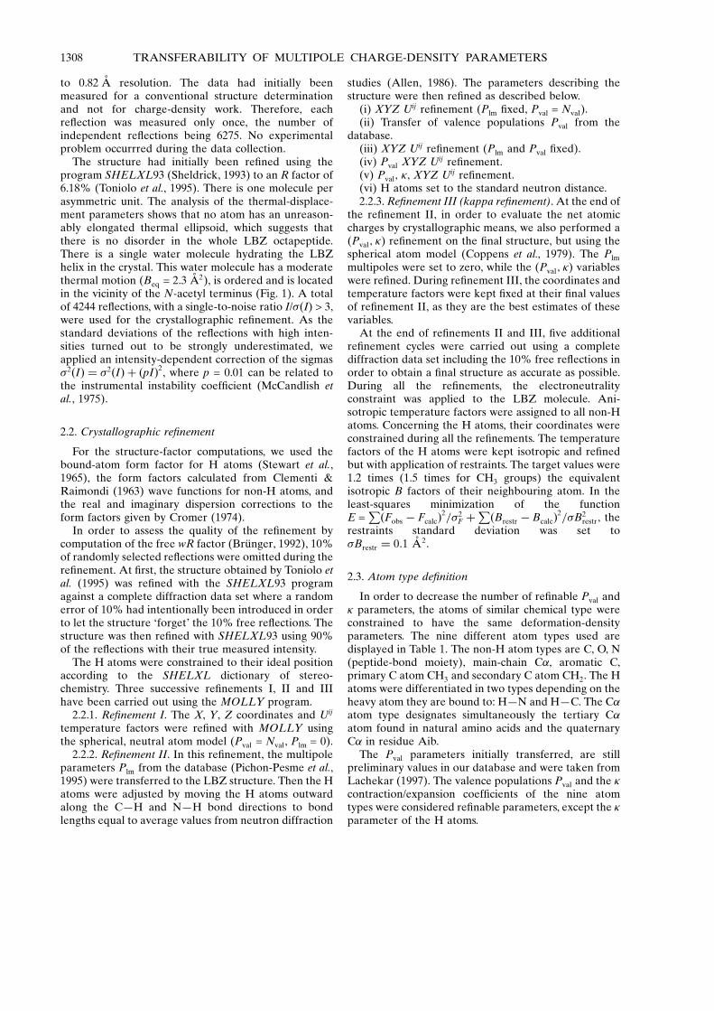
For the transfer of the multipole Plm parameters(Pichon-Pesme et al., 1995), all H atoms were consideredto have a same single dipole along the XÐH bond. Thequaternary C� atoms from the unnatural amino acidAib, which were absent in our database, were given thesame multipoles as the tertiary C� from natural aminoacids. The list of transferable multipoles is given inTable 2.
2.4. Electron-density maps
After crystallographic re®nement I with the sphericalneutral atom approximation, we computed residualelectron-density maps using the Fourier coef®cients(Fobs ÿ Fsph; 'sph) where the Fsph and 'sph are the struc-ture factor and phase calculated from the structure withthe spherical neutral atom density model
��res�r� � Vÿ1P
h
�kÿ1jFobs�h�j ÿ jFsph�h�j�
� exp i�'sph ÿ 2�h:r�: �1�
After the multipolar re®nement II, the same corre-sponding residual maps were computed with the coef-®cients (Fobs ÿ Fmult; 'mult).
The dynamic deformation electron-density maps werecomputed using the following equation,
��def�r� � Vÿ1P
h
�kÿ1jFobs�h�j exp�i'mult�
ÿ jFsph�h�j exp�i'sph�� exp�ÿ2i�h:r�: �2�
In this case, the 'mult phases were calculated from thestructure at the end of re®nement II. Spherical atomicscattering factors were used in the calculation of Fsph
and 'sph. For the calculation of all the terms in equation(2), the coordinates and temperature factors used arederived from the multipolar re®nement II, as they arethe best estimate of the true parameters.
Static deformation maps, corresponding to thedeformation electron density that would be observed foran immobile molecule (B = 0 AÊ 2), can also be computeddirectly using the XYZ, Plm, Pval and � parameters of themolecular structure.
2.5. Electrostatic potential
The electrostatic potential of the isolated moleculehas been computed using the program ELECTROS(Bouhmaida et al., 1997; Ghermani et al., 1993). Theelectrostatic potential � corresponding to the molecularcharge-density distribution is given by the integratedform of the Poisson equation
��r� � Patoms
Za=jrÿ raj ÿR��r0�=jrÿ r0jd3r0; �3�
where the Za and ra are the nuclear charges and posi-tions.
3. Results and discussion
3.1. Structure re®nement of the LBZ octapeptide
A representation of the LBZ helical structure isshown in Fig. 1. No static disorder is found in the crystalstructure. After re®nement II, the isotropic temperaturefactor B is in the range 1.0±3.8 AÊ 2 for the non-H atoms,the average B factor being 1.9 AÊ 2. The total number ofvariables re®ned (XYZ, Uij; �, Pval) is of 785 comparedto 4244 re¯ections used, which results in a ratio of dataper variable of 5.4.
The signal-to-noise ratio for the diffraction intensitiesis shown in Fig. 2. The average I/�(I) is 15 and at thehighest resolution its value is 5.2. The percentage ofre¯ections with I/�(I) lower than 3, which were excludedfrom the re®nement, is also shown. The average good-ness of ®t (g.o.f.), after correction of the intensity errors,
Table 1. Atomic charges Nval ÿ Pval and expansion/contraction coef®cients � for chemically similar atoms in the LBZmolecule
For convenience, the values Nvalÿ Pval rather than the valence populations Pval are displayed, as they represent the atomic charges. The standarddeviations obtained by full-matrix least-squares re®nement with the program MOLLY are given in parentheses. The charges from the AMBER4.1 dictionary `all_amino94.in' (Pearlman et al., 1995; Bayly et al., 1993) are mostly derived from the alanine residue. The aromatic carbon charge istaken from the phenylalanine residue and the secondary carbon CH2 charge from the lysine residue. In the AMBER dictionary, the charge of agiven atom type takes different values depending on its position on the residue. The charge of a H atom (HÐC) also varies slightly depending onthe type of C atom to which it is bonded.
Multipolar model Spherical modelAtom Type No.
atomsTransferedNval ÿ Pval
Re®nedNval ÿ Pval
Re®ned � Re®nedNval ÿ Pval
AMBERdictionary
Re®ned �
H (HÐN) 11 0.37 0.17 (3) 1.16 ®xed 0.20 (2) 0.27 1.16 ®xed(HÐC) 70 0.19 0.09 (2) 1.16 ®xed 0.10 (3) 0.06:0.14 1.16 ®xed
C (C O) 11 ÿ0.01 0.12 (9) 1.04 (2) 0.42 (9) 0.60 1.16 (2)(C�) 11 ÿ0.25 0.02 (8) 1.01 (2) ÿ0.05 (8) 0.03 1.13 (2)Arom 12 ÿ0.15 ÿ0.05 (3) 1.05 (2) 0.01 (3) ÿ0.17:0.01 1.15 (2)(CH3) 14 ÿ0.15 ÿ0.17 (6) 1.03 (2) ÿ0.24 (6) ÿ0.18 1.13 (2)(CH2) 8 ÿ0.15 0.11 (4) 1.02 (1) 0.02 (4) ÿ0.04:0.02 1.12 (1)
N (NÐH) 11 ÿ0.34 ÿ0.25 (8) 1.00 (2) ÿ0.44 (8) ÿ0.42 1.06 (2)O (C O) 11 ÿ0.31 ÿ0.41 (6) 0.97 (1) ÿ0.65 (6) ÿ0.57 0.99 (1)
JELSCH, PICHON-PESME, LECOMTE AND AUBRY 1309
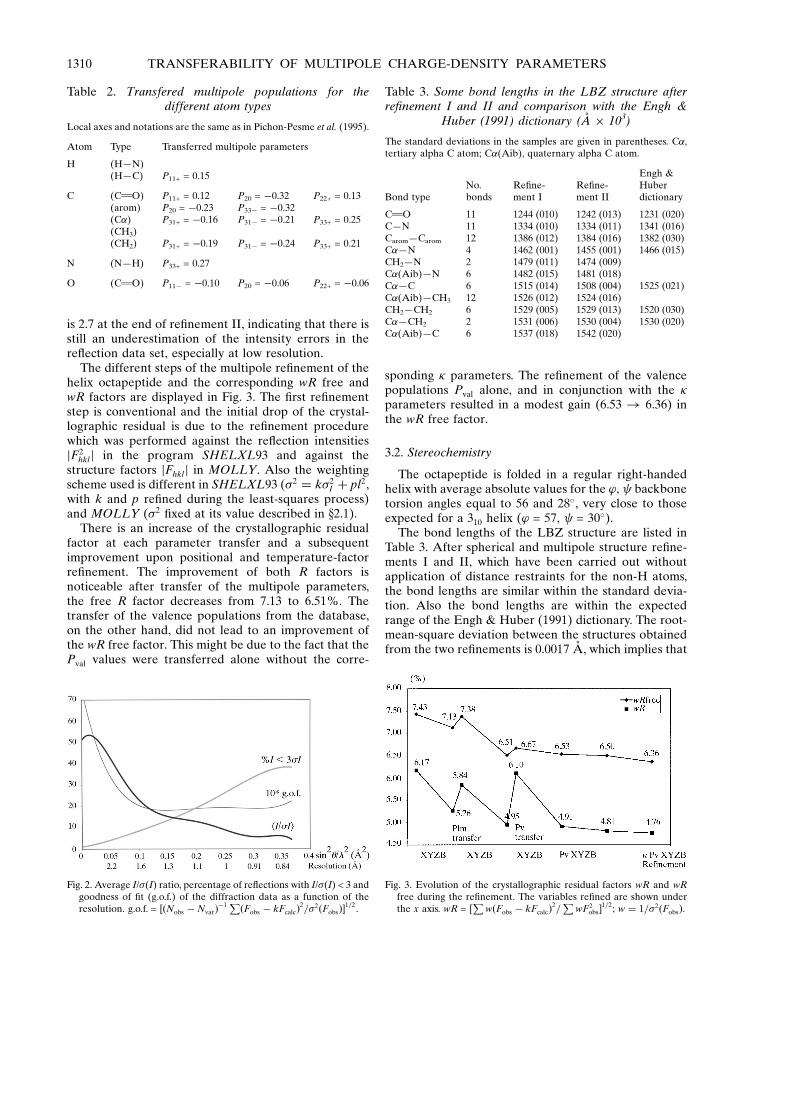
is 2.7 at the end of re®nement II, indicating that there isstill an underestimation of the intensity errors in there¯ection data set, especially at low resolution.
The different steps of the multipole re®nement of thehelix octapeptide and the corresponding wR free andwR factors are displayed in Fig. 3. The ®rst re®nementstep is conventional and the initial drop of the crystal-lographic residual is due to the re®nement procedurewhich was performed against the re¯ection intensitiesjF2
hklj in the program SHELXL93 and against thestructure factors jFhklj in MOLLY. Also the weightingscheme used is different in SHELXL93 (�2 � k�2
I � pl2,with k and p re®ned during the least-squares process)and MOLLY (�2 ®xed at its value described in x2.1).
There is an increase of the crystallographic residualfactor at each parameter transfer and a subsequentimprovement upon positional and temperature-factorre®nement. The improvement of both R factors isnoticeable after transfer of the multipole parameters,the free R factor decreases from 7.13 to 6.51%. Thetransfer of the valence populations from the database,on the other hand, did not lead to an improvement ofthe wR free factor. This might be due to the fact that thePval values were transferred alone without the corre-
sponding � parameters. The re®nement of the valencepopulations Pval alone, and in conjunction with the �parameters resulted in a modest gain (6.53 ! 6.36) inthe wR free factor.
3.2. Stereochemistry
The octapeptide is folded in a regular right-handedhelix with average absolute values for the '; backbonetorsion angles equal to 56 and 28�, very close to thoseexpected for a 310 helix (' = 57, = 30�).
The bond lengths of the LBZ structure are listed inTable 3. After spherical and multipole structure re®ne-ments I and II, which have been carried out withoutapplication of distance restraints for the non-H atoms,the bond lengths are similar within the standard devia-tion. Also the bond lengths are within the expectedrange of the Engh & Huber (1991) dictionary. The root-mean-square deviation between the structures obtainedfrom the two re®nements is 0.0017 AÊ , which implies that
Fig. 3. Evolution of the crystallographic residual factors wR and wRfree during the re®nement. The variables re®ned are shown underthe x axis. wR = [
Pw�Fobs ÿ kFcalc�2=
PwF2
obs�1=2; w � 1=�2�Fobs�.
Table 3. Some bond lengths in the LBZ structure afterre®nement I and II and comparison with the Engh &
Huber (1991) dictionary (AÊ � 103)
The standard deviations in the samples are given in parentheses. C�,tertiary alpha C atom; C�(Aib), quaternary alpha C atom.
Bond typeNo.bonds
Re®ne-ment I
Re®ne-ment II
Engh &Huberdictionary
C O 11 1244 (010) 1242 (013) 1231 (020)CÐN 11 1334 (010) 1334 (011) 1341 (016)CaromÐCarom 12 1386 (012) 1384 (016) 1382 (030)C�ÐN 4 1462 (001) 1455 (001) 1466 (015)CH2ÐN 2 1479 (011) 1474 (009)C�(Aib)ÐN 6 1482 (015) 1481 (018)C�ÐC 6 1515 (014) 1508 (004) 1525 (021)C�(Aib)ÐCH3 12 1526 (012) 1524 (016)CH2ÐCH2 6 1529 (005) 1529 (013) 1520 (030)C�ÐCH2 2 1531 (006) 1530 (004) 1530 (020)C�(Aib)ÐC 6 1537 (018) 1542 (020)
Table 2. Transfered multipole populations for thedifferent atom types
Local axes and notations are the same as in Pichon-Pesme et al. (1995).
Atom Type Transferred multipole parameters
H (HÐN)(HÐC) P11+ = 0.15
C (C O) P11+ = 0.12 P20 = ÿ0.32 P22+ = 0.13(arom) P20 = ÿ0.23 P33ÿ = ÿ0.32(C�) P31+ = ÿ0.16 P31ÿ = ÿ0.21 P33+ = 0.25(CH3)(CH2) P31+ = ÿ0.19 P31ÿ = ÿ0.24 P33+ = 0.21
N (NÐH) P33+ = 0.27
O (C O) P11ÿ = ÿ0.10 P20 = ÿ0.06 P22+ = ÿ0.06
Fig. 2. Average I/�(I) ratio, percentage of re¯ections with I/�(I) < 3 andgoodness of ®t (g.o.f.) of the diffraction data as a function of theresolution. g.o.f. = ��Nobs ÿ Nvar�ÿ1 P�Fobs ÿ kFcalc�2=�2�Fobs��1=2:
1310 TRANSFERABILITY OF MULTIPOLE CHARGE-DENSITY PARAMETERS

the bond lengths are very similar. The main effect of themultipole transfer and charge re®nement in the LBZstructure shows up in the thermal-motion parametersrather than in the coordinates, as will be seen later.
3.3. Atomic charges
3.3.1. Atomic charges in re®nement II. The concept ofatomic charge is fundamental to chemistry as it givespowerful insights towards the understanding of chemicalreactivity and physical properties. There is however noclear de®nition of the true atomic charge, given thedistributed nature of the electrons around the nuclei in amolecule. The Nval ÿ Pval values can be considered asatomic charges obtained by crystallographic ®tting in thereciprocal space. The values of the valence populationstransferred from the database and after re®nement IIwith MOLLY are given in Table 1. The (Nval ÿ Pval)values in the case of re®nement II cannot be consideredas the total atomic charges as the multipoles contributealso to the electron transfers from one atom to the other.
The transferred and re®ned Pval parameters agreemoderately as the correlation coef®cient reaches 73%.The O and N atoms have negative Nval ÿ Pval charges asexpected. The two H-atom types have positive charges,the values obtained in our re®nement are, however,lower than those transferred from the database. For theC atoms, the transferred and re®ned (Nval ÿ Pval) inTable 1 are not always in good agreement as they aresometimes of opposite signs. In the case of the C atom,as there are four electrons involved in the covalentbonds, the impact of the multipoles on the total charge isthe highest.
We have attempted to de®ne more atom types, forinstance differentiating the tertiary and quaternary C�atoms. But as there were only two tertiary C� atoms, there®ned charge turned out to be physically unrealisticand with a high estimated error. Similarly, distinguishingthe H atoms, according to the type of C atom they arebound to, lead to unrealistic values. The re®nement ofindividual atomic charges, without application of severalequality constraints over equivalent atoms, would nothave been meaningful for this 0.82 AÊ resolution struc-ture. We have thus considered only nine different atomstypes for a total number of 156 atoms in the LBZmolecule (Table 1). The averaging of the parametersthrough application of constaints on chemicallyequivalent atoms, rendered the Pval and � variablesre®nement relevant.
3.3.2. Atomic charges in re®nement III. In order toobtain a better estimation of the atomic charges,unbiased by the multipoles, the valence populations Pval
have also been re®ned, under the spherical atomapproximation (Coppens et al., 1979). The re®nedcharges with the spherical model, depend howeverpartly on the transferred multipoles, as the coordinatesand temperature factors used are those after re®nement
II. The transferable multipoles describing the peptidemain chain are the best determined in our database asthey are the most represented in our peptide sample.The charges obtained in re®nement III are generallymeaningful and agree well with the charges used in theAMBER molecular modelling software (Pearlman et al.,1995). The point charges in the AMBER dictionary, havebeen obtained by a restrained electrostatic potential ®t,where the potential is calculated from the quantummechanical wavefunction using a medium-sized 6-31G*basis set (Bayly et al., 1993). The agreement is especially
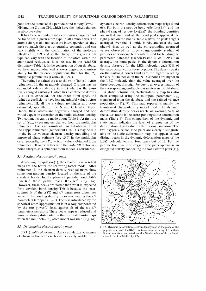
Fig. 4. Residual electron-density maps in the plane of the peptide bondAib2-Lys(Bz)3. (a) after re®nement I using the spherical atommodel, (b) after re®nement II using the multipole±Pval atom model.Contours: �0.05 e AÊ ÿ3, positive contours: solid line, negativecontours: dashed line, zero contour omitted.
JELSCH, PICHON-PESME, LECOMTE AND AUBRY 1311
good for the atoms of the peptide-bond moiety O CÐNH and the C atom CH3 which have the highest chargesin absolute value.
It has to be reminded that a consensus charge cannotbe found for a given atom type in all amino acids. Theatomic charges of a molecule are interdependent as theyhave to match the electroneutrality constraint and canvary slightly with the conformation of the molecule(Bayly et al., 1993). Also the charge for a given atomtype can vary with the location of the atom within theamino-acid residue, as it is the case in the AMBERdictionary (Table 1). In the construction of our database,we have indeed observed a lower degree of transfer-ability for the valence populations than for the Plm
multipole parameters (Lachekar, 1997).The re®ned � values are also shown in Table 1. After
re®nement II, the negatively charged O atom has anexpanded valence density (� < 1) whereas the posi-tively charged carbonyl C atom has a contracted density(� > 1) as expected. For the other atom types, there®ned � coef®cients have less meaningful values. Afterre®nement III, all the � values are higher and over-estimated, specially for the N and CH3 atom types.Hence, these atoms are negatively charged and onewould expect an extension of the radial electron density.Two comments can be made about Table 1. At ®rst theset of (Pval; �) parameters derived from the multipolarre®nement II is more consistent than that obtained fromthe kappa re®nement (re®nement III). This may be dueto the better valence electron density modelling andimproved phase estimate (see x3.6) in the multipolarcase. Secondly, the (Pval ÿ Nval) values obtained fromre®nement III agree better with the AMBER dictionarypoint charges as a spherical atom model is considered.
3.4. Residual electron-density maps
According to equation (1), the cleaner these residualmaps are, the better the scattering factor model. Afterre®nement I, the electron-density residual maps showsome non-random density, located at the site of thecovalent bonds. In the plane of peptide bond Aib2-Lys(Bz)3 these peaks reach 0.3 e AÊ ÿ3 (Fig. 4a).However, these peaks are ¯atter than what is expectedfor a covalent bond density. This is because the least-squares ®t of the XYZ and Uij parameters takes intoaccount the bonding density by overestimating the Uij
parameters (Coppens, 1967). The bias introduced by thespherical atom approximation is in a way compensatedby the too powerful least-squares ®t of the six Uij
parameters per atom. These peaks appear reduced andmore randomly distributed in the residual density mapswhen the multipole±Pval atom model was used (Fig. 4b).
3.5. Deformation electron-density maps
3.5.1. Quality of the maps. An accumulation of valenceelectrons in the covalent bonds is clearly visible in the
dynamic electron-density deformation maps (Figs. 5 and6a). For both the peptide bond Aib2-Lys(Bz)3 and thephenyl ring of residue Lys(Bz)6 the bonding densitiesare well de®ned and all the bond peaks appear at theright place on the bonds. Table 4 gives the peak heightsaveraged over the 11 amide bonds, and over the twophenyl rings, as well as the corresponding averagedvalues observed in three charge-density studies ofpeptides at cryogenic temperature used for building theparameter database (Pichon-Pesme et al., 1995). Onaverage, the bond peaks in the dynamic deformationdensity observed for the LBZ molecule, reach 85% ofthe value observed for these peptides. The density peakson the carbonyl bonds C O are the highest reaching0.5 e AÊ ÿ3. The peaks on the NÐC� bonds are higher inthe LBZ molecule than the value averaged over thethree peptides, this might be due to an overestimation ofthe corresponding multipole parameters in the database.
A static deformation electron-density map has alsobeen computed using the multipole parameters Plm
transferred from the database and the re®ned valencepopulations (Fig. 7). This map represents mainly thetransferred charge-density model used. The dynamicdeformation density peaks reach, on average, 52% ofthe values found in the corresponding static deformationmaps (Table 4). This comparison of the dynamic andstatic maps indicates the level of attenuation of thedeformation density due to the thermal smearing. Thetwo oxygen electron lone pairs are clearly distinguish-able in the static deformation map, but appear as twodistinct peaks in the dynamic deformation maps of theLBZ molecule only in four cases out of 11. For thepeptide bond 2±3, the oxygen lone pairs appear as anelongated density connecting the two electron pairs (Fig.
Fig. 5. Dynamic deformation electron-density map in the plane of thepeptide bond Aib2-Lys(Bz)3. Contours: same as in Fig. 4. The thickline represents a contracted van der Waals surface of the molecule(atomic radii multiplied by 0.7).
1312 TRANSFERABILITY OF MULTIPOLE CHARGE-DENSITY PARAMETERS

5). The oxygen lone pairs are the ®nest feature in thenon-spherical electron density of peptides; they requirea good description by Fourier synthesis data up to 0.5 AÊ
resolution.3.5.2. Effect of a high- and low-resolution cutoff. The
effect of high-resolution diffraction data truncation onthe dynamic deformation maps has been analysed on thearomatic ring of residue Lys(Bz)6. This phenyl ring hasmoderate temperature factors (1.25 < B < 2.5 AÊ 2) andthe deformation density has a regular shape (Fig. 6a).This dynamic deformation density computed by Fouriersynthesis is nearly the same when using all the re¯ec-tions or when truncating the data at 0.9 AÊ resolution.However, when a 1 AÊ resolution cutoff is applied, a lot
of information has been lost (Figs. 6b and 6c). Thecovalent-bond density peaks decrease by 0.1 e AÊ ÿ3 onthe benzene ring of residue LysBz6 when the 1 AÊ reso-lution cutoff is applied.
Low-resolution cutoffs were also applied in order todetermine the resolution range which is important fordeformation-density parameters. The correspondingdynamic deformation density maps were unaltered forthe 2.5 and 2 AÊ cutoffs and largely attenuated at 1.5 AÊ
(data not shown). The contribution of the very lowresolution diffraction data to the deformation density islimited as the number of re¯ections involved is small.These results show that the essential information on thedeformation electron density is contained in the re¯ec-
Fig. 6. Dynamic deformation electron-density map in the phenyl ringplane of residue Lys(Bz)6 computed at different resolution cutoffs.(a) All re¯ections 0.82±1 AÊ . (b) Resolution cutoff 0.9±1 AÊ . (c)resolution cutoff 1.0±1 AÊ . Contours: the same as in Fig. 4.
JELSCH, PICHON-PESME, LECOMTE AND AUBRY 1313

tions between 0.9 and 2 AÊ resolution, a region of thereciprocal space where the contribution of the solventmolecules to the X-ray diffraction is small in the case ofprotein crystals.
3.5.3. Deformation density and thermal motion. Thein¯uence of the atomic thermal motion on the height ofthe deformation density has also been analyzed. Thiseffect can be readily seen in the dynamic deformationdensity of the aromatic ring for the residue Lys(Bz)3
(Fig. 8a). The atomic B factors for the residue Lys(Bz)3
increase gradually along this elongated side chain from1.5 AÊ 2 to higher values close to 4 AÊ 2 (Fig. 8b). Thisaromatic ring has the highest thermal-displacementparameters in the helix octapeptide. Despite theirchemical equivalence, the dynamic CÐC bondingdensities decrease from 0.45 to 0.25 e AÊ ÿ3 as thethermal-displacement parameters increase. The defor-mation density is well de®ned on the three bonds formedby the atom CF and its peak value reaches 0.4±0.5 e AÊ ÿ3.At the end of the side chain, the peaks of the defor-
mation density are very attenuated and their shape isdeformed by thermal displacements. This effect hasalready been observed by Pichon-Pesme et al. (1995).From these observations, we can draw the conclusionthat the multiple description of the electron density ismeaningful for X-ray structures, when the thermaldisplacement or the non-resolved disorder is moderate(B < 4 AÊ 2). For greater displacement parameters, thedynamic deformation density of the atoms is largelydiluted.
3.6. Phase and deformation density
The phase error has been evaluated using the Rfree
likelihood estimates method, implemented in the
Table 4. Selected bond peaks (e AÊ ÿ3) in the dynamic and static deformation electron-density maps of the LBZ helixand average values found in dynamic deformation maps for three peptide structures
Peptide structures: N-acetyl l-tryptophane methylamide (Souhassou et al., 1991), N-acetyl-(��)-dehydro-phenylalanine methylamide (Souhassouet al., 1992) and leuenkephaline trihydrate (Wiest et al., 1994).
LBZ CÐN NÐH NÐC� C�ÐC C O O lone pairs CaromÐH CaromÐCarom
Average 0.37 0.38 0.42 0.39 0.49 0.26 0.33 0.35R.m.s. deviation 0.12 0.06 0.07 0.09 0.08 0.06 0.07 0.06
Static 0.85 0.75 0.75 0.6 1.05 0.45 0.65 0.75Average in Enk, AcTrp, AcPhe 0.51 0.46 0.36 0.4 0.51 0.38 0.39 0.56
Fig. 7. Static electron-density deformation map in the plane of thepeptide bond Aib2-Lys(Bz)3. Contours: �0.1 e AÊ ÿ3, positivecontours: solid line, negative contours: dashed line, zero contouromitted.
Fig. 8. (a) Dynamic deformation electron-density map in the plane ofthe benzene ring [residue Lys(Bz)3]. Contours: the same as in Fig. 4.(b) The equivalent B factors (AÊ 2) of the (non-H) atoms areindicated.
1314 TRANSFERABILITY OF MULTIPOLE CHARGE-DENSITY PARAMETERS
program RFLEXPL (Urzhumtsev et al., 1996). Theaverage phase error of the acentric re¯ections afterre®nements I and II are 3.3 and 2.6�, respectively. This0.7� gain in the phase error denotes the bene®t of themultipole parameters transfer to the LBZ structure.
The phase shift due to the atom model has also beenanalyzed. The unweighted root-mean-square (r.m.s.)phase difference between re®nements I and II is 2.6� onthe acentric re¯ections used in the re®nement. The r.m.s.phase differences of 2.6 and 2.5� between re®nements IIand III, and re®nements I and III, respectively, are quitesimilar. These ®gures illustrate that in the case of theLBZ crystal structure, the phase error and phase shiftdue to the deformation density are of the same magni-tude. Thus, a re®nement instead of a transfer of themultipole charge density would not have been physicallymeaningful.
The dynamic deformation density can be decomposedas the sum of an amplitude-difference density and aphase-difference density (Souhassou et al., 1991),
��def � ��Fobs ÿ Fsph; 'mult� � ��Fsph; 'mult ÿ 'sph�� ���F� � ���'� :
The total deformation density has an r.m.s. value of0.103 e AÊ ÿ3 over the asymmetric unit; the density termsdue to �F and �' have r.m.s. values of 0.099 and0.029 e AÊ ÿ3, respectively. The LBZ molecule crystallizesin an orthorhombic space group and up to 20% of there¯ections are centric, which explains the relatively lowcontribution of the phase difference �' to the defor-mation density.
3.7. Scattering factors of the electron-density components
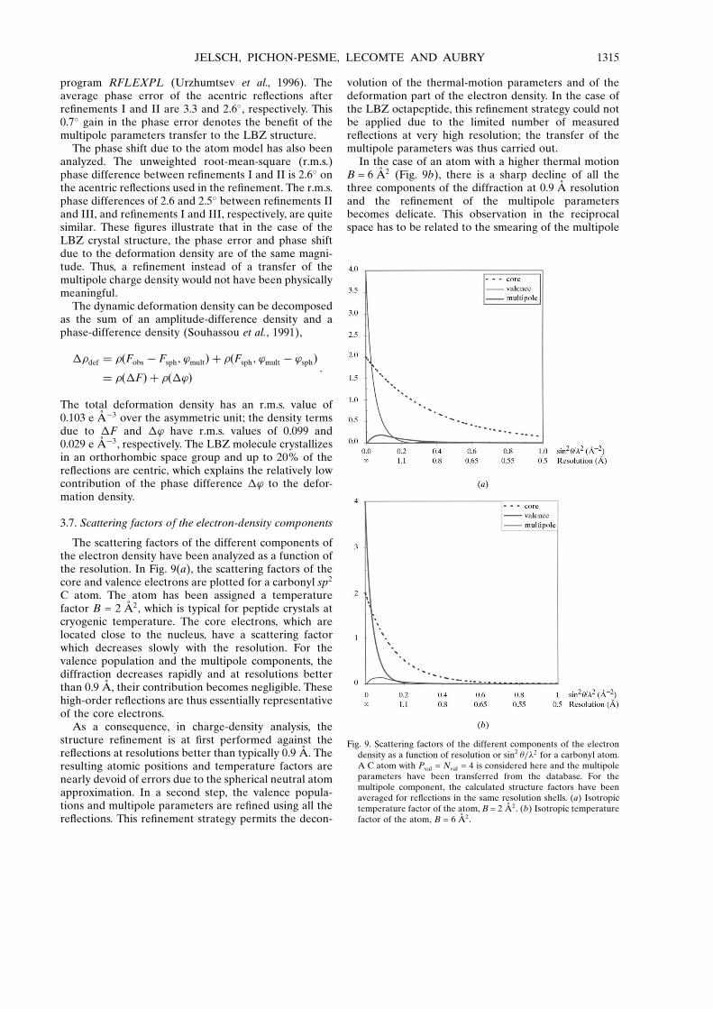
The scattering factors of the different components ofthe electron density have been analyzed as a function ofthe resolution. In Fig. 9(a), the scattering factors of thecore and valence electrons are plotted for a carbonyl sp2
C atom. The atom has been assigned a temperaturefactor B = 2 AÊ 2, which is typical for peptide crystals atcryogenic temperature. The core electrons, which arelocated close to the nucleus, have a scattering factorwhich decreases slowly with the resolution. For thevalence population and the multipole components, thediffraction decreases rapidly and at resolutions betterthan 0.9 AÊ , their contribution becomes negligible. Thesehigh-order re¯ections are thus essentially representativeof the core electrons.
As a consequence, in charge-density analysis, thestructure re®nement is at ®rst performed against there¯ections at resolutions better than typically 0.9 AÊ . Theresulting atomic positions and temperature factors arenearly devoid of errors due to the spherical neutral atomapproximation. In a second step, the valence popula-tions and multipole parameters are re®ned using all there¯ections. This re®nement strategy permits the decon-
volution of the thermal-motion parameters and of thedeformation part of the electron density. In the case ofthe LBZ octapeptide, this re®nement strategy could notbe applied due to the limited number of measuredre¯ections at very high resolution; the transfer of themultipole parameters was thus carried out.
In the case of an atom with a higher thermal motionB = 6 AÊ 2 (Fig. 9b), there is a sharp decline of all thethree components of the diffraction at 0.9 AÊ resolutionand the re®nement of the multipole parametersbecomes delicate. This observation in the reciprocalspace has to be related to the smearing of the multipole
Fig. 9. Scattering factors of the different components of the electrondensity as a function of resolution or sin2 �=�2 for a carbonyl atom.A C atom with Pval = Nval = 4 is considered here and the multipoleparameters have been transferred from the database. For themultipole component, the calculated structure factors have beenaveraged for re¯ections in the same resolution shells. (a) Isotropictemperature factor of the atom, B = 2 AÊ 2. (b) Isotropic temperaturefactor of the atom, B = 6 AÊ 2.
JELSCH, PICHON-PESME, LECOMTE AND AUBRY 1315
electron density in the dynamic maps when the B factorincreases (Fig. 8).
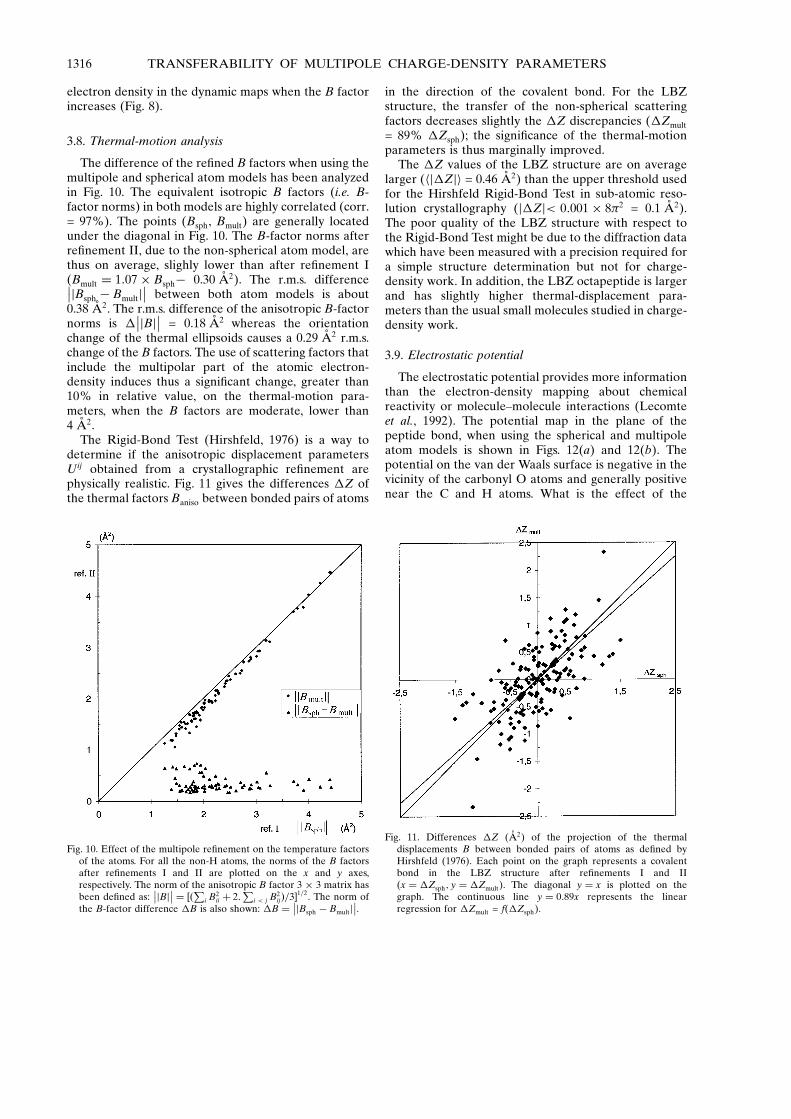
3.8. Thermal-motion analysis
The difference of the re®ned B factors when using themultipole and spherical atom models has been analyzedin Fig. 10. The equivalent isotropic B factors (i.e. B-factor norms) in both models are highly correlated (corr.= 97%). The points (Bsph, Bmult) are generally locatedunder the diagonal in Fig. 10. The B-factor norms afterre®nement II, due to the non-spherical atom model, arethus on average, slighly lower than after re®nement I(Bmult � 1:07� Bsphÿ 0.30 AÊ 2). The r.m.s. difference��jBsph ÿ Bmultj
�� between both atom models is about0.38 AÊ 2. The r.m.s. difference of the anisotropic B-factornorms is �
��jBj�� = 0.18 AÊ 2 whereas the orientationchange of the thermal ellipsoids causes a 0.29 AÊ 2 r.m.s.change of the B factors. The use of scattering factors thatinclude the multipolar part of the atomic electron-density induces thus a signi®cant change, greater than10% in relative value, on the thermal-motion para-meters, when the B factors are moderate, lower than4 AÊ 2.
The Rigid-Bond Test (Hirshfeld, 1976) is a way todetermine if the anisotropic displacement parametersUij obtained from a crystallographic re®nement arephysically realistic. Fig. 11 gives the differences �Z ofthe thermal factors Baniso between bonded pairs of atoms
in the direction of the covalent bond. For the LBZstructure, the transfer of the non-spherical scatteringfactors decreases slightly the �Z discrepancies (�Zmult
= 89% �Zsph); the signi®cance of the thermal-motionparameters is thus marginally improved.
The �Z values of the LBZ structure are on averagelarger (hj�Zji = 0.46 AÊ 2) than the upper threshold usedfor the Hirshfeld Rigid-Bond Test in sub-atomic reso-lution crystallography (j�Zj< 0:001� 8�2 = 0.1 AÊ 2).The poor quality of the LBZ structure with respect tothe Rigid-Bond Test might be due to the diffraction datawhich have been measured with a precision required fora simple structure determination but not for charge-density work. In addition, the LBZ octapeptide is largerand has slightly higher thermal-displacement para-meters than the usual small molecules studied in charge-density work.
3.9. Electrostatic potential
The electrostatic potential provides more informationthan the electron-density mapping about chemicalreactivity or molecule±molecule interactions (Lecomteet al., 1992). The potential map in the plane of thepeptide bond, when using the spherical and multipoleatom models is shown in Figs. 12(a) and 12(b). Thepotential on the van der Waals surface is negative in thevicinity of the carbonyl O atoms and generally positivenear the C and H atoms. What is the effect of the
Fig. 10. Effect of the multipole re®nement on the temperature factorsof the atoms. For all the non-H atoms, the norms of the B factorsafter re®nements I and II are plotted on the x and y axes,respectively. The norm of the anisotropic B factor 3 � 3 matrix hasbeen de®ned as:
��jBj�� � ��Pi B2ii � 2:
Pi < j B2
ij�=3�1=2. The norm ofthe B-factor difference �B is also shown: �B � ��jBsph ÿ Bmultj
��.
Fig. 11. Differences �Z (AÊ 2) of the projection of the thermaldisplacements B between bonded pairs of atoms as de®ned byHirshfeld (1976). Each point on the graph represents a covalentbond in the LBZ structure after re®nements I and II(x � �Zsph; y � �Zmult). The diagonal y � x is plotted on thegraph. The continuous line y � 0:89x represents the linearregression for �Zmult = f(�Zsph).
1316 TRANSFERABILITY OF MULTIPOLE CHARGE-DENSITY PARAMETERS
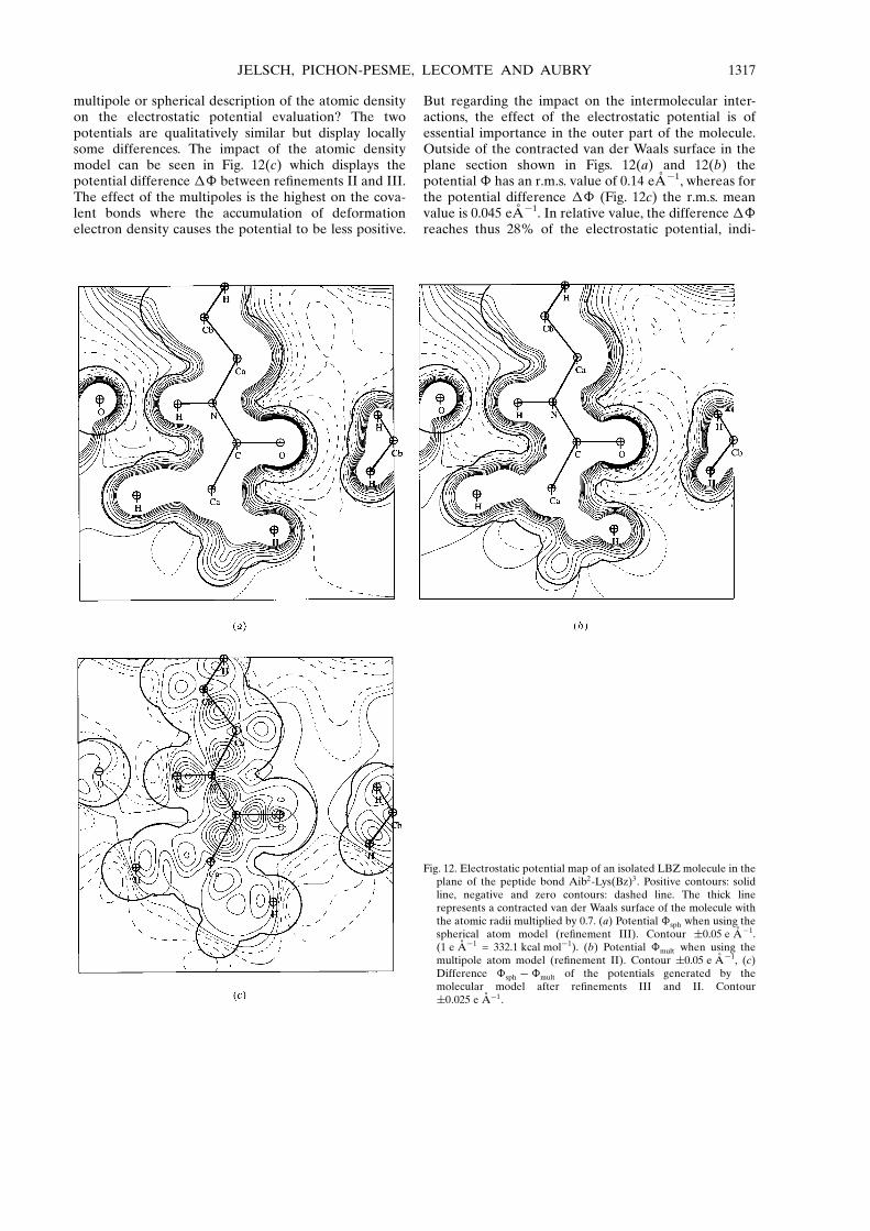
multipole or spherical description of the atomic densityon the electrostatic potential evaluation? The twopotentials are qualitatively similar but display locallysome differences. The impact of the atomic densitymodel can be seen in Fig. 12(c) which displays thepotential difference �� between re®nements II and III.The effect of the multipoles is the highest on the cova-lent bonds where the accumulation of deformationelectron density causes the potential to be less positive.
But regarding the impact on the intermolecular inter-actions, the effect of the electrostatic potential is ofessential importance in the outer part of the molecule.Outside of the contracted van der Waals surface in theplane section shown in Figs. 12(a) and 12(b) thepotential � has an r.m.s. value of 0.14 eAÊ ÿ1, whereas forthe potential difference �� (Fig. 12c) the r.m.s. meanvalue is 0.045 eAÊ ÿ1. In relative value, the difference ��reaches thus 28% of the electrostatic potential, indi-
Fig. 12. Electrostatic potential map of an isolated LBZ molecule in theplane of the peptide bond Aib2-Lys(Bz)3. Positive contours: solidline, negative and zero contours: dashed line. The thick linerepresents a contracted van der Waals surface of the molecule withthe atomic radii multiplied by 0.7. (a) Potential �sph when using thespherical atom model (re®nement III). Contour �0.05 e AÊ ÿ1.(1 e AÊ ÿ1 = 332.1 kcal molÿ1). (b) Potential �mult when using themultipole atom model (re®nement II). Contour �0.05 e AÊ ÿ1, (c)Difference �sph ÿ�mult of the potentials generated by themolecular model after re®nements III and II. Contour�0:025 e AÊ ÿ1.
JELSCH, PICHON-PESME, LECOMTE AND AUBRY 1317

cating that a multipole description of the electrondensity has a non-negligible impact on the electrostaticpotential calculation, even in the intermolecular region.
4. Concluding remarks
We are building an aspherical scattering-factor table forthe chemical atom types found in all natural amino acids.Such a table includes the valence population Pval, the �contraction/expansion coef®cients and the multipole Plm
parameters. In the future this will allow an experimentaldetermination of the charges for all the atom typesfound in peptides and proteins. Also the multipoledescription of the electron density permits the evalua-tion of a more accurate electrostatic potential.
This study on the LBZ helix octapeptide has shownthat the re¯ection intensities should be collected to atleast 0.9 AÊ resolution, if one wants all the deformation-density information to be re¯ected in the diffractiondata. Moreover the temperature factors should be low:when the atomic B factors are higher than 4 AÊ 2, thedynamic deformation density starts to be dispersed. Inthe LBZ molecule, most of the atoms have moderatetemperature factors and have well de®ned dynamicdeformation densities.
What are the potential proteins for which this multi-pole formalism can be applied? With the availability ofsynchrotron X-ray sources to biocrystallography and theuse of cryogenics, an increasing number of proteinstructures are known to atomic resolution (Dauter et al.,1995). The protein crambin (Teeter et al., 1993) is anexceptional candidate for an extension of the multipoleformalism to proteins. The diffraction data for this 46amino acids protein have been collected up to 0.54 AÊ
resolution at the EMBL synchrotron in Hamburg. Theatomic temperature factors of crambin are remarkablylow for a protein, they are typically between 2 and 5 AÊ 2
for most of the atoms. For the crambin structure, thetransfer of the deformation-density parameters shouldhave a meaningful impact on the crystallographicre®nement and the valence populations and multipolesmight even be re®nable. Further work is under way inthat direction.
References
Allen, F. H. (1986). Acta Cryst. B42, 515±522.Bayly, C. I., Cieplak, P., Cornell, W. D. & Kollman, P. A. (1993).
J. Phys. Chem. 97, 10269±10280.Bouhmaida, N., Ghermani, N. E., Lecomte, C. & Thalal, K.
(1997). Acta Cryst. A53, 556±563.BruÈ nger, A. T. (1992). Nature (London), 355, 472±474.Clementi, E. & Raimondi, D. L. (1963). J. Phys. Chem. 41,
2686±2689.Coppens, P. (1967). Science, 158, 1577.Coppens, P. (1997). X-ray Charge Densities and Chemical
Bonding. Oxford: IUCr/Oxford University Press.
Coppens, P., Guru Row, T. N., Leung, P., Stevens, E. D., Becker,P. J. & Yang, Y. W. (1979). Acta Cryst. A35, 63±72.
Cromer, D. T. (1974). International Tables for X-ray Crystal-lography, edited by J. A. Ibers & W. E. Hamilton, Vol. IV, pp.148±151. Birmingham: Kynoch Press.
Dauter, Z., Lamzin, V. S. & Wilson, K. S. (1995). Curr. Opin.Struct. Biol. 5, 784±790.
Engh, R. A. & Huber, R. (1991). Acta Cryst. A47. 392±400.Espinosa, E., Lecomte, C., Molins, E., Veintemillas, S.,
Cousson, A. & Paulus, W. (1996). Acta Cryst. B52, 519±534.Ghermani, N. E., Bouhmaida, N. & Lecomte, C. (1993). Acta
Cryst. A49, 781±789.Hansen, N. K. & Coppens, P. (1978). Acta Cryst. A34, 909±921.Hirshfeld, F. L. (1976). Acta Cryst. A32, 239±244.Howell, P. L., Pangborn, W. A., Marshall, G. R., Zabrocki, J. &
Smith, G. D. (1995). Acta Cryst. C51, 2575±2579.Lachekar, H. (1997). PhD thesis, Universite Henri PoincareÂ
Nancy I, France.Lecomte, C. (1995). Advances in Molecular Structure Research,
Vol. 1, edited by I. Hargittai & M. Hargittai, pp. 261±302.Greenwich, CT, USA: JAI Press Inc.
Lecomte, C., Ghermani, N. E., Pichon-Pesme, V. & Souhassou,M. (1992). J. Mol. Struct. (Theochem.), 255, 241±260.
McCandlish, L. E., Stout, G. H. & Andrews, L. C. (1975). ActaCryst. A31, 245±249.
Mata, I., Maniukiewicz, W., Molins, E., Espinosa, E. &Lecomte, C. (1997). 17th Meeting of European Crystal-lographic Association, Abstracts, p. 131. Lisbon University,Lisbon, Portugal.
Pearlman, D. A., Case, D. A., Caldwell, J. W., Ross, W. S.,Cheatham, T. E., Ferguson, D. M., Seibel, G. L., Singh, U. C.,Weiner, P. K. & Kollman, P. A. (1995). AMBER 4.1,University of California, San Francisco, USA.
Pichon-Pesme, V. & Lecomte, C. (1998). Acta Cryst. B54, 485±493.
Pichon-Pesme, V., Lecomte, C. & Lachekar, H. (1995). J. Phys.Chem. 99, 6242±6250.
Pichon-Pesme, V., Lecomte, C., Wiest, R. & BeÂnard, M. (1992).J. Am. Chem. Soc. 114, 2713±2715.
Sheldrick, G. M., (1993). SHELXL93, Program for crystalstructure re®nement. University of GoÈ ttingen, Germany.
Souhassou, M., Lecomte, C., Blessing, R. H., Aubry, A.,Rohmer, M. M., Wiest, R., BeÂnard, M. & Marraud, M.(1991). Acta Cryst. B47, 253±266.
Souhassou, M., Lecomte, C., Ghermani, N. E., Rohmer, M. M.,Wiest, R., BeÂnard, M. & Blessing, R. H. (1992). J. Am. Chem.Soc. 114, 2371±2382.
Stewart, R. F., Davidson, E. R. & Simpson, W. T. (1965). J.Chem. Phys. 43, 175±187.
Teeter, M. M., Lamzin, V. S., Dauter, Z. & Wilson, K. (1997).Am. Crystallogr. Assoc. Invited lecture. Ann. Meet. St Louis.Abstracts p. 67. Buffalo: ACA.
Teeter, M. M., Roe, S. M. & Heo, N. H. (1993). J. Mol. Biol. 230,292±311.
Toniolo, C., Bianco, A., Formaggio, F., Crisma, M., Bonora, G.M., Benedetti, E., Del Duca, V., Savanio, M., Di Blasio, B.,Pedone, C. & Aubry, A. (1995). Bioorg. Med. Chem. 3, 1211±1221.
Urzhumtsev, A. G., Skovoroda, T. P. & Lunin, V. Y. (1996). J.Appl. Cryst. 29, 741±744.
Wiest, R., Pichon-Pesme, V., BeÂnard, M. & Lecomte, C. (1994).J. Phys. Chem. 98, 1351±1362.
1318 TRANSFERABILITY OF MULTIPOLE CHARGE-DENSITY PARAMETERS