Highly Parallel Alternating Directions Algorithm for Time Dependent Problems
Upload
independentCategory
view
2download
0
2005;11:7093s-7103s. Clin Cancer Res Robert Ivkov, Sally J. DeNardo, Wolfgang Daum, et al. for Heat Induction of Nanoparticles Localized in CancerApplication of High Amplitude Alternating Magnetic Fields
Updated version
http://clincancerres.aacrjournals.org/content/11/19/7093s
Access the most recent version of this article at:
E-mail alerts related to this article or journal.Sign up to receive free email-alerts
Subscriptions
Reprints and
To order reprints of this article or to subscribe to the journal, contact the AACR Publications
Permissions
To request permission to re-use all or part of this article, contact the AACR Publications
Research. on June 13, 2013. © 2005 American Association for Cancerclincancerres.aacrjournals.org Downloaded from
Application of High AmplitudeAlternatingMagnetic Fieldsfor Heat Induction of Nanoparticles Localized in CancerRobert Ivkov,1Sally J. DeNardo,2 Wolfgang Daum,1Allan R. Foreman,1Robert C. Goldstein,3
Valentin S. Nemkov,3 and Gerald L. DeNardo2
Abstract Objective:Magnetic nanoparticles conjugated to a monoclonal antibody can be i.v. injected totarget cancer tissue andwill rapidly heat when activated by an external alternating magnetic field(AMF). The result is necrosis of the microenvironment provided the concentration of particlesand AMF amplitude are sufficient. High-amplitude AMF causes nonspecific heating in tissuesthrough induced eddy currents, which must be minimized. In this study, application of high-amplitude, confined, pulsed AMF to a mouse model is explored with the goal to provide datafor a concomitant efficacy study of heating i.v. injected magnetic nanoparticles.Methods:Thirty-seven female BALB/c athymic nude mice (5-8 weeks) were exposed to anAMF with frequency of 153 kHz, and amplitude (400-1,300 Oe), duration (1-20 minutes), duty(15-100%), and pulse ON time (2-1,200 seconds). Mice were placed in a water-cooled four-turnhelical induction coil. Two additional mice, used as controls, were placed in the coil but receivedno AMF exposure. Tissue and core temperatures as the response were measured in situ andrecorded at 1-second intervals.Results:No adverse effects were observed forAMF amplitudes of V700 Oe, even at continuouspower application (100% duty) for up to 20 minutes. Mice exposed toAMF amplitudes in excessof 950 Oe experiencedmorbidity and injury when the duty exceeded 50%.Conclusion: High-amplitude AMF (up to 1,300 Oe) was well tolerated provided the duty wasadjusted to dissipate heat. Results presented suggest that further tissue temperature regulationcan be achieved with suitable variations of pulse width for a given amplitude and duty combina-tion.These results suggest that it is possible to apply high-amplitudeAMF (>500Oe)withpulsingfor a time sufficient to treat cancer tissue inwhichmagnetic nanoparticles have been embedded.
When living tissues are heated to temperatures between 42jCand 46jC the result is cellular inactivation in a dose-dependentmanner (1, 2). Some success has been achieved in the devel-opment of cancer treatments based on this classic hyperthermiaresponse (3–6). Tissues heated to above 46jC undergoextensive necrosis known as thermoablation. The promise ofthermoablation has been realized for some local and regionaldiseases (7, 8).
The concept of thermoablative cancer therapy requires that acontrolled amount of heat be focused in the tumor areawithout excessively heating the intervening tissues. A variety ofstrategies have been developed that use the alternatingmagnetic field (AMF) component of electromagnetic fields inthe radiofrequency spectrum to localize and concentrate
ablative heat for cancer treatment by either directly heatingthe tissue or activating a susceptor material. These techniquestake advantage of local heat production through either (a)induction heating of a material embedded within the tissue, or(b) direct tissue heating caused by the interaction of radio-frequency with the tissue, or (c) both.
Induction heating of magnetic (susceptor) materials embed-ded in cancer tissue, also called hysteresis heating, results fromthe interaction of the magnetic moment of the susceptormaterial with the AMF. On the other hand, interaction of AMFwith tissue can produce heat directly, as with any electricallyconductive material. The mechanism that dominates this typeof heating results from the production of (electric) eddycurrents producing heat that scales as
SAREC / f 2�H2�r2; ðAÞ
where SAR is the tissue-specific absorption rate, measured asW/g tissue, r is the radius of exposed region, and f and H arethe AMF frequency and amplitude, respectively. Thus, theenergy source for the susceptor material is also the heat sourcefor nonspecific heating of intervening tissue.
An approach using AMF to heat magnetic nanoparticlesembedded in cancer tissue has the potential to provide selectiveheating to the microenvironments of cells containing theexcited particles (9–11). The degree to which this approach is
Experimental Studies
Authors’ Affiliations: 1Triton BioSystems, Inc., Chelmsford, Massachusetts;2Radiodiagnosis and Therapy, Molecular Cancer Institute, University of California,Davis, Sacramento, California; and 3Centre for Induction Technology, Inc., AuburnHills, MichiganGrant support:Triton BioSystems, Inc.Presented at the Tenth Conference on Cancer Therapy with Antibodies andImmunoconjugates, October 21-23, 2004, Princeton, New Jersey.Requests for reprints:Robert Ivkov, Triton BioSystems, Inc., 200 Turnpike Road,Chelmsford, MA 01824. Phone: 978-856-4154; Fax: 978-250-4533; E-mail:[email protected].
F2005 American Association for Cancer Research.doi:10.1158/1078-0432.CCR-1004-0016
www.aacrjournals.org Clin Cancer Res 2005;11(19 Suppl) October1, 20057093s
Research. on June 13, 2013. © 2005 American Association for Cancerclincancerres.aacrjournals.org Downloaded from

practical depends upon (a) the ability to systemically delivernanoparticles to the cancer in sufficient concentrations toachieve thermoablation when AMF is applied and (b) acombination of magnetic nanoparticle and AMF characteristicsthat result in extremely rapid heating of the specific cancermicroenvironment while maintaining temperatures tolerable tonormal tissues.
AMF combinations that are ideal for rapid particle heating(i.e., high amplitude and long durations) can deposit power totissues challenging mammalian thermoregulatory mechanisms.These mechanisms maintain the body temperature within aprescribed temperature range under conditions in which thethermal load on the body may vary. Exposure to high-amplitude AMF in the radiofrequency range provides a uniqueexception to energy flows normally encountered by mammals(12). Thus, it is beneficial to explore AMF variables thatselectively induce eddy current heating without significantlyreducing potential for particle heating.
This report describes the safety implications of a newtechnique to treat cancer that exploits the combination ofmagnetic nanoparticles localized in cancer using specificantibodies (bioprobes) and an AMF device that can beswitched on and off to heat the targeted nanoparticles therebyinducing heat at the cancer sufficient to destroy the cancercells. This novel treatment is unique, because the heat isfocused on the cancer cells by virtue of the localization of thenanoparticles so that when the magnetic field is applied, thebioprobes heat and destroy the cancer cells but provide relativesparing of the healthy tissues. This study was intended todefine the implications of the nonspecific heating incident tothe AMF.
The limits of safe application of confined, pulsed, high-amplitude (400-1,300 Oe) alternating magnetic fields withfrequency of 150 kHz in vivo in a mouse model were exploredin anticipation of a future study intended to treat cancer usingsubmilligram quantities of bioprobes embedded in cancertissue. An AMF inductor was built to confine high-amplitudemagnetic fields to a 1-cm wide band of the interior of a 35-mminternal diameter induction coil. Mice were subjected to varyingcombinations of AMF by adjusting amplitude, duty, pulse ON/OFF time combinations, and total duration of exposure.
High-amplitude AMF was well tolerated even at 1,300 Oe forup to 20-minute exposures provided the duty and ON/OFFcombinations were adjusted to allow sufficient OFF time todissipate heat. At lower field amplitudes of V700 Oe, no adverseeffects were observed even at continuous power application(100% duty) for up to 20 minutes.
Materials andMethods
MiceA total of 39 female BALB/c nu/nu mice (Harlan Sprague-Dawley,
Frederick, MD), 5 to 8 weeks old and weighing 20.0 to 29.2 g (mean,26.3 g), were maintained according to University of Californiaguidelines on a normal diet, ad libitum .
Each mouse was anesthetized by injecting 0.02 mL i.p. per grambody weight of a solution prepared by dissolving 0.5 g of 2,2,2-tribromoethanol in 1.0 mL warm tert-amyl alcohol then diluting thesolution with 40 mL distilled water and filtering through a 0.2-Am filter.Anesthesia was determined by lack of reflexive response when a hindpaw was lightly compressed.
After the mouse was anesthetized, four fiber optical temperatureprobes (FISO, Inc., Quebec, Canada) were placed. One was inserted
s.c. proximal to the lower spine (spine) by inserting a 16-gauge � 1.5in. hypodermic needle at the base of the tail and threading the fiber
optical probe through the needle under the skin. This procedure wasrepeated for a second probe placed s.c. in the flank (flank). A third
probe was taped onto the skin of a hind limb (skin) using wounddressing, and a fourth probe was inserted one cm into the rectum(rectal). After the probes were in place, the mouse was wrapped lightly
in absorbent paper and inserted into a 50-mL centrifuge tube with thebottom removed. This tube with the mouse was inserted into the felt-
lined AMF coil, so that the intended abdominal tumor location ofeach mouse was positioned in the 1-cm high-amplitude region of the
induction coil. Once the mouse was in place and the variablesprogrammed into the controls, the AMF generator was turned on. Themagnetic field and time variables sampled in this study are
summarized in Table 1.After exposure, each mouse was left in the coil until the core (rectal)
temperature began to decrease and all probes were removed. The mouse
was removed from the coil and centrifuge tube and placed on a warmrecovery pad on its back. When the righting reflex returned, the mouse
was returned to its cage.
The mice were observed for 48 hours for signs of morbidity. Micethat died (total of five, cf. Table 1) during that period were necropsied.
One mouse that showed no sign of injury was randomly selected andeuthanized for comparison (Table 1).
Temperatures were recorded at 1-second intervals for each probe,
beginning after each mouse was positioned in the coil and30 seconds before AMF exposure. All mice placed in the coil exhibited
decreasing temperature (hypothermia) before initiation of AMFexposure, likely due to the combination of anesthesia (12) and the
14jC inductor coil. Initial temperatures varied from 26jC to 32jC.
Two mice that received no AMF exposure but were otherwise preparedin the same manner were placed in the coil to provide control data to
compensate for this temperature change. For each mouse receivingAMF, the initial temperature was subtracted from temperature at time
t = 1, 10, 20 minutes and corrected using averaged control data. A
summary of the corrected temperatures due to AMF, DTcorr, is providedin Table 2.
Alternating magnetic field systemA system was designed and built to provide high-amplitude AMF in
an f1-cm-wide band to the lower abdomen of a mouse. The lowerabdomen was the intended site of future cancer xenograft, and by
confining exposure to high-amplitude AMF to this region, exposure oftissue outside the intended area was minimized. The system consisted
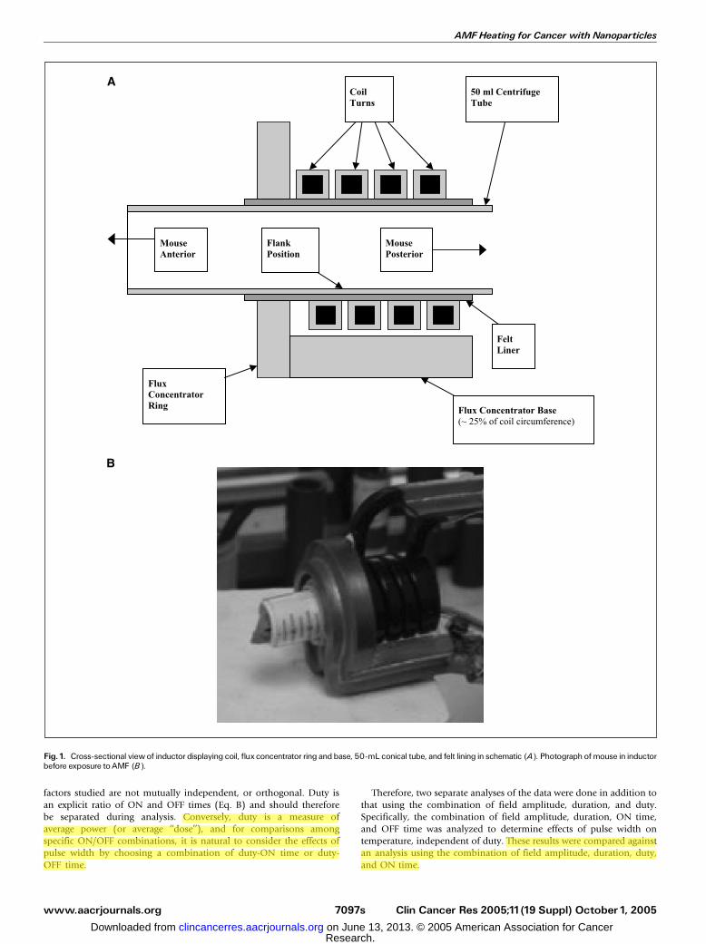
of three main components: (a) the induction coil, or inductor (Fig. 1A
and B); (b) a capacitance network that, when combined with theinductor, forms a resonant circuit; and (c) the power supply.
Inductor. The inductor was designed using Flux 2D (Magsoft,Inc., Troy, NY) finite element analysis software. The induction coil
was manufactured from square cross-sectional, oxygen-free, and high-
conductivity copper tubing. The length of the induction coil was40 mm with an internal diameter of 36 mm, which enabled
insertion of a 50-mL conical centrifuge tube containing a mouse andallowing for 2.5 mm of thermal insulation around the circumference
of the 50-mL tube.A low-reluctance flux-concentrating ring made of Fluxtrol 50 (Centre
for Induction Technology, Auburn Hills, MI) was added to the end ofthe solenoid coil where the mouse was inserted to divert magnetic fluxaway from areas not intended to receive high-amplitude AMF exposure.A low-reluctance magnetic return path was also added to one fourthof the outer diameter of the coil. These components provided apreferential path for the magnetic flux that minimized AMF exposure tomuch of the mouse body and directed the flux in the interior of thesolenoid producing a 1-cm band of high-amplitude AMF in the ventralregion, the intended tumor location.
Experimental Studies
www.aacrjournals.orgClin Cancer Res 2005;11(19 Suppl) October1, 2005 7094s
Research. on June 13, 2013. © 2005 American Association for Cancerclincancerres.aacrjournals.org Downloaded from
Measurements of the AMF flux with a current probe confirmed that ifthe rear third of the mouse was inserted into the induction coil, a 1-cmregion of the abdomen could receive as much as 1,380 Oe, whereastissue outside the concentrating ring (over 50% of the mouse) wouldnot be exposed to >200 Oe. The field amplitude within the 1-cm bandwas measured before each set of trials and for each generator powersetting used. It is the amplitude within this region that is reported asAMF amplitude.
During operation, the induction coil itself heats because it is aconductor carrying a high current load, particularly at high amplitudeand high duty. To compensate, the inductor was cooled using a closed-loop circulating water system maintained at 14 F 2jC duringoperation.
Capacitance network and power supply. The capacitance networkwas built into the power supply and was adjusted for stable oscillation
at 153 F 0.5 kHz. The power supply was a 25-kW (0-25 kW) generatormanufactured by PPECO (Watsonville, CA). A pulse-timer circuitmanufactured by Giltron, Inc. (Medfield, MA) was installed allowing0.5- to 9,999-second pulses at any duty (0-100%), defined as:
Duty ¼ Pulse ON time ðsecondsÞPulse ON time ðsecondsÞ þ Pulse OFF time ðsecondsÞ �100% ðBÞ
Experimental design and data analysisStatistical design and data analysis. For both study design and data
analysis, we used a statistical approach that did not require a prioriknowledge of the effects and interactions of input variables andresponses (i.e., AMF conditions and in vivo temperature, respectively).A total of five input variables were simultaneously varied: (a) field
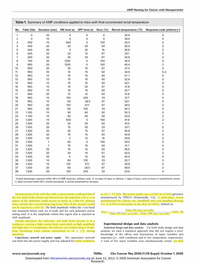
Table1. Summary of AMF conditions applied to micewith final uncorrected rectal temperature
No. Field (Oe) Duration (min) ON time (s) OFF time (s) Duty (%) Rectal temperature (�C) Response code (arbitrary*)
1 0 20 0 0 0 26.6 02 0 10 0 0 0 28.4 03 400 10 600 0 100 30.4 04 400 20 30 30 50 26.9 05 400 20 5 25 15 26.8 06 400 20 20 10 67 25.1 07 400 20 40 20 67 24.8 08 700 20 1200 0 100 36.9 09 950 20 1200 0 100 45.4 2
10 950 20 30 15 67 37.4 011 950 20 15 15 50 34.9 0, 412 950 10 15 15 50 31.1 013 950 10 15 15 50 32.6 014 950 10 15 15 50 32.1 015 950 10 15 30 67 31.8 016 950 10 15 15 50 32.7 017 950 20 10 10 50 31.8 018 950 10 120 240 33 31.7 019 950 10 60 28.5 67 30.1 020 950 20 120 57.1 67 36.0 021 950 20 60 120 33 34.2 022 1,150 15 90 90 50 35.3 023 1,150 15 90 90 50 33.9 024 1,300 10 600 0 100 41.6 225 1,300 20 30 30 50 41.0 126 1,300 20 15 30 33 33.7 027 1,300 20 30 15 67 45.8 228 1,300 20 15 15 50 40.8 029 1,300 20 2 13 15 29.8 030 1,300 1 60 0 100 29.0 031 1,300 1 15 15 50 31.7 032 1,300 20 15 15 50 36.5 033 1,300 20 20 10 67 43.5 234 1,300 20 5 10 33 34.5 035 1,300 10 60 120 33 32.7 036 1,300 10 120 57.1 67 36.8 037 1,300 20 60 28.5 67 48.9 238 1,300 20 120 240 33 33.6 0
*Coded physiologic response within 48 h of AMF exposure, arbitrary units: 0, no signs of injury or distress; 1, signs of injury, such as burns or hyperthermic stress;2, death occurred within 48 h, animal necropsied; 4, animal euthanized for necropsy.
AMFHeating for Cancer with Nanoparticles
www.aacrjournals.org Clin Cancer Res 2005;11(19 Suppl) October1, 20057095s
Research. on June 13, 2013. © 2005 American Association for Cancerclincancerres.aacrjournals.org Downloaded from

amplitude (Oe), (b) total duration of exposure (minutes), (c) duty (%),(d) pulse ON time (seconds), and (e) pulse OFF time (seconds). Thefirst three of these variables were sufficient to define the total AMFexposure. The latter two variables have an influence upon the unsteadystate thermal evolution, particularly with respect to physiologic andenvironmental heat dissipation.
Trial design was done using Stat-Expert software (Stat-Ease, Inc.,Minneapolis, MN) using both D-optimal and Box-Behnken designs toselect trial conditions and numbers of mice that minimized bothnumbers of mice and morbidity.
Mice were divided into two groups, defined by short ON pulses (0-30seconds) with field amplitudes of 400, 950, and 1,300 Oe and long ONpulses (60-240 seconds) with field amplitudes of 700, 1,150, and 1,300combinations. In the second group of mice, factors selected from thoseused in the first group were repeated to maintain statistical continuity,allowing us to combine data from both groups for analysis. Also in thefirst group of mice, some mice were exposed to continuous power AMF(100% duty) for up to 1,200 seconds.
Temperature was measured as the response to AMF exposure. Foranalysis, a mathematical model of the form
Y ¼ b0 þ b1X1 þ b2X2 þ b3X3 þ b12X1X2 þ b13X1X3 ðCÞ
þ b23X2X3 þ b11X21 þ b22X2
2 þ b33X23 þ . . .
was fit to the temperature data using the method of least squares,where Y is the response, or temperature, and X1, X2, . . . represent thefactors duty, field amplitude, and duration. Temperature response tothe combination of field amplitude, duration, and duty was analyzed.The effects of each term were determined using standard ANOVAtechniques. Analysis and graphical representation of the data weredone using Stat-Expert software (Stat-Ease). Physiologic responseswere also noted; however, a systematic study of these was notundertaken.
Further analysis of the data including ON/OFF time combinationswas undertaken to ensure the validity of the model, because all
Table 2. Summary of temperature change data for spine, flank, skin, and rectal temperature probes
No. Field (G) Duration (min) ON time (s) OFF time (s) Duty (%) #Tspine (�C) #Tflank (�C) #Tskin (�C) #Trectal (�C)
1 0 20 0 0 0 �5.6 �5.4 �5.8 �5.32 0 10 0 0 0 �3.0 �3.8 �3.5 �3.33 400 10 600 0 100 2.9 2.3 5.2 0.34 400 20 30 30 50 �4.1 �4.6 �3.7 �4.35 400 20 5 25 15 �6.2 �5.9 �6.3 �6.46 400 20 20 10 67 �4.6 �5.0 �4.1 �5.17 400 20 40 20 67 �6.2 �6.3 �5.8 �6.48 700 20 1,200 0 100 10.9 9.7 12.8 9.99 950 20 1,200 0 100 20.5 18.1 22.8 15.4
10 950 20 30 15 67 10.7 9.5 15.6 8.711 950 20 15 15 50 7.8 6.1 9.1 4.412 950 10 15 15 50 6.1 5.4 8.3 2.913 950 10 15 15 50 4.9 2.7 5.9 0.414 950 10 15 15 50 4.4 1.6 8.0 0.715 950 10 15 30 67 0.0 2.7 4.0 �0.116 950 10 15 15 50 0.0 3.3 7.1 0.717 950 20 10 10 50 4.4 2.1 6.1 0.818 950 10 120 240 33 0.9 1.2 3.4 �0.919 950 10 60 28.5 67 10.0 2.3 6.2 1.820 950 20 120 57.1 67 8.7 7.6 9.6 5.821 950 20 60 120 33 1.3 �0.7 0.5 �1.322 1,150 15 90 90 50 5.2 4.0 6.2 1.823 1,150 15 90 90 50 6.0 3.9 9.1 1.424 1,300 10 600 0 100 21.1 15.6 16.0 13.025 1,300 20 30 30 50 14.9 14.0 14.0 12.926 1,300 20 15 30 33 7.7 6.6 9.9 6.927 1,300 20 30 15 67 21.6 19.1 20.9 17.128 1,300 20 15 15 50 18.6 16.8 15.2 12.829 1,300 20 2 13 15 1.9 0.7 2.3 1.130 1,300 1 60 0 100 0.8 �0.1 1.4 �0.331 1,300 1 15 15 50 0.4 �0.2 0.8 �0.232 1,300 20 15 15 50 8.9 5.4 11.2 5.133 1,300 20 20 10 67 17.0 15.1 22.0 13.834 1,300 20 5 10 33 7.6 4.6 7.2 2.835 1,300 10 60 120 33 4.0 2.5 4.5 0.836 1,300 10 120 57.1 67 13.4 11.6 20.7 8.837 1,300 20 60 28.5 67 22.6 18.7 22.4 16.538 1,300 20 120 240 33 4.5 3.6 7.3 1.3
Experimental Studies
www.aacrjournals.orgClin Cancer Res 2005;11(19 Suppl) October1, 2005 7096s
Research. on June 13, 2013. © 2005 American Association for Cancerclincancerres.aacrjournals.org Downloaded from
factors studied are not mutually independent, or orthogonal. Duty isan explicit ratio of ON and OFF times (Eq. B) and should thereforebe separated during analysis. Conversely, duty is a measure ofaverage power (or average ‘‘dose’’), and for comparisons amongspecific ON/OFF combinations, it is natural to consider the effects ofpulse width by choosing a combination of duty-ON time or duty-OFF time.
Therefore, two separate analyses of the data were done in addition tothat using the combination of field amplitude, duration, and duty.Specifically, the combination of field amplitude, duration, ON time,and OFF time was analyzed to determine effects of pulse width ontemperature, independent of duty. These results were compared againstan analysis using the combination of field amplitude, duration, duty,and ON time.
Fig.1. Cross-sectional viewof inductor displaying coil, flux concentrator ring and base, 50-mL conical tube, and felt lining in schematic (A). Photograph of mouse in inductorbefore exposure toAMF (B).
AMFHeating for Cancer with Nanoparticles
www.aacrjournals.org Clin Cancer Res 2005;11(19 Suppl) October1, 20057097s
Research. on June 13, 2013. © 2005 American Association for Cancerclincancerres.aacrjournals.org Downloaded from

Results
Table 1 contains a summary of AMF exposure conditionsand final uncorrected rectal temperatures for the mice, whereasTable 2 provides a summary of DTcorr for all temperature probes.Mice that experienced final rectal temperatures approachingor exceeding 42jC consistently died, with one exception, forwhich a rectal temperature of 47.3jC was recorded (data notshown). This datum was excluded from analysis by standardstatistical criteria.
Mice exposed to field amplitudes in excess of 950 Oe and atduties >50% experienced consistent morbidity and injury. Inthese mice, maximum uncorrected rectal temperature ap-proached or exceeded 42jC (Table 1), and DTcorr was observedto be greater than f12jC (Table 2). The average starttemperature for all mice was 30.5 F 0.3jC for the rectum,29.0 F 0.2jC for the skin, 30.7 F 0.3jC for the flank, and28.8 F 0.3jC for the spine.
When injury or death occurred, necropsies of mice that diedwithin 48 hours after AMF exposure (Table 1) showedconsistent findings. The skin under the first inductor turn wasnoticeably red. Sometimes the exterior of the hind legs showedpetechiae. Inside the abdomen, intestine that had been withinthe first inductor turn closest to the abdominal wall were redbut not hemorrhagic. The cecum sometimes showed petechiae.Intestine deeper in the abdomen were pale and sometimesblanched. Lungs were red but not hemorrhagic. By comparison,necropsy results were normal in all respects of the one mouserandomly selected from the group showing no sign of injury(Table 1).
Observed temperature changes (i.e., DT) with time as aresponse to AMF amplitude of 1,300 Oe and 33% duty fortemperature probes placed on the skin and in the rectum areshown in Fig. 2A and B, respectively. These temperatures arenot corrected against controls and represent ON/OFF combi-nations of 5/10, 15/30, and 120/240 seconds corresponding,respectively, to mouse numbers 34, 26, and 38 in Tables 1and 2.
Fitting the model equation (Eq. C) to corrected temperatures(DTcorr) with field, duty, and duration as factors producedstatistically significant (P < 0.05) results for spine, flank, skin,and rectal temperature responses for cubic (total sum ofexponents, V3) combinations of the factors and their inter-actions. Contributions from higher order terms (i.e., exponent>3) were not significant nor was a model that included theseterms. Other statistical measures of significance such as R2
values, analysis of residuals and outliers, lack of fit tests, andtests of adequate precision (signal to noise) confirm the validityof the model. Thus, analysis and predictions within the variablespace were validated.
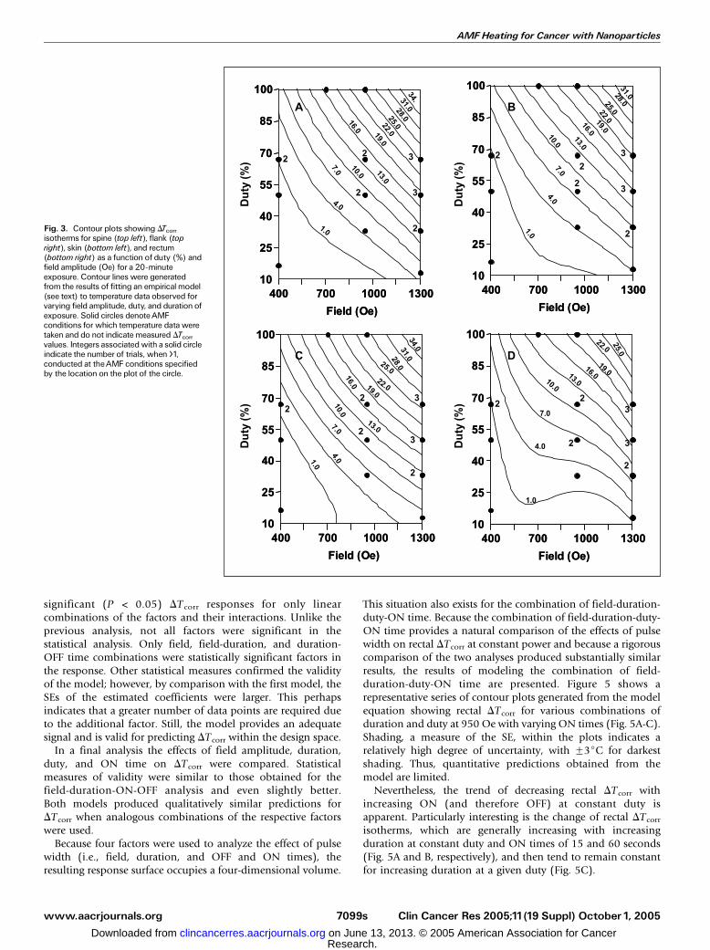
The resulting model equation was used to generate a series oftwo-dimensional contour plots on which contour linesrepresent isotherms of response (constant DTcorr). A sampleof contour plots for 20-minute exposure showing the relation-ship between duty and field amplitude for spine, flank, skin,and rectal DTcorr is displayed in Fig. 3. The contour plots showthat the variable space displayed was well represented byexperiment. Note, however, that the circles represent conditionsat which data were taken and do not indicate the measuredDTcorr nor do they indicate by their placement a relationshipwith the predicted DTcorr.
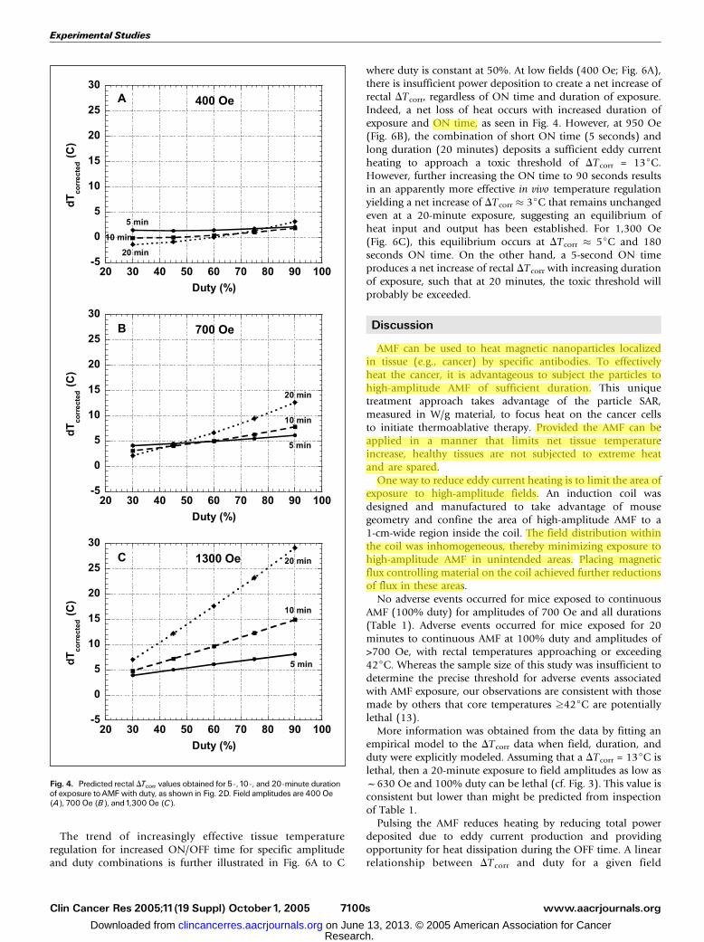
The effects of duration of exposure on rectal DTcorr arecompared in Fig. 4A to C for 5-, 10-, and 20-minute exposuresat 400 Oe (Fig. 4A), 700 Oe (Fig. 4B), and 1,300 Oe (Fig. 4C).In general, increasing DTcorr accompanies increasing durationof exposure and duty. However, at low duties and low-amplitude AMF (Fig. 4A-B), long durations in the coil do notproduce substantial temperature increases and may evenaccompany a decrease of rectal DTcorr. This indicates thatAMF heating is insufficient to overcome heat loss. With AMFamplitudes of >700 Oe, sufficient eddy current heating occursat duties >50%, eventually becoming acute and leading toadverse events. However, at the highest amplitudes (f1,300Oe), duty has a profound effect on AMF-induced eddy currentheating (cf. Fig. 4C).
A similar analysis was done for field amplitude, duration,ON time, and OFF time. These factors produced statistically
Fig. 2. Comparison of temperature response at fixed duty (33%)with varyingpulse width obtained by probes placed on the skin of a hind limb (A) and insertedinto the rectum (B). AMF conditions were1,300 Oe and 33% duty.Total duration ofexposure in each case was 20 minutes (1,200 seconds). Labels indicated pulsewidth with 5-second ON/10-second OFF,15-second ON/30-second OFF, and120-second ON/240-second OFF.Temperatures were recorded at1-secondintervals, and initial temperature at the start of AMF exposure (t = 0 seconds) wassubtracted from subsequent temperatures. Solid line at DT = 12jC indicates theobserved temperature threshold associated with adverse events.
Experimental Studies
www.aacrjournals.orgClin Cancer Res 2005;11(19 Suppl) October1, 2005 7098s
Research. on June 13, 2013. © 2005 American Association for Cancerclincancerres.aacrjournals.org Downloaded from
significant (P < 0.05) DT corr responses for only linearcombinations of the factors and their interactions. Unlike theprevious analysis, not all factors were significant in thestatistical analysis. Only field, field-duration, and duration-OFF time combinations were statistically significant factors inthe response. Other statistical measures confirmed the validityof the model; however, by comparison with the first model, theSEs of the estimated coefficients were larger. This perhapsindicates that a greater number of data points are required dueto the additional factor. Still, the model provides an adequatesignal and is valid for predicting DTcorr within the design space.
In a final analysis the effects of field amplitude, duration,duty, and ON time on DTcorr were compared. Statisticalmeasures of validity were similar to those obtained for thefield-duration-ON-OFF analysis and even slightly better.Both models produced qualitatively similar predictions forDTcorr when analogous combinations of the respective factorswere used.
Because four factors were used to analyze the effect of pulsewidth (i.e., field, duration, and OFF and ON times), theresulting response surface occupies a four-dimensional volume.
This situation also exists for the combination of field-duration-duty-ON time. Because the combination of field-duration-duty-ON time provides a natural comparison of the effects of pulsewidth on rectal DTcorr at constant power and because a rigorouscomparison of the two analyses produced substantially similarresults, the results of modeling the combination of field-duration-duty-ON time are presented. Figure 5 shows arepresentative series of contour plots generated from the modelequation showing rectal DTcorr for various combinations ofduration and duty at 950 Oe with varying ON times (Fig. 5A-C).Shading, a measure of the SE, within the plots indicates arelatively high degree of uncertainty, with F3jC for darkestshading. Thus, quantitative predictions obtained from themodel are limited.
Nevertheless, the trend of decreasing rectal DTcorr withincreasing ON (and therefore OFF) at constant duty isapparent. Particularly interesting is the change of rectal DTcorr
isotherms, which are generally increasing with increasingduration at constant duty and ON times of 15 and 60 seconds(Fig. 5A and B, respectively), and then tend to remain constantfor increasing duration at a given duty (Fig. 5C).
Fig. 3. Contour plots showing DTcorr
isotherms for spine (top left), flank (topright), skin (bottom left), and rectum(bottom right) as a function of duty (%) andfield amplitude (Oe) for a 20-minuteexposure. Contour lines were generatedfrom the results of fitting an empirical model(see text) to temperature data observed forvarying field amplitude, duty, and duration ofexposure. Solid circles denoteAMFconditions for which temperature data weretaken and do not indicate measured DTcorr
values. Integers associatedwith a solid circleindicate the number of trials, when >1,conducted at theAMF conditions specifiedby the location on the plot of the circle.
AMFHeating for Cancer with Nanoparticles
www.aacrjournals.org Clin Cancer Res 2005;11(19 Suppl) October1, 20057099s
Research. on June 13, 2013. © 2005 American Association for Cancerclincancerres.aacrjournals.org Downloaded from
The trend of increasingly effective tissue temperatureregulation for increased ON/OFF time for specific amplitudeand duty combinations is further illustrated in Fig. 6A to C
where duty is constant at 50%. At low fields (400 Oe; Fig. 6A),there is insufficient power deposition to create a net increase ofrectal DTcorr, regardless of ON time and duration of exposure.Indeed, a net loss of heat occurs with increased duration ofexposure and ON time, as seen in Fig. 4. However, at 950 Oe(Fig. 6B), the combination of short ON time (5 seconds) andlong duration (20 minutes) deposits a sufficient eddy currentheating to approach a toxic threshold of DTcorr = 13jC.However, further increasing the ON time to 90 seconds resultsin an apparently more effective in vivo temperature regulationyielding a net increase of DTcorr 3jC that remains unchangedeven at a 20-minute exposure, suggesting an equilibrium ofheat input and output has been established. For 1,300 Oe(Fig. 6C), this equilibrium occurs at DTcorr 5jC and 180seconds ON time. On the other hand, a 5-second ON timeproduces a net increase of rectal DTcorr with increasing durationof exposure, such that at 20 minutes, the toxic threshold willprobably be exceeded.
Discussion
AMF can be used to heat magnetic nanoparticles localizedin tissue (e.g., cancer) by specific antibodies. To effectivelyheat the cancer, it is advantageous to subject the particles tohigh-amplitude AMF of sufficient duration. This uniquetreatment approach takes advantage of the particle SAR,measured in W/g material, to focus heat on the cancer cellsto initiate thermoablative therapy. Provided the AMF can beapplied in a manner that limits net tissue temperatureincrease, healthy tissues are not subjected to extreme heatand are spared.
One way to reduce eddy current heating is to limit the area ofexposure to high-amplitude fields. An induction coil wasdesigned and manufactured to take advantage of mousegeometry and confine the area of high-amplitude AMF to a1-cm-wide region inside the coil. The field distribution withinthe coil was inhomogeneous, thereby minimizing exposure tohigh-amplitude AMF in unintended areas. Placing magneticflux controlling material on the coil achieved further reductionsof flux in these areas.
No adverse events occurred for mice exposed to continuousAMF (100% duty) for amplitudes of 700 Oe and all durations(Table 1). Adverse events occurred for mice exposed for 20minutes to continuous AMF at 100% duty and amplitudes of>700 Oe, with rectal temperatures approaching or exceeding42jC. Whereas the sample size of this study was insufficient todetermine the precise threshold for adverse events associatedwith AMF exposure, our observations are consistent with thosemade by others that core temperatures z42jC are potentiallylethal (13).
More information was obtained from the data by fitting anempirical model to the DTcorr data when field, duration, andduty were explicitly modeled. Assuming that a DTcorr = 13jC islethal, then a 20-minute exposure to field amplitudes as low asf630 Oe and 100% duty can be lethal (cf. Fig. 3). This value isconsistent but lower than might be predicted from inspectionof Table 1.
Pulsing the AMF reduces heating by reducing total powerdeposited due to eddy current production and providingopportunity for heat dissipation during the OFF time. A linearrelationship between DT corr and duty for a given field
Fig. 4. Predicted rectal DTcorr values obtained for 5-,10-, and 20-minute durationof exposure toAMFwith duty, as shown in Fig. 2D. Field amplitudes are 400 Oe(A), 700 Oe (B), and1,300 Oe (C).
Experimental Studies
www.aacrjournals.orgClin Cancer Res 2005;11(19 Suppl) October1, 2005 7100s
Research. on June 13, 2013. © 2005 American Association for Cancerclincancerres.aacrjournals.org Downloaded from

amplitude and duration is expected, because total powerdeposition due to eddy current production varies linearly withduty. Contour plots of field and duty at 20-minute duration(Fig. 3A-D) show that DTcorr for spine, flank, skin, and rectumincreases as duty is increased for a given field amplitude. Thedistance between successive DTcorr isotherms seems constant forconstant field, except at the lowest amplitudes and duties wheresome deviation from linearity appears. This apparent deviationis not significant, because in these regions the uncertainties areF1jC, comparable with the predicted DTcorr.
The value of DTcorr for the rectum is lower than thecorresponding temperatures for the spine, flank, and skin,particularly at high amplitude and duty (cf. Fig. 3A-D andTable 2). In general, eddy current production is greatest in areaswith large radius and zero at the electrical center (i.e., r = 0;cf. Eq. A). The temperature probe placed on the skin is atmaximum r for the mouse, whereas the temperature probeplaced in the rectum is near the electrical center (i.e., r 0).The temperature probe placed on the skin provides a direct
measure of eddy current heating during an ON cycle, withconcomitant heat loss during an OFF cycle (Fig. 2A). Hence,temperature oscillations due to pulsing are pronounced,particularly with long ON/OFF cycles.
On the other hand, the temperature probe placed in therectum does not measure eddy current heating directly butrather the net heat balance resulting from heating of thesurrounding tissue during exposure to AMF, thermoregulatoryresponse to eddy currents, heat loss to the environment (coil),and heat produced by metabolic processes. Consequently,temperature oscillations seem damped (Fig. 2B) when com-pared against skin measurements (Fig. 2A). Because mortalityis associated more directly with core temperature (i.e., rectal;refs. 12, 13), maintaining a rectal temperature within safe limitshas significant implications for the safe application of AMF.
Eddy current heating is most affected by duty at the highestamplitudes (f1,300 Oe; Fig. 4). That duty has such an effectupon DTcorr for AMF exposure is not surprising as this is directlyrelated to average power or dose, even for mice, which are
Fig. 5. Contour plots showing rectalDTcorr isothermsas a function of duty (%) and duration of exposure(minutes) for a field amplitude of 950 Oe and ONtimes of15 seconds (top left), 60 seconds (top right),and120 seconds (bottom left). Contour lines aregenerated from fit results of an empirical model (seetext) to temperature data obtained for varying fieldamplitude, duty, and duration of exposure, and ONtime. Filled circles represent design points (data) anddo not indicate measured DTcorr values. Integersassociated with a filled circle indicate the number ofsubjects used for the design point. Shading is ameasure of SE with lightest corresponding to a valueof 0.3jC and darkest corresponding toF3jC.
AMFHeating for Cancer with Nanoparticles
www.aacrjournals.org Clin Cancer Res 2005;11(19 Suppl) October1, 20057101s
Research. on June 13, 2013. © 2005 American Association for Cancerclincancerres.aacrjournals.org Downloaded from

known to have poor thermoregulatory systems (12), comparedwith humans. In addition to decreasing the total eddy currentheating in the mouse, decreasing duty also provides time forheat dissipation during OFF cycles.
The effect of ON/OFF pulse variation for a given duty andamplitude was observed directly as illustrated in temperatureplots shown in Fig. 2. Increasing ON time from 5 to 15 secondsresults in an increase of eddy current heating and a net increasein temperature. However, this trend is reversed for very longON/OFF cycles (J15 seconds ON and J30 seconds OFF),provided one ON cycle does not produce an acute level of eddycurrent heating (cf. Tables 1 and 2). Within this constraint, verylong ON/OFF cycles can produce a net rectal temperature(Fig. 2B) that is substantially lower than that produced by shortON/OFF AMF exposure even after a 20-minute duration (Figs. 4and 5). This is an intriguing observation.
Analysis of the data taking into account pulse width providesfurther evidence of the complex interaction of AMF powerdeposition (or tissue SAR) through eddy current productionand resulting changes in tissue temperature. Contour plotsshowing rectal DTcorr isotherms as a function of duty andduration of exposure for 950 Oe that result from an analysis oftemperature response to the factors field amplitude-duration-duty-ON time is shown in Fig. 5A to C, where ON time is variedfrom 15 seconds (Fig. 5A) to 120 seconds (Fig. 5B). As ON timeincreases, the temperature in the rectum decreases, particularlyat high duty and long duration of exposure, as does the slopeand curvature of the individual isotherms. This qualitativechange in temperature response with ON time suggests aprofound shift in thermoregulatory response to AMF eddycurrent production as ON time is increased when fieldamplitude and duty, or total AMF ‘‘dose’’, are held constant.
This effect is further illustrated in Fig. 6A to C, which show acomparison of predicted rectal DTcorr for a duty of 50% and ONtimes of 5, 90, and 180 seconds for field amplitudes of 400 Oe(Fig. 6A), 950 Oe (Fig. 6B), and 1,300 Oe (Fig. 6C). Note that foreach field amplitude the average AMF ‘‘dose’’ (or tissue SAR) isconstant, because it is defined by field amplitude, frequency,radius of exposure (cf. Eq. A), and duty. Conversely, thephysiologic temperature response to this dose is complex andcan vary dramatically depending upon the algorithm used toapply the AMF in vivo . Such a complex relationship of powerdeposition, tissue SAR, and tissue temperature response has beenreported in both preclinical and clinical trials using microwaveantenna arrays (12, 14–18) and ultrasound (19) for thetreatment of cancer. Although the clinical significance of theseobservations at the frequency chosen in this study is unknown,it provides an interesting line for further investigation.
Safe application of high-amplitude AMF sufficient to heatantibody-conjugated magnetic nanoparticles (bioprobes) local-ized in cancer tissue is possible provided the area of exposureand duty are controlled. The choice of duration and duty atamplitudes >700 Oe are integral to limit nonspecific heating.Results presented suggest that pulse width is also important forsafety and can be maximized, within limits, to apply a highthermal dose locally with nanoparticles to cancer cells. Undersuch conditions, heat delivered by bioprobes can potentially bemodulated by suitable selection of AMF variables to enhance atherapeutic effect, whereas normal tissues can recover anddissipate heat produced by eddy currents during OFF times.
Acknowledgments
We thank Dr. Douglas Gwost and CarmenTraxler for their contributions to thestudy design and Laird Meiers for mouse care and handling.
Fig. 6. Predicted rectal DTcorr values obtained for 5-, 90-, and180-second ONtimes with duration of exposure at 50%. Field amplitudes are 400 Oe (A), 950 Oe(B), and1,300 Oe (C).
Experimental Studies
www.aacrjournals.orgClin Cancer Res 2005;11(19 Suppl) October1, 2005 7102s
Research. on June 13, 2013. © 2005 American Association for Cancerclincancerres.aacrjournals.org Downloaded from

References1.OvergaardJ. History and heritage: an introduction. In:Overgaard J, editor. Hyperthermic oncology.Vol. 2.London:Taylor and Francis; 1985. p. 8^9.2. Streffer C, van Beuningen D. The biological basis fortumor therapy by hyperthermia and radiation. In:Streffer J, editor. Hyperthermia and the therapy ofmalignant tumors. Berlin: Springer; 1987. p. 24^70.3. Zaffaroni N, Fliorentini G, De Giorgi U. Hyperthermiaand hypoxia: newdevelopments in anticancer chemo-therapy. EurJSurg Oncol 2001;27:340^2.4. Hildebrandt B, Wust P, Ahlers O, et al. The cellularand molecular basis of hyperthermia. Crit Rev OncolHematol 2002;43:33^56.5.Moroz P, Jones SK, Gray BN.Tumor response to arte-rial embolization hyperthermia and direct injectionhyperthemia in a rabbit liver tumor model. J SurgOncol 2002;80:149^56.6.Moroz P, Jones SK, Gray BN. Magnetically mediatedhyperthermia: current status and future directions. IntJHyperthermia 2002;18:267^84.7. Jeffrey S, Birdwell R, Ikeda D, et al. Radiofrequencyablation of breast cancer: first report of an emergingtechnology. Arch Surg1999;134:1064^2008.8. Izzo F, Barnett CC, Curley SA. Radiofrequency abla-
tion of primary and metastatic malignant liver tumors.Adv Surg 2001;35:225^50.9. Gordon RT, Hines JR, Gordon D. Intracellular hyper-thermia: a biophysical approach to cancer treatmentvia intracellular temperature and biophysical alteration.Med Hypotheses1979;5:83^102.10. Jordan A,Wust P, Scholz R, Faehling H, Krause J,Felix R. Magnetic fluid hyperthermia (MFH). In: HafeliU, et al, editors. Scientific and Clinical Appl MagnCarriers. NewYork: Plenum Press; 1997. p. 569^95.11. Jordan A, Scholz R,Wust P, Fahling H, Felix R.Magnetic fluid hyperthermia: cancer treatment withACmagnetic field induced excitation of biocompatiblesuperparamagnetic nanoparticles. J Mag Mag Mat1999;201:413^9.12. Adair ER, Black DR.Thermoregulatory responses toRF energy absorption. Bioelectromagnetics 2003;6:S17^38.13.DewhirstMW,Viglianti BL, Lora-MichielsM,HansonM, Hoopes PJ. Basic principles of thermal dosimetryand thermal thresholds for tissue damage from hyper-thermia. IntJHyperthermia 2003;19:267^94.14. Mechling JA, Strohbehn JW, Ryan TP. Three-dimensional theoretical temperature distributions
produced by 915 MHz dipole antenna arrayswith varying insertion depths in muscle tissue.Int J Radiat Oncol Biol Phys 1992;22:131^8.15. Ryan TP. Comparison of six microwave antennasfor hyperthermia treatment of cancer: Sar results forsingle antennas and arrays. Int J Radiat Oncol BiolPhys 1991;21:403^13.16. RyanTP, Mechling JA, Strohbehn JW. Absorbedpower depostion for various insertion depths for 915MHz interstitial dipole antenna arrays: experimentversus theory. Int J Radiat Oncol Biol Phys 1990;19:377^87.17. Trembly BS, Douple EB, RyanTP, Hoopes PJ. Effectof phasemodulationon the temperature distributionofa microwave hyperthermia antenna array in vivo. Int JHyperthermia1994;10:691^705.18. Ryan TP, Hoopes PJ, Taylor JH, et al. Experimentalbrain hyperthermia: techniques for heat delivery andthermometry. Int J Radiat Oncol Biol Phys 1991;20:739^50.19. RyanTP, HartrovA, ColacchioTA, Coughlin CT,StaffordJH, Hoopes PJ. Analysis and testing of a con-centric ringapplicator forultrasoundhyperthermiawithclinical results. IntJHyperthermia1991;7:587^603.
AMFHeating for Cancer with Nanoparticles
www.aacrjournals.org Clin Cancer Res 2005;11(19 Suppl) October1, 20057103s
Research. on June 13, 2013. © 2005 American Association for Cancerclincancerres.aacrjournals.org Downloaded from
Copyright © 2022 FDOKUMEN