Crystal structure and characterization of L-arginine chlorate and L-arginine bromate
9
Crystal structure and characterization of L-arginine chlorate and L-arginine bromate * A.M. Petrosyan a, * , H.A. Karapetyan a , R.P. Sukiasyan a , A.E. Aghajanyan b , V.G. Morgunov c , E.A. Kravchenko c , A.A. Bush d a Molecule Structure Research Center, NAS, 26 Azatutyan Ave., Yerevan 375014, Armenia b Institute of Biotechnology, 14 Gyurjyan St., Yerevan 375056, Armenia c Institute of General and Inorganic Chemistry, RAS, 31 Leninskii pr., Moscow 123812, Russia d Moscow State Institute of Radioengineering, Electronics and Automation, 78 Vernadskii St., Moscow 119454, Russia Received 17 January 2005; revised 25 May 2005; accepted 25 May 2005 Available online 20 July 2005 Abstract The salts L-arginine.HClO 3 and L-arginine.HBrO 3 , were synthesized. The crystals obtained were characterized by IR and NQR spectroscopy, thermal analysis, Nd:YAG laser radiation second harmonic generation, dielectric, piezoelectric and pyroelectric measurements and their crystal structures were determined. L-arginine.HClO 3 and L-arginine.HBrO 3 are crystallized in orthorhombic (space group P2 1 2 1 2 1 ) and triclinic (space group P1) systems, respectively. Their structures consist of the protonated arginine cation [ C (H 2 N) 2 CNH(CH 2 ) 3- CH(NH 3 C )COO K ] and respective ClO 3 K or BrO 3 K anions. Hydrogen bonds (stronger in the bromate) connect anions with cations and the latter with each other. However, these salts have essential distinctions in crystal packing. q 2005 Elsevier B.V. All rights reserved. Keywords: L-Arginine chlorate; Bromate; Crystal structure; Solution growth; Properties 1. Introduction L-Arginine phosphate monohydrate L-Arg.H 3 PO 4 .H 2 O (LAP) is one of the best nonlinear optical crystals [1]. However, growth of the LAP crystals has attendant growth of microorganisms, which can be detrimental to crystal quality. Hence, the preparation procedure requires special precautions. It has been suggested that the appearance of microorganisms can be prevented by either adding H 2 O 2 [2] or Hg [3] into solution or by covering the solution with a layer of n-hexane [4]. Ongoing investigations [5–9], however, showed that this does not completely solve the problem. Search for new members of the LAP family may produce crystals, whose parameters can surpass those of the LAP. In addition, the preparation of such crystals may be free of the problem of microorganisms [10–13]. Systematic search for new LAP-analogs was started by Monaco et al. [14], who investigated some previously known and a number of new crystals. The interaction of L-arginine with iodic acid only, among the halogenic acids HClO 3 , HBrO 3 , HIO 3 , was investigated to give powdered L-Arg.HIO 3 . The weak broad iodine-127 lines were observed in the nuclear quadrupole resonance (NQR) spectrum [15], which to a certain extent, was evidence for the crystallinity of the compound. In addition, it was found that L-Arg.2HIO 3 comprising a doubly charged arginine cation [ C (H 2 N) 2- CNH(CH 2 ) 3 CH(NH 3 C )COOH] exists, which, in contrast to L-Arg.HIO 3 crystallizes easily [15]. In this respect, we found it interesting to study the possibility of formation and crystallization of the respective salts of L-arginine with chloric (HClO 3 ) and bromic (HBrO 3 ) acids. 2. Experimental As initial compounds, we used L-arginine, synthesized in the Institute of Biotechnology (Yerevan), and a reagent Journal of Molecular Structure 752 (2005) 144–152 www.elsevier.com/locate/molstruc 0022-2860/$ - see front matter q 2005 Elsevier B.V. All rights reserved. doi:10.1016/j.molstruc.2005.05.053 * CCDC 259335 and CCDC 259336 contain the supplementary crystallographic data for this paper. These data can be obtained free of charge via www.ccdc.cam.ac.uk/conts/retrieving.html (or from the Cambridge Crystallographic Data Centre, 12 Union Road, Cambridge CB2 1EZ, UK; fax: C44 1223 336033). * Corresponding author. Tel.: C374 1 281764; fax: C374 1 282267. E-mail address: [email protected] (A.M. Petrosyan).
-
Upload
independent -
Category
Documents
-
view
0 -
download
0
Transcript of Crystal structure and characterization of L-arginine chlorate and L-arginine bromate

Crystal structure and characterization of L-arginine chlorate
and L-arginine bromate*
A.M. Petrosyana,*, H.A. Karapetyana, R.P. Sukiasyana, A.E. Aghajanyanb,
V.G. Morgunovc, E.A. Kravchenkoc, A.A. Bushd
aMolecule Structure Research Center, NAS, 26 Azatutyan Ave., Yerevan 375014, ArmeniabInstitute of Biotechnology, 14 Gyurjyan St., Yerevan 375056, Armenia
cInstitute of General and Inorganic Chemistry, RAS, 31 Leninskii pr., Moscow 123812, RussiadMoscow State Institute of Radioengineering, Electronics and Automation, 78 Vernadskii St., Moscow 119454, Russia
Received 17 January 2005; revised 25 May 2005; accepted 25 May 2005
Available online 20 July 2005
Abstract
The salts L-arginine.HClO3 and L-arginine.HBrO3, were synthesized. The crystals obtained were characterized by IR and NQR
spectroscopy, thermal analysis, Nd:YAG laser radiation second harmonic generation, dielectric, piezoelectric and pyroelectric measurements
and their crystal structures were determined. L-arginine.HClO3 and L-arginine.HBrO3 are crystallized in orthorhombic (space group P212121)
and triclinic (space group P1) systems, respectively. Their structures consist of the protonated arginine cation [C(H2N)2CNH(CH2)3-
CH(NH3C)COOK] and respective ClO3
K or BrO3K anions. Hydrogen bonds (stronger in the bromate) connect anions with cations and the
latter with each other. However, these salts have essential distinctions in crystal packing.
q 2005 Elsevier B.V. All rights reserved.
Keywords: L-Arginine chlorate; Bromate; Crystal structure; Solution growth; Properties
1. Introduction
L-Arginine phosphate monohydrate L-Arg.H3PO4.H2O
(LAP) is one of the best nonlinear optical crystals [1].
However, growth of the LAP crystals has attendant growth
of microorganisms, which can be detrimental to crystal
quality. Hence, the preparation procedure requires special
precautions. It has been suggested that the appearance of
microorganisms can be prevented by either adding H2O2 [2]
or Hg [3] into solution or by covering the solution with a
layer of n-hexane [4]. Ongoing investigations [5–9],
however, showed that this does not completely solve the
problem. Search for new members of the LAP family may
0022-2860/$ - see front matter q 2005 Elsevier B.V. All rights reserved.
doi:10.1016/j.molstruc.2005.05.053
* CCDC 259335 and CCDC 259336 contain the supplementary
crystallographic data for this paper. These data can be obtained free of
charge via www.ccdc.cam.ac.uk/conts/retrieving.html (or from the
Cambridge Crystallographic Data Centre, 12 Union Road, Cambridge
CB2 1EZ, UK; fax: C44 1223 336033).* Corresponding author. Tel.: C374 1 281764; fax: C374 1 282267.
E-mail address: [email protected] (A.M. Petrosyan).
produce crystals, whose parameters can surpass those of the
LAP. In addition, the preparation of such crystals may be
free of the problem of microorganisms [10–13]. Systematic
search for new LAP-analogs was started by Monaco et al.
[14], who investigated some previously known and a
number of new crystals. The interaction of L-arginine with
iodic acid only, among the halogenic acids HClO3, HBrO3,
HIO3, was investigated to give powdered L-Arg.HIO3. The
weak broad iodine-127 lines were observed in the nuclear
quadrupole resonance (NQR) spectrum [15], which to a
certain extent, was evidence for the crystallinity of the
compound. In addition, it was found that L-Arg.2HIO3
comprising a doubly charged arginine cation [C(H2N)2-
CNH(CH2)3CH(NH3C)COOH] exists, which, in contrast to
L-Arg.HIO3 crystallizes easily [15]. In this respect, we
found it interesting to study the possibility of formation and
crystallization of the respective salts of L-arginine with
chloric (HClO3) and bromic (HBrO3) acids.
2. Experimental
As initial compounds, we used L-arginine, synthesized in
the Institute of Biotechnology (Yerevan), and a reagent
Journal of Molecular Structure 752 (2005) 144–152
www.elsevier.com/locate/molstruc

A.M. Petrosyan et al. / Journal of Molecular Structure 752 (2005) 144–152 145
purchased from «Sigma» Chemical Company. For prep-
aration of HClO3 and HBrO3, we used, respectively, KClO3
and Ba(BrO3)2.H2O, both of reagent grade. The IR spectra
were registered using Specord 75 IR spectrophotometer
(Carl Zeiss, Jena) by the technique of nujol mull suspension
(4000–400 cmK1) and Nicolet ‘Nexus’ FT-IR spectrometer
with ZnSe prism (4000–650 cmK1). The thermal properties
were studied using a Paulik-Paulik- Erdey Derivatograph
(type 3427–904) and a Boetius-type microscope. The
nuclear quadrupole resonance (NQR) spectra were
measured using a pulse NQR spectrometer operating within
2–300 MHz.
The measurements of dielectric constant 3 and the
tangent of the loss tand were performed by means of the ac
bridge P5083 at the frequency of measuring field 1 kHz. The
piezoelectric effect in crystals was induced by alternating
mechanical load. The sample was sandwiched between the
faces of two vertical metallic rods, and 0.7 kHz longitudinal
oscillations were generated using a piezoelectric transducer.
The resulting charge is proportional to the piezoelectric
charge coefficient d 033 and gives rise to an A.C. voltage. The
installation was calibrated using X-cut quartz plates. The
pyroelectric measurements were carried out by the quasi-
static method. The pyroelectric constant g was calculated by
formula gZI/[S(dT/dt)], where I is the pyroelectric current
measured in a circuit sample-electrometer (V7-30) during
uniform change of the sample’s temperature with the rate of
dT/dt w0.1 grad/sec, S is the area of the Ag electrodes on
the base surfaces of the sample. The dielectric, piezoelectric
and pyroelectric measurements were performed on single
crystal plates with w1 mm thickness and Sw5 mm2 for L-
Arg.HClO3 and Sw50 mm2 for L-Arg.HBrO3. In order to
prepare electrodes the base surfaces of plates were coated by
silver paste. The orientations of the crystallographic axes for
the crystals were determined by X-ray studies on a DRON-4
diffractometer. X-ray diffraction data for structure solution
Fig. 1. ATR FTIR spectrum o
were collected using CAD-4 automatic diffractometer
(ENRAF NONIUS). The structures were determined by
the direct method and refined using the SHELX86 and
SHELXL93 programs [16,17].
3. Results and discussions
3.1. Synthesis, growth and characterization of crystals
Chloric and bromic acids (unlike iodic) exist only as
dilute solutions. The solution of HClO3 was obtained by
passing an aqueous solution of KClO3 through an exchange
column with sulphocationite KU-2-8 in HC form. The
solution of HBrO3 was obtained after precipitation and
isolation of barium sulphate by the reaction
BaðBrO3Þ2$H2O CH2SO4/2HBrO3 CBaSO4Y :
Then the solution of L-arginine was added to the HClO3
and HBrO3 solutions to obtain mixtures with required L-
Arg: acid ratios. Crystallization was reached by slow
evaporation of the solutions at room temperature. The
crystals L-Arg.HClO3 and L-Arg.HBrO3 were grown from
aqueous solutions with equimolar L-Arg: acid ratio. No
crystals were obtained from the solutions with 1:2 and 1:3
ratios. If the concentration of the above solutions increased,
the HBrO3-system decomposed and that with HClO3
exploded, probably because of instability of HClO3 and
HBrO3 at high concentrations. Using spontaneously formed
crystals as the seeds and evaporating small volumes of
solutions at room temperature, we obtained single crystals
of L-Arg.HBrO3, which measured 13!11!6 mm3, and L-
Arg.HClO3 of 28!8!7 mm3 in size. These crystals were
used for measurements of physical properties. The crystals
of L-Arg.HClO3 were of optical quality. No microorganisms
appeared in the solutions during crystallization.
f L-Arg$HClO3 crystals.

Fig. 2. ATR FTIR spectrum of L-Arg$HBrO3 crystals.
Table 135Cl and 79Br NQR frequencies and relaxation times at 77 K
Crystals n(G1/2/G3/2), MHz
T1, ms T2, ms
L-Arg$HClO3 29.948(7) 270 1100
L-Arg$HBrO3 175.69(2) 12 230
175.99(2) 14 280
A.M. Petrosyan et al. / Journal of Molecular Structure 752 (2005) 144–152146
The crystals of L-arginine chlorate and bromate were
expected to form via singly-charged cation L-ArgHC(or L-
ArgC), namely, C[(H2N)2CNH](CH2)3CH(NH3C)COOK
and the respective ClO3K and BrO3
K anions, which is the
usual mechanism for salts of 1:1 composition. The IR and
NQR spectra are in accordance with this expectation. The IR
spectra are shown in Figs. 1 and 2. Comparison of the IR
spectrum for L-Arg.HClO3 with that for KClO3 enables one
to assign the absorption band at 900–1000 cmK1 to the
stretching (n1, n3) vibration of Cl–O bonds in the ClO3K ion
(920.37 cmK1, 949.46 cmK1). The respective bands of
deformation vibrations (not shown in Fig. 1) are at
603 cmK1 (n2) and 487 cmK1 (n4). The absorption bands
of n(N–H) stretching vibrations of guanidyl and NH3-Cgroups are at 3000–3500 cmK1(3111.71 cmK1,
3168.01 cmK1, 3284.02 cmK1, 3392.42 cmK1,
3432.70 cmK1). The absorption band with peaks at
1663.29 cmK1 and 1642.79 cmK1 correspond to asym-
metric stretching vibration of carboxylate group and
deformation vibration of NH2C and NH3
C groups. Similar
absorption bands of L-ArgC can also be found in the
spectrum of L-Arg.HBrO3. The strong band near 800 cmK1
(783.93 cmK1 and 809.15 cmK1) represents absorption
caused by the stretching vibrations of Br–O bonds in the
BrO3K ion. A similar absorption band with the same number
of peaks was observed in the same region of the spectrum of
Ba(BrO3)2.H2O. In the region of the n(N–H) stretching
vibrations there are peaks at 3065.04 cmK1, 3122.70 cmK1,
3263.13 cmK1 and 3343.75 cmK1. Decrease in the n(N–H)
stretching vibration frequencies in the spectrum of L-
Arg.HBrO3 compared to L-Arg.HClO3 is probably caused
by stronger N–H/O hydrogen bonds in L-arginine bromate.
In the range of 1700–1550 cmK1, there is a strong
absorption band with peaks at 1692.50 cmK1,
1670.23 cmK1, 1627.48 cmK1, 1602.34 cmK1 and
1579.12 cmK1. An increase in the vibration frequencies of
the carboxylate group COOK (1692.50 cmK1 and
1670.23 cmK1) may result from the difference in the C–O
bond lengths due to hydrogen bond formation [15].
The L-Arg.HClO3 and L-Arg.HBrO3 crystals were also
studied using 35 Cl and 79 Br nuclear quadrupole resonances
(NQR). Table 1 lists the measured frequencies and
relaxation times of the resonant lines at 77 K. The NQR
spectrum of L-Arg.HClO3 consists of a strong singlet, and
that of L-Arg.HBrO3 - of a strong, well-resolved doublet.
The intensity of resonant lines, however, rapidly decreased
with increasing temperature, and at room temperature, the
signals were not observed. As evidenced by the number of
resonant lines at 77 K, all the ClO3K ions are crystal-
lographicaly equivalent in the unit cell of L-Arg.HClO3,
while in the unit cell of L-Arg.HBrO3 two non-equivalent
BrO3K ions are present. Thus L-Arg.HClO3 and L-
Arg.HBrO3 are not isostructural. The 35 Cl and 79 Br NQR
frequencies are characteristic for the ClO3K and BrO3
K ions
and close to the 35 Cl and 79 Br NQR frequencies for initial
KClO3 and Ba(BrO3)2.H2O (28.954 MHz and
176.223 MHz, respectively) [18].
On heating L-Arg.HClO3 melts at 170 8C, and above
180 8C the compound begins to decompose with an
exothermal effect. L-Arg.HBrO3 decomposes at a lower
temperature (145 8C). Both crystal forms were checked for
the second harmonic generation (Nd:YAG laser), and in the
higher quality L-Arg.HClO3 crystal a strong phase-matched
second harmonic generation signal was observed.

Table 2
The Miller indexes (hkl) of basic plane of the studied crystals, dielectric
constant 3, dielectric loss tan d, piezoelectric d 033 and pyroelectric g
coefficients at room temperature
Crystals (hkl) 3 tan d d 033,
10K12 C/N
g, nC/
cm2 K
L-Arg$HClO3 (020) 5.1 0.019 0.70 !4.0.10K3
L-Arg$HBrO3 (1–10) 3.3 0.010 0.32 0.60
A.M. Petrosyan et al. / Journal of Molecular Structure 752 (2005) 144–152 147
The results of dielectric, piezoelectric and pyroelectric
measurements of crystals are presented in Table 2 and
Fig. 3. The L-Arg.HBrO3 crystal exhibits a rather strong
pyroelectric effect in accordance with its polar symmetry
P1. The temperature dependencies of the dielectric constant
and dielectric loss of these crystals show smooth changes
without any anomaly in the 100–400 K temperature range
(Fig. 3). The temperature dependence of the pyroelectic
constant is also smooth in the 125–360 K range. The rather
sharp increase of apparent pyroelectric signal at TO360 K
and its maximum at 375 K are evidently caused by the
appearance of thermostimulated currents in this temperature
range. In the temperature range 125–360 K the measured
signal changes its sign upon switching from heating to
cooling of the sample and on the contrary, at TO360 K the
sign of signal does not depend on regime of the temperature
change.
100 150 200 250 300 350 400 4503.0
3.2
3.4
3.6
3.8
4.0
100 150 200 250 300 350 400 4500.00
0.02
0.04
0.06
0.08
100 150 200 250 300 350 400 450
0.4
0.8
1.2
1.6
T, K
tan
δε
γ, n
C/c
m2 .K
Fig. 3. Temperature dependencies of dielectric constant 3, dielectric loss
tan d and pyroelectric coefficient g measured perpendicular to (1–10) plane
of L-Arg$HBrO3 single crystals.
3.2. Crystal structure of L-Arg.HClO3 and L-Arg.HBrO3
The crystal structure of L-arginine chlorate was
determined at room temperature, and that of L-arginine
bromate, at 233 K. The crystal data and additional
information on the determination of both structures are
presented in Table 3. The hydrogen atoms positions were
determined from the difference electron density synthesis.
The chlorate and bromate of L-arginine are not isostructural.
L-Arg.HClO3 crystallizes in orthorhombic space group
P212121 with ZZ4, while L-Arg.HBrO3, in triclinic space
group P1 with ZZ2. Figs. 4 and 5 show the independent
parts of the unit cells of the crystals. Packing in the crystal
structures are shown in the Figs. 6 and 7. Bonds lengths and
angles are presented in Tables 4 and 5, and hydrogen bond
parameters in Tables 6 and 7.
As one can see from Figs. 4 and 5, the formation
mechanism of L-arginine chlorate and bromate is common
for salts of 1:1 composition. In the singly charged L-ArgC
cation, guanidyl and a-amino groups are protonated at the
expense of protons of the respective acids (HClO3 and
HBrO3) and deprotonation of their own carboxyl group. The
presence of positively charged guanidyl and a-amino
groups and negatively charged carboxylate group in the L-
ArgC cation, results in interaction of neighboring cations
via N–H/O hydrogen bonds between guanidyl and
carboxylate groups in crystalline salts of arginine. This
interaction takes place in both structures. However, the
detailed comparison revealed essential differences in
interactions between cations in both structures, in cation
conformations and also in their interactions with anions. As
can be seen from Figs. 5 and 7, two crystallographicaly
independent cations in the structure of L-arginine bromate
are connected with each other through hydrogen bonds
between N(3) and N(4) atoms of guanidyl groups and
oxygen atoms of carboxylate groups. This type of
interaction was designated by Salunke and Vijayan as
type A [19]. Arginine cations form a kind of dimer. These
dimers are connected with each other by stronger H-bonds
between protonated a-amino groups N(1A) and N(1B),
accordingly, and oxygen atoms O(2B) and O(2A) (compare
H/O distances in the N(1)-H/O(2) hydrogen bonds with
respective distances in the N(3)–H/O(2) and N(4)–H/O(1) hydrogen bonds). This leads to lengthening of the
C(1)–O(2) bonds in both carboxylate groups in comparison
with the C(1)–O(1) bonds (Table 5). The essential
distinction in bond lengths of C(1)–O(1) and C(1)–O(2)
can explain an increase of asymmetric stretching vibration
frequencies of COOK groups (Fig. 2). The aforementioned
dimers form layers parallel to the diagonal (110) plane
(Fig. 7).
In the structure of L-arginine chlorate the cations interact
in a different way (Fig. 6). O(1) and O(2) oxygen atoms of
the carboxylate group form H-bonds with N(3) and N(2)
nitrogen atoms, respectively. This type of interaction was
designated as type B [19]. At the same time, the O(1) atom

Table 3
Crystal data and structure refinement for L-Arg$HClO3, L-Arg$HBrO3
Empirical formula C6H15ClN4O5 C6H15BrN4O5
Formula weight 258.67 303.13
Temperature 293 K 233 K
Wavelength, A 0.71073 0.71073
Crystal system Orthorhombic Triclinic
Space group P212121 P1
Unit cell dimensions aZ5.1928(10) A, aZ908, bZ13.852(3) A, bZ908,
cZ15.734(3) A, gZ908
aZ8.426(2) A, aZ111.05(3)8, bZ8.737(2) A,
bZ99.43(3)8, cZ9.301(2) A, gZ110.90(3)8
Volume, Z 1131.8 A3, 4 562.5(2) A3, 2
Density (calculated) 1.518 Mg/m3 1.790 Mg/m3
Density (measured) 1.518(2) Mg/m3 1.77(2) Mg/m3
Absorption coefficient 0.352 mmK1 3.669 mmK1
F(000) 544 308
Crystal size (mm) 0.28!0.21!0.1 0.28!0.21!0.08
q range for data collection 1.96–27.968 2.49–25.978
Limiting indices 0%h%6, 0%k%18, 0%l%20 K9%h%10, K10%k%10, K10%l%11
Reflections collected 1615 2441
Independent reflections 1595 (RintZ0.0000) 2301 (RintZ0.0000)
Absorption correction None None
Refinement method Full-matrix least-squares on F2 Full-matrix least-squares on F2
Data/restraints/parameters 1352/0/191 2297/3/290
Goodness-of-fit on F2 1.055 1.107
Final R indices [IO2s(I)] R1Z0.0349, wR2Z0.0915 R1Z0.0461, wR2Z0.1106
R indices (all data) R1Z0.0349, wR2Z0.0915 R1Z0.0464, wR2Z0.1106
Extinction coefficient 0.004(3) 0.020(5)
Largest diff. Peak and hole 0.300 and K0.240 eAK3 1.967 and K1.768 eAK3
A.M. Petrosyan et al. / Journal of Molecular Structure 752 (2005) 144–152148
forms H-bonds with N(3) and N(4) atoms of neighboring
cation (O(1)/H(B)–N(3) and O(1)/H(B)–N(4)). The
interaction of amino groups of guanidyl group with the
same oxygen atom of the carboxylate group was designated
as type C [19]. So, in the structure of L-arginine chlorate,
both types of interactions, B and C take place. Due to type C
interactions, a chain of cations along the c-axis is formed,
while the connection via type B forms cation layers
perpendicular to the a axis. One more difference is that
the O(2) oxygen atom forms an intramolecular H-bond
Fig. 4. Independent part of unit cell and numb
N(1)–H(A)/O(2). As a result, a five-member pseudo-cycle
with conformation of highly flattened semi chair is formed
by the atoms HA(N1), N(1), C(2), C(1) and O(2), in which
the C(2) and N(1) atoms are displaced from the plane,
formed by the remaining atoms, by 0.13 and K0.07 A,
respectively.
Let us now consider the arrangement of anions in both
structures and their connection with cations. The presence
of crystallographicaly equivalent ClO3K ions in the structure
of L-Arg.HClO3 and two crystallographicaly independent
ering of atoms of L-Arg$HClO3 crystals.

Fig. 5. Independent part of unit cell and numbering of atoms of L-Arg$HBrO3 crystals.
Fig. 6. A stereoscopic view of packing in the crystal structure of L-Arg$HClO3.
A.M. Petrosyan et al. / Journal of Molecular Structure 752 (2005) 144–152 149
Br(1)O3K and Br(2)O3
K ions in the structure of L-Arg.HBrO3
is consistent with the NQR data at 77 K. The average values
of Cl–O bond lengths and O–Cl–O angles in the L-Arg.
HClO3 are equal to 1.475 A and 106.58, which are
characteristic for the ClO3K ion [20]. The average values
of Br(1)–O bond lengths and O–Br(1)–O angles (1.641 A
and 104.48, respectively) and those for Br(2)O3K (1.651 A
and 104.68, respectively) are characteristic of the BrO3K ion
Fig. 7. A stereoscopic view of packing in t
[20]. In addition to electrostatic interaction, the cations of
arginine are connected to anions via H-bonds and ones
formed by bromate-ions are stronger than those formed by
chlorate-ions. Each of the bromate-ions forms five H-bonds.
The Br(1)O3K ion forms four H-bonds with hydrogen atoms
of cation B and one H-bond with cation A, while Br(2)O3K
forms four H-bonds with cation A and one with cation B
(Table 7). The ClO3K anion forms six H-bonds of O/H–N
he crystal structure of L-Arg$HBrO3.
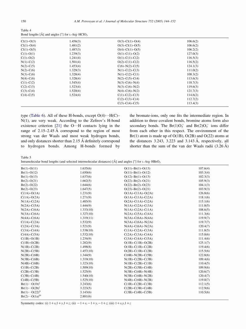
Table 4
Bond lengths [A] and angles [8] for L-Arg$HClO3
Cl(1)–O(3) 1.456(3) O(3)–Cl(1)–O(4) 106.6(2)
Cl(1)–O(4) 1.481(2) O(3)–Cl(1)–O(5) 106.6(2)
Cl(1)–O(5) 1.487(3) O(4)–Cl(1)–O(5) 106.2(2)
C(1)–O(1) 1.239(3) O(1)–C(1)–O(2) 127.0(3)
C(1)–O(2) 1.241(4) O(1)–C(1)–C(2) 116.5(3)
N(1)–C(2) 1.501(4) O(2)–C(1)–C(2) 116.5(2)
N(2)–C(5) 1.453(4) C(6)–N(2)–C(5) 124.1(3)
N(2)–C(6) 1.329(3) N(1)–C(2)–C(3) 111.0(2)
N(3)–C(6) 1.328(4) N(1)–C(2)–C(1) 108.3(2)
N(4)–C(6) 1.328(4) N(2)–C(5)–C(4) 113.6(3)
C(1)–C(2) 1.545(4) N(3)–C(6)–N(4) 118.7(3)
C(2)–C(3) 1.523(4) N(3)–C(6)–N(2) 119.6(3)
C(3)–C(4) 1.520(4) N(4)–C(6)–N(2) 121.7(3)
C(4)–C(5) 1.524(4) C(1)–C(2)–C(3) 114.6(2)
C(2)–C(3)–C(4) 112.7(2)
C(3)–C(4)–C(5) 113.4(3)
A.M. Petrosyan et al. / Journal of Molecular Structure 752 (2005) 144–152150
type (Table 6). All of these H-bonds, except O(4)/H(C)–
N(1), are very weak. According to the Zefirov’s H-bond
existence criterion [21] the O/H contacts lying in the
range of 2.15–2.45 A correspond to the region of most
strong van der Waals and most weak hydrogen bonds,
and only distances shorter than 2.15 A definitely correspond
to hydrogen bonds. Among H-bonds formed by
Table 5
Intramolecular bond lengths (and selected intermolecular distances) [A] and angl
Br(1)–O(11) 1.635(6) O(1
Br(1)–O(12) 1.650(6) O(1
Br(1)–O(13) 1.637(6) O(1
Br(2)–O(21) 1.662(5) O(2
Br(2)–O(22) 1.644(6) O(2
Br(2)–O(23) 1.647(5) O(2
C(1A)–O(1A) 1.231(9) O(1
C(1A)–O(2A) 1.271(9) O(1
N(1A)–C(2A) 1.485(9) O(2
N(2A)–C(5A) 1.444(9) N(1
N(2A)–C(6A) 1.333(10) N(1
N(3A)–C(6A) 1.327(10) N(2
N(4A)–C(6A) 1.319(11) N(3
C(1A)–C(2A) 1.532(9) N(3
C(2A)–C(3A) 1.521(9) N(4
C(3A)–C(4A) 1.538(10) C(1
C(4A)–C(5A) 1.532(10) C(2
C(1B)–O(1B) 1.234(9) C(3
C(1B)–O(2B) 1.282(9) O(1
N(1B)–C(2B) 1.498(8) O(1
N(2B)–C(5B) 1.457(10) O(2
N(2B)–C(6B) 1.344(9) C(6
N(3B)–C(6B) 1.319(10) N(1
N(4B)–C(6B) 1.323(10) N(1
C(1B)–C(2B) 1.509(10) N(2
C(2B)–C(3B) 1.525(9) N(3
C(3B)–C(4B) 1.540(10) N(3
C(4B)–C(5B) 1.525(10) N(4
Br(1)/O(1b)i 3.243(6) C(1
Br(1)/O(2b)i 3.223(5) C(2
Br(1)/O(22)ii 3.143(6) C(3
Br(2)/O(1a)iii 2.881(6)
Symmetry codes: (i) 1Cx,1Cy,1Cz; (ii) K1Cx,K1Cy,K1Cz; (iii) 1Cx,y,1C
the bromate-ions, only one fits the intermediate region. In
addition to three covalent bonds, bromine atoms form also
secondary bonds. The Br(1)O3K and Br(2)O3
K ions differ
from each other in this respect. The environment of the
Br(1) atom is made up of O(1B), O(2B) and O(22) atoms at
the distances 3.243, 3.223 and 3.143 A, respectively, all
shorter than the sum of the van der Waals radii (3.26 A)
es [8] for L-Arg$HBrO3
1)–Br(1)–O(13) 107.6(4)
1)–Br(1)–O(12) 103.3(4)
2)–Br(1)–O(13) 102.3(3)
2)–Br(2)–O(21) 105.9(3)
2)–Br(2)–O(23) 104.1(3)
3)–Br(2)–O(21) 103.9(3)
A)–C(1A)–O(2A) 126.8(6)
A)–C(1A)–C(2A) 118.1(6)
A)–C(1A)–C(2A) 115.1(6)
A)–C(2A)–C(3A) 111.0(5)
A)–C(2A)–C(1A) 108.6(5)
A)–C(5A)–C(4A) 111.3(6)
A)–C(6A)–N(4A) 119.9(7)
A)–C(6A)–N(2A) 119.7(7)
A)–C(6A)–N(2A) 120.4(7)
A)–C(2A)–C(3A) 111.8(5)
A)–C(3A)–C(4A) 115.0(6)
A)–C(4A)–C(5A) 111.4(6)
B)–C(1B)–O(2B) 125.1(7)
B)–C(1B)–C(2B) 119.4(6)
B)–C(1B)–C(2B) 115.5(6)
B)–N(2B)–C(5B) 122.8(6)
B)–C(2B)–C(3B) 109.4(6)
B)–C(2B)–C(1B) 110.4(5)
B)–C(5B)–C(4B) 109.9(6)
B)–C(6B)–N(4B) 120.6(7)
B)–C(6B)–N(2B) 120.4(7)
B)–C(6B)–N(2B) 119.0(7)
B)–C(2B)–C(3B) 112.1(5)
B)–C(3B)–C(4B) 112.9(6)
B)–C(4B)–C(5B) 110.5(6)
z
Table 6
The geometric parameters [A, 8] of D–H/A (D-donor, A-acceptor) H-bonds in L-Arg$HClO3
D–H/A Symmetry code of A D/A (A) D–H(A) H/A(A) D–H–A (8)
N(1)–H(A)/O(2) x,y,z 2.604(3) 0.86(5) 2.06(4) 121(3)
N(1)–H(A)/O(5) x, KyC0.5, Kz 3.046(4) 0.86(5) 2.38(5) 134(3)
N(1)–H(B)/O(5) xC0.5, KyC0.5, Kz 3.033(4) 0.95(4) 2.17(4) 150(3)
N(1)–H(B)/O(3) xC0.5, KyC0.5, Kz 3.047(4) 0.95(4) 2.40(4) 125(3)
N(1)–H(C)/O(4) x,y,z 2.952(4) 1.06(4) 1.92(4) 163(3)
N(1)–H(C)/O(5) x,y,z 3.092(4) 1.06(4) 2.32(4) 129(3)
N(2)–H(2)/O(2) KxK1, y K0.5, Kz K0.5 2.838(3) 0.86(4) 1.98(4) 175(4)
N(3)–H(A)/O(1) KxK1, yK0.5, Kz K0.5 2.919(4) 0.81(4) 2.11(4) 176(4)
N(3)–H(B)/O(1) Kx K0.5,Ky, zC0.5 2.881(4) 1.04(4) 1.90(4) 156(4)
N(4)–H(A)/O(4) xC1,y,z 3.087(4) 0.85(4) 2.27(4) 160(4)
N(4)–H(B)/O(1) Kx K0.5,Ky, zC0.5 3.000(4) 0.88(4) 2.22(4) 148(4)
N(1)–H(C)/Cl x,y,z 3.653(3) 1.06(4) 2.63(4) 161(3)
N(1)–H(B)/Cl xC0.5, KyC0.5,Kz 3.657(3) 0.95(4) 2.78(4) 153(3)
Table 7
The geometric parameters [A, 8] of D–H/A (D-donor, A-acceptor) H-bonds in L-Arg$HBrO3
D–H/A Symmetry code of A D/A (A) D–H (A) H/A (A) D–H–A (8)
N(1A)–H(A)/O(13) x, yC1,z 2.954(8) 0.89 2.10 160
N(1A)–H(B)/O(21) x,y, z K1 2.856(8) 0.89 2.02 155
N(1A)–H(C)/O(2B) xC1, yC1, zC1 2.787(8) 0.89 1.96 155
N(2A)–H(B)/O(22) xK1, yK1, zK1 2.926(8) 0.86 2.07 175
N(3A)–H(D)/O(23) xK1, yK1, zK2 2.947(9) 0.86 2.11 163
N(3A)–H(C)/O(2B) x,y,z 2.843(8) 0.86 2.02 161
N(4A)–H(C)/O(1B) x,y,z 2.897(9) 0.86 2.05 167
N(4A)–H(D)/O(21) xK1, yK1, zK1 2.975(9) 0.86 2.12 171
N(1B)–H(A)/O(11) 2Kx, yK1, zK1 2.831(9) 0.89 2.00 154
N(1B)–H(B)/O(2A) xK1, yK1, z 2.751(8) 0.89 1.95 149
N(1B)–H(C)/O(23) XK2, yK2, zK2 2.829(8) 0.89 1.96 165
N(2B)–H(B)/O(12) xK1,y,z 2.853(8) 0.86 2.00 171
N(3B)–H(C)/O(2A) x,y,z 2.827(9) 0.86 2.01 158
N(3B)–H(D)/O(13) xK1, y, zK1 3.005(9) 0.86 2.15 171
N(4B)–H(C)/O(1A) x,y,z 2.863(9) 0.86 2.01 172
N(4B)–H(D)/O(11) xK1,y,z 3.115(11) 0.86 2.32 155
A.M. Petrosyan et al. / Journal of Molecular Structure 752 (2005) 144–152 151
[22]. The Br(2) atom forms a secondary bond only with
O(1A) atom, but the distance Br(2)/O(1A) being equal to
2.881 A, is essentially shorter than that in the preceding
case and shorter than the minimum distance 3.04 A for the
van der Waals interaction between bromine and oxygen
atoms [21].
In the ClO3K anion the chlorine atom has no secondary
bonds with oxygen atoms at distances close to the sum of
van der Waals radii for Cl and O. Instead, the environment
of the chlorine atom is made up of two hydrogen atoms from
the two closest a-amino groups at distances of 2.633 A
(Cl/H(N1C) (x,y,z)) and 2.781 A (Cl/H(N1B)(0.5Cx, 0.
5Ky, Kz)). The sum of Cl and H van der Waals radii is 3.
06 A, while the minimum Cl/H distance at their van der
Waals interaction is 2.67 A. Such a difference in behavior of
ClO3K and BrO3
K anions may be caused by the difference in
electronegativity of chlorine and bromine atoms. Among the
Cl, Br and I atoms, the bromine atom is intermediate in
electronegativity. Due to the large electronegativity differ-
ence between oxygen and iodine, the formal charge on the
iodine atom in the IO3K ion is positive [23]. As a result,
the secondary I/O bonds (2.7–2.9 A) shorter than the
minimum (3.17 A by [21]) distance for I/O van der Waals
interactions are usual in the structures of iodates. Moreover,
the so-called intermediate bonds between the first and
second coordination spheres with distances 2.4–2.5 A were
sometimes observed (see Ref. [24] and references therein).
In the ClO3K ion the formal charge on the central atom is
negative [23], which may probably explain the absence of
Cl/O secondary bonds and the presence of the shortened
Cl/H contacts in the structure of L-Arg.HClO3. In the case
of intermediate bromine, the BrO3K ion is more similar to
IO3K due to existence of Br/O secondary bonds, whereas
its electronic structure is more similar to that of the ClO3K
ion with the negative charge less than in the ClO3K ion.
4. Conclusion
Although both salts L-Arg.HClO3 and L-Arg.HBrO3 are
formed by the same mechanism (L-ArgC.ClO3K, L-ArgC.
BrO3K) however, they are not isostructural (space groups:

A.M. Petrosyan et al. / Journal of Molecular Structure 752 (2005) 144–152152
P212121 and P1 respectively) and have essential distinctions
in crystal packing.
Acknowledgements
The research described in this publication was made
possible in part by CRDF Grant No. AE2-2533-YE-03. The
piezo- and pyroelectric studies were supported by Russian
Foundation of Basic Research, Grant No. 02-02-17798.
We thank Dr Martirosyan G.G. for his help in recording the
FT-IR ATR spectra. We thank also reviewer for useful
comment.
References
[1] G.C. Bhar, A.V. Rudra, P.K. Datta, U.N. Roy, V.K. Wadhawan,
T. Sasaki, Pramana: J. Phys. 44 (1995) 45.
[2] A. Yokotani, T. Sasaki, K. Fujioka, S. Nakai, Ch. Yamanaka, J. Cryst.
Growth 99 (1990) 815.
[3] V. Venkataramanan, G. Dhanaraj, H.L. Bhat, J. Cryst. Growth 140
(1994) 336.
[4] G. Dhanaraj, T. Shripathi, H.L. Bhat, J. Cryst. Growth 113 (1991) 456.
[5] J.F. Carvalho, A.C. Hernandes, F.D. Nunes, L.B.O.A. de Moraes,
L. Misoguti, S.C. Zilio, J. Cryst. Growth 173 (1997) 487.
[6] G. Arunmozhi, R. Jayavel, C. Subramanian, J. Cryst. Growth 178
(1997) 387.
[7] L. Aidong, X. Chongquan, L. Aiben, M. Naiben, J. Cryst. Growth 220
(2000) 291.
[8] A.S. Haja Hameed, G. Ravi, P. Ramasamy, J. Cryst. Growth 229
(2001) 547.
[9] R. Shanmugavadivu, G. Ravi, R. Jayavel, R. Mohankumar, A. Nixon
Azariah, J. Cryst. Growth 271 (2004) 252.
[10] A.M. Petrosyan, R.P. Sukiasyan, H.A. Karapetyan, S.S. Terzyan, R.S.
Feigelson, J. Cryst. Growth 213 (2000) 103.
[11] A.S. Haja Hameed, P. Anandan, R. Jayavel, P. Ramasamy, G. Ravi, J.
Cryst. Growth 249 (2003) 316.
[12] D. Xu, X.Q. Wang, W.T. Yu, S.X. Xu, G.H. Zhang, J. Cryst. Growth
253 (2003) 481.
[13] K. Vasantha, S. Dhanuskodi, J. Cryst. Growth 269 (2004) 333.
[14] S.B. Monaco, L.E. Davis, S.P. Velsko, F.T. Wang, D. Eimerl, A.
Zalkin, J. Cryst. Growth 85 (1987) 252.
[15] A.M. Petrosyan, S.S. Terzyan, V.M. Burbelo, R.P. Sukiasyan, Z.
Naturforschung 53a (1998) 528.
[16] G.M. Sheldrick, SHELX86. Program for the solution of crystal
structures. University of Gottingen, Germany, 1985.
[17] G.M. Sheldrick, SHELXL93. Program for the refinement of crystal
structures. University of Gottingen, Germany, 1993.
[18] G.K. Semin, T.A. Babushkina, G.G. Yakobson, Primenenie yader-
nongo kvardrupol’nogo rezonansa v khimii, ‘Khimia’ Leningrad,
1972; G.K. Semin, T.A. Babushkina, G.G. Yakobson, Nuclear
Quadrupole Resonance in Chemistry, Wiley, New York, 1975.
[19] D.M. Salunke, M. Vijayan, Int. J. Pept. Protein Res. 18 (1981) 348.
[20] A.F. Wells, Structural Inorganic Chemistry, Fifth ed., Oxford
University Press, Moscow, 1986 (Russian edition in vol. 3, hMiri,
Moscow).
[21] Yu.V. Zefirov, Kristallografia 44 (1999) 1091.
[22] Yu.V. Zefirov, P.M. Zorky, Uspekhi khimii 64 (1995) 446.
[23] B.D. El-Issa, A. Hinchliffe, J. Mol. Struct. 67 (1980) 317.
[24] A.M. Petrosyan, V.A. Shishkin, Z. Naturforschung, 51a (1996)
667.