
Reverse Turns in Peptides and Protein
85
October 1980 315 REVERSE TURNS IN PEPTIDES AND PROTEINS Authors: John A. Smith' Department of Medicine Harvard Medical School Boston, Massachusetts Lila G. Pease* Department of Chemistry Amherst College Amherst, Massachusetts Referee: Kenneth D. Kopple Illinois Institute of Technology Chicago, Illinois I. INTRODUCTION Reverse turns are structural features of peptides and proteins, involving three (i.e., a y-turn)'.' or four (i.e., a B-turn)' consecutive residues, hallmarked by the folding back on itself of the peptide chain and by the presence of an intramolecular hydrogen bond. Since their occurrence in globular proteins accounts for about one third of the residues in these molecule^.^.^ as well as a substantial proportion of the surface resi- dues.6 it is likely that certain reverse turns provide recognition sites for the initiation of complex immunological, endocrinological, or metabolic reactions. The identifica- tion of reverse turn conformations in various peptide hormones is also compatible with this suggestion. Further, globular protein chains, themselves linear polymers, are known to fold into low energy conformations.' In order to achieve these conformations, which result in the formation of compact globular proteins, the chain frequently changes direction during the process of folding. The regions in which this change of direction occurs are reverse turns. Further, it has been proposed that reverse turns are the nucleation sites of protein folding,@ and, as discussed in Section V , the conservation of the location of reverse turns, although not of amino acid sequence, among homologous proteins may be the result of such a crucial role of reverse turns in governing the initiation of protein f ~ l d i n g . ~ The presence of a /&turn structure was initially proposed to explain the C2 symmetry of the cyclic decapeptide, gramicidin S, determined by X-ray diffraction,'O as well as the ease of cyclization of this peptide during synthesis.ll Additional proof for the ex- istence of fl-turn structures determined by various experimental techniques was as fol- lows: 1. Infrared spectroscopy detected the concentration dependence of several blocked, linear tetrapeptides in nonaqueous solution, which indicated the localization of an intramolecular hydrogen bond in these peptides.'2,13 X-ray diffraction patterns of Chysopa silk provided the first atomic coordinates for a B-turn structure." 2. Dr. Smith is now Clinical Fellow and Milton Research Fellow in the Department of Pathology, Peter Bent Brigham Hospital, Boston, Massachusetts; Dr. Pease is Assistant Professor, Department of Chem- istry. University of Delaware, Newark, Delaware. Critical Reviews in Biochemistry and Molecular Biology Downloaded from informahealthcare.com by 54.248.73.153 on 05/20/14 For personal use only.
-
Upload
independent -
Category
Documents
-
view
1 -
download
0
Transcript of Reverse Turns in Peptides and Protein
October 1980 315
REVERSE TURNS IN PEPTIDES AND PROTEINS
Authors: John A. Smith' Department of Medicine Harvard Medical School Boston, Massachusetts
Lila G. Pease* Department of Chemistry Amherst College Amherst, Massachusetts
Referee: Kenneth D. Kopple Illinois Institute of Technology Chicago, Illinois
I. INTRODUCTION
Reverse turns are structural features of peptides and proteins, involving three (i.e., a y-turn)'.' or four (i.e., a B-turn)' consecutive residues, hallmarked by the folding back on itself of the peptide chain and by the presence of an intramolecular hydrogen bond. Since their occurrence in globular proteins accounts for about one third of the residues in these molecule^.^.^ as well as a substantial proportion of the surface resi- dues.6 it is likely that certain reverse turns provide recognition sites for the initiation of complex immunological, endocrinological, or metabolic reactions. The identifica- tion of reverse turn conformations in various peptide hormones is also compatible with this suggestion.
Further, globular protein chains, themselves linear polymers, are known to fold into low energy conformations.' In order to achieve these conformations, which result in the formation of compact globular proteins, the chain frequently changes direction during the process of folding. The regions in which this change of direction occurs are reverse turns. Further, it has been proposed that reverse turns are the nucleation sites of protein folding,@ and, as discussed in Section V , the conservation of the location of reverse turns, although not of amino acid sequence, among homologous proteins may be the result of such a crucial role of reverse turns in governing the initiation of protein f ~ l d i n g . ~
The presence of a /&turn structure was initially proposed to explain the C2 symmetry of the cyclic decapeptide, gramicidin S, determined by X-ray diffraction,'O as well as the ease of cyclization of this peptide during synthesis.ll Additional proof for the ex- istence of fl-turn structures determined by various experimental techniques was as fol- lows:
1. Infrared spectroscopy detected the concentration dependence of several blocked, linear tetrapeptides in nonaqueous solution, which indicated the localization of an intramolecular hydrogen bond in these peptides.'2,13 X-ray diffraction patterns of Chysopa silk provided the first atomic coordinates for a B-turn structure."
2.
Dr. Smith is now Clinical Fellow and Milton Research Fellow in the Department of Pathology, Peter Bent Brigham Hospital, Boston, Massachusetts; Dr. Pease is Assistant Professor, Department of Chem- istry. University of Delaware, Newark, Delaware.
Cri
tical
Rev
iew
s in
Bio
chem
istr
y an
d M
olec
ular
Bio
logy
Dow
nloa
ded
from
info
rmah
ealth
care
.com
by
54.2
48.7
3.15
3 on
05/
20/1
4Fo
r pe
rson
al u
se o
nly.
316 CRC Critical Reviews in Biochemistry
3. Nuclear magnetic resonance (NMR) indicated by various experimental ap- proaches that certain cyclic oligopeptides contained / j - t u r n ~ . ~ ~ ' ~ - "
The initial theoretical analysis of /3-turn conformations was completed using hard- sphere computation.'n Subsequently, theoretical circular dichroism (CD) calculations were used to predict characteristic spectral shapes and magnitudes of CD spectra at- tributable to B-turns.19
Since the time of these signal advances in our understanding of the structure and function of reverse turns in peptides and proteins, a prodigious expansion of the liter- ature dealing with this conformational feature has occurred. The goal of this review is to assess critically this literature. Since much more is known about /3-turns than y- turns, due emphasis will be placed on p-turns. It should be stated that our knowledge of reverse turns is not complete, and, although this review purports to give a summary of what is known, many details regarding the structural and functional roles of reverse turns remain to be learned.
11. DESCRIPTION AND NOMENCLATURE OF REVERSE TURNS
The authors are aware that the nomenclature in a given field should be formulated by a commission or a consensus of those individuals working in the field, and they do not wish to supplant such a consensus. Rather, the authors hope that the nomenclature proposed herein will provide a working terminology which may be found useful and acceptable.
Reverse turns are considered as two different intramolecularly hydrogen-bonded ar- rangements of a polypeptide chain: one in which hydrogen bonding occurs between the C=O of residue i (Le., the first residue of a turn) and the N-H of residue i + 3 (i.e., the residue located three residues towards the carboxyl terminus), and another in which hydrogen bonding occurs between the C=O of residue i and the N-H of residue i + 2. The former will be referred to as a p-turn and the latter as a y-turn. To aid readers in the identification of previous descriptions dealing with the p-turn con- formation, the authors point out that /I-turns have been called: 310 (or 3-10) bend,6.'* 4-1 intramolecularly hydrogen-bonded nonhelical conformation,'n.20 N,H,. . . O,C, hy- drogen-bonded conformation,'n~z' folded p-conformation (or p-fold),13 f i - t ~ r n , ~ p- bend,'.'' hairpin corner," /3-loop," 1-4 bend,' chain reverse turn,s U-bend,22 10-membered hydrogen-bonded ring,24 1,4 turn,2s p-twist, 2o and tight turn.'6 Further, the authors' definition of a y-turn is a modification of the initial description in which two intramolecular hydrogen bonds (NIH, . . * 0 3 C , and N3H3. . . O,C, were proposed.' However, with regard to currently studied y-turn-containing peptides, the requirement for a single intramolecular hydrogen bond is a more appropriate distinc- tion."."
The requirement that a true reverse turn contain an intramolecular hydrogen bond leads to a more restricted definition than has been used p r e v i ~ u s l y . ~ ~ ~ ~ The authors will use the term "open reverse turn" for those instances when a peptide chain changes direction by 180" without concomitant intramolecular hydrogen-bond formation.s
Various arrangements of a polypeptide chain into p- and y-turns are shown diagram- matically in Figure 1. The authors will adhere to Venkatachalam's original nomencla- ture for classifying p-turns into conformational types: I, 11. I11 and their mirror im- ages, I' , 11', 111'. These types are defined in terms of 0 , w angles in Table 1.
Two additional nomenclatures for 8-turn types have been introduced. Both were based on results of conformational energy calculations as opposed to hard sphere com- putations used by Venkatachalam." Lewis et aLZ3 retained the three /3-turn conforma-
Cri
tical
Rev
iew
s in
Bio
chem
istr
y an
d M
olec
ular
Bio
logy
Dow
nloa
ded
from
info
rmah
ealth
care
.com
by
54.2
48.7
3.15
3 on
05/
20/1
4Fo
r pe
rson
al u
se o
nly.
317 October 1980
d
b C
e f
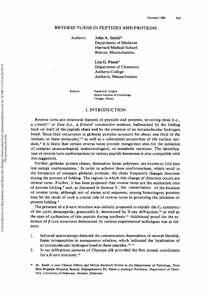
FIGURE 1. Diagrams of structures of various reverse turns. Only those hydrogens involved in hydrogen bonding are shown. Side chain (Cp) carbons are shown in those positions which are considered to be pre- ferred for L-residues according to Venkatachlam'* (8-turns). (a) Type I p-turn; (b) Type I' p-turn; (c) Type I I p-turn; (d) Type 11' p-turn; (e) y-turn; (f) inverse-y-turn. Types 111 and Ill' p-turns, which are parts of 3,0 helices, are not illustrated because they closely resemble types I and 1'. respectively.
Table 1
TYPES DIHEDRAL ANGLES OF /3-TURN
* i + I . i + 1 ' i + 1 ' i + Z
-60 -30 -90 0 60 30 90 0
-60 I20 80 0
60 -120 -80 0
-60 -30 -60 -30
60 30 60 30
a Actually a 3,0-helix.
tional types I. 11, and 111, but they also posited four more types. Their expansion of "type" notation, based on an approximate range of dihedral angles, is an artificial
Cri
tical
Rev
iew
s in
Bio
chem
istr
y an
d M
olec
ular
Bio
logy
Dow
nloa
ded
from
info
rmah
ealth
care
.com
by
54.2
48.7
3.15
3 on
05/
20/1
4Fo
r pe
rson
al u
se o
nly.
318 CRC Critical Reviews in Biochemistry
180
Y O
-180 -180 0 + 180
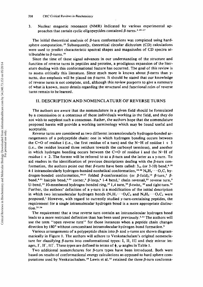
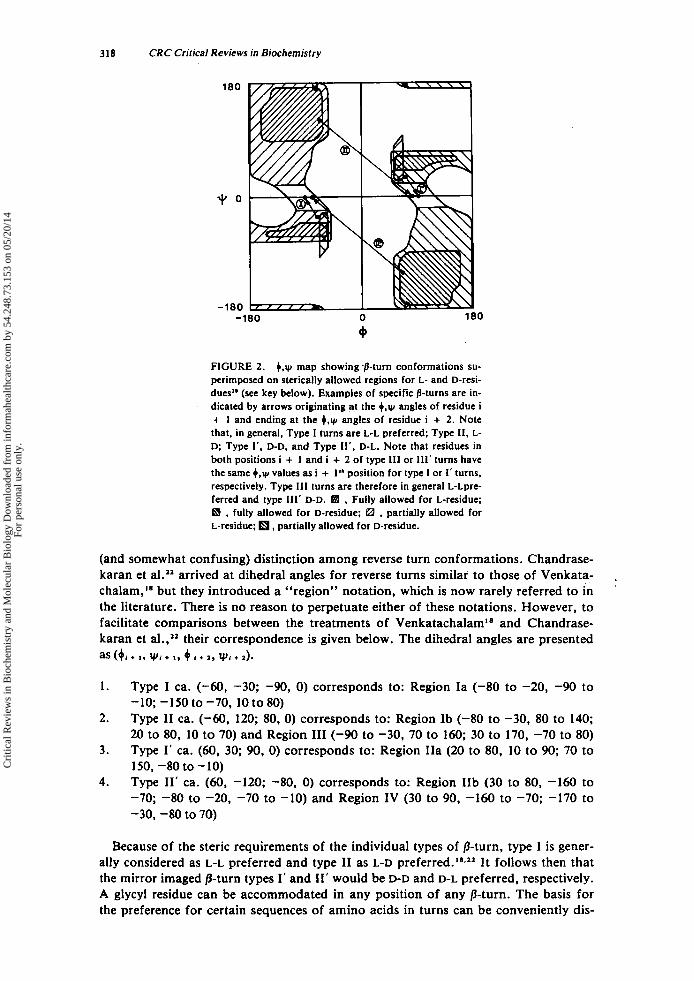
FIGURE 2. +,y map showing .p-turn conformations su- perimposed on sterically allowed regions for L- and D-resi- dues” (see key below). Examples of specific /3-turns are in- dicated by arrows originating at the 4,y angles of residue i + I and ending at the 4.y angles of residue i + 2. Note that, in general, Type I turns are L-L preferred; Type 11, L- D; Type 1‘. D-D, and Type I I ‘ , D-L. Note that residues in both positions i + I and i + 2 of type Ill or 111’ turns have the same +,y values as i + 1” position for type I or I’ turns, respectively. Type 111 turns are therefore in general L-Lpre- ferred and type 111’ D-D. a , Fully allowed for L-residue; a , fully allowed for D-residue; e3 , partially allowed for L-residue; , partially allowed for D-residue.
(and somewhat confusing) distinction among reverse turn conformations. Chandrase- karan et aLZz arrived at dihedral angles for reverse turns similar to those of Venkata- chalam,” but they introduced a “region” notation, which is now rarely referred to in the literature. There is no reason to perpetuate either of these notations. However, to facilitate comparisons between the treatments of Venkatachalam“ and Chandrase- karan et al.,” their correspondence is given below. The dihedral angles are presented as (6, + I , wi + I . 4 + z, vi + d .
1.
2.
3.
4.
Type I ca. (-60, -30; -90, 0) corresponds to: Region Ia (-80 to -20, -90 to -10; -150 to -70, 10 to 80) Type I1 ca. (-60, 120; 80, 0) corresponds to: Region Ib (-80 to -30, 80 to 140; 20 to 80, 10 to 70) and Region I11 (-90 to -30, 70 to 160; 30 to 170, -70 to 80) Type I’ ca. (60, 30; 90, 0) corresponds to: Region IIa (20 to 80, 10 to 90; 70 to 150, -80 to -10) Type 11’ ca. (60, -120; -80, 0) corresponds to: Region IIb (30 to 80, -160 to -70; -80 to -20, -70 to -10) and Region IV (30 to 90, -160 to -70; -170 to -30, -80 to 70)
Because of the steric requirements of the individual types of /%turn, type I is gener- ally considered as L-L preferred and type I1 as L-D It follows then that the mirror imaged fl-turn types I’ and 11’ would be D-D and D-L preferred, respectively. A glycyl residue can be accommodated in any position of any /%turn. The basis for the preference for certain sequences of amino acids in turns can be conveniently dis-
Cri
tical
Rev
iew
s in
Bio
chem
istr
y an
d M
olec
ular
Bio
logy
Dow
nloa
ded
from
info
rmah
ealth
care
.com
by
54.2
48.7
3.15
3 on
05/
20/1
4Fo
r pe
rson
al u
se o
nly.
October 1980 319
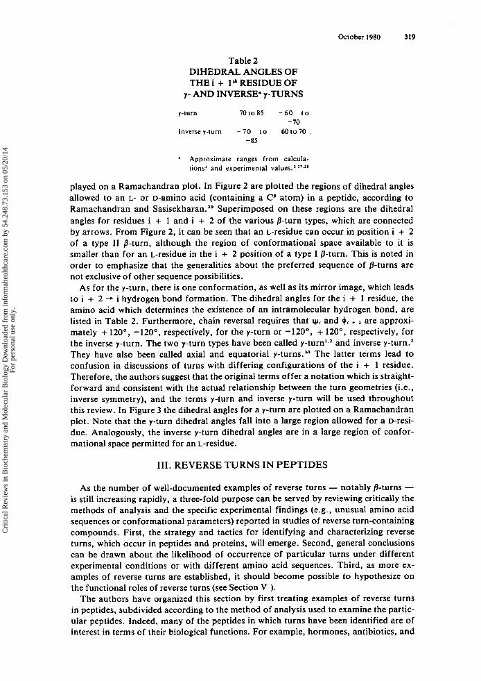
Table 2 DIHEDRAL ANGLES OF THE i + le* RESIDUE OF
7- AND INVERSE’ Y-TURNS
y-turn 70t085 - 6 0 to
lnversey-turn - 7 0 to 60to70 - 70
-85
Approximate ranges from calcula- tions’ and experimental values.’ I ’ In
played on a Ramachandran plot. In Figure 2 are plotted the regions of dihedral angles allowed to an L- or D-aminO acid (containing a Cp atom) in a peptide, according to Ramachandran and Sa~isekharan.’~ Superimposed on these regions are the dihedral angles for residues i + 1 and i + 2 of the various /3-turn types, which are connected by arrows. From Figure 2 , it can be seen that an L-residue can occur in position i + 2 of a type I1 /3-turn, although the region of conformational space available to it is smaller than for an L-residue in the i + 2 position of a type I /3-turn. This is noted in order to emphasize that the generalities about the preferred sequence of /3-turns are not exclusive of other sequence possibilities.
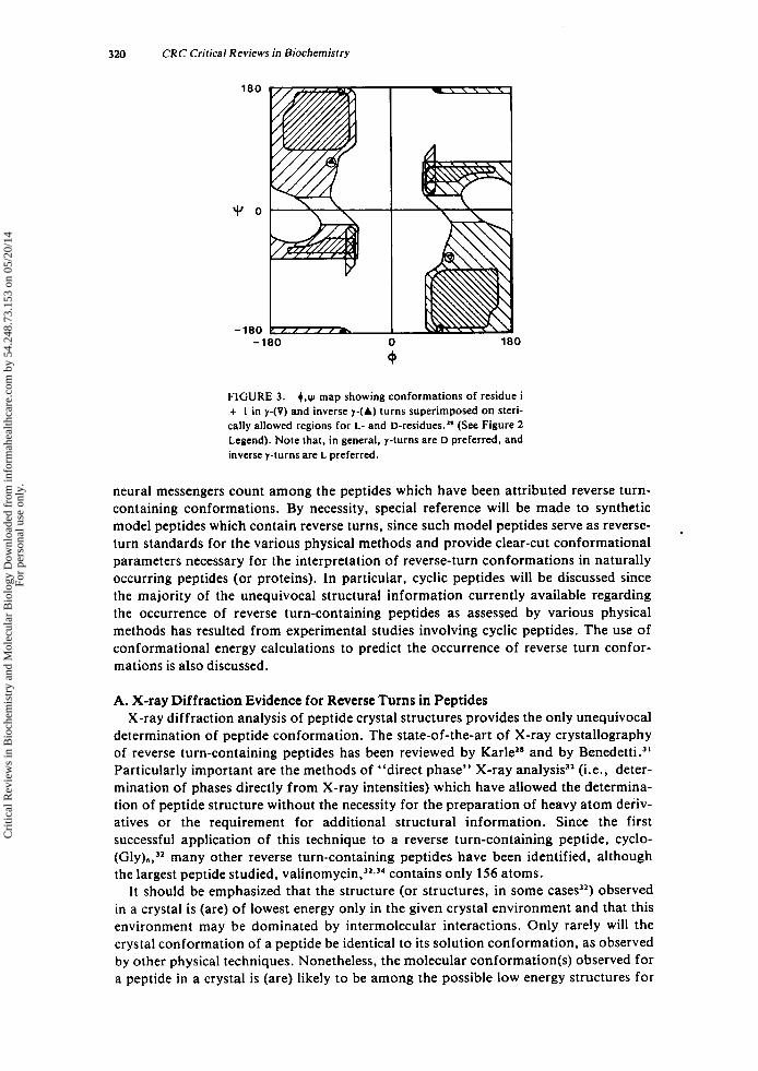
As for the y-turn, there is one conformation, as well as its mirror image, which leads to i + 2 -. i hydrogen bond formation. The dihedral angles for the i + 1 residue, the amino acid which determines the existence of an intramolecular hydrogen bond, are listed in Table 2. Furthermore, chain reversal requires that y, and +, + are approxi- mately + 120°, -120”. respectively, for the y-turn or -120°, + 120°, respectively, for the inverse y-turn. The two y-turn types have been called y-turn’.’ and inverse y-turn.’ They have also been called axial and equatorial y-turns.” The latter terms lead to confusion in discussions of turns with differing configurations of the i + 1 residue. Therefore, the authors suggest that the original terms offer a notation which is straight- forward and consistent with the actual relationship between the turn geometries (i.e., inverse symmetry), and the terms y-turn and inverse y-turn will be used throughout this review. In Figure 3 the dihedral angles for a y-turn are plotted on a Ramachandran plot. Note that the y-turn dihedral angles fall into a large region allowed for a D-reSi- due. Analogously, the inverse y-turn dihedral angles are in a large region of confor- mational space permitted for an L-residue.
111. REVERSE TURNS IN PEPTIDES
As the number of well-documented examples of reverse turns - notably /3-turns - is still increasing rapidly, a three-fold purpose can be served by reviewing critically the methods of analysis and the specific experimental findings (e.g., unusual amino acid sequences or conformational parameters) reported in studies of reverse turn-containing compounds. First, the strategy and tactics for identifying and characterizing reverse turns, which occur in peptides and proteins, will emerge. Second, general conclusions can be drawn about the likelihood of occurrence of particular turns under different experimental conditions or with different amino acid sequences. Third, as more ex- amples of reverse turns are established, it should become possible to hypothesize on the functional roles of reverse turns (see Section V ).
The authors have organized this section by first treating examples of reverse turns in peptides, subdivided according to the method of analysis used to examine the partic- ular peptides. Indeed, many of the peptides in which turns have been identified are of interest in terms of their biological functions. For example, hormones, antibiotics, and
Cri
tical
Rev
iew
s in
Bio
chem
istr
y an
d M
olec
ular
Bio
logy
Dow
nloa
ded
from
info
rmah
ealth
care
.com
by
54.2
48.7
3.15
3 on
05/
20/1
4Fo
r pe
rson
al u
se o
nly.
320 CRC Critical Reviews in Biochemistry
180
-180 -100 0 180
9
FIGURE 3. +,w map showing conformations of residue i + 1 in y-(V) and inverse y-(A) turns superimposed on steri- cally allowed regions for L- and D-residues.*’ (See Figure 2 Legend). Note that, in general, y-turns are D preferred, and inverse y-turns are L preferred.
neural messengers count among the peptides which have been attributed reverse turn- containing conformations. By necessity, special reference will be made to synthetic model peptides which contain reverse turns, since such model peptides serve as reverse- turn standards for the various physical methods and provide clear-cut conformational parameters necessary for the interpretation of reverse-turn conformations in naturally occurring peptides (or proteins). In particular, cyclic peptides will be discussed since the majority of the unequivocal structural information currently available regarding the occurrence of reverse turn-containing peptides as assessed by various physical methods has resulted from experimental studies involving cyclic peptides. The use of conformational energy calculations to predict the occurrence of reverse turn confor- mations is also discussed.
.
A. X-ray Diffraction Evidence for Reverse Turns in Peptides X-ray diffraction analysis of peptide crystal structures provides the only unequivocal
determination of peptide conformation. The state-of-the-art of X-ray crystallography of reverse turn-containing peptides has been reviewed by Karlel” and by Benedetti.3’ Particularly important are the methods of “direct phase” X-ray analysis3’ (i.e., deter- mination of phases directly from X-ray intensities) which have allowed the determina- tion of peptide structure without the necessity for the preparation of heavy atom deriv- atives or the requirement for additional structural information. Since the first successful application of this technique to a reverse turn-containing peptide, cyclo- (Gly)s,’’ many other reverse turn-containing peptides have been identified, although the largest peptide studied, val in~mycin,’~~’~ contains only 156 atoms.
I t should be emphasized that the structure (or structures, in some cases3’) observed in a crystal is (are) of lowest energy only in the given crystal environment and that this environment may be dominated by intermolecular interactions. Only rarely will the crystal conformation of a peptide be identical to its solution conformation, as observed by other physical techniques. Nonetheless, the molecular conformation(s) observed for a peptide in a crystal is (are) likely to be among the possible low energy structures for
Cri
tical
Rev
iew
s in
Bio
chem
istr
y an
d M
olec
ular
Bio
logy
Dow
nloa
ded
from
info
rmah
ealth
care
.com
by
54.2
48.7
3.15
3 on
05/
20/1
4Fo
r pe
rson
al u
se o
nly.
321 October 1 980
the particular peptide in other environments (e.g., membrane receptors). It is for this important reason that due emphasis is placed on the X-ray diffraction data contained in Table 3.
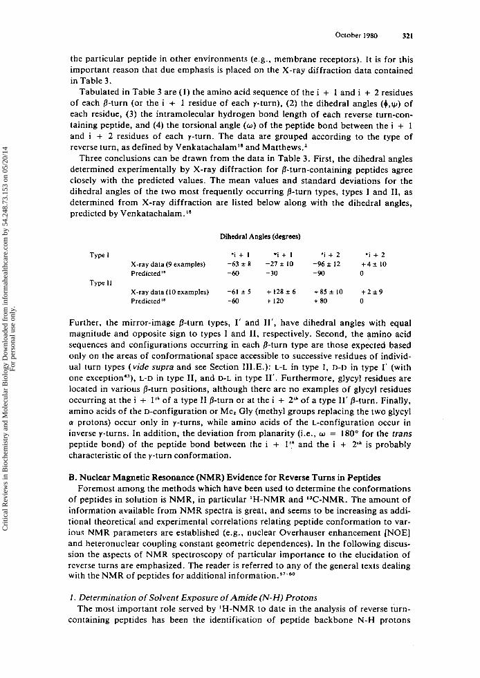
Tabulated in Table 3 are (1) the amino acid sequence of the i + 1 and i + 2 residues of each p-turn (or the i + I residue of each y-turn), (2) the dihedral angles ( 0 . ~ ) of each residue, (3) the intramolecular hydrogen bond length of each reverse turn-con- taining peptide, and (4) the torsional angle ( w ) of the peptide bond between the i + 1 and i + 2 residues of each y-turn. The data are grouped according to the type of reverse turn, as defined by Venkatachalam" and Matthews.'
Three conclusions can be drawn from the data in Table 3. First, the dihedral angles determined experimentally by X-ray diffraction for /3-turn-containing peptides agree closely with the predicted values. The mean values and standard deviations for the dihedral angles of the t w o most frequently occurring /3-turn types, types I and 11, as determined from X-ray diffraction are listed below along with the dihedral angles, predicted by Venkatachalam.'*
Dihedral Angles (degrees)
Type I 'i + I *i + I 'i + 2 "i + 2 X-ray data (9 examples) -63 i 8 -27 * 10 -96 i 12 + 4 * 10 Predicted'' -60 - 30 -90 0
X-ray data (10 examples) -61 * 5 + 128 i 6 + 85 * 10 + 2 i 9 Predicted" -60 + 120 + 80 0
Type I1
Further, the mirror-image /3-turn types, 1' and 11'. have dihedral angles with equal magnitude and opposite sign to types I and 11, respectively. Second, the amino acid sequences and configurations occurring in each p-turn type are those expected based only on the areas of conformational space accessible to successive residues of individ- ual turn types (vide supra and see Section 1II.E.): L-L in type I, D-D in type I' (with one ex~eption.~), L-D in type 11, and D-L in type 11'. Furthermore, glycyl residues are located in various p-turn positions, although there are no examples of glycyl residues occurring at the i + l r h of a type I1 p-turn or at the i + 2'* of a type 11' /3-turn. Finally, amino acids of the D-configuration or Me, Gly (methyl groups replacing the two glycyl Q protons) occur only in y-turns, while amino acids of the L-configuration occur in inverse y-turns. In addition, the deviation from planarity (i.e., o = 180" for the trans peptide bond) of the peptide bond between the i + I r h and the i + 2Ih is probably characteristic of the y-turn conformation.
B. Nuclear Magnetic Resonance (NMR) Evidence for Reverse Turns in Peptides Foremost among the methods which have been used t o determine the conformations
of peptides in solution is NMR, in particular 'H-NMR and W-NMR. The amount of information available from NMR spectra is great, and seems to be increasing as addi- tional theoretical and experimental correlations relating peptide conformation to var- ious NMR parameters are established (e.g., nuclear Overhauser enhancement [NOE] and heteronuclear coupling constant geometric dependences). In the following discus- sion the aspects of NMR spectroscopy of particular importance to the elucidation of reverse turns are emphasized. The reader is referred to any of the general texts dealing with the NMR of peptides for additional information."-60
I . Determination o f Solvent Exposure o f Amide (N-H) Protons The most important role served by IH-NMR to date in the analysis of reverse turn-
containing peptides has been the identification of peptide backbone N-H protons
Cri
tical
Rev
iew
s in
Bio
chem
istr
y an
d M
olec
ular
Bio
logy
Dow
nloa
ded
from
info
rmah
ealth
care
.com
by
54.2
48.7
3.15
3 on
05/
20/1
4Fo
r pe
rson
al u
se o
nly.
Tab
le 3
R
EV
ER
SE TUR
NS IN
PE
PTID
ES
DE
TE
RM
INE
D F
RO
M X-R
AY D
IFFR
AC
TIO
N'
A. @
-Tur
n-C
onta
inin
g Pep
tide
s
Sequ
ence
D
ihed
ral a
nglu
(deg
rees
)'
@-turn
Typ
e Pe
ptid
e
1. T
ype
1 C
yclo
-(G
ly).
Sben
zyl-
L-C
ys-L
-Pro
-L-L
eu-G
ly-N
H,
pBrC
bz-G
ly-L
-Pro
-L-L
eu-G
ly-O
H
cFBrCbz-Gly-L-Pro-L-Leu-Gly-L-Pro-
NA
c-L
-Pro
-L-L
ac-N
HC
H,
Cyc
lo-(
Gly
,-D -
Ala
l)
Li-
anta
rnan
ide
OH
Boc-L-Pro-Aib-L-Ala-Aib-OBz'
Cyclo-(L-Alal-Gly,-L-Ala-Gly)
Cyclo-(L-Alar-Gly-L-Ala-Glyl)
Cyc
lo-(
Gly
.-D-A
lar)
Cyclo-(L-Alal-Gly-L-Ala-Glyl)
[Leu
']-en
keph
alin
H-L
-Pro
-L-L
eu-G
ly-N
Hl
NA
c-L
-Pro
-D-L
ac-N
HC
H,
Val
inor
nyci
n
2. T
ype
1'
Cyc
lo-(
Gly
),
3. T
ype
II
Ferr
ichr
orne
A
Val
inor
nyci
n-K
' C
yclo
-(L
-Ala
-L-P
ro-D
-Phe
)z
Cyc
lo-(
Gly
-L-P
ro-G
I y-D
- Ala
-L-P
ro)
Cyc
lo-(
Gly
-L-P
ro-D
-Ala
),
Cyc
lo-(
L-V
al-D
-Pro
-D-V
al-L
-Pro
), C
yclo
-(G
ly-L
-Pro
-D-P
he),
i+
l
G~
Y
L-P
ro
L-P
ro
L-P
ro
L-P
ro
L-A
la
(L-P
he
L-P
ro
{Aib
L
-Ala
L
-Ala
G
IY
DA
la
L-A
la
G~
Y
L-Se
r L-
Leu
L-P
ro
L-V
al
L-V
al
L-V
al
L-P
ro
L-P
ro
L-P
ro
L-P
ro
L-V
al
L-P
ro
G~
Y
i+
2
GIY
L
-Leu
L
-Leu
L-
Leu
L-L
ac
GlY
L
-Phe
L
-Phe
A
ib
L-A
la
L-A
la
L-A
la
G~
Y
D-A
la
G~
Y
G~
Y
G~
Y
GIY
D
-Lac
D
-Hyl
V
D-H
ylv
D-H
ylv
D-P
he
D-A
la
D-A
la
D-P
ro
D-P
he
G~
Y
'i+
1
ri+
1
*i+
2
Ti+
2
NH
..-O
C(i
).
Ref
.
-69
-66
-58
-65
-55
-70
-69
-79
-56
-52
-53
-62
+ 69
+66
+ 5
4 +
59
-57
-6 1
-6
2 -6
3 -6
7 -5
8 -60
-52
-54
-70
-64
-30
-94
-29
-115
-2
7 -1
04
-27
-105
-22
-81
-I5
-117
-1
3 -8
4 -1
3 -90
-35
-52'
-3
8 -9
3 -4
3 -8
4 -3
3 -9
5 +
33
+92
+
I5
+I31
+
38
+91
+
25
+97
+
I32
+82
+
I28
+7
2
+I4
0
+91
+
I29
+96
+
I30
+82
+
I32
+79
+ 1
22
+78
+ 1
26
+74
+ 1
25
+94
+
I16
+79
+
I31
+83
-6
4 +
I32
+lo
6
+ 11
+
13
+8
+
8
-1 1
+
16
-6
+8
-3
8'
-13 0
+ 14
-7
-3 1
-6
-7
-1
-8
-3
+3
+
3
+9
+
12
-5
+ 19
+
6
-
-14
2.96
2.96
3 .
OO
2.89
3.
04
3.00
3.09
2.
92
3.10
2.
96
3.16
2.
99
2.99
2.
98
3.04
2.
97
-
2.93
3.
20
2.87
3.42
3.
041
3.0
3.52
32
35
36,3
7 38
39
40
41
42
43
43
32
40
43
44
45
46
47
42
34
48
49,5
0 51
52
53
Cri
tical
Rev
iew
s in
Bio
chem
istr
y an
d M
olec
ular
Bio
logy
Dow
nloa
ded
from
info
rmah
ealth
care
.com
by
54.2
48.7
3.15
3 on
05/
20/1
4Fo
r pe
rson
al u
se o
nly.
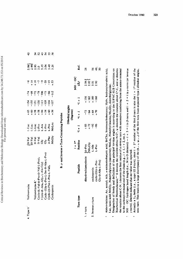
4. T
ype
11'
Val
inom
ycin
D
-Val
L
-Lac
+
63
-1
34
-74
{D-V
al
L-L
ac
+60
-1
35
-98
Val
inom
ycin
-K'
D-V
al
L-L
ac
+5
8
-131
-7
2 C
yclo
-( L
-Val
-D-P
ro-D
-Val
-L-P
ro)3
D
-Val
L
-Pro
+
58
-130
-7
8 Cyclo-(L-Alal-Gly2-L-Ala-Gly)
GlY
L
-Ala
+
84
-1
13
-106
C
yclo
-(G
ly-L
-Pro
-L-S
er-D
-Ala
-L-P
ro)
Gly
L
-Pro
+
58
-1
28
-75
Cyc
lo-(
D-P
he-L
-Pro
-L-V
al),
D
-Phe
L
-Pro
+6
0 -1
34
-86
Cyc
losp
orin
M
eGly
M
eLeu
+
56
-1
37
-112
Turn
type
1. y
-tur
n
2. I
nver
se y
-tur
n
b .
B. 7-
and
Inve
rse
7-T
urn-
Con
tain
ing
Pept
ides
-6
+ 14
+
18
-5
-9
-20
-I5
+ 53
Dihedral a
ngle
s (d
egre
es)
i +
1"
NH
...O
C
Pept
ide
Res
idue
'i
+ I
*i +
1 -i
+ 1
(AY
2.82
2.
94 1
dihy
droc
hlam
ydoc
in
+ 83
-73
+ 15
6 i L2
1Y
+ 7
2 -6
4 +
162
cycl
ospo
rin
L-A
la
-92
+6
4
+I8
0
3.01
cy
clo-
(Gly
-L-P
ro-
L-P
ro
-86
+7
0
-160
2.
92
Gly
-D-A
la-L
-Pro
)
42
2.99
2.
93
34
3.0
52
3.35
43
3.
04
54
3.40
55
3.
21
56
Ref
.
28
56
49 9
50
Abb
revi
atio
ns:
Ac,
ace
tyl;
Aib
, a-
amin
oiso
buty
ric
acid
; B
rCbz
, br
omoc
arbo
benz
oxy;
Hyl
v, h
ydro
xyis
oval
eric
aci
d;
Lac
, lac
tic a
cid;
MeG
ly,
Nm
ethy
lgly
cine
(sa
rcos
ine)
; M
eLeu
, N
met
hyl l
euci
ne; M
elG
ly, d
imet
hylg
lyci
ne.
Des
igna
tion
of b
onds
and
def
initi
on o
f th
e pr
inci
pal
tors
ion
angl
es i
s ac
cord
ing
to t
he I
UPA
C-I
UB
Com
mis
ion on
B
ioch
emic
al N
omen
clat
ure;
."
0 den
otes
the
rot
atio
n ab
out
N-C
', u, d
enot
es t
he r
otat
ion
abou
t C'-C
. an
d w d
enot
es
the
rota
tion
abo
ut C
-N. i
den
otes
the
firs
t res
idue
of
a re
vers
e tu
rn w
ith s
ucce
ssiv
e num
beri
ng f
rom
the
amin
o-te
rmin
al
to th
e ca
rbox
yl-t
erm
inal
end
of
the
chai
n (e
.g.,
i +
1, i
+ 2,
etc
.).
NH
. ' ' O
C:
hydr
ogen
bon
d le
ngth
(i.e
., N
to 0 d
ista
nce)
- i
+ 3 -. i i
n /3
-tur
ns a
nd i
+ 2 -. i
in y
-tur
ns (
or in
vers
e y-
turn
s).
Das
hes
indi
cate
that
no
valu
e w
as re
port
ed.
Act
ually
a 3
,0 h
elix
(i.e
., a
type
111
P-t
urn)
. si
nce
i +
2''
resi
due
of t
he f
irst
0-t
urn
is al
so t
he i
+ 1'''
resi
due
of
the
next
tur
n. T
his
lead
s to
a la
rge
disc
repa
ncy
of t
he (
+, . ,.
yli.
z) o
f th
e fi
rst
turn
fro
m e
xpec
ted
type
I d
ihed
ral
angl
es.
W 8 W
h)
w
Cri
tical
Rev
iew
s in
Bio
chem
istr
y an
d M
olec
ular
Bio
logy
Dow
nloa
ded
from
info
rmah
ealth
care
.com
by
54.2
48.7
3.15
3 on
05/
20/1
4Fo
r pe
rson
al u
se o
nly.

324 CRC Critical Reviews in Biochemistry
which participate in intramolecular hydrogen bonding. The interpretations of ‘H-NMR N-H proton data have been based on a number of experiments designed to assess the accessibility of these protons to interact with bulk solvent or other peptide molecules intermolecularly. The premise then routinely invoked is that any N-Hs not exposed to these intermolecular interactions are likely to be involved in intramolecular hydrogen bonding.
A list of possible experiments which are variations on this theme include: (1) tem- perature dependence of N-H chemical shifts (Ad/AT); (2) solvent dependence of N-H chemical shifts; (3) concentration dependence of N-H chemical shifts in nonpolar sol- vents; (4) deuterium exchange kinetics (T~ ,~ [ ’H-~H]) ; and ( 5 ) sensitivity of N-H chem- ical shift or line-width to addition of reagents which act as perturbants. Each of these approaches suffers to varying degrees from the underlying problem that any alteration of an experimental variable (e.g.. temperature, solvent, etc.) required for the assess- ment of N-H exposure may appreciably alter the conformation or the distribution among conformations, if more than one conformation co-exists, of the peptide.
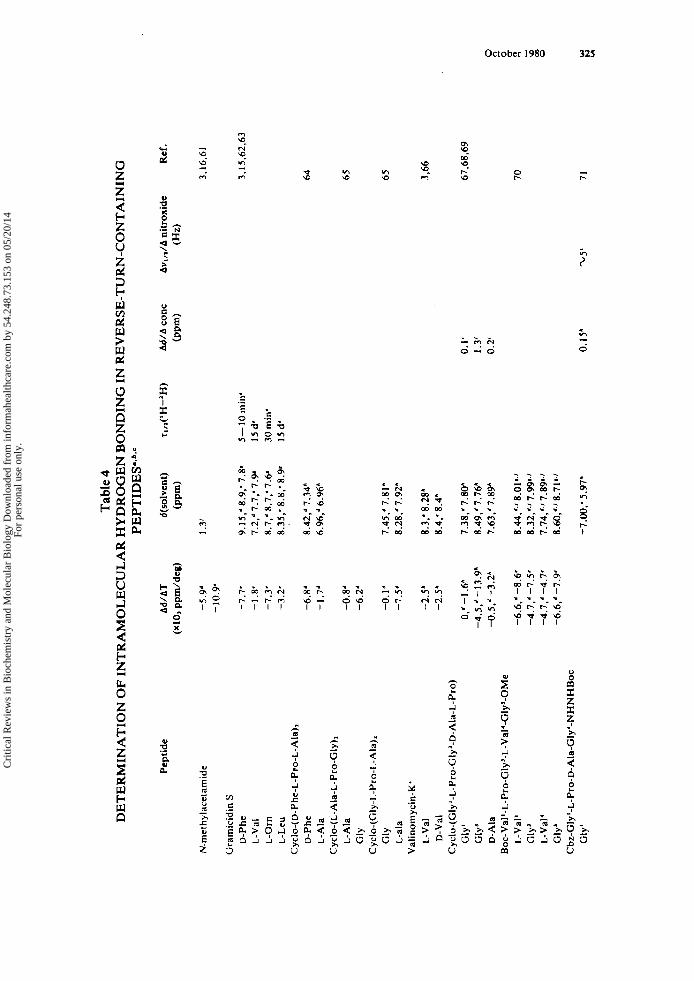
The first approach, temperature dependence of N-H chemical shift, has been the most frequently employed and, provided certain limitations are acknowledged, can be an extremely straightforward and powerful indicator of the participation of N-Hs in intramolecular interactions. In general, use of a solvent which acts as a hydrogen bond acceptor (most commonly, dimethyl sulfoxide-d6 [MezSO-ds] or water) facilitates the interpretation of observed shifts. The premise of such an experiment is that the exist- ence of intermolecular hydrogen bonding is signaled by a large temperature depen- dence of the resonance of the proton involved in the hydrogen bond. This dependence reflects the perturbation of the equilibrium between a free proton and its hydrogen- bonded states with increasing temperature. The N-H resonance is shifted to a high- field position with increasing temperature as a consequence of the low-field position of a hydrogen bonded N-H resonance. This discussion applies both to peptide-peptide and peptide-solvent intermolecular hydrogen bonding. A small temperature depen- dence suggests either intramolecular hydrogen bonding or a “buried” (i.e., unexposed to solvent) N-H, which does not participate in intramolecular hydrogen bonding. In either case, little or no temperature dependence would be expected since the N-H in question is not involved in an equilibrium between bound and free states. In solvents which are weak hydrogen bond acceptors, the same arguments can be presented, al- though interactions of peptide molecules with one another must be occurring in order to cause exposed N-Hs to undergo temperature-dependent chemical shift changes, since any intermolecular hydrogen bonds between peptide molecules and solvent are weak if present. Hence, it is advisable to ensure that the peptide concentration is high enough to lead to peptide-peptide interactions before attempting to interpret Ad/AT values in nonpolar or weakly interacting solvents (vide infra). Representative data for Ad/AT of N-Hs of a few well-studied peptides in various solvents are presented in Table 4.
There are several intrinsic problems involving the interpretation of the experimental data from this type of experiment:61
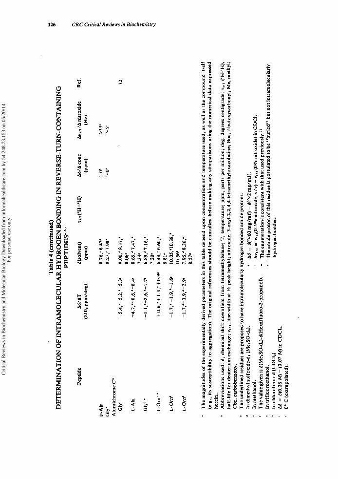
1. It is difficult to differentiate whether a low Ad/AT is due to the involvement of an N-H proton in an intramolecular hydrogen bond or to the inaccessibility of the N-H proton to bulk solvent (see alumichrome data in Table 4,,). There are no absolute thresholds of Ad/AT values below which hydrogen bonding or inaccessibility of a N-H proton is unequivocal. There is no available experimental method for determining the effect of reorien- tation of magnetically anisotropic groups on chemical shift and hence on Ad/AT. Conformational averaging can cause misleading
2.
3.
4.
Cri
tical
Rev
iew
s in
Bio
chem
istr
y an
d M
olec
ular
Bio
logy
Dow
nloa
ded
from
info
rmah
ealth
care
.com
by
54.2
48.7
3.15
3 on
05/
20/1
4Fo
r pe
rson
al u
se o
nly.
Tab
le 4
PEPT
IDE
SO.*
.' D
ET
ER
MIN
AT
ION
OF
INT
RA
MO
LE
CU
LA
R H
YD
RO
GE
N B
ON
DIN
G IN
RE
VE
RSE
-TU
RN
-CO
NT
AIN
ING
Pept
ide
N-m
ethy
lace
tam
ide
Gra
mic
idin
S
D-P
he
L-V
al
L-O
rn
L-L
eu
D-P
he
L-A
la
L-A
la
GlY
GlY
L
-ala
L-V
al
D-V
al
Cyc
lo-(
D-P
he-L
-Pro
-L-A
la),
Cyc
lo-(
L-A
la-L
-Pro
-Gly
)l
Cyc
lo-(
Gly
-L-P
ro-L
-Ala
),
Val
inom
ycin
-K'
Cyc
lo-(
Gly
' -L
-Pro
-GIy
'-D
-Ala
-L-P
ro)
Gly
' G
ly'
D-A
la
L-V
al'
Gly
' L
-Val
' G
ly'
Gly
'
Boc-Val'-L-Pro-Gly'-L-Val'-Gly'-OMe
Cbz
-Gly
'-L
-Pro
-D-A
la-G
ly'-
NH
NH
Boc
Ad/
AT
(~1
0, pp
mld
eg)
-5.9
' -1
0.9'
-7.7
' -1
.8-
-7.3
' -3
.2'
-6.8
' -1
.7'
-0.8
' -6
.2'
-0.l
d -7
,s
-2.5
h
-2.Y
0,'
-1.6
" -4
.5,'
-13.
9b
-0.5
," -
3.2*
-6.6
,' -8
.6'
-4.7
,'-7.
5'
-4.7
,4 -
4.7'
-6
.6,'
-7.9
'
d(so
1ven
t)
@pm
)
1.3'
9.15
.d 8
.9,'
7.8'
7.
2.'7
.7,'
7.9'
8.
7,d
8.7,'
7.61
8.
35,'
8.8.
' 8.
9'
8.42
,* 7
.34*
6.
96,d
6.96
*
7.45
,4 7
.81*
8.
28,'7
.92'
8.3,
' 8
.2V
8.
4;
8.4h
7.38
,'7.
80"
8.49
,. 7
.76*
7.
63,'
7.89
*
8.44
,'.'
8.01
'.'
8.32
,'J 7
.99'
.' 7.
74,'J
7.8
9'.'
8.60
,dJ 8
.71s
.'
-7.0
0;
5.97
"
Ref
. T
,~~(
'H-'
H)
Ad/
A c
onc
Av,
,JA
ni
trox
ide
(PPm
) (W
3.16
.61
5-10
m
in'
I5 d
' 30
min
' 15
d'
0. I
' I .
3'
0.2
0.15
'
3.15
.62.
63
64
65
65
3.66
67,6
8,69
70
71
Cri
tical
Rev
iew
s in
Bio
chem
istr
y an
d M
olec
ular
Bio
logy
Dow
nloa
ded
from
info
rmah
ealth
care
.com
by
54.2
48.7
3.15
3 on
05/
20/1
4Fo
r pe
rson
al u
se o
nly.
Tab
le 4
(continued)
PEPT
IDE
S'*b
*'
DE
TE
RM
INA
TIO
N O
F IN
TR
AM
OL
EC
UL
AR
HY
DR
OG
EN
BO
ND
ING
IN R
EV
ER
SE-T
UR
N-C
ON
TA
ININ
G
Pept
ide
D-A
la
Gly
' A
lum
ichr
ome
C"
Gly
'
L-A
la
GIY
~ -
L-O
rn' "
L- O
rrr'
-5.4,'-5.2,'-5.3*
-4.7,'-8.6,'-8.48
-1.1,'-2.6,"-1.7'
+0.6,'+
1.4."+0.9*
-1.7,' -1.9.'-1.6'
-1 .7,'-3.9,h-2.9'
8.76,' 6.47"
8.27.' 7.9W
9.00,' 8.37,'
8.06'
8.65,' 7.47,'
7.34'
6.89,' 7.16,'
7.208
6.44,' 6.60,'
6.811
10.05,'10.38.'
10.56.
7.%,'
8.36,'
8.57
'
72
The
mag
nitu
des
of t
he e
xper
imen
tall
y de
rive
d pa
ram
eter
s in
thi
s ta
ble
depe
nd u
pon
conc
entr
atio
n an
d te
mpe
ratu
re u
sed,
as
wel
l as
the
com
poun
d it
self
(e
.g.,
its
susc
epti
bili
ty to
agg
rega
tion
). T
he o
rigi
nal
refe
renc
es s
houl
d be
con
sult
ed b
efor
e m
akin
g an
y co
mpa
riso
ns u
sing
the
num
eric
al d
ata
expr
esse
d he
rein
. A
bbre
viat
ions
use
d: d
, ch
emic
al s
hift
dow
nfie
ld f
rom
tet
ram
ethy
lsil
ane;
T,
tem
pera
ture
; pp
m,
part
s pe
r m
illio
n; d
eg,
degr
ees
cent
igra
de;
T,,,
('H
-lH
),
half
-lif
e fo
r de
uter
ium
exc
hang
e; w
,,,,
line
-wid
th a
t Yz
peak
hei
ght;
nit
roxi
de, 3-oxyl-2,2,4,4-tetramethyloxazolidine;
Boc
. t-
buto
xyca
rbon
yl; M
e, m
ethy
l;
Cbz
, car
bobe
nzox
y.
The
und
erli
ned
resi
dues
are
pro
pose
d to
hav
e in
tram
olec
ular
ly h
ydro
gen
bond
ed a
mid
e pr
oton
s.
In d
imet
hyl s
ulfo
xide
-dr (
Me,
SO-d
,).
In m
etha
nol.
The
val
ue g
iven
is d(MelSO-d,)-d(Hexafluoro-2-~ro~and).
In t
rifl
uoro
etha
nol.
In c
hlor
ofor
m-d
(CD
CI,)
. hy
drog
enbo
nded
. Ad
= d(0.26
M) -
(0.07 M
) in
CD
CI,.
0"
C (e
xtra
pola
ted)
.
' ' - - Ad =
d(1.60
mg/
mf)
- d(1.2
mg/
mf)
. Aw,,,
= ~,,~(0.5% nitr
oxid
e. v
/v) - w
II,
(0%
nit
roxi
de) i
n C
DC
I,.
The
enu
mer
atio
nis c
onsi
sten
t with
tha
t use
d pr
evio
usly
."
The
am
ide
prot
on o
f thi
s res
idue
is p
ostu
late
d to
be
"bur
ied"
bu
t not
intr
amol
ecul
arly
s c,
Cri
tical
Rev
iew
s in
Bio
chem
istr
y an
d M
olec
ular
Bio
logy
Dow
nloa
ded
from
info
rmah
ealth
care
.com
by
54.2
48.7
3.15
3 on
05/
20/1
4Fo
r pe
rson
al u
se o
nly.
October 1980 327
The extent of perturbation of N-H resonances during a solvent titration or simply comparisons of chemical shifts in different solvents can be informative, provided that conformational changes occurring concomitantly are negligible. In order to establish that a conformational change has not occurred as the solvent is varied, a rule-of-thumb is that the chemical shifts of the upfield protons and their coupling constants should be relatively unchanged - by (0.2 ppm and <0.5 Hz, respectively. Representative data for various reverse turn-containing peptides are shown in Table 4.
As mentioned above, i t is expected that peptide-peptide intermolecular hydrogen bonding would be favored at higher peptide concentrations in solvents which interact weakly with the peptide. This can be exploited to distinguish exposed and sequestered N-Hs by examining the concentration dependences of their chemical shifts. A buried or intramolecularly hydrogen-bonded N-H would display little or no change in chemi- cal shift throughout a range of peptide concentration which goes high enough for ag- gregation to be favored. The exact concentration range in which aggregation will occur varies considerably from peptide to peptide. Again, it is necessary to emphasize that conformational changes can lead to spurious results and erroneous interpretations. Examples of the successful application of this approach to reverse turn-containing pep- tides are given in Table 4. In all cases, as expected, the N-Hs which are exposed undergo larger changes in chemical shifts as the peptide concentration is varied than those N-Hs which are shielded.
Deuterium-exchange kinetics of the N-'H to N-'H are measured by the rate of de- crease in area of the N-H peak (usually reported as half-life for exchange, T ~ , ~ ; , hydro- gen bonds being assigned to the slowly exchanging N-H protons. Although the isotopic exchange method had been used on gramicidin S1' (see Table 4) and in other applica- tionS,16 74.75 the approach is now seldom used because of inherent problems in the in- terpretation of experimental results.6' In light of several cases of conflict between tem- perature dependence and 'H-exchange data, criticisms were raised against the latter method, citing the fact that it measures kinetics of solvent and peptide reaching a transition state, which may be only indirectly related to the equilibrium conformation of the peptide.6' In addition, the general weaknesses of most of the methods described here are also present:
I .
2.
The method cannot determine whether a slowly exchanging N-H proton is hydro- gen-bonded or solvent-shielded. The approach cannot discriminate whether rapidly exchanging N-H protons are nonhydrogen-bonded or are N-H protons in rapid equilibrium between two dif- ferent conformations. There is no absolute criterion for establishing the half-life for the exchange which may be correlated with N-H hydrogen bonding.
3 .
The final experimental approach relies on detecting differences in the line-broaden- ing of N-H proton resonances (i.e., increase of peak width at Yi peak height, vl,') that are produced by the addition of small amounts (<3%, v/v) of a stable hydrogen-bond- accepting free radical - for example, a nitroxide. 3-oxyl-2,2,4,4-tetramethyloxazoli- dine (see Table 4).61.71.7J Because concentrations of free radical as small as 0.2% can be used, the likelihood that the free radical is perturbing the conformation of a peptide is minimized. Since the free radicals used are good hydrogen-bond acceptors, N-H line-width differences in a weakly hydrogen-bonding solvent (e.g., chloroform) will occur only for N-H protons which are exposed (not buried or intramolecularly hydro- gen-bonded). The method has also been used in fairly strongly hydrogen-bonding sol- vents, where presumably a competition occurs between nitroxide and solvent for avail- able hydrogen-bonding sites of the peptides. As pointed out by Kopple et al..7' the
Cri
tical
Rev
iew
s in
Bio
chem
istr
y an
d M
olec
ular
Bio
logy
Dow
nloa
ded
from
info
rmah
ealth
care
.com
by
54.2
48.7
3.15
3 on
05/
20/1
4Fo
r pe
rson
al u
se o
nly.
328 CRC Critical Reviews in Biochemistry
major limitation of the approach is that spectral overlaps produced by free-radical- induced line-broadening allow only a qualitative determination of intramolecular hy- drogen bonding. As is generally the case, conformational averaging must be considered as a possible c~mpl i ca t ion .~~
The possibility of conformational averaging is a general problem encountered during an evaluation of the accessibility of N-H protons. It is likely that most linear peptides have multiple conformations among which they interconvert. Unless these conforma- tions are separated by energy barriers greater than approximately 10 kcal/mol (for chemical shift differences of approximately 50 Hz), the observed NMR spectrum will be a weighted average of the spectra of the multiple-conformer population. Urry and O h n i ~ h i ~ ~ have suggested that for a given N-H in a peptide, the mole fractions of the solvent-exposed and solvent-shielded protons may be calculated from the experimen- tally measured temperature and solvent dependences of N-H chemical shift. Such cal- culations rely on reference values acquired from model peptides which have well-de- fined populations of shielded or exposed protons (e.g., gramicidin S or valinomycin). In addition, Urry and Long'O considered that the interactions of carbonyl groups with solvent might alter appreciably the observed solvent or temperature dependences of the amide proton of the same peptide bond. This idea has been discussed in detail by Llina's and Klein." However, this approach, as noted by Urry and Long, is a qualita- tive one, since it is impossible to verify that the required reference values are applicable to a peptide with a different sequence, conformation, or distribution of magnetically anisotropic groups.
Since the con formational space available to cyclic peptides is considerably more re- stricted than that available to linear peptides, it is usually assumed that the problem of conformational averaging with cyclic peptide is less important. Indeed, the proba- bility of more than two or three discrete conformers existing simultaneously in signifi- cant proportions and interconverting rapidly on the NMR timescale is quite small. However, examples of the occurrence of conformational averaging in cyclic peptides, leading to an apparent conflict between N-H accessibility data obtained by two differ- ent experimental approaches, are k n o ~ n . ~ ' In addition, the temperature coefficients (AMAT) of the N-H chemical shifts of the cyclic hexapeptide C~CIO-(GI~-D-XXX-L-Y~~)~ proved unreliable when they were compared with the data obtained from the hydrogen- bond-accepting free radical approach for assessing N-H In both cases, the equilibria among the averaging conformations is perturbed to differing extents by the different approaches utilized. In all likelihood, the addition of a polar nitroxide (<3%, v/v) to a solution of peptide is a more gentle approach than altering the temperature by 40 to 60°, although whether the former approach results in a more accurate deter- mination of N-H accessibility remains to be established.
In conclusion, the determination of N-H solvent accessibility by NMR is best done by the use of several corroborative techniques. Hydrogen-bonding nitroxide radicals as line-broadening agents appear to offer the most reliable data, although the tech- nique has not been utilized extensively. Nonetheless, careful use of temperature, con- centration, and solvent dependence data can be highly informative. In instances where any discrepancy is found among results from different experimental approaches, the peptide system must be analyzed in terms of possible conformational averaging and/ or conformational alterations, resulting from the experimental approach used. Throughout such investigations repeated validation of a lack of change in the chemical shifts and coupling constants must be made.
2. Conformational Dependence of Coupling Constants As originally formulated by K a r p l u ~ ' ~ in his widely used semiempirical relationship
between dihedral angles and vicinal 'H-'H coupling constants (3Jln-rn), geometric in-
Cri
tical
Rev
iew
s in
Bio
chem
istr
y an
d M
olec
ular
Bio
logy
Dow
nloa
ded
from
info
rmah
ealth
care
.com
by
54.2
48.7
3.15
3 on
05/
20/1
4Fo
r pe
rson
al u
se o
nly.

October 1980 329
8 120 150 180 -150 " ( !: 0 -30 -60 - 90 0 30 60 90 " -120 -150 -180 150
FIGURE 4. Plots of the J,,. coupling constant in Hz as a function of the dihedral angle 0 according to various Kar- plus-type correlations. The equivalent 4 angles for L- or D- residues are given below the corresponding O values. JN. =
6 0 G 180" (---);" J,,. = 9.4 cos10 - 1.1 cos 0 + 0.4 (mean permissible values) (-);'' J,,. = 7.9 cos't3 - 0.55 cos 0 + 0.35 sin'b (...);79 Jw. = (5.41 f 0.02) cosV3 - (1.27 i 0.15)
8.5 cosw - 0.28.00 < e G 900. J,,. = 9.5 c o s v - 0.28,900
cOS e + (2 .17 * 0.54) ( 1 1 1 ) . ~
formation can be derived from 'H-NMR spectra. Of particular interest to investigators dealing with peptide conformation are 3 J N n - H . values ( J N . ) , which can be related to the 0 angles, as well as l J H i H i for protons in the side chains, which can be related to the x angles. Several correlations have been introduced for the former, based on the same basic premises as Karplus' initial work, but specifically adapted to peptides by incor- porating experimental data from well-studied peptides (see Figure 4).7B-82 These corre- lations vary somewhat among themselves, with regard to their minimal and maximal values of J N . for 8 = 0, 90, or 180" (8 defined in Figure 4) and to the mathematical expressions describing the relationship between vicinal coupling constants and dihedral angles (see Figure 4). However, from a practical standpoint, it is less important to choose a particular form of the J N o vs. 8 relation than to recognize the inherent limi- tations associated with all these relations.
1. The actual nature of the coupling interaction, which is a function of both the distribution of electrons and the geometric arrangement of molecular orbitals. may vary among compounds and their conformers. The relations in Figure 4 are generalizations which may be applied to any molecule so long as a range of 8 (directly related to 0) values is associated with a particular J N I . There is an ambiguity in these mathematical functions, since more than one value for 0 may be associated with a given value of J N o , except for the extremely small or large values of J N o . In addition, two values of 0 are possible for each value of 8, except for 0 = 0 or 180".
2.
Cri
tical
Rev
iew
s in
Bio
chem
istr
y an
d M
olec
ular
Bio
logy
Dow
nloa
ded
from
info
rmah
ealth
care
.com
by
54.2
48.7
3.15
3 on
05/
20/1
4Fo
r pe
rson
al u
se o
nly.
330 CRC Critical Reviews in Biochemistry
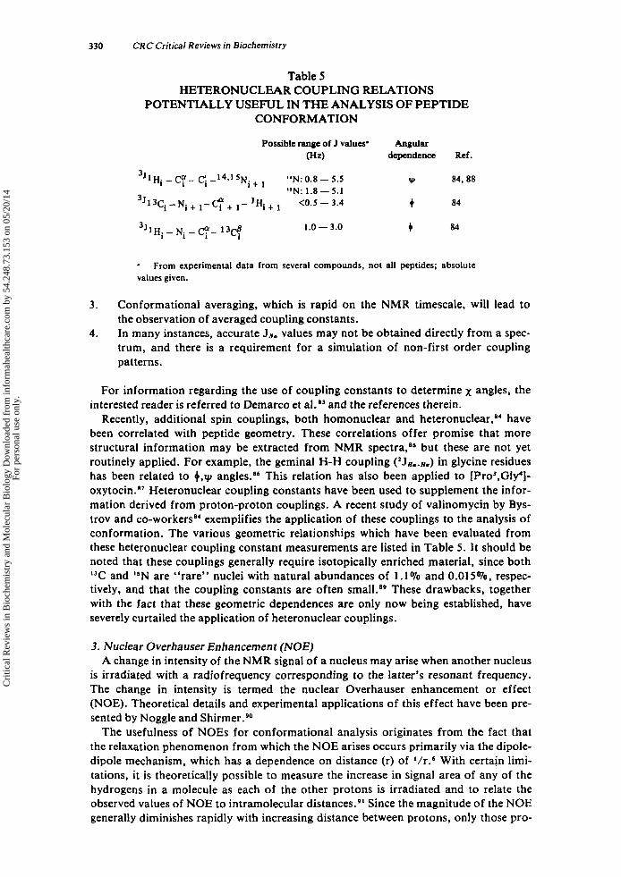
Table 5 HETERONUCLEAR COUPLING RELATIONS
POTENTIALLY USEFUL IN THE ANALYSIS OF PEPTIDE CONFORMATION
Possible range of J values' Angular (Hz) dependence Ref.
3 J l ~ i - c'Y- c". - 1 4 , 1 5 ~ + "N: 0.8 - 5.5 v, 84,88
3J 1 3Ci - Ni + - + - 'Hi + <0.5 - 3.4 t 84
I 1 "N: 1.8-5.1
3 ~ l H . - 1.0 - 3.0 84
a
values given. From experimental data from several compounds, not all peptides; absolute
3 .
4.
Conformational averaging, which is rapid on the NMR timescale, will lead to the observation of averaged coupling constants. In many instances, accurate Jn. values may not be obtained directly from a spec- trum, and there is a requirement for a simulation of non-first order coupling patterns.
For information regarding the use of coupling constants to determine x angles, the interested reader is referred to Demarco et aI.O3 and the references therein.
Recently, additional spin couplings, both homonuclear and heteronuclear,'' have been correlated with peptide geometry. These correlations offer promise that more structural information may be extracted from NMR spectra,aS but these are not yet routinely applied. For example, the geminal H-H coupling (2JHI.H.) in glycine residues has been related to 4.y angles.86 This relation has also been applied to [Pro3,Gly']- oxytocin." Heteronuclear coupling constants have been used to supplement the infor- mation derived from proton-proton couplings. A recent study of valinomycin by Bys- trov and co-workers" exemplifies the application of these couplings to the analysis of conformation. The various geometric relationships which have been evaluated from these heteronuclear coupling constant measurements are listed in Table 5 . It should be noted that these couplings generally require isotopically enriched material, since both IJC and I5N are "rare" nuclei with natural abundances of 1.1% and 0.015%, respec- tively, and that the coupling constants are often sma1LB9 These drawbacks, together with the fact that these geometric dependences are only now being established, have severely curtailed the application of heteronuclear couplings.
3. Nuclear Overhauser Enhancement (NOE) A change in intensity of the NMR signal of a nucleus may arise when another nucleus
is irradiated with a radiofrequency corresponding to the latter's resonant frequency. The change in intensity is termed the nuclear Overhauser enhancement or effect (NOE). Theoretical details and experimental applications of this effect have been pre- sented by Noggle and Shirme~.~O
The usefulness of NOES for conformational analysis originates from the fact that the relaxation phenomenon from which the NOE arises occurs primarily via the dipole- dipole mechanism, which has a dependence on distance (r) of '/r.'j With certain limi- tations, it is theoretically possible to measure the increase in signal area of any of the hydrogens in a molecule as each of the other protons is irradiated and to relate the observed values of NOE to intramolecular distance^.^' Since the magnitude of the NOE generally diminishes rapidly with increasing distance between protons, only those pro-
Cri
tical
Rev
iew
s in
Bio
chem
istr
y an
d M
olec
ular
Bio
logy
Dow
nloa
ded
from
info
rmah
ealth
care
.com
by
54.2
48.7
3.15
3 on
05/
20/1
4Fo
r pe
rson
al u
se o
nly.
October 1980 33 1
tons in close proximity to one another will exhibit NOEs. NOES between protons have a maximum value of 50% at 100 MHz. The actual value of an observed NOE depends on what fraction of the relaxation of the observed nucleus occurs via the dipole mech- anism with the irradiated protons. This fraction in turn is related to the distance be- tween these protons and to the availability of other nearby protons or other mecha- nisms of relaxation.
The authors' purpose in emphasizing the physical origin of measured NOES is to provide a framework for critical interpretation and application of NOE experiments. The method should extend markedly the information derivable from 'H-NMR spectra. Nonetheless, limitations inherent in the method are several.
1 . The contributions to relaxation of the observed nucleus from protons other than the one irradiated, and mechanisms other than dipolar are not, in general, known. Con formational averaging complicates interpretation of N O E S . ~ ~ In large molecules spin diffusion may become imp~r tan t .~ ' Molecules which tumble anisotropically must be analyzed in terms of the rela- tionship of the internuclear vector to the axes of tumbling of the
2. 3. 4.
Despite these complications, it is possible to use NOEs profitably in conformational a n a l y ~ i s . ~ ~ . ~ ' Approaches are available which circumvent the theoretical difficulties.
1. A triangulation technique, which entails measuring NOES among three types of protons where the distance between two is constant and known,9s allows for quantitative conclusions about the other interproton distance. I f conclusions are drawn on the basis only of large NOES (,lo%), qualitative conclusions are possible: namely, that a relatively short distance and a rigid re- lationship between the nuclei obtains. A semiquantitative relation between the internuclear distance and observed NOE, first suggested by Bell and S a u n d e r ~ , ~ ~ can be successfully applied in certain in- stances, The limitations on its appropriateness are described in detail by Noggle and Schirmer.Po Leach et al.97 have applied the direct distance/NOE correlation to peptides, proposing that observed NOES can be directly related to y angles, but because of the limitations enumerated by Noggle and S c h i r m e ~ , ~ ~ the gener- ality of their proposal must be closely considered for each application.
2.
3.
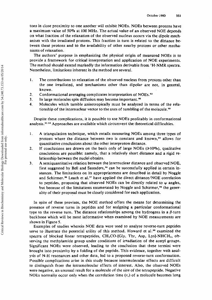
In spite of these provisos, the NOE method offers the means for determining the presence of reverse turns in peptides and for assigning a particular conformational type to the reverse turn. The distance relationships among the hydrogens in a /3-turn backbone which will be most informative when examined by NOE measurements are shown in Figure 5 .
Examples of studies wherein NOE data were used to analyze reverse-turn peptides serve to illustrate the potential utility of this method. Howard et aL9' examined the spectra of blocked linear tetrapeptides, CH,CO-[Gly, Thr, Asp, Lysl-NHCH,, ob- serving the methylamide group under conditions of irradiation of the acetyl groups. Significant NOES were observed, leading to the conclusion that these termini were brought into proximity by a folding of the peptide. This evidence, together with anal- ysis of N-H resonances and other data, led to a proposed reverse-turn conformation. Possible complications arise in this study because intermolecular effects are difficult to distinguish from the intramolecular effects of interest. Also, the observed NOES were negative, an unusual result for a molecule of the size of the tetrapeptide. Negative NOEs normally occur only when the correlation time (T.) of a molecule becomes long
Cri
tical
Rev
iew
s in
Bio
chem
istr
y an
d M
olec
ular
Bio
logy
Dow
nloa
ded
from
info
rmah
ealth
care
.com
by
54.2
48.7
3.15
3 on
05/
20/1
4Fo
r pe
rson
al u
se o
nly.
332 CRC Critical Reviews in Biochemistry
h
0
\ R /
C
FIGURE 5 . 0-turn which might be determinable from NOE measurements.
Examples of hydrogen-hydrogen distances ( - ) in a
(T, ca. 10-9s). depending on field strength.99 Other studies have used similar approaches to Howard et al.loo.lol
Jones et al.95 used the triangulation approach (i.e., analysis of a three-spin system) to refine the conformation of gramicidin S in the region of the D-Phe-L-Pro /3-turns. Since the distance between the Pro Hd's is fixed and the distances between the D-Phe N-H and each of the Pro Hd's depends on yD-PL., a comparison of NOES observed in this three-spin system can yield the distance from the D-Phe N-H to the Pro Hd's and yD+.,,. y is not directly obtainable from any of the usual NMR experiments.
Khaled and Urry'O' claimed to have demonstrated by NOE that a linear tetrapeptide adopted a type I1 B-turn conformation, as opposed to a type I, the other possible turn type. Their conclusion was based on a measurement of a 10% NOE between the Pro H' (residue i + 1 of the proposed turn) and the Gly N-H (residue i + 2) (see Figure 5 ) . As they pointed out, the distance between these protons would be markedly larger for a type I turn than for a type I1 turn. Hence, their measured value was interpreted as proof for a type I1 turn. A comparison was drawn with the value of 1.9% measured for valinomycin-K', which these authors mistakenly attributed to an NOE value char- acteristic of a type I turn. However, it has been established that the valinomycin-K' conformation in solution and crystal is comprised of alternating type I1 and 11' turns."
4 . IJC NMR Chemical Shifts and Relaxation Times "C NMR spectroscopy has become feasible, and in fact routine, even at natural
abundance, with the development of pulsed NMR method^.^' Consequently, the spec- tra of many compounds, including peptides, have been measured and tabulated."' With a couple of exceptions, such chemical shift data have not been demonstrated to be very useful in peptide conformational analysis. However, in certain cases carbon NMR shifts have been used for the determination of reverse turns.
One correlation of "C chemical shifts with conformational aspects which has
Cri
tical
Rev
iew
s in
Bio
chem
istr
y an
d M
olec
ular
Bio
logy
Dow
nloa
ded
from
info
rmah
ealth
care
.com
by
54.2
48.7
3.15
3 on
05/
20/1
4Fo
r pe
rson
al u
se o
nly.
October 1980 333
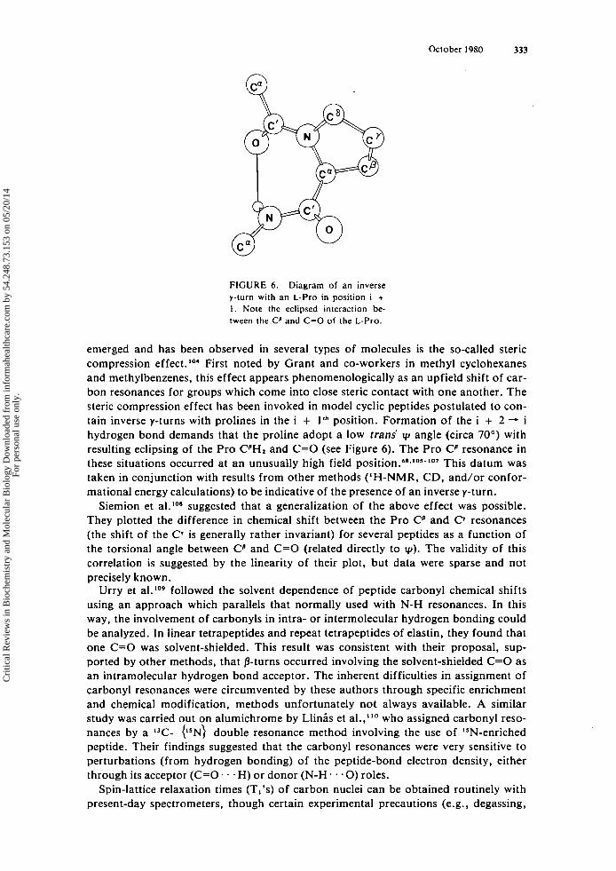
FIGURE 6 . Diagram of an inverse y-turn with an L-Pro in position i + 1 . Note the eclipsed interaction be- tween the Cp and C=O of the L-Pro.
emerged and has been observed in several types of molecules is the so-called steric compression effect. First noted by Grant and co-workers in methyl cyclohexanes and methylbenzenes, this effect appears phenomenologically as an upfield shift of car- bon resonances for groups which come into close steric contact with one another. The steric compression effect has been invoked in model cyclic peptides postulated to con- tain inverse y-turns with prolines in the i + 1'' position. Formation of the i + 2 -, i hydrogen bond demands that the proline adopt a low trans' v, angle (circa 70") with resulting eclipsing of the Pro CPH2 and C=O (see Figure 6). The Pro CP resonance in these situations occurred at an unusually high field p ~ s i t i o n . ~ ~ . ~ ~ ~ - ' ~ ' This datum was taken in conjunction with results from other methods ('H-NMR, CD, and/or confor- mational energy calculations) to be indicative of the presence of an inverse y-turn.
Siemion et al.loS suggested that a generalization of the above effect was possible. They plotted the difference in chemical shift between the Pro Cp and C* resonances (the shift of the Cv is generally rather invariant) for several peptides as a function of the torsional angle between CP and C=O (related directly to q ~ ) . The validity of this correlation is suggested by the linearity of their plot, but data were sparse and not precisely known.
Urry et ,].Io9 followed the solvent dependence of peptide carbonyl chemical shifts using an approach which parallels that normally used with N-H resonances. In this way, the involvement of carbonyls in intra- or intermolecular hydrogen bonding could be analyzed. In linear tetrapeptides and repeat tetrapeptides of elastin, they found that one C=O was solvent-shielded. This result was consistent with their proposal, sup- ported by other methods, that 0-turns occurred involving the solvent-shielded C=O as an intramolecular hydrogen bond acceptor. The inherent difficulties in assignment of carbonyl resonances were circumvented by these authors through specific enrichment and chemical modification, methods unfortunately not always available. A similar study was carried out on alumichrome by Llinis et al.,"' who assigned carbonyl reso- nances by a "C- C5N) double resonance method involving the use of ISN-enriched peptide. Their findings suggested that the carbonyl resonances were very sensitive to perturbations (from hydrogen bonding) of the peptide-bond electron density, either through its acceptor (C=O. . . H) or donor (N-H . . . O ) roles.
Spin-lattice relaxation times (T,'s) of carbon nuclei can be obtained routinely with present-day spectrometers, though certain experimental precautions (e.g., degassing,
Cri
tical
Rev
iew
s in
Bio
chem
istr
y an
d M
olec
ular
Bio
logy
Dow
nloa
ded
from
info
rmah
ealth
care
.com
by
54.2
48.7
3.15
3 on
05/
20/1
4Fo
r pe
rson
al u
se o
nly.
334 CRC Critical Reviews in Biochemistry
accurate measurement of flip angle, and adequate delays between pulses) must be taken. The efficiency of dipolar relaxation (the usually predominant mechanism) of carbons depends in general on the number of bound protons and the frequency of the motion, relative to the Larmor frequency, of the relaxing nucleus. For most small peptides, molecular motions are rapid enough so that the extreme narrowing condition is satisfied, and the statement can be made that faster molecular motion involving the relaxing nucleus will lead to a longer relaxation time. The molecular motion referred to can arise from overall tumbling or from segmental motion (in a flexible molecule). Although the description presented here is qualitative, it comprises the essential aspects of relaxation time applications to small peptide conformational analysis.
For example, the result that T,'s of all of the carbons of a linear peptide are similar has been interpreted to mean that a specific conformation obtains, so that the molecule tumbles as a unit without flexible parts. This idea lead Smith and co-workers"' to suggest that the melanocyte-stimulating-hormone-release-inhibiting-factor, H-Pro- Leu-Gly-NH,, adopted a p-turn conformation in dimethyl sulfoxide. Similarly, the relaxation time data for angiotensin were interpreted to reflect a favored, folded con- formation. 'I2
relaxation times of (Xxx-L-Pro-Yyy), cyclic he~apeptides"~ which take up a conformation containing two p-turns, it was observed that relative values of proline carbon Ti's varied depending on whether the proline was in the i + 1'" position of a type I1 turn or the i + 2Ik position of a type 11' turn. Without other examples, it is not safe to rely on a single finding as diagnostic of a structural feature, but such studies suggest that relaxation times may potentially yield detailed conformational in- formation in reverse turn-containing peptides.
In a study of
5. I5N NMR 15N NMR spectroscopy is a relatively recent innovation, and as yet infrequent use
has been made of this method in analysis of reverse turn-containing peptides. How- ever, ISN studies of three examples, gramicidin S,"' ferr i~hrome,"~ and the model cyclic pentapeptide, cyclo-(Gly-Pro-Gly-D-Ala-Pro)'16 have been reported. Although trends, particularly arising from involvement in intra- or intermolecular hydrogen bonding, were postulated, no clearcut conformational applications of lSN NMR data have been established.
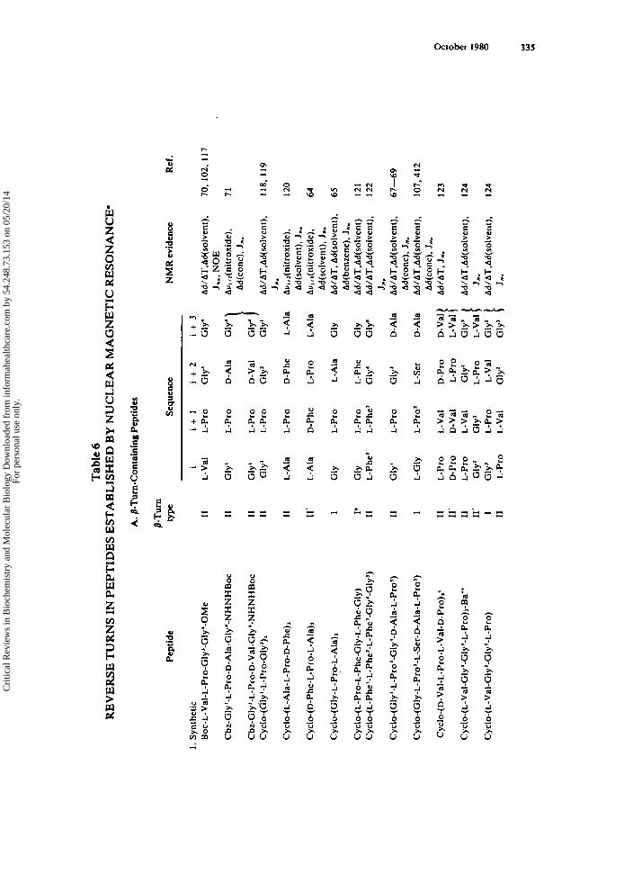
6. Summary Equipped with the methods described for applications of NMR techniques to reverse.
turn-containing peptides, researchers have ascribed a conformation containing p- and/ or y-turns to a large number of peptides. A summary of those examples in which the sequence and type of the turn(s) are well-established is presented in Table 6. The list of peptides is not intended to be exhaustive, but instead to be illustrative of the fre- quent, fruitful applications of NMR in the elucidation of reverse turns in peptides. It is clear that many synthetic model turn compounds have been characterized in detail and that these are useful as reference compounds for turn geometries and spectral parameters. Moreover, they may serve as test compounds for correlation of amino acid sequence with preferred turn conformations. Further, several examples of reverse turn conformations have been described in studies of antibiotics, hormones, and neu- ropeptides. The biological implications of these findings is discussed more fully in Sections V.G , V.H , and V.J.
C. Circular Dichroism (CD) Evidence for Reverse Turns in Peptides Circular dichroism is sensitive to peptide conformation and is used extensively as an
Cri
tical
Rev
iew
s in
Bio
chem
istr
y an
d M
olec
ular
Bio
logy
Dow
nloa
ded
from
info
rmah
ealth
care
.com
by
54.2
48.7
3.15
3 on
05/
20/1
4Fo
r pe
rson
al u
se o
nly.
Tab
le 6
R
EV
ER
SE T
UR
NS
IN P
EPT
IDE
S E
STA
BL
ISH
ED
BY
NU
CL
EA
R M
AG
NE
TIC
RE
SON
AN
CE
‘
A. p
-Tur
n-C
onta
inin
g Pe
ptid
es
Pept
ide
1. S
ynth
etic
B
oc-L
-Val
-L-P
ro-G
lyJ-
Gly
4-0M
e
Cbz
-Gly
‘-L
-Pro
-D-A
la-G
ly‘-
NH
NH
Boc
Cyc
lo-(
L-P
ro-L
-Phe
-Gly
-L-P
he-G
ly)
Cyclo-(L-Phel-L-Phe’-L-Phe’-Gly‘-Gly’)
Cyc
lo-(
Gly
‘-L
-Pro
’-G
1y’-
D-A
la-L
-Pro
’)
Cyclo-(Gly-L-Pro’-L-Ser-D-Ala-L-Pro’)
Cyc
lo-(
D-V
al-L
-Pro
-L-V
al-D
-Pro
),‘
Cyclo-(L-Val-Gly’-GIy’-L-Pro)
p-T
urn
type
11
I1
I1
I1
I1
11‘ I I’ I1
I1 1 I1
11’
I1
11‘ I I1
Sequ
ence
N
MR
evid
ence
I
L-V
al
Gly
‘
Gly
‘ G
ly‘
L-A
la
L-A
la
GlY
GlY
L-
Phe’
Gly
‘
L-G
ly
L-P
ro
D-P
ro
L-P
ro
Gly
’ G
lyJ
L-P
ro
i+
l
L-P
ro
L-P
ro
L-P
ro
L-P
ro
L-P
ro
D-P
he
L-P
ro
L-P
ro
L-Ph
e’
L-P
ro
L-P
ro’
L-V
al
D-V
al
L-V
al
Gly
J L
-Pro
L-
Val
i+
2
Gly
’
D-A
la
D-V
al
Gly
’
D-P
he
L-P
ro
L-A
la
L-P
he
~1
~4
Gly
’
L-Se
r
D-P
ro
L-P
ro
Gly
’ L
-Pro
L-
Val
G
ly’
i+
3
Gly
‘
Gly
’
L-A
la
L-A
la
G~
Y
GlY
G
ly’
D-A
la
D-A
la
D-V
al
L-V
al
L-V
al
Ad/
AT,
Ad(
solv
ent),
Av,
,I(n
itrox
ide)
. A
d(co
nc),
J,.
J,.,
NO
E
Ad/
AT,
Ad(
solv
ent),
J N
.
Au,
,2(n
itro
xide
),
Ad(
so1v
ent).
JNI
Au
, ,,(n
itrox
ide)
. A
d(so
1ven
t). J
N.
Ad/
AT,
Ad(
solv
ent)
, A
d(be
nzen
e).
JN.
Ad/
AT,
Ad(
solv
ent)
Ad/
AT,
Ad(
solv
ent),
Ad/
AT.
Ad(
solv
ent),
Ad/
AT,
Ad(
solv
ent).
Ad/
AT,
J,.
JN.
Ad(
conc
). J,.
Ad(
conc
). J,
Ad/
AT,
Ad(
solv
en t)
,
Ad/
AT,
Ad(
solv
ent),
J N. J,.
Ref
.
70,
102,
I17
71
118,
119
120
64
65
121
122
67-69
107,412
123
I24
I24
Cri
tical
Rev
iew
s in
Bio
chem
istr
y an
d M
olec
ular
Bio
logy
Dow
nloa
ded
from
info
rmah
ealth
care
.com
by
54.2
48.7
3.15
3 on
05/
20/1
4Fo
r pe
rson
al u
se o
nly.

Tab
le 6
(con
tinue
d)
RE
VE
RSE
TU
RN
S IN
PE
PTID
ES
EST
AB
LIS
HE
D B
Y N
UC
LE
AR
MA
GN
ET
IC R
ESO
NA
NC
E-
W
bI
0,
A. /
3-T
urn-
Con
tain
ing P
epti
des
Pept
ide
Sequ
ence
N
MR
evi
denc
e R
ef.
2. N
atur
ally
-occ
urri
ng
Gra
mic
idin
S
L-Le
u D
-Phe
D
-Phe
L
-Pro
L-
Val
A
d/A
T,A
d(so
lven
t).
T,,~
('H
-'H
), N
OE
, JN
.
3, 1
5.62
.63.
95
II'
3.66
.84,
93
. 12
5, 1
26
72,8
1 1 1
5, 1
27
128,
129
. 130
Val
inom
y An'
I1
11
' II
11
I1
I
L-L
ac
D- H
ylv
LL
ac
~1
~3
~1
~3
L-P
ro'
L-V
al
D-V
al
L-V
al
L-A
la'
Gly
'
L-A
la
D-H
yIv
L-L
ac
Gly
' G
ly'
L- P
he'
D-H
ylv
Ad/
AT
,Ad(
solv
ent)
. J(
hete
ro),
JN..
NO
E.
Val
inom
ycin
' Fe
rric
hrom
e C
and
alu
mic
hrom
e C'
Fe
rric
hrom
e and
alu
mic
hrom
e'
Ant
aman
ide-
Na'
Ad/
AT,
Ad(
solv
ent).
",:",:
$ )
JN..
"N
A~/
AT
.T,,~
('H-'H
),
JN.,
"C
J N.
L-V
al'
L-A
sn
Ad/
AT
.T,,z
(lH
-zH
). I
-1
L- Pro'
L-Ty
r L
-Phe
9 L
-lle
L-
Phe
" L
-Gln
13
1-13
4 O
xyto
cin
B. In
vers
e y-
Tur
n C
onta
inin
g Pe
ptid
es
Pept
ide
1. S
ynth
etic
C
yclo
-(L
-Pro
-Gly
), C
yclo
-(L
-Pro
-Gly
), Cyclo-(L-Pro-L-Phe'-Gly3-L-Phe4-Glys)
Cyclo-(L-Phe'-L-Phe'-L-Phe3-Gly4-Gly')
NMR ev
iden
ce
Ref
. Se
quen
ce
~~
I i
+l
i
+2
G
~Y
L
-Pro
G
ly
G~
Y
L-P
ro
Gly
~
1~
3
L-Ph
e' G
ly'
~iy
5
L-P
he'
L-P
he'
Ju.,
"C
105,
106
JN.,
"C
106.
135
A
d/A
T,A
d(so
lven
t)
121
JN.,
Ad/
12
2 A
T,A
d(so
lven
t)
AT
,Ad(
solv
ent)
, A
d(co
nc), "C
JN.,
Ad/
61
-69
Cyclo-(Gly'-L-Pro'-Gly3-D-Ala-L-Pro')
D-A
la
L-P
ro'
Gly
'
Cri
tical
Rev
iew
s in
Bio
chem
istr
y an
d M
olec
ular
Bio
logy
Dow
nloa
ded
from
info
rmah
ealth
care
.com
by
54.2
48.7
3.15
3 on
05/
20/1
4Fo
r pe
rson
al u
se o
nly.

Cyclo-(Gly-L-Pro’-L-Ser-D-Ala-L-Pro’)
D-A
la
L-P
ro’
Gly
JN
., A
d/
107,
412
AT
,Ad(
solv
ent)
, A
d(co
nc),
”C
* A
bbre
viat
ions
use
d: d
. che
mic
al s
hift
dow
nfie
ld f
rom
tetr
amet
hyls
ilane
; A
d/A
T, c
hang
e in
che
mic
al s
hift
with
tem
pera
ture
; Ad(
solv
ent)
, ch
ange
in c
hem
ical
shi
ft u
pon
chan
ging
sol
vent
s; J
N.,
’JN
a.,,o
; N
OE
, nu
clea
r O
verh
ause
r ef
fect
; Aw
,,,(n
itrox
ide)
, ch
ange
in l
ine-
wid
th
with
add
ed n
itrox
ide;
7,,,
(’H
-*H
), h
alf-
life
for
deut
eriu
m e
xcha
nge;
J(h
eter
o),
hete
ronu
clea
r co
upli
ng c
onst
ant
anal
ysis
; B
oc.
t-bu
ty-
loxy
carb
onyl
; O
Me,
met
hyl e
ster
; Cbz
, car
bobe
nzox
y.
Alt
houg
h no
t sp
ecif
ical
ly a
ssig
ned
by D
erne
l and
Kes
sler
,’”
a ty
pe I
turn
is t
he m
ost
likel
y st
ruct
ure
base
d on
seq
uenc
e co
nsid
erat
ions
. T
urns
list
ed a
re p
rese
nt i
n bo
th th
e un
com
plex
ed p
epti
de a
nd in
its
K’ c
ompl
ex.
Tur
ns li
sted
are
pre
sent
in b
oth
the
unco
mpl
exed
pep
tide
and
its
K’
com
plex
in n
onpo
lar
solv
ents
(e.g
.. ch
loro
form
).
Tur
n lis
ted
is p
rese
nt i
n so
lven
ts of
med
ium
pol
arit
y (e
.g..
met
hano
l).
The
enu
mer
atio
n is
con
sist
ent w
ith t
hat u
sed
prev
ious
ly.”
T
urn
type
is n
ot d
efin
itive
ly e
stab
lishe
d.
’ * ‘ ‘ ’ a
Cri
tical
Rev
iew
s in
Bio
chem
istr
y an
d M
olec
ular
Bio
logy
Dow
nloa
ded
from
info
rmah
ealth
care
.com
by
54.2
48.7
3.15
3 on
05/
20/1
4Fo
r pe
rson
al u
se o
nly.
338 CRC Critical Reviews in Biochemistry
experimental technique for studying the conformations of peptides and A CD spectrum displays the sum of the individual spectral contributions donated by the various conformations present within a given peptide. Hence, the application of decon- volution techniques to the analysis of a CD spectrum can yield an estimate of the contribution of each individual conformation (e.g., a-helix, p-sheet, extended struc- ture, or reverse turn) present in a peptide to the overall spectrum.
Circular dichroism is the difference in absorption of left and right circularly polar- ized light, and is defined as E ~ - E , , where and 6, are the molecular extinction coeffi- cients for the left and right components. The origin of CD lies in the rotatory strength, R, of an electronic absorption band, which is expressed as:
where M is the magnetic dipole moment of a transition whose electronic transition moment is p. The term Im refers to the imaginary part (i.e.. the part which contains i = f-1) of the dot or scalar product shown. This expression indicates that the rotatory strength depends on the magnitudes of the electronic and magnetic transition moments and the cosine of the angle between them.
The CD derives from relative orientations of the transition moments, and hence is very sensitive to molecular geometries (at least to relative geometries of chromo- phores). As summarized by S ~ h e l l m a n , ~ ~ ' three mechanisms can contribute to rotatory strength
1. The electric and magnetic moments in one chromophore couple because of the perturbing field of the remainder of the molecule (called the one-electron or Con- don, Altar, and Eyring theory).138 Two groups with single electric transitions couple to produce a magnetic moment (Kuhn-Kirkwood mechanism).139 Conditionally, if the excited states are degener- ate, the exciton theory of MoffittI4O can be used. One group with an electric transition and another with a magnetic transition cou- ple: the p - m mechani~m.'~'
2.
3 .
Bayley et al.I4l derived a general matrix formulation for the rotatory strength of molecules containing two peptide groups, each assumed to possess only the two lowest excited electronic states: n -. n*, n + n*. They calculated the resulting rotatory strengths, including contributions from the three mechanisms above, as a function of dihedral angles (4.1~). Their results demonstrated that the optical rotation arises from all three mechanisms and that the relative contribution of each depends on the confor- mation. Those calculations were extended to larger peptides, and the CD spectra for cyclo-(L-Pro-Gly), as a function of molecular conformation were predicted.l" Good correlation between these predicted and the experimentally determined spectra was found for this inverse y-turn-containing peptide. Theoretical CD spectra of isolated /3-turns calculated in a similar manner by Woody19 further suggested that the gramici- din S-A (i.e., gramicidin S with acetylated ornithine side chains) experimental CD spec- t r u ~ n ' ~ ~ derives largely from its two type 11' /3-turns with little contribution from the phenylalanyl chromophores.
Prior to these studies, the shape of CD curves was analyzed in terms of the relative contribution of u-helical, /3-sheet, and random coil structures which would result in such a curve.14s Thus, the contribution of p-turns to CD of peptides and proteins was
Cri
tical
Rev
iew
s in
Bio
chem
istr
y an
d M
olec
ular
Bio
logy
Dow
nloa
ded
from
info
rmah
ealth
care
.com
by
54.2
48.7
3.15
3 on
05/
20/1
4Fo
r pe
rson
al u
se o
nly.
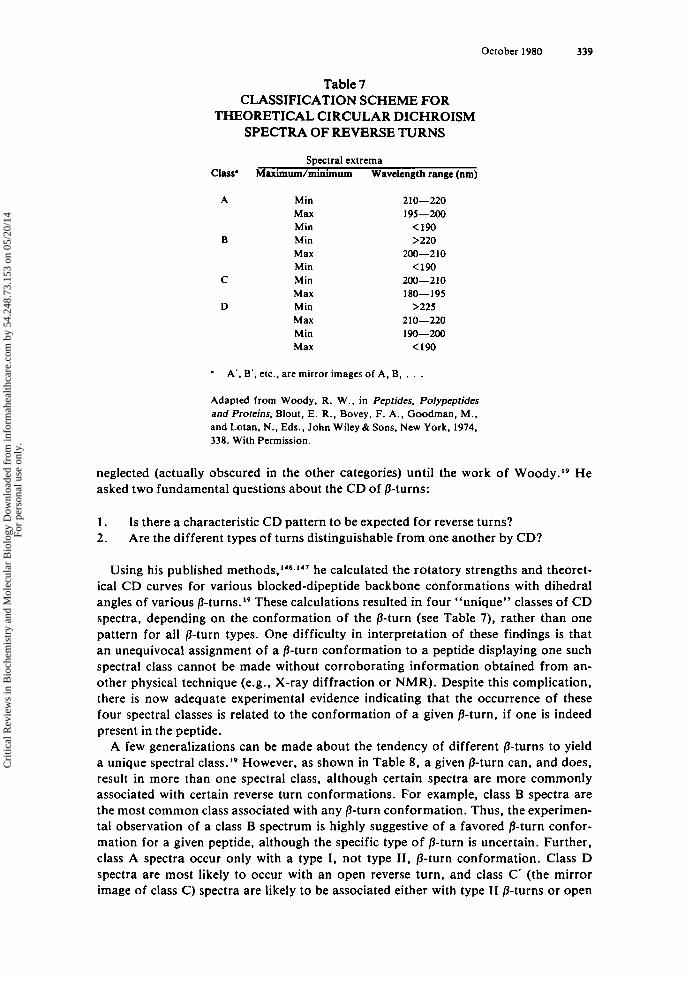
October 1980 339
neglected (actually
Table 7 CLASSIFICATION SCHEME FOR
THEORETICAL CIRCULAR DICHROISM SPECTRA OF REVERSE TURNS
Spectral extrema Class* Maximudminimum Wavelength range (run)
A Min Max Min
B Min Max Min
C Min Max
D Min Max Min Max
2 10-220 195-200
(190 >220
200-210 < I 9 0
200-210 180- 195
>225 210-220 190-200 <I90
A’, B’, etc., are mirror images of A, B,
Adapted from Woody, R. W. . in Peptides, Polypeptides and Proteins, Blout. E. R . , Bovey, F. A . , Goodman, M., and Lotan, N. . Eds., John Wiley & Sons, New York, 1974. 338. With Permission.
obscured in the other categories) until the work of Woody.19 He asked two fundamental questions about the CD of /3-turns:
1. 2.
Is there a characteristic CD pattern to be expected for reverse turns? Are the different types of turns distinguishable from one another by CD?
Using his published he calculated the rotatory strengths and theoret- ical CD curves for various blocked-dipeptide backbone conformations with dihedral angles of various P- turn~. ’~ These calculations resulted in four “unique” classes of CD spectra, depending on the conformation of the p-turn (see Table 7), rather than one pattern for all P-turn types. One difficulty in interpretation of these findings is that an unequivocal assignment of a p-turn conformation to a peptide displaying one such spectral class cannot be made without corroborating information obtained from an- other physical technique (e.g., X-ray diffraction or NMR). Despite this complication, there is now adequate experimental evidence indicating that the occurrence of these four spectral classes is related to the conformation of a given /3-turn, if one is indeed present in the peptide.
A few generalizations can be made about the tendency of different /3-turns to yield a unique spectral ~ 1 a s s . l ~ However, as shown in Table 8, a given P-turn can, and does, result in more than one spectral class, although certain spectra are more commonly associated with certain reverse turn conformations. For example, class B spectra are the most common class associated with any P-turn conformation. Thus, the experimen- tal observation of a class B spectrum is highly suggestive of a favored P-turn confor- mation for a given peptide, although the specific type of p-turn is uncertain. Further, class A spectra occur only with a type I, not type 11, P-turn conformation. Class D spectra are most likely to occur with an open reverse turn, and class C’ (the mirror image of class C) spectra are likely to be associated either with type I1 p-turns or open
Cri
tical
Rev
iew
s in
Bio
chem
istr
y an
d M
olec
ular
Bio
logy
Dow
nloa
ded
from
info
rmah
ealth
care
.com
by
54.2
48.7
3.15
3 on
05/
20/1
4Fo
r pe
rson
al u
se o
nly.
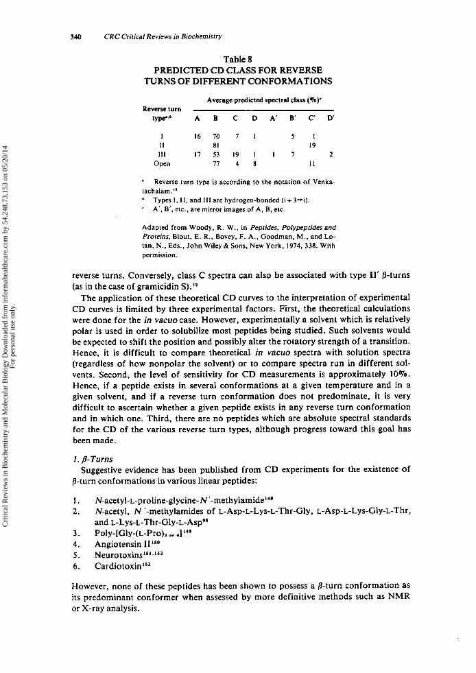
340 CRC Critical Reviews in Biochemistry
Table 8 PREDICTED CD CLASS FOR REVERSE
TURNS OF DIFFERENT CONFORMATIONS
Average predicted spectral class (a)' Reverse turn
@ p p b A B C D A' B' C' D'
I 16 70 7 1 5 1 I 1 81 19
1 1 1 17 53 19 1 I 7 2 Open 77 4 8 11
*
tachalam." ' '
Reverse turn type is according to the notation of Venka-
Types I , 11, and 111 are hydrogen-bonded (i + 3+). A', B'. etc., are mirror images of A, B, etc.
Adapted from Woody, R . W., in Pepfides, Polypeptides and Profeins. Blout. E. R . , Bovey, F. A,, Goodman, M . . and Lo- tan, N . . Eds., John Wiley & Sons, New York, 1974, 338. With permission.
reverse turns. Conversely, class C spectra can also be associated with type 11' /3-turns (as in the case of gramicidin S).',
The application of these theoretical CD curves to the interpretation of experimental CD curves is limited by three experimental factors. First, the theoretical calculations were done for the in vacuo case. However, experimentally a solvent which is relatively polar is used in order to solubilize most peptides being studied. Such solvents would be expected to shift the position and possibly alter the rotatory strength of a transition. Hence, it is difficult to compare theoretical in vacuo spectra with solution spectra (regardless of how nonpolar the solvent) or to compare spectra run in different sol- vents. Second, the level of sensitivity for CD measurements is approximately 10%. Hence, if a peptide exists in several conformations at a given temperature and in a given solvent, and if a reverse turn conformation does not predominate, it is very difficult to ascertain whether a given peptide exists in any reverse turn conformation and in which one. Third, there are no peptides which are absolute spectral standards for the CD of the various reverse turn types, although progress toward this goal has been made.
1. /&Turns
/3-turn conformations in various linear peptides:
1. N-acetyl-L-proline-glycine-N '-methylamide"' 2.
3. Poly-[Gly-(L-Pro), o, 4]149
4. Angiotensin IIL50 5 . neurotoxin^^^^^^^' 6. C a r d i o t o ~ i n ' ~ ~
Suggestive evidence has been published from CD experiments for the existence of
N-acetyl, N '-methylamides of L-Asp-L-Lys-L-Thr-Gly, L-Asp-L-Lys-Gly-L-Thr. and ~-Lys-~-Thr-Gly-~-Asp~'
However, none of these peptides has been shown to possess a p-turn conformation as its predominant conformer when assessed by more definitive methods such as NMR or X-ray analysis.
Cri
tical
Rev
iew
s in
Bio
chem
istr
y an
d M
olec
ular
Bio
logy
Dow
nloa
ded
from
info
rmah
ealth
care
.com
by
54.2
48.7
3.15
3 on
05/
20/1
4Fo
r pe
rson
al u
se o
nly.
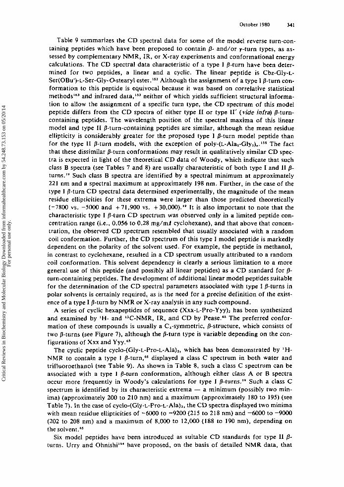
October 1980 341
Table 9 summarizes the C D spectral data for some of the model reverse turn-con- taining peptides which have been proposed to contain /3- and/or y-turn types, as as- sessed by complementary NMR, IR, or X-ray experiments and conformational energy calculations. The CD spectral data characteristic of a type I B-turn have been deter- mined for two peptides, a linear and a cyclic. The linear peptide is Cbz-Gly-L- Ser(0Bu')-L-Ser-Gly- Ostearyl ester . I s 3 Although the assignment of a type I 8-turn con- formation to this peptide is equivocal because it was based on correlative statistical
and infrared data,Is3 neither of which yields sufficient structural informa- tion to allow the assignment of a specific turn type, the CD spectrum of this model peptide differs from the CD spectra of either type I1 or type 11' (vide infra) /3-turn- containing peptides. The wavelength position of the spectral maxima of this linear model and type I1 /3-turn-containing peptides are similar, although the mean residue ellipticity is considerably greater for the proposed type I P-turn model peptide than for the type I1 P-turn models, with the exception of p o l y - ( ~ - A l a ~ - G l y ~ ) , . ~ ~ ~ The fact that these dissimilar /3-turn conformations may result in qualitatively similar CD spec- tra is expected in light of the theoretical CD data of Woody, which indicate that such class B spectra (see Tables 7 and 8) are usually characteristic of both type I and I1 /3-
Such class B spectra are identified by a spectral minimum at approximately 221 nm and a spectral maximum at approximately 198 nm. Further, in the case of the type I P-turn C D spectral data determined experimentally, the magnitude of the mean residue ellipticities for these extrema were larger than those predicted theoretically (-7800 vs. -5000 and +71,900 vs. +30,000).'9 It is also important to note that the characteristic type I P-turn CD spectrum was observed only in a limited peptide con- centration range (i.e.. 0.056 to 0.28 mg/mf cyclohexane), and that above that concen- tration, the observed CD spectrum resembled that usually associated with a random coil conformation. Further, the CD spectrum of this type I model peptide is markedly dependent on the polarity of the solvent used. For example, the peptide in methanol, in contrast to cyclohexane, resulted in a CD spectrum usually attributed to a random coil conformation. This solvent dependency is clearly a serious limitation to a more general use of this peptide (and possibly all linear peptides) as a CD standard for /3- turn-containing peptides. The development of additional linear model peptides suitable for the determination of the CD spectral parameters associated with type I P-turns in polar solvents is certainly required, as is the need for a precise definition of the exist- ence of a type I P-turn by NMR or X-ray analysis in any such compound.
A series of cyclic hexapeptides of sequence (Xxx-L-Pro-Yyy), has been synthesized and examined by IH- and IT-NMR, IR, and CD by Pease.65 The preferred confor- mation of these compounds is usually a C,-symmetric, /3-structure, which consists of two /3-turns (see Figure 7), although the /3-turn type is variable depending on the con- figurations of Xxx and Y Y Y . ~ ~
The cyclic peptide cyclo-(Gly-L-Pro-L-Ala),, which has been demonstrated by IH-
NMR to contain a type I f l - t u ~ n , ~ ~ displayed a class C spectrum in both water and trifluoroethanol (see Table 9). As shown in Table 8. such a class C spectrum can be associated with a type I P-turn conformation, although either class A or B spectra occur more frequently in Woody's calculations for type I /3- turn~. '~ Such a class C spectrum is identified by its characteristic extrema - a minimum (possibly two min- ima) (approximately 200 to 210 nm) and a maximum (approximately 180 to 195) (see Table 7). In the case of cyclo-(Gly-L-Pro-L-Ala),, the CD spectra displayed two minima with mean residue ellipticities of -6OOO to -9200 (215 to 218 nm) and -6000 to -9000 (202 to 208 nm) and a maximum of 8,000 to 12,000 (188 to 190 nm), depending on the
Six model peptides have been introduced as suitable CD standards for type I1 /3- turns. Urry and O h n i ~ h i ~ ~ ' have proposed, on the basis of detailed NMR data, that
Cri
tical
Rev
iew
s in
Bio
chem
istr
y an
d M
olec
ular
Bio
logy
Dow
nloa
ded
from
info
rmah
ealth
care
.com
by
54.2
48.7
3.15
3 on
05/
20/1
4Fo
r pe
rson
al u
se o
nly.
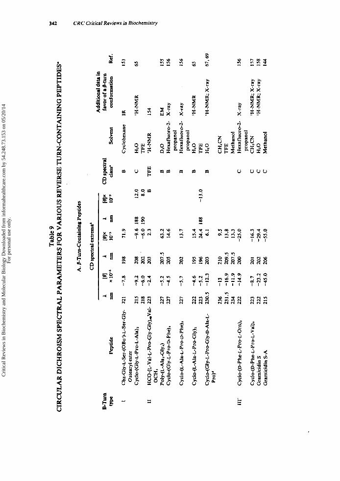
Tab
le 9
C
IRC
UL
AR
DIC
HR
OIS
M SPE
CTRA
L PA
RA
ME
TE
RS
FOR
VA
RIO
US
RE
VE
RSE
TU
RN
-CO
NT
AIN
ING
PE
PTID
ES'
A. /
%T
urn-
Con
tain
in#
Pept
ides
CD
spec
tral
exw
ema'
5Tur
n A
re1
type
Pe
ptid
e ~ull
x 10
-3
I Cbz-Gly-L-Scr-(0Bu')-L-Ser-Gly- 2
21
-7.8
Cyc
lo-(
GL
y-L
-Pro
-L-A
la)I
21
5 -9
.2
218
-6.0
I1
H
CO
-(L
-Val
-L-P
ro-G
ly-G
ly)a
Val
- 22
3 -2
.4
Ost
eary
l est
er
OC
H3
Poly
-(L
-Ala
I-G
lyl)
C
yclo
-(G
ly-L
-Pro
-D-P
he)I
Cyc
lo-(
Gly
-L-P
ro-G
ly-D
- A
la-L
- Pr
o)'
111'
Cyc
lo-(
D-P
he-L
-Pro
-L-O
rn)2
Cyc
lo-(
D-P
he-L
-Pro
-L-V
al)2
G
rarn
icid
in S
G
ram
icid
in S
-A
227
-5.2
22
7 -4
.5
227
-5.7
222
-4.6
22
3 -5
.2
230.
5 -1
2.3
236
-13
231.
5 -1
6.9
234
-11.
9 22
2 -1
4.9
223
-8.7
22
2 -2
3.2
215
-45.
0
A Iull
198
208
202
203
207.
5 20
5
202
I95
196
203
210
209.
5 20
7.5
200
20 1
202
206
PIX
A
10-3
11111
71.9
-9.6
18
8 -6
.0
190
2.3
63.2
14
.6
13.7
15.4
24
.4
188
6. I
9.5
13.8
13
.3
-25.
0
-16.
3 -2
9.4
-55.
0
Add
itio
nal d
ata
in
favo
r of a
/3-tu
rn
B
12.0
C
8.
0 B T
FE
B B B B B -1
3.0
Solv
ent
conf
orm
atio
n
Cyc
lohe
xane
1R
HIO
'H
-NM
R
TF
E
'H-N
MR
I5
4
DIO
E
M
Hex
aflu
oro-
2-
X-r
ay
Hex
aflu
oro-
2-
X-r
ay
prop
anol
prop
anol
H
zO
'H-N
MR
T
FE
HIO
'H
-NM
R;
X-r
ay
CH
SCN
T
FE
M
etha
nol
Hex
aflu
oro-
2-
X-r
ay
prop
anol
C
HSC
N
'H-N
MR
; X
-ray
H
zO
'H-N
MR
; X
-ray
M
etha
nol
Ref
.
153
65
155
I56
156
65
67.6
9
156
I57
I58
144
W R 8'
Cri
tical
Rev
iew
s in
Bio
chem
istr
y an
d M
olec
ular
Bio
logy
Dow
nloa
ded
from
info
rmah
ealth
care
.com
by
54.2
48.7
3.15
3 on
05/
20/1
4Fo
r pe
rson
al u
se o
nly.
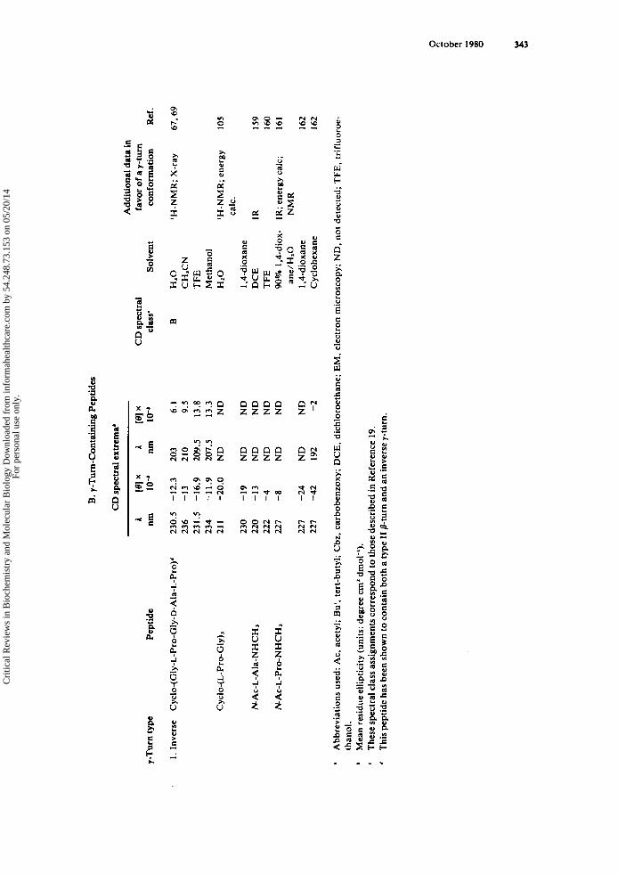
I-T
urn
type
Pe
ptid
e
. I.
Inve
rse
Cyclo-(Gly-L-Pro-Gly-D-Ala-L-Pro)'
Cyc
lo-(
L-P
ro-G
ly),
NA
c-L
-Ala
-NH
CH
,
NA
c-L
-Pro
-NH
CH
,
B. y
-Tur
n-C
onta
inin
g Pep
tida
CD sp
ectr
al e
xtrc
ma'
230.
5 -1
2.3
236
-13
231.
5 -1
6.9
234
-11.
9 21
1 -2
0.0
230
-19
220
-13
222
-4
227
-8
221
-24
227
-42
A nm
203
210
209.
5 20
7.5
ND
ND
N
D
ND
N
D
ND
19
2
re1 x
10'' 6.
1 9.
5 13
.8
13.3
N
D
ND
N
D
ND
N
D
ND
-2
CD sp
ectr
al
clas
s' So
lven
t
B H
Z0
CH
,CN
T
FE
Met
hano
l H
z0
1.4-
diox
ane
DC
E
TFE
90
% 1
.4-d
iox-
an
e/H
IO
1,4-
diox
ane
Cyc
lohe
xane
Add
itio
nal d
ata
in
favo
r of
a I-
turn
co
nfor
mat
ion
'H-N
MR
; X
-ray
'H-N
MR
; en
ergy
ca
lc.
IR
1R; e
nerg
y ca
lc;
NM
R
Ref
.
67.6
9
105
I59
160
161
162
I62
* A
bbre
viat
ions
use
d: A
c, a
cety
l; Bu'. t
ert-
buty
l; C
bz, c
arbo
benz
oxy;
DC
E,
dich
loro
etha
ne; E
M. e
lect
ron
mic
rosc
opy;
ND
. no
t de
tect
ed; T
FE
. tr
iflu
oroe
- th
anol
. M
ean
resi
due
elli
ptic
ity
(uni
ts: d
egre
e cm
' dm
ol-')
. T
hese
spe
ctra
l cla
ss as
sign
men
ts c
orre
spon
d to
thos
e de
scri
bed
in R
efer
ence
19.
T
his p
epti
de h
as b
een
show
n to
con
tain
bot
h a
type
11
/?-tu
rn a
nd a
n in
vers
e y-t
urn.
' ' '
Cri
tical
Rev
iew
s in
Bio
chem
istr
y an
d M
olec
ular
Bio
logy
Dow
nloa
ded
from
info
rmah
ealth
care
.com
by
54.2
48.7
3.15
3 on
05/
20/1
4Fo
r pe
rson
al u
se o
nly.
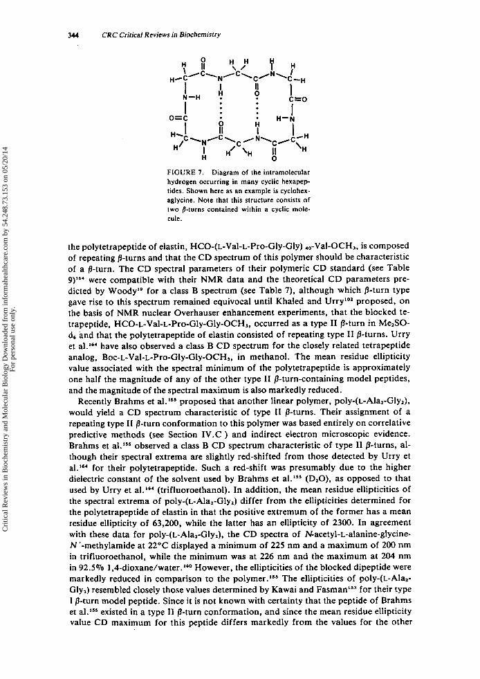
344 CRC Critical Reviews in Biochemistry
FIGURE 7. Diagram of the intramolecular hydrogen occurring in many cyclic hexapep- tides. Shown here as an example is cyclohex- aglycine. Note that this structure consists of two /?-turns contained within a cyclic mole- cule.
the polytetrapeptide of elastin, HCO-(L-Val-L-Pro-Gly-Gly) ,,-Val-OCH,, is composed of repeating p-turns and that the CD spectrum of this polymer should be characteristic of a p-turn. The CD spectral parameters of their polymeric CD standard (see Table ,)I6' were compatible with their NMR data and the theoretical CD parameters pre- dicted by Woodya9 for a class B spectrum (see Table 7), although which p-turn type gave rise to this spectrum remained equivocal until Khaled and Urry''' proposed, on the basis of NMR nuclear Overhauser enhancement experiments, that the blocked te- trapeptide, HCO-L-Val-L-Pro-Gly-Gly-OCH3, occurred as a type I1 pturn in Me2SO- ds and that the polytetrapeptide of elastin consisted of repeating type I1 /3-turns. Urry et a1.16, have also observed a class B CD spectrum for the closely related tetrapeptide analog, Boc-L-Val-L-Pro-Gly-Gly-OCH3, in methanol. The mean residue ellipticity value associated with the spectral minimum of the polytetrapeptide is approximately one half the magnitude of any of the other type I1 /3-turn-containing model peptides, and the magnitude of the spectral maximum is also markedly reduced.
Recently Brahms et al.lSS proposed that another linear polymer, poly-(L-Ala2-Gly2), would yield a CD spectrum characteristic of type I1 p-turns. Their assignment of a repeating type I1 p-turn conformation to this polymer was based entirely on correlative predictive methods (see Section 1V.C ) and indirect electron microscopic evidence. Brahms et al.lS5 observed a class B CD spectrum characteristic of type I1 /3-turns, al- though their spectral extrema are slightly red-shifted from those detected by Urry et al.'", for their polytetrapeptide. Such a red-shift was presumably due to the higher dielectric constant of the solvent used by Brahms et al.lSS (D,O), as opposed to that used by Urry et al.16, (trifluoroethanol). In addition, the mean residue ellipticities of the spectral extrema of poly-(L-Ala,-Gly,) differ from the ellipticities determined for the polytetrapeptide of elastin in that the positive extremum of the former has a mean residue ellipticity of 63,200, while the latter has an ellipticity of 2300. In agreement with these data for poly-(L-Ala,-Gly,), the CD spectra of Nacetyl-L-alanine-glycine- N 'methylamide at 22°C displayed a minimum of 225 nm and a maximum of 200 nm in trifluoroethanol, while the minimum was at 226 nm and the maximum at 204 nm in 92.5% 1 ,4-dioxane/water.160 However, the ellipticities of the blocked dipeptide were markedly reduced in comparison to the The ellipticities of poly-(L-Ala2- Gly,) resembled closely those values determined by Kawai and FasmanlS3 for their type I p-turn model peptide. Since it is not known with certainty that the peptide of Brahms et a1.l" existed in a type I1 p-turn conformation, and since the mean residue ellipticity value CD maximum for this peptide differs markedly from the values for the other
Cri
tical
Rev
iew
s in
Bio
chem
istr
y an
d M
olec
ular
Bio
logy
Dow
nloa
ded
from
info
rmah
ealth
care
.com
by
54.2
48.7
3.15
3 on
05/
20/1
4Fo
r pe
rson
al u
se o
nly.
October 1980 345
type I 1 0-turn model peptides, it may be that the class B C D spectrum attributable to this peptide resulted from a repeating type I /&turn conformation rather than the type I 1 0-turn conformation. This possibility has not been disproven, and definitive NMR experiments are required.
Various cyclic penta- and hexapeptides in which type I1 /3-turns have been established by X-ray crystallography and/or IH-NMR have also been used as model peptides to establish the C D spectral parameters of such 0-turns (see Table 9). Pease65 has shown that cyclo-(L-Ala-L-Pro-Gly), in water or trifluoroethanol displays a class B CD spec- trum and that the extrema are located at a slightly shorter wavelength than two other cyclic hexapeptides (containing type I1 0-turns),156 poly- (~-Ala , -Gly~) , '~~ and the poly- tetrapeptide (minimum only).164 The values of the mean residue ellipticities of the ex- trema of this peptide are remarkably similar to the other two cyclic hexapeptides, cy- clo-(Gly-~-Pro-D-Phe)~ and c y ~ l o - ( ~ - A l a - ~ - P r o - ~ - P h e ) ~ , as well as poly-(~-Ala~-Gly,) (minimum only). Bush et a1.ls6 have shown that cyclo-(Gly-L-Pro-D-Phe)Z and cyclo- (L-Ala-L-Pro-D-Phe), in hexafluoro-2-propanol both display a class B CD spectrum with extrema spectral positions and mean residue ellipticities that are very similar to each other. Although they also displayed minima that are similar to the repeating linear peptides of Urry et al.164 and Brahms et al.lr5 the values of the positive extrema of these peptides in comparison to the linear polymers differ appreciably. The origin of these differences is not readily explicable, but it is likely to result from contributions to the C D from conformations other than 0-turns in the repeating linear polymers examined. Recently, the CD of cyclo-(Gly-L-Pro-Gly-D-Ala-L-Pro), which contains a type I1 /&turn, as well as an inverse y-turn, has been e ~ a m i n e d . ~ ~ . ~ ~ Because of the coexistence of a 0- and an inverse y-turn. the unequivocal interpretation of the C D data for this compound cannot be made without resorting to theoretical CD calcula- tions to elucidate the relative contribution of each of these structures to the CD spec- trum. However, the magnitude of the mean residue ellipticities of the minima of this cyclic pentapeptide in the various solvents is considerably greater than the other type I1 0-turn-containing model peptides, but similar to the magnitude of the other inverse y-turn-containing peptides. This suggests, but does not prove, that the inverse y-turn moiety of the cyclic pentapeptide dominates the C D spectrum in the 220 to 230 nm spectral region. In contrast, the mean residue ellipticity of the CD spectral maximum resembles closely those values found for the other type I1 model compounds. Since a similar maximum was not detected for the other inverse y-turn-containing model pep- tides, it seems likely that the maximum of the CD spectrum of the cyclic pentapeptide is dominated by the type I1 0-turn moiety. Overall, the C D of this pentapeptide appears to be a sum of the expected contributions from the two reverse turns present.
The CD spectra of four type 11' /3-turn-containing peptides have been determined (see Table 9). The wavelength positions of the extrema and the mean residue ellipticities of cyclo-(D-Phe-L-Pr~-L-Orn)~,~~~ cy~lo-(D-Phe-L-Pro-L-Val)~,'~~ and gramicidin SI5O are in close agreement (see Table 9). Since the accepted conformation of gramicidin S consists of two anti-parallel /3-chains connected by two type 11' D-Phe-L-Pro turns, the occurrence of two virtually identical turn sequences in the two cyclic hexapeptides should have mimicked closely the CD behavior of the antibiotic, as was observed. In another CD study, Laiken et al.144 have reported the circular dichroism spectrum of gramicidin S-A (gramicidin S with acetylated ornithine residues) with the phenylalanyl aromatic side chains hydrogenated. As in the case of the other three type 11' model compounds, the CD spectrum in methanol of this fully hydrogenated cyclic decapep- tide (i.e., containing only peptide backbone contributions to the CD) displayed two overlapping minima at approximately 206 nm and 215 nm with mean residue ellipticity values which are larger (i.e., two- to five-fold) than gramicidin S and the cyclic hexa-
Cri
tical
Rev
iew
s in
Bio
chem
istr
y an
d M
olec
ular
Bio
logy
Dow
nloa
ded
from
info
rmah
ealth
care
.com
by
54.2
48.7
3.15
3 on
05/
20/1
4Fo
r pe
rson
al u
se o
nly.

346 CRC Critical Reviews in Biochemistry
peptides. Theoretical CD calculations of the spectra of isolated /3-turns by Woody19 strongly suggested that the CD spectrum of gramicidin S-A derived in large measure from the presence of /3-turns. His predictions for the CD of a peptide which assumes a type 11' /3-turn indicated a 50% probability (based on a sample of 81 conformations generated by successively varying Venkatachalam'sL8 type 11' /3-turn angles by loo) of a spectrum like that observed for gramicidin S-A.
In some cases where /3-turn-containing peptides have tyrosyl or phenylalanyl residues as their corner residues, the L. t r a n ~ i t i o n , ' ~ ~ associated with phenolic rings, may con- tribute significantly to the far UV (<250 nm) CD spectrum and obscure the spectral details attributable to the backbone conformation,16 although this has not been ob- served for any of the phenylalanine-containing cyclic hexapeptides and gramicidin S as displayed in Table 9. The contribution of phenylalanyl and tyrosyl residues at the i + I r k and i + 21h residue of type I /3-turns or the i + 1'" residue of a type I1 /3-turn have recently been published. lSs Those results need to be thoroughly considered when interpreting the CD spectra of certain reverse turns containing phenylalanine or tyro- sine.
2. y-and Inverse y-Turns The origin of the CD of a y- or inverse y-turn, unlike a /3-turn, can be rationalized
qualitatively in terms of a one-electron mechanism using Schellman's CD quadrant rule for amides.137 Madison and Schellman, 1s2.167 and later Cann et al.,Is1 noted that model proline compounds (e.g., N-acetyl-L-proline-N '-methylamide; see Table 9) dis- played a negative CD extremum at approximately 230 nm, which they attributed to an inverse y-turn conformation containing a i + 2 -. i hydrogen bond. As shown in Table 9, a similar observation has been made for Nacetyl-L-alanine-N'methylamide in dich- l o r ~ e t h a n e ' ~ ~ and trifluoroethanol, lSo which displayed spectral minima at 220 nm and 225 nm, respectively. Since this compound contained an intramolecular hydrogen bond (as assessed by infrared spectroscopy in d ich lor~e thane) , '~~ the CD minimum pre- sumably resulted from a n -. n* transition associated with the inverse y-turn confor- mation of the peptide.
Madison has predicted CD spectra for cyclo-(~-Pro-Gly), as functions of molecular c~nformation"~ and has found good correlation with the experimental spectra.'05 The input (unperturbed) wavelengths and band widths were those observed for monoam- ides. It was found using complementary information from CD, NMR, and theoretical calculations that in nonpolar solvents the cyclic hexapeptide adopted a C,-symmetric all-trans conformation which was stabilized by three inverse y-turns with intramolecu- lar hydrogen bonds between the C=O of the Gly preceding Pro and the N-H of the Gly following Pro. According to the theoretical calculations, this latter structural fea- ture, a nonlinear hydrogen bond, was responsible for a negative CD extremum at 230 nm (vide supra).
As discussed above (see Section 1II.C. 1 ), the spectral minima of cyclo-(Gly-L-Pro- Gly-D-Ala-L-Pro) at 230.5 to 236 nm. depending on solvent used, may result from the presence of an inverse y-turn, and the overall CD appears to be the sum of contribu- tions from a type I1 /3-turn and the inverse y - t ~ r n . ~ ~ . ~ ~ A theoretical CD calculation is required to establish this rigorously.
D. Infrared, Raman, and Charge Transfer Evidence for Reverse Turns in Peptides 1. Infrared Spectroscopy (IR) IR has been extensively employed in the conformational analysis of model amides
and polypeptides. 168-171 In particular, the characteristic amide vibration frequencies have been found to be sensitive to hydrogen bonding. So well documented are the
Cri
tical
Rev
iew
s in
Bio
chem
istr
y an
d M
olec
ular
Bio
logy
Dow
nloa
ded
from
info
rmah
ealth
care
.com
by
54.2
48.7
3.15
3 on
05/
20/1
4Fo
r pe
rson
al u
se o
nly.
October 1980 347
hydrogen bonding shifts of IR bands that they have been incorporated into the working definition of this interaction as put forth by Pimentel and M~Clellan. '~ ' Hence, the absence of such shifts in a n IR spectrum can be correlated either with the lack of any specific, strong hydrogen bonds or with anomalous band positions due to bond angle or bond length distortions.
Simple stretching vibrational bands involving the peptide bond hydrogen atom and their overtones occur between 3300 and 3500 Although these bands are generally unaffected by peptide conformation per se, they are markedly affected by interactions of the hydrogen (NU) atom, for example, a hydrogen bond.
Shimanouchi and M i ~ u s h i m a ' ~ ~ and Mizushima et al.I7' made the first assignments for the association of certain vibrational bands with hydrogen-bonded or nonhydro- gen-bonded species. Tsuboi et al.175 first observed that there were four characteristic NH stretching bands (3350, 3420, 3440, and 3460 cm-l) in Nacetyl-N 'methylamides of single amino acids. Avignon et al.'7s synthesized three types of model compounds: (1) AcNHCHR'CONHR'; (2) AcNMeCHR'CONHR'; and (3) AcNHCHR'CON(R~)~. Two types of intramolecular hydrogen bonds were observed and were attributed to different cyclized conformations. These conformations'were called the C, and C7 con- formations, which contain five and seven atoms in their hydrogen bond-locked rings. In the case of the C, conformation there are theoretically two forms, an axial (i.e., a y-turn) and an equatorial (i.e., an inverse y-turn). (See Section 11.) The inverse y-turn is much more likely to occur for an L-residue in the i + 1'" because of its lower con- formational energy. 177 .378 Using the above model compounds, Avignon et al.'" as- cribed the absorption bands located at 3340 and 3420 cm-' to the N-H oscillators within the hydrogen bonds locking the Cs, y-turn and inverse y-turn conformations, and the other two bands were assigned to nonhydrogen-bonded NHs. Their assignments agree with those of Burgess and Scheraga,'79 but differ from those of Tsuboi et a1.I7' Crippen and YangIs9 have also found evidence for the occurrence of a hydrogen-bonded inverse y-turn conformation in 1,2-dichloroethane, but the addition of water or an increase in temperature resulted in less intramolecular hydrogen-bonding (the 3300 cm-l peak de- creased and the 3400 cm-a peak increased) in Nacetyl-alanine-N 'methylamide. More recently, the same compound was trapped at 20 K in an argon matrix and examined by 1R spectroscopy.a8o The C, and inverse y-turn isomers were shown to occur, al- though the y-turn isomer was not excluded or confirmed. This is essentially the same conclusion as that reached by Maurraud et a1.l8' with carbon tetrachloride solutions of this peptide. Shields and McDowell" and Shields et al.', attributed the concentra- tion-independent adsorption bands at 3330 and 3430 cm-' to the presence of an intra- molecular hydrogen bond in the peptides they were studying, and they interpreted the intramolecular hydrogen bonds as originating from a p-turn confoFmation in these linear peptides.
The observations of Deber" suggested that for some Boc dipeptides (Gly-L-Pro, L- Pro-Gly and L-Pro-D-Pro) there was a 30 cm-' shift to a lower frequency of the ure- thane (carbamate) carbonyl band in the solid state. Although this was suggestive of the formation of a /3-turn locked by a hydrogen bond, it is now clear from the X-ray structures of these compounds that intermolecular hydrogen bonds were the source of the carbonyl shift.'O However, /3-turn conformations have been identified in carbon tetrachloride or tetrachloroethylene solutions of dipeptides of the type RCO-L-Pro- Xxx-NHCH,. lb2 Similarly, evidence for the occurrence of intramolecular hydrogen bonds in linear tetrapeptides containing Pro has been reported by Kopple et al.,' How- ever, insolubility and structural 'complexity of these tetrapeptides limited the spectral detail achieved by Kopple et al.7' in comparison to the detailed analysis made by Bous- sard et al.'82 In complex peptides, tetrapeptides or larger, the best estimate of intra-
Cri
tical
Rev
iew
s in
Bio
chem
istr
y an
d M
olec
ular
Bio
logy
Dow
nloa
ded
from
info
rmah
ealth
care
.com
by
54.2
48.7
3.15
3 on
05/
20/1
4Fo
r pe
rson
al u
se o
nly.
348 CRC Critical Reviews in Biochemistry
molecular hydrogen bonding is the ratio of absorbance at the 3300 cm-' maximum (attributable to hydrogen bonding) to that near 3430 cm-l (attributable to nonhydrogen bonding), if two separate maxima are observed. The measurement of the 3300 to 3430 cm-l ratio in a nonpolar solvent at various peptide concentrations yields an extrapola- tion to infinite dilution which can be interpreted in terms of intramolecular hydrogen bonding. If the absorbance ratio is greater than 0.4 and if the ratio is not strongly concentration dependent, intramolecular hydrogen-bonding is indicated. l3
Pease65 examined the IR spectra of nine cyclic hexapeptides, with the general for- mula, cyclo-(Xxx-L-Pro-Yyy)z, in KBr discs and/or CHCI, or D20 solutions. Although NMR and CD data for the same series of hexapeptides indicated the presence of reverse turn conformations in certain of these, there was no indication from IR that strong hydrogen bonding occurred within any of these molecules in chloroform or DzO solu- tion. In contrast, IR revealed significant intramolecular hydrogen bonding in the case of the cyclic pentapeptide studied by Pease and W a t s ~ n . ~ ' . ~ ~
In addition, a method has been introduced for assigning either a hydrogen bonded or nonbonded state to each amide (N-H) in a given peptide by simultaneously obtaining the NMR and IR spectra during the time course of an 'H-2H exchange.IB3 The NMR spectral changes can be used to determine the amino acid proton(s) which is (are) undergoing 'H-*H exchange. Using this information in combination with that deter- mined from changes concomitantly occurring in the IR spectrum offers the potential of measuring free and intramolecularly hydrogen bonded N-Hs within reverse turn- containing peptides. This method has not been applied extensively, but holds promise for such studies.
In summary, I R can yield information about the presence of a hydrogen bond in a reverse turn only in those cases where a specific, strong hydrogen bond exists without additional anomalous band positions due to bond angle or bond length distortions. Hence, weak hydrogen bonding interactions such as i + 2 - i hydrogen bonds in y- turns and inverse y-turns or nonoptimal i + 3 + i hydrogen bonds in /3-turns might not display significant shifts of IR vibrational bands, and the presence or absence of a hydrogen bond in a given reverse turn containing peptide cannot be determined une- quivocally by IR.
2. Raman Spectroscopy As discussed in the previous section, IR spectroscopy has been used to study peptides
in a crystalline state, in nonpolar solvents, and in argon matrices. However, IR spec- troscopy cannot be used in aqueous solutions since water is opaque to 1R below about 1500 cm-'. In contrast, Raman spectroscopy is ideally suited for aqueous solutions except for the important spectral regions near 3400 cm-' and 1650 ~ m - ' . ' ~ ' However, by replacing H 2 0 with D20, spectra can be recorded in these regions. The disadvantage of Raman spectroscopy is its low-sensitivity which necessitates using peptide concen- trations in excess of 1 mM.184.185
Raman spectroscopy is a type of vibrational spectroscopy which relies on a change in electrical polarizability during the vibration, even though there need be no change in the dipole moment (necessary for IR activity).'*' Thus, Raman spectroscopy is a complementary technique to IR spectroscopy in the study of the conformation of pep- tides. Although Raman spectroscopy does not yield direct information about backbone conformation since geometric parameters cannot be obtained from such spectra, the method does yield the following structural information: (1) the number of conformers can be determined (e.g., Cr, y-turn, inverse y-turn, or /3-turn), as well as changes in the proportions of these isomers at different temperatures, solvent polarities, and salt concentrations; (2) the frequency of an absorption band, which is proportional to the vibrational energy state and is related to its environment, can be interpreted in terms
Cri
tical
Rev
iew
s in
Bio
chem
istr
y an
d M
olec
ular
Bio
logy
Dow
nloa
ded
from
info
rmah
ealth
care
.com
by
54.2
48.7
3.15
3 on
05/
20/1
4Fo
r pe
rson
al u
se o
nly.
October 1980 349
of local structure (e.g., hydrogen bonding); and (3) the vibrational couplings yield direct information on the orientation of the coupled o s ~ i l l a t o r s . ' ~ ~ ~ ~ ~ ~
The structure of simple amino acid derivatives, N-Ac-Gly-NHCH, and N-Ac-L-Ala- NHCH,, have been examined, and Avignon et al."' have claimed that the preferred conformation of such molecules is an inverse y-turn conformation stabilized by two additional intermolecular hydrogen bonds involving one water molecule. That finding is consistent with the results of Koyama and Shimanouchi.187.188
Although Raman spectroscopy has the potential to yield useful structural informa- tion about reverse turn conformations in peptides, i t has not been used extensively. However, recently the peptide backbone conformation of oxytocin was examined by laser Raman Spectra were obtained in the solid phase, in water, and in deuterated water. The presence of the amide I band a t 1666 cm-' (solid phase) and at 1663 cm-' (in aqueous or deuterated solutions), as well as a distinct amide 111 band at 1266 cm-' (solid phase) or 1260 cm-' (aqueous) which shifts to 994 cm-' upon deu- teration of the peptide hydrogens was interpreted as arising from a P-turn conforma- tion in the molecule. In particular, the amide III band a t 1260 cm-' was attributed to an open 0-turn conformation (i.e., lacking intramolecular hydrogen bonding).
3. Charge Transfer Donzel et al. I9O prepared three synthetic analogs of luteinizing hormone-releasing
hormone (LH-RH), [Nic' Orn'] LH-RH .CI-, [L-Ala6, Nic', Orn8] LH-RH.Cl-, and [D-Ala', Nic', OrnB] LH-RH Cl-, in which ornithine was substituted for Arg8 and its d-NHz group was quaternized to a nicotinamidium (Nic). This Nic moiety is known to give a specific charge transfer interaction with a nearby tryptophanyl indole ring,I9' and hence the effect of substituting a D-alanyl or L-alanyl residue for a glycyl residue at position 6 in LH-RH (i.e., the i + 2Ih residue of a proposed type I 1 /3-turn) could be assessed. As expected, there was a higher charge transfer intensity for the D-Ala6 substitution in comparison to another analog containing L-Ala6. This suggested, but did not prove, that the residues (4 to 7) of LH-RH are involved in a P-turn conforma- tion. The application of the charge transfer approach to the elucidation of P-turn con- formations in other peptides has not been attempted, perhaps due to the relative lack of detailed structural information derived from considerable synthetic effort and the requirement for a tryptophanyl (or other chromophoric) group in the peptide sequence.
E. Conformational Energy Calculation Evidence for Reverse Turns in Peptides I . Semiempirical, Nonquantum Mechanical Energy Calculations
The underlying hypothesis of the semiempirical energy calculation method is that the conformations of a molecule which are generated with low calculated energies are probable conformations for the given molecule. A detailed summary of the methods used for such calculations is beyond the scope of this review, but these can be found in other review^.*^^^^^-'^^
As discussed by Ramachandran and Sa~isekharan, '~ the total energy of a confor- mation can be expressed as the sum of various potential energy terms: (1) nonbonded interactions (van der Waals attractions and short-range repulsions), (2) electrostatic energies, (3) hydrogen bonding, (4) unfavorable energies associated with bond length or angle deformation, and ( 5 ) hydrophobic effects. The sum of these contributions is minimized as the conformational space available to the molecule is searched. Several potential problems should be kept in mind when examining the results of semiempirical energy calculations.
1. The various energy terms referred to above rely strongly on experimental deter- minations of the forms and quantitative contribution of the various potential energy functions, and this may have associated errors.
Cri
tical
Rev
iew
s in
Bio
chem
istr
y an
d M
olec
ular
Bio
logy
Dow
nloa
ded
from
info
rmah
ealth
care
.com
by
54.2
48.7
3.15
3 on
05/
20/1
4Fo
r pe
rson
al u
se o
nly.
350
2.
3.
4.
5 .
CRC Critical Reviews in Biochemistry
I t is possible that important energy terms are neglected, either through ignorance of the correct form of the potential function or through the need to keep calcu- lations at a manageable size (i.e., within the capacity of available computation facilities). Other energy terms may be weighted incorrectly, leading to overprediction of conformations dominated by the accentuated energy term and to underprediction of conformations primarily influenced by other terms. For example, by over- weighting the hydrogen bonding energy contribution within a given molecule, it is likely that the predicted conformation will contain intramolecular hydrogen bonds. In the past, the effect of solvation was not included in energy calculations. The fact that solvation is usually not accounted for in energy calculations of peptides suspected to contain low energy reverse turn conformations lends credence to the suggestion that any experimental verification of the predictions made by energy calculations may only be attainable in solutions of nonpolar (i.e., weakly solvat- ing) solvents. Although certain methods for dealing with solvation are avai1able,l9' these methods have not been applied to most reverse turn-containing molecules. In addition, it should be noted that in most of the calculations no relaxation of bond angles is allowed. Such a restriction may in-some cases make acceptable conformations appear as if they have high energies.
However, the advantage of semiempirical energy calculations is that the predictions have real experimental relevance, in contrast to quantum mechanical predictions, which are not always constrained by realistic energies.
The conformational details of p-turns have been the subject of a number of semiem- pirical energy calculations. The results of the first of these were reported by Venka- tachalam.'* He specified the requirement of a i + 3 + i hydrogen bond and calculated sterically permissible arrangements of four linked amino acids. Important to a critical consideration of his results are the criteria that he employed to insure hydrogen bond- ing: N-0 distance between 2.6 and 3.2 A and the NH-N. * S O angle less than 30". There are three broad conformational regions where i + 3 -. i hydrogen-bonded conformers were found to occur; their mirror images are fully allowed as well. The type 1 p-turn had dihedral angles for the i + lfh and i + 2Ih residues in the turn of approximately (-60, -30; -90,O); type I1 had dihedral angles approximately (-60,120; 80.0); while type 111, part of a 3 ,0 helix, had dihedral angles approximately (-60,-30; -60,-30).
A more sophisticated approach to the problems of characterizing the probabilities of specific 0-turn types for various sequences than the simple stereochemical criteria applied by Venkatachalam18 is exemplified in the work of Chandrasekaran et aLZ2 They included further refinements on the calculations of 0-turn potential energies by sum- ming nonbonded interactions and a hydrogen-bonding potential (V,,*) of the form:
Vhb (kcal/mol) = -4.5 + 25 (R - 2.95)' + 0.0018' (2)
where R is the distance in A between the N and 0 atoms in the NH. . -0 hydrogen bond and 8 is the angle in degrees between the N-H and N - . .O directions. In this potential function, a maximum energy for a hydrogen bond of -4.5 kcal was chosen, with penalties for angular deviation and linear deviation becoming important (>1 kcal) at 13>31", and R-2.95>0.2 A. These geometric parameters were based on a compilation of X-ray data. The main contribution of this study was to delineate quantitatively
Cri
tical
Rev
iew
s in
Bio
chem
istr
y an
d M
olec
ular
Bio
logy
Dow
nloa
ded
from
info
rmah
ealth
care
.com
by
54.2
48.7
3.15
3 on
05/
20/1
4Fo
r pe
rson
al u
se o
nly.
October 1980 35 I
those conformations leading to a particular geometry of the hydrogen bond and to a favorable sum of nonbonded and hydrogen bonded interactions. No important devia- tions from the general /%turn types put forth by Venkatachalarnt8 were reported.
Semiempirical energy calculations have also been performed on y-turn conforma- tions. In the original study by Nimethy and Printz,' the dihedral angles for a stable y- turn structure involving an L-residue were proposed, and concurrently an example was found in the X-ray structure of thermolysin by Matthews.' Later studies by Ramachan- dran and Chandra~eka ran , '~~ as well as Lewis et al.,178 have demonstrated that for non-glycine L-amino acids an inverse y-turn is the favored i + 2 -. i hydrogen-bonded conformation because of steric hindrance between the CP of the i + l I h residue and the carbonyl and the amide N of the iIh and i + 2 1 h residues in the alternative y-turn.
Semiempirical energy calculations have been completed on various peptides which have been shown to contain /3-turns by various physical methods.
Karle et al.40 determined the structure of cyclo-(Gly,-D-Ala,) by X-ray diffraction as a conformation with two 0-turns having Gly-Gly sequences for their i + lth and i + 2'* residues. The dihedral angles of this conformation are very close to one of the four structures seen in the unit cell of CyclO-(Gly)6 hemihydrate crystal~.~ ' Using con- formational energy calculations to generate low energy conformers which fitted their measured NMR coupling constants, Tonelli and B r e w ~ t e r ' ~ ~ described 25 conforma- tions for cyclo-(Gly,-D-Ala,). They pointed out that according to their calculations, the crystal structure is a very high energy one, approximately 14 kcal above their pre- dicted conformers. This discrepancy is apparently compensated in the crystal by the inter- and intramolecular hydrogen bonds, although it is not clear why hydrogen-bond- ing to solvent could not play a similar role in solution. Nonetheless, the NMR data are not consistent with one conformer, and the authors conclude that an equilibrium among several of the low energy conformers that they generated is the most likely situation. None of the cyclic structures generated had two intramolecular hydrogen bonds, so that the conclusions of these workers supported only one hydrogen bond between the Gly N-H preceding the D-Ala and the second D-Ala C=O. Coincidently, the enantiomer of this compound was included among those peptides examined by Portnova et al. l 7 and their conclusions, although including two intramolecular hydro- gen bonds, were also consistent with several low energy conformers in equilibrium.
Because of the problem of multiple conformers separated by low energy barriers, the most simple cyclic hexapeptide, CYCb(Gly)s, cannot be analyzed in detail by NMR. However, in the unit cell of CyClO-(GIy)s hemihydrate four distinct conformations have been identified.'' All of these are involved in intermolecular hydrogen bonding. and one, which recurred four times per asymmetric unit, contains an i + 3 -. i intramole- cular hydrogen bond. Calculations performed on CyClO-(Gly)6, assuming cz symmetry and i + 3 -. i intramolecular hydrogen bonding have been c ~ m p l e t e d . ' ~ ~ . ' ~ ~ The more recent studylP8 added electrostatic and hydrogen-bonding potentials to the nonbonded interactions to arrive at four low energy conformations containing p-turns with intra- molecular hydrogen bonding. G6 and S ~ h e r a g a ' ~ ~ . ' ~ ~ have also completed an extensive study of cy~lo-(Gly)~ conformations, including those with c6, s 6 , C3, I, and C2 sym- metries. Among their C,-symmetric structures, they found nine local minima, with all but one stabilized by intramolecular hydrogen bonds. In fact, they observed the occur- rence of both i + 2 -. i and i + 3 + i hydrogen bonds, in some cases simultaneously. This result was influenced primarily by the degree of directional dependence included in the hydrogen-bonding potential energy term employed by them.
The ambiguities in NMR analyses arising from rapidly interconverting conformers are largely resolved in the proline-containing cyclic hexapeptides. The most simple case is the C,-symmetric peptide, cyclo-(Gly-L-Pro-Gly)2. On the basis of 'H-NMR studies, this peptide was ascribed as C2-symmetry'01 and its conformation was elucidated by
Cri
tical
Rev
iew
s in
Bio
chem
istr
y an
d M
olec
ular
Bio
logy
Dow
nloa
ded
from
info
rmah
ealth
care
.com
by
54.2
48.7
3.15
3 on
05/
20/1
4Fo
r pe
rson
al u
se o
nly.
352 CRC Critical Reviews in Biochemistry
examination of specifically deuterated and I3C-enriched analogs.118.119 Energy calcula- tions by Madisonzo2 were also consistent with the presence of /3-turns in the low energy conformation of this peptide.
After the initial studies with cyclo-(Gly-L-Pro-Gly)2, NMR measurements were em- ployed to determine the position of proline as a function of the sequence of cyclic hexapeptide. The first proline-containing cyclic hexapeptides examined in detail, cyclo- (L-Ser-L-Pro-Gly), and cyclo-(Gly-L-Pro-L-Ser)l, were both found to take up solution conformations with prolyl residues at positions i + 1 of the component /%turns, as judged by the extent of solvent-shielding of the N-Hs of the residue preceding pro- line.20’.204 Energy calculations performed by TonelliZoS fully supported the experimen- tally derived results favoring the all- trans conformers of cyclo-(Gly-L-Pro-L-Ser)2 and cyclo-(L-Ser-L-Pro-Gly)l. Tonelli used the following assumptions in his calculations: (1) C2-symmetry was assumed, based on the observation of single NMR resonances, (2) only trans Xxx-Pro bonds were considered, (3) unfavorable transannular interac- tions were excluded by inspection of models, (4) the actual value of potential energy was a summation of independent residue energies, and (5) ring closure was insured by requiring a C’. . .C* distance of 3.7 to 3.9 A.
Momany et a1.’06 used semiempirical energy calculations to derive a model for gram- icidin S in which the p-sheet segments were joined by type 111 /3-turns. This prediction agreed closely with the experimental NMR data of Stern et a1.I’ Later, Ovchinnikov et al.63 demonstrated by IR and additional NMR data that the preferred conformation has intramolecular hydrogen bonding of the Val and Leu N-Hs and turns involving the D-Phe-L-Pro sequences. These turns can now be categorized as type 11’ /3-turns. Subsequently, semiempirical energy calculations yielded results consistent with this ob- serv at ion. 22.207 .208
The conformation of cyclo-(L-Pro-Gly), was originally deduced by NMR to contain three cis’ Pro C”-C=O bonds and three cisGly-Pro bonds.’OP However, a reevaluation of these NMR data, prompted by energy calculation^,^^^ has led to the currently ac- cepted all- trans inverse y-turn-containing conformation.1o5
Madison and Schellman’a’ have also demonstrated, in their multifaceted study em- ploying energy calculations, CD, and NMR, that N-acetyl-L-Pro-N ’methylamide ex- isted as an inverse y-turn conformation.
Among those compounds for which energy calculations have been done but for which experimental verification is necessary are the following examples.
Lewis et al.” have found the conformational energy minima of three N-acety1,N I -
rnethylamide tetrapeptide sequences from a-chymotrypsin reverse turn regions using five different starting conformations for the energy minimization procedure. They found that the final conformational energy minima were not independent of the start- ing conformation, that short range interactions between side chains and adjacent back- bone stabilize turn conformations, and that reverse turn conformations have lower conformational energies than non-turn conformations. Howard et al.98 reexamined the conformation of the a-chymotrypsin sequence (35 to 38) examined p r e v i o ~ s l y ~ ~ and three of its sequence variants. They found evidence of reverse turn formation in three of the four tetrapeptides calculated by their energy minimization. Another sequence variant, N-acetyl-L-Thr-L-Asp-Gly-L-Lys-N-methylamide, another permutation of the a-chymotrypsin sequence (35 to 38), has recently been predicted not to have a /3-turn as its low energy conformation.”O Hurwitz and Hopfinger”l have also published a complete conformational analysis of the a-chymotrypsin region (35 to 38). They reached three interesting conclusions:
1. The conformation of region (35 to 38) was determined to be a reverse turn con- formation, although it differed from that of native a-chymotrypsin.
Cri
tical
Rev
iew
s in
Bio
chem
istr
y an
d M
olec
ular
Bio
logy
Dow
nloa
ded
from
info
rmah
ealth
care
.com
by
54.2
48.7
3.15
3 on
05/
20/1
4Fo
r pe
rson
al u
se o
nly.

October 1980 353
2. I f the energy of the tetrapeptides derived by adding residue 34 o r 39 and removing residue 38 or 35, respectively, was minimized, a reverse turn involving region (34 to 37) was found to be more stable than the native (35 to 38) reverse turn. A minimization of region (34 to 39) resulted in a structure similar t o that found in the native protein. The latter finding suggests that the occurrence of a reverse turn in linear peptides may be enhanced by sequences longer than the di- and tetrapeptides usually examined.
3.
Chandrasekaran et al. zz have demonstrated by energy calculations that peptide chains containing alternating L- and D-amino acid residues (regardless of the order) tend to assume reverse turn conformations because of a favorable stabilization energy for this conformation. Chandrasekaran and Prasad”’ have thoroughly reviewed this subject.
Hiltner and Walton2” explored theoretically the forces directing reverse turn for- mation in region (52 to 59) of hen egg-white lysozyme, although they found that reverse turn formation in aqueous solution (using a hydration shell model) was unfavorable by approximately 2 kcallturn. They suggested, without proof, that the energy of for- mation of all reverse turns is likely to be positive (i.e., unfavorable) and that the driving force for their formation comes from more distant interactions within the folding chain.
Nishikawa et al. 214 have shown that the low energy conformer of N-acetyl-L-Ala-L- Ala-N ’-methylamide resembles closely a type I1 /3-turn. However, in another study, the lowest energy conformation for N-acetyl-(L-Ala),-N ’-methylamide was predicted to be a repeating inverse y - t ~ r n . ’ ~
Semiempirical energy calculations have also predicted the occurrence of /3-turns in other peptides, for example, N-acetyl, N ‘-methylamides of L - P ~ O - X X X , ” ~ L-Ala-Xxx and Xxx-L-Ala,z’6 and Gly-Xxx and Xxx-Gly,’I7 L-Ser-Xxx and Xxx-L-Ser.218 and elas- tin peptides. 1 1 7 . 2 1 9 ~ 2 2 0
The results of the energy calculations of various peptides, including melanotropin release-inhibiting factor,221 TRF,zz2.223 enkephalin,z24-2z7 contraceptive peptide,”’ an- tamanide,*z9 and v a l i n o r n y ~ i n ~ ~ ~ are discussed in Section V of this review. In each case no definite proof that the predicted reverse turn conformation is important for recep- tor binding has been attained.
2. Quantum-Mechanical Energy Calculations A detailed discussion of the methods employed for the quantum-mechanical treat-
ment of the conformation of peptides and polypeptides is beyond the scope of this review, although the interested reader should consult the review of Pullman and Pull- manZ3O and the references therein. Although quantum-mechanical calculations are not prejudiced by the appropriateness of empirical input, the results of these calculations are also not constrained by realistic energies. In general, the peptide length that can be easily accommodated in such calculations seldom exceeds four amino acids. Fur- ther, there are few examples of reverse turn-containing peptides which have been ex- amined by both quantum-mechanical and semiempirical energy calculations. However, in the case of [Mets]-enkephalin, the quantum-mechanical calculations of Loew and
predicted p-turn conformations which were quantitatively similar t o their semi- empirical energy calculations, as well as t o those of o t h e r ~ . ’ ~ ~ - ~ ~ ~ In contrast, the recent quantum-mechanical calculations of thyrotropin-releasing factor by Flurry et al.231 predicted an extended structure for the molecule in agreement with the semiempirical energy calculations of Burgess et but in marked contrast t o the semiempirical prediction of Blagdon et which suggested a reverse turn within the molecule.
Cri
tical
Rev
iew
s in
Bio
chem
istr
y an
d M
olec
ular
Bio
logy
Dow
nloa
ded
from
info
rmah
ealth
care
.com
by
54.2
48.7
3.15
3 on
05/
20/1
4Fo
r pe
rson
al u
se o
nly.
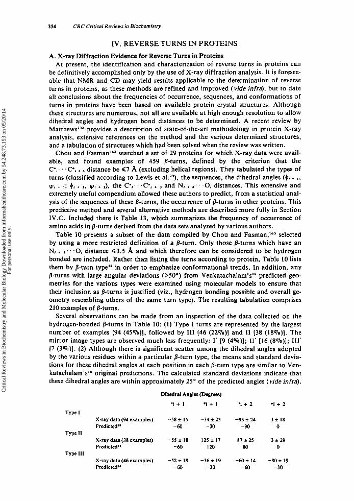
354 CRC Critical Reviews in Biochemistry
IV. R E V E R S E T U R N S I N P R O T E I N S
A. X-ray Diffraction Evidence for Reverse Turns in Proteins At present, the identification and characterization of reverse turns in proteins can
be definitively accomplished only by the use of X-ray diffraction analysis. It is foresee- able that NMR and CD may yield results applicable to the determination of reverse turns in proteins, as these methods are refined and improved (vide infra), but to date all conclusions about the frequencies of occurrence, sequences, and conformations of turns in proteins have been based on available protein crystal structures. Although these structures are numerous, not all are available at high enough resolution to allow dihedral angles and hydrogen bond distances to be determined. A recent review by Mat thewP provides a description of state-of-the-art methodology in protein X-ray analysis, extensive references on the method and the various determined structures, and a tabulation of structures which had been solved when the review was written.
Chou and F a ~ m a n ' ~ ~ searched a set of 29 proteins for which X-ray data were avail- able, and found examples of 459 /3-turns, defined by the criterion that the C',. . '(7, + distance be 97 A (excluding helical regions). They tabulated the types of turns (classified according to Lewis et al.13), the sequences, the dihedral angles (4, +
yi + I ; 4, + 1, y, + *), the C',. . acei + and N, + 3 * - -0, distances. This extensive and extremely useful compendium allowed these authors to predict, from a statistical anal- ysis of the sequences of these /%turns, the occurrence of /3-turns in other proteins. This predictive method and several alternative methods are described more fully in Section 1V.C. Included there is Table 13, which summarizes the frequency of occurrence of amino acids in /3-turns derived from the data sets analyzed by various authors.
Table 10 presents a subset of the data compiled by Chou and F a ~ m a n , " ~ selected by using a more restricted definition of a /3-turn. Only those /3-turns which have an N, + 3 - * '0, distance G3.5 A and which therefore can be considered to be hydrogen bonded are included. Rather than listing the turns according to protein, Table 10 lists them by /3-turn typea8 in order to emphasize conformational trends. In addition, any /3-turns with large angular deviations (>SO") from Venkatachalam's18 predicted geo- metries for the various types were examined using molecular models to ensure that their inclusion as /3-turns is justified (viz., hydrogen bonding possible and overall ge- ometry resembling others of the same turn type). The resulting tabulation comprises 210 examples of /3-turns.
Several observations can be made from an inspection of the data collected on the hydrogen-bonded /3-turns in Table 10: (1) Type 1 turns are represented by the largest number of examples (94 (45%)1, followed by 111 (46 (22Oro)I and I1 [38 (l8%)]. The mirror image types are observed much less frequently: I' [9 (4%)]; 11' [16 (SOro)]; 111' I7 (3Oro)l. (2) Although there is significant scatter among the dihedral angles adopted by the various residues within a particular /3-turn type, the means and standard devia- tions for these dihedral angles at each position in each /3-turn type are similar to Ven- katachalam's" original predictions. The calculated standard deviations indicate that these dihedral angles are within approximately 25" of the predicted angles (vide infra).
Dihedral An& (Degrees)
*i + 1 .i + I *i + 2 .i + 2
X-ray data (94 examples) -58 f I5 -34 f 23 -93 f 24 3 f 18 Predicted" -60 -30 -90 0
X-ray data(38 examples) -55 f 18 125 f 17 87 f 25 3 f 29 Predictedis -60 120 80 0
X-ray data (46 examples) -52 2 18 -36 f 19 -60 f 14 -30 f 19 Predicted" -60 - 30 -60 -30
Type I
Type I1
Type 111
Cri
tical
Rev
iew
s in
Bio
chem
istr
y an
d M
olec
ular
Bio
logy
Dow
nloa
ded
from
info
rmah
ealth
care
.com
by
54.2
48.7
3.15
3 on
05/
20/1
4Fo
r pe
rson
al u
se o
nly.
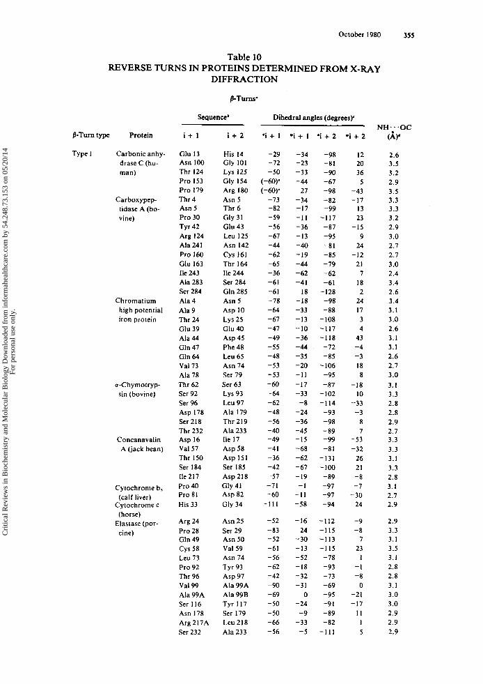
October 1980 355
Table 10
DIFFRACTION REVERSE TURNS IN PROTEINS DETERMINED FROM X-RAY
Sequence' Dihedral angles (degrees)' NH ... . OC
fl-Turn type Protein i + 1 i + 2 ' i + I . i + 1 * i + 2 . i + 2 (A)'
Type I Carbonic anhy- drase C (hu- man)
Carboxypep- tidase A (bo- vine)
Chrornatiurn high potential iron protein
o-Chyrnotryp- sin (bovine)
Concanavalin A (jack bean)
Cytochrorne b, (calf liver)
Cytochrorne c (horse)
Elastase (por- cine)
Glu 13 Asn 100 Thr I24 Pro 153 Pro 179 Thr 4 Asn 5 Pro 30 Tyr 42 Arg 124 Ala 241 Pro 160 Glu 163 Ile 243 Ala 283 Ser 284 Ala 4 Ala 9 Thr 24 Glu 39 Ala 44 Gln 47 Gln 64 Val 73 Ala 78 Thr 62 Ser 92 Ser 96 Asp 178 Ser 218 Thr 232 Asp 16 Val 57 Thr 150 Ser 184 Ile 217 Pro 40 Pro 81 His 33
Arg 24 Pro 28 Gln 49 Cys 58 Leu 73 Pro 92 Thr 96 Val 99 Ala 99A Ser I16 Asn 178 Arg217A Ser 232
His 14 Gly 101 Lys 125 Gly 154 Arg 180 Asn 5 Thr 6 Gly 31 Glu 43 Leu 125 Asn 142 Cys 161 Thr 164 Ile 244 Ser 284 Gln 285 Asn 5 Asp 10 Lys 25 Glu 40 Asp 45 Phe 48 Leu 65 Asn 74 Ser 79 Ser 63 Lys 93 Leu 97 Ala 179 Thr 219 Ala 233 Ile 17 Asp 58 Asp 151 Ser 185 Asp218 Gly 41 Asp 82 Gly 34
Asn 25 Ser 29 Asn 50 Val 59 Asn 74 Tyr 93 Asp 97 Ala 99A Ala 99B Tyr 117 Ser 179 Leu 218 Ala 233
-29 -72 -50
(-W' (-60)'
-73 -82 -59 -56 -67 -44 - 62 -65 -36 -61 -61 -78 -64 -67 -47 -49 -55 -48 -53 -53 -60 -64 -62 -48 -56 - 40 -49 -4 I -36 -42 -57 -71 -60
- 1 1 1
-52 -83 -52 -61 -56 -62 -42 -90 -69 -50 - 50 -66 -56
-34 -23 -33 -44
27 -34 -17 - 1 1 -36 -13 -40 -19 -44 -62 -4 I
18 -18 -33 -13 -10 -36 -44 -35 - 20 - 1 1 -17 -33
-8 -24 -36 -45 -I5 -68 -62 -67 -19
- 1 - 1 I -58
-16 24
-30 -13 -52 -18 -32 -3 1
0 -24 -9
-33 -5
-98 -81 -90 -67 -98 -82 - 99
-117 -87 -95 -81 -85 -79 -62 -61
-128 -98 -88
-108 - 1 17 -118 -72 -85
-106 -95 -87
-102 -114 -93 -98 -89 - 99 -8 1
-131 -100 -89 -97 -97 -94
- 1 12 -115 -113 -115
-78 -93 -73 -69 -95 -91 -89 -82
- 1 1 1
12 20 36 5
-43 -17
13 23
-I5 9
24 -12
21 7
18 2
24 17 3 4
43 -4 -3 18 8
-18 10
-33 -3
8 7
-53 -32
26 21 -8 -7
-30 24
-9
7 23
1 - 1 -8
0 -21 -17
1 1 1 5
-a
2.6 3.5 3.2 2.9 3.5 3.3 3.3 3.2 2.9 3.0 2.7 2.1 3.0 2.4 3.4 2.6 3.4 3.1 3.0 2.6 3.1 3. I 2.6 2.7 3.0 3. I 3.3 2.8 2.8 2.9 2.7 3.3 3.3 3.1 3.3 2.8 3.1 2.7 2.9
2.9 3.3 3. I 3.5 3.1 2.8 2.8 3.1 3.0 3 .O 2.9 2.9 2.9
Cri
tical
Rev
iew
s in
Bio
chem
istr
y an
d M
olec
ular
Bio
logy
Dow
nloa
ded
from
info
rmah
ealth
care
.com
by
54.2
48.7
3.15
3 on
05/
20/1
4Fo
r pe
rson
al u
se o
nly.
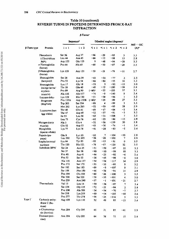
356 CRC Critical Reviews in Biochemistry
Table 10 (continued)
DIFFRACTION
P-Turns’
REVERSE TURNS IN PROTEINS DETERMINED FROM X-RAY
Sequence‘ Dihedral angles (degrees)’
P-Turn type Protein
Flaxodoxin ( Clostridium Mp)
u-Hemoglobin (horse)
/3-Hemoglobin (horse) Hemoglobin (lamprey)
Hemoglobin (midge larval erythro- cruorin)
Lactate dehy- drogenase (dogfish)
Lysozyrne (hen egg-white)
Myogen (carp muscle)
Myoglobin (sperm whale)
Papain (pa-
Staphylococcal nuclease Subtilisin BPN’
paya)
Thermolysin
Type I’ Carbonic anhy- drase C (hu- man)
o-Chymotryp- sin (bovine)
Elastase (por- cine)
i + l
Ser 36 Leu 44 Asp 122 Pro 44
Lys 120
Ser 28 Pro 53 Lys 17 Thr 39 Pro 89 Ala 116 Lys 129 Pro 132A Trp 203 Met 262 Thr 40 Thr 47 Ile 55 Leu 75 Ala 3 Gln 52 Lys 77
Gln 9 Leu 202 Lys 84 Thr 120 Ser 24 Ser 37 Pro 40, Pro 52 Ala 116 Pro 172 Ser 182 Ser 188 Pro 194 Thr 220 Pro 239 Val 13 Ser 134 Pro 208 Ser 218 Pro 277 Asp 109
Asn 204
Asn 204
i + 2
Asp 37 Asn 45 Gly 123 His 45
Asp 121
Asn 29 Lys 54 Gly 18 Gln 40 Arg 90 Ala I17 His 130 Asp 132B Ser 204 Lys 263 Gln 41 Asp 48 Leu 56 Cys 76 Gly 4 Asp 53 Lys 78
Lys 10 Tyr 203 Tyr E5 His I21 Asn 25 Ser 38 Asp 41 Ser 53 Asn 117 Ser 173 Ser 183 Phe 189 Glu 195 Ser 221 Asn 240 Leu 14 Gly 135 Ala 209 Lys219 Thr 278 Lys 110
Gly 205
Gly 205
*i + 1
-66 -66
9 -68
-59
-43 -60 -53 -48
(-60)’ -74 -5 1
(-60)’ -80 -52 -69 -52 -62 -62 -32 -45 -41
-63 -36 -83 -79 -51 - 90 -44 -38 - 54 -53 - 69 -46 -69 -59 -57 -59 -72 -54 -60 -58
52
45
64
ri + 1
- 29 -12 -48 -10
-21
-54 -82
5 -I5 -22
-9 -38 -29
4 -50 -17 -37 -3 1 -23 -56 -76 - 20
7 -28 -13 -17 -59 -10 -23 -43 -24 -10 -9
-38 -26 -10
- I -36 -21 -34 -14 -25
49
31
38
‘i + 2
-95 -98 -94 -87
-79
-77 -55 -92
-100 -103 -140 -96 -89 -99 -95 -84 -81
-108 -86
-139 -74 -83
-104 -102 -91
-120 -87 -99 -92 -98
-117 -95
-107 -9 I
-100 -101 -101
-87 - 84 -70
-105 - I 16
85
85
75
ri + 2
3 13
-28 -28
-11
2 31
-15 -26
31 9 1
27 3
28 -3 -3
7 -15
30 -1 -5
-10 7 0
32 22 18
-6 -6 34 9 9
11 3 4
-21 7 2
-7 -I8
5 -15
-4 1
I5
NH...OC (4‘
3.3 2.8 2.2 3.5
2.7
2.3 3.3 3.0 2.6 3. I 3.4 3.2 3.3 3.3 2.9 3.4 3.2 3.3 2.9 2.8 3.4 2.4
2.8 2.9 3.5 3.5 3.2 3.3 2.4 3.0 2.9 3.2 3.1 2.9 3.5 3.0 2.7 3.4 2.8 2.7 2.6 3.5 3.4
2.9
2.9
Cri
tical
Rev
iew
s in
Bio
chem
istr
y an
d M
olec
ular
Bio
logy
Dow
nloa
ded
from
info
rmah
ealth
care
.com
by
54.2
48.7
3.15
3 on
05/
20/1
4Fo
r pe
rson
al u
se o
nly.
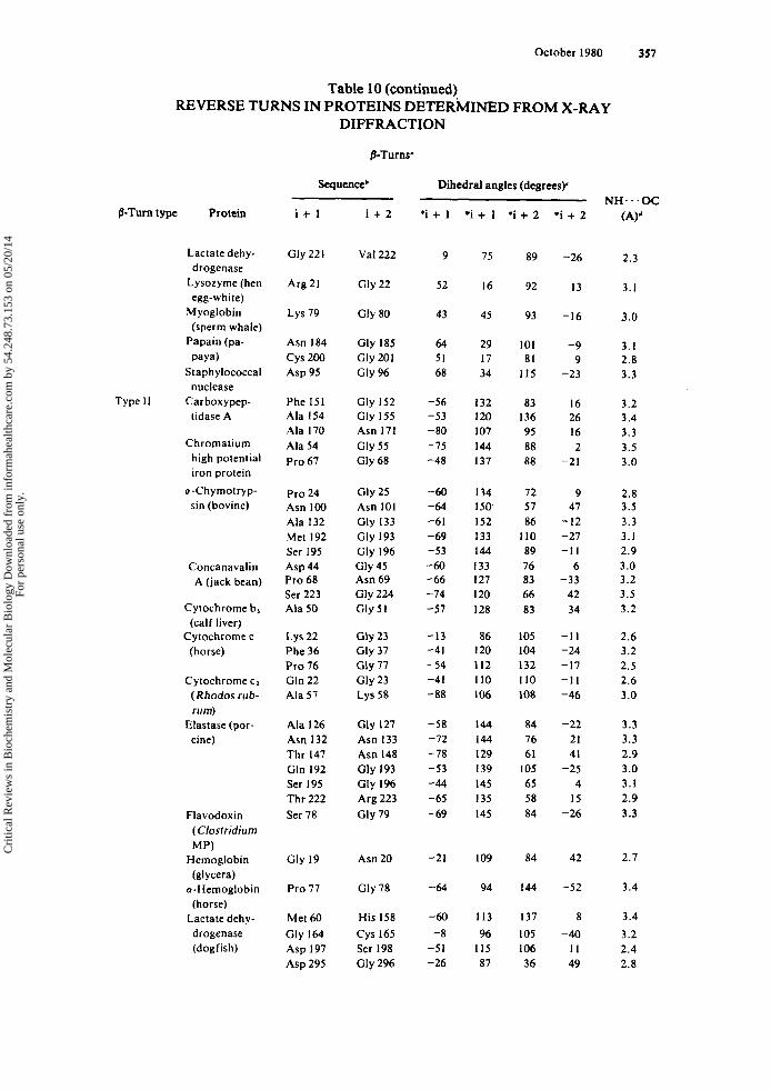
October 1980 357
Table 10 (continued)
DIFFRACTION
&Turns'
REVERSE TURNS IN PROTEINS DETERMINED FROM X-RAY
Sequence' Dihedral angles (degrees)'
B-Turn type Protein
Lactate dehy- drogenase
Lysozyme (hen egg- whi te)
Myoglobin (sperm whale)
Papain (pa-
Staphylococcal paya)
nuclease
tidase A Type 11 Carboxypep-
Chromatiurn high potential iron protein
u-Chymotryp- sin (bovine)
Concanavalin A (jack bean)
Cytochrome b, (calf liver)
Cytochrome c (horse)
Cytochrome c1 (Rhodos rub- rum)
cine) Elastase (por-
Flavodoxin ( Clostridium MP)
Hemoglobin (glycera)
u-Hemoglobin (horse)
Lactate dehy- drogenase (dogfish)
i + l
Gly 221
Arg 21
Lys 79
Asn 184
Asp 95
Phe 151 Ala 154 Ala 170 Ala 54 Pro 67
Pro 24 Asn 100 Ala 132 Met 192 Ser 195 Asp 44 Pro 68 Ser 223 Ala 50
cys 200
Lys 22 Phe 36 Pro 76 Gln 22 Ala 51
Ala 126 Asn 132 Thr 147 Gln 192 Ser 195 Thr 222 Ser 78
Gly 19
Pro 17
Met 60 Gly 164 Asp 197 Asp 295
i + 2
Val 222
Cly 22
Gly 80
Gly 185 Gly 201 Gly 96
Gly 152 Gly 155 Asn 171 Gly 55 Gly 68
Gly 25 Asn 101 Gly 133 Gly 193 Gly 196
Asn 69 Gly 224
Gly 45
Gly 51
Gly 23 Gly 37 Gly 77 Gly 23 Lys 58
Gly 127 Asn 133 Asn 148 Gly 193 Gly 196 Arg 223 Gly 19
Asn 20
Gly 78
His 158 Cys 165 Ser 198 Gly 296
*i + 1
9
52
43
64 51 68
-56 -53 - 80 -75 -48
-60 -64 -61 -69 -53 -60 -66 - 74 -57
-13 -41 - 54 -41 -88
-58 -72 - 78 -53 -44 -65 - 69
-21
-64
-60 -8
- 5 1 -26
.i + 1
75
16
45
29 17 34
132 I20 107 144 137
I34 I50 152 I33 144 I33 I27 120 I28
86 I20 112 110 I06
144 144 129 I39 145 I35 145
109
94
I13 96
1 I5 87
'i + 2
89
92
93
101 81
I I5
83 136 95 88 88
12 57 86 I10 89 76 83 66 83
105 104 I32 I10 108
84 76 61
105 65 58 84
84
144
I37 105 106 36
'i + 2
-26
13
-16
-9 9
-23
16 26 16 2
-21
9 41
-12 -27 - I 1
6 -33
42 34
- 1 1 -24 -17 -1 I -46
-22 21 41
-25 4
I5 - 26
42
-52
8 -40
I 1 49
NH. , .OC (A)'
2.3
3.1
3.0
3. I 2.8 3.3
3.2 3.4 3.3 3.5 3.0
2.8 3.5 3.3 3. I 2.9 3.0 3.2 3.5 3.2
2.6 3.2 2.5 2.6 3.0
3.3 3.3 2.9 3.0 3.1 2.9 3.3
2.7
3.4
3.4 3.2 2.4 2.8
Cri
tical
Rev
iew
s in
Bio
chem
istr
y an
d M
olec
ular
Bio
logy
Dow
nloa
ded
from
info
rmah
ealth
care
.com
by
54.2
48.7
3.15
3 on
05/
20/1
4Fo
r pe
rson
al u
se o
nly.
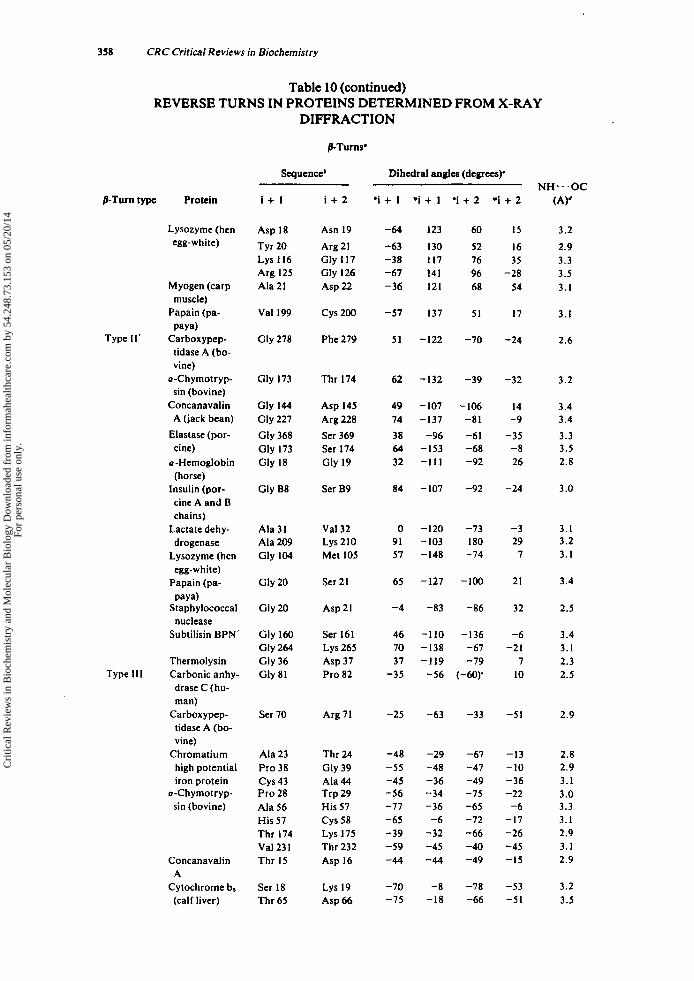
358 CRC Critical Reviews in Biochemistry
Table 10 (continued)
DIFFRACTION
PTurns'
REVERSE TURNS IN PROTEINS DETERMINED FROM X-RAY
Sequence' Dihedral angles (degrees)"
P-Turn type Protein
Lysozyme (hen egg-w hi te)
Myogen (carp
Papain (pa- muscle)
paya)
tidase A (bo- vine)
u-Ch ymotryp- sin (bovine)
Concanavalin A (jack bean)
Elastase (por- cine)
a-Hemoglobin (horse)
Insulin (por- cine A and B chains)
Lactate dehy- drogenase
Lysozyme (hen egg- w h i te)
Papain (pa-
Staphylococcal
Subtilisin BPI"
Type 11' Carboxypep-
paya)
nuclease
Thermolysin
drase C (hu- man)
Carboxypep- tidase A (bo- vine)
Chromatium high potential iron protein
u-Chymotryp- sin (bovine)
Type 111 Carbonic anhy-
Concanavalin
Cytochrome b, A
(calf liver)
i + 1
Asp 18 Tyr 20 Lys I16 Arg 125 Ala 21
Val 199
Gly 278
Gly 173
Gly 144 Gly 227 Gly 368 Gly 173 Gly 18
Gly B8
Ala 31 Ala 209 Gly 104
Gly 20
Gly 20
Gly 160 Gly 264 Gly 36 Gly 81
Ser 70
Ala 23 Pro 38 c y s 43 Pro 28 Ala 56 His 57 Thr 174 Val 231 Thr 15
Ser 18 Thr 65
i + 2
Asn 19 Arg 21 Gly I17 Gly 126 Asp 22
c y s 200
Phe 279
Thr 174
Asp 145 Arg 228 Ser 369 Ser 174 Gly 19
Ser B9
Val 32
Met I05
Ser 21
Asp 21
Ser 161 Lys 265 Asp 37 Pro 82
Lys 210
Arg 71
Thr 24
Ala 44 Trp 29 His 57 Cys 58 Lys 175 Thr 232 Asp 16
Lys 19 Asp 66
Gly 39
'i + 1
-64 -63 -38 -67 -36
-57
51
62
49 74 38 64 32
84
0 91 57
65
-4
46 70 31
-35
-25
-48 -55 -45 - 56 -77 -65 -39 -59 -44
-70 -75
. i + 1 ' i + 2 . i + 2
I23
130 117 141 I21
137
-122
-132
-107 -137
-96 -153 -111
-107
-120 -103 -148
-127
-83
- 1 10 -138 -119
-56
-63
-29 -48 -36 -34 -36
-6 -32 -45 -44
-8 -18
60 52 76 96 68
51
-70
-39
-106 -81 -61 -68 -92
-92
-73 I80 -74
-100
-86
-136 -67 -79
(-60)'
-33
-67 -47 -49 -75 -65 -72 -66 -40 -49
-78 -66
IS 16 35
-28 54
17
-24
-32
14 -9
-35 -8 26
-24
-3 29
7
21
32
-6 -21
7 10
-5 1
-13 -10 -36 -22 -6
-17 -26 -45 -I5
-53 -5 1
NH. . .OC (4'
3.2
2.9 3.3 3.5 3. I
3. I
2.6
3.2
3.4 3.4 3.3 3.5 2.8
3 .O
3. I 3.2 3. I
3.4
2.5
3.4 3. I 2.3 2.5
2.9
2.8 2.9 3.1 3.0 3.3 3.1 2.9 3.1 2.9
3.2 3.5
Cri
tical
Rev
iew
s in
Bio
chem
istr
y an
d M
olec
ular
Bio
logy
Dow
nloa
ded
from
info
rmah
ealth
care
.com
by
54.2
48.7
3.15
3 on
05/
20/1
4Fo
r pe
rson
al u
se o
nly.

October 1980 359
Table 10 (continued)
DIFFRACTION
PTurns'
REVERSE TURNS IN PROTEINS DETERMINED FROM X-RAY
/3-Turn type Protein
Elastase (por- cine)
Flavodoxin ( CIostridium M P)
u-Hemoglobin (horse)
/3-Hemoglobin (horse)
Hemoglobin (midge larva/ erythro- cruorin)
Lactate dehy- drogenase (dogfish)
Lysozyme (hen egg-white)
Myogen (carp muscle)
Myoglobin
Papain (pa- (sperm whale)
paya)
Ribonuclease S
Subtilisin BPN' (bovine)
Thermolysin
Type 111' Concanavalin A (jack bean)
Cytochrome b, (calf liver)
Flavodoxin ( Clostridiurn MP)
Lysozyme (hen egg-white)
Thcrmolysin
i + l
Ala 56 His 57 Ser 170A Val 35 Ile 40
Pro 114
Asp 43 Lys 82 Pro 32
Val 87 Ser 184 Cys 185 Lys 213 Ser 238 Pro 309 Pro 70 Val 99 Met 105 Asn 106 Thr 36 Ala 72 Ser 91 Asp 44 Lys 47 Arg 58 Arg 98 Ser 196 Lys 66
Gly 169 Ser 260 Ser 65 Gly 160 Ala 209 Leu 230
His 26
Gly 57
Asn 37
Lys 45 Asp 226 Asn 227
i + 2
His 57 Cys 58 Ser 170B Ser 36 Asp 41
Asn 115
Ser 44 Gly 83 Ser 33
Ser 88 Cys 185 Leu 186 Asp 214 Asn 239 Asp 310 Gly 71 Ser 100 Asn 106 Ala 107 Ser 37 Asp 73 Asp 92 Arg 45 His 48 Arg 59 Glu 99 Tyr 197 Asn 67
Lys 170 Phe 261 Tyr 66 Ser 161 Lys 210 Gly 23 1
Tyr 27
Asp 58
Phe 38
Tyr 46 Asn 227 Gly 228
*i + 1
-34 -41 -72 -53 -63
-67
-69 -23
(-60)'
-64 2
-40 -57 -24
(-60)' -53 -62 -74 -72 -34 -36 -40 -6 1 -38 -92 -47 -62 -35
-64 -30 -54 -67 -70
20
35
- I
57
26 34 44
. i + 1 * i + 2 * i + 2
-45 -52 -25 -33 -55
-37
7 -29 -19
-19 -58 -74 -32 -75 -35 -40 -25
7 -16 -68 -37 -66 -42 -53 -14 -50 -38 -42
-44 -43 -25 -44
-7 22
57
71
39
35 58 52
-4 1 -6 I -77 -66 -17
-74
-54 -79 -55
-72 -40 -65 -44 -42 -58 -66 -76 -72 -66 -44 -76 -49 -67 -4 I -68 -65 -62 -59
-66 -78 -7 I -59 -55
53
59
66
59
74 44 83
-52 -13 -23 -29 -50
-36
-21 -63 -59
-28 -74 -55 -60 -38 -36 -30 -23 -16 -16 -36 -18 - 1 1
-9 -35 -20 -36 -53
6
-18 -19 -23 -38 -35
36
4
63
19
29 52 44
Sequence' Dihedral angles (degrees)' NH...OC
(A)'
3.2 3 .O 2.9 2.6 3.1
3.3
3.2 2.6 3.3
3.0 2.9 3.5 2.5 3.4 2.9 3.3 3.3 3.4 3.1 2.6 2.6 2.8 3.2 2.5 3.4 2.9 3.1 2.5
3.3 3.2 2.8 3.1 3.3 2.9
2.3
2.4
3.1
2.9 3.4 2.3
Cri
tical
Rev
iew
s in
Bio
chem
istr
y an
d M
olec
ular
Bio
logy
Dow
nloa
ded
from
info
rmah
ealth
care
.com
by
54.2
48.7
3.15
3 on
05/
20/1
4Fo
r pe
rson
al u
se o
nly.
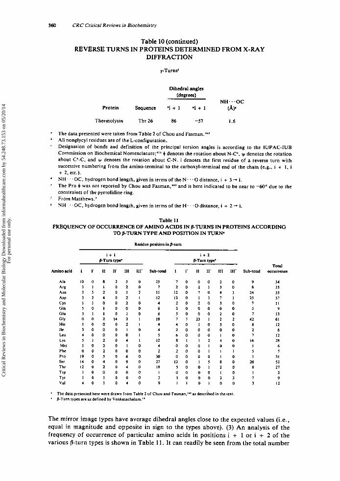
360 CRC Critical Reviews in Biochemistry
Table 10 (continued)
DIFFRACTION REVERSE TURNS IN PROTEINS DETERMINED FROM X-RAY
Dihedral angles (degrees)
NH.. .OC Protein Sequence 'i + 1 .i + 1 (AP
Thermolysin Thr 26 86 -51 1.6
The data presented were taken from Table 2 of Chou and Fa~rnan . '~ ' All nonglycyl residues are of the L-configuration. Designation of bonds and definition of the principal torsion angles is according to the IUPAC-IUB Commission on Biochemical Nomenclature;"' 0 denotes the rotation about N-C', v, denotes the rotation about C'-C, and w denotes the rotation about C-N. i denotes the first residue of a reverse turn with successive numbering from the amino-terminal to the carboxyl-terminal end of the chain (e.g., i + I , i + 2, etc.). NH . . ' OC, hydrogen bond length. given in terms of the N . . . O distance, i + 3 -. i. The Pro 0 was not reported by Chou and Fasman,'63 and is here indicated to be near to -60" due to the constraints of the pyrrolidine ring.
NH ' .OC. hydrogen bond length, given in terms of the H . . . O distance, i + 2 -. i.
' '
' From Matthews.'
Table 11
TO BTURN TYPE AND POSITION IN TURN' FREQUENCY OF OCCURRENCE OF AMINO ACIDS IN /3-TURNS IN PROTEINS ACCORDING
Residue position in P-turn
i + I i + 2 pTm type' &Turn type'
Aminoacid 1 I ' I1 11' 111 111' Sub-total I I' I1 11' 111 Ill' Sub-total occurrence
Ala Arg Asn ASP CYS Gln Clu G ~ Y His Ile Leu LYS Met Phe Pro Ser Thr Trp Tyr Val
1 0 0 8 2 5 0 25 7 0 0 0 2 0 9 3 1 1 0 2 0 7 2 0 2 1 3 0 8 3 3 2 0 1 2 I 1 1 2 0 7 0 4 I 24 3 2 4 0 2 I I2 1 3 0 1 3 7 I 25 I I O O 2 0 4 2 0 2 0 3 0 7 5 0 1 0 0 0 6 3 0 0 0 0 0 3 3 1 1 0 1 0 6 5 0 0 0 2 0 7 0 0 2 1 4 2 I 19 7 7 2 3 1 2 2 42 I 0 0 0 2 I 4 4 0 1 0 3 0 8 3 0 0 0 I O 4 2 0 0 0 0 0 2 4 0 0 0 0 I 5 6 0 0 0 I 0 7 5 1 2 0 4 I 12 8 1 1 2 4 0 16 I 0 2 0 1 0 4 0 0 0 1 0 0 I 0 0 2 0 0 0 2 2 0 0 1 I I 5 I 9 0 5 0 6 0 30 0 0 0 0 I 0 I 1 4 0 4 0 9 0 27 I 2 0 1 5 8 0 26 I 2 0 2 0 4 0 18 5 0 0 1 2 0 8 I 0 0 0 0 0 I 0 0 0 0 I 0 I 1 0 1 0 0 0 2 3 0 0 0 2 2 7 4 0 1 0 4 0 9 I 1 0 1 0 0 3
' ' The data presented here were drawn from Table 2 of Chou and Faman.'" as described in the text.
@-Turn types are as defined by Venkatachalam."
34 I5 35 37 I I 9
13 61 12 6
I2 28 6 7
31 53 27
2 9
12
The mirror image types have average dihedral angles close to the expected values (i.e., equal in magnitude and opposite in sign to the types above). (3) An analysis of the frequency of occurrence of particular amino acids in positions i + 1 or i + 2 of the various fl-turn types is shown in Table 11. It can readily be seen from the total number
Cri
tical
Rev
iew
s in
Bio
chem
istr
y an
d M
olec
ular
Bio
logy
Dow
nloa
ded
from
info
rmah
ealth
care
.com
by
54.2
48.7
3.15
3 on
05/
20/1
4Fo
r pe
rson
al u
se o
nly.

October 1980 361
Table 12 RECURRENT DIPEPTIDE SEQUENCES AT THE i +
1 ’* AND i + 2‘* POSITIONS OF P-TURNS IN PROTEINS
Sequence and Occurrence within various fl-turn p-turn position types*
i + 6 i + 2 Occurrence I 1’ 11 11’ 111 111’ Total
Pro Gly Ala Gly Asn Gly Gly Ser Lys Gly Ser Gly Thr Asp Ala Asn Gly Asp Pro Asp Pro Ser Ser Ser Ala Lys Asp Gly Pro Asn Pro Glu Ser Lys Ser Tyr Ser Asn Thr Lys Thr Ser Val Ser
7 6 S 5 5 5 5 4 4 4 4 4 3 3 3 3 3 3 3 3 3 3
2 0 4 0 1 0 1 0 s 0 0 0 1 3 0 0 0 1 0 0 0 5 0 0 1 1 2 0 1 0 1 0 4 0 0 0 3 0 0 0 2 0 3 0 1 0 0 0 0 0 0 3 0 I 3 0 0 0 I 0 3 0 0 0 1 0 3 0 0 0 1 0 0 0 1 1 1 0 0 1 2 0 0 0 1 0 1 0 1 0 2 0 0 0 1 0 2 0 0 0 1 0 1 0 0 0 2 0 2 0 0 0 1 0 2 0 0 0 1 0 2 0 0 0 1 0 0 0 0 0 3 0
* /3-Turn types are as defined by Venkatachalam.”
of times each amino acid occurs that Gly (61 examples), Ser (53), Asp (37), Asn (35). Ala (34), Pro (31), Lys (28), and Thr (27) are frequent residues in /3-turns. Many in- sights into this general finding are provided by the breakdown according to position and /%turn type in Table 1 1 . For example,
1 . The i + lrh position of the type 11’ p-turns is occupied 14 out of 16 times by Gly and the remaining two by Ala.
2. Pro occurs 30 times in the i + I r h position and only once in the i + 2’h position. In addition, Pro is seen most often in type I turns.
3. The i + 2Ih position of type I1 /3-turns is occupied 23 out of 38 times by Gly. 4. Eight residues have high frequencies of occurrence in the 1 + position: Pro
(30), Ser (27), Ala (25), Gly (19). Thr (19), Lys (12), Asp (12). and Asn (11). Among these, Asn and Asp occur in all turn types except 11’. Gly occurs fre- quently in type II’, and the other residues are predorninatly found in types I , 11, or 111. Five residues are particularly frequent in the i + 21h position: Gly (42 examples), Ser (26), Asp (25), Asn (24), and Lys (16). Among these residues the nonglycyl residues are encountered most often in type I and 111, although Ser is seen five times in type 11’ 0-turns.
5 .
Table 12 presents the 22 dipeptide sequences which recur three or more times in the
Cri
tical
Rev
iew
s in
Bio
chem
istr
y an
d M
olec
ular
Bio
logy
Dow
nloa
ded
from
info
rmah
ealth
care
.com
by
54.2
48.7
3.15
3 on
05/
20/1
4Fo
r pe
rson
al u
se o
nly.
362 CRC Critical Reviews in Biochemistry
set of 210 /.?-turns. Both their overall frequencies of occurrence and a compilation of the occurrences in the various /3-turn types are shown. Only eight out of the 88 /3-turns deviate from the expected P-turn types predicted from stereochemical consideration (see Section 11), and three of these involve Asn-Gly sequences which occur in type I‘ 0-turns.
Multiple factors, including steric requirements, hydrophilicity, and conf ormation required for activity or recognition, contribute to the occurrence of the particular res- idues or sequences in the observed P-turns. Hence, it is difficult to speculate about rationales for the trends and frequencies presented. Nonetheless, it is tempting to as- cribe to Gly the role of a “pseudo-D” residue, which may readily occur in the i + l ‘h position of type 11’ p-turns or the i + 2’k position of type I1 /%turns, due to its lack of a side chain. Beyond this suggestion, and the previously recognized prevalence of polar residues,6 too many factors are intertwined to warrant definitive conclusions at this time.
Only one example of a protein y-turn has been reported to date, namely, that de- scribed by Matthews’ from his X-ray structure analysis of thermolysin (Ser’S-Thr26-
The lack of other reported examples may derive from the fact that appropriate searches have not been carried out, rather than from the rarity of y-turns in proteins. However, a definitive search is necessary in order to establish the frequency of occurrence of y- turns. Interestingly, the one turn reported does not follow the expected conformational preference for the i + lrh residue of a y-turn, since it is an example of an L-residue occurring in a y-turn, and not in an inverse y-turn (see Section I1 ).
TyrZ7: 0’s = 148”, ~ 2 s = 92”; 0’6 = 86’, Was = - 5 7 O ; 4 2 7 = -114”, 1c)27 = 148”).
B. Circular Dichroism (CD) and Nuclear Magnetic Resonance (NMR) Evidence for Reverse Turns in Proteins
Although few studies using CD1S1.1s2 and none using NMR to assign reverse turn structures in proteins have been reported, both of these methods show promise of making significant contributions to this field in the future.
The approach to conformational analysis of proteins based on CD has been briefly mentioned in Section 1II.C. Such an analysis depends upon a breakdown of the ob- served CD spectrum for a protein into contributions from the various types of second- ary structure (a-helix, 0-structure, reverse turns, and “random coil”).’4s Woody’sI9 CD calculations for P-turns unfortunately reveal that there is not a unique CD which can be said to be characteristic of a /3-turn structure (see Tables 7 and 8). Hence, it is difficult to deduce unequivocally the presence of /3-turns in proteins from CD.
NMR analyses of protein conformation have become feasible due to new methods of spectral simplification and to improvements in instrumentation. No studies thus far have been specifically concerned with the elucidation of turns, but the following ex- amples illustrate state-of-the-art techniques which provide details of protein structures in solution, and may be applied to turn regions: (1) high field, high resolution ‘H NMR of well-separated resonance^;'^^-'^^ (2) paramagnetic p r ~ b e s ’ ~ ” ~ ’ ~ ~ or lanthanide shift reagent^;"^ and (3) transient NOE analysis.24’
C. Correlative Predictive Methods: Evidence for Reverse Turns in Proteins Three reviews dealing exhaustively with the methods for empirical prediction of pro-
tein structure and their applications have been p~blished.’~’-’~~ Although these reviews have discussed the prediction of various types of protein secondary structure (e.g., (I- helices, /3-sheets, and reverse turns), only the application and success of these methods for the prediction of reverse turns will be discussed in this section.
In all the correlative methods, the first stage is the development of sets of frequencies
Cri
tical
Rev
iew
s in
Bio
chem
istr
y an
d M
olec
ular
Bio
logy
Dow
nloa
ded
from
info
rmah
ealth
care
.com
by
54.2
48.7
3.15
3 on
05/
20/1
4Fo
r pe
rson
al u
se o
nly.
October 1980 363
of occurrence or equilibrium constants, which describe the likelihood of adoption of specific secondary structures by the 20 common amino acids. Such a development rests on the statistical analysis of protein crystallographic data or on semiempirical physi- cochemical parameters associated with the amino acids. The predictive capacities of these methods are limited in their accuracy by (1) the extent of the input data (i.e., the number of proteins or the number of experimental measurements), (2) the correct- ness of conformational assignments, and (3) the appropriateness of the data set to the protein conformation being predicted. For example, the interpretation of protein crys- tallographic data suffers from potential distortions due to the subjective nature of the analysis. In addition, the criteria for the identification of the various structural features have differed among studies.
Early attempts to determine the conformational preferences of amino acids for pro- tein /3-turns relied on X-ray crystallographic data which identified p-turn regions by specifying a maximum distance between the C* atoms belonging to the ith and i + 3 1 h
residues (e.g., (7 A;8.23.24s.246 s4.8-4.9 A;6 and c5.7 As) or by Venkatachalam’sL8 cri- teria.I4’ Rose and suggested an objective method for finding turns from pro- tein X-ray structures based on a geometric analysis which involves searching for local minima in the radius of curvature of the protein chain.
The second stage is the application of the frequencies or equilibrium constants using a specific mathematical approach to elucidate the secondary structure of a protein. In one type of analysis exemplified by the approach of Chou and F a ~ m a n , ~ ~ ~ . ’ ~ ~ the fre- quencies were analyzed statistically using groups of four contiguous residues as basic units. The premise of the Chou-Fasman method is that the predictive accuracy will be sufficient using tetrapeptide sequences. Initially, their data set was based on a statisti- cal analysis of 15 proteins.1s0 Later they added 14 more proteins to their As was the case for the /3-turn criteria proposed by Lewis et al.’.’’ Chou and FasmanL6’ consid- ered any four-residue sequence whose C‘, to Coi + distance was (7 A and whose sequence was not included in an helical region to be a /3-turn. Chou and F a ~ m a n ’ ~ ’ also observed that hydrophobic residues occur more frequently than hydrophilic resi- dues in the regions immediately adjacent to /3-turns and that these regions often consist of p-sheets and/or a-helices, as was also found by nag an^.'^' Using their algorithm, Chou and F a ~ m a n ’ ~ ~ predicted 78% of the /3-turns in 29 proteins (i.e., the same pro- teins used to derive these frequencies initially) to within f 2 residues in all conforma- tions.
The Chou and F a ~ m a n ’ ~ ~ . ’ ~ ’ . ’ ~ ‘ ~ ’ ~ ~ correlative method is the most widely employed procedure for predicting the occurrence of reverse turn conformations in proteins.2S’ Its widespread use has resulted primarily from the easy application of its predictive rules and the advantage that little mathematical skill is required.
Analyses by other statistical methods have also been applied to X-ray crystallo- graphic data from larger sets of proteins. NaganoZs3 analyzed conformational data from 95 proteins belonging to 13 homologous families and correctly predicted the lo- cation of 64.4% of the /3-turns in seven proteins not included in his original data set. His data also indicated that the localization of /3-turns in proteins involves interactions other than short-~ange.’~’ In fact, when interactions between contiguous a-helices and /3-sheets were included in the statistical analysis, 71 Vo of the /3-turns were then correctly predicted. 2s1
Burgess et al.14s introduced a nonapeptide predictive algorithm, for which they con- sidered the frequencies of occurrence of /3-turn residues in a set of eight proteins, to predict the location of p-turns in 13 additional proteins. This method also indicated that residues adjacent to /3-turns contributed to the localization of these structures.
Levitt and GreerZs4 introduced an automated procedure which assigns secondary
Cri
tical
Rev
iew
s in
Bio
chem
istr
y an
d M
olec
ular
Bio
logy
Dow
nloa
ded
from
info
rmah
ealth
care
.com
by
54.2
48.7
3.15
3 on
05/
20/1
4Fo
r pe
rson
al u
se o
nly.
364 CRC Critical Reviews in Biochemistry
structure within a protein on the basis of local conformation and the pattern of hydro- gen bonding. In a subsequent paper, L e ~ i t t ~ ~ ~ analyzed these assignments to yield fre- quencies of occurrence of the twenty naturally occurring amino acids within reverse turns. In contrast to the results of Chou and F a ~ m a n , ’ ~ ~ . ’ ~ ~ where a given amino acid preferred more than one secondary structure (e.g., Tyr in reverse turns and /3-sheets), the Levitt analysis allows the convenient compartmentalization of each individual res- idue into a unique conformational preference. Furthermore, Gln, Lys, Tyr, Thr, Gly, and Arg were found to be indifferent to their location in a reverse turn conformation; while Ala, Leu, Met, His, Val, Ile, Phe, Cys, and Arg were reverse turn-breaking amino
An alternative approach to the mathematical analysis of frequencies or equilibrium constants of the individual amino acids relies on statistical mechanical methods such as those applied by Tanaka and S~heraga .~” They have derived a four-state (i.e., a- helix, coil, /3-turn, and extended structure), one-dimensional, short-range (Ising) model to predict the reverse turn conformations in proteins. Conformational sequence prob- abilities for finding a given residue and for observing two successive residues in a re- verse turn conformation were calculated for 23 proteins. Their results predicted cor- rectly 219 reverse turn regions out of 372 turns observed by X-ray analysis of the same set of proteins. Since they did not present data to indicate their degree of overpredic- tion, it is not straightforward to compare their results to the other correlative methods. Various other approaches to the prediction of turns differing in some aspect(s) from those described above have been proposed but not extensively applied.
Kuntz6 first observed that hydrophilic residues occur frequently within /%turns and suggested that this finding might be used to predict the occurrence of these structures, using a simple compositional rule. Subsequently, Rose and Wetlaufer25* demonstrated an approximate linear relationship between the number of /3-turns in a globular protein and its molecular weight, and they also suggested that local juxtapositioned sequences of amino acids are the major determinant of p-turn formation. This idea was extended by Rosezs9 in his prediction method based on hydrophobicity. A similar notion was applied to the nucleation of protein folding as initiated by hydrophobic contacts.’60
An information theory approach, based on that introduced by Robson and Pain,z6’ was utilized in a p-turn predicting scheme by Maxfield and Scheraga.262 D e n i ~ o v ’ ~ ~ used another method involving pattern recognition to predict the occurrence, but not the location, of turns.
Finkelstein and P t i t ~ y n ’ ~ ~ coupled a statistical mechanical analysis with a thermo- dynamic semiempirical calculation of the equilibrium constants for amino acids in var- ious types of secondary structure, including /&turns.
Outgrowths of the various predictive methods are the compilations of the frequen- cies of occurrence of amino acids in /3 -turns.
As shown in Table 13, the amino acid residues that occur most frequently, as deter- mined by those statistical analytical methods, are those with short polar side chains (i.e., Asp, Asn, Ser), with a cyclic side chain (Pro), or without a side chain (Gly). There is also an increased occurrence of Tyr, as determined by four of the eight anal- yses.
In addition, Chou and FasmanZs6 have shown that certain amino acid residues occur preferentially within p-turns, as follows: at the i’“ position: Asn, Asp, and Cys; at the i + 1’” position: Pro, Ser, Lys; at the i + 2‘“ position: Asn, Asp, and Gly; and at the i + 3‘’ position: Trp, Gly, and Tyr. These observations serve to illustrate that addi- tional amino acids (e.g., Cys, Lys, and Trp) do occur frequently at specific positions within fi-turns, but that the effect of including all the residues at the other positions in the /3-turns has tended to mask the position-specific frequency of some residues. The
Cri
tical
Rev
iew
s in
Bio
chem
istr
y an
d M
olec
ular
Bio
logy
Dow
nloa
ded
from
info
rmah
ealth
care
.com
by
54.2
48.7
3.15
3 on
05/
20/1
4Fo
r pe
rson
al u
se o
nly.
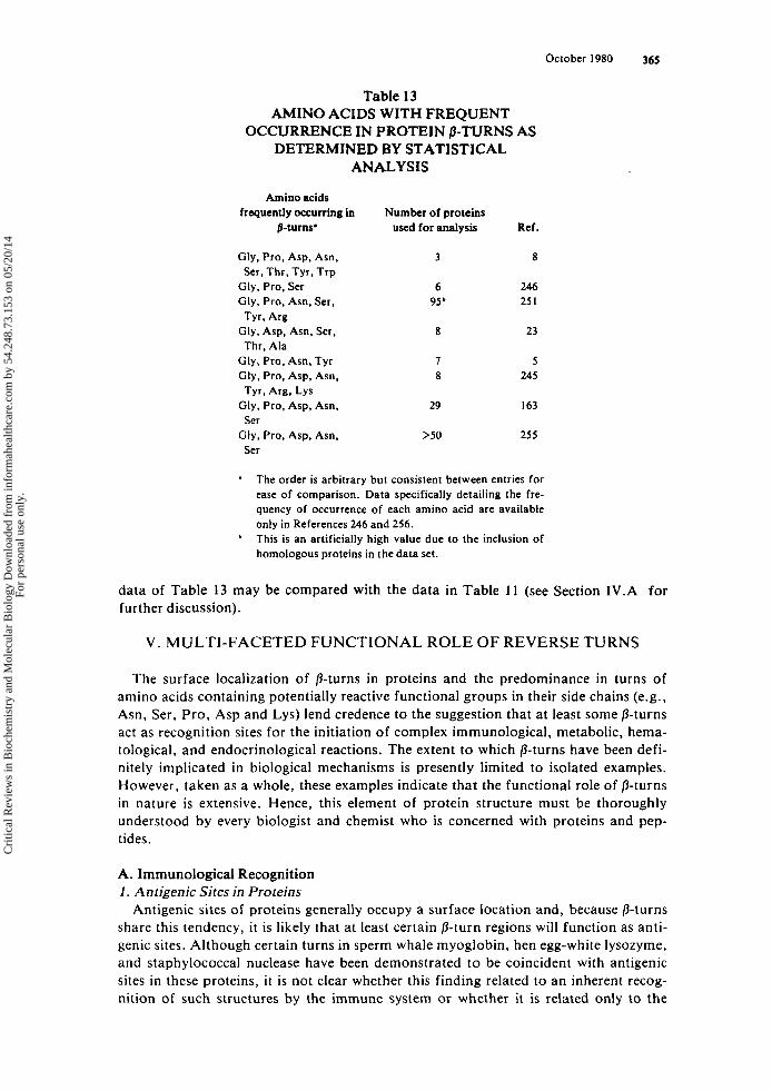
October 1980 365
Table 13 AMINO ACIDS WITH FREQUENT
DETERMINED BY STATISTICAL ANALYSIS
OCCURRENCE IN PROTEIN 0-TURNS AS
Amino acids frequently occurring in
p-turns.
Gly. Pro, Asp, Asn, Ser, Thr. Tyr, Trp
Gly, Pro, Ser Gly. Pro, Asn, Ser,
Gly, Asp, Asn. Ser,
Gly, Pro, Asn, Tyr Gly, Pro. Asp. Asn,
Gly, Pro, Asp, Asn,
Gly. Pro, Asp, Asn,
Tyr. Arg
Thr. Ala
Tyr, Arg. LYS
Ser
Ser
Number of proteins uscd for analysis
3
6 95b
8
1 8
29
>50
Ref.
8
246 25 1
23
5 245
163
255
a The order is arbitrary but consistent between entries for ease of comparison. Data specifically detailing the fre- quency of occurrence of each amino acid are available only in References 246 and 256. This is an artificially high value due to the inclusion of homologous proteins in the data set.
data of Table 13 may be compared with the data in Table 11 (see Section 1V.A for further discussion).
V . MULTI-FACETED FUNCTIONAL ROLE OF REVERSE TURNS
The surface localization of /%turns in proteins and the predominance in turns of amino acids containing potentially reactive functional groups in their side chains (e.g., Asn, Ser, Pro, Asp and Lys) lend credence to the suggestion that a t least some /3-turns act as recognition sites for the initiation of complex immunological, metabolic, hema- tological, and endocrinological reactions. The extent to which /3-turns have been defi- nitely implicated in biological mechanisms is presently limited to isolated examples. However, taken as a whole, these examples indicate that the functional role of /%turns in nature is extensive. Hence, this element of protein structure must be thoroughly understood by every biologist and chemist who is concerned with proteins and pep- tides.
A. Immunological Recognition 1 . Antigenic Sites in Proteins
Antigenic sites of proteins generally occupy a surface location and, because fi-turns share this tendency, it is likely that at least certain /3-turn regions will function as anti- genic sites. Although certain turns in sperm whale myoglobin, hen egg-white lysozyme, and staphylococcal nuclease have been demonstrated t o be coincident with antigenic sites in these proteins, i t is not clear whether this finding related to an inherent recog- nition of such structures by the immune system or whether it is related only to the
Cri
tical
Rev
iew
s in
Bio
chem
istr
y an
d M
olec
ular
Bio
logy
Dow
nloa
ded
from
info
rmah
ealth
care
.com
by
54.2
48.7
3.15
3 on
05/
20/1
4Fo
r pe
rson
al u
se o
nly.
366 CRC Critical Reviews in Biochemistry
frequency of occurrence of /3-turn structural elements among the other structures mon- itored by immune surveillance. However, by examining the location of antigenic sites in relation to the position of 8-turns in those various well-characterized protein anti- gens, several definite conclusions can be drawn.
Atassi and co-workers have elucidated the antigenicity of sperm whale m y ~ g l o b i n . ~ ~ ~ . ~ ~ ~ This protein contained five continuous antigenic ~ i t e s . ~ ~ ’ . ~ ~ * As shown in Table 14, two of these sites contain reverse turn conformations as assessed by X- ray ~rystallography.’~~ Although not shown in Table 14, other myoglobin reverse turns exist (see Section 1V.A ), although they have not been implicated as myoglobin anti- genic sites. Thus, in some cases a reverse turn structural element is a sufficient but not a necessary condition for an antigenic site.
Atassi and c o - w o r k e r ~ ~ ~ ~ . ~ ’ ~ . ~ ~ ’ have also identified three discontinuous antigenic sites in hen egg-white lysozyme (see Table 14). As shown in Table 14, site 1 consisted of residues 6, 7, 10, 13, 14, 126, and 128. Only residue 126, which is part of the type I1 /3-turn (124-127),23~163 was present in this antigenic site. Their antigenic site 2, con- sisting of residues 62, 87, 89, 93, 96, and 97, had only one residue, residue 62, contrib- uted by a 8-turn. Interestingly, residue 62 occupied the i + 3 I h position of a type 111 /3-turn (59 to 62). as well as the i + 2Ih position of a type 1 /3-turn (60 to 63). Site 3 consisted of residues 20, 21, 23, 33, 34, 113, 114, and 116. This site had four residues contributed by /3-turns. Residue 20 occupied the i + 3 I h position in a type 11 /3-turn, the i + l th position in an adjacent type I1 /3-turn, and the i‘“ residue in another adjacent type I’ /3-turn. Residue 21 occupied the i + 2Ih position of the type I1 /3-turn, as well as the i + l’* position of a type I‘ /3-turn. Residue 23 occupied only the i + jrh position of a type I’ /3-turn. Residue 116 occupied the i +
The fact that 6 out of the 22 residues implicated in these antigenic sites were residues contributed by /3-turns is consistent with the observation that approximately one third of the residues of globular proteins are in a reverse turn conf~rmation‘ .~ and in fact argues against /3-turns being more antigenic than other protein surface structural ele- ments.
However, the antigenicity of protein regions containing an isolated series of three adjacent and overlapping /3-turns, in particular those turns containing residues 20, 21, and 23, may be a real phenomenon, since a continuous antigenic site in staphylococcal nuclease region (46 to 52). containing an isolated series of three adjacent and overlap- ping 8-turns, has also been observed‘60 (see Table 14). Certainly, one effect of three adjacent and overlapping 8-turns is that they form a compact loop which extends from the surface of a protein, possibly in such a way that it enhanced the interaction of the antigen with an antibody. However, it should not be generalized that such regions will always be antigenic because, at least in the case of lysozyme, its residues (103 to 106). (104 to 107) and (105 to 108) are indeed overlapping, adjacent /3-turns without apparent antigenicity. Whether this is due to a loss of the compactness of such a structure, invoked by the proximity of two additional adjacent and themselves overlapping /3- turns (98 to 101) and (99 to 102), is not known. Although only the conformational aspects of various antigenic sites have been considered here, antigenicity is certainly also a function of the sequence and side chain orientations within a given site. Until the detailed antigenic structures of various other proteins have been elucidated unequi- vocally, the exact relationship between antigenicity and the occurrence of /3-turns in proteins will remain a mystery.
position of a type I1 /3-turn.
2. Antigenicity of the Lysozyme “Loop” Region Arnon and SelaZ7’ have attributed an antigenic site to the loop formed by the disul-
fide 64-80. However, it has now been unambiguously demonstrated that no such con-
Cri
tical
Rev
iew
s in
Bio
chem
istr
y an
d M
olec
ular
Bio
logy
Dow
nloa
ded
from
info
rmah
ealth
care
.com
by
54.2
48.7
3.15
3 on
05/
20/1
4Fo
r pe
rson
al u
se o
nly.
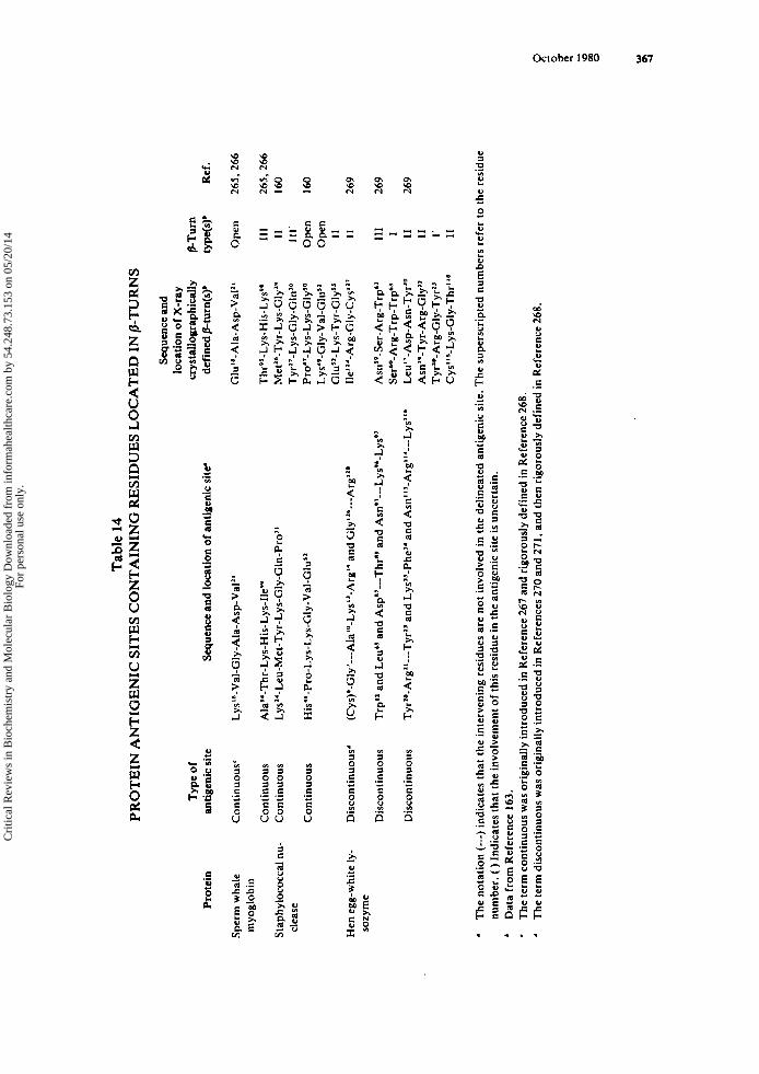
Prot
ein
Sper
m w
hale
m
yogl
obin
Stap
hylo
cocc
al nu
- cl
ease
Hen
egg
-whi
te ly
- so
zym
e
Tab
le 1
4 P
RO
TE
IN A
NT
IGE
NIC
SIT
ES
CO
NT
AIN
ING
RE
SID
UE
S L
OC
AT
ED
IN B
-TU
RN
S
Typ
e of
antig
enic
site
Con
tinu
ous‘
Con
tinu
ous
Con
tinu
ous
Con
tinu
ous
Dis
cont
inuo
us‘
Dis
cont
inuo
us
Dis
cont
inuo
us
Sequ
ence
and location o
f an
tige
nic
site
Lys’6-Val-Gly-Ala-Asp-Val”
Ala94-Thr-Lys-His-Lys-lle99
Lys”-Leu-Met-Tyr-Lys-Gly-GIn-Pro’
’
His
‘*-P
ro-L
ys-L
ys-G
Iy-V
al-G
lu’~
(Cy~)*-Gly’---Ala’~-Lys’’-Arg~‘
and
Gly
’*’-
--A
rg’*
’
Trp
“ an
d L
eub5
and
Asp
”---T
hr’O
an
d A
snP3
---L
ys9’
-Lys
9’
Tyr’
O-A
rg”-
--Ty
rZ3
and
Lys
”-Ph
e”
and
Asn
“3-A
rg“‘
---L
ys”*
Sequ
ence
and
lo
cati
on of X
-ray
cr
ysta
llogr
aphi
cally
de
fine
d p-
turn
(s)’
Glu
”’-A
la-A
sp-V
al”
Thr
s5-L
ys-H
is-L
ysP8
M
et”-
Tyr-
Lys-
Gly
’P
Tyr
”-L
ys-G
ly-G
ln’o
P
r~~
’-L
ys-L
ys-G
ly’~
Ly
s‘v-
Gly
-Val
-Glu
’l G
lu”-
Lys
-Tyr
-Gly
S’
Ile”‘
-Arg
-Gly
-Cys
’l’
A~n
’~-S
er-A
rg-T
rp”
Ser6
0-A
rg-T
rp-T
rp”
Leu”
-Asp
-Asn
-Tyr
’O
Asn
”-T
yr-A
rg-G
ly”
Tyr
’o-A
rg-G
ly-T
yr”
Cy~
“’-L
ys-G
Iy-T
hr‘~
*
P-T
urn
type
(s)’
Ope
n
Ill I1
111’
O
pen
Ope
n I1
I1
111 I I1
I1
1’
I1
Ref
.
265,
266
265.
266
160
160
269
269
269
. ’ D
ata
from
Ref
eren
ce 1
63.
‘
The
not
atio
n (-
--) i
ndic
ates
tha
t th
e in
terv
enin
g re
sidu
es a
re n
ot in
volv
ed i
n th
e de
linea
ted
antig
enic
site
. The
supe
rscr
ipte
d nu
mbe
rs r
efer
to
the
resi
due
num
ber.
( )
Indi
cate
s tha
t the
invo
lvem
ent o
f th
is r
esid
ue in
the
antig
enic
site
is u
ncer
tain
.
The
term
con
tinu
ous
was
ori
gina
lly i
ntro
duce
d in
Ref
eren
ce 2
67 a
nd ri
goro
usly
def
ined
in R
efer
ence
268
. T
he te
rm d
isco
ntin
uous
was
ori
gina
lly in
trod
uced
in R
efer
ence
s 27
0 an
d 27
1, a
nd th
en r
igor
ousl
y de
fine
d in
Ref
eren
ce 2
68.
B 6 W 9
Cri
tical
Rev
iew
s in
Bio
chem
istr
y an
d M
olec
ular
Bio
logy
Dow
nloa
ded
from
info
rmah
ealth
care
.com
by
54.2
48.7
3.15
3 on
05/
20/1
4Fo
r pe
rson
al u
se o
nly.
368 CRC Critical Reviews in Biochemistry
tinuous antigenic sites occur in native l y ~ o z y m e ’ ~ ~ (see Section V.A.l ), so that the antigenicity being measured by Arnon and Sela272 and Maron et al.273 represented the contribution of a portion of a discontinuous antigenic site and hence was determined with less efficiency than might be achieved with a complete antigenic
Despite these limitations, Arnon et al.274 have correlated changes in the binding ca- pacity of synthetic loop analogs having various residues replaced by alanine to antiloop antibodies with the relative probability of occurrence of /3-turns in these synthetic an- alogs. By substituting one amino acid residue a t a time, they were able to establish a relationship between differences in antigenicity and differences in the theoretically pre- dicted probability for 0-turn formation. Since no direct measurements of the changes in P-turn conformation in the various synthetic analogs were made, and since the an- tigenicity of any antigenic site depends on its sequence, as well as its conformation, this correlation must be viewed with caution.
An alternative experimental approach correlating immunological measurements of the degree of cross-reaction between proteins from different species in which only amino acid substitutions occurred (i.e., n o additional inversions, additions, or dele- tions) with the probability of p-turn formation was also carried and for the reasons above no direct interpretation is possible. However, a n interesting, albeit inci- dental finding of this study was that there a re quantitative differences in the antiloop antibodies raised in rabbits as compared to goats. Whether this represented differences in the specific immunological recognition of P-turn conformations between species has not been satisfactorily determined, but the resolution of this question remains as a worthwhile challenge to immunochemists.
3. Tuftsin Tuftsin is a basic tetrapeptide, H-L-Thr-L-Lys-L-Pro-L-Arg-OH isolated from a spe-
cific cytophilic y-globulin, l e u c ~ k i n i n . ’ ~ ~ ~ ’ ~ ’ This peptide has been shown to increase the p h a g o ~ y t i c ~ ~ ~ . ~ ~ ~ and bactericidal278 activities of macrophages and polymorphonu- clear neutrophil leukocytes a t concentrations as low as 1 x M.279 Although the replacement of the N-terminal threonyl residue by L-Ser,280.281 D-Ser,’80 L-Leu”’ or p GluZ8’ yielded active analogs, the retroenantio-tuftsin has been observed to have n o biological An evaluation of various additional analogs revealed that only one basic group is required for biological activity since the replacement of either the lysyl or arginyl residue by an alanyl did not appreciably diminish the biological activity of the molecule.’80 However, when L-Ala was substitued for L-Pro, there was a marked activity loss.28o Konopinska and co-workers’80 suggested that tuftsin might contain a /&turn like those found for the two D-Phe-L-Pro type 11’ turns of gramicidin S. How- ever, since tuftsin contains only L-amino acids, a type 1 /3-turn conformation should be preferred (see Section I1 ). Further; the C D of tuftsin in methanol, trifluoroethanol, and 80% dioxane-water, but not water, were also inconsistent with such a type 11’ /3- turn, I 6 O since those spectra resembled other class B spectra for model peptides contain- ing either type I or I1 /%turns (see Table 9). Recently, Blumenstein and Najja? have confirmed by ‘H-NMR the presence of a /&turn in DMSO-d6, but not in H 2 0 . Energy calculations by Fitzwater et al.2s6 also suggested that the peptide has a high probability of forming an open reverse turn.
As pointed out by Smith160 and Tzehoval et al.,287 Arg-Pro and Pro-Arg sequences often occur in biologically active peptides. Although the reason for this occurrence is unknown, it is well known that an Arg-Pro sequence is normally highly resistant t o proteolytic degradation. Since Xxx-Pro and Pro-Xxx sequences are likely to occur in /3-turn conformation^,^^^ it may be that such a -conformational feature involving the Arg-Pro sequence protects it from enzyme cleavage. However, the sequence Gln-Arg-
Cri
tical
Rev
iew
s in
Bio
chem
istr
y an
d M
olec
ular
Bio
logy
Dow
nloa
ded
from
info
rmah
ealth
care
.com
by
54.2
48.7
3.15
3 on
05/
20/1
4Fo
r pe
rson
al u
se o
nly.
October 1980 369
Pro-Gly has been demonstrated to undergo rapid proteolytic digestion,Z88 and this may have resulted from a n altered conformation of this sequence, possibly involving a p- turn with L-Pro and Gly as the i + l f h and i + 2‘h residues, respectively. Further, it has not been shown that such a /3-turn conformation, i f present, was necessary for binding to certain membrane although the selective advantage of being able to bind peptides containing Arg-Pro or Pro-Arg sequences without concomitant enzymic degradation by membrane-bound esterases may account for the biological potency of these peptides and, in particular, tuftsin.
B. Phosphorylation of Proteins Protein kinase dependent phosphorylation of proteins is one of the mechanisms
leading to post-translational m ~ d i f i c a t i o n ’ ~ ~ of protein ~ t r u c t u r e . ~ ~ ~ Intracellular pro- tein kinases have been demonstrated to transfer a phosphate from adenosine triphos- phate (ATP) to certain seryl or threonyl residues (so-called phosphorylation sites) in various protein substrates, including membrane-bound sugar-transport proteins, basic (i.e., histones) and acidic nuclear proteins, ribosomal proteins, and mitochondria1 pro- teins. Although aberrant membrane protein phosphorylation has been shown to be associated with various disease states, including muscular d y s t r o p h i e ~ , ~ ~ ~ . ~ ~ ~ hereditary stomatocytosis and ~ p h e r o c y t o ~ i ~ , ~ ~ ~ - ~ ~ ~ and sickle cell d i ~ e a s e , ~ ~ ~ . ~ ~ ~ the relationship between such abnormal phosphorylation and a n alteration of membrane protein struc- ture has not been elucidated.
However, Williams298 has pointed out that the location of basic amino acid residues (i.e., Lys, Arg, or His) within the amino sequence adjacent t o the phosphorylated hydroxy-amino acid residue may function as the recognition site for the phosphoryl- ating kinase. He analyzed the amino acid sequences surrounding 25 phosphorylation sites in 1 1 proteins and concluded that a phosphorylated seryl and threonyl residue is generally separated from an adjacent basic amino acid residue by only one intervening residue. Although he emphasized the importance of the primary structure of a given site in determining whether or not phosphorylation would occur, he conceded that phosphorylation might also be governed b y the array (in a three-dimensional sense) of amino acid residues surrounding a phosphorylation site or by a specific conformation associated with the site. However, only recently has a correlation between the occur- rence of a specific conformational feature (i.e., p-turns) and phosphorylation sites been completed. Small et al.z99 demonstrated that 24 out of 30 phosphorylated residues found in their set of 14 proteins, including many of the phosphorylation sites examined by Williams,z98 existed within their predicted 8-turn conformations. It must be empha- sized that this location of phosphorylation sites primarily to 8-turn-containing regions was based on a correlative method of prediction (see Section 1V.C ) and that these predictions have not been confirmed by X-ray diffraction. Among those phosphoryl- ated residues predicted to occur with 8-turn conformations, 69% were located at either the i I h or i + 3 1 h positions of the /%turns. However, phosphorylation sites were also observed that were not located within 8-turn-containing regions. The validation of the correlation of the high frequency of occurrence of p-turn conformations found in as- sociation with phosphorylation sites must await detailed X-ray analysis of: ( 1 ) the lo- cation of all phosphorylated residues inside and outside p-turn conformations, as well as (2) the location of all potentially phosphorylated residues inside and outside 8-turn conformations.
As shown in Table 15, i f the da ta of Small et al.*99 and Williams298 are examined in conjunction with one another, several interesting relationships between the location of the phosphorylated residue inside or outside a predicted 8-turn and the location of basic amino acids with respect to the phosphorylated residue are evident.
Cri
tical
Rev
iew
s in
Bio
chem
istr
y an
d M
olec
ular
Bio
logy
Dow
nloa
ded
from
info
rmah
ealth
care
.com
by
54.2
48.7
3.15
3 on
05/
20/1
4Fo
r pe
rson
al u
se o
nly.
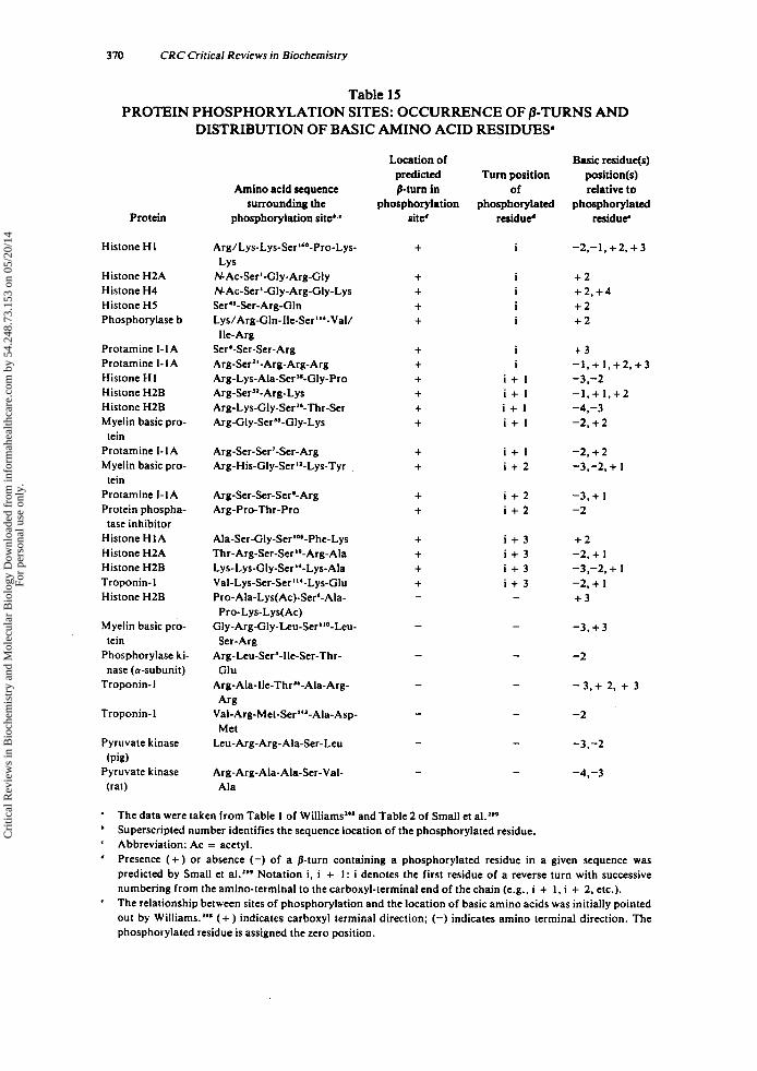
370 CRC Critical Reviews in Biochemistry
Table 15
DISTRIBUTION OF BASIC AMINO ACID RESIDUES' PROTEIN PHOSPHORYLATION SITES: OCCURRENCE OF P-TURNS AND
Location of Basic residue(s) predicted Turn position position(s)
Amino acid sequence 8-turn in of relative to surrounding the phosphorylation phosphorylated phosphorylatcd
Protein phosphorylation site'.' site' residue' residue
Histone HI Arg/Lys-Lys-Ser"-Pro-Lys-
NAc-Serl-Gly-Arg-Gly NAc-Ser I-Gly-Arg-Gly-Lys Ser''-Ser-Arg-Gln Ly~/Arg-GIn-lle-Ser"~-Val/
Sere-Ser-Ser- Arg Arg-Serl'-Arg-Arg-Arg Arg-Lys-Ala-Scr3*-GIy-Pro Arg-Ser3'-Arg-Lys Arg-Lys-Gly-Ser"-Thr-Ser Arg-Gly-Ser"-Gly-Lys
LY s
He-Arg
+ i -2,-1,+2.+3
Histone H2A Histone H4 Histone HJ Phosphorylase b
+ + + +
+ 2 + 2 , + 4 + 2 + 2
Protamine I-1A Protamine I-1A Histone HI Histone H2B Histone H2B Myelin basic pro-
Protamine I-1A Myelin basic pro-
Protamine I-1A Protein phospha- tase inhibitor
Histone HIA Histone H2A Histone H2B Troponin-1 Histone H2B
tein
tein
I
I
i + 1 i + 1 i + l i + 1
+ 3 -1. + 1, + 2 , + 3 -3.-2 - I , + l . + 2 -4,-3 -2. + 2
Arg-Ser-Ser'-Ser-Arg Arg-His-Gly-Ser"-Lys-Tyr .
i + l i + 2
-2. + 2 -3,-2, + 1
+ +
Arg-Ser-Ser-Ser'- Arg Arg-Pro-Thr-Pro
i + 2 i + 2
-3.+ 1 -2
+ +
Ala-Ser-Gly-Serla'-Phe-Lys Thr-Arg-Ser-Ser "-Arg-Ala Ly~-Lys-Gly-Ser~~-Lys-Ala Val-Lys-Ser-Ser "'-Lys-Glu Pro-Ala-Lys(Ac)-Ser6-Ala-
Gly-Arg-Gly-Le~-Scr"~-Leu-
Arg-Leu-Ser'-lle-Ser-Thr-
Arg-Ala-lle-Thr"-Ala-Arg-
Val-Arg-Met-Ser "'-Ala-Asp-
Leu- Arg- Arg- Ala-Ser-Leu
Pro-Lys-Lys(Ac)
Ser-Arg
Glu
Arg
Met
i + 3 i + 3 i + 3 i + 3 -
+ 2 -2, + I -3,-2, + 1 -2. + I + 3
Myelin basic pro- tein
Phosphorylase ki- nase (a-subunit)
Troponin-1
- 3 , + 3
-2
- 3 , + 2. + 3
Troponin-1 -2
Pyruvate kinase
Pyruvate kinase (pig)
(rat)
-3,-2
Arg-Arg-Ala-Ala-Ser-Val- Ala
-4.-3
The data were taken from Table 1 of Williams"* and Table 2 of Small et al.lee Superscripted number identifies the sequence location of the phosphorylated residue. Abbreviation: Ac = acetyl. Presence (+ ) or absence (-) of a p-turn containing a phosphorylated residue in a given sequence was predicted by Small et al.a9* Notation i, i + I : i denotes the first residue of a reverse turn with successive numbering from the amino-terminal to the carboxyl-terminal end of the chain (e.g., i + I , i + 2, etc.). The relationship between sites of phosphorylation and the location of basic amino acids was initially pointed out by Williams.'g* (+ ) indicates carboxyl terminal direction; (-) indicates amino terminal direction. The phosphorylated residue is assigned the zero position.
'
'
'
Cri
tical
Rev
iew
s in
Bio
chem
istr
y an
d M
olec
ular
Bio
logy
Dow
nloa
ded
from
info
rmah
ealth
care
.com
by
54.2
48.7
3.15
3 on
05/
20/1
4Fo
r pe
rson
al u
se o
nly.
October 1980 371
1 .
2 .
3.
4.
5 .
If the phosphorylated residue occupied the i'hposition of a predicted /I-turn (7 examples), the basic amino acid residue(s) occurred in six cases on the carboxyl terminal side of this residue and was (were) located at least one residue away where they occasionally occurred as a pair of basic residues. I f the phosphorylated residue occupied the i + I f h position of a predicted /I-turn (5 examples), the basic amino acid residue on the carboxyl side of the phospho- rylated residue never occurred more than one residue away. Further, there was a tendency (with one exception) for basic residues to be located one residue away from the phosphorylated residue in either or in both directions. I f the phosphorylated residue occupied the i + 21h position of a predicted /3-turn (3 examples), the basic residues in two cases were located adjacent in the carboxyl terminal direction in combination with another basic residue located three resi- dues toward the amino terminus. In one case a third basic residue two amino acids toward the amino terminus was also present, although in one other example a basic amino acid in this position without the presence of the other two residues was also observed. I f the phosphorylated residue occupied the i + 3 I h position of a predicted /I-turn (4 examples) (with one exception), the basic residues were located adjacent to the phosphorylated residue in the carboxyl terminal direction and one residue away in the amino terminal direction (with one exception). In phosphorylation sites which were not predicted t o be /3-turns (7 examples), the basic residues were located one or more residues away from the phosphorylated residue in either direction. In two cases a pair of basic residues was located one and two residues away in the amino terminal direction from the phosphorylated residue as was the case for i + 2'* positioned sites in /3-turns. In two other se- quences, a basic residue occurred two residues away from the phosphorylated residue in both the amino and carboxyl terminal direction.
In summary, the data presented in Table 15 indicate that there may be a relationship between the occurrence of a /3-turn conformation and the distribution of basic amino acid residues (with respect to the position of the phosphorylated residue) within a spe- cific phosphorylation site. The intricacies of these relationships, as well as rigorous proof for the mandatory recognition of a /3-turn conformation by a protein kinase remain to be achieved. However, the circumstantial evidence implicates both structural requirements, turns and basic residues, as necessary for the post-translational modifi- cation.
C. Glycosylation of Proteins Two different modes of linkage between protein and carbohydrate have been iden-
tified: (1) the carbohydrate chain is linked N-glycosidically to an asparagine residue of the protein, or (2) the carbohydrate is linked Oglycosidically to a serine. threonine, hydroxylysine, or hydroxyproline residue of the In those glycoproteins which contain more than one carbohydrate chain per molecule, both types of glycosidic linkage may occur.3oo
I . N-Glycosidic Linkages Although much remains to be determined about the conformational requirements
of the glycosylation sites in proteins, it has been observed in glycoproteins containing carbohydrate linked N-glycosidically to an Asn residue that the associated sequence is invariably - A ~ n - X x x - [ ~ ~ ~ ~ . , ] - . ~ ~ ~ When the sequences of numerous glycoproteins were carefully examined, it became apparent that this tripeptide sequence was a necessary
Cri
tical
Rev
iew
s in
Bio
chem
istr
y an
d M
olec
ular
Bio
logy
Dow
nloa
ded
from
info
rmah
ealth
care
.com
by
54.2
48.7
3.15
3 on
05/
20/1
4Fo
r pe
rson
al u
se o
nly.
312 CRC Critical Reviews in Biochemistry
but not a sufficient requirement for glycosylation to O C C U ~ . ’ ~ ’ Aubert et aI.’O’ realized that asparagine, serine, and threonine are among the most frequently occurring amino acids within /3-turns (see Tables 11 and 13), and they analyzed the sequences around the glycosylated residues in various glycoproteins by the correlative methods of Chou and F a ~ m a n ~ ~ ~ . ~ ~ ~ in an effort to determine whether or not a /3-turn conformation might be involved in addition to the invariant tripeptide sequence requirement. For their set of 28 glycosylated Asn residues from 14 different glycoproteins they found that in 19 ‘of the 28 sites, the Asn residue was located within a predicted /3-turn, usually at the i + 21h position of the /3-turn. In those cases where Asn occupied the i + 2’“ position, the Thr (or Ser) residue would not be situated within the turn. However, a central position of the Asn within a turn, a position where the residue of the Asn side chain is furthest removed from the protein surface, might serve to facilitate the specific trans- ferase catalyzed transfer of oligosaccharide from its lipid (i.e., dolichol pyrophos- phate)-linked intermediate’O’ to an acceptor protein.
As in the case of the factors controlling the site specificity of the phosphorylation of proteins (see Section V.B ), the control of N-glycosylation, although linked in part to the presence of a unique receptor sequence and/or the occurrence of a /3-turn con- formation, remains to be determined. In particular, various experimental observations on non-native glycosylated substrates need to be considered.
For example, Pless and Lennarz304 have found that RNase A and a-lactalbumin, both containing the sequence -Asn-Xx~-(~~‘~.,]-, were converted from non-acceptors to acceptors after covalent modification of their cysteine residues under denaturing con- ditions. Additional fragmentation of reduced and alkylated a-lactalbumin by treat- ment with cyanogen bromide followed by tryptic or chymotryptic digestion has led also to the production of polypeptide acceptors of 7 to 123 residues in length, differing little in their rates of in vitro glyc~sylat ion.’~~ Recently, seven more proteins which do not normally occur in a glycosylated form and which contain one or more of the nec- essary tripeptide sequences, were examined as potential acceptors for ~ a r b o h y d r a t e . ~ ~ ~ None of these proteins in their native state was found to undergo glycosylation. How- ever, after reduction and alkylation, only two proteins, rabbit muscle triosephosphate isomerase and ovine prolactin, were converted to acceptors of oligosaccharide- chains.’06 In the native triosephosphate isomerase molecule the glycosylation site (195 to 197) occurs within an a-helical c ~ n f o r m a t i o n , ’ ~ ~ and it may be that this structural feature protected the sequence from glycosylation until after the protein was denatured by reduction and carbo~ymethylation.’~~ After fragmentation by cyanogen bromide cleavage, peptides from two other proteins, catalase and concan- avalin A, were also found to serve as carbohydrate acceptors. Although the two poten- tial glycosylation sites in concanavalin A, regions (1 18 to 120) and (162 to 164),’06 are known to occur in regions containing three juxtaposed and overlapped /3-turns located between two P-shee t~ , ’~~ no glycosylation occurred until fragmentation was carried out. This observation suggested that the presence of a /3-turn, even in a highly exposed position characteristic of repeating /3-turns,160 was insufficient for glycosylation. This indicated that there may be unique conformations (possibly /3-turns) available only in the free peptide that were required for recognition of the tripeptide as a receptor. This suggestion might also explain the effective glycosylation of other linear peptides, in- cluding a heptapeptide from human thy rog l~bu l in ’~~ and peptides of various lengths from a-lactalbumin.305
2. OGlycosidic Linkages A great deal remains to be learned about the structural requirements necessary for
Oglycosylation of a protein. However, as in the case of Nglycosylation the recogni- tion of a /3-turn conformation by a glycosyl transferase has been implicated, although
Cri
tical
Rev
iew
s in
Bio
chem
istr
y an
d M
olec
ular
Bio
logy
Dow
nloa
ded
from
info
rmah
ealth
care
.com
by
54.2
48.7
3.15
3 on
05/
20/1
4Fo
r pe
rson
al u
se o
nly.

October 1980 373
this conformational requirement has not been rigorously defined. Aubert et al.302 used the correlative methods of Chou and F a ~ m a n * ' ~ . ~ ~ ~ to predict possible @urn confor- mations in nine glycosylation sites in five glycoproteins. They also noted the presence of one or more prolyl residues within or adjacent to their predicted p-turns. In addi- tion, although they did not point it out, their data indicated that one or more glycyl residues were also frequently located within the sequences of their predicted turns. Since Gly and Pro residues are known to have a high frequency of occurrence in /3- turns, their findings were not surprising. However, what is surprising was the consist- ency of the occurrence of P ro and Gly residues which occurred in 89% of the glycosy- lation sites which they examined. In addition, in two thirds of their sites the glycosy- lated residue occupied a central residue (i.e., the i + 1'" or i + 2'*) of a predicted p- turn. Their observations were also consistent with the amino acid compositions of various proteoglycan fractions from ox and pig laryngeal ~ a r t i l a g e , ~ ~ ~ ~ ~ ~ ~ which had an increased proportion of Ser, Gly, Glu, and Pro. Recently, Robinson et al.310 have demonstrated that another proteoglycan, heparin, isolated from pronase-digested rat skin, consists of a polypeptide composed entirely of Ser and Gly residues. They have assumed, without experimental proof, that these residues occur in an alternating se- quence. Since Ser-Gly or Gly-Ser-containing sequences have a high probability of forming B-turn conformations,218 it may be that this conformational feature is neces- sary for the xylosidic linkage between the seryl hydroxyl and the polysaccharide in heparin. The exact structural features required for the 0- glycosylation of heparin remain to be determined. In the case of heparin, a correlation between the solution conformation of Gly/Ser copolymers and their level of in vitro glycosylation may elu- cidate these features, as has been done for elastin and collagen hydroxylation"' (see Section V.D ). It may then be possible to establish the detailed structural requirements in other more complicated amino acid sequences which occur in glycosylation sites in other proteoglycans and glycoproteins.
D. Hydroxylation of Proline in Collagen and Elastin A conformational specificity for the enzyme, proline hydroxylase (E.C. 1.14.1 1.2),
which hydroxylates prolyl residues in procollagen has been suggested by Brahmachari and Ananthanarayanan."' They proposed that this conformational requirement for hydroxylation resides in the recognition by the enzyme of an Xxx-Pro-Gly-Yyy se- quence, where Pro and Gly occupy the i + and i + 21h positions of a p-turn, respectively. They also pointed out that the Yyy residue plays a major factor in deter- mining whether or not a given sequence Xxx-Pro-Gly-Yyy will adopt a /3-turn confor- mation. Bhatnagar et al.311 have also found that the nature of the Xxx side chain may be equally important in the modulation of hydroxylation in collagen-like polymers. In addition, the sequence Xxx-Gly-Pro-Yyy has been demonstrated by energy calcula- t i o n ~ ~ ~ ~ and various physical methods (NMR, CD, and IR)148 not to favor the forma- tion of a p-turn, in comparison to the sequence Xxx-Pro-Gly-Yyy. Whether or not all procollagen Xxx-Pro-Gly-Yyy sequences are equally well hydroxylated has not been demonstrated satisfactorily a t the present time. Indeed, since hydroxyproline forma- tion in collagen has been associated with enhanced stability of the collagen triple he- l i ~ , " ~ . ~ " presumably due to hydrogen bonding between the Hyp O'H and the backbone carbonyl it may be that certain restraints imposed by such hydrogen bond- ing on conformational freedom of either a /I-turn region or a region juxtaposed to a /3-turn may alter the rate or site of hydroxylation. However, there are collagen-like synthetic peptides of the sequence (Pro-Pro-Gly). which, although they cannot occupy a P-turn c o n f ~ r m a t i o n , ' ~ ~ are nevertheless optimally hydr~xy la t ed .~" Further, the mere presence of a defined p-turn conformation within an Xxx-Pro-Gly-Yyy peptide (e.g., (Val-Pr~-Gly-Gly) .~~~) is not sufficient to lead to enhanced levels of hydr~xylat ion.~"
Cri
tical
Rev
iew
s in
Bio
chem
istr
y an
d M
olec
ular
Bio
logy
Dow
nloa
ded
from
info
rmah
ealth
care
.com
by
54.2
48.7
3.15
3 on
05/
20/1
4Fo
r pe
rson
al u
se o
nly.
314 CRC Critical Reviews in Biochemistry
As reviewed by Urry and Long,’O much of the proline in elastin occurs in Xxx-Pro- Gly-Yyy sequences. Taking advantage of the five polypeptide models of elastin devel- oped by Urry and c o - ~ o r k e r s , ~ ~ ~ ~ ~ ~ ~ - ~ * ~ Bhatnagar et al.3” examined the ability of each of these model polypeptides to inhibit the hydroxylation of collagen by chick embryo prolyl hydroxylase. Two of the polypeptides, (Val-Pro-Gly-Val-Gly). and (Ala-Pro- Gly-Gly)., resulted in significant inhibition of collagen hydroxylation, although the amount of endogenous hydroxylation of each of these polypeptides was not signifi- cantly different from the non-inhibiting pep tide^.^" This suggests that the structural requirements for collagen and elastin are not equivalent and that additional work is necessary in order to elucidate the structural requirements which are mandatory for the proline hydroxylase-induced post-translational modification of the prolyl residues in collagen and elastin.
In summary, proline hydroxylation in the connective tissue proteins elastin and col- lagen has been shown to be dependent in part on the recognition of (Pro-G1y)-contain- ing sequences which occupy p-turn conformations. The subtleties of this recognition and the resultant post-translational modification of protein structure on subsequent hydroxylation are unclear.
E. Elastin Urry and Long’O recently reviewed the prodigious amount of synthetic and spectro-
scopic studies carried out in the Urry laboratory on three repeating sequences found in tr~poelastin:~” a tetrapeptide Va11-Pro’-Gly3-Gly4, a pentapeptide, Va11-Pro2-Gly3- Va14-GlyS, and a hexapeptide, Ala1-Pro2-Gly3-Val4-Glys-Val6. This group of peptides represents one of the most thoroughly studied series of reverse turn-containing peptides in the literature and should be thoroughly reviewed by the interested reader. Additional papers dealing primarily with c o a ~ e r v a t i o n ~ ~ ~ and c r ~ s s - l i n k a g e ~ ~ ~ of these model elas- tin peptides have also appeared, but the details of these papers are beyond the scope of this review.
F. Antifreeze Peptides and Proteins Approximately one third of the freezing temperature depression activity, located in
the sera of certain extensively studied polar fishes, has been accounted for by a group of eight glycoproteins, so-called antifreeze glycoproteins (AFGP).323 These glycopro- teins differed from one another by charge and molecular AFGP-1 to -5 were shown to have molecular weights in the range of 10,500 to 30,000 and were found to depress the freezing point of water as effectively as NaCl, when they were compared on a weight basis. The structure of the AFGP-1 to -5 was determined to be a repeating glycotripeptide with the following sequence: -Ala-Ala-Thr- with a disac- charide linked Oglycosidically to each threonyl residue.324 AFGP-6 to -8 differed from AFGP-1 to -5 in that they have a lower molecular weight, have little’25.326 or no323.324.327 antifreeze activity when tested alone, (although when combined with AFGP-1 to -5 they extensively potentiated the antifreeze effect of the larger g l y c ~ p r o t e i n s ) , ~ ~ ~ and have additional proline residues inserted in their sequence after some of the threonine
The mechanism by which these AFGPs function remained obscure until DeVries and Lin3z9 determined the sequence of an antifreeze peptide, not a glycopep- tide, whose sequence revealed the presence of clusters of polar amino acids, usually threonine and aspartic acid, separated from one another by multiple alanine residues, which spaced the polar clusters by approximately 4.5 A.
Since this is the distance known to separate the oxygen atoms of water in an ice crystal lattice, the authors suggested that the polar side chains of their antifreeze pep- tide might occupy positions in an ice lattice normally occupied by hydrogen bonds
Cri
tical
Rev
iew
s in
Bio
chem
istr
y an
d M
olec
ular
Bio
logy
Dow
nloa
ded
from
info
rmah
ealth
care
.com
by
54.2
48.7
3.15
3 on
05/
20/1
4Fo
r pe
rson
al u
se o
nly.
October 1980 375
between water molecules. The effect would be to diminish the freezing point of water. Using the empirical predictive methods of Chou and Fasman,256 Louche~x-LeFebvre~~~ predicted that every polar amino acid cluster in the antifreeze peptide of DeVries and Lin329 had a relatively high probability of occurring in a p-turn conformation. Since the CD spectrum of the antifreeze peptide indicated that the major conformational feature of the peptide was a u - h e l i ~ , ~ ~ ’ its structure was predicted to be made up of helical regions of repeating alanyl residues, interspersed among /3-turns containing po- lar amino acid clusters.
A Thr-Pro-Ala-Thr sequence found in AFGP-83’4.33’ has also been predicted to oc- cur in a 8-turn onf formation.'^^ In the case of this glycopeptide, with little or no de- tectable antifreeze activity of its it remains to be established whether this pre- dicted p-turn conformation has any function in the cooperative potentiation of the antifreeze activity of AFGP-I to - 5 .
In summary, i t appears likely that /3-turns interspersed among non-polar regions in peptides may play a role in the adsorption of certain antifreeze peptides to ice. Definite experimental verification of this hypothesis has not been achieved.
G. Morphine-like Activity in a Neuropeptide: Enkephalin Eokephalin was initially isolated from pig brain as a mixture of two pentapeptides:
H-Tyr-Gly-Gly-Phe-Met-OH ([MetS]-enkephalin) and H-Tyr-Gly-Gly-Phe-Leu-OH ([Le~~]-enkephal in) .~’~ Enkephalins have been demonstrated to bind to brain, gut, and spinal cord opiate receptors,”’ as well as to compete with naloxone, a strong opiate antagonist, for receptor binding.333 Because oE the likelihood that a detailed under- standing of the molecular structure of enkephalin might lead to the rational design of effective and n o n - a d d i c t i ~ e ~ ~ ~ analgesics and to the elucidation of the intricacies of the opiate receptor, the structural features of this molecule have been intensively examined by ‘H-335-339 and 13C-NMR,337.340.341 fluorescence energy X-ray diffraction,“ model building,’43 and theoretical energy ca l c~ la t ions~’~-”~ (see Table 16).
Bradbury et a1.343 originally proposed on theoretical grounds that the conformation of enkephalin was likely to be a p-turn with an intramolecular hydrogen bond between the NH of Phe and the C=O of Tyr. Hence the Gly2-Gly3 sequence of enkephalin would occupy the i + I’* and i + 2‘* positions, respectively, of the “G-G” /3-turn.*’S Such a structure also resulted in significant spatial overlap with the functional groups in morphine-like opiate^,"^ known to be necessary for opiate Since this conformation was shown by conformational energy calculations to be a high energy structure, it should not be the favored conformation for the m 0 1 e c u l e . ~ ’ ~ ~ ~ ~ ~ In spite of this apparent discrepancy, the crystal structure of [Le~~l-enkephalin has been dem- onstrated to be a “G-G” p-turn with dihedral angles consistent with a type I’ confor- m a t i ~ n * ~ (see Section 11). Thus, if the resemblance of enkephalin to rigid opiates is a requirement for binding and if large perturbations of the conformation of enkephalin at the opiate receptor do not occur, then a “G-G” P-turn conformation is highly likely to be involved in favorable interactions with the r e~ep to r . ”~ In contrast, overwhelming evidence accumulated from ‘H-NMR indicated that the conformation of enkephalin, although a folded conformation, was probably a “G-P” p-turn with the Qly3The4 sequence occupying the central positions of the turn and with an intramolecular hydro- gen bond between the amide proton of the methionyl (or leucyl) residue and the car- bony1 of Gly’. Results gleaned from theoretical energy calculations also suggested that such a “G-P” /3-turn was a low energy structure and hence a favored conformation.
These differences between the conformation of enkephalin deduced from ‘H-NMR and theoretical energy calculations, as compared to the conformation determined from
Cri
tical
Rev
iew
s in
Bio
chem
istr
y an
d M
olec
ular
Bio
logy
Dow
nloa
ded
from
info
rmah
ealth
care
.com
by
54.2
48.7
3.15
3 on
05/
20/1
4Fo
r pe
rson
al u
se o
nly.
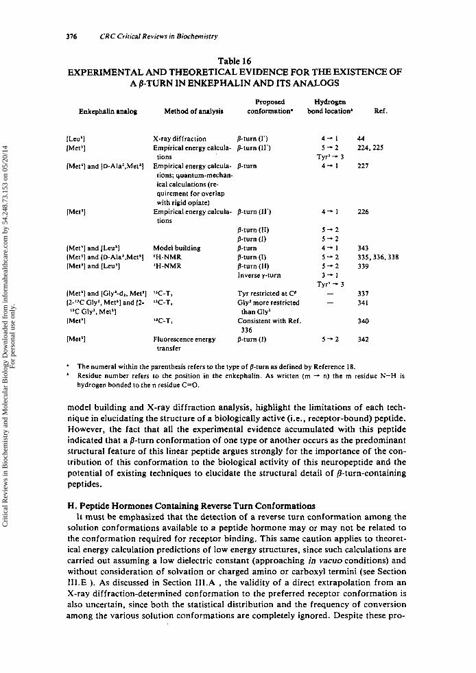
316 CRC Critical Reviews in Biochemistry
Table 16 EXPERIMENTAL AND THEORETICAL EVIDENCE FOR THE EXISTENCE OF
A P-TURN IN ENKEPHALIN AND ITS ANALOGS
Enkephalin analog
[Leu’] [Met’]
[Met’] and [D-Ala’,Met’]
[Met’]
[Met’] and [Leu’] [Met’] and [D-Ala’.Met’] [Met’] and [Leu’]
[Met’] and [Gly’-dl, Met’] [2-”C Gly*, Met’] and 12-
[Met’] “C Gly’, Met’]
[Met’]
Proposed Method of analysis conformation’
X-ray diffraction 0-turn (1‘) Empirical energy calcula- 0-turn (11’)
Empirical energy calcula- 0-turn tions
tions; quantum-mechan- ical calculations (re- quirement for overlap with rigid opiate)
tions Empirical energy calcula- 0-turn (11’)
0-turn (11) 0-turn (I)
Model building p-turn ‘H-NMR 0-turn (I) ‘H-NMR 0-turn (11)
Inverse y-turn
”C-T, ‘JC-T,
Tyr restricted at Co Gly’ more restricted
than Gly’
Hydrogen bond location’ Ref.
4 - 1 5 - 2
Tyr’ - 3 4 + 1
4 - 1
5 - 2 5 - 2 4 - 1 5 - 2 5 - 2 3 - 1
Tyr’ - 3 - -
”C-To Consistent with Ref. 336
Fluorescence energy 0-turn (I) 5 - 2 transfer
44 224,225
227
226
343 335,336,338 339
337 341
340
342
a
’ The numeral within the parenthesis refers to the type of 0-turn as defined by Reference 18. Residue number refers to the position in the enkephalin. As written (m -. n) the m residue N-H is hydrogen bonded to the n residue C=O.
model building and X-ray diffraction analysis, highlight the limitations of each tech- nique in elucidating the structure of a biologically active (i.e., receptor-bound) peptide. However, the fact that all the experimental evidence accumulated with this peptide indicated that a P-turn conformation of one type or another occurs as the predominant structural feature of this linear peptide argues strongly for the importance of the con- tribution of this conformation to the biological activity of this neuropeptide and the potential of existing techniques to elucidate the structural detail of /3-turn-containing peptides.
H. Peptide Hormones Containing Reverse Turn Conformations It must be emphasized that the detection of a reverse turn conformation among the
solution conformations available to a peptide hormone may or may not be related to the conformation required for receptor binding. This same caution applies to theoret- ical energy calculation predictions of low energy structures, since such calculations are carried out assuming a low dielectric constant (approaching in vacuo conditions) and without consideration of solvation or charged amino or carboxyl termini (see Section 1II.E ). As discussed in Section 1II.A , the validity of a direct extrapolation from an X-ray diffraction-determined conformation to the preferred receptor conformation is also uncertain, since both the statistical distribution and the frequency of conversion among the various solution conformations are completely ignored. Despite these pro-
Cri
tical
Rev
iew
s in
Bio
chem
istr
y an
d M
olec
ular
Bio
logy
Dow
nloa
ded
from
info
rmah
ealth
care
.com
by
54.2
48.7
3.15
3 on
05/
20/1
4Fo
r pe
rson
al u
se o
nly.
October 1980 311
visos, the occurrence of a reverse turn in solution, in a crystal or in vacuo, does argue that such a conformation is a stable structural feature and that its recognition may be involved in its interaction with the receptor. At present there are no experimental ap- proaches available that allow the detailed assessment of the conformation of a peptide hormone when it is interacting with its receptor within the complex macromolecular array of a membrane. Until such techniques have been developed which prove unequi- vocally whether flexible or inflexible (i.e., conformationally restricted) molecules are involved in peptide hormone-receptor interactions, the generalization that such inter- actions always rely on precise structural complementary should not be made.
As shown in Table 17, data have been accumulated which imply that a reverse-turn conformation in various peptides may be important in eliciting the biological activity associated with these molecules in vivo. However, as pointed out by Williams,362 this may be an oversimplification, and the intrinsic flexibility of a peptide hormone, rather than some unique conformation, may be the signal feature governing its interaction with a receptor.
1 . Melanotropin Release Inhibiting Factor (MSH-R-IF) MSH-R-IF, H-L-Pro-L-Leu-Gly-NH,, has been isolated from bovine pituitary ex-
tractsJ6J and was shown to be an oxytocin-derived enzymatic cleavage product, which inhibited the release of pituitary m e l a n o t r ~ p i n . ~ ~ ~ This peptide has been examined by high resolution (i.e., 300 Mhz) 1H-NMR,345 X-ray d i f f r a ~ t i o n , ~ ~ and energy calcula- tionS,ZZl.347 and the preferred conformation of this peptide was suggestive of a type I1 0-turn with a i + 3 -. i hydrogen bond between the glycinamidyl carboxamide and the prolyl carbonyl (see Table 17). The "C-T, measurements completed on this mole- cule, although these measurements gave no direct evidence favoring a b-turn, indicated that the peptide was a flexible and seemingly compact structure in aqueous solution."' Conspicuously neglected in the above 'H-NMR was an examination of the temperature coefficient of the carboxamide chemical shift which would have allowed an assessment of the proposed intramolecular hydrogen bonding (see Section 1II.B. 1 .). When this approach was finally used, Frif et al.346 failed to demonstrate an intramo- lecular hydrogen bond. However, their choice of a markedly elevated temperature range (30 to 75°C) for that study may have contributed to their negative results. How- ever, their C D data, which indicated a spectral minimum at 230 nm and a maximum at 210 nm for the peptide in methanol, were compatible with CD parameters for other type I1 /I-turn-containing model pep tide^^'^ (see Table 9).
2. Thyrotropin Release Factor (TRF) TRF, pGlu-L-His-L-Pro-NH,, was originally isolated from bovine hypothalami and
has been demonstrated to control the secretion of pituitary thyroid stimulating hor- mone (TSH).365.366 Although the initial semiempirical energy calculations done on this peptide suggested that its preferred conformation was a /I-turn with an intramolecular hydrogen bond between the prolinamide NH, moiety and the carbonyl of the pyroglu- tamyl residue,z2z additional empiricalzz3 and quantum mechanicalz3' energy calcula- tions, as well as 1H-348.349 and 13C-NMR350 data, indicated that an extended conforma- tion was the preferred conformation (see Table 17).
3. Contraceptive Peptide Contraceptive peptide, H-L-Thr-L-Pro-L-Arg-L-Lys-OH, has been isolated from the
lumen of oviducts of progravid The possible structural relationship of this tetrapeptide to a sequence variant, H-L-Thr-L-Lys-L-Pro-L-Arg-OH (tuftsin) has been pointed out160.z'8 (see Section V.A.3 ). Energy calculations on contraceptive peptide have demonstrated that the lowest energy calculation is an open reverse turn with two
Cri
tical
Rev
iew
s in
Bio
chem
istr
y an
d M
olec
ular
Bio
logy
Dow
nloa
ded
from
info
rmah
ealth
care
.com
by
54.2
48.7
3.15
3 on
05/
20/1
4Fo
r pe
rson
al u
se o
nly.

Tab
le 1
7 E
XPE
RIM
EN
TA
L A
ND
TH
EO
RE
TIC
AL
EV
IDE
NC
E F
AV
OR
ING
TH
E O
CC
UR
RE
NC
E OF R
EV
ER
SE
Pept
ide
horm
one
Mel
anot
ropi
n re
- le
ase
inhi
bitin
g fa
ctor
(MSH
-RIF
)
TU
RN
S IN
VA
RIO
US
PE
PT
IDE
HO
RM
ON
ES
Sequ
ence
'
H-L
-Pro
-L-L
eu-G
ly-N
H,
Thy
rotr
opin
re-
pGlu
-L-H
is-L
-Pro
-NH
I le
ase
fact
or (
TR
F)
Con
trac
eptiv
e pep
tide
H-L
-Thr
-L-P
ro-L
-Arg
-L-L
ys-O
H
Ang
iote
nsin
I1 (A
11)
H-L-Asp-L-Arg-L-Val-L-Tyr-L-Val-L-His-
L-P
ro-L
-Phe
-OH
Bra
dyki
nin
Oxy
toci
n
H-L-Arg-L-Pro-L-Pro-Gly-L-Phe-L-Ser-L-
Pro-
L-P
he-L
-Arg
-OH
1 H
-L-C
ys-L
-Tyr
-L-I
le
L-C
ys-i
-Asn
-L-G
ln
L-Pr
o-L-
Leu-
Gly
-NH
1 I
Lut
eini
zing
hor
mon
e re
- pG
lu-L
-His
-L-T
rp-L
-Ser
-L-T
yr-G
ly-L
- le
asin
g fa
ctor
(LR
F or
L
eu-L
-Arg
-L-P
ro-G
ly-N
H,
LH
-RH
)
Met
hod
of a
naly
sis'
X-r
ay d
iffr
actio
n 'H
-NM
R
'H-N
MR
"C
-T,
CD
Ene
rgy
calc
ulat
ions
E
nerg
y ca
lcul
atio
ns
Ene
rgy
calc
ulat
ions
'H
-NM
R
"C-N
MR
E
nerg
y ca
lcul
atio
ns
CD
E
nerg
y ca
lcul
atio
ns
'H-N
MR
'T
-NM
R
CD
Fluo
resc
ence
ener
gy
ESR
tr
ansf
er
'H-N
MR
"C-T
I
X-r
ay d
iffr
actio
n C
harg
e-tr
ansf
er
'H-N
MR
Prop
osed
co
nfor
mat
ion'
.'.'
P-tu
rn
0-tu
rn
NT
E
p-
turn
(11)
or
in-
0-tu
rn (1
1)
P-tu
rn
NT
N
T
E
Ope
n re
vers
e tu
rn
/%tu
rn
Inve
rse y
-turn
(or
NT
E
Inve
rse
y-tu
rn
vers
e y-
turn
p-tu
rn)
Inve
rse
y-tu
rn
Inve
rse
7-tu
rn
0-tu
rn
E
p-tu
rn (I
) @
-tur
n (11)
N
T
Ref
.
46
345
346
Ill
346
221.347
222
223,23 1
348,349
350
228
150
21
35 I
I I2
161
352
352
13 1
-1 34,353
354
35
190.335
356
W
-J
00 6 0
Cri
tical
Rev
iew
s in
Bio
chem
istr
y an
d M
olec
ular
Bio
logy
Dow
nloa
ded
from
info
rmah
ealth
care
.com
by
54.2
48.7
3.15
3 on
05/
20/1
4Fo
r pe
rson
al u
se o
nly.
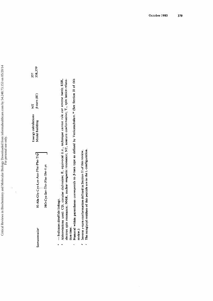
Ene
rgy
calc
ulat
ions
NT
357
Som
atos
tati
n'
-~
H-A
la-G
ly-C
ys-L
ys- A
sn-P
he-P
he-T
rp
Mod
el b
uild
ing
p-tu
rn (1
1')
HO
-Cys
-Ser
-Thr
-Phe
-Thr
-Lys
1
* ---
Indi
cate
s dis
ulfi
de li
nkag
e.
* A
bbre
viat
ions
use
d: C
D.
circ
ular
dic
hroi
sm;
E.
equi
voca
l (i
.e.,
tech
niqu
e ca
nnot
rul
e ou
t re
vers
e tu
rn);
ESR
, el
ectr
on s
pin
reso
nanc
e; N
MR
, nu
clea
r m
agne
tic
reso
nanc
e; N
T,
nont
urn
conf
orm
atio
n; T
I, sp
in l
attic
e-re
iaxa
- tio
n tim
e.
Num
eral
with
in p
aren
thes
es c
orre
spon
ds t
o /3
-turn
typ
e as
def
ined
by
Ven
kata
chal
am."
(S
ee S
ectio
n II
of
this
re
view
.) In
vers
e y-
turn
con
form
atio
n de
fine
d in
Sec
tion
I1 o
f th
is re
view
. T
he n
ongl
ycyl
resi
dues
of
this
pep
tide
are
in th
e L
-con
figu
rati
on.
' *
358,
359
w 4
W
Cri
tical
Rev
iew
s in
Bio
chem
istr
y an
d M
olec
ular
Bio
logy
Dow
nloa
ded
from
info
rmah
ealth
care
.com
by
54.2
48.7
3.15
3 on
05/
20/1
4Fo
r pe
rson
al u
se o
nly.
380 CRC Critical Reviews in Biochemistry
side chain-backbone hydrogen bonds.2zB Experimental confirmation of this predicted conformation has not been achieved.
4 . Angiorensin I1 (A 11) Although a number of conformations have been proposed for A 11, one likely struc-
ture is the inverse y-turn proposed by Printz et al.” (see Table 17). Although they suggested that the inverse y-turn conformation was the most likely structure, they also accepted the possibility that a p-turn conformation was a possible alternative. C D da ta have also been interpreted as indicating th,e presence of two /3-turns within the mole- cule,lSo although the contribution of the tyrosyl and phenylalanyl chromophores to the observed C D leads to certain ambiguity in this assignment (see Section III.C.1 ). In contrast, the ‘H-NMR study by Glickson et al.3s1 revealed that A 11 either exists in a conformation unlike the previously proposed /3- or inverse y-turn models, or occurs a s a statistical average of various conformations including fi- and inverse y-turns. The exact solution conformation of this hormone remains unknown.
5. Bradykinin Bradykinin is a pharmacologically active substance, produced by the kinin-generat-
ing system, which causes vasodilatation, increased capillary permeability, and either contraction or relaxation of extravascular smooth muscle. The substance was originally discovered by Rocha e S i l ~ a , ~ ~ ’ although the structure was established 11 years later by Elliott et al.369 Two C D studies have indicated that the molecule contains an inverse y-turn c ~ n f o r m a t i o n ’ ~ ’ ~ ~ ~ ~ (see Table 17). Such an inverse y-turn conformation is ex- perimentally consistent with various intramolecular distances estimated from fluores- cence energy and electron spin resonance.352
6. Oxytocin Oxytocin is a powerful stimulant of uterine contraction, which is secreted by the
posterior p i t ~ i t a r y . ’ ~ ~ Overwhelming ‘H-NMR evidence has been accumulated which indicates that the loop, formed by a disulfide linkage between residues 1 and 6 of this peptide, contains a /%turn structure, 131-13433 (see Table 17). In addition, X-ray diffrac- tion obtained from a model peptide for the oxytocin tail, H-L-CYS (Bzl) L-Pro-L-Leu- G I Y - N H ’ , ~ ~ indicated that this portion of the molecule occurs as a type 1 0-turn, a t least in the crystal state (see Table 3) .
7 . Luteinizing Hormone-Releasing Hormone (LH-R H) LH-RH (or LRF) is a hypothalamic factor responsible for stimulating the secretion
of the hormones from the anterior pituitary, which regulate ov~la t ion .” ’ Experimental evidence f rom charge-transfer experiments indicates that the molecule contains a type I I /3-turn190~355 (see Section lII.D.3.). However, high resolution IH-NMR experiments have yielded no evidence to substantiate the existence of a fi-turn (i.e., an intramole- cular hydrogen bond). 356 Empirical energy calculations have been completed for this molecule, and there was also n o indication of a favored /3-turn conf~rmat ion . ’~’
8. Somatosta tin Somatostatin is a 14-amino acid peptide isolated initially from bovine
hypothalami.”’ It has been established to have diverse biological activities including the inhibition of the release of insulin, growth hormone, glucagon, gastric acid, and pancreatic exocrine s e c r e t i o n ~ . ~ ~ ’ . ~ ’ ~ From direct and difference C D studies it was con- cluded that somatostatin contains a p-turn onf formation.^^^.^^^ This observation is also consistent with the results from model building based on the biological activity of
Cri
tical
Rev
iew
s in
Bio
chem
istr
y an
d M
olec
ular
Bio
logy
Dow
nloa
ded
from
info
rmah
ealth
care
.com
by
54.2
48.7
3.15
3 on
05/
20/1
4Fo
r pe
rson
al u
se o
nly.
October 1980 381
various synthetic bicyclic analogs of somatostatin, which led to the suggestion that this peptide hormone contains a /3-turn involving residues 7 through 10.358.359 Definitive proof for this /3-turn conformation remains to be established by ’H-NMR.
I. Protein Folding: 0-Turns as Possible Nucleation Sites Protein folding is the molecular mechanism which converts an open polypeptide
chain into the characteristic three-dimensional structure of the native protein mole- cule.’ Although numerous reviews dealing with general aspects of protein conforma- tion and of protein folding have been written, the role of the /3-turns in the folding of proteins has been discussed adequately in only two reviews.’.z41 The interested reader should consult both of these for further details. With the exception of presenting cer- tain of the most accepted concepts which implicate /3-turns in the mechanism of protein folding, the prodigious literature dealing with this subject will of necessity be neglected herein.
During the process of protein folding, the folding chain frequently changes direc- tion. The regions in which these changes of direction occur are called /3-turns. In ad- dition, the number of /3-turns in a protein is a linear function of the number of amino acid In such /3-turn-containing regions, short range interactions would be expected to dominate and to lead to an ordering of a short segment of the polypeptide chain. Such ordered segments may then function as nucleation sites where folding be- gins and then continues until the folding process is complete.6-8 Although this general concept has been espoused qualitatively over the years by several workers, a quantita- tive evaluation first resulted from the application of Monte Carlo, predictive methods by Tanaka a n d S ~ h e r a g a . ” ~ In their subsequent paper,376 they proved that 8-turn con- formations in rubredoxin, lysozyme, and ferricytochrome C played a significant role in the initial stages of protein folding.
Hiltner and Walton2’’ have favored a n alternative viewpoint that chain folding at reverse turns occurred not because specific residues in the turn favor the folding, but because these regions are such that they can have their intrinsic destabilization over- come by the driving force of long-range interactions.
Regardless of which model is demonstrated experimentally to be correct (and the evidence is certainly not conclusive at present), i t can be stated that certain p-turn regions in globular proteins are probably nucleation sites either because of their intrin- sic short-range interactions or because of additional interactions which lead to appar- ent nucleation involving a P-turn, although the interactions themselves might not be a consequence of initial /3-turn formation. This may account for the remarkably high degree of conservation of /3-turns in proteins, demonstrated for 10 mammalian proin- ~ u l i n s , ~ for 7 proteinase inhibitor^,^ and for 12 pancreatic ribonucleases, as well as for immunoglobulin light and heavy chain.z46
In summary, /3-turns and protein folding appear on theoretical grounds to be inex- tricable. However, the experimental proof from spectroscopic experiments investigat- ing the tendency of short, linear peptides to form stable p-turn conformations (as as- sessed by C D or NMR) has been equivocal.
J. Reverse Turns in Antibiotics, Toxins, Antitoxins, Ionophores, and Metabolic Prod- ucts 1 . Grarnicidin S and the Tyrocidines
Gramicidin S, a cyclic decapeptide with a repeating pentapeptide sequence L-Val-L- Orn-L-Leu-D-Phe-L-Pro, which was first isolated from Bacillus brevis by Gause and B r a z h n i k o ~ a , ~ ” shows antimicrobial activity. This antibiotic has played an interesting role in the history of peptide conformational analysis. It was the subject of studies
Cri
tical
Rev
iew
s in
Bio
chem
istr
y an
d M
olec
ular
Bio
logy
Dow
nloa
ded
from
info
rmah
ealth
care
.com
by
54.2
48.7
3.15
3 on
05/
20/1
4Fo
r pe
rson
al u
se o
nly.
382 CRC Critical Reviews in Biochemistry
using various methods including X-ray diffraction,‘O. NMR,15,J79,Jw and conforma- tional energy calculations,’06-208 but a consistent interpretation of the conformation was elusive until Ovchinnikov and co-worker~~’ postulated, primarily from ‘H NMR, the currently accepted C2-symmetric 0-turn-containing structure, which has the D-Phe- L-Pro residues occurring in the i + l’* and i + 2Ih positions of type 11’ &turns. This conformation has now been established as the favored one in crystals by X-ray diffrac- t i ~ n . ~ ~ ’
It has been found that the activity of gramicidin S depends critically on the proximity of the two charged ornithine side chain^.^^'^^^^ The average distance between these groups was estimated using electron spin resonance of a spin-labeled derivative to be 8 to 10 A with alcohol as a solvent. This finding, together with the observation that gramicidin S solubilizes lecithin, led to a model for its antibiotic activity which requires a step involving association of the positively-charged gramicidin S side chains and the negative phosphates of the phospholipid head g r o ~ p s . ’ ” ~ ’ ~ ~ Essential for this step would be a rigid conformation of the cyclic peptide, such that the ornithine side chains remain in proximity. The presence of 0-turns and intramolecular hydrogen bonding are principal factors in the maintenance of the peptide conformation.
Tyrocidines A-E are closely related to gramicidin S, since they are cyclic decapep- tides which share a common pentapeptide fragment with gramicidin S. Although a conformation similar to that of gramicidin S is suggested for the t y r o ~ i d i n e s , ’ ~ ~ . ~ ~ ~ no structure-activity studies have been reported.
2. Valinomycin Valinomycin is a cyclic dodecadepsipeptide antibiotic, isolated from Streptomyces
fu lv i s~imus , ’~~ which has been extensively studied with regard to both its structure and f~nc t ion . ”~ It acts on membranes, causing a large and specific increase in their K’ permeability.’e7~3ee This ionophoric role is attributed to the formation by valinomycin of a strong, selective, hydrophobic complex with K+.’“ The K’ in the complex can then move readily through the membrane, “carried” by valinomycin. The valinomycin-K’ complex is made of six 0-turns (alternating types I1 and 11‘). arranged so that the six non-hydrogen bonded carbonyls are oriented toward a central cavity. Uncomplexed valinomycin adopts different conformations depending upon solvent; many contain /3-turns. These conformations and the cation complexes of valinomycin have been stud- ied in detail by NMR66.e4.93.1zs.116,3e9 and by X-ray diffra~tion.~’*’~*~~~*~~’ I n addition, the relationship between structure and function in this ionophore has been extensively discussed.
3. Toxic and Antitoxic Peptides from Amanita phalloides For over a century, extracts of the deadly poisonous Amanita mushrooms have been
studied in efforts to determine the active components and to elucidate their modes of action. Three classes of peptides have been isolated from these toxic mushrooms and characterized as to structure and function. A comprehensive review of the chemistry and toxicology of these peptides has been written by Wieland and Faulsti~h.’~’
Two types of toxic peptides, the octapeptide amatoxins and the heptapeptide phal- lotoxins, are bicyclic, with unusual cross-linkages between Trp and Cys side chains. Nonetheless, their mechanisms of toxicity are reported to be d i s t i n ~ t . ~ ~ * ~ ’ ~ ’ The former group is considered to be the cause of death in humans after consumption of Amanita
The conformation of one phallotoxin, phalloidin, has been investigated by NMR.’93 N o unique structure was proposed, although it was concluded that the molecule is rigid, and has two N-Hs which are either buried or intramolecularly hydrogen-bonded. X-ray diffraction analyses of the phallotoxins have not been reported.
Cri
tical
Rev
iew
s in
Bio
chem
istr
y an
d M
olec
ular
Bio
logy
Dow
nloa
ded
from
info
rmah
ealth
care
.com
by
54.2
48.7
3.15
3 on
05/
20/1
4Fo
r pe
rson
al u
se o
nly.
October 1980 383
The structure of /I-amanitin, an amatoxin, has been solved by X-ray crystallogra- ~ h y . ’ ~ ~ The results agree fairly well with an NMR and reveal that a type I1 Ile-Gly /3-turn is present.
The most extensively studied peptide from Amanita phalloides is. interestingly, a phalloidin antagonist, antamanide, which is a cyclic decapeptide. Studies based on NMR, 128.129.362 energy and X-ray d i f f r a ~ t i o n ~ ’ . ’ ~ ~ . ~ ~ ~ agree in general on a conformation which contains two p-turns for the alkali metal complex of antaman- ides (and for a C2-symmetric, biologically active analog, [Phe4,Va16] antamamide). An- tamanide is thought to be membrane active,382 and, as was the case for gramicidin S and valinomycin. an important feature of the conformation is likely to be the arrange- ment in space of hydrophobic side chains. The adoption of a rigid, /3-turn-containing conformation is consistent with this requirement. Uncomplexed [Phe4.VaI6] antaman- ide in crystals does not contain 0- or y - t ~ r n s . ’ ~ ~ . ’ ~ ~ The N-Hs either hydrogen bond to water molecules or are involved in i + 4 -. i intramolecular interactions. In solution, a conformational equilibrium has been
4. Ferrichromes Siderochromes constitute a group of hydroxamic acid-containing iron chela tor^.^^^
One class of these compounds, the ferrichromes. isolated from the smut fungus Usti- lago s p h a e r ~ g e n a , ~ ~ ~ has as a major structural feature a cyclic hexapeptide backbone. X-ray diffraction analysis4s and nuclear magnetic resonan~e’~’ have established that the ferrichromes adopt a conformation containing an i + 3 - 1 intramolecular hydro- gen bond in a type I1 p-turn. The extreme rigidity of ferrichrome and various analogs, and the apparent close resemblance between the conformation of the chelate in crystals and in solution has led to extensive use of these peptides as models in conformational
Ferrichromes show potent growth factor activity, and are thought to act as cellular transport factors for iron,401 functioning via a cellular membrane transport system which is specific f o r the metal-bound hydroxamic acid.
analysis. 72.8 I .83.110.127
5. Other Antibiotics and Metabolic Products The crystal structures of two reverse turn-containing peptides with tuberculostatic
activity, isolated from bacterial cultures of Streptomyces griseoverticullatus var. tub- eracticus, have been r e p ~ r t e d . ~ ~ ’ . ~ ~ ~ Tuberact in~mycin‘~~ and viomycin403 have similar sequences, and both antibiotics take up crystal conformations containing pseudo-type I 1 p-turns with dehydro amino acids in the i + 2‘’ position, which is usually occupied by a D-residue in this type of p-turn.
Bouvardin is a metabolic product of the plant Bouvardia ternifolia, which has been found to have tumor-inhibiting activity.404 It is composed of a cyclic hexapeptide ring with an ether linkage between two tyrosine side chains. The resulting rigid conforma- tion, as revealed by an X-ray analysis,4(” is stabilized by one intramolecular hydrogen bond of the i + 3 + i, /3-turn type, although the +,y angles differ appreciably from the predicted values, I * presumably due to constraints from the bicyclic system.
Two natural peptides with i + 2 -. i intramolecular hydrogen bonding have been examined: dihydrochlamydocin, a cyclic tetrapeptide containing two y-turns;28 and cy- closporin A, a cyclic undecapeptide with both a p-turn (type I) and an inverse y - t ~ r n . ~ ~ The +,y angles of the corner residue in the y- or inverse y-turn are given in Table 3.
Ilamycin B,, an antibiotic whose crystal structure has been reported, and evolidine, a metabolic product from the tree Evodia xanthoxyloides, are both cyclic heptapep- tides which contain all ~-residues.’~’ Two p-turns were described in the crystal confor- mation of ilamycin B1, but the i + l’* and i + 2‘’ residues are linked by cis peptide
Cri
tical
Rev
iew
s in
Bio
chem
istr
y an
d M
olec
ular
Bio
logy
Dow
nloa
ded
from
info
rmah
ealth
care
.com
by
54.2
48.7
3.15
3 on
05/
20/1
4Fo
r pe
rson
al u
se o
nly.
384 CRC Critical Reviews in Biochemistry
In addition, one of the hydrogen bonds is bifurcated, with the ith C=O inter- acting with both the i + 31h and i + 21h N-Hs. NMR studies on both ilamycin B I 4 O 6
and e~olidine‘~’ suggested the existence of intramolecular hydrogen bonding in solu- tion, although structural details were not conclusively elucidated.
Several other natural products have been proposed to contain reverse turns (e.g., ~ t e n d o r n y c i n , ~ ~ ~ t e l o m y ~ i n , ~ ~ ~ , ~ ~ ~ patricin A,’ vernamycin B,3 and polymixin B3), but their conformations are not definitively established.
ACKNOWLEDGMENTS
John A. Smith was supported as a Senior Research Associate of the Howard Hughes Medical Institute. He acknowledges the further support and encouragement of Profes- sor K. Frank Austen. Lila G. Pease was supported by grants from the National Insti- tutes of Health (GM 23691-01A1) and the Petroleum Research Fund (10861-B4). The authors express their thanks to Patricia Grady and Lina Lau for the typing of this manuscript.
REFERENCES
I . Nhe thy , G. and Printz, M. P., The r turn, a possible folded conformation of the polypeptide chain. Comparison with the p-turn. Macromolecules, 5.755, 1972.
2. Matthcws, B. W., The pturn. Evidence of a new folded conformation in proteins, Macromolecules, 5, 818. 1972.
3. Urry, D. W. and Ohnishi, M., Nuclear magnetic resonance and the conformation of cyclic polypep- tide antibiotics, in Spectroscopic Approaches to Biomolecular Conformation, Urry. D. W., Ed., American Medical Association. Chicago, 1970.263.
4. Zimmerman. S. S. and Scheraga, H. A.. Local interactions in bends of proteins, Proc. Natl. Acad. Sci. U.S.A., 74,4126, 1977.
5 . Crawford, J. L., Lipscomb. W. N., and ScheUman, C. G., The reverse turn as a polypeptide confor- mation in globular proteins, Proc. Natl. Acad. Sci. U.S.A.,70,538, 1973.
6. Kuntz. I. D.. Protein folding, J. Am. Chem. Soc., 94,4009, 1972. 7. Anfinsen, C. B. and Scheraga, H. A., Experimental and theoretical aspects of protein folding, in
Advances in Protein Chemistry, Vol. 29. Anfinsen, C. B.. Edsall, J. T.. and Richards, F. M., Eds., Academic Press, New York, 1975.205.
8. Lewis. P. N.. Momany, F. A., and Scheraga, H. A., Folding of polypeptide chains in proteins: a proposed mechanism of folding, Proc. Natl. Acad. Sci. U.S.A.,68,2293, 1971.
9. Chou, P. Y. and Fasman. 0. D.. personal communication, 1978. 10. Schmidt, 0. M. J., Hodgkin. D. C., and Oughton, B. M., A crystallographic study of some deriva-
tives of gramicidin, Biochem. J . , 65.744, 1957. 1 I . Schwyzer. R., Sieber. P.. and Gorup. B.. Synthese zyklisher Peptide mit der Methode de (activierten
Ester*, Chimia, 12.90, 1958. 12. Shields, J. E. and McDoweU, S. T., Conformation of small peptides. I. Secondary structure in a
tetrapeptide, J. Am. Chem. Soc., 89, 2499. 1967. 13. Shields, J. E., McDoweU, S. T., Pavlos, J., and Gray, G. R.. Conformation of small peptides. 11.
Synthesis and infrared studies of small peptides. J. Am. Chem. SOC., 90.35, 1968. 14. Geddes. A. J., Parker, K. D., Atkins, E. D. T., and Brighton, E., “Cross 8’’ conformation in
proteins, J. Mol. Biol., 32,343, 1968. IS. Stem, A.. Gibbons, W. A., and Craig, L. C., A conformational analysis of gramicidin S-A by nuclear
magnetic resonance, Proc. Natl. Acad. Sci. U.S.A., 61,734, 1968. 16. Kopple. K. D., Ohnishi, M.. and Go, A.. Conformations of cyclic peptides. IV. Nuclear magnetic
resonance studies of cyclo-pentaglycyl-L-leucyl and cyclo-diglycyl-L-histidyl-diglycyl-L-tyrosyl, Bio- chemistry, 8.4087, 1969.
Cri
tical
Rev
iew
s in
Bio
chem
istr
y an
d M
olec
ular
Bio
logy
Dow
nloa
ded
from
info
rmah
ealth
care
.com
by
54.2
48.7
3.15
3 on
05/
20/1
4Fo
r pe
rson
al u
se o
nly.

October 1980 385
17. Portnova, S. L.. Shilin, V. V.. Balashova, T. A.. Biernat, J., Bystrov. V. F., Ivanov, V. T.. and Ovchinnikov. Y. A.. Conformational studies of cyclic peptides in solution. Nmr spectra of cyclo- hexapeptides consisting of L(D)-alanine and glycine residues, Tetrahedron Lett.. p.3085, 1971.
18. Venkatachalam, C. M.. Stereochemical criteria for polypeptides and proteins. V . Conformation of a system of three linked peptide units, Biopolymers, 6, 1425, 1968.
19. Woody, R. W., Studies of theoretical circular dichroism of polypeptides: contributions of 0-turns. in Peptides. Polypeptides and Proteins, Blout, E. R., Bovey, F. A.. Goodman, M.. and Lotan. N., Eds., John Wiley& Sons, New York. 1974,338.
20. Benedetti, E., Palumbo. M., Bonora, G. M., and Toniolo, C.. On the oxy analogues to the 4 -c 1 intramolecularly hydrogen-bonded peptide conformations, Macromolecules. 9.41 7, 1976.
21. Printz, M. P., N h e t h y , G., and Blcich. H., Proposed models for angiotensin I1 in aqueous solution and conclusions about receptor topography, Nature, 237, 135, 1972.
22. Chandrasekaran, R., Lakshminarayanan, A. V., Pandya, U. V., and Ramachandran, G. N., Con- formation of the LL and LD hairpin bends with internal hydrogen bonds in proteins and peptides, Biochim. Biophys. Acta, 303. 14, 1973.
23. Lewis, P. N., Momany. F. A., and Schcraga. H. A., Chain reversals in proteins, Biochim. Biophys. Acta, 303,211, 1973.
24. Deber, C. M., Evidence for /%turn analogs in proline peptides in the solid state. An infrared study, Macromolecules, 7,47 , 1974.
25. Hruby, V. J . . Conformations of peptides in solution as determined by nmr spectroscopy and other physical methods, in Chemistry and Biochemistry of Amino Acids, Peptides and Proteins, Vol. 3, Weinstein, 9.. Ed., Marcel Dekker, New York, 1974, 1 .
26. Richardson, J . S., Getzoff. E. D., and Richardson, D. C., The /3 bulge: a common small unit of nonrepetitive protein structure, Proc. Natl. Acad. Sci. U.S.A., 75, 2574, 1978.
27. Bystrov, V. F., Portnova, S. L., Tsetlin. V. 1.. Ivanov, V. T., and Ovchinnikov, Y. A., Conforma- tional studies of peptide systems. The rotational states of the NH-CH fragment of alanine dipeptides by nuclear magnetic resonance, Tetrahedron. 25,493. 1969.
28. Karle. I . L.. The state of the art of X-ray crystallography of peptides, in Peptides: Chemistry, Struc- tureandBiology, Walter, R. and Meienhofer, J . , Eds., Ann Arbor Science, Ann Arbor, Mich.. 1975, 61.
29. Ramachandran, G. N. and Sasisekharan, V., Conformation of polypeptides and proteins, Adv. Pro- tein Chem., 23,283, 1968.
30. Maigret, B.. Pullman. B., and Perahia, D., Molecular orbital calculations on the conformation of polypeptides and proteins. VII . Refined calculations on the alanyl residue, J . Theor. Biol., 31, 269, 1971.
31. Benedetti, E.. Structure and conformation of peptides: a critical analysis of crystallographic data, in Peptides,Goodman, M. and Meienhofer, J . , Eds., John Wiley& Sons, New York. 1977,257.
32. Karle, I. L. and Karle, J., An application of a new phase determination procedure to the structure of cyclo(hexaglycyl) hemihydrate, Acta Crystallogr., 16, 969, 1963.
33. Smith, 0. D., Duax, W. L., Langs, D. A., DeTitta. G. T., Edmonds, J. W., Rohrer, D. C., and Weeks. C. M., The crystal and molecular structure of the triclinic and monoclinic forms of valino- rnycin, C,,H,N,O,., J . Am. Chem. SOC., 97,7242, 1975.
34. Neupert-Laves, K. and Dobler, M., The crystal structure of a K' complex of valinomycin, Helv. Chim. Acta. 58,432, 1975.
35. Rudko. A. D., Lovell, F. M.. and Low, 9. W., Crystal structure of the C-terminal tetrapeptides of oxytocin, NatureLondon New Biol..232. 12, 1971.
36. Ueki, T., Ashida, T., Kakudo, M., Sasada. Y., and Katsube, Y., Molecular structure of p-bromo- carbobenzoxy-glycyl-L-propyl-L-leucyl-glycine, Nature (London), 216, 1205, 1967.
37. Ueki. T.. Ashida, T.. Kakudo, M., Sasada, Y., and Katsube, Y., Structure of p-bromocarbobenzoxy- glycyl-propyl-leucyl-glycine, Acta Crystallogr.. Sec. B, 25, 1840, 1969.
38. Ukei, T.. Bando. S., Ashida, T., and Kakudo, M.. The structure of o-bromocarbobenzoxy-glycyl- L-propyl-L-leucyl-glycyl-L-proline ethyl acetate monohydrate: a substrate of the enzyme, collagen- ase, Acta Crystallogr., Sec. B,27, 221 9, I97 I .
39. Lecomte. C., Aubury, A., Protas, J., Boussard, G., and Maurraud, M., Etude de la conformation rnol~culaire 2 ]';tat solide de la N-ac&yl-L-propyl-L-lactyl-m6thylamide, Acta Crystallogr., Sec. B, 30, 1992, 1974.
40. Karle, I . L., Gibson, J. W.. and Karle, J.. The conformation and crystal structure of the cyclic polypeptide Gly-Gly-D-Ala-D-Ala-GJy-Gly ' H,O, J . Am. Chem. SOC., 92, 3755, 1970.
41. Karle, 1. L.. Conformation of the lithium ion complex of antamanide, cyclic decapeptide and ion carrier, in the crystalline state, 1. Am. Chem. SOC., 96,4000, 1974.
Cri
tical
Rev
iew
s in
Bio
chem
istr
y an
d M
olec
ular
Bio
logy
Dow
nloa
ded
from
info
rmah
ealth
care
.com
by
54.2
48.7
3.15
3 on
05/
20/1
4Fo
r pe
rson
al u
se o
nly.
386 CRC Critical Reviews in Biochemistry
42. Smith, 0. D., D u x . W. L., Kcndrick, N. F., Marshall. G. R.. and Mathews, F. S., The crystal and molecular structure of a tetrapeptide, the benzyl ester of Boc-L-Pro-Aib-L-Ala-Aib. in Peptides, Goodman, M. and Meinhofer, J., Eds., John Wiley & Sons, New York. 1977,277.
43. Hossain. M. B. and van der Helm, D., Conformation and crystal structures of two cycloisomeric hexapeptides: cyclo-(L-alanyl-L-alanyl-glycylglycyl-L-alanylglycyl) monohydrate (1) and cyclo-(L- alanyl-L-alanylglycyl-L-alanylglycylglycyl) dihydrate (II), J . Am. Chem. Soc., 100.5 191, 1978.
44. Smith, 0. D. and Griffin, J. F.. Conformation of [Leu’] enkephalin from X-ray diffraction: features important for recognition at opiate receptor, Science, 199, 1214, 1978.
45. Zalkin, A.. Forrester, J. D.. and Templeton, D. H., Ferrichrome A hemihydrate. Determination of crystal and molecular structure, J. Am. Chem. SOC., 88, 1810, 1966.
46. Reed, L. L. and Johnson, P. L., Solid state conformation of the C-terminal tripeptide of oxytocin, L-Pro-L-Leu-Gly-NH2.0.5 H1O, J . Am. Chem. Soc.. 95,7523, 1973.
47. LeComte, C., Aubry, A., Protas. J., Boussard. G.. and Maurraud, M., Etude de la conformation mol6culaire ‘a I’6tat solide de la N-acetyl-L-prolyl-D-lactyl-methylamide, Acta Crystallogr., Sec. B, 30,2343, 1974.
48. Brown, J. N. and Teller, R. G., Crystal structure and molecular conformation of the hydrated cyclic hexapeptide cyclo-(L-Ala-L-Pro-D-Phe)2, 1. Am. Chem. Soc., 98,7565. 1976.
49. Karle, I. L., 3 -. 1 and 4 + 1 intramolecular hydrogen bonds in cyclo(G1y-Pro-Gly-D-Ala-Pro) (crys- tal structure analysis). in Peptides, Goodman, M. and Meienhofer, J., Eds., Halsted Press, New York, 1977,274.
50. Karle, I. L.,Crystal structure and conformation of cyclo(glycylprolylglycyl-D-alanylpolyl) containing 4 - 1 and 3 -. 1 intramolecular hydrogen bonds, J. Am. Chem. SOC., 100, 1286, 1978.
51. Kostansek. E. C.. Lipscomb, W. N., and Thiessen, W. E., The crystal structure and conformation of the cyclic hexapeptide cyclo(Gly-L-Pro-D-Ala)t. 1. Am. Chem. Soc.. 101,834,1979.
52. GWin, B. F. and Steinrauf. L. K., personal communication, 1978. 53. Brown, J. N. and Yang. C. H.. The crystal and molecular structure of the cyclic hexapeptide cyclo-
(Gly-Pro-D-Phe)2, J. Am. Chem. SOC., 101,445, 1979. 54. Karle. I. L,, Conformation of the cyclic pentapeptide Gly-L-Pro-L-Ser-D-Ala-L-Pro in the crystalline
state and example of rotational “isomerism” between analogs, 1. Am. Chem. Soc.. 101. 181, 1979. 55. Flippen-Anderson, J. and Karlc. I. L., personal communication, 1978. 56. Petcha, T, J., Weber, H. -P.. and Rkgger. A.. Crystal and molecular structure of an iodo-derivative
of the cyclic undecapcptide cyclosporin A, Helv. Chim. Acta, 59. 1480. 1976. 57. Farrar, T. C. and Becker, E. D.. Pulse and Fourier Transform NMR, Academic Press, New York.
1971. 58. Bovey. F. A., High Resolution NMR of Macromolecules, Academic Press, New York. 1972. 59. Dwck. R. A., Nuclear Magnetic Resonance in Biochemistry: Applications ro Enzyme Systems, Clar-
endon Press, Oxford, 1973.
60. Withrich. K.. NMR in Biological Research: Peprides and Proteins. North-Holland, Amsterdam, 1976.
61. Kopple, K. D. and Schamper. T. J., Determining solvent exposure of peptide protons by proton magnetic resonance, in Chemistry and Biology of Peptides, Meienhofer. J.. Ed., Ann Arbor Science, Ann Arbor, Mich., 1972.75.
62. Pitncr. T. P. and Urry. D. W.. Proton magnetic resonance studies in trifluoroethanol. Solvent mix- tures as a means of delineating peptide protons, J . Am. Chem. Soc., 94, 1399. 1972.
63. Ovchinnikov. Y. A., Ivanov, V. T., Bystrov, V. F., Miroshnikov. A. I., Shepel. E. N.. Abdullaev. N. D., Efremov. E. S.. and Senyavina, L. B., The conformation of gramicidin S and its N.N’-diacetyl derivative in solutions, Biochem. Biophys. Res. Commun., 39.217, 1970.
64. Kopple, K. D.. GO, A.. Scharnper. T. J., and Wilcox. C. S.. Conformation of cyclic peptides. VII. Cyclic hexapeptides containing the D-Phe-L-Pro sequence, J . Am. Chem. SOC., 95,6090. 1973.
65. Pease, L. G., Solution Conformations of (X-L-Pro-Y)2 Cyclic Hexapeptides: Sequence and Solvent as Conformational Determinants, Ph.D. thesis, Harvard University, Boston, 1975.
66. Davis. D. 0. and Tosteson. D. C., Nuclear magnetic resonance studies of the interactions of anions and solvent with cation complexes of valinomycin, Biochemistry, 14, 3962, 1975.
67. Watson, C.. Cyclic-(Gly-Pro-Gly(d2)-D-Ala-Pro): Study of a Cyclic Pentapeptide, B.A. thesis, Am- herst College, Amherst, Mass., 1976.
68. Pease, L. G. and Watson, C., Conformational and ion binding studies on a cyclic pentapeptide: evidence for and y turns in solution, in Peptides, Goodman, M. and Meienhofer, J . , Eds., Halsted Press, New York. 1977,346.
69. Pease, L. G. and Watson, C.. Conformational and ion binding studies of a cyclic pentapeptide. Evidence for B and y turns in solution, J . Am. Chem. Soc., 100, 1279, 1978.
Cri
tical
Rev
iew
s in
Bio
chem
istr
y an
d M
olec
ular
Bio
logy
Dow
nloa
ded
from
info
rmah
ealth
care
.com
by
54.2
48.7
3.15
3 on
05/
20/1
4Fo
r pe
rson
al u
se o
nly.
October 1980 387
70. Urry, D. W. and Long, M. M., Conformations of the repeat peptides of elastin in solution: an application of protein and carbon-I 3 magnetic resonance to the determination of polypeptide second- ary structure, CRC Crit. Rev. Biocbem., 4, I , 1976.
71. Kopple. K. D., Go. A., and Pilipaukas, D. R., Studies of peptide conformation. Evidence for f l structures in solution of linear tetrapeptides containing proline, J . Am. Chem. SOC., 97, 6830. 1975.
72. Llinh, M. and Klein, M. P.. Charge relay at the peptide band. A proton magnetic resonance study of solvation effects on the amide electron density distribution, J. Am. Cbem. SOC., 97, 4731, 1975.
73. Kopple. K. D., Go. A., and Schamper, T. J., Conformation of cyclic peptides. X. Conformational averaging in peptides with the sequence cycl~-(Gly-D-Xxx-L-Yyy)~, J . Am. Cbem. SOC., 100, 4289, 1978.
74. Kopple, K. D., Ohnishi, M.. and Go, A., Conformations of cyclic peptides. 111. Cyclo-pentaglycyl- tyrosyl and related compounds, 1. Am. Cbem. SOC., 91,4264, 1969.
75. Glickson. J. D., Cunningham, W. D.. and Marshall, 0. R.. Proton magnetic resonance study of angiotensin I I (Asn',Val') in aqueous solution, in Chemistry and Biology of Peptides, Meienhofer, J . , Ed., Ann Arbor Science, Ann Arbor, Mich., 1972, 563.
76. Urry, D. W. and Ohnishi, T., Recurrence of f l turns in repeat peptides of elastin: the hexapeptide Ala-Pro-Gly-Val-Gly-Val sequences and derivatives, in Peptides, Polypeptides and Proteins, Blout, E. R.. Bovey. F. A., Goodman, M., and Lotan, N., Eds., John Wiley & Sons, New York, 1974, 230.
77. Karplus, M., Contact electron-spin coupling of nuclear magnetic resonance, J . Cbem. Pbys., 30, 11, 1959.
78. Bystrov, V. F., Portnova, S. L., Balashova, T. A., Koz'min, S. A., Gavrilov, Y. D., and Afanas'ev, V. A.. Some aspects of nmr techniques for the conformational analysis of peptides. Pure Appl Chem., 36, 19. 1973.
79. Ramachandran, G. N., Chandrasakaran. R.. and Kopple, K. D., Variation of the NH-C'H coupling constant with dihedral angle in the nmr spectra of peptides, Biopolymers. 10, 21 13. 1971.
80. Aubry, A., Giessner-Pettre. C., Marraud. M., and Neel, J., Caractiristiques g;om;triques de la sequence H'-C'-NH de I'isoquinuclidone-3 examinee dans differents etats physiques. Influence sur la constante de couplage vicinal JHaH, Biopolymers, 13, 523, 1974.
81. Demarco, A., Llink. M., and Wiithrich, K.. Analysis of the 'H-nmr spectra of ferrichrome peptides. I I . The amide resonances, Biopolymers, 17,637, 1978.
82. Barfield. M. and Gearhart, H. L., Conformational dependence of vicinal H-N-C-H coupling con- stants in peptides, J. Am. Cbem. SOC., 95, 641, 1973.
83. Demarco. A., Llinb, M., and withrich, K., Analysis of the 'H-nmr spectra of ferrichrome peptides. I. The non-amidc protons, Biopolymers, 17,617, 1978.
84. Bystrov. V. F.. Gavrilov, Y. D., Ivanov, V. T.. and Ovchinnikov, Y. A., Refinement of the solution conformation of valinomycin with the aid of coupling constants from the "C-nuclear-magnetic-res- onance spectra, Eur. 3. Biochem., 78,63, 1977.
85. Cowburn. D., Fischman. A. J., Live, D. H.. Agosta. W. C.. and Wyssbrod, H. R., An approach to the unequivocal determination of peptide conformations, in Peptides, Goodman, M. and Meienho- fer. J., Eds., John Wiley& Sons, New York. 1977, 322.
86. Barfield, M., Hruby, V. J., and Meraldi, J.-P., The dependence of geminal H-H spin-spin coupling constants on +and p angles of peptides in solution, 1. Am. Chem. S O C . , ~ ~ , 1308, 1976.
87. Ballardin. A.. Fischman, A. J., Gibbons, W. A., Roy, J.. Schwartz, I. L.. Smith, C. W., Walter, R., and Wyssbrod, H., Conformational studies on [Pro'.Gly4]-oxytocin in dirnethyl sulfoxide by 'H nuclear magnetic resonance spectroscopy: evidence for a type I1 /%turn in the cyclic moiety, Biocbem- istry, 17, 4443, 1978.
88. Karplus. S. and Karplus, M., Nuclear magnetic resonance determination of the angle p in peptides, Proc. Natl. Acad. Sci. U.S.A.,69.3204, 1972.
89. Berger, S. and Roberts, J. D.. Nuclear magnetic resonance spectroscopy. "C-"N coupling constants as a conformational probe, J. Am. Cbem. SOC., 96,6757, 1974.
90. No&. J. H. and Schirmer, R. E., The Nuclear Overhauser Effect. Cbemical Applications, Aca- demic Press, New York, 1971.
91. Rowan, R., 111, McCammon, J. A., and Sykes, B. D., A study of the distances obtained from nuclear magnetic resonance nuclear Overhauser effect and relaxation time measurements in organic structure determination. Distances involving internally rotating methyl groups. Application to cis- and trans- crotonaldehyde, J . Am. Cbem. Soc.,96,4773,1974.
92. Shirmer, R. E., Davis, J. D., Noggle, J. H., and Hart, P. A., Conformational analysis of nucleosides in solution by quantitative application of the nuclear Overhauser effect, J . Am. Cbem. SOC., 94, 2561, 1972.
Cri
tical
Rev
iew
s in
Bio
chem
istr
y an
d M
olec
ular
Bio
logy
Dow
nloa
ded
from
info
rmah
ealth
care
.com
by
54.2
48.7
3.15
3 on
05/
20/1
4Fo
r pe
rson
al u
se o
nly.
388 CRC Critical Reviews in Biochemistry
93. Glickson, J. D.. Gordon, S. L., Pitner. T. P., Agresti, D. G., and Walter. R., Intramolecular 'H- nuclear Overhauser effect study of the solution conformation of valinomycin in dimethyl sulfoxide. Biochemistry, IS. 5721, 1976.
94. Rae, I. D., Stimson, E. R., and Scheraga, H. A., Nuclear Overhauser effects and the conformation of gramicidin S, Biochem. Biophys. Res. Commun.. 77,225, 1977.
95. Jones, C. R.. Sikakana, C. T., Kuo. M., and Gibbons, W. A., lnterproton distances for the p-turn residues of the peptide gramicidin S determined from nuclear Overhauser effect ratios, J . Am. Chem. SOC., 100,5960. 1978.
96. Bell, R. A. and Saunders, J. K., Correlation of the intramolecular nuclear Overhauser effect with internuclear distance, Can. J. Chem., 48, 11 16, 1970.
97. Leach. S. J. . Nimethy, G., and Scheraga. H. A., Use of proton nuclear Overhauser effects for the determination of conformations of amino acid residues in oligopeptides, Biochem. Biophys. Res. Commun., 75,207, 1977.
98. Howard, J. C., Ali. A., Schaaga, H. A., and Momany. F. A., Investigation of the conformations of four tetrapeptides by nuclear magnetic resonance and circular dichroism spectroscopy, and con- formational energy calculations, Macromolecules, 8,607, 1975.
99. Pitner. T. P., Walter. R., and Glickson, J. D., Mechanism of the intramolecular 'H nuclear Over- hauser effect in peptides and depsipeptides. Biochem. Biophys. Res. Commun., 70, 746, 1976.
100. Urry. D. W., Khaled, M. A., Rapaka. R. S., and Okamoto. K., Nuclear Overhauser enhancement evidence for inverse temperature dependence of hydrophobic side chain proximity in the polytetra- peptide of tropoelastin, Biochem. Biophys. Res. Commun.. 79,700. 1977.
101. Von Dreele, P. H., Rae. 1. D.. and Schaaga, H. A.. Nuclear magnetic resonance study of fibrinogen- like peptides and their structure in dimethyl sulfoxide and water, Biochemistry, 17, 956, 1978.
102. Khaled. M. A. and Urry. D. W., Nuclear Overhauser enhancement demonstration of the type I I p- turn in repeat peptide of tropoelastin, Biochem. Biophys. Res. Commun., 70,485, 1976.
103. Smith. I. C. P., Deslauriers, R.. Saito, H., Walter. R.. Garrigou-Lagrange. C., McGregor. H., and Sarantakis, D., Carbon-I3 nmr studies of peptide hormones and their components, Ann. N. Y. Acad. Sci.,222, 597, 1973.
104. Grant. D. M. and Cheney, B. V., Carbon-13 magnetic resonance. VII. Steric perturbation of the carbon-13 chemical shift, J. Am. Chem. Soc.,89,5315. 1967.
105. Madison, V.. Atreyi. M., Deber, C. M., and Blout. E. R.. Cyclic peptides. IX. Conformations of a synthetic ion-binding cyclic peptide, cyclo-(Pro-Gly),, from circular dichroism and 'H and nuclear magnetic resonance, J. Am. Chem. SOC., 96,6725, 1974.
106. Deber. C. M.. Madison, V.. and Blout, E. R.. Why cyclic peptides? Acc. Chem. Res., 9, 106, 1976. 107. Peasc, L. 0.. Niu. C. H., and Zimmcrman. G., Solution conformation of cyclo-(Gly-Pro-Ser-D-Ala-
Phe). Hydrogen-bonded reverse turns in cyclic pentapeptides, J. Am. Chem. SOC.. 101, 184, 1979. 108. Siemion. 1. Z., Wieland, T., and Pook, K. H., Influence of the distance of the proline carbonyl from
the p and y carbon on the "C chemical shifts, Angew. Chem. Int. Ed. Engl., 14,702, 1975. 109. Urry. D. W.. Mitchell. L. W., and Ohnishi, T., Solvent dependence of peptide carbonyl carbon
chemical shifts and polypeptide secondary structures: the repeat tetrapeptide of elastin, Biochem. Biophys. Res. Commun., 59.62, 1974.
110. Llinb, M.. Wilson, D. M., and Klcin. M. P.. Peptide hydrogen bonding. Conformation dependence of the carbonyl carbon-13 nuclear magnetic resonance chemical shifts in ferrichrome. A study by "C-['sN] Fourier double resonance spectroscopy, 1. Am. Chem. SOC., 99,6846, 1977.
1 I I . Smith, I. C. P., Deslauriers. R.. and Schaumburg, K.. Conformational mobility of peptides as eval- uated from spin-lattice relaxation times of I3C and 'H, in Peptides: Chemistry, Structure and Biology, Walter, R. and Meienhofer, J., Eds., Ann Arbor Science, Ann Arbor, Mich., 1975,97.
112. Deslauriers, R., Paiva, A. C. M.. Schaumburg. K.. and Smith, I. C. P.. Conformational flexibility of angiotensin 11. A carbon-I3 spin-lattice relaxation study, Biochemistry, 14, 878, 1975.
113. Niu, C.-H., Pease, L. G., and Blout, E. R.. Cyclic peptides. XVIII. 'T spin-lattice relaxation times of (X-Pro-Y)' cyclic hexapeptides. Biopolymers, 17, 115, 1978.
114. Hawkes. 0. E., Randall, E. W., and Bradley, C. H., Theory and practice for studies of peptides by "N nuclear magnetic resonance at natural abundance: gramicidin S. Nature (London), 257, 767, 1975.
115. Llinks, M., Horsely, W. J., and Klein, M. P., Nitrogen-15 nuclear magnetic resonance spectrum of alumichrome. Detection by a double resonance Fourier transform technique, J . Am. Chem. SOC., 98,7554, 1976.
116. Williamson, K. L., Pease, L. G., and Robem. J. D., Conformational analysis by nuclear magnetic resonance spectroscopy: "N nmr of a cyclic pentapeptide, 1. Am. Chem. SOC., 101,714,1979.
117. Khaled. M. A., Renugopalakrishnan, V., and Urry, D. W., Proton magnetic resonance and confor- mational energy calculations of repeat peptides of tropoelastin: the tetrapeptide. J . Am. Chem. SOC., 98.7547, 1976.
Cri
tical
Rev
iew
s in
Bio
chem
istr
y an
d M
olec
ular
Bio
logy
Dow
nloa
ded
from
info
rmah
ealth
care
.com
by
54.2
48.7
3.15
3 on
05/
20/1
4Fo
r pe
rson
al u
se o
nly.
October 1980 389
118. Pease, L. G., Deber, C. M.. and Blout, E. R., Cyclic peptides. V. 'H and "C nuclear magnetic resonance determination of the preferred 0 conformation for proline-containing cyclic hexapeptides. J. Am. Chem. S O C . , ~ ~ , 258, 1973.
119. Schwyzer, R., Grathwohl, Ch., Meraldi, J. P., Tun-Kyi, A., Vogel, R., and Wuthrich. K., The solution conformation of cycl~(Glycyl-L-prolyI-glycyl-glycyl-L-prolyl-glycyl), Helv. Chim. Acta, 5 5 . 2545, 1972.
120. Kopple, K. D., Schamper, T. J.. and Go, A., Conformation of cyclic peptides. V111. Cyclic hexapep- tides containing the L-Pro-D-Phe sequence, 1. Am. Chem. SOC., 95. 2597, 1974.
121. Demel, D. and Kessler, H., Conformation of cyclo [Pro-Phe-Gly-Phe-Gly], Tetrahedron Lett., p.2801, 1976.
122. Bara, Y. A.. Friedrich, A., Kessler, H., and Molter. M., Conformation of peptides. 11. 'H nmr investigation of the conformation of cyclo[Phel-GlyJ, Chem. Ber.. 1 1 1, 1045, 1978.
123. Davis, D. G., Gisin, B. F., and Tosteson, D. C.. Conformational studies of peptide cycle(D-Val-L- Pro-L-Val-D-Pro),. a cation-binding analogue of valinomycin, Biochemistry, 15, 768, 1976.
124. Easwaran. K. R. K., Pease, L. G., and Blout. E. R., Conformations of an ion-binding cyclic peptide analogue of valinomycin, cycb(L-Val-Gly-Gly-L-Pro),. Biochemistry, 18.61, 1979.
125. Patel, D. J . and Tonelli, A. E., Solvent-dependent conformations of valinomycin in solution, Bio- chemistry, 12.486. 1973.
126. Patel, D. J.. Carbon framework of valinomycin and its metal ion complex in solution, Biochemistry, 12, 496. 1973.
127. Llink, M.. Klein. M. P., and Neilands, J. B., Solution conformation of ferrichrome, a microbial iron transport cyclohexapeptide, as deduced by high resolution proton magnetic resonance, J. Mol. Biol., 52, 399. 1970.
128. Ivanov. V. T., Miroshnikov, A. I., Abdullaev, N. D., Senyavina, L. B., Arkhipova, S. F., Uvarova, N. N., Khalilulina, K. K . , Bystrov. V. F., and Ovchinnikov, Y. A., Conformation of the Na' complex of antamanide in solution, Biochem. Biophys. Res. Commun.,42,654, 1971.
129. Patel. D. J.. Conformation of antamanide-sodium in solution, Biochemistry, 12.677, 1973. 130. Patel, D. J. and Tonelli, A. E., A comparison of the solution and crystal conformation for the alkali
metal ion complex of antamanide, Biochemisrry, 13,788, 1974. 131. Urry. D. W., Ohnishi, M., and Walter, R., Secondary structure of the cyclic moiety of the peptide
hormone oxytocin and its deamino analog, Proc. Natl. Acad. Sci. U.S.A., 66, 1 1 I , 1970. 132. Urry, D. W. and Walter, R.. Proposed conformation of oxytocin in solution, Proc. Nail. Acad. Sci.
U.S.A.,68.956, 1971. 133. Brewster, A. I . R., Hruby, V. J., Glasel, J. A.. and Tonelli. A. E., Proposed conformations of
oxytocin and selected analogues in dimethyl sulfoxide as deduced from proton magnetic resonance studies, Biochemistry, 12, 5294, 1973.
134. Walter, R., Wyssbrod, H. R., and Glickson, J. D.. Conformational studies on [3-D aianinel-oxytoxin and [4-D-alanine]-oxytocin in dimethyl sulfoxide by 'H nuclear magnetic resonance spectroscopy. Interpretation in terms of a p turn in the cyclic moiety, J . Am. Chem. SOC., 99.7326. 1977.
135. Madison, V., Deber, C. M.. and Blout. E. R., Cyclic peptides. XVII. Metal and amino acid com- plexes of cycl@Pro-Gly), and analogues studied by nuclear magnetic resonance and circular dichro- ism. J. Am. Chem. Soc.,99, 4788, 1977.
136. Sears, D. W. and Beychok. S., Circular dichroism. in Physical Principles and Techniques of Protein Chemistry, (Part C), Leach, S . J . , Ed., Academic Press, New York. 1973,445.
137. Schellman, J. A.. Symmetry rules for optical rotation. Acc. Chem. Res., I , 144. 1968. 138. Condon, E. U., Altar. W., and Eyring, H., One-electron rotatory power, 1. Chem. Phys., 5 , 753,
139. Kirkwood. J . G., On the theory of optical rotatory power, J. Chem. Phys., 5,479, 1937. 140. Moffitt, W.. Optical rotatory dispersion of helical polymers, J. Chem. Phys., 25,467, 1956. 141. Hbhn, E. G . and Weigang, 0. E., Jr., Electron correlation models for optical activity. J. Chem.
142. Bayley, P. M.. Nielsen, E. B., and Schellman. J. A., The rotatory properties of molecules containing
143. Madison, V., Conformational energy and circular dichroism computed for cyclo-(Pro-Gly),. Biopo-
144. Laiken, S., Printz, M.. and Craig, L. C., Circular dichroism of the tyrocidines and gramicidin S-A.,
145. Greenfield, N. and Fasman, 0. D., Computed circular dichroism spectra for the evaluation of protein
146. Woody, R. W., Improved calculation of the nn* rotational strength in polypeptides, J . Chem. Phys.,
1937.
Phys.,48, 1127, 1968.
two peptide groups: theory, J. Phys. Chem., 73,228, 1969.
lymers, 12, 1837. 1973.
J. Biol. Chem., 244.4454. 1969.
conformation, Biochemistry. 8,4108. 1969.
49.4797, 1968.
Cri
tical
Rev
iew
s in
Bio
chem
istr
y an
d M
olec
ular
Bio
logy
Dow
nloa
ded
from
info
rmah
ealth
care
.com
by
54.2
48.7
3.15
3 on
05/
20/1
4Fo
r pe
rson
al u
se o
nly.

390 CRC Critical Reviews in Biochemistry
147. Woody, R. W., Optical properties of polypeptides in the /3-conformation, Biopolymers, 8,669, 1969. 148. Stimson. E. R.. Zimmerman. S. S.. and Scheraga, H. A., Conformational studies of oligopeptides
containing proline and glycine. Macromolecules, 10. 1049. 1977. 149. Helbecque, N. and Loucheux-Lefebvre, M.-H., Synthesis and circular dichroism studies of two poly-
peptides H-[Gly-(Pro),].-OHand H-[Gly-(Pro),].-OH, Int. J. Pept. Protein Res., 11, 353. 1978. 150. Grcff, D., Fermandjian. S., Fromageot, P., Khosla, M. C., Smeby. R. R., and Bumpus, F. M.,
Circular-dichroism spectra of truncated and other analogues of angiotensin 11, Eur. J . Biochem., 61, 297, 1976.
151. Chen. Y.-H., Lo, T.-B., and Yang, J. T., Optical activity and conformation of cobra neurotoxin, Biochemistry, 16, 1826, 1977.
152. Visser. L. and Louw. A. I., The conformation of cardiotoxins and neurotoxins from snake venoms, Biochim. Biophys. Acta, 533,80, 1978.
153. Kawai. M. and Fasman, G., A model /3 turn. Circular dichroism and infrared spectra of a tetrapep- tide. 1. Am. Chem. SOC., 100,3630, 1978.
154. Urry, D. W. and Ohnishi, T.. Studies on the conformations and interactions of elastin. Proton mag- netic resonance of the repeating tetramer. Biopolymers. 13. 1223. 1974.
155. Brahms, S., Brahms, J., Spach, G., and Brack, A., Identification of /3, /3-turns and unordered con- formations in polypeptide chains by vacuum ultraviolet circular dichroism, Proc. Natl. Acad. Sci. lJ.S.A..74,3208, 1977.
156. Bush, C. A., Sarkar, S. K., and Kopple, K. D., Circular dichroism of /3 turns in peptides and proteins, Biochemistry, 17,4951, 1978.
157. Pease, L. 0.. Gilbert. D. A., Bingaman, B. M.. and Freilich, S. C., unpublished data. 1978. 158. Urry, D. W., Ruiter, A. L., Starcher, B. C., and Hinners, T. A., Conformational studies on poly-
peptide antibiotics. I. Circular dichroism studies on tyrocidine B. gramicidin S, and their hydrogen- ated derivatives, Antimicrobial Agents Chemother.. p.87, 1968.
159. Crippen. 0. M. and Yang. J. T.. Conformation of N-acetyl-L-alanine-N’-methylamide in I ,2-dich- loroethane by circular dichroism and optical rotatory dispersion. 1. Phys. Chem., 78. 1127, 1974.
160. Smith, J. A.. Synthesis, Structure and lmmunochemistry of Peptide Sequences in Globins and Staph- ylococcal Nuclease, Ph.D. thesis, University of Melbourne, Parkville. Vic.. Australia, 1977.
161. Cann, J. R.. Stewart, J. M.. and Matsueda. G. R., A circular dichroism study of the secondary structure of bradykinin, Biochemistry, 12,3780, 1973.
162. Madison, V. and Schellman, J., Location of proline derivatives in conformational space. 1. Confor- mational calculations; optical activity and nmr experiments, Biopolymers, 9, 5 11 , 1970.
163. Chou, P. Y. and Fasman, G. D., /3-turns in proteins, J. Mol. Biol., 115. 135, 1977. 164. Urry, D. W.. Long, M. M., Ohnishi, T., and Jacobs, M.. Circular dichroism and absorption of the
polytetrapeptide of elastin: a polymeric model for the /3-turn, Biochem. Biophys. Res. Commun., 61, 1427, 1974.
165. Strickland, E. H., Aromatic contributions to circular dichroism spectra of proteins, CRC Crit. Rev. Biochem., 2, 113, 1974.
166. Woody, R. W.. Aromatic side-chain contributions to the far ultraviolet circular dichroism of peptides and proteins, Biopolymers, 17, 1451, 1978.
167. Madison, V. and Schehan , J. A., Location of proline derivatives in conformational space. 11. The- oretical optical activity, Biopolymers, 9, 569, 1970.
168. Miyazawa, T., Infrared spectra and helical conformations, in Poly-o-Amino Acids, Fasman, G. D.. Ed., Marcel Dekker, New York, 1967,69.
169. Susi. H.. Infrared spectra of biological macromolecules and related systems, in Structure and Stability of Biological Macromolecules, Timasheff. S. N. and Fasman, G. D.. Eds.. Marcel Dekker, New York, 1969,575.
170. Hanlon, S., Infrared studies on biopolymers and related models, in Spectroscopic Approaches to Biomolecular Conformation, Urry. D. W., Ed., American Medical Association, Chicago, 1970, 161.
171. Fraser. R. D. B. and Suzuki, E., Infrared methods, in Physical Principles and Techniques of Protein Chemistry. (Part B). Leach, S. J.. Ed., Academic Press, New York, 1970,213.
172. Pimental, 0. C. and McClellan. A. L.. The Hydrogen Bond, W. H. Freeman, San Francisco, 1960. 173. Shimanouchi. T. and Mizushima. S.. lntramolecular rotation and the structure of high polymers. I.
The structure of polypeptide chain, Bull. Chem. SOC. Jpn., 21, 1, 1948. 174. Mizushima. S., Shmanouchi, T., Tsuboi, M., and Souda. R.. Additional studies on the intramole-
cular hydrogen bonding in acetylglycine N-methylamide, 1. Am. Chem. SOC., 74,270, 1952. 175. Tsuboi, M., Shimanouchi, T., and Mizushima. S., Near infrared spectra of compounds with two
peptide bonds and the configuration of a polypeptide chain. VII. On the extended forms of polypep- tide chains, J. Am. Chem. SOC., 81. 1406. 1959.
Cri
tical
Rev
iew
s in
Bio
chem
istr
y an
d M
olec
ular
Bio
logy
Dow
nloa
ded
from
info
rmah
ealth
care
.com
by
54.2
48.7
3.15
3 on
05/
20/1
4Fo
r pe
rson
al u
se o
nly.
October 1980 391
176.
177.
178.
179.
180.
181.
Avignon. M., Huong. P. V., Lascombe, J., Marraud, M., and Neal, J., Etude, par spectroscopie infra-rouge. de la conformation de quelques composes peptideques modeles. Biopolymers. 8. 69, 1969.. Smolikov;, J., V h k , A., and B l h a , K., Amino acids and peptides. CIII. Infrared spectra and conformations of methylamides of N-acylated amino acids with a hydroxyl group in the side chain, Collect. Czech. Chem. Commun., 36,2424. 1971. Lewis, P. N.. Momany, F. A., and Scheraga, H. A., Energy parameters in polypeptides. Vl. Con- formational energy analysis of the N-acetyl N’methylamides of the twenty naturally occurring amino acids, Isr. J. Chem., 11. 121, 1973. Burgess, A. W. and Scheraga, H. A.. Stable conformations of dipeptides. Biopolymers, 12, 2177. 1973. Grenie, Y., Avignon. M., and Garrigou-Lagrange. C., Molecular structure study of dipeptides iso- lated in an argon matrix by infrared spectroscopy, J . Mol. Struct., 24. 293, 19:s. Marraud, M., Ncel, J., Avignon, M., and Huong. P. Y., Contribution a I’etude conformationnelle des compos6s dipeptideques en solution, J. Chim. Phys., 67,959, 1970.
182. Boussard. G., Marraud, M., and Neel, J.. Etude experimentale du mode de repliement p dans qu- elques mol;cules tripeptideques modiles. Interpritation des spectres d’absorption infrarouge. 1.
183.
184.
185.
186.
187.
188.
Chim. Phys.,7l, 1081. 1974. von Drcelc. P. H. and Stenhouse, I. A., A method for assigning hydrogen bonds using isotope effects in nuclear magnetic resonance and infrared spectroscopy, 1. Am. Chem. Soc., 96,7546. 1974. Stanley, H. E., Asher, I. M., Rothschild, K. J., Phillies. G. D. J.. Carew, E. B., Bansil. R.. and Michaels, I. A., Resonance and non-resonance Raman spectroscopy: a probe of peptide and protein conformation, in Peptides: Chemistry, Structure and Biology. Walter, R. and Meienhofer. J.. Eds., Ann Arbor Science, Ann Arbor, Mich., 1975.227. Stephens, R. M., Infrared and Raman spectroscopy, in Amino-acids, Peptides and Proteins, Vol. 5. Sheppard, R. C., Ed., Thechemical Society, London, 1974,223. Avignon, M., Garrigou-Lagrange. C.. and Bothorel, P., Conformational analysis of dipeptides in aqueous solution. I I . Molecular structure of glycine and alanine dipeptides by depolarized Rayleigh scattering and laser Raman spectroscopy, Biopolymers, 12, 1651, 1973. Koyama, Y. and Shimanouchi. T., An experimental study of the internal rotation potentials about the N-C. and C.-C’ axes of the peptide backbone, in Peptides, Polypeptides and Proteins. Blout, E. R., Bovey, F. A.. Goodman, M., and Lotan, N., Eds., John Wiley&Sons. New York, 1974,396. Koyama, Y. and Shimanouchi, T., Conformations of model compounds of protein. I . Acetyl glycine N-methylamide, Biopolymers, 6, 1037, 1968.
189. Tu, A. T., Bjamason, J. B., and Hruby, V. J., Conformation of oxytocin studies by laser Raman spectroscopy, Biochim. Biophys. Acta, 533.530. 1978.
190. Donzel, B., Gilon, C., Blagdon, D., Erisman, M., Burnier, J., Goodman, M.. Rivier. J., and Mon- ahan, M.. Conformational studies on the luteinizing hormone releasing factor (LRF) and related compounds, in Peptides: Chemistry, Structure and Biology, Walter, R. and Meienhofer. J., Eds., Ann Arbor Science, Ann Arbor, Mich.. 1975. 165.
191. Deranleau, D. A. and Schwyzer, R., Charge transfer as a molecular probe in systems of biological interest. lntermolecular interactions of the indole-pyridinium type, Biochemistry, 9, 126, 1969.
192. Scheraga. H. A.. Calculations of conformations of polypeptides, Adv. Phys. Org. Chem., 6, 103. 1968.
193. Momany, F. A.. McOuue, R. F., Burgess, A. W.. and Scheraga, H. A., Energy parameters in poly- peptides. VII. Geometric parameters, partial atomic charges, nonbonded interactions, hydrogen bond interactions, and intrinsic torsional potentials for the naturally occurring amino acids, J . Phys. Chem., 79,2361. 1974.
194. Scheraga. H. A.. Conformational energy calculations on peptides. in Peptides, Goodman, M. and Meienhofer. J . , Eds., Halsted Press, New York, 1977, 246.
195. Ramachandran, G. N. and Chandrasekaran, R., Conformational energy map of a dipeptide unit in relation to infrared and nuclear magnetic resonance data, Biopolymers. 10.935, 1971.
196. Tonelli. A. E. and Brewster, A. I., The conformational characteristics in solution of the cyclic hexa- peptide Gly-Gly-D-Ala-D-Ala-Gly-GJy, J. Am. Chem. Soc., 94,2851, 1972.
197. Ramakrishnan, C. and Sarathy, K. P., Stereochemical studies on cyclic peptides. V. Conformational analysis of cyclohexapeptides, Inr. J. Protein Res., I , 103. 1969.
198. Sarathy, K. P. and Ramakrishnan, C.. Stereochemical studies on cyclic peptides. V11. Effect of dif- ferent types of energies on the hydrogen-bonded conformations of cyclic hexapeptides, Inr. 1. Protein Res.,4. I , 1972.
199. Go. N. and Scheraga, H. A.. Calculation of the conformation of cyclo-hexaglycyl, Macromolecules, 6. 525, 1973.
Cri
tical
Rev
iew
s in
Bio
chem
istr
y an
d M
olec
ular
Bio
logy
Dow
nloa
ded
from
info
rmah
ealth
care
.com
by
54.2
48.7
3.15
3 on
05/
20/1
4Fo
r pe
rson
al u
se o
nly.
392 CRC Critical Reviews in Biochemistry
200. G6. N.. and Scheraga. H. A., Calculation of the conformation of cyclo-hexaglycyl (corrections). Macromolecules, 7, 148, 1974.
201. Schwyzer, R. and Ludescher, U., Untersuchungen uber die Konformation des cyclischen Hexapeptids cyclo-Glycyl-L-prolyl-glycyl-glycyl-L-prolyl-glycyl mittels protonenmagnetischer Resonanz und Par- allen zum cyclodecapeptid Gramicidin s, Helv. Chirn. Acta, 52,2033, 1969.
202. Madison, V. S., Guides to the evaluation of peptidc conformation, in Peptides. Polypeptides and Protein, Blout, E. R., Bovey, F. A.. Goodman, M.. and Lotan, N., Eds., John Wiley & Sons, New York, 1974,89.
203. Torchia. D. A., di Corato, A., Wong, S. C. E., Deber. C. M., and Blout, E. R., Cyclic peptides. 11. Solution conformations of cyclo(Prolylserylglycylprolylsery1glycyl) from nuclear magnetic resonance, J . Am. Chem. Soc.,94.609, 1972.
204. Torchia, D. A., Wong, S. C. K., Deber. C. M., and Blout, E. R., Cyclic peptides. 111. Solution conformations of cyclo(Serylprolylg1ycylserylprolylglycyl) from nuclear magnetic resonance, 1. Am. Chem. SOC., 94.616.1972.
205. Tonelli, A. E., An approximate treatment of the conformational characteristics of the cyclic hexa-L- peptides (Pro-Ser-Gly-Pro-Ser-Gly) and (Ser-Pro-Gly-Ser-Pro-Gly), J. Am. Chem. Soc., 94. 346, 1972.
206. Momany, F. A., Vanderkooi, G., Tuttle, R. W., and Scheraga, H. A., Minimization of polypeptide energy. IV. Further studies of gramicidin S, Biochemistry, 8. 744, 1969.
207. De Santis. P. and Liquori. A. M.. Conformation of grarnicidin S, Biopolymers. 10,699, 1971. 208. Dygert. M., GE, N., and Schcraga, H. A., Use of a symmetry condition to compute the conformation
of gramicidin S, Macromolecules, 8.750, 1975. 209. Deber, C. M., Torchia, D. A., Wong, S. C. K., and Blout, E. R.. Conformational interconversions
of the cyclic hexapeptide cyclo (Pro-Gly),, Proc. Natl. Acad. Sci. U.S.A.,69, 1825. 1972. 210. Simon, I., Nimethy. G., and Scheraga, H. A., Conformational energy calculations of the effects of
sequence variations on the conformations of two tetrapeptides, Macromolecules, 11,797,1978. 21 1. Hunvitz, F. I. and Hopfinger. A. J., Conformational analysis of a chain reversal in a-chymotrypsin,
Int. J. Pept. Protein Res., 8.543, 1976. 212. Chandrasekaran, R. and Prasad, B. V. V. V., Conformation of polypeptides containing alternating
L-amino and D-amino acids, CRCCrir. Rev. Biochem., 5. 125, 1978. 213. Hiltner, W. A. and Walton, A. G., Energetics of folding of a lysozyme p-bend. J. Mol. Biol., 92,
567, 1975. 214. Nishikawa, K., Momany, F. A.. and Scheraga, H. A., Low-energy structures of two dipeptides and
their relationship to bend conformations, Macromolecules, 7,797, 1974. 215. Zimmcrman. S. S. and Scheraga, H. A., Influence of local interactions on protein structure. 1. Con-
formational energy studies of N-acetyl-"methylamides of Pro-X and X-Pro dipeptides, Biopoly- men, 16,811, 1977.
216. Zimmcrman, S. S. and Scheraga, H. A.. Influence of local interactions on protein structure. 11. Conformational energy studies of N-acetyl-"methylamides of Ala-X and X-Ala dipeptides. Biopo- lymers, 17. 1849, 1978.
217. Zimmerman. S. S. and Scheraga. H. A., Influence of local interactions on protein structure. 111. Conformational energy studies of N-acetyl-N'methylamides of GIy-X and X-Gly dipeptides, Biopo- lymers, 17. 1871, 1978.
218. Zimmerman, S. S. and Schcraga, H. A.. lnfluence of local interactions on protein structure. 1V. Conformational energy studies of N-acetyl-N'methylamides of Ser-X and X-Ser dipeptides, Biopo- lymers, 17, 1885, 1978.
219. Renugopalakrishnan, V., Khaled, M. A.. and Urry, D. W., Proton magnetic resonance and confor- mational energy calculations of repeat peptides of tropoelastin: the pentapeptide, 1. Chem. SOC. Perkin Trans. 2,111, 1978.
220. Renugopalakrishnan, V., Khaled, M. A.. Rapaka. R. S.. and Urry, D. W., Proton magnetic reso- nance and conformational energy calculations of repeat peptides of tropoelastin: a permutation of the hexapeptide, Biochim. Biophys. Acta, 536,421, 1978.
221. Kang, S. and Walter. R., Theoretical studies on Pro-Leu-Gly-NH, conformation, Proc. Natl. Acad. Sci. US. A., 73, 1 199, 1976.
222. Blagdon, D. E., Rider. J., and Goodman, M., Proposed tertiary structure for the hypothalamic thyrotropin-releasing factor, Proc. Natl. Acad. Sci. U.S.A., 70, 1166, 1973.
223. Burgess, A. W.. Momany, F. A., and Scheraga, H. A., Conformational analysis of thyrotropin releasing factor, Proc. Natl. Acad. Sci. U.S.A., 70, 1456, 1973.
224. Momany. F. A., Conformational analysis of methionine-enkephalin and some analogs, Biochcm. Biophys. Res. Commun., 75. 1098, 1977.
225. Isogai, Y., Nimethy. 0.. and Scheraga, H. A.. Enkephalin: conformational analysis by means of empirical energy calculations, Proc. Netl. Acad. Sci. U.S.A., 74,414, 1977.
Cri
tical
Rev
iew
s in
Bio
chem
istr
y an
d M
olec
ular
Bio
logy
Dow
nloa
ded
from
info
rmah
ealth
care
.com
by
54.2
48.7
3.15
3 on
05/
20/1
4Fo
r pe
rson
al u
se o
nly.
October 1980 393
226. DeCoen, J . L., Humblet, C.. and Koch, M. H., Theoretical conformational analysis of met-enke- phalin, FEBSLett.,73. 38, 1977.
227. Loew, G. H. and Burt, S. K., Energy conformation study of met-enkephalin and its D-Ala' analogue and their resemblance to rigid opiates, Proc. Natl. Acad. Sci. U.S.A., 75, 7, 1978.
228. Anderson. J . S. and Scheraga, H. A., Conformational energy calculations on the contraceptive tetra- peptide H-Thr-Pro-Arg-Lys-OH, Macromolecules, 1 I , 812, 1978.
229. Tonelli, A. E., Approximate treatment of the conformational characteristics of the cyclic deca-L- peptide antamanide and its sodium complex in solution, Biochemistry, 12, 689, 1973.
230. Pullman, B. and Pullman, A., Molecular orbital calculations o n the conformation of amino acid residues of proteins, Adv. Protein Chem., 28, 348. 1974.
231. Flurry. R. L.. Jr., Abdulnur, S. F., and Bopp, J. M., Jr., Studies of the thyrotropin-releasing factor. 11. Conformations of TRF and some analogs, Biopofymers, 17,2679, 1978.
232. Matthews, B. W., X-ray structure of proteins, in The Proteins, Vol. 111, Neurath, H. and Hill, R. L.. Eds., Academic Press, New York, 1977,403.
233. Campbell, 1. D., Dobson, C. M., and Williams. R. J. P., N.m.r. studies on the structure of lysozyme in solution, Proc. R . SOC. London Ser. A, 345.41. 1975.
234. Campbell. I. D., Dobson, C. M., and Williams, R. J. P.. Proton magnetic resonance studies of tyrosine residues of hen lysozyme; assignment and detection of conformational mobility, Proc. R. SOC. London Ser. B, 189,503, 1975.
235. Wagner, G. . DeMarco, A., and Wzthrich, K.. Dynamics of the aromatic amino acid residues in the globular conformation of the basic pancreatic trypsin inhibitor (BPTI). I. 'H nmr studies, Biophys. Struct. Mechanism. 2. 139, 1976.
236. DeMarco, A., Tschesche, H., Wagner, G., and Wcthrich. K., 'H nmr studies at 360 Mhz of the methyl groups in native and chemically modified basic pancreatic trypsin inhibitor (BPTI). Biophys. Sfrucr. Mechanism, 3 . 303, 1977.
237. Snyder, G. H., Rowan, R., 111, and Sykes. B. D., Complete tyrosine assignments in the high-field 'H nuclear magnetic resonance spectrum of bovine pancreatic trypsin inhibitor selectively reduced and carboxamidomethylated at cystine 14-38. Biochemistry, 15,2275, 1976.
238. Dwek. R. A.. Wain-Hobson, S., Dower, S. K., Gettins, P., Sutton, 9. J., Perkin. S. J., and Givol, D., Structure of an antibody combining site by magnetic resonance, Nature (London). 266, 31, 1977.
239. Morris. A. T. and Dwek, R. A., Some recent applications of the use of paramagnetic centres to probe biological systems using nuclear magnetic resonance, 0. Rev. Biophys., 10,421, 1977.
240. Perkins, S. J . and WGthrich, K., Structural interpretation of lanthanide binding to the basic pan- creatic trypsin inhibitor by 'H nmr at 360 Mhz, Biochim. Biophys. Acta, 536,406, 1978.
241. Gordon, S. L. and W;'thrich, K., Transient proton-proton Overhauser effects in horse ferrocyto- chromec, 1. Am. Chem. SOC., 100,7094, 1978.
242. Nimcthy, G. and Scheraga. H. A.. Protein folding, 0. Rev. Biophys., 10,239, 1977. 243. Chou, P. Y. and Fasman, G. D., Prediction of the secondary structure of proteins from their amino
acid sequence, Adv. Enzymol. Relat. Areas Mol. Biol.,47,45, 1978. 244. Chou, P. Y. and Fasman, G. D., Empirical predictions of protein conformation, Annu. Rev.
Biochem.,47,251, 1978. 245. Burgess, A. W., Ponnuswamy, P. I., and Scheraga, H. A.. Analysis of conformations of amino
acids of prediction of backbone topography in proteins, Isr. J. Chem., 12.259, 1974. 246. Bunting, J. R.. Athey, T. W., and Cathou, R. E., Backbone folding of immunoglobulin light and
heavy chains: a comparison of predicted P-bend positions, Biochim. Biophys. Acfa, 285.60, 1972. 247. Krigbaum. W. R. and Knutton. S. P., Prediction of the amount of secondary structure in a globular
protein from its amino acid composition, Proc. Natl. Acad. Sci. U.S.A.. 70.2809, 1973. 248. Rose, G. and Seltzer. J., A new algorithm for finding the peptide chain turns in a globular protein,
J. Mof. Biol., 1 13, 153,1977. 249. Chou, P. Y. and Fasman, G. D., Conformational parameters for amino acids in helical, P-sheet,
and random coil regions calculated from proteins, Biochemistry, 13, 21 1 , 1974. 250. Chou, P. Y. and Fasman, G. D., Prediction of protein conformation, Biochemistry. 13,222, 1974. 251. Nagano, K., Logical analysis of the mechanism of protein folding. 11. The nucleation process, 1.
252. Argos, P., Hanei. M., and Garavito, R. M., The Chou-Fasman secondary structure prediction
253. Nagano, K., Logical analysis of the mechanism of protein folding. I . Predictions of helices, loops
254. Levitt, M. and Greer, J., Automatic identification of secondary structure in globular proteins, J.
255. Levitt, M., Conformational preferences of amino acids in globular proteins, Biochemistry. 17, 4277,
Mol. Biol.,84,337, 1974.
method with an extended data base, FEBSLett., 93, 19, 1978.
and P-structures from primary structure, J. Mof. Biol., 75,401, 1973.
Mof . Biof., 114, 181. 1977.
1978.
Cri
tical
Rev
iew
s in
Bio
chem
istr
y an
d M
olec
ular
Bio
logy
Dow
nloa
ded
from
info
rmah
ealth
care
.com
by
54.2
48.7
3.15
3 on
05/
20/1
4Fo
r pe
rson
al u
se o
nly.
394 CRC Critical Reviews in Biochemistry
256. Chou. P. Y. and Fasman. 0. D.. Prediction of protein structure, in Peptides, Goodman, M. and Meienhofer, J., Eds., Halsted Press, New York, 1977, 284.
257. Tanaka. S. and Scheraga. H. A., Statistical mechanical treatment of protein conformation. IV . A four-state model for specific-sequence copolymers of amino acids, Macromolecules, 9, 812, 1976.
258. Rose, 0. D. and Wetlaufer. D. B., The number of turns in globular proteins, Nature (London), 268. 769. 1977.
259. Rose, 0. D.. Prediction of chain turns in globular proteins on a hydrophobic basis, Nature (London), 272,586. 1978.
260. Matheson. R. R., Jr. and Scheraga, H. A.. A method for predicting nucleation sites for protein folding based on hydrophobic contacts, Macromolecules, 11. 819, 1978.
261. Robson. B. and Pain, R. H.. Analysis of the code relating sequence to conformation in globular proteins: the distribution of residue pairs in turns and kinks in the backbone chain, Biochem. J . , 141,869, 1974.
262. Maxficld, F. R. and Scheraga. H. A.. Status of empirical methods for the prediction of protein backbone topography, Biochemistry, IS. 5138. 1976.
263. Denisov, D. A., Pattern recognition methods for prediction of secondary structure of proteins. 11. Elimination of “contradicting” signs and prediction of turns of polypeptide chains in proteins, J . Theor. Biol.,55. 107, 1975.
264. Finkelstein. A. V. and Ptitsyn, 0. B., A theory of protein molecule self-organization. 1V. Helical and irregular local structures of unfolded protein chains, J. Mol. Biol.. 103, 15, 1976.
265. Atassi, M. Z., Antigenic structure of myoglobin: the complete immunochemical anatomy of a protein and conclusions relating to antigenic structures of proteins. lmmunochemistry, 12,423, 1975.
266. Atassi, M. Z., The complete antigenic structure of myoglobin: approaches and conclusions for anti- genic structures of proteins, in Immunochemistry of Proteins, Vol. 2, Atassi, M. Z., Ed., Plenum Press, New York, 1977.77.
267. Kazim. A. L. and Atassi. M. Z.. Prediction and confirmation by synthesis of two antigenic sites in human hemoglobin by extrapolation from the known antigenic structure of sperm-whale myoglobin. Biochem. 1.. 167,275, 1977.
268. Atassi, M. Z. and Smith, J. A.. A proposal for the nomenclature of antigenic sites in peptides and proteins, Immunochemistry, 15,609, 1978.
269. Atassi, M. Z. and Habeeb. A. F. S. A., The antigenic structure of hen egg-white lysozyme: a model for disulfide-containing proteins, in Immunochemistry of Proteins, Vol. 2. Atassi, M. Z., Ed., Plenum Press, New York, 1977. 177.
270. Atassi. M. Z., The precise and entire antigenic structure of lysozyme: implications of “surface-stim- ulation” synthesis and the molecular features of protein antigenic sites, in Immunology of Proteins and Peptides, Vol. 1. Atassi, M. A. and Stavitsky. A. B., Eds., Plenum Press, New York, 1978, 41.
271. Atassi. M. Z.. Precise determination of the entire antigenic structure of lysozyme: molecular features of protein antigenic structures and potential of “surface-stimulation” synthesis, a powerful new con- cept for protein binding sites, Immunochemistry, 15,909, 1978.
272. Arnon, R. and Sela, M.. Antibodies to a unique region in lysozyme provoked by a synthetic antigen conjugate, Proc. Natl. Acad. Sci. U.S.A..62, 163, 1969.
273. Maron, E., Shiozawa. C.. Amon, R., and Sela, M.. Chemical and immunological characterization of a unique antigenic region in lysozyme. Biochemistry, 10. 763, 1971.
274. Arnon. R., Teicha. E., and Schcraga, H. A., Correlation of conformation and biological activity in lysozyme ‘‘loop’’ homologs, 1. Mol. Biol., 90,403. 1974.
275. Fainanr. M., Wilson, A. C.. and Amon, R., Correlation of structural differences in several bird lysozymes and their loop regions with immunological cross-reactivity, J. Mol. Biol., 84, 635, 1974.
276. Najjar, V. A. and Nishioka. K., Tuftsin: a natural phagocytosis stimulating peptide, Nature (Lon- don), 228, 672, 1970.
277. Najjar, V. A. and Constmtopoulos. A.. A new phagocytosis-stimulating tetrapeptide hormone, tuft- sin. and its role in disease, 1. Reticuloendothel. Soc., 12, 197. 1972.
278. Stabinsky. Y.. Gottlieb, P.. Zakuth, V.. Spirer. Z.. and Fridkin, M., Specific binding sites for the phagocytosis stimulating peptide tuftsin on human polymorphonuclear leukocytes and monocytes, Biochem. Biophys. Res. Commun., 83,599, 1978.
279. Fridkm, M., Stabinsky. Y., Zakuth. V., and Spirer. Z., Synthesis, structure-activity relationships and radioimmunoassay of tuftsin, in Peptides 1976, Loffet, A., Ed., Editions de I’UniversitC de Bruxelles, Belgique. 1976.541.
280. Konopinska, D.. Nawrocka. E., Siemion, 1. Z., Szymanicc, St.. and Slopek. S.. Synthetic and con- formational studies with tuftsin and its analogues, in Peptides 1976, Loffet, A., Ed., Editions de 1’Universit;de Bruxelles. Belgique. 1976,535.
281. Fridkin, M., Stabinsky, Y., Zakuth, V., and Spirer, Z., Tuftsin and some analogs: synthesis and interaction with human polymorphonuclear leukocytes, Biochim. Biophys. Acta, 4%. 203, 1977.
Cri
tical
Rev
iew
s in
Bio
chem
istr
y an
d M
olec
ular
Bio
logy
Dow
nloa
ded
from
info
rmah
ealth
care
.com
by
54.2
48.7
3.15
3 on
05/
20/1
4Fo
r pe
rson
al u
se o
nly.
October 1980 395
282. KEnig, W. and Seiler. F. R.. Tetrapeptides with phagocytosis-stimulating action, Chem. Absts., 83. 10892. 1975.
283. Matsuura. S., Takasaki, A., Hiratani, H., Kotera, T., and Fujiwara, S.. Tetrapeptide promoting phagocytosis activity of the leukocyte, Chem. Absts., 83, 114937. 1975.
284. Hayward. C. F. and Morley. J. S., The effect of reversal of the direction of peptide bonds on the interaction between peptide hormones and receptors, in Peptides, 1974, Wolman, Y., Ed., John Wiley & Sons, New York, 1975,287.
285. Blumenstein, M. and Najjar, V. A., unpublished data, 1978. 286. Fitzwater, S.. Hodes, Z. I., and Scheraga. H. A., Conformational energy study of tuftsin, Macro-
molecules. 1 1 . 805, 1978. 287. Tzehoval. E., Segal, S., Stabinsky, Y., Fridkin, M., Spirer. Z., and Feldman, M.. Tuftsin (an Ig-
associated tetrapeptide) triggers the immunogenic function of macrophages. Implications for activa- tion of programmed cells, Proc. Natl. Acad. Sci. U.S.A.. 75, 3400, 1978.
288. Carnegie. P. R., Digestion of an Arg-Pro bond by trypsin in the encephalitogenic basic protein of human myelin, Nature, 223.958, 1969.
289. Uy. R. and Wold, F.. Posttranslational covalent modification of proteins. Science, 198. 890, 1977. 290. Huijing, F. and Lee, E. Y. C., Eds., Protein Phosphorylation in Control Mechanisms, Academic
291. Taborsky. G.. Phosphoproteins, Adv. Protein Chem., 28, I , 1974. 292. Roses, A. D. and Appel, S. H., Phosphorylation of component a of the human erythrocyte mem-
brane in myotonic muscular dystrophy, J . Membr. Biol.. 20. 51, 1975. 293. Roses, A. D., Roses, M. J., Miller, S. E., Hull, K. L., Jr.. and Appel, S. H., Carrier detection in
Duchenne muscular dystrophy, N. Engl. J. Med., 294, 193, 1976. 294. &utler, E.. Guinto, E.. and Johnson, C., Human red cell protein kinase in normal subjects and
patients with hereditary spherocytosis. sickle cell disease, and autoimmune hemolytic anemia, Blood, 48, 887. 1976.
295. Greenquist. A. C. and Shohet. S. B., Phosphorylation in erythrocyte membranes from abnormally shaped cells, Blood, 48, 877, 1976.
296. Wyatt, J. L., Greenquist. A. C., and Shohet, S. B.. Analyses of phosphorylated tryptic peptide of spectrin f rom human erythrocyte membrane, Biochern. Biophys. Res. Cornrnun., 79, 1279, 1977.
297. Hosey, M. and Tao, M., Altered erythrocyte membrane phosphorylation in sickle cell disease, Nature (London), 263,424. 1976.
298. Williams. R. E., Phosphorylated sites in substrates of intracellular protein kinases: a common feature in amino acid sequences, Science, 192, 473. 1976.
299. Small. D.. Chou. P. Y., and Fasman. 0. D., Occurrence of phosphorylated residues in predicted p- turns: implications for p-turn participation in control mechanisms, Biochem. Biophys. Res. Com- mun.,79, 341, 1977.
300. Neubcrger, A., Gottschalk, A., Marshall, R. D., and Spiro, R. G., Carbohydrate-peptide linkages in glycoproteins and methods for their elucidation, in Glycoproteins: Their Composition, Structure and Function, Vol. 5, (Part A), Gottschalk, A., Ed., Elsevier, Amsterdam, 1972, 450.
301. Marshall. R. D., The nature and metabolism of the carbohydrate-peptide linkages of glycoproteins, Biochern. SOC. Symp., 40, 17. 1974.
302. Aubert, J.-P., Biserte, G . , and Loucheux-Lefebvre, M.-H., Carbohydrate-peptide linkage in glyco- proteins, Arch. Biochem. Biophys., 175,410, 1976.
303. Wacchter, C. J. and Lennarz, W. J.. The role of polyprenol-linked sugars in glycoprotein synthesis, Annu. Rev. Biochem..45, 95, 1976.
304. Pless, D. D. and Lennarz. W. J., Enzymatic conversion of proteins to glycoproteins, Proc. Natl. Acad. Sci. U.S.A., 74, 134, 1977.
305. Struck, D. K., Lennarz, W. J., and Brew, K., Primary structural requirements for the enzymatic formation of the N-glycosidic bond in glycoproteins. J. Biol. Chem., 253.5786, 1978.
306. Kronquist, K. E. and Lennarz, W. J., Enzymatic conversion of proteins to glycoproteins by lipid- linked saccharides: a study of potential exogenous acceptor proteins, J . Supramol. Struct., 8. 51. 1978.
307. Ronin, C., Granier, C., van Reitschoten, J., and Bouchilloux. S.. Enzymatic transfer of oligosac- charide from oligosaccharide-lipids to an Asn-Ala-Thr containing heptapeptide, Biochem. Biophys. Res. Cornrnun., 81,772, 1978.
308. Baxter, E. and Muir, H., The nature of the protein moieties of cartilage proteoglycans of pig and ox, Biochern. J . , 149,657, 1975.
309. Hardingham, T. E.. Ewins. R. J. F., and Muir. H.. Cartilage proteoglycans: structure and hetero- geneity of the protein core and the effects of specific protein modifications on the binding to hyalu- ronate, Biochem. 1.. 157. 127. 1976.
Press, New York, 1973.
Cri
tical
Rev
iew
s in
Bio
chem
istr
y an
d M
olec
ular
Bio
logy
Dow
nloa
ded
from
info
rmah
ealth
care
.com
by
54.2
48.7
3.15
3 on
05/
20/1
4Fo
r pe
rson
al u
se o
nly.
3% CRC Critical Reviews in Biochemistry
310. Robinson, H. C.. Homer, A. A.. Hal . M., bgren. S., and Lindahl, U., A proteoglycan form of heparin and its degradation to single-chain molecules, J . Biol. Chem., 253,6687, 1978.
31 1. Bhatnagar, R. S., Rapaka, R. S., and Urry. D. W., Interaction of polypeptide models of elastin with prolyl hydroxylase, FEBS Lett., 95.61, 1978.
312. Brahmachari. S. K. and Ananthanarayanan, V. S., Conformational criteria for the enzymatic hy- droxylation of proline in collagen, Curr. Sci., 47. 107, 1978.
313. Rosenbloom, J., Harsh, M., and Jimenez, S. A., Hydroxyproline content determines the denatura- tion temperature of chick tendon collagen, Arch. Biochem. Biophys., 158.478, 1973.
314. Berg. R. A. and Prockop. D. J., The thermal transition of a non-hydroxylated form of collagen. Evidence for a role for hydroxyproline in stabilizing the triple-helix of collagen, Biochem. Biophys. Res. Commun., 52, I 15, 1973.
3 15. Torchia. D. A., The poly (hydroxy-L-proline) ring conformation determined by proton magnetic resonance, Macromolecules, 5,566, 1972.
3 16. Ramachandran, 0. N., Bansal. M.. and Bhatnagar, R. S., A hypothesis on the role of hydroxyproline in stabilizing collagen structure, Biochim. Biophys. Acta. 322, 166, 1973.
317. Ramachandran, 0. N., Bansal, M., and Ramakrishnan. C., Hydroxyproline stabilizes both intrafi- brillar structure as well as interprotofibrillar linkages in collagen, Curr. Sci., 44, I , 1975.
318. Urry, D. W., Mitchell, L. W., Ohnishi, T., and Long, M. M.. Proton and carbon magnetic resonance studies of the synthetic polypentapeptide of elastin, J . Mol. Biol., 96, 101, 1975.
319. Rapaka, R. S. and Urry, D. W., Coacervation of sequential polypeptide models of tropoelastin. Synthesis of H-(Val-Ala-Pro-Gly).-Val-OMe and H-(Val-Pro-Gly-Gly).-Val-OMe. Int. 1. Pep. Pro- tein Res., 1 1 , 97, 1978.
320. Rapaka, R. S., Okamoto, K., and Uny. D. W., Non-elastomeric polypeptide models of elastin. Synthesis of polyhexapeptides and a cross-linked polyhexapeptide, Int. J . Pep. Protein Res., 1 I , 109, 1978.
321. Rapaka, R. S., Okamoto, K., and Uny, I). W., Coacervation properties in sequential polypeptide models of elastin, Int. 1. Pep. Protein Res., 12, 81, 1978.
322. Foster, J. A. Bruenger. E., Gray, W. R., and Sandberg. L. B., Isolation and amino acid sequences of tropoelastin peptides, J. Biol. Chem., 248,2876, 1973.
323. Feency, R. E. and Yeh, Y.. Antifreeze proteins from fish bloods, Adv. Prot. Chem., 32, 191, 1978. 324. Moms, H. R., Thompson, M. R.. Osuga, D. T., Ahmed, A. I., Chan, S. M., Vandenheede. J. R.,
and Feeney, R. E., Antifreeze glycoproteins from the blood of an antarctic fish: the structure of the proline-containing glycopeptides, 1. Biol. Chem., 253. 5155, 1978.
325. DcVries, A. L., Survival at freezing temperature, in Biochemical and Biophysical Perspectives in Marine Biology, Vol. 1, Malins, D. C. and Sargent, J. R., Eds., Academic Press, London, 1974, 290.
326. Feeney, R. E.. A biological antifreeze, Am. Sci.,62, 712, 1974. 327. Osuga, D. T. and Feeney, R. E., Antifreeze glycoproteins from arctic fish, J. Biol. Chem., 253,
5338, 1978. 328. Osuga, D. T.. Ward, F. C.. Ych, Y., and Fancy , R. E., Cooperative functioning between antifreeze
glycoproteins. J. Biol. Chem., 253.6669, 1978. 329. DeVries, A. L. and Lin, Y., Structure of a peptide antifreeze and mechanism of adsorption to ice,
Biochim. Biophys. Acta, 495,388, 1977. 330. Loucheux-Lefebvrc, M.-H., Predicted p-turns in peptide and glycopeptide anti-freezes, Biochem.
Biophys. Res. Commun.,ll, 1352, 1978. 331. Raymond, J. A. and DeVries, A. L., Circular dichroism of protein and glycoprotein fish antifreezes,
Biopolymers. 16,2575, 1977. 332. Lin. Y., Duman, J. G., and DeVrics, A. L., Studies on the structure and activity of low molecular
weight glycoproteins from an antarctic fish, Biochem. Biophys. Res. Commun., 46, 87, 1972. 333. Hughb. J.. Smith, T. W., Kosterlitz. H. W.. Fothergill. L. A.. Morgan, B. A.. and Morris, H. R..
ldentification of two related pentapeptides from the brain with potent opiate agonist activity, Nature (London), 258.577, 1975.
334. lvers.cn, L., Chemical identification of a natural opiate receptor agonist in brain, Nature, (London), 256,567,1975.
335. Jones. C. R.. Gibbons, W. A.. and Garsky, V., Proton magnetic resonance studies of conformation and flexibility of enkephalin peptides, Nature (London), 262,779, 1976.
336. Garbay-Jaureguibcny, C., Roques, B. P.. Obcrlin, R., Antcunis, M., and Lala, A. K.. Preferential conformation of the endogenous opiate-like pentapeptide met-enkephalin in DMSO-d, solution de- termined by high field 'H nmr, Biochem. Biophys. Res. Commun., 71.558, 1976.
337. Bleich, H. E.. CutneU, J. D., Day, A. R., Freer, R. J., Glasel, J. A., and McKelvy, J. F., Preliminary analysis of 'H and "C spectral and relaxation behavior in methionine-enkephalin, Proc. Natl. Acad. Sci. U.S.A.,73,2589, 1976.
Cri
tical
Rev
iew
s in
Bio
chem
istr
y an
d M
olec
ular
Bio
logy
Dow
nloa
ded
from
info
rmah
ealth
care
.com
by
54.2
48.7
3.15
3 on
05/
20/1
4Fo
r pe
rson
al u
se o
nly.

October 1980 391
338. Jones, C. R., Alper. J. B., Kuo, M.-C., and Gibbons, W. A., Multiple conformations of the zwitter- ionic and cationic forms of enkephalins and other peptides, in Peptides, Goodman, M. and Meien- hofer. J., Eds.. Halsted Press, New York, 1977, 329.
339. Khaled, M. A., Long, M. M., Thompson, W. D., Bradley, R. J., Brown, G. B., and Uny, D. W.. Conformational states of enkephalins in solution, Biochem. Biophys. Res. Commun., 76, 224, 1977.
340. Combrisson, S., Roques. B. P.. and Oberlin, R., Analyse conformationelle d e la met-enkephaline par etude en RMN "C des temps de relaxation longitudinale T I , Tetrahedron Lett., p.3455, 1976.
341. TancrBde. P., Deslauriers, R., McGregor, W. H., Ralston. E., Sarantakis, D., Somorjai, R. L., and Smith, I. C. P., A carbon-I3 nuclear magnetic resonance study of the molecular dynamics of methi- onine-enkephalin and a-endorphin in aqueous solution, Biochemistry, 17,2905, 1978.
342. Schiller. P. W., Yam, C. F., and Lis, M., Evidence for topographical analogy between methionine- enkephalin and morphine derivatives, Biochemistry, 16, 1831, 1977.
343. Bradbury, A. F., Smyth, D. G., and Snell, C. R., Biosynthetic origin and receptor conformation of methionine enkephalin, Nature (London), 260, 165, 1976.
344. Horn, A. S. and Rodgers, J. R., Structural and conformational relationships between the enkephalins and the opiates, Nature (London), 260, 795, 1976.
345. Walter, R., Bernal, I., and Johnson. L. F., Has the MSH-release-inhibiting hormone a preferred conformation? in Chemistry and Biology of Peptides, Meienhofer, J., Ed., Ann Arbor Science, Ann Arbor, Mich., 1972, 131.
346. FriE, I., Bud&nsky, M., Brtnlk, F., and Zaoral. M., The conformation of C-terminal tripeptides of neurohypophyseal hormones. Proton magnetic resonance and circular dichroism studies, Collect. Czech. Chem. Commun., 41, 1704, 1976.
347. Ralston. E., DcCoen, J.-L., and Walter, R.. Tertiary structure of H-Pro-Leu-Gly-NHI, the factor that inhibits release of melanocyte stimulating hormone, derived by conformational energy calcula- tions, Proc. Natl. Acad. Sci. U.S.A.,71, 1142, 1974.
348. Montagut. M., Lemanceau, B., and Bellocq, A.-M., Conformational analysis of thyrotropin releas- ing factor by proton magnetic resonance spectroscopy, Biopolymers, 13.2615. 1974.
349. Donzel, B., Rivier, J., and Goodman, M., Conformational studies on the hypothalamic thyrotropin releasing factor and related compounds by 'H nuclear magnetic resonance spectroscopy, Biopoly- mers, 13,2631, 1974.
350. Deslauriers, R.. Garrigou-Lagrange, C., Bellocq. A. M., and Smith, I. C. P., Carbon-I3 nuclear magnetic resonance studies on thyrotropin-releasing factor and related peptides. FEES Lett.. 3 1, 59, 1973.
351. Glickson, J. D., Cunningham. W. D., and Marshall, G. R.. Proton magnetic resonance study of angiotensin I I (Asn'Val') in aqueous solution, Biochemistry. 12. 3684, 1973.
352. Ivanov, V. T., Filatova, M. P., Reissman, Z., Reutova. T. 0.. Efremov, E. S., Pashkov., V. S.. Galaktionov, S. G., Grigoryan, G. L.. and Ovchinnikov, Y. A.. The solution conformation of bra- dykinin, in Peptides: Chemistry, Biology a n d Structure, Walter, R. and Meienhofer, J., Eds., Ann Arbor Science, Ann Arbor, Mich.. 1975. 151.
353. Glickson. J. D.. Urry. D. W.. Havran. R. T., and Walter, R., Method for correlation of proton magnetic resonance assignments in different solvents: conformational transition of oxytocin and lys- ine vasopressin from dimethylsulfoxide to water, Proc. Natl. Acad. Sci. U.S.A., 69, 2566, 1972.
354. Walter, R., Smith, I. C. P., and Deslauriers, R., The conformation of oxytocin in dimethylsulfoxide as revealed by carbon-13 spin-lattice relaxation times, Biochem. Biophys. Res. Commun., 58, 216. 1974.
355. Donzel;B.. Sakarellos, C., and Goodman, M., Study of the conformational and dynamic properties of the luteinizing hormone-releasing factor and various analogs using intramolecular charge transfer complexes, in Peptides, Goodman, M. and Meienhofer, J., Eds., Halsted Press, New York 1977, 171.
356. Wessels, P. L., Fceney, J., Gregory, H., and Gormley. J. L., High resolution nuclear magnetic resonance studies of the conformation of luteinizing hormone-releasing hormone (LH-RH) and its component peptides. 1. Chem. SOC. Perkin Trans. 2,1691, 1973.
357. Momany, F. A., Conformational energy analysis of the molecule, luteinizing hormone-releasing hor- mone. 1. Native decapeptide. J . Am. Chem. Soc., 98. 2990. 1976.
358. Veber, D. F., Holly, F. W., Paleveda, W. J., Nutt. R. F., Bergstrand, S. J., Torchiana, M., Glitzer, M. S.. Saperstein. R., and Hirschmann, R., Conformationally restricted bicyclic analogs of somatos- tatin, Proc. Natl. Acad. Sci. U.S.A.,75,2636. 1978.
359. Veber, D. F. and Hirschmann. R.. Unpublished data, 1978. 360. Holladay, L. A. and Puett, D., Somatostatin conformation: evidence for a stable intramolecular
structure from circular dichroism. diffusion, and sedimentation equilibrium, Proc. Natl. Acad. Sci. U.S.A., 73, 1199. 1976.
Cri
tical
Rev
iew
s in
Bio
chem
istr
y an
d M
olec
ular
Bio
logy
Dow
nloa
ded
from
info
rmah
ealth
care
.com
by
54.2
48.7
3.15
3 on
05/
20/1
4Fo
r pe
rson
al u
se o
nly.
398 CRC Critical Reviews in Biochemistry
361. Holladay, L. A., Rivier, J., and Puett, D.. Conformational studies on somatostatin and analogues, Biochemistry, 16,4895. 1977.
362. Williams, R. J . P.. Flexible drug molecules and dynamic receptors, Angew. Chem. Int. Ed. Engl., 16. 766. 1977.
363. Nair, R. M. G., Kastin. A. J., and Schally. A. V.. Isolation and structure of hypothalmic MSH release inhibiting hormone, Biochem. Biophys. Res. Commun..43, 1376, 1971.
364. Celis. M. E., Taleisnik, S., and Walter, R., Regulation of formation and proposed structure of the factor inhibiting the release of melanocyte-stimulating hormone, Proc. Natl. Acad. Sci. U.S.A., 68, 1428, 1971.
365. Folkers. K., Enzmann, F., Boler, J., Bowers, C. Y.. and Schally, A. V., Discovery of modification of the synthetic tripeptide-sgquence of the thyrotropin releasing hormone having activity, Biochem. Biophys. Res. Commun., 37. 123, 1969.
366. Burgus. R., Dunn. T. F., Desiderio. D. M., and Guillemin, R.. Structure moliculaire du facteur hypothalamique hypophysiotrope TRF d'origine ovine: mise en evidence par spectromitrie de mass de la sequence PCA-His-Pro-NHI, C.R. Acad. Sci. Ser. D. 269. 1870, 1969.
367. Kent, H. A., Jr.. Contraceptive polypeptide from hamster embryos: sequence of amino acids in the compound, Biol. Reprod., 12,504, 1975.
368. Rocha e Silva, M., Beraldo, W. T.. and Rosenfield, G., Bradykinin. a hypotensive and smooth muscle stimulating factor released from plasma globulin by snake venoms and by trypsin, Am. J . Physiol., 156, 26 I , 1949.
369. Elliott, D. F., Lewis, 0. P.. and Horton, E. W., The structure of bradykinin-a plasma kinin from ox blood, Biochem. Biophys. Res. Commun., 3,87, 1960.
370. Du Vigneaud. V., Hormones of the mammalian posterior pituitary gland and their naturally occur- ring analogues, Johns HopkinsMed. J . , 124.53, 1969.
371. Matsuo, H., Baba. Y., Nair. R. M. 0.. Arimura, A.. and Schally, A. V.. Structure of the porcine LH- and FSH-releasing hormone. 1. The proposed amino acid sequence, Biochem. Biophys. Res. Commun.,43. 1334, 1971.
372. Brazeau, P., Vale, W., Burgus. R.. Ling, N., Butcher, M., Rivier, J., and Guillemin, R., Hypoth- almic polypeptide that inhibits the secretion of immunoreactive pituitary growth hormone, Science, 179.78, 1973.
373. Guillemin. R. and Gerich, J. E., Somatostatin: physiological and clinical significance, Annu. Rev. Med., 27,379, 1976.
374. Vale, W.. Rivier. C., and Brown, M., Regulatory peptides of the hypothalamus, Annu. Rev. Phys- iol.,39,473, 1977.
375. Tanaka, S. and Scheraga. H. A., Model of protein folding: inclusion of short- , medium- , and long- rangeinteractions, Proc. Natl. Acad. Sci. U.S.A., 72, 3802, 1975.
376. Tanaka, S. and Scheraga. H. A.. Hypothesis about the mechanism of protein folding, Macromole- cules, 10.291, 1977.
377. Gause. 0. F. and Brazhnikova. M. G., Gramicidin Sand its use in the treatment of infected wounds, Nature(London). 154.703, 1944.
378. Hodgkin, D. C. and Oughton. B. M., Possible molecular models for gramicidin S and their relation- ship to present ideas of protein structure, Biochem. 1.. 65,752, 1957.
379. Schwyzer, R. and Ludescher, U.. Conformational study of gramicidin S using the phthalimide group as nuclear magnetic resonance marker, Biochemistry, 7,2519, 1968.
380. Ohnishi, M. and Uny. D. W.. Temperature dependence of amide proton chemical shifts: the second- ary structures of gramicidin S and valinomycin, Biochem. Biophys. Res. Commun., 36. 194. 1969.
381. Hull, S. E., Karlsson, R.. Main, P., Woolfson, M. M.. and Dodson. E. J., The crystal structure of a hydrated gramicidin S-urea complex, Nature (London), 275,206, 1978.
382. Ovchinnikov, Y. A. and Ivanov, V. T., Conformational states and biological activity of cyclic pep- tides, Tetrahedron. 31,2177, 1975.
383. Ivanov. V. T., Solution structures of peptides. in Peptides, Goodman, M. and Meienhofer, J . , Eds., Halsted Press, New York, 1977, 307.
384. Gibbons. W. A., Jhyer. C. F., Dadok, J.. Sprecher. R. F.. and Wyssbrod, H. R.. Studies of individ- ual amino acid residues of the decapeptide tyrocidine A by proton double-resonance difference spec- troscopy in the correlation mode, Biochemistry, 14,420, 1975.
385. Brockmann. H. and Schmidt-Kastner, G., Valinomycin 1. XXVII. Mitteil Vber Antibiotica aus Acti- nomyceten. Chem. Ber., 88.57, 1955.
386. Ovchinnikov, Y. A., Ivanov, V. T., and Shkrob, A. M., Membrane-Active Complexones, Elsevier, New York, 1974.
387. Moore, C. and Pressman, B. C., Mechanism of action of valinomycin on mitochondria, Biochem. Biophys. Res. Commun., 15, 562. 1964.
Cri
tical
Rev
iew
s in
Bio
chem
istr
y an
d M
olec
ular
Bio
logy
Dow
nloa
ded
from
info
rmah
ealth
care
.com
by
54.2
48.7
3.15
3 on
05/
20/1
4Fo
r pe
rson
al u
se o
nly.
October 1980 399
388. Pressman, B. C., Harris, E. J., Jagger, W. S., and Johnson, J. H., Antibiotic-mediated transport of alkali ions across lipid barriers, Proc. Natl. Acad. Sci. U.S.A., 58. 1949, 1967.
389. Ivanov, V. T., Laine, I. A., Abdulaev, N. D., Senyavina. L. B., Popov. E. M., Ovchinnikov. Y. A., and Shemyakin, M. M., The physicochemical basis of the functioning of biological membranes: the conformation of valinomycin and its K' complex in solution, Biochem. Biophys. Res. Commun., 34, 803. 1969.
390. Pinkerton. M., Steinrauf, L. K., and, in part, Dawkins, P.. The molecular structure and some trans- port properties of valinomycin, Biochem. Biophys. Res. Commun.. 35,512. 1969.
391. Duax, W. L., Hauptman, H., Weeks, C. M., and Norton, D. A., Valinomycin crystal structure determination by direct methods, Science, 176, 91 1, 1972.
392. Wieland, T. and Faulstich. H., Amatoxins, phallotoxins, phallolysin. and antamanide: the biologi- cally active components of poisonous Amanira mushrooms, CRC Crit. Rev. Biochem., 5, 185, 1978.
393. Patel, D. J., Tonelli, A. E., Pfaender, P.. Faulstich, H., and Wieland, T., Experimental and calcu- lated conformational characteristics of the bicyclic heptapeptide phalloidin. J . Mol. Biof., 79. 185, 1973.
394. Kostansek. E. C., Lipscomb, W. N., Yocum, R. R., and Tiessen, W. E.. The crystal structure of the mushroom toxin 8-amanitin, J . Am. Chem. Soc.,99, 1273, 1977.
395. Tonelli, A. E., Patel, D. J.. Wieland. T., and Faulstich, H., The structure of o-amanitin in dimeth- ylsulfoxide solution. Biopofymers, 17, 1973, 1978.
396. Karle, I. L., Karle. J., Wieland, T.. Burgermeister, W.. Faulstich, H., and Witkop. B.. Conforma- tions of the Li-antamanide complex and Na-[Phe',Val'lantamanide complex in the crystalline state, Proc. Natl. Acad. Sci. U.S.A.,70. 1836, 1973.
397. Karle, 1. L., The conformation of the sodium complex of a biologically active analog of antamanide in the crystalline state, Biochemistry, 13,2155, 1974.
398. Karle, I. L., Karle, J.. Wieland, T.. Burgermeister, W., and Witkop, B.. Conformation of uncom- plexed [Phe'.Va16] antamanide crystallized from nonpolar solvents, Proc. Natl. Acad. Sci. U.S.A.. 73, 1782, 1976.
399. Karle, J. L.,[Phe',Val'jantamanide crystallized from methyl acetate/n-hexane. Conformation and packing, J . Am. Chem. Soc.,99,5152, 1977.
400. Nielands, J. B., A crystalline organo-iron pigment from a rust fungus (Ustifago sphaerogena), J. Am. Chem. Soc.,74,4846, 1952.
401. Emery, T. F., Role of ferrichrome as a ferric ionophore in Ustifago sphaerogena, Biochemistry, 10, 1483. 1971.
402. Yoshioka. H.. Aoki, T., Goko. H.. Nakatsu, K., Noda, T., Sakakibara, H.. Take, T., Nagata, A., Abe, J.. Wakamiya, T., Shiba, T., and Kaneko, T., Chemical studies on tuberactinomycin. 11. The structure of tuberactinomycin 0, Tetrahedron Lett., p.2043, 1971.
403. Bycroft, B. W., The crystal structure of viomycin. a tuberculostatic antibiotic, J . Chem. SOC. Chem. Common., 660. 1972.
404. Jolad, S. D., Hoffman, J. J., Torrance, S. J., Wiedhopf, R. M., Cole. J. R., Arora, S. K.. Bates. R. B., Gargiulo, R. L., and Kriek, 0. R., Bouvardin and deoxybouvardin, antitumor cyclic hexapep- tides from Bouvardia ternifolia(Rubiaceae), J . Am. Chem. SOC., 99. 8040. 1977.
405. Iitaka, Y., Nakamura, H., Takada, K., and Takita, T.. An X-ray study of ilamycin B,, a cyclic heptapeptide antibiotic, Acta Crystallogr. Sect. B,30, 2817, 1974.
406. Cary. L. W., Takita, T.. and Ohnishi, M.. A study of the secondary structure of ilamycin B, by 300 M H z proton magnetic resonance, FEBSLett.. 17, 145, 1971.
407. Kopple. K. D., Conformations of cyclic peptides. V. A proton magnetic resonance study of evolidine, CycloSer-Phe-Leu-Pro-Val-Asn-Leu, Biopofymers. 10, 1 139. 1972.
408. Pitner, T. P. and Uny, D. W., Conformational studies of polypeptide antibiotics. Proton magnetic resonance of stendomycin, Biochemistry, I I . 4132, 1972.
409. Kumar, N. G . and Urry, D. W., Proton magnetic resonance assignments of the polypeptide antibiotic telomycin, Biochemistry. 12, 381 I . 1973.
410. Kumar, N. 0. and Urry. D. W., Conformational analysis of the polypeptide antibiotic telomycin by nuclear magnetic resonance, Biochemistry. 12.4392. 1973.
41 1 . IUPAC-IUB Commission on Biochemical Nomenclature, Abbreviations and symbols for the descrip- tion of the conformation of polypeptide chains. Tentative rules (1969). Biochemistry, 9, 3471, 1970.
41 2. Pease, L. G., Preferred hydrogen-bonded conformations of cyclic pentapeptides, in Peptides: Struc- lure and Biological Function, Gross, E. and Meienhofer, J . , Eds., Pierce Chemical, Rockford, I l l . , 1980. in press.
Cri
tical
Rev
iew
s in
Bio
chem
istr
y an
d M
olec
ular
Bio
logy
Dow
nloa
ded
from
info
rmah
ealth
care
.com
by
54.2
48.7
3.15
3 on
05/
20/1
4Fo
r pe
rson
al u
se o
nly.