Isoconazole and Clemizole Hydrochloride Partially Reverse ...
18
International Journal of Molecular Sciences Article Isoconazole and Clemizole Hydrochloride Partially Reverse the Xeroderma Pigmentosum C Phenotype Farah Kobaisi 1,2 , Eric Sulpice 1 , Caroline Barette 3 , Nour Fayyad 4 , Marie-Odile Fauvarque 3 , Bassam Badran 2 , Mohammad Fayyad-Kazan 2 , Hussein Fayyad-Kazan 2 , Xavier Gidrol 1 and Walid Rachidi 1, * Citation: Kobaisi, F.; Sulpice, E.; Barette, C.; Fayyad, N.; Fauvarque, M.-O.; Badran, B.; Fayyad-Kazan, M.; Fayyad-Kazan, H.; Gidrol, X.; Rachidi, W. Isoconazole and Clemizole Hydrochloride Partially Reverse the Xeroderma Pigmentosum C Phenotype. Int. J. Mol. Sci. 2021, 22, 8156. https://doi.org/10.3390/ijms 22158156 Academic Editors: Giovanni Minervini, Castrense Savojardo and Emanuela Leonardi Received: 22 June 2021 Accepted: 27 July 2021 Published: 29 July 2021 Publisher’s Note: MDPI stays neutral with regard to jurisdictional claims in published maps and institutional affil- iations. Copyright: © 2021 by the authors. Licensee MDPI, Basel, Switzerland. This article is an open access article distributed under the terms and conditions of the Creative Commons Attribution (CC BY) license (https:// creativecommons.org/licenses/by/ 4.0/). 1 Biomics, IRIG-BGE U1038, INSERM, Univ. Grenoble Alpes, 38000 Grenoble, France; [email protected] (F.K.); [email protected] (E.S.); [email protected] (X.G.) 2 Laboratory of Cancer Biology and Molecular Immunology, Faculty of Sciences I, Lebanese University, Hadath, Lebanon; [email protected] (B.B.); [email protected] (M.F.-K.); [email protected] (H.F.-K.) 3 CEA/IRIG/Gen & Chem, Univ. Grenoble Alpes, 38000 Grenoble, France; [email protected] (C.B.); [email protected] (M.-O.F.) 4 SYMMES/CIBEST UMR 5819 UGA-CNRS-CEA, IRIG/CEA-Grenoble, Univ. Grenoble Alpes, 38000 Grenoble, France; [email protected] * Correspondence: [email protected]; Tel.: +33-438-785-011 Abstract: Xeroderma Pigmentosum protein C (XPC) is involved in recognition and repair of bulky DNA damage such as lesions induced by Ultra Violet (UV) radiation. XPC-mutated cells are, therefore, photosensitive and accumulate UVB-induced pyrimidine dimers leading to increased cancer incidence. Here, we performed a high-throughput screen to identify chemicals capable of normalizing the XP-C phenotype (hyper-photosensitivity and accumulation of photoproducts). Fibroblasts from XP-C patients were treated with a library of approved chemical drugs. Out of 1280 tested chemicals, 16 showed ≥25% photo-resistance with RZscore above 2.6 and two drugs were able to favor repair of 6-4 pyrimidine pyrimidone photoproducts (6-4PP). Among these two compounds, Isoconazole could partially inhibit apoptosis of the irradiated cells especially when cells were post-treated directly after UV irradiation while Clemizole Hydrochloride-mediated increase in viability was dependent on both pre and post treatment. No synergistic effect was recorded following combined drug treatment and the compounds exerted no effect on the proliferative capacity of the cells post UV exposure. Amelioration of XP-C phenotype is a pave way towards understanding the accelerated skin cancer initiation in XP-C patients. Further examination is required to decipher the molecular mechanisms targeted by these two chemicals. Keywords: XPC; DNA repair; UV; skin cancer; chemical screen; DNA damage 1. Introduction The skin is a barrier that shields our body against pathogens, mechanical, chemical and physical stress and water loss [1]. However, direct contact with the environment makes it highly susceptible to different stimulants such as xenobiotics and UV radiation that can disrupt skin cells’ metabolism [2]. Solar UV radiation is sub-classified into UVA (320–400 nm), UVB (280–320 nm) and UVC (200–280 nm). UVB irradiation, comprising 5% of solar UV radiation, generates direct damages to the DNA in the form of dimeric pyrimi- dine photoproducts: Cyclobutane pyrimidine dimers (CPD), 6-4 pyrimidine-pyrimidone photoproducts (6-4PP), and Dewar isomers [3]. UVB also contributes to the generation of double-strand breaks [4] via collapsing the replication forks at dimer sites [5] and triggering the formation of reactive oxygen species (ROS) [6]. The majority of UVB-induced muta- tions are C → T or CC → TT transitions [7]. In wild-type situation/healthy individuals, these lesions can be removed by a process termed nucleotide excision repair (NER). The latter is divided into global genome repair (GGR) occurring throughout the genome and Int. J. Mol. Sci. 2021, 22, 8156. https://doi.org/10.3390/ijms22158156 https://www.mdpi.com/journal/ijms
-
Upload
khangminh22 -
Category
Documents
-
view
0 -
download
0
Transcript of Isoconazole and Clemizole Hydrochloride Partially Reverse ...
International Journal of
Molecular Sciences
Article
Isoconazole and Clemizole Hydrochloride Partially Reverse theXeroderma Pigmentosum C Phenotype
Farah Kobaisi 1,2 , Eric Sulpice 1, Caroline Barette 3, Nour Fayyad 4 , Marie-Odile Fauvarque 3 ,Bassam Badran 2, Mohammad Fayyad-Kazan 2, Hussein Fayyad-Kazan 2, Xavier Gidrol 1 and Walid Rachidi 1,*
�����������������
Citation: Kobaisi, F.; Sulpice, E.;
Barette, C.; Fayyad, N.; Fauvarque,
M.-O.; Badran, B.; Fayyad-Kazan, M.;
Fayyad-Kazan, H.; Gidrol, X.; Rachidi,
W. Isoconazole and Clemizole
Hydrochloride Partially Reverse the
Xeroderma Pigmentosum C
Phenotype. Int. J. Mol. Sci. 2021, 22,
8156. https://doi.org/10.3390/ijms
22158156
Academic Editors:
Giovanni Minervini,
Castrense Savojardo and
Emanuela Leonardi
Received: 22 June 2021
Accepted: 27 July 2021
Published: 29 July 2021
Publisher’s Note: MDPI stays neutral
with regard to jurisdictional claims in
published maps and institutional affil-
iations.
Copyright: © 2021 by the authors.
Licensee MDPI, Basel, Switzerland.
This article is an open access article
distributed under the terms and
conditions of the Creative Commons
Attribution (CC BY) license (https://
creativecommons.org/licenses/by/
4.0/).
1 Biomics, IRIG-BGE U1038, INSERM, Univ. Grenoble Alpes, 38000 Grenoble, France;[email protected] (F.K.); [email protected] (E.S.); [email protected] (X.G.)
2 Laboratory of Cancer Biology and Molecular Immunology, Faculty of Sciences I, Lebanese University,Hadath, Lebanon; [email protected] (B.B.); [email protected] (M.F.-K.);[email protected] (H.F.-K.)
3 CEA/IRIG/Gen & Chem, Univ. Grenoble Alpes, 38000 Grenoble, France; [email protected] (C.B.);[email protected] (M.-O.F.)
4 SYMMES/CIBEST UMR 5819 UGA-CNRS-CEA, IRIG/CEA-Grenoble, Univ. Grenoble Alpes,38000 Grenoble, France; [email protected]
* Correspondence: [email protected]; Tel.: +33-438-785-011
Abstract: Xeroderma Pigmentosum protein C (XPC) is involved in recognition and repair of bulkyDNA damage such as lesions induced by Ultra Violet (UV) radiation. XPC-mutated cells are,therefore, photosensitive and accumulate UVB-induced pyrimidine dimers leading to increasedcancer incidence. Here, we performed a high-throughput screen to identify chemicals capableof normalizing the XP-C phenotype (hyper-photosensitivity and accumulation of photoproducts).Fibroblasts from XP-C patients were treated with a library of approved chemical drugs. Out of1280 tested chemicals, 16 showed ≥25% photo-resistance with RZscore above 2.6 and two drugswere able to favor repair of 6-4 pyrimidine pyrimidone photoproducts (6-4PP). Among these twocompounds, Isoconazole could partially inhibit apoptosis of the irradiated cells especially when cellswere post-treated directly after UV irradiation while Clemizole Hydrochloride-mediated increase inviability was dependent on both pre and post treatment. No synergistic effect was recorded followingcombined drug treatment and the compounds exerted no effect on the proliferative capacity of thecells post UV exposure. Amelioration of XP-C phenotype is a pave way towards understanding theaccelerated skin cancer initiation in XP-C patients. Further examination is required to decipher themolecular mechanisms targeted by these two chemicals.
Keywords: XPC; DNA repair; UV; skin cancer; chemical screen; DNA damage
1. Introduction
The skin is a barrier that shields our body against pathogens, mechanical, chemicaland physical stress and water loss [1]. However, direct contact with the environmentmakes it highly susceptible to different stimulants such as xenobiotics and UV radiationthat can disrupt skin cells’ metabolism [2]. Solar UV radiation is sub-classified into UVA(320–400 nm), UVB (280–320 nm) and UVC (200–280 nm). UVB irradiation, comprising 5%of solar UV radiation, generates direct damages to the DNA in the form of dimeric pyrimi-dine photoproducts: Cyclobutane pyrimidine dimers (CPD), 6-4 pyrimidine-pyrimidonephotoproducts (6-4PP), and Dewar isomers [3]. UVB also contributes to the generation ofdouble-strand breaks [4] via collapsing the replication forks at dimer sites [5] and triggeringthe formation of reactive oxygen species (ROS) [6]. The majority of UVB-induced muta-tions are C→ T or CC→ TT transitions [7]. In wild-type situation/healthy individuals,these lesions can be removed by a process termed nucleotide excision repair (NER). Thelatter is divided into global genome repair (GGR) occurring throughout the genome and
Int. J. Mol. Sci. 2021, 22, 8156. https://doi.org/10.3390/ijms22158156 https://www.mdpi.com/journal/ijms
Int. J. Mol. Sci. 2021, 22, 8156 2 of 18
transcription-coupled repair (TCR) in actively transcribed genes [8]. GGR and TCR differin their recognition step. On the one hand, XPC-Rad23B-Centrin2 and XPE -DDB1 arerequired for damage recognition in GGR. 6-4PP can be readily recognized by XPC whileCPDs require also the cooperation of XPE-DDB1 [9]. On the other hand, CSA and CSBproteins are required for the recognition step of TCR. The following steps converge betweenthe two sub-pathways where helicase XPD and XPB, as part of the TFIIH, are recruitedto allow unwinding of the region around the damaged bases. XPA is then employed fordamage verification while the nucleases XPF and XPG mediate the incision 5′ and 3′ to thedamage allowing its removal, thus creating a gap. This gap is then filled by synthesizingdamage-free DNA via polymerases and is eventually ligated, thus sealing the nick [10].Failure of this repair mechanism leads to the accumulation of DNA damage, generation ofmutations and cell transformation. Hereditary mutations in one of the NER proteins canhinder the repair process and lead to the generation of a wide range of diseases. Mutationsin TCR sub-pathway underlie the origin of Cockayne Syndrome (CS) and UV-SensitiveSyndrome (UVSS), while patients with Xeroderma Pigmentosum (XP), trichothiodystrophy(TTD), XP-CS, XP-TTD and Cerebro-oculo-facio-skeletal Syndrome (COFS) are lacking pro-ficiency in either GGR or both sub-pathways. Some of these diseases are characterized byan early onset of skin tumors. Several studies indicated that the origin of skin tumorigene-sis could be mutations in different genes: TP53 for squamous cell carcinoma [11], hedgehogsignaling genes for basal cell carcinoma [12] and BRAF and RAS genes for melanoma [13].It is thus an expected outcome for NER deficiency to be associated with skin carcinogenesisdue to the mutagenic profile of the accumulated DNA damage and the UVB characteristicmutational profile seen in genes involved in carcinogenesis.
Xeroderma Pigmentosium C (XP-C) is a rare autosomal recessive genodermatosis. XP-Cpatients carry a mutation in the NER DNA damage recognition protein recognizing helixdistortions opposite to pyrimidine dimers, notably 6-4PP distorting the helix at a higherangle compared to CPDs [14]. This mutation generates a diseased phenotype characterizedby extreme photosensitivity and the accumulation of UV–DNA lesions. The onset of skincancer is early in XP-C patients, often in childhood, who present a 2000 and 10,000 foldincrease compared to healthy individuals in the risk of melanoma and non-melanomaskin cancers, respectively. Currently, there is no treatment for XP-C syndrome but onlypreventive measures including UV shielding and protection. In addition to its role in NER,XPC is also involved in several other DNA repair pathways or cellular mechanisms. Forinstance, XPC also participates in the initial phase of Base Excision Repair (BER), especiallyin the removal of oxidative DNA damage as in the case of XP-C deficient cells showing greatsensitivity to the latter [15,16]. XPC has also a role in the regulation of cellular homeostasis.Silencing of XPC leads to a decrease in catalase activity leading to an increase in thelevels of reactive oxygen species [17]. In addition, another study demonstrated that theaccumulation of damage due to XPC deficiency increases the activation of DNA-dependentprotein kinases ultimately leading to the activation of AKT1 [18]. The latter leads to theactivation of NADPH oxidase1 (NOX1) which produces ROS [19]. XPC was also reportedto be involved in transcription regulation. Bidon et al. demonstrated that XPC, even in theabsence of DNA damage, interacts with E2F1 favoring the recruitment of ATAC (histoneacetyl transferase) complex to gene promoters, thus conveying to XPC the title of RNApolymerase II cofactor [20]. These additional functions of XPC can explain the fact thatXP-C patients also exhibit tumors in non-photo-exposed areas where the tumorigenesis isnot linked directly to UVB induced DNA damage pointing out towards other tumorigenicpathways mediated via XPC [21].
In this study, we screened a library of approved FDA and EMA chemical drugs onXP-C patient-derived fibroblasts with the aim to identify compounds that could help innormalizing or at least ameliorating the XP-C-associated cell phenotype following UVexposure. We identified two drugs, isoconazole and clemizole hydrochloride, that canpartially reverse this phenotype. This attempt of drug repurposing aims at the identificationof new roles for existing drugs whose pharmacodynamics, pharmacokinetics and toxicology
Int. J. Mol. Sci. 2021, 22, 8156 3 of 18
characteristics are already well established, thus speeding up the benefits of the use of thesedrugs for a new therapeutic purpose for XP-C patients who do not have any treatmentoption presently.
2. Results2.1. Characterization of XP-C and Wild-Type (WT) Fibroblasts Used in This Study2.1.1. XPC Protein Expression Is Lost in XP-C Cells Compared to WT Cells
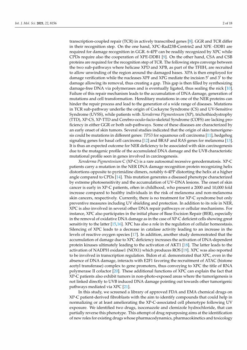
The expression of XPC protein was examined in both WT and XP-C cells immortalizedfrom patient primary fibroblasts. Immuno-staining of both cells using an antibody againstthe XPC protein was carried out. In contrast to WT cells, XP-C cells showed a total absenceof XPC protein (Figure 1a).
Int. J. Mol. Sci. 2021, 22, x FOR PEER REVIEW 4 of 18
Figure 1. Characterization of XP-C and WT cells. (a) XP-C cells lack the expression of XPC protein. XP-C and WT cells
were fixed and stained with anti-XPC antibody to analyze its differential expression in both cell lines. XP-C cells, unlike
WT cells, do not manifest XPC protein expression in their nuclei. (b) Viability of fibroblasts 24 h post UVB. XP-C cells
manifest significantly increased photosensitivity compared to WT cells. Both XP-C and WT cells were seeded in 96-well
plates to be irradiated at 80% confluency with increasing UVB doses, then their viability was quantified 24 h later by the
incubation with PrestoBlue. XP-C cells show a sharper significant decrease in viability as a function of increased UVB dose
compared to WT cells. Viability was calculated by means of percent of control with 100% control being non-irradiated
cells. *** p < 0.001, unpaired t-test. Results presented are the mean of three technical replicates ± SEM. (c) Quantification of
DNA damage accumulation post UV. WT cells manifest faster repair kinetics of both CPD and 6-4PP lesions post UV
compared to impaired repair in XP-C cells. Both cell lines were subjected to UVB then incubated for different time points
post UV. At each time points, cells were collected, their DNA extracted and digested to be then analyzed by LC-MS/MS.
Four different CPD lesion types were quantified with TT-CPD being the most frequent. Normal cells show a decrease in
the % of CPD lesions as a function of time signifying repair with different kinetics per lesion time. XP-C cells, however,
manifest an increase in CPD lesion at 2 h post UV and continue to have a large amount of lesion % as time elapses, a sign
of impaired repair. Two lesion types were quantified for 6-4PP with TC-6-4PP being more abundant. Similar to CPD,
impaired repair is visualized in XP-C cells manifested by a slow decrease in lesion % as a function of time compared to
the normal cells, which in contrast to CPD lesions show even faster repair for 6-4PP. CPD: cyclobutane pyrimidine dimer,
Figure 1. Characterization of XP-C and WT cells. (a) XP-C cells lack the expression of XPC protein. XP-C and WT cells werefixed and stained with anti-XPC antibody to analyze its differential expression in both cell lines. XP-C cells, unlike WTcells, do not manifest XPC protein expression in their nuclei. (b) Viability of fibroblasts 24 h post UVB. XP-C cells manifestsignificantly increased photosensitivity compared to WT cells. Both XP-C and WT cells were seeded in 96-well plates to be
Int. J. Mol. Sci. 2021, 22, 8156 4 of 18
irradiated at 80% confluency with increasing UVB doses, then their viability was quantified 24 h later by the incubation with PrestoBlue.XP-C cells show a sharper significant decrease in viability as a function of increased UVB dose compared to WT cells. Viability wascalculated by means of percent of control with 100% control being non-irradiated cells. *** p < 0.001, unpaired t-test. Results presentedare the mean of three technical replicates ± SEM. (c) Quantification of DNA damage accumulation post UV. WT cells manifest fasterrepair kinetics of both CPD and 6-4PP lesions post UV compared to impaired repair in XP-C cells. Both cell lines were subjected toUVB then incubated for different time points post UV. At each time points, cells were collected, their DNA extracted and digested tobe then analyzed by LC-MS/MS. Four different CPD lesion types were quantified with TT-CPD being the most frequent. Normalcells show a decrease in the % of CPD lesions as a function of time signifying repair with different kinetics per lesion time. XP-C cells,however, manifest an increase in CPD lesion at 2 h post UV and continue to have a large amount of lesion % as time elapses, a sign ofimpaired repair. Two lesion types were quantified for 6-4PP with TC-6-4PP being more abundant. Similar to CPD, impaired repair isvisualized in XP-C cells manifested by a slow decrease in lesion % as a function of time compared to the normal cells, which in contrastto CPD lesions show even faster repair for 6-4PP. CPD: cyclobutane pyrimidine dimer, 6-4PP: 6-4 photoproducts, WT: wild type, XP-C:Xeroderma Pigmentosum C, LD50: lethal dose 50, R2: coefficient of determination.
2.1.2. Increased Photosensitivity of XP-C Cells in Response to UVB Irradiation Comparedto WT Cells
To examine the photosensitivity of XP-C cells relative to WT, both kinds of cell lineswere seeded until 80% confluency then subjected to increasing doses of UVB. Twenty-four hours after UV treatment, cell viability was recorded as a measurement of the cells’reducing capacity (Presto blue, Thermofisher Scientific, Waltham, MA, USA). Viability wasnormalized based on the calculation of the percentage of viability compared to the viabilityof control non-irradiated cells set as 100% viability. Both cell lines showed a decreasein viability as a function of increased UVB doses. XP-C cells were more photosensitivecompared to the WT ones showing a sharper significant decrease in viability at each UVBdose (p < 0.001). The UVB dose leading to 50% of mortality was determined for both XP-Cand wild-type cells (LD50). XP-C cells showed a much lower LD50 (about 0.02 J/cm2)compared to WT cells (about 0.19 J/cm2) (Figure 1b). These results confirm the strongsensitivity to UVB of XP-C cells used in this study as described in the other model [22].
2.1.3. Absence of XPC Impairs DNA Repair of UVB-Induced Lesions
XPC protein is essential for the lesion recognition step of GGR [23]. For that, weaimed to determine the effect of XPC mutation on the repair of lesions. The two majorphotoproducts generated after UVB exposure are CPDs and 6-4PPs that are formed betweenpyrimidine dimers, either TT, TC, CT or CC. The cells were seeded until 80% confluencythen irradiated at 0.02 J/cm2, corresponding to the previously determined XP-C LD50,then collected post UVB treatment at different time points. In order to monitor DNAlesions, DNA was extracted and digested to be analyzed by LC-MS/MS. For CPDs, thefour different lesion types were quantified. WT cells showed a decrease in different CPDslesions’ amounts as a function of time to reach a minimum after 48 h indicating efficientrepair of DNA damage, while in contrast, XP-C cells showed constant elevated amountsof lesions as a function of time (Figure 1c). It should be noted that not all lesions werepresent in the DNA in the same quantities: the majority of CPD lesions were of TT-CPDnature followed by TC-CPD, CT-CPD, while the least abundant lesion was CC-CPD inboth kinds of cells. In addition, the repair kinetics in wild-type cells differed betweenlesion types with the fastest repair observed for the CT-CPD. Only the TT and TC lesionscould be detected for the 6-4PPs, as the two remaining lesions were less frequent. 6-4PPswere repaired faster than CPD with total repair 24 h post UV in WT cells compared to thehigher amount of lesions in XP-C cells. There were no discrepancies in the repair kineticsbetween the TT and TC 6-4PP lesions in normal cells (Figure 1c). Taken together, our resultsshow that wild-type but not XP-C cells efficiently repair photoproducts resulting fromUVB irradiation.
Int. J. Mol. Sci. 2021, 22, 8156 5 of 18
2.2. Primary Screen Identified 16 Candidate Compounds That Increase XP-C Cells Viability PostUVB Irradiation
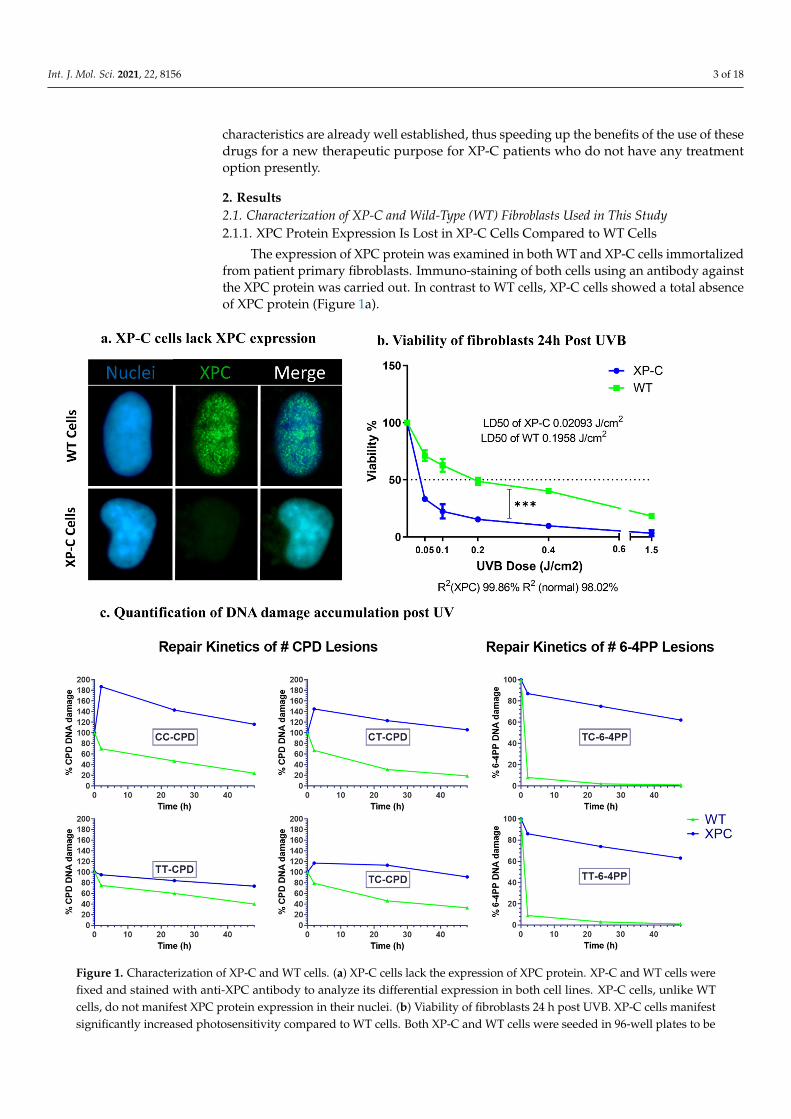
To identify compounds that would correct the photosensitive phenotype of XP-C cells,we did set up a robotic assay in 96-well microplate to monitor cell viability after UVBirradiation (see methods and Supplementary Figure S1, see Supplementary Materials). Therobustness of the assay was calculated as a Z’ factor [24] at various UVB doses on a Z’plate including 48 bioactive controls mimicking the desired effect (here, non-irradiatedprotected XPC cells in DMSO) and 48 bioinactive controls (here, irradiated cells in DMSO).A library of 1280 FDA and EMA approved drugs (Prestwick Library) was then screenedon XP-C cells at 10 µM for 24 h in duplicates using this assay. Briefly, following drugtreatment, the cells were UVB irradiated at 0.05 J/cm2 based on Z’ Factor calculation(Supplementary Figure S1), then post-treated with the same drugs and incubated at 37 ◦Cfor an additional 24 h period before the measurement of cell viability. In this setup, the effectof drug treatment on cell viability can be either preventive or curative to the UVB-induceddamage. The bioactivity of each tested compound, signifying the drugs’ beneficial effectson photo-resistance, was calculated by determining the fluorescence of each well withrespect to the fluorescence of non-irradiated DMSO treated wells set as 100% (bioactivecontrol) and irradiated DMSO treated wells set as 0% (bioinactive control). The robust Zscore was also calculated per plate as an additional normalization technique [24]. The hitselection was based on two criteria: compounds that possess a bioactivity ≥25% and arobust Z score ≥2.5. Sixteen molecules were identified as primary hits on this basis andselected for further characterization of their bioactivity on XP cells (Figure 2).
Int. J. Mol. Sci. 2021, 22, x FOR PEER REVIEW 5 of 18
6-4PP: 6-4 photoproducts, WT: wild type, XP-C: Xeroderma Pigmentosum C, LD50: lethal dose 50, R2: coefficient of deter-
mination.
2.2. Primary Screen Identified 16 Candidate Compounds That Increase XP-C Cells Viability Post
UVB Irradiation
To identify compounds that would correct the photosensitive phenotype of XP-C
cells, we did set up a robotic assay in 96-well microplate to monitor cell viability after UVB
irradiation (see methods and supplementary Figure 1, see Supplementary Materials). The
robustness of the assay was calculated as a Z’ factor [24] at various UVB doses on a Z’
plate including 48 bioactive controls mimicking the desired effect (here, non-irradiated
protected XPC cells in DMSO) and 48 bioinactive controls (here, irradiated cells in DMSO).
A library of 1280 FDA and EMA approved drugs (Prestwick Library) was then screened
on XP-C cells at 10 µM for 24 h in duplicates using this assay. Briefly, following drug
treatment, the cells were UVB irradiated at 0.05 J/cm2 based on Z’ Factor calculation (sup-
plementary Figure S1), then post-treated with the same drugs and incubated at 37 °C for
an additional 24 h period before the measurement of cell viability. In this setup, the effect
of drug treatment on cell viability can be either preventive or curative to the UVB-induced
damage. The bioactivity of each tested compound, signifying the drugs’ beneficial effects
on photo-resistance, was calculated by determining the fluorescence of each well with re-
spect to the fluorescence of non-irradiated DMSO treated wells set as 100% (bioactive con-
trol) and irradiated DMSO treated wells set as 0% (bioinactive control). The robust Z score
was also calculated per plate as an additional normalization technique [24]. The hit selec-
tion was based on two criteria: compounds that possess a bioactivity ≥25% and a robust Z
score ≥2.5. Sixteen molecules were identified as primary hits on this basis and selected for
further characterization of their bioactivity on XP cells (Figure 2).
Figure 2. Identification of 16 compounds reversing XP-C photo-sensitivity upon UVB irradiation. Prestwick library of
chemical drugs was screened on XP-C cells. The cells were treated with the library for 24 h, then further on, irradiated.
Post Irradiation, the cells were then incubated with the library for an additional 24 h prior to having their viability assessed
24 h post UV via the addition of Presto Blue reagent and the recording of the fluorescent values. Based on two different
Figure 2. Identification of 16 compounds reversing XP-C photo-sensitivity upon UVB irradiation. Prestwick library ofchemical drugs was screened on XP-C cells. The cells were treated with the library for 24 h, then further on, irradiated. PostIrradiation, the cells were then incubated with the library for an additional 24 h prior to having their viability assessed24 h post UV via the addition of Presto Blue reagent and the recording of the fluorescent values. Based on two differentnormalization criteria, control based % of activity and non-control based RZscore, 16 compounds were selected manifesting>25% increase or >2.5 value for both % of activity and RZscore respectively. % Activity was calculated by normalizing theobtained values between the interval of 100% donated to non-irradiated DMSO treated cells and 0% that donated irradiatedDMSO treated cells. RZscore was measured by normalizing the fluorescence values to the median and median absolutedeviation. RZscore: Robust Z score.
Int. J. Mol. Sci. 2021, 22, 8156 6 of 18
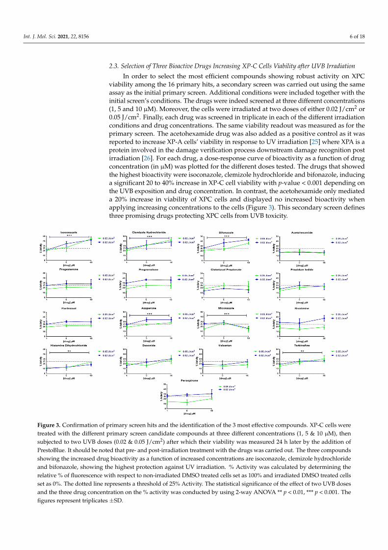
2.3. Selection of Three Bioactive Drugs Increasing XP-C Cells Viability after UVB Irradiation
In order to select the most efficient compounds showing robust activity on XPCviability among the 16 primary hits, a secondary screen was carried out using the sameassay as the initial primary screen. Additional conditions were included together with theinitial screen’s conditions. The drugs were indeed screened at three different concentrations(1, 5 and 10 µM). Moreover, the cells were irradiated at two doses of either 0.02 J/cm2 or0.05 J/cm2. Finally, each drug was screened in triplicate in each of the different irradiationconditions and drug concentrations. The same viability readout was measured as for theprimary screen. The acetohexamide drug was also added as a positive control as it wasreported to increase XP-A cells’ viability in response to UV irradiation [25] where XPA is aprotein involved in the damage verification process downstream damage recognition postirradiation [26]. For each drug, a dose-response curve of bioactivity as a function of drugconcentration (in µM) was plotted for the different doses tested. The drugs that showedthe highest bioactivity were isoconazole, clemizole hydrochloride and bifonazole, inducinga significant 20 to 40% increase in XP-C cell viability with p-value < 0.001 depending onthe UVB exposition and drug concentration. In contrast, the acetohexamide only mediateda 20% increase in viability of XPC cells and displayed no increased bioactivity whenapplying increasing concentrations to the cells (Figure 3). This secondary screen definesthree promising drugs protecting XPC cells from UVB toxicity.
Int. J. Mol. Sci. 2021, 22, x FOR PEER REVIEW 6 of 18
normalization criteria, control based % of activity and non-control based RZscore, 16 compounds were selected manifest-
ing >25% increase or >2.5 value for both % of activity and RZscore respectively. % Activity was calculated by normalizing
the obtained values between the interval of 100% donated to non-irradiated DMSO treated cells and 0% that donated
irradiated DMSO treated cells. RZscore was measured by normalizing the fluorescence values to the median and median
absolute deviation. RZscore: Robust Z score.
2.3. Selection of Three Bioactive Drugs Increasing XP-C Cells Viability after UVB Irradiation
In order to select the most efficient compounds showing robust activity on XPC via-
bility among the 16 primary hits, a secondary screen was carried out using the same assay
as the initial primary screen. Additional conditions were included together with the initial
screen’s conditions. The drugs were indeed screened at three different concentrations (1,
5 and 10 µM). Moreover, the cells were irradiated at two doses of either 0.02 J/cm2 or 0.05
J/cm2. Finally, each drug was screened in triplicate in each of the different irradiation con-
ditions and drug concentrations. The same viability readout was measured as for the pri-
mary screen. The acetohexamide drug was also added as a positive control as it was re-
ported to increase XP-A cells’ viability in response to UV irradiation [25] where XPA is a
protein involved in the damage verification process downstream damage recognition post
irradiation [26]. For each drug, a dose-response curve of bioactivity as a function of drug
concentration (in µM) was plotted for the different doses tested. The drugs that showed
the highest bioactivity were isoconazole, clemizole hydrochloride and bifonazole, induc-
ing a significant 20 to 40% increase in XP-C cell viability with p-value < 0.001 depending
on the UVB exposition and drug concentration. In contrast, the acetohexamide only me-
diated a 20% increase in viability of XPC cells and displayed no increased bioactivity when
applying increasing concentrations to the cells (Figure 3). This secondary screen defines
three promising drugs protecting XPC cells from UVB toxicity.
Figure 3. Confirmation of primary screen hits and the identification of the 3 most effective compounds. XP-C cells were
treated with the different primary screen candidate compounds at three different concentrations (1, 5 & 10 µM), then
subjected to two UVB doses (0.02 & 0.05 J/cm2) after which their viability was measured 24 h later by the addition of
PrestoBlue. It should be noted that pre- and post-irradiation treatment with the drugs was carried out. The three com-
pounds showing the increased drug bioactivity as a function of increased concentrations are isoconazole, clemizole hy-
Figure 3. Confirmation of primary screen hits and the identification of the 3 most effective compounds. XP-C cells weretreated with the different primary screen candidate compounds at three different concentrations (1, 5 & 10 µM), thensubjected to two UVB doses (0.02 & 0.05 J/cm2) after which their viability was measured 24 h later by the addition ofPrestoBlue. It should be noted that pre- and post-irradiation treatment with the drugs was carried out. The three compoundsshowing the increased drug bioactivity as a function of increased concentrations are isoconazole, clemizole hydrochlorideand bifonazole, showing the highest protection against UV irradiation. % Activity was calculated by determining therelative % of fluorescence with respect to non-irradiated DMSO treated cells set as 100% and irradiated DMSO treated cellsset as 0%. The dotted line represents a threshold of 25% Activity. The statistical significance of the effect of two UVB dosesand the three drug concentration on the % activity was conducted by using 2-way ANOVA ** p < 0.01, *** p < 0.001. Thefigures represent triplicates ±SD.
Int. J. Mol. Sci. 2021, 22, 8156 7 of 18
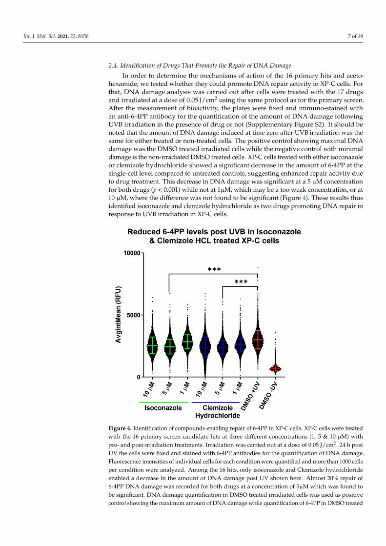
2.4. Identification of Drugs That Promote the Repair of DNA Damage
In order to determine the mechanisms of action of the 16 primary hits and aceto-hexamide, we tested whether they could promote DNA repair activity in XP-C cells. Forthat, DNA damage analysis was carried out after cells were treated with the 17 drugsand irradiated at a dose of 0.05 J/cm2 using the same protocol as for the primary screen.After the measurement of bioactivity, the plates were fixed and immuno-stained withan anti-6-4PP antibody for the quantification of the amount of DNA damage followingUVB irradiation in the presence of drug or not (Supplementary Figure S2). It should benoted that the amount of DNA damage induced at time zero after UVB irradiation was thesame for either treated or non-treated cells. The positive control showing maximal DNAdamage was the DMSO treated irradiated cells while the negative control with minimaldamage is the non-irradiated DMSO treated cells. XP-C cells treated with either isoconazoleor clemizole hydrochloride showed a significant decrease in the amount of 6-4PP at thesingle-cell level compared to untreated controls, suggesting enhanced repair activity dueto drug treatment. This decrease in DNA damage was significant at a 5 µM concentrationfor both drugs (p < 0.001) while not at 1µM, which may be a too weak concentration, or at10 µM, where the difference was not found to be significant (Figure 4). These results thusidentified isoconazole and clemizole hydrochloride as two drugs promoting DNA repair inresponse to UVB irradiation in XP-C cells.
Int. J. Mol. Sci. 2021, 22, x FOR PEER REVIEW 7 of 18
drochloride and bifonazole, showing the highest protection against UV irradiation. % Activity was calculated by deter-
mining the relative % of fluorescence with respect to non-irradiated DMSO treated cells set as 100% and irradiated DMSO
treated cells set as 0%. The dotted line represents a threshold of 25% Activity. The statistical significance of the effect of
two UVB doses and the three drug concentration on the % activity was conducted by using 2-way ANOVA ** p < 0.01, ***
p < 0.001. The figures represent triplicates ±SD.
2.4. Identification of Drugs That Promote the Repair of DNA Damage
In order to determine the mechanisms of action of the 16 primary hits and acetohex-
amide, we tested whether they could promote DNA repair activity in XP-C cells. For that,
DNA damage analysis was carried out after cells were treated with the 17 drugs and irra-
diated at a dose of 0.05 J/cm2 using the same protocol as for the primary screen. After the
measurement of bioactivity, the plates were fixed and immuno-stained with an anti-6-4PP
antibody for the quantification of the amount of DNA damage following UVB irradiation
in the presence of drug or not (supplementary Figure S2). It should be noted that the
amount of DNA damage induced at time zero after UVB irradiation was the same for
either treated or non-treated cells. The positive control showing maximal DNA damage
was the DMSO treated irradiated cells while the negative control with minimal damage
is the non-irradiated DMSO treated cells. XP-C cells treated with either isoconazole or
clemizole hydrochloride showed a significant decrease in the amount of 6-4PP at the sin-
gle-cell level compared to untreated controls, suggesting enhanced repair activity due to
drug treatment. This decrease in DNA damage was significant at a 5 µM concentration
for both drugs (p < 0.001) while not at 1µM, which may be a too weak concentration, or at
10 µM, where the difference was not found to be significant (Figure 4). These results thus
identified isoconazole and clemizole hydrochloride as two drugs promoting DNA repair
in response to UVB irradiation in XP-C cells.
Figure 4. Identification of compounds enabling repair of 6-4PP in XP-C cells. XP-C cells were treated
with the 16 primary screen candidate hits at three different concentrations (1, 5 & 10 µM) with pre-
and post-irradiation treatments. Irradiation was carried out at a dose of 0.05 J/cm2. 24 h post UV the
Figure 4. Identification of compounds enabling repair of 6-4PP in XP-C cells. XP-C cells were treatedwith the 16 primary screen candidate hits at three different concentrations (1, 5 & 10 µM) withpre- and post-irradiation treatments. Irradiation was carried out at a dose of 0.05 J/cm2. 24 h postUV the cells were fixed and stained with 6-4PP antibodies for the quantification of DNA damage.Fluorescence intensities of individual cells for each condition were quantified and more than 1000 cellsper condition were analyzed. Among the 16 hits, only isoconazole and Clemizole hydrochlorideenabled a decrease in the amount of DNA damage post UV shown here. Almost 20% repair of6-4PP DNA damage was recorded for both drugs at a concentration of 5µM which was found tobe significant. DNA damage quantification in DMSO treated irradiated cells was used as positivecontrol showing the maximum amount of DNA damage while quantification of 6-4PP in DMSO treated
Int. J. Mol. Sci. 2021, 22, 8156 8 of 18
non-irradiated cells was taken as a negative control with the minimum amount of detected damage.*** p < 0.001, freedman non-parametric test with Dunn’s post hoc analysis. Up to 10,000 cells wereanalyzed per condition with ±SD.
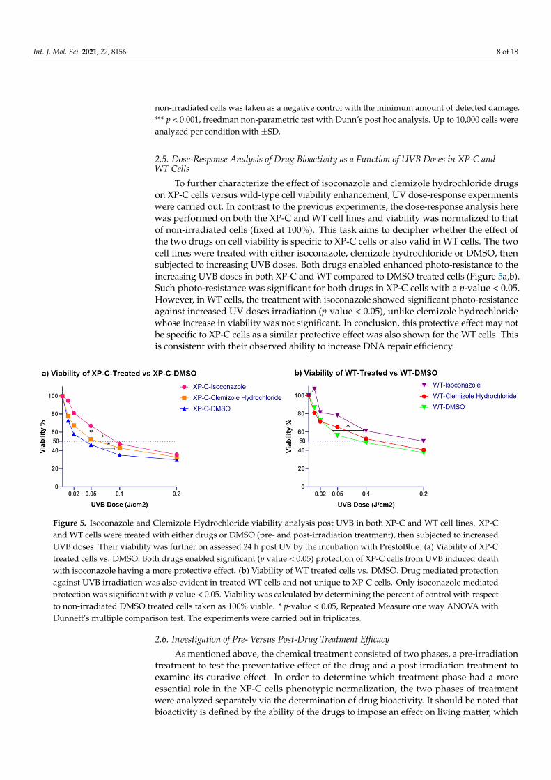
2.5. Dose-Response Analysis of Drug Bioactivity as a Function of UVB Doses in XP-C andWT Cells
To further characterize the effect of isoconazole and clemizole hydrochloride drugson XP-C cells versus wild-type cell viability enhancement, UV dose-response experimentswere carried out. In contrast to the previous experiments, the dose-response analysis herewas performed on both the XP-C and WT cell lines and viability was normalized to thatof non-irradiated cells (fixed at 100%). This task aims to decipher whether the effect ofthe two drugs on cell viability is specific to XP-C cells or also valid in WT cells. The twocell lines were treated with either isoconazole, clemizole hydrochloride or DMSO, thensubjected to increasing UVB doses. Both drugs enabled enhanced photo-resistance to theincreasing UVB doses in both XP-C and WT compared to DMSO treated cells (Figure 5a,b).Such photo-resistance was significant for both drugs in XP-C cells with a p-value < 0.05.However, in WT cells, the treatment with isoconazole showed significant photo-resistanceagainst increased UV doses irradiation (p-value < 0.05), unlike clemizole hydrochloridewhose increase in viability was not significant. In conclusion, this protective effect may notbe specific to XP-C cells as a similar protective effect was also shown for the WT cells. Thisis consistent with their observed ability to increase DNA repair efficiency.
Int. J. Mol. Sci. 2021, 22, x FOR PEER REVIEW 8 of 18
cells were fixed and stained with 6-4PP antibodies for the quantification of DNA damage. Fluores-
cence intensities of individual cells for each condition were quantified and more than 1000 cells per
condition were analyzed. Among the 16 hits, only isoconazole and Clemizole hydrochloride enabled
a decrease in the amount of DNA damage post UV shown here. Almost 20% repair of 6-4PP DNA
damage was recorded for both drugs at a concentration of 5µM which was found to be significant.
DNA damage quantification in DMSO treated irradiated cells was used as positive control showing
the maximum amount of DNA damage while quantification of 6-4PP in DMSO treated non-irradi-
ated cells was taken as a negative control with the minimum amount of detected damage. *** p <
0.001, freedman non-parametric test with Dunn’s post hoc analysis. Up to 10,000 cells were analyzed
per condition with ±SD.
2.5. Dose-Response Analysis of Drug Bioactivity as a Function of UVB Doses in XP-C and WT
Cells
To further characterize the effect of isoconazole and clemizole hydrochloride drugs
on XP-C cells versus wild-type cell viability enhancement, UV dose-response experiments
were carried out. In contrast to the previous experiments, the dose-response analysis here
was performed on both the XP-C and WT cell lines and viability was normalized to that
of non-irradiated cells (fixed at 100%). This task aims to decipher whether the effect of the
two drugs on cell viability is specific to XP-C cells or also valid in WT cells. The two cell
lines were treated with either isoconazole, clemizole hydrochloride or DMSO, then sub-
jected to increasing UVB doses. Both drugs enabled enhanced photo-resistance to the in-
creasing UVB doses in both XP-C and WT compared to DMSO treated cells (Figure 5a,b).
Such photo-resistance was significant for both drugs in XP-C cells with a p-value < 0.05.
However, in WT cells, the treatment with isoconazole showed significant photo-resistance
against increased UV doses irradiation (p-value < 0.05), unlike clemizole hydrochloride
whose increase in viability was not significant. In conclusion, this protective effect may
not be specific to XP-C cells as a similar protective effect was also shown for the WT cells.
This is consistent with their observed ability to increase DNA repair efficiency.
Figure 5. Isoconazole and Clemizole Hydrochloride viability analysis post UVB in both XP-C and WT cell lines. XP-C and
WT cells were treated with either drugs or DMSO (pre- and post-irradiation treatment), then subjected to increased UVB
doses. Their viability was further on assessed 24 h post UV by the incubation with PrestoBlue. (a) Viability of XP-C treated
cells vs. DMSO. Both drugs enabled significant (p value < 0.05) protection of XP-C cells from UVB induced death with
isoconazole having a more protective effect. (b) Viability of WT treated cells vs. DMSO. Drug mediated protection against
UVB irradiation was also evident in treated WT cells and not unique to XP-C cells. Only isoconazole mediated protection
was significant with p value < 0.05. Viability was calculated by determining the percent of control with respect to non-
irradiated DMSO treated cells taken as 100% viable. * p-value < 0.05, Repeated Measure one way ANOVA with Dunnett’s
multiple comparison test. The experiments were carried out in triplicates.
Figure 5. Isoconazole and Clemizole Hydrochloride viability analysis post UVB in both XP-C and WT cell lines. XP-Cand WT cells were treated with either drugs or DMSO (pre- and post-irradiation treatment), then subjected to increasedUVB doses. Their viability was further on assessed 24 h post UV by the incubation with PrestoBlue. (a) Viability of XP-Ctreated cells vs. DMSO. Both drugs enabled significant (p value < 0.05) protection of XP-C cells from UVB induced deathwith isoconazole having a more protective effect. (b) Viability of WT treated cells vs. DMSO. Drug mediated protectionagainst UVB irradiation was also evident in treated WT cells and not unique to XP-C cells. Only isoconazole mediatedprotection was significant with p value < 0.05. Viability was calculated by determining the percent of control with respectto non-irradiated DMSO treated cells taken as 100% viable. * p-value < 0.05, Repeated Measure one way ANOVA withDunnett’s multiple comparison test. The experiments were carried out in triplicates.
2.6. Investigation of Pre- Versus Post-Drug Treatment Efficacy
As mentioned above, the chemical treatment consisted of two phases, a pre-irradiationtreatment to test the preventative effect of the drug and a post-irradiation treatment toexamine its curative effect. In order to determine which treatment phase had a moreessential role in the XP-C cells phenotypic normalization, the two phases of treatmentwere analyzed separately via the determination of drug bioactivity. It should be noted thatbioactivity is defined by the ability of the drugs to impose an effect on living matter, which

Int. J. Mol. Sci. 2021, 22, 8156 9 of 18
in this case, is its ability to increase photo-resistance of the cells between non-irradiatedcells set at 100% and irradiated non-treated cells set at 0%. The effect of each of the pre- orpost-treatment was compared to that of the combined pre- plus post-treatment regimen.For isoconazole, the post-irradiation treatment was sufficient to induce similar protectionas the combined treatment at both 5 and 10 µM (with no significant differences) whilethe pre-treatment had no effect on XP-C cells viability at any of the tested concentrations(Figure 6a). At 10 µM, the difference in bioactivity was significant between pre and post-treatment as well as between the pre-and combined treatment with a p-value < 0.01,signifying that both pre-treatment and combined regimens share significant protectioncompared to pretreatment. In the case of clemizole hydrochloride, the pre-irradiationtreatment showed slight bioactivity reaching 20% increased cell viability at the highestconcentration of drug of 10 µM. The post-treatment, however, was again more effectivethan the pre-treatment on XP-C viability reaching an average of 50% increase of cell viabilityat 10 µM. The combination of both pre-and post-irradiation treatments showed the highestbioactivity with a significant difference compared to the pretreatment at both 5 and 10 µMp-value < 0.05, and no significant difference with the post-treatment only. Thus, despitea slight protective potential of clemizole hydrochloride, both drugs (isoconazole andclemizole hydrochloride) may essentially serve as curative remedy post-UVB irradiation.
Int. J. Mol. Sci. 2021, 22, x FOR PEER REVIEW 9 of 18
2.6. Investigation of Pre- Versus Post-Drug Treatment Efficacy
As mentioned above, the chemical treatment consisted of two phases, a pre-irradia-
tion treatment to test the preventative effect of the drug and a post-irradiation treatment
to examine its curative effect. In order to determine which treatment phase had a more
essential role in the XP-C cells phenotypic normalization, the two phases of treatment
were analyzed separately via the determination of drug bioactivity. It should be noted
that bioactivity is defined by the ability of the drugs to impose an effect on living matter,
which in this case, is its ability to increase photo-resistance of the cells between non-irra-
diated cells set at 100% and irradiated non-treated cells set at 0%. The effect of each of the
pre- or post-treatment was compared to that of the combined pre- plus post-treatment
regimen. For isoconazole, the post-irradiation treatment was sufficient to induce similar
protection as the combined treatment at both 5 and 10 µM (with no significant differences)
while the pre-treatment had no effect on XP-C cells viability at any of the tested concen-
trations (Figure 6a). At 10 µM, the difference in bioactivity was significant between pre
and post-treatment as well as between the pre-and combined treatment with a p-value <
0.01, signifying that both pre-treatment and combined regimens share significant protec-
tion compared to pretreatment. In the case of clemizole hydrochloride, the pre-irradiation
treatment showed slight bioactivity reaching 20% increased cell viability at the highest
concentration of drug of 10 µM. The post-treatment, however, was again more effective
than the pre-treatment on XP-C viability reaching an average of 50% increase of cell via-
bility at 10 µM. The combination of both pre-and post-irradiation treatments showed the
highest bioactivity with a significant difference compared to the pretreatment at both 5
and 10 µM p-value < 0.05, and no significant difference with the post-treatment only. Thus,
despite a slight protective potential of clemizole hydrochloride, both drugs (isoconazole
and clemizole hydrochloride) may essentially serve as curative remedy post-UVB irradi-
ation.
Figure 6. Cont.

Int. J. Mol. Sci. 2021, 22, 8156 10 of 18Int. J. Mol. Sci. 2021, 22, x FOR PEER REVIEW 10 of 18
Figure 6. Drug treatment regimen separation and double drug treatment. To determine which treatment regimen was
more effective, XP-C cells were treated with the drugs either pre-irradiation, post-irradiation or both pre and post-irradi-
ation. Drug Bioactivity % was then calculated 24 h post UV. Such calculation is based on determining the relative percent-
age considering non-irradiated DMSO treated samples as 100% and irradiated ones as 0%. Thus, the obtained fluorescence
values normalized to these two controls. (a) Drug treatment regimen separation. Isoconazole post-irradiation or both treat-
ments showed a significant p value < 0.01 increase in bioactivity at 10 µM, while only both treatments showed significant
enhancement of bioactivity at 5 µM compared to pre-treatment procedure. Clemizole hydrochloride, however, seems to
have better bioactivity when both pre and post-irradiation regimes were used where the increase of bioactivity was sig-
nificant, p value < 0.05 for both 5 µM and 10 µM treatment compared to pre-treatment at each concentration, respectively.
Moreover, double drug treatment was also carried out to test whether both drugs possess synergistic or additive effect.
(b) Double drug treatment. XP-C cells were treated with either Isoconazole, clemizole hydrochloride or double treated at
10µM, then irradiated at 0.02 J/cm2. % of drug bioactivity was measured 24 h later. Double treatment showed the same
protective profile as isoconazole with no added protection against UVB irradiation. * p value < 0.05, ** p value < 0.01, 2-
way ANOVA with Tukey’s multiple comparison test. The experiments were performed in triplicate with ±SD.
2.7. Double Drug Treatment Has no Synergistic nor Additive Effect
Both compounds, isoconazole and clemizole hydrochloride, have an azole ring in
their structure (and may thus target the same molecular biological target or similar mech-
anisms). We, therefore, examined whether the combined treatment with both drugs at 10
µM could improve further or not the acquired photo-resistance compared to single-drug
treatment. Accordingly, XP-C cells were treated with either drug alone or with both and
then subjected to irradiation at a dose of 0.02 J/cm2. The double treatment had the same
bioactivity as the single treatment with isoconazole with no benefit (Figure 6b). Hence, no
synergetic effect was obtained upon double drug treatment.
2.8. Isoconazole and Clemizole Hydrochloride Do Not Affect Cell Proliferation
In a first step, and in order to analyze the proliferative state of the irradiated cells and
whether the different treatments can modify this profile, cells were stained with Ki67 an-
tibodies and analysis was carried out at the single-cell level. Ki67 antigen was expressed
during all phases of the cell cycle (G1, S, G2 and M) but not in quiescent cells. Both XP-C
and WT cell lines were positive to Ki67, signifying that the cells are not in quiescent state
post UV. Cells treated with either DMSO, isoconazole, clemizole hydrochloride or both
drugs showed no difference in the Ki67 expression profile (supplementary Figure S3).
Ki67 antibodies stain cells in various stages of the cell cycle, and it was previously
reported that some non-proliferating cells tend to test positive for Ki67 due to antigen
retention [27]. Moreover, bulky adducts generated by UV tend to block the progression of
replication forks, decreasing DNA replication. Therefore, in a second step, EdU incorpo-
ration assay was performed to clarify whether this positive staining was due to antigen
retention or whether rather the cells were capable of recovering from the DNA replication
Figure 6. Drug treatment regimen separation and double drug treatment. To determine which treatment regimen was moreeffective, XP-C cells were treated with the drugs either pre-irradiation, post-irradiation or both pre and post-irradiation.Drug Bioactivity % was then calculated 24 h post UV. Such calculation is based on determining the relative percentageconsidering non-irradiated DMSO treated samples as 100% and irradiated ones as 0%. Thus, the obtained fluorescencevalues normalized to these two controls. (a) Drug treatment regimen separation. Isoconazole post-irradiation or bothtreatments showed a significant p value < 0.01 increase in bioactivity at 10 µM, while only both treatments showed significantenhancement of bioactivity at 5 µM compared to pre-treatment procedure. Clemizole hydrochloride, however, seemsto have better bioactivity when both pre and post-irradiation regimes were used where the increase of bioactivity wassignificant, p value < 0.05 for both 5 µM and 10 µM treatment compared to pre-treatment at each concentration, respectively.Moreover, double drug treatment was also carried out to test whether both drugs possess synergistic or additive effect.(b) Double drug treatment. XP-C cells were treated with either Isoconazole, clemizole hydrochloride or double treated at10 µM, then irradiated at 0.02 J/cm2. % of drug bioactivity was measured 24 h later. Double treatment showed the sameprotective profile as isoconazole with no added protection against UVB irradiation. * p value < 0.05, ** p value < 0.01, 2-wayANOVA with Tukey’s multiple comparison test. The experiments were performed in triplicate with ±SD.
2.7. Double Drug Treatment Has no Synergistic nor Additive Effect
Both compounds, isoconazole and clemizole hydrochloride, have an azole ring in theirstructure (and may thus target the same molecular biological target or similar mechanisms).We, therefore, examined whether the combined treatment with both drugs at 10 µM couldimprove further or not the acquired photo-resistance compared to single-drug treatment.Accordingly, XP-C cells were treated with either drug alone or with both and then subjectedto irradiation at a dose of 0.02 J/cm2. The double treatment had the same bioactivity as thesingle treatment with isoconazole with no benefit (Figure 6b). Hence, no synergetic effectwas obtained upon double drug treatment.
2.8. Isoconazole and Clemizole Hydrochloride Do Not Affect Cell Proliferation
In a first step, and in order to analyze the proliferative state of the irradiated cellsand whether the different treatments can modify this profile, cells were stained with Ki67antibodies and analysis was carried out at the single-cell level. Ki67 antigen was expressedduring all phases of the cell cycle (G1, S, G2 and M) but not in quiescent cells. Both XP-Cand WT cell lines were positive to Ki67, signifying that the cells are not in quiescent statepost UV. Cells treated with either DMSO, isoconazole, clemizole hydrochloride or bothdrugs showed no difference in the Ki67 expression profile (Supplementary Figure S3).
Ki67 antibodies stain cells in various stages of the cell cycle, and it was previouslyreported that some non-proliferating cells tend to test positive for Ki67 due to antigenretention [27]. Moreover, bulky adducts generated by UV tend to block the progression ofreplication forks, decreasing DNA replication. Therefore, in a second step, EdU incorpo-ration assay was performed to clarify whether this positive staining was due to antigenretention or whether rather the cells were capable of recovering from the DNA replication
Int. J. Mol. Sci. 2021, 22, 8156 11 of 18
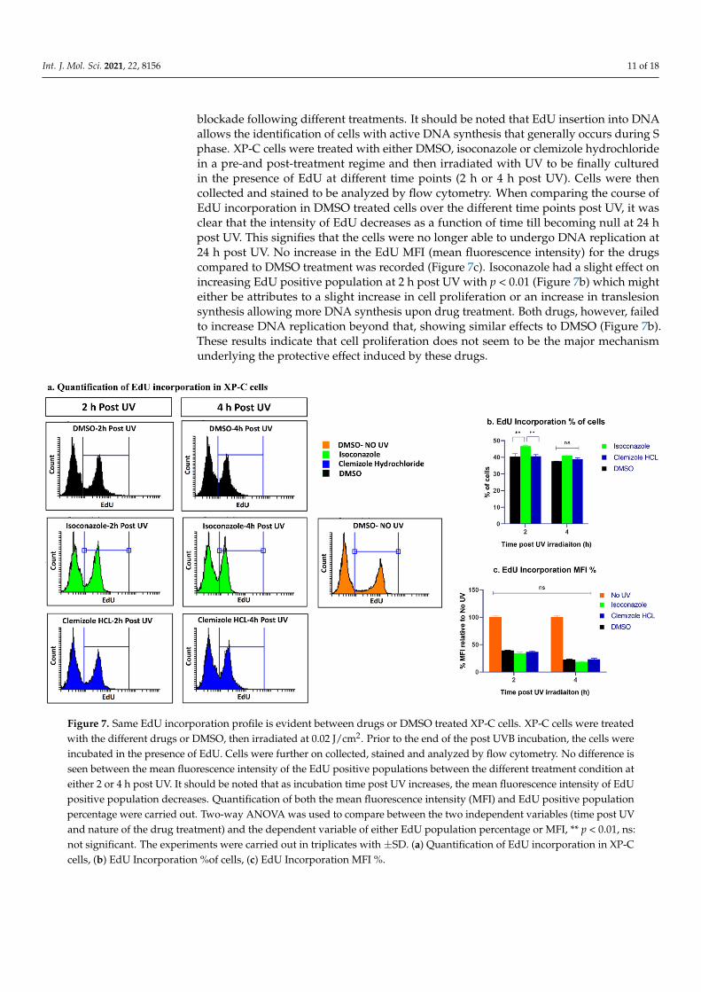
blockade following different treatments. It should be noted that EdU insertion into DNAallows the identification of cells with active DNA synthesis that generally occurs during Sphase. XP-C cells were treated with either DMSO, isoconazole or clemizole hydrochloridein a pre-and post-treatment regime and then irradiated with UV to be finally culturedin the presence of EdU at different time points (2 h or 4 h post UV). Cells were thencollected and stained to be analyzed by flow cytometry. When comparing the course ofEdU incorporation in DMSO treated cells over the different time points post UV, it wasclear that the intensity of EdU decreases as a function of time till becoming null at 24 hpost UV. This signifies that the cells were no longer able to undergo DNA replication at24 h post UV. No increase in the EdU MFI (mean fluorescence intensity) for the drugscompared to DMSO treatment was recorded (Figure 7c). Isoconazole had a slight effect onincreasing EdU positive population at 2 h post UV with p < 0.01 (Figure 7b) which mighteither be attributes to a slight increase in cell proliferation or an increase in translesionsynthesis allowing more DNA synthesis upon drug treatment. Both drugs, however, failedto increase DNA replication beyond that, showing similar effects to DMSO (Figure 7b).These results indicate that cell proliferation does not seem to be the major mechanismunderlying the protective effect induced by these drugs.
Int. J. Mol. Sci. 2021, 22, x FOR PEER REVIEW 11 of 18
blockade following different treatments. It should be noted that EdU insertion into DNA
allows the identification of cells with active DNA synthesis that generally occurs during
S phase. XP-C cells were treated with either DMSO, isoconazole or clemizole hydrochlo-
ride in a pre-and post-treatment regime and then irradiated with UV to be finally cultured
in the presence of EdU at different time points (2 h or 4 h post UV). Cells were then col-
lected and stained to be analyzed by flow cytometry. When comparing the course of EdU
incorporation in DMSO treated cells over the different time points post UV, it was clear
that the intensity of EdU decreases as a function of time till becoming null at 24 h post UV.
This signifies that the cells were no longer able to undergo DNA replication at 24 h post
UV. No increase in the EdU MFI (mean fluorescence intensity) for the drugs compared to
DMSO treatment was recorded (Figure 7c). Isoconazole had a slight effect on increasing
EdU positive population at 2 h post UV with p < 0.01 (Figure 7b) which might either be
attributes to a slight increase in cell proliferation or an increase in translesion synthesis
allowing more DNA synthesis upon drug treatment. Both drugs, however, failed to in-
crease DNA replication beyond that, showing similar effects to DMSO (Figure 7b). These
results indicate that cell proliferation does not seem to be the major mechanism underly-
ing the protective effect induced by these drugs.
Figure 7. Same EdU incorporation profile is evident between drugs or DMSO treated XP-C cells. XP-C cells were treated
with the different drugs or DMSO, then irradiated at 0.02 J/cm2. Prior to the end of the post UVB incubation, the cells were
incubated in the presence of EdU. Cells were further on collected, stained and analyzed by flow cytometry. No difference
is seen between the mean fluorescence intensity of the EdU positive populations between the different treatment condition
at either 2 or 4 h post UV. It should be noted that as incubation time post UV increases, the mean fluorescence intensity of
EdU positive population decreases. Quantification of both the mean fluorescence intensity (MFI) and EdU positive popu-
lation percentage were carried out. Two-way ANOVA was used to compare between the two independent variables (time
post UV and nature of the drug treatment) and the dependent variable of either EdU population percentage or MFI, ** p <
0.01, ns: not significant. The experiments were carried out in triplicates with ±SD.
2.9. Isoconazole and Not Clemizole Hydrochloride Increases Live Cell Population in the Course of
Apoptosis and Necrosis Analysis
In an attempt to decipher the cellular mechanisms of photo-resistance of the drug
treatment, different cell phenotypes were analyzed. Apoptosis was therefore quantified
using Cell Event which allows the indirect measurement of caspase 3/7 activity, key
Figure 7. Same EdU incorporation profile is evident between drugs or DMSO treated XP-C cells. XP-C cells were treatedwith the different drugs or DMSO, then irradiated at 0.02 J/cm2. Prior to the end of the post UVB incubation, the cells wereincubated in the presence of EdU. Cells were further on collected, stained and analyzed by flow cytometry. No difference isseen between the mean fluorescence intensity of the EdU positive populations between the different treatment condition ateither 2 or 4 h post UV. It should be noted that as incubation time post UV increases, the mean fluorescence intensity of EdUpositive population decreases. Quantification of both the mean fluorescence intensity (MFI) and EdU positive populationpercentage were carried out. Two-way ANOVA was used to compare between the two independent variables (time post UVand nature of the drug treatment) and the dependent variable of either EdU population percentage or MFI, ** p < 0.01, ns:not significant. The experiments were carried out in triplicates with ±SD. (a) Quantification of EdU incorporation in XP-Ccells, (b) EdU Incorporation %of cells, (c) EdU Incorporation MFI %.
Int. J. Mol. Sci. 2021, 22, 8156 12 of 18
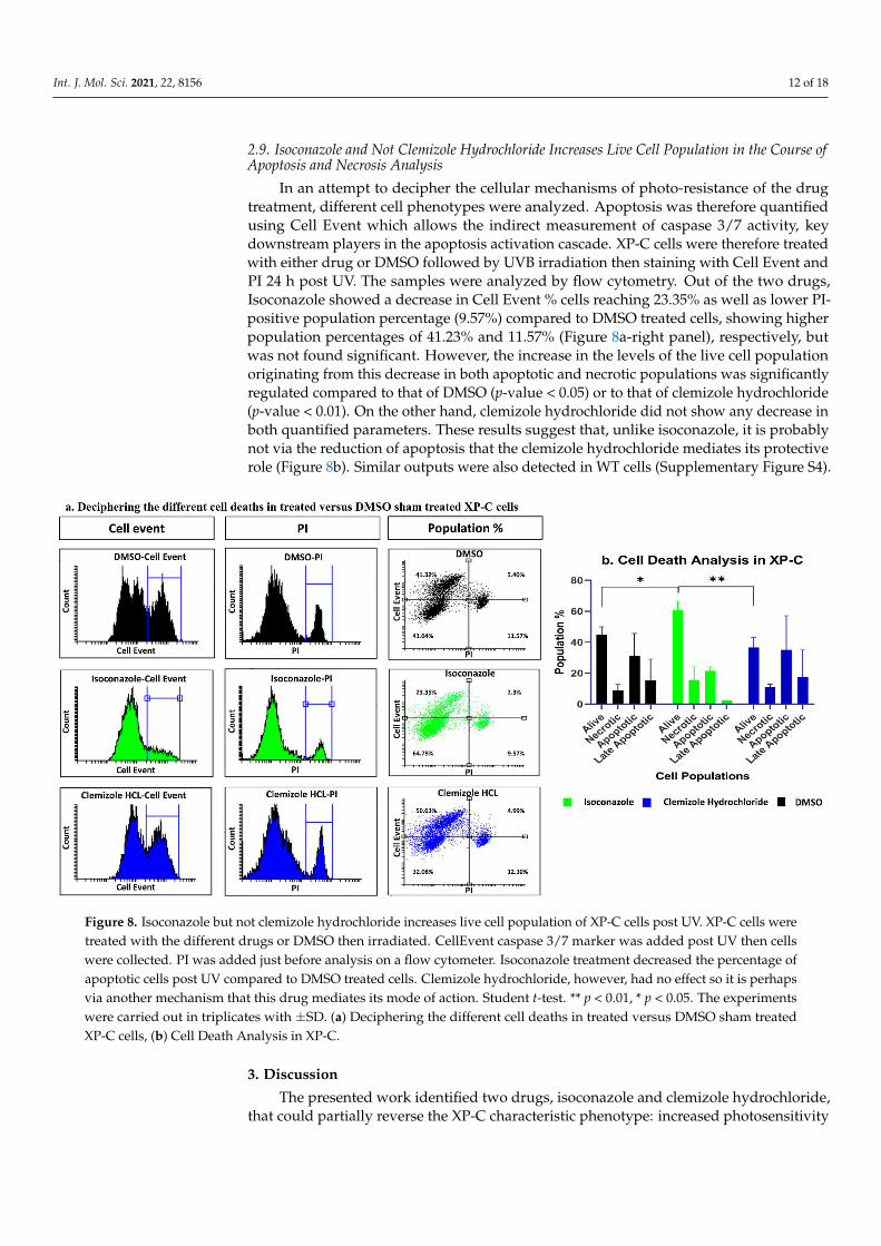
2.9. Isoconazole and Not Clemizole Hydrochloride Increases Live Cell Population in the Course ofApoptosis and Necrosis Analysis
In an attempt to decipher the cellular mechanisms of photo-resistance of the drugtreatment, different cell phenotypes were analyzed. Apoptosis was therefore quantifiedusing Cell Event which allows the indirect measurement of caspase 3/7 activity, keydownstream players in the apoptosis activation cascade. XP-C cells were therefore treatedwith either drug or DMSO followed by UVB irradiation then staining with Cell Event andPI 24 h post UV. The samples were analyzed by flow cytometry. Out of the two drugs,Isoconazole showed a decrease in Cell Event % cells reaching 23.35% as well as lower PI-positive population percentage (9.57%) compared to DMSO treated cells, showing higherpopulation percentages of 41.23% and 11.57% (Figure 8a-right panel), respectively, butwas not found significant. However, the increase in the levels of the live cell populationoriginating from this decrease in both apoptotic and necrotic populations was significantlyregulated compared to that of DMSO (p-value < 0.05) or to that of clemizole hydrochloride(p-value < 0.01). On the other hand, clemizole hydrochloride did not show any decrease inboth quantified parameters. These results suggest that, unlike isoconazole, it is probablynot via the reduction of apoptosis that the clemizole hydrochloride mediates its protectiverole (Figure 8b). Similar outputs were also detected in WT cells (Supplementary Figure S4).
Int. J. Mol. Sci. 2021, 22, x FOR PEER REVIEW 12 of 18
downstream players in the apoptosis activation cascade. XP-C cells were therefore treated
with either drug or DMSO followed by UVB irradiation then staining with Cell Event and
PI 24 h post UV. The samples were analyzed by flow cytometry. Out of the two drugs,
Isoconazole showed a decrease in Cell Event % cells reaching 23.35% as well as lower PI-
positive population percentage (9.57%) compared to DMSO treated cells, showing higher
population percentages of 41.23% and 11.57% (Figure 8a-right panel), respectively, but
was not found significant. However, the increase in the levels of the live cell population
originating from this decrease in both apoptotic and necrotic populations was signifi-
cantly regulated compared to that of DMSO (p-value < 0.05) or to that of clemizole hydro-
chloride (p-value < 0.01). On the other hand, clemizole hydrochloride did not show any
decrease in both quantified parameters. These results suggest that, unlike isoconazole, it
is probably not via the reduction of apoptosis that the clemizole hydrochloride mediates
its protective role (Figure 8b). Similar outputs were also detected in WT cells (supplemen-
tary Figure S4).
Figure 8. Isoconazole but not clemizole hydrochloride increases live cell population of XP-C cells post UV. XP-C cells were
treated with the different drugs or DMSO then irradiated. CellEvent caspase 3/7 marker was added post UV then cells
were collected. PI was added just before analysis on a flow cytometer. Isoconazole treatment decreased the percentage of
apoptotic cells post UV compared to DMSO treated cells. Clemizole hydrochloride, however, had no effect so it is perhaps
via another mechanism that this drug mediates its mode of action. Student t-test. ** p < 0.01, * p < 0.05. The experiments
were carried out in triplicates with ±SD.
3. Discussion
The presented work identified two drugs, isoconazole and clemizole hydrochloride,
that could partially reverse the XP-C characteristic phenotype: increased photosensitivity
and absence of DNA damage repair of photoproducts. In addition, both drugs also ena-
bled photo-protection in WT cells exposed to high doses of UVB irradiation.
XP-C patient cells carry a mutation in the XPC gene required for the recognition of
DNA damage. This mutation renders the cells photosensitive [28] and unable to repair
UVB induced DNA damage. Here we confirmed the photosensitive profile of XP-C as they
show a significantly (p-value < 0.001) reduced viability compared to WT cells as a function
of increased UV doses. Such cells lack the XPC proteins, as confirmed by immunostaining,
signifying that such a mutation leads to the complete loss of the XPC protein. Moreover,
Figure 8. Isoconazole but not clemizole hydrochloride increases live cell population of XP-C cells post UV. XP-C cells weretreated with the different drugs or DMSO then irradiated. CellEvent caspase 3/7 marker was added post UV then cellswere collected. PI was added just before analysis on a flow cytometer. Isoconazole treatment decreased the percentage ofapoptotic cells post UV compared to DMSO treated cells. Clemizole hydrochloride, however, had no effect so it is perhapsvia another mechanism that this drug mediates its mode of action. Student t-test. ** p < 0.01, * p < 0.05. The experimentswere carried out in triplicates with ±SD. (a) Deciphering the different cell deaths in treated versus DMSO sham treatedXP-C cells, (b) Cell Death Analysis in XP-C.
3. Discussion
The presented work identified two drugs, isoconazole and clemizole hydrochloride,that could partially reverse the XP-C characteristic phenotype: increased photosensitivity
Int. J. Mol. Sci. 2021, 22, 8156 13 of 18
and absence of DNA damage repair of photoproducts. In addition, both drugs also enabledphoto-protection in WT cells exposed to high doses of UVB irradiation.
XP-C patient cells carry a mutation in the XPC gene required for the recognition ofDNA damage. This mutation renders the cells photosensitive [28] and unable to repairUVB induced DNA damage. Here we confirmed the photosensitive profile of XP-C as theyshow a significantly (p-value < 0.001) reduced viability compared to WT cells as a functionof increased UV doses. Such cells lack the XPC proteins, as confirmed by immunostaining,signifying that such a mutation leads to the complete loss of the XPC protein. Moreover,upon the quantification of the different UVB induced DNA lesions by LC-MS/MS, it wasevident that XP-C cells manifest slower repair kinetics compared to WT cells. UVB inducedlesions include either CPD or 6-4PP that are formed between two adjacent pyrimidinesgiving four possible lesions T-T, C-C, C-T and T-C [29]. The most abundantly formedlesions are the T-T and T-C lesions for both the CPD and the 6-4PP. In WT cells, both lesiontypes of 6-4PP are readily repaired by the cells a few hours post irradiation, while CPDsrequire more time, with lesion dependent repair kinetics being the slowest repair for T-T,T-C, C-C then C-T, CPD showing the fastest repair, confirmed by us and others [30]. XP-Ccells show no or reduced repair for both CPD and 6-4PP different lesion types as a functionof time.
Drug repurposing is the process of testing previously approved drugs, used for aparticular therapeutic purpose, for their potential use in the treatment of other common orrare diseases. This cuts down drug discovery costs and the long process of toxicity andpharmacokinetics testing [31]. For that, our aim was to test a library of 1280 approveddrugs for their potential use in normalizing the XP-C phenotype. XP-C cells were treatedwith the compounds at 10 µM for 24 h then irradiated with UVB and then treated againwith the same compounds at the same concentration for an additional period of 24 h.The overall viability of the treated cells, proportional to the amount of metabolicallyactive cells, was then measured via incubation with PrestoBlue. The fluorescent valuesobtained were normalized via either the calculation of drug activity %, control basedwith 100% for non-irradiated DMSO treated cells and 0% for irradiated ones or via thecalculation of the non-control based RZscore. Drugs that showed an activity ≥25% withan RZscore above 2.6 were selected as primary hits to be confirmed via a secondaryscreening which were 16 drugs. Acitohexamide was added to the secondary screen druglist due to its previously described photo-protective effect on XPA cells via the enablingof damage repair [25]. Isoconazole, clemizole hydrochloride and bifonazole manifestedthe highest bioactivity after secondary screening enabling photo-protection in XP-C cellscompared to DMSO treated controls with a range of 20 to 40% increase of cell viabilitydepending on the conditions. However, the enabling of photo-resistance is not sufficientby itself for a satisfying curative outcome. The persistence of cells with accumulated DNAdamage can lead to the conversion of such lesions into mutations which, depending ontheir localizations, can lead to carcinogenesis [32]. The main lesion in XP cells mediatingthe UV signature mutation is the C-C lesion where the slow repair of such lesion in XPcells compared to its fast repair in WT cells favors the deamination and induction ofmutations [29]. Therefore, photo-resistance should be associated with increased DNAdamage repair in the perspective of therapeutic treatment. Indeed, we tested the capacityof the 16 primary hits and acetohexamide in mediating the decrease of DNA damagepost UVB irradiation. Out of this collection, two compounds, isoconazole and clemizolehydrochloride, decreased the amount of DNA damage post UV in XP-C cells by about 20%(p-value < 0.001 at 5 µM).
Isoconazole is an azole antifungal drug with no reported data on its effect in sun-shielding upon its use on the skin, while clemizole hydrochloride is a Histamine H1antagonist. Other documented functions for clemizole hydrochloride include its role in thetreatment of HCV infection [33] and its blocking effect on TRPC5 ion channels [34]. Thesefunctions fail to explain these drugs’ effect on XP-C cells, suggesting possible alternativemode(s) of action than the ones documented that need(s) to be further on explored.

Int. J. Mol. Sci. 2021, 22, 8156 14 of 18
One similar approach to ours was conducted on XP-A cells, where the anti-diabeticdrug acetohexamide was found to be effective in reducing the cells’ photosensitivity andenabling damage repair [25], mediating the identification of an additional mode of actionfor this drug not related to its documented effect. Acetohexamide had minimal effect inthe reversal of XP-C phenotype when tested in our screen, yet we managed to identifyother drugs that can aid in this phenotypic partial reversal. Acetohexamide enabled thedegradation of MUTYH, a protein involved in the removal of adenine residues mispairedwith 8-oxo-guanine residues after oxidative stress [35]. The authors hypothesized thatthe degradation of such protein might be enabling spatial access of the lesions to otherrepair machinery independent of nucleotide excision repair [25] which was not yet furtherexplored to date. It is therefore possible that the hit drugs identified in this study mightsimilarly impose spatial access effect or that they interfere with one or several mediators ofDNA repair, a hypothesis which needs to be further on examined.
Collectively, our data show that isoconazole and clemizole hydrochloride mediatedan increase in cell viability post UV irradiation and enabled to a lower extent the repairof accumulated DNA damage in the form of UVB-induced 6-4PP. Isoconazole, on the onehand, mediated a curative effect post UV irradiation by enabling the decrease of apoptoticcells’ population as measured by flow cytometry with Cell Event caspase 3/7 activitystaining. Clemizole hydrochloride’s effect, on the other hand, was both preventative preUV and curative post UV irradiation showing a p-value < 0.05, while the difference betweenthe post-irradiation alone and the pre-irradiation treatment was not significant. Such aneffect is mediated via a yet unknown mechanism. The simultaneous double treatment withboth drugs revealed no synergistic or additive effect. This suggests that both drugs interferewith a similar biological process or target which is consistent with the fact that they share asimilar chemical scaffold (azole ring). Moreover, both drugs were shown not to affect thecells’ proliferation rate. The photoprotective effect of the drugs was also evident in WT cellspost UV irradiation signifying the involvement of a protective pathway indirectly related tothe mutated XPC gene. This work will mark the first attempt in discovering compounds forthe amelioration of the XP-C phenotype, yet the exact regulated target needs to be furtheron explored to allow the identification of the effectors aiding in this phenotypic reversaland thus paving a way for a potential therapy for this yet untreatable genodermatosis.
4. Materials and Methods4.1. Cell Line
Wild type (AG10076) and XP-C (GM15983) immortalized patient derived-fibroblastswere purchased from Coriell Biorepository. XP-C fibroblasts possess a two-base pairshift mutation at codon 431 of the XPC gene. The cells were cultured in DMEM highglucose, GlutaMAX media (Gibco, Waltham, MA, USA) supplemented with 10% FBS and1% penicillin/streptomycin at 37 ◦C in a 5% CO2 incubator.
4.2. Chemical Drug Screening
The Prestwick chemical library was utilized. It consists of 1280 drugs of high chemicaland pharmacological diversity. The drugs are all approved (FDA, EMA or other agencies).The drugs cover all main ATC groups and are dedicated to either CNS, cardiovascular,metabolism or infectiology diseases with either enzymatic or GPCRs targets. The librarywas screened at a final concentration of 10 µM. For the screening procedure, the cellswere incubated with the drugs for 24 h then media was recuperated and the cells washedwith PBS. Irradiation was carried out in the presence of PBS. Following that same drug,containing media were returned back to the cells for the course of post treatment to beincubated for further 24 h before the assessment of the readouts.
4.3. UV Dose-Response
To examine the photosensitivity of XP-C cells relative to WT in response to UVBirradiation, both cells were seeded in 96 well plates until 80% confluency, washed with
Int. J. Mol. Sci. 2021, 22, 8156 15 of 18
PBS, then subjected to increasing doses of UVB. Twenty-four hours post UV, the viabilityof the cells was recorded as a measurement of the cells’ reducing capacity by PrestoBlue(Thermofisher Scientific, Waltham, MA, USA) according to the manufacturer’s suggestion.The LD50 for each cell line was calculated.
4.4. XPC Immuno-Staining and Associated Microscopy
For the characterization of XPC expression, both cell lines were fixed with 4% paraformalde-hyde followed by permeabilization using 0.2% Triton X-100 and saturation with 3% FBSin PBS. Primary antibody targeting XPC (mouse monoclonal, Thermofisher Scientific,Waltham, MA, USA) was utilized followed by incubation with Alexa Fluor 488-coupledsecondary antibody (goat anti-mouse, Invitrogen, Waltham, MA, USA). Nuclear DNA wascounter-stained with Hoechst (Sigma-Aldrich, Burlington, MA, USA). Cell images wereacquired by the Zen Axio-observer at 40X.
4.5. LC-MS/MS DNA Damage Quantification
The quantification of pyrimidine dimers (CPD & 6-4PP) was carried out by LC-MS/MSaccording to previous protocol [36]. Briefly, both cell lines were seeded in 100 mm dishesuntil sub-confluency where they were subjected to a UVB dose of 0.01 J/cm2. At differenttime points post UV (0, 2, 24 and 48 h), the cells were collected and their DNA extractedwith Qiagen DNEasy Kit (Hilden, Germany). Sample preparation was carried out in twosteps; the first includes incubation of DNA for 2 h at 37 ◦C and pH 6 in the presence ofnuclease P1, DNase II and phosphodiesterase II. The pH was then raised to 8 by the additionof Tris. A second incubation period in the presence of phosphodiesterase I and alkalinephosphatase at 37 ◦C for 2 h yielded digested DNA with normal bases as nucleosides andphotoproducts as dinucleoside monophosphates. Samples were then injected on an HPLCsystem (Agilent, Massy, France) connected to a reverse-phase HPLC column (150_2mmID, 5 mm particle size, ODB, France). The detection was provided first by a UV detectoraimed at quantifying normal nucleosides. Photoproducts were detected by a tandem massspectrometer (API 3000, SCIEX, Thorn hill, ON, Canada) used in the reaction-monitoringmode as previously described [37]. The four CPDs and the four 6-4PPs (TT, TC, CT and CCderivatives) were quantified individually in the same HPLC run. Results were expressedin the number of lesions per million normal bases.
4.6. 6-4PP DNA Damage Staining and Quantification
For the quantification of DNA damage, cells were seeded until confluency, thensubjected to UVB irradiation at a dose of 0.02 J/cm2. Twenty-four hours post UV, the cellswere stained according to a previous protocol [38], consisting first of cell fixation with4% paraformaldehyde, and permeabilized with 0.2% Triton X-100. Cells were further ontreated with 2M HCL to denaturate the DNA and thus allow access to the DNA damage-targeting antibody. After saturation, the cells were incubated with 6-4PP antibody (64M-2cosmo bio, Japan) followed by AF488 secondary antibody (Invitrogen, Waltham, MA, USA)and the DNA was counterstained with Hoechst (Sigma Aldrich, Burlington, MA, USA).Image acquisition and quantification were carried out by a Cell-insight NXT high contentscreening platform at 10X magnification. Single-cell fluorescence was quantified for all thecells under each particular treatment condition.
4.7. Ki67 Staining and Single-Cell Quantification
The cells were seeded until sub-confluency, then subjected to UVB irradiation ata dose of 0.02 J/cm2. Twenty-four hours post-irradiation, the cells were fixed with 4%paraformaldehyde and permeabilized with 0.2% Triton X-100. After saturation, the cellswere incubated with ki67 antibody (clone MIB-1, Clinisciences, France) followed by AF488secondary antibody (Invitrogen, Waltham, MA, USA), and the DNA was counterstainedwith Hoechst (Sigma Aldrich, Burlington, MA, USA). Image acquisition and quantification
Int. J. Mol. Sci. 2021, 22, 8156 16 of 18
were carried out by Cell-insight NXT high content screening platform at 10X magnification.Single-cell fluorescence was quantified for all the cells under each particular treatment condition.
4.8. EdU Cell Proliferation Assay
Cell health and genotoxicity can be assessed by measuring the cell’s ability to pro-liferate via the incorporation of the nucleoside analog EdU. The latter can be further onquantified by a click-it covalent reaction between an azide and alkyne catalyzed by copper.For that, both cell lines were seeded in 100 mm dishes subjected to different forms oftreatment followed by irradiation. Sham irradiated and sham-treated controls were alsoutilized. Two hours before the end of post UV incubation, EdU was diluted in the cellmedia at different time points. The cells were then collected and stained according to themanufacturer’s protocol. The samples were analyzed by flow cytometry (FACScan, BDLSRII flow cytometer, BD Biosciences, Franklin Lakes, NJ, USA). The post analysis wascarried out using flowing software [39] (Turku Bioimaging, Finland).
4.9. Cell Event-PI Cell Death Quantification
To follow the effect of treatment on the induction of apoptosis post UV irradiation,CellEvent (Thermofisher Scientific) was utilized, which allows the measurement of caspase3/7 activity. For that, cells were seeded in 100 mm dishes, treated with the molecules,irradiated, then Cell Event was added post UV and incubated for 24 h. The cells were thencollected and PI was added before their analysis by flow cytometer (FACScan, BD LSRIIflow cytometer, BD Biosciences). The post analysis was carried using flowing software [39](Turku Bioimaging, Finland).
4.10. Statistical Analysis
Drug screening hit selection and single-cell analysis was carried out using R soft-ware [40]. Statistical analysis was conducted using GraphPad Prism v.8 after data normal-ization and quantification of normality to allow the choice of the respective statistical test(parametric or non-parametric) for each particular set of experiments.
Supplementary Materials: The following are available online at https://www.mdpi.com/article/10.3390/ijms22158156/s1, Figure S1: Primary Screen UV dose optimization and reproducibilityassessment. Figure S2: Single-cell analysis of nuclear DNA damage quantification. Figure S3: Drugtreatment has no effect on ki67 proliferation marker expression. Figure S4: Isoconazole and notclemizole hydrochloride decreases the population % of apoptotic WT cells post UV.
Author Contributions: F.K. performed all the experiments and wrote the manuscript. E.S. aidedin flow cytometry, data normalization and manuscript revision. C.B. designed and performed thechemical primary screening. M.-O.F. supervised the screening campaign. N.F. assisted in the LC-MS/MS experiment. X.G., H.F.-K. and W.R. supervised the project. M.F.-K. and B.B. revised themanuscript. All authors have read and agreed to the published version of the manuscript.
Funding: F.K. is supported by a fund from the Lebanese University (UL) and Alternative Energiesand Atomic Energy Commission (CEA). W.R.’s contribution was funded by ANR grant PG2HEAL(ANR-18-CE17-0017) and supported by the French National Research Agency in the framework ofthe “Investissements d’avenir” program (ANR-15-IDEX-02). Automated molecular screening hasbeen performed through the ChemBioFrance infrastructure at the Grenoble Alpes Probes and DrugDiscovery platform (GAP2D). Support of the Labex GRAL, ANR-10-LABX-49-01 financed within theUniversity of Grenoble Alpes graduate school CBH-EUR-GS (ANR-17-EURE-0003) is acknowledged.
Institutional Review Board Statement: Not applicable.
Informed Consent Statement: Not applicable.
Data Availability Statement: Not applicable.
Acknowledgments: This article was previously published as a pre-print on research square accessedon 16th February 2021 (https://www.researchsquare.com/article/rs-197694/v1).

Int. J. Mol. Sci. 2021, 22, 8156 17 of 18
Conflicts of Interest: The authors declare that the research was conducted in the absence of anycommercial or financial relationships that could be construed as a potential conflict of interest.
References1. Tan, N.S.; Wahli, W. The emerging role of Nrf2 in dermatotoxicology. EMBO Mol. Med. 2014, 6, 431–433. [CrossRef]2. Gegotek, A.; Skrzydlewska, E. The role of transcription factor Nrf2 in skin cells metabolism. Arch. Dermatol. Res. 2015, 307,
385–396. [CrossRef] [PubMed]3. Ravanat, J.L.; Douki, T.; Cadet, J. Direct and indirect effects of UV radiation on DNA and its components. J. Photochem. Photobiol.
B 2001, 63, 88–102. [CrossRef]4. Slieman, T.A.; Nicholson, W.L. Artificial and solar UV radiation induces strand breaks and cyclobutane pyrimidine dimers in
Bacillus subtilis spore DNA. Appl. Environ. Microbiol. 2000, 66, 199–205. [CrossRef]5. Baumstark-Khan, C.; Hentschel, U.; Nikandrova, Y.; Krug, J.; Horneck, G. Fluorometric analysis of DNA unwinding (FADU) as
a method for detecting repair-induced DNA strand breaks in UV-irradiated mammalian cells. Photochem. Photobiol. 2000, 72,477–484. [CrossRef]
6. Rastogi, R.P.; Singh, S.P.; Hader, D.P.; Sinha, R.P. Detection of reactive oxygen species (ROS) by the oxidant-sensing probe2’,7’-dichlorodihydrofluorescein diacetate in the cyanobacterium Anabaena variabilis PCC 7937. Biochem. Biophys. Res. Commun.2010, 397, 603–607. [CrossRef] [PubMed]
7. Armstrong JD and Kunz BA (1990) Site and strand specificity of UVB mutagenesis in the SUP4-o gene of yeast. Proc. Natl. Acad.Sci. USA 1990, 87, 9005–9009. [CrossRef]
8. Kobaisi, F.; Fayyad, N.; Rezvani, H.R.; Fayyad-Kazan, M.; Sulpice, E.; Badran, B.; Fayyad-Kazan, H.; Gidrol, X.; Rachidi, W.Signaling Pathways, Chemical and Biological Modulators of Nucleotide Excision Repair: The Faithful Shield against UVGenotoxicity. Oxidative Med. Cell. Longev. 2019, 2019, 4654206. [CrossRef] [PubMed]
9. Spivak, G. Nucleotide excision repair in humans. DNA Repair 2015, 36, 3–18. [CrossRef] [PubMed]10. Iyama, T.; Wilson, D.M., III. DNA repair mechanisms in dividing and non-dividing cells. DNA Repair 2013, 12, 620–636. [CrossRef]
[PubMed]11. Giglia-Mari, G.; Sarasin, A. TP53 mutations in human skin cancers. Hum. Mutat. 2003, 21, 217–228. [CrossRef] [PubMed]12. Tampa, M.; Georgescu, S.R.; Mitran, C.I.; Mitran, M.I.; Matei, C.; Scheau, C.; Constantin, C.; Neagu, M. Recent Advances in
Signaling Pathways Comprehension as Carcinogenesis Triggers in Basal Cell Carcinoma. J. Clin. Med. 2020, 9, 3010. [CrossRef][PubMed]
13. Curtin, J.A.; Fridlyand, J.; Kageshita, T.; Patel, H.N.; Busam, K.J.; Kutzner, H.; Cho, K.H.; Aiba, S.; Bröcker, E.B.; LeBoit, P.E.; et al.Distinct sets of genetic alterations in melanoma. N. Engl. J. Med. 2005, 353, 2135–2147. [CrossRef]
14. Melis, J.P.; Luijten, M.; Mullenders, L.H.; van Steeg, H. The role of XPC: Implications in cancer and oxidative DNA damage.Mutat. Res. 2001, 728, 107–117. [CrossRef] [PubMed]
15. D’Errico, M.; Parlanti, E.; Teson, M.; de Jesus, B.M.; Degan, P.; Calcagnile, A.; Jaruga, P.; Bjoras, M.; Crescenzi, M.;Pedrini, A.M.; et al. New functions of XPC in the protection of human skin cells from oxidative damage. EMBO J. 2006, 25,4305–4315. [CrossRef] [PubMed]
16. Fayyad, N.; Kobaisi, F.; Beal, D.; Mahfouf, W.; Ged, C.; Morice-Picard, F.; Fayyad-Kazan, M.; Fayyad-Kazan, H.; Badran, B.;Rezvani, H.R.; et al. Xeroderma Pigmentosum C (XPC) Mutations in Primary Fibroblasts Impair Base Excision Repair Pathwayand Increase Oxidative DNA Damage. Front. Genet. 2020, 11, 561687. [CrossRef]
17. Rezvani, H.R.; Ged, C.; Bouadjar, B.; de Verneuil, H.; Taieb, A. Catalase overexpression reduces UVB-induced apoptosis in ahuman xeroderma pigmentosum reconstructed epidermis. Cancer Gene Ther. 2008, 15, 241–251. [CrossRef]
18. Rezvani, H.R.; Kim, A.L.; Rossignol, R.; Ali, N.; Daly, M.; Mahfouf, W.; Bellance, N.; Taieb, A.; de Verneuil, H.; Mazurier, F.; et al.XPC silencing in normal human keratinocytes triggers metabolic alterations that drive the formation of squamous cell carcinomas.J. Clin. Investig. 2011, 121, 195–211. [CrossRef]
19. Raad, H.; Serrano-Sanchez, M.; Harfouche, G.; Mahfouf, W.; Bortolotto, D.; Bergeron, V.; Kasraian, Z.; Dousset, L.; Hosseini, M.;Taieb, A.; et al. NADPH Oxidase-1 Plays a Key Role in Keratinocyte Responses to UV Radiation and UVB-Induced SkinCarcinogenesis. J. Investig. Dermatol. 2017, 137, 1311–1321. [CrossRef]
20. Bidon, B.; Iltis, I.; Semer, M.; Nagy, Z.; Larnicol, A.; Cribier, A.; Benkirane, M.; Coin, F.; Egly, J.M.; Le May, N. XPC is an RNApolymerase II cofactor recruiting ATAC to promoters by interacting with E2F1. Nat. Commun. 2018, 9, 2610. [CrossRef]
21. Chen, Z.; Yang, J.; Wang, G.; Song, B.; Li, J.; Xu, Z. Attenuated expression of xeroderma pigmentosum group C is associated withcritical events in human bladder cancer carcinogenesis and progression. Cancer Res. 2007, 67, 4578–4585. [CrossRef]
22. de Feraudy, S.; Ridd, K.; Richards, L.M.; Kwok, P.Y.; Revet, I.; Oh, D.; Feeney, L.; Cleaver, J.E. The DNA damage-binding proteinXPC is a frequent target for inactivation in squamous cell carcinomas. Am. J. Pathol. 2010, 177, 555–562. [CrossRef]
23. Pascucci, B.; D’Errico, M.; Parlanti, E.; Giovannini, S.; Dogliotti, E. Role of nucleotide excision repair proteins in oxidative DNAdamage repair: An updating. Biochemistry 2011, 76, 4–15. [CrossRef]
24. Birmingham, A.; Selfors, L.M.; Forster, T.; Wrobel, D.; Kennedy, C.J.; Shanks, E.; Santoyo-Lopez, J.; Dunican, D.J.; Long, A.;Kelleher, D.; et al. Statistical methods for analysis of high-throughput RNA interference screens. Nat. Methods 2009, 6, 569–575.[CrossRef]

Int. J. Mol. Sci. 2021, 22, 8156 18 of 18
25. Mazouzi, A.; Battistini, F.; Moser, S.C.; da Ferreira, S.J.; Wiedner, M.; Owusu, M.; Lardeau, C.H.; Ringler, A.; Weil, B.;Neesen, J.; et al. Repair of UV-Induced DNA Damage Independent of Nucleotide Excision Repair Is Masked by MUTYH.Mol. Cell 2017, 68, 797–807. [CrossRef]
26. Borszéková Pulzová, L.; Ward, T.A.; Chovanec, M. XPA: DNA Repair Protein of Significant Clinical Importance. Int. J. Mol. Sci.2020, 6, 2182. [CrossRef] [PubMed]
27. van Oijen, M.G.; Medema, R.H.; Slootweg, P.J.; Rijksen, G. Positivity of the proliferation marker Ki-67 in noncycling cells. Am. J.Clin. Pathol. 1998, 110, 24–31. [CrossRef] [PubMed]
28. Black, J.O. Xeroderma Pigmentosum. Head Neck Pathol. 2016, 10, 139–144. [CrossRef]29. Mouret, S.; Charveron, M.; Favier, A.; Cadet, J.; Douki, T. Differential repair of UVB-induced cyclobutane pyrimidine dimers in
cultured human skin cells and whole human skin. DNA Repair 2008, 7, 704–712. [CrossRef] [PubMed]30. Courdavault, S.; Baudouin, C.; Charveron, M.; Canguilhem, B.; Favier, A.; Cadet, J.; Douki, T. Repair of the three main types of
bipyrimidine DNA photoproducts in human keratinocytes exposed to UVB and UVA radiations. DNA Repair 2005, 4, 836–844.[CrossRef]
31. Pushpakom, S.; Iorio, F.; Eyers, P.A.; Escott, K.J.; Hopper, S.; Wells, A.; Doig, A.; Guilliams, T.; Latimer, J.; McNamee, C.; et al.Drug repurposing: Progress, challenges and recommendations. Nat. Rev. Drug Discov. 2019, 18, 41–58. [CrossRef]
32. Basu, A.K. DNA Damage, Mutagenesis and Cancer. Int. J. Mol. Sci. 2018, 19, 970. [CrossRef] [PubMed]33. Einav, S.; Gerber, D.; Bryson, P.D.; Sklan, E.H.; Elazar, M.; Maerkl, S.J.; Glenn, J.S.; Quake, S.R. Discovery of a hepatitis C target
and its pharmacological inhibitors by microfluidic affinity analysis. Nat. Biotechnol. 2008, 26, 1019–1027. [CrossRef] [PubMed]34. Richter, J.M.; Schaefer, M.; Hill, K. Clemizole hydrochloride is a novel and potent inhibitor of transient receptor potential channel
TRPC5. Mol. Pharmacol. 2014, 86, 514–521. [CrossRef] [PubMed]35. Kashfi, S.M.; Golmohammadi, M.; Behboudi, F.; Nazemalhosseini-Mojarad, E.; Zali, M.R. MUTYH the base excision repair gene
family member associated with colorectal cancer polyposis. Gastroenterol. Hepatol. Bed Bench 2013, 6, S1–S10. [PubMed]36. Matallana-Surget, S.; Meador, J.A.; Joux, F.; Douki, T. Effect of the GC content of DNA on the distribution of UVB-induced
bipyrimidine photoproducts. Photochem. Photobiol. Sci. 2008, 7, 794–801. [CrossRef]37. Douki, T.; Cadet, J. Individual determination of the yield of the main UV-induced dimeric pyrimidine photoproducts in DNA
suggests a high mutagenicity of CC photolesions. Biochemistry 2001, 40, 2495–2501. [CrossRef]38. Kuschal, C.; DiGiovanna, J.J.; Khan, S.G.; Gatti, R.A.; Kraemer, K.H. Repair of UV photolesions in xeroderma pigmentosum group
C cells induced by translational readthrough of premature termination codons. Proc. Natl. Acad. Sci. USA 2013, 110, 19483–19488.[CrossRef]
39. Flowing Software Version 2.5.1 bPT, Turku Centre for Biotechnology University of Turku, Finland, In Colaboration with TurkuBioimaging. Available online: http://flowingsoftware.btk.fi/ (accessed on 16 February 2021).
40. R Core Team. R: A Language and Environment for Statistical Computing. R Foundation for Statistical Computing V, Austria.2018. Available online: http://www.R-project.org/ (accessed on 16 February 2021).