Covalent conjugation of extracellular vesicles with peptides ...
23
Covalent conjugation of extracellular vesicles with peptides and nanobodies for targeted therapeutic delivery Pham, Tin Chanh; Jayasinghe, Migara Kavishka; Pham, Thach Tuan; Yang, Yuqi; Wei, Likun; Waqas, Muhammad Usman; Chen, Huan; Pirisinu, Marco; Gong, Jinhua; Kim, Seongkyeol; Peng, Boya; Wang, Weixi; Chan, Charlene; Ma, Victor; Nguyen, Nhung T.H. ; Kappei, Dennis; Nguyen, Xuan-Hung; Cho, William C.; Shi, Jiahai; Le, Minh T.N. Published in: Journal of Extracellular Vesicles Published: 01/02/2021 Document Version: Final Published version, also known as Publisher’s PDF, Publisher’s Final version or Version of Record License: CC BY Publication record in CityU Scholars: Go to record Published version (DOI): 10.1002/jev2.12057 Publication details: Pham, T. C., Jayasinghe, M. K., Pham, T. T., Yang, Y., Wei, L., Waqas, M. U., Chen, H., Pirisinu, M., Gong, J., Kim, S., Peng, B., Wang, W., Chan, C., Ma, V., Nguyen, N. T. H., Kappei, D., Nguyen, X-H., Cho, W. C., Shi, J., & Le, M. T. N. (2021). Covalent conjugation of extracellular vesicles with peptides and nanobodies for targeted therapeutic delivery. Journal of Extracellular Vesicles, 10(4), [e12057]. https://doi.org/10.1002/jev2.12057 Citing this paper Please note that where the full-text provided on CityU Scholars is the Post-print version (also known as Accepted Author Manuscript, Peer-reviewed or Author Final version), it may differ from the Final Published version. When citing, ensure that you check and use the publisher's definitive version for pagination and other details. General rights Copyright for the publications made accessible via the CityU Scholars portal is retained by the author(s) and/or other copyright owners and it is a condition of accessing these publications that users recognise and abide by the legal requirements associated with these rights. Users may not further distribute the material or use it for any profit-making activity or commercial gain. Publisher permission Permission for previously published items are in accordance with publisher's copyright policies sourced from the SHERPA RoMEO database. Links to full text versions (either Published or Post-print) are only available if corresponding publishers allow open access. Take down policy Contact [email protected] if you believe that this document breaches copyright and provide us with details. We will remove access to the work immediately and investigate your claim. Download date: 14/01/2022
-
Upload
khangminh22 -
Category
Documents
-
view
0 -
download
0
Transcript of Covalent conjugation of extracellular vesicles with peptides ...

Covalent conjugation of extracellular vesicles with peptides and nanobodies for targetedtherapeutic delivery
Pham, Tin Chanh; Jayasinghe, Migara Kavishka; Pham, Thach Tuan; Yang, Yuqi; Wei, Likun;Waqas, Muhammad Usman; Chen, Huan; Pirisinu, Marco; Gong, Jinhua; Kim, Seongkyeol;Peng, Boya; Wang, Weixi; Chan, Charlene; Ma, Victor; Nguyen, Nhung T.H. ; Kappei, Dennis;Nguyen, Xuan-Hung; Cho, William C.; Shi, Jiahai; Le, Minh T.N.Published in:Journal of Extracellular Vesicles
Published: 01/02/2021
Document Version:Final Published version, also known as Publisher’s PDF, Publisher’s Final version or Version of Record
License:CC BY
Publication record in CityU Scholars:Go to record
Published version (DOI):10.1002/jev2.12057
Publication details:Pham, T. C., Jayasinghe, M. K., Pham, T. T., Yang, Y., Wei, L., Waqas, M. U., Chen, H., Pirisinu, M., Gong, J.,Kim, S., Peng, B., Wang, W., Chan, C., Ma, V., Nguyen, N. T. H., Kappei, D., Nguyen, X-H., Cho, W. C., Shi, J.,& Le, M. T. N. (2021). Covalent conjugation of extracellular vesicles with peptides and nanobodies for targetedtherapeutic delivery. Journal of Extracellular Vesicles, 10(4), [e12057]. https://doi.org/10.1002/jev2.12057
Citing this paperPlease note that where the full-text provided on CityU Scholars is the Post-print version (also known as Accepted AuthorManuscript, Peer-reviewed or Author Final version), it may differ from the Final Published version. When citing, ensure thatyou check and use the publisher's definitive version for pagination and other details.
General rightsCopyright for the publications made accessible via the CityU Scholars portal is retained by the author(s) and/or othercopyright owners and it is a condition of accessing these publications that users recognise and abide by the legalrequirements associated with these rights. Users may not further distribute the material or use it for any profit-making activityor commercial gain.Publisher permissionPermission for previously published items are in accordance with publisher's copyright policies sourced from the SHERPARoMEO database. Links to full text versions (either Published or Post-print) are only available if corresponding publishersallow open access.
Take down policyContact [email protected] if you believe that this document breaches copyright and provide us with details. We willremove access to the work immediately and investigate your claim.
Download date: 14/01/2022

Received: 24 March 2020 Revised: 16 October 2020 Accepted: 14 December 2020
DOI: 10.1002/jev2.12057
RESEARCH ARTICLE
Covalent conjugation of extracellular vesicles with peptides andnanobodies for targeted therapeutic delivery
Tin Chanh Pham, Migara Kavishka Jayasinghe,,, Thach Tuan Pham,,,
Yuqi Yang LikunWei, Waqas Muhammad Usman Huan Chen
Marco Pirisinu Jinhua Gong, Seongkyeol Kim Boya Peng,, Weixi Wang,
Charlene Chan Victor Ma Nhung T.H. Nguyen Dennis Kappei,
Xuan-Hung Nguyen William C. Cho Jiahai Shi, Minh T.N. Le,,
1 Department of Pharmacology, Yong Loo LinSchool of Medicine, National University ofSingapore, Singapore2 Department of Biomedical Sciences, College ofVeterinary Medicine and Life Sciences, CityUniversity of Hong Kong, Hong Kong3 Institute for Digital Medicine, ImmunologyProgramme and Cancer Programme, Yong Loo LinSchool of Medicine, National University ofSingapore, Singapore4 N.1 Institute for Health, National University ofSingapore, Singapore5 City University of Hong Kong Shenzhen Institute,Shenzhen, China6 Cancer Science Institute of Singapore, NationalUniversity of Singapore, Singapore7 Department of Clinical Oncology, QueenElizabeth Hospital, Hong Kong8 Vinmec Institute of Applied Science andRegenerative Medicine, and College of HealthSciences, Vinmec Healthcare systemVinUniversity, Hanoi, Vietnam9 Department of Biochemistry, Yong Loo LinSchool of Medicine, National University ofSingapore, Singapore
CorrespondenceMinhT.N. Le,Department of Pharmacology, YongLooLin School ofMedicine,NationalUniversity ofSingapore, Singapore.Email: [email protected] Shi,Department of Biomedical Sciences,College ofVeterinaryMedicine andLife Sciences,CityUniversity ofHongKong,HongKong.Email: [email protected]
TinChanhPham,MigaraKavishka Jayasinghe,ThachTuanPhamandYuqiYang contributedequally to this study.
AbstractNatural extracellular vesicles (EVs) are ideal drug carriers due to their remarkablebiocompatibility. Their delivery specificity can be achieved by the conjugation of tar-geting ligands. However, existing methods to engineer target-specific EVs are tediousor inefficient, having to compromise between harsh chemical treatments and tran-sient interactions. Here, we describe a novel method for the covalent conjugation ofEVs with high copy numbers of targeting moieties using protein ligases. Conjugationof EVs with either an epidermal growth factor receptor (EGFR)-targeting peptideor anti-EGFR nanobody facilitates their accumulation in EGFR-positive cancer cells,both in vitro and in vivo. Systemic delivery of paclitaxel by EGFR-targeting EVs at alow dose significantly increases drug efficacy in a xenograftedmousemodel of EGFR-positive lung cancer. The method is also applicable to the conjugation of EVs withpeptides and nanobodies targeting other receptors, such as HER2 and SIRP alpha,and the conjugated EVs can deliver RNA in addition to small molecules, supportingthe versatile application of EVs in cancer therapies. This simple, yet efficient and ver-satile method for the stable surface modification of EVs bypasses the need for geneticand chemical modifications, thus facilitating safe and specific delivery of therapeuticpayloads to target cells.
KEYWORDSconjugation, delivery, extracellular vesicles, targeted, therapeutics
This is an open access article under the terms of the Creative Commons Attribution License, which permits use, distribution and reproduction in any medium, provided the originalwork is properly cited.© 2021 The Authors. Journal of Extracellular Vesicles published by Wiley Periodicals, LLC on behalf of the International Society for Extracellular Vesicles
J Extracell Vesicles. 2021;10:e12057. wileyonlinelibrary.com/journal/jev2 of https://doi.org/10.1002/jev2.12057

of PHAM et al.
INTRODUCTION
Extracellular vesicles (EVs), a natural means of intercellular communication and RNA exchange in eukaryotes, are emerging asnovel drug delivery vehicles (Andaloussi et al., 2013; Pitt et al., 2016). The natural functions of EVs in mammalian cells suggesttheir ability to bypass cellular barriers as well as other hurdles to drug delivery, including cytotoxicity, RNase susceptibility,endosomal accumulation, multidrug resistance and immunogenicity (Andaloussi et al., 2013; Pitt et al., 2016). In a recent study,we identified human red blood cells (RBCs) as an ideal source of EVs with promising properties for RNA drug delivery (Usmanet al., 2018). We are able to produce RBCEVs on a large scale without the need for cell culture, thereby reducing the cost ofproduction and the risk of contamination. Using RBCEVs for antisense oligonucleotide (ASO) delivery, we observed efficientknockdown of an oncogenic microRNA and suppression of leukaemia and breast cancer development both in vitro and in vivo(Usman et al., 2018). RBCEVs promise to be a simple and efficient platform for drug delivery that is safe and easily scalable.However, the nonspecific uptake of RBCEVs may cause unwanted side effects in normal tissues when RBCEVs are used todeliver drugs.To convert EVs into therapies that home specifically onto their target cells, EVs are usually equipped with targeting peptides or
antibodies by overexpressing these molecules in the EV-donor cells using transfection as well as retroviral or lentiviral infection(Andaloussi et al., 2013; Pitt et al., 2016). The most common strategy is to express the targeting epitope with a transmembranedomain or to fuse it with an EV membrane protein such as Lamb2b and CD63, so that the epitope is displayed on the surfaceof EVs. A well-known example is the successful generation of brain-targeting EVs from dendritic cells transfected with a plas-mid encoding EV protein Lamb2b and brain-targeting RVG peptide. The engineered EVs can cross the blood-brain barrier anddeliver RNA therapeutics into the brain (Alvarez-Erviti et al., 2011). This study initiated an exciting wave of research on brain-targeting EVs that was thought to facilitate the development of new therapies against neurological diseases (Cooper et al., 2014).A similar approach was used to generate tumour-targeting EVs. For example, a fusion of the platelet derived growth factor recep-tor transmembrane domain and an epidermal growth factor receptor (EGFR)-targeting peptide is expressed from a retroviralplasmid in HEK-293T cells to produce EGFR-targeting EVs that deliver anti-cancer let-7 to breast tumours (Ohno et al., 2013).Similarly, a Neuro2A (N2A) cell line expressing membrane-anchored-α-EGFR nanobody also produces EVs with a high affinityfor EGFR on cancer cells (Kooijmans et al., 2016).
Genetic engineering of EV-donor cells in the pioneering studies above is tedious and costly with multiple steps of cloning,transfection or viral transduction, selection, large-scale cell culture and EV purification. Although it can enable stable conju-gation of EVs with targeting moieties, genetic manipulation poses a high risk of horizontal gene transfer because the EVs mayincorporate high-copy plasmids or transgenes that are eventually transferred to target cells. If EVs are produced by immortalizedcell lines, their oncogenic factors including mutated DNA, RNA, and proteins could be packed into EVs and delivered into targetcells, leading to the risk of tumorigenesis (Balaj et al., 2011). Moreover, most stem cells and primary cells are not amenable togenetic engineering methods, as they are typically difficult to transduce (Kooijmans et al., 2018).An alternative approach is to engineer EVs post-isolation via chemical or affinity-basedmethods. There are several methods of
coating EVs with EV-binding peptides or antibodies based on affinity binding, but these conjugations are transient and unstable(Antes et al., 2018; Chiu et al., 2002; Gao et al., 2018; Kooijmans et al., 2018; Yamamoto et al., 2016; Yerneni et al., 2019; Zou et al.,2019). For example, Kooijman et al. developed an α-EGFR nanobody fused to the C1C2 domain of lactadherin that binds tophosphatidylserine (PS) on the surface of EVs (Kooijmans et al., 2018). The α-EGFR-C1C2-EVs were applied to target EGFR-positive cancer cells in vitro but not tested in vivo. It is unclear if such an EV-nanobody complex could survive in the circulationand accumulate in the tumour. Moreover, nanobodies with a hydrophobic domain such as C1C2 often form aggregates and theefficiency of affinity-based conjugation varies depending on the abundance of the binding ligand, such as PS in this case. Chemicalengineeringmethods have been developed to covalently conjugate peptides to EVs (Nakase et al., 2016; Nakase et al., 2016; Smythet al., 2014). However, they are not widely used due to the excessive harshness of chemical treatments to the surface of EVs, whichcould cause undesirable loss of function (Armstrong et al., 2017). Therefore, a method for stable and gentle conjugation of EVsis highly desirable.Here, we have developed a simple enzymatic method to conjugate peptides and nanobodies onto EVs without any genetic
or chemical modification of donor cells. We used protein ligating enzymes, including Sortase A and OaAEP1 ligase, to createpermanent covalent bonds between EVs and peptides. The catalytic reaction occurred at a neutral pH, avoiding any risk ofEV damage. OaAEP1 ligase was particularly efficient in catalyzing the reactions, ligating ∼380 copies of peptides to each EV.We used this method to conjugate RBCEVs with an EGFR-targeting peptide to facilitate the specific uptake of RBCEVs byEGFR-positive cells. We also conjugated RBCEVs with a self-peptide that prevented phagocytosis and increased the availabilityof RBCEVs in the circulation. We further developed a 2-step-ligation protocol to conjugate RBCEVs with α-EGFR, α-mCherry,and α-HER2 nanobodies that facilitates the specific uptake of EVs by target cells expressing the corresponding receptors. Surface-modified RBCEVs could be loaded with mRNA or paclitaxel (PTX), promoting the specific delivery of these payloads to targetcells. Furthermore, we demonstrated that EGFR-targeting peptides and nanobodies facilitated specific uptake of RBCEVs byEGFR-positive lung cancer cells in vivo. Targeted delivery of EV-encapsulated PTX at a low dose (10-20 times lower than theclinical-equivalent dose) significantly enhanced the drug efficacy in suppressing tumour growth in an EGFR-positive lung cancer
PHAM et al. of
xenografted mouse model. Thus, the conjugation of EVs with targeting peptides or nanobodies is useful for achieving targeteddrug deliverywith high specificity. The enzymatic EVmodificationmethodwe described here is simple and gentle but efficient forconjugation of EVs with either peptides or nanobodies. The stable covalent conjugation facilitates the accumulation of functionalpeptide/nanobody-coated EVs at target sites, leading to their specific uptake by target cells and subsequent delivery of therapeuticmolecules. Importantly, this conjugation method does not cause any EV damage, toxicity or any risk of oncogenesis.
RESULTS
. Protein ligases and RBCEVs are produced at high purity
For RBCEV conjugation, we employed two protein-ligating enzymes, Sortase A heptamutant and OaAEP1 Cys247Ala ligase thatare known to catalyze covalent-bonding reactions between two peptides, one bearing the enzyme-recognition motif on its Cterminus and another one with a free glycine-bearing N terminus (Figure 1a,c) (Popp et al., 2007; Yang et al., 2017). We producedSortase A and OaAEP1 ligase from E. coli at high purity and high yield using affinity and size exclusion chromatography (FigureS1A-C). Since Sortase A has been used to conjugate mature RBCs with peptides (Pishesha et al., 2017), we hypothesized thatthe RBCEV surface might possess RBC membrane proteins that can act as substrates for Sortase A or OaAEP1 ligase. RBCEVspurified according to our previous protocol (Usman et al., 2018) were intact and free of debris with a typical cup-shaped appear-ance under a transmission electron microscope, TEM (Figure S2A). RBCEVs express glycophorin A (GPA) on their surface asa unique marker of human RBCs (Figure S2A). We have shown before that RBCEVs also expressed some common markers ofEVs including ALIX and TSG101, but not the endoplasmic reticulum marker CANX (Usman et al., 2018). They were relativelyhomogenous in size, 120–200 nm in diameter, as we observed using TEMand aNanosight particle analyzer (Figure S2A-B). Usinglabel-free quantitative (LFQ) mass spectrometry (Cox et al, 2014), we identified nearly 100 proteins that were highly abundant inRBCEV protein extracts from 4 separate donors, including some well-known RBC proteins (Figure S2C, Table S2).
. Sortase A and OaAEP ligase mediate the conjugation of EVs with peptides
Wedesigned biotinylated peptides containing a knownEGFR targeting (ET) site and a recognitionmotif for Sortase A orOaAEP1ligase at the C terminus (Figure 1a, c and Table S1) (Li et al., 2005; Popp et al., 2007). Of note, OaAEP1 ligase also binds to Sor-tase’s recognition motif (Hemu et al., 2019). In aWestern blot analysis using HRP-conjugated streptavidin, we observed multiplebiotinylated protein bands after the sortagging or ligation reaction (Figure 1b, d). Sortagging reactions created 2 bands at 20–25 kDa as intermediate products, and 2more bands at ∼35 kDa and ∼50 kDa remained after washing, indicating RBCEV surfaceproteins conjugated with the biotinylated ET peptide (Figure 1b). In the reactions catalyzed by OaAEP1, after optimization, weobserved only 2 intense protein bands at ∼35 and ∼45 kDa, both of which remained after extensive washing (Figure 1d & S3A).These bands are slightly different from the sortagging products, likely because the OaAEP1 ligase preferably acts on proteins withboth N-terminal glycine and leucine residues (GL) and OaAEP1 reactions do not generate any stable intermediate (Yang et al.,2017). Both the sortagged and ligated products survived under the denaturing condition of SDS-PAGE, indicating the formationof stable covalent bonds between RBCEV membrane proteins and ET peptides. Subsequently, we ligated the ET peptide to 3different batches of RBCEVs purified from three independent donors and found the same spectrum of ligated protein bands,suggesting that RBCEVs from each donor contained the same surface proteins that consistently reacted with biotinylated ETpeptides after incubation with the OaAEP1 ligase (Figure 1e).To quantify the number of ET peptides that were conjugated to RBCEVs using OaAEP1 ligase, we compared the biotin signals
from the ET-RBCEVs to a serial dilution of dibiotinylated HRP. This comparison indicated that there were ∼380 copies of pep-tides ligated to each RBCEV on average (Figure 1e-f). Similarly, we quantified the conjugation efficiency of a biotinylated peptideto RBCEVs using Sortase A and compared the sortagged products with a serial dilution of dibiotinylated HRP (Figure S2D). Theaverage sortagged peptide number was estimated to be ∼65 copies per EV, much less than the peptide number resulted fromOaAEP1-ligase-mediated conjugation.TEM images of peptide-coated and uncoated RBCEVs showed similar morphology, indicating that the ligation did not affect
the EVs’ structure or integrity (Figure 2a). To estimate the efficiency of conjugation, we analyzed peptide-coated RBCEVs usingsingle-EV flow cytometry according to the MIFlowCyt-EV guidelines (Welsh et al., 2020). RBCEVs were clearly distinguishedfrom background noise, and were identified as a distinct population (Figure S3B). We detected the biotinylated-peptide (TR5)on the surface of the RBCEVs via a sequential incubation with streptavidin and a biotinylated antibody which in turn wasdetected using an AF488- conjugated secondary antibody. The TR5-ligated EVs showed a significant increase in fluorescentintensity, with ∼80% of RBCEVs positive for AF488 fluorescence (Figure 2b-c). Uncoated RBCEVs amplified in a similar waydid not show an increase in fluorescence; neither did the reagent control which consisted of the antibody only incubated in PBS(Figure S3C).

of PHAM et al.
F IGURE Protein ligating enzymes mediate a covalent conjugation of RBCEVs with peptides. (a)Design of an EGFR-targeting (ET) peptide with a sortasebinding site and biotin (bi) conjugation (bi-ETS peptide). Sortagging reaction occurs between the bi-ETS peptide and proteins with N-terminal Glycine (G) onRBCEVs, mediated by Sortase A. (b) Western blot (WB) analysis of biotin following an SDS-PAGE separation of RBCEV proteins conjugated with the bi-ETS
peptide. Sortase intermediates were removed in three washes with PBS. Biotin was detected using HRP-conjugated streptavidin. Molecular weights (kDa) ofprotein markers are shown on the left. (c)Design of a typical OaAEP1-ligase-mediated reaction between a biotinylated ET peptide with a ligase binding site (bi-ETL peptide) and proteins containing N-terminal GL (preferred but not required) on RBCEVs. (d) Western blot analysis of biotin resulted from the OaAEP1-ligase-mediated conjugation of RBCEVs with the bi-ETL peptide, similar to (b). (e)Western Blot analysis of RBCEVs from three different donors (D1-D3) ligatedwith a biotinylated control peptide using OaAEP1 ligase. Dibiotinylated HRPwas used as a reference for quantification, and a particle analyzer was used to obtainthe number of ligated EVs loaded per well. (f) Average number of peptides ligated to each EV ± SEM (n = 8 donors).
To identify proteins on RBCEVs that were ligated to the biotinylated peptide, we pulled down the biotinylated peptide-ligated RBCEV proteins using streptavidin magnetic beads and analyzed precipitated proteins using label-free quantitative massspectrometry (Figure 2d, Table S3). Among proteins that were significantly enriched in the biotin-streptavidin pulldown (foldchange > 4, P-value < 0.01), we found 6 membrane proteins within the molecular weight range of 25–50 kDa (ACKR1, AP2M1,ERMAP, F11R, ICAM4 and RELL1), similar to that of the ligated products observed using Western blot. Hence, these proteinsare likely substrates of the OaAEP1 ligase on the RBCEV membrane. Of note, an N-terminal glycine is not absolutely required

PHAM et al. of
F IGURE Characterization of peptide-conjugatedRBCEVs. (a)RepresentativeTEM images of uncoated and coatedRBCEVs (conjugatedwith biotinylatedpeptide TR5 using OaAEP1 ligase). Scale bar, 200 nm. (b-c) Single-EV FACS analysis of RBCEVs conjugated with biotinylated peptide (using OaAEP1 ligase).The biotin on RBCEVs was detected via sequential incubation of the EVs with streptavidin followed by a biotinylated antibody which was subsequently detectedusing an AF488-conjugated secondary antibody (2o Ab). Background noise (VSSC < 104) was excluded to obtain a distinct EV population. (d) Identification ofRBCEV proteins associated with biotinylated TR5 peptide after OaAEP1-mediated ligation using biotin-streptavidin pulldown assay and label-free quantitativemass spectrometry analysis. Volcano plot of biotin pulldown with lysates from TR5-ligated RBCEVs (ligated) compared to uncoated RBCEVs incubated withbiotinylated TR5 peptide without ligase addition (control). Specifically enriched proteins (numbered circles) are distinguished from background binders by atwo-dimensional cut-off of> 4-fold enrichment and P< 0.01. Two-dimensional error bars represent the standard deviation based on iterative imputation cyclesduring the label-free analysis to substitute zero values. Membrane proteins with the molecular weight of 25–50 kDa are highlighted in blue. Student’s t-test ***P < 0.001

of PHAM et al.
for the ligase binding (Yang et al., 2017); hence, we did not exclude membrane proteins without N-terminal glycine from the 6identified membrane proteins. Other proteins identified in the pulldown may have been enriched due to their association withthe ligated proteins, but were not ligated themselves.To evaluate the applicability of this conjugationmethod to other types of EVs, we isolated EVs from leukaemia THP1 cells and
ligated them to a biotinylated peptide harbouring a ligase-binding site. This reaction resulted in multiple biotinylated proteinbands from 25 to 75 kDa, suggesting that THP1 EVs contain many different membrane proteins that act as substrates for theligase reaction (Figure S3E-F).
. Conjugation of EVs with EGFR-targeting peptides promotes their uptake by EGFR-positivecells
EGFR is a tyrosine kinase encoded by a proto-oncogene that is elevated in many types of solid cancers, and its elevation isconsidered a common cancer biomarker (Normanno et al., 2006). Among cell lines available in our lab, we found that humanEGFR was highly abundant in lung cancer H358 and HCC827 cells, but negative in leukaemia MOLM13 and neuroblastomaN2A cells (Figure S4A). Consistently, biotinylated ET peptide bound to the surface of H358 and HCC827 cells but not MOLM13and N2A cells (Figure S4B). To test the cellular uptake of RBCEVs, we labelled the peptide-conjugated RBCEVs with CalceinAM, which became fluorescent only in the presence of esterases when it was loaded into RBCEVs or internalized into cells.The fluorescence-labelled ET-RBCEVs were washed extensively with size exclusion chromatography (SEC) and centrifugation.Unbound Calcein AM was eluted in fractions 18–20 of the SEC, separated well from RBCEVs which were eluted in fraction 7–9(Figure S4C). We incubated H358 cells with a suboptimal (non-saturated) dose of the fluorescent ET-RBCEVs for 2 h and ana-lyzed fluorescence inH358 cells. The Calcein fluorescence intensity inH358 cells treated with the ET-peptide-ligated-RBCEVs orET-peptide-sortagged-RBCEVs was significantly higher than that in H358 cells incubated with control-peptide-coated RBCEVs(Figure 3a-b). Furthermore, the percentage of Calcein-positive cells was also significantly higher in EGFR-positive H358 cellscompared to EGFR-negative N2A cells treated with ET-peptide-ligated RBCEVs and H358 cells incubated with control-peptide-ligated RBCEVs (Figure 3a and Fig. S5A). The fold change in ET-RBCEVuptake was higher withOaAEP1-mediated ligation thanwith Sortase A-mediated conjugation (Figure 3b). Hence, we demonstrated that the conjugation of EVs with an EGFR-specificpeptide did not disrupt the EV’s payload delivery function and in fact significantly increased the specific uptake of the EVs byEGFR-positive cells. To further confirm that the EV uptake specificity resulted from the interaction between EGFR and the ETpeptide, and not from other binding interactions due to the formation of de novo peptides after ligation, we added 120 μM freeET peptide to the incubation of H358 cells with ET-RBCEVs. The competitive binding of the free ET peptide blocked the uptakeof fluorescent ET-RBCEVs by H358 cells (Figure 3c), indicating that the EGFR-ET binding interaction is necessary for cellularuptake of ET-RBCEVs.We also visualized the uptake of RBCEVs using immunofluorescence analysis (Figure 4a-c). Following an incubation of H358
cells with CFSE-labelled RBCEVs, CFSE was observed as bright green fluorescence in a punctate pattern, with higher intensityand frequency in ET-RBCEV treated samples. The accumulation of CFSE signals was observed mainly inside the cells as shownin a video scanning through the Z-stack of fluorescent images (Movie S1). Indeed, the punctate pattern of CFSE indicates anaccumulation of RBCEVs at high density in intracellular vesicles, suggesting that RBCEVs were internalized into the cells viaendocytosis.To identify the route of RBCEV uptake, we added three different endocytosis inhibitors to the incubation of H358 cells
with RBCEVs: Filipin, which blocks caveolin-mediated and lipid raft-mediated endocytosis; 5-(N-ethyl-N-isopropyl) amiloride(EIPA), which blocks macropinocytosis and wortmannin, which blocks phagocytosis (Figure 4d). All three inhibitors reducedthe uptake of Calcein-AM-labelled uncoated RBCEVs in a dose dependent manner (Figure 4e). These effects were not due to anychanges in cell viability at the selected doses (Figure S5B-E). However, only Filipin reduced the uptake of ET-peptide-coated-RBCEVs (Figure 4f). Therefore, the uptake of uncoated RBCEVs depends on multiple endocytic pathway but the uptake ofET-peptide-conjugated RBCEVs depends only on caveolin-mediated and lipid raft-mediated endocytosis.
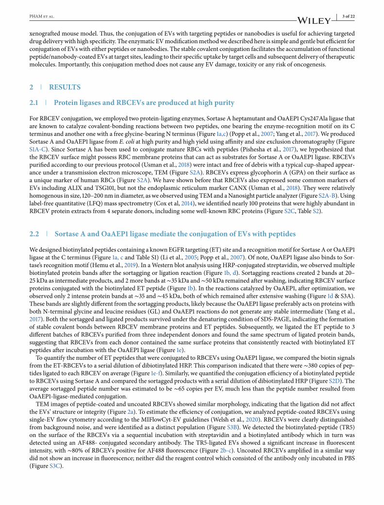
. Conjugation of EVs with a ‘self’ peptide reduces their phagocytosis and increases EVcirculation
We further investigated if RBCEVs displayed CD47 and PS as ‘don’t eat me’ and ‘eat me’ signals, respectively. These signalsdetermine the recognition of EVs for clearance by leukocytes (Rodriguez et al., 2013 ). Staining of RBCEVs on latex beads withan anti-CD47 antibody revealed that ∼60% of the beads were positive for CD47 on average (Figure 5a); whereas, nearly 95%of RBCEV-bound latex beads were stained with Annexin V which binds to PS (Figure 5b). These data suggest that PS wasrelatively more abundant than CD47 on the surface of RBCEVs. Hence, we tested if RBCEVs can be conjugated with a ‘self’peptide derived from CD47, to increase the ‘don’t eat me’ signals on RBCEVs and avoid phagocytosis by leukocytes (Rodriguez

PHAM et al. of
F IGURE Conjugation of RBCEVs with EGFR-targeting peptides increases the uptake of the EVs by EGFR-positive cells. (a) Uptake of Cont. or ET-ligated-RBCEVs (using OaAEP1 ligase) by EGFR+ H358 cells, quantified based on FACS analysis of Calcein AM, the fluorescent label of RBCEVs. (b) Uptakeof Cont/ET-sortagged-RBCEVs (using Sortase A) by H358 cells, quantified using FACS analysis of Calcein AM. (c) Effect of blocking peptides, which competefor binding to EGFR, on the uptake of ligated RBCEVs, indicated by Calcein AM intensity in H358 cells treated with Cont/ET-peptide-ligated RBCEVs. Ineach uptake assay, 200,000 cells were incubated with 5 μg Calcein-AM labelled RBCEVs (2.5 × 10⁹ particles) at 37◦C for 2 h. Control cells were treated withthe flowthrough of the last wash of Calcein-AM-labelled RBCEVs. The graphs present the mean ± SEM (n = 3 donors). Student’s one-tailed t-test *P < 0.05,***P < 0.001
et al., 2013). Conjugation with the self-peptide significantly reduced the uptake of RBCEVs by twomonocytic cell lines, MOLM13and THP1 cells (Figure 5c). We further labelled self-peptide-coated RBCEVs with CFSE and injected them into the tail vein ofimmunodeficient NOD-scid-gamma (NSG) mice. After 5, 10 and 15 min, we captured RBCEVs in the blood using anti-human-GPA-antibody-coatedmagnetic beads (Figure 5d). As GPA is a RBC specificmarker that is expressed in RBCEVs but not in otherEVs, and the antibody is specific to human GPA, we could separate the injected human RBCEVs from mouse EVs in the bloodand quantify them based on FACS analysis of CFSE fluorescence. The analysis revealed that self-peptide-ligated RBCEVs weresignificantly more abundant than control-peptide-ligated RBCEVs in the blood of injected mice (Figure 5d). These data indicatethat the self-peptide can be used to reduce the non-specific phagocytosis of EVs and increase the circulatory flux of RBCEVs.
. Conjugation of nanobodies to EVs using a two-step ligation method
In addition to peptides, we sought to use nanobodies to guide the specific delivery of RBCEVs to target cell types, becausenanobodies are single-domain antibodies known for their high affinity, high specificity and ease of modification (Li et al., 2018).

of PHAM et al.
F IGURE Conjugation of RBCEVs with EGFR-targeting peptides increases the uptake of the EVs by EGFR-positive cells. (a) Representative images of theuptake of CFSE-labelled RBCEVs by H358 cells, obtained using confocal microscopy. Scale bar, 50 μm. (b) Representative Z-stacked images of H358 cells takingup CFSE-labelled RBCEVs, also stained with CellMask (red) and Hoechst (blue). Scale bar, 10 μm. (c)Mean CFSE signal per cell area unit was determined fromthe quantification of CFSE intensity in 1200-1600 cells per condition. (d) Possible effects of chemical inhibitors, EIPA, Filipin andWortmannin on the uptake ofRBCEVs via separate routes of endocytosis. Image was created using biorender.com. (e) Uptake of uncoated Calcein-AM-labelled RBCEVs by H358 cells aftertreatments with EIPA, Filipin and Wortmanin at indicated concentrations, determined using flow cytometry and presented as the percentage of Calcein-AM-positive cells. (f)Uptake of Calcein-AM-labelled RBCEVs that are conjugated with ET peptide byH358 cells after treatments with 100 μMEIPA, 10 μg/ml Filipin,and 0.5 μM Wortmanin, presented as fold change in Calcein AM intensity relative to the untreated control. In each uptake assay, 200,000 cells were incubatedwith 5 μg RBCEVs at 37◦C for 2 h with or without 1-h prior treatment with indicated inhibitors. Peptide conjugation was performed using OaAEP1 ligase. Thegraphs present the mean ± SEM (n = 3 donors). Student’s one-tailed t-test ***P < 0.001
We purified an anti-EGFR camelid biparatopic nanobody (also called variable homodimers, VHH) with an additional sequencecontaining a 6xHis tag, a FLAG tag, and a ligase binding site (Figure S1D) (Roovers et al., 2011). The purified α-EGFR VHHnanobody was approximately 31 kDa (Figure S1D). To reduce the steric hindrance, which may prevent the ligase-mediated con-jugation of RBCEVswith the α-EGFRVHHnanobody, we designed a linker peptide with three glycine residues at theN terminusand a sortase/ligase binding site at the C terminus to bridge the VHH and RBCEVs (Figure 6a). This design allows a sequentialligation of the linker peptide to RBCEVs first, and then to the VHH nanobodies (Figure 6a). The linker peptide was designed
PHAM et al. of
F IGURE Conjugation with self-peptide prevents phagocytosis of RBCEVs and enhances the availability of RBCEVs in the circulation. (a) Flow cytometryanalysis of CD47 on RBCEV-bound beads. (b) Flow cytometry analysis of Annexin V binding to PS on RBCEVs that were immobilized on latex beads. (c) FACSanalysis of Calcein AM inMOLM13 and THP1monocytes that were treated with control or self-peptide (SP) ligated Calcein-labelled RBCEVs. 200,000 cells wereincubated with 5 μg RBCEVs (2.5 × 10⁹ particles) at 37◦C for 2 h. The graphs present the average percentage of Calcein-positive cells ± SEM (n= 3 to 6 donors).(d) FACS analysis of CFSE-labelled RBCEVs that were captured by anti-GPA-antibody-coated streptavidin beads from the plasma of NSG mice, 5–15 min aftera tail vein injection of 0.5 mg CFSE-labelled human RBCEVs (2.5 × 10¹¹ particles). RBCEVs were uncoated or ligated with the control or self-peptide. The graphpresents the mean intensity of CFSE ± SEM (n = 5 mice). Student’s one-tailed t-test *P < 0.05, **P < 0.01, ***P < 0.001
such that it does not circularize or form oligomers because OaAEP1 ligase can bind to the linker peptide’s C-terminal LPETGGmotif and efficiently conjugate it to the N terminal of EV proteins, but not to its own GGG motif. RBCEVs were ligated to theα-EGFR nanobody with or without an addition of the linker peptide and washed extensively using SEC and 4 rounds of cen-trifugation. We observed the free α-EGFR nanobody as a ∼31 kDa band. After the two-step VHH-ligation to RBCEVs using thelinker peptide, several protein bands, particularly two prominent bands at ∼60 and 75 kDa, were detected using an anti-FLAG

of PHAM et al.
F IGURE Nanobodies are conjugated to RBCEVs via a linker peptide, increasing the specific uptake of RBCEVs. (a) Two-step conjugation of RBCEVswith nanobodies: EVs were first ligated with a linker peptide which was then ligated to a VHH nanobody. (b) Western blot analysis of α-EGFR VHH (usingα-FLAG-tag antibody), with or without conjugation to RBCEVs, after SDS-PAGE separation. (c) Uptake of Calcein-labelled α-EGFR-VHH-ligated RBCEVsby EGFR+ lung cancer HCC827 cells. (d) Uptake of Calcein-labelled α-mCherry-VHH-ligated RBCEVs by mCherry-expressing breast cancer CA1a cells. (e)Uptake of Calcein-labelled α-HER2-VHH-ligated RBCEVs by HER2-expressing breast cancer SKBR3 cells. (f) Uptake of CFSE-labelled RBCEVs ligated withα-EGFR or control (α-mCherry) VHH after 2–10 h of incubation with EGFR-positive HCC827 or H358 cells versus EGFR-negative MOLM13 cells. Graphs in(c) – (f) present the mean intensity of Calcein AM or CFSE ± SEM (n = 3 donors), analyzed using FACS. Student’s one-tailed t-test: **P < 0.01, ***P < 0.001
antibody (Figure 6b). No band, other than the unligated VHH band, was observed in the ligation reaction without the linkerpeptide, suggesting that the ligation of VHH nanobodies to RBCEVs required the linker peptide. The spectrum of α-EGFR-VHH-ligated protein bands was consistent in RBCEVs from three independent blood donors (Figure S6A). By comparing thetotal intensity of these bands to a serial dilution of free α-EGFR VHH, we estimated that each EV was ligated to ∼49 copiesof α-EGFR-VHH on average (Figure S6A). FACS analysis of His tag on RBCEV-bound beads suggested that a large fraction ofRBCEVs were conjugated to α-EGFR-VHH via the linker peptide (Figure S6B). This observation was confirmed using FACSanalysis of FLAG tag on single EVs, showing ∼30% of the ligated EVs with VHH (Figure S6C). Nanobody conjugation wasless efficient than peptide conjugation, probably due to the larger size of nanobodies that limits their number on the surface ofeach EV.

PHAM et al. of
. Conjugation of EVs with nanobodies promotes their specific uptake by target cell types
Consistently, we observed a significant increase in RBCEV uptake by HCC827 cells only when RBCEVs were conjugated withα-EGFR VHH via the linker peptide (Figure 6c). Although the nanobody conjugation to RBCEVs was less efficient than thepeptide conjugation, the increase in RBCEV uptake by the α-EGFR VHH conjugation was comparable to that by the ET peptideconjugation, probably due to the higher affinity of α-EGFR VHH towards EGFR (Li et al., 2005; Li et al., 2018). Using the samemethod, we found that conjugation of RBCEVs with α-mCherry VHH and with α-HER2 VHH promoted RBCEV uptake bymCherry-expressing CA1a cells and HER2-expressing SKBR3 cells, respectively (Figure 6d-e). α-mCherry and α-HER2 VHHwere expressed in E. coli, purified using FPLC (Figure S1E-F) and validated for their specific binding to CA1a-mCherry andSKBR3 cells, respectively (Figure S7A-D). Omission of the linker peptide in the VHH ligation reaction abrogated the increase intargeted RBCEVuptake (Figure 6c-e). Hence, the two-step ligation is a robustmethod to ligate RBCEVswith various nanobodies.The α-EGFR VHH exhibited a very high affinity to EGFR-positive cells, including H358 and HCC827 cells, but not to EGFR-
negative MOLM13 cells (Figure S7E). Hence, RBCEVs conjugated with α-EGFR VHH bound specifically to H358 and HCC827cells but not to MOLM13 cells (Figure S8A-C). This binding was detected using FACS analysis of GPA, a specific marker ofRBCEVs, on the surface of the cells after 1 h of incubation with α-EGFR conjugated RBCEVs at 4◦C, suggesting that the bindingdid not require energy and RBCEVs were not internalized at this temperature (Figure S8A-C). During an incubation at 37◦C,α-EGFR-VHH-ligated RBCEVs were taken up specifically by H358 and HCC827 cells, but not MOLM13 cells (Figure 6f). Thistrend of specific targeting of EGFR-positive cells by α-EGFR-VHH-RBCEVs held true over time (Figure 6f).
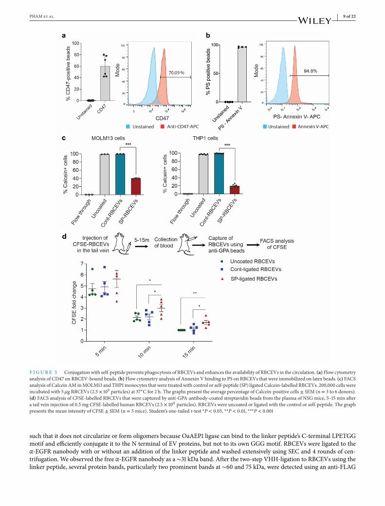
In addition, the increased uptake of α-EGFR-VHH-ligated RBCEVs in H358 cells was also confirmed using immunofluores-cence analysis and confocal microscopy (Figure 7a-b). The punctate pattern of CFSE signals suggests that α-EGFR-VHH-ligatedRBCEVs were taken up mainly by endocytosis, consistent with our observation with the ET-peptide-coated RBCEVs.
. Surface-modified RBCEVs promote specific delivery of RNA and chemotherapeutic payloads
We have shown before that RBCEVs could be used to deliver ASOs, gRNAs or mRNAs to cancer cells (Usman et al., 2018). Here,we sought to test if the ligation reaction would affect RNA loading into EVs.We first conjugated RBCEVs with α-EGFR or a con-trol VHH (α-mCherry-VHH), washedRBCEVs twice using centrifugation, and subsequently loaded themwith luciferasemRNAusingREG1 (Figure 7c).H358 cells expressing EGFR, but notmCherry, were treatedwith these RBCEVs and the luciferase activitywas compared after 24 h. The delivery of luciferasemRNA by α-EGFR-VHH-RBCEVs resulted in 100% higher luciferase activity,compared to that by uncoated RBCEVs or α-mCherry-RBCEVs albeit all the RBCEV-treated cells showed higher luciferase sig-nals than untreated cells (Figure 7c). Thus, our data demonstrated that the conjugation of RBCEVswith α-EGFRVHH enhancedthe efficiency of mRNA delivery to EGFR-positive cancer cells.We also optimized a protocol for loading PTX, a generic chemotherapy drug commonly used for lung cancer treatments,
into RBCEVs using sonication (Figure 7d). Drug-loaded RBCEVs were washed thoroughly and ligated with the ET peptide. Weobserved that PTX was loaded into RBCEVs at a capacity of 5%–6% (in weight) and an efficiency of ∼25%, as determined byHPLC (Figure S8D). We treated H358 cells with a serial dilution of PTX-loaded RBCEVs. Interestingly, we found a significantdifference only at lower doses of PTX, suggesting that ET-RBCEVs can enhance the efficacy of low dose PTX on EGFR-positivecancer cells (Figure 7d).
. EGFR-targeting RBCEVs accumulate in xenografted EGFR-positive lung cancer cells
To generate an in vivo model of lung cancer, we injected luciferase-mCherry-labelled H358 cells into the tail vein of NSG mice(Figure 8a). After 3 weeks, when tumour cells were stably engrafted and detected in the lungs, we treated the mice with DiR-labelled RBCEVs and observed the biodistribution of RBCEVs by imaging. Since DiR is a fluorescent lipophilic dye, we foundthat it formed somemicelles in PBS but themicelles and free DiR dye were eluted in separate SEC fractions (with lowDiR signalsin the DiR-only sample), distinct from the fractions containing DiR-labelled RBCEVs (fraction 7 to 10 with high DiR signals inthe DiR-EV sample) as shown in Figure S9A.We collected DiR-labelled RBCEVs in fraction 7 to 9 for the in vivo biodistributionexperiment because these fractions contained the highest concentration of RBCEVs (Figure S4C). The flowthrough of the lastEV wash (the last round of centrifugation after the SEC) was used as a negative control to ensure that we could detect DiRfluorescent signals from RBCEVs above the background generated by any leftover unbound dyes. Bioluminescent cells weredetected consistently in the lungs of NSG mice 3 weeks after the injection of H358-luciferase cells, but no signal was detected inother organs, except occasionally in the tail due to the tail vein injection (Figure S9B). RBCEVs were conjugated with a controlpeptide or ET peptide using OaAEP1 ligase, then labelled with DiR fluorescent dye, washed extensively, and injected equallyin the tail vein of tumour-bearing mice. Eight hours after RBCEV injections, we observed uncoated RBCEVs in the spleen,
of PHAM et al.
F IGURE Conjugation of RBCEVs with targeting nanobodies and peptides facilitates cell-specific delivery of therapeutic payloads. (a) Representativeimages of H358 cells that have taken up CFSE-labelled RBCEVs, also stained with CellMask (red) and Hoechst (blue). (b)Mean CFSE signal per cell area unitwas determined from the quantification of CFSE intensity in 1200-1600 cells per condition. (c) Delivery of luciferase-expressing (luc) mRNA using control (α-mCherry-VHH) or α-EGFR-VHH-ligated RBCEVs, quantified based on luciferase activity in H358 cells after a 24-h incubation with mRNA-loaded RBCEVs(uncoated or ligated with VHH). Luciferase mRNA was loaded in RBCEVs using REG1 loading reagent. Graph presents luciferase signal ± SEM (n= 6 repeats).(d) Delivery of anti-cancer drug paclitaxel (PTX) to lung cancer cells using ET-peptide coated RBCEVs. PTX was loaded into RBCEVs using sonication. Theloading capacity, percentage of PTX in RBCEVs (by weight) was determined using HPLC. Viability of H358 cells treated with different concentrations of PTXdelivered by ET-peptide-ligated RBCEVs was calculated based on CCK8 assay readings ± SEM (n = 3 EV donors). Student’s one-tailed t-test: **P < 0.01,***P < 0.001
liver, lung and bone (Figure 8a). Peptide-coated RBCEVs showed similar pattern of uptake in the same organs. Remarkably,the accumulation of ET-RBCEVs was significantly increased in the lung and reduced in the liver, compared to control-peptide-ligated RBCEVs (Figure 8a-b). No significant change was observed in other organs (Figure 8a-b). Conjugation of RBCEVs withET peptide using Sortase A also led to an increased accumulation of EVs in the EGFR-positive lung tumour (Figure S9C).
In order to analyze the specific uptake of RBCEVs by tumour cells in vivo, we labelled the peptide/VHH-conjugated RBCEVswith CFSE and injected them into mice bearing mCherry-H358 lung tumours (Figure 8c). After 8 h, we digested the lung tissueand analyzed the mCherry-H358 tumour cells, using FACS. Interestingly, the conjugation of RBCEVs with ET peptide increasedthe percentage of CFSE-positive tumour cells from 3%–6% in the controls to 20%–30% in the targeted ET-RBCEV treatment(Figure 8c). Conjugation of RBCEVs with α-EGFR VHH also increased the percentage of CFSE-positive tumour cells to ∼20%in the targeted α-EGFR-RBCEV treatment (Figure 8c). These data suggest that both the ET peptide and the α-EGFR VHHnanobody can driveRBCEVs specifically into EGFR-positive lung tumour cells in vivo. Since the ET-peptide-conjugatedRBCEVsperformed better than α-EGFR-VHH-conjugated RBCEVs in enhancing the specific uptake of the EVs, we further used ET-peptide-conjugated RBCEVs for drug delivery in the treatment of lung cancer shown below.

PHAM et al. of
F IGURE EGFR-targeting RBCEVs accumulate in xenografted EGFR-positive lung cancer cells. (a) Biodistribution of DiR-labelled RBCEVs in NSGmice bearing EGFR+ H358 lung cancer. Shown are representative DiR fluorescent images of organs from lung-cancer bearing mice preconditioned and injectedwith uncoated RBCEVs, control/ET-RBCEVs or with the flowthrough of the RBCEV wash. (b)Average DiR intensity in each organ relative to the average meanintensity of all organs, subtracted by signals detected in flowthrough controls. Abbreviations: Panc, pancreas; GI, gastro-intestinal tract. (c) In vivo uptake ofCFSE-labelled RBCEVs by mCherry-positive H358 cancer cells, gated based on mCherry expression, in the lung of the mice that were treated with cont/ET-peptide or cont/α-EGFR-VHH ligated RBCEVs, analyzed using FACS. Student’s one-tailed t-test: *P < 0.05, **P < 0.01 (n = 3 to 5 mice)

of PHAM et al.
. Delivery of paclitaxel (PTX) by EGFR-targeting EVs enhances treatment efficacy inxenografted mice
Subsequently, PTX-loaded RBCEVs or an equivalent dose of free PTX (1 mg/kg) were injected into NSGmice bearing H358 lungcancer every 3 days (Figure 9a). Indeed, 1 mg/kg of PTX is considered a very low dose as the clinically-equivalent dose of PTXis about 20 mg/kg in mice which causes many side effects (Wang et al., 2017). Bioluminescent imaging of the tumour revealedthat ET-RBCEVs significantly enhanced the tumour-inhibitory effect of PTX, compared to the effect of free PTX or PTX loadedin uncoated and control-peptide coated RBCEVs (Figure 9a, c). H & E staining and TUNEL staining of lung sections furtherconfirmed that the treatment with ET-RBCEVs containing PTX reduced the size of lung tumours while increasing apoptosis inthese tumours compared to the treatment with free PTX or uncoated PTX-loaded RBCEVs (Figure 9b, d). These data suggestthat EGFR-targeted RBCEVs can enhance the specific delivery of anti-cancer drugs to targeted tumour cells in vivo.
DISCUSSION
In summary, we describe here a simple and robust protein ligase-mediated method for conjugation of EVs with peptides andnanobodies to create a new generation of targeted therapies. Enzyme-mediated surface functionalization of EVs hasmany advan-tages over existing EV modification methods, including: (1) no requirements for viral transduction and cell proliferation, thusreducing the cost and time for EV production; (2) no risk of transformation by viral insertion and DNA transfer; (3) stability ofcovalent bonds with peptides and nanobodies; (4) high-purity peptides, enzymes and nanobodies can be produced in large scaleat low cost; (5) ligase-catalyzed reactions are reproducible and controllable with predictable rates and products; (6) ligases do notalter the physiochemical properties of EVs and the small amounts used can be removed easily by washing, hence the conjugationhas no unwanted side effects.We have shown that surface functionalization of EVs with target-specific peptides and nanobodies can enhance the delivery
of therapeutic molecules to cancer cells expressing the corresponding ligands, thereby increasing drug efficacy and decreasingside effects. We further demonstrated the versatility of this approach, both in terms of the variability of target ligands and thenature of the therapeutic molecules. Moreover, we showed that this targeting platformmaintains functionality in in vivo settings.This indicates the potential for a versatile platform capable of targeting multiple ligands using EVs loaded with different types oftherapeutic molecules for the treatment of a range of diseases.We also demonstrated that modifying the surface of EVs with self-peptide, which binds to SIRP alpha, can decrease phagocy-
tosis, thereby increasing the bioavailability of EVs in circulation. This approachmay also be applied to enhance the effects of EVsthat have been proven to possess endogenous therapeutic properties, such as EVs frommesenchymal stem cells for cardiovascu-lar diseases or EVs from dendritic cells for vaccination (Dang et al., 2020). Based on this study, we can surmise that conjugatingantigens or biomarker-binding (poly) peptides to EVs may facilitate future efforts in therapeutics and diagnostics.Despite the versatility of this approach, there are certain limitations, presented primarily in the form of the targeting moieties
used. Nanobody and peptide libraries are not very extensive and nanobodies with high affinity and specificity are not availableagainst many cellular targets. Generation of novel nanobodies involves immunization and screening of phage display librarieswhich is a time-consuming and tedious process. A more promising alternative is to develop a method for the stable conjuga-tion of monoclonal antibodies to the EV surface, as monoclonal antibodies are readily available against most known antigens.There is also the risk of potential immunogenicity that could be introduced through the presence of camelid-specific amino-acidsequences in the nanobody (Vincke et al., 2009). If nanobody conjugated EVs are to be used for therapeutic applications, thenanobodies would need to be humanized by replacing these residues.
MATERIALS ANDMETHODS
. Purification and characterization of RBCEVs
Blood samples were provided by Red Cross from healthy donors with informed consents in Hong Kong. Briefly, 200–250 mlof O-group blood was collected in single-system blood bags (Macopharma, France). Blood sample processing was performedaccording to the guidelines laid out by the City University of Hong Kong, following approval of all related procedures. RBCs wereseparated from plasma using centrifugation (1000 × g for 8 min at 4˚C) and washed three times with PBS (1000 × g for 8 min at4˚C).White blood cells were removed by using centrifugation and leukodepletion filters (Nigale, China). Isolated RBCs were col-lected in Nigale buffer (0.2 g/l citric acid, 1.5 g/l sodium citrate, 7.93 g/l glucose, 0.94 g/l sodium dihydrogen phosphate, 0.14 g/ladenine, 4.97 g/l sodium chloride, 14.57 g/l mannitol) and diluted 3 times in PBS containing 0.1 mg/ml calcium chloride andincubated overnight with 10 μM calcium ionophore (Sigma, USA) at 37◦C with 5% CO2. RBCEVs were purified using a similar

PHAM et al. of
F IGURE Delivery of PTX using EGFR-targeting RBCEVs increases the treatment efficacy in an EGFR-positive lung cancer mouse model. (a) Represen-tative bioluminescent images of NSG mice with EGFR+ luciferase-expressing H358 cancer cells in the lung during a course of systemic (i.v.) treatments with1mg/kg PTX only or the same dose of PTX loaded in cont/ET-RBCEVs. Treatments were repeated every 3 days and images were taken 1 day after every treatment.Colours indicate bioluminescent signals (photon/s) in two scales (the images are divided into two groups, day 1–28 and day 31–43 from the treatment start date,to avoid saturated signals). (b)Representative images of H& E staining and TUNEL assay (green fluorescence) of lung sections from the lung cancer mice treatedwith PTX, with or without RBCEV-mediated delivery. Nuclei were stained with DAPI (blue). Scale bar, 100 μm. (c) Average bioluminescent signals quantifiedin the lung area during the development of H358 lung tumours (photons/s), normalized by the signals at the start of the treatments, and presented as mean ±SEM. (d) Average fold change in TUNEL staining signals relative to the untreated control ± SEM. Two-way ANOVA test (c) and Student’s one-tailed t-test (d):*P < 0.05, ***P < 0.001 (n = 3 to 4 mice)

of PHAM et al.
procedure as described in our previous study (Usman et al., 2018), but with ultracentrifugation at 50,000 × g. Purified RBCEVswere stored in PBS containing 4% trehalose at -80˚C. The size distribution and concentration of RBCEVs were quantified utiliz-ing a NanoSight NS300 Tracking Analysis NS300 nanoparticle tracking system (Malvern, UK) or a ZetaView Particle TrackingAnalysis instrument (Particle Metrix, Germany). The haemoglobin contents of RBCEVs were measured using a haemoglobinquantification assay (Abcam, UK), comparing to a serial dilution of purified haemoglobin protein. Because haemoglobin is themajor constituent of RBCEVs, RBCEV quantity is indicated by haemoglobin quantity throughout this study.For transmission electron microscopy analysis, RBCEVs were fixed with 2% paraformaldehyde and loaded on a glow-
discharged copper grid (200 mesh, coated with formvar carbon film). RBCEVs on the grid were incubated with biotinylatedanti-human CD235ab antibody (catalogue number 306618, Biolegend, USA) followed by streptavidin protein (10 nm Gold con-jugation, Abcam, ab81369). Samples were washed with PBS. A total of 4% uranyl acetate was added for negative staining ofRBCEVs. Images were acquired using a Tecnai 12 BioTWIN transmission electron microscope (FEI/ Philips, USA) operating at100 kV.
. Purification of leukaemia EVs from THP cells
THP1 cells were obtained from the American Type Culture Collection (ATCC, USA) and maintained in RPMI (Thermo FisherScientific, USA), containing 10% foetal bovine serum (Biosera, USA) and 1% penicillin/streptomycin (Thermo Fisher Scientific).To make EV-free FBS, EVs were removed from FBS using ultracentrifugation at 110,000 × g for 18 h at 4˚C. THP1 cells werecultured at 106 cells/ml in the above medium with EV-free FBS and 0.2 μM calcium ionophore for 48 h. Culture supernatantswere collected from 5 flasks of treated THP1 cells. Cells and debris were separated by differential centrifugation at 300 × g for10 min, 400 × g for 15 min, 900 × g for 15 min at 4˚C. The supernatant was further passed through a 0.45 μm filter, placed ontop of 2 ml frozen 60% sucrose, and enriched by ultracentrifugation with a SW32 rotor at 100,000 × g for 90 min at 4˚C. THP-1derived EVs were collected from the interface and diluted 1:1 in cold PBS, and layered above 2ml frozen 60% sucrose cushion in aSW41 rotor and centrifuged at 100,000 × g for 12 h at 4˚C (Beckman Coulter, USA) with reduced braking speed. 500 μl EVs werecollected from the interface and added to a qEV SEC column (Izon, New Zealand). A total of 500 μl eluate was collected in eachfraction. The concentration of EVs and protein were measured in 30 fractions using a Nanosight analyzer and bicinchoninic acid(BCA) assay (Pierce BCA Protein Assay Kit, Thermo Fisher Scientific). For ligation, the EVs from fraction 7 to 11 were combinedand concentrated using centrifugation at 15,000 × g for 20 min in an Amicon-15 filter with 100 kDa cut-off.
. Peptide and nanobody design
Peptides listed in Table S1 were designed with non-targeting sequences as negative controls or with an EGFR-targeting sequenceor ‘self’ sequence obtained from previous reports (Li et al., 2005; Normanno et al., 2006). A sortase (LPETG) or ligase (NGL)binding motif was added to the C termini and a biotin was added to the N termini of the peptides. Peptides were synthesizedusing 96/102 well automated peptide synthesizers and purified using HPLC (GL Biochem Ltd., Shanghai, China). The EGFR-VHH sequence was obtained from Roovers et al. (Li et al., 2018) and cloned with a 6xHis tag, a FLAG tag, and a ligase-bindingsite in this order: 6xHis-GSG-VHH-GSG-FLAG-NGL. Similarly, the sequences of an α-mCherry and a α-HER2 nanobody wereobtained from Fridy et al. (Fridy et al., 2014) and Farasat et al. (Farasat et al., 2017) with an additional sequence as described abovefor α-EGFR VHH. The VHH-coding DNA was synthesized and inserted into pET32(a+) plasmid, following a T7 promoter, byGuangzhou IGE Biotechnology Ltd (China).
. Expression and purification of proteins
Competent BL21 (DE3) E. coli bacteria were transformed with pET30b-7 M-SrtA plasmid (Addgene 51140, USA), OaAEP1-Cys247Ala plasmid (provided by Dr. Bin Wu, Nanyang Technology University) or with pET32(a+)-VHH plasmid (cloned withspecific VHH sequences). Protein expression was induced using Isopropyl β-D-1-thiogalactopyranoside (IPTG 0.5 mM) in LB at25◦C for 16 hwith shaking. The culturewas collected and centrifuged at 6000× g for 15min at 4◦C.The bacteriawere resuspendedin 50 ml binding buffer (500 mM NaCl, 25 mM Tris-HCl, 1 mM phenylmethylsulfonyl fluoride sulfonyl fluoride (PMSF), 5%glycerol). Bacteria were lysed using a high-pressure homogenizer (JnBio, China) at 1000 psi for 4–6 rounds. The cell lysate wascentrifuged at 8000 rpm for 60 min at 4◦C. The supernatant was collected and filtered through a 0.45 μmmembrane (Millipore,USA). The proteins were purified using a NGC-QUEST-10 fast protein liquid chromatography (FPLC) system (BioRad, USA).Briefly, the sample was loaded into a 5-ml-Ni-charged cartridge (BioRad) equilibrated with the binding buffer. The column waswashed with 3% elution buffer (500 mMNaCl, 25 mM Tris-HCl, 1 mM imidazole, 1 mM PMSF and 5% glycerol) and then elutedusing a linear imidazole gradient from 40 to 500 mM. Fractions of 2 ml were collected. The proteins were concentrated using a

PHAM et al. of
3-kDa-cutoff centrifugal filter (Millipore) and 4000 ×g centrifugation in a swinging-bucket rotor and filtered through a 0.22 μmmembrane. The proteins were further purified using a HiLoad 16/600 Superdex 200 pg size exclusion chromatography column(GE Healthcare, USA) with the FPLC system, in low ionic strength buffer (150 mMNaCl, 50 mM Tris-HCl), at 0.5 ml/min. Thetarget protein was collected at the appropriate UV280 peak and validated using gel electrophoresis with Coomassie blue staining.ForOaAEP1 ligase activation, a buffer comprised of 1mMEDTA and 0.5mMTris (2-carboxyethyl) phosphine hydrochloride wasadded to the immature protein and the pH of the solution was adjusted to 3.7 with glacial acetic acid. The mixture was incubatedfor 10 days at 4◦C. Activated proteins were concentrated by ultracentrifugation using a 3-kDa-cutoff concentrator and stored at-80◦C.
. Conjugation of EVs with peptides using Sortase A or OaAEP ligase
For sortagging, 20 μM Sortase A was mixed with 500 μM peptide in 1x Sortase buffer (50 mM Tris-HCl, 12 mM CaCl2, pH6.5, 150 mM NaCl), and kept for 30 min. Subsequently, 100 μg RBCEVs (amount based on haemoglobin contents, equivalentto ∼5 × 10¹⁰ EVs) were added into the Sortase-A-peptide mixture in a total volume of 40 μl. We incubated the reaction for1 h at room temperature with gentle agitation (20 rpm) on an end-over-end shaker. For ligation, 100 μg RBCEVs (∼5 × 10¹⁰particles) were incubated with 500 μM peptide or VHH and 0.26 mg/ml ligase in PBS buffer, pH 7, in a total volume of 40 μl, atroom temperature for 3 h with gentle agitation (30 rpm) on an end-over-end shaker. For VHH ligation, linker-peptide-ligatedRBCEVs were washed twice with PBS and incubated with 500 μM VHH and 0.26 mg/ml ligase at the same condition as forthe peptide ligation. After sortagging or ligation reactions, RBCEVs were washed with PBS using qEV SEC columns. Fraction7 to 9 (with pink-red colour) were collected and washed again with PBS 3 times by centrifugation at 21,000 × g for 15 minat 4˚C.After washing, some peptide/nanobody-coated RBCEVs were incubated with 10 μM Calcein AM (Biolegend) for 20 min at
room temperature or with 20 μM CFSE (Life Technologies, USA) for 1 h at 37◦C or with 2 μM DiR (Thermo Fisher Scientific)for 15 min at room temperature. All the dyes, including Calcein AM, CFSE and DiR, were dissolved in DMSO (Sigma) as 500xor 1000x stock solutions. The labelled RBCEVs were washed once using centrifugation and loaded into a SEC column (Izon) andeluted with PBS to wash the unbound dye away. RBCEVs, collected from SEC fraction 7 to 9, were washed twice with PBS bycentrifugation at 21,000 × g for 15 min at 4˚C.
. Loading RNAs and drugs into RBCEVs
1 μg luciferase mRNA (TriLink, USA) was loaded into 50 μg VHH-ligated RBCEVs using REG1 (Carmine Therapeutics, Singa-pore) according to themanufacturer’s instructions.mRNA-loaded RBCEVswere thenwashed 3 timeswith PBS by centrifugationat 21,000 × g for 30 min. For drug loading, 1 mg uncoated RBCEVs were incubated with 200 μg PTX (Sigma) in 1 ml PBS at 37˚C for 15 min. The mixture was sonicated using a Bioruptor (Biogenode) for 12 min at 4˚C then recovered at 37˚C for 1 h. Theloaded RBCEVs were washed with PBS at 21,000× g for 15 min, quantified using the haemoglobin assay and coated with peptidesas described above. The coated RBCEVs were repurified using SEC as described. To measure the amount of PTX loaded intoRBCEVs, an aliquot of loaded RBCEVs was centrifuged at 21,000 × g for 15 min. The pellet was dried at 75˚C and resuspendedin acetonitrile and centrifuged at 21,000 × g for 10 min. The supernatant was filtered and analyzed using HPLC.Tomeasure the effect of PTX-loaded RBCEVs on cell proliferation, a serial dilution of RBCEVs containing 0.22 to 28.22 μg/ml
PTX were added to each well of H358 cells in a 96-well plate (5000 cells/well) at 37◦C. After 60 h of incubation, the mediumwas replaced with 100 μl fresh medium containing 10 μl CCK8 solution (Biosharp, China) and incubated for 2 h at 37◦C. CCK8absorbance was measured at 450 nm using a Synergy H1 microplate reader (BioTek, USA).
. Western blot analysis
Conjugated EVs were incubated with RIPA buffer (Thermo Fisher Scientific) supplemented with protease inhibitors (Biotool,USA) for 5 min on ice. A total of 50–200 μg protein lysates were loaded in 10% polyacrylamide gels and proteins were transferredto a polyvinylidene difluoridemembrane (Immobilon-P,Millipore). 3-color protein ladder PM5100 ExcelBand (SmoBio, Taiwan)was included at two sides of the samples in the gel. Membranes were blocked using 5% milk in Tris buffered saline containing0.1% Tween-20 (TBST) for 1 h followed by an incubation with primary antibodies overnight at 4◦C: mouse anti-FLAG (Sigma,Cat# F3165, dilution 1:500). The blot was washed 3 times with TBST then incubated with HRP-conjugated anti-mouse secondaryantibody (Jackson ImmunoResearch, USA, dilution 1:10,000) for 1 h at room temperature. For biotinylated peptide detection, theblot was incubated directly with HRP-conjugated streptavidin (Thermo Fisher Scientific, dilution 1:4000). The blot was imagedusing a BioRad Chemidoc gel documentation system.
of PHAM et al.
. Biotin pulldown and mass spectrometry analysis
Equal quantities of unmodified RBCEVs or biotinylated TR5 peptide ligated RBCEVs were lysed in RIPA buffer on ice for 10min.The lysate from each sample was incubated with streptavidin-magnetic beads from the Pierce MS-Compatible Magnetic IP Kit(Thermo Fisher Scientific) for 1 h to allow binding of biotinylated proteins to the beads. The resulting beads were washed 3times using a DynaMag-5 Magnet (Thermo Fisher Scientific) in wash buffer before being subjected to denaturation in Laemmlibuffer (BioRad). The beads were subsequently boiled at 95◦C for 5 min. For total EV mass spectrometry, 100 μg RBCEVs werelysed in RIPA buffer on ice for 10 min. The protein lysate was denatured in Laemmli buffer and boiled at 95◦C for 5 min. Biotinpulldown samples or RBCEVprotein extracts were separated on a 12%NuPAGEBis-Tris gel (Life Technologies) for 10min (LFQ)at 170 V in 1×MOPS buffer (Thermo Fisher Scientific). The gel was fixed using the Colloidal Blue Staining Kit (Thermo FisherScientific). For in-gel digestion, samples were washed in the destaining buffer (25 mM ammonium bicarbonate; 50% ethanol),reduced in 10 mMDTT for 1h at 56◦C followed by alkylation with 55 mM iodoacetamide (Sigma) for 45 min in the dark. Trypticdigest was performed in 50 mM ammonium bicarbonate buffer with 2 μg trypsin (Promega) at 37◦C overnight. Peptides weredesalted on StageTips and analyzed by nanoflow liquid chromatography on an EASY-nLC 1200 system coupled to a Q-Exactive-HFmass spectrometer (Thermo Fisher Scientific). Peptides were separated on aC18-reversed phase PicoFrit column (25 cm long,75 μm inner diameter; New Objective) packed in-house with ReproSil-Pur C18-AQ 1.9 μm resin (Dr Maisch). The column wasmounted on an Easy Flex Nano Source and temperature controlled by a column oven (Sonation) at 40◦C. A 105-min gradient(biotin pulldown) or a 215-min gradient (RBCEV proteomes) from 2% to 40% acetonitrile in 0.5% formic acid at a flow of 225nl/min. Spray voltage was set to 2.2 kV. The Q-Exactive-HF was operated with a TOP20 MS/MS spectra acquisition methodper mass spectrometry full scan. Mass spectrometry scans were conducted with 60,000 at a maximum injection time of 20 msand MS/MS scans with 15,000 resolution at a maximum injection time of 50 ms. The raw files were processed with MaxQuantversion 1.5.2.8 with standard settings for LFQ samples and the match between runs option was activated for the biotin pulldownsamples (Cox & Mann, 2008). The quantification was based on unique peptides only. Carbamidomethylation was set as fixedmodification while methionine oxidation and protein N-acetylation were considered as variable modifications. Search resultswere filtered with a false discovery rate of 0.01. Known contaminants, proteins groups only identified by site, and reverse hits ofthe MaxQuant results were removed and only proteins that were quantified by LFQ intensity were kept.
. Treatment of cancer cells with peptide or nanobody-coated EVs
Human breast cancer SKBR3 cells, human lung cancer H358 and HCC827 cells were obtained from the American Type CultureCollection (ATCC, USA). Human breast cancer MCF10CA1a (CA1a) were obtained from Karmanos Cancer Institute (WayneState University, USA). Acute myeloid leukaemia MOLM13 cells were obtained from DSMZ Collection of Microorganisms andCell Cultures (Braunschweig, Germany).mCherry expressingCA1a cells were generated by our group as described previously (Vuet al., 2019). All the solid cancer and leukaemia cells weremaintained inDMEMor RPMI (Thermo Fisher Scientific), respectively,with 10% foetal bovine serum and 1% penicillin/streptomycin (Thermo Fisher Scientific, USA). For the EV binding assay, 100,000cells were incubated with 10 μg RBCEVs in 500 μl growth medium on an end-over-end shaker for 1 h at 4◦C. The cells werewashed twice with PBS and incubated with 1 μl APC anti-GPA antibody (Biolegend Cat# 306608) for 1 h at 4◦C, washed 3 timeswith PBS and analyzed by FACS. To test the EV uptake, 200,000 cells were incubated with 5 μg Calcein AM or CFSE-labelledRBCEVs in 500 μl growth medium per well in 24-well plates for 2–10 h at 37 ◦C. In the peptide competing assay, H358 cells werepreincubated with 120 μM control or ET peptide for 1 h at 37◦C then with 5 μg Calcein AM-labelled RBCEVs in 500 μl growthmedium per well in 24-well plates for 2 h at 37 ◦C. To identify the route of EV uptake, we added EIPA, Filipin, or Wortmannin(at indicated concentrations) to H358 cells 1 h before adding Calcein-AM-labelled RBCEVs and incubated for 2 h as describedabove.
. Flow cytometry (FACS) analysis
Cells were washed 2x with PBS, resuspended in 100 μl FACS buffer (PBS with 0.5% foetal bovine serum). For surface proteinanalysis, the cells were incubated with 3 μl fluorescent-conjugated antibody such as AF488-α-EGFR antibody (Biolegend Cat#352908), APC-α-His antibody (Biolegend Cat# 362605), APC anti-GPA antibody (Biolegend Cat# 306608), APC-α-HER2 anti-body (Biolegend Cat# 324407) for 15 min on ice, in the dark, and washed twice with 1 ml FACS buffer. To quantify the peptideconjugation efficiency, 100 μg biotinylated-peptide-coated RBCEVs or uncoated RBCEVs (as a negative control) were incubatedovernight with 2.5 μg latex beads (Thermo Fisher Scientific) at 4◦C on a shaker, washed three times with PBS and resuspendedin 100 μl FACS buffer containing 1 μl streptavidin labelled with Alexa Fluor 647 (AF647), incubated on ice for 15 min and washedtwice with FACS buffer. FACS analysis of latex beads or cells in FACS buffer was performed using a CytoFLEX-S cytometer(Beckman Coulter) and analyzed using Flowjo V10 (Flowjo, USA). The beads or cells were first gated by FSC-A versus SSC-A,

PHAM et al. of
excluding debris and dead cells (Figure S4A). Single cells were then gated by FSC-width versus FSC-height, excluding doubletsand aggregates. The fluorescent-positive population of beads or cells was subsequently gated by targeted fluorescent channels,such as FITC for AF488, CFSE or Calcein AM, APC for AF647 and ECD for mCherry.
. Single-EV flow cytometry
Single-EV flow cytometry was carried out using a CytoFLEX LX flow cytometer (BeckmanCoulter). RBCEVswere distinguishedfrom background noise via side scatter detected using the 405 nm laser (violet side scatter or VSSC) which allowed for greatersensitivity (Figure S3B). 100 nm and 200 nm control PCS mixed kit latex beads (Beckman Coulter) were used as a reference forsize. Subsequently, RBCEVs were diluted in 0.2 μm-filtered PBS and analyzed. Data was acquired using the following settings:FSC 138, SSC 180, FITC 3000, VSSC 800with the threshold of the trigger signal (VSSC) setmanually to 4000. The sample line wascleaned with sterile distilled water for 2min at maximumflow rate (240 μl/min) before starting the experiment and between run-ning each sample. Samples were analyzed at a slow flow rate (10 μl/min asmeasured by themachine), and analyzed only when theflow rate returned to the baseline level recorded at the start of the experiment to ensure that any remnants of the previous samplewere not present and to maintain a constant level of background noise throughout the experiment. Staining of EVs was carriedout at 4◦C for 3 h, followed by 1 washing step using sterile filtered PBS to remove unbound antibody. Biotinylated peptide ligatedEVs were detected by sequentially incubating ligated EVs with an excess of streptavidin (Abcam) followed by extensive wash-ing and incubation with a biotinylated mouse isotype control monoclonal antibody (BioLegend). These EV-TR5-Streptavidin-B-antibody complexes were stained with an anti-mouse-AF488 conjugated secondary antibody (Jackson ImmunoResearch) ata concentration of 0.5 μg/ml. For staining of α-EGFR VHH coated RBCEVs, 5 × 10⁹ EVs were incubated with 3 μl of anti-body and the final volume made up to 300 μl with PBS. Uncoated EVs were also stained for each condition as controls. For thereagent/antibody control, antibodies were incubated in PBS under the same conditions as EVs and briefly spun down once beforebeing analyzed. The buffer-only control (filtered PBS), reagent control and all RBCEV samples were recorded at the same flowcytometer acquisition settings stated above, maintaining constant gain, triggering threshold, and flow rate. Following staining,EVs were resuspended to achieve a concentration of 2.5 × 10⁵ EVs/μl before being subjected to 2-fold serial dilution 6 times.These serially diluted EV samples were analyzed by flow cytometry to establish the dilution range between which there was alinear relationship between dilution factor and EV count per minute (Figure S3D). In addition, the fluorescence of stained EVswas also measured at each dilution to ensure that the fluorescence signal was maintained constant, and that swarming did nottake place. We found that EV suspensions within the range of 3.9 × 10³ to 2.5 × 10⁵ EVs/μl allowed analysis of single EVs accu-rately. As such, all EV samples were diluted to a concentration of 3 × 10⁴ EVs/μl before being analyzed at a flow rate of 10 μl/min,which yielded an average event rate of ∼5000 events/second. The abort rate was monitored throughout the experiment and keptwithin 0%–2%.
. Immunostaining and imaging of RBCEV uptake
H358 cells were seeded on 13-mm coverslips (Citoglass, China) coated with Poly-D-Lysine at a density of 20,000 cells per well(Sigma). After 2 days, the cells were incubated with 20 μg CFSE-labelled RBCEVs coated with peptides or nanobodies in 500 μlmedium for 4 h at 37◦C, 5% CO2. After that, the cells were washed with PBS twice and incubated with 400 μl CellMask DeepRed (Thermo Fisher Scientific) per well for 10 min at 37◦C. The cells were washed with PBS once and incubated with 400 μl4% paraformaldehyde for 20 min at room temperature. The cells were rinsed twice with PBS before being stained with Hoechst33342 (Cell Signalling Technology). Coverslips were washed three times with PBS and once with water and mounted on slideswith mounting media (Abcam). Specimens were imaged using an inverted Zeiss LSM710 confocal microscope with 20X (0.5NA)air objectives and 100X (1.4NA) oil objectives. Images were acquired and analyzed using Zeiss Zen software (2011). Four to fiverandom areas were taken using 20× 0.5NA objective and subjected to semi-quantification using Image J 1.8.0v software (NationalInstitute of Health). Cell areas were selected as regions of interest (ROIs) based on the dilated mask of Hoechst signals. CFSEsignals were measured as mean pixel intensity of ROIs. Total measurement area covered 1200 to 1600 cells in each condition.
. Generation of in vivo cancer models and treatment with RBCEVs
All experiments inmice were conducted according to our protocols approved by the Institutional Animal Care andUse Commit-tee under National University of Singapore and the Animal Ethics Committee at City University of Hong Kong. Mice of similarages were tagged and grouped randomly for control and test treatments. Experiments were performed in a blinded manner.Exclusion was applied to the mice that became pregnant during the experiment or accidentally died due to anaesthesia. Micewith unsuccessful injections were also excluded to avoid false-negative results.

of PHAM et al.
H358 cells were transduced with a lentiviral vector (pLV-Fluc-mCherry-Puro) and selected with puromycin to create a stablemCherry-luciferase expressing cell line. A total of 1 millionH358-luc cells were injected into the tail vein of NSGmice (6-7 weeksold). The same inoculation was repeated after 2 days. After 3 weeks, bioluminescence in the lung was detected using IVIS LuminaII (Perkin Elmer, USA) after an i.p. injection of D-luciferin.Mice with comparable bioluminescent signals were injectedwith 1mgRBC membrane lysate (ghosts) or an equivalent amount of live RBCs via a retro-orbital route 1 h before RBCEV treatment.To analyze RBCEV biodistribution, preconditionedmice with 3 week-old lung tumours were injectedwith 100 μgDiR-labelled
RBCEVs, which were ligated with a control or EGFR-targeting peptide, in the tail vein. After 8 h, mice were sacrificed and DiRfluorescence was measured immediately in the organs using the IVIS. Similarly, to analyze the specific uptake of RBCEVs bytumour cells, mice with 5-week-old lung tumours were injected with 800 μg CFSE-labelled RBCEVs that were ligated with acontrol or EGFR-targeting peptide/VHH in the tail vein. After 8 h, the mice were perfused with PBS and the lung was excised.Lung cells were dissociated using a gentleMACS dissociator (Miltenyi, Germany) and incubated with collagenase IV (Life Tech-nologies) in a shaker for 40 min at 37◦C. The cells were passed through a 70 μm strainer and centrifuged at 1500 rpm for 5 min.RBCs were lysed using ACK lysis buffer (Thermo Fisher Scientific). The rest of the cells were washed with PBS and resuspendedin FACS buffer for FACS analysis of mCherry and CFSE.For drug treatment, preconditioned mice were treated i.v. with 1 mg/kg PTX alone or an equivalent dose of PTX in RBCEVs
with or without ET-peptide ligation every 3 days. The same amount of unloaded RBCEVs was used as a negative control. Biolu-minescent signals were measured every 3 days using IVIS as described above. At the end point (43 days from the beginning ofthe treatment, when the untreated mice showed many signs of pain and distress), the lung was fixed in 10% buffered formalin(Thermo Fisher Scientific) at room temperature overnight.
. Quantification of RBCEVs in the circulation
500 μg CFSE-labelled peptide-ligated RBCEVs were injected into the tail vein of NSG mice. After 5–15 min, 100 μl blood wascollected from the eye. Blood cells were removed and 20 μl plasma was incubated with 5 μl biotinylated anti-GPA antibody(Biolegend Cat# 306618) for 2 h at room temperature with gentle rotation. The mixture was then incubated with 20 μl Dyn-abeads MyOne streptavidin beads (Thermo Fisher Scientific) for 1 h at room temperature. The beads were washed 3 times andresuspended in 500 μl FACS buffer for analysis of CFSE.
. Histopathology analysis and TUNEL assay
After the overnight fixation, lung samples were sequentially dehydrated in 70%, 95% and 100% alcohol at 37◦C. Samples werecleared in three baths of xylene (Thermo Fisher Scientific) and impregnated in 3 baths of paraffin wax (Thermo Fisher Scientific)each for 1.5 h at 37◦C and 62◦C, respectively. Paraffin blocks were sectioned at 5 μm using a HM330 MICROM microtome.Sections were dried at 37◦C and dewaxed in 2 baths of xylene, then immersed in 2 baths of absolute alcohol and 1 bath of70% alcohol, for 10 min each. Samples were rehydrated in water, stained with Gill 3 haematoxylin (Cancer Diagnostics, USA) for15min.Afterwashingwithwater, the sectionswere treatedwith 0.3% acid alcohol, washed and bluedwith 2% sodiumbicarbonate.Sections were subsequently stained with 0.5% Eosin (Merck, USA) for 2min. After washing with water, samples were dehydratedin 95% and absolute alcohol, cleared in xylene then mounted using a synthetic mountant (Shandon). Apoptosis was evaluatedusing a TUNEL BrightGreen Apoptosis Detection Kit (Vazyme, China) according to the manufacturer’s protocol. After staining,images of TUNEL staining were acquired with a confocal fluorescence microscope.
. Statistical analysis
Student’s t-tests (one tail) were computed using GraphPad Prism 8 to determine significant differences between treated samplesand control. Two-way ANOVA, also computed using GraphPad Prism 8, was applied to analyze the differences among multiplegroups of treatments. We considered a P-value < 0.05 to be significant. In all the graphs, data are presented as median or meanand standard error of the mean (SEM). For statistical analysis, we repeated each experiment three times using RBCEVs fromthree donors or using cells from three passages. Animal experiments were performed in groups of 3 to 5 mice. The minimumsample size was determined by G*Power analysis, set for one-tail t-test that compared the mean difference of two independentgroups with the α- err prob = 0.05, effect size d = 5, and power = 0.95.
ACKNOWLEDGEMENTSWe would like to thank Hong Kong Red Cross for providing all blood samples. Special thanks also go to Prof. Harvey Lodish(Whitehead Institute) and Dr. Ng Shyh Chang (Chinese Academy of Sciences) for their advice on the manuscript. We also thank

PHAM et al. of
Dr. Wu Bin (Nanyang Technological University), Dr. Wai Leong Tam, Dr. Deron Herr, Dr. Polly Chen and Prof. Fred Wong,Dr. Matthias Gerhard Wacker, Dr. Long Nguyen, Prof. Kini Manjunatha (National University of Singapore), Dr. Lap Nguyen(Hanoi University of Pharmacy), Dr. Liang Zhang, Dr. Linfeng Huang, Kwok Chun Kit and Dr. Jianbo Yue (City University ofHong Kong), Dr. Ronne Yeo (Carmine Therapeutics), Prof. Anskar Leung (Hong Kong University) and Prof. Judy Lieberman(Harvard Medical School) for providing valuable reagents and advice. We also thank Yang Jun, Luyen Tien Vu, Melissa Tan,Harshvardhan Modh (National University of Singapore), Ya Gao, Xin Zhang, Yeokyeong Kim, Abdullah Faqeer (City Universityof Hong Kong) for technical assistance. Schematic images were generated using BioRender.com.
CONFL ICT OF INTERESTMinh TN Le and Jiahai Shi are scientific co-founders, and advisors of Carmine Therapeutics. Other authors declare no conflictof interest.
FUNDINGThis project is funded by the National University of Singapore (grant NUHSRO/2019/076/STARTUP/02), the Hong KongInnovation and Technology Commission (grant ITS/201/18), the Hong Kong Health and Medical Research Fund (05160296and 06172196), the National Natural Science Foundation of China (81770099, 81773246, 81972865), the Shenzhen Innovationand Technology Fund (grant JCYJ20180507181636165, JCYJ20170413115637100 and JCYJ20170412152916724), the Hong KongResearch Grants Council (21101218), the Hong Kong Children’s Thalassemia Foundation (2018/01), the National Research Foun-dation Singapore and the Singapore Ministry of Education under its Research Centre of Excellence initiative.
DATA AVAILAB IL ITY STATEMENTThe mass spectrometry data have been deposited to the ProteomeXchange Consortium via PRIDE partner repository with thedataset identifier PDX021926. All raw data are available upon request.
GEOLOCATION INFORMATIONThis project was performed in Singapore, Hong Kong, China and Vietnam.
ORCIDMinhT.N. Le https://orcid.org/0000-0002-6077-1566
REFERENCESAlvarez-Erviti, L., Seow, Y., Yin, H., Betts, C., Lakhal, S., &Wood,M. J. A. (2011). Delivery of siRNA to themouse brain by systemic injection of targeted exosomes.
Nature Biotechnology , 341–345.Andaloussi, S. E. L., Mäger, I., Breakefield, X. O., &Wood,M. J. A. (2013). Extracellular vesicles: Biology and emerging therapeutic opportunities.Nature Reviews
Drug Discovery , 347–357.Antes, T. J., Middleton, R. C., Luther, K. M., Ijichi, T., Peck, K. A., Liu, W. J., Valle, J., Echavez, A. K., & Marbán, E. (2018). Targeting extracellular vesicles to
injured tissue using membrane cloaking and surface display. Journal of Nanobiotechnology, , 61.Armstrong, J. P. K., Holme, M. N., & Stevens, M. M. (2017). Re-engineering extracellular vesicles as smart nanoscale therapeutics. ACS Nano. , 69–83.Balaj, L., Lessard, R., Dai, L., Cho, Y. - J., Pomeroy, S. L., Breakefield, X. O., & Skog, J. (2011). Tumour microvesicles contain retrotransposon elements and
amplified oncogene sequences. Nature Communications , 180.Chiu, G. N. C., Bally, M. B., & Mayer, L. D. (2002). Effects of phosphatidylserine on membrane incorporation and surface protection properties of exchangeable
poly(ethylene glycol)-conjugated lipids. Biochimica et Biophysica Acta , 37–50.Cooper, J. M., Wiklander, P. O., Nordin, J. Z., Al-Shawi, R., Wood, M. J., Vithlani, M., Schapira, A. H. V., Simons, J. P., El-Andaloussi, S., & Alvarez-Erviti, L.
(2014). Systemic exosomal siRNA delivery reduced alpha-synuclein aggregates in brains of transgenic mice.Movement Disorders , 1476–1485.Cox, J., & Mann, M. (2008). MaxQuant enables high peptide identification rates, individualized p.p.b.-range mass accuracies and proteome-wide protein quan-
tification. Nature Biotechnology (12), 1367–1372–.Cox Jürgen, Hein Marco Y., Luber Christian A., Paron Igor, Nagaraj Nagarjuna, Mann Matthias (2014). Accurate Proteome-wide Label-free Quantification by
Delayed Normalization andMaximal Peptide Ratio Extraction, TermedMaxLFQ.Molecular & Cellular Proteomics, , (9), 2513–2526. https://doi.org/10.1074/mcp.m113.031591.
Dang, X. T. T., Kavishka, J. M., Zhang, D. X., Pirisinu, M.&, Le, M. T. N. (2020). Extracellular vesicles as an efficient and versatile system for drug delivery. Cells, 2191.
Farasat, A., Rahbarizadeh, F., Ahmadvand, D., & Yazdian, F. (2017). Optimization of an anti-HER2 nanobody expression using the Taguchi method. PreparativeBiochemistry & Biotechnology , 795–803.
Fridy, P. C., Li, Y., Keegan, S., Thompson, M. K., Nudelman, I., Scheid, J. F., Oeffinger, M., Nussenzweig, M. C., Fenyö, D., Chait, B. T., & Rout, M. P. (2014). Arobust pipeline for rapid production of versatile nanobody repertoires. Nature Methods , 1253–1260.
Gao, X., Ran, N., Dong, X., Zuo, B., Yang, R., Zhou, Q., Moulton, H. M., Seow, Y., & Yin, H. (2018). Anchor peptide captures, targets, and loads exosomes ofdiverse origins for diagnostics and therapy. Science Translational Medicine, , eaat0195.
Hemu, X., Sahili, A. E., Hu, S., Wong, K., Chen, Y., Wong, Y. H., Zhang, X., Serra, A., Goh, B. C., Darwis, D. A., Chen, M. W., Sze, S. K., Liu, C. - F., Lescar, J.,& Tam, J. P. (2019). Structural determinants for peptide-bond formation by asparaginyl ligases. Proceedings of the National Academy of Sciences of the UnitedStates of America , 11737–11746.

of PHAM et al.
Kooijmans, S. A. A., Aleza, C. G., Roffler, S. R., van Solinge, W. W., Vader, P., & Schiffelers, R. M. (2016). Display of GPI-anchored anti-EGFR nanobodies onextracellular vesicles promotes tumour cell targeting. Journal of extracellular vesicles , 31053.
Kooijmans, S. A. A., Gitz-Francois, J. J. J. M., Schiffelers, R. M., & Vader, P. (2018). Recombinant phosphatidylserine-binding nanobodies for targeting of extra-cellular vesicles to tumor cells: A plug-and-play approach. Nanoscale. , 2413–2426.
Li, C., Tang, Z., Hu, Z., Wang, Y., Yang, X., Mo, F., & Lu, X. (2018). Natural single-domain antibody-nanobody: A novel concept in the antibody field. Journal ofBiomedical Nanotechnology , 1–19.
Li, Z., Zhao, R., Wu, X., Sun, Y., Yao, M., Li, J., Xu, Y., & Gu, J. (2005). Identification and characterization of a novel peptide ligand of epidermal growth factorreceptor for targeted delivery of therapeutics. FASEB Journal : Official Publication of the Federation of American Societies for Experimental Biology , 1978–1985.
Nakase, I., Noguchi, K., Fujii, I., & Futaki, S. (2016). Vectorization of biomacromolecules into cells using extracellular vesicles with enhanced internalizationinduced by macropinocytosis. Scientific Reports , 1–10.
Nakase, I., Ueno, N., Katayama,M., Noguchi, K., Takatani-Nakase, T., Kobayashi, N. B., Yoshida, T., Fujii, I., & Futaki, S. (2016). Receptor clustering and activationby multivalent interaction through recognition peptides presented on exosomes. Chemical Communications , 317–320.
Normanno, N., De Luca, A., Bianco, C., Strizzi, L., Mancino, M., Maiello, M. R., Carotenuto, A., De Feo, G., Caponigro, F., & Salomon, D. S. (2006). Epidermalgrowth factor receptor (EGFR) signaling in cancer. Gene. , 2–16.
Ohno, S., Takanashi,M., Sudo, K., Ueda, S., Ishikawa, A.,Matsuyama,N., Fujita, K.,Mizutani, T., Ohgi, T., Ochiya, T., Gotoh,N., &Kuroda,M. (2013). Systemicallyinjected exosomes targeted to EGFR deliver antitumor microRNA to breast cancer cells.Molecular Therapy , 185–191.
Pishesha, N., Bilate, A. M., Wibowo, M. C., Huang, N. - J., Li, Z., Deshycka, R., Bousbaine, D., Li, H., Patterson, H. C., Dougan, S. K., Maruyama, T., Lodish, H. F.,& Ploegh, H. L. (2017). Engineered erythrocytes covalently linked to antigenic peptides can protect against autoimmune disease. Proceedings of the NationalAcademy of Sciences of the United States of America , 3157–3162.
Pitt, J.M., Kroemer, G., & Zitvogel, L. (2016). Extracellular vesicles:Masters of intercellular communication and potential clinical interventions. Journal of ClinicalInvestigation , 1139–1143.
Popp, M. W., Antos, J. M., Grotenbreg, G. M., Spooner, E., & Ploegh, H. L. (2007). Sortagging: A versatile method for protein labeling. Nature Chemical Biology, 707–708.
Rodriguez, P. L., Harada, T., Christian, D. A., Pantano, D. A., Tsai, R. K., & Discher, D. E. (2013). Minimal “Self” peptides that inhibit phagocytic clearance andenhance delivery of nanoparticles. Science. , 971–975.
Roovers, R. C., Vosjan, M. J. W. D., Laeremans, T., el Khoulati, R., de Bruin, R. C. G., Ferguson, K. M., Verkleij, A. J., van Dongen, G. A. M. S., & van Bergen enHenegouwen, P.M. P. (2011). A biparatopic anti-EGFR nanobody efficiently inhibits solid tumour growth. International Journal of Cancer. Journal Internationaldu Cancer , 2013–2024.
Smyth, T., Petrova, K., Payton, N. M., Persaud, I., Redzic, J. S., Graner, M.W., Smith-Jones, P., & Anchordoquy, T. J. (2014). Surface functionalization of exosomesusing click chemistry. Bioconjugate Chemistry , 1777–1784.
Usman,W.M., Pham, T. C., Kwok, Y. Y., Vu, L. T., Ma, V., Peng, B., Chan, Y. S.,Wei, L., Chin, S. M., Azad, A., He, A. B. - L., Leung, A. Y. H., Yang,M., Shyh-Chang,N., Cho, W. C., Shi, J., & Le, M. T. N. (2018). Efficient RNA drug delivery using red blood cell extracellular vesicles. Nature Communications , 2359.
Vincke, C., Loris, R., Saerens, D., Martinez-Rodriguez, S., Muyldermans, S., & Conrath, K. (2009). General strategy to humanize a camelid single-domainantibody and identification of a universal humanized nanobody scaffold., Journal of Biological Chemistry , 3273–3284.
Vu, L. T., Peng, B., Zhang, D. X., Ma, V., Mathey-Andrews, C. A., Lam, C. K., Kiomourtzis, T., Jin, J., McReynolds, L., Huang, L., Grimson, A., Cho, W. C.,Lieberman, J., & Le, M. T. (2019). Tumor-secreted extracellular vesicles promote the activation of cancer-associated fibroblasts via the transfer of microRNA-125b. Journal of Extracellular Vesicles , 1599680.
Wang, F., Porter,M., Konstantopoulos, A., Zhang, P., &Cui,H. (2017). Preclinical development of drug delivery systems for paclitaxel-based cancer chemotherapy.Journal of Controlled Release: Official Journal of the Controlled Release Society , 100–118.
Welsh, J. A., Pol, E., Bettin, B. A., Carter, D. R. F., Hendrix, An, Lenassi,M., Langlois,M. -. A., Llorente, A., Nes, A. S., Nieuwland, R., Tang, V.,Wang, L.,Witwer, K.W., & Jones, J. C. (2020). Towards defining reference materials for measuring extracellular vesicle refractive index, epitope abundance, size and concentration.Journal of Extracellular Vesicles , 1816641.
Yamamoto, T., Teramura, Y., Itagaki, T., Arima, Y., Iwata, H. (2016). Interaction of poly(ethylene glycol)-conjugated phospholipids with supported lipid mem-branes and their influence on protein adsorption. Science and Technology of Advanced Materials , 677–684.
Yang, R., Wong, Y. H., Nguyen, G. K. T., Tam, J. P., Lescar, J., & Wu, B. (2017). Engineering a catalytically efficient recombinant protein ligase. Journal of theAmerican Chemical Society , 5351–5358.
Yerneni, S. S., Lathwal, S., Shrestha, P., Shirwan, H., Matyjaszewski, K., Weiss, L., Yolcu, E. S., Campbell, P. G., & Das, S. R. (2019). Rapid on-demand extracellularvesicle augmentation with versatile oligonucleotide tethers. ACS Nano. , 10555–10565.
Zou, J., Shi,M., Liu, X., Jin, C., Xing, X., Qiu, L., & Tan,W. (2019). Aptamer-functionalized exosomes: Elucidating the cellular uptakemechanism and the potentialfor cancer-targeted chemotherapy. Analytical Chemistry , 2425–2430.
SUPPORTING INFORMATIONAdditional supporting information may be found online in the Supporting Information section at the end of the article.
How to cite this article: Pham TC, Jayasinghe MK, Pham TT, et al. Covalent conjugation of extracellular vesicles withpeptides and nanobodies for targeted therapeutic delivery. J Extracell Vesicles. 2021;10:e12057.https://doi.org/10.1002/jev2.12057