Phosphatidylserine Vesicles Enable Efficient En Bloc Transmission of Enteroviruses
13
Article Phosphatidylserine Vesicles Enable Efficient En Bloc Transmission of Enteroviruses Graphical Abstract Highlights d Clusters of viruses are packaged and released non-lytically in PS lipid vesicles d PS lipids are co-factors in mediating subsequent infectivity and transmission d PS vesicles provide greater infection efficiency for viruses d PS vesicles enable viral genome clusters to be transmitted en bloc cell-to-cell Authors Ying-Han Chen, WenLi Du, ..., Gre ´ goire Altan-Bonnet, Nihal Altan-Bonnet Correspondence [email protected] In Brief Clusters of enteroviruses are packaged in phosphatidylserine (PS)-enriched vesicles, thereby enhancing the infection efficiency of the viruses and enabling collective transmission of multiple viral genomes from cell-to-cell. Chen et al., 2015, Cell 160, 619–630 February 12, 2015 ª2015 Elsevier Inc. http://dx.doi.org/10.1016/j.cell.2015.01.032
-
Upload
independent -
Category
Documents
-
view
1 -
download
0
Transcript of Phosphatidylserine Vesicles Enable Efficient En Bloc Transmission of Enteroviruses

Article
Phosphatidylserine Vesicles Enable Efficient EnBloc
Transmission of EnterovirusesGraphical Abstract
Highlights
d Clusters of viruses are packaged and released non-lytically
in PS lipid vesicles
d PS lipids are co-factors in mediating subsequent infectivity
and transmission
d PS vesicles provide greater infection efficiency for viruses
d PS vesicles enable viral genome clusters to be transmitted
en bloc cell-to-cell
Chen et al., 2015, Cell 160, 619–630February 12, 2015 ª2015 Elsevier Inc.http://dx.doi.org/10.1016/j.cell.2015.01.032
Authors
Ying-Han Chen, WenLi Du, ...,
Gregoire Altan-Bonnet,
Nihal Altan-Bonnet
In Brief
Clusters of enteroviruses are packaged in
phosphatidylserine (PS)-enriched
vesicles, thereby enhancing the infection
efficiency of the viruses and enabling
collective transmission of multiple viral
genomes from cell-to-cell.

Article
Phosphatidylserine Vesicles EnableEfficient En Bloc Transmissionof EnterovirusesYing-Han Chen,1,2 WenLi Du,1 Marne C. Hagemeijer,1 Peter M. Takvorian,2 Cyrilla Pau,2 Ann Cali,2 Christine A. Brantner,3
Erin S. Stempinski,3 Patricia S. Connelly,3 Hsin-Chieh Ma,4 Ping Jiang,4 Eckard Wimmer,4 Gregoire Altan-Bonnet,5
and Nihal Altan-Bonnet1,*1Laboratory of Host-Pathogen Dynamics, National Heart Lung and Blood Institute, NIH, Bethesda, MD 20892, USA2Federated Department of Biological Sciences, Rutgers University, Newark, NJ 07102, USA3Electron Microscopy Core Facility, National Heart Lung and Blood Institute, NIH, Bethesda, MD 20892, USA4Department of Molecular Genetics and Microbiology, Stony Brook University, Stony Brook, NY 11794, USA5Program in Computational Biology and Immunology, Memorial Sloan Kettering Cancer Center, New York, NY 10065, USA*Correspondence: [email protected]
http://dx.doi.org/10.1016/j.cell.2015.01.032
SUMMARY
A central paradigm within virology is that each viralparticle largely behaves as an independent infectiousunit. Here, we demonstrate that clusters of entero-viral particles are packaged within phosphatidylser-ine (PS) lipid-enriched vesicles that are non-lyticallyreleased from cells and provide greater infection effi-ciency than free single viral particles. We show thatvesicular PS lipids are co-factors to the relevantenterovirus receptors inmediating subsequent infec-tivity and transmission, in particular to primary hu-man macrophages. We demonstrate that clusteredpackaging of viral particles within vesicles enablesmultiple viral RNA genomes to be collectively trans-ferred into single cells. This study reveals a novelmode of viral transmission, where enteroviral ge-nomes are transmitted from cell-to-cell en blocin membrane-bound PS vesicles instead of as singleindependent genomes. This has implications forfacilitating genetic cooperativity among viral quasis-pecies as well as enhancing viral replication.
INTRODUCTION
Enteroviruses are a large genus of single positive-stranded RNA
viruses whose members including poliovirus (PV), Coxsackie-
virus, rhinovirus, and enterovirus 68 are the causative agents
of a number of important and widespread human diseases
including poliomyelitis, myocarditis, hand foot and mouth dis-
ease, the common cold, and more recently, a severe respiratory
disease with paralytic symptoms. In addition to >70 enteroviral
serotypes identified in humans, enteroviral quasispecies are
common largely as a result of inherent error making and lack of
proofreading mechanisms of viral RNA-dependent RNA poly-
merases (RdRp).
Enteroviral RNA genomes serve as templates for both transla-
tion and replication, and these processes take place on host
intracellular membranes (den Boon and Ahlquist, 2010; Hsu
et al., 2010). After enteroviruses have bound their specific host
receptors either at the cell surface or within endocytic vesicles
(Brandenburg et al., 2007), the capsid undergoes a conforma-
tional change that allows the viral RNA to be transferred
across the plasma membrane and/or endosomal membrane
into the cytoplasm through a yet completely defined mechanism
(Strauss et al., 2013). In the cytoplasm, the enteroviral RNA is first
translated into non-structural proteins and structural proteins,
where the formermakes up the RNA genome replicationmachin-
ery and the latter the capsid. The viral RNA replication machinery
are then assembled on the cytoplasmic membrane leaflet of ER-
derived membranes that are subsequently modified by viral and
host proteins to have a specific lipid blueprint of enrichment in
phosphatidylinositol-4-phosphate (PI4P) and cholesterol lipids.
These lipids regulate the membrane association, assembly,
and activity of the viral replication protein complex, including
the RdRp, and thus facilitate viral RNA synthesis (Hsu et al.,
2010; Ilnytska et al., 2013; Nchoutmboube et al., 2013).
Once the enteroviral RNA is synthesized, little is known about
where in the host cell it is packaged in capsids and how these
capsids are released from cells. While enteroviruses have his-
torically been considered non-enveloped (i.e., lacking a host-
derived membrane bilayer around their capsids) and thus rely
on cell lysis to exit, recent reports of extracellular Coxsackievirus
B3 (CVB3) being present in vesicles (Robinson et al., 2014) and
PV being able to spread non-lytically among host cells (Bird
et al., 2014) have raised important questions regarding the extra-
cellular nature of enteroviral particles and the significance of
non-lytic exit in the viral life cycle. Moreover hepatitis A, hepatitis
E and blue tongue viral particles, all long considered non-envel-
oped, have been observed surrounded by membranes (Feng
et al., 2013; Takahashi et al., 2008; Owens et al., 2004).
A central paradigm in virology is that viruses behave as inde-
pendent infectious units (Flint et al., 2009). While there are ex-
ceptions to this, such as vaccinia virus particles preventing
superinfection by inducing the host cell to repel other virions
(Doceul et al., 2010), it is largely accepted that the fate of individ-
ual viral genomes are not dependent on one another during exit
from one cell and entry into another (Brandenburg and Zhuang
Cell 160, 619–630, February 12, 2015 ª2015 Elsevier Inc. 619
2007). Here, we investigate the assembly, exit, and subsequent
infection processes of enteroviral particles using a combination
of imaging techniques including confocal microscopy, super-
resolution light microscopy, correlative light, and electron micro-
scopy along with single molecule RNA fluorescence in situ
hybridization (FISH), proteomic, and biochemical approaches.
We show that multiple infectious enteroviral particles are clus-
tered within individual phosphatidylserine (PS) lipid-enriched
vesicles and non-lytically secreted out of cells. These viral parti-
cles in vesicles are more efficient in establishing infection than
free viral particles. We demonstrate that vesicles encapsulate
and traffic large numbers of mature infectious viral particles be-
tween cells and consequently enable the transfer of multiple viral
RNA genomes collectively into new host cells by a mechanism
that is dependent on both the virus-specific receptor of the recip-
ient host cell as well as the vesicular PS lipids.
RESULTS
Assembled Poliovirus Capsids Are Localized to ViralRNA Replication OrganellesWe first investigated the intracellular spatio-temporal dynamics
of newly synthesized PV particles. The generation of PV parti-
cles, as well as enterovirus assembly in general, comprises a
multistep process where capsid subunits (VP0, VP1, VP3) form
pentamers, which polymerize into capsids (Liu et al., 2010).
Once RNA is packaged, the VP0 subunits get cleaved into VP2
and VP4 to generate mature infectious virions (Liu et al., 2010).
From screening a large number of neutralizing antibodies, we
identified the A12 antibody, that binds deep inside the canyon
bridging both rims of two adjacent pentamers and thus recog-
nizing assembled capsids (Chen et al., 2011, 2013), to visualize
PV capsids within infected cells. We fixed cells at various inter-
vals after PV infection and co-immunolabeled with A12, anti-viral
VP1 to detect individual VP1 subunits, and anti-viral 3AB anti-
bodies, the latter to detect viral 3AB to localize replication organ-
elles where viral RNA is synthesized (Hsu et al., 2010). Newly
assembled capsids were clearly detectable from 3–4 hr post-
infection (p.i.) and onward, and theywere localized to the replica-
tion organelles (Figure 1A). Note that in contrast to A12 labeling,
VP1 was localized to both the replication organelles and
dispersed across the cytoplasm, consistent with its cytosolic na-
ture. At 6–7 hr p.i., capsids were dispersed from the replication
sites to the cytoplasm and sequestered in puncta (Figure 1A,
6–7 hr p.i. inset). At this time, there is a known cessation in
viral RNA synthesis (Ehrenfeld et al., 1970), and the timing of
capsid release from replication organelles was coupled to viral
RNA synthesis since prematurely inhibiting viral RNA synthesis
with the inhibitor Guanidine HCl (Barton and Flanegan, 1997)
triggered capsids to rapidly disperse into the cytoplasm
(Figure S1A).
Poliovirus Capsids Are Captured by PhosphatidylserineLipid-Enriched Autophagosome-like Organelles andReleased Non-Lytically from CellsBetween 6 and 7 hr p.i., we found that >85% of capsids (n = 10
cells) were on punctate cytoplasmic structures that colocalized
with the autophagosomal membrane protein, LC3-II (Figure 1B).
620 Cell 160, 619–630, February 12, 2015 ª2015 Elsevier Inc.
By transmission electron microscopy, we observed numerous
double-membraned autophagosome-like organelles containing
capsids (Figure 1C). Previous reports (Taylor et al., 2009; Kirke-
gaard and Jackson, 2005; Jackson et al., 2005) had found that
perturbation of the host autophagy pathway led to a decrease
in PV release from infected cells. Consistent with that, either dis-
rupting autophagy by small interfering RNA (siRNA) depletion of
autophagy machinery LC3 or beclin 1, or acutely stimulating
autophagy by treating cells with tat-beclin 1 peptides, blocked
or enhanced PV release by �10-fold, respectively (Figures S1B
and S1C) while replication was unaffected (Figure S1D). How-
ever, these capsid containing autophagosome-like organelles
did not follow the conventional autophagy pathway and fuse
their contents with lysosomes as inhibiting lysosomal enzymes
did not further increase LC3-II levels beyond the 4-fold increase
observed in PV-infected cells (Figures S1E and S1F) and none
of the A12/LC3-II co-labeled structures contained lysosomal
enzymes at any point during infection (Figures S1F and S1G).
Notably, the SNARE protein syntaxin 17, normally localized to
autophagosomes and required for fusion with lysosomes (Ita-
kura et al., 2012), did not localize to these A12/LC3-II co-labeled
structures (Figure S1I).
However, we found that themembranes of both the replication
organelles and the autophagosome-like organelles contained
negatively charged phosphatidylserine (PS) lipids. PS lipids in
uninfected cells are primarily located at the cytoplasmic leaflets
of the plasma and endosomal membranes as well as at the
lumenal leaflet of the ER membrane (Leventis and Grinstein,
2010; Kay et al., 2012). Cells were co-transfected with GFP-
LactC2 and FAPP1mRFP, cytosolic live-cell reporters for PS
(Kay et al., 2012), and PI4P lipids, respectively, the latter to report
on the location of the ER-derived replication organelle mem-
branes (Hsu et al., 2010). At 4 hr p.i., cells were imaged live by
structured illumination microscopy. Highly localized PS-rich
membrane domains exposed to the cytoplasm were found
distributed across the replication organelles (Figure 1D, inset).
Later, between 6 and 7 hr p.i., cells were fixed and immunola-
beled with anti-GFP and A12 antibodies and imaged by confocal
microscopy. Numerous A12 positive structures were found also
co-labeled with GFP-LactC2 indicating the presence of PS lipids
on their membrane leaflets exposed to the cytoplasm (Fig-
ure 1E, arrows). Consistent with this, in PV-infected live cells
co-expressing LC3-mRFP and GFP-LactC2, >90% (organelles
measured across ten cells) of the LC3-mRFP-labeled autopha-
gosome-like organelles were co-labeled with GFP-LactC2
(Figure 1F, inset).
We then investigated the fate of these capsid-containing or-
ganelles during the rest of the infection time period. Between
7 hr and 8 hr p.i., there was a 70% ± 10% (n = 15) decrease in
the number of capsids within the cytoplasm (Figure 1G). Using
the cell impermeable Trypan blue dye, we found that the plasma
membrane remained intact during this time while there was an
�6-fold increase in extracellular viral titers (Figure 1H). This
lack of plasma membrane permeability during PV infection had
also been previously observed (Taylor et al., 2009; Bird et al.,
2014). Although by 12 hr p.i. the cells eventually lysed (not
shown), this data indicated that the majority of PV particles
were released prior to cell lysis.

A B
C
D
G
E F
H
Figure 1. Poliovirus Capsids Are Captured by Phosphatidylserine Lipid-Enriched Autophagosome-like Organelles and Released Non-Lyti-
cally from Cells
(A) Capsids undergo dynamic spatial transitions during infection. HeLa cells infected with PV and immunolabeled with A12, anti-VP1, and anti-3AB antibodies.
Scale bars represent 5 mm.
(B) Capsids (A12) colocalized with autophagosome marker LC3-II. PV-infected HeLa cells were immunolabeled with A12 and anti-LC3-II antibodies. Scale bar
represents 5 mm.
(C) Electronmicrographs of PV-infected cells showPVcapsids in double-membrane autophagosome-like organelles. Scale bars represent 5 mmand200 nm (inset).
(D) PV-infected cells at 4 hr p.i. expressing GFP-LactC2 and FAPP1mRFP imaged by structured illumination microscopy. Region of interest is magnified in right
panel. Scale bar represents 5 mm.
(E) PV-infected cells at 7 hr p.i. expressing GFP-LactC2 were immunolabeled with anti-GFP and A12 antibodies. Region of interest is magnified in right panel.
Arrows indicate A12 positive autophagosome-like organelles co-labeled with GFP-LactC2. Scale bar represents 5 mm.
(F) Cells co-expressing GFP-LactC2 and LC3-RFP were infected with PV and imaged by confocal microscopy at 7 hr pi. Region of interest is magnified in right
panel. Scale bar represents 5 mm.
(G) Capsid distribution between 7 and 8 hr p.i. PV-infected cells were immunolabeled with A12, anti-VP1, and anti-3AB antibodies. Scale bar represents 5 mm.
(H) Plasma membrane integrity remains intact when PV exits cells. Trypan Blue diffusion across the plasma membrane was measured concurrently with
measurements of extracellular PV titer, the latter plotted in plaque-forming units/ml (pfu/ml).
See also Figure S1.
Extracellular PV Particles Are Found in Uniformly LargeVesiclesScanning electron microscopy (SEM) of cells at 7 hr p.i. cells re-
vealed numerous vesicular structures of similar size docked at
the extracellular side of the intact plasma membrane (Figure 2A
and inset). While these SEM images may not reflect the true
shape and size of these vesicles within live cells, they do point
to a striking uniformity in size.Measurements of thecross-section
Cell 160, 619–630, February 12, 2015 ª2015 Elsevier Inc. 621

A B
C
Figure 2. Extracellular PV Particles Are
Found in Large Uniform-Sized Vesicles
(A) Scanning electron microscopy of a PV-infected
cell at 7 hr p.i. Scale bar represents 3 mm. Inset
shows higher magnification of uniform size vesicles
docked on the extracellular side of the plasma
membrane. Scale bar represents 1 mm.
(B) Extracellular vesicle size distribution in PV-
infected cells. Cross section diameter of a 100
randomly selected extracellular vesicles from four
different cells, were measured from scanning
electron micrographs and plotted. Data repre-
sented as mean ± SEM.
(C) Correlative fluorescence and scanning elec-
tron microscopy (SEM). PV-infected cell was im-
munolabeled with A12 at 7 hr pi, epifluorescence
image was obtained (right) and then sample
was processed for SEM (left). Arrows point to
A12-labeled extracellular vesicles. Scale bar rep-
resents 1 mm.
diameter from 100 randomly selected extracellular vesicles from
four different cells, showed that �90% of the vesicles were be-
tween 250 nm and 350 nm in diameter (Figure 2B). Correlative
fluorescence imaging in conjunction with SEM confirmed that
these vesicles contained A12-labeled capsids (Figure 2C).
Mature Enteroviral Particles Are Released inPS-Enriched VesiclesWe next investigated whether the extracellular vesicles contain-
ing PV retained the PS lipids that had been components of the
autophagosome-like organelles (Figures 1D and 1F). Incubation
of PV-infected cells with Alexa 568 coupled Annexin V, a non-cell
permeable fluorescent reporter protein for PS lipids (Koopman
et al., 1994), revealed numerous fluorescent puncta dotting the
surface of the cell at 7hr p.i. (Figure 3A). Similar results were
also observedwith CVB3 (at 7 hr p.i.) and human rhinovirus infec-
tions (at 12 hr p.i.) (Figure 3A). Note that this pattern of Annexin V
labeling of enterovirus-infected cells was different from both
apoptotic- and mock-infected cells: in the former, the entire
plasma membrane was labeled with Annexin V as a result of
PS lipids being on the extracellular membrane leaflet of the
cell, a hallmark of apoptosis (Figure 3B, apoptosis) while in the
latter there was no labeling since PS lipids were on the cytosolic
leaflet of the plasma membrane (Figure 3B, mock).
We performed time-lapse confocal/differential interference
contrast (DIC) imaging on PV-infected cells in the presence of
Alexa 568-Annexin V to determine if these vesicles were being
released. We observed Annexin V-labeled vesicles emerging
from the cell surface at discrete domains and being rapidly
released into the extracellular medium (Figure 3C; arrow; Movie
S1). We then quantified the amount of PS vesicles released dur-
ing PV, CVB3, or rhinovirus infection relative to mock-infected
cells (for each respective virus). We collected the extracellular
medium, removed large cell debris, and enriched for vesicles
622 Cell 160, 619–630, February 12, 2015 ª2015 Elsevier Inc.
of size range 100–500 nm using differen-
tial centrifugation. This size range was
chosen based on our previous light and
electron microscopy data (Figure 2). The enrichment for vesicles
of this size range was confirmed by transmission electron micro-
scopy (Figure S2A). The vesicles were incubated with Alexa
568-Annexin V, and following the wash to remove any unbound
Annexin V, placed in a spectrofluorometer. Fluorescence mea-
surements revealed a net�9-fold,�3-fold, and�3-fold increase
in amounts of PS vesicles collected from the supernatants
of PV-, CVB3-, and rhinovirus-infected cells, respectively,
compared to mock-infected cells (Figure 3D).
We subsequently enriched for PS vesicles from the collected
vesicle fraction usingmagnetic separation with Annexin V-conju-
gated magnetic microbeads. In parallel, vesicles were also incu-
bated with magnetic microbeads lacking Annexin V to control
for nonspecific binding. Post-magnetic separation, the samples
were processed for SDS-PAGE/western analysis. We found that
the extracellular vesicles both pre- and post-Annexin V isolation,
had a VP2/VP0 ratio �2-fold greater than the whole cell lysate
(Figure 3E). This indicated that these extracellular released PS
vesicles were not non-specific shedding of host membrane but
rather conduits for the selective release of mature PV particles.
Similar results were also obtained from human rhinovirus 2-
infected cells where PS vesicles were found to contain mature
viral particles (Figure 3F).
Infection by PV Particles in Vesicles Is Dependent onBoth the Virus-Specific Receptor and the PS LipidsWe next compared the infection capability of PV particles within
vesicles released from cells compared to free viral particles. Free
viral particles were obtained by three cycles of quick freeze-thaw
of the vesicle fraction. Freeze-thaw does not significantly impact
PV infectivity (Strazynski et al., 2002), and no difference in
infectivity was observed by plaque assay between equivalent
numbers of PV particles isolated by freeze thaw or collection
from the supernatant of post-vesicle enrichment fractions (data

C
D F E
A B
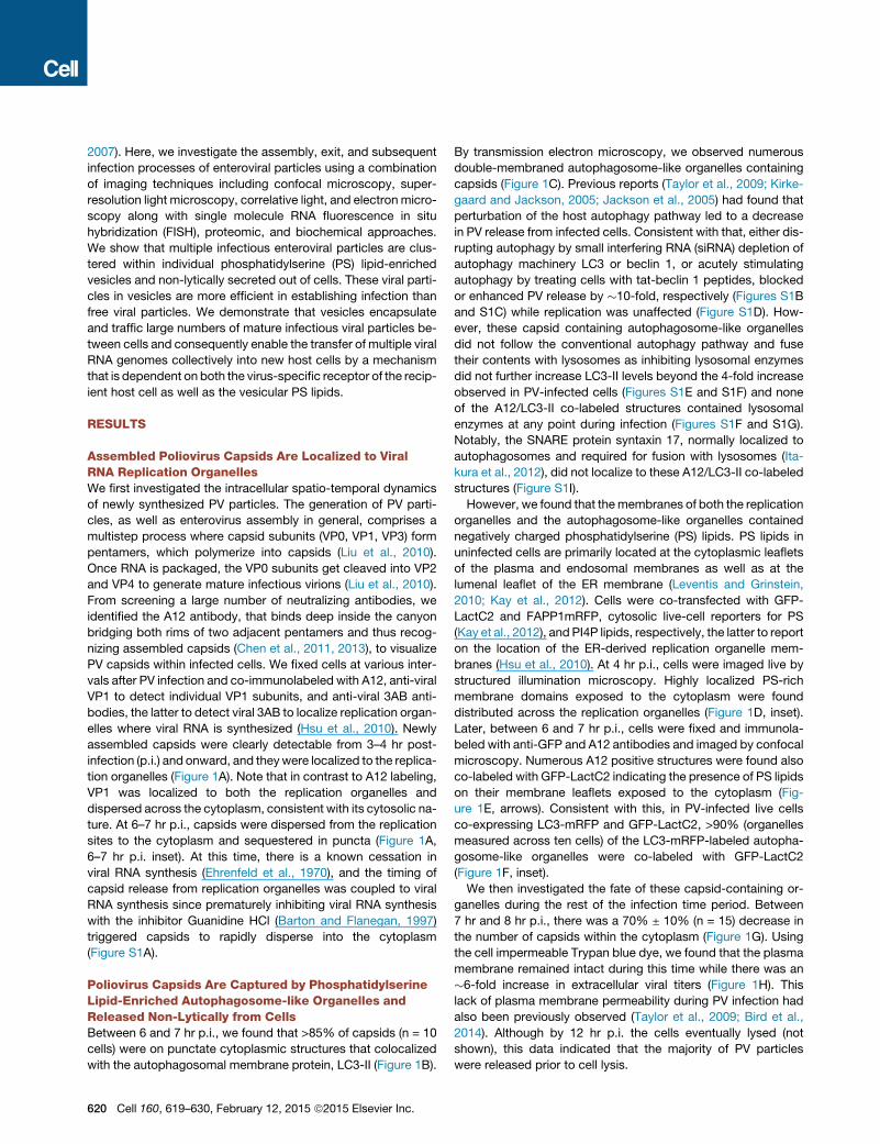
Figure 3. Mature PV, CVB3, and Rhinovirus Particles Are Released in Phosphatidylserine Lipid Vesicles
(A) Cells infected with PV, CVB3, or rhinovirus were incubated with Alexa 568-Annexin V and imaged by confocal/DICmicroscopy at 7 hr p.i. Scale bar represents
5 mm for PV and rhinovirus, 10 mm for CVB3.
(B) Mock and apoptotic HeLa cells labeled with Alexa 568-Annexin V and imaged by confocal/DIC microscopy. Scale bar represents 10 mm.
(C) Dynamics of Alexa 568-Annexin V-labeled PS vesicle release from plasma membrane projections of PV-infected cells at 7 hr p.i.
(D) Quantification of PS vesicles released from enterovirus-infected cells.
(E) VP2/VP0 ratio of whole cell lysate and PS vesicles in PV-infected cells. Annexin V-isolated PS vesicles from PV-infected cells at 7 hr p.i. were analyzed by
SDS-PAGE/western with anti-PV VP2 antibody.
(F) PS vesicles from human rhinovirus-infected cells contain mature rhinoviral particles. Isolated PS vesicles were processed for SDS-PAGE/western analysis
with anti-HRV2/VP2 (neutralizing) antibodies.
See also Figure S2 and Movie S1.
not shown). The collected extracellular vesicles fromPV-infected
cells or free PV particles were then incubated with a confluent
layer of HeLa cells (Figure 4A). After 4 hr of infection with either
vesicles or free viral particles, numerous PV-replicating (based
on immunofluorescence labeling of VP1) infected cells were
found in both cell populations (Figure 4A).
To determine if the PV particles within vesicles still required the
PV receptor CD155 on the host cell for infection, cells were incu-
bated with CD155 neutralizing antibodies prior to exposure to
vesicles and viral 3AB replication protein levels were measured
after 4 hr of infection. In the presence of neutralizing antibodies,
vesicle infectivity was inhibited by >95% indicating that PV ves-
icles were not just simply ‘‘fusing’’ with cells but that the viral
particles within the vesicles still required binding to PV receptor
for transfer of viral RNA into the host cell cytoplasm (Figure 4B).
We next determined if the infection was dependent on PS
lipids of the vesicles. Vesicles isolated by differential centrifuga-
tion were incubated with different amounts of Annexin V protein,
which binds and masks the PS head-groups on the lipids
(Swairjo et al., 1995). After removing any unbound Annexin V,
the vesicles were added to HeLa cells and replication measured
after 4 hr. Strikingly, masking of the vesicle-associated PS lipids
by Annexin V, inhibited PV infection of the host cells in a dose-
dependent manner (Figures 4C and 4D). These data indicated
Cell 160, 619–630, February 12, 2015 ª2015 Elsevier Inc. 623

A
B C D
E F
Figure 4. Infection by PV Particles in Vesi-
cles Is More Efficient Than Free Viral
Particles and Is Dependent on Both the
Poliovirus Receptor and PS Lipids
(A) Free or vesicle-associated PV particles were
incubated with new recipient cells and replication
was detected at 4 hr p.i. by immunolabeling with
anti-VP1 antibodies. Scale bar represents 500 mm.
(B) CD155/PVR neutralizing antibodies block
infection PV particles in vesicles.
(C) Blocking PS lipids on vesicles containing PV
particles block infection. Vesicles collected by
differential centrifugation were incubated with
different amounts of Annexin V protein prior to in-
cubation with recipient HeLa cells. Replicationwas
measured at 4 hr p.i., by SDS-PAGE/western
analysis with anti-3AB antibody.
(D) Quantification of western results in (C).
(E) HeLa cells were incubated with equal titers of
free and vesicle-associated PV particles. Infection
efficiency was determined by quantifying viral 3AB
protein levels at peak replication time (4 hr p.i.).
Data represented as mean ± SD.
(F) Primary human macrophages were incubated
with equal titers of free and vesicle-associated PV
particles. Infection efficiency was determined
by quantifying viral 3AB protein levels at peak
replication time (8 hr p.i.). Data represented as
mean ± SD.
that PS lipids are cofactors for PV infection and that mature in-
fectious PV particles are predominantly in the PS vesicle fraction
of vesicles collected by differential centrifugation.
Infection by PV Particles in Vesicles Is More EfficientThan Free PV ParticlesWe next measured and compared the level of infection of host
cellswhen infectedwith equivalent numbers of PVparticles either
in vesicles or as free. As a measure of infection efficiency, we
quantified and plotted the levels of viral 3AB replication proteins
at peak replication times (Figures 4E and 4F). Due to viral RNA
synthesis feeding back on viral RNA translation (and vice versa),
3AB levels reflect viral RNA levels (Hsu et al., 2010).We found that
at 4 hr p.i. of HeLa cells, viral 3AB levels were �40% greater in
cells incubatedwith PV particles in vesicles than free PV particles
(Figure 4E). This difference was even more striking when primary
human macrophages, cells that are specialized to recognize and
take upPS lipid-containing cells and vesicles (Fadok et al., 1992),
were used as the recipient host. Here vesicle-enclosed PV parti-
cles were almost 2-fold greater in infection efficiency compared
to free PV particles (Figure 4F).
Unilamellar PS Vesicles Released from PV-InfectedCells Contain Multiple Viral ParticlesTo quantify the clustering of viral particles within vesicles, we
collected free and vesicle-associated PV particles and immuno-
624 Cell 160, 619–630, February 12, 2015 ª2015 Elsevier Inc.
labeled them with A12 antibodies. We
then imaged the cover-glass-immobilized
PV particles by total-internal reflection
fluorescence (TIRF) and by DIC micro-
scopy (Figure 5A). Viruses were deposited on coverslip in two
formats: vesicle-free fraction (‘‘Free’’), or vesicle-embedded
fraction (‘‘Vesicular’’). Imaging revealed cluster of viruses within
vesicles, while free viruses yielded a more diffuse distribution
(Figure 5A). We quantified the difference in distribution of
measured fluorescence by computing the radial autocorrelation
functions gðrÞ for the intensity map Ið u!Þ :
gðrÞ=D#k v!k= r
Ið u!ÞIð u!+ v!Þd u!E
u!:
The observed exponential decay of these autocorrelation
functions enabled us to quantify the length scales that charac-
terize the imaged aggregates (Figure 5B). We found that
vesicle-embedded viruses yielded a characteristic clustering
scale of 2.0 ± 0.1 mm (n = 4 images), while free virus yielded
a five-time smaller characteristic scale of 0.4 ± 0.1 mm
(n = 2 images). However, these estimates are too close to
the spatial resolution of TIRF imaging and thus only qualita-
tive. To circumvent this diffraction limit, we then applied the
direct stochastic optical reconstruction microscopy (dSTORM)
methodology to achieve super-resolution of these viral parti-
cles (Figure 5C). dSTORM relies on sequential imaging and
fitting to achieve a spatial resolution of individual dyes within
30 nm (Bates et al., 2007; Baddeley et al., 2009; Dempsey
et al., 2011).

In order to assess the degree of clustering within these viral
spreads we used the Ripley’s K statistical test. By definition,
KðrÞ= A
n2
Xisj
dðdij
�rÞ; with dðxÞ=
�1 for x%10 for x > 0
;
with dij being the distance between the ith and jth points, A is the
image area. By definition,KðrÞ=pr2 is around 1 for a homogenous
distribution of points, and larger than 1 for clustered spatial dis-
tribution (Veatch et al., 2012; Termini et al., 2014). Based on the
electronmicroscopy images (Figures 2A, 2B, and S2A), we antic-
ipated clusters of viruses within vesicles of 200—400 nm diam-
eter. Hence, we calculated K(r = 200 nm) from the dSTORM
data to test whether vesicles contained clusters of viruses: we
found that K(r = 200 nm) = 2.4 ± 0.9 for free viruses and K(r =
200 nm) = 13.7 ± 6.7 for viruses within vesicles (Figure 5D).
Hence, super-resolution microscopy does confirm that these
viral vesicles do pack large numbers of viruses.
Consistent with these findings, transmission electron micro-
graphs of PS vesicles (after isolation by Annexin V-coupled mi-
crobeads) revealed multiple clustered viral particles surrounded
by a single membrane bilayer (Figure 5E). Per 200 nm cross-sec-
tion, a single vesicle contained on average 19 ± 3 PV particles
(n = 6 vesicles).
The presence of a single bilayer, as opposed to multiple bila-
yers, also indicated that the double-membraned autophago-
some-like organelles had fused with the plasma membrane,
rather than budded, in order to release PS vesicles. Thus, the
PS lipids on the extracellular membrane leaflet of the vesicle
are in a compartment that is topologically equivalent to the
lumenal membrane leaflet of the double-membraned autopha-
gosome-like organelle. The ER is a major membrane source for
autophagosomes (Hamasaki et al., 2013) and has lumenal mem-
brane leaflet enriched in PS lipids (Kay et al., 2012). Given that
the isolated PS vesicles also contain ER-resident proteins (Fig-
ure S2B), it is highly likely that the autophagosome-like organ-
elles, and thereby the released PS vesicles, originated from ER
and/or ER-derived replication organelle membranes.
Vesicles Allow Multiple Viral RNA Molecules to BeCollectively Transferred into CellsGiven our dSTORM and TEM finding, we conjectured that multi-
ple PV particles within a single vesicle might allow multiple viral
genomes to be simultaneously transferred into a single cell. To
test this hypothesis, we performed single molecule fluorescence
in situ hybridization (single molecule RNA FISH) (Raj et al., 2008).
Multiple oligonucleotide fluorescent probes hybridizing to an
RNA molecule allow sufficient sensitivity to detect single RNA
molecules within cells (Shaffer et al., 2013; Lubeck and Cai
2012).
Forty-eight fluorescently labeled nucleic acid probes, each 22
nucleotides in length, complementary to the PV genome, were
synthesized. Fixed numbers of HeLa cells were incubated with
different titers of either free PV particles or vesicle-associated
PV particles for 1.5 hr and processed for FISH labeling as
described previously (Shaffer et al., 2013) (Figure 5F). Note that
we could exclude the possibility of any viral RNA synthesis occur-
ring during this incubation time because cells with and without
GuanidineHCL,an inhibitor of viralRNAsynthesis, showedsimilar
amounts of viral RNA molecules per cell (Figures 5G and S3).
For cells incubated with free PV particles, discrete fluorescent
puncta, spatially segregated from one another in the cytoplasm
were detected in individual cells (Figure 5F, free virus). In
contrast for cells incubated with equivalent titers of PV particles
in vesicles, there were many more fluorescent puncta, spatially
juxtaposed (Figure 5F, virus in vesicle). The lowest multiplicity
of infection with free viral particles was observed at 1.5 3 107
pfu/ml where there was on average only one fluorescent puncta
per cell (Figures 5F and 5H, free virus). Hence, the size of these
puncta was approximated as a single viral RNA molecule and
used in subsequent quantification and analysis of FISH data.
At each titer, for either free virus or vesicle-associated virus, 55
cells were randomly chosen and viral RNA molecules counted
in each cell. The number of viral RNA molecules per cell were
subsequently plotted in Figure 5H.
From our quantification, we found that at 123 107 pfu/ml and
63 107 pfu/ml, there were�40% and�75%, respectively, more
viral RNAmolecules per cell when PV particles were presented in
vesicles than as free virus (Figure 5H). Indeed, for cells incubated
with free PV particles, we found that as the viral titer decreased
4-fold (from 12 3 107 pfu/ml to 3 3 107 pfu/ml), there was a
�90% decrease in the number of PV RNA molecules per cell
whereas for cells incubated with PV particles in vesicles, this
decrease was significantly less, only �40% (Figure 5H). Further-
more, at any given titer there were significantly more cells
with >15 PV RNA molecules within them when they had been in-
fected with PV in vesicles as opposed to free PV (Figure 5I).
These data are consistent with our dSTORM findings (Figures
5A–5D) and transmission electron micrographs of isolated PS
vesicles (Figure 5E) and indicate that vesicles contain multiple
PV particles, which enable multiple viral RNA genomes to be
transferred en bloc into a cell. Note that there was a large varia-
tion in the number of viral RNA genomes per cell when cells were
infected with PV particles in vesicles as opposed to free PV par-
ticles (Figures 5H and 5I). Since de novo viral RNA synthesis can
be ruled out, this variation is likely due to differences in the num-
ber of viral particles packaged per vesicle as well as contamina-
tion from free particles due to possible vesicle lysis.
DISCUSSION
Here, we have shown that multiple mature infectious enteroviral
particles are released in single unilamellar PS lipid vesicles,
which in turn enables multiple viral genomes to be collectively
transferred to an individual cell in a new round of infection. These
PS vesicles containing multiple viral genomes appear to be
significantly more efficient in infection and may facilitate genetic
cooperation among viral genomes.
We first detected assembled PV capsids at replication organ-
elles where viral RNA was synthesized (Figures 1A and 6). This
close juxtaposition of capsids with viral RNA synthesis sites
would serve to immediately encapsidate the viral RNA and
thereby facilitate efficient genome packaging as well as protec-
tion of viral genomes from host defenses. These viral particles
then translocated into the cytoplasm, which was not only tempo-
rally coincident with a cessation of viral RNA synthesis but also
Cell 160, 619–630, February 12, 2015 ª2015 Elsevier Inc. 625

A B E
C D
F
G H I
(legend on next page)
626 Cell 160, 619–630, February 12, 2015 ª2015 Elsevier Inc.

Viral RNA Replication organelles
PS rich membranes
PS vesicles
PI4P/sterol
Enterovirus
Viral RNA
PS receptor
PS
Enterovirus receptors
(e.g. CD155)
Viral RNA genomes
Figure 6. Model
Assembledmature enteroviruses are released from
the replication organelles into the cytoplasm.
Clusters of multiple viral particles are selectively
captured by double-membraned organelles that
originate from the ER and ER-derived replication
organelles. These double-membraned organelles,
which resemble autophagosomes, contain PS
lipids on both the lumenal and cytoplasmic leaflets
of their membranes. They fuse with the plasma
membrane and release a unilamellar PS-lipid-en-
riched vesicle, containing multiple viral particles,
into the extracellular medium. This vesicle then
facilitates infection in a PS-lipid and viral receptor-
dependent mechanism resulting in the collective
transfer to a new recipient host cell of multiple viral
RNA genomes. This mode of viral transmission
enhances infection efficiency and potentially allows
for genetic complementation among quasispecies.
could be prematurely triggered by inhibiting RNA synthesis with
GuHCL (Figure S1A). Enteroviral 2C proteins may modulate the
close coupling between RNA synthesis kinetics and capsid
release from replication organelles as 2C proteins are not only
localized to the replication organelles and required for viral
RNA synthesis but also physically interact with capsids (Liu
et al., 2010).
Once the capsids dissociated from the replication organelles,
they were sequestered within double-membraned LC3-II-
positive autophagosome-like organelles. How the viral cargo is
recognized and captured within these organelles, the capsid-
associated determinants, and whether LC3-II proteins play a
role in these processes as they do in canonical autophagy path-
ways (Rogov et al., 2014) is currently unknown. Interestingly, un-
like canonical autophagosomes that fuse with lysosomes and
degrade their cytoplasmic cargo, these capsid-containing or-
Figure 5. PS Vesicles Contain Clustered PV Particles, which Enable Multiple Viral RNA Genom
Host Cell
(A) TIRF and DIC images of free and vesicle-associated PV particles labeled with Atto488-labeled A12 antib
(B) Difference in distribution of fluorescence in (A) by computation of the radial autocorrelation function g(r).
(C) dSTORM imaging free and vesicle-associated viral particles labeled with Atto488-A12 antibody.
(D) Calculation of Ripley’s K function to assess the degree of clustering of vesicle-associated PV partic
mean ± SEM.
(E) PS vesicles isolated from PV-infected cells using Annexin V microbeads were imaged by transmission
dense viral particles in each vesicle and the unilamellar surrounding membrane. Asterisk shows Annexin V
Scale bars represent 100 nm.
(F) Collected intact vesicles or free viral particles from PV-infected cells were incubated with a confluent laye
were monitored by single molecule RNA FISH and imaged by dual confocal/DIC microscopy at 1.5 hr p.i. S
either free viral particles or vesicle-associated viral particles. Images presented were acquired with the sam
(G) Collected vesicles were incubated with cells with/without GuHCL for 1.5 hr and viral RNA molecules were
Figure S3 for quantification). Scale bar represents 2 mm.
(H) Quantification of the number of viral RNAmolecules per cell in cells infected with either free (n = 55 cells) or
2.10�3; ****p < 10�4.
(I) Percent of cells with 15 or greater PV RNA puncta was quantified for cells infected with either free or vesicl
Data represented as mean ± SEM.
Cell 160, 619–630,
ganelles fused with the plasma mem-
brane to non-lytically release >80% of
the PV particles into the extracellular envi-
ronment within unilamellar vesicles of size range 200–400 nm.
Notably, the SNARE protein syntaxin 17 was not localized to
the autophagosome-like organelles to regulate their fusion with
lysosomes, but instead was found sequestered away at the
replication organelles (Figure S1I). Whether specific enteroviral
proteins activelymodulate its subcellular localization or it is an in-
direct consequence of the affinity of the syntaxin 17 hydrophobic
hairpin tail for the PI4P/cholesterol rich replication organelle
membranes remains to be investigated. This type of non-con-
ventional secretion of autophagosomal membranes from the
cell has never been reported and identifying the machinery regu-
lating this process, including determining which cytoskeletal
components and SNARE proteins are utilized for movement
out to the periphery and fusion with the plasma membrane,
may provide novel therapeutic targets to block enterovirus
release from cells.
es to Be Collectively Transferred into a New
ody.
les relative to free particles. Data represented as
electron microscopy. Note the numerous electron-
microbead attached to the exterior of one vesicle.
r of new host cells at different viral titers. Viral RNAs
hown are images of single HeLa cells infected with
e microscopy settings. Scale bar represents 2 mm.
monitored by single molecule RNA FISH (see also
vesicle-associated PV particles (n = 55 cells). ***p <
e-associated PV particles for each viral titer shown.
February 12, 2015 ª2015 Elsevier Inc. 627

PS vesicles non-lytically released from cells were selectively
enriched in mature enteroviral particles (Figure 3). While we
cannot exclude the possibility that non-PS vesicles may also
contribute to the non-lytic release of enteroviral particles, the
observed significant inhibition of subsequent infection when
PS is blocked (Figures 4C and 4D) suggest that PS vesicles
constitute a large fraction of the non-lytic conduit for enteroviral
release from cells. Live-cell time-lapse imaging in the presence
of fluorescently labeled Annexin V protein revealed that the outer
membrane leaflet of the released vesicle and hence topologically
equivalent to the lumenal membrane leaflet of the double
membraned organelle pre-fusion, contained PS lipids (Figures
3A–3D; Movie S1). The ER and/or the ER-derived replication or-
ganelles both have PS lipids on their lumenal and cytoplasmic
leaflets (Leventis and Grinstein, 2010; Lev, 2012) (Figure 1D),
and moreover, the PS can flip between these two leaflets (Clark,
2011). Thus it is likely that the host source for the autophago-
some-like organelles is the ER and/or ER-derived replication or-
ganelles. Supporting this conclusion the released PS vesicles
indeed contained ER markers including the integral ER mem-
brane protein calnexin (Figure S2B).
Super-resolution imaging and transmission electron micro-
scopy revealed extracellular PS vesicles to be containing multi-
ple viral particles, at least 20 per 200 nm cross-section (Figures
5A–5E). Supporting these data, in single molecule RNA FISH ex-
periments, we found that infection by PV particles in vesicles al-
lowed the collective transfer of multiple viral genomes into a sin-
gle host cell (Figures 5F–5I). One important implication of these
findings is that it ties the fate of individual viral genomes from
previous rounds of replication to each other and thereby may
provide selective advantages in terms of replication kinetics
and genetic diversity relative to free viral particle genomes
(i.e., not in vesicle). Indeed, infection efficiency was significantly
higher when cells were infected with PV particles in vesicles as
opposed to an equivalent number of free virus particles (Figures
4E and 4F). Enteroviruses as well as all other positive-stranded
RNA viruses, are enormously diverse in genomic variety due to
the inherent error rates and lack of proof reading in their RNA
polymerases that can generate large numbers of viral quasispe-
cies after even a single round of infection (Borderıa et al., 2011).
Vesicular transfer of multiple particles among cells would in-
crease the chances of genetic complementation among viral
quasispecies, potentially benefiting the replication efficiency of
otherwise attenuated or weak genomes and enabling them to
maintain a presence in the genetic pool. Indeed our findings
may provide a cellular mechanism to explain the results of Vi-
gnuzzi et al. (2006) where cooperative interactions between
neurotropic and non-neurotropic PV quasispecies were re-
ported. Second, when multiple viral genomes are transferred
to a single cell, the likelihood of one or more genomes surviving
the hostile host environment to override host defenses and repli-
cate may be higher. Third, for positive-stranded RNA viruses,
rather than a single genome having to switch between RNA
translation and RNA synthesis activities (until sufficient levels
of viral RNA have been synthesized to partition those functions
among genomes), multiple genomes could right from the start
of infection partition RNA translation and RNA synthesis func-
tions among themselves and enhance overall replication kinetics
628 Cell 160, 619–630, February 12, 2015 ª2015 Elsevier Inc.
and viral protein levels. Finally the PS lipids on the vesicles
themselves could enhance infection efficiency by attracting PS
scavenging cells, such as macrophages and dendritic cells, to
take up the viral genomes and provide a host environment for
replication. In particular, we found that PV particles within vesi-
cles could replicate significantly more efficiently within primary
macrophages than free PV particles (Figure 4F).
What is the mechanism whereby viral particles within vesicles
infect host cells?We found infection to be dependent not only on
the virus-specific receptor expressed by the host but also on the
PS lipids associated with the vesicle having access to the host
cell (Figures 4C and 4D). One potential mechanism to explain
these findings is that PS lipids on vesicles engage PS receptors
on the recipient host cell prior to the viral particles engaging their
own receptors. The binding to PS receptors can trigger phago-
cytic uptake of vesicles (Hoffmann et al., 2001) followed by lysis
or permeabilization of the vesicle within the endosomal compart-
ment to then enable viral particles to engage their specific recep-
tors (Figure 6). Equally possible, binding to PS receptors may
lead to permeabilization of the vesicle on the cell surface, result-
ing in the release of a concentrated bolus of viral particles in the
immediate vicinity of the cell. The latter mechanism may also
provide access to neutralizing antibodies which bind capsids
and block infection. Reliance on PS lipids and PS receptors for
infection has been documented for a number of other viruses
including vaccinia, Dengue, Ebola, hepatitis A virus, and HIV
(Sui et al., 2006; Mercer and Helenius, 2009; Feng et al., 2014;
Morizano and Chen, 2014). The specific use of PS lipids by en-
teroviruses as well as other viruses to traffic between cells may
have significant in vivo implications for viral pathogenesis and
tissue tropism. In particular, the infection of primary macro-
phages, the major PS sensing cells in the body, may provide
enteroviruses with the ability to target a key cell subset of the im-
mune system while suppressing its inflammatory responses, as
PS lipids have been well documented to inhibit inflammatory
cytokine production by macrophages (Hochreiter-Hufford and
Ravichandran, 2013). PS-lipid vesicles may also help enterovi-
ruses exploit the natural motility of macrophages and help
spread them distant sites including perhaps the CNS (Ousman
and Kubes, 2012).
In summary, we report here a novel mode of viral transmission
among cells where multiple viral particles are clustered and
collectively released within PS-lipid-enriched vesicles. This pro-
vides greater infection efficiency and potentially an opportunity
for cooperation and complementation among viral quasispecies.
This mode of transmission links the fate of multiple viral particles
to one another and may have implications for maintaining viral
genetic diversity within viral evolution.
EXPERIMENTAL PROCEDURES
All detailed protocols and information regarding plasmids, antibodies, cell cul-
ture, virus infection, and propagation are provided in the Extended Experi-
mental Procedures.
Immunofluorescence
Cells were plated on glass coverslips and fixed with 4% PFA for 15 min at RT.
Cells were permeabilized with either 0.2% Saponin or 0.1% Triton X-100 and
sequentially incubated with primary and fluorophore-tagged secondary

antibodies. Coverslips were mounted in Fluoromount-G (Southern Biotech)
and imaged.
Confocal Microscopy
All confocal images were obtained with an LSM780 laser scanning confocal
microscope system (Carl Zeiss) and images were analyzed with either Zen
(Carl Zeiss) or Image J (NIH) software.
dSTORM
Free PV particles and PV particles in vesicles were plated on gridded glass bot-
tom dishes and fixed with 4% PFA for 15 min at room temperature. Subse-
quently they were permeabilized with 0.2% saponin and incubated with
Atto488-conjugated A12 antibodies. An oxygen-scavenging PBS solution
(10 mM NaCl, 0.5 mg/ml glucose oxidase, 40 g/ml catalase, 2% glucose,
and 10 mM MEA) was used for imaging. dSTORM images were obtained on
a Zeiss ELYRA PS.1 system (Carl Zeiss). Images were acquired with a Plan-
Apochromat 1003/1.46 oil immersion objective and an Andor iXon 885
EMCCD camera. A total of 20,000 images were acquired per sample with an
exposure time of 33 ms. Raw images were reconstructed and analyzed with
ZEN software (Carl Zeiss) and MATLAB (MathWorks) using methodology
from Veatch et al. (2012) and Termini et al. (2014).
Structured Illumination Microscopy
Super-resolution 3D-structured illumination microscopy (SIM) imaging was
performed on a Zeiss ELYRA PS.1 system (Carl Zeiss). Images were acquired
with a Plan-Apochromat 633/1.40 oil immersion objective and an Andor iXon
885 EMCCD camera. Fifteen images per plane (five phases, three rotations)
and 0.125mm z section of 3mmheight were required for generating super res-
olution images. Raw images were reconstructed and processed to demon-
strate structure with greater resolution by the ZEN 2011 software (Carl Zeiss).
Single Molecule RNA FISH
We performed single molecule RNA FISH according to Shaffer et al. (2013).
Cells were incubated with either free or vesicle-associated PV particles for
1.5 hr. Cells were subsequently fixed in pre-chilled methanol (�20�C) for10 min. The methanol was removed and the cells were hybridized with 10 ml
of hybridization buffer containing 4 mM PV probe, 10% formamide, 23 SSC,
and 10% dextran sulfate for 10 min at 37�C. Next, the samples were washed
three times with pre-warmed wash buffer (10% formamide and 23 SSC) at
37�C for 1 min and imaged with Zeiss LSM780 confocal microscope.
Annexin V Labeling
Cells for live imaging were grown on coverslip-bottomed Lab-Tek chambers
(Thermo Fisher) and infected with PV for 7 hr. Cells were then replaced in im-
agingmedia (DMEMPhenol Red free supplemented with 10%FBS and 50mM
HEPES [pH 7.3]). Alexa 568-Annexin V was added on the cells and imaging
was performed on a Zeiss LSM780 Confocal Laser Scanning microscope
(Carl Zeiss) equipped with 458 nm, 488 nm, 514 nm, 565 nm, and 633 nm laser
lines and detecting system for fluorescence and DIC imaging. The microscope
was additionally equipped with a heating stage and incubator with tempera-
ture, humidity, and CO2 control for live-cell imaging.
SUPPLEMENTAL INFORMATION
Supplemental Information includes Extended Experimental Procedures, three
figures, and onemovie and can be foundwith this article online at http://dx.doi.
org/10.1016/j.cell.2015.01.032.
ACKNOWLEDGMENTS
The authors would like to thank Yasmine Belkaid, Konstantin Chumakov, Ana
Maria Cuervo, James Hogle, Gerald Feldman, Jennifer Jones, Jennifer Lippin-
cott-Schwartz, Sanford Simon, Radek Dobrowolski, Wilma Friedman, Ellie
Ehrenfeld and members of the Altan-Bonnet lab for fruitful discussions. The
authors would like to especially thankWen-Chin Tseng for help in constructing
the graphical cartoons. The authors also thank Frank Macaluso and Geoffrey
Perumal of the Albert Einstein College of Medicine Analytical Imaging facility
for technical support. A.C., P.T., and C.P. were supported by grant
AI091985-01A1; G.A.B. was supported by grant AI083408; H.M., P.J., and
E.W. were supported by grant AI15122; and Y.C., W.D., M.H., C.B., P.C.,
E.S., and N.A.B. were supported by intramural NIH funds.
Received: October 2, 2014
Revised: December 13, 2014
Accepted: January 12, 2015
Published: February 12, 2015
REFERENCES
Baddeley, D., Jayasinghe, I.D., Cremer, C., Cannell, M.B., and Soeller, C.
(2009). Light-induced dark states of organic fluochromes enable 30 nm reso-
lution imaging in standard media. Biophys. J. 96, L22–L24.
Barton, D.J., and Flanegan, J.B. (1997). Synchronous replication of poliovirus
RNA: initiation of negative-strand RNA synthesis requires the guanidine-
inhibited activity of protein 2C. J. Virol. 71, 8482–8489.
Bates, M., Huang, B., Dempsey, G.T., and Zhuang, X. (2007). Multicolor super-
resolution imaging with photo-switchable fluorescent probes. Science 317,
1749–1753.
Bird, S.W., Maynard, N.D., Covert, M.W., and Kirkegaard, K. (2014). Nonlytic
viral spread enhanced by autophagy components. Proc. Natl. Acad. Sci.
USA 111, 13081–13086.
Borderıa, A.V., Stapleford,K.A., andVignuzzi,M. (2011).RNAviruspopulationdi-
versity: implications for inter-species transmission. Curr. Opin. Virol. 1, 643–648.
Brandenburg, B., and Zhuang, X. (2007). Virus trafficking - learning from single-
virus tracking. Nat. Rev. Microbiol. 5, 197–208.
Brandenburg, B., Lee, L.Y., Lakadamyali, M., Rust, M.J., Zhuang, X., and
Hogle, J.M. (2007). Imaging poliovirus entry in live cells. PLoS Biol. 5, e183.
Chen, Z., Chumakov, K., Dragunsky, E., Kouiavskaia, D., Makiya, M., Neverov,
A., Rezapkin, G., Sebrell, A., and Purcell, R. (2011). Chimpanzee-human
monoclonal antibodies for treatment of chronic poliovirus excretors and emer-
gency postexposure prophylaxis. J. Virol. 85, 4354–4362.
Chen, Z., Fischer, E.R., Kouiavskaia, D., Hansen, B.T., Ludtke, S.J., Bidzhieva,
B., Makiya, M., Agulto, L., Purcell, R.H., and Chumakov, K. (2013). Cross-
neutralizing human anti-poliovirus antibodies bind the recognition site for
cellular receptor. Proc. Natl. Acad. Sci. USA 110, 20242–20247.
Clark, M.R. (2011). Flippin’ lipids. Nat. Immunol. 12, 373–375.
Dempsey, G.T., Vaughan, J.C., Chen, K.H., Bates, M., and Zhuang, X. (2011).
Evaluation of fluorophores for optimal performance in localization-based
super-resolution imaging. Nat. Methods 8, 1027–1036.
den Boon, J.A., and Ahlquist, P. (2010). Organelle-like membrane compart-
mentalization of positive-strand RNA virus replication factories. Annu. Rev.
Microbiol. 64, 241–256.
Doceul, V., Hollinshead, M., van der Linden, L., and Smith, G.L. (2010). Repul-
sion of superinfecting virions: a mechanism for rapid virus spread. Science
327, 873–876.
Ehrenfeld, E., Maizel, J.V., and Summers, D.F. (1970). Soluble RNA polymer-
ase complex from poliovirus-infected HeLa cells. Virology 40, 840–846.
Fadok, V.A., Voelker, D.R., Campbell, P.A., Cohen, J.J., Bratton, D.L., and
Henson, P.M. (1992). Exposure of phosphatidylserine on the surface of
apoptotic lymphocytes triggers specific recognition and removal by macro-
phages. J. Immunol. 148, 2207–2216.
Feng, Z., Hensley, L., McKnight, K.L., Hu, F., Madden, V., Ping, L., Jeong, S.H.,
Walker, C., Lanford, R.E., and Lemon, S.M. (2013). A pathogenic picornavirus
acquires an envelope by hijacking cellular membranes. Nature 496, 367–371.
Feng, Z., Li, Y., McKnight, K.L., Hensley, L., Lanford, R.E., Walker, C.M., and
Lemon, S.M. (2014). Human pDCs preferentially sense enveloped hepatitis A
virions. J. Clin. Invest. 125, 169–176.
Flint, S.J., Enquist, L.W., Racaniello, V.R., and Skalka, A.M. (2009). Principles
of Virology, Third Edition (ASM Press).
Cell 160, 619–630, February 12, 2015 ª2015 Elsevier Inc. 629

Hamasaki, M., Furuta, N., Matsuda, A., Nezu, A., Yamamoto, A., Fujita, N.,
Oomori, H., Noda, T., Haraguchi, T., Hiraoka, Y., et al. (2013). Autophago-
somes form at ER-mitochondria contact sites. Nature 495, 389–393.
Hochreiter-Hufford, A., and Ravichandran, K.S. (2013). Clearing the dead:
apoptotic cell sensing, recognition, engulfment, and digestion. Cold Spring
Harb. Perspect. Biol. 5, a008748.
Hoffmann, P.R., deCathelineau, A.M., Ogden, C.A., Leverrier, Y., Bratton, D.L.,
Daleke, D.L., Ridley, A.J., Fadok, V.A., and Henson, P.M. (2001). Phosphatidyl-
serine (PS) induces PS receptor-mediated macropinocytosis and promotes
clearance of apoptotic cells. J. Cell Biol. 155, 649–659.
Hsu, N.Y., Ilnytska, O., Belov, G., Santiana, M., Chen, Y.H., Takvorian, P.M.,
Pau, C., van der Schaar, H., Kaushik-Basu, N., Balla, T., et al. (2010). Viral reor-
ganization of the secretory pathway generates distinct organelles for RNA
replication. Cell 141, 799–811.
Ilnytska, O., Santiana, M., Hsu, N.Y., Du, W.L., Chen, Y.H., Viktorova, E.G.,
Belov, G., Brinker, A., Storch, J., Moore, C., et al. (2013). Enteroviruses
harness the cellular endocytic machinery to remodel the host cell cholesterol
landscape for effective viral replication. Cell Host Microbe 14, 281–293.
Itakura, E., Kishi-Itakura, C., and Mizushima, N. (2012). The hairpin-type tail-
anchored SNARE syntaxin 17 targets to autophagosomes for fusion with
endosomes/lysosomes. Cell 151, 1256–1269.
Jackson, W.T., Giddings, T.H., Jr., Taylor, M.P., Mulinyawe, S., Rabinovitch,
M., Kopito, R.R., and Kirkegaard, K. (2005). Subversion of cellular autophago-
somal machinery by RNA viruses. PLoS Biol. 3, e156.
Kay, J.G., Koivusalo, M., Ma, X., Wohland, T., and Grinstein, S. (2012). Phos-
phatidylserine dynamics in cellular membranes. Mol. Biol. Cell 23, 2198–2212.
Kirkegaard, K., and Jackson, W.T. (2005). Topology of double-membraned
vesicles and the opportunity for non-lytic release of cytoplasm. Autophagy
1, 182–184.
Koopman, G., Reutelingsperger, C.P., Kuijten, G.A., Keehnen, R.M., Pals, S.T.,
and van Oers, M.H. (1994). Annexin V for flow cytometric detection of
phosphatidylserine expression on B cells undergoing apoptosis. Blood 84,
1415–1420.
Lev, S. (2012). Non-vesicular lipid transfer from the ER. Cold Spring Harb.
Perspect. Biol. 4, 1–17.
Leventis, P.A., and Grinstein, S. (2010). The distribution and function of phos-
phatidylserine in cellular membranes. Annu. Rev. Biophys. 39, 407–427.
Liu, Y., Wang, C., Mueller, S., Paul, A.V., Wimmer, E., and Jiang, P. (2010).
Direct interaction between two viral proteins, the nonstructural protein 2C
and the capsid protein VP3, is required for enterovirus morphogenesis.
PLoS Pathog. 6, e1001066.
Lubeck, E., and Cai, L. (2012). Single-cell systems biology by super-resolution
imaging and combinatorial labeling. Nat. Methods 9, 743–748.
Mercer, J., and Helenius, A. (2009). Virus entry by macropinocytosis. Nat. Cell
Biol. 11, 510–520.
Morizano, K., and Chen, I.S. (2014). Role of phosphatidlyserine receptors in
enveloped virus infection. J. Virol. 88, 4275–4290.
630 Cell 160, 619–630, February 12, 2015 ª2015 Elsevier Inc.
Nchoutmboube, J.A., Viktorova, E.G., Scott, A.J., Ford, L.A., Pei, Z., Watkins,
P.A., Ernst, R.K., and Belov, G.A. (2013). Increased long chain acyl-Coa
synthetase activity and fatty acid import is linked to membrane synthesis
for development of picornavirus replication organelles. PLoS Pathog. 9,
e1003401.
Ousman, S.S., and Kubes, P. (2012). Immune surveillance in the central
nervous system. Nat. Neurosci. 15, 1096–1101.
Owens, R.J., Limn, C., and Roy, P. (2004). Role of an arbovirus nonstructural
protein in cellular pathogenesis and virus release. J. Virol. 78, 6649–6656.
Raj, A., van den Bogaard, P., Rifkin, S.A., van Oudenaarden, A., and Tyagi, S.
(2008). Imaging individual mRNA molecules using multiple singly labeled
probes. Nat. Methods 5, 877–879.
Robinson, S.M., Tsueng, G., Sin, J., Mangale, V., Rahawi, S., McIntyre, L.L.,
Williams, W., Kha, N., Cruz, C., Hancock, B.M., et al. (2014). Coxsackievirus
B exits the host cell in shed microvesicles displaying autophagosomal
markers. PLoS Pathog. 10, e1004045.
Rogov, V., Dotsch, V., Johansen, T., and Kirkin, V. (2014). Interactions between
autophagy receptors and ubiquitin-like proteins form the molecular basis for
selective autophagy. Mol. Cell 53, 167–178.
Shaffer, S.M., Wu, M.T., Levesque, M.J., and Raj, A. (2013). Turbo FISH: a
method for rapid single molecule RNA FISH. PLoS ONE 8, e75120.
Strauss, M., Levy, H.C., Bostina, M., Filman, D.J., and Hogle, J.M. (2013). RNA
transfer from poliovirus 135S particles across membranes is mediated by long
umbilical connectors. J. Virol. 87, 3903–3914.
Strazynski, M., Kramer, J., and Becker, B. (2002). Thermal inactivation of polio-
virus type 1 in water, milk and yoghurt. Int. J. Food Microbiol. 74, 73–78.
Sui, L., Zhang, W., Chen, Y., Zheng, Y., Wan, T., Zhang, W., Yang, Y., Fang, G.,
Mao, J., and Cao, X. (2006). Human membrane protein Tim-3 facilitates hep-
atitis A virus entry into target cells. Int. J. Mol. Med. 17, 1093–1099.
Swairjo, M.A., Concha, N.O., Kaetzel, M.A., Dedman, J.R., and Seaton, B.A.
(1995). Ca(2+)-bridging mechanism and phospholipid head group recognition
in the membrane-binding protein annexin V. Nat. Struct. Biol. 2, 968–974.
Takahashi, M., Yamada, K., Hoshino, Y., Takahashi, H., Ichiyama, K., Tanaka,
T., and Okamoto, H. (2008). Monoclonal antibodies raised against the ORF3
protein of hepatitis E virus (HEV) can capture HEV particles in culture superna-
tant and serum but not those in feces. Arch. Virol. 153, 1703–1713.
Taylor, M.P., Burgon, T.B., Kirkegaard, K., and Jackson, W.T. (2009). Role of
microtubules in extracellular release of poliovirus. J. Virol. 83, 6599–6609.
Termini, C.M., Cotter, M.L., Marjon, K.D., Buranda, T., Lidke, K.A., and Gillette,
J.M. (2014). The membrane scaffold CD82 regulates cell adhesion by altering
a4 integrin stability and molecular density. Mol. Biol. Cell 25, 1560–1573.
Veatch, S.L., Machta, B.B., Shelby, S.A., Chiang, E.N., Holowka, D.A., and
Baird, B.A. (2012). Correlation functions quantify super-resolution images
and estimate apparent clustering due to over-counting. PLoS ONE 7, e31457.
Vignuzzi, M., Stone, J.K., Arnold, J.J., Cameron, C.E., and Andino, R. (2006).
Quasispecies diversity determines pathogenesis through cooperative interac-
tions in a viral population. Nature 439, 344–348.